EP1021989A2 - Cannula having integral suture tourniquet - Google Patents
Cannula having integral suture tourniquet Download PDFInfo
- Publication number
- EP1021989A2 EP1021989A2 EP00100274A EP00100274A EP1021989A2 EP 1021989 A2 EP1021989 A2 EP 1021989A2 EP 00100274 A EP00100274 A EP 00100274A EP 00100274 A EP00100274 A EP 00100274A EP 1021989 A2 EP1021989 A2 EP 1021989A2
- Authority
- EP
- European Patent Office
- Prior art keywords
- cannula
- tourniquet
- lumen
- surgical device
- suture
- Prior art date
- Legal status (The legal status is an assumption and is not a legal conclusion. Google has not performed a legal analysis and makes no representation as to the accuracy of the status listed.)
- Withdrawn
Links
Images

Classifications
-
- A—HUMAN NECESSITIES
- A61—MEDICAL OR VETERINARY SCIENCE; HYGIENE
- A61B—DIAGNOSIS; SURGERY; IDENTIFICATION
- A61B17/00—Surgical instruments, devices or methods
- A61B17/04—Surgical instruments, devices or methods for suturing wounds; Holders or packages for needles or suture materials
-
- A—HUMAN NECESSITIES
- A61—MEDICAL OR VETERINARY SCIENCE; HYGIENE
- A61B—DIAGNOSIS; SURGERY; IDENTIFICATION
- A61B17/00—Surgical instruments, devices or methods
- A61B17/12—Surgical instruments, devices or methods for ligaturing or otherwise compressing tubular parts of the body, e.g. blood vessels or umbilical cord
- A61B17/12009—Implements for ligaturing other than by clamps or clips, e.g. using a loop with a slip knot
- A61B17/12013—Implements for ligaturing other than by clamps or clips, e.g. using a loop with a slip knot for use in minimally invasive surgery, e.g. endoscopic surgery
Definitions
- This invention relates to fluid cannulas and, more particularly, to fluid conveying cannulas having a suture tourniquet integrally formed or mounted thereto.
- the tourniquet according to the invention is particularly adapted for use in cardiovascular surgical applications.
- Fluid conveying cannulas have a wide variety of applications during invasive surgical operations.
- coronary operations such as heart bypass surgery utilizes a venous cannula for removing oxygen depleted blood from the vascular system and arterial cannulas for returning oxygenated blood to the vascular system.
- Both venous and arterial cannulae are usually inserted through incisions cut into the right or left atrium, the vena cava oratium and aorta, respectively.
- the incision in which the cannula is inserted is surrounded by purse-string sutures which, when drawn tight around the inserted cannula will effectively prevent fluid from escaping the vessel.
- Tourniquets are commonly used to contain the free ends of the suture thread.
- the tourniquets are free lengths of tubing through which the free ends of the suture thread are pulled through after creation of the purse-string suture.
- the free end of the suture thread is held in a fixed position with respect to the tourniquet by one of several conventional means such as wrapping the suture thread around a button, frictionally retaining the free end of the suture thread by inserting a cone-shaped plug into the end of the tourniquet or, alternatively, clamping the tourniquet closed around the suture thread by use of a conventional surgical clamp.
- the tourniquet tubes are typically manually tied to the cannula to hold the sutures, tourniquet and cannula in place.
- a recent development in cardiovascular surgical procedures is to conduct minimally invasive surgery which utilizes smaller apertures formed in the chest wall rather than cutting the sternum and spreading open the entire rib cage, as usually done in conventional open heart surgery. While there are many benefits being realized by minimally invasive surgical procedures, these procedures place many challenges on the surgeon as a result of the far smaller access aperture available to the surgeon as he or she performs the surgery. In view of the relative size of the access aperture, simplification of any and all equipment utilized in the surgical procedure can simplify these challenging procedures.
- the cannula having integral suture tourniquet takes less space in the surgical field than a conventional cannula and thus it is particularly adapted for minimally invasive surgery.
- the cannula according to the invention overcomes the problems of the prior art by fully integrating the suture tourniquet with the cannula thereby simplifying the equipment needed to perform surgical procedures.
- the invention comprises an insertable surgical device configured to assist a user in creating a suture thread tourniquet around the surgical device, the surgical device comprising:
- a pair of tourniquets are integrally molded into the cannula and the distal ends of the tourniquet are spaced a short distance approximately from the distal end of the cannula.
- An alternative to integrally molding the tourniquets to the cannula would be to securely mount the tourniquets by other mechanical means such as adhesive, sonic welding, and the like.
- the suture retainer comprises a button provided adjacent the proximal end of the tourniquet.
- the suture thread is wrapped around the button and is frictionally retained thereon.
- An alternative suture retainer is a plug configured to be telescopically received in the proximal end of the tourniquet lumen.
- the plug When the plug is telescopically received in the proximal end of the tourniquet lumen, it is dimensioned to frictionally retain suture thread in the lumen.
- the invention provides a cannula having a proximal end, a distal end and a main lumen extending between the proximal and distal ends, the cannula further comprising:
- the preferred method of inserting the surgical device into a vessel comprises providing a cannula substantially as described above and creating a purse-string suture in the vessel using conventional suture thread.
- the free end of the suture thread is pulled through the lumen of the tourniquet.
- an incision is made in the vessel wall within the bounds of the purse-string suture.
- the distal end of the cannula is inserted into the incision and then the suture thread is quickly drawn tight by pulling the free end of the suture thread through the tourniquet lumen. Finally, the free end of the suture thread is fixed with respect to the cannula.
- the surgical device can be removed by freeing the end of the suture thread from the suture retainer thereby releasing the purse-string suture. Next, the distal end of the cannula is withdrawn from the incision of the vessel.
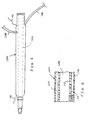
- the cannula 12 comprises a cannula body 14 having a primary lumen 16 extending between the proximal 18 and distal ends 20.
- the cannula 12 as seen in FIG. 1 is an arterial cannula and therefore the proximal end 18 is adapted to be fluidly connected to the extracorporeal bypass circuit.
- the distal end 20 of the cannula has a discharge tip 22 securely mounted therein.
- the discharge tip 22 is preferably formed of plastic, but may also be formed of metal, and is curved to deflect the fluid flow therefrom substantially normal to the longitudinal axis of the lumen 16.
- FIG. 1 What distinguishes the structure of the arterial cannula seen in FIG. 1 from prior art is the integration of suture tourniquets into the cannula body.
- two tourniquets 26, 28 are mounted on opposite sides of the cannula body 14.
- the tourniquets 26, 28 are securely mounted to the exterior surface of the cannula body 14 by any medically approved adhesive 24.
- a suitable adhesive includes ultraviolet curing adhesive.
- Alternatives to adhesive include solvent bonding and sonic welding.
- the tourniquets are substantially identical and therefore only one tourniquet will be described in detail.
- the tourniquet 26 comprises a tourniquet lumen 30 extending from the proximal end 32 to the distal 34 of the tourniquet 26.
- a suture button 36 Securely fastened to the proximal end 32 of the lumen 30 is a suture button 36 which extends outwardly from a button mounting 38.
- the button mounting 38 has a lumen (not shown) extended therethrough which is substantially contiguous with the tourniquet lumen 30.
- a shaft 40 extends between the suture button 36 and the button mounting 38.
- the proximal end 32 of the tourniquet lumen 30 and the button mounting 38 are spaced a short distance from the proximal end 18 of the cannula.
- the distal end 34 of the tourniquet lumen 30 is spaced a short distance from the distal end 20 of the cannula to accommodate receipt of the suture thread 42.
- the cannula having an integrated tourniquet according to the invention is ideally suited for use during minimally invasive cardiac surgical procedures.
- This surgical technique differs from the traditional open heart surgery primarily in the size of the openings that are formed in the chest wall.
- the entirety of the chest cavity is opened by cutting through the sternum and spreading open the sternum and ribs to expose the entirety of the pericardium.
- much smaller apertures are cut into the chest wall to access only portions of the pericardium. For example, an aperture could be cut between two adjacent ribs or alternatively, a small portion of one rib could be removed to expose a portion of the pericardium.
- the cannula according to the invention is an improvement over the conventional cannula and tourniquet structures in that all of these various elements are integrated thereby simplifying the insertion and removal of surgical equipment through the chest wall resulting in more efficient use of the limited area of the chest wall aperture.
- the surgeon creates a purse-string suture in the vessel, for example aorta, using conventional suture thread 42.
- the free ends of the suture thread 42 are captured in the hooked, distal end of a conventional wire thread retriever 48.
- the distal end 50 of the retriever 48 is telescopically received in the tourniquet lumen 30 so that the distal end 50 extends outwardly from the distal end of the lumen.
- the free end of the suture thread 42 is threaded through the hook 52 of the retriever and then the retriever 48 is withdrawn through the tourniquet lumen 30 pulling the suture thread 42 through the tourniquet lumen 30.
- the purse-string suture has been created, the free ends of the suture thread 42 have been pulled through the tourniquet lumens 30 and the discharge tip 22 of the cannula 12 is positioned immediately adjacent the wall of the vessel at the point of the purse string suture.
- the surgeon makes an incision in the vessel wall in the center of the purse string suture and thereafter immediately inserts the discharge tip 22 of the cannula 12 into the vessel through the incision.
- the surgeon quickly draws tight the suture thread 42 by grasping the free end thereof and pulls tight the purse string suture around the distal end 20 of the cannula 12.
- the thread is wrapped around the shaft 40 of the suture button 36 a sufficient number of revolutions until the suture thread 42 is frictionally retained thereon. As long as the suture thread 42 remains around the suture button 36, a substantially fluid tight seal should remain in place between the vessel wall and the cannula 12.
- the free end of the suture thread 42 is merely unwound from the suture button 36 thereby releasing the purse string suture.
- the cannula in its entirety, can be withdrawn from the vessel leaving behind the suture thread 42.
- the suture is used to close the aortotomy.
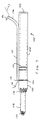
- FIGS. 3 and 4 A second embodiment of the cannula according to the invention is seen in FIGS. 3 and 4. Similar to the first embodiment, this embodiment is ideally suited for use as an arterial cannula.
- the cannula 70 and tourniquets are integrally molded with one another.
- the cannula 70 is somewhat oval in cross section and has a main lumen 72 extending between the proximal 74 and the distal end 76 thereof.
- a discharge tip 22 is securely mounted in the distal end 76. While the preferred embodiment utilizes a separately formed discharge tip, it is to be understood that the discharge tip could be integrally formed with the cannula body.
- a pair of tourniquet lumens 78, 80 Located adjacent the main lumen 72 are a pair of tourniquet lumens 78, 80.
- the tourniquet lumens 78, 80 have diameters sufficient to receive both the suture thread 42 and wire thread retriever 48 as discussed above.
- the distal end 82 of the tourniquet lumen is positioned closely adjacent to the distal end 76 of the cannula 70.
- the proximal end 84 of the tourniquet lumen 78 is spaced distally from the proximal end 74 of the cannula 72.
- the second embodiment shows a second means for retaining the free end of the suture thread 42 in a fixed position.
- a plug 86 is telescopically received in the proximal end of the tourniquet lumen 78.
- the plug 86 has a conical shaped body 88 with a tab 90 provided at the terminal end thereof.
- the body 88 is dimensioned so that it tapers from a diameter which is less than the diameter of the tourniquet lumen to a diameter which exceeds that of the tourniquet lumen.
- the body 88 of the plug can be telescopically received in the tourniquet lumen after the free end of the suture thread 42 has been pulled therethrough.
- the friction between the thread 42, the plug 86 and the interior surface of the tourniquet lumen 78 will be sufficient to prevent movement of the suture thread 42 relative to the cannula 70.
- FIG. 5 shows a third embodiment of a cannula having an integrated tourniquet according to the invention.
- this cannula is ideally suited for use as an arterial cannula.
- this embodiment of the cannula 100 has a main lumen 102 and a pair of other lumens integrally formed therewith, namely a tourniquet lumen 104 and a pressure sensing lumen 106.
- the cross section of the cannula body is substantially identical to the cross section seen in FIG. 4, the only difference being that in this third embodiment one of the tourniquet lumens in FIG. 4 is utilized as a pressure sensing lumen 106.
- the proximal and distal ends of the tourniquet lumen 104 are dimensioned to accommodate and receive the suture thread 42 therethrough.
- the tourniquet lumen 104 is clamped shut by the opposed tips of a conventional surgical clamp 108. After the free end of the suture thread 42 has been pulled through the tourniquet lumen 104 and the thread has been pulled taut to seal the purse string suture, then the opposed tines of the conventional surgical clamp grasp the cannula 100 on opposite sides of the tourniquet lumen 104 and are squeezed against one another to frictionally retain the suture thread therein.
- a pressure sensing lumen 106 One of the tourniquet lumens in this embodiment has been replaced by a pressure sensing lumen 106.
- the pressure sensing lumen 106 extends outwardly from the cannula a short distance to be mounted to conventional pressure sensing equipment.
- the purpose of the pressure sensing lumen and sensing equipment is to evaluate the pressure of the fluid passing through the main lumen 102 of the cannula.
- a small fluid aperture 110 is cut in the web 112 separating the pressure sensing lumen 106 from the main lumen 102.
- the pressure sensing lumen 106 is fluidly sealed distally of the fluid aperture 110 by inserting an acceptable adhesive or other suitable compound in the distal end of the lumen.
- the pressure sensing equipment which is mounted to the proximal end of the pressure sensing lumen 106 can effectively monitor the pressure of the fluid flowing through the main lumen 102. All of this is accomplished with the ease and simplicity of the integral tourniquet as described above.
- the preferred form of the cannula incorporating the pressure sensing lumen utilizes a single tourniquet lumen, however, it is to be understood that more than one tourniquet lumen could be incorporated into this particular embodiment of the cannula 100 depending upon the requirements for the particular application of the cannula 100.
- the additional lumens could merely be spaced around the periphery of the main lumen 102.
- FIG. 7 depicts still yet another embodiment of the cannula having an integrated tourniquet according to invention.
- the cannula is ideally suited for use as a venous cannula for use in a coronary operation.
- the cannula 120 comprises a cannula body 122 having a proximal end 124 and a distal end 126.
- the proximal end 124 is adapted to be fluidly connected to the extracorporeal bypass circuit and the distal end 126 is adapted to be received in the vena cava.
- a two stage tip 128 is provided at the distal end 126 of the cannula body.
- the particular structure and configuration of the two stage tip is described more completely in U.S. Patent No. 4,129,129.
- the two stage tip 128 has a first set of fluid apertures 130 provided at the distal end of the tip and a second set of apertures 132 which are spaced proximally from the first set 130.
- the preferred form of the venous cannula utilizes only a single tourniquet 134. Similar to the first embodiment, this tourniquet 134 is secured to the exterior surface of the cannula body 122 by a suitable adhesive as described above. The distal end 136 of the tourniquet is spaced a prescribed distance from the second set of fluid apertures 132. In this embodiment, the proximal end 138 of the tourniquet 134 is not adhered to the cannula body. In other words, this portion of the tourniquet 134 can be manipulated and deflected away from the cannula body.
- the distal end of the tourniquet 136 can be manipulated a short distance independently from the cannula body 122.
- the free end of the suture thread 42 is frictionally retained in the tourniquet by a plug 86 as described above.
- insertion markings 140, 142 are provided on the exterior surface of the cannula body 122 and are plainly visible by the surgeon. These markings help to guide the surgeon in properly positioning the cannula in the vena cava.
- each of the several embodiments incorporate a variety of different features. It is to be understood that the features and structures depicted in the various embodiments can be interchanged depending upon the particular application.
- the venous cannula depicted in FIG. 7 can be integrally molded similar to the structure seen in the second embodiment of FIGS. 3 and 4.
- a person skilled in the art can easily appreciate the various combinations of the structural features embodied in the various embodiments.
Landscapes
- Health & Medical Sciences (AREA)
- Life Sciences & Earth Sciences (AREA)
- Surgery (AREA)
- Molecular Biology (AREA)
- Engineering & Computer Science (AREA)
- Biomedical Technology (AREA)
- Heart & Thoracic Surgery (AREA)
- Medical Informatics (AREA)
- Nuclear Medicine, Radiotherapy & Molecular Imaging (AREA)
- Animal Behavior & Ethology (AREA)
- General Health & Medical Sciences (AREA)
- Public Health (AREA)
- Veterinary Medicine (AREA)
- Reproductive Health (AREA)
- Vascular Medicine (AREA)
- Surgical Instruments (AREA)
Abstract
Description
Claims (16)
- An insertable surgical device configured to assist a user in creating a suture thread tourniquet around the surgical device, the surgical device comprising:a cannula (12) having a proximal end (18), a distal end (20) and a main lumen (16) extending at least partially through the length of the cannula;at least one tourniquet (26, 28) having a proximal end (32), a distal end (34) and a tourniquet lumen (30) extending therethrough, the tourniquet being fixedly mounted to and extending at least partially along the length of the cannula (12), the tourniquet lumen (30) being adapted to receive suture thread (42) and;at least one suture retainer (36) configured to selectively hold a portion of the suture thread to the surgical device.
- The insertable surgical device of claim 1, wherein the surgical device has two tourniquets (26, 28) fixedly mounted to the cannula.
- The insertable surgical device of claim 2, wherein the two tourniquets (26, 28) are diametrically opposed with respect to the cannula.
- The insertable surgical device of claim 2 or 3, wherein the two tourniquets (26, 28) are fixedly mounted to the cannula by an adhesive.
- The insertable surgical device of claim 2 or 3, wherein the lumens (30) of the two tourniquets (26, 28) are integrally molded with the cannula.
- The insertable surgical device of any preceding claim wherein the at least one suture retainer comprises:a button mounting (38), having a button mounting lumen extending therethrough, the button mounting lumen being substantially coaxially aligned with the at least one tourniquet lumen (30); anda suture button (36) extending from the button mounting.
- The insertable surgical device of any of claims 1 to 5, wherein the at least one suture retainer comprises:a plug (86) configured to be telescopically received in the proximal end of the tourniquet lumen (78) and adapted to frictionally retain the suture thread (42).
- The insertable surgical device of claim 7 wherein the plug (86) has a tab (90) extending from the plug configured to facilitate insertion and removal from the proximal end of the tourniquet lumen.
- The insertable surgical device of any preceding claim further comprising:a pressure sensing lumen (106), the pressure sensing lumen being mounted to the cannula, and fluidly interconnected with the main lumen (102) of the cannula.
- The insertable surgical device of any preceding claim, wherein the cannula comprises:a first fluid aperture (110) provided adjacent the proximal end of the cannula; anda second fluid aperture spaced distally from the first fluid aperture.
- The insertable surgical device of any preceding claim wherein the cannula has at least one insertion marking (140, 142) provided on the cannula to assist proper insertion of the surgical device.
- A cannula having a proximal end (18), a distal end (20) and a main lumen (16) extending between the proximal and distal ends, the cannula further comprising:at least one tourniquet (26, 28) integrally molded into the cannula so that it extends along the length of at least a portion of the cannula, the at least one tourniquet having a proximal end (32), a distal end (34) and a lumen (30) extending through at least a portion thereof, the lumen of the at least one tourniquet being adapted to telescopically receive suture thread (42) therethrough and the distal end of the tourniquet lumen being spaced proximally from the distal end of the cannula.
- A cannula according to claim 12 wherein two tourniquet lumens are integrally molded into the cannula and are diametrically opposed to one another.
- A cannula according to claim 12 or 13 and further comprising a pressure sensing lumen (106) provided in the cannula and being fluidly connected to the main lumen of the cannula.
- An improved cannula according to claim 12, 13 or 14 and further comprising a suture thread retainer comprising:a suture button (36) provided adjacent the proximal end of the tourniquet lumen, the button being adapted to have suture thread wrapped thereabout and to frictionally retain the suture thread thereon.
- An improved cannula according to claim 12, 13 or 14 and further comprising a suture thread retainer comprising:a plug (86) configured to be telescopically received in the proximal end of the tourniquet lumen and adapted to frictionally retain suture thread in the lumen.
Applications Claiming Priority (2)
| Application Number | Priority Date | Filing Date | Title |
|---|---|---|---|
| US232593 | 1988-08-15 | ||
| US09/232,593 US6110185A (en) | 1999-01-19 | 1999-01-19 | Cannula having integral suture tourniquet |
Publications (2)
| Publication Number | Publication Date |
|---|---|
| EP1021989A2 true EP1021989A2 (en) | 2000-07-26 |
| EP1021989A3 EP1021989A3 (en) | 2001-06-27 |
Family
ID=22873772
Family Applications (1)
| Application Number | Title | Priority Date | Filing Date |
|---|---|---|---|
| EP00100274A Withdrawn EP1021989A3 (en) | 1999-01-19 | 2000-01-19 | Cannula having integral suture tourniquet |
Country Status (2)
| Country | Link |
|---|---|
| US (1) | US6110185A (en) |
| EP (1) | EP1021989A3 (en) |
Cited By (1)
| Publication number | Priority date | Publication date | Assignee | Title |
|---|---|---|---|---|
| WO2004064652A1 (en) * | 2003-01-17 | 2004-08-05 | Hammersmith Hospital's Nhs Trust | A cannula |
Families Citing this family (40)
| Publication number | Priority date | Publication date | Assignee | Title |
|---|---|---|---|---|
| EP1196093B1 (en) | 1999-07-02 | 2006-06-14 | Quickpass, Inc. | Suturing device |
| US8323228B2 (en) | 2007-04-12 | 2012-12-04 | Rex Medical L.P. | Dialysis catheter |
| US7160245B2 (en) * | 2003-11-17 | 2007-01-09 | Virginijus Burneikis | Method and device for umbilicus protection during abdominal surgery |
| EP2260776A1 (en) * | 2003-11-21 | 2010-12-15 | Vnus Medical Technologies, Inc. | Apparatus for treating the carotid artery |
| US20050209607A1 (en) | 2004-03-22 | 2005-09-22 | John Lipchitz | Medical cannula assembly |
| US9713465B1 (en) * | 2004-04-19 | 2017-07-25 | Granit Medical Innovation Llc | Surgical closure device and associated method |
| EP2510883B1 (en) | 2005-06-20 | 2018-04-11 | Nobles Medical Technologies, Inc. | Apparatus for applying a knot to a suture |
| US9283314B2 (en) * | 2005-09-21 | 2016-03-15 | Abiomed, Inc. | Cannula systems |
| EP2134266B1 (en) | 2007-03-29 | 2024-12-25 | Nobles Medical Technologies, Inc. | Suturing devices for closing a patent foramen ovale |
| US20100191261A1 (en) * | 2007-05-22 | 2010-07-29 | Sally Carter | Suture management port |
| US8858490B2 (en) | 2007-07-18 | 2014-10-14 | Silk Road Medical, Inc. | Systems and methods for treating a carotid artery |
| JP5290290B2 (en) | 2007-07-18 | 2013-09-18 | シルク・ロード・メディカル・インコーポレイテッド | Method and system for establishing regurgitation of carotid blood flow |
| EP3789069B1 (en) | 2008-02-05 | 2024-04-03 | Silk Road Medical, Inc. | Systems for establishing retrograde carotid arterial blood flow |
| JP5848125B2 (en) | 2008-05-09 | 2016-01-27 | ノーブルズ メディカル テクノロジーズ、インコーポレイテッド | Suture device and method for suturing anatomic valves |
| WO2010019719A2 (en) | 2008-08-13 | 2010-02-18 | Silk Road Medical, Inc. | Suture delivery device |
| US8574245B2 (en) | 2008-08-13 | 2013-11-05 | Silk Road Medical, Inc. | Suture delivery device |
| WO2010071810A1 (en) * | 2008-12-19 | 2010-06-24 | Andy Christopher Kiser | Methods and devices for endoscopic access to the heart |
| US20100198018A1 (en) * | 2009-01-30 | 2010-08-05 | Tyco Healthcare Group Lp | Suture management system for surgical portal apparatus including internal tubes |
| US20100198019A1 (en) * | 2009-01-30 | 2010-08-05 | Tyco Healthcare Group Lp | Suture management apparatus for surgical portal apparatus including interlocking cap |
| US20100210912A1 (en) * | 2009-02-17 | 2010-08-19 | Tyco Healthcare Group Lp | Access port with suture management system including flapper with inserts |
| US20100211084A1 (en) * | 2009-02-18 | 2010-08-19 | Tyco Healthcare Group Lp | Suture management system for surgical portal apparatus including numbered clips |
| US20100249810A1 (en) * | 2009-03-24 | 2010-09-30 | Tyco Healthcare Group Lp | Suture management system for surgical portal apparatus including slotted ring |
| US20110022064A1 (en) * | 2009-07-27 | 2011-01-27 | Tyco Healthcare Group Lp | Access apparatus including hinged suture traps |
| US8591450B2 (en) | 2010-06-07 | 2013-11-26 | Rex Medical L.P. | Dialysis catheter |
| US8517971B2 (en) * | 2010-07-09 | 2013-08-27 | E. Benson Hood Laboratories | Cannula implant device for use in salivary gland ducts |
| US9662195B2 (en) | 2010-07-09 | 2017-05-30 | E. Benson Hood Laboratories | Implant device for use in salivary gland duct |
| CN106388888B (en) | 2011-04-15 | 2019-12-27 | 心脏缝合有限公司 | Suturing device and method for suturing an anatomical valve |
| US9706988B2 (en) | 2012-05-11 | 2017-07-18 | Heartstitch, Inc. | Suturing devices and methods for suturing an anatomic structure |
| US10159479B2 (en) | 2012-08-09 | 2018-12-25 | Silk Road Medical, Inc. | Suture delivery device |
| EA032962B1 (en) | 2013-07-02 | 2019-08-30 | Мед-Венче Инвестментс, Ллс | Suturing device for suturing an anatomic structure |
| US9918712B2 (en) | 2013-08-02 | 2018-03-20 | Covidien Lp | Devices, systems, and methods for providing surgical access and facilitating closure of surgical access openings |
| US10512458B2 (en) | 2013-12-06 | 2019-12-24 | Med-Venture Investments, Llc | Suturing methods and apparatuses |
| WO2015175537A1 (en) | 2014-05-16 | 2015-11-19 | Silk Road Medical, Inc. | Vessel access and closure assist system and method |
| US10178993B2 (en) | 2014-07-11 | 2019-01-15 | Cardio Medical Solutions, Inc. | Device and method for assisting end-to-side anastomosis |
| EP3442437B1 (en) | 2016-04-11 | 2020-11-11 | Nobles Medical Technologies II, Inc. | Tissue suturing device with suture spool |
| WO2018236822A1 (en) | 2017-06-19 | 2018-12-27 | Heartstitch, Inc. | SUTURE DEVICES AND METHODS OF SUTURING OPENING IN HEART APEX |
| WO2018236766A1 (en) | 2017-06-19 | 2018-12-27 | Heartstitch, Inc. | Suturing systems and methods for suturing body tissue |
| WO2019035095A1 (en) | 2017-08-18 | 2019-02-21 | Nobles Medical Technologies Ii, Inc. | Apparatus for applying a knot to a suture |
| US11129609B2 (en) | 2018-04-24 | 2021-09-28 | Covidien Lp | Devices, systems, and methods for providing surgical access and facilitating closure of surgical access openings |
| CN108992109A (en) * | 2018-08-29 | 2018-12-14 | 中国人民解放军第四军医大学 | Cardiovascular surgical procedure purse wire fixator |
Family Cites Families (6)
| Publication number | Priority date | Publication date | Assignee | Title |
|---|---|---|---|---|
| US4129129A (en) * | 1977-03-18 | 1978-12-12 | Sarns, Inc. | Venous return catheter and a method of using the same |
| US4535770A (en) * | 1983-11-02 | 1985-08-20 | Lemole Gerald M | Cardiovascular tourniquet |
| US4796626A (en) * | 1987-04-15 | 1989-01-10 | Dlp Inc. | Tourniquet tube |
| FR2628964B1 (en) * | 1988-03-22 | 1990-11-30 | Roux Martial | DEVICES FOR PLACING AND FIXING CANNULAS ON THE HEART OR LARGE VESSELS IN ORDER TO CONDUCT EXTRACORPOREAL CIRCULATION FOR CARDIAC SURGERY |
| US5487730A (en) * | 1992-12-30 | 1996-01-30 | Medtronic, Inc. | Balloon catheter with balloon surface retention means |
| US5846253A (en) * | 1995-07-14 | 1998-12-08 | C. R. Bard, Inc. | Wound closure apparatus and method |
-
1999
- 1999-01-19 US US09/232,593 patent/US6110185A/en not_active Expired - Lifetime
-
2000
- 2000-01-19 EP EP00100274A patent/EP1021989A3/en not_active Withdrawn
Cited By (1)
| Publication number | Priority date | Publication date | Assignee | Title |
|---|---|---|---|---|
| WO2004064652A1 (en) * | 2003-01-17 | 2004-08-05 | Hammersmith Hospital's Nhs Trust | A cannula |
Also Published As
| Publication number | Publication date |
|---|---|
| US6110185A (en) | 2000-08-29 |
| EP1021989A3 (en) | 2001-06-27 |
Similar Documents
| Publication | Publication Date | Title |
|---|---|---|
| US6110185A (en) | Cannula having integral suture tourniquet | |
| JP7565972B2 (en) | Flow Cannula and Blood Pump | |
| US4309994A (en) | Cardiovascular cannula | |
| US6443957B1 (en) | Suture-free clamp and sealing port and methods of use | |
| US8388628B2 (en) | Expandable sheath for delivering instruments and agents into a body lumen and methods for use | |
| EP0791332B1 (en) | A perfusion device for maintaining blood flow in a vessel while isolating an anastomosis | |
| EP2067499B1 (en) | Expandable guide sheath and apparatus and methods using such sheaths | |
| US3877434A (en) | Vascular tourniquet | |
| US7591832B2 (en) | Expandable guide sheath and apparatus with distal protection and methods for use | |
| US7544203B2 (en) | Temporary seal and method for facilitating anastomosis | |
| JP5112288B2 (en) | Catheter with large diameter proximal end | |
| US4129129A (en) | Venous return catheter and a method of using the same | |
| EP0604803A2 (en) | Multi purpose perfusion cannula | |
| WO1993000868A1 (en) | Tubular surgical implant | |
| JPH08322851A (en) | Surgical attachment | |
| US6238404B1 (en) | Multipurpose medical device | |
| US6482184B1 (en) | Attachable catheter | |
| EP4312816B1 (en) | Apparatus for implanting an arteriovenous graft | |
| US7479126B1 (en) | Cannula with incision blade | |
| WO2002058566A1 (en) | Blood vessel anastomosing auxiliary tool | |
| JPS62231673A (en) | Method for attaching a medical tube body and cap members used to carry out this method |
Legal Events
| Date | Code | Title | Description |
|---|---|---|---|
| PUAI | Public reference made under article 153(3) epc to a published international application that has entered the european phase |
Free format text: ORIGINAL CODE: 0009012 |
|
| AK | Designated contracting states |
Kind code of ref document: A2 Designated state(s): DE FR IT NL |
|
| AX | Request for extension of the european patent |
Free format text: AL;LT;LV;MK;RO;SI |
|
| PUAL | Search report despatched |
Free format text: ORIGINAL CODE: 0009013 |
|
| RIC1 | Information provided on ipc code assigned before grant |
Free format text: 7A 61B 17/04 A, 7A 61B 17/12 B |
|
| AK | Designated contracting states |
Kind code of ref document: A3 Designated state(s): AT BE CH CY DE DK ES FI FR GB GR IE IT LI LU MC NL PT SE |
|
| AX | Request for extension of the european patent |
Free format text: AL;LT;LV;MK;RO;SI |
|
| 17P | Request for examination filed |
Effective date: 20011221 |
|
| AKX | Designation fees paid |
Free format text: DE FR IT NL |
|
| RAP1 | Party data changed (applicant data changed or rights of an application transferred) |
Owner name: MEDTRONIC, INC. |
|
| 17Q | First examination report despatched |
Effective date: 20050215 |
|
| STAA | Information on the status of an ep patent application or granted ep patent |
Free format text: STATUS: THE APPLICATION IS DEEMED TO BE WITHDRAWN |
|
| 18D | Application deemed to be withdrawn |
Effective date: 20060328 |