EP1019438B1 - Apoptosis-related compounds and their use - Google Patents
Apoptosis-related compounds and their use Download PDFInfo
- Publication number
- EP1019438B1 EP1019438B1 EP98945735A EP98945735A EP1019438B1 EP 1019438 B1 EP1019438 B1 EP 1019438B1 EP 98945735 A EP98945735 A EP 98945735A EP 98945735 A EP98945735 A EP 98945735A EP 1019438 B1 EP1019438 B1 EP 1019438B1
- Authority
- EP
- European Patent Office
- Prior art keywords
- antibody
- acid sequence
- apoptosis
- amino acid
- antigen
- Prior art date
- Legal status (The legal status is an assumption and is not a legal conclusion. Google has not performed a legal analysis and makes no representation as to the accuracy of the status listed.)
- Expired - Lifetime
Links
Classifications
-
- C—CHEMISTRY; METALLURGY
- C07—ORGANIC CHEMISTRY
- C07K—PEPTIDES
- C07K5/00—Peptides containing up to four amino acids in a fully defined sequence; Derivatives thereof
- C07K5/04—Peptides containing up to four amino acids in a fully defined sequence; Derivatives thereof containing only normal peptide links
- C07K5/10—Tetrapeptides
- C07K5/1021—Tetrapeptides with the first amino acid being acidic
-
- A—HUMAN NECESSITIES
- A61—MEDICAL OR VETERINARY SCIENCE; HYGIENE
- A61P—SPECIFIC THERAPEUTIC ACTIVITY OF CHEMICAL COMPOUNDS OR MEDICINAL PREPARATIONS
- A61P35/00—Antineoplastic agents
-
- C—CHEMISTRY; METALLURGY
- C07—ORGANIC CHEMISTRY
- C07K—PEPTIDES
- C07K14/00—Peptides having more than 20 amino acids; Gastrins; Somatostatins; Melanotropins; Derivatives thereof
- C07K14/435—Peptides having more than 20 amino acids; Gastrins; Somatostatins; Melanotropins; Derivatives thereof from animals; from humans
- C07K14/46—Peptides having more than 20 amino acids; Gastrins; Somatostatins; Melanotropins; Derivatives thereof from animals; from humans from vertebrates
- C07K14/47—Peptides having more than 20 amino acids; Gastrins; Somatostatins; Melanotropins; Derivatives thereof from animals; from humans from vertebrates from mammals
- C07K14/4701—Peptides having more than 20 amino acids; Gastrins; Somatostatins; Melanotropins; Derivatives thereof from animals; from humans from vertebrates from mammals not used
- C07K14/4741—Keratin; Cytokeratin
-
- C—CHEMISTRY; METALLURGY
- C07—ORGANIC CHEMISTRY
- C07K—PEPTIDES
- C07K14/00—Peptides having more than 20 amino acids; Gastrins; Somatostatins; Melanotropins; Derivatives thereof
- C07K14/435—Peptides having more than 20 amino acids; Gastrins; Somatostatins; Melanotropins; Derivatives thereof from animals; from humans
- C07K14/46—Peptides having more than 20 amino acids; Gastrins; Somatostatins; Melanotropins; Derivatives thereof from animals; from humans from vertebrates
- C07K14/47—Peptides having more than 20 amino acids; Gastrins; Somatostatins; Melanotropins; Derivatives thereof from animals; from humans from vertebrates from mammals
- C07K14/4701—Peptides having more than 20 amino acids; Gastrins; Somatostatins; Melanotropins; Derivatives thereof from animals; from humans from vertebrates from mammals not used
- C07K14/4747—Apoptosis related proteins
-
- C—CHEMISTRY; METALLURGY
- C07—ORGANIC CHEMISTRY
- C07K—PEPTIDES
- C07K16/00—Immunoglobulins [IG], e.g. monoclonal or polyclonal antibodies
- C07K16/18—Immunoglobulins [IG], e.g. monoclonal or polyclonal antibodies against material from animals or humans
-
- A—HUMAN NECESSITIES
- A61—MEDICAL OR VETERINARY SCIENCE; HYGIENE
- A61K—PREPARATIONS FOR MEDICAL, DENTAL OR TOILETRY PURPOSES
- A61K38/00—Medicinal preparations containing peptides
Definitions
- the present invention relates to antibodies or antigen-binding part of said antibodies recognizing apoptosis-related antigenic compounds, such as fragments of cytokeratin 18, which are useful in medicaments, diagnostics and methods for the detection, monitoring, measurement and regulation of the type of cell death called programmed cell death or apoptosis.
- the determination of the occurrence of cell apoptosis may further be used for the monitoring of the effect of therapeutic treatment.
- Apoptosis is seen in all sorts of higher eucaryocytes from plants and insects to vertebrates ( Kroemer G, Petit P, Zanzami N, Vayssiere J-L, Mignotte B: The biochemistry of programmed cell death. The FASEB Journal 9:1277-1287 (1995 )).
- Apoptosis is a general phenomenon of vital importance. Decreased apoptosis leads to malformation, cancer and autoimmune disease, and enhanced apoptosis results in degenerative diseases, acute diseases such as infection by toxin-producing microorganisms and in rejection of transplanted organs.
- Keratin 18 was cleaved in vitro by caspase-6, -3, and -7, and it was stated that the cleavage site common for the three caspases was the sequence VEVD/A, located in the conserved L1-2 linker region of K18.
- MAb monoclonal antibody
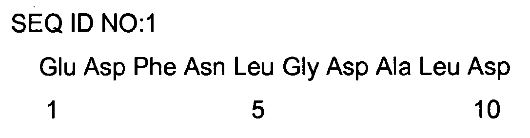
- the different aspects of the present invention are based on the optimal binding site of the mAb 30, i.e. the amino-acid sequence EDFNLGDALD,
- sequence DALD does not occur in such other intermediate filaments as CK 8, vimentin, desmin, different actins, neurofilaments, and lamins.
- sequences are found: LALD, MALD, MALD, VALD, MALD, AND LALD, respectively.
- the mAb 30 recognizes apoptotic cells in cardiac muscle, megakaryocytes, myeloblasts and neural tissues of fetus - which are not known to contain CK18 - it is now believed that detection of protein fragments having the C-terminal sequence ALD indicate early apoptosis.
- one aspect of the invention is directed to antibodies or the antigen-binding part of said antibodies recognizing an apoptosis-related antigenic compound which specifically binds to an antibody which in turn specifically binds to the amino-acid sequence i.e. an apoptosis-related antigenic compound comprising an antigenic site having the amino acid sequence SEQ ID NO: 1.
- the sequence should comprise at least the sequence Ala Leu Asp.
- the antigenic compound of the invention therefore comprises compounds which are based on the structure of the SEQ ID NO:1, i.e. the sequence as such or compounds having replacements of one or several amino acids in the above specified amino-acid sequence with other molecular parts, D- forms of the amino acids, non-natural amino acids and/or derivatives, as long as the three-dimensional structure of the SEQ ID NO:1 is mimicked.
- the common feature of the antigenic compounds of the invention is that they bind specifically to an antibody which in turn binds specifically to the aa sequence SEQ ID NO:1, i.e. antigenic compounds comprising an antigenic site having the amino acid sequence SEQ ID NO: 1.
- CK 18 molecules have the above-specified sequence hidden, and can therefore not react with antibodies specifically binding to said sequence, thereby making the recognition specific to apoptosis, i.e. those cases where the CK 18 molecule has been already cleaved after the C-terminal Asp of the sequence Asp Ala Leu Asp.
- apoptosis-related compound is a peptide or a protein fragment comprising the C-terminal amino acid sequence Ala Leu Asp.
- the peptide may be a homologue to, an extension of or a truncation of the SEQ ID NO: 1, i.e. may have a homologous sequence having some amino-acid substitutions, extensions, truncations and/or deletions which do not lead to the elimination of the capability of the peptide to bind to the same antibodies as the amino-acid sequence SEQ ID NO: 1.
- the antigenic compound is a peptide or a protein fragment comprising the C-terminal amino acid sequence Xaa Ala Leu Asp, wherein Xaa is selected from Asp, Leu, Met, and Val.
- An example of a peptide recognized by an antibody or an antigen-binding part of said antibody of the invention is an oligopeptide having the amino-acid sequence
- Another example is an oligopeptide having the amino-acid sequence (DG EDFNLGDALD SSNSMQTIQ)(i.e. SEQ ID NO: 1 extended by 2 amino acids at the N-terminal and 9 at the C-terminal)
- oligopeptide having the amino-acid sequence (RRLLED GEDFNLGDAL DSSNS)
- Still another example is an oligopeptide having the amino-acid sequence (G EDFNLGDALD)
- specific binding of an antigenic compound to an antibody requires an affinity constant of at least 10 7 liters/mole, preferably at least 10 9 liters/mole.
- the antigenic compound is may be coupled to a carrier and/or label.
- the carrier may be e.g. plastic surfaces, such as microplates, beads etc.; organic molecules such as biotin; proteins, such as bovine serum albumin; peptide linkers, polypeptides e.g. resulting in fusion proteins.
- the label may be selected from a variety of different types of labels, such as radioactive isotopes, enzymes, fluorescent markers, etc.
- the invention is directed to an antibody or an antigen-binding part of said antibody recognizing an antigenic compound disclosed above.
- the antibody of the invention may be a monoclonal antibody or monospecific polyclonal antibody recognizing or specifically binding to an antigenic compound according to the invention, i. e. recognizing or specifically binding to the amino-acid sequence SEQ ID NO: 1.
- the antigen-binding peptide recognizing an antigenic compound according to the invention may be the antigen-binding part of an antibody. It is possible that it may also be a protein or peptide which has an amino-acid sequence that at least in the part corresponds to the amino-acid sequence SEQ ID NO: 1, but has amino acids with the opposite charges. Such an antigen-binding peptide will function as an antagonist. In the development of antagonists the following guideline may be used:
- a number of antagonist candidates may be designed with the aid of the above guideline, especially variants where K and R are used for the + positions.
- the antibody or the antigen-binding part of said antibody is coupled to a carrier and/or label.
- the carrier may be e.g. plastic surfaces, such as microplates, beads etc.; organic molecules such as biotin; proteins, such as bovine serum albumin; peptide linkers, polypeptides e.g. resulting in fusion proteins.
- the label may be selected from a variety of different types of labels, such as radioactive isotopes, enzymes, fluorescent markers, etc.
- a further aspect of the invention is directed to the antibody and the antigen-binding part of said antibody of the invention for use in a medicament.
- an antibody or an antigen-binding part of said antibody of the invention is used in the production of a medicament for the stimulation of apoptosis, particularly in conditions such as malformation, cancer and autoimmune disease, i.e. conditions relating to uncontrolled or excessive cell proliferation.
- the produced medicament is desirable in the treatment of diseases with involvement of enhanced apoptosis, such as many degenerative diseases, or acute diseases such as infection by toxin-producing microorganisms or ischemic-reperfusion damage and rejection of transplanted tissue or organs.
- diseases with involvement of enhanced apoptosis such as many degenerative diseases, or acute diseases such as infection by toxin-producing microorganisms or ischemic-reperfusion damage and rejection of transplanted tissue or organs.
- medical control of apoptosis may be of utmost importance.
- Still another aspect of the invention is directed to a method of determining the occurrence of cell apoptosis in a biological sample including a sample of an organ, tissue or body fluid from a mammal, including man, wherein the presence of protein fragments comprising the C-terminal amino-acid sequence Ala Leu Asp, is determined.
- the determination is performed with an immunological assay using the antibody or the antigen-binding part of the antibody of the invention.
- the rate of occurrence of cell apoptosis is determined.
- the determined rate of cell apoptosis may be used in the diagnosis of diseases with involvement of apoptosis, such as degenerative diseases and cancer, and in the monitoring of the effect of therapy.
- any organs, normal or changed, tissues, specimens and fluids of the human or animal body for example liver, lung, kidney, heart, spleen, brain, viscera, lymphatic organs, bone-marrow, reproductive organs, skeleton, muscle, skin, sensory organs, glands, blood, serum, urine, ascitic fluid, pleural fluid, cerebrospinal fluid, amniotic fluid, abscess fluid, wash fluids, punctures, slices and any preparation of cellular or fluid origin in this context.
- the determination of said protein fragments may be performed by any technique which can detect and preferably quantify the amount of peptide fragments comprising at least the amino acid sequence or sequences specifically binding to the same antibodies or antigen-binding part of said antibodies as the amino acid sequence in a sample of body fluid.
- Monoclonal antibodies which specifically bind to said fragments can be used in different immunoassays, optionally labeled in accordance with the actual assay used.
- the determination is performed with the aid of an immunoassay.
- detection systems adapted for the use of antibody-site carrying structures and/or antibodies may be used.
- Specific examples of the numerous immunoassays which may be used in the invention are Enzyme-linked immunosorbent assay (ELISA), Radioimmunoassay (RIA, IRMA), Fluorescence immunoassay (FIA), Chemiluminescent enzyme-labeled immunometric assay, Luminescence immunoassay (LIA), Dissociation enhancement time-resolved fluoroimmunoassay (DELFIA).
- the assay may be manual or automatic.
- Treating diseases with involvement of apoptosis, such as cancer and degenerative diseases, in a patient comprising administration of a cell apoptosis-regulating amount of an antibody or antigen-binding peptide according to the invention or of an antigenic compound according to the invention, to said patient is also made possible.
- the peptides useful for the invention can be produced by any known method of producing an amino-acid sequence, such as, controlled degradation of a purified protein by proteases or other chemical methods ( Allen G., Sequencing of proteins and peptides, 1989, Elsevier Science Publishers B.V .). Chemical synthesis is commonly performed by coupling of the amino acid residues or peptide fragments to one another in correct order in liquid phase to produce the desired peptide.
- Another common strategy is the coupling of the amino acids to one another starting with a solid phase (resin) to which the C-terminal of the last amino acid of the sequence is coupled, whereupon the C-terminal of the penultimate amino acid is coupled to the N-terminal of the last amino acid, etc., finally releasing the built-up peptide from the solid phase (so called solid-phase technique).
- oligopeptides made in order to illustrate the invention were synthesized using the multipin peptide synthesis approach using polyethylene supports derivatized with an acid handle (Cf. Valerio,R.M. ,Bray,A.M. and Maeji,N.J. (1994) Multiple peptide synthesis on acid-labile handle derivatized polyethylene supports. Int. J. Peptide Protein Res. 44:158-165 .). The synthesis was carried out on detachable pins grafted with hydroxyethylmethacrylate and functionalized with the trifluoroacetic acid-labile Rink amide forming handle.
- RP-HPLC reverse phase high performance liquid chromatography
- IS-MS ion spray mass spectrometry
- Ion spectra were collected as positive ions, obtained by vaporizing the peptide from acidic (trifluoroacetic or acetic acid) solution. Percentage of total ion count versus molecular weight (Dalton) was derived from the spectrum of raw mass-to-charge ratio versus ion count. The IS-MS purity value is the ion count, attributable to the target peptide, expressed as percentage of the total ion count.
- the starting material for antigen purification consisted of the supernatant of cell culture medium from human colon carcinoma cell line WiDR (ATCC No. CCL 218) ( Rydlander L, Ziegler E, Bergman T, Schöberl E, Steiner G, Bergman A-C, Zetterberg A, Marberger M, Björklund P, Skern T, Einarsson R and Jörnvall H (1996): Molecular characterization of a tissue-polypeptide-specific-antigen epitope and its relationship to human cytokeratin 18. Eur.J.Biochem. 241:309-314 .)
- the first step utilized precipitation with 50% (mass/vol) ammonium sulfate.
- the fraction eluted at 0.12 M NaCl was about 882 times purified and contained two active components, one of 13 kD, another of 22 kD. They were identified as subtypes of cytokeratin 18, both ending at aa 396.
- mice received one intraperitoneal (i.p.) injection of 100 ⁇ l of Freund's complete adjuvant suspended in 100 ⁇ l balanced salt solution (PBS) containing 107 ⁇ g of the first batch of the purified antigen. Three, six and nine weeks later, the mice received similar injections and amounts of an additional batch of the antigen, but with incomplete instead of complete Freund's adjuvant. Eight weeks after the last injection, a booster injection of 53 ⁇ g of a new batch of antigen without adjuvant in 100 ⁇ l PBS was given i.v. and the same amount was introduced i.p.
- PBS balanced salt solution
- mice Three days after the booster injection of the mice, the spleen cells were hybridized with mouse myeloma cells (cell line: P3x63-Ag 8.653, G.Köhler)). Mediums from a large number of resulting hybridoma cultures were reacted with rabbit anti-mouse antibodies (solid phase), whereupon immunizing antigen and blocking mouse globulin were added, followed by HRP-labeled, detecting antibody against CK 18.
- Active caspase-3 enzyme recognizes an DXXD pattern in target substrates ( Nicholson DW, Ali A, Thornberry NA, Vaillancourt JP, Ding CK, Gallant M, Gareau Y, Griffin PR, Labelle M, Lazebnik YA, Munday NA, Raju SR, Smulson ME, Yamin T-T, Yu VL, Miller DK, et al. (1995). Identification and inhibition of the ICE/CED-3 protease necessary for mammalian apoptosis. Nature. 376:37-43 .).
- caspase-6 and caspase-7 are able to cleave substrates with a DXXD sequence ( Talanian RV, Quinlan C, Trautz S, hacket MC, Mankovich JA., Banach D, Ghayur T, Brady KD. Wong WW. (1997). Substrate specificities of caspase family proteases. J.Biol.Chem.272:9677-9682 .). So far, the new sequence described here has not been known before to indicate apoptosis.
- proteases of the caspase subfamily are involved in mammalian apoptosis is supported by compelling evidence ranging from observation of DXXD-X cleaving activity and the activation of proenzymes in apoptotic cells, and the inhibition of apoptosis in a variety of systems by specific substrate-mimicking peptide inhibitors ( Nobel S.(1997). Thiol redox state in apoptosis: Physiological and toxicant modulation. Dissertation, Karolinska Institutet, Sweden. ISBN 91-628-2502-X .).
- the expression of the M30-epitope correlated very well with other accepted staining methods for the detection of apoptotic cells i.e. annexin V-, TdT-mediated dUTP Nick End Labeling (TUNEL)-assay and morphological criteria.
- the majority of the M30-positive cells were in the "apoptotic" sub G1-peak.
- the expression of the M30-epitope occurred early in the apoptotic cascade, before annexin V reactivity or positive Nick end labeling.
- the epitope was not detectable in vital epithelial cells.
- the epitope consisting of the aa sequence EDFNLGDALD, allows quantification of apoptotic cells at the level of the cytoskeleton using simple immunocytochemical techniques.
- M30 is superior to existing apoptosis detection assays in cell lines and routinely obtained biopsies of human origin. M30 specifically recognizes apoptotic cells in the early stages of the process.
- M30 recognizes the intrinsic early marker EDFNLGDALD of apoptosis, it is applicable to fresh and also formalin fixed, paraffin embedded tissue sections of routinely obtained biopsies. This is one of the advantages of M30.
- M30-immunoreactivity also precedes both loss of membrane asymmetry, as is detected by annexin V-binding and DNA fragmentation, which is detected by the TUNEL assay, it offers distinctive advantages over presently used routine assays. Since apoptosis is implicated in many diseases, the specific, early detection and quantification of apoptotic cells is of utmost importance.
- immunoreactive assays can be used with MAb M30 just as with other serum assays to detect and quantify the concentration of special products in serum and body fluids. In this way one can differentiate between illnesses caused by or connected with enhanced or insufficient apoptosis i.e. increased or decreased levels of the MAb M30 binding site EDFNLGDALD. Specimens from implicated organs or tissues can contribute to a more specific diagnosis.
- the process of apoptosis can be subdivided into three phases: the induction phase, the effector phase and the degradation phase.
- Cytokeratin 18 degradation and production of fragments containing EDFNLGDALD takes place very early and long before any of the known methods for assay of apoptosis turn positive. This makes it possible to interfere very early with the production and function of EDFNLGDALD with the aid of antagonistic medicals, specific antibodies to EDFNLGDALD, and also inhibitors or activators of the enzymes that contribute to the production of EDFNLGDALD.
Landscapes
- Chemical & Material Sciences (AREA)
- Health & Medical Sciences (AREA)
- Organic Chemistry (AREA)
- Life Sciences & Earth Sciences (AREA)
- General Health & Medical Sciences (AREA)
- Medicinal Chemistry (AREA)
- Proteomics, Peptides & Aminoacids (AREA)
- Biophysics (AREA)
- Biochemistry (AREA)
- Genetics & Genomics (AREA)
- Molecular Biology (AREA)
- Gastroenterology & Hepatology (AREA)
- Zoology (AREA)
- Toxicology (AREA)
- Immunology (AREA)
- Pharmacology & Pharmacy (AREA)
- Nuclear Medicine, Radiotherapy & Molecular Imaging (AREA)
- General Chemical & Material Sciences (AREA)
- Animal Behavior & Ethology (AREA)
- Public Health (AREA)
- Veterinary Medicine (AREA)
- Chemical Kinetics & Catalysis (AREA)
- Peptides Or Proteins (AREA)
- Medicines That Contain Protein Lipid Enzymes And Other Medicines (AREA)
- Medicinal Preparation (AREA)
- Medicines Containing Antibodies Or Antigens For Use As Internal Diagnostic Agents (AREA)
- Pharmaceuticals Containing Other Organic And Inorganic Compounds (AREA)
Abstract
Description
- The present invention relates to antibodies or antigen-binding part of said antibodies recognizing apoptosis-related antigenic compounds, such as fragments of cytokeratin 18, which are useful in medicaments, diagnostics and methods for the detection, monitoring, measurement and regulation of the type of cell death called programmed cell death or apoptosis. The determination of the occurrence of cell apoptosis may further be used for the monitoring of the effect of therapeutic treatment.
- Apoptosis is seen in all sorts of higher eucaryocytes from plants and insects to vertebrates (Kroemer G, Petit P, Zanzami N, Vayssiere J-L, Mignotte B: The biochemistry of programmed cell death. The FASEB Journal 9:1277-1287 (1995)). Apoptosis is a general phenomenon of vital importance. Decreased apoptosis leads to malformation, cancer and autoimmune disease, and enhanced apoptosis results in degenerative diseases, acute diseases such as infection by toxin-producing microorganisms and in rejection of transplanted organs.
- Therefore, the detection, monitoring, measurement and regulation of apoptosis are important factors in the diagnosis and therapeutic treatment of the mentioned conditions.
- Recently, Caulin C., et al (The Journal of Cell Biology, Vol 138, pp. 1379 -1394) studied caspase cleavage of Keratin 18 and reorganization of intermediate filaments during epitelial cell apoptosis. Keratin 18 was cleaved in vitro by caspase-6, -3, and -7, and it was stated that the cleavage site common for the three caspases was the sequence VEVD/A, located in the conserved L1-2 linker region of K18.
- During research work aiming at finding new useful specific monoclonal antibodies reacting with human cytokeratin 18 (CK 18), in addition to those already at hand, it was surprisingly found that a monoclonal antibody (MAb) (obtained after immunization of mice with a specified part of the amino-acid sequence of CK 18) recognized early apoptotic changes in cultured epithelial cells. This MAb was designated M30.
- A detailed analysis, including synthesis and assay of a large number of amino-acid (aa) sequences revealed, that the specific epitope and binding-site for the MAb M30 consists of the aa sequence: EDFNLGDALD. This 10 aa peptide sequence starts from the second coil of human CK 18 and represents aa No.387-396. This specific epitope is a neo-epitope and it is not exposed in the intact CK 18 molecule. The complete aa sequence of CK 18 was published by Oshima et al. (Oshima RG, Millan JL, and Ceccena G. (1986). Comparison of mouse and human keratin 18: a component of intermediate filaments expressed prior to implantation. Differentiation 33:61-68.).
- The different aspects of the present invention are based on the optimal binding site of the mAb 30, i.e. the amino-acid sequence EDFNLGDALD,

- Initially it was believed that an important part for apoptosis is related to cleavage by caspase enzymes immediately after DALD (Asp Ala Leu Asp), since this sequence occurs in CK18 and caspases are known to cleave after DXXD wherein X stands for not defined aa.
- However, the sequence DALD does not occur in such other intermediate filaments as CK 8, vimentin, desmin, different actins, neurofilaments, and lamins. In these the following sequences are found: LALD, MALD, MALD, VALD, MALD, AND LALD, respectively.
- Since the mAb 30 recognizes apoptotic cells in cardiac muscle, megakaryocytes, myeloblasts and neural tissues of fetus - which are not known to contain CK18 - it is now believed that detection of protein fragments having the C-terminal sequence ALD indicate early apoptosis.
- Thus, one aspect of the invention is directed to antibodies or the antigen-binding part of said antibodies recognizing an apoptosis-related antigenic compound which specifically binds to an antibody which in turn specifically binds to the amino-acid sequencei.e. an apoptosis-related antigenic compound comprising an antigenic site having the amino acid sequence SEQ ID NO: 1. The sequence should comprise at least the sequence Ala Leu Asp.

- It is the three-dimensional structure of the exposed antigenic site, or antibody-binding site, that defines the structure of the binding portion of an antigenic compound which specifically binds to the corresponding three-dimensional structure of an antibody. The antigenic compound of the invention therefore comprises compounds which are based on the structure of the SEQ ID NO:1, i.e. the sequence as such or compounds having replacements of one or several amino acids in the above specified amino-acid sequence with other molecular parts, D- forms of the amino acids, non-natural amino acids and/or derivatives, as long as the three-dimensional structure of the SEQ ID NO:1 is mimicked. Therefore the common feature of the antigenic compounds of the invention is that they bind specifically to an antibody which in turn binds specifically to the aa sequence SEQ ID NO:1, i.e. antigenic compounds comprising an antigenic site having the amino acid sequence SEQ ID NO: 1.
- It can be mentioned that intact CK 18 molecules have the above-specified sequence hidden, and can therefore not react with antibodies specifically binding to said sequence, thereby making the recognition specific to apoptosis, i.e. those cases where the CK 18 molecule has been already cleaved after the C-terminal Asp of the sequence Asp Ala Leu Asp.
- An example of the apoptosis-related compound is a peptide or a protein fragment comprising the C-terminal amino acid sequence Ala Leu Asp. The peptide may be a homologue to, an extension of or a truncation of the SEQ ID NO: 1, i.e. may have a homologous sequence having some amino-acid substitutions, extensions, truncations and/or deletions which do not lead to the elimination of the capability of the peptide to bind to the same antibodies as the amino-acid sequence SEQ ID NO: 1.
- In another example the antigenic compound is a peptide or a protein fragment comprising the C-terminal amino acid sequence Xaa Ala Leu Asp, wherein Xaa is selected from Asp, Leu, Met, and Val.
- An example of a peptide recognized by an antibody or an antigen-binding part of said antibody of the invention is an oligopeptide having the amino-acid sequence


- Another example is an oligopeptide having the amino-acid sequence (DGEDFNLGDALDSSNSMQTIQ)(i.e. SEQ ID NO: 1 extended by 2 amino acids at the N-terminal and 9 at the C-terminal)

- Yet another example is an oligopeptide having the amino-acid sequence (RRLLEDGEDFNLGDALDSSNS)

- These peptides having the amino acid sequences SEQ ID NO: 2 and 3 have been synthesized and in an ELISA they bind to the mAb 30. However, these two peptides are not expected to represent cleaved fragments of CK18 specific for apoptosis, since the mAb 30 cannot bind to CK18 fragments until they expose the neo-epitope having the C-terminal sequence Ala Leu Asp.
- Still another example is an oligopeptide having the amino-acid sequence (GEDFNLGDALD)

- In the present specification and claims, specific binding of an antigenic compound to an antibody, or specific binding of an antibody to an amino-acid sequence, requires an affinity constant of at least 107 liters/mole, preferably at least 109 liters/mole.
- The invention the antigenic compound is may be coupled to a carrier and/or label. The carrier may be e.g. plastic surfaces, such as microplates, beads etc.; organic molecules such as biotin; proteins, such as bovine serum albumin; peptide linkers, polypeptides e.g. resulting in fusion proteins.
- The label may be selected from a variety of different types of labels, such as radioactive isotopes, enzymes, fluorescent markers, etc.
- The invention is directed to an antibody or an antigen-binding part of said antibody recognizing an antigenic compound disclosed above.
- The antibody of the invention may be a monoclonal antibody or monospecific polyclonal antibody recognizing or specifically binding to an antigenic compound according to the invention, i. e. recognizing or specifically binding to the amino-acid sequence SEQ ID NO: 1.
- The antigen-binding peptide recognizing an antigenic compound according to the invention may be the antigen-binding part of an antibody. It is possible that it may also be a protein or peptide which has an amino-acid sequence that at least in the part corresponds to the amino-acid sequence SEQ ID NO: 1, but has amino acids with the opposite charges. Such an antigen-binding peptide will function as an antagonist.
In the development of antagonists the following guideline may be used: - o = hydrophobic amino acid
- + = positive amino acid
- - = negative amino acid
- ± = neutral amino acid
- A number of antagonist candidates may be designed with the aid of the above guideline, especially variants where K and R are used for the + positions.
- In an embodiment of this aspect of the invention the antibody or the antigen-binding part of said antibody is coupled to a carrier and/or label.
- The carrier may be e.g. plastic surfaces, such as microplates, beads etc.; organic molecules such as biotin; proteins, such as bovine serum albumin; peptide linkers, polypeptides e.g. resulting in fusion proteins.
- The label may be selected from a variety of different types of labels, such as radioactive isotopes, enzymes, fluorescent markers, etc.
- A further aspect of the invention is directed to the antibody and the antigen-binding part of said antibody of the invention for use in a medicament.
- In a preferred embodiment of this aspect of the invention an antibody or an antigen-binding part of said antibody of the invention is used in the production of a medicament for the stimulation of apoptosis, particularly in conditions such as malformation, cancer and autoimmune disease, i.e. conditions relating to uncontrolled or excessive cell proliferation.
- In another embodiment of the invention the produced medicament is desirable in the treatment of diseases with involvement of enhanced apoptosis, such as many degenerative diseases, or acute diseases such as infection by toxin-producing microorganisms or ischemic-reperfusion damage and rejection of transplanted tissue or organs. Here, medical control of apoptosis may be of utmost importance.
- Still another aspect of the invention is directed to a method of determining the occurrence of cell apoptosis in a biological sample including a sample of an organ, tissue or body fluid from a mammal, including man, wherein the presence of protein fragments comprising the C-terminal amino-acid sequence
Ala Leu Asp, is determined. - The determination is performed with an immunological assay using the antibody or the antigen-binding part of the antibody of the invention.
- In another preferred embodiment of this method the rate of occurrence of cell apoptosis is determined. The determined rate of cell apoptosis may be used in the diagnosis of diseases with involvement of apoptosis, such as degenerative diseases and cancer, and in the monitoring of the effect of therapy.
As origin of samples in this aspect the following may be mentioned: in vivo or in vitro, any organs, normal or changed, tissues, specimens and fluids of the human or animal body, for example liver, lung, kidney, heart, spleen, brain, viscera, lymphatic organs, bone-marrow, reproductive organs, skeleton, muscle, skin, sensory organs, glands, blood, serum, urine, ascitic fluid, pleural fluid, cerebrospinal fluid, amniotic fluid, abscess fluid, wash fluids, punctures, slices and any preparation of cellular or fluid origin in this context. - The determination of said protein fragments may be performed by any technique which can detect and preferably quantify the amount of peptide fragments comprising at least the amino acid sequenceor sequences specifically binding to the same antibodies or antigen-binding part of said

antibodies as the amino acid sequencein a sample of body fluid. Monoclonal antibodies which specifically bind to said fragments can be used in different immunoassays, optionally labeled in accordance with the actual assay used.
- In one embodiment of said method of the invention, the determination is performed with the aid of an immunoassay. Also, detection systems adapted for the use of antibody-site carrying structures and/or antibodies may be used. Specific examples of the numerous immunoassays which may be used in the invention are Enzyme-linked immunosorbent assay (ELISA), Radioimmunoassay (RIA, IRMA), Fluorescence immunoassay (FIA), Chemiluminescent enzyme-labeled immunometric assay, Luminescence immunoassay (LIA), Dissociation enhancement time-resolved fluoroimmunoassay (DELFIA). The assay may be manual or automatic.
- Treating diseases with involvement of apoptosis, such as cancer and degenerative diseases, in a patient comprising administration of a cell apoptosis-regulating amount of an antibody or antigen-binding peptide according to the invention or of an antigenic compound according to the invention, to said patient is also made possible.
- The present invention will now be further illustrated by reference to the following description of experiments and specific embodiments of the invention, which are not to be considered as limitations to the scope of the invention defined in the claims.
-
- Figure 1 shows a diagram of the reactivity of amino-acid sequences with the monoclonal antibody M 30 of the invention.
- The peptides useful for the invention can be produced by any known method of producing an amino-acid sequence, such as, controlled degradation of a purified protein by proteases or other chemical methods (Allen G., Sequencing of proteins and peptides, 1989, Elsevier Science Publishers B.V.). Chemical synthesis is commonly performed by coupling of the amino acid residues or peptide fragments to one another in correct order in liquid phase to produce the desired peptide. Another common strategy is the coupling of the amino acids to one another starting with a solid phase (resin) to which the C-terminal of the last amino acid of the sequence is coupled, whereupon the C-terminal of the penultimate amino acid is coupled to the N-terminal of the last amino acid, etc., finally releasing the built-up peptide from the solid phase (so called solid-phase technique).
- The oligopeptides made in order to illustrate the invention were synthesized using the multipin peptide synthesis approach using polyethylene supports derivatized with an acid handle (Cf. Valerio,R.M. ,Bray,A.M. and Maeji,N.J. (1994) Multiple peptide synthesis on acid-labile handle derivatized polyethylene supports. Int. J. Peptide Protein Res. 44:158-165.). The synthesis was carried out on detachable pins grafted with hydroxyethylmethacrylate and functionalized with the trifluoroacetic acid-labile Rink amide forming handle. Peptides representing both the N and C terminal truncation series of the sequence EDFNLGDALD and other sequences were synthesized. Peptides were capped with biotin using the tetra peptide linker sequence -SGSG- or -SGSB- (B = β alanine), and attached to streptavidin coated plates in the enzyme-linked immunosorbent assay.
- The aa sequences were checked for purity by reverse phase high performance liquid chromatography (RP-HPLC) and by ion spray mass spectrometry (IS-MS) (Cf. Van Dorsselear et al., (1990) Application of electrospray mass spectrometry to characterization of recombinant proteins up to 44 kDa. Biomed.Environ.Mass Spectrom. 19:692-704).
- Ion spectra were collected as positive ions, obtained by vaporizing the peptide from acidic (trifluoroacetic or acetic acid) solution. Percentage of total ion count versus molecular weight (Dalton) was derived from the spectrum of raw mass-to-charge ratio versus ion count. The IS-MS purity value is the ion count, attributable to the target peptide, expressed as percentage of the total ion count.
- The starting material for antigen purification consisted of the supernatant of cell culture medium from human colon carcinoma cell line WiDR (ATCC No. CCL 218) (Rydlander L, Ziegler E, Bergman T, Schöberl E, Steiner G, Bergman A-C, Zetterberg A, Marberger M, Björklund P, Skern T, Einarsson R and Jörnvall H (1996): Molecular characterization of a tissue-polypeptide-specific-antigen epitope and its relationship to human cytokeratin 18. Eur.J.Biochem. 241:309-314.) The first step utilized precipitation with 50% (mass/vol) ammonium sulfate. This was followed by hydrophobic-interaction chromatography on phenyl Sepharose in 14 mM phosphate/85 mM ammonium sulfate, pH 7.5. After washing of the column, hydrophobic proteins were eluted with water, and fractions collected. The third step consisted of Sephacryl S-300 exclusion chromatography in 8 M urea, 0.1 M Tris/HCl, pH 8.0. In the fourth step, Q Sepharose, equilibrated with 8 M urea in 0.1 M Tris/HCL, pH 8.0. was employed.
- The fraction eluted at 0.12 M NaCl was about 882 times purified and contained two active components, one of 13 kD, another of 22 kD. They were identified as subtypes of cytokeratin 18, both ending at aa 396.
- Four BalbC mice received one intraperitoneal (i.p.) injection of 100 µl of Freund's complete adjuvant suspended in 100 µl balanced salt solution (PBS) containing 107 µg of the first batch of the purified antigen. Three, six and nine weeks later, the mice received similar injections and amounts of an additional batch of the antigen, but with incomplete instead of complete Freund's adjuvant. Eight weeks after the last injection, a booster injection of 53 µg of a new batch of antigen without adjuvant in 100 µl PBS was given i.v. and the same amount was introduced i.p.
- Immunization of BalbC mice with an appropriate dose of the synthesized peptide EDFNLGDALD, coupled to the lipopeptide adjuvant: (Pam3cysSer(Lys)4, Boehringer 1428 764, lot 1312 7024), will result in the creation of hybrid cells producing monoclonal antibodies against the synthetic oligopeptide.
- Three days after the booster injection of the mice, the spleen cells were hybridized with mouse myeloma cells (cell line: P3x63-Ag 8.653, G.Köhler)). Mediums from a large number of resulting hybridoma cultures were reacted with rabbit anti-mouse antibodies (solid phase), whereupon immunizing antigen and blocking mouse globulin were added, followed by HRP-labeled, detecting antibody against CK 18.
- By immunocytochemical analyses it was observed that one of the resulting hybrid cell lines (No. 30) produced antibodies that reacted with cultured cells, that were neither vital nor necrotic. Among the cells tested were HeLa cells (Gey 1952), bladder carcinoma cell line T24, mammary cell line T47d, colon carcinoma cell line WiDr CCL 218 grown in IMDM 90% + 10% fetal bovine serum + 2 mm L-glutamine and 10 µg Gentamycine. The unexpected observations gave rise to detailed studies of the antibody specificity by immunochemical and immunocytochemical techniques. It became evident that the MAb, designated M30, reacted specifically with apoptotic cells and fluids.
- In order to characterize the MAb M30-epitope chemically, the M30 was tested against a large number of synthetic peptides, that were especially made for this purpose. It was found that the specific epitope consisted of the aa sequence EDFNLGDALD. This and related sequences are presented in Table 1 and illustrated in Figure 1.
Table 1 Peptide No. Link to Biotin Absorbance at 405/492 nm Sequence 63 -SGSG- 0.05 LEDGEDFNLGDAL 61 -SGSG- 0.1 LEDGEDFNLGDALDS 46 -SGSG- 0.77 DGEDFNLGDALD 45 -SGSG- 1.05 EDGEDFNLGDALD 47 -SGSG- 1.23 GEDFNLGDALD 302 -SGSB- 1.41 GEDFNLGDALD 301 -SGSB- 1.46 EDFNLGDALD 401 -SGSB- 0.62 DFNLGDALD 402 -SGSB- 0.27 FNLGDALD 403 -SGSB- 0.14 NLGDALD 404 -SGSB- 0.1 LGDALD 405 -SGSB- 0.1 GDALD 406 -SGSB- 0.1 DALD Note. B = β alanine - As is evident from Table 1 and Figure 1, gradual shortening of the sequence from the amino end to EDFNLGDALD, gave maximal activity. At the other end, removal of the last D (Asp) or addition of an S (Ser) to the last D resulted in complete loss of activity.
- The need for the last D in DALD is clear-cut and should be seen in connection with what is known about structures required for apoptosis: Active caspase-3 enzyme recognizes an DXXD pattern in target substrates (Nicholson DW, Ali A, Thornberry NA, Vaillancourt JP, Ding CK, Gallant M, Gareau Y, Griffin PR, Labelle M, Lazebnik YA, Munday NA, Raju SR, Smulson ME, Yamin T-T, Yu VL, Miller DK, et al. (1995). Identification and inhibition of the ICE/CED-3 protease necessary for mammalian apoptosis. Nature. 376:37-43.). In addition, both caspase-6 and caspase-7 are able to cleave substrates with a DXXD sequence (Talanian RV, Quinlan C, Trautz S, Hacket MC, Mankovich JA., Banach D, Ghayur T, Brady KD. Wong WW. (1997). Substrate specificities of caspase family proteases. J.Biol.Chem.272:9677-9682.). So far, the new sequence described here has not been known before to indicate apoptosis.
- That proteases of the caspase subfamily are involved in mammalian apoptosis is supported by compelling evidence ranging from observation of DXXD-X cleaving activity and the activation of proenzymes in apoptotic cells, and the inhibition of apoptosis in a variety of systems by specific substrate-mimicking peptide inhibitors (Nobel S.(1997). Thiol redox state in apoptosis: Physiological and toxicant modulation. Dissertation, Karolinska Institutet, Stockholm, Sweden. ISBN 91-628-2502-X.).
- Today there are several methods for the detection of apoptosis, but none is based on a defined reacting part of cytokeratin 18 with known amino acid sequence. The most widely used methods to identify apoptotic cells are light and electron microscopic analysis, flow cytometry, agarose gel electrophoresis, in situ nick-end labeling (ISEL) and the TdT-mediated dUTP Nick End Labeling (TUNEL)-technique.
- To substantiate the preliminary findings, and to fit the expression of the epitope, recognized by the MAb M30, the effect of the different apoptosis systems were analyzed and a comparison made between M30-immunocytochemistry and accepted apoptosis assays. Comparisons were made using e.g. the non small cell lung cancer cell line MR65 with the annexin-, the TUNEL- and flow cytometric assays next to established morphological criteria. Confocal laser microscopic analysis revealed that apoptotic cells showed a bright immunofluorescence cytoplasmic staining after incubation with the MAb M30, while viable and necrotic cells turned out to be negative.
- The expression of the M30-epitope correlated very well with other accepted staining methods for the detection of apoptotic cells i.e. annexin V-, TdT-mediated dUTP Nick End Labeling (TUNEL)-assay and morphological criteria. The majority of the M30-positive cells were in the "apoptotic" sub G1-peak. The expression of the M30-epitope occurred early in the apoptotic cascade, before annexin V reactivity or positive Nick end labeling. The epitope was not detectable in vital epithelial cells. The epitope, consisting of the aa sequence EDFNLGDALD, allows quantification of apoptotic cells at the level of the cytoskeleton using simple immunocytochemical techniques.
- The major drawbacks of the hitherto accepted methods for detection of apoptosis are that they are time consuming, relatively expensive, labor-intensive and not always specific for apoptotic cells. In addition, the TUNEL assay and the ISEL can only detect intermediate and late apoptosis.
- When the effects of different apoptosis induction systems were analyzed and a comparison made between M30-immunocytochemistry and accepted apoptosis-assays, it turned out, that M30 is superior to existing apoptosis detection assays in cell lines and routinely obtained biopsies of human origin. M30 specifically recognizes apoptotic cells in the early stages of the process.
- Since M30 recognizes the intrinsic early marker EDFNLGDALD of apoptosis, it is applicable to fresh and also formalin fixed, paraffin embedded tissue sections of routinely obtained biopsies. This is one of the advantages of M30.
- Since M30-immunoreactivity also precedes both loss of membrane asymmetry, as is detected by annexin V-binding and DNA fragmentation, which is detected by the TUNEL assay, it offers distinctive advantages over presently used routine assays. Since apoptosis is implicated in many diseases, the specific, early detection and quantification of apoptotic cells is of utmost importance.
- When apoptotic products containing EDFNLGDALD are released from apoptotic cellular areas in the body into body fluids such as serum, ascites, pleura, etc., immunoreactive assays can be used with MAb M30 just as with other serum assays to detect and quantify the concentration of special products in serum and body fluids. In this way one can differentiate between illnesses caused by or connected with enhanced or insufficient apoptosis i.e. increased or decreased levels of the MAb M30 binding site EDFNLGDALD. Specimens from implicated organs or tissues can contribute to a more specific diagnosis.
- The process of apoptosis can be subdivided into three phases: the induction phase, the effector phase and the degradation phase. Cytokeratin 18 degradation and production of fragments containing EDFNLGDALD takes place very early and long before any of the known methods for assay of apoptosis turn positive. This makes it possible to interfere very early with the production and function of EDFNLGDALD with the aid of antagonistic medicals, specific antibodies to EDFNLGDALD, and also inhibitors or activators of the enzymes that contribute to the production of EDFNLGDALD.
- By using the knowledge obtained from the analysis of concentration and localization of M30-positive specimens, it is possible to tell, if enhancement or inhibition of the process of apoptosis (programmed cell death) is desirable and if one should try to influence this process by labeled or unlabeled M30-antibody, more labeled or unlabeled EDFNLGDALD, labeled or unlabeled antagonist to EDFNLGDALD such as KRWQFGRLAR or similar, or by inhibiting or enhancing the EDFNLGDALD-producing enzyme. The choice is based upon the exact determination of the apoptotic amino acid sequence EDFNLGDALD with the aid of the monoclonal antibody M30.
-
- (1) GENERAL INFORMATION:
- (i) APPLICANT:
- (A) NAME: Beki AB (Publ.)
- (B) STREET: Fredsforsstigen 22
- (C) CITY: Bromma
- (E) COUNTRY: Sweden
- (F) POSTAL CODE (ZIP): 168 66
- (G) TELEPHONE: 08-290370
- (ii) TITLE OF INVENTION: Apoptosis-related compounds and their use
- (iii) NUMBER OF SEQUENCES: 4
- (iv) COMPUTER READABLE FORM:
- (A) MEDIUM TYPE: Floppy disk
- (B) COMPUTER: IBM PC compatible
- (C) OPERATING SYSTEM: PC-DOS/MS-DOS
- (D) SOFTWARE: Patentln Release #1.0, Version #1.30 (EPO)
- (i) APPLICANT:
- (2) INFORMATION FOR SEQ ID NO: 1:
- (i) SEQUENCE CHARACTERISTICS:
- (A) LENGTH: 10 amino acids
- (B) TYPE: amino acid
- (C) STRANDEDNESS:
- (D) TOPOLOGY: linear
- (ii) MOLECULE TYPE: peptide
- (iii) HYPOTHETICAL: NO (xi) SEQUENCE DESCRIPTION: SEQ ID NO: 1:

- (i) SEQUENCE CHARACTERISTICS:
- (2) INFORMATION FOR SEQ ID NO: 2:
- (i) SEQUENCE CHARACTERISTICS:
- (A) LENGTH: 20 amino acids
- (B) TYPE: amino acid
- (C) STRANDEDNESS:
- (D) TOPOLOGY: linear
- (ii) MOLECULE TYPE: peptide
- (iii) HYPOTHETICAL: NO
- (xi) SEQUENCE DESCRIPTION: SEQ ID NO: 2:

- (i) SEQUENCE CHARACTERISTICS:
- (2) INFORMATION FOR SEQ ID NO: 3:
- (i) SEQUENCE CHARACTERISTICS:
- (A) LENGTH: 18 amino acids
- (B) TYPE: amino acid
- (C) STRANDEDNESS:
- (D) TOPOLOGY: linear
- (ii) MOLECULE TYPE: peptide
- (iii) HYPOTHETICAL: NO
- (xi) SEQUENCE DESCRIPTION: SEQ ID NO: 3:

- (i) SEQUENCE CHARACTERISTICS:
- (2) INFORMATION FOR SEQ ID NO: 4:
- (i) SEQUENCE CHARACTERISTICS:
- (A) LENGTH: 11 amino acids
- (B) TYPE: amino acid
- (C) STRANDEDNESS:
- (D) TOPOLOGY: linear
- (ii) MOLECULE TYPE: peptide
- (iii) HYPOTHETICAL: NO (xi) SEQUENCE DESCRIPTION: SEQ ID NO: 4:

- (i) SEQUENCE CHARACTERISTICS:

Claims (9)
- An antibody or the antigen-binding part of said antibody recognizing an antigenic peptide or protein fragment having the amino acid sequencewherein Xaa is selected from Asp, Leu, Met, and Val, and wherein the last three amino acid residues in the C-terminus of the amino acid sequence Ala Leu Asp being involved in the antibody-binding portion of the antigenic peptide.

- An antibody or the antigen-binding part of said antibody according to claim 1, recognizing a peptide or protein fragment having the amino acid sequence
- An antibody or the antigen-binding part of said antibody according to claim 1 or 2, which recognizes the amino acid sequence

- An antibody or the antigen-binding part of said antibody according to any one of claims 1- 3 coupled to a carrier and/or label.
- An antibody or the antigen-binding part of said antibody according to any one of claims 1 - 4 for use in a medicament.
- An antibody or the antigen-binding part of said antibody according to any one of claim 1-4 for use in the production of a medicament for the stimulation of cell apoptosis.
- Method of determining the occurrence of cell apoptosis in a biological sample including a sample of an organ, tissue or body fluid from a mammal, including man, wherein the presence of protein fragments that have a C-terminal amino acid sequence in which the last three amino acid residues of the sequence are Ala Leu Asp
is determined with an immunological assay using the antibody or the antigen-binding part of said antibody according to any one of claims 1 - 4. - Method according to claim 7, wherein the rate of occurrence of cell apoptosis is determined.
- Method according to claim 8, wherein the determined rate of cell apoptosis is used in the diagnosis of diseases with involvement of apoptosis, such as degenerative diseases and cancer and/or monitoring of the effect of therapy.
Applications Claiming Priority (5)
| Application Number | Priority Date | Filing Date | Title |
|---|---|---|---|
| US6055697P | 1997-09-30 | 1997-09-30 | |
| SE9703546 | 1997-09-30 | ||
| US60556P | 1997-09-30 | ||
| SE9703546A SE9703546D0 (en) | 1997-09-30 | 1997-09-30 | Apoptosis-related compounds and their use |
| PCT/SE1998/001721 WO1999016789A1 (en) | 1997-09-30 | 1998-09-25 | Apoptosis-related compounds and their use |
Publications (2)
| Publication Number | Publication Date |
|---|---|
| EP1019438A1 EP1019438A1 (en) | 2000-07-19 |
| EP1019438B1 true EP1019438B1 (en) | 2009-01-21 |
Family
ID=26663087
Family Applications (1)
| Application Number | Title | Priority Date | Filing Date |
|---|---|---|---|
| EP98945735A Expired - Lifetime EP1019438B1 (en) | 1997-09-30 | 1998-09-25 | Apoptosis-related compounds and their use |
Country Status (9)
| Country | Link |
|---|---|
| US (3) | US6296850B1 (en) |
| EP (1) | EP1019438B1 (en) |
| JP (1) | JP4372340B2 (en) |
| AT (1) | ATE421531T1 (en) |
| AU (1) | AU9291198A (en) |
| CA (1) | CA2305681C (en) |
| DE (1) | DE69840509D1 (en) |
| ES (1) | ES2321351T3 (en) |
| WO (1) | WO1999016789A1 (en) |
Families Citing this family (20)
| Publication number | Priority date | Publication date | Assignee | Title |
|---|---|---|---|---|
| WO2000017648A1 (en) * | 1998-09-24 | 2000-03-30 | Promega Corporation | Apoptosis marker antibodies and methods of use |
| MXPA02006778A (en) * | 2000-01-12 | 2004-04-05 | Ventana Inc | Method for determining the response to cancer therapy. |
| US6858587B2 (en) | 2001-11-02 | 2005-02-22 | Novo Nordisk Pharmaceuticals, Inc. | Use of tissue factor agonist or tissue factor antagonist for treatment of conditions related to apoptosis |
| WO2003037361A2 (en) * | 2001-11-02 | 2003-05-08 | Novo Nordisk Health Care Ag | Use of tissue factor agonist or tissue factor antagonist for treatment of conditions related to apoptosis |
| US20040230893A1 (en) | 2003-05-16 | 2004-11-18 | Dethe Elza | Method and system for enabling collaborative authoring of hierarchical documents with node privileges |
| WO2005074968A2 (en) * | 2004-02-10 | 2005-08-18 | Universiteit Maastricht | Medical use of basic peptides |
| US20070243561A1 (en) * | 2006-02-16 | 2007-10-18 | Ben Geeraerts | Prognostic markers in chronic lymphocytic leukemia |
| DE602007007595D1 (en) * | 2006-04-24 | 2010-08-19 | Univ Wien Med | Ten |
| US7883904B2 (en) | 2006-05-19 | 2011-02-08 | The Cleveland Clinic Foundation | Detection and monitoring of liver damage |
| US20090246179A1 (en) * | 2008-02-11 | 2009-10-01 | The Cleveland Clinic Foundation | Method of treating myocardial injury |
| GB0911569D0 (en) | 2009-07-03 | 2009-08-12 | Ulive Entpr Ltd | Method for the detection of organ or tissue injury |
| KR101061017B1 (en) * | 2009-10-23 | 2011-08-31 | (주) 수파드엘릭사 | Pharmaceutical composition for inhibiting growth and / or metastasis of cancer cells |
| WO2012061732A1 (en) | 2010-11-05 | 2012-05-10 | Cell Signaling Technology, Inc. | Motif-specific and context-independent antibodies to a cleaved caspase motif or a sumoylated lysine-containing motif |
| EP3173788A3 (en) * | 2012-03-14 | 2017-07-12 | Marx, Stephen | Means and methods for diagnostics and therapeutics of diseases |
| WO2014057522A1 (en) | 2012-10-12 | 2014-04-17 | Mochida Pharmaceutical Co., Ltd. | Compositions and methods for treating non-alcoholic steatohepatitis |
| CA2904129A1 (en) | 2013-03-15 | 2014-09-18 | Mochida Pharmaceutical Co., Ltd. | Compositions and methods for treating non-alcoholic steatohepatitis |
| US10441560B2 (en) | 2013-03-15 | 2019-10-15 | Mochida Pharmaceutical Co., Ltd. | Compositions and methods for treating non-alcoholic steatohepatitis |
| CN108265034A (en) * | 2016-12-30 | 2018-07-10 | 苏州和锐生物科技有限公司 | Hybridoma cell strain and its anti-CK18-M30 monoclonal antibodies of generation and application |
| JP7527559B2 (en) * | 2020-06-05 | 2024-08-05 | 国立大学法人東京農工大学 | Adhesion-inducing peptides for barnacle cypris larvae and their applications |
| JP7685340B2 (en) | 2021-02-22 | 2025-05-29 | シスメックス株式会社 | Monoclonal antibody, cytokeratin 18 fragment measuring reagent, reagent kit and measuring method |
Family Cites Families (2)
| Publication number | Priority date | Publication date | Assignee | Title |
|---|---|---|---|---|
| US5660994A (en) * | 1986-11-10 | 1997-08-26 | Progen Biotechnik Gmbh | Method of detecting tissue-specific, insoluble cytoskeletal proteins |
| SE470273B (en) * | 1990-09-24 | 1993-12-20 | Idl Immunodeveloplab Ab | Cytokeratin fragments, their preparation and use, production of monoclonal antibodies and test kits for epithelial cancer |
-
1998
- 1998-09-25 ES ES98945735T patent/ES2321351T3/en not_active Expired - Lifetime
- 1998-09-25 JP JP2000513871A patent/JP4372340B2/en not_active Expired - Lifetime
- 1998-09-25 CA CA2305681A patent/CA2305681C/en not_active Expired - Lifetime
- 1998-09-25 DE DE69840509T patent/DE69840509D1/en not_active Expired - Lifetime
- 1998-09-25 EP EP98945735A patent/EP1019438B1/en not_active Expired - Lifetime
- 1998-09-25 WO PCT/SE1998/001721 patent/WO1999016789A1/en not_active Ceased
- 1998-09-25 AU AU92911/98A patent/AU9291198A/en not_active Abandoned
- 1998-09-25 AT AT98945735T patent/ATE421531T1/en active
- 1998-09-29 US US09/162,366 patent/US6296850B1/en not_active Expired - Lifetime
-
2001
- 2001-07-27 US US09/915,374 patent/US6706488B2/en not_active Expired - Lifetime
- 2001-07-27 US US09/915,306 patent/US6716968B2/en not_active Expired - Lifetime
Also Published As
| Publication number | Publication date |
|---|---|
| WO1999016789A1 (en) | 1999-04-08 |
| US6706488B2 (en) | 2004-03-16 |
| CA2305681A1 (en) | 1999-04-08 |
| CA2305681C (en) | 2013-01-08 |
| ATE421531T1 (en) | 2009-02-15 |
| US6716968B2 (en) | 2004-04-06 |
| JP4372340B2 (en) | 2009-11-25 |
| US20020081310A1 (en) | 2002-06-27 |
| DE69840509D1 (en) | 2009-03-12 |
| US6296850B1 (en) | 2001-10-02 |
| AU9291198A (en) | 1999-04-23 |
| JP2001518287A (en) | 2001-10-16 |
| ES2321351T3 (en) | 2009-06-04 |
| US20020197657A1 (en) | 2002-12-26 |
| EP1019438A1 (en) | 2000-07-19 |
Similar Documents
| Publication | Publication Date | Title |
|---|---|---|
| CA2305681C (en) | Apoptosis-related compounds and their use | |
| Haigler et al. | Characterization of lipocortin I and an immunologically unrelated 33-kDa protein as epidermal growth factor receptor/kinase substrates and phospholipase A2 inhibitors. | |
| US4666829A (en) | Polypeptide marker for Alzheimer's disease and its use for diagnosis | |
| US7479371B2 (en) | Method of judging leukemia, pre-leukemia or aleukemic malignant blood disease and diagnostic therefor | |
| EP0537276B1 (en) | Cancer-associated scm-recognition factor, preparation and method of use | |
| WO1994017197A1 (en) | ANTIBODY AGAINST β-AMYLOID OR DERIVATIVE THEREOF AND USE THEREOF | |
| WO1995031728A1 (en) | Method of detecting cancer involving fragments of cytokeratin 18 and corresponding antibodies | |
| JPS61204135A (en) | Inhibitor for cancer initiative gene producer | |
| US7029864B2 (en) | Antibody against cleavage product of vimentin | |
| EP1258494A1 (en) | Multiprotein complexes from eukaryotes | |
| US5369038A (en) | Method for immunological assay of free lipoprotein associated coagulation inhibitor (LACI) and kit therefor | |
| Hayashi et al. | Characterizations of two distinct Ca2+-dependent phospholipid-binding proteins of 68-kDa isolated from human placenta | |
| US5641860A (en) | Cell surface protein JL-1 expressed on human cortical thymocyte and its use | |
| JP2003503040A (en) | Synthetic peptide immunogens and antibodies thereto | |
| Scherson et al. | Mapping of distinct structural domains on microtubule‐associated protein 2 by monoclonal antibodies | |
| US5516643A (en) | Immunochemical assays for cancer-associated SCM-recognition factor | |
| Lesage et al. | Immunological characterization of rat kininogens with monoclonal antibodies to T‐kininogen. Distinction between the different domains of T‐kininogen and the multiple rat kininogens | |
| JPH06153981A (en) | LACI immunoassay method, kit used therefor, and monoclonal antibody | |
| Gerke et al. | Intestinal Ca2+/Lipid-binding | |
| EP0574621A1 (en) | Bioassay for Von Willebrand's disease | |
| AU5000893A (en) | Immunochemical assays for cancer-associated scm recognition factor |
Legal Events
| Date | Code | Title | Description |
|---|---|---|---|
| PUAI | Public reference made under article 153(3) epc to a published international application that has entered the european phase |
Free format text: ORIGINAL CODE: 0009012 |
|
| 17P | Request for examination filed |
Effective date: 20000310 |
|
| AK | Designated contracting states |
Kind code of ref document: A1 Designated state(s): AT BE CH CY DE DK ES FI FR GB GR IE IT LI LU MC NL PT SE |
|
| 17Q | First examination report despatched |
Effective date: 20030203 |
|
| 17Q | First examination report despatched |
Effective date: 20030203 |
|
| GRAP | Despatch of communication of intention to grant a patent |
Free format text: ORIGINAL CODE: EPIDOSNIGR1 |
|
| RIC1 | Information provided on ipc code assigned before grant |
Ipc: A61K 38/39 20060101ALI20080911BHEP Ipc: C07K 16/18 20060101ALI20080911BHEP Ipc: C07K 14/47 20060101AFI20080911BHEP |
|
| GRAS | Grant fee paid |
Free format text: ORIGINAL CODE: EPIDOSNIGR3 |
|
| GRAA | (expected) grant |
Free format text: ORIGINAL CODE: 0009210 |
|
| AK | Designated contracting states |
Kind code of ref document: B1 Designated state(s): AT BE CH CY DE DK ES FI FR GB GR IE IT LI LU MC NL PT SE |
|
| REG | Reference to a national code |
Ref country code: GB Ref legal event code: FG4D |
|
| REG | Reference to a national code |
Ref country code: CH Ref legal event code: EP |
|
| REG | Reference to a national code |
Ref country code: IE Ref legal event code: FG4D |
|
| REF | Corresponds to: |
Ref document number: 69840509 Country of ref document: DE Date of ref document: 20090312 Kind code of ref document: P |
|
| REG | Reference to a national code |
Ref country code: SE Ref legal event code: TRGR |
|
| REG | Reference to a national code |
Ref country code: ES Ref legal event code: FG2A Ref document number: 2321351 Country of ref document: ES Kind code of ref document: T3 |
|
| PG25 | Lapsed in a contracting state [announced via postgrant information from national office to epo] |
Ref country code: FI Free format text: LAPSE BECAUSE OF FAILURE TO SUBMIT A TRANSLATION OF THE DESCRIPTION OR TO PAY THE FEE WITHIN THE PRESCRIBED TIME-LIMIT Effective date: 20090121 |
|
| PG25 | Lapsed in a contracting state [announced via postgrant information from national office to epo] |
Ref country code: PT Free format text: LAPSE BECAUSE OF FAILURE TO SUBMIT A TRANSLATION OF THE DESCRIPTION OR TO PAY THE FEE WITHIN THE PRESCRIBED TIME-LIMIT Effective date: 20090622 |
|
| PG25 | Lapsed in a contracting state [announced via postgrant information from national office to epo] |
Ref country code: DK Free format text: LAPSE BECAUSE OF FAILURE TO SUBMIT A TRANSLATION OF THE DESCRIPTION OR TO PAY THE FEE WITHIN THE PRESCRIBED TIME-LIMIT Effective date: 20090121 |
|
| PLBE | No opposition filed within time limit |
Free format text: ORIGINAL CODE: 0009261 |
|
| STAA | Information on the status of an ep patent application or granted ep patent |
Free format text: STATUS: NO OPPOSITION FILED WITHIN TIME LIMIT |
|
| 26N | No opposition filed |
Effective date: 20091022 |
|
| PG25 | Lapsed in a contracting state [announced via postgrant information from national office to epo] |
Ref country code: MC Free format text: LAPSE BECAUSE OF NON-PAYMENT OF DUE FEES Effective date: 20090930 |
|
| PG25 | Lapsed in a contracting state [announced via postgrant information from national office to epo] |
Ref country code: GR Free format text: LAPSE BECAUSE OF FAILURE TO SUBMIT A TRANSLATION OF THE DESCRIPTION OR TO PAY THE FEE WITHIN THE PRESCRIBED TIME-LIMIT Effective date: 20090422 |
|
| PG25 | Lapsed in a contracting state [announced via postgrant information from national office to epo] |
Ref country code: LU Free format text: LAPSE BECAUSE OF NON-PAYMENT OF DUE FEES Effective date: 20090925 |
|
| PG25 | Lapsed in a contracting state [announced via postgrant information from national office to epo] |
Ref country code: CY Free format text: LAPSE BECAUSE OF FAILURE TO SUBMIT A TRANSLATION OF THE DESCRIPTION OR TO PAY THE FEE WITHIN THE PRESCRIBED TIME-LIMIT Effective date: 20090121 |
|
| REG | Reference to a national code |
Ref country code: DE Ref legal event code: R082 Ref document number: 69840509 Country of ref document: DE Representative=s name: HOFFMANN - EITLE, DE |
|
| REG | Reference to a national code |
Ref country code: CH Ref legal event code: PUE Owner name: VIVALAVIDA AB, SE Free format text: FORMER OWNER: PEVIVA AB, SE Ref country code: CH Ref legal event code: NV Representative=s name: AMMANN PATENTANWAELTE AG BERN, CH |
|
| REG | Reference to a national code |
Ref country code: AT Ref legal event code: PC Ref document number: 421531 Country of ref document: AT Kind code of ref document: T Owner name: VIVALADIVA AB, SE Effective date: 20140213 |
|
| BECA | Be: change of holder's address |
Owner name: LOEFSTROEMS ALLE 5A,SE-172 66 SUNDBYBERG Effective date: 20140319 Owner name: VIVALAVIDA A.B. Effective date: 20140319 |
|
| REG | Reference to a national code |
Ref country code: GB Ref legal event code: 732E Free format text: REGISTERED BETWEEN 20140306 AND 20140312 |
|
| REG | Reference to a national code |
Ref country code: DE Ref legal event code: R082 Ref document number: 69840509 Country of ref document: DE Representative=s name: HOFFMANN - EITLE PATENT- UND RECHTSANWAELTE PA, DE Effective date: 20140305 Ref country code: DE Ref legal event code: R082 Ref document number: 69840509 Country of ref document: DE Representative=s name: HOFFMANN - EITLE, DE Effective date: 20140305 Ref country code: DE Ref legal event code: R081 Ref document number: 69840509 Country of ref document: DE Owner name: VIVALAVIDA AB, SE Free format text: FORMER OWNER: PEVIVA AB, BROMMA, FR Effective date: 20140305 |
|
| REG | Reference to a national code |
Ref country code: ES Ref legal event code: PC2A Owner name: VIVALAVIDA AB Effective date: 20140415 |
|
| REG | Reference to a national code |
Ref country code: FR Ref legal event code: TP Owner name: VIVALAVIDA AB, SE Effective date: 20140414 |
|
| REG | Reference to a national code |
Ref country code: NL Ref legal event code: SD Effective date: 20140516 |
|
| REG | Reference to a national code |
Ref country code: FR Ref legal event code: PLFP Year of fee payment: 19 |
|
| REG | Reference to a national code |
Ref country code: FR Ref legal event code: PLFP Year of fee payment: 20 |
|
| PGFP | Annual fee paid to national office [announced via postgrant information from national office to epo] |
Ref country code: IT Payment date: 20170925 Year of fee payment: 20 Ref country code: GB Payment date: 20170920 Year of fee payment: 20 Ref country code: FR Payment date: 20170810 Year of fee payment: 20 Ref country code: CH Payment date: 20170912 Year of fee payment: 20 Ref country code: DE Payment date: 20170920 Year of fee payment: 20 |
|
| PGFP | Annual fee paid to national office [announced via postgrant information from national office to epo] |
Ref country code: IE Payment date: 20170912 Year of fee payment: 20 Ref country code: AT Payment date: 20170825 Year of fee payment: 20 Ref country code: SE Payment date: 20170912 Year of fee payment: 20 Ref country code: NL Payment date: 20170915 Year of fee payment: 20 Ref country code: BE Payment date: 20170724 Year of fee payment: 20 |
|
| PGFP | Annual fee paid to national office [announced via postgrant information from national office to epo] |
Ref country code: ES Payment date: 20171002 Year of fee payment: 20 |
|
| REG | Reference to a national code |
Ref country code: DE Ref legal event code: R071 Ref document number: 69840509 Country of ref document: DE |
|
| REG | Reference to a national code |
Ref country code: NL Ref legal event code: MK Effective date: 20180924 |
|
| REG | Reference to a national code |
Ref country code: CH Ref legal event code: PL |
|
| REG | Reference to a national code |
Ref country code: GB Ref legal event code: PE20 Expiry date: 20180924 |
|
| REG | Reference to a national code |
Ref country code: IE Ref legal event code: MK9A |
|
| REG | Reference to a national code |
Ref country code: BE Ref legal event code: MK Effective date: 20180925 |
|
| REG | Reference to a national code |
Ref country code: AT Ref legal event code: MK07 Ref document number: 421531 Country of ref document: AT Kind code of ref document: T Effective date: 20180925 |
|
| PG25 | Lapsed in a contracting state [announced via postgrant information from national office to epo] |
Ref country code: GB Free format text: LAPSE BECAUSE OF EXPIRATION OF PROTECTION Effective date: 20180924 |
|
| PG25 | Lapsed in a contracting state [announced via postgrant information from national office to epo] |
Ref country code: IE Free format text: LAPSE BECAUSE OF EXPIRATION OF PROTECTION Effective date: 20180925 |
|
| REG | Reference to a national code |
Ref country code: ES Ref legal event code: FD2A Effective date: 20200904 |
|
| PG25 | Lapsed in a contracting state [announced via postgrant information from national office to epo] |
Ref country code: ES Free format text: LAPSE BECAUSE OF EXPIRATION OF PROTECTION Effective date: 20180926 |