EP1011712B1 - Combination of cytokines having neurotrophic activity - Google Patents
Combination of cytokines having neurotrophic activity Download PDFInfo
- Publication number
- EP1011712B1 EP1011712B1 EP98952613A EP98952613A EP1011712B1 EP 1011712 B1 EP1011712 B1 EP 1011712B1 EP 98952613 A EP98952613 A EP 98952613A EP 98952613 A EP98952613 A EP 98952613A EP 1011712 B1 EP1011712 B1 EP 1011712B1
- Authority
- EP
- European Patent Office
- Prior art keywords
- tgf
- pharmaceutical composition
- gdf
- fgf
- cntf
- Prior art date
- Legal status (The legal status is an assumption and is not a legal conclusion. Google has not performed a legal analysis and makes no representation as to the accuracy of the status listed.)
- Expired - Lifetime
Links
- 230000000508 neurotrophic effect Effects 0.000 title claims abstract description 14
- 102000004127 Cytokines Human genes 0.000 title claims abstract description 10
- 108090000695 Cytokines Proteins 0.000 title claims abstract description 10
- 102000004887 Transforming Growth Factor beta Human genes 0.000 claims abstract description 46
- 108090001012 Transforming Growth Factor beta Proteins 0.000 claims abstract description 46
- ZRKFYGHZFMAOKI-QMGMOQQFSA-N tgfbeta Chemical compound C([C@H](NC(=O)[C@H](C(C)C)NC(=O)CNC(=O)[C@H](CCC(O)=O)NC(=O)[C@H](CCCNC(N)=N)NC(=O)[C@H](CC(N)=O)NC(=O)[C@H](CC(C)C)NC(=O)[C@H]([C@@H](C)O)NC(=O)[C@H](CCC(O)=O)NC(=O)[C@H]([C@@H](C)O)NC(=O)[C@H](CC(C)C)NC(=O)CNC(=O)[C@H](C)NC(=O)[C@H](CO)NC(=O)[C@H](CCC(N)=O)NC(=O)[C@@H](NC(=O)[C@H](C)NC(=O)[C@H](C)NC(=O)[C@@H](NC(=O)[C@H](CC(C)C)NC(=O)[C@@H](N)CCSC)C(C)C)[C@@H](C)CC)C(=O)N[C@@H]([C@@H](C)O)C(=O)N[C@@H](C(C)C)C(=O)N[C@@H](CC=1C=CC=CC=1)C(=O)N[C@@H](C)C(=O)N1[C@@H](CCC1)C(=O)N[C@@H]([C@@H](C)O)C(=O)N[C@@H](CC(N)=O)C(=O)N[C@@H](CCC(O)=O)C(=O)N[C@@H](C)C(=O)N[C@@H](CC=1C=CC=CC=1)C(=O)N[C@@H](CCCNC(N)=N)C(=O)N[C@@H](C)C(=O)N[C@@H](CC(C)C)C(=O)N1[C@@H](CCC1)C(=O)N1[C@@H](CCC1)C(=O)N[C@@H](CCCNC(N)=N)C(=O)N[C@@H](CCC(O)=O)C(=O)N[C@@H](CCCNC(N)=N)C(=O)N[C@@H](CO)C(=O)N[C@@H](CCCNC(N)=N)C(=O)N[C@@H](CC(C)C)C(=O)N[C@@H](CC(C)C)C(O)=O)C1=CC=C(O)C=C1 ZRKFYGHZFMAOKI-QMGMOQQFSA-N 0.000 claims abstract description 44
- 239000008194 pharmaceutical composition Substances 0.000 claims abstract description 21
- 108091010837 Glial cell line-derived neurotrophic factor Proteins 0.000 claims abstract description 18
- 102000024452 GDNF Human genes 0.000 claims abstract 3
- 108010025020 Nerve Growth Factor Proteins 0.000 claims description 22
- 108010005939 Ciliary Neurotrophic Factor Proteins 0.000 claims description 18
- 102100031614 Ciliary neurotrophic factor Human genes 0.000 claims description 18
- 108090000379 Fibroblast growth factor 2 Proteins 0.000 claims description 18
- 102000003974 Fibroblast growth factor 2 Human genes 0.000 claims description 17
- 102000015336 Nerve Growth Factor Human genes 0.000 claims description 13
- 108010090254 Growth Differentiation Factor 5 Proteins 0.000 claims description 12
- 102100035379 Growth/differentiation factor 5 Human genes 0.000 claims description 12
- 241000124008 Mammalia Species 0.000 claims description 5
- 102000006747 Transforming Growth Factor alpha Human genes 0.000 claims description 5
- 101800004564 Transforming growth factor alpha Proteins 0.000 claims description 5
- 208000015114 central nervous system disease Diseases 0.000 claims description 5
- 230000002093 peripheral effect Effects 0.000 claims description 5
- 208000027232 peripheral nervous system disease Diseases 0.000 claims description 4
- 208000018737 Parkinson disease Diseases 0.000 claims description 3
- 210000003169 central nervous system Anatomy 0.000 claims description 3
- 239000003085 diluting agent Substances 0.000 claims description 3
- 208000015122 neurodegenerative disease Diseases 0.000 claims description 3
- 208000024827 Alzheimer disease Diseases 0.000 claims description 2
- 206010012289 Dementia Diseases 0.000 claims description 2
- 206010002026 amyotrophic lateral sclerosis Diseases 0.000 claims description 2
- 206010012601 diabetes mellitus Diseases 0.000 claims description 2
- 239000003937 drug carrier Substances 0.000 claims description 2
- 230000002068 genetic effect Effects 0.000 claims description 2
- 208000033808 peripheral neuropathy Diseases 0.000 claims description 2
- 210000002569 neuron Anatomy 0.000 description 27
- 102000034615 Glial cell line-derived neurotrophic factor Human genes 0.000 description 15
- 210000004027 cell Anatomy 0.000 description 15
- 108091003079 Bovine Serum Albumin Proteins 0.000 description 14
- 230000004083 survival effect Effects 0.000 description 13
- 229940053128 nerve growth factor Drugs 0.000 description 12
- 238000002474 experimental method Methods 0.000 description 11
- 108090000742 Neurotrophin 3 Proteins 0.000 description 9
- 102100029268 Neurotrophin-3 Human genes 0.000 description 9
- 230000000694 effects Effects 0.000 description 9
- 229940098773 bovine serum albumin Drugs 0.000 description 7
- 239000012894 fetal calf serum Substances 0.000 description 7
- 239000006144 Dulbecco’s modified Eagle's medium Substances 0.000 description 6
- 102000007072 Nerve Growth Factors Human genes 0.000 description 6
- 102000046299 Transforming Growth Factor beta1 Human genes 0.000 description 6
- 101800002279 Transforming growth factor beta-1 Proteins 0.000 description 6
- LOKCTEFSRHRXRJ-UHFFFAOYSA-I dipotassium trisodium dihydrogen phosphate hydrogen phosphate dichloride Chemical compound P(=O)(O)(O)[O-].[K+].P(=O)(O)([O-])[O-].[Na+].[Na+].[Cl-].[K+].[Cl-].[Na+] LOKCTEFSRHRXRJ-UHFFFAOYSA-I 0.000 description 6
- 239000003102 growth factor Substances 0.000 description 6
- 210000001259 mesencephalon Anatomy 0.000 description 6
- 239000002953 phosphate buffered saline Substances 0.000 description 6
- 102000004889 Interleukin-6 Human genes 0.000 description 5
- 108090001005 Interleukin-6 Proteins 0.000 description 5
- 102000004140 Oncostatin M Human genes 0.000 description 5
- 108090000630 Oncostatin M Proteins 0.000 description 5
- 210000005064 dopaminergic neuron Anatomy 0.000 description 5
- 229940100601 interleukin-6 Drugs 0.000 description 5
- 230000003472 neutralizing effect Effects 0.000 description 5
- 102000018233 Fibroblast Growth Factor Human genes 0.000 description 4
- 108050007372 Fibroblast Growth Factor Proteins 0.000 description 4
- 238000003556 assay Methods 0.000 description 4
- 235000013601 eggs Nutrition 0.000 description 4
- 229940126864 fibroblast growth factor Drugs 0.000 description 4
- 210000000609 ganglia Anatomy 0.000 description 4
- 230000001404 mediated effect Effects 0.000 description 4
- 239000002609 medium Substances 0.000 description 4
- 239000003900 neurotrophic factor Substances 0.000 description 4
- 239000003656 tris buffered saline Substances 0.000 description 4
- 229920000936 Agarose Polymers 0.000 description 3
- 102000007350 Bone Morphogenetic Proteins Human genes 0.000 description 3
- 108010007726 Bone Morphogenetic Proteins Proteins 0.000 description 3
- 241001465754 Metazoa Species 0.000 description 3
- 102000003683 Neurotrophin-4 Human genes 0.000 description 3
- 108090000099 Neurotrophin-4 Proteins 0.000 description 3
- 102000056172 Transforming growth factor beta-3 Human genes 0.000 description 3
- 108090000097 Transforming growth factor beta-3 Proteins 0.000 description 3
- 239000000654 additive Substances 0.000 description 3
- 229940112869 bone morphogenetic protein Drugs 0.000 description 3
- DVGHHMFBFOTGLM-UHFFFAOYSA-L fluorogold Chemical compound F[Au][Au]F DVGHHMFBFOTGLM-UHFFFAOYSA-L 0.000 description 3
- 239000012528 membrane Substances 0.000 description 3
- 230000032405 negative regulation of neuron apoptotic process Effects 0.000 description 3
- 230000001737 promoting effect Effects 0.000 description 3
- 239000000523 sample Substances 0.000 description 3
- 238000009738 saturating Methods 0.000 description 3
- 210000000278 spinal cord Anatomy 0.000 description 3
- 230000002195 synergetic effect Effects 0.000 description 3
- 102000004219 Brain-derived neurotrophic factor Human genes 0.000 description 2
- 108090000715 Brain-derived neurotrophic factor Proteins 0.000 description 2
- 102000009024 Epidermal Growth Factor Human genes 0.000 description 2
- SXRSQZLOMIGNAQ-UHFFFAOYSA-N Glutaraldehyde Chemical compound O=CCCCC=O SXRSQZLOMIGNAQ-UHFFFAOYSA-N 0.000 description 2
- 239000012981 Hank's balanced salt solution Substances 0.000 description 2
- 101000996663 Homo sapiens Neurotrophin-4 Proteins 0.000 description 2
- 108060001084 Luciferase Proteins 0.000 description 2
- 239000005089 Luciferase Substances 0.000 description 2
- 102100033857 Neurotrophin-4 Human genes 0.000 description 2
- 239000000020 Nitrocellulose Substances 0.000 description 2
- 101710098940 Pro-epidermal growth factor Proteins 0.000 description 2
- 108010001648 Proto-Oncogene Proteins c-ret Proteins 0.000 description 2
- 241000700159 Rattus Species 0.000 description 2
- 238000000692 Student's t-test Methods 0.000 description 2
- 102000011117 Transforming Growth Factor beta2 Human genes 0.000 description 2
- 101800000304 Transforming growth factor beta-2 Proteins 0.000 description 2
- 102000004142 Trypsin Human genes 0.000 description 2
- 108090000631 Trypsin Proteins 0.000 description 2
- 208000027418 Wounds and injury Diseases 0.000 description 2
- 238000004166 bioassay Methods 0.000 description 2
- 210000004556 brain Anatomy 0.000 description 2
- 238000004113 cell culture Methods 0.000 description 2
- 230000001886 ciliary effect Effects 0.000 description 2
- 230000006378 damage Effects 0.000 description 2
- 230000003247 decreasing effect Effects 0.000 description 2
- 230000007850 degeneration Effects 0.000 description 2
- 238000001378 electrochemiluminescence detection Methods 0.000 description 2
- 238000001962 electrophoresis Methods 0.000 description 2
- BRZYSWJRSDMWLG-CAXSIQPQSA-N geneticin Chemical compound O1C[C@@](O)(C)[C@H](NC)[C@@H](O)[C@H]1O[C@@H]1[C@@H](O)[C@H](O[C@@H]2[C@@H]([C@@H](O)[C@H](O)[C@@H](C(C)O)O2)N)[C@@H](N)C[C@H]1N BRZYSWJRSDMWLG-CAXSIQPQSA-N 0.000 description 2
- 238000009396 hybridization Methods 0.000 description 2
- 238000007901 in situ hybridization Methods 0.000 description 2
- 238000011534 incubation Methods 0.000 description 2
- 208000014674 injury Diseases 0.000 description 2
- 238000000034 method Methods 0.000 description 2
- 229920001220 nitrocellulos Polymers 0.000 description 2
- 239000002773 nucleotide Substances 0.000 description 2
- 125000003729 nucleotide group Chemical group 0.000 description 2
- 210000002856 peripheral neuron Anatomy 0.000 description 2
- 102000013415 peroxidase activity proteins Human genes 0.000 description 2
- 108040007629 peroxidase activity proteins Proteins 0.000 description 2
- 238000002135 phase contrast microscopy Methods 0.000 description 2
- 108010055896 polyornithine Proteins 0.000 description 2
- 102000005962 receptors Human genes 0.000 description 2
- 108020003175 receptors Proteins 0.000 description 2
- 210000001044 sensory neuron Anatomy 0.000 description 2
- 210000002966 serum Anatomy 0.000 description 2
- 230000011664 signaling Effects 0.000 description 2
- 238000001228 spectrum Methods 0.000 description 2
- 230000002889 sympathetic effect Effects 0.000 description 2
- 238000012360 testing method Methods 0.000 description 2
- 238000001665 trituration Methods 0.000 description 2
- 230000001228 trophic effect Effects 0.000 description 2
- 239000012588 trypsin Substances 0.000 description 2
- WZUVPPKBWHMQCE-XJKSGUPXSA-N (+)-haematoxylin Chemical compound C12=CC(O)=C(O)C=C2C[C@]2(O)[C@H]1C1=CC=C(O)C(O)=C1OC2 WZUVPPKBWHMQCE-XJKSGUPXSA-N 0.000 description 1
- SGTNSNPWRIOYBX-UHFFFAOYSA-N 2-(3,4-dimethoxyphenyl)-5-{[2-(3,4-dimethoxyphenyl)ethyl](methyl)amino}-2-(propan-2-yl)pentanenitrile Chemical compound C1=C(OC)C(OC)=CC=C1CCN(C)CCCC(C#N)(C(C)C)C1=CC=C(OC)C(OC)=C1 SGTNSNPWRIOYBX-UHFFFAOYSA-N 0.000 description 1
- LHYQAEFVHIZFLR-UHFFFAOYSA-L 4-(4-diazonio-3-methoxyphenyl)-2-methoxybenzenediazonium;dichloride Chemical compound [Cl-].[Cl-].C1=C([N+]#N)C(OC)=CC(C=2C=C(OC)C([N+]#N)=CC=2)=C1 LHYQAEFVHIZFLR-UHFFFAOYSA-L 0.000 description 1
- HIYAVKIYRIFSCZ-CYEMHPAKSA-N 5-(methylamino)-2-[[(2S,3R,5R,6S,8R,9R)-3,5,9-trimethyl-2-[(2S)-1-oxo-1-(1H-pyrrol-2-yl)propan-2-yl]-1,7-dioxaspiro[5.5]undecan-8-yl]methyl]-1,3-benzoxazole-4-carboxylic acid Chemical compound O=C([C@@H](C)[C@H]1O[C@@]2([C@@H](C[C@H]1C)C)O[C@@H]([C@@H](CC2)C)CC=1OC2=CC=C(C(=C2N=1)C(O)=O)NC)C1=CC=CN1 HIYAVKIYRIFSCZ-CYEMHPAKSA-N 0.000 description 1
- 108010019393 Fibrin Foam Proteins 0.000 description 1
- 102100024785 Fibroblast growth factor 2 Human genes 0.000 description 1
- 108090000380 Fibroblast growth factor 5 Proteins 0.000 description 1
- 102100028073 Fibroblast growth factor 5 Human genes 0.000 description 1
- 241000287828 Gallus gallus Species 0.000 description 1
- 208000003098 Ganglion Cysts Diseases 0.000 description 1
- WQZGKKKJIJFFOK-GASJEMHNSA-N Glucose Natural products OC[C@H]1OC(O)[C@H](O)[C@@H](O)[C@@H]1O WQZGKKKJIJFFOK-GASJEMHNSA-N 0.000 description 1
- 108010090250 Growth Differentiation Factor 6 Proteins 0.000 description 1
- 102000004858 Growth differentiation factor-9 Human genes 0.000 description 1
- 108090001086 Growth differentiation factor-9 Proteins 0.000 description 1
- 102100035368 Growth/differentiation factor 6 Human genes 0.000 description 1
- 102100035363 Growth/differentiation factor 7 Human genes 0.000 description 1
- 101710204283 Growth/differentiation factor 7 Proteins 0.000 description 1
- 102100039939 Growth/differentiation factor 8 Human genes 0.000 description 1
- 229940121710 HMGCoA reductase inhibitor Drugs 0.000 description 1
- WZUVPPKBWHMQCE-UHFFFAOYSA-N Haematoxylin Natural products C12=CC(O)=C(O)C=C2CC2(O)C1C1=CC=C(O)C(O)=C1OC2 WZUVPPKBWHMQCE-UHFFFAOYSA-N 0.000 description 1
- -1 LIF Proteins 0.000 description 1
- 108010056852 Myostatin Proteins 0.000 description 1
- 241000772415 Neovison vison Species 0.000 description 1
- 108010032605 Nerve Growth Factor Receptors Proteins 0.000 description 1
- 102000007339 Nerve Growth Factor Receptors Human genes 0.000 description 1
- 102100021584 Neurturin Human genes 0.000 description 1
- 108010015406 Neurturin Proteins 0.000 description 1
- 229930182555 Penicillin Natural products 0.000 description 1
- JGSARLDLIJGVTE-MBNYWOFBSA-N Penicillin G Chemical compound N([C@H]1[C@H]2SC([C@@H](N2C1=O)C(O)=O)(C)C)C(=O)CC1=CC=CC=C1 JGSARLDLIJGVTE-MBNYWOFBSA-N 0.000 description 1
- WDVSHHCDHLJJJR-UHFFFAOYSA-N Proflavine Chemical compound C1=CC(N)=CC2=NC3=CC(N)=CC=C3C=C21 WDVSHHCDHLJJJR-UHFFFAOYSA-N 0.000 description 1
- 102000001708 Protein Isoforms Human genes 0.000 description 1
- 108010029485 Protein Isoforms Proteins 0.000 description 1
- 102000000813 Proto-Oncogene Proteins c-ret Human genes 0.000 description 1
- 101000997959 Rattus norvegicus GDNF family receptor alpha-1 Proteins 0.000 description 1
- 208000005400 Synovial Cyst Diseases 0.000 description 1
- 102000009618 Transforming Growth Factors Human genes 0.000 description 1
- 108010009583 Transforming Growth Factors Proteins 0.000 description 1
- 108010023082 activin A Proteins 0.000 description 1
- 230000000996 additive effect Effects 0.000 description 1
- 230000001919 adrenal effect Effects 0.000 description 1
- 210000004100 adrenal gland Anatomy 0.000 description 1
- 210000001943 adrenal medulla Anatomy 0.000 description 1
- 238000000540 analysis of variance Methods 0.000 description 1
- 238000010171 animal model Methods 0.000 description 1
- 230000003190 augmentative effect Effects 0.000 description 1
- 230000002567 autonomic effect Effects 0.000 description 1
- 210000002453 autonomic neuron Anatomy 0.000 description 1
- 230000008827 biological function Effects 0.000 description 1
- 230000033228 biological regulation Effects 0.000 description 1
- 239000000872 buffer Substances 0.000 description 1
- AIXAANGOTKPUOY-UHFFFAOYSA-N carbachol Chemical compound [Cl-].C[N+](C)(C)CCOC(N)=O AIXAANGOTKPUOY-UHFFFAOYSA-N 0.000 description 1
- 229960004484 carbachol Drugs 0.000 description 1
- 239000003153 chemical reaction reagent Substances 0.000 description 1
- 210000003737 chromaffin cell Anatomy 0.000 description 1
- 150000001875 compounds Chemical class 0.000 description 1
- 239000003636 conditioned culture medium Substances 0.000 description 1
- 210000002808 connective tissue Anatomy 0.000 description 1
- 238000001514 detection method Methods 0.000 description 1
- 239000003814 drug Substances 0.000 description 1
- 210000000105 enteric nervous system Anatomy 0.000 description 1
- 210000002919 epithelial cell Anatomy 0.000 description 1
- 230000001747 exhibiting effect Effects 0.000 description 1
- 230000006870 function Effects 0.000 description 1
- 239000008103 glucose Substances 0.000 description 1
- 210000003714 granulocyte Anatomy 0.000 description 1
- 239000001963 growth medium Substances 0.000 description 1
- 238000002513 implantation Methods 0.000 description 1
- 238000000338 in vitro Methods 0.000 description 1
- 238000001727 in vivo Methods 0.000 description 1
- 230000002401 inhibitory effect Effects 0.000 description 1
- 239000007924 injection Substances 0.000 description 1
- 238000002347 injection Methods 0.000 description 1
- 210000003734 kidney Anatomy 0.000 description 1
- 208000032839 leukemia Diseases 0.000 description 1
- 235000020121 low-fat milk Nutrition 0.000 description 1
- 210000004072 lung Anatomy 0.000 description 1
- 239000006166 lysate Substances 0.000 description 1
- 239000012139 lysis buffer Substances 0.000 description 1
- 210000002540 macrophage Anatomy 0.000 description 1
- 239000000463 material Substances 0.000 description 1
- 230000007246 mechanism Effects 0.000 description 1
- 210000001616 monocyte Anatomy 0.000 description 1
- 210000002161 motor neuron Anatomy 0.000 description 1
- 230000035772 mutation Effects 0.000 description 1
- 239000013642 negative control Substances 0.000 description 1
- 210000005036 nerve Anatomy 0.000 description 1
- 210000002241 neurite Anatomy 0.000 description 1
- 230000004770 neurodegeneration Effects 0.000 description 1
- 210000004498 neuroglial cell Anatomy 0.000 description 1
- 230000006576 neuronal survival Effects 0.000 description 1
- 238000006386 neutralization reaction Methods 0.000 description 1
- 238000001543 one-way ANOVA Methods 0.000 description 1
- 239000012188 paraffin wax Substances 0.000 description 1
- 239000008188 pellet Substances 0.000 description 1
- 229940049954 penicillin Drugs 0.000 description 1
- 238000007747 plating Methods 0.000 description 1
- 239000013641 positive control Substances 0.000 description 1
- 239000000843 powder Substances 0.000 description 1
- 210000004129 prosencephalon Anatomy 0.000 description 1
- 230000022983 regulation of cell cycle Effects 0.000 description 1
- 230000004043 responsiveness Effects 0.000 description 1
- 210000001116 retinal neuron Anatomy 0.000 description 1
- 239000012723 sample buffer Substances 0.000 description 1
- 230000001953 sensory effect Effects 0.000 description 1
- 230000019491 signal transduction Effects 0.000 description 1
- 239000000243 solution Substances 0.000 description 1
- 210000003594 spinal ganglia Anatomy 0.000 description 1
- 210000000273 spinal nerve root Anatomy 0.000 description 1
- 230000000638 stimulation Effects 0.000 description 1
- 210000002222 superior cervical ganglion Anatomy 0.000 description 1
- 238000001356 surgical procedure Methods 0.000 description 1
- 239000000725 suspension Substances 0.000 description 1
- 238000012353 t test Methods 0.000 description 1
- 229940124597 therapeutic agent Drugs 0.000 description 1
- 210000000115 thoracic cavity Anatomy 0.000 description 1
- 230000001131 transforming effect Effects 0.000 description 1
- 102000015533 trkA Receptor Human genes 0.000 description 1
- 108010064884 trkA Receptor Proteins 0.000 description 1
- 229960001722 verapamil Drugs 0.000 description 1
- 238000001262 western blot Methods 0.000 description 1
Images


Classifications
-
- A—HUMAN NECESSITIES
- A61—MEDICAL OR VETERINARY SCIENCE; HYGIENE
- A61K—PREPARATIONS FOR MEDICAL, DENTAL OR TOILETRY PURPOSES
- A61K38/00—Medicinal preparations containing peptides
- A61K38/16—Peptides having more than 20 amino acids; Gastrins; Somatostatins; Melanotropins; Derivatives thereof
- A61K38/17—Peptides having more than 20 amino acids; Gastrins; Somatostatins; Melanotropins; Derivatives thereof from animals; from humans
- A61K38/18—Growth factors; Growth regulators
- A61K38/1875—Bone morphogenic factor; Osteogenins; Osteogenic factor; Bone-inducing factor
-
- A—HUMAN NECESSITIES
- A61—MEDICAL OR VETERINARY SCIENCE; HYGIENE
- A61K—PREPARATIONS FOR MEDICAL, DENTAL OR TOILETRY PURPOSES
- A61K38/00—Medicinal preparations containing peptides
- A61K38/16—Peptides having more than 20 amino acids; Gastrins; Somatostatins; Melanotropins; Derivatives thereof
- A61K38/17—Peptides having more than 20 amino acids; Gastrins; Somatostatins; Melanotropins; Derivatives thereof from animals; from humans
- A61K38/18—Growth factors; Growth regulators
- A61K38/1825—Fibroblast growth factor [FGF]
-
- A—HUMAN NECESSITIES
- A61—MEDICAL OR VETERINARY SCIENCE; HYGIENE
- A61K—PREPARATIONS FOR MEDICAL, DENTAL OR TOILETRY PURPOSES
- A61K38/00—Medicinal preparations containing peptides
- A61K38/16—Peptides having more than 20 amino acids; Gastrins; Somatostatins; Melanotropins; Derivatives thereof
- A61K38/17—Peptides having more than 20 amino acids; Gastrins; Somatostatins; Melanotropins; Derivatives thereof from animals; from humans
- A61K38/18—Growth factors; Growth regulators
- A61K38/1841—Transforming growth factor [TGF]
-
- A—HUMAN NECESSITIES
- A61—MEDICAL OR VETERINARY SCIENCE; HYGIENE
- A61K—PREPARATIONS FOR MEDICAL, DENTAL OR TOILETRY PURPOSES
- A61K38/00—Medicinal preparations containing peptides
- A61K38/16—Peptides having more than 20 amino acids; Gastrins; Somatostatins; Melanotropins; Derivatives thereof
- A61K38/17—Peptides having more than 20 amino acids; Gastrins; Somatostatins; Melanotropins; Derivatives thereof from animals; from humans
- A61K38/18—Growth factors; Growth regulators
- A61K38/185—Nerve growth factor [NGF]; Brain derived neurotrophic factor [BDNF]; Ciliary neurotrophic factor [CNTF]; Glial derived neurotrophic factor [GDNF]; Neurotrophins, e.g. NT-3
-
- A—HUMAN NECESSITIES
- A61—MEDICAL OR VETERINARY SCIENCE; HYGIENE
- A61P—SPECIFIC THERAPEUTIC ACTIVITY OF CHEMICAL COMPOUNDS OR MEDICINAL PREPARATIONS
- A61P25/00—Drugs for disorders of the nervous system
-
- A—HUMAN NECESSITIES
- A61—MEDICAL OR VETERINARY SCIENCE; HYGIENE
- A61P—SPECIFIC THERAPEUTIC ACTIVITY OF CHEMICAL COMPOUNDS OR MEDICINAL PREPARATIONS
- A61P25/00—Drugs for disorders of the nervous system
- A61P25/02—Drugs for disorders of the nervous system for peripheral neuropathies
-
- A—HUMAN NECESSITIES
- A61—MEDICAL OR VETERINARY SCIENCE; HYGIENE
- A61P—SPECIFIC THERAPEUTIC ACTIVITY OF CHEMICAL COMPOUNDS OR MEDICINAL PREPARATIONS
- A61P25/00—Drugs for disorders of the nervous system
- A61P25/14—Drugs for disorders of the nervous system for treating abnormal movements, e.g. chorea, dyskinesia
- A61P25/16—Anti-Parkinson drugs
-
- A—HUMAN NECESSITIES
- A61—MEDICAL OR VETERINARY SCIENCE; HYGIENE
- A61P—SPECIFIC THERAPEUTIC ACTIVITY OF CHEMICAL COMPOUNDS OR MEDICINAL PREPARATIONS
- A61P25/00—Drugs for disorders of the nervous system
- A61P25/28—Drugs for disorders of the nervous system for treating neurodegenerative disorders of the central nervous system, e.g. nootropic agents, cognition enhancers, drugs for treating Alzheimer's disease or other forms of dementia
-
- A—HUMAN NECESSITIES
- A61—MEDICAL OR VETERINARY SCIENCE; HYGIENE
- A61P—SPECIFIC THERAPEUTIC ACTIVITY OF CHEMICAL COMPOUNDS OR MEDICINAL PREPARATIONS
- A61P43/00—Drugs for specific purposes, not provided for in groups A61P1/00-A61P41/00
Definitions
- the present invention relates to a pharmaceutical composition having neurotro phic activity, for treating peripheral and/or CNS-disorders in mammals.
- GDF-5 is a bone morphogenetic protein like molecule which, similar to other members of the transforming growth factor beta (TGF- ⁇ ) superfamily, has been implicated in neurotrophic functions. For example, TGF- ⁇ 1, - ⁇ 2, and - ⁇ 3, as well as activin A, bone morphogenetic proteins (BMP) -2, -4, -6, -7, -12, glial cell line-derived neurotrophic-like factors (GDNF-like), and GDF-5 have all been shown to promote in vitro the survival of midbrain dopaminergic neurons by various mechanisms. GDNF also acts on a wide spectrum of peripheral neurons.
- TGF- ⁇ 1, - ⁇ 2, and - ⁇ 3 as well as activin A, bone morphogenetic proteins (BMP) -2, -4, -6, -7, -12, glial cell line-derived neurotrophic-like factors (GDNF-like), and GDF-5 have all been shown to promote in vitro the survival of midbrain dopaminergic neurons by various mechanisms.
- GDNF neurotrophic factor for midbrain dopaminergic neurons was a hallmark in the search for novel molecules that may have relevance in the treatment of neurodegenerative diseases, as e.g. Parkinson's disease.
- the significance of GDNF is further underscored by its efficacy in several animal models of PD including non-human primates, ubiquitous expression in neurons of the CNS, and its widening spectrum of responsive neuron populations.
- GDNF signals via the tyrosine kinase receptor c-ret in co-operativity with a GPI-linked ⁇ receptor, the GDNFR ⁇ .
- GDNF is a member of the TGF-ß superfamily, its closest relatives being neurturin.
- GDNF is essentially required for the development of the kidney, major portions of the enteric nervous system and the sympathetic superior cervical ganglion.
- GDNF does not support the survival of most peripheral neurons in low-density dissociated cultures and defined media.
- follow-up experiments in which GDNF has been shown to promote the survival of enriched peripheral autonomic and sensory neurons were all performed using serum throughout the whole cuiture period.
- the dopaminotrophic effect of GDNF was established in an extremely complex culture system where its most prominent effect did not become apparent until day 7 in culture.
- TGF- ⁇ s are widely distributed and contextually acting cytokines with prominent roles in development and cell cycle control. TGF- ⁇ s have been implicated in the regulation of neuronal survival of e.g. motoneurons, sensory and midbrain dopaminergic neurons. It should be noted, however, that TGF- ⁇ shows no or marginal effects on highly enriched, serum-free neuron cultures, as e.g. sensory neurons.
- WO-A-9711965 describes a method for treating injury or degeneration of basal forebrain cholinic neurons by administering GDNF alone or in combination with a second therapeutic agent such as NGF, BDNF, NT-3, NT-4/5, bFGF or CNTF.
- WO-A-9719694 describes a method for treating injury or degeneration of retinal neurons by administering GDNF alone or in combination with BDNF, NT-3, NT-4/5, NT-6, ILGF, FGF-5 or TGF- ⁇ .
- EP-A-0454400 describes that TGF- ⁇ alone or in combination with CSF increases the number of granulocytes and monocytes/macrophages in mammals.
- the technical problem underlying the present invention is to provide a new system having improved neurotrophic activity, for the treatment of peripheral and/or CNS-disorders in mammals.
- the present invention relates to a pharmaceutical composition having a neurotrophic activity, comprising a biologically active amount of at least a combination of TGF- ⁇ and NGF ou NT or CNTF or FGF or EGF or GDF or a combination of TGF- ⁇ and GDF-5 or functionally active derivatives or parts thereof and optionally a pharmaceutically acceptable carrier and/or diluent.
- TGF- ⁇ includes TGF- ⁇ 1, TGF- ⁇ 2 and TGF- ⁇ 3.
- the terms "functionally active derivative” and “functionally active part” refer to a proteinous compound exhibiting at least part of the biological function of the respective cytokine.
- the cytokines used in the pharmaceutical composition according to the present invention may be selected from the group consisting of GDF such as GDF-5, GDF-6, GDF-7, GDF-8 and GDF-9, TGF such as TGF- ⁇ or TGF- ⁇ , e.g. TGF- ⁇ 1, TGF- ⁇ 2 or TGF- ⁇ 3, NGF, neurotrophines such as NT-3 or NT-4, EGF, CNTF and FGF such as FGF-2.
- GDF such as GDF-5, GDF-6, GDF-7, GDF-8 and GDF-9
- TGF such as TGF- ⁇ or TGF- ⁇ , e.g. TGF- ⁇ 1, TGF- ⁇ 2 or TGF- ⁇ 3, NGF
- neurotrophines such as NT-3 or NT-4
- EGF EGF
- CNTF CNTF
- FGF such as FGF-2.
- the pharmaceutical composition comprise the following combinations: TGF- ⁇ and FGF-2 or CNTF or NT-3 or NGF.
- TGF- ⁇ or GDF-5 is present in the above defined pharmaceutical composition, a synergistic effect can be observed resulting in an increased neurotrophic activity.
- the pharmaceutical composition according to the present invention may be used for the treatment of peripheral and/or CNS-disorders in mammals, preferably in man, such as Parkinson's disease, Alzheimer's disease, ALS or other dementia, other neurodegenerative disorders of the central nervous system and peripheral neuropathies including diabetes, cisplatinium or other genetic or acquired peripheral nerve diseases.
- CNS-disorders in mammals, preferably in man, such as Parkinson's disease, Alzheimer's disease, ALS or other dementia, other neurodegenerative disorders of the central nervous system and peripheral neuropathies including diabetes, cisplatinium or other genetic or acquired peripheral nerve diseases.
- TGF- ⁇ mediates the neurotrophic action of FGF-2, CNTF and neurotrophins
- Fibroblast growth factor (FGF)-2 is known to act as a neurotrophic factor for dopaminergic neurons in the ventral midbrain. Earlier studies suggested that the neurotrophic effect exerted by FGF-2 is mainly indirect and mediated by mesencephalic ganglia (Engele and Bohn, J. Neurosci. 11 (1991) 3070-3078). As TGF- ⁇ is known to be a product of astroglial cells, it was tested whether TGF- ⁇ -neutralizing antibodies can abolish the neurotrophic effect of FGF-2 on cultured neurons derived from the embryonic midbrain floor.
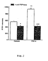
- FIG. 2 shows that the treatment of the cultured neurons with FGF-2 (10 ng/ml) led to an 1.7fold increase in survival of the cells compared to the negative control (medium alone). In contrast, addition of antibiodies specific for TGF- ⁇ 1/- ⁇ 2/- ⁇ 3 completely abolished the survival promoting effect of FGF-2. These data show that the neurotrophic effect of FGF-2 on dopaminergic neurons in the ventral midbrain is mediated by TGF- ⁇ .
- TGF- ⁇ was able to operate synergistically with FGF-2 and CNTF in the control of chick ciliary ganglion (CG) neuron survival.
- An antibody recognizing the TGF- ⁇ isoforms - ⁇ 1/- ⁇ 2/- ⁇ 3 was employed to immunoneutralize TGF- ⁇ in cultures of CG neurons treated with CNTF or FGF-2, respectively (Fig. 3).
- Neutralization of TGF- ⁇ reduced CNTF- or FGF-2-mediated neuron survival by 40 to 60%.
- CNTF shares receptor and signal transduction components with members of a family of neuropoetic cytokines, which include leukemia inhibiting factor (LIF), oncostatin M (OSM), and interleukin-6 (IL-6) (Stahl and Yanco-poulos, J. Neurobiol. 25 (1994) 1454-1466).
- LIF leukemia inhibiting factor
- OSM oncostatin M
- IL-6 interleukin-6
- TGF- ⁇ clearly augmented the efficacy of a non-saturating concentration of CNTF in terms of promoting CG neurons survival.
- OSM, LIF, and IL-6 did not support CG neurons, and further addition of TGF- ⁇ did not increase the effects of these cytokines beyond background levels.
- Neurotrophins are not established trophic factors for CG neurons (Collins, Brain Res.
- CMF Ca 2+ /Mg 2+ -free Hanks' balanced salt solution
- DMEM Dulbeccos' modified Eagle's medium
- PSN were purchased from Gibco.
- Horse serum (HS) was purchased from Gibco
- FCS fetal calf serum
- FCS fetal calf serum
- Rh FGF-2 for the experiments according to Fig. 2 was obtained from Progen, and for all other experiments from IC Chemikalien. Lyophilized factors were resuspended in culture medium (see below) to give a final concentration of 1 ⁇ g/ml and stored in aliquots of 50-100 ⁇ l at -70°C until use.
- the neutralizing antibody to TGF ⁇ -1,2,3 was purchased from Genzyme and to GDNF from Santa Cruz Biotechnology.
- Fertilized white Leghorns chick eggs were obtained from a local aviary and incubated in a humidified egg chamber at 37.8°C.
- Fertilized white Leghorns chick eggs were obtained from a local aviary and incubated in a humidified egg chamber at 38°C until E8.
- CG Embryonic chick ciliary
- DRG dorsal root
- SG sympathetic ganglia at embryonic day(s) (E) 8, 10, and 12, were dissected, freed from nerve roots and connective tissue and collected in CMF.
- Ganglia were incubated in trypsin (ICN), washed and dissociated by trituration using fire-polished Pasteur pipettes.
- ICN trypsin
- Cultures were set up in 96-well microtiter plates (Costar, A/2), precoated with poly-L-ornithine and laminin at a density of 1,200-1,500 cells/well in DMEM supplemented with N1 additives, 0.25% BSA, and 0.1% PSN (DME/N1) and incubated at 37°C in a 5% CO 2 incubator.
- CG 24 h
- DRG 48 h
- SG SG
- numbers of surviving neurons were determined by direct counting of 30% of the surface area using phase contrast microscopy.
- the cell lines B49, 3T3 and COS were grown in 10% FCS/DMEM with 1 % PSN and the cell line BHK was set up in DMEM/F 12 with 5% FCS and 1 % PSN in plastic culture flasks. Conditioned medium of these different cell lines was collected and aliquots were stored at -80°C and then prepared for MLEC-bioassay to determine TGF ⁇ activity.
- nitrocellulose membrane was blocked with 3% low fat milk powder/0.1 % BSA in Tris buffered saline (TBS, pH 7.3), incubated with primary antibody (1:200 in 0.1 % BSA/TBS) overnight at 4°C followed by peroxidase conjugated secondary antibody (1:2000 in 0.1 % BSA/TBS). Finally the membrane was developed using the Amersham enhanced chemiluminescence (ECL) detection system.
- ECL Amersham enhanced chemiluminescence
- TGF- ⁇ activity was performed by using the mink lung epithelial cell (MLEC) (kindly provided by Dr. Rifkin, New York University, New York, USA) bioassay (Abe et al., Anal. Biochem. 216 (1994) 276-284).
- MLEC mink lung epithelial cell
- Transfected MLEC cultures were set up in a 96-well microtiter plate (Costar) at a density of 1.6 x 10 4 cells/well in DMEM (high glucose) with 10% FCS and geneticin (250 ⁇ g/ml) and allowed to attach for 3 h at 37°C in a 5% CO 2 incubator.
- the medium was then replaced with 100 ⁇ l of test sample in DME/N1 (activated with HCl) and incubated overnight at 37°C in a 5% CO 2 incubator.
- Cells were washed twice with PBS and lysed by using 100 ⁇ l of lysis buffer (Promega) for 2-3 h at RT.
- lysis buffer Promega
- 80 ⁇ l of the lysates were transferred to a test tube and analyzed using a Luminometer (Lumat, Berthold/Germany) by 100 ⁇ l injections of luciferase reagent (Promega). Luciferase activity was reported as relative light units (RLU) and all assays were performed in triplicate.
- DRGs embryonic chicken dorsal root ganglia
- CMF free Hank's balanced salt solution
- ganglia were dissociated by gentle trituration using fire-polished Pasteur pipettes.
- NGF nerve growth factor
- Adrenomedullectomy was performed on adult male Hanover-Wistar rats.
- Pre-ganglionic neurons in the spinal cord innervating adrenal medullary chomaffin cells were identified by retrograde tracing with Fast Blue or Fluoro-Gold (FG).
- Factors (1 ⁇ g each) were soaked in gelfoam (Spongostan, Ferrosan, Soeburg, Denmark) prior to implantation into unilaterally medullectomized adrenal glands. Numbers of FG-labeled preganglionic neurons were determined by cell counts of complete series of longitudinal sections through spinal cord segments T7-T10 four weeks post-surgery. Only brightly fluorescent neurons containing a clearly visible nucleus were counted.
- In situ hybridizations were performed on paraffin sections of the thoracic spinal cord essentially as described in Arumäe et al. (J. Cell Biol. 122 (1993) 1053-1065). Probes were derived from rat GFR ⁇ -1, nucleotides 294-1039, and mouse cRet, nucleotides 2534-3217. After hybridization, all sections were dipped and exposed for 2-3 weeks, counterstained with haematoxylin, air dried and embedded in DPX. No hybridization signal was detected with the probes on sense orientation.
Landscapes
- Health & Medical Sciences (AREA)
- Life Sciences & Earth Sciences (AREA)
- Engineering & Computer Science (AREA)
- Bioinformatics & Cheminformatics (AREA)
- Animal Behavior & Ethology (AREA)
- Chemical & Material Sciences (AREA)
- Veterinary Medicine (AREA)
- Medicinal Chemistry (AREA)
- Public Health (AREA)
- General Health & Medical Sciences (AREA)
- Pharmacology & Pharmacy (AREA)
- Immunology (AREA)
- Gastroenterology & Hepatology (AREA)
- Epidemiology (AREA)
- Proteomics, Peptides & Aminoacids (AREA)
- Biomedical Technology (AREA)
- Neurology (AREA)
- Neurosurgery (AREA)
- Zoology (AREA)
- Organic Chemistry (AREA)
- Chemical Kinetics & Catalysis (AREA)
- General Chemical & Material Sciences (AREA)
- Nuclear Medicine, Radiotherapy & Molecular Imaging (AREA)
- Psychology (AREA)
- Orthopedic Medicine & Surgery (AREA)
- Hospice & Palliative Care (AREA)
- Psychiatry (AREA)
- Medicines That Contain Protein Lipid Enzymes And Other Medicines (AREA)
Abstract
Description
- The present invention relates to a pharmaceutical composition having neurotro phic activity, for treating peripheral and/or CNS-disorders in mammals.
- GDF-5 is a bone morphogenetic protein like molecule which, similar to other members of the transforming growth factor beta (TGF-β) superfamily, has been implicated in neurotrophic functions. For example, TGF-β1, -β2, and -β3, as well as activin A, bone morphogenetic proteins (BMP) -2, -4, -6, -7, -12, glial cell line-derived neurotrophic-like factors (GDNF-like), and GDF-5 have all been shown to promote in vitro the survival of midbrain dopaminergic neurons by various mechanisms. GDNF also acts on a wide spectrum of peripheral neurons.
- The discovery of GDNF as a neurotrophic factor for midbrain dopaminergic neurons was a hallmark in the search for novel molecules that may have relevance in the treatment of neurodegenerative diseases, as e.g. Parkinson's disease. The significance of GDNF is further underscored by its efficacy in several animal models of PD including non-human primates, ubiquitous expression in neurons of the CNS, and its widening spectrum of responsive neuron populations. GDNF signals via the tyrosine kinase receptor c-ret in co-operativity with a GPI-linked α receptor, the GDNFRα. GDNF is a member of the TGF-ß superfamily, its closest relatives being neurturin. Targeted mutations of the GDNF or c-Ret genes have indicated that GDNF is essentially required for the development of the kidney, major portions of the enteric nervous system and the sympathetic superior cervical ganglion. However, GDNF does not support the survival of most peripheral neurons in low-density dissociated cultures and defined media. Follow-up experiments in which GDNF has been shown to promote the survival of enriched peripheral autonomic and sensory neurons were all performed using serum throughout the whole cuiture period. Furthermore, the dopaminotrophic effect of GDNF was established in an extremely complex culture system where its most prominent effect did not become apparent until day 7 in culture.
- TGF-βs are widely distributed and contextually acting cytokines with prominent roles in development and cell cycle control. TGF-βs have been implicated in the regulation of neuronal survival of e.g. motoneurons, sensory and midbrain dopaminergic neurons. It should be noted, however, that TGF-β shows no or marginal effects on highly enriched, serum-free neuron cultures, as e.g. sensory neurons.
- WO-A-9711965 describes a method for treating injury or degeneration of basal forebrain cholinic neurons by administering GDNF alone or in combination with a second therapeutic agent such as NGF, BDNF, NT-3, NT-4/5, bFGF or CNTF. WO-A-9719694 describes a method for treating injury or degeneration of retinal neurons by administering GDNF alone or in combination with BDNF, NT-3, NT-4/5, NT-6, ILGF, FGF-5 or TGF-β. EP-A-0454400 describes that TGF-β alone or in combination with CSF increases the number of granulocytes and monocytes/macrophages in mammals.
- Thus, the technical problem underlying the present invention is to provide a new system having improved neurotrophic activity, for the treatment of peripheral and/or CNS-disorders in mammals.
- The solution to the above technical problem is achieved by the embodiments characterized in the claims.
- In particular, the present invention relates to a pharmaceutical composition having a neurotrophic activity, comprising a biologically active amount of at least a combination of TGF-β and NGF ou NT or CNTF or FGF or EGF or GDF or a combination of TGF-α and GDF-5 or functionally active derivatives or parts thereof and optionally a pharmaceutically acceptable carrier and/or diluent. The term "TGF-β" includes TGF-β1, TGF-β2 and TGF-β3. The terms "functionally active derivative" and "functionally active part" refer to a proteinous compound exhibiting at least part of the biological function of the respective cytokine. The cytokines used in the pharmaceutical composition according to the present invention may be selected from the group consisting of GDF such as GDF-5, GDF-6, GDF-7, GDF-8 and GDF-9, TGF such as TGF-α or TGF-β, e.g. TGF-β1, TGF-β2 or TGF-β3, NGF, neurotrophines such as NT-3 or NT-4, EGF, CNTF and FGF such as FGF-2.
- Preferred embodiments of the present invention the pharmaceutical composition comprise the following combinations: TGF-β and FGF-2 or CNTF or NT-3 or NGF.
- As a surprising fact, if at least one of TGF-β or GDF-5 is present in the above defined pharmaceutical composition, a synergistic effect can be observed resulting in an increased neurotrophic activity.
- The pharmaceutical composition according to the present invention may be used for the treatment of peripheral and/or CNS-disorders in mammals, preferably in man, such as Parkinson's disease, Alzheimer's disease, ALS or other dementia, other neurodegenerative disorders of the central nervous system and peripheral neuropathies including diabetes, cisplatinium or other genetic or acquired peripheral nerve diseases.
- The figures show:
- Fig. 1:
- Survival of E8 DRG neurons cultured for 48 hours in the presence of various trophic factors at saturating concentrations with or without the addition of GDF-5 at a constant concentration of 20 ng/ml. Values represent the means + SEM of at least two independent experiments with triplicate determinations.
- Fig. 2:
- Effect of neutralizing endogenous TGF-ß in mesencephalic neuron cultures. Numbers of surviving TH-immunoreactive neurons of mesence-phalic cultures (E14/DIV8) after 8 days in culture treated with FGF-2 (10 ng/ml), medium only (control), in presence or absence of neutralizing antibodies to TGF-β1/-β2/-β3 (10 µg/ml). Data are given as mean ± SEM (n = 3), P-values are ***P < 0.001 for increased survival as compared with control cultures and + P < 0.05 or + + + P < 0.001 for decreased survival following antibody treatment.
- Fig. 3:
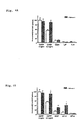
- Neutralizing endogenous TGF-ß reduces the survival promoting effects of CNTF and FGF-2 on chick CG neurons. Cultures were treated with CNTF (A) or FGF-2 (B) alone (--) at the indicated concentrations or in combination with neutralizing antibodies (-▪-) to TGF-β1, -β2, -β3 (1 µg/ml). Data are given as mean ± SEM (n=3). P-values are *P < 0.05; **P < 0.01 for decreased survival in the combination of growth factor plus anti-TGF-β as compared to growth factor-treatment alone.
- Fig. 4:
- TGF-β induces responsiveness of chick CG neurons to CNTF, NGF and NT-3. Cultures were treated with (A) CNTF (5 or 0.3 ng/ml), Onco-statin M, LIF, IL-6 (at 10 ng/ml), or (B) NGF, NT-3, NT-4 in the absence or presence of TGF-β (2.5 ng/ml). Data are given as percent of CNTF-plateau-survival as means ± SEM (n=3). *P < 0.05, **P < 0.01 for increased survival in the combination with TGF-β3 as compared to the corresponding single factor treatment alone.
- The following examples illustrate the invention:
- The growth factors TGP-β1 and -β3, transforming growth factor-a, TGF-α; show also synergistic or additive effects when added together with GDF-5 at any tested concentrations (Fig.1).
- Fibroblast growth factor (FGF)-2 is known to act as a neurotrophic factor for dopaminergic neurons in the ventral midbrain. Earlier studies suggested that the neurotrophic effect exerted by FGF-2 is mainly indirect and mediated by mesencephalic ganglia (Engele and Bohn, J. Neurosci. 11 (1991) 3070-3078). As TGF-β is known to be a product of astroglial cells, it was tested whether TGF-β-neutralizing antibodies can abolish the neurotrophic effect of FGF-2 on cultured neurons derived from the embryonic midbrain floor. Figure 2 shows that the treatment of the cultured neurons with FGF-2 (10 ng/ml) led to an 1.7fold increase in survival of the cells compared to the negative control (medium alone). In contrast, addition of antibiodies specific for TGF-β1/-β2/-β3 completely abolished the survival promoting effect of FGF-2. These data show that the neurotrophic effect of FGF-2 on dopaminergic neurons in the ventral midbrain is mediated by TGF-β.
- TGF-β was able to operate synergistically with FGF-2 and CNTF in the control of chick ciliary ganglion (CG) neuron survival. An antibody recognizing the TGF-β isoforms -β1/-β2/-β3 was employed to immunoneutralize TGF-β in cultures of CG neurons treated with CNTF or FGF-2, respectively (Fig. 3). Neutralization of TGF-β reduced CNTF- or FGF-2-mediated neuron survival by 40 to 60%. These data indicate that TGF-β released from cultured CG neurons synergistically acts with exogenous neurotrophic factors to assure neuron survival.
- CNTF shares receptor and signal transduction components with members of a family of neuropoetic cytokines, which include leukemia inhibiting factor (LIF), oncostatin M (OSM), and interleukin-6 (IL-6) (Stahl and Yanco-poulos, J. Neurobiol. 25 (1994) 1454-1466). As shown in Figure 4A, TGF-β clearly augmented the efficacy of a non-saturating concentration of CNTF in terms of promoting CG neurons survival. OSM, LIF, and IL-6 did not support CG neurons, and further addition of TGF-β did not increase the effects of these cytokines beyond background levels. Neurotrophins are not established trophic factors for CG neurons (Collins, Brain Res. 467 (1988) 111-116). However, in combination with TGF-β, both NGF and NT-3 promoted survival at 35 to 40% of the CNTF-plateau (Fig. 4B). Although expression of trkA receptors, which permit signalling of NGF and NT-3 on CG neurons has not been shown as yet, the data presented indicate that TGF-β may cooperate neurotrophin receptor-mediated signalling on CG neurons.
- CMF: Ca2+/Mg2+-free Hanks' balanced salt solution, DMEM: Dulbeccos' modified Eagle's medium, and PSN were purchased from Gibco. Poly-L-ornithine, laminin, A23187, carbachol, and verapamil was purchased from Sigma, bovine serum albumin (BSA) was from Serva (Heidetberg, Germany) and PIPLC from Boehringer Mannheim (Germany).
Horse serum (HS) was purchased from Gibco, fetal calf serum (FCS)batches FCS batches FCS batches - Growth factors were obtained from Boehringer Mannheim (NGF, 2.5 S NGF and recombinant human (rh) LIF), IC Chemikalien (recombinant rat (rr) CNTF, rh GDNF, rh Oncostatin M (OSM), rh IL-6, rh NT-3 and rh NT-4 GDF-5 was produced as described (Krieglstein et al., J. Neurosci. Res., 42(5) (1995) 724-732). Rh FGF-2 for the experiments according to Fig. 2 was obtained from Progen, and for all other experiments from IC Chemikalien. Lyophilized factors were resuspended in culture medium (see below) to give a final concentration of 1 µg/ml and stored in aliquots of 50-100 µl at -70°C until use.
- The neutralizing antibody to TGFβ-1,2,3 was purchased from Genzyme and to GDNF from Santa Cruz Biotechnology.
- Agarose conjugated antibodies to TGFβ-1 and to GDNF were from Santa Cruz Biotechnology. The peroxidase conjugated α-rabbit antibody was purchased from Sigma.
- Fertilized white Leghorns chick eggs were obtained from a local aviary and incubated in a humidified egg chamber at 37.8°C.
- Fertilized white Leghorns chick eggs were obtained from a local aviary and incubated in a humidified egg chamber at 38°C until E8.
- Cell culture for experiments according to Fig. 1.
- Embryonic chick ciliary (CG), dorsal root (DRG), and sympathetic (SG) ganglia at embryonic day(s) (E) 8, 10, and 12, were dissected, freed from nerve roots and connective tissue and collected in CMF. Ganglia were incubated in trypsin (ICN), washed and dissociated by trituration using fire-polished Pasteur pipettes.
Cultures were set up in 96-well microtiter plates (Costar, A/2), precoated with poly-L-ornithine and laminin at a density of 1,200-1,500 cells/well in DMEM supplemented with N1 additives, 0.25% BSA, and 0.1% PSN (DME/N1) and incubated at 37°C in a 5% CO2 incubator.
At appropriate times (CG: 24 h, DRG: 48 h, SG: 72 h) cultures were fixed by addition of 2.5% glutardialdehyde in phosphate buffered saline (PBS). Numbers of surviving neurons were determined by direct counting of 30% of the surface area using phase contrast microscopy. - The cell lines B49, 3T3 and COS were grown in 10% FCS/DMEM with 1 % PSN and the cell line BHK was set up in DMEM/F 12 with 5% FCS and 1 % PSN in plastic culture flasks.
Conditioned medium of these different cell lines was collected and aliquots were stored at -80°C and then prepared for MLEC-bioassay to determine TGFβ activity. - To 1 ml of the stimulation buffer with or without additives from
chromaffin cells 10 µl of the antibody agarose conjugate were added and incubated at 4°C overnight with mixing. The pellet was washed with PBS and then resuspended in 50 µl of electrophoresis sample buffer and boiled for 2 minutes. Samples were separated by electrophoresis on a 12.5% SDS-PAA gel and transferred to nitrocellulose membrane (Hybond, Amersham). The nitrocellulose membrane was blocked with 3% low fat milk powder/0.1 % BSA in Tris buffered saline (TBS, pH 7.3), incubated with primary antibody (1:200 in 0.1 % BSA/TBS) overnight at 4°C followed by peroxidase conjugated secondary antibody (1:2000 in 0.1 % BSA/TBS). Finally the membrane was developed using the Amersham enhanced chemiluminescence (ECL) detection system. - Determination of TGF-β activity was performed by using the mink lung epithelial cell (MLEC) (kindly provided by Dr. Rifkin, New York University, New York, USA) bioassay (Abe et al., Anal. Biochem. 216 (1994) 276-284). Transfected MLEC cultures were set up in a 96-well microtiter plate (Costar) at a density of 1.6 x 10⁴ cells/well in DMEM (high glucose) with 10% FCS and geneticin (250µg/ml) and allowed to attach for 3 h at 37°C in a 5% CO2 incubator. The medium was then replaced with 100 µl of test sample in DME/N1 (activated with HCl) and incubated overnight at 37°C in a 5% CO2 incubator. Cells were washed twice with PBS and lysed by using 100 µl of lysis buffer (Promega) for 2-3 h at RT. To determine TGFβ activity, 80 µl of the lysates were transferred to a test tube and analyzed using a Luminometer (Lumat, Berthold/Germany) by 100 µl injections of luciferase reagent (Promega). Luciferase activity was reported as relative light units (RLU) and all assays were performed in triplicate.
- Statistical comparisons were made with one-way ANOVA, included in the MicroCalOrigin software. Differences were considered statistically significant at * P < 0.05, ** P < 0.005, and *** P < 0.0005.
- Dissociated cultures of embryonic chicken dorsal root ganglia (DRGs) were generated as desribed in details by Krieglstein and Unsicker (Dev. Brain. Rees. 93 (1996) 10-17). Briefly, DRGs were dissected in Ca2+-Mg2+ free Hank's balanced salt solution (CMF). After incubation in 0.08% trypsin (BioWhittaker) for 15 min, ganglia were dissociated by gentle trituration using fire-polished Pasteur pipettes. Cells in suspension (neuron/non-neuron ratio 1:2) were seeded in polyornithin-laminin coated 96-well microtiter plates (A/2 Costar) at a density of 1,200 cells/well. Growth factors were applied at the time of plating in a final volume of 50 µl Dulbecco's Modified Eagle's Medium supplemented with 0.25% bovine serum albumin, N1 additives (Bottenstein et al., Exp. Cell Res. 125 (1980) 183-190) and 100 U/ml penicillin. As a positive control, nerve growth factor (NGF) was used at the saturating concentration of 10 ng/ml. After 48 h of incubation at 37°C in a humidified atmosphere containing 5% CO2, cultures were fixed with 2.5% glutaraldehyde in phosphate buffered saline (PBS). Neuronal cells were identified by their phase-bright and neurite-bearing morphologies and counted within 30% of the total surface area using phase contrast microscopy. All experiments were performed in at least triplicate in two independent experiments. Data are presented as mean ± SEM. Statistical comparisons were made with student's double t-test, ANOVA and MicrocalOrigin software. Differences were considered statistically significant at *P < 0.05, **P < 0.01, ***P < 0.001.
- Adrenomedullectomy was performed on adult male Hanover-Wistar rats. Pre-ganglionic neurons in the spinal cord innervating adrenal medullary chomaffin cells were identified by retrograde tracing with Fast Blue or Fluoro-Gold (FG). Factors (1 µg each) were soaked in gelfoam (Spongostan, Ferrosan, Soeburg, Denmark) prior to implantation into unilaterally medullectomized adrenal glands. Numbers of FG-labeled preganglionic neurons were determined by cell counts of complete series of longitudinal sections through spinal cord segments T7-T10 four weeks post-surgery. Only brightly fluorescent neurons containing a clearly visible nucleus were counted. Total numbers were corrected for possible double counts of split nuclei according to Abercombie's formula. Numbers of preganglionic neurons to the adrenal medulla surviving in sham-operated animals and on the unlesioned side were set as 100%. Data are given as mean values ± SEM and the statistical significance of intergroup differences was determined by Student's t-test.
- In situ hybridizations were performed on paraffin sections of the thoracic spinal cord essentially as described in Arumäe et al. (J. Cell Biol. 122 (1993) 1053-1065). Probes were derived from rat GFRα-1, nucleotides 294-1039, and mouse cRet, nucleotides 2534-3217. After hybridization, all sections were dipped and exposed for 2-3 weeks, counterstained with haematoxylin, air dried and embedded in DPX. No hybridization signal was detected with the probes on sense orientation.
Claims (15)
- A pharmaceutical composition having neurotrophic activity, comprising a biologically active amount of at least a combination of TGF-β and NGF or NT or CNTF or FGF or EGF or GDF, or functionally active derivatives or parts thereof and optionally a pharmaceutical acceptable carrier and/or diluent.
- The pharmaceutical composition according to claim 1, comprising TGF-β and FGF-2.
- The pharmaceutical composition according to claim 1, comprising TGF-β and CNTF.
- The pharmaceutical composition according to claim 1, comprising TGF-β and NT-3.
- The pharmaceutical composition according to claim 1, comprising TGF-β and NGF.
- The pharmaceutical composition according to claim 1, comprising TGF-β and GDF-5.
- A pharmaceutical composition having neurotrophic activity, comprising a biologically active amount of at least a combination of TGF-α and GDF-5.
- Use of biologically active amounts of at least two cytokines or functionally active derivatives or parts thereof and optionally a pharmaceutically acceptable carrier and/or diluent for the preparation of a pharmaceutical composition for treating peripheral and/or CNS-disorders in mammals, wherein the at least two cytokines comprise a combination of TGF-β and GDNF or NGF or NT or CNTF or FGF or EGF or GDF or a combination of TGF-α and GDF-5.
- The use according to claim 8, wherein the pharmaceutical composition comprises GDNF and TGF-β.
- The use according to claim 8, wherein the pharmaceutical composition comprises TGF-β and FGF-2.
- The use according to claim 8, wherein the pharmaceutical composition comprises TGF-β and CNTF.
- The use according to claim 8, wherein the pharmaceutical composition comprises TGF-β and NT-3.
- The use according to claim 8, wherein the pharmaceutical composition comprises TGF-β and NGF.
- The use according to claim 8, wherein the pharmaceutical composition comprises TGF-β and GDF-5.
- The use according to anyone of claims 8 to 14, wherein the CNS-disorder is Parkinson's disease, Alzheimer's disease, ALS or other dementia, other neurodegenerative disorders of the central nervous systems and peripheral neuropathies including diabetes, cisplatinium or other genetic or acquired peripheral nerve diseases.
Priority Applications (1)
| Application Number | Priority Date | Filing Date | Title |
|---|---|---|---|
| EP98952613A EP1011712B1 (en) | 1997-09-19 | 1998-09-21 | Combination of cytokines having neurotrophic activity |
Applications Claiming Priority (4)
| Application Number | Priority Date | Filing Date | Title |
|---|---|---|---|
| EP97116373 | 1997-09-19 | ||
| EP97116373 | 1997-09-19 | ||
| PCT/EP1998/006004 WO1999015191A2 (en) | 1997-09-19 | 1998-09-21 | Cytokines having neurotrophic activity |
| EP98952613A EP1011712B1 (en) | 1997-09-19 | 1998-09-21 | Combination of cytokines having neurotrophic activity |
Publications (2)
| Publication Number | Publication Date |
|---|---|
| EP1011712A2 EP1011712A2 (en) | 2000-06-28 |
| EP1011712B1 true EP1011712B1 (en) | 2004-02-11 |
Family
ID=8227378
Family Applications (1)
| Application Number | Title | Priority Date | Filing Date |
|---|---|---|---|
| EP98952613A Expired - Lifetime EP1011712B1 (en) | 1997-09-19 | 1998-09-21 | Combination of cytokines having neurotrophic activity |
Country Status (8)
| Country | Link |
|---|---|
| US (1) | US6790824B1 (en) |
| EP (1) | EP1011712B1 (en) |
| JP (1) | JP2001517634A (en) |
| AU (1) | AU1024899A (en) |
| CA (1) | CA2301693A1 (en) |
| DE (1) | DE69821629T2 (en) |
| ES (1) | ES2214743T3 (en) |
| WO (1) | WO1999015191A2 (en) |
Cited By (1)
| Publication number | Priority date | Publication date | Assignee | Title |
|---|---|---|---|---|
| WO2014031883A1 (en) * | 2012-08-23 | 2014-02-27 | Susan Marie Metcalfe | Neurotherapeutic nanoparticle compositions and devices |
Families Citing this family (24)
| Publication number | Priority date | Publication date | Assignee | Title |
|---|---|---|---|---|
| AU3667100A (en) * | 1999-04-23 | 2000-11-10 | Sulzer Orthopedics Ltd | Composition for enhancing functional recovery of a mammal from central and/or peripheral nervous system injury of traumatic or pathological origin |
| CA2372119A1 (en) * | 1999-05-17 | 2000-11-23 | Biopharm Gesellschaft Zur Biotechnologischen Entwicklung Und Zum Vertrie B Von Pharmaka Mbh | Neuroprotective properties of gdf-15, a novel member of the tgf-.beta. superfamily |
| NZ502796A (en) * | 2000-06-15 | 2003-02-28 | Agres Ltd | Nucleotide and amino acid sequences of oocyte factors related to GDF-9B or GDF-9 |
| WO2002006341A1 (en) * | 2000-07-14 | 2002-01-24 | Children's Medical Center Corporation | A trophic factor capable of producing a neurosalutary effect in a subject |
| WO2003000257A1 (en) * | 2001-06-26 | 2003-01-03 | Takeda Chemical Industries, Ltd. | TGF-ß SUPERFAMILY PRODUCTION/SECRETION PROMOTER |
| EP1423509A2 (en) * | 2001-08-30 | 2004-06-02 | Stem Cell Therapeutics Inc. | Differentiation of neural stem cells and therapeutic use thereof |
| EP1430114B1 (en) | 2001-09-14 | 2012-01-18 | Stem Cell Therapeutics Inc. | Prolactin induced increase in neural stem cell numbers and therapeutical use thereof |
| WO2004011497A1 (en) | 2002-07-31 | 2004-02-05 | Stem Cell Therapeutics Inc. | Method of enhancing neural stem cell proliferation, differentiation, and survival using pituitary adenylate cyclase activating polypeptide (pacap) |
| ES2392282T3 (en) | 2004-02-13 | 2012-12-07 | Stem Cell Therapeutics Corp. | Use of luteinizing hormone (LH) and chorionic gonadotropin (hCG) for neural precursor cell proliferation and neurogenesis |
| US20060069008A1 (en) * | 2004-09-28 | 2006-03-30 | Sanjay Mistry | Treatment of neurological deficits in the striatum or substanta nigra pars compacta |
| US20060269525A1 (en) * | 2005-05-25 | 2006-11-30 | Sean Lilienfeld | Co-grafts for treating Parkinson's disease |
| CA2613551C (en) * | 2005-06-23 | 2017-06-20 | Tissuegene, Inc. | Use of cells for treating a neurodegenerative condition |
| KR20080074108A (en) | 2005-09-27 | 2008-08-12 | 스템 셀 테라퓨틱스 코포레이션 | Oligodendrosite precursor cell proliferation regulated by prolactin |
| PT1948689E (en) * | 2005-11-18 | 2012-05-09 | Bioph Biotech Entw Pharm Gmbh | High activity growth factor mutants |
| WO2007106986A1 (en) | 2006-03-17 | 2007-09-27 | Stem Cell Therapeutics Corp. | Dosing regimes for lh or hcg and epo for treatment of neurological disorders |
| SI2019683T2 (en) | 2006-04-25 | 2022-10-28 | The Regents Of The University Of California | Administration of growth factors for the treatment of cns disorders |
| JP2009535360A (en) * | 2006-04-26 | 2009-10-01 | ザ リージェンツ オブ ザ ユニバーシティ オブ カリフォルニア | Compositions and methods for convection enhanced delivery of high molecular weight neurotherapeutic agents |
| SI2121142T1 (en) | 2007-01-25 | 2012-12-31 | Biopharm Gesellschaft Zur Biotechnologischen Entwicklung Von Pharmaka Mbh | Use of gdf-5 for the improvement or maintenance of dermal appearance |
| US20100204123A1 (en) * | 2009-02-12 | 2010-08-12 | Mary Elizabeth Pecquet Goad | Peripheral Administration of Proteins Including TGF-beta Superfamily Members for Treatment of Systemic Disorders and Disease |
| EP2396026A2 (en) * | 2009-02-12 | 2011-12-21 | Stryker Corporation | Compositions and methods for minimally-invasive systemic delivery of proteins including tgf- superfamily members |
| ES2363353B1 (en) * | 2010-01-19 | 2012-02-22 | Universidad De Sevilla | COMPOSITION INCLUDING GDNF AND TGF-BETA1 AND ITS USE FOR THE TREATMENT OF A NEUROLOGICAL DISEASE. |
| AU2011284657B2 (en) | 2010-07-30 | 2013-11-14 | Biopharm Gesellschaft Zur Biotechnologischen Entwicklung Von Pharmaka Mbh | Drug delivery devices and growth factor formulations for accelerated wound healing |
| EP2537538A1 (en) | 2011-06-22 | 2012-12-26 | Biopharm Gesellschaft Zur Biotechnologischen Entwicklung Von Pharmaka mbH | Bioresorbable Wound Dressing |
| CN114624450A (en) * | 2022-04-26 | 2022-06-14 | 江苏睿源生物技术有限公司 | ELISA detection method for detecting transforming growth factor-beta in cell and application thereof |
Family Cites Families (6)
| Publication number | Priority date | Publication date | Assignee | Title |
|---|---|---|---|---|
| EP0105014B1 (en) * | 1982-09-24 | 1992-05-20 | THE UNITED STATES OF AMERICA as represented by the Secretary United States Department of Commerce | Repair of tissue in animals |
| US5149804A (en) * | 1990-11-30 | 1992-09-22 | The Board Of Trustees Of The University Of Illinois | Ecteinascidins 736 and 722 |
| US5147799A (en) | 1990-04-25 | 1992-09-15 | Isia Bursuker | Repopulation of macrophages and granulocytes using transforming growth factor-beta |
| US5731284A (en) * | 1995-09-28 | 1998-03-24 | Amgen Inc. | Method for treating Alzheimer's disease using glial line-derived neurotrophic factor (GDNF) protein product |
| US5641750A (en) * | 1995-11-29 | 1997-06-24 | Amgen Inc. | Methods for treating photoreceptors using glial cell line-derived neurotrophic factor (GDNF) protein product |
| WO1999012560A1 (en) | 1997-09-09 | 1999-03-18 | Creative Biomolecules, Inc. | Synergistic effects of op/bmp morphogens and gdnf/ngf neurotrophic factors |
-
1998
- 1998-09-21 WO PCT/EP1998/006004 patent/WO1999015191A2/en not_active Ceased
- 1998-09-21 EP EP98952613A patent/EP1011712B1/en not_active Expired - Lifetime
- 1998-09-21 AU AU10248/99A patent/AU1024899A/en not_active Abandoned
- 1998-09-21 ES ES98952613T patent/ES2214743T3/en not_active Expired - Lifetime
- 1998-09-21 CA CA002301693A patent/CA2301693A1/en not_active Abandoned
- 1998-09-21 DE DE1998621629 patent/DE69821629T2/en not_active Expired - Fee Related
- 1998-09-21 JP JP2000512560A patent/JP2001517634A/en active Pending
-
2000
- 2000-03-17 US US09/527,275 patent/US6790824B1/en not_active Expired - Fee Related
Cited By (2)
| Publication number | Priority date | Publication date | Assignee | Title |
|---|---|---|---|---|
| WO2014031883A1 (en) * | 2012-08-23 | 2014-02-27 | Susan Marie Metcalfe | Neurotherapeutic nanoparticle compositions and devices |
| US10835611B2 (en) | 2012-08-23 | 2020-11-17 | Yale University | Neurotherapeutic nanoparticle compositions comprising leukemia inhibitory factor |
Also Published As
| Publication number | Publication date |
|---|---|
| ES2214743T3 (en) | 2004-09-16 |
| DE69821629D1 (en) | 2004-03-18 |
| AU1024899A (en) | 1999-04-12 |
| US6790824B1 (en) | 2004-09-14 |
| WO1999015191A3 (en) | 1999-05-20 |
| JP2001517634A (en) | 2001-10-09 |
| EP1011712A2 (en) | 2000-06-28 |
| DE69821629T2 (en) | 2005-01-13 |
| WO1999015191A2 (en) | 1999-04-01 |
| CA2301693A1 (en) | 1999-04-01 |
Similar Documents
| Publication | Publication Date | Title |
|---|---|---|
| US6790824B1 (en) | Cytokines having neurotrophic activity | |
| Krieglstein et al. | Glial cell line-derived neurotrophic factor requires transforming growth factor-β for exerting its full neurotrophic potential on peripheral and CNS neurons | |
| Henderson | Role of neurotrophic factors in neuronal development | |
| Neufeld et al. | Heparin modulation of the neurotropic effects of acidic and basic fibroblast growth factors and nerve growth factor on PC12 cells | |
| Davies et al. | Neurotrophin-4/5 is a mammalian-specific survival factor for distinct populations of sensory neurons | |
| Thoenen et al. | Trophic support of motoneurons: physiological, pathophysiological, and therapeutic implications | |
| Hunter et al. | Transforming growth factor‐βs inhibit mitogen‐stimulated proliferation of astrocytes | |
| Kalcheim et al. | Neurotrophin 3 is a mitogen for cultured neural crest cells. | |
| Hughes et al. | Members of several gene families influence survival of rat motoneurons in vitro and in vivo | |
| Apfel | Nerve growth factor for the treatment of diabetic neuropathy: what went wrong, what went right, and what does the future hold? | |
| Chalazonitis et al. | Transforming growth factor β has neurotrophic actions on sensory neurons in vitro and is synergistic with nerve growth factor | |
| Krieglstein et al. | TGF‐β regulates the survival of ciliary ganglionic neurons synergistically with ciliary neurotrophic factor and neurotrophins | |
| Widmer et al. | Neurotrophin‐4/5 promotes survival and differentiation of rat striatal neurons developing in culture | |
| Sarder et al. | Interleukin-2 promotes survival and neurite extension of cultured neurons from fetal rat brain | |
| Roussa et al. | TGF-β promotes survival on mesencephalic dopaminergic neurons in cooperation with Shh and FGF-8 | |
| Davies et al. | Neural crest-derived proprioceptive neurons express nerve growth factor receptors but are not supported by nerve growth factor in culture | |
| Zhou et al. | The stimulatory effect of brain-derived neurotrophic factor on dopaminergic phenotype expression of embryonic rat cortical neurons in vitro | |
| Krieglstein et al. | Distinct modulatory actions of TGF-β and LIF on neurotrophin-mediated survival of developing sensory neurons | |
| Unsicker et al. | Co-activation of TGF-ss and cytokine signaling pathways are required for neurotrophic functions | |
| Steljes et al. | Neurotrophic factor regulation of developing avian oculomotor neurons: differential effects of BDNF and GDNF | |
| Onesto et al. | Growth factors as axon guidance molecules: Lessons from in vitro studies | |
| Mehler et al. | Growth factor regulation of neuronal development | |
| Grothe et al. | The high molecular weight fibroblast growth factor-2 isoforms (21,000 mol. wt and 23,000 mol. wt) mediate neurotrophic activity on rat embryonic mesencephalic dopaminergic neurons in vitro | |
| Thaler et al. | Leukemia inhibitory factor and neurotrophins support overlapping populations of rat nodose sensory neurons in culture | |
| Bampton et al. | Osteonectin is a Schwann cell‐secreted factor that promotes retinal ganglion cell survival and process outgrowth |
Legal Events
| Date | Code | Title | Description |
|---|---|---|---|
| PUAI | Public reference made under article 153(3) epc to a published international application that has entered the european phase |
Free format text: ORIGINAL CODE: 0009012 |
|
| 17P | Request for examination filed |
Effective date: 20000418 |
|
| AK | Designated contracting states |
Kind code of ref document: A2 Designated state(s): DE ES FR GB IT |
|
| 17Q | First examination report despatched |
Effective date: 20010606 |
|
| GRAP | Despatch of communication of intention to grant a patent |
Free format text: ORIGINAL CODE: EPIDOSNIGR1 |
|
| RIC1 | Information provided on ipc code assigned before grant |
Ipc: 7A 61P 25/28 B Ipc: 7A 61P 25/02 B Ipc: 7A 61K 38/19 B Ipc: 7A 61K 38/18 A |
|
| RTI1 | Title (correction) |
Free format text: COMBINATION OF CYTOKINES HAVING NEUROTROPHIC ACTIVITY |
|
| GRAS | Grant fee paid |
Free format text: ORIGINAL CODE: EPIDOSNIGR3 |
|
| GRAA | (expected) grant |
Free format text: ORIGINAL CODE: 0009210 |
|
| AK | Designated contracting states |
Kind code of ref document: B1 Designated state(s): DE ES FR GB IT |
|
| REG | Reference to a national code |
Ref country code: GB Ref legal event code: FG4D |
|
| REF | Corresponds to: |
Ref document number: 69821629 Country of ref document: DE Date of ref document: 20040318 Kind code of ref document: P |
|
| REG | Reference to a national code |
Ref country code: ES Ref legal event code: FG2A Ref document number: 2214743 Country of ref document: ES Kind code of ref document: T3 |
|
| ET | Fr: translation filed | ||
| PLBE | No opposition filed within time limit |
Free format text: ORIGINAL CODE: 0009261 |
|
| STAA | Information on the status of an ep patent application or granted ep patent |
Free format text: STATUS: NO OPPOSITION FILED WITHIN TIME LIMIT |
|
| 26N | No opposition filed |
Effective date: 20041112 |
|
| PGFP | Annual fee paid to national office [announced via postgrant information from national office to epo] |
Ref country code: GB Payment date: 20060922 Year of fee payment: 9 Ref country code: FR Payment date: 20060922 Year of fee payment: 9 |
|
| PGFP | Annual fee paid to national office [announced via postgrant information from national office to epo] |
Ref country code: ES Payment date: 20060926 Year of fee payment: 9 |
|
| PGFP | Annual fee paid to national office [announced via postgrant information from national office to epo] |
Ref country code: DE Payment date: 20060928 Year of fee payment: 9 |
|
| PGFP | Annual fee paid to national office [announced via postgrant information from national office to epo] |
Ref country code: IT Payment date: 20060930 Year of fee payment: 9 |
|
| GBPC | Gb: european patent ceased through non-payment of renewal fee |
Effective date: 20070921 |
|
| PG25 | Lapsed in a contracting state [announced via postgrant information from national office to epo] |
Ref country code: DE Free format text: LAPSE BECAUSE OF NON-PAYMENT OF DUE FEES Effective date: 20080401 |
|
| REG | Reference to a national code |
Ref country code: FR Ref legal event code: ST Effective date: 20080531 |
|
| PG25 | Lapsed in a contracting state [announced via postgrant information from national office to epo] |
Ref country code: FR Free format text: LAPSE BECAUSE OF NON-PAYMENT OF DUE FEES Effective date: 20071001 |
|
| PG25 | Lapsed in a contracting state [announced via postgrant information from national office to epo] |
Ref country code: GB Free format text: LAPSE BECAUSE OF NON-PAYMENT OF DUE FEES Effective date: 20070921 |
|
| REG | Reference to a national code |
Ref country code: ES Ref legal event code: FD2A Effective date: 20070922 |
|
| PG25 | Lapsed in a contracting state [announced via postgrant information from national office to epo] |
Ref country code: ES Free format text: LAPSE BECAUSE OF NON-PAYMENT OF DUE FEES Effective date: 20070922 |
|
| PG25 | Lapsed in a contracting state [announced via postgrant information from national office to epo] |
Ref country code: IT Free format text: LAPSE BECAUSE OF NON-PAYMENT OF DUE FEES Effective date: 20070921 |