EP1009435B1 - Method for preventing hiv-1 infection of cd4?+ cells - Google Patents
Method for preventing hiv-1 infection of cd4?+ cells Download PDFInfo
- Publication number
- EP1009435B1 EP1009435B1 EP98931261A EP98931261A EP1009435B1 EP 1009435 B1 EP1009435 B1 EP 1009435B1 EP 98931261 A EP98931261 A EP 98931261A EP 98931261 A EP98931261 A EP 98931261A EP 1009435 B1 EP1009435 B1 EP 1009435B1
- Authority
- EP
- European Patent Office
- Prior art keywords
- agent
- hiv
- infection
- chemokine receptor
- complex
- Prior art date
- Legal status (The legal status is an assumption and is not a legal conclusion. Google has not performed a legal analysis and makes no representation as to the accuracy of the status listed.)
- Expired - Lifetime
Links
- 208000031886 HIV Infections Diseases 0.000 title claims abstract description 88
- 238000000034 method Methods 0.000 title claims abstract description 66
- 239000003795 chemical substances by application Substances 0.000 claims abstract description 189
- 102000009410 Chemokine receptor Human genes 0.000 claims abstract description 121
- 108050000299 Chemokine receptor Proteins 0.000 claims abstract description 121
- 230000027455 binding Effects 0.000 claims abstract description 80
- 230000002401 inhibitory effect Effects 0.000 claims abstract description 76
- 239000008194 pharmaceutical composition Substances 0.000 claims abstract description 23
- 239000003937 drug carrier Substances 0.000 claims abstract description 15
- 102100035875 C-C chemokine receptor type 5 Human genes 0.000 claims description 32
- 101710149870 C-C chemokine receptor type 5 Proteins 0.000 claims description 31
- 102100031650 C-X-C chemokine receptor type 4 Human genes 0.000 claims description 27
- 108010043277 recombinant soluble CD4 Proteins 0.000 claims description 27
- 208000015181 infectious disease Diseases 0.000 claims description 17
- 239000011159 matrix material Substances 0.000 claims description 12
- 239000007787 solid Substances 0.000 claims description 12
- YBJHBAHKTGYVGT-ZKWXMUAHSA-N (+)-Biotin Chemical compound N1C(=O)N[C@@H]2[C@H](CCCCC(=O)O)SC[C@@H]21 YBJHBAHKTGYVGT-ZKWXMUAHSA-N 0.000 claims description 10
- 101000922348 Homo sapiens C-X-C chemokine receptor type 4 Proteins 0.000 claims description 7
- 229960002685 biotin Drugs 0.000 claims description 5
- 235000020958 biotin Nutrition 0.000 claims description 5
- 239000011616 biotin Substances 0.000 claims description 5
- 239000002502 liposome Substances 0.000 claims description 4
- 239000012528 membrane Substances 0.000 claims description 4
- 239000003550 marker Substances 0.000 claims description 3
- 241000713772 Human immunodeficiency virus 1 Species 0.000 abstract description 103
- 102000019034 Chemokines Human genes 0.000 abstract description 88
- 108010012236 Chemokines Proteins 0.000 abstract description 88
- 230000004927 fusion Effects 0.000 abstract description 73
- 210000004027 cell Anatomy 0.000 description 180
- 102100036011 T-cell surface glycoprotein CD4 Human genes 0.000 description 102
- 238000002165 resonance energy transfer Methods 0.000 description 51
- 238000003556 assay Methods 0.000 description 36
- 102000001902 CC Chemokines Human genes 0.000 description 34
- 108010040471 CC Chemokines Proteins 0.000 description 34
- 108010055166 Chemokine CCL5 Proteins 0.000 description 33
- 102100032367 C-C motif chemokine 5 Human genes 0.000 description 30
- 102000001326 Chemokine CCL4 Human genes 0.000 description 30
- 108010055165 Chemokine CCL4 Proteins 0.000 description 30
- 241000700605 Viruses Species 0.000 description 29
- 102100027723 Endogenous retrovirus group K member 6 Rec protein Human genes 0.000 description 27
- 101710121417 Envelope glycoprotein Proteins 0.000 description 26
- 108091034117 Oligonucleotide Proteins 0.000 description 26
- 108020004707 nucleic acids Proteins 0.000 description 26
- 102000039446 nucleic acids Human genes 0.000 description 26
- 150000007523 nucleic acids Chemical class 0.000 description 26
- 239000002773 nucleotide Substances 0.000 description 26
- 125000003729 nucleotide group Chemical group 0.000 description 26
- 230000001404 mediated effect Effects 0.000 description 24
- 230000034217 membrane fusion Effects 0.000 description 23
- 108700012434 CCL3 Proteins 0.000 description 21
- 102000000013 Chemokine CCL3 Human genes 0.000 description 21
- 108090000765 processed proteins & peptides Proteins 0.000 description 20
- 102000004196 processed proteins & peptides Human genes 0.000 description 19
- 239000000203 mixture Substances 0.000 description 15
- 102000005962 receptors Human genes 0.000 description 15
- 108020003175 receptors Proteins 0.000 description 15
- 108010061299 CXCR4 Receptors Proteins 0.000 description 14
- 150000001875 compounds Chemical class 0.000 description 14
- 229920001184 polypeptide Polymers 0.000 description 14
- 210000001744 T-lymphocyte Anatomy 0.000 description 13
- LOKCTEFSRHRXRJ-UHFFFAOYSA-I dipotassium trisodium dihydrogen phosphate hydrogen phosphate dichloride Chemical compound P(=O)(O)(O)[O-].[K+].P(=O)(O)([O-])[O-].[Na+].[Na+].[Cl-].[K+].[Cl-].[Na+] LOKCTEFSRHRXRJ-UHFFFAOYSA-I 0.000 description 13
- 238000002474 experimental method Methods 0.000 description 13
- 230000005764 inhibitory process Effects 0.000 description 13
- 239000002953 phosphate buffered saline Substances 0.000 description 13
- 235000018102 proteins Nutrition 0.000 description 13
- 102000004169 proteins and genes Human genes 0.000 description 13
- 108090000623 proteins and genes Proteins 0.000 description 13
- PXIPVTKHYLBLMZ-UHFFFAOYSA-N Sodium azide Chemical compound [Na+].[N-]=[N+]=[N-] PXIPVTKHYLBLMZ-UHFFFAOYSA-N 0.000 description 12
- 108010008951 Chemokine CXCL12 Proteins 0.000 description 9
- 102000006573 Chemokine CXCL12 Human genes 0.000 description 9
- 239000000975 dye Substances 0.000 description 9
- 239000012634 fragment Substances 0.000 description 9
- 230000014509 gene expression Effects 0.000 description 9
- 230000003993 interaction Effects 0.000 description 9
- 125000001429 N-terminal alpha-amino-acid group Chemical group 0.000 description 8
- 235000001014 amino acid Nutrition 0.000 description 8
- 150000001413 amino acids Chemical class 0.000 description 8
- 239000003112 inhibitor Substances 0.000 description 8
- 239000013612 plasmid Substances 0.000 description 8
- 102100036664 Adenosine deaminase Human genes 0.000 description 7
- 108010038807 Oligopeptides Proteins 0.000 description 7
- 102000015636 Oligopeptides Human genes 0.000 description 7
- 238000012217 deletion Methods 0.000 description 7
- 230000037430 deletion Effects 0.000 description 7
- 230000006870 function Effects 0.000 description 7
- 210000002540 macrophage Anatomy 0.000 description 7
- 230000010076 replication Effects 0.000 description 7
- 101710082513 C-X-C chemokine receptor type 4 Proteins 0.000 description 6
- 101100005713 Homo sapiens CD4 gene Proteins 0.000 description 6
- 239000005089 Luciferase Substances 0.000 description 6
- 230000000694 effects Effects 0.000 description 6
- 239000003016 pheromone Substances 0.000 description 6
- 239000006228 supernatant Substances 0.000 description 6
- UEUPDYPUTTUXLJ-UHFFFAOYSA-N 1-[[4-(1,4,8,11-tetrazacyclotetradec-1-ylmethyl)phenyl]methyl]-1,4,8,11-tetrazacyclotetradecane;octahydrochloride Chemical compound Cl.Cl.Cl.Cl.Cl.Cl.Cl.Cl.C=1C=C(CN2CCNCCCNCCNCCC2)C=CC=1CN1CCCNCCNCCCNCC1 UEUPDYPUTTUXLJ-UHFFFAOYSA-N 0.000 description 5
- 102100021943 C-C motif chemokine 2 Human genes 0.000 description 5
- 101710155857 C-C motif chemokine 2 Proteins 0.000 description 5
- 108091008927 CC chemokine receptors Proteins 0.000 description 5
- 108020004414 DNA Proteins 0.000 description 5
- 108060001084 Luciferase Proteins 0.000 description 5
- FAPWRFPIFSIZLT-UHFFFAOYSA-M Sodium chloride Chemical compound [Na+].[Cl-] FAPWRFPIFSIZLT-UHFFFAOYSA-M 0.000 description 5
- 238000001727 in vivo Methods 0.000 description 5
- 210000000265 leukocyte Anatomy 0.000 description 5
- 239000003446 ligand Substances 0.000 description 5
- AEUKDPKXTPNBNY-XEYRWQBLSA-N mcp 2 Chemical compound C([C@@H](C(=O)N[C@@H](CS)C(=O)N[C@@H](CCCNC(N)=N)C(=O)N[C@@H]([C@@H](C)CC)C(=O)N[C@@H](CCCNC(N)=N)C(=O)NCC(=O)N[C@@H](CCCNC(N)=N)C(=O)N[C@@H]([C@@H](C)CC)C(=O)N[C@@H](CC=1NC=NC=1)C(=O)N1[C@@H](CCC1)C(=O)N[C@@H](CC(C)C)C(=O)N[C@@H](CS)C(=O)N[C@@H](CS)C(=O)N[C@@H](CCCNC(N)=N)C(=O)N[C@@H](CCCNC(N)=N)C(O)=O)NC(=O)CNC(=O)[C@H](C)NC(=O)[C@H](CCCNC(N)=N)NC(=O)[C@H](CCCNC(N)=N)NC(=O)[C@H](CCC(O)=O)NC(=O)[C@H](CC(C)C)NC(=O)[C@H]1N(CCC1)C(=O)[C@H](CC(C)C)NC(=O)[C@H](CS)NC(=O)[C@H](CC(C)C)NC(=O)[C@H](C)NC(=O)[C@H](CCCNC(N)=N)NC(=O)[C@H](CCCNC(N)=N)NC(=O)[C@H](CS)NC(=O)[C@H](C)NC(=O)[C@H](CS)NC(=O)[C@@H](NC(=O)[C@@H](N)C(C)C)C(C)C)C1=CC=CC=C1 AEUKDPKXTPNBNY-XEYRWQBLSA-N 0.000 description 5
- 229930182817 methionine Natural products 0.000 description 5
- 238000005406 washing Methods 0.000 description 5
- 101710149814 C-C chemokine receptor type 1 Proteins 0.000 description 4
- 102100031172 C-C chemokine receptor type 1 Human genes 0.000 description 4
- DHMQDGOQFOQNFH-UHFFFAOYSA-N Glycine Chemical compound NCC(O)=O DHMQDGOQFOQNFH-UHFFFAOYSA-N 0.000 description 4
- 101000946926 Homo sapiens C-C chemokine receptor type 5 Proteins 0.000 description 4
- FFEARJCKVFRZRR-BYPYZUCNSA-N L-methionine Chemical compound CSCC[C@H](N)C(O)=O FFEARJCKVFRZRR-BYPYZUCNSA-N 0.000 description 4
- 239000002202 Polyethylene glycol Substances 0.000 description 4
- 240000004808 Saccharomyces cerevisiae Species 0.000 description 4
- MTCFGRXMJLQNBG-UHFFFAOYSA-N Serine Natural products OCC(N)C(O)=O MTCFGRXMJLQNBG-UHFFFAOYSA-N 0.000 description 4
- 101800001690 Transmembrane protein gp41 Proteins 0.000 description 4
- 210000005260 human cell Anatomy 0.000 description 4
- 210000004698 lymphocyte Anatomy 0.000 description 4
- 125000001151 peptidyl group Chemical group 0.000 description 4
- 229920001223 polyethylene glycol Polymers 0.000 description 4
- 210000005253 yeast cell Anatomy 0.000 description 4
- 102100032366 C-C motif chemokine 7 Human genes 0.000 description 3
- 101710155834 C-C motif chemokine 7 Proteins 0.000 description 3
- 102100034871 C-C motif chemokine 8 Human genes 0.000 description 3
- 101710155833 C-C motif chemokine 8 Proteins 0.000 description 3
- 102000000844 Cell Surface Receptors Human genes 0.000 description 3
- 108010001857 Cell Surface Receptors Proteins 0.000 description 3
- WQZGKKKJIJFFOK-GASJEMHNSA-N Glucose Natural products OC[C@H]1OC(O)[C@H](O)[C@@H](O)[C@@H]1O WQZGKKKJIJFFOK-GASJEMHNSA-N 0.000 description 3
- 239000004471 Glycine Substances 0.000 description 3
- 241000725303 Human immunodeficiency virus Species 0.000 description 3
- 241001529936 Murinae Species 0.000 description 3
- 102000003992 Peroxidases Human genes 0.000 description 3
- DNIAPMSPPWPWGF-UHFFFAOYSA-N Propylene glycol Chemical compound CC(O)CO DNIAPMSPPWPWGF-UHFFFAOYSA-N 0.000 description 3
- 230000009471 action Effects 0.000 description 3
- 239000002299 complementary DNA Substances 0.000 description 3
- 239000008121 dextrose Substances 0.000 description 3
- 230000005284 excitation Effects 0.000 description 3
- 102000048160 human CCR5 Human genes 0.000 description 3
- 238000011534 incubation Methods 0.000 description 3
- 238000012986 modification Methods 0.000 description 3
- 230000004048 modification Effects 0.000 description 3
- -1 olive oil Chemical compound 0.000 description 3
- 108040007629 peroxidase activity proteins Proteins 0.000 description 3
- MFBOGIVSZKQAPD-UHFFFAOYSA-M sodium butyrate Chemical compound [Na+].CCCC([O-])=O MFBOGIVSZKQAPD-UHFFFAOYSA-M 0.000 description 3
- 239000011780 sodium chloride Substances 0.000 description 3
- 239000000243 solution Substances 0.000 description 3
- 230000010415 tropism Effects 0.000 description 3
- 239000013598 vector Substances 0.000 description 3
- 230000003612 virological effect Effects 0.000 description 3
- MTCFGRXMJLQNBG-REOHCLBHSA-N (2S)-2-Amino-3-hydroxypropansäure Chemical compound OC[C@H](N)C(O)=O MTCFGRXMJLQNBG-REOHCLBHSA-N 0.000 description 2
- 102100024167 C-C chemokine receptor type 3 Human genes 0.000 description 2
- 101710149862 C-C chemokine receptor type 3 Proteins 0.000 description 2
- 102100031092 C-C motif chemokine 3 Human genes 0.000 description 2
- 101710155856 C-C motif chemokine 3 Proteins 0.000 description 2
- 101710155855 C-C motif chemokine 4 Proteins 0.000 description 2
- 102100031102 C-C motif chemokine 4 Human genes 0.000 description 2
- 108700011778 CCR5 Proteins 0.000 description 2
- 210000004366 CD4-positive T-lymphocyte Anatomy 0.000 description 2
- OAKJQQAXSVQMHS-UHFFFAOYSA-N Hydrazine Chemical compound NN OAKJQQAXSVQMHS-UHFFFAOYSA-N 0.000 description 2
- AYFVYJQAPQTCCC-GBXIJSLDSA-N L-threonine Chemical compound C[C@@H](O)[C@H](N)C(O)=O AYFVYJQAPQTCCC-GBXIJSLDSA-N 0.000 description 2
- 108010023347 Met- RANTES Proteins 0.000 description 2
- 102000016979 Other receptors Human genes 0.000 description 2
- 108010076504 Protein Sorting Signals Proteins 0.000 description 2
- 108091058545 Secretory proteins Proteins 0.000 description 2
- 102000040739 Secretory proteins Human genes 0.000 description 2
- AYFVYJQAPQTCCC-UHFFFAOYSA-N Threonine Natural products CC(O)C(N)C(O)=O AYFVYJQAPQTCCC-UHFFFAOYSA-N 0.000 description 2
- 239000004473 Threonine Substances 0.000 description 2
- 230000004913 activation Effects 0.000 description 2
- 239000005557 antagonist Substances 0.000 description 2
- 238000013459 approach Methods 0.000 description 2
- 239000007864 aqueous solution Substances 0.000 description 2
- WQZGKKKJIJFFOK-VFUOTHLCSA-N beta-D-glucose Chemical compound OC[C@H]1O[C@@H](O)[C@H](O)[C@@H](O)[C@@H]1O WQZGKKKJIJFFOK-VFUOTHLCSA-N 0.000 description 2
- 210000004970 cd4 cell Anatomy 0.000 description 2
- 230000007910 cell fusion Effects 0.000 description 2
- 239000013592 cell lysate Substances 0.000 description 2
- 230000001413 cellular effect Effects 0.000 description 2
- 238000007385 chemical modification Methods 0.000 description 2
- 238000006243 chemical reaction Methods 0.000 description 2
- 239000000839 emulsion Substances 0.000 description 2
- 238000000684 flow cytometry Methods 0.000 description 2
- 125000003630 glycyl group Chemical group [H]N([H])C([H])([H])C(*)=O 0.000 description 2
- 239000001963 growth medium Substances 0.000 description 2
- 230000001939 inductive effect Effects 0.000 description 2
- 230000028709 inflammatory response Effects 0.000 description 2
- 238000003670 luciferase enzyme activity assay Methods 0.000 description 2
- 239000002609 medium Substances 0.000 description 2
- 108020004999 messenger RNA Proteins 0.000 description 2
- 239000003068 molecular probe Substances 0.000 description 2
- 230000010807 negative regulation of binding Effects 0.000 description 2
- 210000003819 peripheral blood mononuclear cell Anatomy 0.000 description 2
- 239000000047 product Substances 0.000 description 2
- 230000009467 reduction Effects 0.000 description 2
- 238000006722 reduction reaction Methods 0.000 description 2
- 230000004044 response Effects 0.000 description 2
- 238000010839 reverse transcription Methods 0.000 description 2
- 230000002441 reversible effect Effects 0.000 description 2
- 230000035945 sensitivity Effects 0.000 description 2
- 239000000758 substrate Substances 0.000 description 2
- 239000000725 suspension Substances 0.000 description 2
- 238000012360 testing method Methods 0.000 description 2
- 238000012546 transfer Methods 0.000 description 2
- 239000003981 vehicle Substances 0.000 description 2
- 230000007501 viral attachment Effects 0.000 description 2
- 108091032973 (ribonucleotides)n+m Proteins 0.000 description 1
- GOJUJUVQIVIZAV-UHFFFAOYSA-N 2-amino-4,6-dichloropyrimidine-5-carbaldehyde Chemical group NC1=NC(Cl)=C(C=O)C(Cl)=N1 GOJUJUVQIVIZAV-UHFFFAOYSA-N 0.000 description 1
- 241001128034 Amphotropic murine leukemia virus Species 0.000 description 1
- 108010017088 CCR5 Receptors Proteins 0.000 description 1
- 102000004274 CCR5 Receptors Human genes 0.000 description 1
- 108010041397 CD4 Antigens Proteins 0.000 description 1
- 108010036239 CD4-IgG(2) Proteins 0.000 description 1
- 108050006947 CXC Chemokine Proteins 0.000 description 1
- 102000019388 CXC chemokine Human genes 0.000 description 1
- OYPRJOBELJOOCE-UHFFFAOYSA-N Calcium Chemical compound [Ca] OYPRJOBELJOOCE-UHFFFAOYSA-N 0.000 description 1
- 108020004635 Complementary DNA Proteins 0.000 description 1
- 238000007399 DNA isolation Methods 0.000 description 1
- 238000001712 DNA sequencing Methods 0.000 description 1
- 238000002965 ELISA Methods 0.000 description 1
- 238000012286 ELISA Assay Methods 0.000 description 1
- LVGKNOAMLMIIKO-UHFFFAOYSA-N Elaidinsaeure-aethylester Natural products CCCCCCCCC=CCCCCCCCC(=O)OCC LVGKNOAMLMIIKO-UHFFFAOYSA-N 0.000 description 1
- 102000004190 Enzymes Human genes 0.000 description 1
- 108090000790 Enzymes Proteins 0.000 description 1
- 229920001917 Ficoll Polymers 0.000 description 1
- 108091006027 G proteins Proteins 0.000 description 1
- 102000030782 GTP binding Human genes 0.000 description 1
- 108091000058 GTP-Binding Proteins 0.000 description 1
- 108010001336 Horseradish Peroxidase Proteins 0.000 description 1
- 102100020873 Interleukin-2 Human genes 0.000 description 1
- 108010002350 Interleukin-2 Proteins 0.000 description 1
- 108090001007 Interleukin-8 Proteins 0.000 description 1
- 239000000232 Lipid Bilayer Substances 0.000 description 1
- 206010025323 Lymphomas Diseases 0.000 description 1
- FYYHWMGAXLPEAU-UHFFFAOYSA-N Magnesium Chemical compound [Mg] FYYHWMGAXLPEAU-UHFFFAOYSA-N 0.000 description 1
- 108020005187 Oligonucleotide Probes Proteins 0.000 description 1
- 102000016978 Orphan receptors Human genes 0.000 description 1
- 108070000031 Orphan receptors Proteins 0.000 description 1
- 238000012408 PCR amplification Methods 0.000 description 1
- 238000002944 PCR assay Methods 0.000 description 1
- 229930040373 Paraformaldehyde Natural products 0.000 description 1
- 108010004729 Phycoerythrin Proteins 0.000 description 1
- 238000010802 RNA extraction kit Methods 0.000 description 1
- 108010008281 Recombinant Fusion Proteins Proteins 0.000 description 1
- 102000007056 Recombinant Fusion Proteins Human genes 0.000 description 1
- 108700008625 Reporter Genes Proteins 0.000 description 1
- 108010090804 Streptavidin Proteins 0.000 description 1
- 206010046865 Vaccinia virus infection Diseases 0.000 description 1
- 108010003533 Viral Envelope Proteins Proteins 0.000 description 1
- JLCPHMBAVCMARE-UHFFFAOYSA-N [3-[[3-[[3-[[3-[[3-[[3-[[3-[[3-[[3-[[3-[[3-[[5-(2-amino-6-oxo-1H-purin-9-yl)-3-[[3-[[3-[[3-[[3-[[3-[[5-(2-amino-6-oxo-1H-purin-9-yl)-3-[[5-(2-amino-6-oxo-1H-purin-9-yl)-3-hydroxyoxolan-2-yl]methoxy-hydroxyphosphoryl]oxyoxolan-2-yl]methoxy-hydroxyphosphoryl]oxy-5-(5-methyl-2,4-dioxopyrimidin-1-yl)oxolan-2-yl]methoxy-hydroxyphosphoryl]oxy-5-(6-aminopurin-9-yl)oxolan-2-yl]methoxy-hydroxyphosphoryl]oxy-5-(6-aminopurin-9-yl)oxolan-2-yl]methoxy-hydroxyphosphoryl]oxy-5-(6-aminopurin-9-yl)oxolan-2-yl]methoxy-hydroxyphosphoryl]oxy-5-(6-aminopurin-9-yl)oxolan-2-yl]methoxy-hydroxyphosphoryl]oxyoxolan-2-yl]methoxy-hydroxyphosphoryl]oxy-5-(5-methyl-2,4-dioxopyrimidin-1-yl)oxolan-2-yl]methoxy-hydroxyphosphoryl]oxy-5-(4-amino-2-oxopyrimidin-1-yl)oxolan-2-yl]methoxy-hydroxyphosphoryl]oxy-5-(5-methyl-2,4-dioxopyrimidin-1-yl)oxolan-2-yl]methoxy-hydroxyphosphoryl]oxy-5-(5-methyl-2,4-dioxopyrimidin-1-yl)oxolan-2-yl]methoxy-hydroxyphosphoryl]oxy-5-(6-aminopurin-9-yl)oxolan-2-yl]methoxy-hydroxyphosphoryl]oxy-5-(6-aminopurin-9-yl)oxolan-2-yl]methoxy-hydroxyphosphoryl]oxy-5-(4-amino-2-oxopyrimidin-1-yl)oxolan-2-yl]methoxy-hydroxyphosphoryl]oxy-5-(4-amino-2-oxopyrimidin-1-yl)oxolan-2-yl]methoxy-hydroxyphosphoryl]oxy-5-(4-amino-2-oxopyrimidin-1-yl)oxolan-2-yl]methoxy-hydroxyphosphoryl]oxy-5-(6-aminopurin-9-yl)oxolan-2-yl]methoxy-hydroxyphosphoryl]oxy-5-(4-amino-2-oxopyrimidin-1-yl)oxolan-2-yl]methyl [5-(6-aminopurin-9-yl)-2-(hydroxymethyl)oxolan-3-yl] hydrogen phosphate Polymers Cc1cn(C2CC(OP(O)(=O)OCC3OC(CC3OP(O)(=O)OCC3OC(CC3O)n3cnc4c3nc(N)[nH]c4=O)n3cnc4c3nc(N)[nH]c4=O)C(COP(O)(=O)OC3CC(OC3COP(O)(=O)OC3CC(OC3COP(O)(=O)OC3CC(OC3COP(O)(=O)OC3CC(OC3COP(O)(=O)OC3CC(OC3COP(O)(=O)OC3CC(OC3COP(O)(=O)OC3CC(OC3COP(O)(=O)OC3CC(OC3COP(O)(=O)OC3CC(OC3COP(O)(=O)OC3CC(OC3COP(O)(=O)OC3CC(OC3COP(O)(=O)OC3CC(OC3COP(O)(=O)OC3CC(OC3COP(O)(=O)OC3CC(OC3COP(O)(=O)OC3CC(OC3COP(O)(=O)OC3CC(OC3COP(O)(=O)OC3CC(OC3CO)n3cnc4c(N)ncnc34)n3ccc(N)nc3=O)n3cnc4c(N)ncnc34)n3ccc(N)nc3=O)n3ccc(N)nc3=O)n3ccc(N)nc3=O)n3cnc4c(N)ncnc34)n3cnc4c(N)ncnc34)n3cc(C)c(=O)[nH]c3=O)n3cc(C)c(=O)[nH]c3=O)n3ccc(N)nc3=O)n3cc(C)c(=O)[nH]c3=O)n3cnc4c3nc(N)[nH]c4=O)n3cnc4c(N)ncnc34)n3cnc4c(N)ncnc34)n3cnc4c(N)ncnc34)n3cnc4c(N)ncnc34)O2)c(=O)[nH]c1=O JLCPHMBAVCMARE-UHFFFAOYSA-N 0.000 description 1
- HMNZFMSWFCAGGW-XPWSMXQVSA-N [3-[hydroxy(2-hydroxyethoxy)phosphoryl]oxy-2-[(e)-octadec-9-enoyl]oxypropyl] (e)-octadec-9-enoate Chemical compound CCCCCCCC\C=C\CCCCCCCC(=O)OCC(COP(O)(=O)OCCO)OC(=O)CCCCCCC\C=C\CCCCCCCC HMNZFMSWFCAGGW-XPWSMXQVSA-N 0.000 description 1
- 230000003213 activating effect Effects 0.000 description 1
- 239000000654 additive Substances 0.000 description 1
- 235000004279 alanine Nutrition 0.000 description 1
- 125000003295 alanine group Chemical group N[C@@H](C)C(=O)* 0.000 description 1
- 230000001476 alcoholic effect Effects 0.000 description 1
- 125000003172 aldehyde group Chemical group 0.000 description 1
- 230000003321 amplification Effects 0.000 description 1
- 238000004458 analytical method Methods 0.000 description 1
- 230000003042 antagnostic effect Effects 0.000 description 1
- 230000008485 antagonism Effects 0.000 description 1
- 230000000840 anti-viral effect Effects 0.000 description 1
- 239000004599 antimicrobial Substances 0.000 description 1
- 239000003963 antioxidant agent Substances 0.000 description 1
- 239000008365 aqueous carrier Substances 0.000 description 1
- 239000003125 aqueous solvent Substances 0.000 description 1
- 239000011324 bead Substances 0.000 description 1
- 230000004071 biological effect Effects 0.000 description 1
- 230000005540 biological transmission Effects 0.000 description 1
- 108091006004 biotinylated proteins Proteins 0.000 description 1
- 230000000903 blocking effect Effects 0.000 description 1
- 239000011575 calcium Substances 0.000 description 1
- 229910052791 calcium Inorganic materials 0.000 description 1
- BPKIGYQJPYCAOW-FFJTTWKXSA-I calcium;potassium;disodium;(2s)-2-hydroxypropanoate;dichloride;dihydroxide;hydrate Chemical compound O.[OH-].[OH-].[Na+].[Na+].[Cl-].[Cl-].[K+].[Ca+2].C[C@H](O)C([O-])=O BPKIGYQJPYCAOW-FFJTTWKXSA-I 0.000 description 1
- 238000004364 calculation method Methods 0.000 description 1
- 210000000170 cell membrane Anatomy 0.000 description 1
- 239000002738 chelating agent Substances 0.000 description 1
- 239000003153 chemical reaction reagent Substances 0.000 description 1
- 230000035605 chemotaxis Effects 0.000 description 1
- 238000003776 cleavage reaction Methods 0.000 description 1
- 238000010367 cloning Methods 0.000 description 1
- 230000000295 complement effect Effects 0.000 description 1
- 238000010668 complexation reaction Methods 0.000 description 1
- 238000011109 contamination Methods 0.000 description 1
- 230000009260 cross reactivity Effects 0.000 description 1
- 125000000151 cysteine group Chemical group N[C@@H](CS)C(=O)* 0.000 description 1
- 230000002950 deficient Effects 0.000 description 1
- 229910003460 diamond Inorganic materials 0.000 description 1
- 239000010432 diamond Substances 0.000 description 1
- 239000003814 drug Substances 0.000 description 1
- 238000007877 drug screening Methods 0.000 description 1
- 239000003792 electrolyte Substances 0.000 description 1
- 238000005516 engineering process Methods 0.000 description 1
- LVGKNOAMLMIIKO-QXMHVHEDSA-N ethyl oleate Chemical compound CCCCCCCC\C=C/CCCCCCCC(=O)OCC LVGKNOAMLMIIKO-QXMHVHEDSA-N 0.000 description 1
- 229940093471 ethyl oleate Drugs 0.000 description 1
- 239000013604 expression vector Substances 0.000 description 1
- 239000012530 fluid Substances 0.000 description 1
- GNBHRKFJIUUOQI-UHFFFAOYSA-N fluorescein Chemical compound O1C(=O)C2=CC=CC=C2C21C1=CC=C(O)C=C1OC1=CC(O)=CC=C21 GNBHRKFJIUUOQI-UHFFFAOYSA-N 0.000 description 1
- 238000001943 fluorescence-activated cell sorting Methods 0.000 description 1
- 239000007850 fluorescent dye Substances 0.000 description 1
- 238000000338 in vitro Methods 0.000 description 1
- 239000011261 inert gas Substances 0.000 description 1
- 230000004054 inflammatory process Effects 0.000 description 1
- 230000000977 initiatory effect Effects 0.000 description 1
- 230000002452 interceptive effect Effects 0.000 description 1
- 238000001990 intravenous administration Methods 0.000 description 1
- 238000001638 lipofection Methods 0.000 description 1
- 239000011777 magnesium Substances 0.000 description 1
- 229910052749 magnesium Inorganic materials 0.000 description 1
- 210000004962 mammalian cell Anatomy 0.000 description 1
- 239000000463 material Substances 0.000 description 1
- 238000005259 measurement Methods 0.000 description 1
- 230000007246 mechanism Effects 0.000 description 1
- 125000001360 methionine group Chemical group N[C@@H](CCSC)C(=O)* 0.000 description 1
- 239000003226 mitogen Substances 0.000 description 1
- 210000001616 monocyte Anatomy 0.000 description 1
- 210000004897 n-terminal region Anatomy 0.000 description 1
- 239000013642 negative control Substances 0.000 description 1
- 238000007857 nested PCR Methods 0.000 description 1
- 239000012457 nonaqueous media Substances 0.000 description 1
- 239000000346 nonvolatile oil Substances 0.000 description 1
- 238000003199 nucleic acid amplification method Methods 0.000 description 1
- 235000015097 nutrients Nutrition 0.000 description 1
- 239000002751 oligonucleotide probe Substances 0.000 description 1
- 239000004006 olive oil Substances 0.000 description 1
- 235000008390 olive oil Nutrition 0.000 description 1
- 150000002895 organic esters Chemical class 0.000 description 1
- 230000003647 oxidation Effects 0.000 description 1
- 238000007254 oxidation reaction Methods 0.000 description 1
- 229920002866 paraformaldehyde Polymers 0.000 description 1
- 230000008506 pathogenesis Effects 0.000 description 1
- KHIWWQKSHDUIBK-UHFFFAOYSA-N periodic acid Chemical compound OI(=O)(=O)=O KHIWWQKSHDUIBK-UHFFFAOYSA-N 0.000 description 1
- 239000013641 positive control Substances 0.000 description 1
- 239000003755 preservative agent Substances 0.000 description 1
- 238000002203 pretreatment Methods 0.000 description 1
- 230000008569 process Effects 0.000 description 1
- 238000000159 protein binding assay Methods 0.000 description 1
- 239000012460 protein solution Substances 0.000 description 1
- 238000001525 receptor binding assay Methods 0.000 description 1
- 238000010188 recombinant method Methods 0.000 description 1
- GKBMIFPNPOSTHB-BJBKLNMKSA-N recombinant soluble cd4 Chemical compound NC(=O)C[C@H](N)C(=O)N[C@@H](CCCCN)C(=O)N[C@@H](C(C)C)C(=O)N[C@@H](C(C)C)C(=O)N[C@@H](CC(C)C)C(=O)NCC(=O)N[C@@H](CCCCN)C(=O)N[C@@H](CCCCN)C(=O)NCC(=O)N[C@@H](CC(O)=O)C(=O)N[C@@H]([C@@H](C)O)C(=O)N[C@@H](C(C)C)C(=O)N[C@@H](CCC(O)=O)C(=O)N[C@@H](CC(C)C)C(=O)N[C@@H]([C@@H](C)O)C(=O)N[C@@H](CS)C(=O)N[C@@H]([C@@H](C)O)C(=O)N[C@@H](C)C(=O)N[C@@H](CO)C(=O)N[C@@H](CCC(O)=O)C(O)=O GKBMIFPNPOSTHB-BJBKLNMKSA-N 0.000 description 1
- 238000006268 reductive amination reaction Methods 0.000 description 1
- 230000002829 reductive effect Effects 0.000 description 1
- 230000027425 release of sequestered calcium ion into cytosol Effects 0.000 description 1
- 230000000717 retained effect Effects 0.000 description 1
- PYWVYCXTNDRMGF-UHFFFAOYSA-N rhodamine B Chemical compound [Cl-].C=12C=CC(=[N+](CC)CC)C=C2OC2=CC(N(CC)CC)=CC=C2C=1C1=CC=CC=C1C(O)=O PYWVYCXTNDRMGF-UHFFFAOYSA-N 0.000 description 1
- 230000007017 scission Effects 0.000 description 1
- 238000012216 screening Methods 0.000 description 1
- 238000010532 solid phase synthesis reaction Methods 0.000 description 1
- 241000894007 species Species 0.000 description 1
- 238000010189 synthetic method Methods 0.000 description 1
- 238000011191 terminal modification Methods 0.000 description 1
- 238000002560 therapeutic procedure Methods 0.000 description 1
- 230000010474 transient expression Effects 0.000 description 1
- 208000007089 vaccinia Diseases 0.000 description 1
- 235000015112 vegetable and seed oil Nutrition 0.000 description 1
- 239000008158 vegetable oil Substances 0.000 description 1
- XLYOFNOQVPJJNP-UHFFFAOYSA-N water Substances O XLYOFNOQVPJJNP-UHFFFAOYSA-N 0.000 description 1
Images



Classifications
-
- C—CHEMISTRY; METALLURGY
- C07—ORGANIC CHEMISTRY
- C07K—PEPTIDES
- C07K14/00—Peptides having more than 20 amino acids; Gastrins; Somatostatins; Melanotropins; Derivatives thereof
- C07K14/005—Peptides having more than 20 amino acids; Gastrins; Somatostatins; Melanotropins; Derivatives thereof from viruses
-
- C—CHEMISTRY; METALLURGY
- C07—ORGANIC CHEMISTRY
- C07K—PEPTIDES
- C07K14/00—Peptides having more than 20 amino acids; Gastrins; Somatostatins; Melanotropins; Derivatives thereof
- C07K14/435—Peptides having more than 20 amino acids; Gastrins; Somatostatins; Melanotropins; Derivatives thereof from animals; from humans
- C07K14/52—Cytokines; Lymphokines; Interferons
- C07K14/521—Chemokines
- C07K14/522—Alpha-chemokines, e.g. NAP-2, ENA-78, GRO-alpha/MGSA/NAP-3, GRO-beta/MIP-2alpha, GRO-gamma/MIP-2beta, IP-10, GCP-2, MIG, PBSF, PF-4, KC
-
- C—CHEMISTRY; METALLURGY
- C07—ORGANIC CHEMISTRY
- C07K—PEPTIDES
- C07K14/00—Peptides having more than 20 amino acids; Gastrins; Somatostatins; Melanotropins; Derivatives thereof
- C07K14/435—Peptides having more than 20 amino acids; Gastrins; Somatostatins; Melanotropins; Derivatives thereof from animals; from humans
- C07K14/52—Cytokines; Lymphokines; Interferons
- C07K14/521—Chemokines
- C07K14/523—Beta-chemokines, e.g. RANTES, I-309/TCA-3, MIP-1alpha, MIP-1beta/ACT-2/LD78/SCIF, MCP-1/MCAF, MCP-2, MCP-3, LDCF-1, LDCF-2
-
- C—CHEMISTRY; METALLURGY
- C07—ORGANIC CHEMISTRY
- C07K—PEPTIDES
- C07K14/00—Peptides having more than 20 amino acids; Gastrins; Somatostatins; Melanotropins; Derivatives thereof
- C07K14/435—Peptides having more than 20 amino acids; Gastrins; Somatostatins; Melanotropins; Derivatives thereof from animals; from humans
- C07K14/705—Receptors; Cell surface antigens; Cell surface determinants
- C07K14/70503—Immunoglobulin superfamily
- C07K14/70514—CD4
-
- C—CHEMISTRY; METALLURGY
- C07—ORGANIC CHEMISTRY
- C07K—PEPTIDES
- C07K14/00—Peptides having more than 20 amino acids; Gastrins; Somatostatins; Melanotropins; Derivatives thereof
- C07K14/435—Peptides having more than 20 amino acids; Gastrins; Somatostatins; Melanotropins; Derivatives thereof from animals; from humans
- C07K14/705—Receptors; Cell surface antigens; Cell surface determinants
- C07K14/715—Receptors; Cell surface antigens; Cell surface determinants for cytokines; for lymphokines; for interferons
- C07K14/7158—Receptors; Cell surface antigens; Cell surface determinants for cytokines; for lymphokines; for interferons for chemokines
-
- G—PHYSICS
- G01—MEASURING; TESTING
- G01N—INVESTIGATING OR ANALYSING MATERIALS BY DETERMINING THEIR CHEMICAL OR PHYSICAL PROPERTIES
- G01N33/00—Investigating or analysing materials by specific methods not covered by groups G01N1/00 - G01N31/00
- G01N33/48—Biological material, e.g. blood, urine; Haemocytometers
- G01N33/50—Chemical analysis of biological material, e.g. blood, urine; Testing involving biospecific ligand binding methods; Immunological testing
- G01N33/53—Immunoassay; Biospecific binding assay; Materials therefor
- G01N33/566—Immunoassay; Biospecific binding assay; Materials therefor using specific carrier or receptor proteins as ligand binding reagents where possible specific carrier or receptor proteins are classified with their target compounds
-
- G—PHYSICS
- G01—MEASURING; TESTING
- G01N—INVESTIGATING OR ANALYSING MATERIALS BY DETERMINING THEIR CHEMICAL OR PHYSICAL PROPERTIES
- G01N33/00—Investigating or analysing materials by specific methods not covered by groups G01N1/00 - G01N31/00
- G01N33/48—Biological material, e.g. blood, urine; Haemocytometers
- G01N33/50—Chemical analysis of biological material, e.g. blood, urine; Testing involving biospecific ligand binding methods; Immunological testing
- G01N33/53—Immunoassay; Biospecific binding assay; Materials therefor
- G01N33/569—Immunoassay; Biospecific binding assay; Materials therefor for microorganisms, e.g. protozoa, bacteria, viruses
- G01N33/56983—Viruses
- G01N33/56988—HIV or HTLV
-
- G—PHYSICS
- G01—MEASURING; TESTING
- G01N—INVESTIGATING OR ANALYSING MATERIALS BY DETERMINING THEIR CHEMICAL OR PHYSICAL PROPERTIES
- G01N33/00—Investigating or analysing materials by specific methods not covered by groups G01N1/00 - G01N31/00
- G01N33/48—Biological material, e.g. blood, urine; Haemocytometers
- G01N33/50—Chemical analysis of biological material, e.g. blood, urine; Testing involving biospecific ligand binding methods; Immunological testing
- G01N33/68—Chemical analysis of biological material, e.g. blood, urine; Testing involving biospecific ligand binding methods; Immunological testing involving proteins, peptides or amino acids
- G01N33/6854—Immunoglobulins
-
- A—HUMAN NECESSITIES
- A61—MEDICAL OR VETERINARY SCIENCE; HYGIENE
- A61K—PREPARATIONS FOR MEDICAL, DENTAL OR TOILETRY PURPOSES
- A61K38/00—Medicinal preparations containing peptides
-
- C—CHEMISTRY; METALLURGY
- C12—BIOCHEMISTRY; BEER; SPIRITS; WINE; VINEGAR; MICROBIOLOGY; ENZYMOLOGY; MUTATION OR GENETIC ENGINEERING
- C12N—MICROORGANISMS OR ENZYMES; COMPOSITIONS THEREOF; PROPAGATING, PRESERVING, OR MAINTAINING MICROORGANISMS; MUTATION OR GENETIC ENGINEERING; CULTURE MEDIA
- C12N2740/00—Reverse transcribing RNA viruses
- C12N2740/00011—Details
- C12N2740/10011—Retroviridae
- C12N2740/16011—Human Immunodeficiency Virus, HIV
- C12N2740/16111—Human Immunodeficiency Virus, HIV concerning HIV env
- C12N2740/16122—New viral proteins or individual genes, new structural or functional aspects of known viral proteins or genes
-
- G—PHYSICS
- G01—MEASURING; TESTING
- G01N—INVESTIGATING OR ANALYSING MATERIALS BY DETERMINING THEIR CHEMICAL OR PHYSICAL PROPERTIES
- G01N2333/00—Assays involving biological materials from specific organisms or of a specific nature
- G01N2333/005—Assays involving biological materials from specific organisms or of a specific nature from viruses
- G01N2333/08—RNA viruses
- G01N2333/15—Retroviridae, e.g. bovine leukaemia virus, feline leukaemia virus, feline leukaemia virus, human T-cell leukaemia-lymphoma virus
- G01N2333/155—Lentiviridae, e.g. visna-maedi virus, equine infectious virus, FIV, SIV
- G01N2333/16—HIV-1, HIV-2
- G01N2333/162—HIV-1, HIV-2 env, e.g. gp160, gp110/120, gp41, V3, peptid T, DC4-Binding site
-
- G—PHYSICS
- G01—MEASURING; TESTING
- G01N—INVESTIGATING OR ANALYSING MATERIALS BY DETERMINING THEIR CHEMICAL OR PHYSICAL PROPERTIES
- G01N2333/00—Assays involving biological materials from specific organisms or of a specific nature
- G01N2333/435—Assays involving biological materials from specific organisms or of a specific nature from animals; from humans
- G01N2333/705—Assays involving receptors, cell surface antigens or cell surface determinants
- G01N2333/70503—Immunoglobulin superfamily, e.g. VCAMs, PECAM, LFA-3
- G01N2333/70514—CD4
-
- G—PHYSICS
- G01—MEASURING; TESTING
- G01N—INVESTIGATING OR ANALYSING MATERIALS BY DETERMINING THEIR CHEMICAL OR PHYSICAL PROPERTIES
- G01N2333/00—Assays involving biological materials from specific organisms or of a specific nature
- G01N2333/435—Assays involving biological materials from specific organisms or of a specific nature from animals; from humans
- G01N2333/705—Assays involving receptors, cell surface antigens or cell surface determinants
- G01N2333/715—Assays involving receptors, cell surface antigens or cell surface determinants for cytokines; for lymphokines; for interferons
-
- G—PHYSICS
- G01—MEASURING; TESTING
- G01N—INVESTIGATING OR ANALYSING MATERIALS BY DETERMINING THEIR CHEMICAL OR PHYSICAL PROPERTIES
- G01N2500/00—Screening for compounds of potential therapeutic value
-
- G—PHYSICS
- G01—MEASURING; TESTING
- G01N—INVESTIGATING OR ANALYSING MATERIALS BY DETERMINING THEIR CHEMICAL OR PHYSICAL PROPERTIES
- G01N2500/00—Screening for compounds of potential therapeutic value
- G01N2500/20—Screening for compounds of potential therapeutic value cell-free systems
Definitions
- Chemokines are a family of related soluble proteins of molecular weight between 8 and 10KDa, secreted by lymphocytes and other cells, which bind receptors on target cell surfaces resulting in the activation and mobilization of leukocytes, for example in the inflammatory process. Recently, Cocchi et al. demonstrated that the chemokines RANTES, MIP-1 ⁇ and MIP-1 ⁇ are factors produced by CD8* T lymphocytes which inhibit infection by macrophage-tropic primary isolates of HIV-1, but not infection by laboratory-adapted strains of the virus (1).
- chemokines are members of the C-C group of chemokines, so named because they have adjacent cysteine residues, unlike the C-X-C group which has a single amino acid separating these residues (2). While Cocchi et al. found that expression of HIV-1 RNA was suppressed by treatment with the chemokines, they did not identify the site of action of these molecules.
- RET resonance energy transfer
- fusion accessory molecules are found on a subset of human CD4 + cells and are required for infection by HIV-1 isolates with particular tropisms.
- macrophage-tropic primary strains of HIV-1 such as HIV-1 JR-FL may have different requirements for accessory molecules compared with laboratory-adapted T lymphotropic strains such as HIV-1 LAI . This phenomenon may explain differences in tropism between HIV-1 strains.
- the current invention relates to new therapies for HIV-1 infection. It was demonstrated for the first time that chemokines act at the fusion step of HIV-1 entry and specifically inhibit membrane fusion mediated by the envelope glycoprotein of primary macrophage-tropic primary viral isolates, not laboratory-adapted T lymphotrophic strains of the virus. Primary macrophage-tropic isolates of the virus are of particular importance since they are the strains usually involved in virus transmission, and may have particular importance in the pathogenesis of HIV-1 infection.
- RET resonance energy transfer
- This invention discloses a method for inhibiting fusion of HIV-1 to CD4 + cells which comprises contacting CD4 cells with a non-chemokine agent capable of binding to a chemokine receptor in an amount and under conditions such that fusion of HIV-1 to the CD4 + cells is inhibited.
- This invention also discloses a method for inhibiting HIV-1 infection of CD4 + cells which comprises contacting CD4 + cells with a non-chemokine agent capable of binding to a chemokine receptor in an amount and under conditions such that fusion of HIV-1 to the CD4 + cells is inhibited, thereby inhibiting the HIV-1 infection.
- This invention further discloses non-chemokine agents capable of binding to the chemokine receptor and inhibiting fusion of HIV-1 to CD4 + cells.
- agent which is capable of binding to fusin and inhibiting infection.
- the agent is an oligopeptide.
- the agent is an polypeptide.
- the agent is an antibody or a portion of an antibody.
- the agent is a nonypeptidyl agent.
- compositions comprising an amount of the above non-chemokine agents or agents capable of binding to fusin effective to inhibit fusion of HIV-1 to CD4 + cells and a pharmaceutically acceptable carrier.
- This invention discloses a composition of matter capable of binding to the chemokine receptor and inhibiting fusion cf HIV-1 to CD4 + cells comprising a non-chemokine agent linked to a ligand capable of binding to a cell surface receptor of the CD4 + cells other than the chemokine receptor such that the binding of the non-chemokine agent to the chemokine receptor does not prevent the binding of the ligand to the other receptor.
- This invention also discloses a pharmaceutical composition
- a pharmaceutical composition comprising an amount of the above-described composition of matter effective to inhibit fusion of HIV-1 to CD4 + cells and a pharmaceutically acceptable carrier.
- This invention discloses a composition of matter capable of binding to the chemokine receptor and inhibiting fusion of HIV-1 to CD4 + cells comprising a non-chemokine agent linked to a compound capable of increasing the in vivo half-life of the non-chemokine agent.
- This invention also discloses a pharmaceutical composition
- a pharmaceutical composition comprising an amount of a composition of matter comprising a non-chemokine agent linked to a compound capable of increasing the in vivo half-life of the non-chemokine agent effective to inhibit fusion of HIV-1 to CD4 + cells and a pharmaceutically acceptable carrier.
- This invention discloses methods for reducing the likelihood of HIV-1 infection in a subject comprising administering an above-described pharmaceutical composition to the subject. This invention also discloses methods for treating HIV-1 infection in a subject comprising administering an above-described pharmaceutical composition to the subject.
- This invention also provides methods for determining whether a non-chemokine agent is capable of inhibiting the fusion of HIV-1 to a CD4 + cell which comprise: (a) contacting (i) a CD4 + cell which is labeled with a first dye and (ii) a cell expressing the HIV-1 envelope glycoprotein on its surface which is labeled with a second dye, in the presence of an excess of the agent under conditions permitting the fusion of the CD4 + cell to the cell expressing the HIV-1 envelope glycoprotein on its surface in the absence of the agent, the first and second dyes being selected so as to allow resonance energy transfer between the dyes; (b) exposing the product of step (a) to conditions which would result in resonance energy transfer if fusion has occurred; and (c) determining whether there is a reduction of resonance energy transfer, when compared with the resonance energy transfer in the absence of the agent, a decrease in transfer indicating that the agent is capable of inhibiting fusion of HIV-1 to CD4 + cells.
- This invention discloses a method for inhibiting fusion of RIV-1 to CD4 + cells which comprises contacting CD4 cells with a non-chemokine agent capable of binding to a chemokine receptor in an amount and under conditions such that fusion of HIV-1 to the CD4 + cells is inhibited.
- This invention also discloses a method for inhibiting HIV-1 infection of CD4 + cells which comprises contacting CD4 + cells with a non-chemokine agent capable of binding to a chemokine receptor in an amount and under conditions such that fusion of HIV-1 to the CD4 + cells is inhibited, thereby inhibiting the HIV-1 infection.
- a chemokine means RANTES, MIP-1- ⁇ , MIP-1- ⁇ or another chemokine which blocks HIV-1 infection.
- a chemokine receptor means a receptor capable of binding RANTES, MIP-1- ⁇ , MIP-1- ⁇ or another chemokine which blocks HIV-1 infection.
- Such chemokine receptor includes but not limited to CCR5, CXCR4, CCR3 and CCR-2b.
- the receptor "fusin” is also named CXCR4 and the chemokine receptor C-C CKR5 is also named CCR5.
- the HIV-1 used in this application unless specified will mean clinical or primary or field isolates or HIV-1 viruses which maintain their clinical characteristics.
- the HIV-1 clinical isolates may be passaged in primary peripheral blood mononuclear cells.
- the HIV-1 clinical isolates may be macrophage-trophic.
- the non-chemokine agents of this invention are capable of binding to chemokine receptors and inhibiting fusion of HIV-1 to CD4 + cells.
- the non-chemokine agents include, but are not limited to, chemokine fragments and chemokine derivatives and analogues, but do not include naturally occurring chemokines.
- the non-chemokine agents include multimeric forms of the chemokine fragments and chemokine derivatives and analogues or fusion molecules which contain chemokine fragments, derivatives and analogues linked to other molecules.
- non-chemokine agents do not include bicyclams and their derivatives as described in U.S.Patent No. 5,021,409, issued June 4, 1991 . Some bicyclam derivatives have been previously described with antiviral activities (15, 16).
- the non-chemokine agent is an oligopeptide. In another embodiment, the non-chemokine agent is a polypeptide. In still another embodiment, the non-chemokine agent is an antibody or a portion thereof. Antibodies against the chemokine receptor may easily be generated by routine experiments. It is also within the level of ordinary skill to synthesize fragments of the antibody capable of binding to the chemokine receptor. In a further embodiment, the non-chemokine agent is a nonpeptidyl agent.
- Non-chemokine agents which are purely peptidyl in composition can be either chemically synthesized by solid-phase methods (Merrifield, 1966) or produced using recombinant technology in either prokaryotic or eukaryotic systems.
- the synthetic and recombinant methods are well known in the art.
- Non-chemokine agents which contain biotin or other nonpeptidyl groups can be prepared by chemical modification of synthetic or recombinant chemokines or non-chemokine agents.
- One chemical modification method involves periodate oxidation of the 2-amino alcohol present on chemokines or non-chemokine agents possessing serine or threonine as their N-terminal amino acid (Geophegan and Stroh, 1992).
- the resulting aldehyde group can be used to link peptidyl or non-peptidyl groups to the oxidized chemokine or non-chemokine agent by reductive amination, hydrazine, or other chemistries well known to those skilled in the art.
- a N-terminus of a protein should mean the terminus of the protein after it has been processed.
- the N-terminus of a secretory protein should be the terminus after the cleavage of a signal peptide.
- This invention provides a method of identifying these non-chemokine agents.
- One way of identifying such agents, including non-peptidyl agents, that bind to a chemokine receptor and inhibit fusion of HIV-1 to CD4 + cells is to use the following assay: 1) Incubate soluble CD4 with biotinylated gp120 from HIV-1 JR-FL or HIV-1 LAI ; 2) Incubate this complex with CCR5 or CXCR4-expressing cells (for HIV-1 JR-FL or HIV-1 LAI gp120s, respectively) that do not express CD4, in the presence of absence of a candidate inhibitor; 3) Wash and then incubate with streptavidin-phycoerythrin; and 4) Wash and then measure the amount of bound gp120 using a flow cytometer or fluorometer and calculate the degree of inhibition of binding by the inhibitor.
- This invention further discloses the non-chemokine agents identified by the above methods.
- the non-chemokine is a polypeptide.
- this polypeptide is a fragment of the chemokine RANTES (Gong et al., 1996).
- the polypeptide may also comprise the RANTES sequence with deletion of the N-terminal amino acids of said sequence. The deletion may be the first eight N-terminal amino acids of the RANTES sequence (SEQ ID NO:5) .
- the polypeptide may comprise the MIP-1 ⁇ sequence with deletion of the N-terminal amino acids of said sequence.
- the deletion may be the first seven, eight, nine or ten N-terminal amino acids of the MIP-1 ⁇ sequence.
- the polypeptide comprises the MIP-1 ⁇ sequence with the N-terminal sequence modified by addition of an amino acid or oligopeptide.
- the polypeptide comprises the MIP-1 ⁇ sequence with the N-terminal sequence modified by removing the N-terminal alanine and replaced it by serine or threonine and additional amino acid or oligopeptide or nonpeptidyl moiety.
- the additional amino acid is methionine.
- fusin a cofactor for HIV-1 fusion and entry was identified and designated "fusin" (Feng et al., 1996).
- This invention provides an agent which is capable of binding to fusin and inhibiting infection.
- the agent is an oligopeptide.
- the agent is an polypeptide.
- the polypeptide comprises SDF-1 with deletion of the N-terminal amino acids of said sequence.
- the deletion may be the first six, seven, eight, or nine N-terminal amino acids of the SDF-1 sequence.
- This invention also discloses the above non-chemokine agent, wherein the polypeptide comprises SDF-1 sequence with the N-terminal sequence modified to produce antagonistic effect to SDF-1.
- One modification is to replace the N-seminal glycine of SDF-1 by serine and derivatized with biotin.
- Another modification is to replace the N-terminal glycine of SDF-1 by serine and derivatized with methionine.
- a further modification is to add the N-terminus of SDF-1 with a methionine before the terminal glycine.
- the agent is an antibody or a portion of an antibody.
- the agent is a nonpeptidyl agent.
- the agents capable of binding to fusin may be identified by screening different compounds for their capability to bind to fusin in vitro.
- yeast cells having a pheromone system are engineered to express a heterologous surrogate of a yeast pheromone system protein.
- the surrogate incorporates fusin and under some conditions performs in the pheromone system of the yeast cell a function naturally performed by the corresponding yeast pheromone system protein.
- Such yeast cells are also engineered to express a library of peptides whereby a yeast cell containing a peptide which binds fusin exhibits modulation of the interaction of surrogate yeast pheromone system protein with the yeast pheromone system and this modulation is a selectable or screenable event. Similar approaches may be used to identify agents capable of binding to both fusin and the chemokine receptor C-C CKR-5.
- compositions comprising an amount of such non-chemokine agents or agents capable of binding to fusin effective to inhibit fusion of HIV-1 to CD4 + cells and a pharmaceutically acceptable carrier.
- Such pharmaceutically acceptable carriers may be aqueous or non-aqueous solutions, suspensions, and emulsions.
- non-aqueous solvents are propylene glycol, polyethylene glycol, vegetable oils such as olive oil, and injectable organic esters such as ethyl oleate.
- Aqueous carriers include water, alcoholic/aqueous solutions, emulsions or suspensions, saline and buffered media.
- Parenteral vehicles include sodium chloride solution, Ringer's dextrose, dextrose and sodium chloride, lactated Ringer's or fixed oils.
- Intravenous vehicles include fluid and nutrient replenishers, electrolyte replenishers such as those based on Ringer's dextrose, and the like. Preservatives and other additives may also be present, such as, for example, antimicrobials, antioxidants, chelating agents, inert gases and the like.
- This invention discloses a composition of matter capable of binding to the chemokine receptor and inhibiting fusion of RIV-1 to CD4 + cells comprising a non-chemokine agent linked to a ligand capable of binding to a cell surface receptor of the CD4 + cells other than the chemokine receptor such that the binding of the non-chemokine agent to the chemokine receptor does not prevent the binding of the ligand to the other receptor.
- the cell surface receptor is CD4.
- the ligand is an antibody or a portion of an antibody.
- This invention also provides a method of obtaining a pharmaceutical composition comprising an amount of an above-described composition of matter effective to inhibit fusion of HIV-1 to CD4 + cells and a pharmaceutically acceptable carrier.
- composition of matter capable of binding to the chemokine receptor and inhibiting fusion of HIV-1 to CD4 + cells comprising a non-chemokine agent linked to a compound capable of increasing the in vivo half-life of the non-chemokine agent.
- the compound is polyethylene glycol.
- This invention also discloses a pharmaceutical composition
- a pharmaceutical composition comprising an amount of a composition of matter comprising a non-chemokine agent linked to a compound capable of increasing the in vivo half-life of the non-chemokine agent effective to inhibit fusion of HIV-1 to CD4 + cells and a pharmaceutically acceptable carrier.
- This invention discloses methods for reducing likelihood of HIV-1 infection in a subject comprising administering the above-described pharmaceutical compositions to the subject. This invention also discloses methods for treating HIV-1 infection in a subject comprising administering the above-described pharmaceutical compositions to the subject.
- This invention also provides methods for determining whether a non-chemokine agent is capable of inhibiting the fusion of HIV-1 to a CD4 + cell which comprise: (a) contacting (i) a CD4 + cell which is labeled with a first dye and (ii) a cell expressing the HIV-1 envelope glycoprotein on its surface which is labeled with a second dye, in the presence of an excess of the agent under conditions permitting the fusion of the CD4 + cell to the cell expressing the HIV-1 envelope glycoprotein on its surface in the absence of the agent, the first and second dyes being selected so as to allow resonance energy transfer between the dyes; (b) exposing the product of step (a) to conditions which would result in resonance energy transfer if fusion has occurred; and (c) determining whether there is a reduction of resonance energy transfer, when compared with the resonance energy transfer in the absence of the agent, a decrease in transfer indicating that the agent is capable of inhibiting fusion of HIV-1 to CD4 + cells.
- HIV-1 only fuses with appropriate CD4 + cells.
- laboratory-adapted T lymphotropic HIV-1 strains fuse with most CD4 + human cells.
- Clinical HIV-1 isolates do not fuse with most transformed CD4 + human cell lines but do fuse with human primary CD4 + cells such as CD4 T lymphocytes and macrophages. Routine experiments may be easily performed to determine whether the CD4 + cell is appropriate for the above fusion assay.
- HIV-1 membrane fusion is monitored by a resonance energy transfer assay.
- the assay was described in the International Application Number, PCT/US94/14561. filed December 16, 1994 with International Publication Number WO 95/16789 .
- This assay is further elaborated in a United States co-pending application no. 08/475,515, filed June 7, 1995 .
- the non-chemokine agent is an oligopeptide. In another embodiment, the non-chemokine agent is a polypeptide. In still another embodiment, the agent is an antibody or a portion thereof. In a further embodiment, the non-chemokine agent is a nonpeptidyl agent.
- the CD4 + cell is a PM1 cell.
- the cell expressing the HIV-1 envelope glycoprotein is a HeLa cell expressing HIV-1 JR-FL gp120/gp41.
- This invention provides a method for determining whether an agent is capable of inhibiting HIV-1 infection comprising steps of: (a) contacting an appropriate concentration of an agent with a chemokine receptor or a portion thereof under conditions permitting the binding of the agent to the chemokine receptor; (b) contacting the chemokine receptor resulting from step (a) with a gp120/CD4 complex under conditions permitting the binding of the gp120/CD4 complex and the chemokine receptor in the absence of the agent; (c) measuring the amount of bound gp120/CD4 complex wherein a decrease in the amount compared with the amount determined in the absence of the agent indicates that the agent is capable of inhibiting HIV-1 infection.
- the portion of the chemokine receptor used in the above method is the portion which maintains the capability of binding to HIV, i.e. capable of interaction with the gp120/CD4 complex. It is the intention of this invention to cover hybrid molecules or genetically engineered molecules which comprise this portion or domain of the chemokine receptor.
- the gp120/CD4 complex used in the assay may include a truncated form of either molecules or hybrid proteins of molecules as long as the domain for binding to the chemokine receptor is retained.
- This invention provides a method for determining whether an agent is capable of inhibiting HIV-1 infection comprising steps of: (a) fixing a chemokine receptor on a solid matrix; (b) contacting the agent with the fixed chemokine receptor under conditions permitting the binding of the agent to the chemokine receptor; (c) removing the unbound agent; (d) contacting the fixed chemokine receptor resulting in step (c) with a gp120 in the presence of CD4 under conditions permitting the binding of the gp120/CD4 complex and the chemokine receptor in the absence of the agent; (e) measuring the amount of bound gp120/CD4 complex; and (f) comparing the amount determined in step (d) with the amount determined in the absence of the agent, a decrease of the amount indicating that the agent is capable of inhibiting HIV-1 infection.
- This invention also provides a method for determining whether an agent is capable of inhibiting HIV-1 infection comprising steps of: (a) fixing a chemokine receptor on a solid matrix; (b) contacting the agent with the fixed chemokine receptor; (c) contacting the mixture in step (b) with a gp120 in the presence of CD4 under conditions permitting the binding of the gp120/CD4 complex and the chemokine receptor in the absence of the agent; (d) measuring the amount of bound gp120/CD4 complex; and (e) comparing the amount determined in step (d) with the amount determined in the absence of the agent, a decrease of the amount indicating that the agent is capable of inhibiting HIV-1 infection.
- This invention also provides a method for determining whether an agent is capable of inhibiting HIV-1 infection comprising steps of: (a) contacting the agent with a gp120/CD4 complex under conditions permitting the binding of the agent to the gp120/CD4 complex; (b) contacting the gp120/CD4 complex resulting from step (a) with a chemokine receptor under conditions permitting the binding of the gp120/CD4 complex and the chemokine receptor in the absence of the agent; (c) measuring the amount of bound chemokine receptor, wherein a decrease of the amount when compared with the amount determined in the absence of the agent indicates that the agent is capable of inhibiting HIV-1 infection.
- This invention also provides a method for determining whether an agent is capable of inhibiting HIV-1 infection comprising steps of: (a) fixing a gp120/CD4 complex on a solid matrix; (b) contacting the agent with the fixed gp120/CD4 complex under conditions permitting the binding of the agent to the gp120/CD4 complex; (c) removing unbound agent;(d) contacting the fixed gp120/CD4 complex resulting from step (c) with a chemokine receptor under conditions permitting the binding of the gp120/CD4 complex and the chemokine receptor in the absence of the agent; (e) measuring the amount of bound chemokine receptor; and (f) comparing the amount determined in step (e) with the amount determined in the absence of the agent, a decrease of the amount indicating that the agent is capable of inhibiting HIV-1 infection.
- This invention provides a method for determining whether an agent is capable of inhibiting HIV-1 infection comprising steps of: (a) fixing a gp120/CD4 on a solid matrix; (b) contacting the agent with the fixed gp120/CD4 complex; (c) contacting the mixture in step (b) with a chemokine receptor under conditions permitting the binding of the gp120/CD4 complex and the chemokine receptor in the absence of the agent; (d) measuring the amount of bound chemokine receptor; (e) comparing the amount determined in step (d) with the amount determined in the absence of the agent, a decrease of the amount indicating that the agent is capable of inhibiting HIV-1 infection.
- CD4 include soluble CD4, fragments of CD4 or polypeptides incorporating the gp120 binding site of CD4 capable of binding gp120 and enabling the binding of gp120 to the appropriate chemokine receptor.
- gp120 is the gp120 from an appropriate strain of HIV-1.
- gp120 from the macrophage tropic clinical isolate HIV-1 JR-FL will bind to the chemokine receptor CCR5
- gp120 from the laboratory adapted T-tropic strain HIV-1 LAI will bind to the chemokine receptor CXCR4.
- the CD4 is a soluble CD4.
- the chemokine receptor which may be used in the above assay includes CCR5, CXCR4, CCR3 and CCR-2b.
- the chemokine receptor is expressed on a cell.
- the chemokine receptor is embedded in liposomes.
- the chemokine receptor is embedded in a membrane derived from cells expressing the chemokine receptor.
- the cell is a L1.2 cell.
- the chemokine receptor is purified and reconstituted in liposomes. Such chemokine receptor embedded in the lipid bilayer of liposomes retains the gp120 binding activity of the receptor.
- the gp120, CD4 or both may be labelled with a detectable marker in the above assays. Markers including radioisotope or enzymes such as horse radish peroxidase may be used in this invention.
- the gp120 or CD4 or the chemokine receptor is labelled with biotin.
- the biotinylated gp120, or CD4 or the chemokine receptor is detected by: (i) incubating with streptavidin-phycoerythrin, (ii) washing the incubated mixture resulting from step (i), and (iii) measuring the amount of bound gp120 using a plate reader, exciting at 530nm, reading emission at 590nm.
- This invention also discloses an agent determined to be capable of inhibiting HIV-1 infection by the above methods, which is previously unknown.
- This invention also discloses a pharmaceutical composition
- a pharmaceutical composition comprising the agent determined to be capable of inhibiting HIV-1 infection by the above methods and a pharmaceutically acceptable carrier.
- the agent is an oligopeptide.
- the agent is a polypeptide.
- the agent is a nonpeptidyl agent.
- This invention also discloses a molecule capable of binding to the chemokine receptor CCR5 and inhibiting fusion of HIV-1 to CD4 + cells comprising the above determined agent linked to a compound capable of increasing the in vivo half-life of the non-chemokine agent.
- the compound is polyethylene glycol.
- This invention also provides a pharmaceutical composition comprising an amount of the above molecule effective to inhibit HIV-1 infection and a pharmaceutically acceptable carrier.
- This invention discloses a method for reducing the likelihood of HIV-1 infection in a subject comprising administering the above pharmaceutical compositions to the subject.
- This invention discloses a method for treating HIV-1 infection in a subject comprising administering the above pharmaceutical composition to the subject.
- Chemokines inhibit fusion mediated by the envelope glycoprotein from a macrophage-tropic primary isolate of HIV-1 but not from a laboratory-adapted T-lymphotrophic strain of the virus
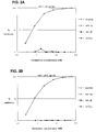
- the chemokines RANTES, MTP-1 ⁇ and MIP-1 ⁇ were obtained from R & D systems (Minneapolis, MN). They were tested in the RET assay for ability to inhibit fusion between HeLa-env JR-FL cells (expressing gp120/gp41 from the macrophage tropic isolate HIV-1 JR-FL ) and PM1 cells, or for inhibition of fusion between HeLa-env LAI cells (expressing gp120/gp41 from the laboratory-adapted strain HIV-1 LAI ) and various CD4' T lymphocyte cell lines. As shown in Figure 1 , all three chemokines inhibited fusion mediated by the macrophage tropic virus envelope glycoprotein, but not that mediated by the laboratory-adapted strain envelope glycoprotein.
- the non-chemokines include chemokine fragments and chemokine derivatives that are tested in the RET assay to determine which are active in inhibiting HIV-1 membrane fusion. Particular attention is focused on fragments or derivatives that inhibit HIV-1 fusion but do not activate leukocyte responses. These non-chemokines include:
- the current invention includes N-terminal deletants of RANTES, MIP-1 ⁇ and MIP-1 ⁇ , lacking the biological activity of the native proteins, which inhibit HIV-1 fusion and HIV-1 infection.
- Chemokine receptors required for HIV-1 fusion are cloned by the following strategy.
- First a cDNA library is made in a mammalian expression vector (e.g. pcDNA3.1 from Invitrogen Corp. San Diego, CA) using mRNA prepared from the PM1 cell line or CD4 + T-lymphocytes or macrophages. Degenerate oligonucleotide probes are used to identify members of the cDNA library encoding members of the chemokine receptor family, for example following previously published methods (2).
- the vectors containing chemokine receptor cDNAs are then individually expressed in one of several mammalian cell lines which express human CD4 but do not fuse with HeLa-env JR-FL cells (e.g.
- HeLa-CD4, CHO-CD4 or COS-CD4) or HeLa-env LAI cells e.g. CHO-CD4 or COS-CD4.
- clones which gain the ability to fuse with HeLa-env JI-FL or HeLa-env LAI are identified and the coding sequences recovered, for example by PCR amplification, following procedures well known to those skilled in the art. DNA sequencing is then performed to determine whether the cDNA recovered encodes a known chemokine receptor.
- monoclonal and polyclonal antibodies are prepared and tested for ability to inhibit infection by a panel of HIV-1 isolates.
- N-C ⁇ -chemokines MIP-1 ⁇ , MIP-1 ⁇ and RANTES (1,2) T-cell line-adapted (TCLA) or syncytium-inducing (SI) primary strains are insensitive (2,3).
- TCLA T-cell line-adapted
- SI syncytium-inducing
- Fusin is not known to be a ⁇ -chemokine receptor (7-9).
- ⁇ -chemokines inhibit HIV-1 replication
- NL4/3 ⁇ env which also carries the luciferase reporter gene
- envelope glycoproteins expressed in trans was used (10,11).
- Various env-complemented viruses were tested in PM1 cells, a variant of HUT-78 that has the unique ability to support replication of primary and TCLA HIV-1 strains, allowing comparison of envelope glycoprotein functions against a common cellular background (2,12).
- MIP-1 ⁇ , MIP-1 ⁇ and RANTES are most active against HIV-1 in combination (2,3), and strongly inhibited infection of PM1 cells by complemented viruses whose envelopes are derived from the NSI primary strains ADA and BaL (Table 1a).
- Table 1 Inhibition of HIV-1 entry in PM1 cells and CD4* T-cells by ⁇ -chemokines % luciferase activity BaL ADA NL4/3 HxB2 MuLV a) PM1 cells control without virus 2 2 2 5 3 control with virus 100 100 100 100 100 +R/M ⁇ /M ⁇ (50/50/50) 2 3 92 117 100 +RANTES (100) 1 1 nd nd nd +MIP-1 ⁇ (100) 54 54 nd nd nd +MIP-1 ⁇ (100) 1 6 nd nd nd +MCP-1 (100) 46 50 nd nd nd +MCP-2 (100) 28 26 nd nd nd +MCP-3 (100) 58 46 nd nd nd b) JR-FL HxB2 MuLV LW4 CD4 + T-cells control without virus 1 1 1 control with virus 100 100 100 +R/M ⁇ /M ⁇ (200/200/200) 14 68 nd LW5 CD4 + T-cells control without
- PM1 cells were cultured as described by Lusso et al (12). Ficoll / hypague-isolated PBMC from laboratory workers (LW) stimulated with PHA for 72h before depletion of CD8+ Lymphocytes with anti-CDB immunomagnetic beads (DYNAL, Great Neck, NY). CD4+ Lymphocytes were maintained in culture medium containing interleukin-2 (100U / ml; Hofmann LaRoche, Nutley, NJ), as described previously (3).
- interleukin-2 100U / ml; Hofmann LaRoche, Nutley, NJ
- Target cells (1-2x10 5 ) were infected with supernatants (10-50ng of HIV-1 p24) from 293-cells co-transfected with an NL4 / 3 ⁇ env-luciferase vector and a HIV-1 env-expressing vector (10,11).
- ⁇ -Chemokines (R & D Systems, Minneapolis) were added to the target cells simultaneously with virus, at the final concentrations (ng / ml) indicated in parentheses in the first column.
- the ⁇ -chemokine concentration range was selected based on prior studies (2,3). After 2h, the cells were washed twice with PBS, resuspended in ⁇ -chemokine-containing media and maintained for 48-96h.
- Luciferase activity in cell lysates was measured as described previously (10,11). The values indicated represent luciferase activity (cpm) / ng p24 / mg protein, expressed relative to that in virus-control cultures lacking ⁇ -chemokines (100%), and are the means of duplicate or sextuplicate determinations. nd, not done. R / M ⁇ / M ⁇ , RANTES + MIP-1 ⁇ + MIP-1 ⁇ .
- RANTES and MIP-1 ⁇ were strongly active when added individually, while other ⁇ -chemokines - MIP-1 ⁇ , MCP-1, MCP-2 and MCP-3 (refs. 13-15) - were weaker inhibitors (Table la).
- MIP-1 ⁇ , MIP-1 ⁇ and RANTES did not inhibit infection of PM1 cells by the TCLA strains NL4/3 and HxB2, or by the amphotropic murine leukemia virus (MuLV-Ampho) pseudotype (Table la).
- MIP-1 ⁇ , MIP-1 ⁇ and RANTES did not inhibit infection of PM1 cells by the TCLA strains NL4/3 and HxB2, or by the amphotropic murine leukemia virus (MuLV-Ampho) pseudotype (Table la).
- MuLV-Ampho amphotropic murine leukemia virus
- the env -complementation assay was used to assess HIV-1 entry into CD4+ T-cells from two control individuals (LW4 and LW5).
- MIP-1 ⁇ , MIP-1 ⁇ and RANTES strongly inhibited infection by the NSI primary strain JR-FL infection of LW4's and LW5's CD4 + T-cells, and weakly reduced HxB2 infection of LW cells (Table 1b), suggesting that there may be some overlap in receptor usage on activated CD4 + T-cells by different virus strains.
- BaL env-mediated replication in normal PBL was also inhibited by MIP-1 ⁇ , MIP-1 ⁇ and RANTES, albeit with significant inter-donor variation in sensitivity (data not shown).
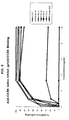
- ⁇ -chemokines inhibited HIV-1 replication by showing that complete inhibition of infection of PM1 cells required the continuous presence of ⁇ -chemokines for up to 5h after addition of ADA or BaL env-complemented virus ( Fig.3a ).
- Pre-treatment of the cells with ⁇ -chemokines for 2h or 24h prior to infection had no inhibitory effect if the cells were subsequently washed before virus addition.
- adding ⁇ -chemokines 2h after virus only minimally affected virus entry ( Fig.3a ).
- a PCR-based assay was next used to detect HIV-1 early DNA reverse transcripts in PM1 cells after 10h of infection; reverse transcription of ADA, but not of NL4/3, could not be detected in the presence of MIP-1 ⁇ and RANTES ( Fig.3b ).
- ⁇ -chemokines requires their presence during at least one of the early stages of HIV-1 replication: virus attachment, fusion and early reverse transcription.
- ⁇ -chemokines inhibited binding of JR-FL or BRU (LAI) gp120 to soluble CD4, or of tetrameric CD4-IgG2 binding to HeLa-JR-FL cells expressing oligomeric envelope glycoproteins (17).
- LAI JR-FL or BRU
- tetrameric CD4-IgG2 binding to HeLa-JR-FL cells expressing oligomeric envelope glycoproteins
- Cell-cell membrane fusion is also induced by the gp120-CD4 interaction, and can be monitored directly by resonance energy transfer (RET) between fluorescent dyes incorporated into cell membranes (17).
- RET resonance energy transfer
- OKT4a completely inhibits membrane fusion of PM1 cells with HeLa cells expressing the envelope glycoproteins of either JR-FL (HeLa-JR-FL, the same cell line referred to above as HeLa-env JR-FL ) or BRU (HeLa-BRU, the same cell line referred to above as HeLa-env LAI ), confirming the specificity of the process (17).
- MIP-1 ⁇ (and to a lesser extent, MIP-1 ⁇ ) strongly inhibited membrane fusion of HeLa-JR-FL cells with PM1 cells, whereas fusion between PM1 cells and HeLa-BRU cells was insensitive to these ⁇ -chemokines ( Fig. 1 and Table 2a).
- CD4 + target cells mitogen-activated CD4 + lymphocytes or PM1 cells
- octadecyl rhodamine Molecular Probes, Eugene, OR
- HeLa-JR-FL cells HeLa-BRU cells (or control HeLa cells, not shown) were labeled with octadecyl fluorescein (Molecular Probes), overnight at 37°C.
- Equal numbers of labeled target cells and env-expressing cells were mixed in 96-well plates and ⁇ -chemokines (or CD4 MAb OKT4a) were added at the final concentrations (ng / ml) indicated in parentheses in the first column. Fluorescence emission values were determined 4h after cell mixing (17).
- RET resonance energy transfer
- the %RET value is defined by a calculation described elsewhere(17), and each is the mean of triplicate determinations. These values were, for HeLa-JR-FL and HeLa-BRU cells respectively: PM1 cells 11.5%, 10.5%; LW5 CD4 + cells, 6.0%, 10.5%; R / M ⁇ / M ⁇ , RANTES + MIP-1 ⁇ + MIP-1 ⁇ .
- C-C CKR-1 and, especially, C-C CKR-5 were identified as the most likely candidates, based on tissue expression patterns and their abilities to bind MIP-1 ⁇ , MIP-1 ⁇ and RANTES (4,7,8,15,22).
- C-C CKR-1, C-C CKR-5 and LESTR are each expressed at the mRNA level in PM1 cells and primary macrophages (data not shown). These and other ⁇ -chemokine receptors were therefore PCR-amplified, cloned and expressed.
- C-C CKR-5 The second receptor function of C-C CKR-5 was confirmed in assays of env-mediated membrane fusion.
- C-C CKR-5 was transiently expressed in COS and HeLa cell lines that permanently expressed human CD4, both cell lines fused strongly with HeLa cells expressing the JR-FL envelope glycoproteins, whereas no fusion occurred when control plasmids were used (data not shown).
- Expression of LESTR instead of C-C CKR-5 did not permit either COS-CD4 or HeLa-CD4 cells to fuse with HeLa-JR-FL cells, but did allow fusion between COS-CD4 cells and HeLa-BRU cells (data not shown).
- the fusion capacity of ⁇ -chemokine receptors was also tested in the RET assay.
- the extent of fusion between HeLa-JR-FL cells and C-C CKR-5-expressing HeLa-CD4 cells was greater than the constitutive level of fusion between HeLa-BRU cells and HeLa-CD4 cells ( Fig.4 ).
- the fusion-conferring function of C-C CKR-5 for primary, NSI HIV-1 strains has therefore been confirmed in two independent fusion assays.
- the chemokine SDF-1 (stromal cell-derived factor 1) is the natural ligand for Fusin/CXCR4 and blocks infection by laboratory-adapted strains of HIV-1 (Ref. 1 and 2).
- SDF-1 exists as at least two forms, SDF-1 ⁇ and SDF-1 ⁇ based on variable splicing of the SDF-1 gene (Ref. 1 and 3)
- this chemokine specifically inhibits membrane fusion mediated by gp120/gp41 form the laboratory-adapted strain HIV LAI but not by gp120/gp41 from the macrophage-tropic isolate HIV-1 JR-FL as shown in Figure 5 .
- This assay has been adapted for drug screening purposes to a 96-well microplate format where binding of the sCD4/gp120 complexes to CCR5 + /CD4 - cells is measured using a fluorometric plate reader.
- One method is as follows:
- the bicyclam JM3100 obtained from Dr. J. Moore (Aaron Diamond AIDS Research Center, NY) was tested for ability to inhibit membrane fusion mediated by the envelope glycoproteins of the LAI or JR-FL strains of HIV-1 using the resonance energy transfer (RET) assay described above. As illustrated in Fig. 7 , this molecule specifically and potently inhibits fusion mediated by gp120/gp41 from the HIV-1 LAI strain, and not from the HIV-1 JR-FL strain. These data suggest that this molecule specifically inhibits HIV fusion by blocking the interaction between HIV-1 LAI gp120 and CXCR4.
- RET resonance energy transfer
Landscapes
- Health & Medical Sciences (AREA)
- Life Sciences & Earth Sciences (AREA)
- Chemical & Material Sciences (AREA)
- Immunology (AREA)
- Molecular Biology (AREA)
- Organic Chemistry (AREA)
- Engineering & Computer Science (AREA)
- General Health & Medical Sciences (AREA)
- Biochemistry (AREA)
- Medicinal Chemistry (AREA)
- Biomedical Technology (AREA)
- Hematology (AREA)
- Proteomics, Peptides & Aminoacids (AREA)
- Urology & Nephrology (AREA)
- Genetics & Genomics (AREA)
- Biophysics (AREA)
- Gastroenterology & Hepatology (AREA)
- Cell Biology (AREA)
- Toxicology (AREA)
- Zoology (AREA)
- Biotechnology (AREA)
- Virology (AREA)
- Pathology (AREA)
- Food Science & Technology (AREA)
- Analytical Chemistry (AREA)
- Physics & Mathematics (AREA)
- Microbiology (AREA)
- General Physics & Mathematics (AREA)
- AIDS & HIV (AREA)
- Tropical Medicine & Parasitology (AREA)
- Medicines That Contain Protein Lipid Enzymes And Other Medicines (AREA)
- Peptides Or Proteins (AREA)
- Medicines Containing Material From Animals Or Micro-Organisms (AREA)
- Pharmaceuticals Containing Other Organic And Inorganic Compounds (AREA)
- Metal-Oxide And Bipolar Metal-Oxide Semiconductor Integrated Circuits (AREA)
- Medicines Containing Plant Substances (AREA)
- Micro-Organisms Or Cultivation Processes Thereof (AREA)
Abstract
Description
- Throughout this application, various references are referred to within parentheses. Full bibliographic citation for these references may be found at the end of each series of experiments.
- Chemokines are a family of related soluble proteins of molecular weight between 8 and 10KDa, secreted by lymphocytes and other cells, which bind receptors on target cell surfaces resulting in the activation and mobilization of leukocytes, for example in the inflammatory process. Recently, Cocchi et al. demonstrated that the chemokines RANTES, MIP-1α and MIP-1β are factors produced by CD8* T lymphocytes which inhibit infection by macrophage-tropic primary isolates of HIV-1, but not infection by laboratory-adapted strains of the virus (1). These chemokines are members of the C-C group of chemokines, so named because they have adjacent cysteine residues, unlike the C-X-C group which has a single amino acid separating these residues (2). While Cocchi et al. found that expression of HIV-1 RNA was suppressed by treatment with the chemokines, they did not identify the site of action of these molecules.
- A resonance energy transfer (RET) assay of HIV-1 envelope glycoprotein-mediated membrane fusion was used to determine whether fusion mediated by the envelope glycoprotein from the primary macrophage-tropic isolate of HIV-1JR-FL would be specifically inhibited by chemokines, when compared with fusion mediated by the envelope glycoprotein from the laboratory-adapted T lymphotropic strain HIV-1LAI. As described below, it was demonstrated that this is indeed the case. This demonstrates that some chemokine receptors are fusion accessory molecules required for HIV-1 infection. Previous studies have indicated that unidentified cell surface molecules are required for virus entry in addition to the HIV-1 receptor, CD4. While CD4 is required for HIV-1 attachment, the accessory molecules are required for the membrane fusion step of entry. These accessory molecules are generally expressed only on human cells, so HIV-1 does not infect non-human CD4+ cells (3-6). Moreover it is possible to complement non-human CD4+ cells by fusing them (using polyethylene glycol) with CD4- human cells, resulting in a heterokaryon which is a competent target for HIV-1 envelope-mediated membrane fusion (7,8). These studies have been performed using laboratory-adapted T lymphotropic strains of the virus.
- In some cases, it appears that fusion accessory molecules are found on a subset of human CD4+ cells and are required for infection by HIV-1 isolates with particular tropisms. For example, macrophage-tropic primary strains of HIV-1 such as HIV-1JR-FL may have different requirements for accessory molecules compared with laboratory-adapted T lymphotropic strains such as HIV-1LAI. This phenomenon may explain differences in tropism between HIV-1 strains.
- The current invention relates to new therapies for HIV-1 infection. It was demonstrated for the first time that chemokines act at the fusion step of HIV-1 entry and specifically inhibit membrane fusion mediated by the envelope glycoprotein of primary macrophage-tropic primary viral isolates, not laboratory-adapted T lymphotrophic strains of the virus. Primary macrophage-tropic isolates of the virus are of particular importance since they are the strains usually involved in virus transmission, and may have particular importance in the pathogenesis of HIV-1 infection.
- These results were obtained using a resonance energy transfer (RET) assay of HIV-1 envelope-mediated membrane fusion. Moreover, this assay is used to identify non-chemokines, including fragments of chemokines and modified chemokines, that inhibit HIV-1 envelope glycoprotein-mediated membrane fusion and thereby neutralize the virus, yet do not induce an inflammatory response.
- This invention discloses a method for inhibiting fusion of HIV-1 to CD4+ cells which comprises contacting CD4 cells with a non-chemokine agent capable of binding to a chemokine receptor in an amount and under conditions such that fusion of HIV-1 to the CD4+ cells is inhibited.
- This invention also discloses a method for inhibiting HIV-1 infection of CD4+ cells which comprises contacting CD4+ cells with a non-chemokine agent capable of binding to a chemokine receptor in an amount and under conditions such that fusion of HIV-1 to the CD4+ cells is inhibited, thereby inhibiting the HIV-1 infection.
- This invention further discloses non-chemokine agents capable of binding to the chemokine receptor and inhibiting fusion of HIV-1 to CD4+ cells.
- This invention discloses an agent which is capable of binding to fusin and inhibiting infection. In an embodiment, the agent is an oligopeptide. In another embodiment, the agent is an polypeptide. In still another embodiment, the agent is an antibody or a portion of an antibody. In a separate embodiment, the agent is a nonypeptidyl agent.
- In addition, this invention discloses pharmaceutical compositions comprising an amount of the above non-chemokine agents or agents capable of binding to fusin effective to inhibit fusion of HIV-1 to CD4+ cells and a pharmaceutically acceptable carrier.
- This invention discloses a composition of matter capable of binding to the chemokine receptor and inhibiting fusion cf HIV-1 to CD4+ cells comprising a non-chemokine agent linked to a ligand capable of binding to a cell surface receptor of the CD4+ cells other than the chemokine receptor such that the binding of the non-chemokine agent to the chemokine receptor does not prevent the binding of the ligand to the other receptor.
- This invention also discloses a pharmaceutical composition comprising an amount of the above-described composition of matter effective to inhibit fusion of HIV-1 to CD4+ cells and a pharmaceutically acceptable carrier.
- This invention discloses a composition of matter capable of binding to the chemokine receptor and inhibiting fusion of HIV-1 to CD4+ cells comprising a non-chemokine agent linked to a compound capable of increasing the in vivo half-life of the non-chemokine agent.
- This invention also discloses a pharmaceutical composition comprising an amount of a composition of matter comprising a non-chemokine agent linked to a compound capable of increasing the in vivo half-life of the non-chemokine agent effective to inhibit fusion of HIV-1 to CD4+ cells and a pharmaceutically acceptable carrier.
- This invention discloses methods for reducing the likelihood of HIV-1 infection in a subject comprising administering an above-described pharmaceutical composition to the subject. This invention also discloses methods for treating HIV-1 infection in a subject comprising administering an above-described pharmaceutical composition to the subject.
- This invention also provides methods for determining whether a non-chemokine agent is capable of inhibiting the fusion of HIV-1 to a CD4+ cell which comprise: (a) contacting (i) a CD4+ cell which is labeled with a first dye and (ii) a cell expressing the HIV-1 envelope glycoprotein on its surface which is labeled with a second dye, in the presence of an excess of the agent under conditions permitting the fusion of the CD4+ cell to the cell expressing the HIV-1 envelope glycoprotein on its surface in the absence of the agent, the first and second dyes being selected so as to allow resonance energy transfer between the dyes; (b) exposing the product of step (a) to conditions which would result in resonance energy transfer if fusion has occurred; and (c) determining whether there is a reduction of resonance energy transfer, when compared with the resonance energy transfer in the absence of the agent, a decrease in transfer indicating that the agent is capable of inhibiting fusion of HIV-1 to CD4+ cells.
-
- Figure 1.
- Membrane fusion mediated by the HIV-1JR-FL envelope glycoprotein is inhibited by RANTES, MIP-1α and MIP-1β.
%RET resulting from the fusion of PM1 cells and HeLa-envJR-FL (■) or HeLa-envLAI (◆) was measured in the presence and absence of recombinant human chemokines at a range of concentrations: RANTES (80 - 2.5 ng/ml), MIP-1α (400 - 12.5 ng/ml) and MIP-1β (200 - 6.25 ng/ml), as indicated. Chemokines were added simultaneously with the cells at the initiation of a four hour incubation. Data are representative of more than three independent experiments which were run in duplicate. The percent inhibition of RET is defined as follows:
- Figure 2.
- CD4:HIV-1 gp120 binding in the presence of human chemokines.
The binding of soluble human CD4 to HIV-1LAI and HIV-1JR-FL gp120 was determined in an ELISA assay in the presence and absence of the monoclonal antibody OKT4A or recombinant human chemokines at a range of concentrations, identical to those used in the RET inhibition studies ofFigure 1 : OKT4A (62 - 0.3 nM), RANTES (10.3 - 0.3 nM), MIP-1α (53.3 - 2.9 nM), and MIP-1β (25.6 - 0.8 nM). Inhibitors were added simultaneously with biotinylated HIV-1 gp120 to soluble CD4 coated microtiter plates (Dynatech Laboratories, Inc., Chantilly, VA). Following a two hour incubation at room temperature and extensive washing, an incubation with streptavidin-horseradish peroxidase was performed for one hour at room temperature. Following additional washes, substrate was added and the OD at 492 nm determined in an ELISA plate reader. Data are representative of two independent experiments which were run in quadruplicate. - Figure 3.
-
Specificity time course and stage of β-chemokine inhibition of HIV-1 replication.
- (a) PM1 cells (1 x10⁶) were preincubated with RANTES + MIP- 1α + MIP-1β (R/Mα/Mβ; 100ng/ml of each) for 24h (-24h) or 2h (-2h), then washed twice with phosphate buffered saline (PBS). HIV-1 (BaL env-complemented) virus (50ng of p24; see legend to Table 1) was added for 2h, then the cells were washed and incubated for 48h before measurement of luciferase activity in cell lysates as described previously (10,11). Alternatively, virus and R/Mα/Mβ were added simultaneously to cells, and at the indicated time points (1h, 3h, etc) the cells were washed twice in PBS, resuspended in culture medium and incubated for 48h prior to luciferase assay.
Time 0 represents the positive control, to which no β-chemokines were added. +2h represents the mixture of virus with cells for 2h prior to washing twice in PBS, addition of R/Mα/Mβ and continuation of the culture for a further 48h before luciferase assay. - (b) PM1 cells (1x10⁶) were infected with HIV-1 (500pg p24) grown in CEM cells (NL4/3; lanes 1-4) or macrophages (ADA; lanes 5-8), in the presence of 500ng/ml of RANTES (
lanes 1 and 5) or MIP-1β (lanes 2 and 6), or with no β-chemokine (lanes 4 and 8).Lanes
5'-AGCAAGCCGAGTCCTGCGTCGAGAG-3' (SEQ ID NO:2) and the second round with primers: LTR-test, 5'-GGGACTTTCCGCTGGGGACTTTC 3'(SEQ ID NO :3) LRC2, 5'-CCTGTTCGGGCGCCACTGCTAGAGATTTTCCAC 3' (SEQ ID NO:4) in a Perkin Elmer 2400 cycler with the following amplification cycles: 94°C for 5 min, 35 cycles of 94°C for 30s, 55°C for 30s, 72°C for 30s, 72°C for 7 min. M indicates 1kb DNA ladder; 1, 10, 100, 1000 indicate number of reference plasmid (pAD8) copies. The assay can detect 100 copies of reverse transcripts.
- (a) PM1 cells (1 x10⁶) were preincubated with RANTES + MIP- 1α + MIP-1β (R/Mα/Mβ; 100ng/ml of each) for 24h (-24h) or 2h (-2h), then washed twice with phosphate buffered saline (PBS). HIV-1 (BaL env-complemented) virus (50ng of p24; see legend to Table 1) was added for 2h, then the cells were washed and incubated for 48h before measurement of luciferase activity in cell lysates as described previously (10,11). Alternatively, virus and R/Mα/Mβ were added simultaneously to cells, and at the indicated time points (1h, 3h, etc) the cells were washed twice in PBS, resuspended in culture medium and incubated for 48h prior to luciferase assay.
- Figure 4:
- HIV-1 env-mediated membrane fusion of cells transiently expressing C-C CKR-5.
Membrane fusion mediated by β-chemokine receptors expressed in HeLa cells was demonstrated as follows: Cells were transfected with control plasmid pcDNA3.1 or plasmid pcDNA3.1-CKR constructs using lipofectin (Gibco BRL). The pcDNA3.1 plasmid carries a T7-polymerase promoter and transient expression of β-chemokine receptors was boosted by infecting cells with 1x10⁷ pfu of vaccinia encoding the T7-polymerase (vFT7.3) 4h post-lipofection (9). Cells were then cultured overnight in R18-containing media and were tested for their ability to fuse with HeLa-JR-FL cells (filled columns) or HeLa-BRU cells (hatched column) in the RET assay. The %RET with control HeLa cells was between 3% and 4% irrespective of the transfected plasmid. - Figure 5
- Membrane fusion mediated by the HIVLAI envelope glycoprotein is inhibited by SDF-1.
% RET resulting from the fusion of PM1 cells and HeLa-envJR-FL or HeLa-envLAI cells (as indicated on the graph) was measured in the presence of recombinant SDF-1α(Gryphon Science, San Francisco) at the indicated concentrations. Experimental method as described in the legend toFig. 1 . - Figure 6.
- Flow cytometric analysis of the binding of sCD4-gp120 complexes to (a)CCR5- and (b)CCR5 L1.2 cells, a murine pre-B lymphoma line.
Cells are incubated for 15 min. with equimolar (-100nM) mixtures of sCD4 and biotinylated HIV-1JRFL gp120 and then stained with a streptavidin-phycoerythrin conjugate, fixed with 2% paraformaldehyde, and analyzed by FACS. Cell number is plotted on the y-axis. - Figure 7.
- Inhibition of HIV-1 envelope-mediated cell fusion by the bicyclam JM3100, measured using the RET assay, with the cell combinations indicated.
- Figure 8.
- Binding of CD4 and gp120 to CCR5. Recombinant soluble CD4 (sCD4) and recombinant gp120 were added in a range of concentrations either individually or as an equimolar molecular complex to recombinant L1.2 cells that express human CCR5 on their cell surface. The recombinant proteins were biotinylated as indicated. Binding was detected by adding a streptavidin-phycoerythrin conjugate and measuring the fluoroescence emission at 590 nm following excitation at 530 nm. The following species were tested:
b-LAI:sCD4: complex formed between scD4 and biotinylated HIV-1LAI gp120
b-JR-FL:sCD4: complex formed between sCD4 and biotinylated HIV-1JR-EL gp120
b-sCD4 alone: biotinylated sCD4 added in the absence of gp120
b-JR-FL alone: biotinylated HIV-1JR-EL gp120 added in the absence of sCD4 These data demonstrate that complexation of soluble CD4 and gp120 is necessary for CCR5 binding, as minimal binding is observed for sCD4 or gp120 alone. The data further demonstrate that binding is observed for sCD4-gp120 complexes when the gp120 is derived from macrophage-tropic (e.g., JR-FL) but not T cell-tropic (e.g., LAI) strains of HIV, as expected from the known relationship between HIV-1 tropism and co-receptor usage. All data have been corrected for residual background binding to nontransfected CCR5- L1.2 cells. To enhance chemokine receptor expression, both transfected and parental L1.2 cells were treated with sodium butyrate prior to assay (Wu et al., J. Exp. Med. 185:1681) - Figure 9.
- The CCR5 Binding assay identifies and determines the potency of inhibitors of the gp120-CCR5 interaction.
HIV-1 Inhibitory monoclonal antibodies were added in a range of concentrations to reecmbinant L1.2 cells that express human CCR5 on their cell surface and used to compete the binding of a complex formed between sCD4 and biotinylated HIV-1JR-FL gp 120, whose binding was detected using a streptavidin-phycoerythrin conjugate. PA-8, -9,-10, -11 and -12 are Progenics' monoclonal antibodies that inhibit HIV-1 entry, while 2D7 is a commercially available (Pharmingen, San Diego, CA) ant-CCR5 monoclonal antibody that inhibits HIV-1 entry. To enhance chemokine receptor expression, both transfected and parental L1.2 cells were treated with sodium butyrate prior to assay (Wu et al., J. Exp. Med. 185:1681). - This invention discloses a method for inhibiting fusion of RIV-1 to CD4+ cells which comprises contacting CD4 cells with a non-chemokine agent capable of binding to a chemokine receptor in an amount and under conditions such that fusion of HIV-1 to the CD4+ cells is inhibited.
- This invention also discloses a method for inhibiting HIV-1 infection of CD4+ cells which comprises contacting CD4+ cells with a non-chemokine agent capable of binding to a chemokine receptor in an amount and under conditions such that fusion of HIV-1 to the CD4+ cells is inhibited, thereby inhibiting the HIV-1 infection.
- In this invention, a chemokine means RANTES, MIP-1-α, MIP-1-β or another chemokine which blocks HIV-1 infection. A chemokine receptor means a receptor capable of binding RANTES, MIP-1-α, MIP-1-β or another chemokine which blocks HIV-1 infection. Such chemokine receptor includes but not limited to CCR5, CXCR4, CCR3 and CCR-2b.
- Throughout this application, the receptor "fusin" is also named CXCR4 and the chemokine receptor C-C CKR5 is also named CCR5.
- The HIV-1 used in this application unless specified will mean clinical or primary or field isolates or HIV-1 viruses which maintain their clinical characteristics. The HIV-1 clinical isolates may be passaged in primary peripheral blood mononuclear cells. The HIV-1 clinical isolates may be macrophage-trophic.
- The non-chemokine agents of this invention are capable of binding to chemokine receptors and inhibiting fusion of HIV-1 to CD4+ cells. The non-chemokine agents include, but are not limited to, chemokine fragments and chemokine derivatives and analogues, but do not include naturally occurring chemokines. The non-chemokine agents include multimeric forms of the chemokine fragments and chemokine derivatives and analogues or fusion molecules which contain chemokine fragments, derivatives and analogues linked to other molecules.
- The non-chemokine agents do not include bicyclams and their derivatives as described in
U.S.Patent No. 5,021,409, issued June 4, 1991 . Some bicyclam derivatives have been previously described with antiviral activities (15, 16). - In an embodiment of this invention, the non-chemokine agent is an oligopeptide. In another embodiment, the non-chemokine agent is a polypeptide. In still another embodiment, the non-chemokine agent is an antibody or a portion thereof. Antibodies against the chemokine receptor may easily be generated by routine experiments. It is also within the level of ordinary skill to synthesize fragments of the antibody capable of binding to the chemokine receptor. In a further embodiment, the non-chemokine agent is a nonpeptidyl agent.
- Non-chemokine agents which are purely peptidyl in composition can be either chemically synthesized by solid-phase methods (Merrifield, 1966) or produced using recombinant technology in either prokaryotic or eukaryotic systems. The synthetic and recombinant methods are well known in the art.
- Non-chemokine agents which contain biotin or other nonpeptidyl groups can be prepared by chemical modification of synthetic or recombinant chemokines or non-chemokine agents. One chemical modification method involves periodate oxidation of the 2-amino alcohol present on chemokines or non-chemokine agents possessing serine or threonine as their N-terminal amino acid (Geophegan and Stroh, 1992). The resulting aldehyde group can be used to link peptidyl or non-peptidyl groups to the oxidized chemokine or non-chemokine agent by reductive amination, hydrazine, or other chemistries well known to those skilled in the art.
- As used herein, a N-terminus of a protein should mean the terminus of the protein after it has been processed. In case of a secretory protein which contains a cleavable signal sequence, the N-terminus of a secretory protein should be the terminus after the cleavage of a signal peptide.
- This invention provides a method of identifying these non-chemokine agents. One way of identifying such agents, including non-peptidyl agents, that bind to a chemokine receptor and inhibit fusion of HIV-1 to CD4+ cells is to use the following assay: 1) Incubate soluble CD4 with biotinylated gp120 from HIV-1JR-FL or HIV-1LAI ; 2) Incubate this complex with CCR5 or CXCR4-expressing cells (for HIV-1JR-FL or HIV-1LAI gp120s, respectively) that do not express CD4, in the presence of absence of a candidate inhibitor; 3) Wash and then incubate with streptavidin-phycoerythrin; and 4) Wash and then measure the amount of bound gp120 using a flow cytometer or fluorometer and calculate the degree of inhibition of binding by the inhibitor.
- Alternative methods to detect bound gp120 can also be used in place of the biotinylated gp120-streptavidin-phycoerythrin method described above. For example, peroxidase-conjugated gp120 could be used in place of the biotinylated gp120 and binding detected using an appropriate colorimetric substrate for peroxidase, with a spectrometric readout.
- This invention further discloses the non-chemokine agents identified by the above methods.
- This invention discloses a non-chemokine agent capable of binding to the chemokine receptor and inhibiting fusion of HIV-1 to CD4+ cells with the proviso that the agent is not a known bicyclam or its known derivatives. In an embodiment, the non-chemokine is a polypeptide. In a further embodiment, this polypeptide is a fragment of the chemokine RANTES (Gong et al., 1996). In a still further embodiment, the polypeptide may also comprise the RANTES sequence with deletion of the N-terminal amino acids of said sequence. The deletion may be the first eight N-terminal amino acids of the RANTES sequence (SEQ ID NO:5) .
- In a separate embodiment, the polypeptide may comprise the MIP-1β sequence with deletion of the N-terminal amino acids of said sequence. The deletion may be the first seven, eight, nine or ten N-terminal amino acids of the MIP-1β sequence.
- In another embodiment of non-chemokine agent, the polypeptide comprises the MIP-1β sequence with the N-terminal sequence modified by addition of an amino acid or oligopeptide. In a separate embodiment, the polypeptide comprises the MIP-1β sequence with the N-terminal sequence modified by removing the N-terminal alanine and replaced it by serine or threonine and additional amino acid or oligopeptide or nonpeptidyl moiety. In a further embodiment, the additional amino acid is methionine.
- As described infra in the section of Experimental Details, a cofactor for HIV-1 fusion and entry was identified and designated "fusin" (Feng et al., 1996). This invention provides an agent which is capable of binding to fusin and inhibiting infection. In an embodiment, the agent is an oligopeptide. In another embodiment, the agent is an polypeptide.
- In a further embodiment, the polypeptide comprises SDF-1 with deletion of the N-terminal amino acids of said sequence. The deletion may be the first six, seven, eight, or nine N-terminal amino acids of the SDF-1 sequence.
- This invention also discloses the above non-chemokine agent, wherein the polypeptide comprises SDF-1 sequence with the N-terminal sequence modified to produce antagonistic effect to SDF-1. One modification is to replace the N-seminal glycine of SDF-1 by serine and derivatized with biotin. Another modification is to replace the N-terminal glycine of SDF-1 by serine and derivatized with methionine. A further modification is to add the N-terminus of SDF-1 with a methionine before the terminal glycine.
- In still another embodiment, the agent is an antibody or a portion of an antibody. In a separate embodiment, the agent is a nonpeptidyl agent.
- The agents capable of binding to fusin may be identified by screening different compounds for their capability to bind to fusin in vitro.
- A suitable method has been described by Fowlkes, et al. (1994), international application number:
PCT/US94/03143 WO 94/23025 - This invention also discloses pharmaceutical compositions comprising an amount of such non-chemokine agents or agents capable of binding to fusin effective to inhibit fusion of HIV-1 to CD4+ cells and a pharmaceutically acceptable carrier.
- Pharmaceutically acceptable carriers are well known to those skilled in the art. Such pharmaceutically acceptable carriers may be aqueous or non-aqueous solutions, suspensions, and emulsions. Examples of non-aqueous solvents are propylene glycol, polyethylene glycol, vegetable oils such as olive oil, and injectable organic esters such as ethyl oleate. Aqueous carriers include water, alcoholic/aqueous solutions, emulsions or suspensions, saline and buffered media. Parenteral vehicles include sodium chloride solution, Ringer's dextrose, dextrose and sodium chloride, lactated Ringer's or fixed oils. Intravenous vehicles include fluid and nutrient replenishers, electrolyte replenishers such as those based on Ringer's dextrose, and the like. Preservatives and other additives may also be present, such as, for example, antimicrobials, antioxidants, chelating agents, inert gases and the like.
- This invention discloses a composition of matter capable of binding to the chemokine receptor and inhibiting fusion of RIV-1 to CD4+ cells comprising a non-chemokine agent linked to a ligand capable of binding to a cell surface receptor of the CD4+ cells other than the chemokine receptor such that the binding of the non-chemokine agent to the chemokine receptor does not prevent the binding of the ligand to the other receptor. In an embodiment, the cell surface receptor is CD4. In another embodiment, the ligand is an antibody or a portion of an antibody.
- This invention also provides a method of obtaining a pharmaceutical composition comprising an amount of an above-described composition of matter effective to inhibit fusion of HIV-1 to CD4+ cells and a pharmaceutically acceptable carrier.
- This invention discloses a composition of matter capable of binding to the chemokine receptor and inhibiting fusion of HIV-1 to CD4+ cells comprising a non-chemokine agent linked to a compound capable of increasing the in vivo half-life of the non-chemokine agent. In an embodiment, the compound is polyethylene glycol.
- This invention also discloses a pharmaceutical composition comprising an amount of a composition of matter comprising a non-chemokine agent linked to a compound capable of increasing the in vivo half-life of the non-chemokine agent effective to inhibit fusion of HIV-1 to CD4+ cells and a pharmaceutically acceptable carrier.
- This invention discloses methods for reducing likelihood of HIV-1 infection in a subject comprising administering the above-described pharmaceutical compositions to the subject. This invention also discloses methods for treating HIV-1 infection in a subject comprising administering the above-described pharmaceutical compositions to the subject.
- This invention also provides methods for determining whether a non-chemokine agent is capable of inhibiting the fusion of HIV-1 to a CD4+ cell which comprise: (a) contacting (i) a CD4+ cell which is labeled with a first dye and (ii) a cell expressing the HIV-1 envelope glycoprotein on its surface which is labeled with a second dye, in the presence of an excess of the agent under conditions permitting the fusion of the CD4+ cell to the cell expressing the HIV-1 envelope glycoprotein on its surface in the absence of the agent, the first and second dyes being selected so as to allow resonance energy transfer between the dyes; (b) exposing the product of step (a) to conditions which would result in resonance energy transfer if fusion has occurred; and (c) determining whether there is a reduction of resonance energy transfer, when compared with the resonance energy transfer in the absence of the agent, a decrease in transfer indicating that the agent is capable of inhibiting fusion of HIV-1 to CD4+ cells.
- HIV-1 only fuses with appropriate CD4+ cells. For example, laboratory-adapted T lymphotropic HIV-1 strains fuse with most CD4+ human cells. Clinical HIV-1 isolates do not fuse with most transformed CD4+ human cell lines but do fuse with human primary CD4+ cells such as CD4 T lymphocytes and macrophages. Routine experiments may be easily performed to determine whether the CD4+ cell is appropriate for the above fusion assay.
- As described in this invention, HIV-1 membrane fusion is monitored by a resonance energy transfer assay. The assay was described in the
International Application Number, PCT/US94/14561. filed December 16, 1994 International Publication Number WO 95/16789 08/475,515, filed June 7, 1995 . - In an embodiment of the above method, the non-chemokine agent is an oligopeptide. In another embodiment, the non-chemokine agent is a polypeptide. In still another embodiment, the agent is an antibody or a portion thereof. In a further embodiment, the non-chemokine agent is a nonpeptidyl agent.
- In a separate embodiment, the CD4+ cell is a PM1 cell. In another embodiment, the cell expressing the HIV-1 envelope glycoprotein is a HeLa cell expressing HIV-1JR-FL gp120/gp41.
- This invention provides a method for determining whether an agent is capable of inhibiting HIV-1 infection comprising steps of: (a) contacting an appropriate concentration of an agent with a chemokine receptor or a portion thereof under conditions permitting the binding of the agent to the chemokine receptor; (b) contacting the chemokine receptor resulting from step (a) with a gp120/CD4 complex under conditions permitting the binding of the gp120/CD4 complex and the chemokine receptor in the absence of the agent; (c) measuring the amount of bound gp120/CD4 complex wherein a decrease in the amount compared with the amount determined in the absence of the agent indicates that the agent is capable of inhibiting HIV-1 infection.
- As used herein, the portion of the chemokine receptor used in the above method is the portion which maintains the capability of binding to HIV, i.e. capable of interaction with the gp120/CD4 complex. It is the intention of this invention to cover hybrid molecules or genetically engineered molecules which comprise this portion or domain of the chemokine receptor.
- The gp120/CD4 complex used in the assay may include a truncated form of either molecules or hybrid proteins of molecules as long as the domain for binding to the chemokine receptor is retained.
- This invention provides a method for determining whether an agent is capable of inhibiting HIV-1 infection comprising steps of: (a) fixing a chemokine receptor on a solid matrix; (b) contacting the agent with the fixed chemokine receptor under conditions permitting the binding of the agent to the chemokine receptor; (c) removing the unbound agent; (d) contacting the fixed chemokine receptor resulting in step (c) with a gp120 in the presence of CD4 under conditions permitting the binding of the gp120/CD4 complex and the chemokine receptor in the absence of the agent; (e) measuring the amount of bound gp120/CD4 complex; and (f) comparing the amount determined in step (d) with the amount determined in the absence of the agent, a decrease of the amount indicating that the agent is capable of inhibiting HIV-1 infection.
- This invention also provides a method for determining whether an agent is capable of inhibiting HIV-1 infection comprising steps of: (a) fixing a chemokine receptor on a solid matrix; (b) contacting the agent with the fixed chemokine receptor; (c) contacting the mixture in step (b) with a gp120 in the presence of CD4 under conditions permitting the binding of the gp120/CD4 complex and the chemokine receptor in the absence of the agent; (d) measuring the amount of bound gp120/CD4 complex; and (e) comparing the amount determined in step (d) with the amount determined in the absence of the agent, a decrease of the amount indicating that the agent is capable of inhibiting HIV-1 infection.
- This invention also provides a method for determining whether an agent is capable of inhibiting HIV-1 infection comprising steps of: (a) contacting the agent with a gp120/CD4 complex under conditions permitting the binding of the agent to the gp120/CD4 complex; (b) contacting the gp120/CD4 complex resulting from step (a) with a chemokine receptor under conditions permitting the binding of the gp120/CD4 complex and the chemokine receptor in the absence of the agent; (c) measuring the amount of bound chemokine receptor, wherein a decrease of the amount when compared with the amount determined in the absence of the agent indicates that the agent is capable of inhibiting HIV-1 infection.
- This invention also provides a method for determining whether an agent is capable of inhibiting HIV-1 infection comprising steps of: (a) fixing a gp120/CD4 complex on a solid matrix; (b) contacting the agent with the fixed gp120/CD4 complex under conditions permitting the binding of the agent to the gp120/CD4 complex; (c) removing unbound agent;(d) contacting the fixed gp120/CD4 complex resulting from step (c) with a chemokine receptor under conditions permitting the binding of the gp120/CD4 complex and the chemokine receptor in the absence of the agent; (e) measuring the amount of bound chemokine receptor; and (f) comparing the amount determined in step (e) with the amount determined in the absence of the agent, a decrease of the amount indicating that the agent is capable of inhibiting HIV-1 infection.
- This invention provides a method for determining whether an agent is capable of inhibiting HIV-1 infection comprising steps of: (a) fixing a gp120/CD4 on a solid matrix; (b) contacting the agent with the fixed gp120/CD4 complex; (c) contacting the mixture in step (b) with a chemokine receptor under conditions permitting the binding of the gp120/CD4 complex and the chemokine receptor in the absence of the agent; (d) measuring the amount of bound chemokine receptor; (e) comparing the amount determined in step (d) with the amount determined in the absence of the agent, a decrease of the amount indicating that the agent is capable of inhibiting HIV-1 infection.
- As used in these assays, CD4 include soluble CD4, fragments of CD4 or polypeptides incorporating the gp120 binding site of CD4 capable of binding gp120 and enabling the binding of gp120 to the appropriate chemokine receptor.
- As used in these assay gp120 is the gp120 from an appropriate strain of HIV-1. For example, gp120 from the macrophage tropic clinical isolate HIV-1JR-FL will bind to the chemokine receptor CCR5, whereas gp120 from the laboratory adapted T-tropic strain HIV-1LAI will bind to the chemokine receptor CXCR4.
- In a preferred embodiment of the above methods, the CD4 is a soluble CD4. The chemokine receptor which may be used in the above assay includes CCR5, CXCR4, CCR3 and CCR-2b.
- In an embodiment, the chemokine receptor is expressed on a cell. In another embodiment, the chemokine receptor is embedded in liposomes. In further embodiment, the chemokine receptor is embedded in a membrane derived from cells expressing the chemokine receptor. In a preferred embodiment, the cell is a L1.2 cell. In a separate embodiment, the chemokine receptor is purified and reconstituted in liposomes. Such chemokine receptor embedded in the lipid bilayer of liposomes retains the gp120 binding activity of the receptor.
- The gp120, CD4 or both may be labelled with a detectable marker in the above assays. Markers including radioisotope or enzymes such as horse radish peroxidase may be used in this invention.
- In an embodiment, the gp120 or CD4 or the chemokine receptor is labelled with biotin. In a further embodiment, the biotinylated gp120, or CD4 or the chemokine receptor is detected by: (i) incubating with streptavidin-phycoerythrin, (ii) washing the incubated mixture resulting from step (i), and (iii) measuring the amount of bound gp120 using a plate reader, exciting at 530nm, reading emission at 590nm.
- This invention also discloses an agent determined to be capable of inhibiting HIV-1 infection by the above methods, which is previously unknown.
- This invention also discloses a pharmaceutical composition comprising the agent determined to be capable of inhibiting HIV-1 infection by the above methods and a pharmaceutically acceptable carrier. In an embodiment, the agent is an oligopeptide. In another embodiment, the agent is a polypeptide. In a still another embodiment, the agent is a nonpeptidyl agent.
- This invention also discloses a molecule capable of binding to the chemokine receptor CCR5 and inhibiting fusion of HIV-1 to CD4+ cells comprising the above determined agent linked to a compound capable of increasing the in vivo half-life of the non-chemokine agent. In an embodiment, the compound is polyethylene glycol. This invention also provides a pharmaceutical composition comprising an amount of the above molecule effective to inhibit HIV-1 infection and a pharmaceutically acceptable carrier.
- This invention discloses a method for reducing the likelihood of HIV-1 infection in a subject comprising administering the above pharmaceutical compositions to the subject.
- This invention discloses a method for treating HIV-1 infection in a subject comprising administering the above pharmaceutical composition to the subject.
- This invention will be better understood by reference to the Experimental Details which follow, but those skilled in the art will readily appreciate that the specific experiments detailed are only illustrative of the invention as described more fully in the claims which follow thereafter.
- The chemokines RANTES, MTP-1α and MIP-1β were obtained from R & D systems (Minneapolis, MN). They were tested in the RET assay for ability to inhibit fusion between HeLa-envJR-FL cells (expressing gp120/gp41 from the macrophage tropic isolate HIV-1JR-FL) and PM1 cells, or for inhibition of fusion between HeLa-envLAI cells (expressing gp120/gp41 from the laboratory-adapted strain HIV-1LAI) and various CD4' T lymphocyte cell lines. As shown in
Figure 1 , all three chemokines inhibited fusion mediated by the macrophage tropic virus envelope glycoprotein, but not that mediated by the laboratory-adapted strain envelope glycoprotein. - The ability of the chemokines to block the interaction between CD4 and HIV-1 gp120 which occurs at virus attachment was then tested. It was found that the chemokines did not inhibit this interaction (
Figure 2 ), demonstrating that their blockade of HIV-1 envelope glycoprotein-mediated membrane fusion occurs at the membrane fusion event itself, rather than the initial CD4-gp120 interaction which precedes fusion. - The non-chemokines include chemokine fragments and chemokine derivatives that are tested in the RET assay to determine which are active in inhibiting HIV-1 membrane fusion. Particular attention is focused on fragments or derivatives that inhibit HIV-1 fusion but do not activate leukocyte responses. These non-chemokines include:
- a) N-terminal derivatives of the chemokines. Addition of residues to the N-terminus of chemokines inhibits the function of these proteins without significantly reducing their ability to bind chemokine receptors. For example, Met-RANTES (RANTES with an N-terminal methionine) has been shown to be a powerful antagonist of native RANTES and is unable to induce chemotaxis or calcium mobilization in certain systems. The mechanism of antagonism appears to be competition for receptor binding (9). Similar results were found using other derivatives of the N terminus of RANTES(9) and also by N-terminal modification of other chemokines, such as IL-8 (a member of the C-X-C chemokines) (10). The current invention includes Met-RANTES and other chemokines derivatised by the addition of methionine, or other residues, to the N-terminus so that they inhibit fusion mediated by the envelope glycoprotein of HIV-1JR-FL, and inhibit infection by many isolates of HIV-1, yet do not activate the inflammatory response.
- b) Chemokines with N-terminal amino acids deleted: Chemokine antagonists have been generated by deleting amino acids in the N-terminal region. For example, deletion of up to 8 amino acids at the N-terminus of the chemokine MCP-1 (a member of the C-C chemokine group), ablated the bioactivity of the protein while allowing it to retain chemokine receptor binding and the ability to inhibit activity of native MCP-1 (11,12).
- The current invention includes N-terminal deletants of RANTES, MIP-1α and MIP-1β, lacking the biological activity of the native proteins, which inhibit HIV-1 fusion and HIV-1 infection.
- c) Other peptides: A series of overlapping peptides (e.g. of 20-67 residues) from all regions of RANTES, MIP-1α and MIP-1β are screened by the same approaches to identify peptides which inhibit HIV-1 fusion most potently without activating leukocytes. Activation of leukocyte responses is measured following routine procedures (9, 10, 11, 12).
- Chemokine receptors required for HIV-1 fusion are cloned by the following strategy. First a cDNA library is made in a mammalian expression vector (e.g. pcDNA3.1 from Invitrogen Corp. San Diego, CA) using mRNA prepared from the PM1 cell line or CD4+ T-lymphocytes or macrophages. Degenerate oligonucleotide probes are used to identify members of the cDNA library encoding members of the chemokine receptor family, for example following previously published methods (2). The vectors containing chemokine receptor cDNAs are then individually expressed in one of several mammalian cell lines which express human CD4 but do not fuse with HeLa-envJR-FL cells (e.g. HeLa-CD4, CHO-CD4 or COS-CD4) or HeLa-envLAI cells (e.g. CHO-CD4 or COS-CD4). Following analysis in the RET assay, clones which gain the ability to fuse with HeLa-envJI-FL or HeLa-envLAI are identified and the coding sequences recovered, for example by PCR amplification, following procedures well known to those skilled in the art. DNA sequencing is then performed to determine whether the cDNA recovered encodes a known chemokine receptor. Following expression of the receptor, monoclonal and polyclonal antibodies are prepared and tested for ability to inhibit infection by a panel of HIV-1 isolates.
-
- 1. Cocchi, F., DeVico, A. L., Garzino-Demo, A., Arya, S. K., Gallo, R. C., Lusso, P. 1995. Science. 270:1811-1815.
- 2. Raport, C. J., Schweickart, V. L., Chantry, D., Eddy Jr., R. L., Shows, T. B., Godiska, R., Gray, P. W. 1996. Journal of Leukocyte Biology. 59: 18-23.
- 3. Maddon PJ., Dalgleish AG., McDougal JS., Clapham PR., Weiss RA., Axel R. 1986. Cell. 47:333-348.
- 4. Ashorn PA., Berger EA., Moss B. 1990. J. Virol. 64:2149-2156.
- 5. Clapham PR., Blanc D., Weiss RA. 1991. Virology. 181:703-715.
- 6. Harrington RD., Geballe AP. 1993. J. Virol. 67:5939-5947.
- 7. Broder CC., Dimitrov DS., Blumenthal R., Berger EA. 1993. Virology. 193:483-491.
- 8. Dragic T., Charneau P., Clavel F., Alizon M. 1992. J. Virol. 66:4794-4802.
- 9. Wells, T. N., Power, C. A., Lusti-Narasimhan, M., Hoogewerf, A. J., Cooke, R. M., Chung, C. W., Peitsch, M. C., Proudfoot, A. E. 1996. Journal of Leukocyte Biology. 59:53-60.
- 10. Moser, B., Dewald, B., Barella, L., Schumacher, C., Baggiolini, M., Clark-Lewis, I. 1993. Journal of Biological Chemistry. 268:7125-7128.
- 11. Gong, J. H., Clark-Lewis, I. 1995. J. Exp. Med. 181:631-640.
- 12. Zhang, Y. J., Rutledge, B. J., Rollins, B. J. 1994. Journal of Biological Chemistry. 269:15918-15924.
- 13. Merrifield, R.B.(1963) J. Am. Chem. Soc. 85: 2149-2154.
- 14. Goeghegan, K.F. Stroh, J.F. (1992) Bioconjugate Chem. 3: 138-146.
- 15. Clercq, E.D. et al. (1994) Antimicrobial Agents and Chemotherapy 38:668-674.
- 16. Clercq, E.D. et al (1992) Proc. Natl. Acad. Sci. USA 89: 5286-5290.
- The replication of primary, non-syncytium-inducing (NSI) HIV-1 isolates in CD4+ T-cells is inhibited by the C-C β-chemokines MIP-1α, MIP-1β and RANTES (1,2), but T-cell line-adapted (TCLA) or syncytium-inducing (SI) primary strains are insensitive (2,3). The β-chemokines are small (8kDa), related proteins active on cells of the lymphoid and monocyte lineage (4-8). Their receptors are members of the 7-membrane-spanning, G-protein-linked superfamily, one of which (the LESTR orphan receptor) has been identified as the second receptor for TCLA HIV-1 strains, and is now designated fusin (9). Fusin is not known to be a β-chemokine receptor (7-9).
- To study how β-chemokines inhibit HIV-1 replication, a virus entry assay based on single-cycle infection by an env-deficient virus, NL4/3Δenv (which also carries the luciferase reporter gene), complemented by envelope glycoproteins expressed in trans was used (10,11). Various env-complemented viruses were tested in PM1 cells, a variant of HUT-78 that has the unique ability to support replication of primary and TCLA HIV-1 strains, allowing comparison of envelope glycoprotein functions against a common cellular background (2,12). MIP-1α, MIP-1β and RANTES are most active against HIV-1 in combination (2,3), and strongly inhibited infection of PM1 cells by complemented viruses whose envelopes are derived from the NSI primary strains ADA and BaL (Table 1a).
Table 1: Inhibition of HIV-1 entry in PM1 cells and CD4* T-cells by β-chemokines % luciferase activity BaL ADA NL4/3 HxB2 MuLV a) PM1 cells control without virus 2 2 2 5 3 control with virus 100 100 100 100 100 +R/Mα/Mβ (50/50/50) 2 3 92 117 100 +RANTES (100) 1 1 nd nd nd +MIP-1α(100) 54 54 nd nd nd +MIP-1β (100) 1 6 nd nd nd +MCP-1 (100) 46 50 nd nd nd +MCP-2 (100) 28 26 nd nd nd +MCP-3 (100) 58 46 nd nd nd b) JR-FL HxB2 MuLV LW4 CD4+ T-cells control without virus 1 1 1 control with virus 100 100 100 +R/Mα/Mβ (200/200/200) 14 68 nd LW5 CD4+ T-cells control without virus 1 1 1 control with virus 100 100 100 +R/Mα/Mβ (200/200/200) 15 73 nd - PM1 cells were cultured as described by Lusso et al (12). Ficoll/hypague-isolated PBMC from laboratory workers (LW) stimulated with PHA for 72h before depletion of CD8+ Lymphocytes with anti-CDB immunomagnetic beads (DYNAL, Great Neck, NY). CD4+ Lymphocytes were maintained in culture medium containing interleukin-2 (100U/ml; Hofmann LaRoche, Nutley, NJ), as described previously (3). Target cells (1-2x10⁵) were infected with supernatants (10-50ng of HIV-1 p24) from 293-cells co-transfected with an NL4/3Δenv-luciferase vector and a HIV-1 env-expressing vector (10,11). β-Chemokines (R & D Systems, Minneapolis) were added to the target cells simultaneously with virus, at the final concentrations (ng/ml) indicated in parentheses in the first column. The β-chemokine concentration range was selected based on prior studies (2,3). After 2h, the cells were washed twice with PBS, resuspended in β-chemokine-containing media and maintained for 48-96h. Luciferase activity in cell lysates was measured as described previously (10,11). The values indicated represent luciferase activity (cpm)/ng p24/mg protein, expressed relative to that in virus-control cultures lacking β-chemokines (100%), and are the means of duplicate or sextuplicate determinations. nd, not done. R/Mα/Mβ, RANTES + MIP-1α + MIP-1β.
- RANTES and MIP-1β were strongly active when added individually, while other β-chemokines - MIP-1α, MCP-1, MCP-2 and MCP-3 (refs. 13-15) - were weaker inhibitors (Table la). However, MIP-1α, MIP-1β and RANTES, in combination, did not inhibit infection of PM1 cells by the TCLA strains NL4/3 and HxB2, or by the amphotropic murine leukemia virus (MuLV-Ampho) pseudotype (Table la). Thus, phenotypic characteristics of the HIV-1 envelope glycoproteins influence their sensitivity to β-chemokines in a virus entry assay.
- The env-complementation assay was used to assess HIV-1 entry into CD4+ T-cells from two control individuals (LW4 and LW5). MIP-1α, MIP-1β and RANTES strongly inhibited infection by the NSI primary strain JR-FL infection of LW4's and LW5's CD4+ T-cells, and weakly reduced HxB2 infection of LW cells (Table 1b), suggesting that there may be some overlap in receptor usage on activated CD4+ T-cells by different virus strains. BaL env-mediated replication in normal PBL was also inhibited by MIP-1α, MIP-1β and RANTES, albeit with significant inter-donor variation in sensitivity (data not shown).
- It was determined when β-chemokines inhibited HIV-1 replication by showing that complete inhibition of infection of PM1 cells required the continuous presence of β-chemokines for up to 5h after addition of ADA or BaL env-complemented virus (
Fig.3a ). Pre-treatment of the cells with β-chemokines for 2h or 24h prior to infection had no inhibitory effect if the cells were subsequently washed before virus addition. Furthermore, adding β-chemokines 2h after virus only minimally affected virus entry (Fig.3a ). A PCR-based assay was next used to detect HIV-1 early DNA reverse transcripts in PM1 cells after 10h of infection; reverse transcription of ADA, but not of NL4/3, could not be detected in the presence of MIP-1β and RANTES (Fig.3b ). Thus, inhibition by β-chemokines requires their presence during at least one of the early stages of HIV-1 replication: virus attachment, fusion and early reverse transcription. - As described in part in the First Series of Experiments, these sites of action were discriminated, first by testing whether β-chemokines inhibited binding of JR-FL or BRU (LAI) gp120 to soluble CD4, or of tetrameric CD4-IgG2 binding to HeLa-JR-FL cells expressing oligomeric envelope glycoproteins (17). No inhibition by any of the β-chemokines was found in either assay, whereas the OKT4a CD4-MAb was strongly inhibitory in both (
Fig. 2 and data not shown). Thus, β-chemokines inhibit a step after CD4 binding, when conformational changes in the envelope glycoproteins lead to fusion of the viral and cellular membranes (18). Cell-cell membrane fusion is also induced by the gp120-CD4 interaction, and can be monitored directly by resonance energy transfer (RET) between fluorescent dyes incorporated into cell membranes (17). In the RET assay, OKT4a completely inhibits membrane fusion of PM1 cells with HeLa cells expressing the envelope glycoproteins of either JR-FL (HeLa-JR-FL, the same cell line referred to above as HeLa-envJR-FL) or BRU (HeLa-BRU, the same cell line referred to above as HeLa-envLAI), confirming the specificity of the process (17). RANTES, MIP-1β (and to a lesser extent, MIP-1α) strongly inhibited membrane fusion of HeLa-JR-FL cells with PM1 cells, whereas fusion between PM1 cells and HeLa-BRU cells was insensitive to these β-chemokines (Fig. 1 and Table 2a).Table 2:Effect of β-chemokines on HIV-1 envelope glycoprotein-mediated membrane fusion measured using the RET assay % Fusion HeLa-JR-FL HeLa-BRU a) PM1 cells no chemokines 100 100 +R/Mα/Mβ (80/400/100) 1 95 +RANTES (80) 8 100 +MIP-1α (400) 39 100 +MIP-1β (100) 13 93 +MCP-1 (100) 99 98 +MCP-2 (100) 72 93 +MCP-3 (100) 98 99 b) LW5 CD4+ cells no chemokines 100 100 +R/Mα/Mβ(106/533/133) 39 100 +RANTES (106) 65 95 +MIP-1α (533) 72 100 +MIP-1β (133) 44 92 +OKT4A (3ug/ml) 0 0 - CD4+ target cells (mitogen-activated CD4+ lymphocytes or PM1 cells) were labeled with octadecyl rhodamine (Molecular Probes, Eugene, OR), and HeLa-JR-FL cells, HeLa-BRU cells (or control HeLa cells, not shown) were labeled with octadecyl fluorescein (Molecular Probes), overnight at 37°C. Equal numbers of labeled target cells and env-expressing cells were mixed in 96-well plates and β-chemokines (or CD4 MAb OKT4a) were added at the final concentrations (ng/ml) indicated in parentheses in the first column. Fluorescence emission values were determined 4h after cell mixing (17). If cell fusion occurs, the dyes are closely associated in the conjoined membrane such that excitation of fluorescein at 450nm results in resonance energy transfer (RET) and emission by rhodamine at 590nm. Percentage fusion is defined as equal to 100 x [(Exp RET - Min RET) / (Max RET - Min RET)], where Max RET = %RET obtained when HeLa-Env and CD4+ cells are mixed, Exp RET = %RET obtained when HeLa-Env and CD4+ cells are mixed in the presence of fusion-inhibitory compounds, and Min RET = %RET obtained when HeLa cells (lacking HIV-1 envelope glycoproteins) and CD4+ cells are mixed. The %RET value is defined by a calculation described elsewhere(17), and each is the mean of triplicate determinations. These values were, for HeLa-JR-FL and HeLa-BRU cells respectively: PM1 cells 11.5%, 10.5%; LW5 CD4+ cells, 6.0%, 10.5%; R/Mα/Mβ, RANTES + MIP-1α + MIP-1β.
- Similar results were obtained with primary CD4+ T-cells from LW5 (Table 2b), although higher concentrations of β-chemokines were required to inhibit membrane fusion in the primary cells than in PM1 cells. Thus, the actions of the β-chemokines are not restricted to the PM1 cell line. The RET assay demonstrates that β-chemokines interfere with env-mediated membrane fusion.
- The simplest explanation of these results is that the binding of certain β-chemokines to their receptor(s) prevents, directly or otherwise, the fusion of HIV-1 with CD4' T-cells. It has been known for a decade that HIV-1 requires a second receptor for entry into CD4+ cells (19-21). This function is supplied, for TCLA strains, by fusin (9). Several receptors for MIP-1α, MIP-1β and RANTES have been identified (6,7), and β-chemokines exhibit considerable cross-reactivity in receptor usage (4-8). However, C-C CKR-1 and, especially, C-C CKR-5 were identified as the most likely candidates, based on tissue expression patterns and their abilities to bind MIP-1α, MIP-1β and RANTES (4,7,8,15,22). C-C CKR-1, C-C CKR-5 and LESTR are each expressed at the mRNA level in PM1 cells and primary macrophages (data not shown). These and other β-chemokine receptors were therefore PCR-amplified, cloned and expressed.
- The expression of C-C CKR-5 in HeLa-CD4 (human), COS-CD4 (simian) and 3T3-CD4 (murine) cells rendered each of them readily infectible by the primary, NSI strains ADA and BaL in the env-complementation assay of HIV-1 entry (Table 3).


- Neither LESTR nor C-C CKR-1, -2a, -3 or -4 could substitute for C-C CKR-5 in this assay. The expression of LESTR in COS-CD4 and 3T3-CD4 cells permitted HxB2 entry, and HxB2 readily entered untransfected (or control plasmid-transfected) HeLa-CD4 cells (Table 3). Entry of BAL and ADA into all three C-C CKR-5-expressing cell lines was almost completely inhibited by the combination of MIP-1α, MIP-1β and RANTES, whereas HxB2 entry into LESTR-expressing cells was insensitive to β chemokines (Table 3). These results suggest that C-C CKR-5 functions as a β-chemokine-sensitive second receptor for primary, NSI HIV-1 strains.
- The second receptor function of C-C CKR-5 was confirmed in assays of env-mediated membrane fusion. When C-C CKR-5 was transiently expressed in COS and HeLa cell lines that permanently expressed human CD4, both cell lines fused strongly with HeLa cells expressing the JR-FL envelope glycoproteins, whereas no fusion occurred when control plasmids were used (data not shown). Expression of LESTR instead of C-C CKR-5 did not permit either COS-CD4 or HeLa-CD4 cells to fuse with HeLa-JR-FL cells, but did allow fusion between COS-CD4 cells and HeLa-BRU cells (data not shown).
- The fusion capacity of β-chemokine receptors was also tested in the RET assay. The expression of C-C CKR-5, but not of C-C CKR-1, -2a, -3 or -4, permitted strong fusion between HeLa-CD4 cells and HeLa-JR-FL cells. The extent of fusion between HeLa-JR-FL cells and C-C CKR-5-expressing HeLa-CD4 cells was greater than the constitutive level of fusion between HeLa-BRU cells and HeLa-CD4 cells (
Fig.4 ). The fusion-conferring function of C-C CKR-5 for primary, NSI HIV-1 strains has therefore been confirmed in two independent fusion assays. - Together, the above results establish that MlP-1α, MIP-1β and RANTES inhibit HIV-1 infection at the entry stage, by interfering with the virus-cell fusion reaction subsequent to CD4 binding. It was also shown that C-C CKR-5 can serve as a second receptor for entry of primary NSI strains of HIV-1 into CD4+ T-cells, and that the interaction of β-chemokines with C-C CKR-5 inhibits the HIV-1 fusion reaction.
-
- 1. Levy,J.A., Mackewicz,C.E. & Barker,E. Immunol. Today 17, 217-224 (1996).
- 2. Cocchi,F. et al. .
- 3. Paxton,W.A. et al. Nat. Med. 2, 412-417 (1996).
- 4. Neote,K., DiGregorio,D., Mak,J.Y., Horuk,R., & Schall,T.J. Cell 72, 415-425 (1993).
- 5. Gao,J.-L. et al. J . Exp. Med. 177, 1421 -1427 (1993) .
- 6. Bacon,K.B., Premack,B.A., Gardner,P. & Schall,T.J. Science 269, 1727-1729 (1995).
- 7. Raport,C.J. etal. J. Leukoc. Biol. 59,18-23 (1996).
- 8. Wells,T.N.C. et al. J. Leukoc. Biol. 59, 53-60 (1996) .
- 9. Feng,Y., Broder,C.C., Kennedy,P.E. & Berger,E.A. Science 272, 872-877 (1996).
- 10. Chen,B.K., Saksela,K., Andino,R. & Baltimore,D. J. Virol. 68, 654-660 (1994).
- 11. Connor,R.I., Chen,B.K., Choe,S., & Landau,N.R. Virology 206, 935-944 (1995).
- 12. Lusso,P. etal. J. Virol. 69, 3712-3720 (1995).
- 13. Charo,I.F. et al. Proc. Natl. Acad. Sci. USA 91, 2752-2756 (1994).
- 14. Ben-Baruch,A. et al. J. Biol. Chem. 270, 22123-22128 (1995).
- 15. Combadiere,C etal. J. Biol. Chem. 270, 29671-29675 (1995).
- 16. Lip,J.P., D'Andrea,A.D., Lodish,H.F. & Baltimore,D. Nature 343, 762-764 (1990).
- 17. Litwin,V. et al. J. Virol. (submitted for publication).
- 18. Moore,J.P., Jameson,B.A., Weiss,R.A. & Sattentau,Q.J. in Viral Fusion Mechanisms (ed Bentz,J.) 233-289 (CRC Press Inc, Boca Raton, USA,1993).
- 19. Maddon,P.J. et al. Cell 47, 333-348 (1986).
- 20. Ashorn,P.A., Berger,E.A. & Moss,B. J.Virol. 64, 2149-2156 (1990).
- 21. Clapham,P.R., Blanc,D. & Weiss,R.A. Virology 181, 703-715 (1991).
- 22. Samson,M., Labbe,o., Mollereau,C., Vassart,G. & Parmentier,.
- 23. Dragic,T., Charneau,P., Clavel,F. & Alizon,M. J.Virol. 66, 4794-4802 (1992)
- 24. Loetscher,M. et al. J.Biol.Chem. 269, 232-237 (1994).
- 25. Moore,J.P. & Ho,D.D. AIDS 9 (suppl A), S117-S136 (1995).
- 26. Trkola,A. & Moore,J.P. (unpublished data).
- 27. Chaudhuri,A., et al. 1994. J.Biol.Chem. 269, 7835-7838 (1994).
- 28. Neote,K., Mak,J.Y., Kolakowski Jr.,L.F. & Schall,T.J. Blood 84, 44-52 (1994).
- 29. Dragic,T., Picard,L. & Alizon,M. J.Virol. 69, 1013-1018 (1995).
- 30. Puri,A., Morris,S.J., Jones,P., Ryan,M. & Blumenthal,R. Virology 219, 262-267 (1996).31
- The chemokine SDF-1 (stromal cell-derived factor 1) is the natural ligand for Fusin/CXCR4 and blocks infection by laboratory-adapted strains of HIV-1 (Ref. 1 and 2). SDF-1 exists as at least two forms, SDF-1α and SDF-1β based on variable splicing of the SDF-1 gene (Ref. 1 and 3) In the RET assay, this chemokine specifically inhibits membrane fusion mediated by gp120/gp41 form the laboratory-adapted strain HIVLAI but not by gp120/gp41 from the macrophage-tropic isolate HIV-1JR-FL as shown in
Figure 5 . -
- 1. Bleul, C.C., et al. (1996) Nature 382:829-833
- 2. Oberlin, E., et al. (1996) Nature 382:833-835
- 3. Shirozu, M., et al. (1995) Genomics 28:495-500
- Direct Binding of HIV-1 JR-FL gp120 to CCR5+ CD4- Cells The direct binding of HIV-1JR-FL gp120 to CCR5+ CD4- cells has been demonstrated. In this case, preincubation of the gp120 with sCD4 or another CD4-based molecule is required, presumably because this results in a conformational change in gp120 that exposes a chemokine receptor binding site.
Figure 6 illustrates the use of flow cytometry to measure the direct binding of sCD4/gp120 complexes to human CCR5-bearing murine L1.2 cells. Background levels of binding were observed with either biotinylated protein alone, or if gp120 from the laboratory-adapted strain HIV-1LAI is used in place of the HIV-1JR-FL gp120 (data not shown). - This assay has been adapted for drug screening purposes to a 96-well microplate format where binding of the sCD4/gp120 complexes to CCR5+/CD4- cells is measured using a fluorometric plate reader. One method is as follows:
- 1) Plate out L1.2-CCR5+ cells (approx. 500,000/well).
- 2) Add inhibitor for 1 hour at room temperature.
- 3) Wash and add biotinylated sCD4(2.5µg/ml) and biotinylated HIV-1JR-FL gp120 (5µg/ml), then incubate for 2 hours at room temperature.
- 4) Wash and incubate with streptavidin-phycoerythrin (100ng/nl).
- 5) Wash and measure the amount of bound gp120/sCD4 using a fluorometric plate reader exciting at 530nm and reading emission at 590nm.
- Using this method, inhibition of binding of gp120/sCD4 to CCR5 by CC-chemokines (
Fig. 7 ) and antibodies to CCR5 that block HIV-1 infection (not shown) have been demonstrated. - The bicyclam JM3100, obtained from Dr. J. Moore (Aaron Diamond AIDS Research Center, NY) was tested for ability to inhibit membrane fusion mediated by the envelope glycoproteins of the LAI or JR-FL strains of HIV-1 using the resonance energy transfer (RET) assay described above. As illustrated in
Fig. 7 , this molecule specifically and potently inhibits fusion mediated by gp120/gp41 from the HIV-1LAI strain, and not from the HIV-1JR-FL strain. These data suggest that this molecule specifically inhibits HIV fusion by blocking the interaction between HIV-1LAI gp120 and CXCR4. -
- 1. CCR5+/L1.2 cell line
- 2. L1.2 cell line
- 3. JRFL-gp120, biotinylated
- 4. sCD4, unconjugated (Intracell, Cat #13101)
- 5. 96-well round bottom plate (Corning, cat #25850)
- 6. Streptavidin, phycoerythrin conjugated [SA-PE] (Becton Dickinson, cat #349023)
- 7. PBS without Calcium and Magnesium [PBS(-)] (Gibco BRL, cat #14190)
-
- 1. Culture CCR5+ and parental L1.2 cells and treat with sodium butyrate as described (Wu et al., J. Exp. Med 185:1681).
- 2. Add cells to 96-well plate (~3x10⁵ cells/well)
- 3. Centrifuge plate and remove supernatant.
- 4. Dilute inhibitory compounds as desired in PBS(-)/0.1% NaN3. Add 40 µl of inhibitory compounds to cells. Add 40 µl of PBS(-)/0.1% NaN3 to wells without inhibitory compounds.
- 5. Shake plate to suspend cells in solution. Incubate at room temperature for 1 hour.
- 6. Prepare an equimolar (~50nM) mixture of sCD4 and biotinylated gp120. Add 40µl of sCD4:biotinylated gp120 complex per well. (Final volume in well = 80µl). Shake plate to suspend cells in protein solution. Incubate at room temperature for one hour.
- 7. Centrifuge plate and remove supernatant. Add 200µl of PBS(-)0.1% NaN3 per well. Repeat this washing procedure, for a total of three washes.
- 8. Centrifuge plate and remove supernatant. Dilute SA-PE 1:50 in PBS(-)/0.1% NaN3 and add 40µl of diluted reagent to cells. Shake plate to suspend cells in solution. Incubate at room temperature for one hour.
- 9. Centrifuge plate as above and remove supernatant. Add 200µl of PBS(-)/0.1% NaN3 per well. Repeat this washing procedure for a total of three washes.
- 10. Centrifuge plate as above and remove supernatant. Add 200µl of PBS(-)/0.1% NaN3 per well.
- 11. Centrifuge plate and measure the fluorescence. Emission at 590nm following excitation at 530nm.
- 12. % Inhibition is calculated by using the following formula:

- Max = Average of values in wells containing [sCD4: biotinylated gp120 w/CCR5+/L1.2 cells, no inhibitor]
- Min = Average of values in wells containing sCD4:biotinylated gp120 w/ L1.2 cells, no inhibitor.
- Reading = Value in specific well
-
- (1) GENERAL INFORMATION:
- (i) APPLICANTS: Progenics Pharmaceuticals, Inc.
- (ii) TITLE OF INVENTION: A Method For Preventing HIV-1 Infection of CD4+ Cells
- (iii) NUMBER OF SEQUENCES: 27
- (iv) CORRESPONDENCE ADDRESS:
- (A) ADDRESSEE: Cooper & Dunham LLP
- (B) STREET: 1185 Avenue of the Americas
- (C) CITY: New York
- (D) STATE: New York
- (E) COUNTRY: USA
- (F) ZIP: 10036
- (v) COMPUTER READABLE FORM:
- (A) MEDIUM TYPE: Floppy disk
- (B) COMPUTER: IBM PC compatible
- (C) OPERATING SYSTEM: PC-DOS/MS-DOS
- (D) SOFTWARE: PatentIn Release #1.0, Version #1.30
- (vi) CURRENT APPLICATION DATA:
- (A) APPLICATION NUMBER:
- (B) FILING DATE:
- (C) CLASSIFICATION:
- (viii) ATTORNEY/AGENT INFORMATION:
- (A) NAME: White, John P
- (B) REGISTRATION NUMBER: 28678
- (C) REFERENCE/DOCKET NUMBER: 50875-F-PCT/JPW/AKC
- (ix) TELECOMMUNICATION INFORMATION:
- (A) TELEPHONE: 212-278-0400
- (B) TELEFAX: 212-391-0525
- (2) INFORMATION FOR SEQ ID NO:1:
- (i) SEQUENCE CHARACTERISTICS:
- (A) LENGTH: 38 nucleotides
- (B) TYPE: nucleic acid
- (C) STRANDEDNESS: single
- (D) TOPOLOGY: linear
- (ii) MOLECULE TYPE: oligonucleotide
- (xi) SEQUENCE DESCRIPTION: SEQ ID NO:1:
CAAGGCTACT TCCCTGATTG GCAGAACTAC ACACCAGG 38
- (i) SEQUENCE CHARACTERISTICS:
- (2) INFORMATION FOR SEQ ID NO:2:
- (i) SEQUENCE CHARACTERISTICS:
- (A) LENGTH: 25 nucleotides
- (B) TYPE: nucleic acid
- (C) STRANDEDNESS: single
- (D) TOPOLOGY: linear
- (ii) MOLECULE TYPE: oligonucleotide
- (xi) SEQUENCE DESCRIPTION: SEQ ID NO:2:
AGCAAGCCGA GTCCTGCGTC GAGAG 25
- (i) SEQUENCE CHARACTERISTICS:
- (2) INFORMATION FOR SEQ ID NO:3:
- (i) SEQUENCE CHARACTERISTICS:
- (A) LENGTH: 23 nucleotides
- (B) TYPE: nucleic acid
- (C) STRANDEDNESS: single
- (D) TOPOLOGY: linear
- (ii) MOLECULE TYPE: oligonucleotide
- (xi) SEQUENCE DESCRIPTION: SEQ ID NO:3:

- (i) SEQUENCE CHARACTERISTICS:
- (2) INFORMATION FOR SEQ ID NO:4:
- (i) SEQUENCE CHARACTERISTICS:
- (A) LENGTH: 33 nucleotides
- (B) TYPE: nucleic acid
- (C) STRANDEDNESS: single
- (D) TOPOLOGY: linear
- (ii) MOLECULE TYPE: oligonucleotide
- (xi) SEQUENCE DESCRIPTION: SEQ ID NO:4:
CCTGTTCGGG CGCCACTGCT AGAGATTTTC CAC 33
- (i) SEQUENCE CHARACTERISTICS:
- (2) INFORMATION FOR SEQ ID NO:5:
- (i) SEQUENCE CHARACTERISTICS:
- (A) LENGTH: 60 amino acids
- (B) TYPE: amino acid
- (C) STRANDEDNESS: n/a
- (D) TOPOLOGY: n/a
- (ii) MOLECULE TYPE: protein
- (xi) SEQUENCE DESCRIPTION: SEQ ID NO:5:

- (i) SEQUENCE CHARACTERISTICS:
- (2) INFORMATION FOR SEQ ID NO:6:
- (i) SEQUENCE CHARACTERISTICS:
- (A) LENGTH: 36 nucleotides
- (B) TYPE: nucleic acid
- (C) STRANDEDNESS: single
- (D) TOPOLOGY: linear
- (ii) MOLECULE TYPE: oligonucleotide
- (xi) SEQUENCE DESCRIPTION: SEQ ID NO:6:
AAGCTTGGAGAACCAGCGGT TACCATGGAG GGGATC 36
- (i) SEQUENCE CHARACTERISTICS:
- (2) INFORMATION FOR SEQ ID NO:7:
- (i) SEQUENCE CHARACTERISTICS:
- (A) LENGTH: 30 nucleotides
- (B) TYPE: nucleic acid
- (C) STRANDEDNESS: single
- (D) TOPOLOGY: linear
- (ii) MOLECULE TYPE: oligonucleotide
- (xi) SEQUENCE DESCRIPTION: SEQ ID NO:7:
GTCTGAGTCT GAGTCAAGCT TGGAGAACCA 30
- (i) SEQUENCE CHARACTERISTICS:
- (2) INFORMATION FOR SEQ ID NO:8:
- (i) SEQUENCE CHARACTERISTICS:
- (A) LENGTH: 41 nucleotides
- (B) TYPE: nucleic acid
- (C) STRANDEDNESS: single
- (D) TOPOLOGY: linear
- (ii) MOLECULE TYPE: oligonucleotide
- (xi) SEQUENCE DESCRIPTION: SEQ ID NO:8:
CTCGAGCATC TGTGTTAGCT GGAGTGAAAA CTTGAAGACT C 41
- (i) SEQUENCE CHARACTERISTICS:
- (2) INFORMATION FOR SEQ ID NO:9:
- (i) SEQUENCE CHARACTERISTICS:
- (A) LENGTH: 30 nucleotides
- (B) TYPE: nucleic acid
- (C) STRANDEDNESS: single
- (D) TOPOLOGY: linear
- (ii) MOLECULE TYPE: oligonucleotide
- (xi) SEQUENCE DESCRIPTION: SEQ ID NO:9:
GTCTGAGTCT GAGTCCTCGA GCATCTGTGT 30
- (i) SEQUENCE CHARACTERISTICS:
- (2) INFORMATION FOR SEQ ID NO:10:
- (i) SEQUENCE CHARACTERISTICS:
- (A) LENGTH: 32 nucleotides
- (B) TYPE: nucleic acid
- (C) STRANDEDNESS: single
- (D) TOPOLOGY: linear
- (ii) MOLECULE TYPE: oligonucleotide
- (xi) SEQUENCE DESCRIPTION: SEQ ID NO:10:
AAGCTTCAGA GAGAAGCCGG GATGGAAACT CC 32
- (i) SEQUENCE CHARACTERISTICS:
- (2) INFORMATION FOR SEQ ID NO:11:
- (i) SEQUENCE CHARACTERISTICS:
- (A) LENGTH: 30 nucleotides
- (B) TYPE: nucleic acid
- (C) STRANDEDNESS: single
- (D) TOPOLOGY: linear
- (ii) MOLECULE TYPE: oligonucleotide
- (xi) SEQUENCE DESCRIPTION: SEQ ID NO:11:
GTCTGAGTCT GAGTCAAGCT TCAGAGAGAA 30
- (i) SEQUENCE CHARACTERISTICS:
- (2) INFORMATION FOR SEQ ID NO:12:
- (i) SEQUENCE CHARACTERISTICS:
- (A) LENGTH: 32 nucleotides
- (B) TYPE: nucleic acid
- (C) STRANDEDNESS: single
- (D) TOPOLOGY: linear
- (ii) MOLECULE TYPE: oligonucleotide
- (xi) SEQUENCE DESCRIPTION: SEQ ID NO:12:
CTCGAGCTGA GTCAGAACCC AGCAGAGAGT TC 32
- (i) SEQUENCE CHARACTERISTICS:
- (2) INFORMATION FOR SEQ ID NO:13:
- (i) SEQUENCE CHARACTERISTICS:
- (A) LENGTH: 30 nucleotides
- (B) TYPE: nucleic acid
- (C) STRANDEDNESS: single
- (D) TOPOLOGY: linear
- (ii) MOLECULE TYPE: oligonucleotide
- (xi) SEQUENCE DESCRIPTION: SEQ ID NO:13:
GTCTGAGTCT GAGTCCTCGA GCTGAGTCAG 30
- (i) SEQUENCE CHARACTERISTICS:
- (2) INFORMATION FOR SEQ ID NO:14:
- (i) SEQUENCE CHARACTERISTICS:
- (A) LENGTH: 32 nucleotides
- (B) TYPE: nucleic acid
- (C) STRANDEDNESS: single
- (D) TOPOLOGY: linear
- (ii) MOLECULE TYPE: oligonucleotide
- (xi) SEQUENCE DESCRIPTION: SEQ ID NO:14:
AAGCTTCAGT ACATCCACAA CATGCTGTCC AC 32
- (i) SEQUENCE CHARACTERISTICS:
- (2) INFORMATION FOR SEQ ID NO:15:
- (i) SEQUENCE CHARACTERISTICS:
- (A) LENGTH: 30 nucleotides
- (B) TYPE: nucleic acid
- (C) STRANDEDNESS: single
- (D) TOPOLOGY: linear
- (ii) MOLECULE TYPE: oligonucleotide
- (xi) SEQUENCE DESCRIPTION: SEQ ID NO:15:
GTCTGAGTCT GAGTCAAGCT TCAGTACATC 30
- (i) SEQUENCE CHARACTERISTICS:
- (2) INFORMATION FOR SEQ ID NO:16:
- (i) SEQUENCE CHARACTERISTICS:
- (A) LENGTH: 31 nucleotides
- (B) TYPE: nucleic acid
- (C) STRANDEDNESS: single
- (D) TOPOLOGY: linear
- (ii) MOLECULE TYPE: oligonucleotide
- (xi) SEQUENCE DESCRIPTION: SEQ ID NO:16:
CTCGAGCCTC GTTTTATAAA CCAGCCGAGA C 31
- (i) SEQUENCE CHARACTERISTICS:
- (2) INFORMATION FOR SEQ ID NO:17:
- (i) SEQUENCE CHARACTERISTICS:
- (A) LENGTH: 30 nucleotides
- (B) TYPE: nucleic acid
- (C) STRANDEDNESS: single
- (D) TOPOLOGY: linear
- (ii) MOLECULE TYPE: oligonucleotide
- (xi) SEQUENCE DESCRIPTION: SEQ ID NO:17:
GTCTGAGTCT GAGTCCTCGA GCCTCGTTTT 30
- (i) SEQUENCE CHARACTERISTICS:
- (2) INFORMATION FOR SEQ ID NO:18:
- (i) SEQUENCE CHARACTERISTICS:
- (A) LENGTH: 29 nucleotides
- (B) TYPE: nucleic acid
- (C) STRANDEDNESS: single
- (D) TOPOLOGY: linear
- (ii) MOLECULE TYPE: oligonucleotide
- (xi) SEQUENCE DESCRIPTION: SEQ ID NO:18:
AAGCTTCAGG GAGAAGTGAA ATGACAACC 29
- (i) SEQUENCE CHARACTERISTICS:
- (2) INFORMATION FOR SEQ ID NO:19:
- (i) SEQUENCE CHARACTERISTICS:
- (A) LENGTH: 30 nucleotides
- (B) TYPE: nucleic acid
- (C) STRANDEDNESS: single
- (D) TOPOLOGY: linear
- (ii) MOLECULE TYPE: oligonucleotide
- (xi) SEQUENCE DESCRIPTION: SEQ ID NO:19:
GTCTGAGTCT GAGTCAAGCT TCAGGGAGAA 30
- (i) SEQUENCE CHARACTERISTICS:
- (2) INFORMATION FOR SEQ ID NO:20:
- (i) SEQUENCE CHARACTERISTICS:
- (A) LENGTH: 33 nucleotides
- (B) TYPE: nucleic acid
- (C) STRANDEDNESS: single
- (D) TOPOLOGY: linear
- (ii) MOLECULE TYPE: oligonucleotide
- (xi) SEQUENCE DESCRIPTION: SEQ ID NO:20:
CTCGAGCAGA CCTAAAACAC AATAGAGAGT TCC 33
- (i) SEQUENCE CHARACTERISTICS:
- (2) INFORMATION FOR SEQ ID NO:21:
- (i) SEQUENCE CHARACTERISTICS:
- (A) LENGTH: 30 nucleotides
- (B) TYPE: nucleic acid
- (C) STRANDEDNESS: single
- (D) TOPOLOGY: linear
- (ii) MOLECULE TYPE: oligonucleotide
- (xi) SEQUENCE DESCRIPTION: SEQ ID NO:21:
GTCTGAGTCT GAGTCCTCGA GCAGACCTAA 30
- (i) SEQUENCE CHARACTERISTICS:
- (2) INFORMATION FOR SEQ ID NO:22:
- (i) SEQUENCE CHARACTERISTICS:
- (A) LENGTH: 34 nucleotides
- (B) TYPE: nucleic acid
- (C) STRANDEDNESS: single
- (D) TOPOLOGY: linear
- (ii) MOLECULE TYPE: oligonucleotide
- (xi) SEQUENCE DESCRIPTION: SEQ ID NO:22:
AAGCTTCTGT AGAGTTAAAA AATGAACCCC ACGG 34
- (i) SEQUENCE CHARACTERISTICS:
- (2) INFORMATION FOR SEQ ID NO:23:
- (i) SEQUENCE CHARACTERISTICS:
- (A) LENGTH: 30 nucleotides
- (B) TYPE: nucleic acid
- (C) STRANDEDNESS: single
- (D) TOPOLOGY: linear
- (ii) MOLECULE TYPE: oligonucleotide
- (xi) SEQUENCE DESCRIPTION: SEQ ID NO:23:
GTCTGAGTCT GAGTCAAGCT TCTGTAGAGT 30
- (i) SEQUENCE CHARACTERISTICS:
- (2) INFORMATION FOR SEQ ID NO:24:
- (i) SEQUENCE CHARACTERISTICS:
- (A) LENGTH: 34 nucleotides
- (B) TYPE: nucleic acid
- (C) STRANDEDNESS: single
- (D) TOPOLOGY: linear
- (ii) MOLECULE TYPE: oligonucleotide
- (xi) SEQUENCE DESCRIPTION: SEQ ID NO:24:
CTCGAGCCAT TTCATTTTTC TACAGGACAG 34
- (i) SEQUENCE CHARACTERISTICS:
- (2) INFORMATION FOR SEQ ID NO:25:
- (i) SEQUENCE CHARACTERISTICS:
- (A) LENGTH: 30 nucleotides
- (B) TYPE: nucleic acid
- (C) STRANDEDNESS: single
- (D) TOPOLOGY: linear
- (ii) MOLECULE TYPE: oligonucleotide
- (xi) SEQUENCE DESCRIPTION: SEQ ID NO:25:
GTCTGAGTCT GAGTCCTCGA GCCATTTCAT 30
- (i) SEQUENCE CHARACTERISTICS:
- (2) INFORMATION FOR SEQ ID NO:26:
- (i) SEQUENCE CHARACTERISTICS:
- (A) LENGTH: 39 nucleotides
- (B) TYPE: nucleic acid
- (C) STRANDEDNESS: single
- (D) TOPOLOGY: linear
- (ii) MOLECULE TYPE: oligonucleotide
- (xi) SEQUENCE DESCRIPTION: SEQ ID NO:26:
GTCTGAGTCT GAGTCAAGCT TAACAAGATG GATTATCAA 39
- (i) SEQUENCE CHARACTERISTICS:
- (2) INFORMATION FOR SEQ ID NO:27:
- (i) SEQUENCE CHARACTERISTICS:
- (A) LENGTH: 39 nucleotides
- (B) TYPE: nucleic acid
- (C) STRANDEDNESS: single
- (D) TOPOLOGY: linear
- (ii) MOLECULE TYPE: oligonucleotides
- (xi) SEQUENCE DESCRIPTION: SEQ ID NO:27:
GTCTGAGTCT GAGTCCTCGA GTCCGTGTCA CAAGCCCAC 39
- (i) SEQUENCE CHARACTERISTICS:
Claims (19)
- A method for determining whether an agent is capable of inhibiting HIV-1 infection of a CD4+ cell susceptible to HIV-1 infection comprising:(a) fixing a chemokine receptor on a solid matrix wherein the chemokine receptor is a co-receptor for HIV-1 infection;(b) contacting the fixed chemokine receptor with the agent under conditions permitting binding of the agent to the chemokine receptor;(c) removing any unbound agent;(d) contacting the resulting fixed chemokine receptor to which the agent is bound with a predetermined amount of gp120/CD4 complex under conditions permitting binding of gp120/CD4 complex to the fixed chemokine receptor in the absence of the agent;(e) removing any unbound gp120/CD4 complex;(f) measuring the amount of gp120/CD4 complex bound to the fixed chemokine receptor; and(g) comparing the amount measured in step (f) with the amount measured in the absence of the agent, a decrease in the amount bound in the presence of the agent indicating that the agent is capable of inhibiting HIV-1 infection.
- A method for determining whether an agent is capable of inhibiting HIV-1 infection of a CD4+ cell susceptible to HIV-1 infection comprising:(a) fixing a chemokine receptor on a solid matrix wherein the chemokine receptor is a co-receptor for HIV-1 infection;(b) contacting the fixed chemokine receptor with the agent and a predetermined amount of gp120/CD4 complex under conditions permitting binding of the gp120/CD4 complex to the fixed chemokine receptor in the absence of the agent;(c) removing any unbound agent or unbound gp120/CD4 complex or both;(d) measuring the amount of gp120/CD4 complex bound to the fixed chemokine receptor; and(e) comparing the amount measured in step (d) with the amount measured in the absence of the agent, a decrease in the amount bound in the presence of the agent indicating that the agent is capable of inhibiting HIV-1 infection.
- A method for determining whether an agent is capable of inhibiting HIV-1 infection of a CD4+ cell susceptible to HIV-1 infection comprising:(a) fixing a gp120/CD4 complex on a solid matrix;(b) contacting the fixed gp120/CD4 complex with the agent under conditions permitting the binding of the agent to the gp120/CD4 complex;(c) removing any unbound agent;(d) contacting the resulting fixed gp120/CD4 complex to which the agent is bound with a predetermined amount of chemokine receptor, wherein the chemokine receptor is a co-receptor for HIV-1 infection, under conditions permitting binding of the chemokine receptor to the fixed the gp120/CD4 complex in the absence of the agent;(e) removing any unbound chemokine receptor;(f) measuring the amount of chemokine receptor bound to the fixed gp120/CD4; and(g) comparing the amount measured in step (f) with the amount measured in the absence of the agent, a decrease in the amount bound in the presence of the agent indicating that the agent is capable of inhibiting HIV-1 infection.
- A method for determining whether an agent is capable of inhibiting HIV-1 infection of a CD4+ cell susceptible to HIV-1 infection comprising:(a) fixing a gp120/CD4 complex on a solid matrix:(b) contacting the fixed gp120/CD4 complex with the agent and a predetermined amount of chemokine receptor, wherein the chemokine receptor is a co-receptor for HIV-1 infection, under conditions permitting binding of the chemokine receptor to the fixed gp120/CD4 complex in the absence of the agent;(c) removing any unbound agent or any unbound chemokine receptor or both;(d) measuring the amount of chemokine receptor bound to the fixed gp120/CD4; and(e) comparing the amount measured in step (d) with the amount measured in the absence of the agent, a decrease in the amount bound in the presence of the agent indicating that the agent is capable of inhibiting HIV-1 infection.
- The method of any of claims 1-4, wherein the CD4 is a soluble CD4.
- The method of any of claim 1-4, wherein the chemokine receptor is CCR5.
- The method of any of claim 1-4, wherein the chemokine receptor is CXCR4.
- The method of any of claims 1-4, wherein the chemokine receptor is embedded in liposomes.
- The method of any of claims 1-4, wherein the chemokine receptor is embedded in a membrane derived from cells expressing the chemokine receptor.
- The method of any of claims 1-4, wherein the chemokine receptor is expressed on a cell.
- The method of claim 10, wherein the cell is a L1.2 cell.
- The method of any of claims 1-2, wherein the gp120, CD4 or both are labeled with a detectable marker.
- The method of any of claims 3-4, wherein the chemokine receptor is labeled with a detectable marker.
- The method of any of claims 1-2, wherein the gp120, CD4 or both are labeled with biotin.
- The method of any of claims 3-4, wherein the chemokine receptor is labeled with biotin.
- A method of obtaining a pharmaceutical composition for inhibiting HIV-1 infection of a CD4+ cell susceptible to HIV-1 infection, which method comprises:(a) determining whether an agent is capable of inhibiting HIV-1 infection of a CD4+ cell susceptible to HIV-1 infection by(i) fixing a chemokine receptor on a solid matrix wherein the chemokine receptor is a co-receptor for HIV-1 infection;(ii) contacting the fixed chemokine receptor with the agent under conditions permitting binding of the agent to the chemokine receptor;(iii) removing any unbound agent;(iv) contacting the resulting fixed chemokine receptor to which the agent is bound with a predetermined amount of gp120/CD4 complex under conditions permitting binding of gp120/CD4 complex to the fixed chemokine receptor in the absence of the agent;(v) removing any unbound gp120/CD4 complex;(vi) measuring the amount of gp120/CD4 complex bound to the fixed chemokine receptor; and(vii) comparing the amount measured in step (vi) with the amount measured in the absence of the agent, a decrease in the amount bound in the presence of the agent indicating that the agent is capable of inhibiting HIV-1 infection; and(b) admixing an agent so determined to be capable of inhibiting the HIV-1 infection with a pharmaceutically acceptable carrier to obtain a pharmaceutical composition for inhibiting HIV-1 infection of a CD4+ cell susceptible to HIV-1 infection.
- A method of obtaining a pharmaceutical composition for inhibiting HIV-1 infection of a CD4+ cell susceptible to HIV-1 infection, which method comprises:(a) determining whether an agent is capable of inhibiting HIV-1 infection of a CD4+ cell susceptible to HIV-1 infection by(i) fixing a chemokine receptor on a solid matrix wherein the chemokine receptor is a co-receptor for HIV-1 infection;(ii) contacting the fixed chemokine receptor with the agent and a predetermined amount of gp120/CD4 complex under conditions permitting binding of the gp120/CD4 complex to the fixed chemokine receptor in the absence of the agent;(iii) removing any unbound agent or unbound gp120/CD4 complex or both;(iv) measuring the amount of gp120/CD4 complex bound to the fixed chemokine receptor; and(v) comparing the amount measured in step (iv) with the amount measured in the absence of the agent, a decrease in the amount bound in the presence of the agent indicating that the agent is capable of inhibiting HIV-1 infection; and(b) admixing an agent so determined to be capable of inhibiting said HIV-1 infection with a pharmaceutically acceptable carrier to obtain a pharmaceutical composition for inhibiting HIV-1 infection of a CD4+ cell susceptible to HIV-1 infection.
- A method of obtaining a pharmaceutical composition for inhibiting HIV-1 infection of a CD4+ cell susceptible to HIV-1 infection, which method comprises:(a) determining whether an agent is capable of inhibiting HIV-1 infection of a CD4+ cell susceptible to HIV-1 infection by(i) fixing a gp120/CD4 complex on a solid matrix;(ii) contacting the fixed gp120/CD4 complex with the agent under conditions permitting the binding of the agent to the gp120/CD4 complex;(iii) removing any unbound agent;(iv) contacting the resulting fixed gp120/CD4 complex to which the agent is bound with a predetermined amount of chemokine receptor, wherein the chemokine receptor is a coreceptor for NIV-1 infection, under conditions permitting binding of the chemokine receptor to the fixed gp120/CD4 complex in the absence of the agent;(v) removing any unbound chemokine receptor;(vi) measuring the amount of chemokine receptor bound to the fixed gp120/CD4; and(vii) comparing the amount measured in step (vi) with the amount measured in the absence of the agent, a decrease in the amount bound in the presence of the agent indicating that the agent is capable of inhibiting HIV-1 infection; and(b) admixing an agent so determined to be capable of inhibiting said HIV-1 infection with a pharmaceutically acceptable carrier to obtain a pharmaceutical composition for inhibiting HIV-1 infection of a CD4+ cell susceptible to HIV-1 infection.
- A method of obtaining a pharmaceutical composition for inhibiting HIV-1 infection of a CD4+ cell susceptible to HIV-1 infection, which method comprises:(a) determining whether an agent is capable of inhibiting HIV-1 infection of a CD4+cell susceptible to HIV-1 infection by(i) fixing a gp120/CD4 complex on a solid matrix;(ii) contacting the fixed gp120/CD4 complex with the agent and a predetermined amount of chemokine receptor, wherein the chemokine receptor is a co-receptor for HIV-1 infection, under conditions permitting binding of the chemokine receptor to the fixed gp120/CD4 complex in the absence of the agent;(iii) removing any unbound agent or any unbound chemokine receptor or both;(iv) measuring the amount of chemokine receptor bound to the fixed gp120/CD4; and(v) comparing the amount measured in step (iv) with the amount measured in the absence of the agent, a decrease in the amount bound in the presence of the agent indicating that the agent is capable of inhibiting HIV-1 infection; and(b) admixing an agent so determined to be capable of inhibiting the HIV-1 infection with a pharmaceutically acceptable carrier to obtain a pharmaceutical composition for inhibiting HIV-1 infection of a CD4+ cell susceptible to HIV-1 infection.
Applications Claiming Priority (3)
| Application Number | Priority Date | Filing Date | Title |
|---|---|---|---|
| US876078 | 1992-04-28 | ||
| US08/876,078 US6107019A (en) | 1996-06-14 | 1997-06-13 | Method for preventing HIV-1 infection of CD4+ cells |
| PCT/US1998/012331 WO1998056421A1 (en) | 1997-06-13 | 1998-06-12 | Method for preventing hiv-1 infection of cd4+ cells |
Publications (3)
| Publication Number | Publication Date |
|---|---|
| EP1009435A1 EP1009435A1 (en) | 2000-06-21 |
| EP1009435A4 EP1009435A4 (en) | 2003-04-09 |
| EP1009435B1 true EP1009435B1 (en) | 2008-05-14 |
Family
ID=25366960
Family Applications (1)
| Application Number | Title | Priority Date | Filing Date |
|---|---|---|---|
| EP98931261A Expired - Lifetime EP1009435B1 (en) | 1997-06-13 | 1998-06-12 | Method for preventing hiv-1 infection of cd4?+ cells |
Country Status (7)
| Country | Link |
|---|---|
| US (1) | US6107019A (en) |
| EP (1) | EP1009435B1 (en) |
| AT (1) | ATE395438T1 (en) |
| AU (1) | AU753362B2 (en) |
| CA (1) | CA2294523A1 (en) |
| DE (1) | DE69839485D1 (en) |
| WO (1) | WO1998056421A1 (en) |
Families Citing this family (32)
| Publication number | Priority date | Publication date | Assignee | Title |
|---|---|---|---|---|
| US6025154A (en) | 1995-06-06 | 2000-02-15 | Human Genome Sciences, Inc. | Polynucleotides encoding human G-protein chemokine receptor HDGNR10 |
| US20040151719A1 (en) * | 1995-06-06 | 2004-08-05 | Human Genome Sciences, Inc. | Human G-protein chemokine receptor (CCR5) HDGNR10 |
| US6743594B1 (en) | 1995-06-06 | 2004-06-01 | Human Genome Sciences, Inc. | Methods of screening using human G-protein chemokine receptor HDGNR10 (CCR5) |
| US6261763B1 (en) * | 1995-06-07 | 2001-07-17 | Progenics Pharmaceuticals, Inc. | Fluorescence resonance energy transfer screening assay for the identification of compounds that are capable of abrogating macrophage-tropic HIV-1 cell fusion |
| US7118859B2 (en) * | 1996-01-17 | 2006-10-10 | Progenics Pharmaceuticals, Inc. | Methods for inhibiting HIV-1 infection |
| US7858298B1 (en) * | 1996-04-01 | 2010-12-28 | Progenics Pharmaceuticals Inc. | Methods of inhibiting human immunodeficiency virus type 1 (HIV-1) infection through the administration of CCR5 chemokine receptor antagonists |
| US6344545B1 (en) * | 1996-06-14 | 2002-02-05 | Progenics Pharmaceuticals, Inc. | Method for preventing HIV-1 infection of CD4+ cells |
| US6258527B1 (en) * | 1996-05-20 | 2001-07-10 | The Aaron Diamond Aids Research Center | Methods of identifying g-coupled receptors associated with macrophage-trophic HIV, and diagnostic and therapeutic uses thereof |
| US20040086528A1 (en) * | 1996-06-14 | 2004-05-06 | Progenics Pharmaceuticals, Inc. | Uses of a chemokine receptor for inhibiting HIV-1 infection |
| US6528625B1 (en) * | 1996-10-28 | 2003-03-04 | Millennium Pharmaceuticals, Inc. | Anti-CCR5 antibodies and kits comprising same |
| US20040228869A1 (en) * | 1998-12-16 | 2004-11-18 | Progenics Pharmaceuticals, Inc. | Synergistic inhibition of HIV-1 fusion and attachment, compositions and antibodies thereto |
| US20080015348A1 (en) * | 1998-12-16 | 2008-01-17 | Progenics Pharmaceuticals, Inc. | Nucleic acids encoding polypeptides of anti-CCR5 antibodies |
| WO2000040964A1 (en) * | 1999-01-08 | 2000-07-13 | The Government Of The United States Of America, As Represented By The Secretary Of Health And Human Services, National Institutes Of Health | Inhibition of cd4-ccr5 interaction |
| US7115262B1 (en) | 1999-03-16 | 2006-10-03 | The United States Of America As Represented By The Department Of Health And Human Services | Chimeric protein for prevention and treatment of HIV infection |
| EP1118858A3 (en) * | 2000-01-12 | 2003-07-09 | Pfizer Limited | Assay method |
| AU2001243145A1 (en) * | 2000-02-09 | 2001-08-20 | Human Genome Sciences, Inc. | Human g-protein chemokine receptor (ccr5) hdgnr10 |
| US6548636B2 (en) * | 2000-02-29 | 2003-04-15 | Progenics Pharmaceuticals, Inc. | Sulfated CCR5 peptides for HIV-1 infection |
| US7138119B2 (en) * | 2000-09-15 | 2006-11-21 | Progenics Pharmaceuticals, Inc. | Compositions and methods for inhibition of HIV-1 infection |
| US7344830B2 (en) | 2000-09-26 | 2008-03-18 | Health Research Inc. | Heteroduplex tracking assay |
| US7718356B2 (en) * | 2000-09-26 | 2010-05-18 | Health Research Inc. | Heteroduplex tracking assay |
| EP2333543B1 (en) | 2000-09-26 | 2018-01-10 | Health Research, Incorporated | Analysis of hiv-1 coreceptor use in the clinical care of hiv-1 infected patients |
| US20020081617A1 (en) * | 2000-11-08 | 2002-06-27 | Tione Buranda | Fluorescence and FRET based assays for biomolecules on beads |
| US7175988B2 (en) * | 2001-02-09 | 2007-02-13 | Human Genome Sciences, Inc. | Human G-protein Chemokine Receptor (CCR5) HDGNR10 |
| US7060273B2 (en) * | 2001-04-06 | 2006-06-13 | Progenics Pharmaceuticals, Inc. | Methods for inhibiting HIV-1 infection |
| US7393934B2 (en) * | 2001-12-21 | 2008-07-01 | Human Genome Sciences, Inc. | Human G-protein chemokine receptor (CCR5) HDGNR10 |
| US7122185B2 (en) * | 2002-02-22 | 2006-10-17 | Progenics Pharmaceuticals, Inc. | Anti-CCR5 antibody |
| CA2559809A1 (en) * | 2004-03-12 | 2005-11-10 | Human Genome Sciences, Inc. | Antibodies against human g-protein chemokine receptor (ccr5) hdgnr10 |
| KR20080035653A (en) * | 2005-07-22 | 2008-04-23 | 프로제닉스 파머슈티컬스, 인코포레이티드 | How to reduce viral load in patients with HIV-1 infection |
| EP2025762A3 (en) * | 2006-01-17 | 2009-09-30 | Health Research Inc. | Heteroduplex tracking assay |
| US20080283557A1 (en) * | 2007-05-17 | 2008-11-20 | Julianne Desautels | Spout for food stuff container |
| WO2009014638A2 (en) * | 2007-07-19 | 2009-01-29 | Progenics Pharmaceuticals, Inc. | Methods of inhibiting infection by diverse subtypes of drug-resistant hiv-1 |
| US20090074766A1 (en) * | 2007-09-14 | 2009-03-19 | Ketas Thomas J | Methods of inhibiting HIV-2 infection |
Family Cites Families (3)
| Publication number | Priority date | Publication date | Assignee | Title |
|---|---|---|---|---|
| US6261763B1 (en) * | 1995-06-07 | 2001-07-17 | Progenics Pharmaceuticals, Inc. | Fluorescence resonance energy transfer screening assay for the identification of compounds that are capable of abrogating macrophage-tropic HIV-1 cell fusion |
| EP0915969A4 (en) * | 1996-04-02 | 2002-04-17 | Progenics Pharm Inc | Method for preventing hiv-1 infection of cd4+ cells |
| JP4183754B2 (en) * | 1996-06-14 | 2008-11-19 | プロジェニクス・ファーマスーティカルス・インコーポレーテッド | Use of chemokine receptors to inhibit HIV-1 infection |
-
1997
- 1997-06-13 US US08/876,078 patent/US6107019A/en not_active Expired - Fee Related
-
1998
- 1998-06-12 EP EP98931261A patent/EP1009435B1/en not_active Expired - Lifetime
- 1998-06-12 DE DE69839485T patent/DE69839485D1/en not_active Expired - Fee Related
- 1998-06-12 AT AT98931261T patent/ATE395438T1/en not_active IP Right Cessation
- 1998-06-12 AU AU81426/98A patent/AU753362B2/en not_active Ceased
- 1998-06-12 WO PCT/US1998/012331 patent/WO1998056421A1/en not_active Ceased
- 1998-06-12 CA CA002294523A patent/CA2294523A1/en not_active Abandoned
Also Published As
| Publication number | Publication date |
|---|---|
| DE69839485D1 (en) | 2008-06-26 |
| CA2294523A1 (en) | 1998-12-17 |
| WO1998056421A1 (en) | 1998-12-17 |
| AU753362B2 (en) | 2002-10-17 |
| US6107019A (en) | 2000-08-22 |
| AU8142698A (en) | 1998-12-30 |
| EP1009435A1 (en) | 2000-06-21 |
| HK1029520A1 (en) | 2001-04-06 |
| ATE395438T1 (en) | 2008-05-15 |
| EP1009435A4 (en) | 2003-04-09 |
Similar Documents
| Publication | Publication Date | Title |
|---|---|---|
| AU753362B2 (en) | Method for preventing HIV-1 infection of CD4+ cells | |
| US6344545B1 (en) | Method for preventing HIV-1 infection of CD4+ cells | |
| AU728512B2 (en) | Method for preventing HIV-1 infection of CD4+ cells | |
| AU735460B2 (en) | Uses of a chemokine receptor for inhibiting HIV-1 infection | |
| Nibbs et al. | LD78β, a non-allelic variant of human MIP-1α (LD78α), has enhanced receptor interactions and potent HIV suppressive activity | |
| Simmons et al. | Co-receptor use by HIV and inhibition of HIV infection by chemokine receptor ligands | |
| Wu et al. | Interaction of chemokine receptor CCR5 with its ligands: multiple domains for HIV-1 gp120 binding and a single domain for chemokine binding | |
| Doranz et al. | A small-molecule inhibitor directed against the chemokine receptor CXCR4 prevents its use as an HIV-1 coreceptor | |
| Ruffing et al. | CCR5 has an expanded ligand-binding repertoire and is the primary receptor used by MCP-2 on activated T cells | |
| US20040086528A1 (en) | Uses of a chemokine receptor for inhibiting HIV-1 infection | |
| US7935797B2 (en) | CCR5 chemokine receptor-specific monoclonal antibodies capable of inhibiting HIV-1 cell fusion | |
| HK1029520B (en) | Method for preventing hiv-1 infection of cd4+ cells | |
| AU776239B2 (en) | Method for preventing HIV-1 infection of CD4+ cells | |
| AU2004233505B2 (en) | Method for preventing HIV-1 infection of CD4+ cells | |
| Owman et al. | Cloning of a novel chemoattractant receptor activated by leukotriene B4 and used by human immunodeficiency virus type 1 to infect CD4-positive immune cells: A therapeutic connection to asthma? | |
| EP1268523A2 (en) | Binding compounds for cc chemokine receptor 5 and methods for identifying them | |
| Combadiere et al. | Identification of CX3CR1 | |
| MXPA98010425A (en) | Uses of a chemiocine receiver to inhibit theinfection with human immunodeficiency viruses | |
| JPWO1998035035A1 (en) | Novel mouse CXC chemokine receptor | |
| AU2004205143A1 (en) | Uses of a chemokine receptor for inhibiting HIV-1 infection | |
| Simmons | Human immunodeficiency virus type 1 cell tropism and inhibition by chemokines and chemokine analogues |
Legal Events
| Date | Code | Title | Description |
|---|---|---|---|
| PUAI | Public reference made under article 153(3) epc to a published international application that has entered the european phase |
Free format text: ORIGINAL CODE: 0009012 |
|
| 17P | Request for examination filed |
Effective date: 20000113 |
|
| AK | Designated contracting states |
Kind code of ref document: A1 Designated state(s): AT BE CH CY DE DK ES FI FR GB GR IE IT LI LU MC NL PT SE |
|
| A4 | Supplementary search report drawn up and despatched |
Effective date: 20030226 |
|
| RIC1 | Information provided on ipc code assigned before grant |
Ipc: 7G 01N 33/50 B Ipc: 7G 01N 33/569 B Ipc: 7C 12Q 1/68 B Ipc: 7C 12Q 1/02 B Ipc: 7C 07K 14/715 B Ipc: 7C 07K 14/705 B Ipc: 7C 07K 14/16 B Ipc: 7C 12Q 1/70 A |
|
| 17Q | First examination report despatched |
Effective date: 20050617 |
|
| GRAP | Despatch of communication of intention to grant a patent |
Free format text: ORIGINAL CODE: EPIDOSNIGR1 |
|
| GRAS | Grant fee paid |
Free format text: ORIGINAL CODE: EPIDOSNIGR3 |
|
| GRAA | (expected) grant |
Free format text: ORIGINAL CODE: 0009210 |
|
| AK | Designated contracting states |
Kind code of ref document: B1 Designated state(s): AT BE CH CY DE DK ES FI FR GB GR IE IT LI LU MC NL PT SE |
|
| REG | Reference to a national code |
Ref country code: GB Ref legal event code: FG4D |
|
| REG | Reference to a national code |
Ref country code: CH Ref legal event code: EP |
|
| REG | Reference to a national code |
Ref country code: IE Ref legal event code: FG4D Free format text: LANGUAGE OF EP DOCUMENT: FRENCH |
|
| REF | Corresponds to: |
Ref document number: 69839485 Country of ref document: DE Date of ref document: 20080626 Kind code of ref document: P |
|
| PG25 | Lapsed in a contracting state [announced via postgrant information from national office to epo] |
Ref country code: FI Free format text: LAPSE BECAUSE OF FAILURE TO SUBMIT A TRANSLATION OF THE DESCRIPTION OR TO PAY THE FEE WITHIN THE PRESCRIBED TIME-LIMIT Effective date: 20080514 Ref country code: ES Free format text: LAPSE BECAUSE OF FAILURE TO SUBMIT A TRANSLATION OF THE DESCRIPTION OR TO PAY THE FEE WITHIN THE PRESCRIBED TIME-LIMIT Effective date: 20080825 |
|
| NLV1 | Nl: lapsed or annulled due to failure to fulfill the requirements of art. 29p and 29m of the patents act | ||
| PG25 | Lapsed in a contracting state [announced via postgrant information from national office to epo] |
Ref country code: NL Free format text: LAPSE BECAUSE OF FAILURE TO SUBMIT A TRANSLATION OF THE DESCRIPTION OR TO PAY THE FEE WITHIN THE PRESCRIBED TIME-LIMIT Effective date: 20080514 Ref country code: AT Free format text: LAPSE BECAUSE OF FAILURE TO SUBMIT A TRANSLATION OF THE DESCRIPTION OR TO PAY THE FEE WITHIN THE PRESCRIBED TIME-LIMIT Effective date: 20080514 |
|
| REG | Reference to a national code |
Ref country code: HK Ref legal event code: GR Ref document number: 1029520 Country of ref document: HK |
|
| PG25 | Lapsed in a contracting state [announced via postgrant information from national office to epo] |
Ref country code: SE Free format text: LAPSE BECAUSE OF FAILURE TO SUBMIT A TRANSLATION OF THE DESCRIPTION OR TO PAY THE FEE WITHIN THE PRESCRIBED TIME-LIMIT Effective date: 20080814 Ref country code: PT Free format text: LAPSE BECAUSE OF FAILURE TO SUBMIT A TRANSLATION OF THE DESCRIPTION OR TO PAY THE FEE WITHIN THE PRESCRIBED TIME-LIMIT Effective date: 20081014 Ref country code: MC Free format text: LAPSE BECAUSE OF NON-PAYMENT OF DUE FEES Effective date: 20080630 Ref country code: DK Free format text: LAPSE BECAUSE OF FAILURE TO SUBMIT A TRANSLATION OF THE DESCRIPTION OR TO PAY THE FEE WITHIN THE PRESCRIBED TIME-LIMIT Effective date: 20080514 |
|
| REG | Reference to a national code |
Ref country code: CH Ref legal event code: PL |
|
| PG25 | Lapsed in a contracting state [announced via postgrant information from national office to epo] |
Ref country code: BE Free format text: LAPSE BECAUSE OF FAILURE TO SUBMIT A TRANSLATION OF THE DESCRIPTION OR TO PAY THE FEE WITHIN THE PRESCRIBED TIME-LIMIT Effective date: 20080514 |
|
| PLBE | No opposition filed within time limit |
Free format text: ORIGINAL CODE: 0009261 |
|
| STAA | Information on the status of an ep patent application or granted ep patent |
Free format text: STATUS: NO OPPOSITION FILED WITHIN TIME LIMIT |
|
| REG | Reference to a national code |
Ref country code: IE Ref legal event code: MM4A |
|
| 26N | No opposition filed |
Effective date: 20090217 |
|
| PG25 | Lapsed in a contracting state [announced via postgrant information from national office to epo] |
Ref country code: IE Free format text: LAPSE BECAUSE OF NON-PAYMENT OF DUE FEES Effective date: 20080612 |
|
| PG25 | Lapsed in a contracting state [announced via postgrant information from national office to epo] |
Ref country code: LI Free format text: LAPSE BECAUSE OF NON-PAYMENT OF DUE FEES Effective date: 20080630 Ref country code: CH Free format text: LAPSE BECAUSE OF NON-PAYMENT OF DUE FEES Effective date: 20080630 |
|
| PG25 | Lapsed in a contracting state [announced via postgrant information from national office to epo] |
Ref country code: IT Free format text: LAPSE BECAUSE OF FAILURE TO SUBMIT A TRANSLATION OF THE DESCRIPTION OR TO PAY THE FEE WITHIN THE PRESCRIBED TIME-LIMIT Effective date: 20080514 |
|
| PGFP | Annual fee paid to national office [announced via postgrant information from national office to epo] |
Ref country code: FR Payment date: 20090615 Year of fee payment: 12 |
|
| PGFP | Annual fee paid to national office [announced via postgrant information from national office to epo] |
Ref country code: GB Payment date: 20090618 Year of fee payment: 12 Ref country code: DE Payment date: 20090622 Year of fee payment: 12 |
|
| PG25 | Lapsed in a contracting state [announced via postgrant information from national office to epo] |
Ref country code: LU Free format text: LAPSE BECAUSE OF NON-PAYMENT OF DUE FEES Effective date: 20080612 |
|
| PG25 | Lapsed in a contracting state [announced via postgrant information from national office to epo] |
Ref country code: CY Free format text: LAPSE BECAUSE OF FAILURE TO SUBMIT A TRANSLATION OF THE DESCRIPTION OR TO PAY THE FEE WITHIN THE PRESCRIBED TIME-LIMIT Effective date: 20080514 |
|
| PG25 | Lapsed in a contracting state [announced via postgrant information from national office to epo] |
Ref country code: GR Free format text: LAPSE BECAUSE OF FAILURE TO SUBMIT A TRANSLATION OF THE DESCRIPTION OR TO PAY THE FEE WITHIN THE PRESCRIBED TIME-LIMIT Effective date: 20080815 |
|
| GBPC | Gb: european patent ceased through non-payment of renewal fee |
Effective date: 20100612 |
|
| REG | Reference to a national code |
Ref country code: FR Ref legal event code: ST Effective date: 20110228 |
|
| PG25 | Lapsed in a contracting state [announced via postgrant information from national office to epo] |
Ref country code: DE Free format text: LAPSE BECAUSE OF NON-PAYMENT OF DUE FEES Effective date: 20110101 |
|
| PG25 | Lapsed in a contracting state [announced via postgrant information from national office to epo] |
Ref country code: FR Free format text: LAPSE BECAUSE OF NON-PAYMENT OF DUE FEES Effective date: 20100630 |
|
| PG25 | Lapsed in a contracting state [announced via postgrant information from national office to epo] |
Ref country code: GB Free format text: LAPSE BECAUSE OF NON-PAYMENT OF DUE FEES Effective date: 20100612 |