EP1007647B1 - High level of expression of ingap - Google Patents
High level of expression of ingap Download PDFInfo
- Publication number
- EP1007647B1 EP1007647B1 EP97912942A EP97912942A EP1007647B1 EP 1007647 B1 EP1007647 B1 EP 1007647B1 EP 97912942 A EP97912942 A EP 97912942A EP 97912942 A EP97912942 A EP 97912942A EP 1007647 B1 EP1007647 B1 EP 1007647B1
- Authority
- EP
- European Patent Office
- Prior art keywords
- ingap
- protein
- nucleotide sequence
- construct
- host cell
- Prior art date
- Legal status (The legal status is an assumption and is not a legal conclusion. Google has not performed a legal analysis and makes no representation as to the accuracy of the status listed.)
- Expired - Lifetime
Links
Images

Classifications
-
- C—CHEMISTRY; METALLURGY
- C07—ORGANIC CHEMISTRY
- C07K—PEPTIDES
- C07K14/00—Peptides having more than 20 amino acids; Gastrins; Somatostatins; Melanotropins; Derivatives thereof
- C07K14/435—Peptides having more than 20 amino acids; Gastrins; Somatostatins; Melanotropins; Derivatives thereof from animals; from humans
- C07K14/46—Peptides having more than 20 amino acids; Gastrins; Somatostatins; Melanotropins; Derivatives thereof from animals; from humans from vertebrates
- C07K14/47—Peptides having more than 20 amino acids; Gastrins; Somatostatins; Melanotropins; Derivatives thereof from animals; from humans from vertebrates from mammals
- C07K14/4701—Peptides having more than 20 amino acids; Gastrins; Somatostatins; Melanotropins; Derivatives thereof from animals; from humans from vertebrates from mammals not used
- C07K14/4733—Acute pancreatitis-associated protein
-
- C—CHEMISTRY; METALLURGY
- C07—ORGANIC CHEMISTRY
- C07K—PEPTIDES
- C07K2319/00—Fusion polypeptide
Definitions
- This invention is related to methods and constructs for achieving high level expression of INGAP, a protein involved in islet cell neogenesis.
- Pancreatic islets of Langerhans are the only organs of insulin production by ⁇ cells in the body. However, they have a limited capacity for regeneration. This limited regeneration capacity predisposes mammals to develop diabetes mellitus. Thus there is a need in the art of endocrinology for products which can stimulate the regeneration of islets of Langerhans to prevent or ameliorate the symptoms of diabetes mellitus.
- Another object of the invention is to provide a method for isolating INGAP protein from a recombinant host cell.
- a method of producing INGAP activity from a recombinant host cell comprises the steps of:
- a host cell comprises a recombinant construct comprising a first nucleotide sequence encoding amino acids 27 to 175 operably linked to a transcriptional initiation site and a translational initiation site, wherein a second nucleotide sequence encoding a signal peptide is not present immediately 5' of said first nucleotide sequence.
- bacterial expression of INGAP can be achieved at high levels by deleting the coding sequence of the signal sequence of INGAP. While not wanting to be bound by any particular theory or mechanism of action, applicants believe that the signal sequence is toxic to bacterial hosts.
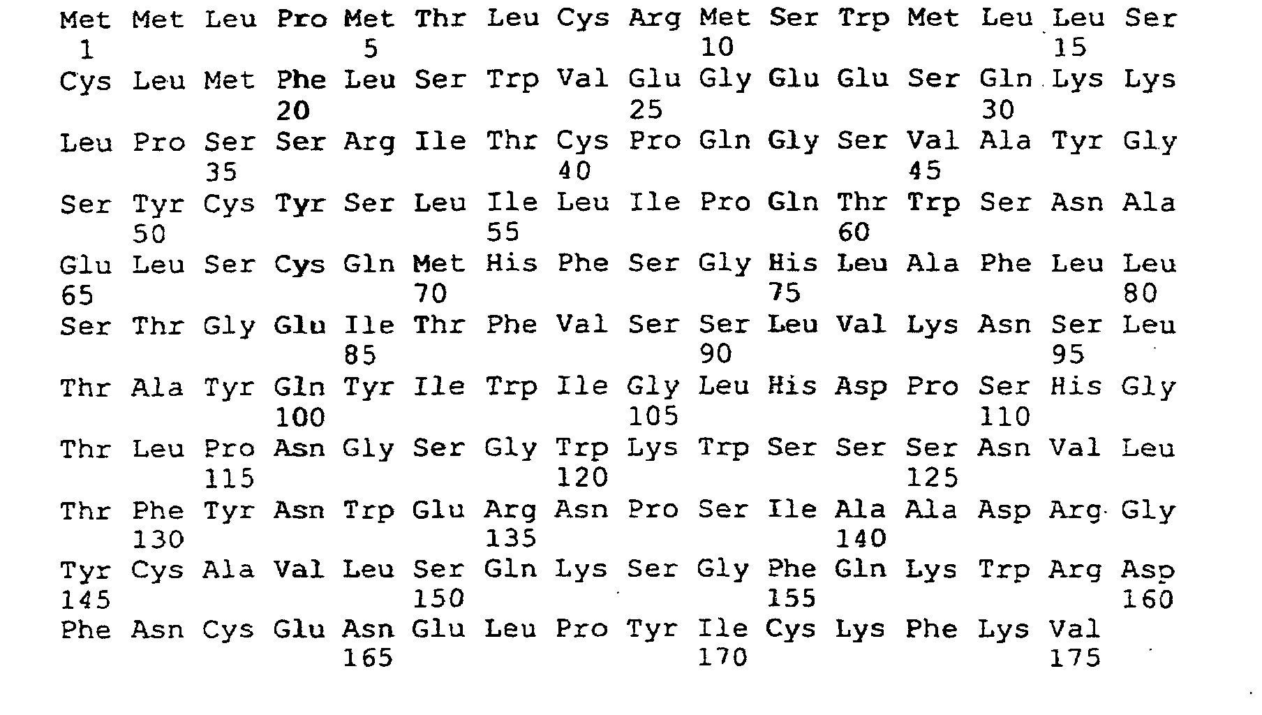
- the signal sequence comprises amino acids 1 to 26 as shown in SEQ ID NO:1. In the constructions tested, the 5' untranslated region comprising nucleotides 1-16 has also been deleted. This deletion may also contribute to the increase in expression which has been observed.
- Suitable inducible transcription initiators include those inducible by isopropyl thiogalactoside (IPTG), such as the lac promoter/operator, as well as others which are known in the art.
- IPTG isopropyl thiogalactoside
- a histidine tag can be engineered to be on the protein.
- the histidine tag can simplify processing and purification.
- a histidine tag is a stretch of histidine residues which are appended to a protein, usually by genetic engineering. Preferably the tag comprises between 3 and 12 histidine residues. They may be contiguous or interrupted by other residues.
- the histidine tag may be appended to the N-terminal or to the C-terminal end of the protein to minimize disruption of protein function. Methods for making and utilizing histidine tags are known in the art.
- the oligohistidine can be used as an affinity moiety using a metal chelate, such as nickel-NTA (N-(5-amino-1-carboxypentyl)-iminodiacetic acid) as the other affinity partner.
- a recombinant construct according to the invention is any DNA molecule which has been engineered so that two segments of DNA are adjacent to each other which are not adjacent to each other in nature.
- Preferably such engineering is performed in vitro , although in vivo engineering can also be performed.
- the construct may be a plasmid, phage, virus, transposable element, minichromosome, or other element, as is suitable for the desired application.
- the coding sequence of amino acid residues 27-175 of INGAP protein are included in the constructs. Preferably the entire signal sequence is deleted. However, it is possible that only a portion of the signal sequence must be deleted to obtain excellent expression. Thus some portion of the signal sequence might be retained in the constructs.
- nucleotides 1-16 Deletion of the 5' untranslated region, nucleotides 1-16, is also desirable. However, it is not known if this is necessary to achieve excellent expression. Thus the 5' untranslated region may be retained in some constructs.
- a host cell according to the invention can be transfected or transformed with a recombinant construct according to the present invention.
- the host may be a bacterial, yeast, insect or mammalian cell. Selection of suitable promoters and translational initiators for use in the appropriate host cell is well within the ken of those of skill in the art.
- Host cells may be transformed, transfected, mated or infected with the recombinant host cell of the present invention. Culturing of host cells can be performed using techniques and media which are well known in the art. Again, a suitable medium and technique can be selected by the ordinarily skilled artisan.
- M15(pREP4) and expression of the protein was induced by IPTG (isopropyl-beta, D-thiogalactopyranoside) which inhibits the repressor, facilitating expression of the protein from the M15 promoter/operator.
- IPTG isopropyl-beta, D-thiogalactopyranoside
- the reason for the intermediate transformation of the ligated material into TOP10F' is that these cells are highly competent increasing the odds of getting insert positive colonies.
- the M15(pREP4) cells that were used for protein expression do not attain competency levels high enough to guarantee transformation of the ligation products.
- the resultant plasmid DNA obtained from the transformation of the TOP10F' was sufficient to enhance the transformation of the M15(pREP4) cells.
- the His-tagged protein was isolated by Ni +2 agarose affinity purification.
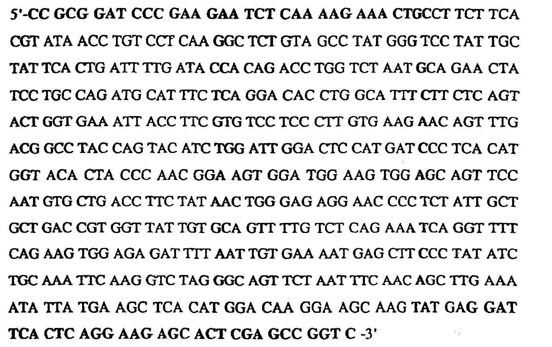
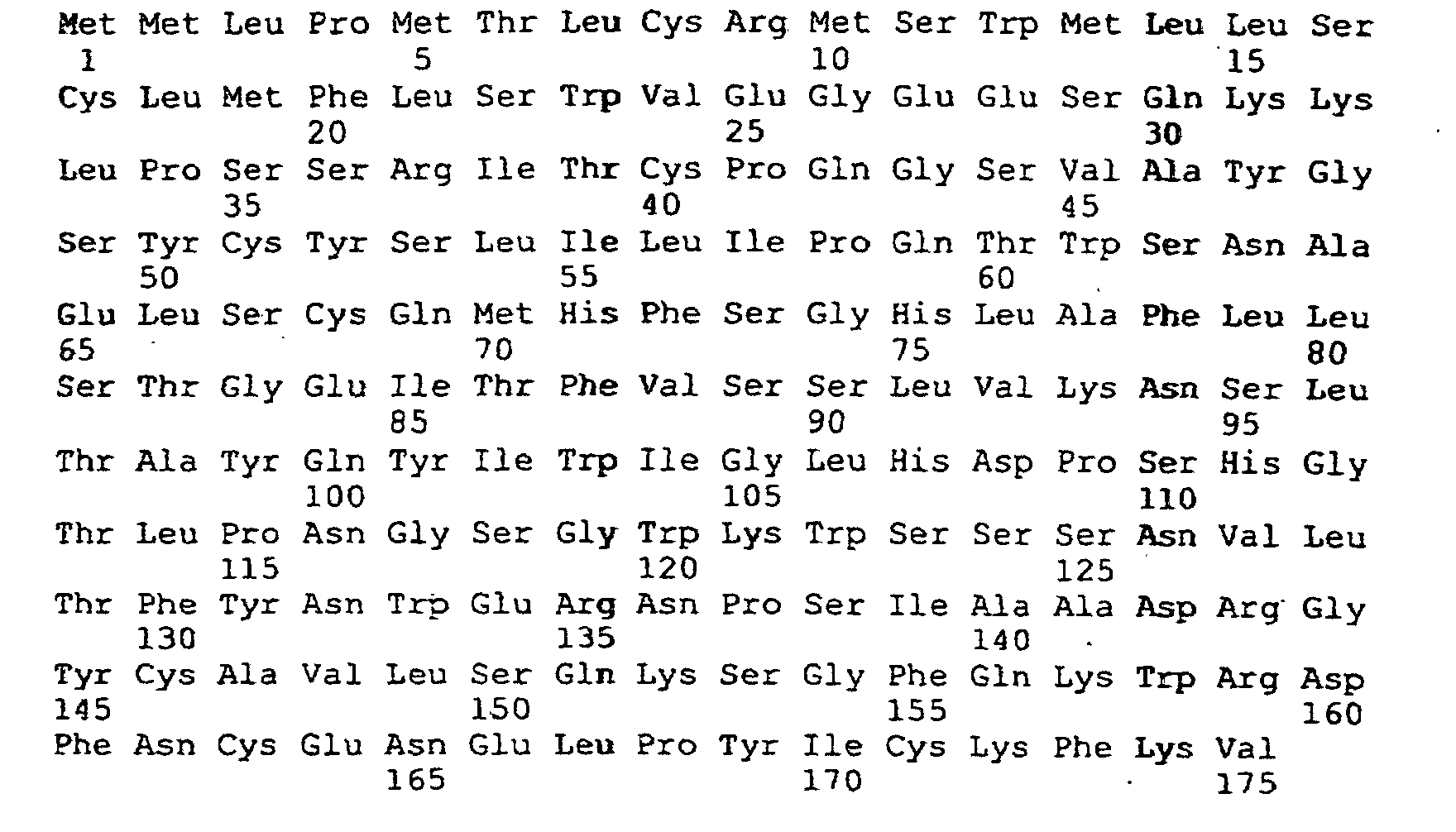
- sequence SEQ ID NO: 1
- the bolded area represents the sequence of the signal peptide
- This example describes the use of polymerase chain reaction to synthesize INGMAT (construct which lacks the signal peptide sequence, i . e ., which encodes the mature protein).
- Oligonucleotides for PCR were designed to incorporate restriction enzyme recognition sites at their respective 5' ends.
- the oligonucleotide designed for the 5' end of the gene incorporates a Bam HI site followed by 20 nucleotides corresponding to the N-terminus of the mature protein.
- the oligonucleotide designed for the 3' end incorporates an Xho I site followed by 20 untranslated nucleotides.
- the PCR product generated from these primers contains the mature INGAP sequence and the native protein termination codon.
- the PCR products were then electrophoresed on a 5% PAGE in TBE. Ethidium bromide stained PCR products corresponding to the expected size for the construct were cut from the gel.
- the gel fragments were electro-eluted into 0.5 ml of TBE, precipitated with 50 ⁇ l 3 M sodium acetate and 1 ml of isopropanol at -80°C for 20 min, centrifuged, washed once with 1 ml of isopropanol, washed once with 1 ml of 70% ethanol, and then dried under vacuum. The dried pellet was resuspended in 50 ⁇ l H 2 O and quantified.
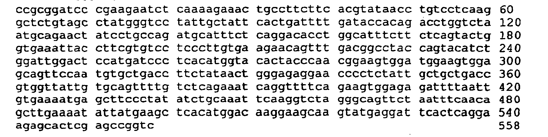
- SEQ ID NO: 4 The bolded areas represent the primers.
- This example describes the creation of a plasmid containing the expression construct.
- the extracted DNA was then precipitated with 2 volumes of ethanol and 1/10 volume 3 M sodium acetate at -80°C for 20 minutes, centrifuged, washed twice with 70% ethanol and dried under vacuum. The pellets were resuspended in 25 ⁇ l H 2 O and quantified.
- the pQE-31 expression system was purchased from QIAGEN Inc. Chatsworth, CA.
- INGMAT Bam HI/Xho I
- pQE-31 Bam HI/Sal I
- Ligation conditions using a 2:1 vector to insert molar ratio.
- pQE31(vector) 517 ng 9 ⁇ l
- INGMAT insert
- Ligase 4u 1 ⁇ l H 2 O 27.5 ⁇ l final volume 50 ⁇ l
- the ligation reactions were incubated at 4°C for 16 hours.
- TOP10F' cells were purchased from Invitrogen, San Diego, CA) with 0.5 ⁇ l of 500mM ⁇ -mercaptoethanol and incubated on ice for 30 minutes, heat shocked for 45 seconds at 42° C, and recovered on ice for 2 minutes. Then we added 1 ml of prewarmed S°C media and incubated at 37°C with shaking at 225 rpm for 1 hour followed by plating all the transformation reaction on LB broth agar plates containing 100 ⁇ g/ml ampicillin.
- Colony containing plates were lifted onto Nytran membranes.
- the colonies were lysed with 0.5 M NaOH, neutralized, and the resultant DNA bound to the membrane by baking at 80°C for 1 hour.
- the membranes were then hybridized in 50% formaldehyde, 5 X SSPE at 50°C for 16 hours with 3 x 10 6 cpm/ml of 32 P random primed INGAP cDNA.
- the membranes were washed at high stringency and exposed to X-ray film. Positive colonies were matched up to the X-ray film and grown up in 3 mls of LB with ampicillin .
- DNA was isolated from the small cultures using alkaline lysis, phenol extracted, precipitated, dried, and resuspended in 50 ⁇ l H 2 O. A small aliquot of each of the isolated DNA were digested with Bam HI and Hind III to release inserts. The digested DNAs were electrophoresed on 1.5% agarose and stained with ethidium bromide and positive inserts identified at approximately 510 bp size range. We took four of the insert containing plasmids and incubated them in the presence of RNAse to remove any residual bacterial RNA.
- This example describes the denaturing metal affinity protein chromatography isolation of the his tagged INGAP protein without signal peptide. (Procedure for a 250 ml pINGMATHIS transformed M15(pREP4) culture. pINGMATHIS is the INGMATHIS construct ligated into the pQE-31 vector.)
- Bacteria growth and protein induction Bacteria growth and protein induction.
- binding/lysis buffer A (6 M Guanidine HCl, 0.1M sodium phosphate, 0.01 M Tris, pH 8.0) and resuspend the resin. Centrifuge for 2 minutes at 800 x G and discard the supernatant. Add 42 ml of binding/lysis buffer A (6 M Guanidine HCL, 0.1M sodium phosphate, 0.01 M Tris, pH 8.0) and resuspend the resin. Centrifuge for 2 minutes at 800 x G and discard the supernatant. Add 42 ml of binding/lysis buffer A (6 M Guanidine HCL, 0.1M sodium phosphate, 0.01 M Tris, pH 8.0) and resuspend the resin. Centrifuge for 2 minutes at 800 x G and discard the supernatant. Bring the total volume up to 10 ml with buffer A. The slurry is now ready for the application of the lysed bacteria.
- lysis buffer A (6 M Guanidine HCL, 0.1M sodium phosphate, 0.01 M Tris, pH 8.0). Transfer the resuspension to a 50 ml centrifuge tube. Freeze the resuspension/lysate at -70 until solid. Thaw at room temperature. Place the lysate on a rotator for 60 minutes at room temperature. Centrifuge the lysate for 15 minutes at 10,000 X G. Collect the supernatant and add the 10 ml of prepared Ni2+NTA. Rotate for 45 minutes. Load the slurry onto a 1.6 cm diameter column and allow to flow through by gravity.
- Dialysis tubing with a molecular weight cut-off of 3000 was prepared by boiling in 5mM EDTA/200 mM sodium bicarbonate for 5 minutes. The tubing was rinsed briefly in deionized water and boiled another 5 minutes in the bicarbonate solution. The tubing was returned to deionized water, covered with aluminum foil and autoclaved for 10 minutes on a liquid cycle. The tubing was handled with latex gloves during the entire procedure.
- This example describes analytical techniques confirming the identity of the product.
- discontinuous denaturing polyacrylamide gel electrophoresis was performed on the dialyzed protein solution using the Hoefer SE250 Mighty Small II apparatus.
- the separating gel was prepared with 15% acrylamide, 1.35% bis-acrylamide in 375mM Tris buffer at pH 8.8 with 0.05% sodium dodecyl sulfate. Polymerization was induced by addition of 0.05% ammonium persulfate and 20 ⁇ l TEMED/15 ml solution. The solution was placed in the gel plate apparatus for polymerization.
- the stacking gel was poured with the same solution, except the Tris buffer was 125mM at pH 6.8, and the acrylamide concentration was 4%.
- the protein samples were diluted 1:1 with sample buffer (125 mM Tris-Cl, pH 6.8, 4% SDS, 20% glycerol, and 10% 2-mercaptoethanol).
- the upper and lower tank buffers were identical, containing 25mM Tris, 192 mM glycine and 0.1% SDS at pH 8.3.
- Two gels were loaded with 20 ⁇ l each of bacterial lysate without transfection (CBL,368 ug/ml), bacterial lysate with transfection (TBL, 341ug/ml), the fractions from Ni-NTA chromatography (eluted at pH6.3, 110 ug/ml; pH 5.9, 100 ug/ml; and pH 4.5, 800ug/ml) and standards (Rainbow Markers, Amersham and Dalton Mark-VII, Sigma).
- Electrophoresis was performed at 20 mA constant current until the dye front entered the separating gel, and at 60 mA constant current until the dye front reached 0.5cm from the bottom. The gels were then removed and one was fixed with 45% methanol/10% acetic acid for one hour, and the other was placed in transfer buffer (25 mM Tris, 192 mM glycine, 20% methanol, pH 8.3) for 20-30 minutes.
- the fixed gel was equilibrated with 2 changes of 10% ethanol/5% acetic acid for 30 minutes each.
- the gel was then exposed to a 0.0032N HNO 3 /K2Cr2O7 solution for 5 minutes.
- the gel was washed in deionized water 3 times for 10 minutes each.
- the gel was impregnated with silver using 0.1g AgNO 3 /50 ml H 2 O for 30 minutes.
- the silver solution was washed off the gel in deionized water for 5 minutes.
- the gel was then exposed to a developer solution (29.7 g anhydrous Na2CO 3 in 1 liter H2O with 0.5 ml formalin) in 5 minute intervals between changes until the desired density was reached.
- the development was stopped with 10% acetic acid, and the gel stored in H 2 O.
- the gel showed a protein band of approximately 19 kD that was prominent in the bacterial lysate from transfected cells and in the elution fraction from pH 4.5 on nickel/NTA ( Figure 1). This protein was not represented in any of the other samples. This is consistent with the size of INGAP protein and with interaction of the inserted histidine tagging region with the nickel/NTA column matrix.
- Immobilon-P PVDF membrane was wetted with 100% methanol, and equilibrated with transfer buffer for 10 minutes. The gel was removed from transfer buffer and placed on the PVDF membrane. All bubbles between the membrane and the gel were removed. The combination was placed between Whatmann 3mm filter paper wetted with transfer buffer and the whole "sandwich" was placed in the cassette of a Hoefer transfer tank. The cassette was placed in the transfer tank filled with transfer buffer with the gel toward the cathode. The transfer was performed at 12V constant voltage for 18 hours.
- the membrane was placed in a blocking buffer of 0.5M Tris, 2M NaCl and 1% polyethylene glycol with 5% bovine serum albumin and 10% goat serum at room temperature for 1 hour.
- the membrane was then placed into 20 ml of blocking buffer containing INGAP antibody 945-2 at a dilution of 1:5000 and incubated at room temperature for 1 hour.
- the membrane was then washed 3 times for 15 minutes each with 50 ml of washing buffer (0.4% Tween-20 in phosphate-buffered saline (PBS) at pH 7.4).
- PBS phosphate-buffered saline
- the membrane was then incubated for 1 hour at room temperature in washing buffer containing anti-rabbit IgG (whole molecule, Sigma Cat # A-0545) peroxidase conjugate at a 1:160,000 dilution.
- the membrane was washed 3 times for 5 minutes in 50 ml of 0.2% Tween-20 in PBS, followed by 3 washes of 5 minutes each with 0.1 % Tween-20 in PBS.
- the blot was revealed using the enzyme chemiluminescence kit from Amersham Corp., Arlington, IL according to instructions.
- the ECL blot was exposed to Kodak X-Omat AR-5 X-ray film for 20 minutes.
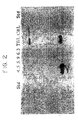
- ECL of the blot revealed strong protein recognition of the overexpressed 19 kD proteins in the whole lysate from transfected bacteria (TBL) and the pH 4.5 fraction that were visualized on the SDS-PAGE gels ( Figure 2).
- TBL transfected bacteria
- Figure 2 the pH 4.5 fraction that were visualized on the SDS-PAGE gels
- there was a protein band recognized in both bacterial lysates at 40 kD implying that this protein is weakly recognized and is a bacterial protein rather than a product of the transfection.
- INGAP protein in a prokaryotic system by excluding the 5'UTR and the signal peptide and insertion of the new construct into a new vector.
- the resultant protein is of the predicted molecular size of INGAP monomer and reacts with the antibody to INGAP in a Western analysis.
- the protein shares with INGAP peptide the ability to induce ductal cell proliferation.
Landscapes
- Chemical & Material Sciences (AREA)
- Health & Medical Sciences (AREA)
- Life Sciences & Earth Sciences (AREA)
- Organic Chemistry (AREA)
- General Health & Medical Sciences (AREA)
- Molecular Biology (AREA)
- Biochemistry (AREA)
- Biophysics (AREA)
- Zoology (AREA)
- Genetics & Genomics (AREA)
- Medicinal Chemistry (AREA)
- Gastroenterology & Hepatology (AREA)
- Proteomics, Peptides & Aminoacids (AREA)
- Toxicology (AREA)
- Preparation Of Compounds By Using Micro-Organisms (AREA)
- Micro-Organisms Or Cultivation Processes Thereof (AREA)
- Steroid Compounds (AREA)
- Peptides Or Proteins (AREA)
- Hall/Mr Elements (AREA)
Abstract
Description
(the bolded area represents the sequence of the signal peptide)



| Template: (50 ng INGAP original cDNA removed from pCDNA3) | 5 µl |
| MgCl2: | 4 µl |
| 10 X PCR buffer | 5 µl |
| dATP | 1 µl |
| dCTP | 1 µl |
| dGTP | 1 µl |
| dTTP | 1 µl |
| 5'primer | 1 µl |
| 3' primer | 1 µl |
| H2O | 29 µl |
| Taq polymerase | 1 µl |
| total volume = 50 µl |

Ligation conditions using a 2:1 vector to insert molar ratio.
| pQE31(vector) 517 ng | 9 µl |
| INGMAT (insert) 165 ng | 2.5 µl |
| 10 X ligation buffer | 5 µl |
| 10 mM rATP | 5 µl |
| T4 Ligase 4u | 1 µl |
| H2O | 27.5 µl |
| final volume = 50 µl |
(Procedure for a 250 ml pINGMATHIS transformed M15(pREP4) culture. pINGMATHIS is the INGMATHIS construct ligated into the pQE-31 vector.)
Grown until ABS600= 0.0.75 to 0.9 (actual OD=0.866). Added 5 ml of 100mM IPTG (2mM final) to induce production of the protein. Continue growing for 4 hours in the case of INGAP. Collected the bacteria and spin at 6000 rpm for 20 minutes, discarded the supernatant. The pellet was frozen until ready to use at -70 C.













Claims (8)
- A recombinant construct for expression of an INGAP protein which induces ductal cell proliferation comprising:a first nucleotide sequence encoding amino acids 27 to 175 of INGAP protein selected from the nucleotide sequences shown in SEQ ID NO: 4, operably linked to a transcriptional initiation site and a translational initiation site, wherein a second nucleotide sequence encoding a signal peptide as shown in SEQ ID NO: 1 is not present immediately 5' of said first nucleotide sequence.
- The construct of claim 1 further comprising a third nucleotide sequence encoding a histidine tag.
- The construct of claim 3 wherein the third nucleotide sequence is immediately 5' or 3' to said first nucleotide sequence.
- The construct of claim 1 wherein the transcriptional initiation site is inducible.
- The construct of claim 1 wherein the transcriptional initiation site is the lac promoter/operator.
- A method of producing in a recombinant host cell, biologically active INGAP protein capable of inducing ductal cell proliferation, comprising the steps of:culturing a host cell comprising a recombinant construct according to claim 1;recovering protein from said cultured host cell.
- The method of claim 7 wherein the construct further comprises a third nucleotide sequence encoding a histidine tag, and INGAP protein is purified using a nickel affinity matrix.
- A host cell comprising a recombinant construct according to claim 1.
Applications Claiming Priority (3)
| Application Number | Priority Date | Filing Date | Title |
|---|---|---|---|
| US74109696A | 1996-10-30 | 1996-10-30 | |
| US741096 | 1996-10-30 | ||
| PCT/US1997/019415 WO1998018913A1 (en) | 1996-10-30 | 1997-10-30 | High level of expression of ingap |
Publications (3)
| Publication Number | Publication Date |
|---|---|
| EP1007647A1 EP1007647A1 (en) | 2000-06-14 |
| EP1007647A4 EP1007647A4 (en) | 2001-09-26 |
| EP1007647B1 true EP1007647B1 (en) | 2004-05-06 |
Family
ID=24979382
Family Applications (1)
| Application Number | Title | Priority Date | Filing Date |
|---|---|---|---|
| EP97912942A Expired - Lifetime EP1007647B1 (en) | 1996-10-30 | 1997-10-30 | High level of expression of ingap |
Country Status (11)
| Country | Link |
|---|---|
| US (2) | US5804421A (en) |
| EP (1) | EP1007647B1 (en) |
| JP (1) | JP2001502916A (en) |
| AT (1) | ATE266094T1 (en) |
| AU (1) | AU727237B2 (en) |
| CA (1) | CA2270412A1 (en) |
| DE (1) | DE69729010T2 (en) |
| DK (1) | DK1007647T3 (en) |
| ES (1) | ES2219759T3 (en) |
| PT (1) | PT1007647E (en) |
| WO (1) | WO1998018913A1 (en) |
Families Citing this family (6)
| Publication number | Priority date | Publication date | Assignee | Title |
|---|---|---|---|---|
| US6593120B1 (en) | 1994-04-01 | 2003-07-15 | Gen-Probe Incorporated | Recombinant DNA encoding a reverse transcriptase derived from moloney murine leukemia virus |
| US6986994B2 (en) * | 2001-01-09 | 2006-01-17 | Gmp Endotherapeutics, Inc. | INGAP displacement assays |
| US6642003B2 (en) | 2001-08-02 | 2003-11-04 | Cedars-Sinai Medical Center | Human glucose-dependent insulin-secreting cell line |
| US7141240B2 (en) * | 2002-03-12 | 2006-11-28 | Cedars-Sinai Medical Center | Glucose-dependent insulin-secreting cells transfected with a nucleotide sequence encoding GLP-1 |
| US8012928B2 (en) * | 2008-12-19 | 2011-09-06 | The Research Foundation Of State University Of New York | Truncated PAP2 and methods of making and using same |
| US20160039877A1 (en) | 2013-03-15 | 2016-02-11 | Shenzhen Hightide Biopharmaceutical, Ltd. | Compositions and methods of using islet neogenesis peptides and analogs thereof |
Family Cites Families (7)
| Publication number | Priority date | Publication date | Assignee | Title |
|---|---|---|---|---|
| US4439521A (en) * | 1981-10-21 | 1984-03-27 | Ontario Cancer Institute | Method for producing self-reproducing mammalian pancreatic islet-like structures |
| US4965188A (en) * | 1986-08-22 | 1990-10-23 | Cetus Corporation | Process for amplifying, detecting, and/or cloning nucleic acid sequences using a thermostable enzyme |
| US4935000A (en) * | 1986-04-03 | 1990-06-19 | East Carolina University | Extracellular matrix induction method to produce pancreatic islet tissue |
| JPH02200700A (en) * | 1989-01-30 | 1990-08-08 | Shionogi & Co Ltd | New reg protein |
| FR2661187B1 (en) * | 1990-04-20 | 1994-08-05 | Inst Nat Sante Rech Med | PROTEIN ASSOCIATED WITH ACUTE PANCREATITIS. MEANS FOR THE DIAGNOSIS OF ACUTE PANCREATITIS. |
| JPH06135998A (en) * | 1992-10-22 | 1994-05-17 | Shionogi & Co Ltd | Human and mouse pancreatitis-related proteins |
| US5834590A (en) * | 1995-02-22 | 1998-11-10 | Eastern Virginia Medical School Of The Medical College Of Hampton Roads | Ingap protein involved in pancreatic islet neogenesis |
-
1997
- 1997-08-12 US US08/909,725 patent/US5804421A/en not_active Ceased
- 1997-10-30 CA CA002270412A patent/CA2270412A1/en not_active Abandoned
- 1997-10-30 ES ES97912942T patent/ES2219759T3/en not_active Expired - Lifetime
- 1997-10-30 AU AU50007/97A patent/AU727237B2/en not_active Ceased
- 1997-10-30 JP JP10520665A patent/JP2001502916A/en active Pending
- 1997-10-30 PT PT97912942T patent/PT1007647E/en unknown
- 1997-10-30 AT AT97912942T patent/ATE266094T1/en not_active IP Right Cessation
- 1997-10-30 DE DE69729010T patent/DE69729010T2/en not_active Expired - Fee Related
- 1997-10-30 DK DK97912942T patent/DK1007647T3/en active
- 1997-10-30 EP EP97912942A patent/EP1007647B1/en not_active Expired - Lifetime
- 1997-10-30 WO PCT/US1997/019415 patent/WO1998018913A1/en not_active Ceased
-
2000
- 2000-09-08 US US09/659,379 patent/USRE39351E1/en not_active Expired - Lifetime
Also Published As
| Publication number | Publication date |
|---|---|
| USRE39351E1 (en) | 2006-10-17 |
| DE69729010D1 (en) | 2004-06-09 |
| AU5000797A (en) | 1998-05-22 |
| EP1007647A1 (en) | 2000-06-14 |
| DK1007647T3 (en) | 2004-06-28 |
| ATE266094T1 (en) | 2004-05-15 |
| US5804421A (en) | 1998-09-08 |
| WO1998018913A1 (en) | 1998-05-07 |
| AU727237B2 (en) | 2000-12-07 |
| JP2001502916A (en) | 2001-03-06 |
| PT1007647E (en) | 2004-07-30 |
| CA2270412A1 (en) | 1998-05-07 |
| HK1028789A1 (en) | 2001-03-02 |
| EP1007647A4 (en) | 2001-09-26 |
| ES2219759T3 (en) | 2004-12-01 |
| DE69729010T2 (en) | 2005-04-28 |
Similar Documents
| Publication | Publication Date | Title |
|---|---|---|
| US5310663A (en) | Affinity peptides | |
| CN104725515B (en) | One species elastin polypeptide and Coxsackie Adenovirus Receptor fusion protein and its preparation method and application | |
| CN105169381B (en) | A kind of helicobacter pylori multivalence epitope vaccine and preparation method thereof | |
| US5804421A (en) | High level of expression of ingap in bacterial and euraryotic cells | |
| KR970002670B1 (en) | Feline interferon and process for production thereof | |
| CN113599512A (en) | Immunity composition | |
| CN112979769A (en) | Amino acid sequence, protein, preparation method and application thereof | |
| RU2001117860A (en) | GENES OF HOMOLOGICAL IMMUNODOMINANT PROTEINS WEIGHT 28-KILODALTON Ehrlichia canis AND THEIR APPLICATION | |
| WO2019143193A1 (en) | N-terminal fusion partner for producing recombinant polypeptide, and method for producing recombinant polypeptide using same | |
| AU758762B2 (en) | High level of expression of ingap | |
| HK1028789B (en) | High level of expression of ingap | |
| EP1639002B1 (en) | Cytokine variant polypeptides | |
| CN114644715B (en) | Preparation method of fusion protein and complex of IGFBP-3 and IGF-1 | |
| CN103214561B (en) | Human hepatitis c virus core antigen and preparation method and application thereof | |
| CN114891819A (en) | Preparation method and application of porcine beta-defensin 129 protein and polyclonal antibody thereof | |
| MXPA99004079A (en) | High level of expression of ingap | |
| JP3315827B2 (en) | Method for producing active human ALT | |
| CN115073558A (en) | Recombinant African swine fever virus DP96R subunit protein and preparation method and application thereof | |
| CN114470160B (en) | Viral Replication Inhibitors and Applications | |
| CN100548384C (en) | A kind of genetic engineering polypeptide vaccine adjuvant for aftosa and its production and application | |
| CN101298611A (en) | Active fragment of chlamydia proteasome-like active factor and its expression method | |
| Fan et al. | Expression and characterization of an epididymis-specific gene | |
| CN110129329A (en) | It is a kind of for expanding the primer and preparation method of Japanese eel interferon correlation factor | |
| CN115851794A (en) | A kind of preparation method of hepatitis B virus X protein | |
| CN121779574A (en) | A visualized fluorescent protein suitable for mouse modeling of autoimmune hepatitis and in vivo imaging. |
Legal Events
| Date | Code | Title | Description |
|---|---|---|---|
| PUAI | Public reference made under article 153(3) epc to a published international application that has entered the european phase |
Free format text: ORIGINAL CODE: 0009012 |
|
| 17P | Request for examination filed |
Effective date: 19990519 |
|
| AK | Designated contracting states |
Kind code of ref document: A1 Designated state(s): AT BE CH DE DK ES FI FR GB GR IE IT LI LU MC NL PT SE |
|
| AX | Request for extension of the european patent |
Free format text: AL PAYMENT 19990519;LT PAYMENT 19990519;LV PAYMENT 19990519;RO PAYMENT 19990519;SI PAYMENT 19990519 |
|
| RIC1 | Information provided on ipc code assigned before grant |
Free format text: 7C 12N 15/00 A, 7C 07H 21/04 B, 7C 07K 1/22 B, 7C 07K 14/47 B |
|
| A4 | Supplementary search report drawn up and despatched |
Effective date: 20010813 |
|
| AK | Designated contracting states |
Kind code of ref document: A4 Designated state(s): AT BE CH DE DK ES FI FR GB GR IE IT LI LU MC NL PT SE |
|
| 17Q | First examination report despatched |
Effective date: 20020822 |
|
| GRAP | Despatch of communication of intention to grant a patent |
Free format text: ORIGINAL CODE: EPIDOSNIGR1 |
|
| GRAS | Grant fee paid |
Free format text: ORIGINAL CODE: EPIDOSNIGR3 |
|
| GRAA | (expected) grant |
Free format text: ORIGINAL CODE: 0009210 |
|
| AK | Designated contracting states |
Kind code of ref document: B1 Designated state(s): AT BE CH DE DK ES FI FR GB GR IE IT LI LU MC NL PT SE |
|
| AX | Request for extension of the european patent |
Extension state: AL LT LV RO SI |
|
| REG | Reference to a national code |
Ref country code: GB Ref legal event code: FG4D |
|
| REG | Reference to a national code |
Ref country code: CH Ref legal event code: EP |
|
| REF | Corresponds to: |
Ref document number: 69729010 Country of ref document: DE Date of ref document: 20040609 Kind code of ref document: P |
|
| REG | Reference to a national code |
Ref country code: GR Ref legal event code: EP Ref document number: 20040401589 Country of ref document: GR |
|
| REG | Reference to a national code |
Ref country code: SE Ref legal event code: TRGR |
|
| REG | Reference to a national code |
Ref country code: IE Ref legal event code: FG4D |
|
| REG | Reference to a national code |
Ref country code: DK Ref legal event code: T3 |
|
| REG | Reference to a national code |
Ref country code: PT Ref legal event code: SC4A Free format text: AVAILABILITY OF NATIONAL TRANSLATION Effective date: 20040513 Ref country code: CH Ref legal event code: NV Representative=s name: RITSCHER & PARTNER AG |
|
| LTIE | Lt: invalidation of european patent or patent extension |
Effective date: 20040506 |
|
| PG25 | Lapsed in a contracting state [announced via postgrant information from national office to epo] |
Ref country code: MC Free format text: LAPSE BECAUSE OF NON-PAYMENT OF DUE FEES Effective date: 20041031 |
|
| REG | Reference to a national code |
Ref country code: ES Ref legal event code: FG2A Ref document number: 2219759 Country of ref document: ES Kind code of ref document: T3 |
|
| ET | Fr: translation filed | ||
| REG | Reference to a national code |
Ref country code: HK Ref legal event code: GR Ref document number: 1028789 Country of ref document: HK |
|
| PLBE | No opposition filed within time limit |
Free format text: ORIGINAL CODE: 0009261 |
|
| STAA | Information on the status of an ep patent application or granted ep patent |
Free format text: STATUS: NO OPPOSITION FILED WITHIN TIME LIMIT |
|
| 26N | No opposition filed |
Effective date: 20050208 |
|
| PGFP | Annual fee paid to national office [announced via postgrant information from national office to epo] |
Ref country code: NL Payment date: 20071003 Year of fee payment: 11 Ref country code: LU Payment date: 20071025 Year of fee payment: 11 Ref country code: ES Payment date: 20071120 Year of fee payment: 11 Ref country code: DK Payment date: 20071015 Year of fee payment: 11 Ref country code: DE Payment date: 20071025 Year of fee payment: 11 |
|
| PGFP | Annual fee paid to national office [announced via postgrant information from national office to epo] |
Ref country code: FI Payment date: 20071012 Year of fee payment: 11 Ref country code: CH Payment date: 20071029 Year of fee payment: 11 Ref country code: AT Payment date: 20071011 Year of fee payment: 11 Ref country code: IT Payment date: 20071027 Year of fee payment: 11 |
|
| PGFP | Annual fee paid to national office [announced via postgrant information from national office to epo] |
Ref country code: BE Payment date: 20071213 Year of fee payment: 11 Ref country code: SE Payment date: 20071004 Year of fee payment: 11 |
|
| REG | Reference to a national code |
Ref country code: CH Ref legal event code: PCAR Free format text: RITSCHER & PARTNER AG;RESIRAIN 1;8125 ZOLLIKERBERG (CH) |
|
| PGFP | Annual fee paid to national office [announced via postgrant information from national office to epo] |
Ref country code: GB Payment date: 20071024 Year of fee payment: 11 Ref country code: FR Payment date: 20071009 Year of fee payment: 11 |
|
| PGFP | Annual fee paid to national office [announced via postgrant information from national office to epo] |
Ref country code: PT Payment date: 20071018 Year of fee payment: 11 Ref country code: GR Payment date: 20070927 Year of fee payment: 11 |
|
| BERE | Be: lapsed |
Owner name: *EASTERN VIRGINIA MEDICAL SCHOOL OF THE MEDICAL CO Effective date: 20081031 |
|
| REG | Reference to a national code |
Ref country code: PT Ref legal event code: MM4A Free format text: LAPSE DUE TO NON-PAYMENT OF FEES Effective date: 20090430 |
|
| REG | Reference to a national code |
Ref country code: CH Ref legal event code: PL |
|
| REG | Reference to a national code |
Ref country code: DK Ref legal event code: EBP |
|
| EUG | Se: european patent has lapsed | ||
| GBPC | Gb: european patent ceased through non-payment of renewal fee |
Effective date: 20081030 |
|
| NLV4 | Nl: lapsed or anulled due to non-payment of the annual fee |
Effective date: 20090501 |
|
| REG | Reference to a national code |
Ref country code: IE Ref legal event code: MM4A |
|
| REG | Reference to a national code |
Ref country code: FR Ref legal event code: ST Effective date: 20090630 |
|
| PG25 | Lapsed in a contracting state [announced via postgrant information from national office to epo] |
Ref country code: NL Free format text: LAPSE BECAUSE OF NON-PAYMENT OF DUE FEES Effective date: 20090501 Ref country code: FI Free format text: LAPSE BECAUSE OF NON-PAYMENT OF DUE FEES Effective date: 20081030 |
|
| PG25 | Lapsed in a contracting state [announced via postgrant information from national office to epo] |
Ref country code: PT Free format text: LAPSE BECAUSE OF NON-PAYMENT OF DUE FEES Effective date: 20090430 Ref country code: IT Free format text: LAPSE BECAUSE OF NON-PAYMENT OF DUE FEES Effective date: 20081030 Ref country code: DE Free format text: LAPSE BECAUSE OF NON-PAYMENT OF DUE FEES Effective date: 20090501 Ref country code: AT Free format text: LAPSE BECAUSE OF NON-PAYMENT OF DUE FEES Effective date: 20081030 |
|
| PGFP | Annual fee paid to national office [announced via postgrant information from national office to epo] |
Ref country code: IE Payment date: 20071011 Year of fee payment: 11 |
|
| PG25 | Lapsed in a contracting state [announced via postgrant information from national office to epo] |
Ref country code: BE Free format text: LAPSE BECAUSE OF NON-PAYMENT OF DUE FEES Effective date: 20081031 |
|
| PG25 | Lapsed in a contracting state [announced via postgrant information from national office to epo] |
Ref country code: LI Free format text: LAPSE BECAUSE OF NON-PAYMENT OF DUE FEES Effective date: 20081031 Ref country code: IE Free format text: LAPSE BECAUSE OF NON-PAYMENT OF DUE FEES Effective date: 20081030 Ref country code: FR Free format text: LAPSE BECAUSE OF NON-PAYMENT OF DUE FEES Effective date: 20081031 Ref country code: DK Free format text: LAPSE BECAUSE OF NON-PAYMENT OF DUE FEES Effective date: 20081031 Ref country code: CH Free format text: LAPSE BECAUSE OF NON-PAYMENT OF DUE FEES Effective date: 20081031 |
|
| PG25 | Lapsed in a contracting state [announced via postgrant information from national office to epo] |
Ref country code: GR Free format text: LAPSE BECAUSE OF NON-PAYMENT OF DUE FEES Effective date: 20090505 Ref country code: GB Free format text: LAPSE BECAUSE OF NON-PAYMENT OF DUE FEES Effective date: 20081030 |
|
| REG | Reference to a national code |
Ref country code: ES Ref legal event code: FD2A Effective date: 20081031 |
|
| PG25 | Lapsed in a contracting state [announced via postgrant information from national office to epo] |
Ref country code: ES Free format text: LAPSE BECAUSE OF NON-PAYMENT OF DUE FEES Effective date: 20081031 |
|
| PG25 | Lapsed in a contracting state [announced via postgrant information from national office to epo] |
Ref country code: LU Free format text: LAPSE BECAUSE OF NON-PAYMENT OF DUE FEES Effective date: 20081030 |
|
| PG25 | Lapsed in a contracting state [announced via postgrant information from national office to epo] |
Ref country code: SE Free format text: LAPSE BECAUSE OF NON-PAYMENT OF DUE FEES Effective date: 20081031 |