EP0989853B1 - Self-regulated apoptosis of inflammatory cells by gene therapy - Google Patents
Self-regulated apoptosis of inflammatory cells by gene therapy Download PDFInfo
- Publication number
- EP0989853B1 EP0989853B1 EP98908778A EP98908778A EP0989853B1 EP 0989853 B1 EP0989853 B1 EP 0989853B1 EP 98908778 A EP98908778 A EP 98908778A EP 98908778 A EP98908778 A EP 98908778A EP 0989853 B1 EP0989853 B1 EP 0989853B1
- Authority
- EP
- European Patent Office
- Prior art keywords
- gene
- tnfα
- promoter
- apoptosis
- caspase
- Prior art date
- Legal status (The legal status is an assumption and is not a legal conclusion. Google has not performed a legal analysis and makes no representation as to the accuracy of the status listed.)
- Expired - Lifetime
Links
- 230000001105 regulatory effect Effects 0.000 title abstract description 5
- 238000001415 gene therapy Methods 0.000 title description 8
- 230000007820 inflammatory cell apoptotic process Effects 0.000 title description 2
- 108090000623 proteins and genes Proteins 0.000 claims abstract description 163
- 108060008682 Tumor Necrosis Factor Proteins 0.000 claims abstract description 112
- 210000004027 cell Anatomy 0.000 claims abstract description 104
- 102000000852 Tumor Necrosis Factor-alpha Human genes 0.000 claims abstract description 95
- 230000001939 inductive effect Effects 0.000 claims abstract description 54
- 230000006907 apoptotic process Effects 0.000 claims abstract description 49
- 238000000034 method Methods 0.000 claims abstract description 41
- 230000014509 gene expression Effects 0.000 claims abstract description 36
- 239000003623 enhancer Substances 0.000 claims abstract description 26
- 102100029855 Caspase-3 Human genes 0.000 claims abstract description 22
- 230000004054 inflammatory process Effects 0.000 claims abstract description 16
- 206010061218 Inflammation Diseases 0.000 claims abstract description 15
- 210000004969 inflammatory cell Anatomy 0.000 claims abstract description 15
- 208000027866 inflammatory disease Diseases 0.000 claims abstract description 13
- 108090000397 Caspase 3 Proteins 0.000 claims abstract description 10
- 102100025597 Caspase-4 Human genes 0.000 claims abstract description 9
- 239000008194 pharmaceutical composition Substances 0.000 claims abstract description 9
- 101710090338 Caspase-4 Proteins 0.000 claims abstract description 8
- 102100038916 Caspase-5 Human genes 0.000 claims abstract description 8
- 101710090333 Caspase-5 Proteins 0.000 claims abstract description 8
- 230000006882 induction of apoptosis Effects 0.000 claims abstract description 8
- 238000004519 manufacturing process Methods 0.000 claims abstract description 8
- 238000003752 polymerase chain reaction Methods 0.000 claims description 13
- 108700008625 Reporter Genes Proteins 0.000 claims description 12
- 238000000338 in vitro Methods 0.000 claims description 12
- 206010039073 rheumatoid arthritis Diseases 0.000 claims description 11
- 210000002437 synoviocyte Anatomy 0.000 claims description 11
- 239000000203 mixture Substances 0.000 claims description 10
- 238000011144 upstream manufacturing Methods 0.000 claims description 8
- 102100035904 Caspase-1 Human genes 0.000 claims description 7
- 108090000426 Caspase-1 Proteins 0.000 claims description 7
- 230000008569 process Effects 0.000 claims description 6
- 238000012217 deletion Methods 0.000 claims description 5
- 230000037430 deletion Effects 0.000 claims description 5
- 238000011282 treatment Methods 0.000 claims description 5
- 102100038902 Caspase-7 Human genes 0.000 claims description 4
- 108090000538 Caspase-8 Proteins 0.000 claims description 4
- 210000002950 fibroblast Anatomy 0.000 claims description 4
- 230000028709 inflammatory response Effects 0.000 claims description 4
- FWMNVWWHGCHHJJ-SKKKGAJSSA-N 4-amino-1-[(2r)-6-amino-2-[[(2r)-2-[[(2r)-2-[[(2r)-2-amino-3-phenylpropanoyl]amino]-3-phenylpropanoyl]amino]-4-methylpentanoyl]amino]hexanoyl]piperidine-4-carboxylic acid Chemical compound C([C@H](C(=O)N[C@H](CC(C)C)C(=O)N[C@H](CCCCN)C(=O)N1CCC(N)(CC1)C(O)=O)NC(=O)[C@H](N)CC=1C=CC=CC=1)C1=CC=CC=C1 FWMNVWWHGCHHJJ-SKKKGAJSSA-N 0.000 claims description 3
- 108090000567 Caspase 7 Proteins 0.000 claims description 3
- 108090000566 Caspase-9 Proteins 0.000 claims description 3
- 238000010367 cloning Methods 0.000 claims description 3
- 238000012360 testing method Methods 0.000 claims description 3
- 108020005345 3' Untranslated Regions Proteins 0.000 claims description 2
- 206010002556 Ankylosing Spondylitis Diseases 0.000 claims description 2
- 102000004018 Caspase 6 Human genes 0.000 claims description 2
- 108090000425 Caspase 6 Proteins 0.000 claims description 2
- 102000004068 Caspase-10 Human genes 0.000 claims description 2
- 108090000572 Caspase-10 Proteins 0.000 claims description 2
- 102000004046 Caspase-2 Human genes 0.000 claims description 2
- 108090000552 Caspase-2 Proteins 0.000 claims description 2
- 206010009900 Colitis ulcerative Diseases 0.000 claims description 2
- 208000011231 Crohn disease Diseases 0.000 claims description 2
- 208000009329 Graft vs Host Disease Diseases 0.000 claims description 2
- 208000035895 Guillain-Barré syndrome Diseases 0.000 claims description 2
- 206010049567 Miller Fisher syndrome Diseases 0.000 claims description 2
- 201000004681 Psoriasis Diseases 0.000 claims description 2
- 201000001263 Psoriatic Arthritis Diseases 0.000 claims description 2
- 208000036824 Psoriatic arthropathy Diseases 0.000 claims description 2
- 208000033464 Reiter syndrome Diseases 0.000 claims description 2
- 201000009594 Systemic Scleroderma Diseases 0.000 claims description 2
- 206010042953 Systemic sclerosis Diseases 0.000 claims description 2
- 206010067584 Type 1 diabetes mellitus Diseases 0.000 claims description 2
- 201000006704 Ulcerative Colitis Diseases 0.000 claims description 2
- 208000024908 graft versus host disease Diseases 0.000 claims description 2
- 206010025135 lupus erythematosus Diseases 0.000 claims description 2
- 201000006417 multiple sclerosis Diseases 0.000 claims description 2
- 208000002574 reactive arthritis Diseases 0.000 claims description 2
- 201000000306 sarcoidosis Diseases 0.000 claims description 2
- 208000035408 type 1 diabetes mellitus 1 Diseases 0.000 claims description 2
- 102100026548 Caspase-8 Human genes 0.000 claims 1
- 102100026550 Caspase-9 Human genes 0.000 claims 1
- 201000001155 extrinsic allergic alveolitis Diseases 0.000 claims 1
- 238000012224 gene deletion Methods 0.000 claims 1
- 208000022098 hypersensitivity pneumonitis Diseases 0.000 claims 1
- 230000001225 therapeutic effect Effects 0.000 abstract description 18
- 102000004127 Cytokines Human genes 0.000 abstract description 9
- 108090000695 Cytokines Proteins 0.000 abstract description 9
- 108091023040 Transcription factor Proteins 0.000 abstract description 5
- 102000040945 Transcription factor Human genes 0.000 abstract description 5
- 230000002757 inflammatory effect Effects 0.000 abstract description 5
- 102000001398 Granzyme Human genes 0.000 abstract 1
- 108060005986 Granzyme Proteins 0.000 abstract 1
- 108020004414 DNA Proteins 0.000 description 25
- 239000012634 fragment Substances 0.000 description 24
- 239000013598 vector Substances 0.000 description 21
- 108060001084 Luciferase Proteins 0.000 description 19
- 239000005089 Luciferase Substances 0.000 description 18
- 239000013615 primer Substances 0.000 description 16
- 102100040247 Tumor necrosis factor Human genes 0.000 description 15
- MZOFCQQQCNRIBI-VMXHOPILSA-N (3s)-4-[[(2s)-1-[[(2s)-1-[[(1s)-1-carboxy-2-hydroxyethyl]amino]-4-methyl-1-oxopentan-2-yl]amino]-5-(diaminomethylideneamino)-1-oxopentan-2-yl]amino]-3-[[2-[[(2s)-2,6-diaminohexanoyl]amino]acetyl]amino]-4-oxobutanoic acid Chemical compound OC[C@@H](C(O)=O)NC(=O)[C@H](CC(C)C)NC(=O)[C@H](CCCN=C(N)N)NC(=O)[C@H](CC(O)=O)NC(=O)CNC(=O)[C@@H](N)CCCCN MZOFCQQQCNRIBI-VMXHOPILSA-N 0.000 description 12
- 101000793880 Homo sapiens Caspase-3 Proteins 0.000 description 12
- 206010003246 arthritis Diseases 0.000 description 12
- 108020004705 Codon Proteins 0.000 description 11
- 230000000694 effects Effects 0.000 description 10
- 108010030074 endodeoxyribonuclease MluI Proteins 0.000 description 10
- 238000002560 therapeutic procedure Methods 0.000 description 10
- 102000000589 Interleukin-1 Human genes 0.000 description 9
- 108010002352 Interleukin-1 Proteins 0.000 description 9
- 238000013459 approach Methods 0.000 description 9
- 239000002245 particle Substances 0.000 description 7
- 101000583086 Bunodosoma granuliferum Delta-actitoxin-Bgr2b Proteins 0.000 description 6
- 102000011727 Caspases Human genes 0.000 description 6
- 108010076667 Caspases Proteins 0.000 description 6
- 108700028146 Genetic Enhancer Elements Proteins 0.000 description 6
- 210000000845 cartilage Anatomy 0.000 description 6
- 238000001476 gene delivery Methods 0.000 description 6
- 238000002347 injection Methods 0.000 description 6
- 239000007924 injection Substances 0.000 description 6
- 239000002502 liposome Substances 0.000 description 6
- 230000009467 reduction Effects 0.000 description 6
- 230000000638 stimulation Effects 0.000 description 6
- 230000006433 tumor necrosis factor production Effects 0.000 description 6
- 230000004913 activation Effects 0.000 description 5
- 238000001994 activation Methods 0.000 description 5
- 238000010276 construction Methods 0.000 description 5
- 230000006378 damage Effects 0.000 description 5
- 238000002474 experimental method Methods 0.000 description 5
- 210000001503 joint Anatomy 0.000 description 5
- 210000001082 somatic cell Anatomy 0.000 description 5
- 238000001890 transfection Methods 0.000 description 5
- 241000701161 unidentified adenovirus Species 0.000 description 5
- -1 variants Substances 0.000 description 5
- WALUVDCNGPQPOD-UHFFFAOYSA-M 2,3-di(tetradecoxy)propyl-(2-hydroxyethyl)-dimethylazanium;bromide Chemical compound [Br-].CCCCCCCCCCCCCCOCC(C[N+](C)(C)CCO)OCCCCCCCCCCCCCC WALUVDCNGPQPOD-UHFFFAOYSA-M 0.000 description 4
- 241000283973 Oryctolagus cuniculus Species 0.000 description 4
- 210000001744 T-lymphocyte Anatomy 0.000 description 4
- 239000002299 complementary DNA Substances 0.000 description 4
- 238000001727 in vivo Methods 0.000 description 4
- 230000002401 inhibitory effect Effects 0.000 description 4
- 210000000629 knee joint Anatomy 0.000 description 4
- 230000001404 mediated effect Effects 0.000 description 4
- 102000004169 proteins and genes Human genes 0.000 description 4
- 239000000126 substance Substances 0.000 description 4
- 230000004083 survival effect Effects 0.000 description 4
- 210000001258 synovial membrane Anatomy 0.000 description 4
- 101150101459 tx gene Proteins 0.000 description 4
- 101100297347 Caenorhabditis elegans pgl-3 gene Proteins 0.000 description 3
- 102000004091 Caspase-8 Human genes 0.000 description 3
- 101000611183 Homo sapiens Tumor necrosis factor Proteins 0.000 description 3
- 108091036066 Three prime untranslated region Proteins 0.000 description 3
- 238000003556 assay Methods 0.000 description 3
- 210000000988 bone and bone Anatomy 0.000 description 3
- 230000001413 cellular effect Effects 0.000 description 3
- 239000003795 chemical substances by application Substances 0.000 description 3
- 238000011161 development Methods 0.000 description 3
- 201000010099 disease Diseases 0.000 description 3
- 208000037265 diseases, disorders, signs and symptoms Diseases 0.000 description 3
- 239000003814 drug Substances 0.000 description 3
- 210000003494 hepatocyte Anatomy 0.000 description 3
- 238000003780 insertion Methods 0.000 description 3
- 230000037431 insertion Effects 0.000 description 3
- 108010027775 interleukin-1beta-converting enzyme inhibitor Proteins 0.000 description 3
- 238000012986 modification Methods 0.000 description 3
- 230000004048 modification Effects 0.000 description 3
- 239000013612 plasmid Substances 0.000 description 3
- 239000007787 solid Substances 0.000 description 3
- 210000005222 synovial tissue Anatomy 0.000 description 3
- 210000001519 tissue Anatomy 0.000 description 3
- 238000010361 transduction Methods 0.000 description 3
- 230000026683 transduction Effects 0.000 description 3
- 238000012546 transfer Methods 0.000 description 3
- 238000003146 transient transfection Methods 0.000 description 3
- 239000013603 viral vector Substances 0.000 description 3
- 102000004039 Caspase-9 Human genes 0.000 description 2
- 208000009386 Experimental Arthritis Diseases 0.000 description 2
- DHMQDGOQFOQNFH-UHFFFAOYSA-N Glycine Chemical compound NCC(O)=O DHMQDGOQFOQNFH-UHFFFAOYSA-N 0.000 description 2
- 108010017213 Granulocyte-Macrophage Colony-Stimulating Factor Proteins 0.000 description 2
- 102100039620 Granulocyte-macrophage colony-stimulating factor Human genes 0.000 description 2
- 241000282412 Homo Species 0.000 description 2
- 241000124008 Mammalia Species 0.000 description 2
- 108091034117 Oligonucleotide Proteins 0.000 description 2
- 108700020796 Oncogene Proteins 0.000 description 2
- 238000012408 PCR amplification Methods 0.000 description 2
- 102000035195 Peptidases Human genes 0.000 description 2
- 108091005804 Peptidases Proteins 0.000 description 2
- 239000004365 Protease Substances 0.000 description 2
- 241000700584 Simplexvirus Species 0.000 description 2
- 108700019146 Transgenes Proteins 0.000 description 2
- 241000700605 Viruses Species 0.000 description 2
- HIHOWBSBBDRPDW-PTHRTHQKSA-N [(3s,8s,9s,10r,13r,14s,17r)-10,13-dimethyl-17-[(2r)-6-methylheptan-2-yl]-2,3,4,7,8,9,11,12,14,15,16,17-dodecahydro-1h-cyclopenta[a]phenanthren-3-yl] n-[2-(dimethylamino)ethyl]carbamate Chemical compound C1C=C2C[C@@H](OC(=O)NCCN(C)C)CC[C@]2(C)[C@@H]2[C@@H]1[C@@H]1CC[C@H]([C@H](C)CCCC(C)C)[C@@]1(C)CC2 HIHOWBSBBDRPDW-PTHRTHQKSA-N 0.000 description 2
- JLCPHMBAVCMARE-UHFFFAOYSA-N [3-[[3-[[3-[[3-[[3-[[3-[[3-[[3-[[3-[[3-[[3-[[5-(2-amino-6-oxo-1H-purin-9-yl)-3-[[3-[[3-[[3-[[3-[[3-[[5-(2-amino-6-oxo-1H-purin-9-yl)-3-[[5-(2-amino-6-oxo-1H-purin-9-yl)-3-hydroxyoxolan-2-yl]methoxy-hydroxyphosphoryl]oxyoxolan-2-yl]methoxy-hydroxyphosphoryl]oxy-5-(5-methyl-2,4-dioxopyrimidin-1-yl)oxolan-2-yl]methoxy-hydroxyphosphoryl]oxy-5-(6-aminopurin-9-yl)oxolan-2-yl]methoxy-hydroxyphosphoryl]oxy-5-(6-aminopurin-9-yl)oxolan-2-yl]methoxy-hydroxyphosphoryl]oxy-5-(6-aminopurin-9-yl)oxolan-2-yl]methoxy-hydroxyphosphoryl]oxy-5-(6-aminopurin-9-yl)oxolan-2-yl]methoxy-hydroxyphosphoryl]oxyoxolan-2-yl]methoxy-hydroxyphosphoryl]oxy-5-(5-methyl-2,4-dioxopyrimidin-1-yl)oxolan-2-yl]methoxy-hydroxyphosphoryl]oxy-5-(4-amino-2-oxopyrimidin-1-yl)oxolan-2-yl]methoxy-hydroxyphosphoryl]oxy-5-(5-methyl-2,4-dioxopyrimidin-1-yl)oxolan-2-yl]methoxy-hydroxyphosphoryl]oxy-5-(5-methyl-2,4-dioxopyrimidin-1-yl)oxolan-2-yl]methoxy-hydroxyphosphoryl]oxy-5-(6-aminopurin-9-yl)oxolan-2-yl]methoxy-hydroxyphosphoryl]oxy-5-(6-aminopurin-9-yl)oxolan-2-yl]methoxy-hydroxyphosphoryl]oxy-5-(4-amino-2-oxopyrimidin-1-yl)oxolan-2-yl]methoxy-hydroxyphosphoryl]oxy-5-(4-amino-2-oxopyrimidin-1-yl)oxolan-2-yl]methoxy-hydroxyphosphoryl]oxy-5-(4-amino-2-oxopyrimidin-1-yl)oxolan-2-yl]methoxy-hydroxyphosphoryl]oxy-5-(6-aminopurin-9-yl)oxolan-2-yl]methoxy-hydroxyphosphoryl]oxy-5-(4-amino-2-oxopyrimidin-1-yl)oxolan-2-yl]methyl [5-(6-aminopurin-9-yl)-2-(hydroxymethyl)oxolan-3-yl] hydrogen phosphate Polymers Cc1cn(C2CC(OP(O)(=O)OCC3OC(CC3OP(O)(=O)OCC3OC(CC3O)n3cnc4c3nc(N)[nH]c4=O)n3cnc4c3nc(N)[nH]c4=O)C(COP(O)(=O)OC3CC(OC3COP(O)(=O)OC3CC(OC3COP(O)(=O)OC3CC(OC3COP(O)(=O)OC3CC(OC3COP(O)(=O)OC3CC(OC3COP(O)(=O)OC3CC(OC3COP(O)(=O)OC3CC(OC3COP(O)(=O)OC3CC(OC3COP(O)(=O)OC3CC(OC3COP(O)(=O)OC3CC(OC3COP(O)(=O)OC3CC(OC3COP(O)(=O)OC3CC(OC3COP(O)(=O)OC3CC(OC3COP(O)(=O)OC3CC(OC3COP(O)(=O)OC3CC(OC3COP(O)(=O)OC3CC(OC3COP(O)(=O)OC3CC(OC3CO)n3cnc4c(N)ncnc34)n3ccc(N)nc3=O)n3cnc4c(N)ncnc34)n3ccc(N)nc3=O)n3ccc(N)nc3=O)n3ccc(N)nc3=O)n3cnc4c(N)ncnc34)n3cnc4c(N)ncnc34)n3cc(C)c(=O)[nH]c3=O)n3cc(C)c(=O)[nH]c3=O)n3ccc(N)nc3=O)n3cc(C)c(=O)[nH]c3=O)n3cnc4c3nc(N)[nH]c4=O)n3cnc4c(N)ncnc34)n3cnc4c(N)ncnc34)n3cnc4c(N)ncnc34)n3cnc4c(N)ncnc34)O2)c(=O)[nH]c1=O JLCPHMBAVCMARE-UHFFFAOYSA-N 0.000 description 2
- 238000009825 accumulation Methods 0.000 description 2
- 238000010171 animal model Methods 0.000 description 2
- 230000003190 augmentative effect Effects 0.000 description 2
- 230000005784 autoimmunity Effects 0.000 description 2
- 230000009286 beneficial effect Effects 0.000 description 2
- 230000008901 benefit Effects 0.000 description 2
- 230000033228 biological regulation Effects 0.000 description 2
- 230000015572 biosynthetic process Effects 0.000 description 2
- 125000002091 cationic group Chemical group 0.000 description 2
- 230000030833 cell death Effects 0.000 description 2
- 239000003153 chemical reaction reagent Substances 0.000 description 2
- 208000037976 chronic inflammation Diseases 0.000 description 2
- 208000037893 chronic inflammatory disorder Diseases 0.000 description 2
- 230000002708 enhancing effect Effects 0.000 description 2
- 108010052621 fas Receptor Proteins 0.000 description 2
- 102000018823 fas Receptor Human genes 0.000 description 2
- 206010020718 hyperplasia Diseases 0.000 description 2
- 210000003127 knee Anatomy 0.000 description 2
- 210000000265 leukocyte Anatomy 0.000 description 2
- 210000002540 macrophage Anatomy 0.000 description 2
- 238000002360 preparation method Methods 0.000 description 2
- 230000000770 proinflammatory effect Effects 0.000 description 2
- 235000018102 proteins Nutrition 0.000 description 2
- 230000005855 radiation Effects 0.000 description 2
- 239000012266 salt solution Substances 0.000 description 2
- 238000012216 screening Methods 0.000 description 2
- 230000008685 targeting Effects 0.000 description 2
- 230000000451 tissue damage Effects 0.000 description 2
- 231100000827 tissue damage Toxicity 0.000 description 2
- 230000010474 transient expression Effects 0.000 description 2
- 230000003827 upregulation Effects 0.000 description 2
- 238000001262 western blot Methods 0.000 description 2
- 101150028074 2 gene Proteins 0.000 description 1
- BFSVOASYOCHEOV-UHFFFAOYSA-N 2-diethylaminoethanol Chemical compound CCN(CC)CCO BFSVOASYOCHEOV-UHFFFAOYSA-N 0.000 description 1
- 102100022584 3-keto-steroid reductase/17-beta-hydroxysteroid dehydrogenase 7 Human genes 0.000 description 1
- 101710135980 3-keto-steroid reductase/17-beta-hydroxysteroid dehydrogenase 7 Proteins 0.000 description 1
- 206010051728 Bone erosion Diseases 0.000 description 1
- SRIAFCMTJYKJQO-UHFFFAOYSA-N CCCC1=C=CCC1 Chemical compound CCCC1=C=CCC1 SRIAFCMTJYKJQO-UHFFFAOYSA-N 0.000 description 1
- 241000282472 Canis lupus familiaris Species 0.000 description 1
- 108010001857 Cell Surface Receptors Proteins 0.000 description 1
- 108091026890 Coding region Proteins 0.000 description 1
- 102000005927 Cysteine Proteases Human genes 0.000 description 1
- 108010005843 Cysteine Proteases Proteins 0.000 description 1
- 239000003155 DNA primer Substances 0.000 description 1
- 229920002307 Dextran Polymers 0.000 description 1
- 206010061818 Disease progression Diseases 0.000 description 1
- 108010039471 Fas Ligand Protein Proteins 0.000 description 1
- 239000004471 Glycine Substances 0.000 description 1
- 102000018713 Histocompatibility Antigens Class II Human genes 0.000 description 1
- 108010027412 Histocompatibility Antigens Class II Proteins 0.000 description 1
- 101100273284 Homo sapiens CASP4 gene Proteins 0.000 description 1
- 101000741014 Homo sapiens Caspase-7 Proteins 0.000 description 1
- 102000003814 Interleukin-10 Human genes 0.000 description 1
- 108090000174 Interleukin-10 Proteins 0.000 description 1
- 206010023232 Joint swelling Diseases 0.000 description 1
- QNAYBMKLOCPYGJ-REOHCLBHSA-N L-alanine Chemical compound C[C@H](N)C(O)=O QNAYBMKLOCPYGJ-REOHCLBHSA-N 0.000 description 1
- FBOZXECLQNJBKD-ZDUSSCGKSA-N L-methotrexate Chemical compound C=1N=C2N=C(N)N=C(N)C2=NC=1CN(C)C1=CC=C(C(=O)N[C@@H](CCC(O)=O)C(O)=O)C=C1 FBOZXECLQNJBKD-ZDUSSCGKSA-N 0.000 description 1
- 241001465754 Metazoa Species 0.000 description 1
- 101100046526 Mus musculus Tnf gene Proteins 0.000 description 1
- 241000699670 Mus sp. Species 0.000 description 1
- 206010028980 Neoplasm Diseases 0.000 description 1
- 108091005461 Nucleic proteins Proteins 0.000 description 1
- 241000906034 Orthops Species 0.000 description 1
- 108010058846 Ovalbumin Proteins 0.000 description 1
- 241001111421 Pannus Species 0.000 description 1
- 206010035664 Pneumonia Diseases 0.000 description 1
- 206010035742 Pneumonitis Diseases 0.000 description 1
- 102000007056 Recombinant Fusion Proteins Human genes 0.000 description 1
- 108010008281 Recombinant Fusion Proteins Proteins 0.000 description 1
- 108700005075 Regulator Genes Proteins 0.000 description 1
- 241000283984 Rodentia Species 0.000 description 1
- 241000011102 Thera Species 0.000 description 1
- FOCVUCIESVLUNU-UHFFFAOYSA-N Thiotepa Chemical compound C1CN1P(N1CC1)(=S)N1CC1 FOCVUCIESVLUNU-UHFFFAOYSA-N 0.000 description 1
- 102100031988 Tumor necrosis factor ligand superfamily member 6 Human genes 0.000 description 1
- 108010067390 Viral Proteins Proteins 0.000 description 1
- 208000036142 Viral infection Diseases 0.000 description 1
- 239000002253 acid Substances 0.000 description 1
- 230000003213 activating effect Effects 0.000 description 1
- 230000002776 aggregation Effects 0.000 description 1
- 238000004220 aggregation Methods 0.000 description 1
- 235000004279 alanine Nutrition 0.000 description 1
- 229940100198 alkylating agent Drugs 0.000 description 1
- 239000002168 alkylating agent Substances 0.000 description 1
- 235000001014 amino acid Nutrition 0.000 description 1
- 150000001413 amino acids Chemical class 0.000 description 1
- 239000005557 antagonist Substances 0.000 description 1
- 239000000427 antigen Substances 0.000 description 1
- 230000000890 antigenic effect Effects 0.000 description 1
- 108091007433 antigens Proteins 0.000 description 1
- 102000036639 antigens Human genes 0.000 description 1
- 230000001640 apoptogenic effect Effects 0.000 description 1
- 238000003782 apoptosis assay Methods 0.000 description 1
- 230000005775 apoptotic pathway Effects 0.000 description 1
- 210000001367 artery Anatomy 0.000 description 1
- 230000002917 arthritic effect Effects 0.000 description 1
- 210000001188 articular cartilage Anatomy 0.000 description 1
- 238000005844 autocatalytic reaction Methods 0.000 description 1
- 239000003124 biologic agent Substances 0.000 description 1
- 230000000903 blocking effect Effects 0.000 description 1
- 239000002775 capsule Substances 0.000 description 1
- 230000008355 cartilage degradation Effects 0.000 description 1
- 230000015556 catabolic process Effects 0.000 description 1
- 229920006317 cationic polymer Polymers 0.000 description 1
- 101150055276 ced-3 gene Proteins 0.000 description 1
- 230000003915 cell function Effects 0.000 description 1
- 239000013592 cell lysate Substances 0.000 description 1
- 230000008859 change Effects 0.000 description 1
- 238000012512 characterization method Methods 0.000 description 1
- 239000013043 chemical agent Substances 0.000 description 1
- 238000006243 chemical reaction Methods 0.000 description 1
- 230000000295 complement effect Effects 0.000 description 1
- 238000011443 conventional therapy Methods 0.000 description 1
- 210000004748 cultured cell Anatomy 0.000 description 1
- 230000001419 dependent effect Effects 0.000 description 1
- 230000001066 destructive effect Effects 0.000 description 1
- 238000003745 diagnosis Methods 0.000 description 1
- 230000005750 disease progression Effects 0.000 description 1
- 231100000673 dose–response relationship Toxicity 0.000 description 1
- 230000002222 downregulating effect Effects 0.000 description 1
- 229940079593 drug Drugs 0.000 description 1
- 238000004520 electroporation Methods 0.000 description 1
- 230000002255 enzymatic effect Effects 0.000 description 1
- 238000006911 enzymatic reaction Methods 0.000 description 1
- 238000001952 enzyme assay Methods 0.000 description 1
- 230000003628 erosive effect Effects 0.000 description 1
- 230000005713 exacerbation Effects 0.000 description 1
- 239000007850 fluorescent dye Substances 0.000 description 1
- 230000006870 function Effects 0.000 description 1
- 230000002068 genetic effect Effects 0.000 description 1
- PCHJSUWPFVWCPO-UHFFFAOYSA-N gold Chemical compound [Au] PCHJSUWPFVWCPO-UHFFFAOYSA-N 0.000 description 1
- 239000010931 gold Substances 0.000 description 1
- 229910052737 gold Inorganic materials 0.000 description 1
- 210000005104 human peripheral blood lymphocyte Anatomy 0.000 description 1
- 238000010166 immunofluorescence Methods 0.000 description 1
- 239000007943 implant Substances 0.000 description 1
- 230000006872 improvement Effects 0.000 description 1
- 238000011065 in-situ storage Methods 0.000 description 1
- 230000006698 induction Effects 0.000 description 1
- 208000015181 infectious disease Diseases 0.000 description 1
- 230000008595 infiltration Effects 0.000 description 1
- 238000001764 infiltration Methods 0.000 description 1
- 230000004968 inflammatory condition Effects 0.000 description 1
- 238000002743 insertional mutagenesis Methods 0.000 description 1
- 230000018276 interleukin-1 production Effects 0.000 description 1
- 238000007912 intraperitoneal administration Methods 0.000 description 1
- 238000001990 intravenous administration Methods 0.000 description 1
- 230000009545 invasion Effects 0.000 description 1
- 238000012977 invasive surgical procedure Methods 0.000 description 1
- 230000000670 limiting effect Effects 0.000 description 1
- 238000011866 long-term treatment Methods 0.000 description 1
- 230000007774 longterm Effects 0.000 description 1
- 238000003670 luciferase enzyme activity assay Methods 0.000 description 1
- 229960004961 mechlorethamine Drugs 0.000 description 1
- HAWPXGHAZFHHAD-UHFFFAOYSA-N mechlorethamine Chemical class ClCCN(C)CCCl HAWPXGHAZFHHAD-UHFFFAOYSA-N 0.000 description 1
- 201000001441 melanoma Diseases 0.000 description 1
- 102000006240 membrane receptors Human genes 0.000 description 1
- 108020004999 messenger RNA Proteins 0.000 description 1
- 229960000485 methotrexate Drugs 0.000 description 1
- 210000001616 monocyte Anatomy 0.000 description 1
- 210000003205 muscle Anatomy 0.000 description 1
- UPSFMJHZUCSEHU-JYGUBCOQSA-N n-[(2s,3r,4r,5s,6r)-2-[(2r,3s,4r,5r,6s)-5-acetamido-4-hydroxy-2-(hydroxymethyl)-6-(4-methyl-2-oxochromen-7-yl)oxyoxan-3-yl]oxy-4,5-dihydroxy-6-(hydroxymethyl)oxan-3-yl]acetamide Chemical compound CC(=O)N[C@@H]1[C@@H](O)[C@H](O)[C@@H](CO)O[C@H]1O[C@H]1[C@H](O)[C@@H](NC(C)=O)[C@H](OC=2C=C3OC(=O)C=C(C)C3=CC=2)O[C@@H]1CO UPSFMJHZUCSEHU-JYGUBCOQSA-N 0.000 description 1
- 230000017074 necrotic cell death Effects 0.000 description 1
- 230000003472 neutralizing effect Effects 0.000 description 1
- 210000000440 neutrophil Anatomy 0.000 description 1
- 238000011587 new zealand white rabbit Methods 0.000 description 1
- 102000039446 nucleic acids Human genes 0.000 description 1
- 108020004707 nucleic acids Proteins 0.000 description 1
- 150000007523 nucleic acids Chemical class 0.000 description 1
- 239000002773 nucleotide Substances 0.000 description 1
- 125000003729 nucleotide group Chemical group 0.000 description 1
- 229940092253 ovalbumin Drugs 0.000 description 1
- 230000007170 pathology Effects 0.000 description 1
- 230000037361 pathway Effects 0.000 description 1
- 210000003240 portal vein Anatomy 0.000 description 1
- 230000002980 postoperative effect Effects 0.000 description 1
- 230000005522 programmed cell death Effects 0.000 description 1
- 230000000069 prophylactic effect Effects 0.000 description 1
- XJMOSONTPMZWPB-UHFFFAOYSA-M propidium iodide Chemical compound [I-].[I-].C12=CC(N)=CC=C2C2=CC=C(N)C=C2[N+](CCC[N+](C)(CC)CC)=C1C1=CC=CC=C1 XJMOSONTPMZWPB-UHFFFAOYSA-M 0.000 description 1
- 238000011555 rabbit model Methods 0.000 description 1
- 230000007115 recruitment Effects 0.000 description 1
- 230000002829 reductive effect Effects 0.000 description 1
- 230000024060 regulation of tumor necrosis factor production Effects 0.000 description 1
- 238000012552 review Methods 0.000 description 1
- 238000010187 selection method Methods 0.000 description 1
- 230000009919 sequestration Effects 0.000 description 1
- 239000000243 solution Substances 0.000 description 1
- 238000010186 staining Methods 0.000 description 1
- 239000000758 substrate Substances 0.000 description 1
- 230000002459 sustained effect Effects 0.000 description 1
- 230000008961 swelling Effects 0.000 description 1
- 230000008409 synovial inflammation Effects 0.000 description 1
- 231100000462 teratogen Toxicity 0.000 description 1
- 239000003439 teratogenic agent Substances 0.000 description 1
- 229940124597 therapeutic agent Drugs 0.000 description 1
- 229960001196 thiotepa Drugs 0.000 description 1
- 230000000699 topical effect Effects 0.000 description 1
- 108091006106 transcriptional activators Proteins 0.000 description 1
- 230000002103 transcriptional effect Effects 0.000 description 1
- 241001430294 unidentified retrovirus Species 0.000 description 1
- 108700026220 vif Genes Proteins 0.000 description 1
- 230000009385 viral infection Effects 0.000 description 1
- 230000003612 virological effect Effects 0.000 description 1
Images



Classifications
-
- C—CHEMISTRY; METALLURGY
- C12—BIOCHEMISTRY; BEER; SPIRITS; WINE; VINEGAR; MICROBIOLOGY; ENZYMOLOGY; MUTATION OR GENETIC ENGINEERING
- C12N—MICROORGANISMS OR ENZYMES; COMPOSITIONS THEREOF; PROPAGATING, PRESERVING, OR MAINTAINING MICROORGANISMS; MUTATION OR GENETIC ENGINEERING; CULTURE MEDIA
- C12N9/00—Enzymes; Proenzymes; Compositions thereof; Processes for preparing, activating, inhibiting, separating or purifying enzymes
- C12N9/14—Hydrolases (3)
- C12N9/48—Hydrolases (3) acting on peptide bonds (3.4)
- C12N9/50—Proteinases, e.g. Endopeptidases (3.4.21-3.4.25)
- C12N9/64—Proteinases, e.g. Endopeptidases (3.4.21-3.4.25) derived from animal tissue
- C12N9/6421—Proteinases, e.g. Endopeptidases (3.4.21-3.4.25) derived from animal tissue from mammals
- C12N9/6472—Cysteine endopeptidases (3.4.22)
- C12N9/6475—Interleukin 1-beta convertase-like enzymes (3.4.22.10; 3.4.22.36; 3.4.22.63)
-
- A—HUMAN NECESSITIES
- A61—MEDICAL OR VETERINARY SCIENCE; HYGIENE
- A61K—PREPARATIONS FOR MEDICAL, DENTAL OR TOILETRY PURPOSES
- A61K31/00—Medicinal preparations containing organic active ingredients
- A61K31/70—Carbohydrates; Sugars; Derivatives thereof
-
- A—HUMAN NECESSITIES
- A61—MEDICAL OR VETERINARY SCIENCE; HYGIENE
- A61P—SPECIFIC THERAPEUTIC ACTIVITY OF CHEMICAL COMPOUNDS OR MEDICINAL PREPARATIONS
- A61P1/00—Drugs for disorders of the alimentary tract or the digestive system
- A61P1/04—Drugs for disorders of the alimentary tract or the digestive system for ulcers, gastritis or reflux esophagitis, e.g. antacids, inhibitors of acid secretion, mucosal protectants
-
- A—HUMAN NECESSITIES
- A61—MEDICAL OR VETERINARY SCIENCE; HYGIENE
- A61P—SPECIFIC THERAPEUTIC ACTIVITY OF CHEMICAL COMPOUNDS OR MEDICINAL PREPARATIONS
- A61P11/00—Drugs for disorders of the respiratory system
-
- A—HUMAN NECESSITIES
- A61—MEDICAL OR VETERINARY SCIENCE; HYGIENE
- A61P—SPECIFIC THERAPEUTIC ACTIVITY OF CHEMICAL COMPOUNDS OR MEDICINAL PREPARATIONS
- A61P17/00—Drugs for dermatological disorders
- A61P17/06—Antipsoriatics
-
- A—HUMAN NECESSITIES
- A61—MEDICAL OR VETERINARY SCIENCE; HYGIENE
- A61P—SPECIFIC THERAPEUTIC ACTIVITY OF CHEMICAL COMPOUNDS OR MEDICINAL PREPARATIONS
- A61P19/00—Drugs for skeletal disorders
- A61P19/02—Drugs for skeletal disorders for joint disorders, e.g. arthritis, arthrosis
-
- A—HUMAN NECESSITIES
- A61—MEDICAL OR VETERINARY SCIENCE; HYGIENE
- A61P—SPECIFIC THERAPEUTIC ACTIVITY OF CHEMICAL COMPOUNDS OR MEDICINAL PREPARATIONS
- A61P29/00—Non-central analgesic, antipyretic or antiinflammatory agents, e.g. antirheumatic agents; Non-steroidal antiinflammatory drugs [NSAID]
-
- A—HUMAN NECESSITIES
- A61—MEDICAL OR VETERINARY SCIENCE; HYGIENE
- A61P—SPECIFIC THERAPEUTIC ACTIVITY OF CHEMICAL COMPOUNDS OR MEDICINAL PREPARATIONS
- A61P3/00—Drugs for disorders of the metabolism
- A61P3/08—Drugs for disorders of the metabolism for glucose homeostasis
- A61P3/10—Drugs for disorders of the metabolism for glucose homeostasis for hyperglycaemia, e.g. antidiabetics
-
- A—HUMAN NECESSITIES
- A61—MEDICAL OR VETERINARY SCIENCE; HYGIENE
- A61P—SPECIFIC THERAPEUTIC ACTIVITY OF CHEMICAL COMPOUNDS OR MEDICINAL PREPARATIONS
- A61P43/00—Drugs for specific purposes, not provided for in groups A61P1/00-A61P41/00
-
- C—CHEMISTRY; METALLURGY
- C07—ORGANIC CHEMISTRY
- C07K—PEPTIDES
- C07K14/00—Peptides having more than 20 amino acids; Gastrins; Somatostatins; Melanotropins; Derivatives thereof
- C07K14/435—Peptides having more than 20 amino acids; Gastrins; Somatostatins; Melanotropins; Derivatives thereof from animals; from humans
- C07K14/46—Peptides having more than 20 amino acids; Gastrins; Somatostatins; Melanotropins; Derivatives thereof from animals; from humans from vertebrates
- C07K14/47—Peptides having more than 20 amino acids; Gastrins; Somatostatins; Melanotropins; Derivatives thereof from animals; from humans from vertebrates from mammals
- C07K14/4701—Peptides having more than 20 amino acids; Gastrins; Somatostatins; Melanotropins; Derivatives thereof from animals; from humans from vertebrates from mammals not used
- C07K14/4747—Apoptosis related proteins
-
- C—CHEMISTRY; METALLURGY
- C07—ORGANIC CHEMISTRY
- C07K—PEPTIDES
- C07K14/00—Peptides having more than 20 amino acids; Gastrins; Somatostatins; Melanotropins; Derivatives thereof
- C07K14/435—Peptides having more than 20 amino acids; Gastrins; Somatostatins; Melanotropins; Derivatives thereof from animals; from humans
- C07K14/52—Cytokines; Lymphokines; Interferons
- C07K14/525—Tumour necrosis factor [TNF]
-
- C—CHEMISTRY; METALLURGY
- C12—BIOCHEMISTRY; BEER; SPIRITS; WINE; VINEGAR; MICROBIOLOGY; ENZYMOLOGY; MUTATION OR GENETIC ENGINEERING
- C12N—MICROORGANISMS OR ENZYMES; COMPOSITIONS THEREOF; PROPAGATING, PRESERVING, OR MAINTAINING MICROORGANISMS; MUTATION OR GENETIC ENGINEERING; CULTURE MEDIA
- C12N15/00—Mutation or genetic engineering; DNA or RNA concerning genetic engineering, vectors, e.g. plasmids, or their isolation, preparation or purification; Use of hosts therefor
- C12N15/09—Recombinant DNA-technology
- C12N15/63—Introduction of foreign genetic material using vectors; Vectors; Use of hosts therefor; Regulation of expression
- C12N15/79—Vectors or expression systems specially adapted for eukaryotic hosts
- C12N15/85—Vectors or expression systems specially adapted for eukaryotic hosts for animal cells
-
- A—HUMAN NECESSITIES
- A61—MEDICAL OR VETERINARY SCIENCE; HYGIENE
- A61K—PREPARATIONS FOR MEDICAL, DENTAL OR TOILETRY PURPOSES
- A61K38/00—Medicinal preparations containing peptides
-
- A—HUMAN NECESSITIES
- A61—MEDICAL OR VETERINARY SCIENCE; HYGIENE
- A61K—PREPARATIONS FOR MEDICAL, DENTAL OR TOILETRY PURPOSES
- A61K48/00—Medicinal preparations containing genetic material which is inserted into cells of the living body to treat genetic diseases; Gene therapy
-
- C—CHEMISTRY; METALLURGY
- C07—ORGANIC CHEMISTRY
- C07K—PEPTIDES
- C07K2319/00—Fusion polypeptide
-
- C—CHEMISTRY; METALLURGY
- C12—BIOCHEMISTRY; BEER; SPIRITS; WINE; VINEGAR; MICROBIOLOGY; ENZYMOLOGY; MUTATION OR GENETIC ENGINEERING
- C12N—MICROORGANISMS OR ENZYMES; COMPOSITIONS THEREOF; PROPAGATING, PRESERVING, OR MAINTAINING MICROORGANISMS; MUTATION OR GENETIC ENGINEERING; CULTURE MEDIA
- C12N2830/00—Vector systems having a special element relevant for transcription
- C12N2830/001—Vector systems having a special element relevant for transcription controllable enhancer/promoter combination
-
- C—CHEMISTRY; METALLURGY
- C12—BIOCHEMISTRY; BEER; SPIRITS; WINE; VINEGAR; MICROBIOLOGY; ENZYMOLOGY; MUTATION OR GENETIC ENGINEERING
- C12N—MICROORGANISMS OR ENZYMES; COMPOSITIONS THEREOF; PROPAGATING, PRESERVING, OR MAINTAINING MICROORGANISMS; MUTATION OR GENETIC ENGINEERING; CULTURE MEDIA
- C12N2830/00—Vector systems having a special element relevant for transcription
- C12N2830/001—Vector systems having a special element relevant for transcription controllable enhancer/promoter combination
- C12N2830/002—Vector systems having a special element relevant for transcription controllable enhancer/promoter combination inducible enhancer/promoter combination, e.g. hypoxia, iron, transcription factor
-
- C—CHEMISTRY; METALLURGY
- C12—BIOCHEMISTRY; BEER; SPIRITS; WINE; VINEGAR; MICROBIOLOGY; ENZYMOLOGY; MUTATION OR GENETIC ENGINEERING
- C12N—MICROORGANISMS OR ENZYMES; COMPOSITIONS THEREOF; PROPAGATING, PRESERVING, OR MAINTAINING MICROORGANISMS; MUTATION OR GENETIC ENGINEERING; CULTURE MEDIA
- C12N2830/00—Vector systems having a special element relevant for transcription
- C12N2830/15—Vector systems having a special element relevant for transcription chimeric enhancer/promoter combination
-
- C—CHEMISTRY; METALLURGY
- C12—BIOCHEMISTRY; BEER; SPIRITS; WINE; VINEGAR; MICROBIOLOGY; ENZYMOLOGY; MUTATION OR GENETIC ENGINEERING
- C12N—MICROORGANISMS OR ENZYMES; COMPOSITIONS THEREOF; PROPAGATING, PRESERVING, OR MAINTAINING MICROORGANISMS; MUTATION OR GENETIC ENGINEERING; CULTURE MEDIA
- C12N2830/00—Vector systems having a special element relevant for transcription
- C12N2830/30—Vector systems having a special element relevant for transcription being an enhancer not forming part of the promoter region
Definitions
- This invention relates to the therapeutic induction of apoptosis in inflammatory cells by introducing into those cells a gene which induces apoptosis (programmed cell death or non-necrotic cell death) in these cells.
- the Apoptosis-Inducing Gene (which will sometimes be referred to herein as AIG) is driven by a TNF ⁇ promoter (TNFp) activated in inflammation.
- TNFp TNF ⁇ promoter
- apoptosis is selectively induced in those cells capable of producing TNF ⁇ .
- the TNFp-AIG or other chimeric gene may be conveniently introduced in vivo using conventional gene therapy techniques.
- the chimeric gene is TNFp-AIG, it is expressed in only those cells producing the inflammatory cytokine, TNF ⁇ .
- the TNFp-AIG chimeric gene contains the TNF ⁇ promoter elements, it also sequesters inducible, TNFp-selective transcription factors. Such sequestration results in a reduction in endogenous production of TNF ⁇ .
- the present invention relates specifically to TNFp-AIG and similar gene constructs, cells containing chimeric genes, methods for induction of apoptosis in cells transfected with chimeric genes, pharmaceutical compositions containing chimeric genes, methods for in vitro selection of TNF ⁇ non-producer somatic cell variants within a TNF ⁇ producing cell population and the like, a method for identifying dominant negative/dominant suppressive genes responsible for inhibiting TNF ⁇ production and therapeutic methods using the chimeric gene.
- cytokines such as IL-1, IL-10, GM-CSF and TNF ⁇ are excessively produced as a result of mass aggregation and accumulation of inflammatory cells (Brennan F.M. et al., British Medical Bulletin 1995, 51/2, 368-384). Upregulation and/or disregulation of cytokines in inflamed tissue may be directly or indirectly responsible for exacerbation of chronic inflammatory diseases. For example, the most marked pathology in rheumatoid arthritis (RA) is displayed at the local site of inflammation (i.e ., the synovial joints). Therefore, it is likely that the cytokines produced in the synovial joints of RA patients play an important role in the disease process.
- RA rheumatoid arthritis
- IL-1 and TNF ⁇ are believed to be responsible for the devastating cartilage destruction and bone erosion which characterizes RA (Dayer J.M. et al., J. Exp. Med., 1985, 162, 1208-1215; Gowen M. et al., Nature , 1983, 306, 378-380).
- the presence of excessive amounts of IL-1 and TNF ⁇ in the synovial joints has been shown to accelerate development of collagen-induced arthritis in rodents (Brennan F.M., et al., Clin. Expt. Immunol ., 1994, 97/1, 1-3).
- TNF ⁇ and IL-1 are produced in the synovial tissue by a variety of cell types at the cartilage-pannus junction, including cells of the macrophage lineage, macrophage-like synoviocytes, activated T-cells and possibly fibroblast-like synoviocytes (Chu C.Q. et al., Arthritis & Rheumatism , 1991, 34, 1125-1132; Deleuran B.W., et al., Arthritis & Rheumatism , 1992, 35, 1170-1178).
- TNF ⁇ plays a ubiquitous and key role in a variety of pro-inflammatory events, such as induction of IL-1 activity in monocytes.
- anti-TNF ⁇ neutralizing antibodies have been shown to reduce overall IL-1 production (Portillo, et al., Immunol ., 1989, 66, 170-175; Brennan F.M., et al., British Medical Bulletin 1995, 51/2, 368-384).
- an added benefit to blocking the effect of the inflammatory cytokine TNF ⁇ is the reduction in production of the equally destructive pro-inflammatory mediator, IL-1.
- TNF ⁇ is a transcriptional activator of other inflammation-related genes.
- TNF ⁇ stimulates production of other cytokines (such as GM-CSF) and cell surface receptors, including HLA class II antigens and adhesion molecules (Alvaro-Garcia J.M., et al., J. Exp. Med ., 1989, 146, 865-875), which results in continuous recruitment of activated T cells and neutrophils resulting in synovial inflammation and hyperplasia and ultimately, in augmented destruction of cartilage and bone (Allen J.B., J. Exp. Med., 1990, 171, 231).
- cytokines such as GM-CSF
- HLA class II antigens and adhesion molecules Alvaro-Garcia J.M., et al., J. Exp. Med ., 1989, 146, 865-875
- inflamed synovium may be removed using surgical (Herold N. and Schroder H.A., Acta Orthop. Scand., 1995, 66, 252-254; Ogilvie-Harris D.J. and Weisleder L., Arthroscopy , 1995, 11, 91-95), chemical (Cruz-Esteban C. and Wilke W.S., Bailliere's Clinical Rheumatol., 1995, 9, 787-801) or radiation-induced synovectomy (Cruz-Esteban C. and Wilke W.S., Bailliere's Clinical Rheumatol. , 1995, 9, 787-801).
- Non-surgical synovectomy is performed using various chemical agents such as osmic acid, alkylating agents such as nitrogen mustard and thiotepa, methotrexate.
- non-surgical synovectomies including chemical and radiation-induced
- most of the non-surgical alternatives are potential teratogens.
- the chemical damage to afflicted tissue in non-surgical synovectomy, as well as surgically-induced tissue damage often cause an inflammatory response themselves.
- these approaches suffer from the risks and side-effects commonly associated with conventional pharmaceutical therapy and invasive surgical procedures, including the expense and inconvenience of hospitalization and rehabilitation.
- Application WO 96/25501 A2 describes the human factor Yama on DNA and aminoacid level as well as diverse forms of it, especially a dominant inhibitory variant. It further discloses mutant forms of factor CrmA. Both factors naturally are part of the Fas receptor pathway and interact with each other to induce apoptosis. This application teaches that a gene coding for a protein with CrmA activity can be introduced into a cell to prevent or inhibit apoptosis of this cell. It also discloses the possibility to introduce Yama nucleic acids or proteins to modulate the cellular function regulated by the Fas receptor. This has been applied to T cells which could be prevented from apoptosis after viral infection. This application further describes Yama specific antibodies and methods for identifying agents that are involved in the apoptotic pathway, based on Yama and CrmA.
- WO 97/07828 A1 lies on the field of diagnosis and treatment of chronic inflammatory diseases such as - amongst others - rheumatoid arthritis, via gene therapy approaches. It describes that inflammatory diseases can be based on an insufficient level of apoptosis of certain accumulated cells. Consequently this application teaches an up-regulation of the level of apoptosis of the affected cells. This is described to be possible through enhancing the amount of factors like p53 or ICE in the respective cells, e.g. via setting them under the control of inducible promoters like the TNF-alpha promotor or others. The use of enhancer elements in this context is only generally mentioned.
- JP 01/137976 A discloses a novel DNA fragment containing a gene which codes for mouse TNF and further includes adjacent regions, especially about 800 base pairs which lie 5'-terminal adjacent to the exon. This fragment is described to be useful for the screening for substances that induce the expression of the gene coding for TNF.
- This invention overcomes the drawbacks associated with previous therapies for treating inflammatory disorders by providing a novel therapeutic approach.
- apoptosis is selectively induced in TNF ⁇ -producing inflammatory cells, causing destruction of these cells without an associated inflammatory reaction.
- One objective of this invention is to provide a therapeutic method comprising the step of introducing into the inflammatory cells of a mammal, or cells at a site of inflammation, a chimeric gene containing a self-regulating apoptosis-inducing gene (AIG).
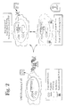
- AIG is driven by a promoter such as a TNF ⁇ promoter (TNFp; see Figures 1 and 2), and a TNF ⁇ promoter enhancer. Therefore, it is expressed in all and only those cells capable of producing TNF ⁇ .
- Another objective of this invention is to provide TNFp-AIG and the like chimeric gene constructs, processes for making them, methods of using them, and preparations containing them.
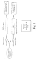
- Yet a further objective of this invention is to provide a method for the induction of apoptosis in cells transfected with the TNFp-AIG chimeric gene, a method for the in vitro selection of TNF ⁇ non-producer somatic cell variants in a population, a method for identifying dominant/negative genes responsible for the genesis of a TNF ⁇ non-producing population and a method for identifying products responsible for regulation of TNF ⁇ production (Figure 10).
- a chimeric gene comprising at least one promoter enhancer attached to at least one functional copy of a minimal promoter, the promoter being a gene or combination of genes activated in inflammatory cells or in cells at a site of inflammation, is attached to at least one copy of an apoptosis-inducing gene (AIG), such that the expression of the apoptosis-inducing gene is driven by the promoter, thus targeting the inflammatory cells.
- AIG apoptosis-inducing gene
- Chimeric genes according to the invention comprise enhancer, promoter, and AIG elements in direct, distal, or proximal attachment, and combinations thereof. As mentioned above and will be discussed in more detail below, in some embodiments, multiple copies of the enhancer, promoter, and/or AIG are employed for maximal efficacy.
- cytokines useful as promoters in the construction of chimeric genes of the invention include, but are not limited to, TNF- ⁇ and functional fragments and mixtures thereof.
- the elements of the TNF ⁇ promoter for preparation of chimeric gene constructs according to this invention are selected from elements which are capable of inducing expression of a therapeutic gene driven by the TNF ⁇ promoter. These promoter elements will be referred to herein as “inducible cis elements”, “cis-inducible elements” or “enhancer elements” of the TNF ⁇ promoter.
- the enhancer elements may be physically linked to the minimal promoter sequence, or separated from the minimal promoter by a linker sequence which may or may not have unique restriction sites.
- enhancer elements may be attached directly, distally, proximally, or any combination thereof, to chimeric genes of the invention. These are typically constructed upstream of the promoter.
- Example TNF ⁇ enhancer elements are set out in SEQ ID NO: 10 and SEQ ID NO: 11; functional fragments or variants and combinations thereof may be employed.
- Some preferred gene constructs according to this invention include those that have multiple copies of the enhancer elements, i . e ., 2 or more copies. Some embodiments have about 2 to 25, more narrowly 2 to 10, and even more narrowly, 2 to 5 copies.
- TNF promoter refers to the entire nucleotide sequence corresponding to a native TNF ⁇ minimal promoter sequence attached to one or more upstream enhancer elements (either present naturally i.e. native, or genetically engineered in the laboratory). Examples include, but are not limited to, SEQ ID NO: 1, SEQ ID NO: 2, and SEQ ID NO: 3, and functional fragments, variants, and mixtures of any of these.
- TNF ⁇ sequences and others described herein share a sequence homology of at least about 80%, and in some cases over 90%, to their native and genetically engineered counterparts, but these are known to skilled workers and defined in the references cited herein.
- apoptosis-inducing gene may be used in the chimeric genes and methods described herein.
- the apoptosis inducing gene used for the chimeric therapeutic genes of this invention can be the same or different from the type of apoptosis inducing gene present in the native sequence of the TNF- ⁇ -producing inflammatory cells (if those cells naturally contain an apoptotic gene).
- AIGs according to the invention are members of the ICE/CED3 family of apoptosis inducing proteases (such as Caspase-1 (ICE), hICE, ICE-LAP45, Mch2 ⁇ ), Caspase-2 (ICHI), Caspase-3 (CPP32, Yama, Apopain), Caspase-4 (TX, ICH2, ICE rel II), Caspase-5 (ICE rel III, TY), Caspase-6 (Mch-2), Caspase-7 (Mch-3, ICE-LAP3, CMH-1), Caspase-8 (MACH, FLICE, Mch-5), Caspase-9 (ICE-LAP6, Mch6) and Caspase-10 (Mch4)) and functional fragments, variants, and mixtures of any of these.
- proteases such as Caspase-1 (ICE), hICE, ICE-LAP45, Mch2 ⁇ ), Caspase-2 (ICHI), Caspase-3 (CPP32, Yama, A
- Some embodiments employ Caspase 3, Caspase 4, Caspase 5 and functional fragments, variants, and mixtures thereof. These genes, when overexpressed following transfection, induce apoptosis in the transfected cells (Miura M., et al., Cell, 1993, 75, 653-660; Chinnayan A.M., et al., Cell , 1995, 81, 505-512; Los, et al., Nature, 1995, 375, 81; Muzio, et al., Cell, 1996, 85, 817-827).
- This invention provides a novel therapeutic method comprising the step of introducing into the cells of a mammal a chimeric gene comprising an apoptosis-inducing gene (AIG) driven by the TNF ⁇ promoter (TNFp).
- AIG apoptosis-inducing gene driven by the TNF ⁇ promoter
- Example chimeric genes of the invention are set out in SEQ ID NOs 5 and 6; functional fragments or variants of these may also be employed.
- AIG is expressed in only those cells producing the inflammatory cytokine, TNF ⁇ . Therefore, any cells expressing TNF ⁇ will be self-destructive, while cells that do not express TNF- ⁇ will be unaffected.
- this methodology can target any TNF ⁇ -producing cells (such as activated macrophages, activated T-cells and macrophage-like and possibly fibroblast-like synoviocytes) without regard to cell type.
- the targeted TNF ⁇ -producing cell can be one which normally does or normally does not carry or expresses an apoptosis gene in its native, unaltered form. Therefore, using the chimeric genes and methods of this invention, the cellular sources of TNF ⁇ can be destroyed in a highly selective manner.
- TNFp-AIG chimeric gene of this invention Another advantage of using the TNFp-AIG chimeric gene of this invention is that TNFp sequesters transcription factors needed by endogenous TNFp, thereby leading to a reduction in endogenous TNF ⁇ production.
- TNFp is present in the therapeutically targeted cell in vast excess. This may be accomplished by introducing multiple copies of the transfected gene into the cell.
- the TNFp-AIG chimeric gene according to this invention can contain multiple copies of the inducible cis elements of the TNF ⁇ promoter. As mentioned above, multiple copies of the "inducible enhancer elements" of TNFp are present in some embodiments of the TNFp-AIG chimeric genes of this invention.
- the transcriptional factors needed by the transfected cell to produce TNF ⁇ are sequestered by the exogenously introduced sequence.
- This preferred chimeric TNFp-AIG construct is characterized by an increased effectiveness in competing for the TNFp-specific transcription factors as compared to chimeric genes of this invention containing only a single enhancer element linked to TNFp.
- the "inducible super promoter" constructed in this way is capable of (1) more effectively competing for TNF ⁇ specific inducible transcription factors and (2) driving expression of the apoptosis inducing gene in an augmented fashion by virtue of multiple enhancing elements.
- synovectomy i.e. , removal of synovial tissue
- the cell-targeted therapeutic method described herein targets only cells producing TNF ⁇ .
- the introduction and expression of the TNFp-AIG chimeric gene, and subsequent induction of apoptosis do not induce an inflammatory response. Accordingly, methods of this invention are comparatively selective and result in minimal tissue damage and a reduction in inflammation.
- inflammatory disorders include, but are not limited to, multiple sclerosis, Guillain-Barre syndrome, Crohn's disease, ulcerative colitis, psoriasis, graft versus host disease, lupus erythematosus, insulin-dependent diabetes mellitus, psoriatic arthritis, sarcoidosis, hypersensivity pneumonitis, ankylosing spondylitis and related spoldyloarthropathies, Reiter's syndrome and systemic sclerosis.
- this invention encompasses methods for treating an inflammatory disorder in a patient by inducing apoptosis in inflammatory cells or cells at a site of inflammation of the patient by introducing into the cells at least one chimeric gene of the invention.
- This is typically accomplished by preparing a pharmaceutical composition containing at least one chimeric gene of the invention and typically a pharmaceutically acceptal carrier, and administering the composition to a patient using standard means.
- the pharmaceutical composition is delivered directly to the site of inflammation using local topical, intravenous, intraperitoneal, and similar methods. Further methodology is discussed below.
- TNF ⁇ non-producer somatic cell variants within a TNF ⁇ producing cell population can be selected in vitro by introducing a TNFp-AIG chimeric gene into the TNF ⁇ producing cell population. Cells producing TNF ⁇ will undergo apoptosis. Cells that do not produce TNF ⁇ will survive. Selection of those cell variants possessing the survival phenotype is an easy way to identify TNF- ⁇ non-producer cells. Such a selection process may be used to determine expression of genes that act in-trans to regulate activity of the TNF- ⁇ promoter, thereby reducing TNF- ⁇ production.
- DN dominant negative
- a TNFp-AIG chimeric gene according to this invention can be used to identify dominant negative genes responsible for the genesis of a TNF ⁇ non-producing cell population.
- a TNFp-AIG chimeric gene according to this invention is introduced into cells that produce TNF ⁇ . Barring the presence of a dominant negative gene, those cells should undergo apoptosis upon activation. Therefore, it can be deduced that surviving variants possess a dominant negative gene capable of down-regulating TNF ⁇ production.
- the dominant negative gene can be readily identified by producing a cDNA library and transfecting cell lines (e.g., Jurkat and THP-1).
- TNFp-AIG transfected cells will be selected for the survival phenotype following in vitro activation; survival phenotype is indicative of the effect of the DN genes.
- survival phenotype is indicative of the effect of the DN genes.
- TNFp-luciferase gene reduction in the luciferase activity will be indicative of the DN gene effect.
- Dominant negative genes identified using this protocol can be used as the future therapeutic agents themselves. Such genes will be the candidates for gene therapy in order to reduce TNF ⁇ production.
- vectors For in vivo transfer, several vectors have been evaluated for their efficacy in gene delivery (Nita, et al., Arthritis & Rheumatism , 1996, 39/5, 820-828).
- the vectors derived from retroviruses are by far the best developed. They are able to insert genetic material in the host genome and produce stable transfectants. These vectors, however are unable to infect non-dividing cells and, since they are inserted in the host genome, the possibility of insertional mutagenesis cannot be ruled out.
- the vectors derived from adenoviruses infect dividing as well as non-dividing cells and deliver DNA episomally.
- adenovirus based vectors continue to produce viral proteins in infected cells making them potentially antigenic.
- a third type of viral based vectors is derived from Herpes simplex viruses (HSV), which are also capable of infecting dividing as well as non dividing cells.
- HSV Herpes simplex viruses
- Liposomes are at the most advanced stage of development, although certain types of cells such as muscle and skin take up, retain and express naked plasmid DNA.
- Liposome-DNA complexes (200 nmoles of DC-Chol complexed with 20 ⁇ g of DNA/ml) in 1 ml balance salt solution are injected per knee.
- Adenovirus-based vector delivery to hepatocytes Rat hepatocytes 1X10 11 PFU in 100 g animal.
- portal vein is perfused with about 1.5X10 11 PFU/kg gives 1 adenovirus genome copy per diploid copy of host DNA
- 1.5X10 13 virus particles (about 1.5X10 11 PFU) gives 100% hepatocyte transduction; 4X10 12 virus particles give 50-75% transduction.
- Gold particle-mediated gene delivery Transfection of mammalian skin tissue- 0.1, 0.5, 1.0 and 2.5 ⁇ g of DNA/mg particle gives linear relationship with transgene expression levels. Nabel, et al ., 297-305 Liposome-mediated gene delivery in humans:
- TNF- ⁇ promoter elements for constructing a chimeric gene .
- the regions of the TNF- ⁇ promoter are amplified by polymerase chain reaction (PCR) using primers encompassing various deletion constructs of the TNF ⁇ promoter ( Figure 3).
- PCR polymerase chain reaction
- Figure 3 The regions identified by other investigators in various other cellular systems are used as reference (Rhoades, et al., J. Biol. Chem ., 1992, 267, 22102-22107; Leitman, et al., Mol. Cell Biol., 1992, 12, 1352-1356; Pauli U., Crit. Reviews Eukaryotic Gene Expression , 1994, 4, 323-344).
- the PCR-amplified genes are then cloned upstream of a reporter gene, such as luciferase, in a commercially available promoterless vector.
- a reporter gene such as luciferase
- These constructs are tested for their constitutive and inducible expression in various cell lines such as Jurkat (T lymphoblastoid), U973 (myelomonocytic), THP-1 (monocytic), fibroblasts and in vitro cultured human synoviocytes. Identification of the regions responsible for inducible expression of the reporter gene is primarily based on the results obtained using two TNF ⁇ -producing cell lines, viz Jurkat (following stimulation with PMA) and THP-1 (following stimulation with LPS) ( Figure 4 a and b).
- TNF ⁇ promoter that are responsible for inducible expression of the reporter gene are then used for constructing TNFp-AIG chimeric genes.
- TNFp-AIG chimeric genes Construction of TNFp-AIG chimeric genes .
- the following genes are preferred:
- the AIGs are used as "prodomain-deleted" truncations in order to potentially augment autocatalysis of Caspases. This is essential for conversion of inactive Caspase to active form.
- Prodomain-deleted CPP32 is amplified using primers corresponding to codons 29-36 and 271-278 (278 is a stop codon).
- the truncated form of CPP32 is referred to as " ⁇ CPP32" or "AIG.1" herein.
- primers corresponding to the sequences in the Ty gene are synthesized. All Caspases discovered so far have homology to the other members of the Caspase family.
- the 3' primer corresponding to the codons 359-365 (codon 365 is a stop codon) shares 100% sequence homology to the codons 372-378 (codon 378 is a stop codon) in the Tx gene.
- the 5' primer corresponding to codons 81-87 in the Ty gene does not share 100 % homology with the corresponding region in the Tx gene (Tx codons 94-100).
- Residue 87 (Alanine) in the Ty gene differs from residue 100 (Glycine) in the Tx gene.
- the PCR amplified product generated from cDNA prepared from activated human peripheral blood lymphocytes possesses the sequence of Tx, due to apparent abundance of Tx transcripts. Therefore, the truncated form of the AIG generated using synthetics oligonucleotide primers corresponding to the sequences in Ty, indeed matches sequences in Tx , albeit flanked by Ty sequences of the primers.
- the Ty sequences of the primers used also match with the sequence of Tx, except for one codon.
- the gene used in this invention matches truncated Tx gene with residue G 100 toA change. This gene is referred to as " ⁇ Ty/x" or "AIG.2" herein.
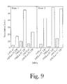
- AIG.1 and AIG.2 are inserted downstream of the TNF ⁇ promoter by replacing the luciferase reporter gene in deletion constructs (-120, -706 and -1005) of the TNF ⁇ promoter ( Figure 5). These constructs are tested for the induction of apoptosis following stimulation of transiently-infected Jurkat and THP-1 cells ( Figures 6 a, b, and c).
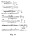
- TNF ⁇ superpromoter-AIG chimeric genes Two broad preferred regions, viz ., ER1(-1005 to -905) (SEQ. ID 10) and ER2(-706 to -517) (SEQ ID NO: 11) of the TNF ⁇ promoter, containing elements responsible for inducible expression of the reporter gene described above ( Figure 4a and 4b) are PCR amplified and are ligated upstream of the minimal native promoter (-120 through TSS, SEQ ID NO: 3), either as a single copy or multiple copies.
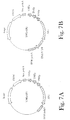
- the super promoter contains multiple (2-10) cassettes of the above mentioned regions containing inducible promoter elements ( Figure 7). This is achieved by PCR amplifying the regions of interest using primers synthesized with restriction sites inserted at the 5' end of each of the primers. These unique restriction sites flank the amplified gene product of interest.
- PCR amplified AIG is cloned downstream of the TNF-( super promoter, replacing the luciferase reporter gene in the original construct as described ( Figure 5) for the native TNF ⁇ promoter.
- At least seven copies of the enhancer regions (ER1, ER2 or ER3, individually or in combination), one at a time, can be added by using one more restriction site upstream of the previous one in PCR amplification of the enhancer regions of choice.
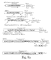
- AIG is inserted downstream of the superpromoter as described in the STEP 5 of the scheme 1.
- TNFp-AIG gene The inducible expression of the chimeric TNFp-AIG gene is tested by transient transfection of the cell lines mentioned above.
- the expression of TNFp-AIG gene is measured by detecting apoptosis of transfected cells, assessing AIG expressed proteins in Western blots using commercially available antibodies and assessing protease activity using commercially available, well documented specific synthetic tetrapeptide substrate.
- the inducible expression of the chimeric TNFp-FasL gene is tested by transient transfection of the same cell lines.
- the cell surface expression of FasL by the transfected cells is quantitated using anti-FasL antibody binding as detected by indirect immunofluorescence and by measuring induction of apoptosis of Fas positive cells.
- TNFp-driven expression of a reporter gene Regulation of the TNFp-driven expression of a reporter gene .
- the 3' untranslated region of the TNF ⁇ gene plays an important role in reulation of the TNF ⁇ biosynthesis. It is involved in translational expression of the TNF ⁇ gene in normal, non-activated states. Importantly, these elements allow de-repression to occur when TNF ⁇ -producing cells are activated by external stimuli (Han, J., et al., J. Immunology , 1991, 146, 1843-1848; Crawford, F.K., et al., J. Biol. Chem ., 1996, 271, 22383-22390).
- Luciferase assay Luciferase activity is determined using commercially available reagents ( Promega).
- Rabbit model of IL-1-induced arthritis (Pettipher E. R., et al., Proc. Natl. Acad. Sci., 1986, 83, 8749-8753): Il-1 is injected into the knee joints of New Zealand White rabbits. Intra-articular injection of IL-1 causes dose-dependent infiltration of leukocytes into the joint space and loss of proteoglycan from the articular cartilage.
- Antigen-Induced arthritis Intra-articular injection of antigen (ovalbumin) into knee joints induces leukocyte accumulation and cartilage degradation that closely resembles rheumatoid arthritis in humans. The joint swelling following the injection was sustained for 14 days.
- antigen ovalbumin
- Synoviocytes are transfected in vitro with the therapeutic gene and transplanted back in rabbits. Arthritis is induced in these rabbits by injecting IL-1 and expression of the therapeutic gene following activation is assessed. Activation-induced expression of the chimeric gene induces apoptosis in transplanted cells.
- Direct Method Intra-articular injection of the chimeric genes. Any of the gene delivery methods described above, including naked plasmid DNA, cationic liposome-mediated delivery can be used. For use of viral vector-based delivery, chimeric genes are cloned in suitable vectors. The vectors are then modified by deleting eukaryotic promoter present in these vectors. Intra-articular injection of the therapeutic genes inserted in appropriate vectors can then be done to assess therapeutic as well as prophylactic efficacy.
- TNFp-AIG TNFp-AIG chimeric gene. After several cycles of stimulation, which induces apoptosis in the cells expressing the TNFp-AIG gene, surviving cells are then collected. A cDNA library from these cells is constructed, which is used for functional cloning (Legerski R and Peterson C., Nature, 1992, 359, 70-73; Jaattela M., et al., Oncogene , 1995, 10, 2297-2305).
- THP-1 and Jurkat cells stably transfected with TNFp-AIG are subjected to repeated cycles of stimulation to activate expression of TNFp-AIG.
- the cells which do not express negative regulatory genes, undergo apoptosis, whereas those expressing dominant negative genes survive.
- DN gene products act in-trans with the TNF ⁇ promoter, thereby inhibiting its activations to transcribe AIG, ultimately resulting in survival phenotype.
- cDNA library is constructed using polyadenylated mRNA from these cells.
- the DN genes which rescue TNFp-AIG-transfected THP-1 or Jurkat cells from apoptosis are identified by functional cloning as described for other genes (Legerski R. and Peterson C., Nature, 1992, 359, 70-73; Jaattela M., et al., Oncogene , 1995, 10, 2297-2305).
Landscapes
- Health & Medical Sciences (AREA)
- Chemical & Material Sciences (AREA)
- Life Sciences & Earth Sciences (AREA)
- Organic Chemistry (AREA)
- Engineering & Computer Science (AREA)
- Genetics & Genomics (AREA)
- General Health & Medical Sciences (AREA)
- Bioinformatics & Cheminformatics (AREA)
- Medicinal Chemistry (AREA)
- Zoology (AREA)
- Molecular Biology (AREA)
- Pharmacology & Pharmacy (AREA)
- Biomedical Technology (AREA)
- Animal Behavior & Ethology (AREA)
- Wood Science & Technology (AREA)
- Public Health (AREA)
- Veterinary Medicine (AREA)
- Biochemistry (AREA)
- Nuclear Medicine, Radiotherapy & Molecular Imaging (AREA)
- General Chemical & Material Sciences (AREA)
- Chemical Kinetics & Catalysis (AREA)
- General Engineering & Computer Science (AREA)
- Biotechnology (AREA)
- Biophysics (AREA)
- Gastroenterology & Hepatology (AREA)
- Microbiology (AREA)
- Toxicology (AREA)
- Proteomics, Peptides & Aminoacids (AREA)
- Diabetes (AREA)
- Rheumatology (AREA)
- Physics & Mathematics (AREA)
- Plant Pathology (AREA)
- Hematology (AREA)
- Physical Education & Sports Medicine (AREA)
- Endocrinology (AREA)
- Obesity (AREA)
- Pain & Pain Management (AREA)
- Immunology (AREA)
- Orthopedic Medicine & Surgery (AREA)
- Emergency Medicine (AREA)
Abstract
Description
- This invention relates to the therapeutic induction of apoptosis in inflammatory cells by introducing into those cells a gene which induces apoptosis (programmed cell death or non-necrotic cell death) in these cells. The Apoptosis-Inducing Gene (which will sometimes be referred to herein as AIG) is driven by a TNFα promoter (TNFp) activated in inflammation. In one embodiment, apoptosis is selectively induced in those cells capable of producing TNFα. The TNFp-AIG or other chimeric gene may be conveniently introduced in vivo using conventional gene therapy techniques. Advantageously, in the embodiment wherein the chimeric gene is TNFp-AIG, it is expressed in only those cells producing the inflammatory cytokine, TNFα. In addition, since the TNFp-AIG chimeric gene contains the TNFα promoter elements, it also sequesters inducible, TNFp-selective transcription factors. Such sequestration results in a reduction in endogenous production of TNFα. The present invention relates specifically to TNFp-AIG and similar gene constructs, cells containing chimeric genes, methods for induction of apoptosis in cells transfected with chimeric genes, pharmaceutical compositions containing chimeric genes, methods for in vitro selection of TNFα non-producer somatic cell variants within a TNFα producing cell population and the like, a method for identifying dominant negative/dominant suppressive genes responsible for inhibiting TNFα production and therapeutic methods using the chimeric gene.
- In many inflammatory conditions, cytokines such as IL-1, IL-10, GM-CSF and TNFα are excessively produced as a result of mass aggregation and accumulation of inflammatory cells (Brennan F.M. et al., British Medical Bulletin 1995, 51/2, 368-384). Upregulation and/or disregulation of cytokines in inflamed tissue may be directly or indirectly responsible for exacerbation of chronic inflammatory diseases. For example, the most marked pathology in rheumatoid arthritis (RA) is displayed at the local site of inflammation (i.e., the synovial joints). Therefore, it is likely that the cytokines produced in the synovial joints of RA patients play an important role in the disease process. Of those cytokines, IL-1 and TNFα are believed to be responsible for the devastating cartilage destruction and bone erosion which characterizes RA (Dayer J.M. et al., J. Exp. Med., 1985, 162, 1208-1215; Gowen M. et al., Nature, 1983, 306, 378-380). The presence of excessive amounts of IL-1 and TNFα in the synovial joints has been shown to accelerate development of collagen-induced arthritis in rodents (Brennan F.M., et al., Clin. Expt. Immunol., 1994, 97/1, 1-3). Excessive amounts of TNFα and IL-1 are produced in the synovial tissue by a variety of cell types at the cartilage-pannus junction, including cells of the macrophage lineage, macrophage-like synoviocytes, activated T-cells and possibly fibroblast-like synoviocytes (Chu C.Q. et al., Arthritis & Rheumatism, 1991, 34, 1125-1132; Deleuran B.W., et al., Arthritis & Rheumatism, 1992, 35, 1170-1178).
- In addition to the above described inflammatory effects, TNFα plays a ubiquitous and key role in a variety of pro-inflammatory events, such as induction of IL-1 activity in monocytes. Indeed, anti-TNFα neutralizing antibodies have been shown to reduce overall IL-1 production (Portillo, et al., Immunol., 1989, 66, 170-175; Brennan F.M., et al., British Medical Bulletin 1995, 51/2, 368-384). Thus, an added benefit to blocking the effect of the inflammatory cytokine TNFα is the reduction in production of the equally destructive pro-inflammatory mediator, IL-1. Furthermore, it is well known that TNFα is a transcriptional activator of other inflammation-related genes. For example, the presence of TNFα stimulates production of other cytokines (such as GM-CSF) and cell surface receptors, including HLA class II antigens and adhesion molecules (Alvaro-Garcia J.M., et al., J. Exp. Med., 1989, 146, 865-875), which results in continuous recruitment of activated T cells and neutrophils resulting in synovial inflammation and hyperplasia and ultimately, in augmented destruction of cartilage and bone (Allen J.B., J. Exp. Med., 1990, 171, 231).
- Conventional therapy against inflammatory disorders is typically directed against symptomatic inflammation. Such therapies provide only temporary relief without significantly delaying disease progression. In contrast, therapies targeting TNFα and other factors induced in the inflammatory process are likely to be more promising. For example, in collagen-induced arthritis animal models, an anti-TNFα antibody and soluble TNFα receptor-IgG chimera effectively reduced paw swelling, joint involvement and cartilage and bone destruction (Williams R.O. et al., Proc. Natl. Acad. Sci., 1992, 89, 9784-9788). Human trials using both humanized anti-TNFα antibodies and TNFα receptor-IgG chimeric molecules produced dramatic results (Elliott M.J., et al., Arthritis and Rheumatism, 1993, 36, 1681-1690; Elliott M.J., et al., Lancet, 343, 1105-1110). Although treatment with these TNFα antagonists appears to be well tolerated, it also results in production of antibodies against the recombinant proteins. Thus, these therapies may not be suitable for long term treatment and do not achieve true disease abatement. In order to actually modify progression of the disease, TNFα must be continuously targeted using TNFα-specific therapies. Such a therapeutic protocol is impractical with these biologic agents and would be difficult to administer in the long term.
- In an alternate therapeutic option, inflamed synovium may be removed using surgical (Herold N. and Schroder H.A., Acta Orthop. Scand., 1995, 66, 252-254; Ogilvie-Harris D.J. and Weisleder L., Arthroscopy, 1995, 11, 91-95), chemical (Cruz-Esteban C. and Wilke W.S., Bailliere's Clinical Rheumatol., 1995, 9, 787-801) or radiation-induced synovectomy (Cruz-Esteban C. and Wilke W.S., Bailliere's Clinical Rheumatol. , 1995, 9, 787-801). The results following arthroscopic surgical synovectomy are good, showing improvement from the preoperative condition to the postoperative condition. Non-surgical synovectomy is performed using various chemical agents such as osmic acid, alkylating agents such as nitrogen mustard and thiotepa, methotrexate. Unfortunately, non-surgical synovectomies (including chemical and radiation-induced) are procedurally complicated, provide only short term relief and show only patchy reduction of the synovial hyperplasia. Furthermore, most of the non-surgical alternatives are potential teratogens. In addition, the chemical damage to afflicted tissue in non-surgical synovectomy, as well as surgically-induced tissue damage, often cause an inflammatory response themselves. Finally, it should be noted that these approaches suffer from the risks and side-effects commonly associated with conventional pharmaceutical therapy and invasive surgical procedures, including the expense and inconvenience of hospitalization and rehabilitation.
- Application WO 96/25501 A2 describes the human factor Yama on DNA and aminoacid level as well as diverse forms of it, especially a dominant inhibitory variant. It further discloses mutant forms of factor CrmA. Both factors naturally are part of the Fas receptor pathway and interact with each other to induce apoptosis. This application teaches that a gene coding for a protein with CrmA activity can be introduced into a cell to prevent or inhibit apoptosis of this cell. It also discloses the possibility to introduce Yama nucleic acids or proteins to modulate the cellular function regulated by the Fas receptor. This has been applied to T cells which could be prevented from apoptosis after viral infection. This application further describes Yama specific antibodies and methods for identifying agents that are involved in the apoptotic pathway, based on Yama and CrmA.
- WO 97/07828 A1 lies on the field of diagnosis and treatment of chronic inflammatory diseases such as - amongst others - rheumatoid arthritis, via gene therapy approaches. It describes that inflammatory diseases can be based on an insufficient level of apoptosis of certain accumulated cells. Consequently this application teaches an up-regulation of the level of apoptosis of the affected cells. This is described to be possible through enhancing the amount of factors like p53 or ICE in the respective cells, e.g. via setting them under the control of inducible promoters like the TNF-alpha promotor or others. The use of enhancer elements in this context is only generally mentioned.
- JP 01/137976 A discloses a novel DNA fragment containing a gene which codes for mouse TNF and further includes adjacent regions, especially about 800 base pairs which lie 5'-terminal adjacent to the exon. This fragment is described to be useful for the screening for substances that induce the expression of the gene coding for TNF.
- Accordingly, a need still exists for an effective therapeutic approach to treating inflammatory disorders in general and RA in particular.
- This invention overcomes the drawbacks associated with previous therapies for treating inflammatory disorders by providing a novel therapeutic approach. According to one embodiment of this invention, apoptosis is selectively induced in TNFα-producing inflammatory cells, causing destruction of these cells without an associated inflammatory reaction.
- One objective of this invention is to provide a therapeutic method comprising the step of introducing into the inflammatory cells of a mammal, or cells at a site of inflammation, a chimeric gene containing a self-regulating apoptosis-inducing gene (AIG). The AIG is driven by a promoter such as a TNFα promoter (TNFp; see Figures 1 and 2), and a TNFα promoter enhancer. Therefore, it is expressed in all and only those cells capable of producing TNFα.
- Another objective of this invention is to provide TNFp-AIG and the like chimeric gene constructs, processes for making them, methods of using them, and preparations containing them.
- Yet a further objective of this invention is to provide a method for the induction of apoptosis in cells transfected with the TNFp-AIG chimeric gene, a method for the in vitro selection of TNFα non-producer somatic cell variants in a population, a method for identifying dominant/negative genes responsible for the genesis of a TNFα non-producing population and a method for identifying products responsible for regulation of TNFα production (Figure 10).
- These and other objectives will be readily appreciated by those of ordinary skill in the art based upon the following detailed disclosure of the invention.
-
- Figure 1 is a schematic representation of TNFp-AIG chimeric genes of this invention. Apoptosis Inducing Gene (AIG) could be any, but not limited to, the genes listed, viz.,
Caspases 1 to 10. - Figure 2 is a schematic drawing depicting the results of gene therapy using a TNFp-AIG chimeric gene of this invention.
- Figure 3 is a summary of deletion constructs used for identification of the inducible cis elements of the TNFα promoter using luciferase gene (Luc) expression as the reporter system.
- Figure 4 (a and b) provide a summary of results obtained using the constructs described in Figure 3. Transient expression of the constructs was assessed in two different TNFα-producing cell lines, viz., Jurkat (Figure 4a) and THP-1 (Figure 4b). Histograms in each figure show stimulation index as a measure of inducibility by activating agents such as PMA (Figure 4a) or LPS (Figure 4b) for individual experiments. The line superimposed in each figure indicates the mean inducibility averaged from 4 to 6 experiments.
- Figure 5 is a flow chart for preparation of the TNFpAIG using selected native elements of the TNFα promoter and prodomain-deleted AIGs (AIGs used are Caspase and
Caspase 4/5). - Figure 6 (a, b, and c) provide a summary of results from representative experiments performed to see expression of the chimeric TNFpAIGs. Apoptosis in transiently-transfected Jurkat cells (Figure 6a and 6b) and THP-1 (Figure 6c) cells was assessed using Cell Death Elisa (CDE assay). In all three figures, histograms with sparse dots represent transfection control, where cells were treated with the transfecting agent in the absence of DNA. Histograms with dense dots represent the TNFp elements driving expression of the luciferase gene and solid histograms represent the same TNFp elements driving expression of either AIG.1 or AIG.2. The number in parenthesis above the solid histograms represent enrichment factor (ratio of apoptosis induced by TNFpAIG to the TNFpLuc control vector).
- Figure 7 (a and b) is a diagrammatic representation of a TNFp-AIG chimeric gene of this invention, comprising multiple copies of the inducible cis elements of the TNFα promoter which, in turn, drive expression of the AIG (Figure 7a). A diagrammatic representation of a TNFpAIG chimeric gene, comprising multiple copies of the inducible cis elements of the TNFα promoter, driving expression of the AIG, downstream of which are 3' untranslated region of the TNFα gene (TNF3'UTR) (Figure 7b). 3'UTR of the TNFα gene is implicated in the regulation of the inducible expression of TNFα (Han, J., et al., J. Immunology, 1991, 146, 1843-1843, Crawford, E.K., et al., J. Biol. Chem., 1997, 272, 21120-21137, and Figure 9).
- Figure 8 (a and b) are flow charts of schemes for preparing TNFα superpromoter-AIG chimeric constructs.
- Figure 9 shows a summary of the results of two experiments to show the regulatory effect of the TNF3'UTR on inducible expression of the luciferase reporter gene. The transient transfection was performed in a fibroblast cell line. Dotted histograms represent inducibility of TNFpLuc in the absence of TNF3'UTR and solid histograms represent inducibility of TNFpLuc in the presence of TNF3'UTR. Similar results are obtained in the Jurkat cell.
- Figure 10 is a diagrammatic representation for the selection of TNFα non-producer somatic cell variants within a TNFα-producing cell population and identification of dominant negative suppressive genes responsible for inhibiting TNF-α production.
- This invention is based upon evidence that apotosis of inflammatory cells in certain inflammatory diseases is therapeutically beneficial. The invention specifically relates to self-regulated apoptosis by gene therapy. Broadly speaking, in the practice of the invention, a chimeric gene comprising at least one promoter enhancer attached to at least one functional copy of a minimal promoter, the promoter being a gene or combination of genes activated in inflammatory cells or in cells at a site of inflammation, is attached to at least one copy of an apoptosis-inducing gene (AIG), such that the expression of the apoptosis-inducing gene is driven by the promoter, thus targeting the inflammatory cells. Chimeric genes according to the invention comprise enhancer, promoter, and AIG elements in direct, distal, or proximal attachment, and combinations thereof. As mentioned above and will be discussed in more detail below, in some embodiments, multiple copies of the enhancer, promoter, and/or AIG are employed for maximal efficacy.
- In order that the invention herein described may be more fully understood, the following detailed description is set forth, with emphasis on chimeric genes comprising at least one TNFα promoter enhancer attached to at least one functional copy of a minimal TNFα promoter and further attached to at least one copy of an AIG for illustrative purposes only. Though the examples that follow also employ these types of constructions, it will be appreciated by skilled workers that the basic constructs described herein may be altered to provide other embodiments that utilize products, processes, methods, and compositions of the invention with other promoters comprising inducible genes activated in inflammation such as the types listed above that exhibit similar functions that can be used to target cells at the site of infection.
- For example, cytokines useful as promoters in the construction of chimeric genes of the invention include, but are not limited to, TNF-α and functional fragments and mixtures thereof.
- To clarify the discussion below of exemplary TNFp-AIG chimeric genes of this invention, the following sequences are illustrated:
- SEQ ID NO: 1 is the nucleotide sequence corresponding to the full-length, reference human TNFα promoter sequence, as published in (Takashiba S., et al., Gene, 1993, 131, 307-308). Nucleotide numbers used herein refer to the numbering of this sequence.
- SEQ ID NO: 2 is the native TNFα promoter sequence of the gene that was used in this invention (-1077 nucleotides from the transcription start site, TSS). There are a few differences in the sequence of the TNFp in SEQ ID NO: 1 and SEQ ID NO: 2. Such differences in the nucleotide sequences of the TNFα promoter have been reported (Takashiba S., et al., Gene, 1993, 131, 307-308).
- SEQ ID NO: 3 is the native minimal TNFα promoter sequence (nucleotide -120 through -TSS, which includes at least one enhancer element (k1 site; see Pauli, U., Crit. Rev. in Eucaryotic Gene Expression, 1994, 4, 323-344; Rhoades K.L., et al., J. Biol. Chem., 1992, 267, 22102-22107; and Takashiba S., et al., Gene, 131, 307-108).
- SEQ ID NO: 4 is the chimeric gene TNFp120 AIG.1 (containing -120 TNFp driving the expression of the prodomain-deleted variant of CPP32 gene (
Caspase 3, published Tewari M. et al., Cell, 1995, 81(5), 801-809, with the variation being V239A). - SEQ ID NO: 5 is the chimeric gene TNFp706 AIG.1 (containing -706TNFp driving expression of the prodomain-deleted CPP 32 gene.
- SEQ ID NO: 6 is the TNFp1005 AIG.1 (containing -1005 TNFp driving expression of the prodomain-deleted CPP 32 gene).
- SEQ ID NO: 7 is the chimeric gene TNFp120 AIG.2 (containing -120 TNFp driving expression of the prodomain-deleted Ty/x gene. (Sequences of Ty (Caspase 5) and Tx (Caspase 4) genes are published in the ref. Faucheu, C., et. al., Eur. J Biochem., 236, 207-213, 1996; Faucheu, C., et. al. EMBO J., 14, 1914-1922,1995).
- SEQ ID NO: 8 is the chimeric gene TNFp706 AIG.2 (containing -706TNFp driving expression of the prodomain-deleted Ty/x gene.
- SEQ ID NO: 9 is the TNFp1005 AIG.1 (containing -1005 TNFp driving expression of the prodomain-deleted Ty/x gene).
- SEQ ID NO: 10 is the enhancer region 1 (ER1) of the TNFα promoter encompassing nucleotides -1005 to -905.
- SEQ ID NO: 11 is the enhancer region 2 (ER2) of the TNFα promoter encompassing nucleotides -706 to -517.
- SEQ ID NO: 12 is additional multiple cloning sites (MCS) genetically engineered upstream of the -120 minimal TNFα promoter in the -120pGL3 construct.
- SEQ ID NO: 13 is the 3' untranslated region (3'UTR) of the TNFα gene (Nedwin, G.E., et al., Nucleic Acid Research, 1985, 13, 6361-6373).
- The elements of the TNFα promoter for preparation of chimeric gene constructs according to this invention are selected from elements which are capable of inducing expression of a therapeutic gene driven by the TNFα promoter. These promoter elements will be referred to herein as "inducible cis elements", "cis-inducible elements" or "enhancer elements" of the TNFα promoter.
- The enhancer elements may be physically linked to the minimal promoter sequence, or separated from the minimal promoter by a linker sequence which may or may not have unique restriction sites. Thus, as summarized above, enhancer elements may be attached directly, distally, proximally, or any combination thereof, to chimeric genes of the invention. These are typically constructed upstream of the promoter. Example TNFα enhancer elements are set out in SEQ ID NO: 10 and SEQ ID NO: 11; functional fragments or variants and combinations thereof may be employed. Some preferred gene constructs according to this invention include those that have multiple copies of the enhancer elements, i.e., 2 or more copies. Some embodiments have about 2 to 25, more narrowly 2 to 10, and even more narrowly, 2 to 5 copies.
- The terms "TNF promoter", "TNFα promoter" and "TNFp" are used interchangeably herein. Unless noted to the contrary, these terms refer to the entire nucleotide sequence corresponding to a native TNFα minimal promoter sequence attached to one or more upstream enhancer elements (either present naturally i.e. native, or genetically engineered in the laboratory). Examples include, but are not limited to, SEQ ID NO: 1, SEQ ID NO: 2, and SEQ ID NO: 3, and functional fragments, variants, and mixtures of any of these. Many functional fragments and variants of these TNFα sequences and others described herein share a sequence homology of at least about 80%, and in some cases over 90%, to their native and genetically engineered counterparts, but these are known to skilled workers and defined in the references cited herein.
- Any apoptosis-inducing gene may be used in the chimeric genes and methods described herein. The apoptosis inducing gene used for the chimeric therapeutic genes of this invention can be the same or different from the type of apoptosis inducing gene present in the native sequence of the TNF-α-producing inflammatory cells (if those cells naturally contain an apoptotic gene). AIGs according to the invention are members of the ICE/CED3 family of apoptosis inducing proteases (such as Caspase-1 (ICE), hICE, ICE-LAP45, Mch2α), Caspase-2 (ICHI), Caspase-3 (CPP32, Yama, Apopain), Caspase-4 (TX, ICH2, ICE rel II), Caspase-5 (ICE rel III, TY), Caspase-6 (Mch-2), Caspase-7 (Mch-3, ICE-LAP3, CMH-1), Caspase-8 (MACH, FLICE, Mch-5), Caspase-9 (ICE-LAP6, Mch6) and Caspase-10 (Mch4)) and functional fragments, variants, and mixtures of any of these. Some embodiments employ
Caspase 3,Caspase 4,Caspase 5 and functional fragments, variants, and mixtures thereof. These genes, when overexpressed following transfection, induce apoptosis in the transfected cells (Miura M., et al., Cell, 1993, 75, 653-660; Chinnayan A.M., et al., Cell, 1995, 81, 505-512; Los, et al., Nature, 1995, 375, 81; Muzio, et al., Cell, 1996, 85, 817-827). - This invention provides a novel therapeutic method comprising the step of introducing into the cells of a mammal a chimeric gene comprising an apoptosis-inducing gene (AIG) driven by the TNFα promoter (TNFp). Example chimeric genes of the invention are set out in
SEQ ID NOs - Another advantage of using the TNFp-AIG chimeric gene of this invention is that TNFp sequesters transcription factors needed by endogenous TNFp, thereby leading to a reduction in endogenous TNFα production. In one preferred embodiment, TNFp is present in the therapeutically targeted cell in vast excess. This may be accomplished by introducing multiple copies of the transfected gene into the cell. Alternatively, the TNFp-AIG chimeric gene according to this invention can contain multiple copies of the inducible cis elements of the TNFα promoter. As mentioned above, multiple copies of the "inducible enhancer elements" of TNFp are present in some embodiments of the TNFp-AIG chimeric genes of this invention. By including multiple copies of the inducible cis elements of the TNFp construct, the transcriptional factors needed by the transfected cell to produce TNFα are sequestered by the exogenously introduced sequence. This preferred chimeric TNFp-AIG construct is characterized by an increased effectiveness in competing for the TNFp-specific transcription factors as compared to chimeric genes of this invention containing only a single enhancer element linked to TNFp. The "inducible super promoter" constructed in this way is capable of (1) more effectively competing for TNFα specific inducible transcription factors and (2) driving expression of the apoptosis inducing gene in an augmented fashion by virtue of multiple enhancing elements.
- For example, in rheumatoid arthritis patients, synovectomy, i.e., removal of synovial tissue, has been shown to be clinically beneficial. Unlike conventional and surgical synovectomy procedures, the cell-targeted therapeutic method described herein targets only cells producing TNFα. Thus, advantageously, the introduction and expression of the TNFp-AIG chimeric gene, and subsequent induction of apoptosis do not induce an inflammatory response. Accordingly, methods of this invention are comparatively selective and result in minimal tissue damage and a reduction in inflammation.
- The products and methods described herein are useful for the treatment of other inflammatory disorders as well. Such inflammatory disorders include, but are not limited to, multiple sclerosis, Guillain-Barre syndrome, Crohn's disease, ulcerative colitis, psoriasis, graft versus host disease, lupus erythematosus, insulin-dependent diabetes mellitus, psoriatic arthritis, sarcoidosis, hypersensivity pneumonitis, ankylosing spondylitis and related spoldyloarthropathies, Reiter's syndrome and systemic sclerosis. Thus, this invention encompasses methods for treating an inflammatory disorder in a patient by inducing apoptosis in inflammatory cells or cells at a site of inflammation of the patient by introducing into the cells at least one chimeric gene of the invention. This is typically accomplished by preparing a pharmaceutical composition containing at least one chimeric gene of the invention and typically a pharmaceutically acceptal carrier, and administering the composition to a patient using standard means. In some embodiments, the pharmaceutical composition is delivered directly to the site of inflammation using local topical, intravenous, intraperitoneal, and similar methods. Further methodology is discussed below.
- In addition to the therapeutic indications, the genes and cells according to this invention can be used in a variety of useful screening and selection methods. In one such method, TNFα non-producer somatic cell variants within a TNFα producing cell population can be selected in vitro by introducing a TNFp-AIG chimeric gene into the TNFα producing cell population. Cells producing TNFα will undergo apoptosis. Cells that do not produce TNFα will survive. Selection of those cell variants possessing the survival phenotype is an easy way to identify TNF-α non-producer cells. Such a selection process may be used to determine expression of genes that act in-trans to regulate activity of the TNF-α promoter, thereby reducing TNF-α production. Such genes are characterized as dominant negative (DN) /dominant suppressive genes in other systems (Behrends S., et al., J. Biol. Chem. 1995, 270, 21109-21113; Zhang S., et al., J. Biol. Chem., 1995, 270, 23934-23936; Watowich S.S., et al., Mol. Cell Biol., 1994, 14/6, 3535-3549).
- In a further in vitro method, a TNFp-AIG chimeric gene according to this invention can be used to identify dominant negative genes responsible for the genesis of a TNFα non-producing cell population. According to this method, a TNFp-AIG chimeric gene according to this invention is introduced into cells that produce TNFα. Barring the presence of a dominant negative gene, those cells should undergo apoptosis upon activation. Therefore, it can be deduced that surviving variants possess a dominant negative gene capable of down-regulating TNFα production. The dominant negative gene can be readily identified by producing a cDNA library and transfecting cell lines (e.g., Jurkat and THP-1). These cells are either stable transfectants of an inducible TNFp-AIG chimeric gene or TNFp-luciferase gene TNFp-AIG transfected cells will be selected for the survival phenotype following in vitro activation; survival phenotype is indicative of the effect of the DN genes. In the cells transfected with TNFp-luciferase gene, reduction in the luciferase activity will be indicative of the DN gene effect. Dominant negative genes identified using this protocol can be used as the future therapeutic agents themselves. Such genes will be the candidates for gene therapy in order to reduce TNFα production.
- The methods utilized for gene transfer are grouped into two broad categories:
- 1. Direct approach : In situ transduction of the therapeutic gene into target cells such as synoviocytes using a suitable vector as a carrier for the therapeutic gene. The vector containing therapeutic gene is injected directly into the affected area (e.g., an arthritic joint).
- 2. Indirect approach : Ex-vivo transfection of the therapeutic gene into target cells such as synoviocytes. In this approach, the synovium is removed from joints, synoviocytes are isolated and cultured in vitro. In vitro cultured cells are transfected with the therapeutic gene, and genetically modified synoviocytes are transplanted back into the synovium.
- For in vivo transfer, several vectors have been evaluated for their efficacy in gene delivery (Nita, et al., Arthritis & Rheumatism, 1996, 39/5, 820-828). Among the vectors used for gene therapy, the vectors derived from retroviruses are by far the best developed. They are able to insert genetic material in the host genome and produce stable transfectants. These vectors, however are unable to infect non-dividing cells and, since they are inserted in the host genome, the possibility of insertional mutagenesis cannot be ruled out. In comparison, the vectors derived from adenoviruses infect dividing as well as non-dividing cells and deliver DNA episomally. The disadvantage of adenovirus based vectors is that these vectors continue to produce viral proteins in infected cells making them potentially antigenic. A third type of viral based vectors is derived from Herpes simplex viruses (HSV), which are also capable of infecting dividing as well as non dividing cells.
- Among the non-viral vector systems, cationic liposomes and naked plasmid DNA have been evaluated. Liposomes are at the most advanced stage of development, although certain types of cells such as muscle and skin take up, retain and express naked plasmid DNA.
- Particle-mediated gene-delivery system is also possible (Rakhmilevich, et al., PNAS, 1996, 93, 6291) and is a promising approach.
- The following "in vivo" gene delivery protocols can be used to deliver the chimeric genes of this invention:
- In vivo experiment in rabbits:
Each vector is injected intra-articularly into 1 knee joint. For viral vectors, between 108 and 109 particles suspended in 0.5 ml balance salt solution are injected per knee. - Liposome-DNA complexes (200 nmoles of DC-Chol complexed with 20 µg of DNA/ml) in 1 ml balance salt solution are injected per knee.
- Adenovirus-based vector delivery to hepatocytes : Rat hepatocytes 1X10¹¹ PFU in 100 g animal.
- In dogs (12-17 kg), portal vein is perfused with about 1.5X10¹¹ PFU/kg gives 1 adenovirus genome copy per diploid copy of host DNA
- In rabbits (2-4kg), 1.5X10¹³ virus particles (about 1.5X10¹¹ PFU) gives 100% hepatocyte transduction; 4X10¹² virus particles give 50-75% transduction. Yang N-S, et al., 281-296
- Gold particle-mediated gene delivery : Transfection of mammalian skin tissue- 0.1, 0.5, 1.0 and 2.5 µg of DNA/mg particle gives linear relationship with transgene expression levels.
Nabel, et al., 297-305
Liposome-mediated gene delivery in humans: - Protocol 1: 15nmol DC-Chol/Dope liposomes combined with 1 µg DNA in 0.7 ml. 0.2 ml of the above mixture is injected into the patient's melanoma nodule. For catheter delivery, 0.6 ml of the solution is delivered into the artery.
- Protocol 2: 15nmol DMRIE/Dope liposomes combined with 5 µg DNA in 1.0 ml.
- Gene transfer to synovium:
- A range of doses, 109-1012 adenovirus particles containing therapeutic gene/joint are used. However, the optimal dose for any particular experimental series needs to be determined empirically, and is dependent on both the properties of the recombinant adenoviral genomic backbone being used as well as the transgene being expressed.
- For the indirect approach, a variety of methods are well established, including utilization of cationic lipid or cationic polymer-based transfection and electroporation.
- Any of the above-referenced techniques can be altered to suit the particular needs of those of ordinary skill in the art. Such modifications are well within the level of skill possessed by ordinary practitioners and do not require undue experimentation. These obvious variations are within the scope of this invention.
- In order that this invention be more fully understood, the following examples are set forth. These examples are for the purpose of illustrating some preferred embodiments of this invention, and are not to be construed as limiting the scope of this invention in any way.
- In order to construct chimeric AIG driven by the enhancer cis elements of the TNF promoter, either in a single or multiple copies of the same region or various regions, identification of the regions of interest responsible for optimal inducible expression of the reporter gene is performed.
- Selection of the TNF-α promoter elements for constructing a chimeric gene. The regions of the TNF-α promoter are amplified by polymerase chain reaction (PCR) using primers encompassing various deletion constructs of the TNFα promoter (Figure 3). The regions identified by other investigators in various other cellular systems are used as reference (Rhoades, et al., J. Biol. Chem., 1992, 267, 22102-22107; Leitman, et al., Mol. Cell Biol., 1992, 12, 1352-1356; Pauli U., Crit. Reviews Eukaryotic Gene Expression, 1994, 4, 323-344). The PCR-amplified genes are then cloned upstream of a reporter gene, such as luciferase, in a commercially available promoterless vector. These constructs are tested for their constitutive and inducible expression in various cell lines such as Jurkat (T lymphoblastoid), U973 (myelomonocytic), THP-1 (monocytic), fibroblasts and in vitro cultured human synoviocytes. Identification of the regions responsible for inducible expression of the reporter gene is primarily based on the results obtained using two TNFα-producing cell lines, viz Jurkat (following stimulation with PMA) and THP-1 (following stimulation with LPS) (Figure 4 a and b). These cells are transiently transfected by using well established methods and commercially available reagents, e.g., DEAE dextran and Superfect. The cis-elements of the TNFα promoter that are responsible for inducible expression of the reporter gene are then used for constructing TNFp-AIG chimeric genes.
- Construction of TNFp-AIG chimeric genes. Of the apoptosis inducing genes described herein, the following genes are preferred:
- i) cysteine protease - CPP32 (also known as Yama, apopain or Caspase 3) and
- ii) Cysteine protease -Tx/Ty (
Caspase 4/Caspase 5) - The AIGs are used as "prodomain-deleted" truncations in order to potentially augment autocatalysis of Caspases. This is essential for conversion of inactive Caspase to active form.
- Prodomain-deleted CPP32 is amplified using primers corresponding to codons 29-36 and 271-278 (278 is a stop codon). The truncated form of CPP32 is referred to as "αCPP32" or "AIG.1" herein.
- For PCR amplification for prodomain-deleted Ty, primers corresponding to the sequences in the Ty gene are synthesized. All Caspases discovered so far have homology to the other members of the Caspase family. The 3' primer corresponding to the codons 359-365 (codon 365 is a stop codon) shares 100% sequence homology to the codons 372-378 (codon 378 is a stop codon) in the Tx gene. However, the 5' primer corresponding to codons 81-87 in the Ty gene does not share 100 % homology with the corresponding region in the Tx gene (Tx codons 94-100). Residue 87 (Alanine) in the Ty gene differs from residue 100 (Glycine) in the Tx gene. The PCR amplified product generated from cDNA prepared from activated human peripheral blood lymphocytes possesses the sequence of Tx, due to apparent abundance of Tx transcripts. Therefore, the truncated form of the AIG generated using synthetics oligonucleotide primers corresponding to the sequences in Ty, indeed matches sequences in Tx , albeit flanked by Ty sequences of the primers. The Ty sequences of the primers used also match with the sequence of Tx, except for one codon. Thus the gene used in this invention matches truncated Tx gene with residue G 100 toA change. This gene is referred to as "ΔTy/x" or "AIG.2" herein.
- AIG.1 and AIG.2 are inserted downstream of the TNFα promoter by replacing the luciferase reporter gene in deletion constructs (-120, -706 and -1005) of the TNFα promoter (Figure 5). These constructs are tested for the induction of apoptosis following stimulation of transiently-infected Jurkat and THP-1 cells (Figures 6 a, b, and c).
- Constuction of TNFα superpromoter-AIG chimeric genes. Two broad preferred regions, viz., ER1(-1005 to -905) (SEQ. ID 10) and ER2(-706 to -517) (SEQ ID NO: 11) of the TNFα promoter, containing elements responsible for inducible expression of the reporter gene described above (Figure 4a and 4b) are PCR amplified and are ligated upstream of the minimal native promoter (-120 through TSS, SEQ ID NO: 3), either as a single copy or multiple copies. Two more regions (-234 to -120) and (-234 to -65) of the TNFα promoter is also identified as a potential enhancer region 3 (ER3) and enhancer region 4 (ER4), respectively, which can be employed in the chimeric constructs using the strategies described below. The super promoter contains multiple (2-10) cassettes of the above mentioned regions containing inducible promoter elements (Figure 7). This is achieved by PCR amplifying the regions of interest using primers synthesized with restriction sites inserted at the 5' end of each of the primers. These unique restriction sites flank the amplified gene product of interest. Preferably, PCR amplified AIG is cloned downstream of the TNF-( super promoter, replacing the luciferase reporter gene in the original construct as described (Figure 5) for the native TNFα promoter.
- The schemes for construction of a TNFα superpromoter and the linker sequences representing unique restriction sites (these restriction sites are absent in the selected elements of the TNF-α promoter and the AIG in question) for efficient directional insertion is outlined below and depicted in Figure 8:
-
- STEP 1: Insertion of the TNF-α minimal promoter (-120 to TSS) into the pGL3 basic (promoterless) luciferase vector (Promega):
The elements of pGL3 basic vectors that are used for construction of the chimeric gene TNFp-AIG are shown below.
_KpnI.SacI.MluI.NheI.SmaI.XhoI.BglII.HindIII. [luciferase].XbaI _
The minimal promoter is PCR amplified using primers containing XhoI and BgIII.HindIII sites, so that XhoI is at the 5'end and BgIII.HindIII sites are at the 3' end of the amplified product. This fragment is inserted into the polylinker of the pGL3 basic vector using these same restriction sites. This construct is referred to as "Construct A1" and is as follows:
_KpnI.SacI.MluI.NheISmaIXhoI. (-120 to TSSBglII). HindIII. [luciferase].XbaI_ - STEP 2: The enhancer fragment (ER1 or ER2) is PCR amplified using the primer containing several restriction sites. The resulting fragment will have restriction sites KpnI.AatII.BssHII at the 5' end and NsiI.SpeI.MluI at the 3'end as follows: 5' KpnI.AatII.BssHII.(ER1 or ER2).
NsiI.SpeI.MluI 3". The fragment is inserted into the "Construct A1" generated inSTEP 1 using KpnI and MluI restriction sites. This construct is referred to as "Construct B1" and is as follows:
_KpnI.AatII.BssHII.(ER1 or ER2).NsiI.SpeI.MluI.NheI.SmaI.XhoI(-120 to TSS BgIII).HindIII. [luciferase].XbaI_ - STEP 3: The TNFα enhancer fragment (ER1 or ER2)) is amplified using the primers containing restriction sites AatII and BssHII to generate the PCR product as follows:
5' AatII.(ER1 or ER2).BssHII 3'. This fragment is cloned into the "Construct
B1" using these same restriction sites. This construct is referred to as "Construct C1" and is as follows:
_KpnI.AatII.(ER1 or ER2).BssHII.( ER1 or ER2).NsiI.SpeI.MluI.NheI SmaI.XhoI(-120 to TSS BglII).HindIII.[luciferase].XbaI_ - STEP 4: The TNFα enhancer fragment (ER1 or ER2) is amplified using the primers containing restriction sites NsiI and SpeI to generate the PCR product as follows:
5' NsiI.(ER1 or ER2).SpeI 3'. This fragment will be cloned into the "Construct
C1" using these same restriction sites. This construct is referred to as "Construct D1" and is as follows:
_KpnI.AatII.(ER1 or ER2).BssHII.(ER1 or ER2).NsiI.(ER1 or ER2).SpeI.MluI.NheI SmaI.XhoI(-120 to TSSBglII).HindIII.[luciferase].XbaI - STEP 5: AIG.1 or AIG.2 (preferred but not limited to AIG.1 and AIG.2; any AIG from the list can be used) coding regions are PCR-amplified using the primers containing BgIII and XbaI restriction sites generating the fragment as follows: 5' BgIII.(AIG.1 or AIG.2).XbaI 3". This fragment is inserted into the "Construct D1" using these same restriction sites. The resulting construct is referred to as "Construct E1" and is as follows:
_KpnI.AatII.(ER1 or ER2).BssHII.( ER1 or ER2).NsiI.( ER1 or ER2).SpeI.MluI.NheI.SmaI.XhoI(-120 to TSS.BglII)[AIG.1 or AIG.2].XbaI_ - Alternatively
scheme 2 is followed: -
- STEP 1: Same as in scheme I giving rise to "Construct A1", which is as follows:
KpnI.SacI.MIuI.NheI.SmaI.XhoI.(-120 to TSS BglII).HindIII.[luciferase].XbaI - STEP 2: Insertion of additional MCS.
Two complementary oligonucleotides (5' phosphorylated) providing _NheI.SacII.EcorV.AflII.AatII.AvrII.SpeI.PvuII.XhoI_are synthesized using commercial sources. These oligonucleotides are annealed and then cloned into Nhel and XhoI sites of the "Construct A1". The resulting construct referred to as "Construct B2" and it is as follows:
_KpnI.SacI.MIuI.NheI. SacII.EcorV.AflII.AatII.AvrII.SpeI.PvuII.XhoI.(-120 to TSS BgIII).HindIII.[luciferase].XbaI_ - STEP 3: The TNF-α enhancer fragment (ER1 or ER2) is amplified using the primers containing restriction sites SpeI.PvuII at the 5' end, and XhoI at the 3' end to generate the PCR product as follows: 5' SpeI.PvuII.(ER1 or ER2) . XhoI 3'. This fragment is cloned into the "Construct B2" using SpeI and XhoI restriction sites. This construct is referred to as "Construct C2" and is as follows:
_KpnI.SacI.MIuI.NheI. SacII.EcorV.AflII.AatII.AvrII.SpeI.PvuII.(ER1 or ER2) XhoI. (-120 to TSS BglII).HindIII.[luciferase].XbaI_ - STEP 4: The TNFα enhancer fragment (ER1 or ER2) is amplified using the primers containing restriction sites AvrII. SpeI at the 5' end, and and PvuII at the 3' end to generate the PCR product as follows: 5' AvrII.SpeI.(ER1 or ER2).PvuII 3'. This fragment is cloned into the "Construct C2" using AvrII and PvuII restriction sites. This construct is referred to as "Construct D2" and is as follows:
_KpnI.SacI.MluI.NheI. SacII.EcorV.AflII.AatII.AvrII.SpeI. (ER1 or ER2) PvuII. (ER1 or ER2) XhoI.(-120 to TSS BglII).HindIII.[luciferase].XbaI_ - Thus, using this strategy at least seven copies of the enhancer regions (ER1, ER2 or ER3, individually or in combination), one at a time, can be added by using one more restriction site upstream of the previous one in PCR amplification of the enhancer regions of choice.
- Once the desired number of copies of the enhancer regions are added, AIG is inserted downstream of the superpromoter as described in the
STEP 5 of thescheme 1. - The inducible expression of the chimeric TNFp-AIG gene is tested by transient transfection of the cell lines mentioned above. The expression of TNFp-AIG gene is measured by detecting apoptosis of transfected cells, assessing AIG expressed proteins in Western blots using commercially available antibodies and assessing protease activity using commercially available, well documented specific synthetic tetrapeptide substrate.
- The inducible expression of the chimeric TNFp-FasL gene is tested by transient transfection of the same cell lines. The cell surface expression of FasL by the transfected cells is quantitated using anti-FasL antibody binding as detected by indirect immunofluorescence and by measuring induction of apoptosis of Fas positive cells.
- Regulation of the TNFp-driven expression of a reporter gene. The 3' untranslated region of the TNFα gene plays an important role in reulation of the TNFα biosynthesis. It is involved in translational expression of the TNFα gene in normal, non-activated states. Importantly, these elements allow de-repression to occur when TNFα-producing cells are activated by external stimuli (Han, J., et al., J. Immunology, 1991, 146, 1843-1848; Crawford, F.K., et al., J. Biol. Chem., 1996, 271, 22383-22390).
- Genetic constructs are made in which the entire 3' untranslated region (SEQ ID NO: 13) is inserted downstream of the luciferase gene driven by deletion fragments, viz., -120, -706 and -1005 of the TNFα promoter. The results of the transient expression of these constructs are summarized in Figure 9.
-
- a) Western blots of the transfected cell lysates are developed using anti-CPP32 antibody as well as anti-PRAP antibody. Anti-PRAP antibody detects both hydrolyzed as well as non-hydrolyzed products of PRAP as an enzymatic action of CPP32.
- b) CPP32 enzyme assay : This assay detects enzymatic reaction of CPP32 and breakdown of colorimetric or fluorogenic substrate. Commercially available (Clonotech, Pharmingen) kit ise used for this assay.
- c) Apoptosis of transfected cells: Apoptosis of transfected cells due to AIG.1 and AIG.2 is determined by staining nuclei by propidium iodide (Krishan, A., J. Cell Biol., 66, 1994, 188-193) and by commercially available Cell Death Elisa kit (Boehringer Mannheim).
- Rabbit model of IL-1-induced arthritis (Pettipher E. R., et al., Proc. Natl. Acad. Sci., 1986, 83, 8749-8753): Il-1 is injected into the knee joints of New Zealand White rabbits. Intra-articular injection of IL-1 causes dose-dependent infiltration of leukocytes into the joint space and loss of proteoglycan from the articular cartilage.
- Antigen-Induced arthritis : Intra-articular injection of antigen (ovalbumin) into knee joints induces leukocyte accumulation and cartilage degradation that closely resembles rheumatoid arthritis in humans. The joint swelling following the injection was sustained for 14 days.
- Scid mice-human synoviocytes model (Houri J.M., et al. Current Opinions in Rheumatol., 1995, 7, 201-205; Sack U., et al., J. Autoimmunity, 1995, 9, 51-58; Geiler T., et al. Arthritis & Rheumatism, 1994, 37, 1664-1671): These are recently developed models for arthritis in which fresh synovial tissue from RA patients is implanted with normal human cartilage into scid mice either subcutaneously, under the renal capsule (Geiler T., et al., Arthritis & Rheumatism, 1994, 37, 1664-1671), or into knee joints (Sack U., et al., J. Autoimmunity, 1995, 9, 51-58). The implants grow with arthritis-like characteristics, including formation of pannus tissue of high cellular density, bone and cartilage erosion, development of multinuclear giant cells, and invasion of cartilage by synovial fibroblasts.
- Indirect Method: Synoviocytes are transfected in vitro with the therapeutic gene and transplanted back in rabbits. Arthritis is induced in these rabbits by injecting IL-1 and expression of the therapeutic gene following activation is assessed. Activation-induced expression of the chimeric gene induces apoptosis in transplanted cells.
- Direct Method: Intra-articular injection of the chimeric genes. Any of the gene delivery methods described above, including naked plasmid DNA, cationic liposome-mediated delivery can be used. For use of viral vector-based delivery, chimeric genes are cloned in suitable vectors. The vectors are then modified by deleting eukaryotic promoter present in these vectors. Intra-articular injection of the therapeutic genes inserted in appropriate vectors can then be done to assess therapeutic as well as prophylactic efficacy.
- Cells (THP-1, Jurkat) are stably transfected in vitro with TNFp-AIG chimeric gene. After several cycles of stimulation, which induces apoptosis in the cells expressing the TNFp-AIG gene, surviving cells are then collected. A cDNA library from these cells is constructed, which is used for functional cloning (Legerski R and Peterson C., Nature, 1992, 359, 70-73; Jaattela M., et al., Oncogene, 1995, 10, 2297-2305).
- THP-1 and Jurkat cells stably transfected with TNFp-AIG are subjected to repeated cycles of stimulation to activate expression of TNFp-AIG. The cells, which do not express negative regulatory genes, undergo apoptosis, whereas those expressing dominant negative genes survive. In these surviving cells DN gene products act in-trans with the TNFα promoter, thereby inhibiting its activations to transcribe AIG, ultimately resulting in survival phenotype. cDNA library is constructed using polyadenylated mRNA from these cells. The DN genes which rescue TNFp-AIG-transfected THP-1 or Jurkat cells from apoptosis are identified by functional cloning as described for other genes (Legerski R. and Peterson C., Nature, 1992, 359, 70-73; Jaattela M., et al., Oncogene, 1995, 10, 2297-2305).
- The above description is for the purpose of teaching the person of ordinary skill in the art how to practice the present invention, and it is not intended to detail all those obvious modifications and variations of it which will become apparent to the skilled worker upon reading the description. It is intended, however, that all such obvious modifications and variations be included within the scope of the present invention, which is defined by the following claims. The claims are intended to cover the claimed components and steps in any sequence which is effective to meet the objectives there intended, unless the context specifically indicates the contrary.
-
- <110> BOEHRINGER INGELHEIM PHARMACEUTICALS INC.
- <120> Self-Regulated Apoptosis of Inflammatory Cells by Gene Thera py
- <140> PCT/US98/03911
<141> 1998-02-27 - <150> 60/039,266
<151> 1997-02-28 - <160> 13
- <170> PatentIn Ver. 2.1
- <210> 1
<211> 1178
<212> DNA - <220>
<221> source
<223> /note ="reference human TNFà promoter" - <300>
<301> Takashiba, S., et al.
<303> Gene
<304> 131
<306> 307-308 - <400> 1


- <210> 2
<211> 1096
<212> DNA - <220>
<221> source
<223> /note ="human TNFà promoter gene" - <300>
<301> Takashiba, S., et al.
<303> gene
<304> 131
<306> 307-308 - <400> 2

- <210> 3
<211> 139
<212> DNA - <220>
<221> source
<223> /note ="native minimal TNFà promoter" - <400> 3

- <210> 4
<211> 904
<212> DNA - <220>
<221> source
<223> /note ="chimeric gene TNFp120 AIG.1
residues 1 to 139 comprise the promoter sequence;
residues 140 to 151, the linker sequence, and
the remaining residues comprise the AIG.1 sequence - <400> 4

- <210> 5
<211> 1490
<212> DNA - <200>
<221> source
<223> /note ="chimeric gene TNFp120 AIG.1
residues 1 to 724 comprise the promoter sequence;
residues 725 to 736, the linker sequence, and
the remaining residues comprise the AIG.1 sequence" - <400> 5

- <210> 6
<211> 1789
<212> DNA - <220>
<221> source
<223> /note ="chimeric gene TNFp1005 AIG.1
residues 1 to 1023 comprise the promoter sequence;
residues 1024 to 1036, the linker sequence,
and the remaining residues comprise the AIG.1 sequence" - <400> 6

- <210> 7
<211> 1008
<212> DNA - <220>
<221> source
<223> /note ="chimeric gene TNFp120 AIG.2
residues 1 to 138 comprise the promoter sequence;
residues 139 to 150, the linker sequence, and
the remaining residues comprise the AIG.2 sequence - <400> 7

- <210> 8
<211> 1587
<212> DNA - <220>
<221> source
<223> /note =" chimeric gene TNFp706 AIG.2
residues 1 to 724 comprise the promoter sequence;
residues 725 to 736, the linker sequence, and
the remaining residues comprise the AIG.2 sequence" - <400> 8

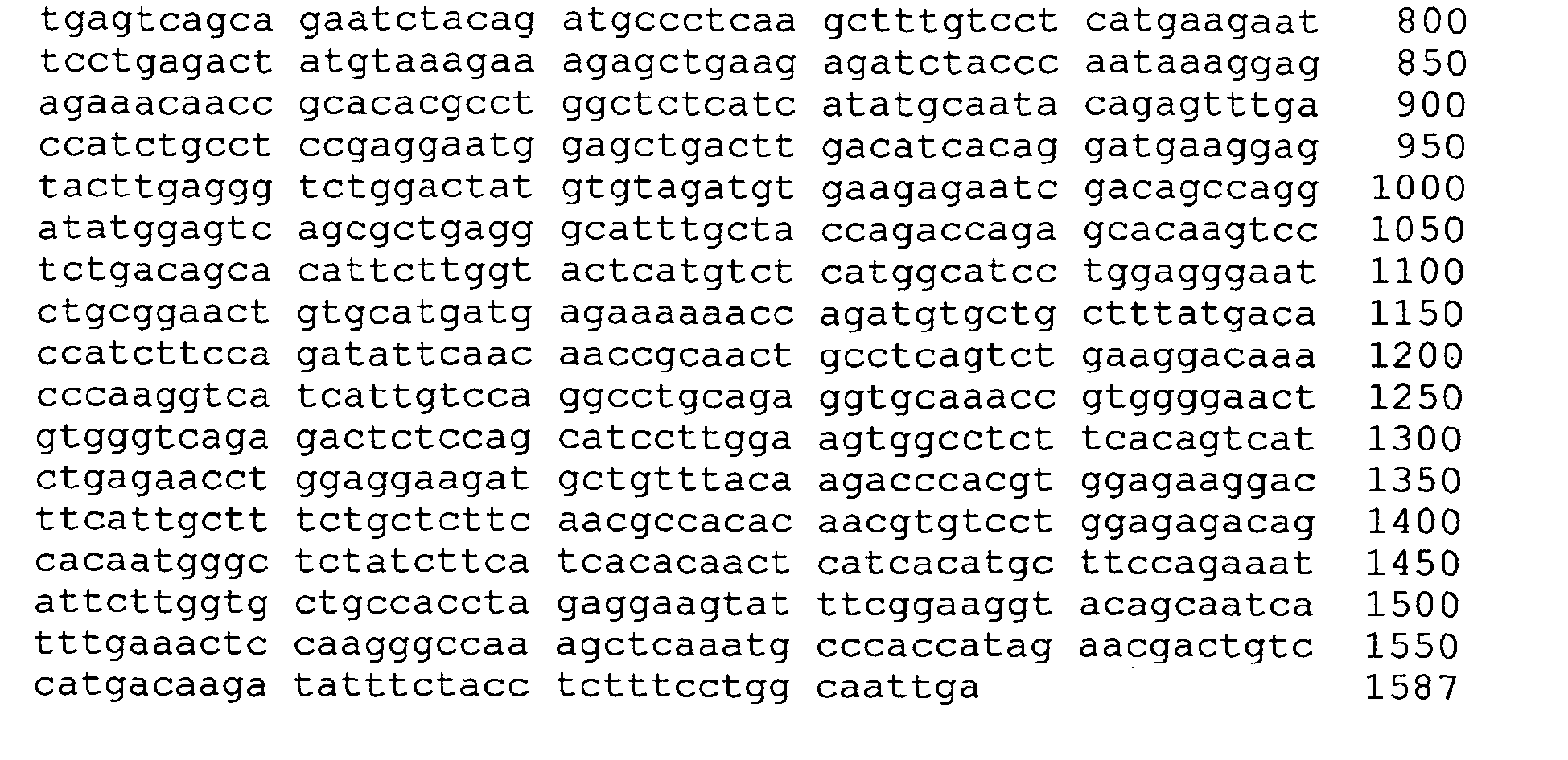


- <210>10
<211> 123
<212> DNA
<220>
<221> source
<223> /note ="TNFà promoter enhancer region 1 (ER1)" - <400> 10

- <210> 11
<211> 190
<212> DNA - <220>
<221> source
<223> /note =" TNFà promoter enhancer region 2 (ER2)" - <400> 11

- <210> 12
<211> 223
<212> DNA - <220>
<221> source
<223> /note: ="genetically engineered multiple clonging sites genetically engineered upstream of the minimal TNFà promoter in the -120pgL3 construct " - <400> 12

- <210> 13
<211> 787
<212> DNA - <220>
<221> source
<223> /note =" NFà intranslated region" - <400> 13

Claims (19)
- A chimeric gene comprising at least one TNFα promoter enhancer selected from SEQ ID NO: 10 or SEQ ID NO: 11, attached to a functional copy of a minimal TNFα promoter selected from the group consisting of SEQ ID NO: 1, SEQ ID NO: 2, SEQ ID NO: 3 and further attached to at least one copy of an apoptosis-inducing gene selected from the group consisting of caspase-1, caspase-2, caspase-3, caspase-4, caspase-5, caspase-6, caspase-7, caspase-8, caspase-9, caspase-10 and mixtures of these, wherein the expression of the apoptosis-inducing gene is driven by the TNFα promoter.
- The gene according to claim 1, wherein the attachment of the enhancer to the promoter and the promoter to the apoptosis-inducing gene is selected from the group consisting of direct attachment, distal attachment, proximal attachment, and combinations thereof.
- The gene according to claim 1 comprising 2 or more copies of the TNFα promoter enhancer.
- The gene according to claim 1, wherein the apoptosis-inducing gene is selected from the group consisting of caspase 3, caspase 4, caspase 5, and mixtures of any of these.
- The gene according to claim 1 selected from the group consisting of SEQ ID NO: 5, or SEQ ID NO: 6.
- The gene according to claim 5, wherein the 3'UTR of the TNFα gene is ligated downstream of the apotosis-inducing gene.
- A pharmaceutical composition comprising a gene according to claim 1.
- A pharmaceutical composition comprising a gene according to claim 6.
- The use of a gene according to claim 1 or 6 for the manufacture of a pharmaceutical composition for the treatment of an inflammatory disorder in a patient by inducing apoptosis in inflammatory cells or cells at a site of inflammation of the patient by introducing into the cells said chimeric gene.
- The use according to claim 9, wherein the induction of apoptosis does not induce an inflammatory response in the patient.
- The use according to claim 9, wherein the inflammatory cell is a TNFα producing cell.
- The use according to claim 9, wherein the inflammatory disorder is selected from the group consisting of rheumatoid arthritis, multiple sclerosis, Guillain-Barre syndrome, Crohn's disease, ulcerative colitis, psoriasis, graft versus host disease, lupus erythematosus, insulin-dependent diabetes mellitus, psoriatic arthritis, sarcoidosis, hypersensitivity pneumonitis, ankylosing spondylitis, Reiter's syndrome, and systemic sclerosis.
- The use according to claim 12, wherein the inflammatory disorder is rheumatoid arthritis.
- A chimeric gene comprising 2 to 10 cassettes of a TNFα promoter enhancer selected from SEQ ID NO: 10 or SEQ ID NO: 11 attached to at least one copy of a minimal TNFα promoter selected from the group consisting of SEQ ID NO: I, SEQ ID NO: 2, SEQ ID NO: 3, and at least one copy of an apoptosis-inducing gene selected from the group consisting of caspase 3, caspase 4, caspase 5, and mixtures of any of these, wherein the expression of the apoptosis-inducing gene is driven by the TNF promoter.
- A pharmaceutical composition comprising a gene according to claim 14.
- An in vitro method for inducing apoptosis in a TNFα-producing inflammatory cell by introducing into the cell a chimeric gene according to claim 1.
- The use of a chimeric gene according to claim 14 for the manufacture of a pharmaceutical composition for the treatment of an inflammatory disorder in a patient by inducing apoptosis in inflammatory cells or cells at the site of inflammation of the patient by introducing into the cells said chimeric gene without inducing an inflammatory response in the patient.
- A process for constructing a chimeric gene according to anyone of claims 1 to 6, or 14, comprising the steps of(a) amplifying the TNFα promoter by a polymerase chain reaction using primers encompassing deletion constructs of the TNFα promoter;(b) cloning the PCR-amplified genes obtained in step (a) upstream of a reporter gene;(c) testing the constructs obtained in step (b) for their constitutive and inducible expression in at least one TNFα-producing cell line;(d) selecting the TNFα promoter responsible for inducible expression of the reporter in the cell line; and either(e) PCR-amplifying the TNFα promoter that enhance expression of the reporter to obtain an enhancer and ligating at least one copy of the enhancer upstream of the promoter; or(f) inserting at least one copy of the apoptosis-inducing gene downstream of the TNFα promoter by replacing the reporter gene with the apoptosis-inducing gene deletion constructs to obtain a chimeric gene or(g) PCR-amplifying a TNFα-3'UTR and ligating downstream of the reporter gene, or any combination of these procedures.
- The process according to claim 18, wherein the cell lines for testing constructs are selected from the group consisting of T lymphoblastoid, myelomonocytic, monocytic, fibroblast, and cultured human synoviocytes.
Applications Claiming Priority (3)
| Application Number | Priority Date | Filing Date | Title |
|---|---|---|---|
| US3926697P | 1997-02-28 | 1997-02-28 | |
| US39266P | 1997-02-28 | ||
| PCT/US1998/003911 WO1998037901A1 (en) | 1997-02-28 | 1998-02-27 | Self-regulated apoptosis of inflammatory cells by gene therapy |
Publications (3)
| Publication Number | Publication Date |
|---|---|
| EP0989853A1 EP0989853A1 (en) | 2000-04-05 |
| EP0989853A4 EP0989853A4 (en) | 2004-03-17 |
| EP0989853B1 true EP0989853B1 (en) | 2007-05-16 |
Family
ID=21904557
Family Applications (1)
| Application Number | Title | Priority Date | Filing Date |
|---|---|---|---|
| EP98908778A Expired - Lifetime EP0989853B1 (en) | 1997-02-28 | 1998-02-27 | Self-regulated apoptosis of inflammatory cells by gene therapy |
Country Status (17)
| Country | Link |
|---|---|
| US (1) | US6525184B1 (en) |
| EP (1) | EP0989853B1 (en) |
| JP (1) | JP2001516210A (en) |
| KR (1) | KR20000075788A (en) |
| CN (1) | CN1286530C (en) |
| AT (1) | ATE362369T1 (en) |
| AU (1) | AU729063C (en) |
| BR (1) | BR9807622A (en) |
| CA (1) | CA2281567A1 (en) |
| DE (1) | DE69837787D1 (en) |
| HU (1) | HUP0002317A3 (en) |
| IL (1) | IL131328A0 (en) |
| NZ (1) | NZ338013A (en) |
| PL (1) | PL191586B1 (en) |
| RU (1) | RU2198683C2 (en) |
| TR (1) | TR199902068T2 (en) |
| WO (1) | WO1998037901A1 (en) |
Families Citing this family (19)
| Publication number | Priority date | Publication date | Assignee | Title |
|---|---|---|---|---|
| US20040039186A1 (en) * | 1997-02-28 | 2004-02-26 | Boehringer Ingelheim Pharmaceuticals, Inc. | Self-regulated apoptosis of inflammatory cells by gene therapy |
| US6537784B1 (en) * | 1998-02-27 | 2003-03-25 | Boehringer Ingelheim Pharmaceuticals Inc. | Self-regulated apoptosis of inflammatory cells by gene therapy |
| AU2004233486B2 (en) * | 1999-01-11 | 2007-09-13 | Leadd B.V. | Use of apoptosis inducing agents in the treatment of (auto)immune diseases |
| DE60003632T2 (en) * | 1999-01-11 | 2004-04-15 | Leadd B.V. | USE OF APOPTOSE-INDUCING AGENTS FOR PRODUCING AN AGENT FOR TREATING (AUTO) IMMUNE DISEASES |
| EP1939300A1 (en) * | 1999-05-28 | 2008-07-02 | Targeted Genetics Corporation | Methods and compositions for lowering the level of tumor necrosis factor (TNF) in TNF-associated disorders |
| US6537540B1 (en) * | 1999-05-28 | 2003-03-25 | Targeted Genetics Corporation | Methods and composition for lowering the level of tumor necrosis factor (TNF) in TNF-associated disorders |
| CA2377730A1 (en) * | 1999-08-19 | 2001-03-01 | Centre For Translational Research In Cancer | Replication protein a binding transcriptional factor (rbt1) and uses thereof |
| US7094891B1 (en) | 1999-08-19 | 2006-08-22 | Center For Translation Research In Cancer | Replication protein a binding transcriptional factor (RBT1) and uses thereof |
| ES2169986B1 (en) * | 2000-03-14 | 2003-06-16 | Ferrer Int | USE OF CDP-HILL FOR PROFILACTIC TREATMENT OF CEREBRAL ISCHEMIA. |
| US8039261B2 (en) | 2000-11-17 | 2011-10-18 | Vascular Biogenics Ltd. | Promoters exhibiting endothelial cell specificity and methods of using same for regulation of angiogenesis |
| AU2003222427B8 (en) | 2000-11-17 | 2010-04-29 | Vascular Biogenics Ltd. | Promoters exhibiting endothelial cell specificity and methods of using same |
| US8071740B2 (en) | 2000-11-17 | 2011-12-06 | Vascular Biogenics Ltd. | Promoters exhibiting endothelial cell specificity and methods of using same for regulation of angiogenesis |
| ZA200305980B (en) | 2001-02-12 | 2007-01-31 | Res Dev Foundation | Modified proteins, designer toxins, and methods of making thereof |
| CA2454048C (en) | 2001-07-17 | 2011-05-03 | Research Development Foundation | Therapeutic agents comprising pro-apoptotic proteins |
| EP1436313B1 (en) | 2001-10-19 | 2010-09-22 | Vascular Biogenics Ltd. | Polynucleotide constructs, pharmaceutical compositions and methods for targeted downregulation of angiogenesis and anticancer therapy |
| EP1506285B1 (en) * | 2001-11-01 | 2010-12-08 | Uab Research Foundation | Combinations of anti-dr5 antibodies and anti-dr4 antibodies and other therapeutic agents |
| ES2357225T3 (en) * | 2001-11-01 | 2011-04-20 | Uab Research Foundation | COMBINATIONS OF ANTI-DR5 ANTIBODIES AND ANTI-DR4 ANTIBODIES AND OTHER THERAPEUTIC AGENTS. |
| JP2009511572A (en) * | 2005-10-16 | 2009-03-19 | イエダ リサーチ アンド ディベロップメント カンパニー リミテッド | Pharmaceutical composition and diagnostic method for inflammatory skin diseases, disorders or conditions |
| RU2388065C1 (en) * | 2008-12-08 | 2010-04-27 | Учреждение Российской академии наук Институт цитологии и генетики Сибирского отделения РАН (ИЦиГ СО РАН) | Method for screening pro- or antiapoptogenous activity of therapeutic preparations |
Family Cites Families (6)
| Publication number | Priority date | Publication date | Assignee | Title |
|---|---|---|---|---|
| JPH01137976A (en) * | 1987-11-20 | 1989-05-30 | Asahi Chem Ind Co Ltd | Novel dna fragment |
| US5792751A (en) | 1992-04-13 | 1998-08-11 | Baylor College Of Medicine | Tranformation of cells associated with fluid spaces |
| US6071890A (en) * | 1994-12-09 | 2000-06-06 | Genzyme Corporation | Organ-specific targeting of cationic amphiphile/DNA complexes for gene therapy |
| US7097972B1 (en) * | 1995-02-13 | 2006-08-29 | Regents Of The University Of Michigan | Method and composition for regulating apoptosis |
| US5744304A (en) * | 1995-05-30 | 1998-04-28 | Board Of Regents, The University Of Texas System | Inflammation-induced expression of a recombinant gene |
| US6004942A (en) * | 1995-08-30 | 1999-12-21 | The Regents Of The University Of California | Methods for treating arthritis by administering an apoptosis regulator |
-
1998
- 1998-02-27 TR TR1999/02068T patent/TR199902068T2/en unknown
- 1998-02-27 CN CNB98802960XA patent/CN1286530C/en not_active Expired - Fee Related
- 1998-02-27 PL PL335409A patent/PL191586B1/en not_active IP Right Cessation
- 1998-02-27 BR BR9807622-1A patent/BR9807622A/en not_active Application Discontinuation
- 1998-02-27 CA CA002281567A patent/CA2281567A1/en not_active Abandoned
- 1998-02-27 DE DE69837787T patent/DE69837787D1/en not_active Expired - Lifetime
- 1998-02-27 AU AU66724/98A patent/AU729063C/en not_active Ceased
- 1998-02-27 HU HU0002317A patent/HUP0002317A3/en unknown
- 1998-02-27 JP JP53790998A patent/JP2001516210A/en not_active Ceased
- 1998-02-27 RU RU99120779/14A patent/RU2198683C2/en not_active IP Right Cessation
- 1998-02-27 US US09/032,297 patent/US6525184B1/en not_active Expired - Lifetime
- 1998-02-27 NZ NZ338013A patent/NZ338013A/en unknown
- 1998-02-27 KR KR1019997007861A patent/KR20000075788A/en not_active Ceased
- 1998-02-27 AT AT98908778T patent/ATE362369T1/en not_active IP Right Cessation
- 1998-02-27 IL IL13132898A patent/IL131328A0/en not_active IP Right Cessation
- 1998-02-27 EP EP98908778A patent/EP0989853B1/en not_active Expired - Lifetime
- 1998-02-27 WO PCT/US1998/003911 patent/WO1998037901A1/en not_active Ceased
Non-Patent Citations (1)
| Title |
|---|
| None * |
Also Published As
| Publication number | Publication date |
|---|---|
| EP0989853A4 (en) | 2004-03-17 |
| PL191586B1 (en) | 2006-06-30 |
| JP2001516210A (en) | 2001-09-25 |
| AU729063C (en) | 2005-02-03 |
| HUP0002317A3 (en) | 2001-11-28 |
| CN1286530C (en) | 2006-11-29 |
| HK1023942A1 (en) | 2000-09-29 |
| EP0989853A1 (en) | 2000-04-05 |
| TR199902068T2 (en) | 1999-12-21 |
| ATE362369T1 (en) | 2007-06-15 |
| AU729063B2 (en) | 2001-01-25 |
| AU6672498A (en) | 1998-09-18 |
| RU2198683C2 (en) | 2003-02-20 |
| HUP0002317A2 (en) | 2000-11-28 |
| NZ338013A (en) | 2001-05-25 |
| KR20000075788A (en) | 2000-12-26 |
| CA2281567A1 (en) | 1998-09-03 |
| BR9807622A (en) | 2000-02-15 |
| US6525184B1 (en) | 2003-02-25 |
| DE69837787D1 (en) | 2007-06-28 |
| IL131328A0 (en) | 2001-01-28 |
| CN1249688A (en) | 2000-04-05 |
| WO1998037901A1 (en) | 1998-09-03 |
| PL335409A1 (en) | 2000-04-25 |
Similar Documents
| Publication | Publication Date | Title |
|---|---|---|
| US6525184B1 (en) | Self-regulated apoptosis of inflammatory cells | |
| Zhang et al. | Amelioration of collagen-induced arthritis by CD95 (Apo-1/Fas)-ligand gene transfer. | |
| AU725832B2 (en) | Protein production and delivery | |
| AU760770B2 (en) | Episomally relicating vector, its preparation and use | |
| AU761497B2 (en) | Self-regulated apoptosis of inflammatory cells by gene therapy | |
| US20040039186A1 (en) | Self-regulated apoptosis of inflammatory cells by gene therapy | |
| MXPA99007769A (en) | Self-regulated apoptosis of inflammatory cells by gene therapy | |
| CZ306199A3 (en) | Chimeric gene | |
| JP4709743B2 (en) | Improved expression elements | |
| KR100512018B1 (en) | Production of human mutant proteins in human cells by homologous recombination | |
| AU1737899A (en) | Adenoviral transfer vector for the gene transport of a DNA sequence | |
| NORDSTROM | Expression plasmids for non-viral gene therapy | |
| MXPA00008367A (en) | Self-regulated apoptosis of inflammatory cells by gene therapy | |
| US20030082675A1 (en) | Transkaryotic delivery of therapeutic products | |
| WO2004070041A1 (en) | Vectors for expression of toxins | |
| CZ20003111A3 (en) | Self-Regulatory Apoptosis of Inflammatory Cells by Gene Therapy | |
| EP1218523A1 (en) | Targeted gene therapy | |
| Miller et al. | 329. Electroporation-Mediated Delivery of Cre-Expressing Plasmids to the Lung Results in Tissue-Specific DNA Excision in Transgenic Mice. | |
| HK1034280A (en) | Self-regulated apoptosis of inflammatory cells by gene therapy |
Legal Events
| Date | Code | Title | Description |
|---|---|---|---|
| PUAI | Public reference made under article 153(3) epc to a published international application that has entered the european phase |
Free format text: ORIGINAL CODE: 0009012 |
|
| 17P | Request for examination filed |
Effective date: 19990928 |
|
| AK | Designated contracting states |
Kind code of ref document: A1 Designated state(s): AT BE CH DE DK ES FI FR GB GR IE IT LI LU MC NL PT SE |
|
| A4 | Supplementary search report drawn up and despatched |
Effective date: 20040127 |
|
| 17Q | First examination report despatched |
Effective date: 20040524 |
|
| GRAP | Despatch of communication of intention to grant a patent |
Free format text: ORIGINAL CODE: EPIDOSNIGR1 |
|
| GRAS | Grant fee paid |
Free format text: ORIGINAL CODE: EPIDOSNIGR3 |
|
| GRAA | (expected) grant |
Free format text: ORIGINAL CODE: 0009210 |
|
| AK | Designated contracting states |
Kind code of ref document: B1 Designated state(s): AT BE CH DE DK ES FI FR GB GR IE IT LI LU MC NL PT SE |
|
| PG25 | Lapsed in a contracting state [announced via postgrant information from national office to epo] |
Ref country code: LI Free format text: LAPSE BECAUSE OF FAILURE TO SUBMIT A TRANSLATION OF THE DESCRIPTION OR TO PAY THE FEE WITHIN THE PRESCRIBED TIME-LIMIT Effective date: 20070516 Ref country code: FI Free format text: LAPSE BECAUSE OF FAILURE TO SUBMIT A TRANSLATION OF THE DESCRIPTION OR TO PAY THE FEE WITHIN THE PRESCRIBED TIME-LIMIT Effective date: 20070516 Ref country code: CH Free format text: LAPSE BECAUSE OF FAILURE TO SUBMIT A TRANSLATION OF THE DESCRIPTION OR TO PAY THE FEE WITHIN THE PRESCRIBED TIME-LIMIT Effective date: 20070516 |
|
| REG | Reference to a national code |
Ref country code: GB Ref legal event code: FG4D |
|
| REG | Reference to a national code |
Ref country code: CH Ref legal event code: EP |
|
| REG | Reference to a national code |
Ref country code: IE Ref legal event code: FG4D |
|
| REF | Corresponds to: |
Ref document number: 69837787 Country of ref document: DE Date of ref document: 20070628 Kind code of ref document: P |
|
| PG25 | Lapsed in a contracting state [announced via postgrant information from national office to epo] |
Ref country code: SE Free format text: LAPSE BECAUSE OF FAILURE TO SUBMIT A TRANSLATION OF THE DESCRIPTION OR TO PAY THE FEE WITHIN THE PRESCRIBED TIME-LIMIT Effective date: 20070816 |
|
| PG25 | Lapsed in a contracting state [announced via postgrant information from national office to epo] |
Ref country code: ES Free format text: LAPSE BECAUSE OF FAILURE TO SUBMIT A TRANSLATION OF THE DESCRIPTION OR TO PAY THE FEE WITHIN THE PRESCRIBED TIME-LIMIT Effective date: 20070827 |
|
| NLV1 | Nl: lapsed or annulled due to failure to fulfill the requirements of art. 29p and 29m of the patents act | ||
| PG25 | Lapsed in a contracting state [announced via postgrant information from national office to epo] |
Ref country code: AT Free format text: LAPSE BECAUSE OF FAILURE TO SUBMIT A TRANSLATION OF THE DESCRIPTION OR TO PAY THE FEE WITHIN THE PRESCRIBED TIME-LIMIT Effective date: 20070516 |
|
| REG | Reference to a national code |
Ref country code: CH Ref legal event code: PL |
|
| PG25 | Lapsed in a contracting state [announced via postgrant information from national office to epo] |
Ref country code: BE Free format text: LAPSE BECAUSE OF FAILURE TO SUBMIT A TRANSLATION OF THE DESCRIPTION OR TO PAY THE FEE WITHIN THE PRESCRIBED TIME-LIMIT Effective date: 20070516 |
|
| EN | Fr: translation not filed | ||
| PG25 | Lapsed in a contracting state [announced via postgrant information from national office to epo] |
Ref country code: PT Free format text: LAPSE BECAUSE OF FAILURE TO SUBMIT A TRANSLATION OF THE DESCRIPTION OR TO PAY THE FEE WITHIN THE PRESCRIBED TIME-LIMIT Effective date: 20071016 Ref country code: NL Free format text: LAPSE BECAUSE OF FAILURE TO SUBMIT A TRANSLATION OF THE DESCRIPTION OR TO PAY THE FEE WITHIN THE PRESCRIBED TIME-LIMIT Effective date: 20070516 Ref country code: DK Free format text: LAPSE BECAUSE OF FAILURE TO SUBMIT A TRANSLATION OF THE DESCRIPTION OR TO PAY THE FEE WITHIN THE PRESCRIBED TIME-LIMIT Effective date: 20070516 |
|
| PLBE | No opposition filed within time limit |
Free format text: ORIGINAL CODE: 0009261 |
|
| STAA | Information on the status of an ep patent application or granted ep patent |
Free format text: STATUS: NO OPPOSITION FILED WITHIN TIME LIMIT |
|
| 26N | No opposition filed |
Effective date: 20080219 |
|
| PG25 | Lapsed in a contracting state [announced via postgrant information from national office to epo] |
Ref country code: IT Free format text: LAPSE BECAUSE OF FAILURE TO SUBMIT A TRANSLATION OF THE DESCRIPTION OR TO PAY THE FEE WITHIN THE PRESCRIBED TIME-LIMIT Effective date: 20070516 Ref country code: GR Free format text: LAPSE BECAUSE OF FAILURE TO SUBMIT A TRANSLATION OF THE DESCRIPTION OR TO PAY THE FEE WITHIN THE PRESCRIBED TIME-LIMIT Effective date: 20070817 Ref country code: DE Free format text: LAPSE BECAUSE OF FAILURE TO SUBMIT A TRANSLATION OF THE DESCRIPTION OR TO PAY THE FEE WITHIN THE PRESCRIBED TIME-LIMIT Effective date: 20070817 |
|
| PG25 | Lapsed in a contracting state [announced via postgrant information from national office to epo] |
Ref country code: FR Free format text: LAPSE BECAUSE OF FAILURE TO SUBMIT A TRANSLATION OF THE DESCRIPTION OR TO PAY THE FEE WITHIN THE PRESCRIBED TIME-LIMIT Effective date: 20080111 |
|
| GBPC | Gb: european patent ceased through non-payment of renewal fee |
Effective date: 20080227 |
|
| PG25 | Lapsed in a contracting state [announced via postgrant information from national office to epo] |
Ref country code: MC Free format text: LAPSE BECAUSE OF NON-PAYMENT OF DUE FEES Effective date: 20080228 |
|
| PG25 | Lapsed in a contracting state [announced via postgrant information from national office to epo] |
Ref country code: IE Free format text: LAPSE BECAUSE OF NON-PAYMENT OF DUE FEES Effective date: 20080227 |
|
| PG25 | Lapsed in a contracting state [announced via postgrant information from national office to epo] |
Ref country code: GB Free format text: LAPSE BECAUSE OF NON-PAYMENT OF DUE FEES Effective date: 20080227 |
|
| PG25 | Lapsed in a contracting state [announced via postgrant information from national office to epo] |
Ref country code: LU Free format text: LAPSE BECAUSE OF NON-PAYMENT OF DUE FEES Effective date: 20080227 |