EP0989805B1 - Nucleic acid sequences for atp-binding cassette transporter - Google Patents
Nucleic acid sequences for atp-binding cassette transporter Download PDFInfo
- Publication number
- EP0989805B1 EP0989805B1 EP98911434A EP98911434A EP0989805B1 EP 0989805 B1 EP0989805 B1 EP 0989805B1 EP 98911434 A EP98911434 A EP 98911434A EP 98911434 A EP98911434 A EP 98911434A EP 0989805 B1 EP0989805 B1 EP 0989805B1
- Authority
- EP
- European Patent Office
- Prior art keywords
- nucleic acid
- acid sequence
- binding cassette
- retina
- cassette transporter
- Prior art date
- Legal status (The legal status is an assumption and is not a legal conclusion. Google has not performed a legal analysis and makes no representation as to the accuracy of the status listed.)
- Expired - Lifetime
Links
- 108010006533 ATP-Binding Cassette Transporters Proteins 0.000 title claims abstract description 204
- 150000007523 nucleic acids Chemical group 0.000 title claims abstract description 84
- 102000005416 ATP-Binding Cassette Transporters Human genes 0.000 title abstract description 153
- 238000000034 method Methods 0.000 claims abstract description 85
- 208000002780 macular degeneration Diseases 0.000 claims abstract description 60
- 102000039446 nucleic acids Human genes 0.000 claims abstract description 34
- 108020004707 nucleic acids Proteins 0.000 claims abstract description 34
- 239000003795 chemical substances by application Substances 0.000 claims abstract description 20
- 125000003275 alpha amino acid group Chemical group 0.000 claims abstract description 10
- 108020004414 DNA Proteins 0.000 claims description 153
- 108091034117 Oligonucleotide Proteins 0.000 claims description 115
- 108090000623 proteins and genes Proteins 0.000 claims description 74
- 210000001525 retina Anatomy 0.000 claims description 54
- 230000035772 mutation Effects 0.000 claims description 52
- 108091028043 Nucleic acid sequence Proteins 0.000 claims description 46
- 241000282414 Homo sapiens Species 0.000 claims description 35
- 230000003321 amplification Effects 0.000 claims description 33
- 238000003199 nucleic acid amplification method Methods 0.000 claims description 33
- 238000003752 polymerase chain reaction Methods 0.000 claims description 31
- 210000004027 cell Anatomy 0.000 claims description 29
- 239000002773 nucleotide Substances 0.000 claims description 29
- 125000003729 nucleotide group Chemical group 0.000 claims description 29
- 239000012634 fragment Substances 0.000 claims description 28
- 208000027073 Stargardt disease Diseases 0.000 claims description 20
- 102000004169 proteins and genes Human genes 0.000 claims description 17
- 210000003583 retinal pigment epithelium Anatomy 0.000 claims description 17
- 150000001413 amino acids Chemical class 0.000 claims description 16
- 210000001519 tissue Anatomy 0.000 claims description 16
- 239000013598 vector Substances 0.000 claims description 15
- 230000014509 gene expression Effects 0.000 claims description 14
- 230000005764 inhibitory process Effects 0.000 claims description 14
- 230000000295 complement effect Effects 0.000 claims description 13
- 238000012216 screening Methods 0.000 claims description 11
- 239000013604 expression vector Substances 0.000 claims description 10
- 229920001184 polypeptide Polymers 0.000 claims description 10
- 108090000765 processed proteins & peptides Proteins 0.000 claims description 10
- 102000004196 processed proteins & peptides Human genes 0.000 claims description 10
- 108091026890 Coding region Proteins 0.000 claims description 9
- 230000009261 transgenic effect Effects 0.000 claims description 9
- YBJHBAHKTGYVGT-ZKWXMUAHSA-N (+)-Biotin Chemical compound N1C(=O)N[C@@H]2[C@H](CCCCC(=O)O)SC[C@@H]21 YBJHBAHKTGYVGT-ZKWXMUAHSA-N 0.000 claims description 8
- 238000012217 deletion Methods 0.000 claims description 8
- 230000037430 deletion Effects 0.000 claims description 8
- 206010003694 Atrophy Diseases 0.000 claims description 7
- 208000035719 Maculopathy Diseases 0.000 claims description 7
- 230000037444 atrophy Effects 0.000 claims description 7
- 230000002146 bilateral effect Effects 0.000 claims description 7
- 238000003780 insertion Methods 0.000 claims description 7
- 230000037431 insertion Effects 0.000 claims description 7
- 210000000608 photoreceptor cell Anatomy 0.000 claims description 7
- 241000124008 Mammalia Species 0.000 claims description 6
- 206010047571 Visual impairment Diseases 0.000 claims description 6
- 230000004075 alteration Effects 0.000 claims description 6
- 230000037433 frameshift Effects 0.000 claims description 6
- 230000000750 progressive effect Effects 0.000 claims description 6
- 208000029257 vision disease Diseases 0.000 claims description 6
- 230000004393 visual impairment Effects 0.000 claims description 6
- 206010025421 Macule Diseases 0.000 claims description 5
- 230000000694 effects Effects 0.000 claims description 5
- ZMMJGEGLRURXTF-UHFFFAOYSA-N ethidium bromide Chemical compound [Br-].C12=CC(N)=CC=C2C2=CC=C(N)C=C2[N+](CC)=C1C1=CC=CC=C1 ZMMJGEGLRURXTF-UHFFFAOYSA-N 0.000 claims description 5
- 229960005542 ethidium bromide Drugs 0.000 claims description 5
- 108091008146 restriction endonucleases Proteins 0.000 claims description 5
- -1 32P Chemical compound 0.000 claims description 4
- 230000004913 activation Effects 0.000 claims description 4
- 229960002685 biotin Drugs 0.000 claims description 4
- 235000020958 biotin Nutrition 0.000 claims description 4
- 239000011616 biotin Substances 0.000 claims description 4
- 201000008615 cone dystrophy Diseases 0.000 claims description 4
- 231100000221 frame shift mutation induction Toxicity 0.000 claims description 4
- 239000013642 negative control Substances 0.000 claims description 4
- 239000013641 positive control Substances 0.000 claims description 4
- 238000011282 treatment Methods 0.000 claims description 4
- 206010064930 age-related macular degeneration Diseases 0.000 claims description 3
- 239000003814 drug Substances 0.000 claims description 3
- 239000003937 drug carrier Substances 0.000 claims description 3
- 239000007850 fluorescent dye Substances 0.000 claims description 3
- 238000004519 manufacturing process Methods 0.000 claims description 3
- 230000037436 splice-site mutation Effects 0.000 claims description 3
- 208000020941 Benign concentric annular macular dystrophy Diseases 0.000 claims description 2
- 208000034247 Pattern dystrophy Diseases 0.000 claims description 2
- 206010036790 Productive cough Diseases 0.000 claims description 2
- 102220557687 Retinal-specific phospholipid-transporting ATPase ABCA4_V1072A_mutation Human genes 0.000 claims description 2
- 208000007014 Retinitis pigmentosa Diseases 0.000 claims description 2
- 210000004381 amniotic fluid Anatomy 0.000 claims description 2
- 210000004369 blood Anatomy 0.000 claims description 2
- 239000008280 blood Substances 0.000 claims description 2
- 210000001185 bone marrow Anatomy 0.000 claims description 2
- 210000001136 chorion Anatomy 0.000 claims description 2
- 210000002751 lymph Anatomy 0.000 claims description 2
- 210000003097 mucus Anatomy 0.000 claims description 2
- 239000008194 pharmaceutical composition Substances 0.000 claims description 2
- 238000002360 preparation method Methods 0.000 claims description 2
- 102220002977 rs121909204 Human genes 0.000 claims description 2
- 102200112017 rs41292677 Human genes 0.000 claims description 2
- 102200110529 rs58331765 Human genes 0.000 claims description 2
- 102200110546 rs61750202 Human genes 0.000 claims description 2
- 102200112015 rs61750643 Human genes 0.000 claims description 2
- 102200112040 rs61750645 Human genes 0.000 claims description 2
- 102200112042 rs61750648 Human genes 0.000 claims description 2
- 102200110579 rs61751398 Human genes 0.000 claims description 2
- 102200112005 rs61751408 Human genes 0.000 claims description 2
- 210000003296 saliva Anatomy 0.000 claims description 2
- 210000002966 serum Anatomy 0.000 claims description 2
- 210000003491 skin Anatomy 0.000 claims description 2
- 210000003802 sputum Anatomy 0.000 claims description 2
- 208000024794 sputum Diseases 0.000 claims description 2
- 210000001138 tear Anatomy 0.000 claims description 2
- 210000002700 urine Anatomy 0.000 claims description 2
- 102000043966 ABC-type transporter activity proteins Human genes 0.000 claims 26
- 238000000338 in vitro Methods 0.000 claims 3
- 230000003213 activating effect Effects 0.000 claims 2
- 238000004113 cell culture Methods 0.000 claims 2
- 102200022513 rs727502811 Human genes 0.000 claims 1
- 230000001131 transforming effect Effects 0.000 claims 1
- 230000008859 change Effects 0.000 abstract description 3
- 239000000523 sample Substances 0.000 description 54
- 239000002299 complementary DNA Substances 0.000 description 43
- 241000699666 Mus <mouse, genus> Species 0.000 description 33
- 239000000047 product Substances 0.000 description 25
- 230000002207 retinal effect Effects 0.000 description 18
- 238000009396 hybridization Methods 0.000 description 16
- 208000037265 diseases, disorders, signs and symptoms Diseases 0.000 description 15
- 238000012360 testing method Methods 0.000 description 15
- 108091032973 (ribonucleotides)n+m Proteins 0.000 description 14
- 210000001106 artificial yeast chromosome Anatomy 0.000 description 14
- 241001465754 Metazoa Species 0.000 description 12
- 241000700159 Rattus Species 0.000 description 11
- 201000007737 Retinal degeneration Diseases 0.000 description 11
- 238000004458 analytical method Methods 0.000 description 11
- 210000000349 chromosome Anatomy 0.000 description 11
- 240000004808 Saccharomyces cerevisiae Species 0.000 description 9
- 235000014680 Saccharomyces cerevisiae Nutrition 0.000 description 9
- 230000000692 anti-sense effect Effects 0.000 description 9
- 201000010099 disease Diseases 0.000 description 9
- 239000000499 gel Substances 0.000 description 9
- 238000012163 sequencing technique Methods 0.000 description 9
- 101000801643 Homo sapiens Retinal-specific phospholipid-transporting ATPase ABCA4 Proteins 0.000 description 8
- XUMBMVFBXHLACL-UHFFFAOYSA-N Melanin Chemical compound O=C1C(=O)C(C2=CNC3=C(C(C(=O)C4=C32)=O)C)=C2C4=CNC2=C1C XUMBMVFBXHLACL-UHFFFAOYSA-N 0.000 description 8
- 238000006243 chemical reaction Methods 0.000 description 8
- 108700024394 Exon Proteins 0.000 description 7
- 238000009826 distribution Methods 0.000 description 7
- 239000000203 mixture Substances 0.000 description 7
- 108700028369 Alleles Proteins 0.000 description 6
- 102000053602 DNA Human genes 0.000 description 6
- PEDCQBHIVMGVHV-UHFFFAOYSA-N Glycerine Chemical compound OCC(O)CO PEDCQBHIVMGVHV-UHFFFAOYSA-N 0.000 description 6
- 208000035475 disorder Diseases 0.000 description 6
- 230000006870 function Effects 0.000 description 6
- 238000013507 mapping Methods 0.000 description 6
- 241000206602 Eukaryota Species 0.000 description 5
- 241000282553 Macaca Species 0.000 description 5
- 102100033617 Retinal-specific phospholipid-transporting ATPase ABCA4 Human genes 0.000 description 5
- 239000011543 agarose gel Substances 0.000 description 5
- 239000000463 material Substances 0.000 description 5
- 239000011159 matrix material Substances 0.000 description 5
- 238000000746 purification Methods 0.000 description 5
- 230000014616 translation Effects 0.000 description 5
- FWMNVWWHGCHHJJ-SKKKGAJSSA-N 4-amino-1-[(2r)-6-amino-2-[[(2r)-2-[[(2r)-2-[[(2r)-2-amino-3-phenylpropanoyl]amino]-3-phenylpropanoyl]amino]-4-methylpentanoyl]amino]hexanoyl]piperidine-4-carboxylic acid Chemical compound C([C@H](C(=O)N[C@H](CC(C)C)C(=O)N[C@H](CCCCN)C(=O)N1CCC(N)(CC1)C(O)=O)NC(=O)[C@H](N)CC=1C=CC=CC=1)C1=CC=CC=C1 FWMNVWWHGCHHJJ-SKKKGAJSSA-N 0.000 description 4
- 241000283690 Bos taurus Species 0.000 description 4
- 108091060211 Expressed sequence tag Proteins 0.000 description 4
- 108091027305 Heteroduplex Proteins 0.000 description 4
- 108090000820 Rhodopsin Proteins 0.000 description 4
- 230000015572 biosynthetic process Effects 0.000 description 4
- 238000012512 characterization method Methods 0.000 description 4
- 210000003161 choroid Anatomy 0.000 description 4
- 230000001419 dependent effect Effects 0.000 description 4
- 238000007901 in situ hybridization Methods 0.000 description 4
- 210000001161 mammalian embryo Anatomy 0.000 description 4
- 239000003550 marker Substances 0.000 description 4
- 238000012986 modification Methods 0.000 description 4
- 230000004048 modification Effects 0.000 description 4
- 208000015122 neurodegenerative disease Diseases 0.000 description 4
- 239000013612 plasmid Substances 0.000 description 4
- 102000054765 polymorphisms of proteins Human genes 0.000 description 4
- 238000013518 transcription Methods 0.000 description 4
- 230000035897 transcription Effects 0.000 description 4
- 238000013519 translation Methods 0.000 description 4
- NCYCYZXNIZJOKI-IOUUIBBYSA-N 11-cis-retinal Chemical compound O=C/C=C(\C)/C=C\C=C(/C)\C=C\C1=C(C)CCCC1(C)C NCYCYZXNIZJOKI-IOUUIBBYSA-N 0.000 description 3
- 101150033922 ABCA2 gene Proteins 0.000 description 3
- HRPVXLWXLXDGHG-UHFFFAOYSA-N Acrylamide Chemical compound NC(=O)C=C HRPVXLWXLXDGHG-UHFFFAOYSA-N 0.000 description 3
- 229920000936 Agarose Polymers 0.000 description 3
- 108010078791 Carrier Proteins Proteins 0.000 description 3
- 241000699800 Cricetinae Species 0.000 description 3
- 238000001712 DNA sequencing Methods 0.000 description 3
- 238000009007 Diagnostic Kit Methods 0.000 description 3
- 102000004190 Enzymes Human genes 0.000 description 3
- 108090000790 Enzymes Proteins 0.000 description 3
- WSFSSNUMVMOOMR-UHFFFAOYSA-N Formaldehyde Chemical compound O=C WSFSSNUMVMOOMR-UHFFFAOYSA-N 0.000 description 3
- 241000699670 Mus sp. Species 0.000 description 3
- 238000000636 Northern blotting Methods 0.000 description 3
- 208000017442 Retinal disease Diseases 0.000 description 3
- 102000004330 Rhodopsin Human genes 0.000 description 3
- 239000007864 aqueous solution Substances 0.000 description 3
- 230000001580 bacterial effect Effects 0.000 description 3
- 230000002759 chromosomal effect Effects 0.000 description 3
- 238000010367 cloning Methods 0.000 description 3
- 150000001875 compounds Chemical class 0.000 description 3
- 238000001514 detection method Methods 0.000 description 3
- 230000029087 digestion Effects 0.000 description 3
- 238000001962 electrophoresis Methods 0.000 description 3
- 238000000605 extraction Methods 0.000 description 3
- 210000003917 human chromosome Anatomy 0.000 description 3
- 230000002209 hydrophobic effect Effects 0.000 description 3
- 239000003999 initiator Substances 0.000 description 3
- 238000002372 labelling Methods 0.000 description 3
- 239000012528 membrane Substances 0.000 description 3
- 108020004999 messenger RNA Proteins 0.000 description 3
- 108091008695 photoreceptors Proteins 0.000 description 3
- 230000008569 process Effects 0.000 description 3
- 230000003362 replicative effect Effects 0.000 description 3
- 230000002441 reversible effect Effects 0.000 description 3
- 108010079094 short-wavelength opsin Proteins 0.000 description 3
- 239000000758 substrate Substances 0.000 description 3
- 238000003786 synthesis reaction Methods 0.000 description 3
- 230000032258 transport Effects 0.000 description 3
- 241000894006 Bacteria Species 0.000 description 2
- 241000282693 Cercopithecidae Species 0.000 description 2
- HEDRZPFGACZZDS-UHFFFAOYSA-N Chloroform Chemical compound ClC(Cl)Cl HEDRZPFGACZZDS-UHFFFAOYSA-N 0.000 description 2
- 102100032053 Elongation of very long chain fatty acids protein 4 Human genes 0.000 description 2
- ZHNUHDYFZUAESO-UHFFFAOYSA-N Formamide Chemical compound NC=O ZHNUHDYFZUAESO-UHFFFAOYSA-N 0.000 description 2
- WQZGKKKJIJFFOK-GASJEMHNSA-N Glucose Natural products OC[C@H]1OC(O)[C@H](O)[C@@H](O)[C@@H]1O WQZGKKKJIJFFOK-GASJEMHNSA-N 0.000 description 2
- 241000282412 Homo Species 0.000 description 2
- 101000921354 Homo sapiens Elongation of very long chain fatty acids protein 4 Proteins 0.000 description 2
- 101000801684 Homo sapiens Phospholipid-transporting ATPase ABCA1 Proteins 0.000 description 2
- FFEARJCKVFRZRR-BYPYZUCNSA-N L-methionine Chemical compound CSCC[C@H](N)C(O)=O FFEARJCKVFRZRR-BYPYZUCNSA-N 0.000 description 2
- 102000003960 Ligases Human genes 0.000 description 2
- 108090000364 Ligases Proteins 0.000 description 2
- 108700026244 Open Reading Frames Proteins 0.000 description 2
- ISWSIDIOOBJBQZ-UHFFFAOYSA-N Phenol Chemical compound OC1=CC=CC=C1 ISWSIDIOOBJBQZ-UHFFFAOYSA-N 0.000 description 2
- 241000288906 Primates Species 0.000 description 2
- 108020005067 RNA Splice Sites Proteins 0.000 description 2
- 102000007056 Recombinant Fusion Proteins Human genes 0.000 description 2
- 108010008281 Recombinant Fusion Proteins Proteins 0.000 description 2
- IQFYYKKMVGJFEH-XLPZGREQSA-N Thymidine Chemical group O=C1NC(=O)C(C)=CN1[C@@H]1O[C@H](CO)[C@@H](O)C1 IQFYYKKMVGJFEH-XLPZGREQSA-N 0.000 description 2
- 238000009825 accumulation Methods 0.000 description 2
- OIRDTQYFTABQOQ-KQYNXXCUSA-N adenosine Chemical compound C1=NC=2C(N)=NC=NC=2N1[C@@H]1O[C@H](CO)[C@@H](O)[C@H]1O OIRDTQYFTABQOQ-KQYNXXCUSA-N 0.000 description 2
- 238000000246 agarose gel electrophoresis Methods 0.000 description 2
- FPIPGXGPPPQFEQ-OVSJKPMPSA-N all-trans-retinol Chemical compound OC\C=C(/C)\C=C\C=C(/C)\C=C\C1=C(C)CCCC1(C)C FPIPGXGPPPQFEQ-OVSJKPMPSA-N 0.000 description 2
- 238000000137 annealing Methods 0.000 description 2
- 238000013459 approach Methods 0.000 description 2
- 239000007900 aqueous suspension Substances 0.000 description 2
- WQZGKKKJIJFFOK-VFUOTHLCSA-N beta-D-glucose Chemical compound OC[C@H]1O[C@@H](O)[C@H](O)[C@@H](O)[C@@H]1O WQZGKKKJIJFFOK-VFUOTHLCSA-N 0.000 description 2
- 239000000872 buffer Substances 0.000 description 2
- 239000002775 capsule Substances 0.000 description 2
- 210000002230 centromere Anatomy 0.000 description 2
- 239000007795 chemical reaction product Substances 0.000 description 2
- 210000004978 chinese hamster ovary cell Anatomy 0.000 description 2
- 238000003776 cleavage reaction Methods 0.000 description 2
- 238000004132 cross linking Methods 0.000 description 2
- 238000005520 cutting process Methods 0.000 description 2
- 125000004122 cyclic group Chemical group 0.000 description 2
- 230000001351 cycling effect Effects 0.000 description 2
- 238000006073 displacement reaction Methods 0.000 description 2
- 239000000975 dye Substances 0.000 description 2
- 210000001671 embryonic stem cell Anatomy 0.000 description 2
- 230000007613 environmental effect Effects 0.000 description 2
- 210000004602 germ cell Anatomy 0.000 description 2
- PCHJSUWPFVWCPO-UHFFFAOYSA-N gold Chemical compound [Au] PCHJSUWPFVWCPO-UHFFFAOYSA-N 0.000 description 2
- 239000010931 gold Substances 0.000 description 2
- 229910052737 gold Inorganic materials 0.000 description 2
- 230000006801 homologous recombination Effects 0.000 description 2
- 238000002744 homologous recombination Methods 0.000 description 2
- 102000051503 human ABCA4 Human genes 0.000 description 2
- 230000001939 inductive effect Effects 0.000 description 2
- 238000002347 injection Methods 0.000 description 2
- 239000007924 injection Substances 0.000 description 2
- 238000007689 inspection Methods 0.000 description 2
- NOESYZHRGYRDHS-UHFFFAOYSA-N insulin Chemical compound N1C(=O)C(NC(=O)C(CCC(N)=O)NC(=O)C(CCC(O)=O)NC(=O)C(C(C)C)NC(=O)C(NC(=O)CN)C(C)CC)CSSCC(C(NC(CO)C(=O)NC(CC(C)C)C(=O)NC(CC=2C=CC(O)=CC=2)C(=O)NC(CCC(N)=O)C(=O)NC(CC(C)C)C(=O)NC(CCC(O)=O)C(=O)NC(CC(N)=O)C(=O)NC(CC=2C=CC(O)=CC=2)C(=O)NC(CSSCC(NC(=O)C(C(C)C)NC(=O)C(CC(C)C)NC(=O)C(CC=2C=CC(O)=CC=2)NC(=O)C(CC(C)C)NC(=O)C(C)NC(=O)C(CCC(O)=O)NC(=O)C(C(C)C)NC(=O)C(CC(C)C)NC(=O)C(CC=2NC=NC=2)NC(=O)C(CO)NC(=O)CNC2=O)C(=O)NCC(=O)NC(CCC(O)=O)C(=O)NC(CCCNC(N)=N)C(=O)NCC(=O)NC(CC=3C=CC=CC=3)C(=O)NC(CC=3C=CC=CC=3)C(=O)NC(CC=3C=CC(O)=CC=3)C(=O)NC(C(C)O)C(=O)N3C(CCC3)C(=O)NC(CCCCN)C(=O)NC(C)C(O)=O)C(=O)NC(CC(N)=O)C(O)=O)=O)NC(=O)C(C(C)CC)NC(=O)C(CO)NC(=O)C(C(C)O)NC(=O)C1CSSCC2NC(=O)C(CC(C)C)NC(=O)C(NC(=O)C(CCC(N)=O)NC(=O)C(CC(N)=O)NC(=O)C(NC(=O)C(N)CC=1C=CC=CC=1)C(C)C)CC1=CN=CN1 NOESYZHRGYRDHS-UHFFFAOYSA-N 0.000 description 2
- 238000002955 isolation Methods 0.000 description 2
- 238000011901 isothermal amplification Methods 0.000 description 2
- 230000000366 juvenile effect Effects 0.000 description 2
- 230000004807 localization Effects 0.000 description 2
- 210000004379 membrane Anatomy 0.000 description 2
- 229930182817 methionine Natural products 0.000 description 2
- 230000000869 mutational effect Effects 0.000 description 2
- 239000003921 oil Substances 0.000 description 2
- 239000013610 patient sample Substances 0.000 description 2
- 239000013600 plasmid vector Substances 0.000 description 2
- 238000006116 polymerization reaction Methods 0.000 description 2
- 238000012545 processing Methods 0.000 description 2
- 239000011541 reaction mixture Substances 0.000 description 2
- 102000005962 receptors Human genes 0.000 description 2
- 108020003175 receptors Proteins 0.000 description 2
- 230000006798 recombination Effects 0.000 description 2
- 238000005215 recombination Methods 0.000 description 2
- 238000009877 rendering Methods 0.000 description 2
- 230000008439 repair process Effects 0.000 description 2
- 230000010076 replication Effects 0.000 description 2
- 239000000790 retinal pigment Substances 0.000 description 2
- 238000003757 reverse transcription PCR Methods 0.000 description 2
- 230000007017 scission Effects 0.000 description 2
- 238000005204 segregation Methods 0.000 description 2
- 210000001082 somatic cell Anatomy 0.000 description 2
- 241000894007 species Species 0.000 description 2
- 210000000130 stem cell Anatomy 0.000 description 2
- 239000000126 substance Substances 0.000 description 2
- 108091035539 telomere Proteins 0.000 description 2
- 210000003411 telomere Anatomy 0.000 description 2
- 102000055501 telomere Human genes 0.000 description 2
- 208000011317 telomere syndrome Diseases 0.000 description 2
- 239000003053 toxin Substances 0.000 description 2
- 231100000765 toxin Toxicity 0.000 description 2
- 108700012359 toxins Proteins 0.000 description 2
- 239000001226 triphosphate Substances 0.000 description 2
- 235000011178 triphosphate Nutrition 0.000 description 2
- 125000002264 triphosphate group Chemical class [H]OP(=O)(O[H])OP(=O)(O[H])OP(=O)(O[H])O* 0.000 description 2
- 102000040650 (ribonucleotides)n+m Human genes 0.000 description 1
- FPIPGXGPPPQFEQ-UHFFFAOYSA-N 13-cis retinol Natural products OCC=C(C)C=CC=C(C)C=CC1=C(C)CCCC1(C)C FPIPGXGPPPQFEQ-UHFFFAOYSA-N 0.000 description 1
- 108020004463 18S ribosomal RNA Proteins 0.000 description 1
- 108020005096 28S Ribosomal RNA Proteins 0.000 description 1
- 102100027337 40S ribosomal protein S26 Human genes 0.000 description 1
- 101150096273 ADE2 gene Proteins 0.000 description 1
- 102000021527 ATP binding proteins Human genes 0.000 description 1
- 108091011108 ATP binding proteins Proteins 0.000 description 1
- 102100021501 ATP-binding cassette sub-family B member 5 Human genes 0.000 description 1
- 102100024643 ATP-binding cassette sub-family D member 1 Human genes 0.000 description 1
- 102100020973 ATP-binding cassette sub-family D member 3 Human genes 0.000 description 1
- 101710152924 ATP-binding cassette sub-family D member 3 Proteins 0.000 description 1
- 201000011452 Adrenoleukodystrophy Diseases 0.000 description 1
- 229920001817 Agar Polymers 0.000 description 1
- 102000002260 Alkaline Phosphatase Human genes 0.000 description 1
- 108020004774 Alkaline Phosphatase Proteins 0.000 description 1
- 102100030343 Antigen peptide transporter 2 Human genes 0.000 description 1
- 108020000948 Antisense Oligonucleotides Proteins 0.000 description 1
- 108090001008 Avidin Proteins 0.000 description 1
- 244000063299 Bacillus subtilis Species 0.000 description 1
- 235000014469 Bacillus subtilis Nutrition 0.000 description 1
- DWRXFEITVBNRMK-UHFFFAOYSA-N Beta-D-1-Arabinofuranosylthymine Natural products O=C1NC(=O)C(C)=CN1C1C(O)C(O)C(CO)O1 DWRXFEITVBNRMK-UHFFFAOYSA-N 0.000 description 1
- 241000282465 Canis Species 0.000 description 1
- 241000282472 Canis lupus familiaris Species 0.000 description 1
- 241001466804 Carnivora Species 0.000 description 1
- 108020004705 Codon Proteins 0.000 description 1
- 108020004635 Complementary DNA Proteins 0.000 description 1
- 108010062580 Concanavalin A Proteins 0.000 description 1
- 230000004543 DNA replication Effects 0.000 description 1
- 108010014303 DNA-directed DNA polymerase Proteins 0.000 description 1
- 102000016928 DNA-directed DNA polymerase Human genes 0.000 description 1
- 108090000626 DNA-directed RNA polymerases Proteins 0.000 description 1
- 102000004163 DNA-directed RNA polymerases Human genes 0.000 description 1
- SHIBSTMRCDJXLN-UHFFFAOYSA-N Digoxigenin Natural products C1CC(C2C(C3(C)CCC(O)CC3CC2)CC2O)(O)C2(C)C1C1=CC(=O)OC1 SHIBSTMRCDJXLN-UHFFFAOYSA-N 0.000 description 1
- 206010059866 Drug resistance Diseases 0.000 description 1
- 208000030453 Drug-Related Side Effects and Adverse reaction Diseases 0.000 description 1
- 241000588921 Enterobacteriaceae Species 0.000 description 1
- 241000283086 Equidae Species 0.000 description 1
- 241000283073 Equus caballus Species 0.000 description 1
- 241000588724 Escherichia coli Species 0.000 description 1
- 241000701959 Escherichia virus Lambda Species 0.000 description 1
- 241000282324 Felis Species 0.000 description 1
- 241000282326 Felis catus Species 0.000 description 1
- 241000724791 Filamentous phage Species 0.000 description 1
- 241000287828 Gallus gallus Species 0.000 description 1
- 208000003098 Ganglion Cysts Diseases 0.000 description 1
- 102000003886 Glycoproteins Human genes 0.000 description 1
- 108090000288 Glycoproteins Proteins 0.000 description 1
- 241000238631 Hexapoda Species 0.000 description 1
- 239000004705 High-molecular-weight polyethylene Substances 0.000 description 1
- 241001272567 Hominoidea Species 0.000 description 1
- 101000862491 Homo sapiens 40S ribosomal protein S26 Proteins 0.000 description 1
- 101000801645 Homo sapiens ATP-binding cassette sub-family A member 2 Proteins 0.000 description 1
- 101000677872 Homo sapiens ATP-binding cassette sub-family B member 5 Proteins 0.000 description 1
- 101000990566 Homo sapiens HEAT repeat-containing protein 6 Proteins 0.000 description 1
- 108090000144 Human Proteins Proteins 0.000 description 1
- 101000829171 Hypocrea virens (strain Gv29-8 / FGSC 10586) Effector TSP1 Proteins 0.000 description 1
- 208000026350 Inborn Genetic disease Diseases 0.000 description 1
- 208000032578 Inherited retinal disease Diseases 0.000 description 1
- 102000004877 Insulin Human genes 0.000 description 1
- 108090001061 Insulin Proteins 0.000 description 1
- 102100034343 Integrase Human genes 0.000 description 1
- 101710203526 Integrase Proteins 0.000 description 1
- 108091092195 Intron Proteins 0.000 description 1
- GUBGYTABKSRVRQ-QKKXKWKRSA-N Lactose Natural products OC[C@H]1O[C@@H](O[C@H]2[C@H](O)[C@@H](O)C(O)O[C@@H]2CO)[C@H](O)[C@@H](O)[C@H]1O GUBGYTABKSRVRQ-QKKXKWKRSA-N 0.000 description 1
- 108091092878 Microsatellite Proteins 0.000 description 1
- 108090000143 Mouse Proteins Proteins 0.000 description 1
- 101100161482 Mus musculus Abca1 gene Proteins 0.000 description 1
- 101100161486 Mus musculus Abca2 gene Proteins 0.000 description 1
- 102000055325 Myelin P0 Human genes 0.000 description 1
- 108050003852 Myelin protein P0 Proteins 0.000 description 1
- WGZDBVOTUVNQFP-UHFFFAOYSA-N N-(1-phthalazinylamino)carbamic acid ethyl ester Chemical compound C1=CC=C2C(NNC(=O)OCC)=NN=CC2=C1 WGZDBVOTUVNQFP-UHFFFAOYSA-N 0.000 description 1
- 206010028980 Neoplasm Diseases 0.000 description 1
- 239000000020 Nitrocellulose Substances 0.000 description 1
- 108020004485 Nonsense Codon Proteins 0.000 description 1
- 108091060545 Nonsense suppressor Proteins 0.000 description 1
- 108091005461 Nucleic proteins Chemical group 0.000 description 1
- 239000004677 Nylon Substances 0.000 description 1
- 241000283973 Oryctolagus cuniculus Species 0.000 description 1
- 238000012408 PCR amplification Methods 0.000 description 1
- 229930040373 Paraformaldehyde Natural products 0.000 description 1
- 241000283089 Perissodactyla Species 0.000 description 1
- 102100033616 Phospholipid-transporting ATPase ABCA1 Human genes 0.000 description 1
- 108010029485 Protein Isoforms Proteins 0.000 description 1
- 102000001708 Protein Isoforms Human genes 0.000 description 1
- 241000589516 Pseudomonas Species 0.000 description 1
- 108010066717 Q beta Replicase Proteins 0.000 description 1
- 108010065868 RNA polymerase SP6 Proteins 0.000 description 1
- 101000702488 Rattus norvegicus High affinity cationic amino acid transporter 1 Proteins 0.000 description 1
- 208000032430 Retinal dystrophy Diseases 0.000 description 1
- 241000283984 Rodentia Species 0.000 description 1
- 241000293869 Salmonella enterica subsp. enterica serovar Typhimurium Species 0.000 description 1
- 229920002684 Sepharose Polymers 0.000 description 1
- 238000012300 Sequence Analysis Methods 0.000 description 1
- 241000607715 Serratia marcescens Species 0.000 description 1
- 108020004682 Single-Stranded DNA Proteins 0.000 description 1
- 238000002105 Southern blotting Methods 0.000 description 1
- 241000256251 Spodoptera frugiperda Species 0.000 description 1
- 229920002472 Starch Polymers 0.000 description 1
- 108010090804 Streptavidin Proteins 0.000 description 1
- 241000282887 Suidae Species 0.000 description 1
- 241001493546 Suina Species 0.000 description 1
- 208000005400 Synovial Cyst Diseases 0.000 description 1
- 101710137500 T7 RNA polymerase Proteins 0.000 description 1
- 101800000849 Tachykinin-associated peptide 2 Proteins 0.000 description 1
- 108010006785 Taq Polymerase Proteins 0.000 description 1
- 239000004098 Tetracycline Substances 0.000 description 1
- 108091036066 Three prime untranslated region Proteins 0.000 description 1
- 108700019146 Transgenes Proteins 0.000 description 1
- 229920004890 Triton X-100 Polymers 0.000 description 1
- 239000013504 Triton X-100 Substances 0.000 description 1
- 208000010796 X-linked adrenoleukodystrophy Diseases 0.000 description 1
- 230000005856 abnormality Effects 0.000 description 1
- 238000010521 absorption reaction Methods 0.000 description 1
- 230000021736 acetylation Effects 0.000 description 1
- 238000006640 acetylation reaction Methods 0.000 description 1
- 239000004480 active ingredient Substances 0.000 description 1
- 238000001042 affinity chromatography Methods 0.000 description 1
- 239000008272 agar Substances 0.000 description 1
- 125000000539 amino acid group Chemical group 0.000 description 1
- 229960000723 ampicillin Drugs 0.000 description 1
- AVKUERGKIZMTKX-NJBDSQKTSA-N ampicillin Chemical compound C1([C@@H](N)C(=O)N[C@H]2[C@H]3SC([C@@H](N3C2=O)C(O)=O)(C)C)=CC=CC=C1 AVKUERGKIZMTKX-NJBDSQKTSA-N 0.000 description 1
- 238000010171 animal model Methods 0.000 description 1
- 239000000074 antisense oligonucleotide Substances 0.000 description 1
- 238000012230 antisense oligonucleotides Methods 0.000 description 1
- 210000004507 artificial chromosome Anatomy 0.000 description 1
- 238000000376 autoradiography Methods 0.000 description 1
- 108010058966 bacteriophage T7 induced DNA polymerase Proteins 0.000 description 1
- 230000037429 base substitution Effects 0.000 description 1
- 239000011324 bead Substances 0.000 description 1
- 108010005774 beta-Galactosidase Proteins 0.000 description 1
- IQFYYKKMVGJFEH-UHFFFAOYSA-N beta-L-thymidine Natural products O=C1NC(=O)C(C)=CN1C1OC(CO)C(O)C1 IQFYYKKMVGJFEH-UHFFFAOYSA-N 0.000 description 1
- 230000003115 biocidal effect Effects 0.000 description 1
- 210000001124 body fluid Anatomy 0.000 description 1
- 239000010839 body fluid Substances 0.000 description 1
- 210000004556 brain Anatomy 0.000 description 1
- 210000004899 c-terminal region Anatomy 0.000 description 1
- 238000010804 cDNA synthesis Methods 0.000 description 1
- 230000000747 cardiac effect Effects 0.000 description 1
- 230000010307 cell transformation Effects 0.000 description 1
- 230000001413 cellular effect Effects 0.000 description 1
- 210000003850 cellular structure Anatomy 0.000 description 1
- 229960005091 chloramphenicol Drugs 0.000 description 1
- WIIZWVCIJKGZOK-RKDXNWHRSA-N chloramphenicol Chemical compound ClC(Cl)C(=O)N[C@H](CO)[C@H](O)C1=CC=C([N+]([O-])=O)C=C1 WIIZWVCIJKGZOK-RKDXNWHRSA-N 0.000 description 1
- 239000013068 control sample Substances 0.000 description 1
- 238000007796 conventional method Methods 0.000 description 1
- 239000006071 cream Substances 0.000 description 1
- 230000000093 cytochemical effect Effects 0.000 description 1
- RGWHQCVHVJXOKC-SHYZEUOFSA-J dCTP(4-) Chemical compound O=C1N=C(N)C=CN1[C@@H]1O[C@H](COP([O-])(=O)OP([O-])(=O)OP([O-])([O-])=O)[C@@H](O)C1 RGWHQCVHVJXOKC-SHYZEUOFSA-J 0.000 description 1
- 230000007547 defect Effects 0.000 description 1
- 230000007850 degeneration Effects 0.000 description 1
- 230000003412 degenerative effect Effects 0.000 description 1
- 230000008021 deposition Effects 0.000 description 1
- 238000013461 design Methods 0.000 description 1
- 239000008121 dextrose Substances 0.000 description 1
- 238000003745 diagnosis Methods 0.000 description 1
- 238000000502 dialysis Methods 0.000 description 1
- QONQRTHLHBTMGP-UHFFFAOYSA-N digitoxigenin Natural products CC12CCC(C3(CCC(O)CC3CC3)C)C3C11OC1CC2C1=CC(=O)OC1 QONQRTHLHBTMGP-UHFFFAOYSA-N 0.000 description 1
- SHIBSTMRCDJXLN-KCZCNTNESA-N digoxigenin Chemical compound C1([C@@H]2[C@@]3([C@@](CC2)(O)[C@H]2[C@@H]([C@@]4(C)CC[C@H](O)C[C@H]4CC2)C[C@H]3O)C)=CC(=O)OC1 SHIBSTMRCDJXLN-KCZCNTNESA-N 0.000 description 1
- 239000003085 diluting agent Substances 0.000 description 1
- 231100000676 disease causative agent Toxicity 0.000 description 1
- 239000007884 disintegrant Substances 0.000 description 1
- 229940079593 drug Drugs 0.000 description 1
- 230000008030 elimination Effects 0.000 description 1
- 238000003379 elimination reaction Methods 0.000 description 1
- 239000000839 emulsion Substances 0.000 description 1
- 108010030074 endodeoxyribonuclease MluI Proteins 0.000 description 1
- 238000005516 engineering process Methods 0.000 description 1
- 239000003623 enhancer Substances 0.000 description 1
- 210000003527 eukaryotic cell Anatomy 0.000 description 1
- 238000002474 experimental method Methods 0.000 description 1
- 208000030533 eye disease Diseases 0.000 description 1
- 239000003889 eye drop Substances 0.000 description 1
- 210000003608 fece Anatomy 0.000 description 1
- 230000001605 fetal effect Effects 0.000 description 1
- 239000000796 flavoring agent Substances 0.000 description 1
- 238000013534 fluorescein angiography Methods 0.000 description 1
- 235000013355 food flavoring agent Nutrition 0.000 description 1
- 235000003599 food sweetener Nutrition 0.000 description 1
- 238000009472 formulation Methods 0.000 description 1
- 201000006321 fundus dystrophy Diseases 0.000 description 1
- 230000002496 gastric effect Effects 0.000 description 1
- 208000016361 genetic disease Diseases 0.000 description 1
- 230000002068 genetic effect Effects 0.000 description 1
- 102000054766 genetic haplotypes Human genes 0.000 description 1
- 238000012254 genetic linkage analysis Methods 0.000 description 1
- 239000008103 glucose Substances 0.000 description 1
- 239000008187 granular material Substances 0.000 description 1
- PJJJBBJSCAKJQF-UHFFFAOYSA-N guanidinium chloride Chemical compound [Cl-].NC(N)=[NH2+] PJJJBBJSCAKJQF-UHFFFAOYSA-N 0.000 description 1
- ZJYYHGLJYGJLLN-UHFFFAOYSA-N guanidinium thiocyanate Chemical compound SC#N.NC(N)=N ZJYYHGLJYGJLLN-UHFFFAOYSA-N 0.000 description 1
- 210000003128 head Anatomy 0.000 description 1
- 238000003505 heat denaturation Methods 0.000 description 1
- 102000045587 human ABCA1 Human genes 0.000 description 1
- 238000002169 hydrotherapy Methods 0.000 description 1
- 238000001727 in vivo Methods 0.000 description 1
- 230000002779 inactivation Effects 0.000 description 1
- 238000011534 incubation Methods 0.000 description 1
- 208000017532 inherited retinal dystrophy Diseases 0.000 description 1
- 239000003112 inhibitor Substances 0.000 description 1
- 238000011081 inoculation Methods 0.000 description 1
- 229940125396 insulin Drugs 0.000 description 1
- 206010022498 insulinoma Diseases 0.000 description 1
- 230000010354 integration Effects 0.000 description 1
- 238000007918 intramuscular administration Methods 0.000 description 1
- 238000007912 intraperitoneal administration Methods 0.000 description 1
- 238000001990 intravenous administration Methods 0.000 description 1
- 238000004255 ion exchange chromatography Methods 0.000 description 1
- 230000037427 ion transport Effects 0.000 description 1
- 229960000318 kanamycin Drugs 0.000 description 1
- 229930027917 kanamycin Natural products 0.000 description 1
- SBUJHOSQTJFQJX-NOAMYHISSA-N kanamycin Chemical compound O[C@@H]1[C@@H](O)[C@H](O)[C@@H](CN)O[C@@H]1O[C@H]1[C@H](O)[C@@H](O[C@@H]2[C@@H]([C@@H](N)[C@H](O)[C@@H](CO)O2)O)[C@H](N)C[C@@H]1N SBUJHOSQTJFQJX-NOAMYHISSA-N 0.000 description 1
- 229930182823 kanamycin A Natural products 0.000 description 1
- 210000003734 kidney Anatomy 0.000 description 1
- 238000011813 knockout mouse model Methods 0.000 description 1
- 239000008101 lactose Substances 0.000 description 1
- 238000007834 ligase chain reaction Methods 0.000 description 1
- 239000007788 liquid Substances 0.000 description 1
- 210000004185 liver Anatomy 0.000 description 1
- 239000006210 lotion Substances 0.000 description 1
- 239000007937 lozenge Substances 0.000 description 1
- 239000000314 lubricant Substances 0.000 description 1
- 210000004072 lung Anatomy 0.000 description 1
- 239000012139 lysis buffer Substances 0.000 description 1
- 210000004962 mammalian cell Anatomy 0.000 description 1
- 230000013011 mating Effects 0.000 description 1
- 230000007246 mechanism Effects 0.000 description 1
- 230000001404 mediated effect Effects 0.000 description 1
- 238000002844 melting Methods 0.000 description 1
- 230000000813 microbial effect Effects 0.000 description 1
- 238000000520 microinjection Methods 0.000 description 1
- 238000010369 molecular cloning Methods 0.000 description 1
- 210000005157 neural retina Anatomy 0.000 description 1
- 230000007935 neutral effect Effects 0.000 description 1
- 229920001220 nitrocellulos Polymers 0.000 description 1
- 230000037434 nonsense mutation Effects 0.000 description 1
- 229920001778 nylon Polymers 0.000 description 1
- 239000002674 ointment Substances 0.000 description 1
- 208000021255 pancreatic insulinoma Diseases 0.000 description 1
- 229920002866 paraformaldehyde Polymers 0.000 description 1
- 238000007911 parenteral administration Methods 0.000 description 1
- 239000006072 paste Substances 0.000 description 1
- 230000010412 perfusion Effects 0.000 description 1
- 230000002093 peripheral effect Effects 0.000 description 1
- 150000008300 phosphoramidites Chemical class 0.000 description 1
- 239000000049 pigment Substances 0.000 description 1
- 229920001223 polyethylene glycol Polymers 0.000 description 1
- 239000000843 powder Substances 0.000 description 1
- 210000001236 prokaryotic cell Anatomy 0.000 description 1
- 238000001243 protein synthesis Methods 0.000 description 1
- 230000005855 radiation Effects 0.000 description 1
- 238000000163 radioactive labelling Methods 0.000 description 1
- 230000009467 reduction Effects 0.000 description 1
- 230000003252 repetitive effect Effects 0.000 description 1
- 238000011160 research Methods 0.000 description 1
- 210000000964 retinal cone photoreceptor cell Anatomy 0.000 description 1
- 230000004258 retinal degeneration Effects 0.000 description 1
- 229960003471 retinol Drugs 0.000 description 1
- 235000020944 retinol Nutrition 0.000 description 1
- 239000011607 retinol Substances 0.000 description 1
- 238000012340 reverse transcriptase PCR Methods 0.000 description 1
- 210000005078 rod cell inner segment Anatomy 0.000 description 1
- 102200110473 rs1800548 Human genes 0.000 description 1
- 102200111996 rs1800552 Human genes 0.000 description 1
- 102200112003 rs1800553 Human genes 0.000 description 1
- 102200110594 rs1801269 Human genes 0.000 description 1
- 102200110531 rs1801581 Human genes 0.000 description 1
- 102200110600 rs201471607 Human genes 0.000 description 1
- 102200111999 rs28938473 Human genes 0.000 description 1
- 102200111964 rs56357060 Human genes 0.000 description 1
- 102200110488 rs61748526 Human genes 0.000 description 1
- 102200110454 rs61748548 Human genes 0.000 description 1
- 102200110464 rs61748559 Human genes 0.000 description 1
- 102200110541 rs61748559 Human genes 0.000 description 1
- 102200110543 rs61749409 Human genes 0.000 description 1
- 102200110526 rs61749433 Human genes 0.000 description 1
- 102200110599 rs61749446 Human genes 0.000 description 1
- 102200110613 rs61750065 Human genes 0.000 description 1
- 102200110580 rs61750120 Human genes 0.000 description 1
- 102200110589 rs61750126 Human genes 0.000 description 1
- 102200110588 rs61750127 Human genes 0.000 description 1
- 102200110597 rs61750130 Human genes 0.000 description 1
- 102200111956 rs61750135 Human genes 0.000 description 1
- 102200111963 rs61750140 Human genes 0.000 description 1
- 102200111954 rs61750141 Human genes 0.000 description 1
- 102200111968 rs61750146 Human genes 0.000 description 1
- 102200111941 rs61750152 Human genes 0.000 description 1
- 102200110508 rs61750200 Human genes 0.000 description 1
- 102200112006 rs61750639 Human genes 0.000 description 1
- 102200112008 rs61750641 Human genes 0.000 description 1
- 102200112039 rs61750646 Human genes 0.000 description 1
- 102200112048 rs61750652 Human genes 0.000 description 1
- 102200112044 rs61750653 Human genes 0.000 description 1
- 102200110586 rs61751399 Human genes 0.000 description 1
- 102200110528 rs61754027 Human genes 0.000 description 1
- 102200111979 rs62642562 Human genes 0.000 description 1
- 102200111959 rs62642573 Human genes 0.000 description 1
- 102200111940 rs62642574 Human genes 0.000 description 1
- 102200111978 rs62642576 Human genes 0.000 description 1
- 102200111987 rs62642579 Human genes 0.000 description 1
- 102200110450 rs62646865 Human genes 0.000 description 1
- 102200110472 rs62646868 Human genes 0.000 description 1
- 102200111976 rs62646875 Human genes 0.000 description 1
- 102200110522 rs76157638 Human genes 0.000 description 1
- 102200110592 rs779426136 Human genes 0.000 description 1
- 150000003839 salts Chemical class 0.000 description 1
- 238000002416 scanning tunnelling spectroscopy Methods 0.000 description 1
- 230000003248 secreting effect Effects 0.000 description 1
- 230000028327 secretion Effects 0.000 description 1
- 238000002864 sequence alignment Methods 0.000 description 1
- 238000010008 shearing Methods 0.000 description 1
- 230000011664 signaling Effects 0.000 description 1
- 238000002415 sodium dodecyl sulfate polyacrylamide gel electrophoresis Methods 0.000 description 1
- 239000007787 solid Substances 0.000 description 1
- 239000000243 solution Substances 0.000 description 1
- 238000001228 spectrum Methods 0.000 description 1
- 210000000952 spleen Anatomy 0.000 description 1
- 238000010186 staining Methods 0.000 description 1
- 238000010561 standard procedure Methods 0.000 description 1
- 239000008107 starch Substances 0.000 description 1
- 235000019698 starch Nutrition 0.000 description 1
- 239000012089 stop solution Substances 0.000 description 1
- 238000003860 storage Methods 0.000 description 1
- 238000007920 subcutaneous administration Methods 0.000 description 1
- 238000006467 substitution reaction Methods 0.000 description 1
- 235000000346 sugar Nutrition 0.000 description 1
- 150000008163 sugars Chemical class 0.000 description 1
- 230000001629 suppression Effects 0.000 description 1
- 239000003765 sweetening agent Substances 0.000 description 1
- 230000002194 synthesizing effect Effects 0.000 description 1
- 235000020357 syrup Nutrition 0.000 description 1
- 239000006188 syrup Substances 0.000 description 1
- 239000003826 tablet Substances 0.000 description 1
- 208000001608 teratocarcinoma Diseases 0.000 description 1
- 229960002180 tetracycline Drugs 0.000 description 1
- 229930101283 tetracycline Natural products 0.000 description 1
- 235000019364 tetracycline Nutrition 0.000 description 1
- 150000003522 tetracyclines Chemical class 0.000 description 1
- 238000002560 therapeutic procedure Methods 0.000 description 1
- 229940104230 thymidine Drugs 0.000 description 1
- 230000000699 topical effect Effects 0.000 description 1
- 238000012876 topography Methods 0.000 description 1
- 238000012546 transfer Methods 0.000 description 1
- 230000009466 transformation Effects 0.000 description 1
- 230000007704 transition Effects 0.000 description 1
- 102000035160 transmembrane proteins Human genes 0.000 description 1
- 108091005703 transmembrane proteins Proteins 0.000 description 1
- 241000701447 unidentified baculovirus Species 0.000 description 1
- 238000011144 upstream manufacturing Methods 0.000 description 1
- 210000004291 uterus Anatomy 0.000 description 1
- 239000013603 viral vector Substances 0.000 description 1
- 230000003612 virological effect Effects 0.000 description 1
- 230000000007 visual effect Effects 0.000 description 1
- 238000012800 visualization Methods 0.000 description 1
Images








Classifications
-
- C—CHEMISTRY; METALLURGY
- C07—ORGANIC CHEMISTRY
- C07K—PEPTIDES
- C07K14/00—Peptides having more than 20 amino acids; Gastrins; Somatostatins; Melanotropins; Derivatives thereof
- C07K14/435—Peptides having more than 20 amino acids; Gastrins; Somatostatins; Melanotropins; Derivatives thereof from animals; from humans
- C07K14/705—Receptors; Cell surface antigens; Cell surface determinants
-
- A—HUMAN NECESSITIES
- A61—MEDICAL OR VETERINARY SCIENCE; HYGIENE
- A61P—SPECIFIC THERAPEUTIC ACTIVITY OF CHEMICAL COMPOUNDS OR MEDICINAL PREPARATIONS
- A61P27/00—Drugs for disorders of the senses
- A61P27/02—Ophthalmic agents
-
- A—HUMAN NECESSITIES
- A01—AGRICULTURE; FORESTRY; ANIMAL HUSBANDRY; HUNTING; TRAPPING; FISHING
- A01K—ANIMAL HUSBANDRY; AVICULTURE; APICULTURE; PISCICULTURE; FISHING; REARING OR BREEDING ANIMALS, NOT OTHERWISE PROVIDED FOR; NEW BREEDS OF ANIMALS
- A01K2217/00—Genetically modified animals
- A01K2217/05—Animals comprising random inserted nucleic acids (transgenic)
Definitions
- Macular degeneration affects approximately 1.7 million individuals in the U.S. and is the most common cause of acquired visual impairment in those over the age of 65.
- Stargardt disease STGD; McKusick Mendelian Inheritance (MIM) #248200
- STGD Stargardt disease
- MIM McKusick Mendelian Inheritance
- RPE macular retinal pigment epithelium
- neuroepithelium the frequent appearance of orange-yellow flecks distributed around the macula and/or the midretinal periphery
- the STGD gene has been localized to a 4 cM region flanked by the recombinant markers DIS435 and DIS236 and a complete yeast artificial chromosome (YAC) contig of the region has been constructed (Anderson et al., 1995). Recently, the location of the STGD/FFM locus on human chromosome 1p has been refined to a 2 cM interval between polymorphic markers D1S406 and DIS236 by genetic linkage analysis in an independent set of STGD families (Hoyng et al., 1996).
- ABC mammalian ATP binding cassette
- ABC genes encode typically four domains that include two conserved ATP-binding domains (ATP) and two domains with multiple transmembrane (TM) segments (Hyde et al. 1990).
- the ATP-binding domains of ABC genes contain motifs of characteristic conserved residues (Walker A and B motifs) spaced by 90-120 amino acids. Both this conserved spacing and the "Signature” or "C” motif just upstream of the Walker B site distinguish members of the ABC superfamily from other ATP-binding proteins (Hyde et al ., 1990; Michaelis and Berkower, 1995).
- the characterization of twenty-one new members of the ABC superfamily may permit characterization and functions assigned to these genes by determining their map locations and their patterns of expression (Allikmets et al ., 1996). That many known ABC genes are involved in inherited human diseases suggests that some of these new loci will also encode proteins mutated in specific genetic disorders. Despite regionally localizing a gene by mapping, the determination of the precise localization and sequence of one gene nonetheless requires choosing the certain gene from about 250 genes, four to about five million base pairs, from within the regionally localized chromosomal site.
- prokaryotes contain a large number of ABC genes suggests that many mammalian members of the superfamily remain uncharacterized.
- the task of studying eukaryote ABC genes is more difficult because of the significantly higher complexity of eukaryotic systems and the apparent difference in function of even highly homologous genes.
- ABC proteins are the principal transporters of a number of diverse compounds in bacterial cells, in contrast, eukaryotes have evolved other mechanisms for the transport of many amino acids and sugars. Eukaryotes have other reasons to diversify the role of ABC genes, for example, performing such functions as ion transport, toxin elimination, and secretion of signaling molecules.
- the present invention provides sequences encoding an ATP binding cassette transporter.
- the invention provides nucleic acid sequences according an ATP binding cassette protein according to any one of claims 1 to 6.
- Nucleic acid sequences, including SEQ ID NO: 1 which is a genomic sequence, and SEQ ID NOS: 2 and 5 which are cDNA sequences, are sequences to which the present invention is directed.
- a further aspect of the present invention provides ATP binding cassette transporter polypeptides and/or proteins according to claim 13.
- SEQ ID NOS: 3 and 6 are novel polypeptides of the invention produced from nucleotide sequences encoding the ATP binding cassette transporter. Also within the scope of the present invention is a purified ATP binding cassette transporter.
- the present invention also provides an expression vector comprising a nucleic acid sequence encoding an ATP binding cassette transporter, a transformed host cell capable of expressing a nucleic acid sequence encoding an ATP binding cassette transporter, a cell culture capable of expressing an ATP binding cassette transporter, and a protein preparation comprising an ATP binding cassette transporter.
- the present invention is also directed to a method of screening for an agent that modifies ATP binding cassette transporter comprising combining purified ATP binding cassette transporter with an agent suspected of modifying ATP binding cassette transporter and observing a change in at least one characteristic associated with ATP binding cassette transporter.
- the present invention provides methods of identifying an agent that inhibits macular degeneration comprising combining purified ATP binding cassette transporter from a patient suspected of having macular degeneration and an agent suspected interacting with the ATP binding cassette transporter and observing an inhibition in at least one of the characteristics of diseases associated with the ATP binding cassette transporter.
- the present invention provides for methods of identifying an agent that induces onset of at least one characteristic associated with ATP binding cassette transporter comprising combining purified wild-type ATP binding cassette transporter with an agent suspected of inducing a macular degenerative disease and observing the onset of a characteristic associated with macular degeneration.
- the present invention is directed to the nucleic acid and protein sequences encoding ATP binding cassette transporter.
- the ATP binding cassette transporter of the present invention is retina specific ATP binding cassette transporter (ABCR); more particularly, ABCR may be isolated from retinal cells, preferably photoreceptor cells.
- the invention provides nucleic acid sequences encoding ABCR according to claims 1 to 6.
- the present invention provides nucleotide sequences of ABCR including genomic sequences, SEQ ID NO: 1, and cDNA sequences SEQ ID NO: 2 and 5. Novel polypeptide sequences, SEQ ID NOS: 3 and 6, for ABCR, are the translated products of SEQ ID NOS: 2 and 5, respectively, and are also included in the present invention.
- SEQ ID NO: 1 provides the human genomic DNA sequence of ABCR.
- SEQ ID NOS: 2 and 5 provide wild-type cDNA sequences of human ABCR, which result in translated products SEQ ID NOS: 3 and 6, respectively. While not intending to be bound by any particular theory or theories of operation, it is believed that SEQ ID NOS: 2 and 5 are isoforms of ABCR cDNA.
- the difference between SEQ ID NOS: 2 and 5 may be accounted for by an additional sequence in SEQ ID NO: 2 which is added between bases 4352 and 4353 of SEQ ID NO: 5. This difference is thought to arise from alternative splicing of the nascent transcript of ABCR. in which an alternative exon 30, SEQ ID NO: 4, is excluded. This alternative exon encodes an additional 38 amino acids, SEQ ID NO: 11.
- Nucleic acids within in the scope of the present invention include cDNA, RNA, genomic DNA, fragments or portions within the sequences, antisense oligonucleotides. Sequences encoding the ABCR also include amino acid, polypeptide, and protein sequences. Variations in the nucleic acid and polypeptide sequences of the present invention are within the scope of the present invention and include N terminal and C terminal extensions, transcription and translation modifications, and modifications in the cDNA sequence to facilitate and improve transcription and translation efficiency. In addition, changes within the wild-type sequences identified herein which changed sequence retains substantially the same wild-type activity, such that the changed sequences are substantially similar to the ABCR sequences identified, are also considered within the scope of the present invention.
- the isolated, or purified, sequences of the present invention may be natural, recombinant, synthetic, or a combination thereof.
- Wild-type activity associated with the ABCR sequences of the present invention include, inter alia , all or part of a sequence, or a sequence substantially similar thereto, that codes for ATP binding cassette transporter.
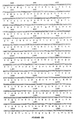
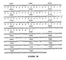
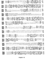
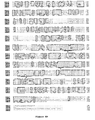
- the genomic, SEQ ID NO: 1, and cDNA, SEQ ID NOS: 2 and 5, sequences are identified in Figure 3 A-H and encode ABCR, certain mutations of which are responsible for the class of retinal disorders known as retinal or macular degenerations.
- Macular degeneration is characterized by macular dystrophy, various alterations of the peripheral retina, central visual impairment, progressive bilateral atrophy of the macular retinal pigment epithelium (RPE) and neuroepithelium, frequent appearance of orange-yellow flecks distributed around the macula and/or the midretinal periphery, and subretinal deposition of lipofuscin-like material.
- RPE macular retinal pigment epithelium
- Retinal and macular degenerative diseases include and are not limited to Stargardt Disease, Fundus Flavimaculatus, age-related macular degeneration, and may include disorders variously called retinitis pigmentosa, combined rod and cone dystrophies, cone dystrophies and degenerations, pattern dystrophy, bull's eye maculopathies, and various other retinal degenerative disorders, some induced by drugs, toxins, environmental influences, and the like.
- Stargardt Disease is an autosomal recessive retinal disorder characterized by juvenile to adult-onset macular and retinal dystrophy. Fundus Flavimaculatus often displays later age of onset and slower progression. Some environmental insults and drug toxicities may create similar retinal degenerations. Linkage analysis reveals that Stargardt Disease and Fundus Flavimaculatus may be allelic autosomal recessive disorders with slightly different clinical manifestations. The identification of the ABCR gene suggests that different mutations within ABCR may be responsible for these clinical phenomena.
- the present invention is also directed to a method of screening for an agent that modifies ATP binding cassette transporter comprising combining purified ATP binding cassette transporter with an agent suspected of modifying ATP binding cassette transporter and observing a change in at least one characteristic associated with ATP binding cassette transporter.
- Modify and variations thereof include changes such as and not limited to inhibit, suppress, delay, retard, slow, suspend, obstruct, and restrict, as well as induce, encourage, provoke, and cause. Modify may also be defined as complete inhibition such that macular degeneration is arrested, stopped, or blocked. Modifications may, directly or indirectly, inhibit or substantially inhibit, macular degeneration or induce, or substantially induce, macular degeneration, under certain circumstances.
- Methods of identifying an agent that inhibits macular degeneration comprise combining purified ATP binding cassette transporter from a patient suspected of having macular degeneration and an agent suspected of interacting with the ATP binding cassette transporter and observing an inhibition in at least one of the characteristics of diseases associated with the ATP binding cassette transporter. Accordingly, such methods serve to reduce or prevent macular degeneration, such as in human patients.
- the present invention provides for methods of identifying an agent that induces onset of at least one characteristic associated with ATP binding cassette transporter comprising combining purified wild-type ATP binding cassette transporter with an agent suspected of inducing a macular degenerative disease and observing the onset of a characteristic associated with macular degeneration.
- ATP binding cassette transporter may be provided for in the methods identified herein in the form of nucleic acids, such as and not limited to SEQ ID NOS: 1, 2, and 5 or as an amino acid, SEQ ID NOS: 3 and 6, for example. Accordingly, transcription and translation inhibitors may be separately identified.
- Characteristics associated with macular degeneration include and are not limited to central visual impairment, progressive bilateral atrophy of the macular retinal pigment epithelium (RPE) and neuroepithelium, and the frequent appearance of orange-yellow flecks distributed around the macula and/or the midretinal periphery. Accordingly, observing one or more of the characteristics set forth above results in identification of an agent that induces macular degeneration, whereas reduction or inhibition of at least one of the characteristics results in identification of an agent that inhibits macular degeneration.
- mutations in wild-type ABCR which result in activities that are not associated with wild-type ABCR are herein referred to as sequences which are associated with macular degeneration.
- Such mutations include missense mutations, deletions, insertions, substantial differences in hydrophobicity, hydrophilicity, acidity, and basicity.
- Characteristics which are associated with retinal or macular degeneration include and are not limited to those characteristics set forth above.
- Mutations in wild-type ABCR provide a method of detecting macular degeneration.
- Retinal or macular degeneration may be detected by obtaining a sample comprising patient nucleic acids from a patient tissue sample; amplifying retina-specific ATP binding cassette receptor specific nucleic acids from the patient nucleic acids to produce a test fragment; obtaining a sample comprising control nucleic acids from a control tissue sample; amplifying control nucleic acids encoding wild-type retina-specific ATP binding cassette receptor to produce a control fragment; comparing the test fragment with the control fragment to detect the presence of a sequence difference in the test fragment, wherein a difference in the test fragment indicates macular degeneration.
- Mutations in the test fragment including and not limited to each of the mutations identified above, may provide evidence of macular degeneration.
- the invention provides nucleic acid sequences encoding mutant ABCR proteins according to claims 2-6.
- a purified ABCR protein is also provided by the present invention.
- the purified ABCR protein may have an amino acid sequence as provided by SEQ ID NOS: 3 and 6.
- the present invention is directed to ABCR sequences obtained from mammals from the Order Rodentia, including and not limited to hamsters, rats, and mice; Order Logomorpha, such as rabbits; more particularly the Order Carnivora, including Felines (cats) and Canines (dogs); even more particularly the Order Artiodactyla, Bovines (cows) and Suines (pigs); and the Order Perissodactyla, including Equines (horses); and most particularly the Order Primates, Ceboids and Simoids (monkeys) and Anthropoids (humans and apes).
- the mammals of most preferred embodiments are humans.
- sequences of the invention may be produced in host cells transformed with an expression vector comprising a nucleic acid sequence encoding ABCR.
- the transformed cells are cultured under conditions whereby the nucleic acid sequence coding for ABCR is expressed. After a suitable amount of time for the protein to accumulate, the protein may be purified from the transformed cells.
- a gene coding for ABCR may be obtained from a cDNA library. Suitable libraries can be obtained from commercial sources such as Clontech, Palo Alto, CA. Libraries may also be prepared using the following non-limiting examples: hamster insulin-secreting tumor (HIT), mouse ⁇ TC-6, and rat insulinoma (RIN) cells. Positive clones are then subjected to DNA sequencing to determine the presence of a DNA sequence coding for ABCR. DNA sequencing is accomplished using the chain termination method of Sanger et al., Proc. Nat'l. Acad. Sci, US.A., 1977 , 74, 5463. The DNA sequence encoding ABCR is then inserted into an expression vector for later expression in a host cell.
- HIT hamster insulin-secreting tumor
- RIN rat insulinoma
- Expression vectors and host cells are selected to form an expression system capable of synthesizing ABCR.
- Vectors including and not limited to baculovirus vectors may be used in the present invention.
- Host cells suitable for use in the invention include prokaryotic and eukaryotic cells that can be transformed to stably contain and express ABCR.
- nucleic acids coding for the recombinant protein may be expressed in prokaryotic or eukaryotic host cells, including the most commonly used bacterial host cell for the production of recombinant proteins, E. coli .
- microbial strains may also be used, however, such as Bacillus subtilis , and other enterobacteriaceae such as Salmonella typhimurium or Serratia marcescens , various species of Pseudomonas , or other bacterial strains.
- the preferable eukaryotic system is yeast, such as Saccharomyces cerevisiae.
- yeast artificial chromosome (YAC) systems are able to accommodate the large size of ABCR gene sequence or genomic clone.
- the principle of the YAC system is similar to that used in conventional cloning of DNA. Large fragments of cDNA are ligated into two "arms" of a YAC vector, and the ligation mixture is then introduced into the yeast by transformation. Each of the arms of the YAC vector carries a selectable marker as well as appropriately oriented sequences that function as telomeres in yeast.
- one of the two arms carries two small fragments that function as a centromere and as an origin of replication (also called an ARS element-autonomously replicating sequences).
- Yeast transformants that have taken up and stably maintained an artificial chromosome are identified as colonies on agar plates containing the components necessary for selection of one or both YAC arms.
- YAC vectors are designed to allow rapid identification of transformants that carry inserts of genomic DNA. Insertion of genomic DNA into the cloning site interrupts a suppressor tRNA gene and results in the formation of red rather than white colonies by yeast strains that carry an amber ade 2 gene.
- genomic DNA from the test organism is prepared under conditions that result in relatively little shearing such that its average size is several million base pairs.
- the cDNA is then ligated to the arms of the YAC vector, which has been appropriately prepared to prevent self-ligation.
- YAC vectors may be used that will accept genomic DNA that has been digested to completion with rarely cutting restriction enzymes such as Not I or Mlu I.
- insect cells such as Spodoptera frugiperda ; chicken cells, such as E3C/O and SL-29; mammalian cells, such as HeLa, Chinese hamster ovary cells (CHO), COS-7 or MDCK cells and the like may also be used.
- mammalian cells such as HeLa, Chinese hamster ovary cells (CHO), COS-7 or MDCK cells and the like may also be used.
- the foregoing list is illustrative only and is not intended in any way to limit the types of host cells suitable for expression of the nucleic acid sequences of the invention.
- expression vectors refer to any type of vector that can be manipulated to contain a nucleic acid sequence coding for ABCR, such as plasmid expression vectors, viral vectors, and yeast expression vectors.
- the selection of the expression vector is based on compatibility with the desired host cell such that expression of the nucleic acid encoding ABCR results.
- Plasmid expression vectors comprise a nucleic acid sequence of the invention operably linked with at least one expression control element such as a promoter.
- plasmid vectors contain replicon and control sequences derived from species compatible with the host cell.
- plasmid vectors may also contain a selectable marker such as a gene coding for antibiotic resistance. Suitable examples include the genes coding for ampicillin, tetracycline, chloramphenicol, or kanamycin resistance.
- Suitable expression vectors, promoters, enhancers, and other expression control elements are known in the art and may be found in Sambrook et al., Molecular Cloning: A Laboratory Manual , second edition, Cold Spring Harbor Laboratory Press, Cold Spring Harbor, NY (1989), incorporated herein by reference in its entirety.
- Transformed host cells containing a DNA sequence encoding ABCR may then be grown in an appropriate medium for the host. The cells are then grown until product accumulation reaches desired levels at which time the cells are then harvested and the protein product purified in accordance with conventional techniques. Suitable purification methods include, but are not limited to, SDS PAGE electrophoresis, phenylboronate-agarose, reactive green 19-agarose, concanavalin A sepharose, ion exchange chromatography, affinity chromatography, electrophoresis, dialysis and other methods of purification known in the art.
- Protein preparations of purified or unpurified ABCR by host cells, are accordingly produced which comprise ABCR and other material such as host cell components and/or cell medium, depending on the degree of purification of the protein.
- the invention also includes a transgenic non-human animal, including and not limited to mammals, such as and not limited to a mouse, rat, or hamster, comprising a sequence encoding ABCR, or fragment thereof that substantially retains ABCR activity, introduced into the animal or an ancestor of the animal.
- the sequence may be wild-type or mutant and may be introduced into the animal at the embryonic or adult stage.
- the sequence is incorporated into the genome of an animal such that it is chromosomally incorporated into an activated state.
- a transgenic non-human animal has germ cells and somatic cells that contain an ABCR sequence.
- Embryo cells may be transfected with the gene as it occurs naturally, and transgenic animals are selected in which the gene has integrated into the chromosome at a locus which results in activation.
- Other activation methods include modifying the gene or its control sequences prior to introduction into the embryo.
- the embryo may be transfected using a vector containing the gene.
- a transgenic non-human animal may be engineered wherein ABCR is suppressed.
- suppression of ABCR includes, and is not limited to strategies which cause ABCR not to be expressed. Such strategies may include and are not limited to inhibition of protein synthesis, pre-mRNA processing, or DNA replication. Each of the above strategies may be accomplished by antisense inhibition of ABCR gene expression.
- Many techniques for transferring antisense sequences into cells are known to those of skill, including and not limited to microinjection, viral-mediated transfer, somatic cell transformation, transgene integration, and the like, as set forth in Pinkert, Carl, Transgenic Animal Technology, 1994 , Academic Press, Inc., San Diego, CA, incorporated herein by reference in its entirety.
- a transgenic non-human animal may be prepared such that ABCR is knocked out.
- a knock-out includes and is not limited to disruption or rendering null the ABCR gene.
- a knock-out may be accomplished, for example, with antisense sequences for ABCR.
- the ABCR gene may be knocked out by injection of an antisense sequence for all or part of the ABCR sequence such as an antisense sequence for all or part of SEQ ID NO: 2.
- correlation of the ABCR to macular degeneration may be tested. Sequences encoding mutations affecting the ABCR may be inserted to test for alterations in various retinal and macular degenerations exhibited by changes in the characteristics associated with retinal and macular degeneration.
- a non-human ABCR knock-out may be engineered by inserting synthetic DNA into the animal chromosome by homologous recombination.
- sequences flanking the target and insert DNA are identical, allowing strand exchange and crossing over to occur between the target and insert DNA.
- Sequences to be inserted typically include a gene for a selectable marker, such as drug resistance.
- Sequences to be targeted are typically coding regions of the genome, in this case part of the ABCR gene. In this process of homologous recombination, targeted sequences are replaced with insert sequences thus disrupting the targeted gene and rendering it nonfunctional. This nonfunctional gene is called a null allele of the gene.
- a DNA construct containing the insert DNA and flanking sequences is made.
- This DNA construct is transfected into pluripotent embryonic stem cells competent for recombination.
- the identical flanking sequences align with one another, and chromosomal recombination occurs in which the targeted sequence is replaced with the insert sequence, as described in Bradley, A., Production and Analysis of Chimeric Mice, in Teratocarcinomas and Embryonic Stem Cells - A Practical Approach, 1987 , E. Roberson, Editor, IRC Press, pages 113-151.
- the stem cells are injected into an embryo, which is then implanted into a female animal and allowed to be born.
- the animals may contain germ cells derived from the injected stem cells, and subsequent matings may produce animals heterozygous and homozygous for the disrupted gene.
- Transgenic non-human animals may also be useful for testing nucleic acid changes to identify additional mutations responsible for macular degeneration.
- a transgenic non-human animal may comprise a recombinant ABCR.
- purified when used to describe the state of nucleic acid sequences of the invention, refers to nucleic acid sequences substantially free of nucleic acid not coding for ABCR or other materials normally associated with nucleic acid in non-recombinant cells, i.e., in its "native state.”
- purified or “in purified form” when used to describe the state of an ABCR nucleic acid, protein, polypeptide, or amino acid sequence, refers to sequences substantially free, to at least some degree, of cellular material or other material normally associated with it in its native state.
- sequence has a purity (homogeneity) of at least about 25% to about 100%, More preferably the purity is at least about 50%, when purified in accordance with standard techniques known in the art.
- tissue sample for testing.
- the tissue sample may be solid or liquid, a body fluid sample such as and not limited to blood, skin, serum, saliva, sputum, mucus, bone marrow, urine, lymph, and a tear; and feces.
- a tissue sample from amniotic fluid or chorion may be provided for the detection of retinal or macular degeneration in utero in accordance with the present invention.
- a test fragment is defined herein as an amplified sample comprising ABCR- specific nucleic acids from a patient suspected of having retinal or macular degeneration.
- a control fragment is an amplified sample comprising normal or wild-type ABCR -specific nucleic acids from an individual not suspected of having retinal or macular degeneration.
- the method of amplifying nucleic acids may be the polymerase chain reaction using a pair of primers wherein at least one primer within the pair is selected from the group consisting of SEQ ID NOS: 40-61, 64, 65, 78, 79, 98-105, 108, and 109.
- a pair of primers may be used such that one primer of the pair is selected from the group consisting of SEQ ID NOS: 40-61, 64, 65, 78, 79, 98-105, 108, and 109.
- Nucleic acids such as DNA (such as and not limited to genomic DNA and cDNA) and/or RNA (such as and not limited to mRNA), are obtained from the patient sample. Preferably RNA is obtained.
- Nucleic acid extraction is followed by amplification of the same by any technique known in the art.
- the amplification step includes the use of at least one primer sequence which is complementary to a portion of ABCR -specific expressed nucleic acids or sequences on flanking intronic genomic sequences in order to amplify exon or coding sequences.
- Primer sequences useful in the amplification methods include and are not limited to SEQ ID NOS: 40-61, 64, 65, 78, 79, 98-105, 108, and 109, which may be used in the amplification methods.
- Any primer sequence of about 10 nucleotides to about 35 nucleotides, more preferably about 15 nucleotides to about 30 nucleotides, even more preferably about 17 nucleotides to about 25 nucleotides may be useful in the amplification step of the methods of the present invention.
- mismatches within the sequences identified above, which achieve the methods of the invention, such that the mismatched sequences are substantially complementary and thus hybridizable to the sequence sought to be identified are also considered within the scope of the disclosure.
- primers may also be unmodified or modified. Primers may be prepared by any method known in the art such as by standard phosphoramidite chemistry. See Sambrook et al ., supra .
- the method of amplifying nucleic acids may be the polymerase chain reaction using a pair of primers wherein at least one primer within the pair is selected from the group consisting of 40-61, 64, 65, 78, 79, 98-105, 108 and 109.
- a pair of primers may be used such that one primer of the pair is selected from the group consisting of SEQ ID NOS: 40-61, 64, 65, 78, 79, 98-105, 108 and 109.
- an amplification method includes the use of two primers, a first primer and a second primer, such as in the polymerase chain reaction, one of the first primer or second primer may be selected from the group consisting of SEQ ID NOS: 40-61, 64, 65, 78, 79, 98-105, 108 and 109. Any primer pairs which copy and amplify nucleic acids between the pairs pointed toward each other and which are specific for ABCR may be used in accordance with the methods of the present invention.
- PCR polymerase chain reaction
- the primers will bind to the target and the polymerase will cause the primers to be extended along the target sequence by adding on nucleotides.
- the extended primers will dissociate from the target to form reaction products, excess primers will bind to the target and to the reaction products and the process is repeated.
- a reverse transcriptase PCR amplification procedure may be performed in order to quantify the amount of mRNA amplified. Polymerase chain reaction methodologies are well known in the art.
- LCR ligase chain reaction
- Qbeta Replicase described in PCT Application No. PCT/US87/00880, may also be used as still another amplification method in the present invention.
- a replicative sequence of RNA which has a region complementary to that of a target is added to a sample in the presence of an RNA polymerase.
- the polymerase will copy the replicative sequence which can then be detected.
- An isothermal amplification method in which restriction endonucleases and ligases are used to achieve the amplification of target molecules that contain nucleotide 5'-[ alpha -thio]triphosphates in one strand of a restriction site (Walker, G. T., et al., Proc. Natl. Acad, Sci. (U.S.A.) 1992, 89 :392-396), may also be useful in the amplification of nucleic acids in the present invention.
- Strand Displacement Amplification is another method of carrying out isothermal amplification of nucleic acids which involves multiple rounds of strand displacement and synthesis, i.e. nick translation.
- a similar method, called Repair Chain Reaction (RCR) is another method of amplification which may be useful in the present invention and which involves annealing several probes throughout a region targeted for amplification, followed by a repair reaction in which only two of the four bases are present. The other two bases can be added as biotinylated derivatives for easy detection.
- RCR Repair Chain Reaction
- ABCR -specific nucleic acids can also be detected using a cyclic probe reaction (CPR).
- CPR a probe having a 3' and 5' sequences of non- ABCR specific DNA and middle sequence of ABCR specific RNA is hybridized to DNA which is present in a sample.
- the reaction is treated with RNaseH, and the products of the probe identified as distinctive products, generate a signal which is released after digestion.
- the original template is annealed to another cycling probe and the reaction is repeated.
- CPR involves amplifying a signal generated by hybridization of a probe to a ABCR -specific expressed nucleic acid.
- modified primers are used in a PCR like, template and enzyme dependent synthesis.
- the primers may be modified by labeling with a capture moiety (e.g., biotin) and/or a detector moiety (e.g., enzyme).
- a capture moiety e.g., biotin
- a detector moiety e.g., enzyme
- an excess of labeled probes are added to a sample.
- the probe binds and is cleaved catalytically. After cleavage, the target sequence is released intact to be bound by excess probe. Cleavage of the labeled probe signals the presence of the target sequence.
- nucleic acid amplification procedures include transcription-based amplification systems (TAS) (Kwoh D., et al., Proc. Natl. Acad Sci. (U.S.A.) 1989 , 86 :1173, Gingeras T. R., et al., PCT Application WO 88/10315, including nucleic acid sequence based amplification (NASBA) and 3SR.
- TAS transcription-based amplification systems
- NASBA nucleic acid sequence based amplification
- 3SR nucleic acid sequence based amplification
- the nucleic acids can be prepared for amplification by standard phenol/chloroform extraction, heat denaturation of a clinical sample, treatment with lysis buffer and minispin columns for isolation of DNA and RNA or guanidinium chloride extraction of RNA.
- amplification techniques involve annealing a primer which has ABCR -specific sequences.
- DNA/RNA hybrids are digested with RNase H while double stranded DNA molecules are heat denatured again. In either case the single stranded DNA is made fully double stranded by addition of second ABCR -specific primer, followed by polymerization.
- the double stranded DNA molecules are then multiply transcribed by a polymerase such as T7 or SP6.
- the RNAs are reverse transcribed into double stranded DNA, and transcribed once again with a polymerase such as T7 or SP6.
- the resulting products whether truncated or complete, indicate ABCR -specific sequences.
- ssRNA single-stranded RNA
- dsDNA double-stranded DNA
- the ssRNA is a first template for a first primer oligonucleotide, which is elongated by reverse transcriptase (RNA-dependent DNA polymerase).
- RNase H ribonuclease H
- the resultant ssDNA is a second template for a second primer, which also includes the sequences of an RNA polymerase promoter (exemplified by T7 RNA polymerase) 5' to its homology to its template.
- This primer is then extended by DNA polymerase (exemplified by the large "Klenow" fragment of E . coli DNA polymerase I), resulting as a double-stranded DNA (“dsDNA”) molecule, having a sequence identical to that of the original RNA between the primers and having additionally, at one end, a promoter sequence.
- This promoter sequence can be used by the appropriate RNA polymerase to make many RNA copies of the DNA. These copies can then re-enter the cycle leading to very swift amplification. With proper choice of enzymes, this amplification can be done isothermally without addition of enzymes at each cycle. Because of the cyclical nature of this process, the starting sequence can be chosen to be in the form of either DNA or RNA.
- Miller, H. I., et al ., PCT application WO 89/06700 disclose a nucleic acid sequence amplification scheme based on the hybridization of a promoter/primer sequence to a target single-stranded DNA ("ssDNA") followed by transcription of many RNA copies of the sequence. This scheme is not cyclic; i.e. new templates are not produced from the resultant RNA transcripts.
- Other amplification methods include "race” disclosed by Frohman, M. A., In: PCR Protocols: A Guide to Methods and Applications 1990 , Academic Press, N.Y.) and "one-sided PCR” (Ohara, O., et al., Proc. Natl. Acad. Sci. (U.S.A.) 1989 , 86 :5673-5677).
- Methods based on ligation of two (or more) oligonucleotides in the presence of nucleic acid having the sequence of the resulting "di-oligonucleotide", thereby amplifying the di-oligonucleotide may also be used in the amplification step of the present invention.
- Test fragment and control fragment may be amplified by any amplification methods known to those of skill in the art, including and not limited to the amplification methods set forth above.
- amplification of sequences encoding patient and wild-type ABCR includes amplification of a portion of a sequence such as and not limited to a portion of an ABCR sequence of SEQ ID NO: 1, such as sequence of a length of about 10 nucleotides to about 1,000 nucleotides, more preferably about 10 nucleotides to about 100 nucleotides, or having at least 10 nucleotides occurring anywhere within the SEQ ID NO: 1, where sequence differences are known to occur within ABCR test fragments.
- a portion of the sequence encoding ABCR of a patient sample and a control sample may be amplified to detect sequence differences between these two sequences.
- test fragment and control fragment Following amplification of the test fragment and control fragment, comparison between the amplification products of the test fragment and control fragment is carried out. Sequence changes such as and not limited to nucleic acid transition, transversion, and restriction digest pattern alterations may be detected by comparison of the test fragment with the control fragment.
- the presence or absence of the amplification product may be detected.
- the nucleic acids are fragmented into varying sizes of discrete fragments.
- DNA fragments may be separated according to molecular weight by methods such as and not limited to electrophoresis through an agarose gel matrix.
- the gels are then analyzed by Southern hybridization. Briefly, DNA in the gel is transferred to a hybridization substrate or matrix such as and not limited to a nitrocellulose sheet and a nylon membrane.
- a labeled probe encoding an ABCR mutation is applied to the matrix under selected hybridization conditions so as to hybridize with complementary DNA localized on the matrix.
- the probe may be of a length capable of forming a stable duplex.
- the probe may have a size range of about 200 to about 10,000 nucleotides in length, preferably about 500 nucleotides in length, and more preferably about 2,454 nucleotides in length. Mismatches which permit substantial similarity to the probe, such as and not limited to sequences with similar hydrophobicity, hydrophilicity, basicity, and acidity, will be known to those of skill in the art once armed with the present disclosure.
- Various labels for visualization or detection are known to those of skill in the art, such as and not limited to fluorescent staining, ethidium bromide staining for example, avidin/biotin, radioactive labeling such as 32 P labeling, and the like.
- the product such as the PCR product
- the product may be run on an agarose gel and visualized using a stain such as ethidium bromide. See Sambrook et al., supra .
- the matrix may then be analyzed by autoradiography to locate particular fragments which hybridize to the probe.
- sequencing of the test fragment and the control fragment is also another alternative.
- Methods of nucleic acid sequencing are known to those of skill in the art, including and not limited to the methods of Maxam and Gilbert, Proc. Natl. Acad. Sci., USA 1977, 74 , 560-564 and Sanger, Proc. Natl. Acad. Sci., USA 1977, 74 , 5463-5467.
- a pharmaceutical composition comprising all or part of a sequence for ABCR may be delivered to a patient suspected of having retinal or macular degeneration.
- the sequence may be an antisense sequence.
- the composition of the present invention may be administered alone or may generally be administered in admixture with a pharmaceutical carrier.
- the pharmaceutically-acceptable carrier may be selected with regard to the intended route of administration and the standard pharmaceutical practice.
- the dosage will be about that of the sequence alone and will be set with regard to weight, and clinical condition of the patient.
- the proportional ratio of active ingredient to carrier will naturally depend, inter alia, on the chemical nature, solubility, and stability of the sequence, as well as the dosage contemplated.
- sequences of the invention may be employed in the method of the invention singly or in combination with other compounds, including and not limited to other sequences set forth in the present invention.
- the method of the invention may also be used in conjunction with other treatments such as and not limited to antibodies, for example.
- the amount to be administered will also depend on such factors as the age, weight, and clinical condition of the patient.
- the composition of the present invention may be administered by any suitable route, including as an eye drop, inoculation and injection, for example, intravenous, intraocular, oral, intraperitoneal, intramuscular, subcutaneous, topically, and by absorption through epithelial or mucocutaneous linings, for example, conjunctival, nasal, oral, vaginal, rectal and gastrointestinal.
- the mode of administration of the composition may determine the sites in the organism to which the compound will be delivered. For instance, topical application may be administered in creams, ointments, gels, oils, emulsions, pastes, lotions, and the like.
- the composition may be used in the form of sterile aqueous or non-aqueous solution which may contain another solute, for example, sufficient salts, glucose or dextrose to make the solution isotonic.
- a non-aqueous solution may be comprise an oil, for example.
- the present invention may be used in the form of tablets, capsules, lozenges, troches, powders, syrups, elixirs, aqueous solutions and suspension, and the like.
- disintegrants such as starch, and lubricating agents may be used.
- useful diluents are lactose and high molecular weight polyethylene glycols.
- certain sweetening and/or flavoring agents may be added.
- a diagnostic kit for detecting retinal or macular degeneration comprising in one or more containers at least one primer which is complementary to an ABCR sequence and a means for visualizing amplified DNA is also within the scope of the present invention.
- the kit may comprise two primers.
- the primers may be selected from the group consisting of SEQ ID NOS: 40-61, 64, 65, 78, 79, 98-105, 108, and 109, for example.
- the diagnostic kit may comprise a pair of primers wherein one primer within said pair is complementary to a region of the ABCR gene, wherein one of said pair of primers is selected from the group consisting of SEQ ID NO: 40-61, 64, 65, 78, 79, 98-105, 108, and 109, a probe specific to the amplified product, and a means for visualizing amplified DNA, and optionally including one or more size markers, and positive and negative controls.
- the diagnostic kit of the present invention may comprise one or more of a fluorescent dye such as ethidium bromide stain, 32 P, and biotin, as a means for visualizing or detecting amplified DNA.
- the kit may include one or more size markers, positive and negative controls, restriction enzymes, and/or a probe specific to the amplified product.
- ABCR retina-specific ABC transporter
- ABCR is closely related to the previously described mouse and human ABC1 and ABC2 genes (Luciani et al., 1994; Allikmets et al., 1995). To determine whether ABCR might cause a disease, the gene was mapped with a whole genome radiation hybrid panel (GeneBridge 4; Research Genetics, Huntsville, AL). ABCR mapped to the human chromosome 1p13-p21 region, close to microsatellite markers D1S236 and D1S188.
- PCR primers 3'UTR-For 5'ATCCTCTGACTCAGCAATCACA, SEQ ID NO: 7, and 3'UTR-Rev 5'TTGCAATTACAAATGCAATGG, SEQ ID NO: 8, from the putative 3' untranslated region were used to screen YACs from the previously described contig between these anonymous markers (Anderson et al ., 1995).
- At least 12 YACs contain the 3' end of the ABCR gene, including 924_e_9, 759_d_7, 775_c_2, 782_b_4, 982_g_5, 775_b_2, 765_a_3, 751_f_2, 848_e_3, 943_h_8, 934_g_7, and 944_b_12 ( Figure 1). These YACs delineate a region containing the STGD gene between markers D1S3361 and D1S236 (Anderson et al ., 1995).
- Clones corresponding to the mouse ortholog of the human ABCR gene were isolated from the mouse retina cDNA library and end-sequenced.
- the chromosomal location of the mouse ABCR gene was determined on The Jackson Laboratory (Bar Harbor, ME) interspecific backcross mapping panel (C57BL/6JEi X SPRET/Ei)F1 X SPRET/Ei (Rowe et al., 1994) known as Jackson BSS. Mapping was performed by SSCP analysis with the primers MABCR1F 5'ATC CAT ACC CTT CCC ACT CC, SEQ ID NO: 9, and MABCR1R 5' GCA GCA GAA GAT AAG CAC ACC, SEQ ID NO. 10. The allele pattern of the Abcr was compared to the 250 other loci mapped previously in the Jackson BSS cross (http://www.jax.org).
- DNA fragments used as probes were purified on a 1% low-melting temperature agarose gel.
- the probe sequences are set forth within the genomic sequence of SEQ ID NO: 1 and Figure 3A-H.
- DNA was labeled directly in agarose with the Random Primed DNA Labeling Kit (Boehringer Mannheim, Indianapolis, IN) and hybridized to multiple tissue Northern blot and a Master blot (Clontech, Palo Alto, CA), according to the manufacturer's instructions. Each blot contained 2 ⁇ g of poly A + RNA from various human tissues.
- cDNA clones containing ABCR sequences were obtained from a human retina cDNA library (Nathans et al., 1986) and sequenced fully. Primers were designed from the sequences of cDNA clones from 5' and 3' regions of the gene and used to link the identified cDNA clones by RT-PCR with retina QUICK-Clone cDNA (Clontech, Palo Alto, CA) as a template. PCR products were cloned into pGEM®-T vector (Promega, Madison, WI). Mouse ABCR cDNA clones were obtained from screening a developing mouse eye cDNA library (H. Sun, A. Lanahan, and J. Nathans, unpublished).
- the pGEM®-T Vector is prepared by cutting pGEM®-5Zf(+) DNA with EcoR V and adding to a 3' terminal thymidine to both ends. These single 3'-T overhangs at the insertion site greatly improve the efficiency ofligation of PCR products because of the nontemplate-dependent addition of a single deoxyadenosine (A) to the 3'-ends of PCR products by many thermostable polymerases.
- the pGEM®-5Zf(+) Vector contains the origin of replication of the filamentous phage f1 and can be used to produce ssDNA.
- the plasmid also contains T7 and SP6 RNA polymerase promoters flanking a multiple cloning region within the ⁇ -peptide coding region for the enzyme ⁇ -galactosidase. Insertional inactivation of the ⁇ -peptide allows recombinant clones to be identified directly by color screening on indicator plates.
- cDNA clones from various regions of the ABCR gene were used as probes to screen a human genomic library in Lambda FIX II (#946203, Stratagene, LaJolla. CA). Overlapping phage clones were mapped by Eco RI and Bam HI digestion. A total of 6.9 kb of the ABCR sequence was assembled. ( Figure 3 A-H) resulting in a 6540 bp (2180 amino acid) open reading frame.
- PCR conditions were 95°C for 8 minutes; 5 cycles at 62 °C for 20 seconds, 72°C for 30 seconds; 35 cycles at 60°C for 20 seconds, 72°C for 30 seconds; 72°C for 5 minutes (except that a was performed at 94° C for 5 minutes); 5 cycles at 94° C for 40 seconds; 60° C for 30 seconds; 72 ° C for 20 seconds; 35 cycles at 94 ° C for 40 seconds; 56 ° C for 30 seconds; 72 ° C for 20 seconds, and 72 ° C for 5 minutes.
- Amplification of exons was performed with AmpliTaq Gold polymerase in a 25 ⁇ l volume in 1X PCR buffer supplied by the manufacturer (Perkin Elmer, Foster City, CA). Samples were heated to 95°C for 10 minutes and amplified for 35-40 cycles at 96°C for 20 seconds; 58°C for 30 seconds; and 72°C for 30 seconds. PCR products were analyzed on 1-1.5% agarose gels and in some cases digested with an appropriate restriction enzymes to verify their sequence. Primer sequences and specific reaction conditions are set forth in Table 1. The sequence of the ABCR cDNA has been deposited with GenBank under accession # U88667.
- ABCR is most closely related to the previously characterized mouse Abc1 and Abc2 genes (Luciani et al ., 1994) and to another human gene ( ABCC ) which maps to chromosome 16p13.3 (Klugbauer and Hofmann, 1996).
- ABCC a human gene which maps to chromosome 16p13.3
- ABCR shows the composition of a typical full-length ABC transporter that consists of two transmembrane domains (TM), each with six membrane spanning hydrophobic segments, as predicted by a hydropathy plot (data not shown), and two highly conserved ATP-binding domains ( Figures 3 A-H and 4 A-D).
- TM transmembrane domain
- Figures 3 A-H and 4 A-D two highly conserved ATP-binding domains
- mice ortholog of ABCR To characterize the mouse ortholog of ABCR , cDNA clones from a developing mouse eye library were isolated. A partial sequence of the mouse cDNA was utilized to design PCR primers to map the mouse Abcr gene in an interspecific backcross mapping panel (Jackson BSS). The allele pattern of Abcr was compared to 2450 other loci mapped previously in the Jackson BSS cross; linkage was found to the distal end of chromosome 3 o ( Figure 5). No recombinants were observed between Abcr and D13Mit13. This region of the mouse genome is syntenic with human chromosome 1p13-p21. Thus far, no eye disease phenotype has been mapped to this region of mouse chromosome 3.
- Genomic DNA samples 50 ng were amplified with AmpliTaq Gold polymerase in 1X PCR buffer supplied by the manufacturer (Perkin Elmer, Foster City, CA) containing [ ⁇ - 32 P] dCTP. Samples were heated to 95°C for 10 minutes and amplified for 35-40 cycles at 96°C for 20 seconds; 58°C for 30 seconds; and 72 °C for 30 seconds. Products were diluted in 1:3 stop solution, denatured at 95°C for 5 minutes, chilled in ice for 5 minutes, and loaded on gels.
- Gel formulations include 6% acrylamide:Bis (2.6% cross-linking), 10% glycerol at room temperature, 12W; and 10% acrylamide:Bis (1.5% cross-linking), at 4°C, 70W. Gels were run for 2-16 hours (3000 Vh/100 bp), dried, and exposed to X-ray film for 2-12 hours. Some exons were analyzed by SSCP with MDE acrylamide (FMC Bioproducts, Rockland, ME) with and without 10% glycerol for 18 hours, 4 watts at room temperature with ⁇ -P 32- dCTP labeled DNA.
- MDE acrylamide FMC Bioproducts, Rockland, ME
- Heteroduplexes were identified from the double-stranded DNA at the bottom of the gels, and SSCPs were identified from the single-stranded region. Samples showing variation were compared with other family members to assess segregation of the alleles and with at least 40 unrelated control samples, from either Caucasian or Saudi Arabian populations, to distinguish mutations from polymorphisms unrelated to STGD. PCR products with SSCP or heteroduplex variants were obtained in a 25 ⁇ l volume, separated on a 1% agarose gel, and isolated by a DNA purification kit (PGC Scientific, Frederick. MD). Sequencing was performed on an ABI sequencer with both dye primer and dye terminator chemistry.
- Mutations are named according to standard nomenclature.
- the column headed “Exon” denotes which of the 51 exons of ABCR contain the mutation.
- the column headed “# Families” denotes the number of Stargardt families which displayed the mutation.
- the column headed “Nucleotide” gives the base number starting from the A in the initiator ATG, followed by the wild type sequence and an arrow indicating the base it is changed to; del indicates a deletion of selected bases at the given position in the ABCR gene; ins indicates an insertion of selected bases at the given position; splice donor site mutations are indicated by the number of the last base of the given exon, followed by a plus sign and the number of bases into the intron where the mutation occurs.
- Amino Acid denotes the amino acid change a given mutation causes; fs indicates a frameshift mutation leading to a truncated protein; splice indicates a splice donor site mutation; del indicates an in-frame deletion of the given amino acids.
- STGD is characterized histologically by a massive accumulation of a lipofuscin-like substance in the retinal pigment epithelium (RPE). This characteristic has led to the suggestion that STGD represents an RPE storage disorder (Blacharski et al ., 1988). It was therefore of interest that ABCR transcripts were found to be abundant in the retina. To identify the site(s) of ABCR gene expression at higher resolution and to determine whether the gene is also expressed in the RPE, the distribution of ABCR transcripts was visualized by in situ hybridization to mouse, rat, bovine, and macaque ocular tissues.
- RPE retinal pigment epithelium
- the templates for probe synthesis were: (1) a 1.6 kb fragment encompassing the 3' end of the mouse Abcr coding region, (2) a full length cDNA clone encoding the mouse blue cone pigment (Chiu et al ., 1994), and (3) a macaque rhodopsin coding region segment encoding residues 133 to 254 (Nickells, R. W., Burgoyne, C.F., Quigley, H.A., and Zack, D.J. (1995)).
- ABCR transcripts are present exclusively within photoreceptor cells (Figure 7). ABCR transcripts are localized principally to the rod inner segments, a distribution that closely matches that of rhodopsin gene transcripts. Interestingly, ABCR hybridization was not observed at detectable levels in cone photoreceptors, as judged by comparisons with the hybridization patterns obtained with a blue cone pigment probe (compare Figure 7A and Figure 7D, Figure 7E with Figure 7F and Figure 7G with Figure 7H). Because melanin granules might obscure a weak hybridization signal in the RPE of a pigmented animal, the distribution of ABCR transcripts was also examined in both albino rats and albino mice.
- Lipids of ocular tissues the effects of essential fatty acid deficiency on the phospholipids of the photoreceptor membranes of rat retina. Arch. Biochem. Biophys. 151 , 270-276.
- the photoreceptor rim protein is an ABC transporter encoded by the gene for recessive Stargardt's disease (ABCR). FEBS Letters. 409(2):247-52.
- Norrie disease gene characterization of deletions and possible function. Genomics 16 , 533-535.
- Murine and bovine blue cone pigment genes cloning and characterization of two new members of the S family of visual pigments. Genomics 21 , 440-443.
- PRIMER a computer program for automatically selecting PCR primers. Whitehead Institute Technical Report.
- Norrie disease is caused by mutations in an extracellular protein resembling c-terminal globular domain of mucins. Nat. Genet. 2 , 139-143.
- a gene ( RPGR ) with homology to the RCC1 guanine nucleotide exchange factor is mutated in X-linked retinitis pigmentosa (RP3). Nat. Genet. 13 , 35-42.
- a Saccharomyces cerevisiae homolog of the human adrenoleukodystrophy transporter is a heterodimer of two half ATP-binding cassette transporters. Proc. Natl. Acad. Sci. USA 93 , 11901-11906.
- MDR1 P-glycoprotein is a lipid translocase of broad specificity, while MDR3 P-glycoprotein specifically translocates phosphatidylcholine. Cell 87 , 507-517.
- Clinical phenotypes of different MPZ (P 0 ) mutations may include Charcot-Marie-Tooth type 1B, Dejerine-Sottas, and congenital hypomyelination. Neuron 17 , 451-460.
- Retina-derived POU-domain factor-1 a complex POU-domain gene implicated in the development of retinal ganglion and amacrine cells. J. Neurosci. 16 , 2261-2274.
Landscapes
- Health & Medical Sciences (AREA)
- Chemical & Material Sciences (AREA)
- Life Sciences & Earth Sciences (AREA)
- Organic Chemistry (AREA)
- Medicinal Chemistry (AREA)
- General Health & Medical Sciences (AREA)
- Ophthalmology & Optometry (AREA)
- Chemical Kinetics & Catalysis (AREA)
- Biochemistry (AREA)
- Biophysics (AREA)
- Zoology (AREA)
- Genetics & Genomics (AREA)
- Toxicology (AREA)
- Molecular Biology (AREA)
- Proteomics, Peptides & Aminoacids (AREA)
- Immunology (AREA)
- Cell Biology (AREA)
- Engineering & Computer Science (AREA)
- Bioinformatics & Cheminformatics (AREA)
- Gastroenterology & Hepatology (AREA)
- General Chemical & Material Sciences (AREA)
- Nuclear Medicine, Radiotherapy & Molecular Imaging (AREA)
- Pharmacology & Pharmacy (AREA)
- Animal Behavior & Ethology (AREA)
- Public Health (AREA)
- Veterinary Medicine (AREA)
- Medicines That Contain Protein Lipid Enzymes And Other Medicines (AREA)
- Micro-Organisms Or Cultivation Processes Thereof (AREA)
- Peptides Or Proteins (AREA)
- Saccharide Compounds (AREA)
- Measuring Or Testing Involving Enzymes Or Micro-Organisms (AREA)
- Pharmaceuticals Containing Other Organic And Inorganic Compounds (AREA)
Abstract
Description
- Macular degeneration affects approximately 1.7 million individuals in the U.S. and is the most common cause of acquired visual impairment in those over the age of 65. Stargardt disease (STGD; McKusick Mendelian Inheritance (MIM) #248200) is arguably the most common hereditary recessive macular dystrophy and is characterized by juvenile to young adult onset, central visual impairment, progressive bilateral atrophy of the macular retinal pigment epithelium (RPE) and neuroepithelium, and the frequent appearance of orange-yellow flecks distributed around the macula and/or the midretinal periphery (Stargardt, 1909; Anderson et al., 1995). A clinically similar retinal disorder (Fundus Flavimaculatus, FFM, Franceschetti, 1963) often displays later age of onset and slower progression (Fishman, 1976; Noble and Carr, 1979). From linkage analysis, it has been concluded that STGD and FFM are most likely allelic autosomal recessive disorders with slightly different clinical manifestations caused by mutation(s) of a gene at chromosome 1p13-p21 (Gerber et al., 1995; Anderson et al., 1995). The STGD gene has been localized to a 4 cM region flanked by the recombinant markers DIS435 and DIS236 and a complete yeast artificial chromosome (YAC) contig of the region has been constructed (Anderson et al., 1995). Recently, the location of the STGD/FFM locus on human chromosome 1p has been refined to a 2 cM interval between polymorphic markers D1S406 and DIS236 by genetic linkage analysis in an independent set of STGD families (Hoyng et al., 1996). Autosomal dominant disorders with somewhat similar clinical phenotypes to STGD, identified in single large North American pedigrees, have been mapped to chromosome 13q34 (STGD2; MIM #153900; Zhang et al., 1994) and to chromosome 6q11-q14 (STGD3; MIM #600110; Stone et al., 1994), although these conditions are not characterized by the pathognomonic dark choroid observed by fluorescein angiography (Gass, 1987).
- Members of the superfamily of mammalian ATP binding cassette (ABC) transporters are being considered as possible candidates for human disease phenotypes. The ABC superfamily includes genes whose products are transmembrane proteins involved in energy-dependent transport of a wide spectrum of substrates across membranes (Childs and Ling, 1994; Dean and Allikmets, 1995). Many disease-causing members of this superfamily result in defects in the transport of specific substrates (CFTR, Riordan et al., 1989; ALD, Mosser et al., 1993; SUR, Thomas et al., 1995; PMP70, Shimozawa et al., 1992; TAP2, de la Salle et al., 1994). In eukaryotes, ABC genes encode typically four domains that include two conserved ATP-binding domains (ATP) and two domains with multiple transmembrane (TM) segments (Hyde et al. 1990). The ATP-binding domains of ABC genes contain motifs of characteristic conserved residues (Walker A and B motifs) spaced by 90-120 amino acids. Both this conserved spacing and the "Signature" or "C" motif just upstream of the Walker B site distinguish members of the ABC superfamily from other ATP-binding proteins (Hyde et al., 1990; Michaelis and Berkower, 1995). These features have allowed the isolation of new ABC genes by hybridization, degenerate PCR, and inspection of DNA sequence databases (Allikmets et al., 1993, 1995; Dean et al., 1994; Luciani et al., 1994).
- The characterization of twenty-one new members of the ABC superfamily may permit characterization and functions assigned to these genes by determining their map locations and their patterns of expression (Allikmets et al., 1996). That many known ABC genes are involved in inherited human diseases suggests that some of these new loci will also encode proteins mutated in specific genetic disorders. Despite regionally localizing a gene by mapping, the determination of the precise localization and sequence of one gene nonetheless requires choosing the certain gene from about 250 genes, four to about five million base pairs, from within the regionally localized chromosomal site.
- While advancements have been made as described above, mutations in retina-specific ABC transporter (ABCR) in patients with recessive macular dystrophy STGD/FFM have not yet been identified to Applicant's knowledge. That ABCR expression is limited to photoreceptors, as determined by the present invention, provides evidence as to why ABCR has not yet been sequenced. Further, the ABC1 subfamily of ABC transporters is not represented by any homolog in yeast (Michaelis and Berkower, 1995), suggesting that these genes evolved to perform specialized functions in multicellular organisms, which also lends support to why the ABCR gene has been difficult to identify. Unlike ABC genes in bacteria, the homologous genes in higher eukaryotes are much less well studied. The fact that prokaryotes contain a large number of ABC genes suggests that many mammalian members of the superfamily remain uncharacterized. The task of studying eukaryote ABC genes is more difficult because of the significantly higher complexity of eukaryotic systems and the apparent difference in function of even highly homologous genes. While ABC proteins are the principal transporters of a number of diverse compounds in bacterial cells, in contrast, eukaryotes have evolved other mechanisms for the transport of many amino acids and sugars. Eukaryotes have other reasons to diversify the role of ABC genes, for example, performing such functions as ion transport, toxin elimination, and secretion of signaling molecules.
- Accordingly, there remains a need for the identification of the sequence of the gene, which in mutated forms is associated with retinal and/or macular degenerative diseases, including Stargardt Disease and Fundus Flavimaculatus, for example, in order to provide enhanced diagnoses and improved prognoses and interventional therapies for individuals affected with such diseases.
The following documents relate to a photoreceptor cell-specific ATP-binding transporter gene (ABCR) and mutations thereof, and its role in Stargardt macular dystrophy: - Allikmets R, et al, Science, US, vol. 277, 19 September 1997, pages 1805-1807;
- Allikmets R et al: Nature Genetics, vol. 15, no. 3, March 1997 (1997-03), pages 236-246;
- Gerber S et al: Genomics, vol. 48, 1998, pages 139-142;
- Azarian S m et al: FEBS Letters, Elsevier Science Pubilshers, Amsterdam, NL vol. 409. no. 2, 9 June 1997 pages 247-252;
- Nasonkin Igor et al: Human Genetics, vol. 102, no. 1, January 1998 (1998-01); and
- Sun H. et al., Nature Genetics, 17 September 1997, Vol. 17, pages 15-16;
- The present invention provides sequences encoding an ATP binding cassette transporter. The invention provides nucleic acid sequences according an ATP binding cassette protein according to any one of
claims 1 to 6. Nucleic acid sequences, including SEQ ID NO: 1 which is a genomic sequence, and SEQ ID NOS: 2 and 5 which are cDNA sequences, are sequences to which the present invention is directed. - A further aspect of the present invention provides ATP binding cassette transporter polypeptides and/or proteins according to claim 13. SEQ ID NOS: 3 and 6 are novel polypeptides of the invention produced from nucleotide sequences encoding the ATP binding cassette transporter. Also within the scope of the present invention is a purified ATP binding cassette transporter.
- The present invention also provides an expression vector comprising a nucleic acid sequence encoding an ATP binding cassette transporter, a transformed host cell capable of expressing a nucleic acid sequence encoding an ATP binding cassette transporter, a cell culture capable of expressing an ATP binding cassette transporter, and a protein preparation comprising an ATP binding cassette transporter.
- The present invention is also directed to a method of screening for an agent that modifies ATP binding cassette transporter comprising combining purified ATP binding cassette transporter with an agent suspected of modifying ATP binding cassette transporter and observing a change in at least one characteristic associated with ATP binding cassette transporter. The present invention provides methods of identifying an agent that inhibits macular degeneration comprising combining purified ATP binding cassette transporter from a patient suspected of having macular degeneration and an agent suspected interacting with the ATP binding cassette transporter and observing an inhibition in at least one of the characteristics of diseases associated with the ATP binding cassette transporter. In addition, the present invention provides for methods of identifying an agent that induces onset of at least one characteristic associated with ATP binding cassette transporter comprising combining purified wild-type ATP binding cassette transporter with an agent suspected of inducing a macular degenerative disease and observing the onset of a characteristic associated with macular degeneration.
-
- Figure 1A and 1B displays the ABCR gene and amplification products. Figure 1A displays a physical map of the ABCR gene. Mega-YAC clones from the CEPH mega-YAC genomic library (Bellane-Chantelot et al., 1992) encompassing the 4cM critical region for STGD are represented by horizontal bars with shaded circles indicating confirmed positives for STSs by landmark mapping. The individual STS markers and their physical order are shown below the YACs with arrows indicating the centromeric (cen) and telomeric (lpter) direction (Anderson et al., 1995). The horizontal double head arrow labeled STGD indicates the refined genetic interval delineated by historical recombinants (Anderson et al., 1995). Figure 1B displays the results of agarose gel electrophoresis of PCR amplification products with primers from the 5' (GGTCTTCGTGTGTGGTCATT, SEQ ID NO: 114, GGTCCAGTTCTTCCAGAG, SEQ ID NO: 115, labeled 5' ABCR) or 3' (ATCCTCTGACTCAGCAATCACA, SEQIDNO:116, TUCAATTACAAATGCAATGG, SEQ ID NO: 117, labeled 3' ABCR) regions of ABCR on the 13 different YAC DNA templates indicated as diagonals above the gel. The asterisk denotes that
YAC 680~b~5 was positive for the 5' ABCR PCR but negative for the 3' ABCR PCR. These data suggest the ABCR gene maps within the interval delineated by markers D1 S3361 - D1S236 and is transcribed toward the telomere, as depicted by the open horizontal box. - Figure 2 exhibits the size and tissue distribution of ABCR transcripts in the adult rat. A blot of total RNA from the indicated tissues was hybridized with a 1.6 kb mouse Abcr probe (top) and a ribosomal protein S26 probe (bottom; Kuwano et al., 1985). The ABCR probe revealed a predominant transcript of approximately 8 kb that is found in retina only. The mobility of the 28S and 18S ribosomal RNAs are indicated at the right. B, brain; H, heart; K, kidney; Li, liver; Lu, lung; R, retina; S, spleen.
- Figure 3 shows the sequence of the ABCR coding region within the genomic ABCR sequence, SEQ ID NO: 1. The sequence of the ABCR cDNA, SEQ ID NO: 2, is shown with the predicted protein sequence, SEQ ID NO: 3, in one-letter amino acid code below. The location of splice sites is shown by the symbol |.
- Figure 4 displays the alignment of the ABCR protein, SEQ ID NO: 3, with other members of the
ABC 1 subfamily. The deduced amino acid sequence of ABCR is shown aligned to known human and mouse proteins that are members of the same subfamily. Abc1, mouse Abc1 (SEQ ID NO: 118), Abc2, mouse Abc2 (SEQ ID NO:119), and ABCC, human ABC gene (SEQ ID NO:120). The Walker A and B motifs and the Signature motif C are designated by underlining and the letters A, B, and C, respectively. - Figure 5 exhibits the location of Abcr from a Jackson BSS Backcross showing a portion of
mouse chromosome 3. The map is depicted with the centromere toward the top. A 3 cM scale bar is also shown. Loci mapping to the same position are listed in alphabetical order. - Figure 6 shows the segregation of SSCP variants in exon 49 of the ABCR gene in kindred AR293. Sequence analysis of SSCP bands revealed the existence of wild-type sequence (
bands 1 and 3) and mutant sequence (bands 2 and 4). DNA sequencing revealed a 15 base pair deletion, while the affected children (lanes 2 and 3) are homozygous. Haplotype analysis demonstrated homozygosity at the STGD locus in the two affected individuals. - Figure 7A-H shows the localization of ABCR transcripts to photoreceptor cells. In situ hybridization was performed with digoxygenin-labeled riboprobes and visualized using an alkaline phosphatase conjugated anti-digoxygenin antibody. Figure 7A-D displays hybridization results of retina and choroid from a pigmented mouse (C57B16); Figure 7E and 7F shows hybridization results of retina and choroid from an albino rat; and Figure 7G and 7H exhibits hybridization results of retina from a macaque monkey. Figure 7A, 7E, and 7G display results from a mouse abcr antisense probe; Figure 7B exhibit results from a mouse abcr sense probe; Figure 7C shows results from a macaque rhodopsin antisense probe; and Figure 7D, 7F, and 7H display results from a mouse blue cone pigment antisense probe. ABCR transcripts are localized to the inner segments of the photoreceptor cell layer, a pattern that matches the distribution of rhodopsin transcripts but is distinct from the distribution of cone visual pigment transcripts. Hybridization is not observed in the RPE or choroid, as seen most clearly in the albino rat eye (arrowhead in Figure 7E). The retinal layers indicated in Figure 7B are: OS, outer segments; IS, inner segments; ONL, outer nuclear layer; OPL, outer plexiform layer; INL, inner nuclear layer; IPL, inner plexiform layer; GCL, ganglion cell layer.
- Figure 8 provides a pGEM®-T Vector map.
- The present invention is directed to the nucleic acid and protein sequences encoding ATP binding cassette transporter. The ATP binding cassette transporter of the present invention is retina specific ATP binding cassette transporter (ABCR); more particularly, ABCR may be isolated from retinal cells, preferably photoreceptor cells. The invention provides nucleic acid sequences encoding ABCR according to
claims 1 to 6. The present invention provides nucleotide sequences of ABCR including genomic sequences, SEQ ID NO: 1, and cDNA sequences SEQ ID NO: 2 and 5. Novel polypeptide sequences, SEQ ID NOS: 3 and 6, for ABCR, are the translated products of SEQ ID NOS: 2 and 5, respectively, and are also included in the present invention. - SEQ ID NO: 1 provides the human genomic DNA sequence of ABCR. SEQ ID NOS: 2 and 5 provide wild-type cDNA sequences of human ABCR, which result in translated products SEQ ID NOS: 3 and 6, respectively. While not intending to be bound by any particular theory or theories of operation, it is believed that SEQ ID NOS: 2 and 5 are isoforms of ABCR cDNA. The difference between SEQ ID NOS: 2 and 5 may be accounted for by an additional sequence in SEQ ID NO: 2 which is added between bases 4352 and 4353 of SEQ ID NO: 5. This difference is thought to arise from alternative splicing of the nascent transcript of ABCR. in which an alternative exon 30, SEQ ID NO: 4, is excluded. This alternative exon encodes an additional 38 amino acids, SEQ ID NO: 11.
- Nucleic acids within in the scope of the present invention include cDNA, RNA, genomic DNA, fragments or portions within the sequences, antisense oligonucleotides. Sequences encoding the ABCR also include amino acid, polypeptide, and protein sequences. Variations in the nucleic acid and polypeptide sequences of the present invention are within the scope of the present invention and include N terminal and C terminal extensions, transcription and translation modifications, and modifications in the cDNA sequence to facilitate and improve transcription and translation efficiency. In addition, changes within the wild-type sequences identified herein which changed sequence retains substantially the same wild-type activity, such that the changed sequences are substantially similar to the ABCR sequences identified, are also considered within the scope of the present invention. Mismatches, insertions, and deletions which permit substantial similarity to the ABCR sequences, such as similarity in residues in hydrophobicity, hydrophilicity, basicity, and acidity, will be known to those of skill in the art once armed with the present disclosure. In addition, the isolated, or purified, sequences of the present invention may be natural, recombinant, synthetic, or a combination thereof. Wild-type activity associated with the ABCR sequences of the present invention include, inter alia, all or part of a sequence, or a sequence substantially similar thereto, that codes for ATP binding cassette transporter.
- The genomic, SEQ ID NO: 1, and cDNA, SEQ ID NOS: 2 and 5, sequences are identified in Figure 3 A-H and encode ABCR, certain mutations of which are responsible for the class of retinal disorders known as retinal or macular degenerations. Macular degeneration is characterized by macular dystrophy, various alterations of the peripheral retina, central visual impairment, progressive bilateral atrophy of the macular retinal pigment epithelium (RPE) and neuroepithelium, frequent appearance of orange-yellow flecks distributed around the macula and/or the midretinal periphery, and subretinal deposition of lipofuscin-like material. Retinal and macular degenerative diseases include and are not limited to Stargardt Disease, Fundus Flavimaculatus, age-related macular degeneration, and may include disorders variously called retinitis pigmentosa, combined rod and cone dystrophies, cone dystrophies and degenerations, pattern dystrophy, bull's eye maculopathies, and various other retinal degenerative disorders, some induced by drugs, toxins, environmental influences, and the like. Stargardt Disease is an autosomal recessive retinal disorder characterized by juvenile to adult-onset macular and retinal dystrophy. Fundus Flavimaculatus often displays later age of onset and slower progression. Some environmental insults and drug toxicities may create similar retinal degenerations. Linkage analysis reveals that Stargardt Disease and Fundus Flavimaculatus may be allelic autosomal recessive disorders with slightly different clinical manifestations. The identification of the ABCR gene suggests that different mutations within ABCR may be responsible for these clinical phenomena.
- The present invention is also directed to a method of screening for an agent that modifies ATP binding cassette transporter comprising combining purified ATP binding cassette transporter with an agent suspected of modifying ATP binding cassette transporter and observing a change in at least one characteristic associated with ATP binding cassette transporter.
- "Modify" and variations thereof include changes such as and not limited to inhibit, suppress, delay, retard, slow, suspend, obstruct, and restrict, as well as induce, encourage, provoke, and cause. Modify may also be defined as complete inhibition such that macular degeneration is arrested, stopped, or blocked. Modifications may, directly or indirectly, inhibit or substantially inhibit, macular degeneration or induce, or substantially induce, macular degeneration, under certain circumstances.
- Methods of identifying an agent that inhibits macular degeneration are embodied by the present invention and comprise combining purified ATP binding cassette transporter from a patient suspected of having macular degeneration and an agent suspected of interacting with the ATP binding cassette transporter and observing an inhibition in at least one of the characteristics of diseases associated with the ATP binding cassette transporter. Accordingly, such methods serve to reduce or prevent macular degeneration, such as in human patients. In addition, the present invention provides for methods of identifying an agent that induces onset of at least one characteristic associated with ATP binding cassette transporter comprising combining purified wild-type ATP binding cassette transporter with an agent suspected of inducing a macular degenerative disease and observing the onset of a characteristic associated with macular degeneration. Thus, such methods provide methods of using laboratory animals to determine causative agents of macular degeneration. The ATP binding cassette transporter may be provided for in the methods identified herein in the form of nucleic acids, such as and not limited to SEQ ID NOS: 1, 2, and 5 or as an amino acid, SEQ ID NOS: 3 and 6, for example. Accordingly, transcription and translation inhibitors may be separately identified. Characteristics associated with macular degeneration include and are not limited to central visual impairment, progressive bilateral atrophy of the macular retinal pigment epithelium (RPE) and neuroepithelium, and the frequent appearance of orange-yellow flecks distributed around the macula and/or the midretinal periphery. Accordingly, observing one or more of the characteristics set forth above results in identification of an agent that induces macular degeneration, whereas reduction or inhibition of at least one of the characteristics results in identification of an agent that inhibits macular degeneration.
- Mutational analysis of ABCR in Stargardt Disease families revealed thus far seventy four mutations including fifty four single amino acid substitutions, five nonsense mutations resulting in early truncation of the protein, six frame shift mutations resulting in early truncation of the protein, three in-frame deletions resulting in loss of amino acid residues from the protein, and six splice site mutations resulting in incorrect processing of the nascent RNA transcript, see Table 2. Compound heterozygotes for mutations in ABCR were found in forty two families. Homozygous mutations were identified in three families with consanguineous parentage. Accordingly, mutations in wild-type ABCR which result in activities that are not associated with wild-type ABCR are herein referred to as sequences which are associated with macular degeneration. Such mutations include missense mutations, deletions, insertions, substantial differences in hydrophobicity, hydrophilicity, acidity, and basicity. Characteristics which are associated with retinal or macular degeneration include and are not limited to those characteristics set forth above.
- Mutations in wild-type ABCR provide a method of detecting macular degeneration. Retinal or macular degeneration may be detected by obtaining a sample comprising patient nucleic acids from a patient tissue sample; amplifying retina-specific ATP binding cassette receptor specific nucleic acids from the patient nucleic acids to produce a test fragment; obtaining a sample comprising control nucleic acids from a control tissue sample; amplifying control nucleic acids encoding wild-type retina-specific ATP binding cassette receptor to produce a control fragment; comparing the test fragment with the control fragment to detect the presence of a sequence difference in the test fragment, wherein a difference in the test fragment indicates macular degeneration. Mutations in the test fragment, including and not limited to each of the mutations identified above, may provide evidence of macular degeneration. The invention provides nucleic acid sequences encoding mutant ABCR proteins according to claims 2-6.
- A purified ABCR protein is also provided by the present invention. The purified ABCR protein may have an amino acid sequence as provided by SEQ ID NOS: 3 and 6.
- The present invention is directed to ABCR sequences obtained from mammals from the Order Rodentia, including and not limited to hamsters, rats, and mice; Order Logomorpha, such as rabbits; more particularly the Order Carnivora, including Felines (cats) and Canines (dogs); even more particularly the Order Artiodactyla, Bovines (cows) and Suines (pigs); and the Order Perissodactyla, including Equines (horses); and most particularly the Order Primates, Ceboids and Simoids (monkeys) and Anthropoids (humans and apes). The mammals of most preferred embodiments are humans.
- Generally, the sequences of the invention may be produced in host cells transformed with an expression vector comprising a nucleic acid sequence encoding ABCR. The transformed cells are cultured under conditions whereby the nucleic acid sequence coding for ABCR is expressed. After a suitable amount of time for the protein to accumulate, the protein may be purified from the transformed cells.
- A gene coding for ABCR may be obtained from a cDNA library. Suitable libraries can be obtained from commercial sources such as Clontech, Palo Alto, CA. Libraries may also be prepared using the following non-limiting examples: hamster insulin-secreting tumor (HIT), mouse αTC-6, and rat insulinoma (RIN) cells. Positive clones are then subjected to DNA sequencing to determine the presence of a DNA sequence coding for ABCR. DNA sequencing is accomplished using the chain termination method of Sanger et al., Proc. Nat'l. Acad. Sci, US.A., 1977, 74, 5463. The DNA sequence encoding ABCR is then inserted into an expression vector for later expression in a host cell.
- Expression vectors and host cells are selected to form an expression system capable of synthesizing ABCR. Vectors including and not limited to baculovirus vectors may be used in the present invention. Host cells suitable for use in the invention include prokaryotic and eukaryotic cells that can be transformed to stably contain and express ABCR. For example, nucleic acids coding for the recombinant protein may be expressed in prokaryotic or eukaryotic host cells, including the most commonly used bacterial host cell for the production of recombinant proteins, E. coli. Other microbial strains may also be used, however, such as Bacillus subtilis, and other enterobacteriaceae such as Salmonella typhimurium or Serratia marcescens, various species of Pseudomonas, or other bacterial strains.
- The preferable eukaryotic system is yeast, such as Saccharomyces cerevisiae. Yeast artificial chromosome (YAC) systems are able to accommodate the large size of ABCR gene sequence or genomic clone. The principle of the YAC system is similar to that used in conventional cloning of DNA. Large fragments of cDNA are ligated into two "arms" of a YAC vector, and the ligation mixture is then introduced into the yeast by transformation. Each of the arms of the YAC vector carries a selectable marker as well as appropriately oriented sequences that function as telomeres in yeast. In addition, one of the two arms carries two small fragments that function as a centromere and as an origin of replication (also called an ARS element-autonomously replicating sequences). Yeast transformants that have taken up and stably maintained an artificial chromosome are identified as colonies on agar plates containing the components necessary for selection of one or both YAC arms. YAC vectors are designed to allow rapid identification of transformants that carry inserts of genomic DNA. Insertion of genomic DNA into the cloning site interrupts a suppressor tRNA gene and results in the formation of red rather than white colonies by yeast strains that carry an amber ade2 gene.
- To clone in YAC vectors, genomic DNA from the test organism is prepared under conditions that result in relatively little shearing such that its average size is several million base pairs. The cDNA is then ligated to the arms of the YAC vector, which has been appropriately prepared to prevent self-ligation. As an alternative to partial digestion with EcoRI, YAC vectors may be used that will accept genomic DNA that has been digested to completion with rarely cutting restriction enzymes such as NotI or MluI.
- In addition, insect cells, such as Spodoptera frugiperda; chicken cells, such as E3C/O and SL-29; mammalian cells, such as HeLa, Chinese hamster ovary cells (CHO), COS-7 or MDCK cells and the like may also be used. The foregoing list is illustrative only and is not intended in any way to limit the types of host cells suitable for expression of the nucleic acid sequences of the invention.
- As used herein, expression vectors refer to any type of vector that can be manipulated to contain a nucleic acid sequence coding for ABCR, such as plasmid expression vectors, viral vectors, and yeast expression vectors. The selection of the expression vector is based on compatibility with the desired host cell such that expression of the nucleic acid encoding ABCR results. Plasmid expression vectors comprise a nucleic acid sequence of the invention operably linked with at least one expression control element such as a promoter. In general, plasmid vectors contain replicon and control sequences derived from species compatible with the host cell. To facilitate selection of plasmids containing nucleic acid sequences of the invention, plasmid vectors may also contain a selectable marker such as a gene coding for antibiotic resistance. Suitable examples include the genes coding for ampicillin, tetracycline, chloramphenicol, or kanamycin resistance.
- Suitable expression vectors, promoters, enhancers, and other expression control elements are known in the art and may be found in Sambrook et al., Molecular Cloning: A Laboratory Manual, second edition, Cold Spring Harbor Laboratory Press, Cold Spring Harbor, NY (1989), incorporated herein by reference in its entirety.
- Transformed host cells containing a DNA sequence encoding ABCR may then be grown in an appropriate medium for the host. The cells are then grown until product accumulation reaches desired levels at which time the cells are then harvested and the protein product purified in accordance with conventional techniques. Suitable purification methods include, but are not limited to, SDS PAGE electrophoresis, phenylboronate-agarose, reactive green 19-agarose, concanavalin A sepharose, ion exchange chromatography, affinity chromatography, electrophoresis, dialysis and other methods of purification known in the art.
- Protein preparations, of purified or unpurified ABCR by host cells, are accordingly produced which comprise ABCR and other material such as host cell components and/or cell medium, depending on the degree of purification of the protein.
- The invention also includes a transgenic non-human animal, including and not limited to mammals, such as and not limited to a mouse, rat, or hamster, comprising a sequence encoding ABCR, or fragment thereof that substantially retains ABCR activity, introduced into the animal or an ancestor of the animal. The sequence may be wild-type or mutant and may be introduced into the animal at the embryonic or adult stage. The sequence is incorporated into the genome of an animal such that it is chromosomally incorporated into an activated state. A transgenic non-human animal has germ cells and somatic cells that contain an ABCR sequence. Embryo cells may be transfected with the gene as it occurs naturally, and transgenic animals are selected in which the gene has integrated into the chromosome at a locus which results in activation. Other activation methods include modifying the gene or its control sequences prior to introduction into the embryo. The embryo may be transfected using a vector containing the gene.
- In addition, a transgenic non-human animal may be engineered wherein ABCR is suppressed. For purposes of the present invention, suppression of ABCR includes, and is not limited to strategies which cause ABCR not to be expressed. Such strategies may include and are not limited to inhibition of protein synthesis, pre-mRNA processing, or DNA replication. Each of the above strategies may be accomplished by antisense inhibition of ABCR gene expression. Many techniques for transferring antisense sequences into cells are known to those of skill, including and not limited to microinjection, viral-mediated transfer, somatic cell transformation, transgene integration, and the like, as set forth in Pinkert, Carl, Transgenic Animal Technology, 1994, Academic Press, Inc., San Diego, CA, incorporated herein by reference in its entirety.
- Further, a transgenic non-human animal may be prepared such that ABCR is knocked out. For purposes of the present invention, a knock-out includes and is not limited to disruption or rendering null the ABCR gene. A knock-out may be accomplished, for example, with antisense sequences for ABCR. The ABCR gene may be knocked out by injection of an antisense sequence for all or part of the ABCR sequence such as an antisense sequence for all or part of SEQ ID NO: 2. Once ABCR has been rendered null, correlation of the ABCR to macular degeneration may be tested. Sequences encoding mutations affecting the ABCR may be inserted to test for alterations in various retinal and macular degenerations exhibited by changes in the characteristics associated with retinal and macular degeneration.
- A non-human ABCR knock-out may be engineered by inserting synthetic DNA into the animal chromosome by homologous recombination. In this method, sequences flanking the target and insert DNA are identical, allowing strand exchange and crossing over to occur between the target and insert DNA. Sequences to be inserted typically include a gene for a selectable marker, such as drug resistance. Sequences to be targeted are typically coding regions of the genome, in this case part of the ABCR gene. In this process of homologous recombination, targeted sequences are replaced with insert sequences thus disrupting the targeted gene and rendering it nonfunctional. This nonfunctional gene is called a null allele of the gene.
- To create the knockout mouse, a DNA construct containing the insert DNA and flanking sequences is made. This DNA construct is transfected into pluripotent embryonic stem cells competent for recombination. The identical flanking sequences align with one another, and chromosomal recombination occurs in which the targeted sequence is replaced with the insert sequence, as described in Bradley, A., Production and Analysis of Chimeric Mice, in Teratocarcinomas and Embryonic Stem Cells - A Practical Approach, 1987, E. Roberson, Editor, IRC Press, pages 113-151. The stem cells are injected into an embryo, which is then implanted into a female animal and allowed to be born. The animals may contain germ cells derived from the injected stem cells, and subsequent matings may produce animals heterozygous and homozygous for the disrupted gene.
- Transgenic non-human animals may also be useful for testing nucleic acid changes to identify additional mutations responsible for macular degeneration. A transgenic non-human animal may comprise a recombinant ABCR.
- The term "purified", when used to describe the state of nucleic acid sequences of the invention, refers to nucleic acid sequences substantially free of nucleic acid not coding for ABCR or other materials normally associated with nucleic acid in non-recombinant cells, i.e., in its "native state."
- The term "purified" or "in purified form" when used to describe the state of an ABCR nucleic acid, protein, polypeptide, or amino acid sequence, refers to sequences substantially free, to at least some degree, of cellular material or other material normally associated with it in its native state. Preferably the sequence has a purity (homogeneity) of at least about 25% to about 100%, More preferably the purity is at least about 50%, when purified in accordance with standard techniques known in the art.
- In accordance with methods of the present invention, methods of detecting retinal or macular degenerations in a patient are provided comprising obtaining a patient tissue sample for testing. The tissue sample may be solid or liquid, a body fluid sample such as and not limited to blood, skin, serum, saliva, sputum, mucus, bone marrow, urine, lymph, and a tear; and feces. In addition, a tissue sample from amniotic fluid or chorion may be provided for the detection of retinal or macular degeneration in utero in accordance with the present invention.
- A test fragment is defined herein as an amplified sample comprising ABCR-specific nucleic acids from a patient suspected of having retinal or macular degeneration. A control fragment is an amplified sample comprising normal or wild-type ABCR-specific nucleic acids from an individual not suspected of having retinal or macular degeneration.
- The method of amplifying nucleic acids may be the polymerase chain reaction using a pair of primers wherein at least one primer within the pair is selected from the group consisting of SEQ ID NOS: 40-61, 64, 65, 78, 79, 98-105, 108, and 109. When the polymerase chain reaction is the amplification method of choice, a pair of primers may be used such that one primer of the pair is selected from the group consisting of SEQ ID NOS: 40-61, 64, 65, 78, 79, 98-105, 108, and 109.
- Nucleic acids, such as DNA (such as and not limited to genomic DNA and cDNA) and/or RNA (such as and not limited to mRNA), are obtained from the patient sample. Preferably RNA is obtained.
- Nucleic acid extraction is followed by amplification of the same by any technique known in the art. The amplification step includes the use of at least one primer sequence which is complementary to a portion of ABCR-specific expressed nucleic acids or sequences on flanking intronic genomic sequences in order to amplify exon or coding sequences. Primer sequences useful in the amplification methods include and are not limited to SEQ ID NOS: 40-61, 64, 65, 78, 79, 98-105, 108, and 109, which may be used in the amplification methods. Any primer sequence of about 10 nucleotides to about 35 nucleotides, more preferably about 15 nucleotides to about 30 nucleotides, even more preferably about 17 nucleotides to about 25 nucleotides may be useful in the amplification step of the methods of the present invention. In addition, mismatches within the sequences identified above, which achieve the methods of the invention, such that the mismatched sequences are substantially complementary and thus hybridizable to the sequence sought to be identified, are also considered within the scope of the disclosure. Mismatches which permit substantial similarity to 40-61, 64, 65, 78, 79, 98-105, 108 and 109 12-113, such as and not limited to sequences with similar hydrophobicity, hydrophilicity, basicity, and acidity, will be known to those of skill in the art once armed with the present disclosure. The primers may also be unmodified or modified. Primers may be prepared by any method known in the art such as by standard phosphoramidite chemistry. See Sambrook et al., supra.
- The method of amplifying nucleic acids may be the polymerase chain reaction using a pair of primers wherein at least one primer within the pair is selected from the group consisting of 40-61, 64, 65, 78, 79, 98-105, 108 and 109. When the polymerase chain reaction is the amplification method of choice, a pair of primers may be used such that one primer of the pair is selected from the group consisting of SEQ ID NOS: 40-61, 64, 65, 78, 79, 98-105, 108 and 109.
- When an amplification method includes the use of two primers, a first primer and a second primer, such as in the polymerase chain reaction, one of the first primer or second primer may be selected from the group consisting of SEQ ID NOS: 40-61, 64, 65, 78, 79, 98-105, 108 and 109. Any primer pairs which copy and amplify nucleic acids between the pairs pointed toward each other and which are specific for ABCR may be used in accordance with the methods of the present invention.
- A number of template dependent processes are available to amplify the target sequences of interest present in a sample. One of the best known amplification methods is the polymerase chain reaction (PCR) which is described in detail in U.S. Patents 4,683,195, 4,683,202 and 4,800,159, and in Innis et al., PCR Protocols, Academic Press, Inc., San Diego CA, 1990. Briefly, in PCR, two primer sequences are prepared which are complementary to regions on opposite complementary strands of the target sequence. An excess of deoxynucleoside triphosphates are added to a reaction mixture along with a DNA polymerase (e.g., Taq polymerase). If the target sequence is present in a sample, the primers will bind to the target and the polymerase will cause the primers to be extended along the target sequence by adding on nucleotides. By raising and lowering the temperature of the reaction mixture, the extended primers will dissociate from the target to form reaction products, excess primers will bind to the target and to the reaction products and the process is repeated. Alternatively, a reverse transcriptase PCR amplification procedure may be performed in order to quantify the amount of mRNA amplified. Polymerase chain reaction methodologies are well known in the art.
- Another method for amplification is the ligase chain reaction (referred to as LCR), disclosed in EPA No. 320,308. In LCR, two complementary probe pairs are prepared, and in the presence of the target sequence, each pair will bind to opposite complementary strands of the target such that they abut. In the presence of a ligase, the two probe pairs will link to form a single unit. By temperature cycling, as in PCR, bound ligated units dissociate from the target and then serve as "target sequences" for ligation of excess probe pairs. U.S. Patent 4,883,750, describes an alternative method of amplification similar to LCR for binding probe pairs to a target sequence.
- Qbeta Replicase, described in PCT Application No. PCT/US87/00880, may also be used as still another amplification method in the present invention. In this method, a replicative sequence of RNA which has a region complementary to that of a target is added to a sample in the presence of an RNA polymerase. The polymerase will copy the replicative sequence which can then be detected.
- An isothermal amplification method, in which restriction endonucleases and ligases are used to achieve the amplification of target molecules that contain nucleotide 5'-[ alpha -thio]triphosphates in one strand of a restriction site (Walker, G. T., et al., Proc. Natl. Acad, Sci. (U.S.A.) 1992, 89:392-396), may also be useful in the amplification of nucleic acids in the present invention.
- Strand Displacement Amplification (SDA) is another method of carrying out isothermal amplification of nucleic acids which involves multiple rounds of strand displacement and synthesis, i.e. nick translation. A similar method, called Repair Chain Reaction (RCR) is another method of amplification which may be useful in the present invention and which involves annealing several probes throughout a region targeted for amplification, followed by a repair reaction in which only two of the four bases are present. The other two bases can be added as biotinylated derivatives for easy detection. A similar approach is used in SDA.
- ABCR-specific nucleic acids can also be detected using a cyclic probe reaction (CPR). In CPR, a probe having a 3' and 5' sequences of non-ABCR specific DNA and middle sequence of ABCR specific RNA is hybridized to DNA which is present in a sample. Upon hybridization, the reaction is treated with RNaseH, and the products of the probe identified as distinctive products, generate a signal which is released after digestion. The original template is annealed to another cycling probe and the reaction is repeated. Thus, CPR involves amplifying a signal generated by hybridization of a probe to a ABCR-specific expressed nucleic acid.
- Still other amplification methods described in GB Application No. 2 202 328, and in PCT Application No. PCT/US89/01025, may be used in accordance with the present invention. In the former application, "modified" primers are used in a PCR like, template and enzyme dependent synthesis. The primers may be modified by labeling with a capture moiety (e.g., biotin) and/or a detector moiety (e.g., enzyme). In the latter application, an excess of labeled probes are added to a sample. In the presence of the target sequence, the probe binds and is cleaved catalytically. After cleavage, the target sequence is released intact to be bound by excess probe. Cleavage of the labeled probe signals the presence of the target sequence.
- Other nucleic acid amplification procedures include transcription-based amplification systems (TAS) (Kwoh D., et al., Proc. Natl. Acad Sci. (U.S.A.) 1989, 86:1173, Gingeras T. R., et al., PCT Application WO 88/10315, including nucleic acid sequence based amplification (NASBA) and 3SR. In NASBA, the nucleic acids can be prepared for amplification by standard phenol/chloroform extraction, heat denaturation of a clinical sample, treatment with lysis buffer and minispin columns for isolation of DNA and RNA or guanidinium chloride extraction of RNA. These amplification techniques involve annealing a primer which has ABCR-specific sequences. Following polymerization, DNA/RNA hybrids are digested with RNase H while double stranded DNA molecules are heat denatured again. In either case the single stranded DNA is made fully double stranded by addition of second ABCR-specific primer, followed by polymerization. The double stranded DNA molecules are then multiply transcribed by a polymerase such as T7 or SP6. In an isothermal cyclic reaction, the RNAs are reverse transcribed into double stranded DNA, and transcribed once again with a polymerase such as T7 or SP6. The resulting products, whether truncated or complete, indicate ABCR-specific sequences.
- Davey, C., et al., European Patent Application Publication No. 329,822, disclose a nucleic acid amplification process involving cyclically synthesizing single-stranded RNA ("ssRNA"), ssDNA, and double-stranded DNA ("dsDNA") which may be used in accordance with the present invention. The ssRNA is a first template for a first primer oligonucleotide, which is elongated by reverse transcriptase (RNA-dependent DNA polymerase). The RNA is then removed from resulting DNA:RNA duplex by the action of ribonuclease H (RNase H, an RNase specific for RNA in a duplex with either DNA or RNA). The resultant ssDNA is a second template for a second primer, which also includes the sequences of an RNA polymerase promoter (exemplified by T7 RNA polymerase) 5' to its homology to its template. This primer is then extended by DNA polymerase (exemplified by the large "Klenow" fragment of E. coli DNA polymerase I), resulting as a double-stranded DNA ("dsDNA") molecule, having a sequence identical to that of the original RNA between the primers and having additionally, at one end, a promoter sequence. This promoter sequence can be used by the appropriate RNA polymerase to make many RNA copies of the DNA. These copies can then re-enter the cycle leading to very swift amplification. With proper choice of enzymes, this amplification can be done isothermally without addition of enzymes at each cycle. Because of the cyclical nature of this process, the starting sequence can be chosen to be in the form of either DNA or RNA.
- Miller, H. I., et al., PCT application WO 89/06700 disclose a nucleic acid sequence amplification scheme based on the hybridization of a promoter/primer sequence to a target single-stranded DNA ("ssDNA") followed by transcription of many RNA copies of the sequence. This scheme is not cyclic; i.e. new templates are not produced from the resultant RNA transcripts. Other amplification methods include "race" disclosed by Frohman, M. A., In: PCR Protocols: A Guide to Methods and Applications 1990, Academic Press, N.Y.) and "one-sided PCR" (Ohara, O., et al., Proc. Natl. Acad. Sci. (U.S.A.) 1989, 86:5673-5677).
- Methods based on ligation of two (or more) oligonucleotides in the presence of nucleic acid having the sequence of the resulting "di-oligonucleotide", thereby amplifying the di-oligonucleotide (Wu, D. Y. et al., Genomics 1989, 4:560,) may also be used in the amplification step of the present invention.
- Test fragment and control fragment may be amplified by any amplification methods known to those of skill in the art, including and not limited to the amplification methods set forth above. For purposes of the present invention, amplification of sequences encoding patient and wild-type ABCR includes amplification of a portion of a sequence such as and not limited to a portion of an ABCR sequence of SEQ ID NO: 1, such as sequence of a length of about 10 nucleotides to about 1,000 nucleotides, more preferably about 10 nucleotides to about 100 nucleotides, or having at least 10 nucleotides occurring anywhere within the SEQ ID NO: 1, where sequence differences are known to occur within ABCR test fragments. Thus, for example, a portion of the sequence encoding ABCR of a patient sample and a control sample may be amplified to detect sequence differences between these two sequences.
- Following amplification of the test fragment and control fragment, comparison between the amplification products of the test fragment and control fragment is carried out. Sequence changes such as and not limited to nucleic acid transition, transversion, and restriction digest pattern alterations may be detected by comparison of the test fragment with the control fragment.
- Alternatively, the presence or absence of the amplification product may be detected. The nucleic acids are fragmented into varying sizes of discrete fragments. For example, DNA fragments may be separated according to molecular weight by methods such as and not limited to electrophoresis through an agarose gel matrix. The gels are then analyzed by Southern hybridization. Briefly, DNA in the gel is transferred to a hybridization substrate or matrix such as and not limited to a nitrocellulose sheet and a nylon membrane. A labeled probe encoding an ABCR mutation is applied to the matrix under selected hybridization conditions so as to hybridize with complementary DNA localized on the matrix. The probe may be of a length capable of forming a stable duplex. The probe may have a size range of about 200 to about 10,000 nucleotides in length, preferably about 500 nucleotides in length, and more preferably about 2,454 nucleotides in length. Mismatches which permit substantial similarity to the probe, such as and not limited to sequences with similar hydrophobicity, hydrophilicity, basicity, and acidity, will be known to those of skill in the art once armed with the present disclosure. Various labels for visualization or detection are known to those of skill in the art, such as and not limited to fluorescent staining, ethidium bromide staining for example, avidin/biotin, radioactive labeling such as 32P labeling, and the like. Preferably, the product, such as the PCR product, may be run on an agarose gel and visualized using a stain such as ethidium bromide. See Sambrook et al., supra. The matrix may then be analyzed by autoradiography to locate particular fragments which hybridize to the probe. Yet another alternative is the sequencing of the test fragment and the control fragment to identify sequence differences. Methods of nucleic acid sequencing are known to those of skill in the art, including and not limited to the methods of Maxam and Gilbert, Proc. Natl. Acad. Sci., USA 1977, 74, 560-564 and Sanger, Proc. Natl. Acad. Sci., USA 1977, 74, 5463-5467.
- A pharmaceutical composition comprising all or part of a sequence for ABCR may be delivered to a patient suspected of having retinal or macular degeneration. The sequence may be an antisense sequence. The composition of the present invention may be administered alone or may generally be administered in admixture with a pharmaceutical carrier. The pharmaceutically-acceptable carrier may be selected with regard to the intended route of administration and the standard pharmaceutical practice. The dosage will be about that of the sequence alone and will be set with regard to weight, and clinical condition of the patient. The proportional ratio of active ingredient to carrier will naturally depend, inter alia, on the chemical nature, solubility, and stability of the sequence, as well as the dosage contemplated.
- The sequences of the invention may be employed in the method of the invention singly or in combination with other compounds, including and not limited to other sequences set forth in the present invention. The method of the invention may also be used in conjunction with other treatments such as and not limited to antibodies, for example. For in vivo applications the amount to be administered will also depend on such factors as the age, weight, and clinical condition of the patient. The composition of the present invention may be administered by any suitable route, including as an eye drop, inoculation and injection, for example, intravenous, intraocular, oral, intraperitoneal, intramuscular, subcutaneous, topically, and by absorption through epithelial or mucocutaneous linings, for example, conjunctival, nasal, oral, vaginal, rectal and gastrointestinal.
- The mode of administration of the composition may determine the sites in the organism to which the compound will be delivered. For instance, topical application may be administered in creams, ointments, gels, oils, emulsions, pastes, lotions, and the like. For parenteral administration, the composition may be used in the form of sterile aqueous or non-aqueous solution which may contain another solute, for example, sufficient salts, glucose or dextrose to make the solution isotonic. A non-aqueous solution may be comprise an oil, for example. For oral mode of administration, the present invention may be used in the form of tablets, capsules, lozenges, troches, powders, syrups, elixirs, aqueous solutions and suspension, and the like. Various disintegrants, such as starch, and lubricating agents may be used. For oral administration in capsule form, useful diluents are lactose and high molecular weight polyethylene glycols. When aqueous suspensions are required for oral use, certain sweetening and/or flavoring agents may be added.
- A diagnostic kit for detecting retinal or macular degeneration comprising in one or more containers at least one primer which is complementary to an ABCR sequence and a means for visualizing amplified DNA is also within the scope of the present invention. Alternatively, the kit may comprise two primers. In either case, the primers may be selected from the group consisting of SEQ ID NOS: 40-61, 64, 65, 78, 79, 98-105, 108, and 109, for example. The diagnostic kit may comprise a pair of primers wherein one primer within said pair is complementary to a region of the ABCR gene, wherein one of said pair of primers is selected from the group consisting of SEQ ID NO: 40-61, 64, 65, 78, 79, 98-105, 108, and 109, a probe specific to the amplified product, and a means for visualizing amplified DNA, and optionally including one or more size markers, and positive and negative controls. The diagnostic kit of the present invention may comprise one or more of a fluorescent dye such as ethidium bromide stain, 32P, and biotin, as a means for visualizing or detecting amplified DNA. Optionally the kit may include one or more size markers, positive and negative controls, restriction enzymes, and/or a probe specific to the amplified product.
- The following examples are illustrative but are not meant to be limiting of the invention.
- One of the 21 new human genes from the ABC superfamily, hereafter called ABCR (retina-specific ABC transporter), was identified (Allikmets et al. 1996) among expressed sequence tags (ESTs) obtained from 5,000 human retina cDNA clones (Wang, Y., Macke, J.P., Abella, B.S., Andreasson, K., Worley, P., Gilbert, D.J., Copeland, N.G., Jenkins, N.A., and Nathans, J. (1996)) and among ESTs obtained from human retina cDNA clones by the I.M.A.G.E. consortium (Lennon et al., 1996). ABCR is closely related to the previously described mouse and human ABC1 and ABC2 genes (Luciani et al., 1994; Allikmets et al., 1995). To determine whether ABCR might cause a disease, the gene was mapped with a whole genome radiation hybrid panel (
GeneBridge 4; Research Genetics, Huntsville, AL). ABCR mapped to the human chromosome 1p13-p21 region, close to microsatellite markers D1S236 and D1S188. To define further the location of the gene, PCR primers, 3'UTR-For 5'ATCCTCTGACTCAGCAATCACA, SEQ ID NO: 7, and 3'UTR-Rev 5'TTGCAATTACAAATGCAATGG, SEQ ID NO: 8, from the putative 3' untranslated region were used to screen YACs from the previously described contig between these anonymous markers (Anderson et al., 1995). At least 12 YACs contain the 3' end of the ABCR gene, including 924_e_9, 759_d_7, 775_c_2, 782_b_4, 982_g_5, 775_b_2, 765_a_3, 751_f_2, 848_e_3, 943_h_8, 934_g_7, and 944_b_12 (Figure 1). These YACs delineate a region containing the STGD gene between markers D1S3361 and D1S236 (Anderson et al., 1995). - Additional support suggesting thatABCR is a candidate STGD gene came from expression studies and inspection of the EST databases.
- Searches of the dbEST (Boguski et al., 1993) database were performed with BLAST on the NCBI file server (Altschul et al., 1990). Amino acid alignments were generated with PILEUP (Feng and Doolittle, 1987). Sequences were analyzed with programs of the Genetics Computer Group package (Devereaux et al., 1984) on a VAX computer.
- Clones corresponding to the mouse ortholog of the human ABCR gene were isolated from the mouse retina cDNA library and end-sequenced. The chromosomal location of the mouse ABCR gene was determined on The Jackson Laboratory (Bar Harbor, ME) interspecific backcross mapping panel (C57BL/6JEi X SPRET/Ei)F1 X SPRET/Ei (Rowe et al., 1994) known as Jackson BSS. Mapping was performed by SSCP analysis with the primers MABCR1F 5'ATC CAT ACC CTT CCC ACT CC, SEQ ID NO: 9, and MABCR1R 5' GCA GCA GAA GAT AAG CAC ACC, SEQ ID NO. 10. The allele pattern of the Abcr was compared to the 250 other loci mapped previously in the Jackson BSS cross (http://www.jax.org).
- DNA fragments used as probes were purified on a 1% low-melting temperature agarose gel. The probe sequences are set forth within the genomic sequence of SEQ ID NO: 1 and Figure 3A-H. DNA was labeled directly in agarose with the Random Primed DNA Labeling Kit (Boehringer Mannheim, Indianapolis, IN) and hybridized to multiple tissue Northern blot and a Master blot (Clontech, Palo Alto, CA), according to the manufacturer's instructions. Each blot contained 2 µg of poly A+ RNA from various human tissues. Total RNA was isolated from adult rat tissues using the guanidinium thiocyanate method (Chomczynski and Saachi, 1987) and resolved by agarose gel electrophoresis in the presence of formaldehyde (Sambrook et al., 1989). Hybridization with the mouse ABCR probe was performed in 50% formamide, 5X SSC at 42°C, and filters were washed in 0.1X SSC at 68°C.
- Hybridization of a 3' ABCR cDNA probe to a multiple tissue Northern blot and a MasterBlot (Clontech, Palo Alto, CA) indicated that the gene was not expressed detectably in any of the 50 non-retinal fetal and adult tissues examined, consistent with the observation that all 12 of the ABCR clones in the EST database originated from retinal cDNA libraries. Furthermore, screening cDNA libraries from both developing mouse eye and adult human retina with ABCR probes revealed an estimated at 0.1 %-1 % frequency of ABCR clones of all cDNA clones in the library. Hybridization of the ABCR probe to a Northern blot containing total RNA from rat retina and other tissues showed that the expression of this gene is uniquely retina-specific (Figure 2). The transcript size is estimated to be 8 kb.
- Several ESTs that were derived from retina cDNA libraries and had high similarity to the mouse Abc1 gene were used to facilitate the assembly of most of the ABCR cDNA sequence. Retina cDNA clones were linked by RT-PCR, and repetitive screening of a human retina cDNA library with 3' and 5' PCR probes together with 5' RACE were used to characterize the terminal sequences of the gene.
- cDNA clones containing ABCR sequences were obtained from a human retina cDNA library (Nathans et al., 1986) and sequenced fully. Primers were designed from the sequences of cDNA clones from 5' and 3' regions of the gene and used to link the identified cDNA clones by RT-PCR with retina QUICK-Clone cDNA (Clontech, Palo Alto, CA) as a template. PCR products were cloned into pGEM®-T vector (Promega, Madison, WI). Mouse ABCR cDNA clones were obtained from screening a developing mouse eye cDNA library (H. Sun, A. Lanahan, and J. Nathans, unpublished). The pGEM®-T Vector is prepared by cutting pGEM®-5Zf(+) DNA with EcoR V and adding to a 3' terminal thymidine to both ends. These single 3'-T overhangs at the insertion site greatly improve the efficiency ofligation of PCR products because of the nontemplate-dependent addition of a single deoxyadenosine (A) to the 3'-ends of PCR products by many thermostable polymerases. The pGEM®-5Zf(+) Vector contains the origin of replication of the filamentous phage f1 and can be used to produce ssDNA. The plasmid also contains T7 and SP6 RNA polymerase promoters flanking a multiple cloning region within the α-peptide coding region for the enzyme β-galactosidase. Insertional inactivation of the α-peptide allows recombinant clones to be identified directly by color screening on indicator plates. cDNA clones from various regions of the ABCR gene were used as probes to screen a human genomic library in Lambda FIX II (#946203, Stratagene, LaJolla. CA). Overlapping phage clones were mapped by EcoRI and BamHI digestion. A total of 6.9 kb of the ABCR sequence was assembled. (Figure 3 A-H) resulting in a 6540 bp (2180 amino acid) open reading frame.
- Screening of a bacteriophage lambda human genomic library with cDNA probes yielded a contig that spans approximately 100 kb and contains the majority of the ABCR coding region. The exon/intron structure of all fifty one exons of the gene were characterized by direct sequencing of genomic and cDNA clones. Intron sizes were estimated from the sizes of PCR products using primers from adjacent exons with genomic phage clones as templates.
- Primers for the cDNA sequences of the ABCR were designed with the PRIMER program (Lincoln et al., 1991). Both ABCR cDNA clones and genomic clones became templates for sequencing. Sequencing was performed with the Taq Dyedeoxy Terminator Cycle Sequencing kit (Applied Biosystems, Foster City, CA), according to the manufacturer's instructions. Sequencing reactions were resolved on an ABI 373A automated sequencer. Positions of introns were determined by comparison between genomic and cDNA sequences. Primers for amplification of individual exons were designed from adjacent intron sequences 20-50 bp from the splice site and are set forth in Table 1.


- In Table 1, "F" indicates forward, i.e., 5' to 3', "R" indicates reverse, i.e., 3' to 5'. PCR conditions were 95°C for 8 minutes; 5 cycles at 62 °C for 20 seconds, 72°C for 30 seconds; 35 cycles at 60°C for 20 seconds, 72°C for 30 seconds; 72°C for 5 minutes (except that a was performed at 94° C for 5 minutes); 5 cycles at 94° C for 40 seconds; 60° C for 30 seconds; 72 ° C for 20 seconds; 35 cycles at 94 ° C for 40 seconds; 56 ° C for 30 seconds; 72 ° C for 20 seconds, and 72 ° C for 5 minutes.
- Amplification of exons was performed with AmpliTaq Gold polymerase in a 25 µl volume in 1X PCR buffer supplied by the manufacturer (Perkin Elmer, Foster City, CA). Samples were heated to 95°C for 10 minutes and amplified for 35-40 cycles at 96°C for 20 seconds; 58°C for 30 seconds; and 72°C for 30 seconds. PCR products were analyzed on 1-1.5% agarose gels and in some cases digested with an appropriate restriction enzymes to verify their sequence. Primer sequences and specific reaction conditions are set forth in Table 1. The sequence of the ABCR cDNA has been deposited with GenBank under accession # U88667.
- A BLAST search revealed that ABCR is most closely related to the previously characterized mouse Abc1 and Abc2 genes (Luciani et al., 1994) and to another human gene (ABCC) which maps to chromosome 16p13.3 (Klugbauer and Hofmann, 1996). These genes, together with ABCR and a gene from C. elegans (GenBank #Z29117), form a subfamily of genes specific to multicellular organisms and not represented in yeast (Michaelis and Berkower, 1995: Allikmets et al., 1996). Alignment of the cDNA sequence of ABCR with the Abc1, Abc2, and ABCC genes revealed, as expected, the highest degree of homology within the ATP-binding cassettes. The predicted amino acid identity of the ABCR gene to mouse Abc1 was 70% within the ATP-binding domains; even within hydrophobic membrane-spanning segments, homology ranged between 55 and 85% (Figure 4 A-D). The putative ABCR initiator methionine shown in Figures 3 A-H and 4 A-D corresponds to a methionine codon at the 5' end of Abc1 (Luciani et al., 1994).
- ABCR shows the composition of a typical full-length ABC transporter that consists of two transmembrane domains (TM), each with six membrane spanning hydrophobic segments, as predicted by a hydropathy plot (data not shown), and two highly conserved ATP-binding domains (Figures 3 A-H and 4 A-D). In addition, the HH1 hydrophobic domain, located between the first ATP and second TM domain and specific to this subfamily (Luciani et al., 1994), showed a predicted 57% amino acid identity (24 of 42 amino acids) with the mouse Abc1 gene.
- 5 To characterize the mouse ortholog of ABCR, cDNA clones from a developing mouse eye library were isolated. A partial sequence of the mouse cDNA was utilized to design PCR primers to map the mouse Abcr gene in an interspecific backcross mapping panel (Jackson BSS). The allele pattern of Abcr was compared to 2450 other loci mapped previously in the Jackson BSS cross; linkage was found to the distal end of chromosome 3 o (Figure 5). No recombinants were observed between Abcr and D13Mit13. This region of the mouse genome is syntenic with human chromosome 1p13-p21. Thus far, no eye disease phenotype has been mapped to this region of
mouse chromosome 3. - One hundred forty-five North American and three Saudi Arabian families with STGD/FFM were examined. Among these, at least four were consanguineous families in which the parents were first cousins. Entry criteria for the characterization of the clinical and angiographic diagnosis of Stargardt disease, ascertainment of the families, and methodology for their collection, including the consanguineous families from Saudi Arabia, were as provided in Anderson et al., 1995; and Anderson, 1996.
- Mutational analysis of the ABCR gene was pursued in the above identified one hundred forty-eight STGD families previously ascertained by strict definitional criteria and shown to be linked to chromosome 1p (Anderson et al., 1995; Anderson, 1996). To date, all 51 exons have been used for mutation analysis.
- Mutations were detected by a combined SSCP (Orita et al., 1989) and heteroduplex analysis (White et al., 1992) under optimized conditions (Glavaand Dean, 1993). Genomic DNA samples (50 ng) were amplified with AmpliTaq Gold polymerase in 1X PCR buffer supplied by the manufacturer (Perkin Elmer, Foster City, CA) containing [α -32P] dCTP. Samples were heated to 95°C for 10 minutes and amplified for 35-40 cycles at 96°C for 20 seconds; 58°C for 30 seconds; and 72 °C for 30 seconds. Products were diluted in 1:3 stop solution, denatured at 95°C for 5 minutes, chilled in ice for 5 minutes, and loaded on gels. Gel formulations include 6% acrylamide:Bis (2.6% cross-linking), 10% glycerol at room temperature, 12W; and 10% acrylamide:Bis (1.5% cross-linking), at 4°C, 70W. Gels were run for 2-16 hours (3000 Vh/100 bp), dried, and exposed to X-ray film for 2-12 hours. Some exons were analyzed by SSCP with MDE acrylamide (FMC Bioproducts, Rockland, ME) with and without 10% glycerol for 18 hours, 4 watts at room temperature with α-P32-dCTP labeled DNA. Heteroduplexes were identified from the double-stranded DNA at the bottom of the gels, and SSCPs were identified from the single-stranded region. Samples showing variation were compared with other family members to assess segregation of the alleles and with at least 40 unrelated control samples, from either Caucasian or Saudi Arabian populations, to distinguish mutations from polymorphisms unrelated to STGD. PCR products with SSCP or heteroduplex variants were obtained in a 25 µl volume, separated on a 1% agarose gel, and isolated by a DNA purification kit (PGC Scientific, Frederick. MD). Sequencing was performed on an ABI sequencer with both dye primer and dye terminator chemistry.

- Some mutations were identified with a heteroduplex analysis protocol (Roa et al., 1993). Equimolar amounts of control and patient PCR products were mixed in 0.2 ml tubes. Two volumes of PCR product from a normal individual served as a negative control, and
MPZ exon 3 from patient BAB731 as a positive control (Roa et al., 1996). Samples were denatured at 95°C for 2 minutes and cooled to 35 °C at a rate of 1 °C/minute. Samples were loaded onto 1.0 mm thick, 40 cm MDE gels (FMC Bioproducts, Rockland, ME), electrophoresed at 600-800 V for 15-20 hours, and visualized with ethidium bromide. Samples showing a variant band were reamplified with biotinylated forward and reverse primers and immobilized on streptavidin-conjugated beads (Warner et al. 1996). The resulting single strands were sequenced by the dideoxy-sequencing method with Sequenase 2.0 (Amersham, Arlington Heights, IL). - A total of seventy five mutations were identified, the majority representing missense mutations in conserved amino acid positions. However, several insertions and deletions representing frameshifts were also found (Table 2). The sequence of two mutations are shown in Figure 6A and 6B. Two missense alterations (D847H. R943Q) were found in at least one control individual, suggesting that they are neutral polymorphisms. The remaining mutations were found in patients having macular degeneration and were not found in at least 220 unrelated normal controls (440 chromosomes), consistent with the interpretation that these alterations represent disease-causing mutations, not polymorphisms. One of the mutations, 5892+1 G-T, occurs in family AR144 in which one of the affected children is recombinant for the flanking marker DIS236 (Anderson et al., 1995). This mutation, however, is present in the father as well as in both affected children. Therefore, the ABCR gene is non-recombinant with respect to the Stargardt disease locus.
- The mutations are scattered throughout the coding sequence of the ABCR gene (see Table 2 and Figure 3 A-H), although clustering within the conserved regions of the ATP-binding domains is noticeable. Homozygous mutations were detected in three likely consanguineous families, two Saudi Arabian and one North American (Anderson et al., 1995), in each of which only the affected individuals inherited the identical disease allele (Table 2; Figure 6C). Forty two compound heterozygous families were identified in which the two disease alleles were transmitted from different parents to only the affected offspring (Table 2).
Table 2. Mutations in the ABCR gene in STGD Families Nucleotide Amino Acid #Families Exon 0223T->G C75G 1 3 0634C->T R212C 1 6 0664de113 fs 1 6 0746A->G D249G 1 6 1018T->G Y340D 2 8 1411G->A E471K 1 11 1569T->G D523E 1 12 1715G->A R572Q 2 12 1715G->C R572P 1 12 1804C->T R602W 1 13 1822T->A F6081 1 13 1917C->A Y639X 1 13 2453G->A G818E 1 16 2461T->A W821R 1 16 2536G- > C D846H 1 16 2588G->C G863A 11 17 2791G->A V931M 1 19 2827C->T R943W 1 19 2884delC fs 1 19 2894A->G N965S 3 19 3083C->T A1028V 14 21 3211delGT fs 1 22 3212C->T S1071L 1 22 3215T->C V1072A 1 22 3259G->A E1087K 1 22 3322C->T R1108C 6 22 3364G->A E1122K 1 23 3385G->T R1129C 1 23 3386G->T R1129L 1 23 3602T->G L1201R 1 24 3610G->A D1204N 1 25 4139C->T P1380L 2 28 4195G->A E1399K 1 28 4222T->C W1408R 3 28 4232insTATG fs 1 28 4253 + 5G- > T splice 1 28 4297G- > A V1433I 1 29 4316G->A G1439D 1 29 4319T->C F1440S 1 29 4346G- > A W1449X 1 29 4462T->C C1488R 1 30 4469G->A C 1490Y 1 31 4577C->T T1526M 6 32 4594G->A D1532N 2 32 4947delC fs 1 36 5041de115 VVAIC1681del 1 37 5196+2T->C splice 1 37 5281de19 PAL1761del 1 38 5459G->C R1820P 1 39 5512C->T H1838Y 1 40 5527C->T R1843W 1 40 5585+1G->A splice 1 41 5657G->A G1886E 1 41 5693G->A R1898H 4 41 5714+5G->A splice 8 41 5882G->A G1961E 16 43 5898+1G->A splice 3 43 5908C->T L1970F 1 44 5929G->A G1977S 1 44 6005+1G->T splice 1 44 6079C->T L2027F 11 45 6088C->T R2030X 1 45 6089G->A R2030Q 1 45 6112C->T R2038W 1 45 6148G->C V2050L 2 46 6166A->T K2056X 1 46 6229C->T R2077W 1 46 6286G->A E2096K 1 47 6316C->T R2106C 1 47 6391G->A E2131K 1 48 6415C->T R2139W 1 48 6445C->T R2149X 1 48 6543del36 1181de112 1 49 6709delG fs 1 49 - Mutations are named according to standard nomenclature. The column headed "Exon" denotes which of the 51 exons of ABCR contain the mutation. The column headed "# Families" denotes the number of Stargardt families which displayed the mutation. The column headed "Nucleotide" gives the base number starting from the A in the initiator ATG, followed by the wild type sequence and an arrow indicating the base it is changed to; del indicates a deletion of selected bases at the given position in the ABCR gene; ins indicates an insertion of selected bases at the given position; splice donor site mutations are indicated by the number of the last base of the given exon, followed by a plus sign and the number of bases into the intron where the mutation occurs. The column headed "Amino Acid" denotes the amino acid change a given mutation causes; fs indicates a frameshift mutation leading to a truncated protein; splice indicates a splice donor site mutation; del indicates an in-frame deletion of the given amino acids.
- Mutations are named according to standard nomenclature. Exon numbering according to the nucleotide position starting from the A in the initiator ATG.
- STGD is characterized histologically by a massive accumulation of a lipofuscin-like substance in the retinal pigment epithelium (RPE). This characteristic has led to the suggestion that STGD represents an RPE storage disorder (Blacharski et al., 1988). It was therefore of interest that ABCR transcripts were found to be abundant in the retina. To identify the site(s) of ABCR gene expression at higher resolution and to determine whether the gene is also expressed in the RPE, the distribution of ABCR transcripts was visualized by in situ hybridization to mouse, rat, bovine, and macaque ocular tissues.
- In situ hybridization with digoxigenin-labeled riboprobes was performed as described by Schaeren-Wiemers and Gerfin-Moser, 1993. For mouse and rat, unfixed whole eyes were frozen and sectioned; macaque retinas were obtained following cardiac perfusion with paraformaldehyde as described (Zhou et al., 1996). An extra incubation of 30 min in 1 % Triton X-100, 1X PBS was applied to the fixed monkey retina sections immediately after the acetylation step. The templates for probe synthesis were: (1) a 1.6 kb fragment encompassing the 3' end of the mouse Abcr coding region, (2) a full length cDNA clone encoding the mouse blue cone pigment (Chiu et al., 1994), and (3) a macaque rhodopsin coding region segment encoding residues 133 to 254 (Nickells, R. W., Burgoyne, C.F., Quigley, H.A., and Zack, D.J. (1995)).
- This analysis showed that ABCR transcripts are present exclusively within photoreceptor cells (Figure 7). ABCR transcripts are localized principally to the rod inner segments, a distribution that closely matches that of rhodopsin gene transcripts. Interestingly, ABCR hybridization was not observed at detectable levels in cone photoreceptors, as judged by comparisons with the hybridization patterns obtained with a blue cone pigment probe (compare Figure 7A and Figure 7D, Figure 7E with Figure 7F and Figure 7G with Figure 7H). Because melanin granules might obscure a weak hybridization signal in the RPE of a pigmented animal, the distribution of ABCR transcripts was also examined in both albino rats and albino mice. In these experiments, the ABCR hybridization signal was seen in the photoreceptor inner segments and was unequivocally absent from the RPE (Figure 7E). Given that ABCR transcripts in each of these mammals, including a primate, are photoreceptor-specific, it is highly likely that the distribution of ABCR transcripts conforms to this pattern as well in the human retina.
- Various modifications of the invention in addition to those shown and described herein will be apparent to those skilled in the art from the foregoing description.
- Allikmets, R., Singh, N., Sun, H., Shroyer, N.F., Hutchinson, A., Chidambaram, A., Gerrard, B., Baird, L., Stauffer, D., Peiffer, A., Rattner, A., Smallwood, P., Li, Y., Anderson, K.L., Lewis, R.A., Nathans, J., Leppert, M., Dean, M., Lupski, J.R., (1997) A photoreceptor cell-specific ATP-binding transporter gene (ABCR) is mutated in recessive Stargardt macular dystrophy. Nature Genetics. 15(3):236-46.
- Allikmets, R., Shroyer, N.F., Singh, N., Seddon, J.M., Lewis, R.A.,Bernsteinm P.S., Peiffer, A., Zabriskie, N.A., Li, Y., Hutchinson, A., Dean, M., Lupski, J.R., Leppert, M., (1997) Mutation of the Stargardt disease gene (ABCR) in age-related macular degeneration. Science. 277(5333):1805-7.
- Allikmets, R., Gerrard, B., Hutchinson, A., and Dean, M. (1996). Characterization of the human ABC superfamily: Isolation and mapping of 21 new genes using the Expressed Sequence Tag database. Hum. Mol. Genet. 5, 1649-1655.
- Allikmets, R., Gerrard, B., Glavac, D., Ravnik-Glavac, M., Jenkins, N.A., Gilbert, D.J., Copeland, N.G., Modi, W., and Dean, M. (1995). Characterization and mapping of three new mammalian ATP-binding transporter genes from an EST database. Mam. Genome 6, 114-117.
- Allikmets, R., Gerrard, B., Court, D. and Dean, M. (1993). Cloning and organization of the abc and mdl genes of Escherichia coli: relationship to eukaryotic multidrug resistance. Gene 136, 231-236.
- Altschul, S.F., Gish, W., Miller, W., Myers, E.W., and Lipman, D.J. (1990). Basic local alignment search tool. J. Mol. Biol. 215, 403-410.
- Anderson, K.L., Baird, L., Lewis, R.A., Chinault, A.C., Otterud, B., Leppert, M., and Lupski, J.R. (1995). A YAC contig encompassing the recessive Stargardt disease gene (STGD) on chromosome 1p. Am. J. Hum. Genet. 57, 1351-1363.
- Anderson, K.L. (July 30, 1996) Towards the Isolation of Genes for Recessively Inherited Ocular.Disorders; Bardet-Biedl Syndrome, Leber Congenital Amaurosis, Primary Congenital Glaucoma, and Stargardt Disease. Ph.D. Thesis. Baylor College of Medicine.
- Anderson, R.E. and Maude, M.B. (1971). Lipids of ocular tissues: the effects of essential fatty acid deficiency on the phospholipids of the photoreceptor membranes of rat retina. Arch. Biochem. Biophys. 151, 270-276.
- Azarian, S.M., Travis, G.H., (1997) The photoreceptor rim protein is an ABC transporter encoded by the gene for recessive Stargardt's disease (ABCR). FEBS Letters. 409(2):247-52.
- Bellanne-Chantelot, C., et al. (1992). Mapping the Whole Human Genome by Fingerprinting Yeast Artificial Chromosomes. Cell 70, 1059-1068.
- Birnbach, C.D., Järveläinen, M., Possin, D.E., and Milam, A.H. (1994). Histopathology and immunocytochemistry of the neurosensory retina in fundus flavimaculatus. Ophthalmology 101, 1211-1219.
- Blacharski, D.A. (1988). Fundus flavimaculatus. In Retinal Dystrophies and Degenerations, D.A. Newsome, ed. (New York: Raven Press), pp. 135-159.
- Boguski, M.S., Lowe, T.M., and Tolstoshev, C.M. (1993), dbEST-database for "expressed sequence tags". Nature Genet. 4, 332-333.
- Chen, Z.-Y., Battinelli, E.M., Hendricks, R.W., Powell, J.F., Middleton-Price, H., Sims, K.B., Breakfield, X.O., and Craig, I.W. (1993). Norrie disease gene: characterization of deletions and possible function.
Genomics 16, 533-535. - Childs, S., and Ling, V. (1994). The MDR superfamily of genes and its biological implications. In Important Advances in Oncology, V.T. DeVita, S. Hellman and S.A. Rosenberg, eds. (Philadelphia, Pennsylvania: Lippincott Company), pp. 21-36.
- Chiu, M. I., Zack, D.J., Wang, Y., and Nathans, J. (1994). Murine and bovine blue cone pigment genes: cloning and characterization of two new members of the S family of visual pigments. Genomics 21, 440-443.
- Chomczynski, P. and Sacchi, N. (1987). Single-step method of RNA isolation by acid guanidinium thiocyanate-phenol-chloroform extraction. Anal. Biochem. 162, 156-159.
- Daemen, F.J.M. (1973). Vertebrate rod outer segment membranes. Biochem. Biophys.
Acta 300, 255-288. - de la Salle, H., Hanau, D., Fricker, D., Urlacher, A., Kelly, A., Salamero, J., Powis, S.H., Donato, L., Bausinger, H., Laforet, M., Jeras, M., Spehner, D., Bieber, T., Falkenrodt, A., Cazenave, J.-P., Trowsdale, J., and Tongio, M.-M. (1994). Homozygous human TAP peptide transporter mutation in HLA class I deficiency. Science 265, 237-241.
- Dean, M., and Allikmets, R. (1995). Evolution of ATP-binding cassette transporter genes. Curr. Opin. Genet. Dev. 5, 779-785.
- Dean, M., Allikmets, R., Gerrard, B., Stewart, C., Kistler, A., Shafer, B., Michaelis, S., and Strathern, J. (1994). Mapping and sequencing of two yeast genes belonging to the ATP-binding cassette superfamily. Yeast 10, 377-383.
- Devereaux, J., Haeberli, P., and Smithies, O. (1984). A comprehensive set of sequence analysis programs for the VAX. Nucleic Acids Res. 12, 387-395.
- Dowling, J. E. (1960). Chemistry of visual adaptation in the rat.
Nature 188, 114-118. - Dryja, T.P. and Li, T. (1995). Molecular genetics of retinitis pigmentosa. Hum. Mol. Genet. 4, 1739-1743.
- Eagle, R.C. Jr., Lucier, A.C., Bemadino, V.B. Jr., and Yanoff, M. (1980). Retinal pigment epithelial abnormalities in Fundus Flavimaculatus: A light and electron microscopic study. Ophthalmology 87, 1189-1200.
- Feeney, L. (1978). Lipofuscin and melanin of human retinal pigment epithelium. Fluorescence, enzyme cytochemical, and ultrastructural studies Invest. Ophthalmol. Vis. Sci. 17, 583-600.
- Feng, D.-F., and Doolittle, R.F. (1987). Progressive sequence alignment as a prerequisite to correct phyllogenetic trees. J. Mol. Evol. 25, 351-360.
- Fishman, G.A. (1976). Fundus flavimaculatus: a clinical classification. Arch. Ophthalmol. 94, 2061-2067.
- Fong, S.-L., Liou, G.I., Landers, R.A., Alvarez, R.A., and Bridges, C.D. (1984). Purification and characterization of a retinol-binding glycoprotein synthesized and secreted by bovine neural retina. J. Biol. Chem. 259, 6534-6542.
- Franceschetti, A. (1963). Über tapeto-retinale Degenerationen im Kindesalter. In H. von Sautter, ed. Entwicklung und Fortschritt in der Augenheilkunde. (Stuttgart: Ferdinand Enke) pp. 107-120.
- Gass, J.D.M. (1987). Stargardt's disease (Fundus Flavimaculatus) in Stereoscopic Atlas of Macular Diseases: Diagnosis and Treatment,
Volume 1, 3rd edition, pages 256-261. The C.V. Mosby Company, St Louis, MO. - Gerber, S., Rozet, J.-M., Bonneau, D., Souied, E., Camuzat, A., Dufier, J.-L., Amalric, P., Weissenbach, J., Munnich, A., and Kaplan, J. (1995). A gene for late-onset fundus flavimaculatus with macular dystrophy maps to chromosome 1p13. Am. J. Hum. Genet. 56, 396-399.
- Glava, D., and Dean, M. (1993). Optimization of the single-strand conformation polymorphism (SSCP) technique for detection of point mutations. Hum. Mutat. 2, 404-414.
- Hayes, K.C. (1974). Retinal degeneration in monkeys induced by deficiencies of vitamin E or A. Invest. Ophthalmology 13, 499-510.
- Hettema, E.H., van Roermund, C.W.T., Distel, B., van den Berg, M., Vilela, C., Rodrigues-Pousada, C., Wanders, R.J.A., and Tabak, H.F. (1996). The ABC transporter proteins Pat1 and Pat2 are required for import of long-chain fatty acids into peroxisomes of Saccharomyces cerevisiae. EMBO J. 15, 3813-3822.
- Hoyng, C.B., Poppelaars, F., van de Pol, T.J.R., Kremer, H., Pinckers, A.J.L.G., Deutman, A.F., and Cremers, F.P.M. (1996). Genetic fine mapping of the gene for recessive Stargardt disease. Hum. Genet. 98, 500-504.
- Hyde, S.C., Emsley, P., Hartshorn, M.J., Mimmack, M.M., Gileadi, U., Pearce, S.R., Gallagher, M.P., Gill, D.R., Hubbard, R.E., and Higgins, C.F. (1990). Structural model of ATP-binding proteins associated with cystic fibrosis, multidrug resistance and bacterial transport. Nature 346, 362-365.
- Klein, B.A. and Krill, A.E. (1967). Fundus Flavimaculatus: clinical, functional, and histopathologic observations. Am. J. Ophthalmol. 64, 3-23.
- Klugbauer, N., and Hofmann, F. (1996). Primary structure of a novel ABC transporter with a chromosomal localization on the band encoding the multidrug resistance-associated protein. FEBS Lett. 391, 61-65.
- Kuwano, Y., Nakanishi, O., Nabeshima, Y.-I., Tanaka, T., and Ogata, K. (1985). Molecular cloning and nucleotide sequence of DNA complementary to rat ribosomal protein S26 messenger RNA. J. Biochem. (Tokyo) 97: 983-992.
- Lennon, G., Auffray, C., Polymeropoulos, M. and Soares, M.B. (1996). The I.M.A.G.E. consortium: An integrated molecular analysis of genomes and their expression. Genomics 33, 151-152.
- Lincoln, A.L., Daly, M., and Lander, E. (1991). PRIMER: a computer program for automatically selecting PCR primers. Whitehead Institute Technical Report.
- Lopez, P.F., Maumenee, I.H., de la Cruz, Z., Green, W.R. (1990). Autosomal-dominant fundus flavimaculatus: clinicopathologic correlation. Ophthalmology 97, 798-809.
- Luciani, M.-F., Denizot, F., Savary, S., Mattei, M.G., and Chimini, G. (1994). Cloning of two novel ABC transporters mapping on human chromosome 9. Genomics 21, 150-159.
- Luciani, M.-F., and Chimini, G. (1996). The ATP binding cassette transporter ABC1, is required for the engulfment of corpses generated by apoptotic cell death. EMBO J. 15, 226-235.
- McDonnell, P.J., Kivlin, J.D., Maumenee, I.H., and Green, W.R. (1986). Fundus flavimaculatus without maculopathy: a clinicopathologic study. Ophthalmology 93, 116-119.
- Meindl, A., Berger, W., Meitinger, T., van de Pol, D., Achatz, H., Dorner, C., Hasseman, M., Hellebrand, H., Gal, A., Cremers, F., and Ropers, H.H. (1992). Norrie disease is caused by mutations in an extracellular protein resembling c-terminal globular domain of mucins. Nat. Genet. 2, 139-143.
- Meindl, A., Dry, K., Herrmann, K., Manson, F., Ciccodicola, A., Edgar, A., Carvalho, M.R.S., Achatz, H., Hellebrand, H., Lennon, A., Mibliaccio, C., Porter, K., Zrenner, E., Bird, A., Jay, M., Lorenz, B., Wittwer, B., D'urso, M., Meitinger, T., and Wright, A. (1996). A gene (RPGR) with homology to the RCC1 guanine nucleotide exchange factor is mutated in X-linked retinitis pigmentosa (RP3). Nat. Genet. 13, 35-42.
- Michaelis, S., and Berkower, C. (1995). Sequence comparison of yeast ATP binding cassette (ABC) proteins. In Cold Spring Harbor Symposium on Quantitative Biology, vol.LX: Protein Kinesis - The Dynamics of Protein Trafficking and Stability. (Cold Spring Harbor Laboratory Press, Cold Spring Harbor).
- Mosser, J., Douar, A.-M., Sarde, C.-O., Kioschis, P., Feil, R., Moser, H., Poustka, A.-M., Mandel, J.-L., and Aubourg, P. (1993). Putative X-linked adrenoleukodystrophy gene shares unexpected homology with ABC transporters. Nature 361, 726-730.
- Nathans, J., Thomas, D., and Hogness, D.S. (1986). Molecular genetics of human color vision: the genes encoding blue, green, and red pigments. Science 232, 193-202.
- Nickells, R. W., Burgoyne, C.F., Quigley, H.A., and Zack, D.J. (1995). Cloning and characterization of rodopsin cDNA from the old world monkey, Macaca fasciclaris. Investigative Ophthalmology and Visual Science 36:72-82.
- Noble, K.G., and Carr, R.E. (1979). Stargardt's disease and fundus flavimaculatus. Arch. Ophthalmol. 97, 1281-1285.
- Orita, M., Suzuki, Y., Sekiya, T., and Hayashi, K. (1989). Rapid and sensitive detection of point mutations and DNA polymorphisms using the polymerase chain reaction.
Genomics 5, 874-879. - Rando, R.R. (1990). The chemistry of vitamin A and vision. Angew. Chem. Int. Ed. Engl. 29, 461-480.
- Riordan, J.R., Rommens, J.M., Kerem, B.-S., Alon, N., Rozmahel, R., Grzelczak, Z., Zielenski, J., Lok, S., Plavsic, N., Chou, J.-L., Drumm, M.L., Ianuzzi, M.C., Collins, F.S., and Tsui, L.-C. (1989). Identification of the cystic fibrosis gene: cloning and characterization of complementary DNA. Science 245, 1066-1073.
- Roa, B.B., Garcia, C.A., Suter, U., Kulpa, D.A., Wise, C.A., Mueller, J., Welcher, A.A., Snipes, G.J., Shooter, E.M., Patel, P.I., and Lupski, J.R. (1993). Charcot-Marie-Tooth disease type 1A, association with a spontaneous point mutation in the PMP22 gene. New Eng. J. Med. 329, 96-101.
- Roa, B.B., Warner, L.E., Garcia, C.A., Russo, D., Lovelace, R., Chance P.F., and Lupski, J.R. (1996). Myelin protein zero (MPZ) gene mutations in
nonduplication type 1 Charcot-Marie-Tooth disease. Hum. Mut. 7, 36-45. - Rowe, L.B., Nadeau, J.H., Turner, R., Frankel, W.N., Letts, V.A., Eppig, J., Ko, M.S.H., Thurston, S.J., and Birkenmeier, E.H. (1994). Maps from two interspecific backcross DNA panels available as a community genetic mapping resource. Mamm.
Genome 5, 253-274. - Sambrook, J., Fritsch, E.F., and Maniatis, T. (1989). Molecular Cloning. (Cold Spring Harbor, New York: Cold Spring Harbor Laboratory).
- Schaeren-Wiemers, N., and Gerfin-Moser, A. (1993). A single protocol to detect transcripts of various types and expression levels in neural tissue and culture cells: in situ hybridization using digoxigenin-labeled cRNA probes.
Histochemistry 100, 431-440. - Seabra, M.C., Brown, M.S., and Goldstein, J.L. (1993). Retinal degeneration in choroideremia: deficiency of Rab geranylgeranyl transferase. Science 259, 377-381.
- Shani, N. and Valle, D. (1996). A Saccharomyces cerevisiae homolog of the human adrenoleukodystrophy transporter is a heterodimer of two half ATP-binding cassette transporters. Proc. Natl. Acad. Sci. USA 93, 11901-11906.
- Shimozawa, N., Tsukamoto, T., Suzuki, Y., Orii, T., Shirayoshi, Y., Mori, T., and Fujiki, Y. (1992). A human gene responsible for Zellweger syndrome that affects peroxisome assembly. Science 255, 1132-1134.
- Smit, J.J.M., Schinkel, A.H., Oude Elferink, R.P.J., Groen, A.K., Wagenaar, E., van Deemter, L., Mol, C.A.A.M., Ottenhoff, R., van der Lugt, N.M.T., van Roon, M.A., van der Valk, M.A., Offerhaus, G.J.A., Berns, A.J.M., and Borst, P. (1993). Homozygous disruption of the murine mdr2 P-glycoprotein gene leads to a complete absence of phospholipid from bile and to liver disease.
Cell 75,451-462. - Stargardt, K. (1909). Über familiäre, progressive Degeneration in der Maculagegend des Auges. Albrecht von Graefes Arch. Klin. Exp. Ophthalmol. 71, 534-550.
- Steinmetz, R.L., Garner, A., Maguire, J.I., and Bird, A.C. (1991). Histopathology of incipient fundus flavimaculatus. Ophthalmology 98, 953-956.
- Stone, E.M., Nichols, B.E., Kimura, A.E., Weingeist, T.A., Drack, A., and Sheffield, V.C. (1994). Clinical features of a Stargardt-like dominant progressive macular dystrophy with genetic linkage to chromosome 6q. Arch. Ophthalmol. 112, 765-772.
- Sun, H., Nathans, J., (1997) Stargardt's ABCR is localized to the disc membrane of retinal rod outer segments. Nature Genetics. 17(1): 15-6.
- Thomas, P.M., Cote, G.J., Wohllk, N., Haddad, B., Mathew, P.M., Rabl, W., Aguilar-Bryan, L., Gagel, R.F., and Bryan, J. (1995). Mutations in the sulfonylurea receptor gene in familial persistent hyperinsulinemic hypoglycemia of infancy. Science 268, 426-429.
- Valle, D. and Simell, O. (1995). The hyperornithinemias. In The Metabolic and Molecular Basis of Inherited Disease, Scriver, C.R., Beaudet, A.L., Sly, W.S., and Valle, D., eds. (New York: McGraw Hill), pp. 1147-1185.
- van Helvoort, A., Smith, A.J., Sprong, H., Fritzsche, I., Schinkel, A.H., Borst, P., and van Meer, G. (1996). MDR1 P-glycoprotein is a lipid translocase of broad specificity, while MDR3 P-glycoprotein specifically translocates phosphatidylcholine. Cell 87, 507-517.
- Wang, Y., Macke, J.P., Abella, B.S., Andreasson, K., Worley, P., Gilbert, D.J., Copeland, N.G., Jenkins, N.A., and Nathans, J. (1996). A large family of putative transmembrane receptors homologous to the product of the Drosophilal tissue polarity gene frizzled. J. Biol. Chem. 271:4468-4476.
- Warner, L.E., Hilz, M.J., Appel, S.H., Killian, J.M., Kolodny, E.H., Karpati, G., Carpenter, S., Watters, G.V., Wheeler, C., Witt, D., Bodell, A., Nelis, E., Van Broeckhoven, C., and Lupski, J.R. (1996). Clinical phenotypes of different MPZ (P0) mutations may include Charcot-Marie-Tooth type 1B, Dejerine-Sottas, and congenital hypomyelination.
Neuron 17, 451-460. - Weber, B.H.F., Vogt, G., Pruett, R.C., Stohr, H., and Felbor, U. (1994). Mutations in the tissue inhibitor of metalloproteinase-3 (TIMP3) in patients with Sorsby's fundus dystrophy. Nat. Genet. 8, 352-356.
- Weiter, J.J., Delori, F.C., Wing, G.L., and Fitch, K.A. (1986). Retinal pigment epithelial lipofuscin and melanin and choroidal melanin in human eyes. Invest. Ophthalmol. Vis. Sci. 27, 145-152.
- White, M.B., Carvalho, M., Derse, D., O'Brien, S.J., and Dean, M. (1992). Detecting single base substitutions as heteroduplex polymorphisms. Genomics 12, 301-306.
- Wing, G.L., Blanchard, G.C., and Weiter, J.J. (1978). The topography and age relationship of lipofuscin concentration in the retinal pigment epithelium. Invest. Ophthalmol. Vis. Sci. 17, 601-607.
- Zhang, K., Bither, P.P., Park, R., Donoso, L.A., Seidman. J.G., and Seidman, C.E. (1994). A dominant Stargardt's macular dystrophy locus maps to chromosome 13q34. Arch. Ophthalmol. 112, 759-764.
- Zhou, H., Yoshioka, T., and Nathans, J. (1996). Retina-derived POU-domain factor-1: a complex POU-domain gene implicated in the development of retinal ganglion and amacrine cells. J. Neurosci. 16, 2261-2274.
- Zinn, K. M. and Marmor, M.F. (1979). The Retinal Pigment Epithelium. (Harvard University Press, Cambridge. MA). pp. 521.
-
- <110> Baylor College of Medicine
Johns Hopkins University
United States of America
University of Utah
Allikmets, Rando
Anderson, Kent L.
Dean, Michael
Leppert, Mark
Lewis, Richard A.
Li, Yixin
Lupski, James R.
Nathans, Jeremy
Rattner, Amir
Shroyer, Noah F.
Singh, Nanda
Smallwood, Philip M.
Sun, Hui - <120> NUCLEIC ACID SEQUENCES FOR ATP-BINDING CASSETTE TRANSPORTER
- <130> BYLR-0079
- <140> PCT/US98/03895
<141> 1998-02-27 - <150>
US 60/039,388
<151> 1997-02-27 - <160> 120
- <170> PatentIn version 3.3
- <210> 1
<211> 7782
<212> DNA
<213> Homo sapiens - <400> 1


- <210> 2
<211> 6819
<212> DNA
<213> Homo sapiens - <400> 2


- <210> 3
<211> 2273
<212> PRT
<213> Homo sapiens - <400> 3


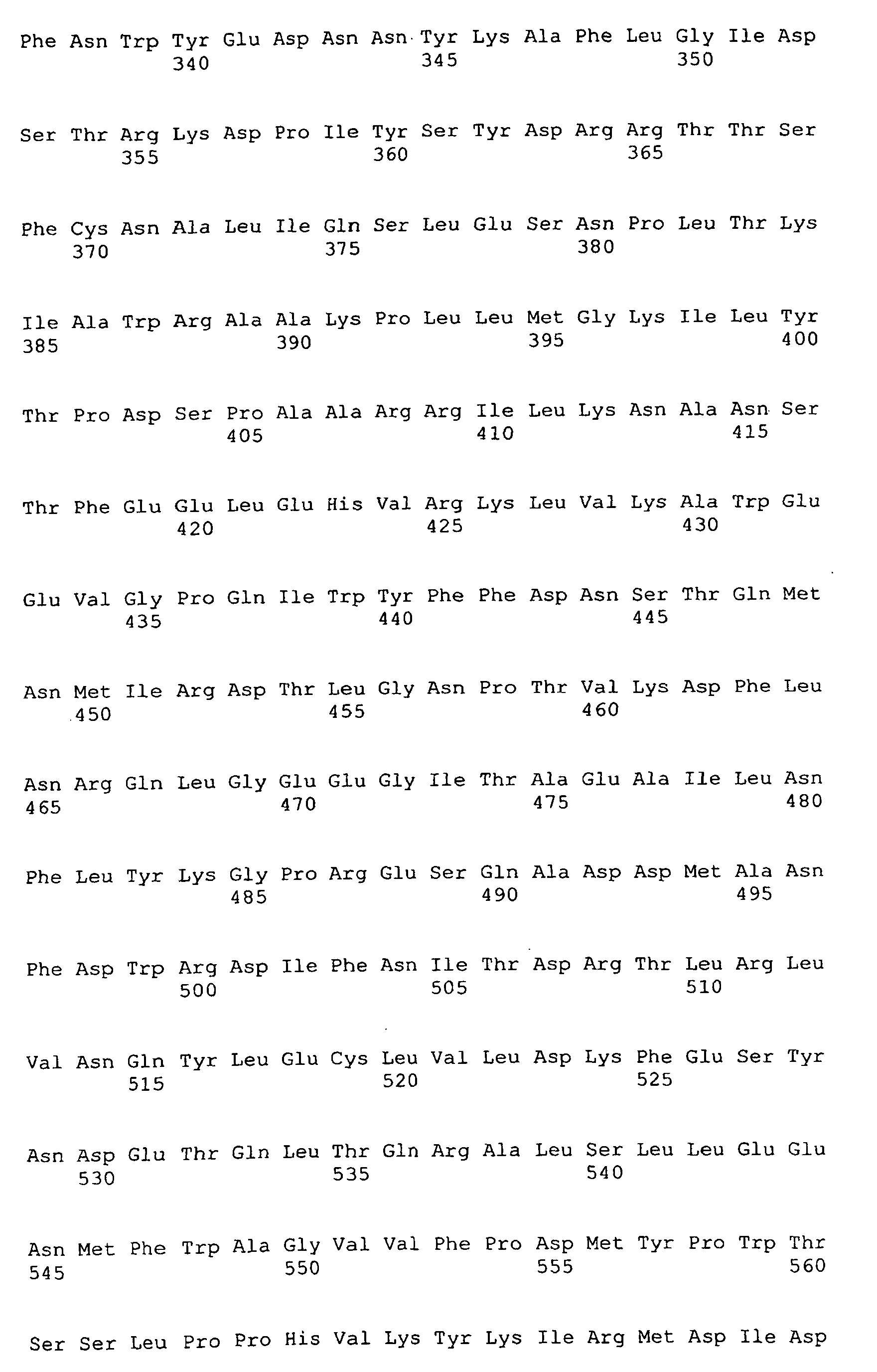


- <210> 4
<211> 114
<212> DNA
<213> Homo sapiens - <400> 4

- <210> 5
<211> 6705
<212> DNA
<213> Homo sapiens - <400> 5


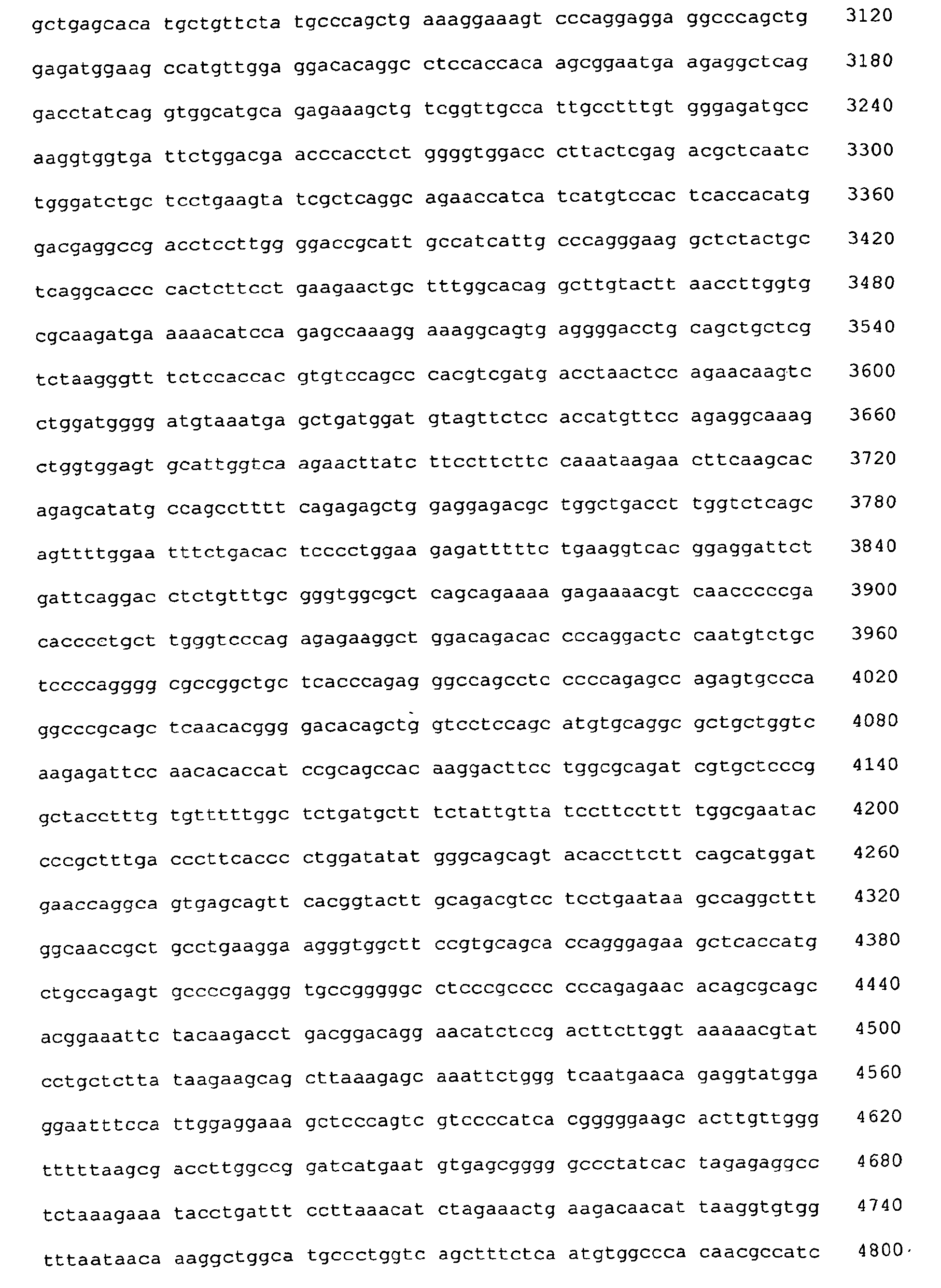


- <210> 6
<211> 2235
<212> PRT
<213> Homo sapiens - <400> 6


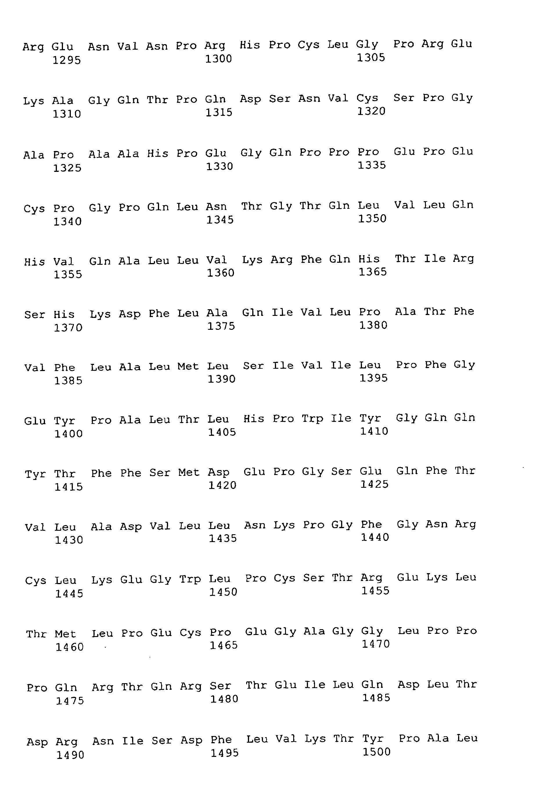


- <210> 7
<211> 22
<212> DNA
<213> Artificial Sequence - <220>
<223> Oligonucleotide primer - <400> 7

- <210> 8
<211> 21
<212> DNA
<213> Artificial Sequence - <220>
<223> Oligonucleotide primer - <400> 8

- <210> 9
<211> 20
<212> DNA
<213> Artificial Sequence - <220>
<223> Oligonucleotide primer - <400> 9

- <210> 10
<211> 21
<212> DNA
<213> Artificial Sequence - <220>
<223> Oligonucleotide primer - <400> 10

- <210> 11
<211> 38
<212> PRT
<213> Homo sapiens - <400> 11

- <210> 12
<211> 20
<212> DNA
<213> Artificial Sequence - <220>
<223> Oligonucleotide primer - <400> 12

- <210> 13
<211> 19
<212> DNA
<213> Artificial Sequence - <220>
<223> Oligonucleotide primer - <400> 13

- <210> 14
<211> 20
<212> DNA
<213> Artificial Sequence - <220>
<223> Oligonucleotide primer - <400> 14

- <210> 15
<211> 19
<212> DNA
<213> Artificial Sequence - <220>
<223> Oligonucleotide primer - <400> 15

- <210> 16
<211> 19
<212> DNA
<213> Artificial Sequence - <220>
<223> Oligonucleotide primer - <400> 16

- <210> 17
<211> 19
<212> DNA
<213> Artificial Sequence - <220>
<223> Oligonucleotide primer - <400> 17

- <210> 18
<211> 22
<212> DNA
<213> Artificial Sequence - <220>
<223> Oligonucleotide primer - <400> 18

- <210> 19
<211> 20
<212> DNA
<213> Artificial Sequence - <220>
<223> Oligonucleotide primer - <400> 19

- <210> 20
<211> 20
<212> DNA
<213> Artificial Sequence - <220>
<223> Oligonucleotide primer - <400> 20

- <210> 21
<211> 21
<212> DNA
<213> Artificial Sequence - <220>
<223> Oligonucleotide primer - <400> 21

- <210> 22
<211> 19
<212> DNA
<213> Artificial Sequence - <220>
<223> Oligonucleotide primer - <400> 22

- <210> 23
<211> 19
<212> DNA
<213> Artificial Sequence - <220>
<223> Oligonucleotide primer - <400> 23

- <210> 24
<211> 21
<212> DNA
<213> Artificial Sequence - <220>
<223> Oligonucleotide primer - <400> 24

- <210> 25
<211> 21
<212> DNA
<213> Artificial Sequence - <220>
<223> Oligonucleotide primer - <400> 25

- <210> 26
<211> 20
<212> DNA
<213> Artificial Sequence - <220>
<223> Oligonucleotide primer - <400> 26

- <210> 27
<211> 18
<212> DNA
<213> Artificial Sequence - <220>
<223> Oligonucleotide primer - <400> 27

- <210> 28
<211> 21
<212> DNA
<213> Artificial Sequence - <220>
<223> Oligonucleotide primer - <400> 28

- <210> 29
<211> 20
<212> DNA
<213> Artificial Sequence - <220>
<223> Oligonucleotide primer - <400> 29

- <210> 30
<211> 19
<212> DNA
<213> Artificial Sequence - <220>
<223> Oligonucleotide primer - <400> 30

- <210> 31
<211> 21
<212> DNA
<213> Artificial Sequence - <220>
<223> Oligonucleotide primer - <400> 31

- <210> 32
<211> 19
<212> DNA
<213> Artificial Sequence - <220>
<223> Oligonucleotide primer - <400> 32

- <210> 33
<211> 20
<212> DNA
<213> Artificial Sequence - <220>
<223> Oligonucleotide primer - <400> 33

- <210> 34
<211> 18
<212> DNA
<213> Artificial Sequence - <220>
<223> Oligonucleotide primer - <400> 34

- <210> 35
<211> 22
<212> DNA
<213> Artificial Sequence - <220>
<223> Oligonucleotide primer - <400> 35

- <210> 36
<211> 21
<212> DNA
<213> Artificial Sequence - <220>
<223> Oligonucleotide primer - <400> 36

- <210> 37
<211> 18
<212> DNA
<213> Artificial Sequence - <220>
<223> Oligonucleotide primer - <400> 37

- <210> 38
<211> 20
<212> DNA
<213> Artificial Sequence - <220>
<223> Oligonucleotide primer - <400> 38

- <210> 39
<211> 20
<212> DNA
<213> Artificial Sequence - <220>
<223> Oligonucleotide primer - <400> 39

- <210> 40
<211> 18
<212> DNA
<213> Artificial Sequence - <220>
<223> Oligonucleotide primer - <400> 40

- <210> 41
<211> 18
<212> DNA
<213> Artificial Sequence - <220>
<223> Oligonucleotide primer - <400> 41

- <210> 42
<211> 21
<212> DNA
<213> Artificial Sequence - <220>
<223> Oligonucleotide primer - <400> 42

- <210> 43
<211> 19
<212> DNA
<213> Artificial Sequence - <220>
<223> Oligonucleotide primer - <400> 43

- <210> 44
<211> 21
<212> DNA
<213> Artificial Sequence - <220>
<223> Oligonucleotide primer - <400> 44

- <210> 45
<211> 21
<212> DNA
<213> Artificial Sequence - <220>
<223> Oligonucleotide primer - <400> 45

- <210> 46
<211> 19
<212> DNA
<213> Artificial Sequence - <220>
<223> Oligonucleotide primer - <400> 46

- <210> 47
<211> 19
<212> DNA
<213> Artificial Sequence - <220>
<223> Oligonucleotide primer - <400> 47

- <210> 48
<211> 19
<212> DNA
<213> Artificial Sequence - <220>
<223> Oligonucleotide primer - <400> 48

- <210> 49
<211> 20
<212> DNA
<213> Artificial Sequence - <220>
<223> Oligonucleotide primer - <400> 49

- <210> 50
<211> 18
<212> DNA
<213> Artificial Sequence - <220>
<223> Oligonucleotide primer - <400> 50

- <210> 51
<211> 19
<212> DNA
<213> Artificial Sequence - <220>
<223> Oligonucleotide primer - <400> 51

- <210> 52
<211> 20
<212> DNA
<213> Artificial Sequence - <220>
<223> Oligonucleotide primer - <400> 52

- <210> 53
<211> 20
<212> DNA
<213> Artificial Sequence - <220>
<223> Oligonucleotide primer - <400> 53

- <210> 54
<211> 18
<212> DNA
<213> Artificial Sequence - <220>
<223> Oligonucleotide primer - <400> 54

- <210> 55
<211> 22
<212> DNA
<213> Artificial Sequence - <220>
<223> Oligonucleotide primer - <400> 55

- <210> 56
<211> 22
<212> DNA
<213> Artificial Sequence - <220>
<223> Oligonucleotide primer - <400> 56

- <210> 57
<211> 20
<212> DNA
<213> Artificial Sequence - <220>
<223> Oligonucleotide primer - <400> 57

- <210> 58
<211> 19
<212> DNA
<213> Artificial Sequence - <220>
<223> Oligonucleotide primer - <400> 58

- <210> 59
<211> 20
<212> DNA
<213> Artificial Sequence - <220>
<223> Oligonucleotide primer - <400> 59

- <210> 60
<211> 19
<212> DNA
<213> Artificial Sequence - <220>
<223> Oligonucleotide primer - <400> 60

- <210> 61
<211> 19
<212> DNA
<213> Artificial Sequence - <220>
<223> Oligonucleotide primer - <400> 61

- <210> 62
<211> 20
<212> DNA
<213> Artificial Sequence - <220>
<223> Oligonucleotide primer - <400> 62

- <210> 63
<211> 19
<212> DNA
<213> Artificial Sequence - <220>
<223> Oligonucleotide primer - <400> 63

- <210> 64
<211> 23
<212> DNA
<213> Artificial Sequence - <220>
<223> Oligonucleotide primer - <400> 64

- <210> 65
<211> 20
<212> DNA
<213> Artificial Sequence - <220>
<223> Oligonucleotide primer - <400> 65

- <210> 66
<211> 18
<212> DNA
<213> Artificial Sequence - <220>
<223> Oligonucleotide primer - <400> 66

- <210> 67
<211> 21
<212> DNA
<213> Artificial Sequence - <220>
<223> Oligonucleotide primer - <400> 67

- <210> 68
<211> 20
<212> DNA
<213> Artificial Sequence - <220>
<223> Oligonucleotide primer - <400> 68

- <210> 69
<211> 19
<212> DNA
<213> Artificial Sequence - <220>
<223> Oligonucleotide primer - <400> 69

- <210> 70
<211> 20
<212> DNA
<213> Artificial Sequence - <220>
<223> Oligonucleotide primer - <400> 70

- <210> 71
<211> 20
<212> DNA
<213> Artificial Sequence - <220>
<223> Oligonucleotide primer - <400> 71

- <210> 72
<211> 21
<212> DNA
<213> Artificial Sequence - <220>
<223> Oligonucleotide primer - <400> 72

- <210> 73
<211> 20
<212> DNA
<213> Artificial Sequence - <220>
<223> Oligonucleotide primer - <400> 73

- <210> 74
<211> 22
<212> DNA
<213> Artificial Sequence - <220>
<223> Oligonucleotide primer - <400> 74

- <210> 75
<211> 20
<212> DNA
<213> Artificial Sequence - <220>
<223> Oligonucleotide primer - <400> 75

- <210> 76
<211> 21
<212> DNA
<213> Artificial Sequence - <220>
<223> Oligonucleotide primer - <400> 76

- <210> 77
<211> 22
<212> DNA
<213> Artificial Sequence - <220>
<223> Oligonucleotide primer - <400> 77

- <210> 78
<211> 22
<212> DNA
<213> Artificial Sequence - <220>
<223> Oligonucleotide primer - <400> 78

- <210> 79
<211> 22
<212> DNA
<213> Artificial Sequence - <220>
<223> Oligonucleotide primer - <400> 79

- <210> 80
<211> 22
<212> DNA
<213> Artificial Sequence - <220>
<223> Oligonucleotide primer - <400> 80

- <210> 81
<211> 19
<212> DNA
<213> Artificial Sequence - <220>
<223> Oligonucleotide primer - <400> 81

- <210> 82
<211> 22
<212> DNA
<213> Artificial Sequence - <220>
<223> Oligonucleotide primer - <400> 82

- <210> 83
<211> 18
<212> DNA
<213> Artificial Sequence - <220>
<223> Oligonucleotide primer - <400> 83

- <210> 84
<211> 19
<212> DNA
<213> Artificial Sequence - <220>
<223> Oligonucleotide primer - <400> 84

- <210> 85
<211> 20
<212> DNA
<213> Artificial Sequence - <220>
<223> Oligonucleotide primer - <400> 85

- <210> 86
<211> 20
<212> DNA
<213> Artificial Sequence - <220>
<223> Oligonucleotide primer - <400> 86

- <210> 87
<211> 20
<212> DNA
<213> Artificial Sequence - <220>
<223> Oligonucleotide primer - <400> 87

- <210> 88
<211> 21
<212> DNA
<213> Artificial Sequence - <220>
<223> Oligonucleotide primer - <400> 88

- <210> 89
<211> 20
<212> DNA
<213> Artificial Sequence - <220>
<223> Oligonucleotide primer - <400> 89

- <210> 90
<211> 20
<212> DNA
<213> Artificial Sequence - <220>
<223> Oligonucleotide primer - <400> 90

- <210> 91
<211> 19
<212> DNA
<213> Artificial Sequence - <220>
<223> Oligonucleotide primer - <400> 91

- <210> 92
<211> 20
<212> DNA
<213> Artificial Sequence - <220>
<223> Oligonucleotide primer - <400> 92

- <210> 93
<211> 19
<212> DNA
<213> Artificial Sequence - <220>
<223> Oligonucleotide primer - <400> 93

- <210> 94
<211> 22
<212> DNA
<213> Artificial Sequence - <220>
<223> Oligonucleotide primer - <400> 94

- <210> 95
<211> 22
<212> DNA
<213> Artificial Sequence - <220>
<223> Oligonucleotide primer - <400> 95

- <210> 96
<211> 21
<212> DNA
<213> Artificial Sequence - <220>
<223> Oligonucleotide primer - <400> 96

- <210> 97
<211> 18
<212> DNA
<213> Artificial Sequence - <220>
<223> Oligonucleotide primer - <400> 97

- <210> 98
<211> 18
<212> DNA
<213> Artificial Sequence - <220>
<223> Oligonucleotide primer - <400> 98

- <210> 99
<211> 21
<212> DNA
<213> Artificial Sequence - <220>
<223> Oligonucleotide primer - <400> 99

- <210> 100
<211> 20
<212> DNA
<213> Artificial Sequence - <220>
<223> Oligonucleotide primer - <400> 100

- <210> 101
<211> 20
<212> DNA
<213> Artificial Sequence - <220>
<223> Oligonucleotide primer - <400> 101

- <210> 102
<211> 20
<212> DNA
<213> Artificial Sequence - <220>
<223> Oligonucleotide primer - <400> 102

- <210> 103
<211> 21
<212> DNA
<213> Artificial Sequence - <220>
<223> Oligonucleotide primer - <400> 103

- <210> 104
<211> 20
<212> DNA
<213> Artificial Sequence - <220>
<223> Oligonucleotide primer - <400> 104

- <210> 105
<211> 21
<212> DNA
<213> Artificial Sequence - <220>
<223> Oligonucleotide primer - <400> 105

- <210> 106
<211> 20
<212> DNA
<213> Artificial Sequence - <220>
<223> Oligonucleotide primer - <400> 106

- <210> 107
<211> 21
<212> DNA
<213> Artificial Sequence - <220>
<223> Oligonucleotide primer - <400> 107

- <210> 108
<211> 20
<212> DNA
<213> Artificial Sequence - <220>
<223> Oligonucleotide primer - <400> 108

- <210> 109
<211> 19
<212> DNA
<213> Artificial Sequence - <220>
<223> Oligonucleotide primer - <400> 109

- <210> 110
<211> 19
<212> DNA
<213> Artificial Sequence - <220>
<223> Oligonucleotide primer - <400> 110

- <210> 111
<211> 20
<212> DNA
<213> Artificial Sequence - <220>
<223> Oligonucleotide primer - <400> 111

- <210> 112
<211> 19
<212> DNA
<213> Artificial Sequence - <220>
<223> Oligonucleotide primer - <400> 112

- <210> 113
<211> 21
<212> DNA
<213> Artificial Sequence - <220>
<223> Oligonucleotide primer - <400> 113

- <210> 114
<211> 20
<212> DNA
<213> Artificial Sequence - <220>
<223> Oligonucleotide primer - <400> 114

- <210> 115
<211> 18
<212> DNA
<213> Artificial Sequence - <220>
<223> Oligonucleotide primer - <400> 115

- <210> 116
<211> 22
<212> DNA
<213> Artificial Sequence - <220>
<223> Oligonucleotide primer - <400> 116

- <210> 117
<211> 21
<212> DNA
<213> Artificial Sequence - <220>
<223> Oligonucleotide primer - <400> 117

- <210> 118
<211> 2261
<212> PRT
<213> Mouse - <400> 118


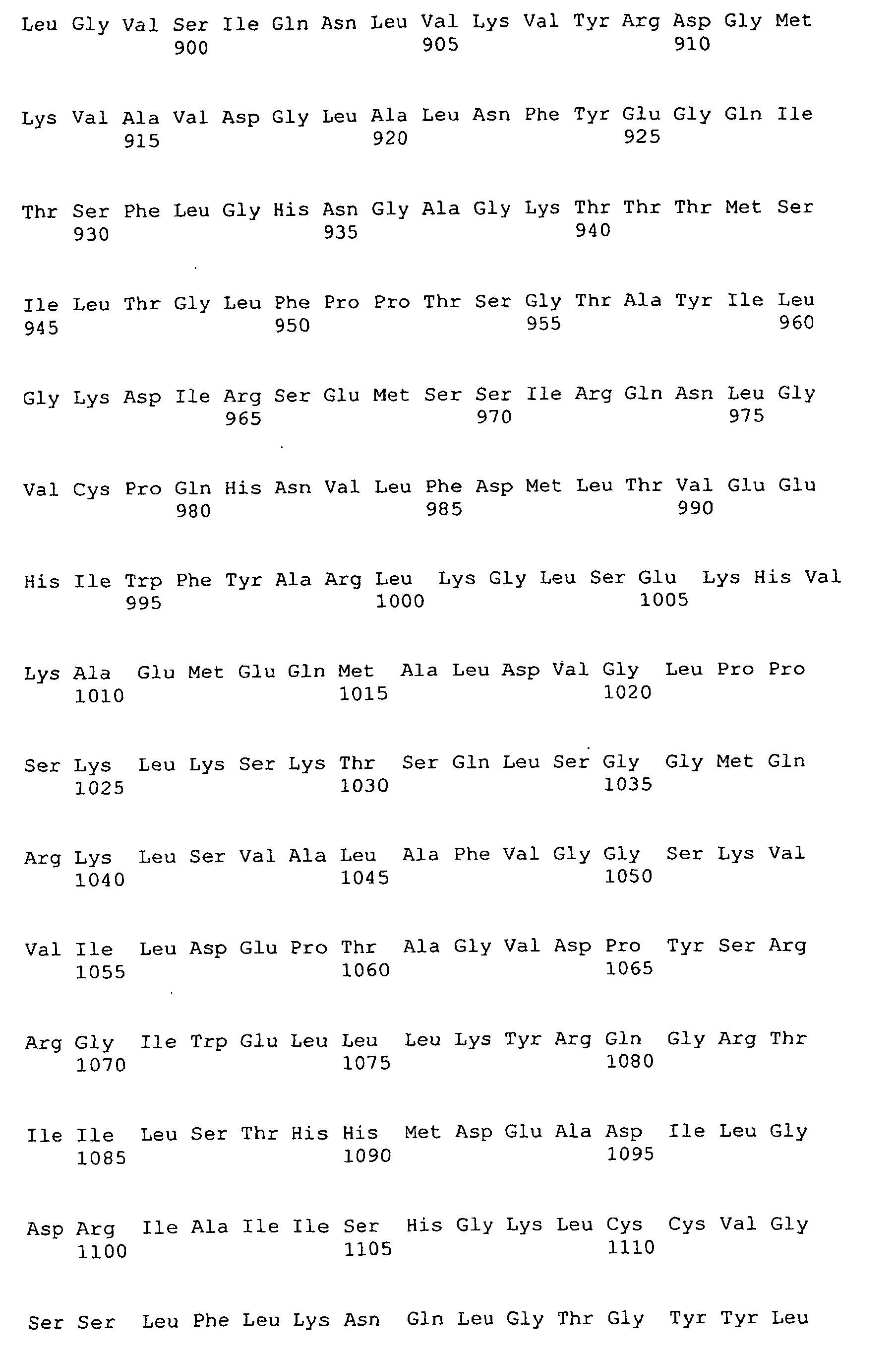


- <210> 119
<211> 1472
<212> PRT
<213> Mouse - <400> 119

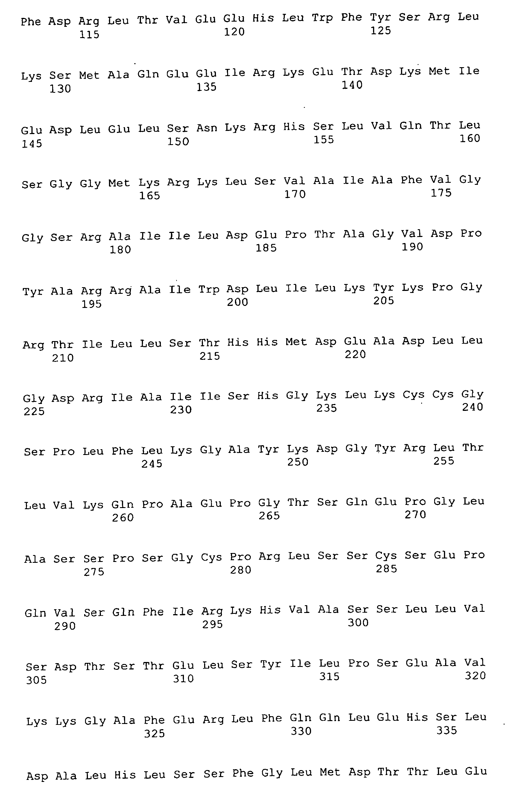


- <210> 120
<211> 1704
<212> PRT
<213> Homo sapiens - <400> 120


Claims (64)
- A nucleic acid sequence encoding a retina-specific ATP binding cassette transporter wherein said nucleic acid sequence comprises SEQ ID NO:4.
- The nucleic acid sequence according to claim 1 wherein said ATP-binding cassette transporter comprises an amino acid selected from the group consisting of 818 (G or E), 863 (G or A), 931 (V or M), 1028 (A or V), 1072 (V or A), 1087 (E or K), 2027 (L or F), 2038 (R or W), 2050 (V or L), 2077 (R or W), and 2106 (R or C) according to numbering of the sequence in Figure 3.
- The nucleic acid sequence of claim 1 comprising a nucleotide selected from the group consisting of 2453 (G or A), 2588 (G or C), 2791 (G or A), 3083 (C or T), 3215 (T or C), 3259 (G or A), 6079 (C or T), 6112 (C or T), 6148 (G or C), 6229 (C or T), and 6316 (C or T) according to the numbering of the sequence in Figure 3.
- The nucleic acid sequence of claim 3 wherein said nucleotide results in a missense mutation.
- The nucleic acid sequence of claim 3 wherein said nucleotide results in a splice site.
- The nucleic acid sequence of claim 1 comprising a nucleotide that results in a frameshift mutation.
- A vector comprising a nucleic acid sequence according to claim 1.
- A cell culture obtained by transforming a cell with an expression vector according to claim 7.
- A host cell capable of recombinantly expressing a nucleic acid sequence according to claim 1.
- A cell culture capable of recombinantly expressing a nucleic acid sequence according to claim 1.
- A protein preparation comprising a recombinantly expressed polypeptide having an amino acid sequence encoded by a nucleic acid sequence according to claim 1.
- A pharmaceutical composition comprising an effective amount of a nucleic acid sequence according to claim 1, and a pharmaceutically acceptable carrier.
- An isolated polypeptide comprising a retina-specific ATP-binding cassette transporter which polypeptide is encoded by the nucleic acid sequence of any one of claims 1 to 3.
- The isolated polypeptide according to claim 13 wherein the polypeptide comprises a mutation associated with macular degeneration.
- A transgenic non-human mammal comprising a recombinant sequence comprising a nucleic acid sequence of according to claim 1 introduced into said mammal or an ancestor of said mammal, or comprising a suppressed retina-specific ATP binding cassette transporter gene comprising SEQ ID NO:4.
- A method of screening for an agent that modulates macular degeneration comprising combining purified retina-specific ATP binding cassette transporter comprising an amino acid sequence encoded by a nucleic acid sequence according to claim 1 and at least one agent suspected of altering retina-specific ATP binding cassette transporter and observing an alteration in the activity of said purified retina-specific ATP binding cassette transporter.
- A method of screening for an agent according to claim 16 wherein the method is a method of screening for an agent that inhibits macular degeneration, the method comprising combining the purified retina-specific ATP binding cassette transporter and at least one agent suspected of activating retina-specific ATP binding cassette transporter and observing an activation of said purified retina-specific ATP binding cassette transporter.
- The method according to claim 17 wherein said purified retina-specific ATP binding cassette transporter originates from a patient suspected of having macular degeneration.
- The method of claim 17 wherein the activation of said purified retina-specific ATP binding cassette transporter is observed by an inhibition of a characteristic associated with macular degeneration selected from the group consisting of inhibition of central visual impairment, inhibition of progressive bilateral atrophy of the macular retinal pigment epithelium, inhibition of progressive bilateral atrophy of the neuroepithelium, inhibition of macula flecks, inhibition of midretinal periphery flecks, and inhibition of retina-specific ATP binding cassette transporter transcripts in photoreceptor cells.
- The method of claim 19 wherein said macular degeneration is selected from the group consisting of Stargardt Disease, Fundus Flavimaculatus, and age-related macular degeneration.
- The method of claim 17 further comprising the step of amplifying said nucleic acid sequence encoding a retina-specific ATP binding cassette transporter using an oligonucleotide primer that specifically hybridizes to said sequence.
- The method of claim 17 further comprising the step of amplifying said nucleic acid sequence encoding a retina-specific ATP binding cassette transporter using an oligonucleotide primer that specifically hybridizes to 17 nucleotides to 25 nucleotides of said sequence.
- A method of screening for an agent according to claim 16 wherein the method is a method of screening for an agent that activates macular degeneration, the method comprising combining the purified wild-type retina-specific ATP binding cassette transporter and at least one agent suspected of activating macular degeneration and observing an inhibition in said purified wild-type retina-specific ATP binding cassette transporter.
- The method of claim 23 wherein said inhibition of said purified retina-specific ATP binding cassette transporter is observed by a characteristic associated with macular degeneration selected from the group consisting of central visual impairment, bilateral atrophy of the macular retinal pigment epithelium, bilateral atrophy of the neuroepithelium, macula flecks, midretinal periphery flecks, and retina-specific ATP binding cassette transporter transcripts in photoreceptor cells.
- An in vitro method of diagnosing an individual with macular degeneration or lack thereof comprising detecting the presence or absence of a mutation associated with macular degeneration in a retina-specific ATP binding cassette transporter-encoding nucleic acid according to claim 1 in a tissue sample of said individual.
- The method of claim 25 wherein said tissue sample is selected from the group consisting of blood, skin, serum, saliva, sputum, mucus, bone marrow, urine, lymph, a tear, chorion, and amniotic fluid.
- The method of claim 25 wherein said mutation is selected from the group consisting of: 2453 (G→A), 2588 (G→C), 2791 (G→A), 2884 del C, 3083 (C→T), 3259 (G→A), 5041del15, 5196 +2(T→C), 6005 + 1 (G→T), 6079 (C→T), 6112 (C→T), 6148 (G→C), 6229 (C→T), and 6316 (C→T) according to the numbering of the sequence in Figure 3.
- The method of claim 25 wherein said mutation results in an amino acid sequence difference selected from the group consisting of G818E, G863A, V931M, A1028V, V1072A, E1087K, VVAIC1681del, L2027F, R2038W, V2050L, R2077W, and R2106C according to the numbering of the sequence in Figure 3.
- The method of claim 25 wherein the mutation is selected from the group consisting of a missense mutation, an intragenic deletion, an intragenic insertion, a frame shift mutation, and a splice site mutation.
- The method of claim 29 wherein said mutation is a missense mutation.
- The method of claim 29 wherein said mutation is a frameshift mutation.
- The method of claim 29 wherein said mutation is a splice site mutation.
- The method of claim 25 further comprising extracting RNA or DNA from said sample.
- The method of claim 25 further comprising the step of amplifying said nucleic acid sequence encoding a retina-specific ATP binding cassette transporter.
- The method of claim 34 wherein said amplification step comprises performing the polymerase chain reaction.
- The method of claim 25 further comprising the step of amplifying said nucleic acid sequence encoding a retina-specific ATP binding cassette transporter using an oligonucleotide primer that specifically hybridizes to said nucleic acid sequence.
- The method of claim 25 further comprising the step of amplifying said nucleic acid sequence encoding a retina-specific ATP binding cassette transporter using a primer having a nucleic acid sequence selected from the group consisting of SEQ ID NOs: 40-61, 64, 65, 78, 79, 98-105, 108, and 109.
- The method of claim 34 further comprising the step of comparing said amplified nucleic acid sequence from said individual with a control nucleic acid sequence encoding wild-type retina-specific ATP binding cassette transporter wherein a mutation in said amplified nucleic acid sequence from said individual as compared to said nucleic acid sequence encoding wild-type retina-specific ATP binding cassette transporter indicates macular degeneration.
- An in vitro method of diagnosing macular degeneration in an individual comprising detecting the presence of a mutated version of a retina-specific ATP binding cassette transporter encoded by a nucleic acid sequence of claim 1.
- Use of a nucleic acid sequence according to claim 1, or a fragment thereof, in the manufacture of a medicament for the treatment of macular degeneration.
- The use of claim 40 wherein said nucleotide encodes a wild-type ATP-binding cassette transporter having an amino acid sequence encoded by SEQ ID NO:4.
- The use of claim 40 wherein the medicament is for treatment of a human.
- The use of claim 40 wherein said macular degeneration is due to Stargardt disease, Fundus Flavimaculatus, age-related macular degeneration, retinitis pigmentosa, combined rod and cone dystrophies, cone dystrophies, pattern dystrophy, or bull's eye maculopathy.
- The use of claim 40 wherein said macular degeneration is due to Stargardt disease.
- A kit for detecting macular degeneration comprising a first container, wherein said container comprises a first primer that is between 15 and 35 nucleotides and is complementary to a first region of a retina-specific ATP-binding cassette transporter gene comprising a nucleic acid sequence according to claim 1 .
- The kit of claim 45 wherein the first region of the gene is a coding region.
- The kit of claim 45 wherein said first region of the gene flanks an exon.
- The kit of claim 45 further comprising a second container wherein said second container comprises a second primer that is between 15 and 35 nucleotides and is complementary to a second region of a retina-specific ATP-binding cassette transporter gene comprising a nucleic acid sequence according to claim 1.
- The kit of claim 48 wherein the second region of the gene is a coding region.
- The kit of claim 48 wherein the second region of the gene flanks an exon.
- A kit according to claim 45, wherein said first primer is selected from the group consisting of SEQ ID NOS: 40-61, 64, 65, 78, 79, 98-105, 108 and 109.
- The kit of claim 48 wherein the second primer is selected from the group consisting of SEQ ID NOS: 40-61, 64, 65, 78, 79, 98-105, 108, and 109, but is not identical to the first primer.
- A kit for detecting macular degeneration comprising a first container, wherein said container comprises a primer pair that is complementary to a first and a second region of a retina-specific ATP-binding cassette transporter gene comprising a nucleic acid sequence according to claim 1 wherein each primer is between 15 and 35 nucleotides.
- The kit of claim 53 wherein the primer pair flank an exon.
- The kit of claim 53 wherein the primer pair span a coding region.
- The kit of claim 53 wherein the primer pair comprises a primer having sequence selected from the group consisting of SEQ ID NOS: 40-61, 64, 65, 78, 79, 98-105, 108, and 109.
- The kit of claim 45 or 53 further comprising a means for visualizing amplified DNA.
- The kit of claim 57 wherein said means is a fluorescent dye selected from the group consisting of ethidium bromide, 32P, and biotin.
- The kit of claim 45 or 53 further comprising one or more size markers.
- The kit of claim 45 or 53 further comprising one or more positive or negative controls.
- The kit of claim 45 or 53 further comprising one or more restriction enzymes.
- An isolated oligonucleotide comprising a nucleic acid sequence complementary to a nucleic acid sequence according to claim 1.
- In vitro use of an oligonucleotide according to claim 62 to inhibit the expression of retina- specific ATP-binding cassette transporter in a cell.
- An isolated nucleic acid sequence comprising a nucleic acid sequence selected from the group consisting of: SEQ ID NOs:40-61, 64, 65, 78, 79, 98-105, 108, and 109.
Applications Claiming Priority (3)
| Application Number | Priority Date | Filing Date | Title |
|---|---|---|---|
| US3938897P | 1997-02-27 | 1997-02-27 | |
| US39388P | 1997-02-27 | ||
| PCT/US1998/003895 WO1998037764A1 (en) | 1997-02-27 | 1998-02-27 | Nucleic acid sequences for atp-binding cassette transporter |
Publications (3)
| Publication Number | Publication Date |
|---|---|
| EP0989805A1 EP0989805A1 (en) | 2000-04-05 |
| EP0989805A4 EP0989805A4 (en) | 2002-04-17 |
| EP0989805B1 true EP0989805B1 (en) | 2006-01-11 |
Family
ID=21905183
Family Applications (1)
| Application Number | Title | Priority Date | Filing Date |
|---|---|---|---|
| EP98911434A Expired - Lifetime EP0989805B1 (en) | 1997-02-27 | 1998-02-27 | Nucleic acid sequences for atp-binding cassette transporter |
Country Status (7)
| Country | Link |
|---|---|
| EP (1) | EP0989805B1 (en) |
| JP (1) | JP4290225B2 (en) |
| AT (1) | ATE315334T1 (en) |
| AU (1) | AU742094B2 (en) |
| CA (1) | CA2281887C (en) |
| DE (1) | DE69833189T2 (en) |
| WO (1) | WO1998037764A1 (en) |
Families Citing this family (9)
| Publication number | Priority date | Publication date | Assignee | Title |
|---|---|---|---|---|
| WO2000018912A2 (en) * | 1998-09-25 | 2000-04-06 | Bayer Aktiengesellschaft | Atp binding cassette genes and proteins for diagnosis and treatment of lipid disorders and inflammatory diseases |
| US6617122B1 (en) | 1999-03-15 | 2003-09-09 | Xenon Genetics, Inc. | Process for identifying modulators of ABC1 activity |
| GB9910693D0 (en) * | 1999-05-07 | 1999-07-07 | Univ Liverpool | A compound for use in medicine |
| FR2796808B1 (en) * | 1999-07-30 | 2004-03-12 | Inst Nat Sante Rech Med | NEW APPLICATIONS OF ABCA TYPE CONVEYORS |
| AU2001239837B2 (en) * | 2000-02-23 | 2006-06-22 | Pxe International, Inc. | Methods and composition for diagnosing and treating pseudoxanthoma elasticum and related conditions |
| JP2004147502A (en) * | 2000-07-26 | 2004-05-27 | Banyu Pharmaceut Co Ltd | Human and rat ABCA2 genes |
| CA2430082A1 (en) * | 2000-11-29 | 2002-06-06 | Lynkeus Biotech Gmbh | Retina-specific human proteins c70rf9, c120rf7, mpp4 and f379 |
| US20030027301A1 (en) * | 2001-06-14 | 2003-02-06 | Yi Hu | Novel human transporter proteins and polynucleotides encoding the same |
| GB201706009D0 (en) | 2017-04-13 | 2017-05-31 | Proqr Therapeutics Ii Bv | Antisense oligonucleotides for the treatment of stargardt disease |
-
1998
- 1998-02-27 EP EP98911434A patent/EP0989805B1/en not_active Expired - Lifetime
- 1998-02-27 AU AU65386/98A patent/AU742094B2/en not_active Expired
- 1998-02-27 DE DE69833189T patent/DE69833189T2/en not_active Expired - Lifetime
- 1998-02-27 WO PCT/US1998/003895 patent/WO1998037764A1/en not_active Ceased
- 1998-02-27 JP JP53790898A patent/JP4290225B2/en not_active Expired - Lifetime
- 1998-02-27 CA CA2281887A patent/CA2281887C/en not_active Expired - Lifetime
- 1998-02-27 AT AT98911434T patent/ATE315334T1/en not_active IP Right Cessation
Also Published As
| Publication number | Publication date |
|---|---|
| EP0989805A1 (en) | 2000-04-05 |
| ATE315334T1 (en) | 2006-02-15 |
| WO1998037764A1 (en) | 1998-09-03 |
| CA2281887C (en) | 2013-09-10 |
| DE69833189T2 (en) | 2006-08-03 |
| EP0989805A4 (en) | 2002-04-17 |
| AU742094B2 (en) | 2001-12-20 |
| CA2281887A1 (en) | 1998-09-03 |
| DE69833189D1 (en) | 2006-04-06 |
| JP4290225B2 (en) | 2009-07-01 |
| AU6538698A (en) | 1998-09-18 |
| JP2001516209A (en) | 2001-09-25 |
Similar Documents
| Publication | Publication Date | Title |
|---|---|---|
| US20120040456A1 (en) | Methods of gene therapy using nucleic acid sequences for atp-binding cassette transporter | |
| Allikmets et al. | A photoreceptor cell-specific ATP-binding transporter gene (ABCR) is mutated in recessive Starqardt macular dystrophy | |
| US7785886B2 (en) | Methods and reagents for modulating cholesterol levels | |
| Goldstein et al. | Exonic SINE insertion in STK38L causes canine early retinal degeneration (erd) | |
| US6207383B1 (en) | Mutations in and genomic structure of HERG—a long QT syndrome gene | |
| AU742094B2 (en) | Nucleic acid sequences for ATP-binding cassette transporter | |
| US6593104B1 (en) | Macular degeneration diagnostics and therapeutics | |
| AU714527B2 (en) | KVLQT1 - A long QT syndrome gene which encodes KVLQT1 which coassembles with minK to form cardiac 1ks potassium channels | |
| WO1998037764A9 (en) | Nucleic acid sequences for atp-binding cassette transporter | |
| Prakash et al. | Characterization of a novel chromo domain gene in xp22. 3 with homology to Drosophila msl-3 | |
| WO1999051779A9 (en) | Glaucoma therapeutics and diagnostics | |
| AU5436498A (en) | Glaucoma-associated protein and corresponding nucleic acid and their therapeut ic and diagnostic uses | |
| Leppert et al. | Methods of screening and diagnostics using ATP-binding cassette transporter | |
| AU2747802A (en) | Nucleic acid sequences for ATP-binding cassette transporter | |
| Class et al. | Patent application title: Methods of gene therapy using nucleic acid sequences for ATP-binding cassette transporter | |
| US20060257868A1 (en) | Tissue specific genes and gene clusters | |
| JP5119430B2 (en) | Genes associated with polycystic kidney disease and uses thereof | |
| WO1998021363A1 (en) | Compositions and methods for treating type ii diabetes involving hnf-4 | |
| WO2000018787A1 (en) | Gene mutated in wolfram syndrome | |
| US6306586B1 (en) | Methods and compositions for the diagnosis and treatment of cataracts | |
| WO1999063078A2 (en) | Retinal calcium channel (alpha)1f-subunit gene | |
| CH696239A5 (en) | Alström Syndrome, Gene Variants, Coded Protein, and Methods for Diagnosing Alström Syndrome. | |
| WO2001029266A2 (en) | Identification of arsacs mutations and methods of use therefor | |
| US20050123960A1 (en) | Glaucoma therapeutics and diagnostics |
Legal Events
| Date | Code | Title | Description |
|---|---|---|---|
| PUAI | Public reference made under article 153(3) epc to a published international application that has entered the european phase |
Free format text: ORIGINAL CODE: 0009012 |
|
| 17P | Request for examination filed |
Effective date: 19990927 |
|
| AK | Designated contracting states |
Kind code of ref document: A1 Designated state(s): AT BE CH DE DK ES FI FR GB GR IE IT LI LU MC NL PT SE |
|
| A4 | Supplementary search report drawn up and despatched |
Effective date: 20020307 |
|
| AK | Designated contracting states |
Kind code of ref document: A4 Designated state(s): AT BE CH DE DK ES FI FR GB GR IE IT LI LU MC NL PT SE |
|
| 17Q | First examination report despatched |
Effective date: 20030311 |
|
| GRAP | Despatch of communication of intention to grant a patent |
Free format text: ORIGINAL CODE: EPIDOSNIGR1 |
|
| GRAS | Grant fee paid |
Free format text: ORIGINAL CODE: EPIDOSNIGR3 |
|
| GRAA | (expected) grant |
Free format text: ORIGINAL CODE: 0009210 |
|
| AK | Designated contracting states |
Kind code of ref document: B1 Designated state(s): AT BE CH DE DK ES FI FR GB GR IE IT LI LU MC NL PT SE |
|
| PG25 | Lapsed in a contracting state [announced via postgrant information from national office to epo] |
Ref country code: IT Free format text: LAPSE BECAUSE OF FAILURE TO SUBMIT A TRANSLATION OF THE DESCRIPTION OR TO PAY THE FEE WITHIN THE PRESCRIBED TIME-LIMIT;WARNING: LAPSES OF ITALIAN PATENTS WITH EFFECTIVE DATE BEFORE 2007 MAY HAVE OCCURRED AT ANY TIME BEFORE 2007. THE CORRECT EFFECTIVE DATE MAY BE DIFFERENT FROM THE ONE RECORDED. Effective date: 20060111 Ref country code: FI Free format text: LAPSE BECAUSE OF FAILURE TO SUBMIT A TRANSLATION OF THE DESCRIPTION OR TO PAY THE FEE WITHIN THE PRESCRIBED TIME-LIMIT Effective date: 20060111 Ref country code: AT Free format text: LAPSE BECAUSE OF FAILURE TO SUBMIT A TRANSLATION OF THE DESCRIPTION OR TO PAY THE FEE WITHIN THE PRESCRIBED TIME-LIMIT Effective date: 20060111 |
|
| REG | Reference to a national code |
Ref country code: CH Ref legal event code: EP |
|
| PGFP | Annual fee paid to national office [announced via postgrant information from national office to epo] |
Ref country code: AT Payment date: 20060201 Year of fee payment: 9 |
|
| REG | Reference to a national code |
Ref country code: IE Ref legal event code: FG4D |
|
| PGFP | Annual fee paid to national office [announced via postgrant information from national office to epo] |
Ref country code: ES Payment date: 20060224 Year of fee payment: 9 |
|
| PGFP | Annual fee paid to national office [announced via postgrant information from national office to epo] |
Ref country code: GR Payment date: 20060228 Year of fee payment: 9 Ref country code: FI Payment date: 20060228 Year of fee payment: 9 |
|
| REF | Corresponds to: |
Ref document number: 69833189 Country of ref document: DE Date of ref document: 20060406 Kind code of ref document: P |
|
| PG25 | Lapsed in a contracting state [announced via postgrant information from national office to epo] |
Ref country code: SE Free format text: LAPSE BECAUSE OF FAILURE TO SUBMIT A TRANSLATION OF THE DESCRIPTION OR TO PAY THE FEE WITHIN THE PRESCRIBED TIME-LIMIT Effective date: 20060411 Ref country code: DK Free format text: LAPSE BECAUSE OF NON-PAYMENT OF DUE FEES Effective date: 20060411 |
|
| PG25 | Lapsed in a contracting state [announced via postgrant information from national office to epo] |
Ref country code: ES Free format text: LAPSE BECAUSE OF FAILURE TO SUBMIT A TRANSLATION OF THE DESCRIPTION OR TO PAY THE FEE WITHIN THE PRESCRIBED TIME-LIMIT Effective date: 20060422 |
|
| PG25 | Lapsed in a contracting state [announced via postgrant information from national office to epo] |
Ref country code: PT Free format text: LAPSE BECAUSE OF FAILURE TO SUBMIT A TRANSLATION OF THE DESCRIPTION OR TO PAY THE FEE WITHIN THE PRESCRIBED TIME-LIMIT Effective date: 20060612 |
|
| ET | Fr: translation filed | ||
| PLBE | No opposition filed within time limit |
Free format text: ORIGINAL CODE: 0009261 |
|
| STAA | Information on the status of an ep patent application or granted ep patent |
Free format text: STATUS: NO OPPOSITION FILED WITHIN TIME LIMIT |
|
| 26N | No opposition filed |
Effective date: 20061012 |
|
| REG | Reference to a national code |
Ref country code: CH Ref legal event code: NV Representative=s name: PATENTANWAELTE SCHAAD, BALASS, MENZL & PARTNER AG |
|
| PG25 | Lapsed in a contracting state [announced via postgrant information from national office to epo] |
Ref country code: GR Free format text: LAPSE BECAUSE OF FAILURE TO SUBMIT A TRANSLATION OF THE DESCRIPTION OR TO PAY THE FEE WITHIN THE PRESCRIBED TIME-LIMIT Effective date: 20060412 |
|
| REG | Reference to a national code |
Ref country code: FR Ref legal event code: PLFP Year of fee payment: 19 |
|
| REG | Reference to a national code |
Ref country code: FR Ref legal event code: PLFP Year of fee payment: 20 |
|
| PGFP | Annual fee paid to national office [announced via postgrant information from national office to epo] |
Ref country code: FR Payment date: 20170112 Year of fee payment: 20 Ref country code: CH Payment date: 20170214 Year of fee payment: 20 Ref country code: MC Payment date: 20170103 Year of fee payment: 20 Ref country code: DE Payment date: 20170221 Year of fee payment: 20 |
|
| PGFP | Annual fee paid to national office [announced via postgrant information from national office to epo] |
Ref country code: NL Payment date: 20170210 Year of fee payment: 20 Ref country code: LU Payment date: 20170208 Year of fee payment: 20 Ref country code: IE Payment date: 20170209 Year of fee payment: 20 Ref country code: BE Payment date: 20161228 Year of fee payment: 20 Ref country code: GB Payment date: 20170222 Year of fee payment: 20 |
|
| REG | Reference to a national code |
Ref country code: DE Ref legal event code: R071 Ref document number: 69833189 Country of ref document: DE |
|
| REG | Reference to a national code |
Ref country code: NL Ref legal event code: MK Effective date: 20180226 |
|
| REG | Reference to a national code |
Ref country code: BE Ref legal event code: MK Effective date: 20180227 |
|
| REG | Reference to a national code |
Ref country code: CH Ref legal event code: PL |
|
| REG | Reference to a national code |
Ref country code: GB Ref legal event code: PE20 Expiry date: 20180226 |
|
| REG | Reference to a national code |
Ref country code: IE Ref legal event code: MK9A |
|
| PG25 | Lapsed in a contracting state [announced via postgrant information from national office to epo] |
Ref country code: IE Free format text: LAPSE BECAUSE OF EXPIRATION OF PROTECTION Effective date: 20180227 Ref country code: GB Free format text: LAPSE BECAUSE OF EXPIRATION OF PROTECTION Effective date: 20180226 |