EP0976835A1 - Method for detecting nucleic acid methylation using AFLPTM - Google Patents
Method for detecting nucleic acid methylation using AFLPTM Download PDFInfo
- Publication number
- EP0976835A1 EP0976835A1 EP98202549A EP98202549A EP0976835A1 EP 0976835 A1 EP0976835 A1 EP 0976835A1 EP 98202549 A EP98202549 A EP 98202549A EP 98202549 A EP98202549 A EP 98202549A EP 0976835 A1 EP0976835 A1 EP 0976835A1
- Authority
- EP
- European Patent Office
- Prior art keywords
- dna
- adapter
- methylation
- methylated
- cutter
- Prior art date
- Legal status (The legal status is an assumption and is not a legal conclusion. Google has not performed a legal analysis and makes no representation as to the accuracy of the status listed.)
- Granted
Links
Images


Classifications
-
- C—CHEMISTRY; METALLURGY
- C12—BIOCHEMISTRY; BEER; SPIRITS; WINE; VINEGAR; MICROBIOLOGY; ENZYMOLOGY; MUTATION OR GENETIC ENGINEERING
- C12Q—MEASURING OR TESTING PROCESSES INVOLVING ENZYMES, NUCLEIC ACIDS OR MICROORGANISMS; COMPOSITIONS OR TEST PAPERS THEREFOR; PROCESSES OF PREPARING SUCH COMPOSITIONS; CONDITION-RESPONSIVE CONTROL IN MICROBIOLOGICAL OR ENZYMOLOGICAL PROCESSES
- C12Q1/00—Measuring or testing processes involving enzymes, nucleic acids or microorganisms; Compositions therefor; Processes of preparing such compositions
- C12Q1/68—Measuring or testing processes involving enzymes, nucleic acids or microorganisms; Compositions therefor; Processes of preparing such compositions involving nucleic acids
- C12Q1/6813—Hybridisation assays
- C12Q1/6827—Hybridisation assays for detection of mutation or polymorphism
- C12Q1/683—Hybridisation assays for detection of mutation or polymorphism involving restriction enzymes, e.g. restriction fragment length polymorphism [RFLP]
Definitions
- the present invention relates to a method for detecting DNA methylation using AFLPTM.
- this method can be used to distinguish between methylated and non-methylated sites (nucleotides) in a nucleic acid, more particular between methylated and non-methylated restriction sites.
- the method of the invention can provide information on the methylation pattern of the DNA, which can be visualised as a DNA-fingerprint.
- AFLP selective restriction fragment amplification
- the amplified DNA-fragments thus obtained can then be analysed and/or visualised, for instance by means of gel-electrophoresis.
- This provides a genetic fingerprint showing specific bands corresponding to the restriction fragments which have been linked to the adapter, have been recognized by the primer, and thus have been amplified during the amplification step.
- the fingerprint thus obtained provides information on the specific restriction site pattern of the starting DNA, and thus on the genetic make-up of the organism from which said DNA has been derived.
- AFLP can therefore be used to identify said DNA; to analyse it for the presence of specific restriction site patterns, restriction fragment length polymorfisms (RFLP's) and/or specific genetic markers (so-called "AFLP-markers), which may be indicative of the presence of certain genes or genetic traits; or for similar purposes, for instance by comparing the results obtained to DNA-samples of known origin or restriction pattern, or data thereon.
- RFLP's restriction fragment length polymorfisms
- AFLP-markers specific genetic markers
- the primers used in AFLP are such that they recognize the adapter and can serve as a starting point for the polymerase chain reaction. To this end, the primers must have a nucleotide sequence that can hybridize with (at least part of) the nucleotide sequence of the adapter adjacent to the 3' end of the restriction fragment to be amplified.
- the primers can also contain one or more further bases (called “selective bases”) at the 3'-end of their sequence, for hybridization with any complementary base or bases at the 3'-end of the adapter ligated restriction fragment.
- the use of these "selective" primers will reduce the total amount of bands in the final fingerprint, thus making the fingerprint more clear and more specific. Also, the use of different selective primers will generally provide differing fingerprints, which can also be used as a tool for the purposes of identification or analysis.
- AFLP provides amplification of both strands of a double stranded starting DNA
- AFLP advantageously allows for exponential amplification of the fragment, i.e. according to the series 2, 4, 8, 16, etc.
- AFLP requires no prior knowledge of the DNA sequence to be analysed, nor prior identification of suitable probes and/or the construction of a gene library from the starting DNA.
- EP-0 534 858 For a further description of AFLP, its advantages, its embodiments, as well as the techniques, enzymes, adapters, primers and further compounds and tools used therein, reference is made to EP-0 534 858, incorporated herein by reference. Also, in the description hereinbelow, the definitions given in paragraph 5.1 of EP-0 534 858 will be used, unless indicated otherwise.
- the DNA of a prokaryotic or eukaryotic organism can contain methylated sites, i.e. that certain nucleotides of said DNA strands can be substituted with a methyl-group.
- cytosine residues as well as adenine residues (in bacteria), can be methylated; for instance, in mammals, it is known that 2-7 % of all cytosine-residues are methylated, and this may be as high as 30% in plants.
- Methylated cytosines can occur as mCG doublets, as small palindromic
- DNA-methylation In prokaryotic organisms, the pattern of DNA-methylation can be used to identify a particular bacterial strain or to distinguish replicated and non-replicated DNA (vide B. Lewin, GENES V, Oxford Univ. Press 1994, chapter 20). DNA-methylation also plays a role in DNA repair and the timing of DNA replication.
- DNA-methylation is known to be involved in several genetic mechanisms, such as the regulation of gene expression, for instance through gene silencing or gene activation (vide B. Lewin, GENES V, Oxford Univ. Press 1994, chapter 28).
- DNA methylation is thought to be associated with genetic diseases through the mechanism of "imprinting", as well as increased susceptibility for mutagenesis and the origin of cancer.
- DNA-methylation which is involved in X-chromosome activation/inactivation, can be used for distinguishing between neoplastic (clonal) cell populations and pseudoplastic or hyperplastic populations, to determine whether a growth of these cells is malignant or not (WO 96/27024).
- the state of methylation of reporter genes has been used in in vivo mutagenicity assays (WO 93/17123 and the references cited therein).
- methylation patterns can be used to distinguish between varieties by detecting restriction fragment length polymorphisms characteristic of these distinctive varieties (WO 90/05195).
- tomato this is carried out by digesting genomic DNA with a non-methylation sensitive restriction enzyme, and screening the fragments thus obtained by means of Southern hybridization using detectably labelled probes, said probes having been obtained by digestion of tomato genomic DNA with a methylation sensitive restriction enzyme.
- the bands in the resulting DNA-fingerprint enable identification of species specific, variety specific or individual RFLP's.
- Nucleotide methylation using sequence specific methylases or restriction methylases has also been used as a tool for marking specific DNA strands or fragments. For instance, in WO 93/22462, thus marked DNA fragments are used in genomic mapping. Of course, when such methylation markers are used, a suitable technique for distinguishing methylated from non-methylated DNA fragments, or even specific methylated and non-methylated sites, is required for subsequent identification and tracking of the marked sequences.
- restriction enzymes cleave their target sites according to the state of methylation thereof, for instance only when the target site is non-methylated.
- Such enzymes well known examples of which are Pst I, Hpa II, Msp I and Cla I, can be used to assess the state and/or degree of methylation of DNA-sequences, or the presence of specific methylated sites therein, by means of selective restriction of (generally) the non-methylated sites, followed by identification of the fragments obtained, usually by means of probing techniques (vide US-A-5,405,760, DE-PS-293 139 and WO 93/22462).
- probing techniques vide US-A-5,405,760, DE-PS-293 139 and WO 93/22462.
- restriction enzymes such as Mse I
- cut their target site irrespective of whether it is methylated or not.
- both types of restriction enzymes can be and are used in the AFLP technique described in EP-0 534 858 (vide for instance Example 2), this method is as such not suitable determining the methylation pattern of the starting DNA.
- this method comprises at least:
- At least the fingerprints resulting from (A) and (B) are generated and compared to each other; more preferably, all three fingerprints resulting from (A), (B) and (C) are generated and compared to each other.
- the above fingerprints can be generated in conjunction with each other, i.e. in parallel and more or less simultaneously, using the same pool or preparation of starting DNA, such as a preparation of intact genomic DNA directly as obtained from the organism of interest.
- the resulting DNA-fingerprints can then be run in seperate lanes of the same electrophoresis gel, allowing immediate and easy comparison of the resulting patterns of bands. In this way, from one gel, information on the methylation pattern can directly be obtained.
- each fingerprint can conveniently be generated in a "one pot reaction" using well-established AFLP-technology and equipment.
- Yet another aspect comprises any data generated by the method of the invention, optionally on a suitable data carrier, such as paper or a computer disk.
- a suitable data carrier such as paper or a computer disk.
- kits for use in the invention comprising at least: a frequent cutter restriction enzyme; a methylation sensitive rare cutter restriction enzyme; (at least) a first and second adapter for use with the frequent cutter; and (at least) a first and second adapter for use with the rare cutter; as well as primers for use with these adapters; wherein these components are essentially as described herein.
- kits can further contain all known components for AFLP kits, such as restriction enzymes (in which case the adapters are preferably suited to be ligated to the restrictes sites generated with said enzyme); a polymerase for amplification, such as Taq -polymerase; nucleotides for use in primer extension; as well as buffers and other solutions and reagentia; manuals, etc..
- restriction enzymes in which case the adapters are preferably suited to be ligated to the restrictes sites generated with said enzyme
- a polymerase for amplification such as Taq -polymerase
- nucleotides for use in primer extension as well as buffers and other solutions and reagentia; manuals, etc.
- the method of the invention comprises and/or combines several features, which as such could also be used in other AFLP-techniques and/or applications, as will be discussed hereinbelow. These form seperate aspects of the invention.
- the starting DNA used in the invention can be any DNA, which contains, is suspected to contain, or is to be investigated for, DNA-methylation (including hemi-methylated DNA).
- native methylated DNA in particular genomic DNA
- genomic DNA will be used; when genomic DNA is used, the method of the invention will generally be used to distinguish between native methylated and native non-methylated sites, in which case method (A) above will be used to generate a first DNA fingerprint, containing bands corresponding only to the native methylated sites of interest; method (B) above will be used to generate a second DNA fingerprint, containing bands corresponding to both the native methylated and native non-methylated sites of interest; and method (C) -if applied- will be used to generate a third DNA fingerprint, containing bands corresponding only to the native non-methylated sites of interest.
- the starting DNA can be derived from any suitable source, such as prokaryotic or eukaryotic organisms (including viruses, yeasts and bacteria), depending upon the intended application.
- eukaryotic DNA more preferably plant or animal (including human) derived DNA, is used.
- DNA that has been provided with methylation (i.e. as a marker) or that has been subject to a methylating treatment can be used.
- the invention can be applied to other types of methylated nucleic acids, such as methylated single strand RNA that can occur naturally in the cell.
- two different restriction enzymes are used: one enzyme which serves the purpose of reducing the size of the restriction fragments to a range of sizes which are amplified efficiently, hereinbelow referred to as the "frequent cutter”, and another enzyme which serves the purpose of targeting rare sequences, hereinbelow referred to as the "rare cutter”.
- rare cutter and frequent cutter reference is also made to EP-A-721 987 by applicant, incorporated herein by reference.
- At least one of the enzymes used must be sensitive to (the state of) methylation of the intended site, i.e. be able to distinguish between methylated and non-methylated sites on the target DNA.
- a methylation-sensitive rare cutter is used.
- Suitable frequent cutter enzymes are Mse I and Taq I.
- methylation-sensitive rare cutters examples include Pst I, Hpa II, Msp I, Cla I, Hha I, EcoR II, Bst BI, Hin P1, Mae II, Bbv I, Pvu II, X ma I, Sma I, Nci I, Ava I, Hae II, Sal I, Xho I and Pvu II, of which Pst I, Hpa II, Msp I, Cla I, EcoR II, Bst BI, Hin P1 and Mae II are preferred.
- Other suitable restriction enzymes are for instance described in US-A-5.487,994, US-A-5,340,733, or will be clear to the skilled person. Methylation-senstive mutants of these and other restriction enzymes can also be used.
- C-methylation sensitive restriction enzymes For analysis of eukaryotic genomic DNA, preferably C-methylation sensitive restriction enzymes are used. Also, in the practice of the invention, generally restriction enzymes will be used that restrict the non-methylated sites, but not the corresponding methylated sites. However, enzymes which restrict the methylated sites, and not the corresponding non-methylated sites, such as Dpn I, can also be used analogously.
- restriction enzymes the use of those enzymes which, after digestion, leave the restricted double stranded DNA with a protruding 3'-end, such as Pst I, are preferred, for reasons which will be discussed hereinbelow.
- the restriction enzymes used in generating the fingerprints resulting from (A), (B) and/or (C) should generally be the same, although the use of different enzymes in these methods is encompassed within the scope of the invention, so long as the fingerprints obtained allow comparison to each other or to known data, or otherwise provide information on the methylation of the starting DNA.
- the method of the invention can further be carried out using well known techniques of genetic manipulation, such as described in Sambrook et al, "Molecular Cloning: A Laboratory Manual” ( 2nd.ed.), Vols. 1-3, Cold Spring Harbor Laboratory (1989) as well as F. Ausubel et al, eds., "Current protocols in molecular biology", Green Publishing and Wiley Interscience, New York (1987), as well as standard AFLP methodology and equipment, which are used in a manner known per se or analogously thereto.
- AFLP reference is again made to EP-A-0 534 858, incorporated herein by reference.
- the restriction enzymes and other commercially available products are generally used according to the manufacturers guidelines.
- This aspect of the invention comprises:
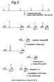
- Method (A) is schematically outlined in Figure 1 and (together with methods B and C) in Figure 5, which both show a starting DNA (generally full length -i.e. uncut- genomic DNA) containing frequent cutter ( Mse I) sites, as well as methylated and non-methylated rare cutter ( Pst I) sites.
- a starting DNA generally full length -i.e. uncut- genomic DNA
- Mse I frequent cutter
- Pst I methylated and non-methylated rare cutter
- step 1 the starting DNA is restricted using both the frequent cutter Mse I and the rare cutter Pst I, in a manner known per se. This restricts both the Mse I sites and the non-methylate d Pst I-sites, but leaves the methylated Pst I-sites intact.
- the Pst I- and Mse I-restrictions are carried out simultaneously using a mixture of these enzymes, although it is also possible to use two seperate restriction steps.
- step 2 the resticted mixture is ligated to adapters, also in a manner known per se, using a mixture of two different Mse I-adapters, i.e. a "first” and a “second” Mse I adapter, as well as a first Pst I-adapter (Said adapters are indicated as “M1", “M2” and “P1” in Figures 1-4, respectively.
- the terms "first”, “second” adapter/primer etc. are only used in this disclosure to denote/distinguish the different adapters/primers used. As such, any adapter/primer which meets the requirements set out below can be used as either the first, the second or any further primer/adapter.)
- the first and second Mse I-adapter, as well as the first Pst I adapter are essentially as described in EP-A-0 534 858, or analogous thereto, in that they are suited for use as an adapter in AFLP and that they can be ligated to the cut ends of the Mse I-fragments or Pst I fragments respectively.
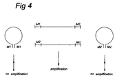
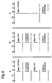
- the first and second Mse I-adapter used are further preferably such that they cannot hybridize with one another under the conditions used. This prevents the formation of DNA-loop structures, as shown in figure 4, which can hinder amplification of the fragment.
- the first and second Mse I-adapters are used about equal amounts.
- the amount of Pst I adapter (as compared to the amount of Mse I) can generally be suitably chosen by the skilled person.
- step 3 the mixture of fragments obtained in step 2 is amplified, using a mixture of primers for the first and second Mse I-adapter.
- primers again are essentially as described in EP-A-0 534 858, or analogous thereto, in that they are suited for use as a primer in AFLP and that they can be hybridize with the first and second Mse I-adapters used, respectively.
- the number of selective nucleotides required in a specific primer/for a specific application may be species-dependant.
- the amplification itself can be carried out in a manner known per se, such as described in EP-A-0 534 858 or in a manner analogous thereto, and is preferably carried out as a (pre)amplification using nonselective primers, i.e. containing no selective nucleotides at the 3'-end of the primer sequence, or at most one selective base.
- the first and second primer are used in about equal amounts.
- the fragments I-A lose their methylation imprint; the corresponding non-methylated fragments as obtained after amplification are indicated as I-A' in Fig.1.
- I-A'-fragments can now be restricted using Pst I in the subsequent restriction step 4. This restriction will not affect the fragments II-A, which originally did not contain any Pst I site.
- a methylation insensitive isoschizomer thereof that cuts the same (non-methylated) restriction site can also be used.
- a methylation insensitive isoschizomer Bst NI can be used.
- the Pst I-cut ends of the restricted fragment I-A' are ligated in step 5 to a second Pst I-adapter, the original fragment I-A providing two fragments I-B and I-C, which contain the second Pst I-adapter on one end, and either the first or second Mse I-adapter on the other.
- a second Pst I-adapter the original fragment I-A providing two fragments I-B and I-C, which contain the second Pst I-adapter on one end, and either the first or second Mse I-adapter on the other.
- the second Pst I adapter (indicated as "P2" in figures 1-4) is again essentially as described in EP-A-0 534 858, or analogous thereto, in that it is suited for use as an adapter in AFLP and that they can be ligated to the Pst I cut ends of fragments I-B and I-C.
- the second rare cutter adapter should differ from the first adapter, i.e. not be able to hybridize with the first rare cutter adapter under the conditions used.
- the second Pst I adapter is preferably the same as the third Pst I primer used in step 5 of method B below.
- fragments I-B and I-C depend upon the length of the original fragment I-A and the position of the methylated Pst I-site therein; usually fragments I-B and I-C will differ from each other in length/molecular weight, and will also differ in length/molecular weight from the non-restricted fragments II-A, so that these fragments can be distinguished using a suitable technique.
- the mixture is amplified in step 6 using suitable primer for the second Pst I-adapter, as well as at least one suitable primer for the first and/or second Mse I-adapters.
- Mse I-primer i.e. for either the first or the second Mse I-adapter
- Mse I-primer i.e. for either the first or the second Mse I-adapter
- the fragments containing a Pst I-adapter at one end, and the corresponding Mse I-adapter at the other end will be amplified exponentially.
- Other fragments present in the mixture will only be amplified linearly and/or less efficiently, or even not at all, either due to loop formation or because only one or none of the primers required for exponential amplification is available, which will reduce both the total number of bands in the final fingerprint, as well as the number of non-informative bands.
- the amplification step itself can be carried out in a manner known per se, such as described in EP-A-0 534 858 or in a manner analogous thereto, and is preferably carried out as a two-step amplification, the first of which is a (pre)amplification using a primer for the second Pst I-adapter with one selective nucleotide at the 3'-end of the primer sequence and a primer for the first (resp. second) Mse I-adapter with one selective nucleotide at the 3'-end of the primer sequence (so-called +1 primers).
- This is followed by amplification using selective primers for the second Pst I- and first (resp. second) Mse I- adapter, for instance using primers containing three selective nucleotides at the 3'-end (+3 primers).
- the respective primers can be used in about equal amounts.
- step 6 the number of selective nucleotides required in a specific primer for a specific application may be species-dependant. For instance, in maize, +2/+3 amplification and the double +3/+3 selectivity for the Pst I/ Mse I primer combination (PC) may be used.
- PC Pst I/ Mse I primer combination
- the resulting amplified fragments are then visualized using a suitable technique, such as the formation of a DNA fingerprint through gel electrophoresis.
- This fingerprint will show at least bands corresponding to fragments of types I-B and I-C, which through comparison with fingerprints generated according to method (B) or (C) or with known data can provide information on the methylaton pattern of the starting DNA, as further discussed below.
- the fingerprint can again contain other minor and/or less informative bands, which -although they can still provide some useful information on the starting DNA- are not critical for the purposes of the invention. These bands will generally not interfere with the information essential for the purposes of the invention.
- This aspect of the invention comprises:
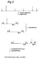
- Method (B) is schematically outlined in Figure 2, which shows a DNA fragment containing frequent cutter ( Mse I) sites (again indicated as minor arrows in the sequence), as well as methylated (large arrow with cross) and non-methylated (large arrow) rare cutter ( Pst I) sites.
- the third and fourth frequent cutter adapter and the second and third rare cutter adapter are again essentially as described in EP-A-0 534 858, or analogous thereto, in that they are suited for use as an adapter in AFLP and that they can be ligated to the cut ends of the frequent cutter and rare cutter fragments, respectively.
- the two frequent cutter adapters are preferably again such that they cannot hybridize with one another under the conditions used, in order to prevent undesired loop formation.
- the third and fourth cutter adapters can differ in sequence and/or number of nucleotides from the first and second frequent cutter adapter used in method (A) above, or method (C) below.
- the third and fourth adapter for the frequent cutter restriction enzyme are the same as the first and second frequent cutter adapter used above, respectively. This also allows an even more direct comparison of the fingerprints obtained, thus making the entire method even more reliable.
- the third rare cutter adapter can differ in both sequence and number of nucleotides from the first and second rare cutter adapter used in method (A), preferably the third frequent cutter adapter used in method (B) is the same as the second rare cutter adapter used in step 5/6 of method (A) above.
- the third and fourth frequent cutter adapter and the third rare cutter adapter will be denoted hereinbelow as the "first and second" Mse I primers and the "second" Pst I-primer.
- step 1 the starting DNA is restricted using the frequent cutter Mse I, in a manner known per se. This restricts the Mse I-sites but leaves both the methylated and non-methylated Pst I-sites intact.
- step 2 the restricted mixture is ligated to adapters, also in a manner known per se, using a mixture of the first and second Mse I-adapters.
- fragments containing the first and the second frequent cutter adapter shown in figure 2 for the purposes of the present method, three different types of fragments can be distinguished, i.e. one type containing the methylated Pst I-site(s) (indicated as V-A in Fig.2), a second type containing the originally non-methylated Pst I-site(s) (indicated as VI-A in Fig.2), and a third type containing no Pst I-sites (indicated as VII in Fig.2).
- step 3 the mixture of fragments obtained in step 2 is amplified, using a mixture of primers for the first and second Mse I-adapter.
- primers again are essentially as described in EP-A-0 534 858, or analogous thereto, in that they are suited for use as a primer in AFLP and that they can hybridize with the first and second Mse I-adapters used, respectively.
- the number of selective nucleotides required in a specific primer/for a specific application may be species-dependant.
- the primers used in conjunction with each of the respective adapters are preferably essentially the same for each of the adapters used, in that they have the same sequence in the region for hybridization with the adapter, and may only differ in the presence of the selective bases at their 3'-end, i.e. the number thereof and/or the specific nucleotides.
- This enables the same primers to be used, or the primers to be synthesised in conjuction wit each other and/or starting from the corresponding non-selective primer sequence, so that a limited number of starting materials (primers) are required for carrying out the three methods of the invention.
- the amplification itself can be carried out in a manner known per se, such as described in EP-A-0 534 858 or in a manner analogous thereto, and is preferably carried out as a (pre)amplification using nonselective primers, i.e. containing no selective nucleotides at the 3'-end of the primer sequence, or at most one selective base.
- the first and second primer are used in about equal amounts.
- V-A lose their methylation imprint during amplification; the corresponding non-methylated fragments as obtained after amplification are indicated as V-A' in Fig.2.
- V-A'-fragments can now be restricted using Pst I in the subsequent restriction step 4, together with the amplified fragments VI-A containing the originally non-methylated Pst I-sites.
- a methylation insensitive isoschizomer thereof that cuts the same (non-methylated) restriction site can also be used.
- the Pst I-cut ends of the restricted fragments are ligated to the third Pst I-adapter, again providing a mixture of different adapter-containing restriction fragments, as shown in Fig.2.
- the third Pst I-adapter is again essentially as described in EP-A-0 534 858, or analogous thereto, in that it is suited for use as an adapter in AFLP and that it can be ligated to the cut ends of the Pst I-fragments.
- the third Pst I adapter is preferably the same as the second Pst I primer used in step 5 of method A above, and is so indicated below.
- the fragment(s) V-A/V-A' will essentially provide two restriction fragments (V-B and V-C), each containing the second Pst I-adapter at one end, and either the first or second Mse I-adapter on the other end. These fragments will differ in length dependent on the position of the methylated Pst I-site in the original Mse I fragment V-A. [Again, as with fragments I-A and I-B/I-C in method (A) above, it should be understood that for each the fragments V-B and V-C shown in figure 2 (i.e.
- fragment(s) VI-A which originally contained the non-methylated Pst I-site, also two different fragments (VI-B and VI-C) are obtained, with different lengths depending on the position of the non-methylated Pst I-site in the origina l Mse I-fragment VI-A.
- the mixture obtained after restriction with Pst I and ligation of the Pst I-adapter will further contain the Mse I fragments VII that originally did not contain either a methylated or non-methylated Pst I-site.
- fragments V-B, V-C, VI-A, VI-B, as well as the fragments VII will have differing lengths/molecular weights, so that they can be distinguished using a suitable technique.
- the mixture is amplified in step 6 using suitable primer for the second Pst I-adapter, as well as at least one suitable primer for the first and/or second Mse I-adapters.
- suitable primers are again essentially as described in EP-A-0 534 858, or analogous thereto, in that they are suited for use as a primer in AFLP and that they can be hybridize with the second Pst I-adapter, and the first and/or second Mse I-adapter, respectively.
- selective primers are used.
- these primers are preferably the same as the corresponding primers used in step 6 of method A above. Also, for the reasons given in method A, most preferably only one Mse I-primer (i.e. for either the first or the second Mse I-adapter) is used, besides the primer for the first Pst I-adapter. Also, it should be noted that, as no primers for the second Pst I-adapter are used in step 6, the Pst I/ Mse I-fragments VI generated in the first restriction step of method B will again only be amplified linearly.
- the amplification step itself can be carried out in a manner known per se, such as described in EP-A-0 534 858 or in a manner analogous thereto, and is preferably carried out as the two-step amplification of step 6 of method A above, i.e. as a +1/+1 (pre)amplification ( Pst I+1/ Mse I+1), followed by a +2/+3 or +3/+3 amplification ( Pst I+2/ Mse I+3 or ( Pst I+3/ Mse I+3).
- the respective primers can be used in about equal amounts.
- the number of selective nucleotides required in a specific primer for a specific application may be species-dependant. For instance, in maize, +2/+3 amplification and the double +2/+3 selectivity for the Pst I/ Mse I primer combination (PC) may be used.
- the resulting amplified fragments are then visualized using a suitable technique, such as the formation of a DNA fingerprint through gel electrophoresis.
- This fingerprint will show at least bands corresponding to fragments of types V-B, V-C, VI-B and VI-C, which though comparison with fingerprints generated according to method (A) or (C) or with known data can provide information on the methylaton pattern of the starting DNA, as discussed below.
- the fingerprint can also contain other minor and/or less informative bands, which -although they can still provide some useful information on the starting DNA- are not critical for the purposes of the invention. These bands will generally not interfere with the information essential for the purposes of the invention.
- Method (C) comprises:
- Method (C) is schematically outlined in Figure 3, which shows a DNA fragment containing frequent cutter ( Mse I) sites (again indicated as minor arrows in the sequence), as well as methylated (large arrow with cross) and non-methylated (large arrow) rare cutter ( Pst I) sites.
- the fifth frequent cutter and the fourth rare cutter adapters are again essentially as described in EP-A-0 534 858, or analogous thereto, in that they are suited for use as an adapter in AFLP and that they can be ligated to the cut ends of the restriction fragments (the frequent cutter and rare cutter fragments, respectively).
- the fifth adapter for the frequent cutter restriction enzyme is preferably the same as the first or the second -preferably the first- frequent cutter adapter used above
- the fourth rare cutter adapters is the same as the first or second - preferably the second- rare cutter adapter used above.
- the fifth frueqent cutter adapter and the fourth rare cutter adapter will be denoted hereinbelow as the "first" Mse I adapter and the "second" Pst I adapter.
- primers used in conjunction with each of the respective adapters are again preferably essentially the same as the corresponding primers used in the methods (A) and (B) above.
- step 1 the starting DNA is restricted using both the frequent cutter Mse I and the rare cutter Pst I, in a manner known per se. This restricts both the Mse I sites and the non-methylated Pst I-sites, but leaves the methylated Pst I-sites intact.
- Pst I- and Mse I-restrictions are preferably carried out simultaneously using a mixture of these enzymes, although it is also possible to use two seperate steps.
- step 2 the resticted mixture is ligated to Mse I- and Pst I-adapters, also in a manner known per se.
- Mse I- and Pst I-adapters also in a manner known per se.
- only one Mse I-adapter is used (i.e. either the first or the second), instead of a mixture of two different Mse I-adapters.
- All these fragments may or may not contain the non-restricted methylated Pst I-sites (only shown for the Mse I/ Mse I-fragments, as VII-A and VII-A' respectively); as this method is only directed to detecting the non-methylated Pst I-sites, these sites are not relevant, nor do they interfere with the resulting fingerprint.
- fragments VII, VIII and IX will depend on the positions of the Mse I-sites and the non-methylated Pst I-sites in the starting DNA (but not the methylated Pst I-sites).
- fragments VII, VIII and IX will differ in length/molecular weight, with the Pst I/ Pst I fragments being appreciably longer than the other fragments due to the relative abundance of the Mse I-sites. Because of their differing lengths/molecular weight, these fragments can distinguished using a suitable technique.
- the mixture is amplified in step 3 using suitable primer for the second Pst I-adapter, as well a suitable primer for the first Mse I-adapters.
- these primers are preferably the same as the corresponding primers used in step 6 of method A and/or B above.
- the amplification step itself can be carried out in a manner known per se, such as described in EP-A-0 534 858 or in a manner analogous thereto, and is preferably carried out as the two-step amplification of step 6 of method A above, i.e. as a (pre)amplification using a +1-primers for the second Pst I-adapter and the first Mse I-adapter, followed by selective amplification using +3-primers.
- the respective primers can be used in about equal amounts.
- the number of selective nucleotides required in a specific primer for a specific application may again be species-dependant. For instance, in maize, +2/+3 amplification and the double +3/+3 selectivity for the Pst I/ Mse I primer combination (PC) may be used.
- the Mse I/ Mse I-fragments VII-A and VII-A' i.e. with or without a methylated Pst I-site
- any Pst I/ Pst I-fragments will generally not be amplified efficiently, as they are to long to be amplified for their full length during the cycle times used.
- the resulting amplified fragments are then visualized using a suitable technique, such as the formation of a DNA fingerprint through gel electrophoresis.
- This fingerprint will show at least bands corresponding to fragments of types VIII-A and and VIII-B, which can be used as a comparison for the fingerprints generated according to method (A) or (B), as discussed below.
- the differing fragments, essential for determining the methylation pattern of the starting DNA, that are generated, amplified and detected in each of the methods (A), (B) and (C), are schematically indicated in Figure 5, which shows a DNA fragment containing frequent cutter ( Mse I) sites, as well as methylated and non-methylated rare cutter ( Pst I) sites.
- method A will provide fragments, and bands in the DNA-fingerprint, corresponding to the methylated (I-B and I-C) sites, but not to the non-methylated (III-A and III-A') sites.
- Method B will provide fragments, and bands in the DNA-fingerprint, corresponding to the methylated (V-B and V-C) and non-methylated (VI-B and VI-C) sites.
- Method C will provide fragments, and bands in the DNA-fingerprint, corresponding to the non-methylated (VIII-B and VIII-C) sites, but not the methylated (VII-A) sites.
- the respective correseponding fragments of interest i.e. I-B/I-C and V-B/V-C on the one hand, and VI-B/VI-C and VIII-A and VIII-B on the other
- the respective correseponding fragments of interest will have the same size/molecular weight, and therefore give corresponding bands in the fingerprint, if the respective electrophoreses are run in parallel lanes of the same gel.
- comparing the fingerprints generated in method (B) to the fingerprint generated in method (A) will provide information on the originally non-methylated sites, which will occur as bands in fingerprint (B) not present in fingerprint (A) (i.e. bands V-A and V-B). Also, these bands will be present in fingerprint (C) (i.e. bands VIII-A and VIII-B).
- Information on the originally methylated sites can be derived from comparing either fingerprint (A), fingerprint (B), and preferably both, to fingerprint (C): these will occur as bands present in (A) and (B) (i.e. bands I-B/I-C and VI-B/VI-C), but not in (C).
- further information can de derived on the position of the methylated and non-methylated sites in the genome, for instance on the relative position of the methylated site to known AFLP-markers or other genetic markers in the starting DNA.
- methylation sensitive rare cutter used, information can be obtained on the methylated sequence; the use of different methylation sensitive rare cutters providing information on the state of methylation of different sites/sequences in the genome.
- the method of the invention can be used as a general tool for detecting DNA-methylation or methylation patterns, both in prokaryotes and eukaryotes, including viruses, yeasts, fungi, bacteria, plants, animals and humans. As such, it can be used to replace known techniques for determining and/or estimating the extend of DNA-methylation, in all applications in which such information is of interest, such as those discussed above.
- the method of the invention is particularly suited for applications in which a speedy, qualitative determination of the state of methylation or the methylation pattern, that can be applied to large scale testing, is required. Generally, these will be applications similar to those in which conventional AFLP-techniques are also the technique of choice in determining genetic make-up or RFLP's.
- methylation AFLPTM The Examples below describe the novel PCR-based method of the invention for detecting methylation of restriction sites randomly over the genome. Said method will hereinbelow be referred to as "methylation AFLPTM”.
- the technique is based on (i) the use of a pair of restriction enzymes consisting of a methylation-sensitive rare cutter and a methylation-insensitive frequent cutter, (ii) the comparison of fragments obtained from native and PCR amplified DNA, and (iii) selective amplification of genomic restriction fragments using PCR.
- the power of methylation AFLP resides in its positive display of the unmethylated and the native methylated sites jointly and seperately, as is obtained after selective amplification of the restriction fragments by PCR.
- the technique is based on selective amplification of genomic restriction fragments obtained directly from native DNA and also from nonselective amplified DNA by using a methylation-sensitive rare cutter and a methylation-insensitive frequent cutter.
- Nonselective amplification of restriction fragments is achieved by using only the adapter and restriction site sequence as target sites for primer annealing.
- Selective amplification is achieved by the use of primers that extend into the restriction fragments, amplifying only those fragments with nucleotides flanking the restriction sites that match the primer extensions.
- methylation AFLP The advantages of methylation AFLP are manifold: no prior sequence knowledge is needed, a limited set of generic primers is used, a high multiplex ratio (said ratio being a function of the selected specific primer sets) is obtained and a positive display of the unmethylated and native methylated sites is provided. Also, the detection of DNA-methylation is genome-wide. Typically 50-100 restriction fragments are co-amplified and resolved on denaturing polyacrylamide gels. The Examples below will further illustrate how methylation AFLP can be used to estimate the extent of CpG and CpNpG methylation, to detect epialleles and additional sequence polymorphism, and to follow the inheritance of C-methylation.
- the first category can be used to analyse the different types of modified bases and to quantify them, but do not provide any information about the precise location of the modified site within a given nucleic acid sequence.
- immunological, chromatographic, electrophoretic and spectrophotometric procedures that follow a complete chemical or enzymatic hydrolysis of the target DNA.
- Another sequence-independent approach involves the use of methylation-sensitive restriction endonucleases where genomic DNA digests obtained with these restriction enzymes are compared using gel electrophoresis ( Saluz, H.P. and Jost, J-P. (1993) Jost, P.
- the second category enables analysis of the precise location of methylated bases within a known DNA sequence.
- fall techniques based on the use of pairs of isoschizomeric restriction enzymes that differ in sensitivity to methylation, in combination with Southern-blot analysis ( Bird, A.P. and Southern, E.M. (1978), J. Mol. Biol. 118 , 27-47; Waalwijk, C. and Flavell, R.A. (1978) Nucl. Acids Res. 5, 3231-3236) or PCR.
- PCR-based methods for detecting DNA-methylation have also been reported; one of these PCR-based methods takes advantage of the fact that PCR amplification occurs only if the DNA between the two primer sites remains uncleaved by the methylation-sensitive restriction enzyme Hpa II (Singer-Sam, J., LeBon, J.M., Tanguay, R.L. and Riggs A.D. (1990) Nucl. Acids Res. 18 , 687; Singer-Sam, J., Grant, M., LeBon, J.M., Okuyama, K., Chapman, V., Monk, M. and Riggs, A.D. (1990) Mol. Cell. Biol.
- PCR-based methods combine PCR with genomic sequencing to identify methylated cytosine residues (Maxam, A.M. and Gilbert, W. (1980) Methods Enzymol . 65, 499-560; Pfeiffer, G.P., Steigerwald, S.D., Mueller, P.R., Wold, B. and Riggs A.D. (1989) Science 246, 810-813), utilizing the Maxam and Gilbert chemical cleavage reactions carried out on genomic DNA with various additional procedures to enhance the signal from the sequence under investigation.
- MSP methylation-specific PCR
- restriction enzyme digestion of PCR products amplified from bisulfite-converted DNA Sadri, R. and Hornsby, P.J. (1996) Nucleic Acids Res. 24, 5058-5059; Xiong, Z. and Laird, P.W. (1997) Nucleic Acids Res . 25, 2532-2534
- bisulfite treatment of DNA followed by single nucleotide primer extension Gonzalgo, M.L. and Jones, P.A. (1997 ) Nucleic Acids Res. 25, 2529-2531.
- FIG. 6 gives a schematic representation of the methylation AFLP technique for the enzyme combination (EC)
- Pst I/ Mse I 'nonselective amplification' stands for the nonselective Mse I/ Mse I + amplification
- 'selective amplification' stands for the selective Pst I/ Mse I amplification.
- subset A or “A-templates”
- A-templates a subset of templates in accordance with the general method "A” above, providing restriction fragments with a native methylated Pst I site, followed by the selective Pst I/ Mse I amplification procedure.
- the middle column shows the generation of a subset of templates (hereinbelow: “subset B” or “B-templates”) in accordance with the general method "B” above, providing restriction fragments with a native methylated or unmethylated Pst I site, followed by the selective Pst I/ Mse I amplification procedure.
- subset B or “B-templates”
- subset C or “C-templates”
- C subset of templates
- the methylation AFLP technique of the invention is based on the amplification by PCR of three derived subsets (A, B, C) of genomic restriction fragments, differing in (i) digestion of the genomic DNA using two restriction enzymes and (ii) ligated double stranded (ds) adapters.
- Subset A representing only the restriction fragments with a native methylated Pst I cutter site, is obtained in the following way (vide Figure 6): the genomic DNA is digested to completion with the two restriction enzymes Pst I and Mse I, and the corresponding ds (AFLP) Pst I-adapter* and MseI-adapters (+) are ligated to the unmethylated Pst I sites and Mse I sites to block the unmethylated Pst I ends and to generate template DNA for the nonselective Mse I/ Mse I + amplification.
- AFLP ds
- Mse I-adapters Two different Mse I-adapters are chosen to avoid stem-loop structure forming of the Mse I- Mse I fragments, which have an inverted repeat at the ends. This is followed by the Mse I/ Mse I + nonselective amplification of the genomic restriction fragments that may carry an internal methylated Pst I site. During this PCR-step, restriction sites lose their methylation imprint. A second digestion of the 'demethylated' Pst I sites followed by ligation of the Pst I adapter generates template DNA for further selective Pst I/ Mse I amplification.
- the subset B-templates representing the restriction fragments with native methylated or unmethylated Pst I sites, are obtained in a similar way as the subset A-templates, with the exception that the genomic DNA is digested to completion with only Mse I, and corresponding ds (AFLP) MseI-adapters (+) are ligated to generate template DNA for the nonselective Mse I/ Mse I + amplification (see Figure 6).
- the subset C-templates, representing only the restriction fragments with a native unmethylated Pst I site are obtained according to the published AFLP procedure (Vos, P., Hogers, R., Bleeker, M., Reijans, M., van de Lee, T., Hornes, M., Frijters, A., Pot, J., Peleman, J., Kuiper, M. and Zabeau, M. (1995) Nucl. Acids Res.
- the A, B and C subsets of templates are simultaneously selectively amplified following the two-step amplification procedure strategy (pre-amplification followed by the AFLP reaction), according to the AFLP fingerprinting method of complex genomes (Vos, P., Hogers, R., Bleeker, M., Reijans, M., van de Lee, T., Hornes, M., Frijters, A., Pot, J., Peleman, J., Kuiper, M. and Zabeau, M. (1995) Nucl. Acids Res. 23, 4407-4414.) and then displayed together in adjacent lanes A, B and C of a methylation AFLP fingerprint.
- Vos, P. Hogers, R., Bleeker, M., Reijans, M., van de Lee, T., Hornes, M., Frijters, A., Pot, J., Peleman, J., Kuiper, M. and Zabeau, M. (1995) Nuc
- Tomato DNA Near Isogenic Lines (NILs) 83M-S and 83M-R obtained from De Ruiter zonen, The Netherlands; cv. Motelle and Mobox obtained from INRA, Montfavet, France; inbred line RC10 obtained from Enza Zaden, The Netherlands; inbred line 52201 obtained from Rijk Zwaan, The Netherlands), maize DNA (inbred lines B73, Mo17 and A7) was obtained from Dr. M.
- NILs Near Isogenic Lines
- RC10 obtained from Enza Zaden, The Netherlands
- inbred line 52201 obtained from Rijk Zwaan, The Netherlands
- maize DNA inbred lines B73, Mo17 and A7 was obtained from Dr. M.
- T4 DNA ligase and T4 polynucleotide kinase were also obtained from Pharmacia. All PCR reagents and consumables were obtained from Perkin Elmer Corp. (Norwalk, CT, USA). Radioactive reagents were purchased from Amersham (Amersham International plc, Little Chalfont, Buckinghamshire, UK) or Isotopchim (Isotopchim SA, Ganagobie, France).
- oligonucleotides were made on a Biotronic Synostat D DNA-synthesizer (Eppendorf GmbH, Maintal, Germany) or Milligen Expedite 8909 DNA-synthesizer (Millipore Corp. Bedford, MA, USA). The quality of the crude oligonucleotides was checked by end-labeling with polynucleotide kinase and [ ⁇ - 33 P]ATP and subsequent electrophoresis on 18% denaturing polyacrylamide gels (Maxam, A.M. and Gilbert, W. (1980) Methods Enzymol. 65, 499-560.).
- Oligonucleotide adapters and primers for AFLPTM analysis were generally used without further purification when they proved to be >85% full length.
- AFLP adapter For the rare cutter site two different AFLP adapters were used: 1) the conventional AFLP adapter (called AFLP adapter) as target site for primer annealing and 2) an AFLP adapter serving as blocking agent (called e.g. Pst I-adapter*). Both adapters consist of a core sequence (CORE) and a site-specific sequence (SITE) (Sadri, R. and Hornsby, P.J. (1996) Nucleic Acids Res. 24, 5058-5059). The blocking adapters differ from the AFLP adapters only in core sequence. This is illustrated below for Pst I and Hpa II-adapters. The conventional and blocking adapter for Msp I and Cla I are identical to those for Hpa II.
- Mse I-adapter + a Mse I-adapter only for nonselective amplification
- Mse I-adapter + a Mse I-adapter
- Mse I-adapter + a Mse I-adapter
- the Mse I-adapters differ only in core sequence. This is illustrated below: AFLP primers consist of three parts, a core sequence, a site-specific sequence (SITE) and a selective extension (EXT) (vide EP-A-0534858).
- AFLP-primers for Msp I and Hpa II have a similar architecture. However, it should be noted that the AFLP-primers for Pst I, Hpa II, Msp I and Cla I are designed only for the AFLP adapters.
- the two Mse I-primers are distinguished in the same way as the two Mse I-adapters: the MseI-primer has the MseI-adapter as annealing target site, while MseI-primer + has the MseI-adapter + as annealing target site.
- the difference between the two Mse I-primers is shown below:
- Protocol C is equivalent to the standard AFLP protocol as described in EP-A-0 534 858 and by Vos et al (Vos, P., Hogers, R., Bleeker, M., Reijans, M., van de Lee, T., Hornes, M., Frijters, A., Pot, J., Peleman, J., Kuiper, M. and Zabeau, M. (1995) Nucl. Acids Res. 23, 4407-4414).
- Genomic DNA (0.5 ⁇ g) is incubated for 1 h at 37°C with 5 U Pst I and 5 U Mse I in 40 ⁇ l 10mM Tris-HAc pH 7.5, 10 mM MgAc, 50 mM KAc, 5 mM DTT, 50 ng/ ⁇ l BSA (acetylated).
- Genomic DNA (0.5 ⁇ g) is incubated for 1 h at 37°C with only 5 U Mse I in 40 ⁇ l 10mM Tris-HAc pH 7.5, 10 mM MgAc, 50 mM KAc, 5 mM DTT, 50 ng/ ⁇ l BSA (acetylated).
- Genomic DNA (0.5 ⁇ g) is incubated for 1 h at 37°C with 5 U Pst I and 5 U Mse I in 40 ⁇ l 10mM Tris-HAc pH 7.5, 10 mM MgAc, 50 mM KAc, 5 mM DTT, 50 ng/ ⁇ l BSA (acetylated).
- Synthesis of unmethylated A- and B-templates is performed by nonselective PCR amplification.
- This nonselective PCR amplification is performed with the following cycle profile for 7 cycles: a 30 s DNA denaturation step at 94°C, a 1 min annealing step at 56°C and a 2 min extension step at 72°C.
- Amplifications are performed in 20 ⁇ l containing 5 ⁇ l template-DNA, 30 ng Mse I-primer, 30 ng Mse I-primer + , 0.4 U Taq polymerase, 10 mM Tris-HCl pH 8.3, 1.5 mM MgCl 2 , 50 mM Kcl and 0.2 mM of all four dNTPs.
- reaction mixtures A and B are incubated again for 1 h at 37°C with 5 U Pst I in 40 ⁇ l 10mM Tris-HAc pH 7.5, 10 mM MgAc, 50 mM KAc, 5 mM DTT, 50 ng/ ⁇ l BSA(acetylated).
- reaction mixture is diluted to 1000 ⁇ l with 10 mM Tris-HCl, 0.1 mM EDTA pH 8.0, and used for PCR amplification or stored at - 20°C.
- A- and B-templates for AFLP reactions using methylation-sensitive rare cutters leaving a 5'-extension involves inactivation of the remaining Taq polymerase after the nonselective amplification, to avoid incorporation of remaining dNTPs after restriction. This is achieved by adding 220 ng TaqStartTM Antibody/ U Taq polymerase to the amplification mixture, prior to further restriction and ligation.
- Amplification reaction conditions are described using DNA templates (A, B and C) for the EC Pst I/ Mse I. With other ECs, the procedure is identical except for the use of appropiate primers.
- AFLP fingerprinting of large genomes generally involves an amplification in two steps.
- the first step of this amplification procedure employes two AFLP primers, one aimed at the Pst I-ends and one at the Mse I-ends, each having a single selective nucleotide. These primers are not radioactively labelled.
- Amplifications are performed in 20 ⁇ l containing 5 ⁇ l template-DNA, 30 ng Mse I-primer, 30 ng Pst I-primer, 0.4 U Taq polymerase, 10 mM Tris-HCl pH 8.3, 1.5 mM MgCl 2 , 50 mM KCl and 0.2 mM of all four dNTPs.
- the second amplification reaction again employes two oligonucleotide primers, one aimed at the Pst I-ends and one at the Mse I-ends, each having two or three selective nucleotides.
- the Pst I-primer is radioactively end-labeled using [ ⁇ - 33 P] ATP and T4 polynucleotide kinase.
- the labelling reactions are performed in 50 ⁇ l 25 mM Tris-HCl pH 7.5, 10 mM MgCl 2 , 5mM DTT, 0.5mM spermidine-3HCl using 500 ng oligonucleotide primer, 100 ⁇ Ci [ ⁇ - 33 P] ATP (1000-3000Ci/mol) and 10 U T4 polynucleotide kinase.
- Amplifications are performed in 20 ⁇ l containing 5 ⁇ l template-DNA, 5 ng labeled Pst I-primer (0.5 ⁇ l from the labelling reaction mixture), 30 ng Mse I-primer, 0.4 U Taq polymerase, 10 mM Tris-HCl pH 8.3, 1.5 mM MgCl 2 , 50mM KCl and 0.2 mM of all four dNTPs.
- AFLP preamplification reactions are performed for 24 cycles (protocol A and B) and 20 cycles (protocol C) with the following cycle profile: a 30 s DNA denaturation step at 94 °C, a 1 min annealing step at 56°C and a 1 min extension step at 72°C.
- AFLP reactions with primers having two or three selective nucleotides are performed for 36 cycles with the following cyle profile: a 30 s DNA denaturation step at 94°C, a 30 s annealing step (see below) and a 1 min extension step at 72°C.
- the annealing temperature in the first cycle is 65 °C, and is subsequently reduced each cycle by 0.7 °C for the next 12 cycles, then continued at 56 °C for the remaining 23 cycles. All amplification reactions are performed in a PE-9600 thermocycler ( Perkin Elmer Corp. Norwalk, CT, USA).
- reaction products are mixed with an equal volume (20 ⁇ l) of formamide dye (98% formamide, 10 mM EDTA pH 8.0, and bromophenol blue and xylene cyanol as tracking dyes).
- formamide dye 98% formamide, 10 mM EDTA pH 8.0, and bromophenol blue and xylene cyanol as tracking dyes.
- the resulting mixture is heated for 3 min at 90 °C, then quickly cooled on ice.
- Each sample (2 ⁇ l) was loaded on a 5% denaturing (sequencing) polyacrylamide gel (Maxam, A.M. and Gilbert, W. (1980) Methods Enzymol. 65, 499-560).
- the gel matrix is prepared using 5% acrylamide, 0.25% methylene bisacryl, 7.5 M urea in 50 mM Tris/50 mM Boric acid/1mM EDTA (pH 8.3).
- Example II Methylation AFLP fingerprinting of large genomes.
- Figures 7A and 7B show methylation AFLP fingerprints of genomic DNAs from the three maize inbred lines A7, B73 and Mo17.
- the two panels show Pst I/ Mse I fingerprints, corresponding with the following PCs (from left to right): I. Pst I+AGW/ Mse I+CTT, and II. Pst I+AGS/ Mse I+CTT .
- Lane A, B and C represent the corresponding A, B and C restriction fragments, referring to the native methylated state of the rare cutter sites.
- the molecular weight size range of the fingerprints is 200-500 nucleotides.
- every band present in lane A or lane C should also be present in lane B, because lane B represents both the native methylated and unmethylated rare cutter sites jointly.
- lane B represents both the native methylated and unmethylated rare cutter sites jointly.
- PCs I this is true for more than 90% of the fragments in lane A and C.
- the percentage of fragments in lane A and C present in lane B can be low.
- short Mse I- Mse I restriction fragments have a competitive advantage over the longer Mse I- Mse I restriction fragments in the nonselective amplification.
- Example III Estimating the extent of 5m CpNpG as presented in Pst I-sites of some large plant genomes.
- the modified C at position 5 is not only confined to CpG dinucleotides as is in animals, but also occurs at a variety of other cytosine containing dinucleotides, all of which are part of the basic trinucleotide CpNpG where N can be any nucleotide (Gruenbaum, Y., Naveh-Maney, T., Cedar, H.
- the restriction enzyme Pst I having 5'-CTGCAG-3' as recognition site, containing two CpNpG trinucleotides, is sensitive to methylation at the 5'-C and the 3'-C (McClelland, M., Nelson, M. and Raschke, E. (1994) Nucl. Acids Res. 22, 3640-3659). Whether simultaneous methylation of both C's of the Pst I recognition site is possible, is not clear. A mean percentage ⁇ standard error of methylate d Pst I-sites in the nuclear DNA of tomato, maize and oilseed rape, are given in Table 1.
- Example IV Estimating the extent of 5m CpG as presented in Hpa II-, Msp I- and Cla I-sites in some large plant genomes.
- Msp I and Hpa II (methylation isoschizomers) have the same recognition site 5'-CCGG-3', containing one CpG dinucleotide.
- Msp I is sensitive to 5'- 5m C
- Hpa II is sensitive to methylation at position 5 of either C (McClelland, M., Nelson, M. and Raschke, E. (1994) Nucl. Acids Res. 22 , 3640-3659). Therefore, Msp I and Hpa II are appropriate for estimating the extent of 5m CpG.
- the extent of 5m CpG methylation by using Hpa II and Msp I as methylation-sensitive rare cutters is measured as the difference in the number of bands counted in a Msp I and a Hpa II fingerprint in lane C; the difference in the number of bands counted in a Msp I and a Hpa II fingerprint in lane A must give the same result.
- the percentages of 5m CpG sites in the nuclear DNA of tomato and maize, as presented in Hpa II- and Msp I-sites, are measured only for one genotype/species, and are given in Table 2.
- Percentages of 5m CpG in the nuclear DNA of tomato, maize and oilseed rape, as presented in Hpa II-, Msp I- and Cla I-sites. n total number of restriction fragments counted. Species 5m CpG as presented in Cla I-sites n 5m CpG as presented in Hpa II/ Msp I-sites n tomato (cv.52201) 57.9 2833 57.6 2169 maize (B73) 53.9 2694 39.6 1791 oilseed rape (T528) 48.4 2973
- Example V 5m C polymorphism and its inheritance.
- AFLP primarily detects variation of the primary DNA sequence, either base substitutions or DNA rearrangements. Nucleotides modified by methylation are not considered to be a part of the primary nucleotide sequence of an individual. This nucleotide modification, resulting from a post-replicative event at defined but not all target sequences (i.e. CpG and CpNpG), represents two additional forms of DNA polymorphism: (i) polymorphism reflecting the variation in the primary nucleotide sequence of the methylated restriction site and/or variation in the restriction size ( m AFLP markers), and (ii) allele-specific methylation ( asm AFLP markers).
- FIG. 8A shows the segregation of a number of m AFLP markers in a Recombinant Inbred (RI) population derived from the cross B73 ⁇ Mo17. The band intensities segregate into two distinct classes, homozygous absent and homozygous present, approximating the Mendelian 1:1 segregation.
- RI Recombinant Inbred
- FIGS. 8A and 8B show the segregation of a pair of asm AFLP markers in the RI population. The band intensities segregate into two distinct classes, homozygous absent and homozygous present.
- m AFLP markers account for 44,6%, which is in good accordance with the 50.78% methylated Pst I-sites in the maize genome (Table 1), while hardly 1% of the marker alleles behave like epialleles.
- the method of invention can be considered a DNA fingerprinting technique that detects genomic restriction fragments and resembles in that respect the AFLP technique, with the major difference that methylation AFLP displays native methylated sites too, but in general only if they are located in the recognition site of the obligate methylation-sensitive rare cutter.
- the multiplex ratio is high (50-100 restriction fragments) and is a function of (i) the cleavage frequency of the methylation-sensitive rare cutter enzyme and (ii) the number and nature of the selective bases of the specific primer set.
- the method of the invention can be used for estimating the extent of 5m CpNpG, resp. 5m CpG, as presented in Pst I-, resp. Cla I-, Hpa I- and Msp I-sites in the genome of few crops.
- Methylation AFLP can also be used with advantage to estimate the low prevalence of these minor nucleotide modifications presented by recognition sites of methylation-sensitive rare cutters, and to shed a light on their distributions over the genome.
- Methylation AFLP allows the exploitation of two additional forms of DNA polymorphism: (i) polymorphism reflecting variation in the primary nucleotide sequence of the methylated restriction site and/or variation in the restriction fragment size, and (ii) allele-specific methylation. Hardly 1% of the marker alleles behave like epialleles.
- AFLP markers Like AFLP markers, most m AFLP markers correspond to unique positions in the genome, and, hence, can be exploited as landmarks in and as bridging tools between genetic and physical maps ( Vos, P., Hogers, R., Bleeker, M., Reijans, M., van de Lee, T., Hornes, M., Frijters, A., Pot, J., Peleman, J., Kuiper, M. and Zabeau, M. (1995) Nucl. Acids Res. 23, 4407-4414). Therefore, methylation AFLP may be useful in genomic research.
- Native methylated sites are present on cloned DNA segments, e.g., yeast artificial chromosomes (YACs) as unmethylated sites. Hence, native methylated sites can not be distinguished from native unmethylated sites on a physical map. However, ligning up the physical map with a genetic map containing m AFLP markers, helps to identify native methylated sites on the physical map.
- yeast artificial chromosomes e.g., yeast artificial chromosomes
- methylation AFLP is an attractive technique to determine the DNA methylation levels at specific gene loci like tumor-suppressor genes and to trace imprinted genes. Note: AFLPTM is a trademark of Keygene N.V.
Landscapes
- Chemical & Material Sciences (AREA)
- Organic Chemistry (AREA)
- Life Sciences & Earth Sciences (AREA)
- Zoology (AREA)
- Wood Science & Technology (AREA)
- Proteomics, Peptides & Aminoacids (AREA)
- Health & Medical Sciences (AREA)
- Engineering & Computer Science (AREA)
- Microbiology (AREA)
- Immunology (AREA)
- Physics & Mathematics (AREA)
- Molecular Biology (AREA)
- Biotechnology (AREA)
- Biophysics (AREA)
- Analytical Chemistry (AREA)
- Biochemistry (AREA)
- Bioinformatics & Cheminformatics (AREA)
- General Engineering & Computer Science (AREA)
- General Health & Medical Sciences (AREA)
- Genetics & Genomics (AREA)
- Measuring Or Testing Involving Enzymes Or Micro-Organisms (AREA)
Abstract
and optionally comprising
and optionally further comprising
Description
- The present invention relates to a method for detecting DNA methylation using AFLP™. In particular, this method can be used to distinguish between methylated and non-methylated sites (nucleotides) in a nucleic acid, more particular between methylated and non-methylated restriction sites. Thus, the method of the invention can provide information on the methylation pattern of the DNA, which can be visualised as a DNA-fingerprint.
- Selective restriction fragment amplification or AFLP is known, for instance from the European patent application 0 534 858 by applicant, incorporated herein by reference. In general, AFLP comprises the steps of:
- (a) digesting a nucleic acid, in particular a DNA, with one or more specific restriction endonucleases, to fragment said DNA into a corresponding series of restriction fragments;
- (b) ligating the restriction fragments thus obtained with at least one double-stranded synthetic oligonucleotide adapter, one end of which is compatible with one or both of the ends of the restriction fragments, to thereby produce tagged restriction fragments of the starting DNA;
- (c) contacting said tagged restriction fragments under hybridizing conditions with at least one oligonucleotide primer;
- (d) amplifying said tagged restriction fragments hybridized with said primers by PCR or a similar technique so as to cause further elongation of the hybridized primers along the restriction fragments of the starting DNA to which said primers hybridized; and
- (e) identifying or recovering the amplified or elongated DNA fragment thus obtained.
-
- The amplified DNA-fragments thus obtained can then be analysed and/or visualised, for instance by means of gel-electrophoresis. This provides a genetic fingerprint showing specific bands corresponding to the restriction fragments which have been linked to the adapter, have been recognized by the primer, and thus have been amplified during the amplification step. The fingerprint thus obtained provides information on the specific restriction site pattern of the starting DNA, and thus on the genetic make-up of the organism from which said DNA has been derived.
- AFLP can therefore be used to identify said DNA; to analyse it for the the presence of specific restriction site patterns, restriction fragment length polymorfisms (RFLP's) and/or specific genetic markers (so-called "AFLP-markers), which may be indicative of the presence of certain genes or genetic traits; or for similar purposes, for instance by comparing the results obtained to DNA-samples of known origin or restriction pattern, or data thereon.
- The primers used in AFLP are such that they recognize the adapter and can serve as a starting point for the polymerase chain reaction. To this end, the primers must have a nucleotide sequence that can hybridize with (at least part of) the nucleotide sequence of the adapter adjacent to the 3' end of the restriction fragment to be amplified. The primers can also contain one or more further bases (called "selective bases") at the 3'-end of their sequence, for hybridization with any complementary base or bases at the 3'-end of the adapter ligated restriction fragment. As, of all the adapter ligated restiction fragments present in the mixture, only those fragments that contain bases complementary to the selective bases will subsequently be amplified, the use of these "selective" primers will reduce the total amount of bands in the final fingerprint, thus making the fingerprint more clear and more specific. Also, the use of different selective primers will generally provide differing fingerprints, which can also be used as a tool for the purposes of identification or analysis.
- As AFLP provides amplification of both strands of a double stranded starting DNA, AFLP advantageously allows for exponential amplification of the fragment, i.e. according to the
series 2, 4, 8, 16, etc.. Also, AFLP requires no prior knowledge of the DNA sequence to be analysed, nor prior identification of suitable probes and/or the construction of a gene library from the starting DNA. - For a further description of AFLP, its advantages, its embodiments, as well as the techniques, enzymes, adapters, primers and further compounds and tools used therein, reference is made to EP-0 534 858, incorporated herein by reference. Also, in the description hereinbelow, the definitions given in paragraph 5.1 of EP-0 534 858 will be used, unless indicated otherwise.
- It is well known that the DNA of a prokaryotic or eukaryotic organism can contain methylated sites, i.e. that certain nucleotides of said DNA strands can be substituted with a methyl-group. In particular, cytosine residues, as well as adenine residues (in bacteria), can be methylated; for instance, in mammals, it is known that 2-7 % of all cytosine-residues are methylated, and this may be as high as 30% in plants. Methylated cytosines can occur as mCG doublets, as small palindromic
- 5'-mC p G -3'
- 3'- G p Cm -5' sequences, with both cytosine residues being methylated, or as mCNG triplets (the latter particular in plants). Often, the majority of CG-sites in the DNA of both eukaryotes and bacteria are methylated.
-
- In prokaryotic organisms, the pattern of DNA-methylation can be used to identify a particular bacterial strain or to distinguish replicated and non-replicated DNA (vide B. Lewin, GENES V, Oxford Univ. Press 1994, chapter 20). DNA-methylation also plays a role in DNA repair and the timing of DNA replication.
- In eukaryotes, DNA-methylation is known to be involved in several genetic mechanisms, such as the regulation of gene expression, for instance through gene silencing or gene activation (vide B. Lewin, GENES V, Oxford Univ. Press 1994, chapter 28).
- Also, in eukaryotes, DNA methylation is thought to be associated with genetic diseases through the mechanism of "imprinting", as well as increased susceptibility for mutagenesis and the origin of cancer. For instance, in female individuals, DNA-methylation, which is involved in X-chromosome activation/inactivation, can be used for distinguishing between neoplastic (clonal) cell populations and pseudoplastic or hyperplastic populations, to determine whether a growth of these cells is malignant or not (WO 96/27024). Also, the state of methylation of reporter genes has been used in in vivo mutagenicity assays (WO 93/17123 and the references cited therein).
- Furthermore, changes in DNA-methylation can occur during gene transformation, making it possible to distinguish transformed and not-transformed genes or sequences. For instance, analysis of (the changes in) DNA methylation pattern has been used for the early detection of transgenic embryo's (WO 92/22647).
- In plants, such as pea and tomato, methylation patterns can be used to distinguish between varieties by detecting restriction fragment length polymorphisms characteristic of these distinctive varieties (WO 90/05195). In tomato, this is carried out by digesting genomic DNA with a non-methylation sensitive restriction enzyme, and screening the fragments thus obtained by means of Southern hybridization using detectably labelled probes, said probes having been obtained by digestion of tomato genomic DNA with a methylation sensitive restriction enzyme. The bands in the resulting DNA-fingerprint enable identification of species specific, variety specific or individual RFLP's.
- However, this method is not suited or intended for specifically distinguishing between methylated and non-methylated sites within the target DNA. Also, the genomic DNA to be analysed is itself not treated with a methylation sensitive restriction enzyme (these are only used in generating the probes). Furthermore, the technique described in WO 90/05195 does not involve any DNA-amplification step, and suffers from the general disadvantages of similar conventional RFLP detection techniques, such as low resolution, as well as being time consuming and laborious (compare EP-A-0 534 858, paragraph 2.1).
- Nucleotide methylation using sequence specific methylases or restriction methylases has also been used as a tool for marking specific DNA strands or fragments. For instance, in WO 93/22462, thus marked DNA fragments are used in genomic mapping. Of course, when such methylation markers are used, a suitable technique for distinguishing methylated from non-methylated DNA fragments, or even specific methylated and non-methylated sites, is required for subsequent identification and tracking of the marked sequences.
- For a further discussion of DNA methylation in prokaryotes and eukaryotes reference is made to the standard handbooks, to US-A-4,405,760, WO 90/05195 and the further above cited applications, as well as the references cited therein, which are incorporated herein by reference.
- It is known that certain restriction enzymes cleave their target sites according to the state of methylation thereof, for instance only when the target site is non-methylated. Such enzymes, well known examples of which are PstI, HpaII, MspI and ClaI, can be used to assess the state and/or degree of methylation of DNA-sequences, or the presence of specific methylated sites therein, by means of selective restriction of (generally) the non-methylated sites, followed by identification of the fragments obtained, usually by means of probing techniques (vide US-A-5,405,760, DE-PS-293 139 and WO 93/22462). However, these methods are cumbersome, inaccurate, and generally require the development of suitable probes and/or at least some prior knowledge of the sequence to be analysed.
- Other restriction enzymes, such as MseI, cut their target site irrespective of whether it is methylated or not. Although both types of restriction enzymes can be and are used in the AFLP technique described in EP-0 534 858 (vide for instance Example 2), this method is as such not suitable determining the methylation pattern of the starting DNA.
- In view of the above, a quick and reliable method for obtaining information on the methylation pattern of a starting or target DNA would be of great value to the art. Therefore, a method for distinguishing between methylated and non-methylated sites in a starting nucleic acid, and in particular for distinguising between methylated and non-methylated restriction sites of genomic DNA, has now been developed. In general, this method comprises at least:
- (A) generating a first DNA fingerprint, containing bands corresponding only to the methylated sites of interest; or
- (B) generating a second DNA fingerprint, containing bands corresponding to
both the methylated and non-methylated sites of interest;
and further optionally comprises - (C) generating a third DNA fingerprint, containing bands corresponding only
to the non-methylated sites of interest;
as well as - (D) analysing and/or comparing the fingerprint(s) thus obtained; the respective fingerprints preferably being generated using an amplification technique, such as the AFLP technique described in EP-A-0 534 858.
-
- By comparing at least two of the fingerprints resulting from (A), (B) and/or (C), information on the methylation pattern of a starting DNA can be derived, as will further be described hereinbelow.
- Preferably, at least the fingerprints resulting from (A) and (B) are generated and compared to each other; more preferably, all three fingerprints resulting from (A), (B) and (C) are generated and compared to each other.
- Conveniently, the above fingerprints can be generated in conjunction with each other, i.e. in parallel and more or less simultaneously, using the same pool or preparation of starting DNA, such as a preparation of intact genomic DNA directly as obtained from the organism of interest. The resulting DNA-fingerprints can then be run in seperate lanes of the same electrophoresis gel, allowing immediate and easy comparison of the resulting patterns of bands. In this way, from one gel, information on the methylation pattern can directly be obtained. Also, each fingerprint can conveniently be generated in a "one pot reaction" using well-established AFLP-technology and equipment.
- However, although for reasons of reliability and easy comparison, it is generally preferred to generate and run the above fingerprints together, preferably starting from the same DNA or DNA-preparation, such as a preparation of intact genomic DNA as isolated from the organism of interest, it is also possible to compare the patterns generated from (1) and/or (2) to known DNA-fingerprints or earlier obtained results, such as a database. This equivalent method is also encompassed within the scope of the present invention. Also, it should be understood that instead of the preferred method of generating a DNA-fingerprint, equivalent methods for analysing and/or visualising restriction fragment mixtures, in particular on the basis of differences in size/molecular weight of the various fragments generated, can also be used, as will be clear to the skilled person.
- Further aspects of the invention reside in the way in which the above indicated DNA fingerprints are obtained, as well as AFLP-methodology used therein. Other aspects reside in the amplified fragments or mixtures of fragements that are obtained according to the methods (A) and (B) described below.
- Yet another aspect comprises any data generated by the method of the invention, optionally on a suitable data carrier, such as paper or a computer disk. This includes the generated DNA-fingerprints (including the gels), photographs or other reproductions thereof, as well as (stored) analog or digital data thereon.
- The invention also comprises kits for use in the invention, comprising at least: a frequent cutter restriction enzyme; a methylation sensitive rare cutter restriction enzyme; (at least) a first and second adapter for use with the frequent cutter; and (at least) a first and second adapter for use with the rare cutter; as well as primers for use with these adapters; wherein these components are essentially as described herein.
- The kits can further contain all known components for AFLP kits, such as restriction enzymes (in which case the adapters are preferably suited to be ligated to the restrictes sites generated with said enzyme); a polymerase for amplification, such as Taq-polymerase; nucleotides for use in primer extension; as well as buffers and other solutions and reagentia; manuals, etc.. Further reference is made to the European patent application 0 534 858, incorporated herein by reference.
- Also, it should be noted that the method of the invention comprises and/or combines several features, which as such could also be used in other AFLP-techniques and/or applications, as will be discussed hereinbelow. These form seperate aspects of the invention.
- The method of the invention will be illustrated by means of the enclosed figures, in which
- Fig. 1 schematically shows the general method (A) for obtaining a DNA fingerprint showing only the methylated sites; and
- Fig. 2 schematically shows the general method (B) for obtaining a DNA fingerprint showing both the methylated and non-methylated sites;
- Fig. 3 schematically shows a general method for obtaining a DNA fingerprint containing only the non-methylated sites.
- Fig. 4 shows the formation of single strand DNA loop structures in fragments of suitable length that have been ligated to the hybridizing or the same adapter at each end.
- Fig. 5 shows the differences in the essential bands obtained in the methods of figures 1, 2 and 3.
- Figure 6 gives a general schematic representation of the method of the invention for the enzyme combination (EC) PstI/MseI, in which the above methods A, B and C (as shown in figures 1-3) are shown side-by-side.
- Figures 7A and B show methylation AFLP fingerprints Figure 2 shows a number of typical methylation AFLP fingerprints obtained with the two step amplification strategy of genomic DNAs from the three maize inbred lines A7, B73 and Mo17.
- Figures 8A and 8B are DNA fingerprints showing the segregation of (A) PstI/MseI mAFLP markers and (B) PstI/MseI AFLP markers in a Recombinant Inbred (RI) population derived from a cross of two maize lines.
-
- The starting DNA used in the invention can be any DNA, which contains, is suspected to contain, or is to be investigated for, DNA-methylation (including hemi-methylated DNA). Usually, native methylated DNA, in particular genomic DNA, will be used; when genomic DNA is used, the method of the invention will generally be used to distinguish between native methylated and native non-methylated sites, in which case method (A) above will be used to generate a first DNA fingerprint, containing bands corresponding only to the native methylated sites of interest; method (B) above will be used to generate a second DNA fingerprint, containing bands corresponding to both the native methylated and native non-methylated sites of interest; and method (C) -if applied- will be used to generate a third DNA fingerprint, containing bands corresponding only to the native non-methylated sites of interest.
- The starting DNA can be derived from any suitable source, such as prokaryotic or eukaryotic organisms (including viruses, yeasts and bacteria), depending upon the intended application. Preferably, eukaryotic DNA, more preferably plant or animal (including human) derived DNA, is used. Also, DNA that has been provided with methylation (i.e. as a marker) or that has been subject to a methylating treatment, can be used.
- Furthermore, instead of a DNA, the invention can be applied to other types of methylated nucleic acids, such as methylated single strand RNA that can occur naturally in the cell.
- In the method of the invention, two different restriction enzymes are used: one enzyme which serves the purpose of reducing the size of the restriction fragments to a range of sizes which are amplified efficiently, hereinbelow referred to as the "frequent cutter", and another enzyme which serves the purpose of targeting rare sequences, hereinbelow referred to as the "rare cutter". For the terms rare cutter and frequent cutter, reference is also made to EP-A-721 987 by applicant, incorporated herein by reference.
- At least one of the enzymes used must be sensitive to (the state of) methylation of the intended site, i.e. be able to distinguish between methylated and non-methylated sites on the target DNA. In practice, generally a methylation-sensitive rare cutter is used.
- Examples of suitable frequent cutter enzymes are MseI and TaqI.
- Examples of commercially available suitable methylation-sensitive rare cutters are PstI, HpaII, MspI, ClaI, HhaI, EcoRII, BstBI, HinP1, MaeII, BbvI, PvuII, XmaI, SmaI, NciI, AvaI, HaeII, SalI, XhoI and PvuII, of which PstI, HpaII, MspI, ClaI, EcoRII, BstBI, HinP1 and MaeII are preferred. Other suitable restriction enzymes are for instance described in US-A-5.487,994, US-A-5,340,733, or will be clear to the skilled person. Methylation-senstive mutants of these and other restriction enzymes can also be used.
- For analysis of eukaryotic genomic DNA, preferably C-methylation sensitive restriction enzymes are used. Also, in the practice of the invention, generally restriction enzymes will be used that restrict the non-methylated sites, but not the corresponding methylated sites. However, enzymes which restrict the methylated sites, and not the corresponding non-methylated sites, such as DpnI, can also be used analogously.
- Of the above restriction enzymes, the use of those enzymes which, after digestion, leave the restricted double stranded DNA with a protruding 3'-end, such as PstI, are preferred, for reasons which will be discussed hereinbelow.
- Generally, only one frequent cutter and one rare cutter will be used in generating the fingerprints resulting from (A), (B) and/or (C). However, the use of more than one frequent cutter, and in particular of more than one rare cutter, is also encompassed within the invention. Also, in order to enable comparison of the fingerprints obtained, the restriction enzymes used in generating the fingerprints resulting from (A), (B) and/or (C) should generally be the same, although the use of different enzymes in these methods is encompassed within the scope of the invention, so long as the fingerprints obtained allow comparison to each other or to known data, or otherwise provide information on the methylation of the starting DNA.
- For reasons of convenience, the invention will be further illustrated hereinbelow using the non-limiting combination of the frequent cutter MseI and the methylation sensitive rare cutter PstI. It should however be understood that any other suitable combinations of frequent cutters and rare cutters, for instance chosen from the enzymes mentioned above, can also be used.
- Also, it should be understood that although the procedures (A), (B) and (C) constitute the most convenient and preferred modes of generating the data (fingerprints) on the methylation pattern, equivalent methods or steps, in particular equivalent amplification techniques, can also be used, so long as the obtained results provide information on the methylation of the starting DNA.
- The method of the invention can further be carried out using well known techniques of genetic manipulation, such as described in Sambrook et al, "Molecular Cloning: A Laboratory Manual" ( 2nd.ed.), Vols. 1-3, Cold Spring Harbor Laboratory (1989) as well as F. Ausubel et al, eds., "Current protocols in molecular biology", Green Publishing and Wiley Interscience, New York (1987), as well as standard AFLP methodology and equipment, which are used in a manner known per se or analogously thereto. For AFLP, reference is again made to EP-A-0 534 858, incorporated herein by reference. The restriction enzymes and other commercially available products are generally used according to the manufacturers guidelines.
- This aspect of the invention comprises:
- 1. restricting the starting DNA with a frequent cutter and a methylation sensitive rare cutter;
- 2. ligating the restriction fragments thus obtained to a first and second adapter for the frequent cutter, as well as a first adapter for the rare cutter;
- 3. amplifying the mixture thus obtained, using primers for the first and second frequent cutter adapter;
- 4. restricting the amplified fragments thus obtained using a methylation sensitive rare cutter;
- 5. ligating the restriction fragments thus obtained to a second adapter for the methylation sensitive rare cutter;
- 6. amplifying the restriction fragments thus obtained using a primer for the first or second frequent cutter adapter, and a primer for the second rare cutter adapter;
- 7. optionally analysing the mixture of amplified fragments thus obtained in a manner known per se, preferably by generating a DNA fingerprint.
-
- Method (A) is schematically outlined in Figure 1 and (together with methods B and C) in Figure 5, which both show a starting DNA (generally full length -i.e. uncut- genomic DNA) containing frequent cutter (MseI) sites, as well as methylated and non-methylated rare cutter (PstI) sites.
- In
step 1, the starting DNA is restricted using both the frequent cutter MseI and the rare cutter PstI, in a manner known per se. This restricts both the MseI sites and the non-methylated PstI-sites, but leaves the methylated PstI-sites intact. Preferably, the PstI- and MseI-restrictions are carried out simultaneously using a mixture of these enzymes, although it is also possible to use two seperate restriction steps. - In
step 2, the resticted mixture is ligated to adapters, also in a manner known per se, using a mixture of two different MseI-adapters, i.e. a "first" and a "second" MseI adapter, as well as a first PstI-adapter (Said adapters are indicated as "M1", "M2" and "P1" in Figures 1-4, respectively. The terms "first", "second" adapter/primer etc. are only used in this disclosure to denote/distinguish the different adapters/primers used. As such, any adapter/primer which meets the requirements set out below can be used as either the first, the second or any further primer/adapter.) - The first and second MseI-adapter, as well as the first PstI adapter are essentially as described in EP-A-0 534 858, or analogous thereto, in that they are suited for use as an adapter in AFLP and that they can be ligated to the cut ends of the MseI-fragments or PstI fragments respectively. In the method of the invention, the first and second MseI-adapter used are further preferably such that they cannot hybridize with one another under the conditions used. This prevents the formation of DNA-loop structures, as shown in figure 4, which can hinder amplification of the fragment. Preferably, the first and second MseI-adapters are used about equal amounts. The amount of PstI adapter (as compared to the amount of MseI) can generally be suitably chosen by the skilled person.
- After ligation of the abovementioned adapters, a mixture of different restriction fragments is obtained. For the purposes of the invention, these can be distinguished in the following types.
- MseI/MseI-fragments, which either do (indicated as I-A in Fig. 1) or do not (indicated as II-A in Fig.1) contain a non-restricted methylated PstI-site. Of these fragments, in Fig.1, only those with the different MseI-adapters at each end are shown. Fragments with the same MseI-adapter at each end (not shown) will also be obtained, but these will not be amplified efficiently due to the loop formation described above.
- PstI/MseI-fragments, which contain either the first or second MseI-adapter at
one end, and the first PstI-adapter at the other (of these two, in Fig. 1, only the
fragment with the first MseI-adapter are shown as III-A/III-B). Rarely, these
fragments can also contain a non-restricted methylated PstI-site. -
PstI/PstI-fragments (shown as IV-A in figure 1). These fragments, which may or may not contain a further methylated PstI-site, will be extremely rare, and in general much longer than the MseI/MseI-fragments I-A/II-A or the PstI/MseI-fragments III-A, due to the relative abundance of MseI-sites in the genome. - As will be clear to the skilled person, there will generally be several different fragments of each type present in the mixture obtained after adapter ligation, with differing lengths or differing positions of the non-restricted methylated PstI-sites, depending upon the restriction site and methylation pattern of the starting DNA. [Also, it should be understood that for each MseI/MseI fragment shown in Fig. 1, there is a corresponding fragment (not shown) in which the first and second MseI adapter are reversed (i.e. bound to the opposite ends of the fragment). For the purposes of the present method, these can essentially be considered equivalent to the fragments shown in Fig. 1, as will become clear hereinbelow.]
- In
step 3, the mixture of fragments obtained instep 2 is amplified, using a mixture of primers for the first and second MseI-adapter. These primers again are essentially as described in EP-A-0 534 858, or analogous thereto, in that they are suited for use as a primer in AFLP and that they can be hybridize with the first and second MseI-adapters used, respectively. The number of selective nucleotides required in a specific primer/for a specific application may be species-dependant. - The amplification itself can be carried out in a manner known per se, such as described in EP-A-0 534 858 or in a manner analogous thereto, and is preferably carried out as a (pre)amplification using nonselective primers, i.e. containing no selective nucleotides at the 3'-end of the primer sequence, or at most one selective base. Preferably, the first and second primer are used in about equal amounts.
- During this amplification step, only the MseI-MseI fragments I-A and II-A, which do or do not contain a methylated PstI-site -depending upon the original DNA and the position of the MseI and methylated PstI sites thereon- will be amplified exponentially and effectively. As no PstI-primers are used, the PstI/MseI-fragments III-A will only be amplified linearly, and the PstI/PstI-fragments IV will not be amplified at all.
- Also, as the amplification is carried out in a medium containing only non-methylated nucleotides, the fragments I-A lose their methylation imprint; the corresponding non-methylated fragments as obtained after amplification are indicated as I-A' in Fig.1. These I-A'-fragments can now be restricted using PstI in the subsequent restriction step 4. This restriction will not affect the fragments II-A, which originally did not contain any PstI site.
- Also, in said step 4, as the amplified fragments thus obtained are now non-methylated, instead of a methylation-sensitive restriction enzyme, a methylation insensitive isoschizomer thereof that cuts the same (non-methylated) restriction site can also be used. For instance, when the methylation sensitive EcoRII is used, in step 4 its methylation-insensitive isoschizomer Bst NI can be used.
- After said restriction step 4, the PstI-cut ends of the restricted fragment I-A' are ligated in step 5 to a second PstI-adapter, the original fragment I-A providing two fragments I-B and I-C, which contain the second PstI-adapter on one end, and either the first or second MseI-adapter on the other. [In fact, as there are originally two equivalent fragments I-A described above, which only differ in the end at which the first and second MseI-adapter, respectively, are bound, also two pairs of equivalent fragments I-B and I-C are obtained (not shown), which have corresponding lengths but only differ in the MseI-adapter opposite to the second PstI-adapter. As will become clear below, for the purposes of the present method, these fragments can be considered equivalent to fragments I-B and I-C, and will not be further distinguished].
- The second PstI adapter (indicated as "P2" in figures 1-4) is again essentially as described in EP-A-0 534 858, or analogous thereto, in that it is suited for use as an adapter in AFLP and that they can be ligated to the PstI cut ends of fragments I-B and I-C. However, the second rare cutter adapter should differ from the first adapter, i.e. not be able to hybridize with the first rare cutter adapter under the conditions used. Also, as mentioned below, the second PstI adapter is preferably the same as the third PstI primer used in step 5 of method B below.
- The respective lengths of fragments I-B and I-C depend upon the length of the original fragment I-A and the position of the methylated PstI-site therein; usually fragments I-B and I-C will differ from each other in length/molecular weight, and will also differ in length/molecular weight from the non-restricted fragments II-A, so that these fragments can be distinguished using a suitable technique.
- To this end, the mixture is amplified in step 6 using suitable primer for the second PstI-adapter, as well as at least one suitable primer for the first and/or second MseI-adapters.
- Most preferably, besides the PstI-primer, only one MseI-primer (i.e. for either the first or the second MseI-adapter) as used. In this way, only the fragments containing a PstI-adapter at one end, and the corresponding MseI-adapter at the other end will be amplified exponentially. Other fragments present in the mixture will only be amplified linearly and/or less efficiently, or even not at all, either due to loop formation or because only one or none of the primers required for exponential amplification is available, which will reduce both the total number of bands in the final fingerprint, as well as the number of non-informative bands.
- The amplification step itself can be carried out in a manner known per se, such as described in EP-A-0 534 858 or in a manner analogous thereto, and is preferably carried out as a two-step amplification, the first of which is a (pre)amplification using a primer for the second PstI-adapter with one selective nucleotide at the 3'-end of the primer sequence and a primer for the first (resp. second) MseI-adapter with one selective nucleotide at the 3'-end of the primer sequence (so-called +1 primers). This is followed by amplification using selective primers for the second PstI- and first (resp. second) MseI- adapter, for instance using primers containing three selective nucleotides at the 3'-end (+3 primers). In both the pre-amplification and the final amplification, the respective primers can be used in about equal amounts.
- In step 6, the number of selective nucleotides required in a specific primer for a specific application may be species-dependant. For instance, in maize, +2/+3 amplification and the double +3/+3 selectivity for the PstI/MseI primer combination (PC) may be used.
- The resulting amplified fragments are then visualized using a suitable technique, such as the formation of a DNA fingerprint through gel electrophoresis. This fingerprint will show at least bands corresponding to fragments of types I-B and I-C, which through comparison with fingerprints generated according to method (B) or (C) or with known data can provide information on the methylaton pattern of the starting DNA, as further discussed below.
- The fingerprint can again contain other minor and/or less informative bands, which -although they can still provide some useful information on the starting DNA- are not critical for the purposes of the invention. These bands will generally not interfere with the information essential for the purposes of the invention.
- This aspect of the invention comprises:
- 1. restricting the starting DNA with a frequent cutter;
- 2. ligating the restriction fragments thus obtained with a third and fourth adapter for the frequent cutter;
- 3. amplifying the mixture thus obtained, using primers for the third and fourth adapter for the frequent cutter;
- 4. restricting the amplified fragments thus obtained using a methylation-sensitive rare cutter;
- 5. ligating the restriction fragments thus obtained to a third adapter for the methylation sensitive rare cutter;
- 6. amplifying the mixture thus obtained, using a primer for the third methylation sensitive rare cutter adapter, as well as a primer for the third or fourth frequent cutter adapter;
- 7. optionally analysing the mixture of amplified fragments thus obtained in a manner known per se, preferably by generating a DNA fingerprint.
-
- Method (B) is schematically outlined in Figure 2, which shows a DNA fragment containing frequent cutter (MseI) sites (again indicated as minor arrows in the sequence), as well as methylated (large arrow with cross) and non-methylated (large arrow) rare cutter (PstI) sites.
- In method (B), the third and fourth frequent cutter adapter and the second and third rare cutter adapter are again essentially as described in EP-A-0 534 858, or analogous thereto, in that they are suited for use as an adapter in AFLP and that they can be ligated to the cut ends of the frequent cutter and rare cutter fragments, respectively. The two frequent cutter adapters are preferably again such that they cannot hybridize with one another under the conditions used, in order to prevent undesired loop formation.
- The third and fourth cutter adapters can differ in sequence and/or number of nucleotides from the first and second frequent cutter adapter used in method (A) above, or method (C) below. Preferably, however, for reasons of convenience and in order to minimize the total amount of different adapters required in carrying out the invention, the third and fourth adapter for the frequent cutter restriction enzyme are the same as the first and second frequent cutter adapter used above, respectively. This also allows an even more direct comparison of the fingerprints obtained, thus making the entire method even more reliable.
- Also, although the third rare cutter adapter can differ in both sequence and number of nucleotides from the first and second rare cutter adapter used in method (A), preferably the third frequent cutter adapter used in method (B) is the same as the second rare cutter adapter used in step 5/6 of method (A) above.
- Hereinbelow, in accordance with these preferred embodiment, the third and fourth frequent cutter adapter and the third rare cutter adapter will be denoted hereinbelow as the "first and second" MseI primers and the "second" PstI-primer.
- In
step 1, the starting DNA is restricted using the frequent cutter MseI, in a manner known per se. This restricts the MseI-sites but leaves both the methylated and non-methylated PstI-sites intact. - In
step 2, the restricted mixture is ligated to adapters, also in a manner known per se, using a mixture of the first and second MseI-adapters. - After ligation of the frequent cutter adapters, a mixture of different restriction fragments is obtained. Of these fragments, in Fig. 2, only the fragments containing different MseI adapters at each end are shown. It should however be understood that corresponding fragments (not shown) containing either the first or the second frequent cutter adapter at both ends of the fragment are also obtained. Due to the loop formation mentioned above, these fragments will not be amplified efficiently during the subsequent amplification step.
- Of the fragments containing the first and the second frequent cutter adapter shown in figure 2, for the purposes of the present method, three different types of fragments can be distinguished, i.e. one type containing the methylated PstI-site(s) (indicated as V-A in Fig.2), a second type containing the originally non-methylated PstI-site(s) (indicated as VI-A in Fig.2), and a third type containing no PstI-sites (indicated as VII in Fig.2).
- As above, there will again be several different fragments of each type present in the mixture obtained after adapter ligation, with differing lengths and/or (in fragments V-A and VI-A) position of the PstI-sites, depending upon the restriction site pattern and methylation pattern of the starting DNA. [Also, it should again be understood that for each fragment type shown in Fig. 2, there is a corresponding fragment (not shown) in which the first and second adapter are reversed (i.e. bound to the opposite ends as shown in Fig. 2). For the purposes of the present method, these can essentially be considered equivalent to the fragments shown in Fig. 2, as will become clear hereinbelow.]
- In
step 3, the mixture of fragments obtained instep 2 is amplified, using a mixture of primers for the first and second MseI-adapter. These primers again are essentially as described in EP-A-0 534 858, or analogous thereto, in that they are suited for use as a primer in AFLP and that they can hybridize with the first and second MseI-adapters used, respectively. Again, the number of selective nucleotides required in a specific primer/for a specific application may be species-dependant. - Also, it should be noted that for methods (A), (B) and (C), where possible the primers used in conjunction with each of the respective adapters are preferably essentially the same for each of the adapters used, in that they have the same sequence in the region for hybridization with the adapter, and may only differ in the presence of the selective bases at their 3'-end, i.e. the number thereof and/or the specific nucleotides. This enables the same primers to be used, or the primers to be synthesised in conjuction wit each other and/or starting from the corresponding non-selective primer sequence, so that a limited number of starting materials (primers) are required for carrying out the three methods of the invention.
- The amplification itself can be carried out in a manner known per se, such as described in EP-A-0 534 858 or in a manner analogous thereto, and is preferably carried out as a (pre)amplification using nonselective primers, i.e. containing no selective nucleotides at the 3'-end of the primer sequence, or at most one selective base. Preferably, the first and second primer are used in about equal amounts.
- During the amplification step, exponential amplification of all fragments containing two different MseI-adapters is obtained. Due to the loop-formation described above, fragments containing the same MseI-adapter at each end are not, or less efficiently amplified, and thus are present in minor amounts in the amplified mixture, which do not interfere with the remainder of the procedure.
- Also, as the amplification is carried out in a medium containing only non-methylated nucleotides, the methylated fragments V-A lose their methylation imprint during amplification; the corresponding non-methylated fragments as obtained after amplification are indicated as V-A' in Fig.2. These V-A'-fragments can now be restricted using PstI in the subsequent restriction step 4, together with the amplified fragments VI-A containing the originally non-methylated PstI-sites.
- Again, in said step 4 of method (A) above, instead of a methylation-sensitive restriction enzyme, a methylation insensitive isoschizomer thereof that cuts the same (non-methylated) restriction site can also be used.
- After said restriction step 4, the PstI-cut ends of the restricted fragments are ligated to the third PstI-adapter, again providing a mixture of different adapter-containing restriction fragments, as shown in Fig.2. The third PstI-adapter is again essentially as described in EP-A-0 534 858, or analogous thereto, in that it is suited for use as an adapter in AFLP and that it can be ligated to the cut ends of the PstI-fragments. Also, as mentioned, the third PstI adapter is preferably the same as the second PstI primer used in step 5 of method A above, and is so indicated below.
- After ligation, the fragment(s) V-A/V-A' will essentially provide two restriction fragments (V-B and V-C), each containing the second PstI-adapter at one end, and either the first or second MseI-adapter on the other end. These fragments will differ in length dependent on the position of the methylated PstI-site in the original MseI fragment V-A. [Again, as with fragments I-A and I-B/I-C in method (A) above, it should be understood that for each the fragments V-B and V-C shown in figure 2 (i.e. containing either the first or second MseI adapter), there will be two corresponding fragments (not shown) which contain the opposite MseI-adapter (i.e. containing the second or first MseI adapter, respectively). Of these four fragments, generally only the two fragments containing the first (or the second) MseI-adapter will be amplified in step 6 below.]
- Correspondingly, the fragment(s) VI-A, which originally contained the non-methylated PstI-site, also two different fragments (VI-B and VI-C) are obtained, with different lengths depending on the position of the non-methylated PstI-site in the original MseI-fragment VI-A. [As with fragment V-A and V-B/V-C, in practice two pairs of equivalent fragments with the same length, but with either the first or the second MseI-adapter opposite the PstI-adapter, are obtained, which again are essentially equivalent and will not be distinguished below].
- The mixture obtained after restriction with PstI and ligation of the PstI-adapter will further contain the MseI fragments VII that originally did not contain either a methylated or non-methylated PstI-site.
- In principle, all fragments V-B, V-C, VI-A, VI-B, as well as the fragments VII will have differing lengths/molecular weights, so that they can be distinguished using a suitable technique.
- To this end, the mixture is amplified in step 6 using suitable primer for the second PstI-adapter, as well as at least one suitable primer for the first and/or second MseI-adapters. These primers are again essentially as described in EP-A-0 534 858, or analogous thereto, in that they are suited for use as a primer in AFLP and that they can be hybridize with the second PstI-adapter, and the first and/or second MseI-adapter, respectively. Preferably, selective primers are used.
- As mentioned above, these primers are preferably the same as the corresponding primers used in step 6 of method A above. Also, for the reasons given in method A, most preferably only one MseI-primer (i.e. for either the first or the second MseI-adapter) is used, besides the primer for the first PstI-adapter. Also, it should be noted that, as no primers for the second PstI-adapter are used in step 6, the PstI/MseI-fragments VI generated in the first restriction step of method B will again only be amplified linearly.
- The amplification step itself can be carried out in a manner known per se, such as described in EP-A-0 534 858 or in a manner analogous thereto, and is preferably carried out as the two-step amplification of step 6 of method A above, i.e. as a +1/+1 (pre)amplification (PstI+1/MseI+1), followed by a +2/+3 or +3/+3 amplification (PstI+2/
Mse I+ 3 or (PstI+3/MseI+3). In both the pre-amplification and the final amplification, the respective primers can be used in about equal amounts. - Again, as in step 6 of method (A), the number of selective nucleotides required in a specific primer for a specific application may be species-dependant. For instance, in maize, +2/+3 amplification and the double +2/+3 selectivity for the PstI/MseI primer combination (PC) may be used.
- The resulting amplified fragments are then visualized using a suitable technique, such as the formation of a DNA fingerprint through gel electrophoresis. This fingerprint will show at least bands corresponding to fragments of types V-B, V-C, VI-B and VI-C, which though comparison with fingerprints generated according to method (A) or (C) or with known data can provide information on the methylaton pattern of the starting DNA, as discussed below.
- The fingerprint can also contain other minor and/or less informative bands, which -although they can still provide some useful information on the starting DNA- are not critical for the purposes of the invention. These bands will generally not interfere with the information essential for the purposes of the invention.
- This method essentially corresponds to the conventional AFLP-technique described in European patent application 0 534 858, and is therefore as such not a seperate aspect of the invention. However, the use of this method in the context of the present invention, in particular for generating a reference for comparison to the fingerprints obtained from in the above methods A and/or B, is encompassed by the present application. Method (C) comprises:
- 1. restricting the starting DNA with a frequent cutter and a methylation sensitive rare cutter;
- 2. ligating the restriction fragments thus obtained to a fifth adapter for the frequent cutter, as well as a fourth adapter for the methylation sensitive restriction;
- 3. amplifying the mixture thus obtained, using a primer for the fifth frequent cutter adapter, as well as a primer for the fourth methylation sensitive rare cutter adapter;
- 4. optionally visualising the amplified fragments thus obtained, preferably by means of a DNA fingerprint.
-
- Method (C) is schematically outlined in Figure 3, which shows a DNA fragment containing frequent cutter (MseI) sites (again indicated as minor arrows in the sequence), as well as methylated (large arrow with cross) and non-methylated (large arrow) rare cutter (PstI) sites.
- The fifth frequent cutter and the fourth rare cutter adapters are again essentially as described in EP-A-0 534 858, or analogous thereto, in that they are suited for use as an adapter in AFLP and that they can be ligated to the cut ends of the restriction fragments (the frequent cutter and rare cutter fragments, respectively).
- Again, although these adapters can differ in sequence and/or number of nucleotides from the frequent cutter adapters and rare cutter adapters used in method (A) and (B) above, preferably, for the reasons of convenience discussed above, the fifth adapter for the frequent cutter restriction enzyme is preferably the same as the first or the second -preferably the first- frequent cutter adapter used above, and the fourth rare cutter adapters is the same as the first or second - preferably the second- rare cutter adapter used above. In accordance with this preferred embodiment, the fifth frueqent cutter adapter and the fourth rare cutter adapter will be denoted hereinbelow as the "first" MseI adapter and the "second" PstI adapter.
- Also, the primers used in conjunction with each of the respective adapters are again preferably essentially the same as the corresponding primers used in the methods (A) and (B) above.
- In
step 1, the starting DNA is restricted using both the frequent cutter MseI and the rare cutter PstI, in a manner known per se. This restricts both the MseI sites and the non-methylated PstI-sites, but leaves the methylated PstI-sites intact. - Again the PstI- and MseI-restrictions are preferably carried out simultaneously using a mixture of these enzymes, although it is also possible to use two seperate steps.
- In
step 2, the resticted mixture is ligated to MseI- and PstI-adapters, also in a manner known per se. However, besides the second PstI-adapter, and different from methods A and B above, only one MseI-adapter is used (i.e. either the first or the second), instead of a mixture of two different MseI-adapters. - After ligation of the frequent cutter adapters, a mixture of different restriction fragments is obtained. For the purposes of the invention, these can be distinguished in MseI/MseI-fragments (indicated as VII in Fig.3), PstI/MseI-fragments (indicated as VIII-A and VIII-A' in Fig.3), and PstI/PstI-fragments (indicated as IX in Fig. 3). All these fragments may or may not contain the non-restricted methylated PstI-sites (only shown for the MseI/MseI-fragments, as VII-A and VII-A' respectively); as this method is only directed to detecting the non-methylated PstI-sites, these sites are not relevant, nor do they interfere with the resulting fingerprint.
- Again, there will be several different fragments of each type present in the mixture obtained after adapter ligation, with differing lengths, depending upon restriction site pattern of the starting DNA. The respective lengths of the fragments VII, VIII and IX will depend on the positions of the MseI-sites and the non-methylated PstI-sites in the starting DNA (but not the methylated PstI-sites). Usually, fragments VII, VIII and IX will differ in length/molecular weight, with the PstI/PstI fragments being appreciably longer than the other fragments due to the relative abundance of the MseI-sites. Because of their differing lengths/molecular weight, these fragments can distinguished using a suitable technique.
- To this end, the mixture is amplified in
step 3 using suitable primer for the second PstI-adapter, as well a suitable primer for the first MseI-adapters. As mentioned above, these primers are preferably the same as the corresponding primers used in step 6 of method A and/or B above. - The amplification step itself can be carried out in a manner known per se, such as described in EP-A-0 534 858 or in a manner analogous thereto, and is preferably carried out as the two-step amplification of step 6 of method A above, i.e. as a (pre)amplification using a +1-primers for the second PstI-adapter and the first MseI-adapter, followed by selective amplification using +3-primers. In both the pre-amplification and the final amplification, the respective primers can be used in about equal amounts.
- Again, the number of selective nucleotides required in a specific primer for a specific application may again be species-dependant. For instance, in maize, +2/+3 amplification and the double +3/+3 selectivity for the PstI/MseI primer combination (PC) may be used.
- It should be noted that during said amplification, the MseI/MseI-fragments VII-A and VII-A' (i.e. with or without a methylated PstI-site) will not be amplified efficiently due to the formation of loop structures. Also, any PstI/PstI-fragments will generally not be amplified efficiently, as they are to long to be amplified for their full length during the cycle times used.
- The resulting amplified fragments are then visualized using a suitable technique, such as the formation of a DNA fingerprint through gel electrophoresis. This fingerprint will show at least bands corresponding to fragments of types VIII-A and and VIII-B, which can be used as a comparison for the fingerprints generated according to method (A) or (B), as discussed below.
- The differing fragments, essential for determining the methylation pattern of the starting DNA, that are generated, amplified and detected in each of the methods (A), (B) and (C), are schematically indicated in Figure 5, which shows a DNA fragment containing frequent cutter (MseI) sites, as well as methylated and non-methylated rare cutter (PstI) sites.
- As can be seen from figure 5, method A will provide fragments, and bands in the DNA-fingerprint, corresponding to the methylated (I-B and I-C) sites, but not to the non-methylated (III-A and III-A') sites.
- Method B will provide fragments, and bands in the DNA-fingerprint, corresponding to the methylated (V-B and V-C) and non-methylated (VI-B and VI-C) sites.
- Method C will provide fragments, and bands in the DNA-fingerprint, corresponding to the non-methylated (VIII-B and VIII-C) sites, but not the methylated (VII-A) sites.
- Also, as will immediately be clear from figure 5, the respective correseponding fragments of interest (i.e. I-B/I-C and V-B/V-C on the one hand, and VI-B/VI-C and VIII-A and VIII-B on the other) will have the same size/molecular weight, and therefore give corresponding bands in the fingerprint, if the respective electrophoreses are run in parallel lanes of the same gel.
- Therefore, comparing the fingerprints generated in method (B) to the fingerprint generated in method (A) will provide information on the originally non-methylated sites, which will occur as bands in fingerprint (B) not present in fingerprint (A) (i.e. bands V-A and V-B). Also, these bands will be present in fingerprint (C) (i.e. bands VIII-A and VIII-B).
- Information on the originally methylated sites can be derived from comparing either fingerprint (A), fingerprint (B), and preferably both, to fingerprint (C): these will occur as bands present in (A) and (B) (i.e. bands I-B/I-C and VI-B/VI-C), but not in (C).
- Also, using well known techniques for analysing the results from DNA-fingerprints derived from a segregating population, further information can de derived on the position of the methylated and non-methylated sites in the genome, for instance on the relative position of the methylated site to known AFLP-markers or other genetic markers in the starting DNA.
- Also, from the restriction site recognised by the methylation sensitive rare cutter used, information can be obtained on the methylated sequence; the use of different methylation sensitive rare cutters providing information on the state of methylation of different sites/sequences in the genome.
- The method of the invention can be used as a general tool for detecting DNA-methylation or methylation patterns, both in prokaryotes and eukaryotes, including viruses, yeasts, fungi, bacteria, plants, animals and humans. As such, it can be used to replace known techniques for determining and/or estimating the extend of DNA-methylation, in all applications in which such information is of interest, such as those discussed above.
- The method of the invention is particularly suited for applications in which a speedy, qualitative determination of the state of methylation or the methylation pattern, that can be applied to large scale testing, is required. Generally, these will be applications similar to those in which conventional AFLP-techniques are also the technique of choice in determining genetic make-up or RFLP's.
- Some non-limiting applications of specific interest are:
- distinguishing, identifying or classifying individuals, varieties or species on the basis of their DNA-methylation, by comparing methylation patterns of different individual sources;
- fingerprinting of genomes and detecting methylation polymorphisms;
- detecting specific methylation patterns corresponding to the presence of specific sequences or genetic traits, as well as inherited DNA-methylation (patterns) and allele-specific methylation (patterns);
- estimating the extent of methylation in a DNA or genome;
- distinguishing transformed and not-transformed genes or sequences, for instance in the detection of transgenes; and distinguishing replicated and non-replicated DNA in bacteria;
- detecting gene silencing or gene activation, and generally investigating the regulation of gene expression, in eukaryotes;
- detecting DNA-methylation associated with genetic diseases, genomic imprinting and cancer;
- determing the methylation of reporter genes in in vivo mutagenicity assays;
- detecting methylated DNA-markers;
- detecting site-specific methylation (enzymatically or chemically), methylation sensitive sites and/or mutation-associated methylation.
- The invention will now be illustrated by means of the following non-limiting Examples.
- The Examples below describe the novel PCR-based method of the invention for detecting methylation of restriction sites randomly over the genome. Said method will hereinbelow be referred to as "methylation AFLP™".
- The technique is based on (i) the use of a pair of restriction enzymes consisting of a methylation-sensitive rare cutter and a methylation-insensitive frequent cutter, (ii) the comparison of fragments obtained from native and PCR amplified DNA, and (iii) selective amplification of genomic restriction fragments using PCR.
- The power of methylation AFLP resides in its positive display of the unmethylated and the native methylated sites jointly and seperately, as is obtained after selective amplification of the restriction fragments by PCR. The technique is based on selective amplification of genomic restriction fragments obtained directly from native DNA and also from nonselective amplified DNA by using a methylation-sensitive rare cutter and a methylation-insensitive frequent cutter. Nonselective amplification of restriction fragments is achieved by using only the adapter and restriction site sequence as target sites for primer annealing. Selective amplification is achieved by the use of primers that extend into the restriction fragments, amplifying only those fragments with nucleotides flanking the restriction sites that match the primer extensions.
- The advantages of methylation AFLP are manifold: no prior sequence knowledge is needed, a limited set of generic primers is used, a high multiplex ratio (said ratio being a function of the selected specific primer sets) is obtained and a positive display of the unmethylated and native methylated sites is provided. Also, the detection of DNA-methylation is genome-wide. Typically 50-100 restriction fragments are co-amplified and resolved on denaturing polyacrylamide gels. The Examples below will further illustrate how methylation AFLP can be used to estimate the extent of CpG and CpNpG methylation, to detect epialleles and additional sequence polymorphism, and to follow the inheritance of C-methylation.
- A variety of techniques to assess the degree of DNA methylation is presently available, which can be divided into sequence-unspecific and sequence-specific methods. The first category can be used to analyse the different types of modified bases and to quantify them, but do not provide any information about the precise location of the modified site within a given nucleic acid sequence. Into this category fall immunological, chromatographic, electrophoretic and spectrophotometric procedures that follow a complete chemical or enzymatic hydrolysis of the target DNA. Another sequence-independent approach involves the use of methylation-sensitive restriction endonucleases where genomic DNA digests obtained with these restriction enzymes are compared using gel electrophoresis ( Saluz, H.P. and Jost, J-P. (1993) Jost, P. and Saluz, H.P.(ed.), DNA Methylation: Molecular Biology and Biological Significance, Saluz Birkhauser Verlag, Basel, Switzerland). Comparison of fragment lengths and intensities of the resulting digestion patterns on the gel, allows estimation of the proportion of methylated sites.
- The second category enables analysis of the precise location of methylated bases within a known DNA sequence. Into this category fall techniques based on the use of pairs of isoschizomeric restriction enzymes that differ in sensitivity to methylation, in combination with Southern-blot analysis ( Bird, A.P. and Southern, E.M. (1978), J. Mol. Biol. 118, 27-47; Waalwijk, C. and Flavell, R.A. (1978) Nucl. Acids Res. 5, 3231-3236) or PCR.
- Other PCR-based methods for detecting DNA-methylation have also been reported; one of these PCR-based methods takes advantage of the fact that PCR amplification occurs only if the DNA between the two primer sites remains uncleaved by the methylation-sensitive restriction enzyme HpaII (Singer-Sam, J., LeBon, J.M., Tanguay, R.L. and Riggs A.D. (1990) Nucl. Acids Res. 18, 687; Singer-Sam, J., Grant, M., LeBon, J.M., Okuyama, K., Chapman, V., Monk, M. and Riggs, A.D. (1990) Mol. Cell. Biol. 10, 4987-4989; Heiskanen M., Syvanen, A.C., Siitari, H., Laine, S. and Palotie, A. (1994) PCR Methods and Applic. 4, 26-30). Other PCR-based methods combine PCR with genomic sequencing to identify methylated cytosine residues (Maxam, A.M. and Gilbert, W. (1980) Methods Enzymol. 65, 499-560; Pfeiffer, G.P., Steigerwald, S.D., Mueller, P.R., Wold, B. and Riggs A.D. (1989) Science 246, 810-813), utilizing the Maxam and Gilbert chemical cleavage reactions carried out on genomic DNA with various additional procedures to enhance the signal from the sequence under investigation.
- The bisulfite genomic sequencing technique described by Frommer et al. (Frommer, M., McDonald L.E., Millar, D.S., Collis, C.M., Watt, F., Grigg, G.W., Molloy, P.L. and Paul C.L. (1992) Proc. Natl. Acad. Sci. 89, 1827-1831) circumvents the drawbacks of the latter methods by eliminating the chemical cleavage reactions and providing a positive identification of 5-methylcytosine residues. Additional methods have been developed recently which utilize bisulfite treatment of DNA as a starting point for methylation analysis. These include methylation-specific PCR (MSP) (Herman, J.G., Graff, J.R., Myohanen, S., Nelkin, B.D. and Baylin, S.B. (1996) Proc. Natl. Acad. Sci., 93, 9821-9826), restriction enzyme digestion of PCR products amplified from bisulfite-converted DNA (Sadri, R. and Hornsby, P.J. (1996) Nucleic Acids Res. 24, 5058-5059; Xiong, Z. and Laird, P.W. (1997) Nucleic Acids Res. 25, 2532-2534) and bisulfite treatment of DNA followed by single nucleotide primer extension ( Gonzalgo, M.L. and Jones, P.A. (1997) Nucleic Acids Res. 25, 2529-2531). With the method of the invention, in contrast with the bisulfite genomic sequencing technique and relatives, the methylation of a virtually unlimited number of loci can be detected.
- The principle of the method of the invention is schematically shown in Figure 6, which gives a schematic representation of the methylation AFLP technique for the enzyme combination (EC) PstI/MseI: 'nonselective amplification' stands for the nonselective MseI/MseI+ amplification; 'selective amplification' stands for the selective PstI/MseI amplification.
- The left hand column shows the generation of a subset of templates (hereinbelow: "subset A" or "A-templates") in accordance with the general method "A" above, providing restriction fragments with a native methylated PstI site, followed by the selective PstI/MseI amplification procedure.
- The middle column shows the generation of a subset of templates (hereinbelow: "subset B" or "B-templates") in accordance with the general method "B" above, providing restriction fragments with a native methylated or unmethylated PstI site, followed by the selective PstI/MseI amplification procedure.
- The right hand column shows the generation of a subset of templates (hereinbelow: "subset C" or "C-templates") in accordance with the general method "C" above, representing the restriction fragments with a native unmethylated PstI site, followed by the selective PstI/MseI amplification procedure.
- The methylation AFLP technique of the invention is based on the amplification by PCR of three derived subsets (A, B, C) of genomic restriction fragments, differing in (i) digestion of the genomic DNA using two restriction enzymes and (ii) ligated double stranded (ds) adapters.
- The general outline of this method is depicted in Figure 6 for the EC PstI/MseI. Subset A, representing only the restriction fragments with a native methylated PstI cutter site, is obtained in the following way (vide Figure 6): the genomic DNA is digested to completion with the two restriction enzymes PstI and MseI, and the corresponding ds (AFLP) PstI-adapter* and MseI-adapters(+) are ligated to the unmethylated PstI sites and MseI sites to block the unmethylated PstI ends and to generate template DNA for the nonselective MseI/MseI+ amplification. Two different MseI-adapters are chosen to avoid stem-loop structure forming of the MseI-MseI fragments, which have an inverted repeat at the ends. This is followed by the MseI/MseI+ nonselective amplification of the genomic restriction fragments that may carry an internal methylated PstI site. During this PCR-step, restriction sites lose their methylation imprint. A second digestion of the 'demethylated' PstI sites followed by ligation of the PstI adapter generates template DNA for further selective PstI/MseI amplification.
- The subset B-templates, representing the restriction fragments with native methylated or unmethylated PstI sites, are obtained in a similar way as the subset A-templates, with the exception that the genomic DNA is digested to completion with only MseI, and corresponding ds (AFLP) MseI-adapters(+) are ligated to generate template DNA for the nonselective MseI/MseI+ amplification (see Figure 6).
- The subset C-templates, representing only the restriction fragments with a native unmethylated PstI site are obtained according to the published AFLP procedure (Vos, P., Hogers, R., Bleeker, M., Reijans, M., van de Lee, T., Hornes, M., Frijters, A., Pot, J., Peleman, J., Kuiper, M. and Zabeau, M. (1995) Nucl. Acids Res. 23, 4407-4414), in the manner shown in Figure 6: digestion to completion of the genomic DNA by the EC PstI/MseI, and ligation of the corresponding ds AFLP adapters to the unmethylated PstI sites and MseI sites to generate template DNA for the selective PstI/MseI amplification.
- Finally, the A, B and C subsets of templates are simultaneously selectively amplified following the two-step amplification procedure strategy (pre-amplification followed by the AFLP reaction), according to the AFLP fingerprinting method of complex genomes (Vos, P., Hogers, R., Bleeker, M., Reijans, M., van de Lee, T., Hornes, M., Frijters, A., Pot, J., Peleman, J., Kuiper, M. and Zabeau, M. (1995) Nucl. Acids Res. 23, 4407-4414.) and then displayed together in adjacent lanes A, B and C of a methylation AFLP fingerprint.
- The Examples below are carried out in accordance with the preferred embodiments described above, in that as few different adapters and primers as possible are used. Accordingly, in total only two MseI adapters (i.e. those indicated above as the "first" and "second" MseI adapter), and their two corresponding MseI primers (or primer combinations), and two PstI adapters (i.e. those indicated above as the "first" and "second" PstI adapter) and their two corresponding PstI primers (or primer combinations) are used. In the Examples and Figures, the second MseI adapter is indicated with a (+), and the first PstI adapter is indicated using an (*), i.e. as MseI+ and PstI* respectively. Accordingly, the corresponding second MseI adapter and the corresponding first PstI primer are also indicated with a (+) and a (*), respectively.
- The sources of the genomic DNAs and enzymes used were as follows: Tomato DNA (Near Isogenic Lines (NILs) 83M-S and 83M-R obtained from De Ruiter zonen, The Netherlands; cv. Motelle and Mobox obtained from INRA, Montfavet, France; inbred line RC10 obtained from Enza Zaden, The Netherlands; inbred line 52201 obtained from Rijk Zwaan, The Netherlands), maize DNA (inbred lines B73, Mo17 and A7) was obtained from Dr. M. Motto, Instituto Sperimentale per La Cerealicoltura, Bergamo, Italy; inbred lines D102, DK105, D107, D118, D140, D503, D44, D01, D403, D406, CO125 and W401 were obtained from Dr. W.G. Polmer, University of Hohenheim, Stuttgart, Germany; 88 recombinant inbred lines derived from the cross B73xMo17 were obtained from Dr. C.W. Stuber, North Carolina State University, Raleigh, NC, USA; oilseed rape DNA (12 genotypes obtained from Van der have, The Netherlands) were isolated using a modified CTAB procedure described by Stewart and Via (Stewart, C.J., Jr and Via L.E. (1993) Biotechniques 14, 748-750.) All restriction enzymes were purchased from Pharmacia (Pharmacia LKB Biotechnology AB, Uppsala, Sweden), except for the restriction enzyme MseI, which was purchased from New England Biolabs Inc. (Beverly, MA, USA). TaqStart™ Antibody was obtained from Clontech (Clontech Laboratories, Palo Alto, CA, USA).
- T4 DNA ligase and T4 polynucleotide kinase were also obtained from Pharmacia. All PCR reagents and consumables were obtained from Perkin Elmer Corp. (Norwalk, CT, USA). Radioactive reagents were purchased from Amersham (Amersham International plc, Little Chalfont, Buckinghamshire, UK) or Isotopchim (Isotopchim SA, Ganagobie, France).
- All oligonucleotides were made on a Biotronic Synostat D DNA-synthesizer (Eppendorf GmbH, Maintal, Germany) or Milligen Expedite 8909 DNA-synthesizer (Millipore Corp. Bedford, MA, USA). The quality of the crude oligonucleotides was checked by end-labeling with polynucleotide kinase and [γ-33P]ATP and subsequent electrophoresis on 18% denaturing polyacrylamide gels (Maxam, A.M. and Gilbert, W. (1980) Methods Enzymol. 65, 499-560.).
- Oligonucleotide adapters and primers for AFLP™ analysis were generally used without further purification when they proved to be >85% full length.
- For the rare cutter site two different AFLP adapters were used: 1) the conventional AFLP adapter (called AFLP adapter) as target site for primer annealing and 2) an AFLP adapter serving as blocking agent (called e.g. PstI-adapter*). Both adapters consist of a core sequence (CORE) and a site-specific sequence (SITE) (Sadri, R. and Hornsby, P.J. (1996) Nucleic Acids Res. 24, 5058-5059). The blocking adapters differ from the AFLP adapters only in core sequence. This is illustrated below for PstI and HpaII-adapters.The conventional and blocking adapter for MspI and ClaI are identical to those for HpaII.

- For the frequent cutter site MseI, also two different AFLP-adapters were used: 1) a MseI-adapter only for nonselective amplification (called MseI-adapter+) and 2) a MseI-adapter (called MseI-adapter) as annealing target site for further nonselective and selective amplification. The MseI-adapters differ only in core sequence. This is illustrated below:AFLP primers consist of three parts, a core sequence, a site-specific sequence (SITE) and a selective extension (EXT) (vide EP-A-0534858). This is illustrated below for PstI- and HpaII-primers with three selective nucleotides (shown as NNN):
 AFLP-primers for MspI and HpaII have a similar architecture. However, it should be noted that the AFLP-primers for PstI, HpaII, MspI and ClaI are designed only for the AFLP adapters.
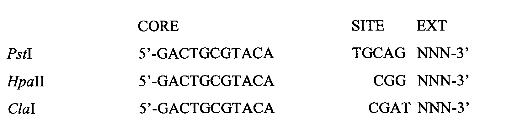
- The two MseI-primers are distinguished in the same way as the two MseI-adapters: the MseI-primer has the MseI-adapter as annealing target site, while MseI-primer+ has the MseI-adapter+ as annealing target site. The difference between the two MseI-primers is shown below:
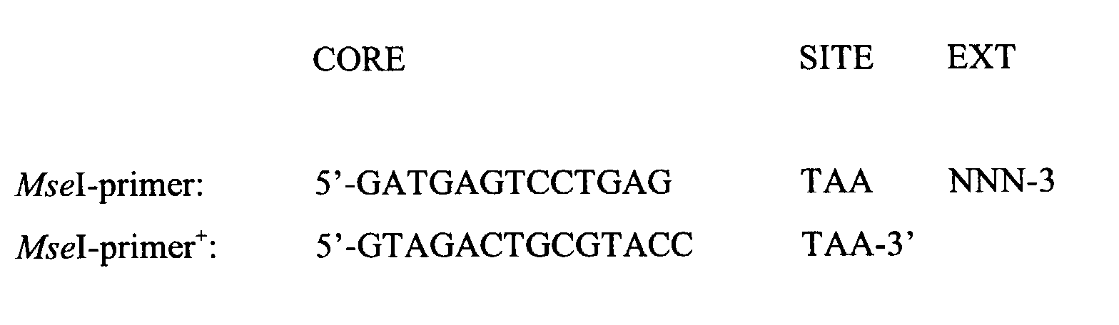
- The protocols A, B and C below describe the generation of A-,B- and C-templates for AFLP reactions using the enzyme combination (EC) PstI/MseI. Protocol C is equivalent to the standard AFLP protocol as described in EP-A-0 534 858 and by Vos et al (Vos, P., Hogers, R., Bleeker, M., Reijans, M., van de Lee, T., Hornes, M., Frijters, A., Pot, J., Peleman, J., Kuiper, M. and Zabeau, M. (1995) Nucl. Acids Res. 23, 4407-4414).
- Genomic DNA (0.5 µg) is incubated for 1 h at 37°C with 5 U PstI and 5 U MseI in 40 µl 10mM Tris-HAc pH 7.5, 10 mM MgAc, 50 mM KAc, 5 mM DTT, 50 ng/µl BSA (acetylated). Next, 10 µl of a solution is added, containing 5 pMol PstI-adapter*, 50 pMol MseI-adapter, 50 pMol MseI-adapter+, 1U T4 DNA-ligase, 1mM ATP in 10 mM Tris-HAc pH 7.5, 10 mM MgAc, 50 mM KAc, 5 mM DTT, 50 ng/µl BSA, and the incubation is continued for 3 h at 37°C. Adapters are prepared by adding equimolar amounts of both strands; adapters are not phosphorylated. After ligation, the reaction mixture is diluted to 250 µl with 10 mM Tris-HCl, 0.1 mM EDTA pH 8.0, and used for PCR amplification or stored at -20°C.
- Genomic DNA (0.5 µg) is incubated for 1 h at 37°C with only 5 U MseI in 40 µl 10mM Tris-HAc pH 7.5, 10 mM MgAc, 50 mM KAc, 5 mM DTT, 50 ng/µl BSA (acetylated). Next, 10 µl of a solution is added, containing 50 pMol MseI-adapter, 50 pMol MseI-adapter+, 1U T4 DNA-ligase, 1mM ATP in 10 mM Tris-HAc pH 7.5, 10 mM MgAc, 50 mM KAc, 5 mM DTT, 50 ng/µl BSA, and the incubation is continued for 3 h at 37°C. Adapters are prepared as above (Protocol A). After ligation, the reaction mixture is diluted to 250 µl with 10 mM Tris-HCl, 0.1 mM EDTA pH 8.0, and used for PCR amplification or stored at - 20°C.
- Genomic DNA (0.5 µg) is incubated for 1 h at 37°C with 5 U PstI and 5 U MseI in 40 µl 10mM Tris-HAc pH 7.5, 10 mM MgAc, 50 mM KAc, 5 mM DTT, 50 ng/µl BSA (acetylated). Next, 10 µl of a solution is added, containing 5 pMol PstI-adapter, 50 pMol MseI-adapter, 1U T4 DNA-ligase, 1mM ATP in 10 mM Tris-HAc pH 7.5, 10 mM MgAc, 50 mM KAc, 5 mM DTT, 50 ng/µl BSA, and the incubation is continued for 3 h at 37°C. Adapters are prepared as above (Protocol A). After ligation, the reaction mixture is diluted to 500 µl with 10 mM Tris-HCl, 0.1 mM EDTA pH 8.0, and used for PCR amplification or stored at - 20°C.
- Synthesis of unmethylated A- and B-templates is performed by nonselective PCR amplification. This nonselective PCR amplification is performed with the following cycle profile for 7 cycles: a 30 s DNA denaturation step at 94°C, a 1 min annealing step at 56°C and a 2 min extension step at 72°C. Amplifications are performed in 20 µl containing 5 µl template-DNA, 30 ng MseI-primer, 30 ng MseI-primer+, 0.4 U Taq polymerase, 10 mM Tris-HCl pH 8.3, 1.5 mM MgCl2, 50 mM Kcl and 0.2 mM of all four dNTPs.
- After amplification, the reaction mixtures A and B are incubated again for 1 h at 37°C with 5 U PstI in 40 µl 10mM Tris-HAc pH 7.5, 10 mM MgAc, 50 mM KAc, 5 mM DTT, 50 ng/µl BSA(acetylated). Next, 10 µl of a solution is added, containing 5 pMol PstI-adapter, 1U T4 DNA-ligase, 1mM ATP in 10 mM Tris-HAc pH 7.5, 10 mM MgAc, 50 mM KAc, 5 mM DTT, 50 ng/µl BSA(acetylated), and the incubation is continued for 3 h at 37°C.
- After ligation, the reaction mixture is diluted to 1000 µl with 10 mM Tris-HCl, 0.1 mM EDTA pH 8.0, and used for PCR amplification or stored at - 20°C.
- Generation of A- and B-templates for AFLP reactions using methylation-sensitive rare cutters leaving a 5'-extension (e.g. HpaII, MspI, ClaI) involves inactivation of the remaining Taq polymerase after the nonselective amplification, to avoid incorporation of remaining dNTPs after restriction. This is achieved by adding 220 ng TaqStart™ Antibody/ U Taq polymerase to the amplification mixture, prior to further restriction and ligation.
- Amplification reaction conditions are described using DNA templates (A, B and C) for the EC PstI/MseI. With other ECs, the procedure is identical except for the use of appropiate primers.
- AFLP fingerprinting of large genomes generally involves an amplification in two steps. The first step of this amplification procedure, named pre-amplification, employes two AFLP primers, one aimed at the PstI-ends and one at the MseI-ends, each having a single selective nucleotide. These primers are not radioactively labelled. Amplifications are performed in 20 µl containing 5 µl template-DNA, 30 ng MseI-primer, 30 ng PstI-primer, 0.4 U Taq polymerase, 10 mM Tris-HCl pH 8.3, 1.5 mM MgCl2, 50 mM KCl and 0.2 mM of all four dNTPs.
- After pre-amplification, the reaction mixtures are diluted 10-fold with 10 mM Tris-HCl, 0.1 mM EDTA pH=8.0, and used as templates for the second amplification reaction.
- The second amplification reaction again employes two oligonucleotide primers, one aimed at the PstI-ends and one at the MseI-ends, each having two or three selective nucleotides. The PstI-primer is radioactively end-labeled using [γ-33P] ATP and T4 polynucleotide kinase. The labelling reactions are performed in 50 µl 25 mM Tris-HCl pH 7.5, 10 mM MgCl2, 5mM DTT, 0.5mM spermidine-3HCl using 500 ng oligonucleotide primer, 100 µCi [γ-33P] ATP (1000-3000Ci/mol) and 10 U T4 polynucleotide kinase. Amplifications are performed in 20 µl containing 5 µl template-DNA, 5 ng labeled PstI-primer (0.5 µl from the labelling reaction mixture), 30 ng MseI-primer, 0.4 U Taq polymerase, 10 mM Tris-HCl pH 8.3, 1.5 mM MgCl2, 50mM KCl and 0.2 mM of all four dNTPs.
- AFLP preamplification reactions are performed for 24 cycles (protocol A and B) and 20 cycles (protocol C) with the following cycle profile: a 30 s DNA denaturation step at 94 °C, a 1 min annealing step at 56°C and a 1 min extension step at 72°C. AFLP reactions with primers having two or three selective nucleotides are performed for 36 cycles with the following cyle profile: a 30 s DNA denaturation step at 94°C, a 30 s annealing step (see below) and a 1 min extension step at 72°C. The annealing temperature in the first cycle is 65 °C, and is subsequently reduced each cycle by 0.7 °C for the next 12 cycles, then continued at 56 °C for the remaining 23 cycles. All amplification reactions are performed in a PE-9600 thermocycler ( Perkin Elmer Corp. Norwalk, CT, USA).
- Following amplification, reaction products are mixed with an equal volume (20 µl) of formamide dye (98% formamide, 10 mM EDTA pH 8.0, and bromophenol blue and xylene cyanol as tracking dyes). The resulting mixture is heated for 3 min at 90 °C, then quickly cooled on ice. Each sample (2 µl) was loaded on a 5% denaturing (sequencing) polyacrylamide gel (Maxam, A.M. and Gilbert, W. (1980) Methods Enzymol. 65, 499-560). The gel matrix is prepared using 5% acrylamide, 0.25% methylene bisacryl, 7.5 M urea in 50 mM Tris/50 mM Boric acid/1mM EDTA (pH 8.3). To 100 ml of gel solution 500 µl of 10 % APS and 100 µl TEMED is added and gels are cast using a SequiGen 38x50 cm gel apparatus (Biorad Laboratories Inc., Hercules, CA, USA). 100 mM Tris/100mM Boric acid/2ml EDTA was used as running buffer. Electrophoresis is performed at constant power, 110W, for about 2 hours. After electrophoresis, gels are fixed for 30 min in 10 % acetic acid, rinsed with water for 10 min, dried on the glass plates and exposed to Fujix phosphorimage screens for 16 h. Fingerprint patterns are visualized using a Fujix BAS-2000 phosphorimage analysis system (Fuji Photo Film Company Ltd., Japan).
- Initial experiments with methylation AFLP fingerprinting of plant genomic DNAs indicated that the complexity of the template libraries is of the same order as in the standard AFLP protocol. For maize, however, the number of the fragments detected in the B reaction (corresponding to the fragments of the A and C reaction jointly) by PstI+2/
Mse I+ 3 primer combinations (PCs) is substantially elevated, so 'tuning' to a slightly higher selectivity of the PCs is desirable and results in combined PstI PCs, e.g. PstI+AGW or PstI+AGS), where W stands for A and T jointly, and where S stands for C and G jointly.
Figures 7A and 7B show methylation AFLP fingerprints of genomic DNAs from the three maize inbred lines A7, B73 and Mo17. The two panels show PstI/MseI fingerprints, corresponding with the following PCs (from left to right): I. PstI+AGW/MseI+CTT, and II. PstI+AGS/MseI+CTT . Lane A, B and C represent the corresponding A, B and C restriction fragments, referring to the native methylated state of the rare cutter sites. The molecular weight size range of the fingerprints is 200-500 nucleotides.
Theoretically, every band present in lane A or lane C should also be present in lane B, because lane B represents both the native methylated and unmethylated rare cutter sites jointly. As seen in Figure 2, for PCs I, this is true for more than 90% of the fragments in lane A and C. In some cases (e.g. PC II), however, the percentage of fragments in lane A and C present in lane B can be low. An explanation for this is the following: short MseI-MseI restriction fragments have a competitive advantage over the longer MseI-MseI restriction fragments in the nonselective amplification. Consequently, rare cutter restriction sites (methylated or not), on average residing more on long MseI-MseI restriction fragments, will have a selective disadvantage. Subsequent restriction and ligation of the underrepresented fragments will generate a reduced amount of templates. - Although, as suggested by the fingerprints, large plant genomes consist predominantly of unique AFLP fragments, the presence of repetitive DNA is reflected by fragments of moderate (arrow 1) or high (arrow 2) band intensities in Figures 7A and 7B, in the good accordance with the correlation that exists between the AFLP band intensity and the original number of restriction fragments (Vos, P., Hogers, R., Bleeker, M., Reijans, M., van de Lee, T., Hornes, M., Frijters, A., Pot, J., Peleman, J., Kuiper, M. and Zabeau, M. (1995) Nucl. Acids Res. 23, 4407-4414). Since these multicopy restriction fragments are more abundantly present in lane A than in lane C, it can be concluded that moderate and highly repetitive sequences are strongly methylated. It should also be noted that the excessive use of primers by these multicopy PstI-MseI restriction fragments also leads to an underrepresentation of fragments in lane B.
- In plants the modified C at position 5 (m5C) is not only confined to CpG dinucleotides as is in animals, but also occurs at a variety of other cytosine containing dinucleotides, all of which are part of the basic trinucleotide CpNpG where N can be any nucleotide (Gruenbaum, Y., Naveh-Maney, T., Cedar, H. and Razin, A., 1981, Nature 292, 860-862) The restriction enzyme PstI, having 5'-CTGCAG-3' as recognition site, containing two CpNpG trinucleotides, is sensitive to methylation at the 5'-C and the 3'-C (McClelland, M., Nelson, M. and Raschke, E. (1994) Nucl. Acids Res. 22, 3640-3659). Whether simultaneous methylation of both C's of the PstI recognition site is possible, is not clear. A mean percentage ± standard error of methylated PstI-sites in the nuclear DNA of tomato, maize and oilseed rape, are given in Table 1.
Mean percentages ± standard error of methylated PstI-sites and 5mCpNpG as presented in PstI-sites in the nuclear DNA of tomato, maize and oilseed rape, and published mean percentages 5mC residues (expressed as % 5mC/5mC +C) in the nuclear DNA of tomato and maize. N= number of genotypes assayed. n = total number of PstI/MseI restriction fragments counted. Species N % PstI-sites methylated % CpNpG-sites methylated n % 5mC/5mC+C tomato 6 39.75 ± 1.00 19.88 ± 0.50 219 - 248 22.8 (19) maize 12 50.78 ± 1.33 25.39 ± 0.67 753 - 820 28.9 (20) oilseed rape 6 33.19 ± 0.62 16.60 ± 0.31 548 - 609 - It is clear from Table 1 that variation in the percentage of methylated PstI-sites is very low within a species. Using the percentage of 5mCpNpG-sites as presented in PstI-sites, calculated as half the number of methylated PstI-sites, as estimation of the percentage of 5mC residues in the nuclear DNA (expressed as 5mC/5mC +C), results in percentages 5mC residues very similar to published percentages, based on HPLC analysis (Messuguer, R., Ganal, M.W., Steffens, J.C., and Tanksley, S.D., 1991, Plant Mol.Biol. 16, 753-770 and Montero, L.M. et al., 1992, Nucl.Acids Res. 20, 3207-3210), for tomato and maize (see Table 1). Therefore, methylation of only one C residue in the PstI recognition site is highly probable.
- MspI and HpaII (methylation isoschizomers) have the same recognition site 5'-CCGG-3', containing one CpG dinucleotide. However, MspI is sensitive to 5'-5mC, whereas HpaII is sensitive to methylation at position 5 of either C (McClelland, M., Nelson, M. and Raschke, E. (1994) Nucl. Acids Res. 22, 3640-3659). Therefore, MspI and HpaII are appropriate for estimating the extent of 5mCpG. The extent of 5mCpG methylation by using HpaII and MspI as methylation-sensitive rare cutters is measured as the difference in the number of bands counted in a MspI and a HpaII fingerprint in lane C; the difference in the number of bands counted in a MspI and a HpaII fingerprint in lane A must give the same result. The percentages of 5mCpG sites in the nuclear DNA of tomato and maize, as presented in HpaII- and MspI-sites, are measured only for one genotype/species, and are given in Table 2.
- Another methylation-sensitive restriction enzyme ClaI, having the CpG-dinucleotide containing recognition site 5'-ATCGAT-3', and affected by 5mCpG (McClelland, M., Nelson, M. and Raschke, E. and (1994) Nucl. Acids Res. 22, 3640-3659) was also found to be suitable as rare cutter in the detection of 5mCPG. The percentages of 5mCpG as presented in HpaII-, MspII- and ClaI-sites in the nuclear DNA of tomato, maize and oilseed rape, are measured for only one genotype/species, and are given in Table 2.
Percentages of 5mCpG in the nuclear DNA of tomato, maize and oilseed rape, as presented in HpaII-, MspI- and ClaI-sites. n = total number of restriction fragments counted. Species 5mCpG as presented in ClaI-sites n 5mCpG as presented in HpaII/MspI-sites n tomato (cv.52201) 57.9 2833 57.6 2169 maize (B73) 53.9 2694 39.6 1791 oilseed rape (T528) 48.4 2973 - In tomato, a similar percentage of 5mCpG as presented in ClaI-sites is obtained as presented in either HpaII- or MspI-sites, in contrast with maize. Estimations of the percentage of 5mC residues as presented in ClaI-sites in the nuclear DNA of tomato, mais and oilseed rape is significantly higher than the the corresponding percentages 5mC residues as presented in PstI-sites. This can suggest that the majority of 5mC residues in the tomato, maize and oilseed rape genome exist at CpG sites.
- AFLP primarily detects variation of the primary DNA sequence, either base substitutions or DNA rearrangements. Nucleotides modified by methylation are not considered to be a part of the primary nucleotide sequence of an individual. This nucleotide modification, resulting from a post-replicative event at defined but not all target sequences (i.e. CpG and CpNpG), represents two additional forms of DNA polymorphism: (i) polymorphism reflecting the variation in the primary nucleotide sequence of the methylated restriction site and/or variation in the restriction size (mAFLP markers), and (ii) allele-specific methylation (asmAFLP markers). Since DNA-methylation is also the only source of allelic difference between epialleles, asmAFLP markers are epiallelic markers. Since methylation AFLP provides a positive display of the native methylated sites, it allows exploitation of this additional sources of DNA polymorphism.
For methylation polymorphism to be useful in e.g. AFLP mapping studies, it must be stably inherited. Figure 8A shows the segregation of a number of mAFLP markers in a Recombinant Inbred (RI) population derived from the cross B73 × Mo17. The band intensities segregate into two distinct classes, homozygous absent and homozygous present, approximating the Mendelian 1:1 segregation. To identify possible asmAFLP marker pairs, the following criteria are applied: (i) an AFLP band and a mAFLP band might be epialleles when derived from different parents, with the same PC; (ii) the AFLP and mAFLP marker map to the same locus (complementary segregation). Figures 8A and 8B show the segregation of a pair of asmAFLP markers in the RI population. The band intensities segregate into two distinct classes, homozygous absent and homozygous present. Of a total of 673 mapped AFLP + mAFLP markers, generated by a set of 14 PstI/MseI PCs, mAFLP markers account for 44,6%, which is in good accordance with the 50.78% methylated PstI-sites in the maize genome (Table 1), while hardly 1% of the marker alleles behave like epialleles.
As can be seen from the above description and Examples, the method of invention can be considered a DNA fingerprinting technique that detects genomic restriction fragments and resembles in that respect the AFLP technique, with the major difference that methylation AFLP displays native methylated sites too, but in general only if they are located in the recognition site of the obligate methylation-sensitive rare cutter. As for AFLP, the multiplex ratio is high (50-100 restriction fragments) and is a function of (i) the cleavage frequency of the methylation-sensitive rare cutter enzyme and (ii) the number and nature of the selective bases of the specific primer set. - Also, as demonstrated in the Examples, the method of the invention can be used for estimating the extent of 5mCpNpG, resp. 5mCpG, as presented in PstI-, resp. ClaI-, HpaI- and MspI-sites in the genome of few crops. Although generalization of these estimates to percentages 5mCpNpG and 5mCpG in the genome, irrespective the recognition site, may in some cases not be fully representative as methylation of C can be biased by the nature of its flanking nucleotides (recognition site), or by the percentage G+C of the genomic region it resides (CG-islands), the method of the invention will still provide valuable information to the artisan.
- Although hereinabove the invention was particularly described with respect to the use of methylation AFLP to estimate the extent of 5mCpG and 5mCpNpG as presented in some recognition sites in the genome, the invention and its possible uses are not only limited to that. Modifications of C at other positions than 5 (e.g. m4C), or modified nucleotides other than C (e.g. m6A), whether present in a symmetrical or a non-symmetrical dinucleotide (e.g. 5mCpC), are reported ((Gruenbaum, Y., Naveh-Maney, T., Cedar, H. and Razin, A., 1981, Nature 292, 860-862); Selker, E.U., Fritz, D.Y. and Singer, M.J., 1993, Science 262, 1724-1728; and Meyer, P., Niedenhof, I., ten Lohuis, M., 1994, The EMBO Journal 13, 2084-2088).
- Methylation AFLP can also be used with advantage to estimate the low prevalence of these minor nucleotide modifications presented by recognition sites of methylation-sensitive rare cutters, and to shed a light on their distributions over the genome.
- Methylation AFLP allows the exploitation of two additional forms of DNA polymorphism: (i) polymorphism reflecting variation in the primary nucleotide sequence of the methylated restriction site and/or variation in the restriction fragment size, and (ii) allele-specific methylation. Hardly 1% of the marker alleles behave like epialleles.
- By monitoring the segregation of mAFLP and asmAFLP markers in a RI population, it was shown that (i) C-methylation segregates in perfect accordance with the primary target sequence, while (ii) the few epialleles observed in the parents inherit in a Mendelian fashion to the offspring.
- Like AFLP markers, most mAFLP markers correspond to unique positions in the genome, and, hence, can be exploited as landmarks in and as bridging tools between genetic and physical maps ( Vos, P., Hogers, R., Bleeker, M., Reijans, M., van de Lee, T., Hornes, M., Frijters, A., Pot, J., Peleman, J., Kuiper, M. and Zabeau, M. (1995) Nucl. Acids Res. 23, 4407-4414). Therefore, methylation AFLP may be useful in genomic research. Native methylated sites are present on cloned DNA segments, e.g., yeast artificial chromosomes (YACs) as unmethylated sites. Hence, native methylated sites can not be distinguished from native unmethylated sites on a physical map. However, ligning up the physical map with a genetic map containing mAFLP markers, helps to identify native methylated sites on the physical map.
- More than in plant genetics, in human genetics the epigenetic effects of DNA methylation at CpG-island containing promotors is well documented. Due to its sensitive, reliable and quantitative nature, methylation AFLP is an attractive technique to determine the DNA methylation levels at specific gene loci like tumor-suppressor genes and to trace imprinted genes.
Note: AFLP™ is a trademark of Keygene N.V.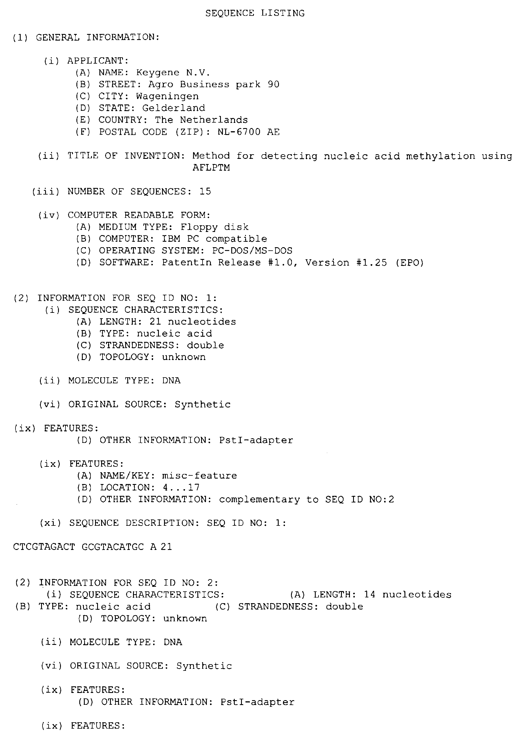
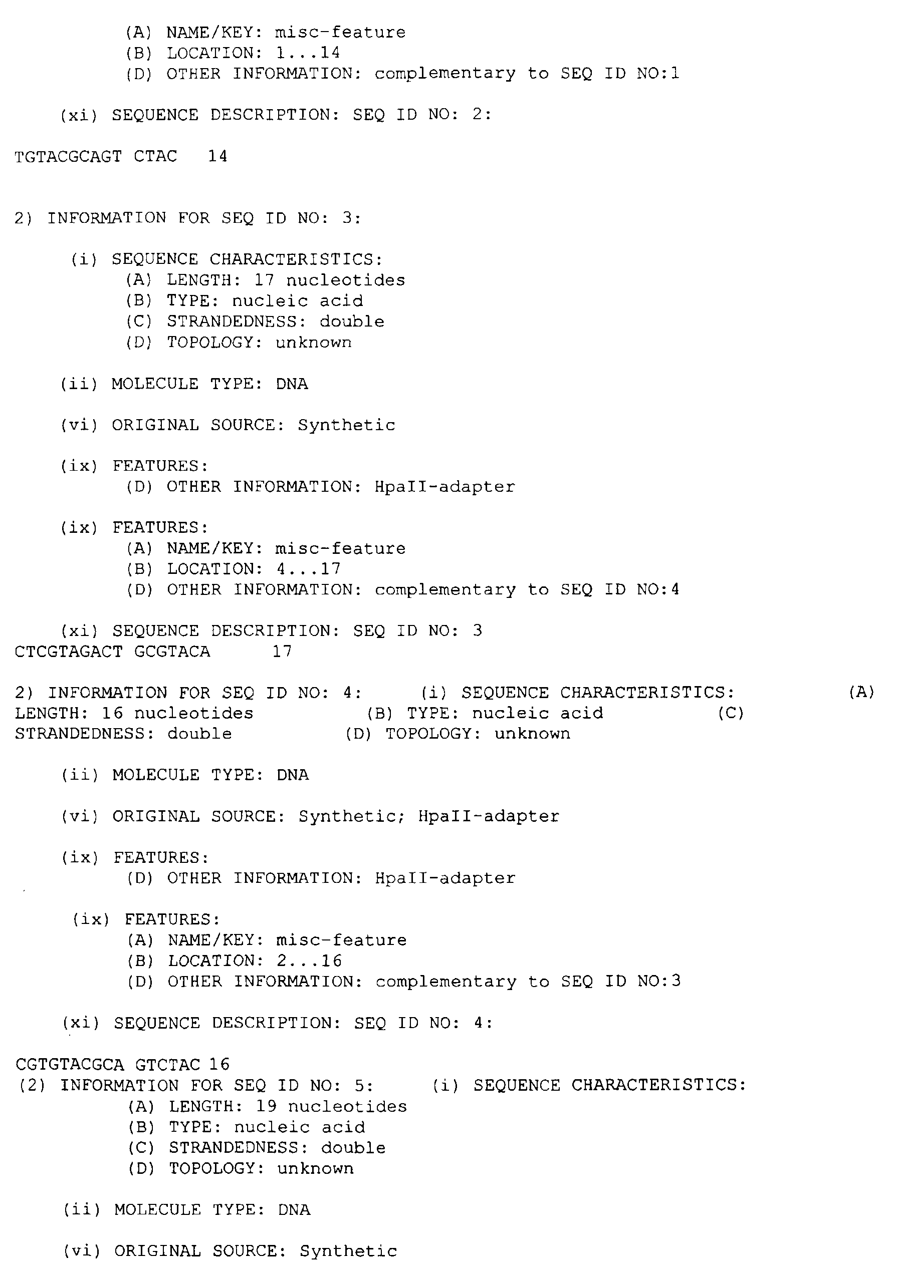
Claims (18)
- Use of AFLP in analysing or determining the state of methylation or of the methylation pattern of a starting DNA and/or in distinguishing between methylated and non-methylated sites in a starting DNA.
- Method for analysing or determining the methylation pattern of a starting DNA and/or for distinguishing between methylated and non-methylated sites in a starting DNA, comprising at least(A) generating a first DNA fingerprint, containing bands corresponding to both the methylated and non-methylated sites of interest; and/or(B) generating a second DNA fingerprint, containing bands corresponding only to the methylated sites of interest;
and optionally comprising(C) generating a third DNA fingerprint, containing bands corresponding only to the non-methylated sites of interest;
and optionally further comprising(D) analysing the fingerprint(s) thus obtained. - Method accoring to claim 2, in which at least two, and preferably all three of the fingerprints resulting from (A), (B) and/or (C) are generated and compared to each other.
- Method according to claim 2 or 3, in which the respective fingerprints resulting from (A), (B) and/or (C) are generated using an amplification technique, preferably AFLP.
- Method according to any of claims 2-4, in which the fingerprints resulting from (A), (B) and/or (C) are generated in conjunction with each other, preferably from the same starting DNA, DNA-pool or DNA-preparation.
- Method according to any of claims 2-5, in which the fingerprints resulting from (A), (B) and/or (C) are run in seperate lanes of the same electrophoresis gel.
- Method for producing a fingerprint containing bands corresponding to both the methylated sites in a starting DNA, in particular for use in the method of claims 2-5, comprising:1. restricting the starting DNA with a frequent cutter and a methylation sensitive rare cutter;2. ligating the restriction fragments thus obtained to a first and second adapter for the frequent cutter, as well as a first adapter for the rare cutter;3. amplifying the mixture thus obtained, using primers for the first and second frequent cutter adapter;4. restricting the amplified fragments thus obtained using a methylation sensitive rare cutter;5. ligating the restriction fragments thus obtained to a second adapter for the methylation sensitive rare cutter;6. amplifying the restriction fragments thus obtained using a primer for the first or second frequent cutter adapter, and a primer for the second rare cutter adapter;7. optionally analysing the mixture of amplified fragments thus obtained in a manner known per se, preferably by generating a DNA fingerprint.
- Method for producing a fingerprint containing bands corresponding to both the methylated and the non-methylated sites in a starting DNA, in particular for use in the method of claims 2-5, comprising:1. restricting the starting DNA with a frequent cutter;2. ligating the restriction fragments thus obtained with to third and fourth adapter for the frequent cutter;3. amplifying the mixture thus obtained, using primers for the third and fourth adapter for the frequent cutter;4. restricting the amplified fragments thus obtained using a methylation-sensitive rare cutter;5. ligating the restriction fragments thus obtained to a third adapter for the methylation sensitive rare cutter;6. amplifying the mixture thus obtained, using a primer for the third methylation sensitive rare cutter adapter, as well a primer for the third or fourth frequent cutter adapter;7. optionally analysing the mixture of amplified fragments thus obtained in a manner known per se, preferably by generating a DNA fingerprint.
- Method according to claim 7 or 8, in which the third and fourth frequent cutter adapter are the essentially same as the first and second frequent cutter adapter.
- Method according to claim 7, 8 or 9, in which the third rare cutter adapter is essentially the same as the second rare cutter adapter.
- Method according to any of claims 7-9, in which the frequent cutter restriction enzyme is chosen from MseI and TaqI and the methylation sensitive rare cutter is chosen from PstI, HpaII, MspI, ClaI, HhaI, EcoRII, BbvI, PvuII, XmaI, SmaI, NciI, AvaI, HaeII, SalI, XhoI and PvuII.
- Method according to any of claims 2-6 or 7-11 for use in distinguishing, identifying or classifying individuals, varieties or species on the basis of their DNA-methylation, by comparing methylation patterns of different individual sources; for fingerprinting of genomes and detecting methylation polymorphisms; or for detecting specific methylation patterns corresponding to the presence of specific sequences or genetic traits, as well as inherited DNA-methylation and allele-specific methylation.
- Method according to any of claims 2-6 or 7-11 for use in distinguishing transformed and not-transformed genes or sequences, for instance in the detection of transgenes; and distinguishing replicated and non-replicated DNA in bacteria.
- Method according to any of claims 2-6 or 7-11 for use in investigating or determining the regulation of gene expression in eukaryotes; in particular for detecting gene silencing or gene activation.
- Method according to any of claims 2-6 or 7-11 for use in detecting of genetic disorders, such as detecting DNA methylation associated with genetic diseases and "imprinting", as well as increased susceptibility for mutagenesis and cancer;
- Method according to any of claims 2-6 or 7-11 for use in detecting of gen for estimating the extend of methylation in a DNA or genome; or for detecting methylated DNA-markers.
- Kit for use with the method of any of claims 1-16, said kit comprising at least a frequent cutter restriction enzyme; a methylation sensitive rare cutter restriction enzyme; (at least) a first and second adapter for use with the frequent cutter; and (at least) a first and second adapter for use with the rare cutter; as well as primers for use with these adapters; and optionally further components for kits known per se.
- Data generated by the method of any of claims 1-16, optionally on a suitable data carrier, including DNA-fingerprints, photographs or other reproductions thereof, as well as (stored) analog or digital data thereon.
Priority Applications (4)
| Application Number | Priority Date | Filing Date | Title |
|---|---|---|---|
| EP98202549A EP0976835B1 (en) | 1998-07-29 | 1998-07-29 | Method for detecting nucleic acid methylation using AFLPTM |
| AT98202549T ATE244771T1 (en) | 1998-07-29 | 1998-07-29 | METHOD FOR DETECTING NUCLEIC ACID METHYLATIONS BY AFLP |
| DE69816286T DE69816286T2 (en) | 1998-07-29 | 1998-07-29 | Method for the detection of nucleic acid methylations by AFLP |
| US09/362,311 US6300071B1 (en) | 1998-07-29 | 1999-07-28 | Method for detecting nucleic acid methylation using AFLP™ |
Applications Claiming Priority (1)
| Application Number | Priority Date | Filing Date | Title |
|---|---|---|---|
| EP98202549A EP0976835B1 (en) | 1998-07-29 | 1998-07-29 | Method for detecting nucleic acid methylation using AFLPTM |
Publications (2)
| Publication Number | Publication Date |
|---|---|
| EP0976835A1 true EP0976835A1 (en) | 2000-02-02 |
| EP0976835B1 EP0976835B1 (en) | 2003-07-09 |
Family
ID=8233987
Family Applications (1)
| Application Number | Title | Priority Date | Filing Date |
|---|---|---|---|
| EP98202549A Expired - Lifetime EP0976835B1 (en) | 1998-07-29 | 1998-07-29 | Method for detecting nucleic acid methylation using AFLPTM |
Country Status (4)
| Country | Link |
|---|---|
| US (1) | US6300071B1 (en) |
| EP (1) | EP0976835B1 (en) |
| AT (1) | ATE244771T1 (en) |
| DE (1) | DE69816286T2 (en) |
Cited By (16)
| Publication number | Priority date | Publication date | Assignee | Title |
|---|---|---|---|---|
| WO2002037398A3 (en) * | 2000-11-02 | 2003-08-21 | Epigenomics Ag | Systems, methods and computer program products for guiding the selection of therapeutic treatment regimens |
| WO2005045069A3 (en) * | 2003-11-09 | 2005-07-21 | Epigenomics Ag | Method for investigating cytosine methylation in dna sequences by means of hemi-methylation sensitive restriction enzymes |
| WO2005090607A1 (en) * | 2004-03-08 | 2005-09-29 | Rubicon Genomics, Inc. | Methods and compositions for generating and amplifying dna libraries for sensitive detection and analysis of dna methylation |
| WO2005108618A3 (en) * | 2004-04-30 | 2006-03-30 | Applera Corp | Methods and kits for methylation detection |
| WO2006038416A1 (en) * | 2004-10-06 | 2006-04-13 | National Institute Of Radiological Sciences | Method of exhaustive analysis of transcriptionally-active domain (non-methylated domain) on genome |
| WO2006137733A1 (en) | 2005-06-23 | 2006-12-28 | Keygene N.V. | Strategies for high throughput identification and detection of polymorphisms |
| EP1438416A4 (en) * | 2001-09-26 | 2007-09-05 | Epigenx Pharmaceutical Inc | Assays for dna methylation changes |
| WO2008111453A1 (en) * | 2007-03-07 | 2008-09-18 | The University Of Tokyo | Method for amplification of dna fragment |
| US7803550B2 (en) | 2005-08-02 | 2010-09-28 | Rubicon Genomics, Inc. | Methods of producing nucleic acid molecules comprising stem loop oligonucleotides |
| EP2330214A1 (en) | 2004-09-03 | 2011-06-08 | F. Hoffmann-La Roche AG | Method for DNA decontamination |
| US8409804B2 (en) | 2005-08-02 | 2013-04-02 | Rubicon Genomics, Inc. | Isolation of CpG islands by thermal segregation and enzymatic selection-amplification method |
| US9062348B1 (en) | 2005-12-22 | 2015-06-23 | Keygene N.V. | Method for high-throughput AFLP-based polymorphism detection |
| US10023907B2 (en) | 2006-04-04 | 2018-07-17 | Keygene N.V. | High throughput detection of molecular markers based on AFLP and high through-put sequencing |
| US10233494B2 (en) | 2005-09-29 | 2019-03-19 | Keygene N.V. | High throughput screening of populations carrying naturally occurring mutations |
| US10316364B2 (en) | 2005-09-29 | 2019-06-11 | Keygene N.V. | Method for identifying the source of an amplicon |
| US10837049B2 (en) | 2003-03-07 | 2020-11-17 | Takara Bio Usa, Inc. | Amplification and analysis of whole genome and whole transcriptome libraries generated by a DNA polymerization process |
Families Citing this family (36)
| Publication number | Priority date | Publication date | Assignee | Title |
|---|---|---|---|---|
| AU2308900A (en) * | 1999-02-05 | 2000-08-25 | Amersham Pharmacia Biotech Uk Limited | Analysis method |
| US6605432B1 (en) * | 1999-02-05 | 2003-08-12 | Curators Of The University Of Missouri | High-throughput methods for detecting DNA methylation |
| US7611869B2 (en) | 2000-02-07 | 2009-11-03 | Illumina, Inc. | Multiplexed methylation detection methods |
| US8076063B2 (en) | 2000-02-07 | 2011-12-13 | Illumina, Inc. | Multiplexed methylation detection methods |
| US7955794B2 (en) | 2000-09-21 | 2011-06-07 | Illumina, Inc. | Multiplex nucleic acid reactions |
| US7582420B2 (en) | 2001-07-12 | 2009-09-01 | Illumina, Inc. | Multiplex nucleic acid reactions |
| WO2001090415A2 (en) | 2000-05-20 | 2001-11-29 | The Regents Of The University Of Michigan | Method of producing a dna library using positional amplification |
| DE60117542T2 (en) * | 2000-05-22 | 2006-12-07 | The Johns Hopkins University | TEST METHOD FOR DETERMINING GEN IMPRINTING AND METHYLATED CPG ISLANDS |
| US20030143554A1 (en) * | 2001-03-31 | 2003-07-31 | Berres Mark E. | Method of genotyping by determination of allele copy number |
| JP2004536596A (en) * | 2001-06-08 | 2004-12-09 | ユー.エス. ジェノミクス, インコーポレイテッド | Methods and products for analyzing nucleic acids based on methylation status |
| AU2002312588A1 (en) * | 2001-06-29 | 2003-03-03 | Rubicon Genomics Inc. | Methods of using nick translate libraries for snp analysis |
| US20030232371A1 (en) * | 2001-10-24 | 2003-12-18 | Bestor Timothy H. | Methods for detecting methylated promoters based on differential DNA methylation |
| WO2003035860A1 (en) * | 2001-10-24 | 2003-05-01 | The Trustees Of Columbia University In The City Of New York | A method for gene identification based on differential dna methylation |
| US20110151438A9 (en) | 2001-11-19 | 2011-06-23 | Affymetrix, Inc. | Methods of Analysis of Methylation |
| EP1470255A2 (en) * | 2002-01-30 | 2004-10-27 | Epigenomics AG | Identification of cell differentiation states based on methylation patterns |
| AU2003224978A1 (en) | 2002-04-12 | 2003-10-27 | Curators Of The University Of Missouri | ECIST microarrays for dual screening of DNA hypermethylation and gene silencing |
| DE60331852D1 (en) * | 2002-12-02 | 2010-05-06 | Illumina Cambridge Ltd | DETERMINATION OF THE METHYLATION OF NUCLEIC ACID SEQUENCES |
| US20050009059A1 (en) * | 2003-05-07 | 2005-01-13 | Affymetrix, Inc. | Analysis of methylation status using oligonucleotide arrays |
| US20050032102A1 (en) * | 2003-07-22 | 2005-02-10 | Affymetrix, Inc. | Mapping genomic rearrangements |
| WO2005012575A1 (en) * | 2003-08-01 | 2005-02-10 | U.S. Genomics, Inc. | Methods and compositions related to the use of sequence-specific endonucleases for analyzing nucleic acids under non-cleaving conditions |
| US20060134650A1 (en) * | 2004-12-21 | 2006-06-22 | Illumina, Inc. | Methylation-sensitive restriction enzyme endonuclease method of whole genome methylation analysis |
| US20060292585A1 (en) * | 2005-06-24 | 2006-12-28 | Affymetrix, Inc. | Analysis of methylation using nucleic acid arrays |
| AU2005339395B2 (en) * | 2005-12-22 | 2012-08-16 | Instytut Hodowli I Aklimatyzacji Roslin (Plant Breeding And Acclimatization Institute) | A method of identifying induced variability in in vitro cultures |
| US7901882B2 (en) | 2006-03-31 | 2011-03-08 | Affymetrix, Inc. | Analysis of methylation using nucleic acid arrays |
| WO2008096146A1 (en) * | 2007-02-07 | 2008-08-14 | Solexa Limited | Preparation of templates for methylation analysis |
| US8642294B2 (en) * | 2007-06-20 | 2014-02-04 | Albert Einstein College Of Medicine Of Yeshiva University | Methods for determining cytosine methylation in DNA and uses thereof |
| EP2340314B8 (en) | 2008-10-22 | 2015-02-18 | Illumina, Inc. | Preservation of information related to genomic dna methylation |
| US8394584B2 (en) | 2008-12-19 | 2013-03-12 | The Board Of Trustees Of The University Of Illinois | Detecting and sorting methylated DNA using a synthetic nanopore |
| US20100273164A1 (en) * | 2009-03-24 | 2010-10-28 | President And Fellows Of Harvard College | Targeted and Whole-Genome Technologies to Profile DNA Cytosine Methylation |
| US8748091B2 (en) * | 2009-12-18 | 2014-06-10 | The Board Of Trustees Of The University Of Illinois | Characterizing stretched polynucleotides in a synthetic nanopassage |
| WO2012162139A1 (en) | 2011-05-20 | 2012-11-29 | The Regents Of The University Of California | Method to estimate age of individual based on epigenetic markers in biological sample |
| WO2013192620A1 (en) | 2012-06-22 | 2013-12-27 | Quantrx Biomedical Corporation | Method for obtaining fetal cells and fetal cellular components |
| US20200347461A1 (en) | 2018-01-17 | 2020-11-05 | The Regents Of The University Of California | Phenotypic age and dna methylation based biomarkers for life expectancy and morbidity |
| WO2020150705A1 (en) | 2019-01-18 | 2020-07-23 | The Regents Of The University Of California | Dna methylation measurement for mammals based on conserved loci |
| EP4493723A1 (en) | 2022-03-15 | 2025-01-22 | Genknowme S.A. | Method determining the difference between the biological age and the chronological age of a subject |
| WO2024256726A1 (en) | 2023-06-15 | 2024-12-19 | Genknowme S.A. | Computer implemented method determining a value of allostatic load of a human being |
Citations (4)
| Publication number | Priority date | Publication date | Assignee | Title |
|---|---|---|---|---|
| EP0534858A1 (en) * | 1991-09-24 | 1993-03-31 | Keygene N.V. | Selective restriction fragment amplification : a general method for DNA fingerprinting |
| WO1996027024A1 (en) * | 1995-03-01 | 1996-09-06 | Avitech Diagnostics, Inc. | Method of identifying clonal cell samples |
| WO1997045560A1 (en) * | 1996-05-31 | 1997-12-04 | North Shore University Hospital Research Corporation | Identification of differentially methylated and mutated nucleic acids |
| WO1998008981A1 (en) * | 1996-08-30 | 1998-03-05 | Life Technologies, Inc. | METHODS FOR IDENTIFICATION AND ISOLATION OF SPECIFIC NUCLEOTIDE SEQUENCES IN cDNA AND GENOMIC DNA |
-
1998
- 1998-07-29 DE DE69816286T patent/DE69816286T2/en not_active Expired - Fee Related
- 1998-07-29 EP EP98202549A patent/EP0976835B1/en not_active Expired - Lifetime
- 1998-07-29 AT AT98202549T patent/ATE244771T1/en not_active IP Right Cessation
-
1999
- 1999-07-28 US US09/362,311 patent/US6300071B1/en not_active Expired - Lifetime
Patent Citations (4)
| Publication number | Priority date | Publication date | Assignee | Title |
|---|---|---|---|---|
| EP0534858A1 (en) * | 1991-09-24 | 1993-03-31 | Keygene N.V. | Selective restriction fragment amplification : a general method for DNA fingerprinting |
| WO1996027024A1 (en) * | 1995-03-01 | 1996-09-06 | Avitech Diagnostics, Inc. | Method of identifying clonal cell samples |
| WO1997045560A1 (en) * | 1996-05-31 | 1997-12-04 | North Shore University Hospital Research Corporation | Identification of differentially methylated and mutated nucleic acids |
| WO1998008981A1 (en) * | 1996-08-30 | 1998-03-05 | Life Technologies, Inc. | METHODS FOR IDENTIFICATION AND ISOLATION OF SPECIFIC NUCLEOTIDE SEQUENCES IN cDNA AND GENOMIC DNA |
Non-Patent Citations (4)
| Title |
|---|
| BIOLOGICAL ABSTRACTS, 1 January 1900, Philadelphia, PA, US; abstract no. 1997:439076, XP002110060 * |
| Medline:AN1998120884 Chen Z. et al.,Brit.J.Cancer 77, 181-185,1998 Methylation of CpG island is not a ubiquitous mechanism for the loss of oestrogen receptor in breast cancer cells * |
| VOS P. ET AL.,: "AFLP: a new technique for DNA fingerprinting", NUCLEIC ACIDS RESEARCH, vol. 23, no. 21, - 1995, pages 4407 - 4414, XP002109691 * |
| XIONG LZ. ET AL.,: "Patterns of cytosine methylation in an elite rice hybrid and its parental lines, detected by a methylation-sensitive amplification polymorphism technique", MOL. GEN. GENET., vol. 261, - April 1999 (1999-04-01), pages 439 - 446, XP002110074 * |
Cited By (57)
| Publication number | Priority date | Publication date | Assignee | Title |
|---|---|---|---|---|
| WO2002037398A3 (en) * | 2000-11-02 | 2003-08-21 | Epigenomics Ag | Systems, methods and computer program products for guiding the selection of therapeutic treatment regimens |
| EP1438416A4 (en) * | 2001-09-26 | 2007-09-05 | Epigenx Pharmaceutical Inc | Assays for dna methylation changes |
| US11661628B2 (en) | 2003-03-07 | 2023-05-30 | Takara Bio Usa, Inc. | Amplification and analysis of whole genome and whole transcriptome libraries generated by a DNA polymerization process |
| US11492663B2 (en) | 2003-03-07 | 2022-11-08 | Takara Bio Usa, Inc. | Amplification and analysis of whole genome and whole transcriptome libraries generated by a DNA polymerization process |
| US10837049B2 (en) | 2003-03-07 | 2020-11-17 | Takara Bio Usa, Inc. | Amplification and analysis of whole genome and whole transcriptome libraries generated by a DNA polymerization process |
| WO2005045069A3 (en) * | 2003-11-09 | 2005-07-21 | Epigenomics Ag | Method for investigating cytosine methylation in dna sequences by means of hemi-methylation sensitive restriction enzymes |
| GB2422902A (en) * | 2003-11-09 | 2006-08-09 | Epigenomics Ag | Method for investigating cytosine methylation in DNA sequences by means of hemimethylation sensitive restriction enzymes |
| US9708652B2 (en) | 2004-03-08 | 2017-07-18 | Rubicon Genomics, Inc. | Methods and compositions for generating and amplifying DNA libraries for sensitive detection and analysis of DNA methylation |
| WO2005090607A1 (en) * | 2004-03-08 | 2005-09-29 | Rubicon Genomics, Inc. | Methods and compositions for generating and amplifying dna libraries for sensitive detection and analysis of dna methylation |
| US8440404B2 (en) | 2004-03-08 | 2013-05-14 | Rubicon Genomics | Methods and compositions for generating and amplifying DNA libraries for sensitive detection and analysis of DNA methylation |
| EP2380993A1 (en) * | 2004-03-08 | 2011-10-26 | Rubicon Genomics, Inc. | Methods and compositions for generating and amplifying DNA libraries for sensitive detection and analysis of DNA methylation |
| EP2290106A1 (en) * | 2004-03-08 | 2011-03-02 | Rubicon Genomics, Inc. | Methods and compositions for generating and amplifying DNA libraries for sensitive detection and analysis of DNA methylation |
| US7364855B2 (en) | 2004-04-30 | 2008-04-29 | Applera Corporation | Methods and kits for methylation detection |
| WO2005108618A3 (en) * | 2004-04-30 | 2006-03-30 | Applera Corp | Methods and kits for methylation detection |
| EP2330214A1 (en) | 2004-09-03 | 2011-06-08 | F. Hoffmann-La Roche AG | Method for DNA decontamination |
| JPWO2006038416A1 (en) * | 2004-10-06 | 2008-05-15 | 独立行政法人放射線医学総合研究所 | Comprehensive analysis of transcriptional active regions (unmethylated regions) on the genome |
| WO2006038416A1 (en) * | 2004-10-06 | 2006-04-13 | National Institute Of Radiological Sciences | Method of exhaustive analysis of transcriptionally-active domain (non-methylated domain) on genome |
| US9023768B2 (en) | 2005-06-23 | 2015-05-05 | Keygene N.V. | Strategies for high throughput identification and detection of polymorphisms |
| EP2302070A3 (en) * | 2005-06-23 | 2011-06-22 | Keygene N.V. | Strategies for high throughput identification and detection fo polymorpisms |
| WO2006137733A1 (en) | 2005-06-23 | 2006-12-28 | Keygene N.V. | Strategies for high throughput identification and detection of polymorphisms |
| CN102925561A (en) * | 2005-06-23 | 2013-02-13 | 科因股份有限公司 | Strategies for high throughput identification and detection of polymorphisms |
| US10978175B2 (en) | 2005-06-23 | 2021-04-13 | Keygene N.V. | Strategies for high throughput identification and detection of polymorphisms |
| US10235494B2 (en) | 2005-06-23 | 2019-03-19 | Keygene N.V. | Strategies for high throughput identification and detection of polymorphisms |
| EP2302070A2 (en) | 2005-06-23 | 2011-03-30 | Keygene N.V. | Strategies for high throughput identification and detection fo polymorpisms |
| US8685889B2 (en) | 2005-06-23 | 2014-04-01 | Keygene N.V. | Strategies for high throughput identification and detection of polymorphisms |
| US10095832B2 (en) | 2005-06-23 | 2018-10-09 | Keygene N.V. | Strategies for high throughput identification and detection of polymorphisms |
| US9896721B2 (en) | 2005-06-23 | 2018-02-20 | Keygene N.V. | Strategies for high throughput identification and detection of polymorphisms |
| US8785353B2 (en) | 2005-06-23 | 2014-07-22 | Keygene N.V. | Strategies for high throughput identification and detection of polymorphisms |
| EP2292788A1 (en) | 2005-06-23 | 2011-03-09 | Keygene N.V. | Strategies for high throughput identification and detection of polymorphisms |
| US9898576B2 (en) | 2005-06-23 | 2018-02-20 | Keygene N.V. | Strategies for high throughput identification and detection of polymorphisms |
| CN102925561B (en) * | 2005-06-23 | 2015-09-09 | 科因股份有限公司 | For the high throughput identification of polymorphism and the strategy of detection |
| US9898577B2 (en) | 2005-06-23 | 2018-02-20 | Keygene N.V. | Strategies for high throughput identification and detection of polymorphisms |
| US9376716B2 (en) | 2005-06-23 | 2016-06-28 | Keygene N.V. | Strategies for high throughput identification and detection of polymorphisms |
| US9447459B2 (en) | 2005-06-23 | 2016-09-20 | Keygene N.V. | Strategies for high throughput identification and detection of polymorphisms |
| US9453256B2 (en) | 2005-06-23 | 2016-09-27 | Keygene N.V. | Strategies for high throughput identification and detection of polymorphisms |
| US9493820B2 (en) | 2005-06-23 | 2016-11-15 | Keygene N.V. | Strategies for high throughput identification and detection of polymorphisms |
| US9598727B2 (en) | 2005-08-02 | 2017-03-21 | Rubicon Genomics, Inc. | Methods for processing and amplifying nucleic acids |
| US8409804B2 (en) | 2005-08-02 | 2013-04-02 | Rubicon Genomics, Inc. | Isolation of CpG islands by thermal segregation and enzymatic selection-amplification method |
| US8071312B2 (en) | 2005-08-02 | 2011-12-06 | Rubicon Genomics, Inc. | Methods for producing and using stem-loop oligonucleotides |
| US11072823B2 (en) | 2005-08-02 | 2021-07-27 | Takara Bio Usa, Inc. | Compositions including a double stranded nucleic acid molecule and a stem-loop oligonucleotide |
| US8778610B2 (en) | 2005-08-02 | 2014-07-15 | Rubicon Genomics, Inc. | Methods for preparing amplifiable DNA molecules |
| US8399199B2 (en) | 2005-08-02 | 2013-03-19 | Rubicon Genomics | Use of stem-loop oligonucleotides in the preparation of nucleic acid molecules |
| US8728737B2 (en) | 2005-08-02 | 2014-05-20 | Rubicon Genomics, Inc. | Attaching a stem-loop oligonucleotide to a double stranded DNA molecule |
| US7803550B2 (en) | 2005-08-02 | 2010-09-28 | Rubicon Genomics, Inc. | Methods of producing nucleic acid molecules comprising stem loop oligonucleotides |
| US10196686B2 (en) | 2005-08-02 | 2019-02-05 | Takara Bio Usa, Inc. | Kits including stem-loop oligonucleotides for use in preparing nucleic acid molecules |
| US10208337B2 (en) | 2005-08-02 | 2019-02-19 | Takara Bio Usa, Inc. | Compositions including a double stranded nucleic acid molecule and a stem-loop oligonucleotide |
| US10233494B2 (en) | 2005-09-29 | 2019-03-19 | Keygene N.V. | High throughput screening of populations carrying naturally occurring mutations |
| US10316364B2 (en) | 2005-09-29 | 2019-06-11 | Keygene N.V. | Method for identifying the source of an amplicon |
| US10538806B2 (en) | 2005-09-29 | 2020-01-21 | Keygene N.V. | High throughput screening of populations carrying naturally occurring mutations |
| US11649494B2 (en) | 2005-09-29 | 2023-05-16 | Keygene N.V. | High throughput screening of populations carrying naturally occurring mutations |
| US10106850B2 (en) | 2005-12-22 | 2018-10-23 | Keygene N.V. | Method for high-throughput AFLP-based polymorphism detection |
| US11008615B2 (en) | 2005-12-22 | 2021-05-18 | Keygene N.V. | Method for high-throughput AFLP-based polymorphism detection |
| US9062348B1 (en) | 2005-12-22 | 2015-06-23 | Keygene N.V. | Method for high-throughput AFLP-based polymorphism detection |
| US9334536B2 (en) | 2005-12-22 | 2016-05-10 | Keygene N.V. | Method for high-throughput AFLP-based polymorphism detection |
| US10023907B2 (en) | 2006-04-04 | 2018-07-17 | Keygene N.V. | High throughput detection of molecular markers based on AFLP and high through-put sequencing |
| JPWO2008111453A1 (en) * | 2007-03-07 | 2010-06-24 | 国立大学法人 東京大学 | DNA fragment amplification method |
| WO2008111453A1 (en) * | 2007-03-07 | 2008-09-18 | The University Of Tokyo | Method for amplification of dna fragment |
Also Published As
| Publication number | Publication date |
|---|---|
| EP0976835B1 (en) | 2003-07-09 |
| US6300071B1 (en) | 2001-10-09 |
| DE69816286D1 (en) | 2003-08-14 |
| DE69816286T2 (en) | 2004-05-27 |
| ATE244771T1 (en) | 2003-07-15 |
Similar Documents
| Publication | Publication Date | Title |
|---|---|---|
| EP0976835B1 (en) | Method for detecting nucleic acid methylation using AFLPTM | |
| EP0534858B2 (en) | Selective restriction fragment amplification : a general method for DNA fingerprinting | |
| Fraga et al. | DNA methylation: a profile of methods and applications | |
| US9702004B2 (en) | Method for high-throughput AFLP-based polymorphism detection | |
| CA2105060C (en) | Nucleic acid typing by polymerase extension of oligonucleotides using terminator mixtures | |
| Xu et al. | AFLP-based detection of DNA methylation | |
| US6027894A (en) | Nucleic acid adapters containing a type IIs restriction site and methods of using the same | |
| EP0663923B1 (en) | Three highly informative microsatellite repeat polymorphic dna markers | |
| US20070015200A1 (en) | Methods for detecting genome-wide sequence variations associated with a phenotype | |
| WO2003060163A2 (en) | Discrimination and detection of target nucleotide sequences using mass spectrometry | |
| WO2003074734A2 (en) | Methods for detecting genome-wide sequence variations associated with a phenotype | |
| US20110059457A1 (en) | Selective restriction fragment amplification: fingerprinting | |
| WO1996017082A2 (en) | Compound microsatellite primers for the detection of genetic polymorphisms | |
| Cheung et al. | Genomic mismatch scanning identifies human genomic DNA shared identical by descent | |
| Azhikina et al. | Study of tissue-specific CpG methylation of DNA in extended genomic loci | |
| CA2087042C (en) | Genomic mapping method by direct haplotyping using intron sequence analysis | |
| WO1999001580A1 (en) | Compositions and methods for detection of genomic imprinting disorders | |
| EP1780292A1 (en) | Gene methylation assay controls | |
| US7026115B1 (en) | Selective restriction fragment amplification: fingerprinting | |
| Oefner | Sequence variation and the biological function of genes: methodological and biological considerations | |
| Xu | Molecular breeding tools: markers and maps. | |
| Rauch et al. | Methods for assessing genome-wide DNA methylation | |
| Ha et al. | Differentiation of Panax ginseng from P. quinquefolius by amplified fragment length polymorphism | |
| Rauch et al. | Methods for Assessing DNA Cytosine Modifications Genome-Wide | |
| Jones | Enzymatic assays and pcr-based dna fingerprinting in mutation detection |
Legal Events
| Date | Code | Title | Description |
|---|---|---|---|
| PUAI | Public reference made under article 153(3) epc to a published international application that has entered the european phase |
Free format text: ORIGINAL CODE: 0009012 |
|
| AK | Designated contracting states |
Kind code of ref document: A1 Designated state(s): AT BE CH CY DE DK ES FI FR GB GR IE IT LI LU MC NL PT SE |
|
| AX | Request for extension of the european patent |
Free format text: AL;LT;LV;MK;RO;SI |
|
| 17P | Request for examination filed |
Effective date: 20000726 |
|
| AKX | Designation fees paid |
Free format text: AT BE CH CY DE DK ES FI FR GB GR IE IT LI LU MC NL PT SE |
|
| 17Q | First examination report despatched |
Effective date: 20011019 |
|
| GRAH | Despatch of communication of intention to grant a patent |
Free format text: ORIGINAL CODE: EPIDOS IGRA |
|
| GRAH | Despatch of communication of intention to grant a patent |
Free format text: ORIGINAL CODE: EPIDOS IGRA |
|
| GRAA | (expected) grant |
Free format text: ORIGINAL CODE: 0009210 |
|
| AK | Designated contracting states |
Designated state(s): AT BE CH CY DE DK ES FI FR GB GR IE IT LI LU MC NL PT SE |
|
| PG25 | Lapsed in a contracting state [announced via postgrant information from national office to epo] |
Ref country code: IT Free format text: LAPSE BECAUSE OF FAILURE TO SUBMIT A TRANSLATION OF THE DESCRIPTION OR TO PAY THE FEE WITHIN THE PRE;WARNING: LAPSES OF ITALIAN PATENTS WITH EFFECTIVE DATE BEFORE 2007 MAY HAVE OCCURRED AT ANY TIME BEFORE 2007. THE CORRECT EFFECTIVE DATE MAY BE DIFFERENT FROM THE ONE RECORDED.SCRIBED TIME-LIMIT Effective date: 20030709 Ref country code: FI Free format text: LAPSE BECAUSE OF FAILURE TO SUBMIT A TRANSLATION OF THE DESCRIPTION OR TO PAY THE FEE WITHIN THE PRESCRIBED TIME-LIMIT Effective date: 20030709 Ref country code: AT Free format text: LAPSE BECAUSE OF FAILURE TO SUBMIT A TRANSLATION OF THE DESCRIPTION OR TO PAY THE FEE WITHIN THE PRESCRIBED TIME-LIMIT Effective date: 20030709 |
|
| REG | Reference to a national code |
Ref country code: GB Ref legal event code: FG4D |
|
| REG | Reference to a national code |
Ref country code: CH Ref legal event code: EP |
|
| PG25 | Lapsed in a contracting state [announced via postgrant information from national office to epo] |
Ref country code: LU Free format text: LAPSE BECAUSE OF NON-PAYMENT OF DUE FEES Effective date: 20030729 Ref country code: IE Free format text: LAPSE BECAUSE OF NON-PAYMENT OF DUE FEES Effective date: 20030729 Ref country code: CY Free format text: LAPSE BECAUSE OF FAILURE TO SUBMIT A TRANSLATION OF THE DESCRIPTION OR TO PAY THE FEE WITHIN THE PRESCRIBED TIME-LIMIT Effective date: 20030729 |
|
| PG25 | Lapsed in a contracting state [announced via postgrant information from national office to epo] |
Ref country code: MC Free format text: LAPSE BECAUSE OF NON-PAYMENT OF DUE FEES Effective date: 20030731 |
|
| REF | Corresponds to: |
Ref document number: 69816286 Country of ref document: DE Date of ref document: 20030814 Kind code of ref document: P |
|
| REG | Reference to a national code |
Ref country code: IE Ref legal event code: FG4D |
|
| PG25 | Lapsed in a contracting state [announced via postgrant information from national office to epo] |
Ref country code: SE Free format text: LAPSE BECAUSE OF FAILURE TO SUBMIT A TRANSLATION OF THE DESCRIPTION OR TO PAY THE FEE WITHIN THE PRESCRIBED TIME-LIMIT Effective date: 20031009 Ref country code: GR Free format text: LAPSE BECAUSE OF FAILURE TO SUBMIT A TRANSLATION OF THE DESCRIPTION OR TO PAY THE FEE WITHIN THE PRESCRIBED TIME-LIMIT Effective date: 20031009 Ref country code: DK Free format text: LAPSE BECAUSE OF FAILURE TO SUBMIT A TRANSLATION OF THE DESCRIPTION OR TO PAY THE FEE WITHIN THE PRESCRIBED TIME-LIMIT Effective date: 20031009 |
|
| PG25 | Lapsed in a contracting state [announced via postgrant information from national office to epo] |
Ref country code: ES Free format text: LAPSE BECAUSE OF FAILURE TO SUBMIT A TRANSLATION OF THE DESCRIPTION OR TO PAY THE FEE WITHIN THE PRESCRIBED TIME-LIMIT Effective date: 20031020 |
|
| REG | Reference to a national code |
Ref country code: CH Ref legal event code: NV Representative=s name: PFEIFFER VACUUM (SCHWEIZ) AG |
|
| PG25 | Lapsed in a contracting state [announced via postgrant information from national office to epo] |
Ref country code: PT Free format text: LAPSE BECAUSE OF FAILURE TO SUBMIT A TRANSLATION OF THE DESCRIPTION OR TO PAY THE FEE WITHIN THE PRESCRIBED TIME-LIMIT Effective date: 20031209 |
|
| ET | Fr: translation filed | ||
| PLBE | No opposition filed within time limit |
Free format text: ORIGINAL CODE: 0009261 |
|
| STAA | Information on the status of an ep patent application or granted ep patent |
Free format text: STATUS: NO OPPOSITION FILED WITHIN TIME LIMIT |
|
| 26N | No opposition filed |
Effective date: 20040414 |
|
| REG | Reference to a national code |
Ref country code: IE Ref legal event code: MM4A |
|
| PGFP | Annual fee paid to national office [announced via postgrant information from national office to epo] |
Ref country code: GB Payment date: 20090722 Year of fee payment: 12 Ref country code: DE Payment date: 20090731 Year of fee payment: 12 Ref country code: CH Payment date: 20090728 Year of fee payment: 12 |
|
| PGFP | Annual fee paid to national office [announced via postgrant information from national office to epo] |
Ref country code: BE Payment date: 20090729 Year of fee payment: 12 |
|
| BERE | Be: lapsed |
Owner name: *KEYGENE N.V. Effective date: 20100731 |
|
| REG | Reference to a national code |
Ref country code: CH Ref legal event code: PL |
|
| GBPC | Gb: european patent ceased through non-payment of renewal fee |
Effective date: 20100729 |
|
| PG25 | Lapsed in a contracting state [announced via postgrant information from national office to epo] |
Ref country code: DE Free format text: LAPSE BECAUSE OF NON-PAYMENT OF DUE FEES Effective date: 20110201 Ref country code: CH Free format text: LAPSE BECAUSE OF NON-PAYMENT OF DUE FEES Effective date: 20100731 Ref country code: LI Free format text: LAPSE BECAUSE OF NON-PAYMENT OF DUE FEES Effective date: 20100731 |
|
| REG | Reference to a national code |
Ref country code: DE Ref legal event code: R119 Ref document number: 69816286 Country of ref document: DE Effective date: 20110201 |
|
| PG25 | Lapsed in a contracting state [announced via postgrant information from national office to epo] |
Ref country code: BE Free format text: LAPSE BECAUSE OF NON-PAYMENT OF DUE FEES Effective date: 20100731 |
|
| PG25 | Lapsed in a contracting state [announced via postgrant information from national office to epo] |
Ref country code: GB Free format text: LAPSE BECAUSE OF NON-PAYMENT OF DUE FEES Effective date: 20100729 |
|
| PGFP | Annual fee paid to national office [announced via postgrant information from national office to epo] |
Ref country code: NL Payment date: 20130724 Year of fee payment: 16 |
|
| PGFP | Annual fee paid to national office [announced via postgrant information from national office to epo] |
Ref country code: FR Payment date: 20130725 Year of fee payment: 16 |
|
| REG | Reference to a national code |
Ref country code: NL Ref legal event code: V1 Effective date: 20150201 |
|
| PG25 | Lapsed in a contracting state [announced via postgrant information from national office to epo] |
Ref country code: NL Free format text: LAPSE BECAUSE OF NON-PAYMENT OF DUE FEES Effective date: 20150201 |
|
| REG | Reference to a national code |
Ref country code: FR Ref legal event code: ST Effective date: 20150331 |
|
| PG25 | Lapsed in a contracting state [announced via postgrant information from national office to epo] |
Ref country code: FR Free format text: LAPSE BECAUSE OF NON-PAYMENT OF DUE FEES Effective date: 20140731 |