-
The invention relates the field of immunology, more
specifically to adjuvants used to increase the
immunogenicity of antigens, e.g. for incorporation in
vaccines. Current adjuvants used in immunology comprise a
variety of substances, such as bacterial cell wall
components or even whole bacteria (e.g BCG, in complete
Freund's adjuvant), mineral oils (e.g in (in)complete
Freund's adjuvant), metal salts (e.g. aluminium hydroxide),
blockpolymers and others that are in general mixed with an
antigen against which an immune response is wanted. Most
adjuvants are in general foreign (non-self) to the body, and
in general function by causing a local inflammatory
response, drawing cells of the immune system to the location
of the antigen-adjuvant complex. By virtue of the (enduring)
attraction of such cells (such as B- and T-lymfocytes or
antigen presenting cells) to the site where the relevant
antigen is also present, adjuvants, besides causing an
aspecific response, also enhance the induction of a specific
immune response directed against said antigen. An intrinsic
disadvantage of current adjuvants lies in the local
inflammation caused by the immune response to the non-self
adjuvant. Such an inflammation can vary from on the one hand
very small and harmless, to on the other hand, painfull and
detrimental to the vaccinated person or animal. As a
consequence, vaccines comprising adjuvants can only be
applied to tissue, such as connective tissue or fat or
muscle tissue (intra-muscularly), where an inflammation can
stay contained and can cause relatively little harm. Other
areas where one preferably might want to vaccinate, such as
mucosal surfaces or the peritoneal cavity, are not suitable
for vaccines comprising above adjuvants, despite the need
for for example mucosal vaccination. In addition, most
adjuvants, such as those containing mineral oils or metal
salts, remain in place at the injection site even long after
the antigen has disappeared. The remains and a surrounding
tissue reaction can be cumbersome or painful. In addition,
such remains are more and more unwanted in animal husbandry.
Remaining adjuvant, often encapsulated in an abcess, is
often found in the meat of farm animals at slaughter, and
invariably needs to be removed before the meat can be sold
to customers. For example, in the pig industry, where
vaccination is widely and repeatedly practiced, Freund
adjuvans is generally not applied, and mineral oil adjuvants
are more and more disliked for the above described effects,
whereas other types of adjuvants are not as effective.
Therefore, new adjuvants are wanted.
-
The invention provides an adjuvant derivable from a
substance capable of inducing self-tolerance in an immune
competent animal. Such a substance is in general native to
the body of said animal and does not elicit detrimental or
unwanted inflammatory immune responses. Since 1900, the
central dogma of immunology has been that the immune system
does not normally react to self. However, it as recently
become apparent that auto-immune responses are not as rare
as once thought and that not all auto-immune responses are
harmful; some responses play a distinct role in mediating
the immune response in general. For example, certain forms
of auto-immune response such as recognition of cell surface
antigens encoded by the major histocompatibility complex
(MHC) and of anti-idiotypic responses against self idiotypes
are important, indeed essential, for the diversification and
normal functioning of the intact immune system.
-
The mechanisms of (self)-tolerance induction and the
means by which immunological reactivity develops against
antigens in immune competent animals (including humans) can
be seen as an intricate system of checks and balances which
is maintained between various subsets of cells (i.e. T-cells)
of the immune system, thereby providing the
individual with an immune system capable of developing and
maintaining self-tolerance irrespective from its capacity to
cope with antigens present on foreign invaders.
-
In a preferred embodiment the invention provides an
adjuvant derivable from a substance capable of inducing
self-tolerance in an immune competent animal, wherein said
substance comprises pulmonary surfactant or an analogue
thereto or a component derived thereof. A pulmonary
surfactant or analogue thereof or a component thereof as
described herein relates to surfactant proteins and to
surfactant lipids and carbohydrates as can be found in the
lungs and intestines of animals, including humans. Pulmonary
surfactant, which lines the alveoli of the lung of animals,
is a complex mixture of lipids, proteins and carbohydrates.
A analogue of pulmonary surfactant can be found in the
intestines where it forms an essential part of the mucus
layer and where it is processed along the lines described
below for pulmonary surfactants.
-
Approximately 90% of surfactant are lipids, 10% are
specific proteins and very small amounts are carbohydrates.
The lipids are largely phospholipids (70%) with some
triacylglycerols and cholesterol. Although surfactant
contains no unique lipids, the composition of the
phospholipids in surfactant is characteristic for surfactant
and well conserved in different species. The major
phospholipids in surfactant are phosphatidylcholine (70%)
and the negatively charged phosphatidylglycerol (12%).
-
The invention provides use of a pulmonary surfactant
or a component thereof as adjuvant substances that are able
to stimulate an immune response but that in general are
homologous to substances native to the body and thus lack
above described side effects. The invention provides the use
of substances that are related to or associated with
pulmonary surfactant proteins as adjuvant in mixtures with
antigens, such as vaccines, thereby providing immunogenic
substances and vaccines for use with animal and man.
-
The invention also provides the use of an adjuvant
derivable from a substance capable of inducing self-tolerance
in an immune competent animal, such as surfactant
protein or an analogue thereof or component thereof, and
lipid as adjuvant and provides such an adjuvant. Lipids can
comprises lipid-like substances related to those found in
pulmonary surfactant but can also comprises fatty acid or
derivatives thereof having preferably a hydrofobic nature
and low or none antigenicity. A preferred embodiment of the
invention is provided by use in said adjuvant as provided by
the invention of surfactant protein A, B, C and or D, or an
analogue such as a hydrophobic peptide derived thereof as
adjuvant. Until thus far four surfactant specific proteins
have been described. Two hydrophilic proteins, surfactant
protein A (SP-A) and surfactant protein D (SP-D), and two
hydrophobic proteins, surfactant protein B (SP-B) and
surfactant protein C (SP-C). Pulmonary surfactant, i.e.
proteins and lipids, are synthesised by alveolar type II
cells. SP-A, SP-B and SP-D are also produced by the
broncheolar CLARA cells as well as cells in the intestines
(SP-A and SP-B) or stomach (SP-D). After synthesis in the
alveolar type II cells pulmonary surfactant is packed into a
secretory granule, the lamellar body. Following the
secretion of surfactant by the type II cells several
different lipid conformations are formed which seem to have
their own particular functions. These forms are:
a) Lattice-like structures (tubular myelin), b) densely
packed multilamellar structures, c) multilamellar and
unilamellar structures and d) a thin surface film.
-
It is hypothesised that tubular myelin which can be
formed from phosphatidyl choline, phosphatidyl glycerol,
SP-A, SP-B and calcium, is the precursor of the surface film
which covers the alveoli of the lung. The surface film is
for example responsible for the lowering of the surface
tension of the alveoli thereby preventing alveolar collapse
at end-expiration. Whether tubular myelin is the only
precursor of the surface film is not clear, since small
liposomes consisting of phosphatidyl choline, phosphatidyl
glycerol and SP-B can readily insert into the surface film
in vitro.
-
After functioning as a surface reducing agent,
surfactant must be removed from the alveoli as there is
active secretion and a constant pool size of surfactant. The
structural form of surfactant leaving the alveolus is
largely unknown, it possibly is a fraction rich in small
vesicles. Although the alveolar macrophages degrade some
surfactant and small amounts of surfactant may exit the
alveolar spaces via the airways and the circulation, the
major cells which internalise surfactant are the alveolar
type II cells. It is postulated that SP-A as well as SP-B
may be involved in the recycling of the surfactant
phospholipids, since both proteins were demonstrated to
enhance the uptake of lipids by alveolar type II cells in
vitro.
-
The invention also provides an immunogenic substance or
vaccine comprising antigen and an adjuvant according to the
invention. For example, the invention provides an
immunogenic substance or vaccine comprising antigen and
surfactant protein or an analogue thereof and optionally
further comprising (surfactant) lipids. Without doubt the
major function of pulmonary surfactant is the reduction of
the surface tension in order to maintain the integrity of
the alveoli. However, there is accruing evidence that the
pulmonary surfactant system also plays an important role in
the defence against airborne pathogens in the lung, and
similarly intestinal surfactant is involved in maintaining
the mucosal immune defence in the intestines. For instance,
SP-A was demonstrated to enhance the phagocytosis of
bacteria and viruses by alveolar macrophages and to inhibit
the infection of cells by influenza viruses. In addition,
the alveolar type II cells were shown to produce a wide
variety of proinflammatory cytokines and chemokines
including IL-3, IL-6, GM-CSF, MCP-1 and RANTES. Besides non-specific
immune defences, as described above, the lung and
intestines also have a specific immune system comprising
Bronchus Associated Lymphoid Tissue (BALT) or Gut Associated
Lymphoid Tissue (GALT) and draining lymph nodes. In these
lymphoid organs specific T cells are formed and the
production of antibodies can take place. However, under
normal conditions the immune system in the lung and gut is
suppressed. Only replicating micro-organisms evoke an immune
response which consists primarily of specific antibodies of
the IgA class. However, as shown in the experimental part,
substances analogous to surfactant proteins or lipids, when
mixed with antigen, can target this antigen to antigen
presenting cells or can cause uptake or presentation of
antigen by antigen presenting cells, resulting in an immune
response against the antigen.
-
As a preferred embodiment of the invention is provided
an immunogenic substance or vaccine comprising antigen and
surfactant protein or an analogue thereof and further
comprising a liposome. An advantage for the use of
surfactant and liposome is that these are naturally
occurring in the form of surfactant and that surfactant
(porcine, bovine) comprising liposomes, in the form of
surfactant, have already been used to treat premature
children to achieve better lung function, showing no side
effects related to the liposomes.
-
Surfactant-antigen-immune cell interaction, which is
possibly akin to the processes that find place during reuptake
of surfactant molecules under physiological
conditions by alveolar type II cells and macrophages, allows
the specific and selective generation of an immune response
against the antigen mixed with substance analogous to
surfactant. However, since (modified) surfactant proteins of
various species show considerable homology, these substances
are not or only barely recognised by the body as non-self
(they actually are capable of inducing self-tolerance in the
immune competent animal, as most if not all substances
native to the animal's body) and will not cause the
aspecific inflammmatory responses so characteristic for the
above indicated adjuvants. Thus, although these substances
are actively involved in generating a specific immune
response against the antigen of choice, no detrimental side
effects such as aspecific inflammatory responses as found
with the above described classical adjuvants will be
generated. In addition, these substances will finally be
cleared from the injection site, by mechanisms routinely
applied for example during processing of surfactant in the
lung, and will not leave remains at the injection site.
-
A vaccine or immunogenic substance provided by the
invention can be applied via various vaccination routes
known in the art. A preferred application is intramuscular
vaccination, but also other application routes, such as
intratracheal, ocular, oral, nasal or aerosol application
are feasible.
-
The invention is further explained in the experimental
part of this description which is not limiting the
invention.
Experimental part
Surfactant proteins.
-
Until thus far four surfactant specific proteins have been
described. Two hydrophilic proteins, surfactant protein A
(SP-A) and surfactant protein D (SP-D), and two hydrophobic
proteins, surfactant protein B (SP-B) and surfactant protein
C (SP-C). The most abundant protein in surfactant is SP-A
(more than 50%). SP-A consists of 28 to 35 kDa peptides
assembled into a large multimeric protein composed of 18
similar subunits. Also SP-D is a large multivalent molecule
comprising 12 monomers of 39 to 43 kDa peptides. Both
proteins have a collagen-like sequence and a COOH-terminal
Ca2+-dependent carbohydrate binding domain. They belong to
the C-type mammalian lectins. The surfactant proteins B and
C are extremely hydrophobic proteins which in general are
dissolved in organic solvents. SP-B is a dimer which
consists of two 8 kDa peptides whereas SP-C is a monomer
with a molecular weight of 8 kDa, containing one (canine) or
two (human and pig) palmitoyl groups.
-
The suppression of the immune-system in the lungs and
gut is mediated by (alveolar) macrophages. In mice after
depletion of their alveolar macrophages by intratracheal
instillation of clodronate containing multilamelar
liposomes, followed by intratracheal instillation of
antigen, a local immune response against the antigen can be
detected. Hence it seems feasible to develop vaccination
protocols for vaccination via for example the airways
comprising depletion of alveolar macrophages by clodronate
containing liposomes, and intratracheal insertion of
antigen.
-
Antigen, TNP-KLH, was entrapped in liposomes consisting
of lipids and various concentrations of surfactant proteins
and intratracheally instilled in mice which were either
depleted of their alveolar macrophages or in control mice.
Subsequently, the primary immune response against the
antigen in these mice was determined after isolation of
their spleen cells using an ELI-spot assay. In addition, the
secondary immune response was measured in mice which were
i.p. injected with antigen after the instillation of antigen
containing liposomes, in their sera using ELISA's specific
for IgG, IgM and IgA.
MATERIAL & METHODS
Animals
-
Young adult (8-12 weeks, female) BALB/c mice were
obtained from Harlan CPB (The Netherlands).
Surfactant proteins and lipids
-
SP-A was isolated from bronchoalveolar lavage fluid of
patients with alveolar proteinosis as described (1). SP-A
was dissolved in 5 mM HEPES pH 7.4 (2.2 mg/ml) and stored in
small aliquots at -70°C. SP-B and SP-C were isolated from
porcine lung lavage. Porcine lungs were obtained from the
slaughterhouse and lavaged 3-5 times with a solution of
154 mM NaCl.
-
Pulmonary surfactant was prepared from the
bronchoalveolar lavage by the method of Hawgood et a1 (2).
Lung surfactant was extracted with 1-butanol (1). Butanol
was dried by rotary evaporation, and the residue was
dissolved in chloroform/methanol/0.1 M HC1 (1:1:0.05,
v/v/v). Insoluble material was removed by centrifugation.
SP-B and SP-C were separated from lipids and purified to
homogeneity by Sephadex LH-60 (Pharmacia, Uppsala, Sweden)
chromatography as described (3). The proteins were stored in
a mixture of chloroform/methanol (1:1; v/v) at-20°C. The
concentration of the proteins was determined by quantitative
amino acid analysis. The LH-60 column fractions which
contained the surfactant lipids were dried by rotary
evaporation. The lipids were dissolved in
chloroform/methanol (1:1; v/v). The phospholipid
concentration was estimated by determining the phosphorus
concentration as described by Bartlett (4).
Lipids
-
Dipalmitoylphophatidylcholine (DPPC),
phosphatidylcholine (eggPC), phosphatidylglycerol (eggPG)
and cholesterol were purchased from Avanti polar lipids
(Alabaster, AL, USA).
Alveolar macrophage-depleting liposomes
-
86 mg eggPC and 8 mg cholesterol were dissolved in 10 ml
chloroform. The chloroform was dried by rotary evaporation.
The lipid film was suspended in either 4 ml PBS (control
liposomes) or 10 ml dichloromethylene-diphosphonate (DMDP)
in PBS (0.189 g/ml). The suspension was kept for 2 hours at
room temperature. Subsequently, it was sonicated for 3 min
in a waterbath sonicator and kept at room temperature for 2
hours. The DMDP-containing liposomes were suspended in 100
ml PBS and centrifuged at 15,000 g to remove free DMDP. The
pellet was suspended in 4ml PBS.
Antigen-containing liposomes
-
Lipids were dissolved in the presence or absence of SP-B
and/or SP-C in chloroform. The chloroform was dried by
rotary evaporation. The lipid film was suspended in 0.8 mg
Trinitrophenyl-Keyhole Limpet Hemocyanin (TNP-KLH)/ml PBS by
vortexing. Subsequently, the liposomes were sonicated for 1
min on ice using an MSE ultrasonic disintegrator. For the
experiments with SP-A, SP-A was either added to the lipid
suspension together with the antigen or after the
preparation of the antigen-containing liposomes.
Administration of liposomes
-
The mice were fixed in an upright position under
anaesthetisation with 20 µl of a 4:3 (v/v) mixture of
Aescoket (Aesculaap, Gent, Belgium) and Rompun (Bayer,
Leverkussen, Germany), intramusculary injected. Using a
nylon tube, connected to an 1 ml syringe, 100 µl liposomes
were injected through the glottis into the trachea.
Immunisation procedures
-
At day 0, mice were intratracheally instilled with
100 µl of either PBS or DMDP-liposomes or PBS liposomes. At
day 2, mice were intratracheally injected with 100 µl of
antigen-containing liposomes. To study the primary immune
response, animals were autopsied at day 9 and their spleen
cells were isolated to determine the number of TNP-antibody
secreting cells using a SPOT-ELISA. Animals used to study
the secondary immune response were injected intratracheally
with the same suspensions at the same times as the mice used
for the primary response, but were injected with 100 µl TNP-KLH
(0.8 mg/ml PBS) intraperitonealy at day 23. The animals
were sacrificed at day 30, and their sera were collected in
order to determine the TNP-antibody titers using an ELISA.
SPOT-ELISA
-
This assay was based on the method described by Sedgwick
and Holt (5). Briefly, microtiter plates were coated
overnight at 4°C with 5 µg per well trinitrophenylated
ovalbumin (TNP-OVA). The plates were emptied and incubated
with 1% (w/v) bovine serum albumin (BSA)/ml PBS for 1 h at
room temperature. The plates were rinsed twice with PBS. A
single cell suspension of spleen cells (107/m1 RPMI 1640
supplemented with 1% HEPES (w/v), 1% BSA (w/v) and 10% fetal
calf serum (v/v) was added to the first well of each row
(150 µl/well) and serial diluted 1:2 in the same medium.
After incubation at 37°C, the wells were rinsed with 0.1%
tween 20 (v/v) in PBS (PBT) till the cells were lysed.
Subsequently, goat anti mouse IgM (1:500; in 1% BSA-PBT;
Sanbio) conjugated with alkaline phosphatase was added to
each well (100 µl/well) and the plates were incubated for 2 h
at 37°C. After rinsing the plates four times, the alkaline
phosphatase substrate, 5-Bromo-4-chloro-3-indolyl-phosphate
(1 mg/ml AMP buffer (Sigma) supplemented with 1% low melting
agarose (w/v) was added to the wells (100 µl/well) at 37°C.
The plates were incubated overnight at 4°C and the cells
secreting TNP antibodies were counted.
ELISA
-
In order to detect specific antibodies against TNP in
the sera of mice, an ELISA was developed based on the method
described by Delemarre et al (6). In short, microtiter
plates were coated with TNP-OVA and incubated with 1% BSA as
described above. The plates were rinsed four times with PBT.
For the determination of the IgM and IgG in the sera, the
sera were diluted with PBT supplemented with 1% BSA (1:7).
In order to measure IgA, the sera were diluted four times
with same buffer. The diluted sera were added to the first
well of each row (200 µl/well) and serially diluted 1:1 in
the PBT-BSA buffer. After incubation of the plates for 2h at
room temperature, the plates were rinsed four times with
PBT. To each well 100 µl of either biotinylated anti IgM, or
anti IgG or anti IgA was added. The plates were incubated
for 1 h at room temperature and rinsed four times with PBT.
The plates were incubated for 1 h at room temperature avidin
conjugated HRP (1:5000 in PBT-BSA), washed and the bound
antibodies were visualised by incubation for 1 h at room
temperature with the substrate o-phenylenediaminedihydrochloride
(2mg/ m1 0.1 M phosphate-citrate
buffer pH 5.5 supplemented with 0.015% H2O2; 100
µl/well). The reaction was terminated by adding 50 µl of 2.5
M H2SO4. The absorbance at 492 nm was measured and the values
were expressed as compared to positive control sera for
either IgM, or IgG or IgA. The control sera were included on
each plate, and were raised in mice by immunisation with
TNP-OVA.
RESULTS
Entrapment of antigen in surfactant liposomes
-
Depletion of AM by DMDP containing liposomes followed by
intratracheal instillation of antigen incorporated in small
unilamellar vesicles (SUV) leads to the production and
secretion of specific IgM antibodies in the spleen cells of
mice (Fig 1). The mice remained healthy throughout the
experiment and no inflammatory responses were observed in
the lungs of the treated mice. A maximal immune response is
obtained at lower phospholipid concentrations, 5 mg/ml
versus 20 mg/ml, when TNP-KLH is incorporated in SUV's which
contain the surfactant lipids and also the surfactant
proteins SP-B and SP-C compared to SUV's which consists of
only the surfactant lipids. However, at high lipid
concentrations, 20 mg/ml, no difference is observed between
SUV's containing the surfactant proteins and SUV's devoid of
the hydrophobic proteins. The half maximal immune response
for the surfactant SUV's which contained SP-B and SP-C was
observed at a phospholipid concentration of 3 mg/ml. This
concentration was used in the following experiments.
The effect of the different surfactant proteins on the
immune response
-
In order to determine which surfactant protein(s)
entrapped in antigen surfactant liposomes can enhance the
immune response, the following experiment was performed.
Antigen liposomes were prepared which consisted of the
surfactant lipids, and the various surfactant proteins. The
lipid to protein ratio was approximately the same as the
physiological ratio, i.e. for SP-A: phospholipid/SP-A
(15:1(w/w), for SP-B: phospholipid/SP-B (100:0.5-1(w/w) and
for SP-C: phospholipid/SP-B (100:0.5-1) (3,7). The antigen
liposomes which contained SP-B yielded a higher immune
response than the other liposomes (Fig 2). The liposomes
which contained only SP-C of SP-A gave a similar immune
response as the liposomes which consisted of the surfactant
lipids. In addition, incorporation of SP-C in SP-B
containing antigen liposomes did not influence the SP-B
mediated enhancement of the immune response. In this
experiment SP-A was encapsulated in the antigen liposomes
together with the antigen, when SP-A was added to the
antigen-liposomes, i.e. was not present in the liposomes,
similar results were obtained (results not shown).
SP-B-mediated enhancement of the immune response
-
To determine the concentration dependency of the SP-B
induced enhancement of the immune response, various
concentrations of SP-B were incorporated in antigen
containing liposomes. An optimal immune response was
observed as a phospholipid to protein ratio of 200:1 (3 mg
phospholipid: 15 µg SP-B) (Fig 3). For stimulation of the
immune response by antigen SP-B liposomes, the presence of
the negatively charged phospholipid, phosphatidylglycerol
(PG), in the liposomes is required (Fig 4). Since no effect
of SP-B inclusion in the antigen-liposomes on the immune
response was noted when the antigen-liposomes consisted of a
lipid mixture of DPPC, eggPC, cholesterol (61:30:9; w/w/w)
whereas the SP-B activity was restored when a mixture of the
lipids DPPC, eggPC, cholesterol and PG (55:27:8:10; w/w/w)
was used.
Influence of AM depletion on the SP-B mediated stimulation
of the immune response
-
In order to determine if depletion of AM by intratracheal
instillation of DMDP-containing liposomes is needed to
elicit an immune response via the airways when antigen SP-B
liposomes are used, the following experiment was performed.
Mice were divided in three groups, group 1 was intratracheally
instilled with only the antigen containing liposomes,
group 2 was intratracheally injected with PBS liposomes
followed two days later by the antigen liposomes, and group
3 received the DMDP liposomes and the antigen liposomes (Fig
4). The use of PBS-containing liposomes instead of DMDP-containing
liposomes or no liposome treatment at all prior
to the intratracheal instillation TNP-KLH encapsulated in
the SP-B-DPPC/PC/cholesterol/PG liposomes, yields an immune
response in the spleen cells which is approximately the same
as when the AM were depleted, Also when surfactant lipids
were used instead of the DPPC/PC/cholesterol/PL lipid
mixture in combination with the hydrophobic surfactant
proteins for antigen liposomes, similar results were
obtained (Fig 5).
Influence of SP-B containing antigen liposomes on the
secondary immune response
-
In these experiments, mice were either intracheally
injected with DMDP-liposomes or not on day 0. At day 2, mice
received intratracheal antigen-liposomes of different
composition. At day 23, mice were boosted with an ip
injection of antigen. At day 30, mice were sacrificed and
the sera were collected to determine the titers of specific
antibodies against the antigen as well as their classes.
-
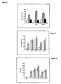
A high IgM titer was detected in the sera of mice which
were not depleted of their AM, intratracheally primed with
SP-B containing antigen liposomes, and boosted with antigen
(Fig 6a).
-
Additional depletion of AM caused only a slight
enhancement of the IgM titers. In contrast, only a poor IgG
titer is observed when the animals were not depleted of
their AM with marginal effects of incorporation of SP-B in
the antigen liposomes (Fig 6b). Higher IgG titers are
observed when the mice were depleted of their AM prior to
the intratracheal instillation of antigen liposomes, which
can be further increased by the use of SP-B containing
antigen liposomes.
DISCUSSION
-
Antigen entrapped in surfactant liposomes can induce a
systemic immune response in mice, as shown after
intratracheal instillation. No depletion of the alveolar
macrophages by an intratracheal injection of DMDP-containing
liposomes is needed to observe this immune response. A
preferred method for the induction of the immune response
via the airways is a method whereby the antigen is entrapped
in liposomes which contain at least SP-B and
phosphatidylglycerol. The SP-B effect is concentration
dependent reaching a maximum at the physiological
phospholipid: SP-B ratio of 100:0,5-1. The observed PG
dependency of SP-B is not surprising, since the positive
charges of SP-B were reported to be essential for its
activity (8) such as the phospholipid insertion in the
surface film, which covers the alveoli, via the negatively
charged PG (9,10).
-
In a previous study from our laboratory, Thepen et al
(11) demonstrated that only a local, IgA, immune response
can be elicited in the lung after prior depletion of the
alveolar macrophages by DMDP-containing liposomes. Depletion
of alveolar macrophages was necessary to induce an immune
response via the airways, because alveolar macrophages were
shown to inhibit immune responses probably via the
suppression of T-cells and dendritic cells.
-
Depletion of alveolar macrophages which leads to the
destruction of cells which may compete with the type II
cells for the uptake of antigen containing liposomes as well
as a reduction of the suppression of the immune response, is
no longer needed to induce a primary immune response when
animals are intratracheally injected with SP-B-containing
antigen liposomes.
Applications of SP-B containing liposomes
-
The advantages for the use of SP-B containing liposomes
are: a. naturally occuring in the form of surfactant, b. the
homology of SP-B derived from different species (canine,
human, porcine rabbit) is high (more than 80%), c. SP-B
(porcine, bovine) containing liposomes, in the form of
surfactant, have already been used to treat premature
children.
Applications
-
- 1) SP-B containing liposomes can be used, as in this study,
for immunisation via the airways. A systemic immune
response, IgM, can be obtained. No depletion necessary.
- 2) In combination with AM depleting liposomes, SP-B-antigen
liposomes can be used for immunisation.
- 3) Since SP-B and surfactant analogue material is present
in the intestines, immunisation via the intestines using SP-B
containing antigen liposomes is effective.
- 4) In the lungs or in the intestines SP-B containing
liposomes can be used to transport the following substances
to the alveolar type II cells (or similar cells in the
intestines):
- a) DNA, SP-B coupled to modified SP-B has been shown to
enhance transfection. Probably SP-B, DNA containing
liposomes may work to get DNA in vivo into the type II
cells.
- b) transport of corticosteroids to alveolar type II cells.
These substances are used for immature children in order to
induce the surfactant production by type II cells.
- c) transport of antioxidants to type II cells. Has been
shown for SP-A containing liposomes.
-
REFERENCES
-
- 1. Haagsman HP, Hawgood S, Sargeant T, Buckley D, White
RT, Drickamer K, Benson BJ. 1987. The major lung
surfactant protein, SP28-36, is a calcium-dependent,
carbohydrate binding protein. J. Biol. Chem.
262:13877-13880.
- 2. Hawgood S, Benson BJ, Hamilton, RL. 1985. Effects of
a surfactant-associated protein and calcium ions on
the structure and surface activity of lung surfactant
lipids. Biochemistry 24: 184-190.
- 3. Oosterlaken-Dijksterhuis MA, van Eijk M, van Buel BLM
van Golde LMG, Haagsman HP. 1991. Surfactant protein
composition of lamellar bodies isolated from rat
lung. Biochem.J. 274: 115-119.
- 4. Bartlett, GR. 1959. Phosphorous assay in column
chromatography. J. Biol. Chem. 234:466-468.
- 5. Sedgwick JD, Holt PGA. 1983. Solid-phase
immunoenzymatic technique for the enumberation of
specific antibody-secreting cells. J. Immunol.Meth.
57:301-309.
- 6. Delemarre FGA, Claassen E, van Rooijen N. 1989. The
primary in situ immune response in popliteal lymph
nodes and spleen of mice after subcutaneous
immunisation with thymus-dependent or thymus-independent
(type 1 and 2) antigens. Anat.Rec.
223:152-157.
- 7. Phizackerley PJR, Town MH, Newman GE. 1979.
Hydrophobic proteins of lamellated osmiophilic bodies
isolated from pig lung. Biochem J. 183:731-736.
- 8. Cochrane CG, Revak SD. 1991. Pulmonary surfactant
protein (SP-B): structure-function relationships.
Science. 253:566-568.
- 9. Yu SH, Possmayer F. 1992. Effect of pulmonary
surfactant protein B (SP-B) and calcium on
phospholipid adsorption and squeeze-out of
phosphatidylglycerol from binary phospholipid
monolayers containing dipalmitoylphosphatidylcholine.
Biochim. Biophys. Acta. 1126:26-34.
- 10. Yu SH, Possmayer F. 1992. Studies on surfactant-associated
protein B mediated adsorption of
surfactant phospholipids. Am.Rev.Respir.Dis.
145:A874.
- 11. Thepen T, van Rooijen N. Kraal G. 1989. Alveolar
macrophage elimination in vivo is associated with an
increase in pulmonary immune response in mice.
J.Exp.Med. 170:499-509
-
LEGENDS
-
- Figure 1. Surfactant liposomes.
BALB/c mice were intratracheally injected on day 0 with
DMDP-containing liposomes (100µl/n\mouse). On day 2, mice
were either intratracheally injected with surfactant
liposomes which contained no surfactant proteins (open
circles; 100 µl liposomes suspension/mouse) or with
surfactant liposomes which contained the surfactant proteins
SP-B and SP-C (closed circles; 100 µl liposomes
suspension/mouse). The mice were sacrificed on the day 9,
and the number of anti (IgM)-TNP positive cells/106 spleen
was determined (n=5, mean +/-SD).
- Figure 2. Influence of the surfactant proteins on the
primary immune response.
BALB/c mice were intratracheally injected on day 0 with
DMDP-containing liposomes (100 µl/mouse). On day 2, mice were
intratracheally injected with 100 µl TNP-KLH (=C), or 100 µl
TNP-KLH-surfactant lipids containing liposomes (3 mg
phospholipids/ml;=S), or 100 µl TNP-KLH surfactant lipids +
SP-A containing liposomes (3mg phospholipids/ml; 200 µg
SP-A/ml; =S+A), or 100 µl TNP-KLH-surfactant lipids + SP-B
containing liposomes (3 mg phospholipids/ml;15 µg SP-B/ml; =
S+B), or 100 µl TNP-KLH-surfactant lipids + SP-C containing
liposomes (3 mg phospholipids/ml; 15 µg SP-C/ml; =S+C), or
100 µl TNP-KLH-surfactant lipids + SP-B + SP-C containing
liposomes (3 mg phospholipids/ml; 15 µg SP-B/ml; 15 µg SP-C/ml;
=S+B+C). The mice were sacrificed on the day 9, and
the number of anti (IgM)-TNP positive cells/106 spleen cells
was determined (n=5,mean+/-SD).
- Figure 3. SP-B concentration dependency.
BALB/c mice were intratracheally injected on day 0 with
DMDP-containing liposomes (100 µl/mouse). On day 2, mice were
intratracheally injected with 100 µl TNP-KLH-surfactant
lipids (3 mg phospholipids/ml) + the indicated
concentrations of SP-B containing liposomes The mice were
sacrificed on the day 9, and the number of anti (IgM)-TNP
positive cells/106 spleen cells was determined (n=5,mean+/-SD).
- Figure 4. The influences of AM depletion and the lipid
composition on the primary immune response.
BALB/c mice were either not disturbed (=C), or
intratracheally injected with PBS-containing liposomes
(100 µl/mouse;=PBS), or DMDP-containing liposomes
(100 µl/mouse;=DMDP). On day 2, mice were intratracheally
injected with either 100 µl TNP-KLH-containing liposomes
which consisted of the following lipids (3 mg
phospholipids/ml; DPPC/eggPC/eggPG/cholesterol; 55:27:10:8
(w/w/w/w) (open bar), or 100 µl TNP-KLH-containing liposomes
which consisted of the following lipids (3 mg
phospholipids/ml; DPPC/eggPC/eggPG/cholesterol:55:27:10:8
(w/w/w/w) supplemented with 15 µg SP-B/ml (cross hatched
bar), or 100 µl TNP-KLH-containing liposomes which consisted
of the following lipids (3 mg phospholipids/ml;
DPPC/eggPC/cholesterol: 61:30:9 (w/w/w) supplemented with
15 µg SP-B/ml (closed bar). The mice were sacrificed on the
day 9, and the number of anti (IgM)-TNP positive cells 106
spleen cells was determined (n=5, mean +/-SD).
- Figure 5. The influences of AM depletion and the surfactant
proteins on the primary immune response.
BALB/c mice were either not disturbed (=open bar), or
intratracheally injected with DMDP-containing liposomes
(100 µl/mouse;=DMDP) (=cross hatched bar). On day 2, mice
were intratracheally injected with 100 µl TNP-KLH-surfactant
lipids containing liposomes (3 mg phospholipids/ml; =S), or
100 µl TNP-KLH-surfactant lipid + SP-B containing liposomes
(3 mg phospholipids/mi; 15 µg SP-B/ml; =S+B), or 100 µl TNP-KLH-surfactant
lipids + SP-B containing liposomes (3 mg
phospholipids/ml; 30 µg SP-B/ml; =S+2B), or 100 µl TNP-KLH-surfactant
lipids + SP-C containing liposomes (3 mg
phospholipids/ml; 15 µg SP-C/ml; = S+C), or 100 µl TNP-KLH-surfactant
lipids + SP-B+SP-C containing liposomes (3 mg
phospholipids/ml; 15 µg SP-B/ml; 15 µg SP-C/ml; =S+B+C).The
mice were sacrificed on the day 9, and the number of anti
(IgM)-TNP positive cells/106 spleen cells was determined
(n=5, mean +/-SD).
- Figure 6. The influences of AM depletion and the surfactant
proteins on the secondary immune response.
BALB/c mice were either not disturbed (=open bar), or
intratracheally injected with DMDP-containing liposomes
(100 µl/mouse;=DMDP) (=cross hatched bar). On day 2, mice
were intratracheally injected as desbribed under legend
figure 5, except for NS which were control mice, which were
not treated at all. On day 23, all mice, except for the
control mice (NS), were injected i.p. with 100 µl TNP-KLH
(0.8 mg/ml PBS). At day 30, the mice were sacrificed and
their sera collected Figure 6, depicts the IgM titers of the
sera compared to a positive control serum, which was
included in every plate.
-