EP0970106B1 - uPAR-MIMICKING PEPTIDES - Google Patents
uPAR-MIMICKING PEPTIDES Download PDFInfo
- Publication number
- EP0970106B1 EP0970106B1 EP98916982A EP98916982A EP0970106B1 EP 0970106 B1 EP0970106 B1 EP 0970106B1 EP 98916982 A EP98916982 A EP 98916982A EP 98916982 A EP98916982 A EP 98916982A EP 0970106 B1 EP0970106 B1 EP 0970106B1
- Authority
- EP
- European Patent Office
- Prior art keywords
- upar
- peptide
- cells
- seq
- sequence
- Prior art date
- Legal status (The legal status is an assumption and is not a legal conclusion. Google has not performed a legal analysis and makes no representation as to the accuracy of the status listed.)
- Expired - Lifetime
Links
- 108090000765 processed proteins & peptides Proteins 0.000 title claims abstract description 131
- 102000004196 processed proteins & peptides Human genes 0.000 title claims abstract description 48
- 206010028980 Neoplasm Diseases 0.000 claims abstract description 40
- 230000003399 chemotactic effect Effects 0.000 claims abstract description 39
- 201000011510 cancer Diseases 0.000 claims abstract description 29
- 230000004936 stimulating effect Effects 0.000 claims abstract description 24
- 230000029663 wound healing Effects 0.000 claims abstract description 10
- 201000010099 disease Diseases 0.000 claims abstract description 8
- 208000037265 diseases, disorders, signs and symptoms Diseases 0.000 claims abstract description 8
- 208000023275 Autoimmune disease Diseases 0.000 claims abstract description 5
- 210000004027 cell Anatomy 0.000 claims description 110
- 230000027455 binding Effects 0.000 claims description 39
- 230000000694 effects Effects 0.000 claims description 35
- 238000000034 method Methods 0.000 claims description 34
- 239000003814 drug Substances 0.000 claims description 28
- 150000001875 compounds Chemical class 0.000 claims description 25
- 150000007523 nucleic acids Chemical class 0.000 claims description 25
- 108020004707 nucleic acids Proteins 0.000 claims description 23
- 102000039446 nucleic acids Human genes 0.000 claims description 23
- 238000004519 manufacturing process Methods 0.000 claims description 22
- 230000014509 gene expression Effects 0.000 claims description 18
- 230000001965 increasing effect Effects 0.000 claims description 17
- 101800002670 p56 Proteins 0.000 claims description 13
- 230000001413 cellular effect Effects 0.000 claims description 11
- 238000003259 recombinant expression Methods 0.000 claims description 9
- 239000003795 chemical substances by application Substances 0.000 claims description 8
- 238000000338 in vitro Methods 0.000 claims description 7
- 108091000080 Phosphotransferase Proteins 0.000 claims description 6
- 102000020233 phosphotransferase Human genes 0.000 claims description 6
- 239000013598 vector Substances 0.000 claims description 6
- 102000007056 Recombinant Fusion Proteins Human genes 0.000 claims description 5
- 108010008281 Recombinant Fusion Proteins Proteins 0.000 claims description 5
- 230000007969 cellular immunity Effects 0.000 claims description 5
- 230000028709 inflammatory response Effects 0.000 claims description 5
- 210000004962 mammalian cell Anatomy 0.000 claims description 5
- 238000012216 screening Methods 0.000 claims description 5
- 239000004033 plastic Substances 0.000 claims description 4
- 229920003023 plastic Polymers 0.000 claims description 4
- 238000012360 testing method Methods 0.000 claims description 4
- 230000003110 anti-inflammatory effect Effects 0.000 claims description 3
- 230000002095 anti-migrative effect Effects 0.000 claims description 3
- 230000005809 anti-tumor immunity Effects 0.000 claims description 3
- 239000011248 coating agent Substances 0.000 claims description 3
- 238000000576 coating method Methods 0.000 claims description 3
- 238000007689 inspection Methods 0.000 claims description 3
- 238000011537 Coomassie blue staining Methods 0.000 claims description 2
- 241000588724 Escherichia coli Species 0.000 claims description 2
- 241000238631 Hexapoda Species 0.000 claims description 2
- 240000004808 Saccharomyces cerevisiae Species 0.000 claims description 2
- 230000000259 anti-tumor effect Effects 0.000 claims description 2
- 238000010322 bone marrow transplantation Methods 0.000 claims description 2
- 208000027866 inflammatory disease Diseases 0.000 claims description 2
- 102100032351 Coiled-coil domain-containing protein 91 Human genes 0.000 claims 2
- 108090000435 Urokinase-type plasminogen activator Proteins 0.000 abstract description 67
- 238000013508 migration Methods 0.000 abstract description 18
- 230000005012 migration Effects 0.000 abstract description 16
- 102000003990 Urokinase-type plasminogen activator Human genes 0.000 abstract description 7
- 229960005356 urokinase Drugs 0.000 abstract description 7
- 230000003213 activating effect Effects 0.000 abstract description 4
- 241000124008 Mammalia Species 0.000 abstract description 3
- 102000009816 urokinase plasminogen activator receptor activity proteins Human genes 0.000 abstract 1
- 108040001269 urokinase plasminogen activator receptor activity proteins Proteins 0.000 abstract 1
- 102100031358 Urokinase-type plasminogen activator Human genes 0.000 description 60
- 150000001413 amino acids Chemical class 0.000 description 45
- 235000001014 amino acid Nutrition 0.000 description 42
- 230000035605 chemotaxis Effects 0.000 description 40
- 239000012634 fragment Substances 0.000 description 27
- 241000282414 Homo sapiens Species 0.000 description 22
- 230000003993 interaction Effects 0.000 description 19
- OBMZMSLWNNWEJA-XNCRXQDQSA-N C1=CC=2C(C[C@@H]3NC(=O)[C@@H](NC(=O)[C@H](NC(=O)N(CC#CCN(CCCC[C@H](NC(=O)[C@@H](CC4=CC=CC=C4)NC3=O)C(=O)N)CC=C)NC(=O)[C@@H](N)C)CC3=CNC4=C3C=CC=C4)C)=CNC=2C=C1 Chemical compound C1=CC=2C(C[C@@H]3NC(=O)[C@@H](NC(=O)[C@H](NC(=O)N(CC#CCN(CCCC[C@H](NC(=O)[C@@H](CC4=CC=CC=C4)NC3=O)C(=O)N)CC=C)NC(=O)[C@@H](N)C)CC3=CNC4=C3C=CC=C4)C)=CNC=2C=C1 OBMZMSLWNNWEJA-XNCRXQDQSA-N 0.000 description 18
- 101710176384 Peptide 1 Proteins 0.000 description 18
- 102000005962 receptors Human genes 0.000 description 17
- 108020003175 receptors Proteins 0.000 description 17
- 101000760337 Homo sapiens Urokinase plasminogen activator surface receptor Proteins 0.000 description 16
- 238000003556 assay Methods 0.000 description 16
- 108091034117 Oligonucleotide Proteins 0.000 description 12
- 210000001744 T-lymphocyte Anatomy 0.000 description 12
- 230000012292 cell migration Effects 0.000 description 12
- 102100022644 26S proteasome regulatory subunit 4 Human genes 0.000 description 11
- 108090000317 Chymotrypsin Proteins 0.000 description 11
- 108020004414 DNA Proteins 0.000 description 11
- JLCPHMBAVCMARE-UHFFFAOYSA-N [3-[[3-[[3-[[3-[[3-[[3-[[3-[[3-[[3-[[3-[[3-[[5-(2-amino-6-oxo-1H-purin-9-yl)-3-[[3-[[3-[[3-[[3-[[3-[[5-(2-amino-6-oxo-1H-purin-9-yl)-3-[[5-(2-amino-6-oxo-1H-purin-9-yl)-3-hydroxyoxolan-2-yl]methoxy-hydroxyphosphoryl]oxyoxolan-2-yl]methoxy-hydroxyphosphoryl]oxy-5-(5-methyl-2,4-dioxopyrimidin-1-yl)oxolan-2-yl]methoxy-hydroxyphosphoryl]oxy-5-(6-aminopurin-9-yl)oxolan-2-yl]methoxy-hydroxyphosphoryl]oxy-5-(6-aminopurin-9-yl)oxolan-2-yl]methoxy-hydroxyphosphoryl]oxy-5-(6-aminopurin-9-yl)oxolan-2-yl]methoxy-hydroxyphosphoryl]oxy-5-(6-aminopurin-9-yl)oxolan-2-yl]methoxy-hydroxyphosphoryl]oxyoxolan-2-yl]methoxy-hydroxyphosphoryl]oxy-5-(5-methyl-2,4-dioxopyrimidin-1-yl)oxolan-2-yl]methoxy-hydroxyphosphoryl]oxy-5-(4-amino-2-oxopyrimidin-1-yl)oxolan-2-yl]methoxy-hydroxyphosphoryl]oxy-5-(5-methyl-2,4-dioxopyrimidin-1-yl)oxolan-2-yl]methoxy-hydroxyphosphoryl]oxy-5-(5-methyl-2,4-dioxopyrimidin-1-yl)oxolan-2-yl]methoxy-hydroxyphosphoryl]oxy-5-(6-aminopurin-9-yl)oxolan-2-yl]methoxy-hydroxyphosphoryl]oxy-5-(6-aminopurin-9-yl)oxolan-2-yl]methoxy-hydroxyphosphoryl]oxy-5-(4-amino-2-oxopyrimidin-1-yl)oxolan-2-yl]methoxy-hydroxyphosphoryl]oxy-5-(4-amino-2-oxopyrimidin-1-yl)oxolan-2-yl]methoxy-hydroxyphosphoryl]oxy-5-(4-amino-2-oxopyrimidin-1-yl)oxolan-2-yl]methoxy-hydroxyphosphoryl]oxy-5-(6-aminopurin-9-yl)oxolan-2-yl]methoxy-hydroxyphosphoryl]oxy-5-(4-amino-2-oxopyrimidin-1-yl)oxolan-2-yl]methyl [5-(6-aminopurin-9-yl)-2-(hydroxymethyl)oxolan-3-yl] hydrogen phosphate Polymers Cc1cn(C2CC(OP(O)(=O)OCC3OC(CC3OP(O)(=O)OCC3OC(CC3O)n3cnc4c3nc(N)[nH]c4=O)n3cnc4c3nc(N)[nH]c4=O)C(COP(O)(=O)OC3CC(OC3COP(O)(=O)OC3CC(OC3COP(O)(=O)OC3CC(OC3COP(O)(=O)OC3CC(OC3COP(O)(=O)OC3CC(OC3COP(O)(=O)OC3CC(OC3COP(O)(=O)OC3CC(OC3COP(O)(=O)OC3CC(OC3COP(O)(=O)OC3CC(OC3COP(O)(=O)OC3CC(OC3COP(O)(=O)OC3CC(OC3COP(O)(=O)OC3CC(OC3COP(O)(=O)OC3CC(OC3COP(O)(=O)OC3CC(OC3COP(O)(=O)OC3CC(OC3COP(O)(=O)OC3CC(OC3COP(O)(=O)OC3CC(OC3CO)n3cnc4c(N)ncnc34)n3ccc(N)nc3=O)n3cnc4c(N)ncnc34)n3ccc(N)nc3=O)n3ccc(N)nc3=O)n3ccc(N)nc3=O)n3cnc4c(N)ncnc34)n3cnc4c(N)ncnc34)n3cc(C)c(=O)[nH]c3=O)n3cc(C)c(=O)[nH]c3=O)n3ccc(N)nc3=O)n3cc(C)c(=O)[nH]c3=O)n3cnc4c3nc(N)[nH]c4=O)n3cnc4c(N)ncnc34)n3cnc4c(N)ncnc34)n3cnc4c(N)ncnc34)n3cnc4c(N)ncnc34)O2)c(=O)[nH]c1=O JLCPHMBAVCMARE-UHFFFAOYSA-N 0.000 description 11
- 229960002376 chymotrypsin Drugs 0.000 description 11
- 230000007246 mechanism Effects 0.000 description 11
- 102100022006 Cell division cycle protein 123 homolog Human genes 0.000 description 10
- 102000019034 Chemokines Human genes 0.000 description 10
- 108010012236 Chemokines Proteins 0.000 description 10
- 101000897353 Homo sapiens Cell division cycle protein 123 homolog Proteins 0.000 description 10
- 229940079593 drug Drugs 0.000 description 10
- 239000000203 mixture Substances 0.000 description 10
- 108090000623 proteins and genes Proteins 0.000 description 10
- 230000006041 cell recruitment Effects 0.000 description 9
- 230000002950 deficient Effects 0.000 description 9
- 238000002474 experimental method Methods 0.000 description 9
- 235000018102 proteins Nutrition 0.000 description 9
- 102000004169 proteins and genes Human genes 0.000 description 9
- 241001529936 Murinae Species 0.000 description 8
- 230000000903 blocking effect Effects 0.000 description 8
- 210000002950 fibroblast Anatomy 0.000 description 8
- 230000006870 function Effects 0.000 description 8
- 230000002401 inhibitory effect Effects 0.000 description 8
- 210000001616 monocyte Anatomy 0.000 description 8
- 230000008569 process Effects 0.000 description 8
- 230000002797 proteolythic effect Effects 0.000 description 8
- 230000007115 recruitment Effects 0.000 description 8
- 230000004913 activation Effects 0.000 description 7
- 239000002299 complementary DNA Substances 0.000 description 7
- 102000006495 integrins Human genes 0.000 description 7
- 108010044426 integrins Proteins 0.000 description 7
- 210000002536 stromal cell Anatomy 0.000 description 7
- 241000699670 Mus sp. Species 0.000 description 6
- 239000002975 chemoattractant Substances 0.000 description 6
- 238000003776 cleavage reaction Methods 0.000 description 6
- 230000001419 dependent effect Effects 0.000 description 6
- 230000007017 scission Effects 0.000 description 6
- 230000000638 stimulation Effects 0.000 description 6
- 239000000758 substrate Substances 0.000 description 6
- 210000004881 tumor cell Anatomy 0.000 description 6
- 241000725303 Human immunodeficiency virus Species 0.000 description 5
- 102000004022 Protein-Tyrosine Kinases Human genes 0.000 description 5
- 108090000412 Protein-Tyrosine Kinases Proteins 0.000 description 5
- 108010031318 Vitronectin Proteins 0.000 description 5
- 102100035140 Vitronectin Human genes 0.000 description 5
- 230000021164 cell adhesion Effects 0.000 description 5
- 239000003112 inhibitor Substances 0.000 description 5
- 239000003446 ligand Substances 0.000 description 5
- 210000002540 macrophage Anatomy 0.000 description 5
- 230000001617 migratory effect Effects 0.000 description 5
- 239000013642 negative control Substances 0.000 description 5
- 210000000440 neutrophil Anatomy 0.000 description 5
- 230000003389 potentiating effect Effects 0.000 description 5
- 150000003384 small molecules Chemical class 0.000 description 5
- 241000894007 species Species 0.000 description 5
- 241000283690 Bos taurus Species 0.000 description 4
- DHMQDGOQFOQNFH-UHFFFAOYSA-N Glycine Chemical compound NCC(O)=O DHMQDGOQFOQNFH-UHFFFAOYSA-N 0.000 description 4
- 206010061218 Inflammation Diseases 0.000 description 4
- 241000699666 Mus <mouse, genus> Species 0.000 description 4
- 241000283973 Oryctolagus cuniculus Species 0.000 description 4
- 102000001253 Protein Kinase Human genes 0.000 description 4
- 239000005557 antagonist Substances 0.000 description 4
- 231100000673 dose–response relationship Toxicity 0.000 description 4
- 239000012133 immunoprecipitate Substances 0.000 description 4
- 210000004969 inflammatory cell Anatomy 0.000 description 4
- 238000000021 kinase assay Methods 0.000 description 4
- 210000004698 lymphocyte Anatomy 0.000 description 4
- 230000036210 malignancy Effects 0.000 description 4
- 230000001404 mediated effect Effects 0.000 description 4
- PHEDXBVPIONUQT-RGYGYFBISA-N phorbol 13-acetate 12-myristate Chemical compound C([C@]1(O)C(=O)C(C)=C[C@H]1[C@@]1(O)[C@H](C)[C@H]2OC(=O)CCCCCCCCCCCCC)C(CO)=C[C@H]1[C@H]1[C@]2(OC(C)=O)C1(C)C PHEDXBVPIONUQT-RGYGYFBISA-N 0.000 description 4
- 125000002924 primary amino group Chemical group [H]N([H])* 0.000 description 4
- 108060006633 protein kinase Proteins 0.000 description 4
- 238000000746 purification Methods 0.000 description 4
- 230000009467 reduction Effects 0.000 description 4
- 230000004044 response Effects 0.000 description 4
- 238000002415 sodium dodecyl sulfate polyacrylamide gel electrophoresis Methods 0.000 description 4
- 238000002255 vaccination Methods 0.000 description 4
- 208000030507 AIDS Diseases 0.000 description 3
- 102100021943 C-C motif chemokine 2 Human genes 0.000 description 3
- 101710155857 C-C motif chemokine 2 Proteins 0.000 description 3
- 102000004127 Cytokines Human genes 0.000 description 3
- 108090000695 Cytokines Proteins 0.000 description 3
- 102000004190 Enzymes Human genes 0.000 description 3
- 108090000790 Enzymes Proteins 0.000 description 3
- 101000818522 Homo sapiens fMet-Leu-Phe receptor Proteins 0.000 description 3
- OKKJLVBELUTLKV-UHFFFAOYSA-N Methanol Chemical compound OC OKKJLVBELUTLKV-UHFFFAOYSA-N 0.000 description 3
- 102000035195 Peptidases Human genes 0.000 description 3
- 108091005804 Peptidases Proteins 0.000 description 3
- 102000013566 Plasminogen Human genes 0.000 description 3
- 108010051456 Plasminogen Proteins 0.000 description 3
- 239000004365 Protease Substances 0.000 description 3
- 241000700159 Rattus Species 0.000 description 3
- 102100024689 Urokinase plasminogen activator surface receptor Human genes 0.000 description 3
- 239000002253 acid Substances 0.000 description 3
- 239000000556 agonist Substances 0.000 description 3
- 125000000539 amino acid group Chemical group 0.000 description 3
- 238000013459 approach Methods 0.000 description 3
- 230000015572 biosynthetic process Effects 0.000 description 3
- 210000004369 blood Anatomy 0.000 description 3
- 239000008280 blood Substances 0.000 description 3
- 238000006243 chemical reaction Methods 0.000 description 3
- 230000031902 chemoattractant activity Effects 0.000 description 3
- 230000002900 effect on cell Effects 0.000 description 3
- 230000002708 enhancing effect Effects 0.000 description 3
- 229940088598 enzyme Drugs 0.000 description 3
- 102100021145 fMet-Leu-Phe receptor Human genes 0.000 description 3
- 230000035876 healing Effects 0.000 description 3
- 238000001727 in vivo Methods 0.000 description 3
- 230000004054 inflammatory process Effects 0.000 description 3
- 230000002452 interceptive effect Effects 0.000 description 3
- 230000003211 malignant effect Effects 0.000 description 3
- 230000001394 metastastic effect Effects 0.000 description 3
- 206010061289 metastatic neoplasm Diseases 0.000 description 3
- 238000004091 panning Methods 0.000 description 3
- 238000010647 peptide synthesis reaction Methods 0.000 description 3
- 238000006366 phosphorylation reaction Methods 0.000 description 3
- 239000000047 product Substances 0.000 description 3
- 239000012679 serum free medium Substances 0.000 description 3
- 230000011664 signaling Effects 0.000 description 3
- FWMNVWWHGCHHJJ-SKKKGAJSSA-N 4-amino-1-[(2r)-6-amino-2-[[(2r)-2-[[(2r)-2-[[(2r)-2-amino-3-phenylpropanoyl]amino]-3-phenylpropanoyl]amino]-4-methylpentanoyl]amino]hexanoyl]piperidine-4-carboxylic acid Chemical compound C([C@H](C(=O)N[C@H](CC(C)C)C(=O)N[C@H](CCCCN)C(=O)N1CCC(N)(CC1)C(O)=O)NC(=O)[C@H](N)CC=1C=CC=CC=1)C1=CC=CC=C1 FWMNVWWHGCHHJJ-SKKKGAJSSA-N 0.000 description 2
- HRPVXLWXLXDGHG-UHFFFAOYSA-N Acrylamide Chemical compound NC(=O)C=C HRPVXLWXLXDGHG-UHFFFAOYSA-N 0.000 description 2
- 101000760336 Bos taurus Urokinase plasminogen activator surface receptor Proteins 0.000 description 2
- 108091003079 Bovine Serum Albumin Proteins 0.000 description 2
- 239000006144 Dulbecco’s modified Eagle's medium Substances 0.000 description 2
- 108010037362 Extracellular Matrix Proteins Proteins 0.000 description 2
- 102000010834 Extracellular Matrix Proteins Human genes 0.000 description 2
- 108010073385 Fibrin Proteins 0.000 description 2
- 102000009123 Fibrin Human genes 0.000 description 2
- BWGVNKXGVNDBDI-UHFFFAOYSA-N Fibrin monomer Chemical compound CNC(=O)CNC(=O)CN BWGVNKXGVNDBDI-UHFFFAOYSA-N 0.000 description 2
- 239000004471 Glycine Substances 0.000 description 2
- 108700026244 Open Reading Frames Proteins 0.000 description 2
- 108010043958 Peptoids Proteins 0.000 description 2
- 102000012288 Phosphopyruvate Hydratase Human genes 0.000 description 2
- 108010022181 Phosphopyruvate Hydratase Proteins 0.000 description 2
- 108010076504 Protein Sorting Signals Proteins 0.000 description 2
- 101000760333 Rattus norvegicus Urokinase plasminogen activator surface receptor Proteins 0.000 description 2
- 108020004511 Recombinant DNA Proteins 0.000 description 2
- 101100221606 Saccharomyces cerevisiae (strain ATCC 204508 / S288c) COS7 gene Proteins 0.000 description 2
- 206010072170 Skin wound Diseases 0.000 description 2
- 230000009471 action Effects 0.000 description 2
- 239000002671 adjuvant Substances 0.000 description 2
- 239000000427 antigen Substances 0.000 description 2
- 102000036639 antigens Human genes 0.000 description 2
- 108091007433 antigens Proteins 0.000 description 2
- 239000012911 assay medium Substances 0.000 description 2
- 238000000376 autoradiography Methods 0.000 description 2
- 210000003719 b-lymphocyte Anatomy 0.000 description 2
- 230000001580 bacterial effect Effects 0.000 description 2
- 229940098773 bovine serum albumin Drugs 0.000 description 2
- 210000004899 c-terminal region Anatomy 0.000 description 2
- 125000003178 carboxy group Chemical group [H]OC(*)=O 0.000 description 2
- 239000013592 cell lysate Substances 0.000 description 2
- 230000008859 change Effects 0.000 description 2
- 238000000749 co-immunoprecipitation Methods 0.000 description 2
- 239000003636 conditioned culture medium Substances 0.000 description 2
- 230000007123 defense Effects 0.000 description 2
- 238000012217 deletion Methods 0.000 description 2
- 230000037430 deletion Effects 0.000 description 2
- 230000001066 destructive effect Effects 0.000 description 2
- 238000004520 electroporation Methods 0.000 description 2
- 238000005516 engineering process Methods 0.000 description 2
- 239000013604 expression vector Substances 0.000 description 2
- 210000002744 extracellular matrix Anatomy 0.000 description 2
- 229950003499 fibrin Drugs 0.000 description 2
- 230000004927 fusion Effects 0.000 description 2
- 239000000411 inducer Substances 0.000 description 2
- 208000015181 infectious disease Diseases 0.000 description 2
- 230000002757 inflammatory effect Effects 0.000 description 2
- 239000002609 medium Substances 0.000 description 2
- 210000003205 muscle Anatomy 0.000 description 2
- 230000004719 natural immunity Effects 0.000 description 2
- 230000026731 phosphorylation Effects 0.000 description 2
- 229940012957 plasmin Drugs 0.000 description 2
- 239000004417 polycarbonate Substances 0.000 description 2
- 238000003752 polymerase chain reaction Methods 0.000 description 2
- 239000013641 positive control Substances 0.000 description 2
- 238000002360 preparation method Methods 0.000 description 2
- 238000004393 prognosis Methods 0.000 description 2
- 230000017854 proteolysis Effects 0.000 description 2
- 230000000717 retained effect Effects 0.000 description 2
- 230000019491 signal transduction Effects 0.000 description 2
- 238000003786 synthesis reaction Methods 0.000 description 2
- 238000010998 test method Methods 0.000 description 2
- 238000002560 therapeutic procedure Methods 0.000 description 2
- 230000036962 time dependent Effects 0.000 description 2
- 210000001519 tissue Anatomy 0.000 description 2
- QFVHZQCOUORWEI-UHFFFAOYSA-N 4-[(4-anilino-5-sulfonaphthalen-1-yl)diazenyl]-5-hydroxynaphthalene-2,7-disulfonic acid Chemical compound C=12C(O)=CC(S(O)(=O)=O)=CC2=CC(S(O)(=O)=O)=CC=1N=NC(C1=CC=CC(=C11)S(O)(=O)=O)=CC=C1NC1=CC=CC=C1 QFVHZQCOUORWEI-UHFFFAOYSA-N 0.000 description 1
- 108020005544 Antisense RNA Proteins 0.000 description 1
- 208000011594 Autoinflammatory disease Diseases 0.000 description 1
- 241000894006 Bacteria Species 0.000 description 1
- 206010006187 Breast cancer Diseases 0.000 description 1
- 208000026310 Breast neoplasm Diseases 0.000 description 1
- 210000001266 CD8-positive T-lymphocyte Anatomy 0.000 description 1
- 102000014914 Carrier Proteins Human genes 0.000 description 1
- 102000000844 Cell Surface Receptors Human genes 0.000 description 1
- 108010001857 Cell Surface Receptors Proteins 0.000 description 1
- 108050000299 Chemokine receptor Proteins 0.000 description 1
- 102000009410 Chemokine receptor Human genes 0.000 description 1
- 101710172503 Chemokine-binding protein Proteins 0.000 description 1
- 101800004419 Cleaved form Proteins 0.000 description 1
- 108091026890 Coding region Proteins 0.000 description 1
- 102000012422 Collagen Type I Human genes 0.000 description 1
- 108010022452 Collagen Type I Proteins 0.000 description 1
- 108091035707 Consensus sequence Proteins 0.000 description 1
- KCXVZYZYPLLWCC-UHFFFAOYSA-N EDTA Chemical compound OC(=O)CN(CC(O)=O)CCN(CC(O)=O)CC(O)=O KCXVZYZYPLLWCC-UHFFFAOYSA-N 0.000 description 1
- 102000008857 Ferritin Human genes 0.000 description 1
- 108050000784 Ferritin Proteins 0.000 description 1
- 238000008416 Ferritin Methods 0.000 description 1
- 108010067306 Fibronectins Proteins 0.000 description 1
- 102000016359 Fibronectins Human genes 0.000 description 1
- 102000004457 Granulocyte-Macrophage Colony-Stimulating Factor Human genes 0.000 description 1
- 108010017213 Granulocyte-Macrophage Colony-Stimulating Factor Proteins 0.000 description 1
- 208000031886 HIV Infections Diseases 0.000 description 1
- 208000037357 HIV infectious disease Diseases 0.000 description 1
- 102000025850 HLA-A2 Antigen Human genes 0.000 description 1
- 108010074032 HLA-A2 Antigen Proteins 0.000 description 1
- 101001046686 Homo sapiens Integrin alpha-M Proteins 0.000 description 1
- 101000935040 Homo sapiens Integrin beta-2 Proteins 0.000 description 1
- 101000617823 Homo sapiens Solute carrier organic anion transporter family member 6A1 Proteins 0.000 description 1
- 108091006905 Human Serum Albumin Proteins 0.000 description 1
- 102000008100 Human Serum Albumin Human genes 0.000 description 1
- 101150106931 IFNG gene Proteins 0.000 description 1
- 102100022338 Integrin alpha-M Human genes 0.000 description 1
- 102100025390 Integrin beta-2 Human genes 0.000 description 1
- 102000000588 Interleukin-2 Human genes 0.000 description 1
- 108010002350 Interleukin-2 Proteins 0.000 description 1
- 102000011782 Keratins Human genes 0.000 description 1
- 108010076876 Keratins Proteins 0.000 description 1
- 241000589902 Leptospira Species 0.000 description 1
- 102000018697 Membrane Proteins Human genes 0.000 description 1
- 108010052285 Membrane Proteins Proteins 0.000 description 1
- 241001465754 Metazoa Species 0.000 description 1
- 101000760331 Mus musculus Urokinase plasminogen activator surface receptor Proteins 0.000 description 1
- 108091028043 Nucleic acid sequence Proteins 0.000 description 1
- 101150038998 PLAUR gene Proteins 0.000 description 1
- 108010047620 Phytohemagglutinins Proteins 0.000 description 1
- 108010022233 Plasminogen Activator Inhibitor 1 Proteins 0.000 description 1
- 102100039418 Plasminogen activator inhibitor 1 Human genes 0.000 description 1
- 102000003923 Protein Kinase C Human genes 0.000 description 1
- 108090000315 Protein Kinase C Proteins 0.000 description 1
- 101710138270 PspA protein Proteins 0.000 description 1
- 239000012979 RPMI medium Substances 0.000 description 1
- 239000006146 Roswell Park Memorial Institute medium Substances 0.000 description 1
- MTCFGRXMJLQNBG-UHFFFAOYSA-N Serine Natural products OCC(N)C(O)=O MTCFGRXMJLQNBG-UHFFFAOYSA-N 0.000 description 1
- 102000012479 Serine Proteases Human genes 0.000 description 1
- 108010022999 Serine Proteases Proteins 0.000 description 1
- 102100021991 Solute carrier organic anion transporter family member 6A1 Human genes 0.000 description 1
- 230000006044 T cell activation Effects 0.000 description 1
- 108091008874 T cell receptors Proteins 0.000 description 1
- 230000005867 T cell response Effects 0.000 description 1
- 102000016266 T-Cell Antigen Receptors Human genes 0.000 description 1
- 108700012920 TNF Proteins 0.000 description 1
- 102100036407 Thioredoxin Human genes 0.000 description 1
- QYSXJUFSXHHAJI-XFEUOLMDSA-N Vitamin D3 Natural products C1(/[C@@H]2CC[C@@H]([C@]2(CCC1)C)[C@H](C)CCCC(C)C)=C/C=C1\C[C@@H](O)CCC1=C QYSXJUFSXHHAJI-XFEUOLMDSA-N 0.000 description 1
- 206010052428 Wound Diseases 0.000 description 1
- 208000027418 Wounds and injury Diseases 0.000 description 1
- 239000012082 adaptor molecule Substances 0.000 description 1
- 102000019997 adhesion receptor Human genes 0.000 description 1
- 108010013985 adhesion receptor Proteins 0.000 description 1
- 230000001919 adrenal effect Effects 0.000 description 1
- 238000001042 affinity chromatography Methods 0.000 description 1
- 238000004458 analytical method Methods 0.000 description 1
- 238000004873 anchoring Methods 0.000 description 1
- 238000010171 animal model Methods 0.000 description 1
- 238000000137 annealing Methods 0.000 description 1
- 229940121363 anti-inflammatory agent Drugs 0.000 description 1
- 239000002260 anti-inflammatory agent Substances 0.000 description 1
- 230000000692 anti-sense effect Effects 0.000 description 1
- 230000005875 antibody response Effects 0.000 description 1
- 230000000890 antigenic effect Effects 0.000 description 1
- 230000003190 augmentative effect Effects 0.000 description 1
- 230000001363 autoimmune Effects 0.000 description 1
- 230000035578 autophosphorylation Effects 0.000 description 1
- 235000015241 bacon Nutrition 0.000 description 1
- 210000002469 basement membrane Anatomy 0.000 description 1
- 230000009286 beneficial effect Effects 0.000 description 1
- 102000005936 beta-Galactosidase Human genes 0.000 description 1
- 108010005774 beta-Galactosidase Proteins 0.000 description 1
- 108091008324 binding proteins Proteins 0.000 description 1
- 210000000601 blood cell Anatomy 0.000 description 1
- 210000001185 bone marrow Anatomy 0.000 description 1
- 229910000389 calcium phosphate Inorganic materials 0.000 description 1
- 239000001506 calcium phosphate Substances 0.000 description 1
- 235000011010 calcium phosphates Nutrition 0.000 description 1
- 230000009400 cancer invasion Effects 0.000 description 1
- 210000001043 capillary endothelial cell Anatomy 0.000 description 1
- 230000020411 cell activation Effects 0.000 description 1
- 230000034196 cell chemotaxis Effects 0.000 description 1
- 210000000170 cell membrane Anatomy 0.000 description 1
- 230000009087 cell motility Effects 0.000 description 1
- 239000006285 cell suspension Substances 0.000 description 1
- 230000036755 cellular response Effects 0.000 description 1
- 239000003153 chemical reaction reagent Substances 0.000 description 1
- 238000010367 cloning Methods 0.000 description 1
- 238000000975 co-precipitation Methods 0.000 description 1
- 229940096422 collagen type i Drugs 0.000 description 1
- 108010047295 complement receptors Proteins 0.000 description 1
- 102000006834 complement receptors Human genes 0.000 description 1
- 239000003184 complementary RNA Substances 0.000 description 1
- 230000001010 compromised effect Effects 0.000 description 1
- 239000012141 concentrate Substances 0.000 description 1
- 230000001276 controlling effect Effects 0.000 description 1
- 239000013078 crystal Substances 0.000 description 1
- 238000005520 cutting process Methods 0.000 description 1
- 210000004292 cytoskeleton Anatomy 0.000 description 1
- 230000006378 damage Effects 0.000 description 1
- 230000003247 decreasing effect Effects 0.000 description 1
- 230000004665 defense response Effects 0.000 description 1
- 230000008021 deposition Effects 0.000 description 1
- 238000003745 diagnosis Methods 0.000 description 1
- 230000004069 differentiation Effects 0.000 description 1
- 230000007646 directional migration Effects 0.000 description 1
- 230000008030 elimination Effects 0.000 description 1
- 238000003379 elimination reaction Methods 0.000 description 1
- 210000002889 endothelial cell Anatomy 0.000 description 1
- 210000002919 epithelial cell Anatomy 0.000 description 1
- 210000003743 erythrocyte Anatomy 0.000 description 1
- 230000034964 establishment of cell polarity Effects 0.000 description 1
- 230000002349 favourable effect Effects 0.000 description 1
- 239000003527 fibrinolytic agent Substances 0.000 description 1
- 230000003480 fibrinolytic effect Effects 0.000 description 1
- 230000003328 fibroblastic effect Effects 0.000 description 1
- 210000001650 focal adhesion Anatomy 0.000 description 1
- 239000012737 fresh medium Substances 0.000 description 1
- 230000002068 genetic effect Effects 0.000 description 1
- 210000005260 human cell Anatomy 0.000 description 1
- 208000033519 human immunodeficiency virus infectious disease Diseases 0.000 description 1
- 210000002865 immune cell Anatomy 0.000 description 1
- 230000028993 immune response Effects 0.000 description 1
- 210000000987 immune system Anatomy 0.000 description 1
- 230000005847 immunogenicity Effects 0.000 description 1
- 238000000099 in vitro assay Methods 0.000 description 1
- 238000011534 incubation Methods 0.000 description 1
- 230000001939 inductive effect Effects 0.000 description 1
- 230000001524 infective effect Effects 0.000 description 1
- 102000017776 integrin beta chain Human genes 0.000 description 1
- 108060004057 integrin beta chain Proteins 0.000 description 1
- 210000002510 keratinocyte Anatomy 0.000 description 1
- 230000007257 malfunction Effects 0.000 description 1
- 239000011159 matrix material Substances 0.000 description 1
- 210000004379 membrane Anatomy 0.000 description 1
- 239000012528 membrane Substances 0.000 description 1
- 230000003278 mimic effect Effects 0.000 description 1
- 210000000066 myeloid cell Anatomy 0.000 description 1
- 210000000822 natural killer cell Anatomy 0.000 description 1
- 230000031990 negative regulation of inflammatory response Effects 0.000 description 1
- 238000010606 normalization Methods 0.000 description 1
- 230000001575 pathological effect Effects 0.000 description 1
- 230000007170 pathology Effects 0.000 description 1
- 238000002823 phage display Methods 0.000 description 1
- 239000002644 phorbol ester Substances 0.000 description 1
- 230000001885 phytohemagglutinin Effects 0.000 description 1
- 239000013612 plasmid Substances 0.000 description 1
- 229920000515 polycarbonate Polymers 0.000 description 1
- 239000001267 polyvinylpyrrolidone Substances 0.000 description 1
- 235000013855 polyvinylpyrrolidone Nutrition 0.000 description 1
- 229920000036 polyvinylpyrrolidone Polymers 0.000 description 1
- 238000010837 poor prognosis Methods 0.000 description 1
- 239000011148 porous material Substances 0.000 description 1
- 230000030503 positive regulation of chemotaxis Effects 0.000 description 1
- 230000003449 preventive effect Effects 0.000 description 1
- 230000001737 promoting effect Effects 0.000 description 1
- 210000001243 pseudopodia Anatomy 0.000 description 1
- 238000011002 quantification Methods 0.000 description 1
- 230000001105 regulatory effect Effects 0.000 description 1
- 230000008521 reorganization Effects 0.000 description 1
- 230000000284 resting effect Effects 0.000 description 1
- 230000002441 reversible effect Effects 0.000 description 1
- 239000007320 rich medium Substances 0.000 description 1
- 238000007423 screening assay Methods 0.000 description 1
- 230000028327 secretion Effects 0.000 description 1
- 238000000926 separation method Methods 0.000 description 1
- 230000001568 sexual effect Effects 0.000 description 1
- 239000007787 solid Substances 0.000 description 1
- 230000002269 spontaneous effect Effects 0.000 description 1
- 102000009076 src-Family Kinases Human genes 0.000 description 1
- 108010087686 src-Family Kinases Proteins 0.000 description 1
- 238000010186 staining Methods 0.000 description 1
- 238000007920 subcutaneous administration Methods 0.000 description 1
- 230000004083 survival effect Effects 0.000 description 1
- 230000001225 therapeutic effect Effects 0.000 description 1
- 108060008226 thioredoxin Proteins 0.000 description 1
- 229940094937 thioredoxin Drugs 0.000 description 1
- 238000010361 transduction Methods 0.000 description 1
- 230000026683 transduction Effects 0.000 description 1
- 230000001131 transforming effect Effects 0.000 description 1
- 230000009495 transient activation Effects 0.000 description 1
- 238000002054 transplantation Methods 0.000 description 1
- QORWJWZARLRLPR-UHFFFAOYSA-H tricalcium bis(phosphate) Chemical compound [Ca+2].[Ca+2].[Ca+2].[O-]P([O-])([O-])=O.[O-]P([O-])([O-])=O QORWJWZARLRLPR-UHFFFAOYSA-H 0.000 description 1
- 229940121358 tyrosine kinase inhibitor Drugs 0.000 description 1
- 239000005483 tyrosine kinase inhibitor Substances 0.000 description 1
- 238000011144 upstream manufacturing Methods 0.000 description 1
- 230000003612 virological effect Effects 0.000 description 1
- QYSXJUFSXHHAJI-YRZJJWOYSA-N vitamin D3 Chemical compound C1(/[C@@H]2CC[C@@H]([C@]2(CCC1)C)[C@H](C)CCCC(C)C)=C\C=C1\C[C@@H](O)CCC1=C QYSXJUFSXHHAJI-YRZJJWOYSA-N 0.000 description 1
- 235000005282 vitamin D3 Nutrition 0.000 description 1
- 239000011647 vitamin D3 Substances 0.000 description 1
- 229940021056 vitamin d3 Drugs 0.000 description 1
- 238000005406 washing Methods 0.000 description 1
- XLYOFNOQVPJJNP-UHFFFAOYSA-N water Substances O XLYOFNOQVPJJNP-UHFFFAOYSA-N 0.000 description 1
Images



Classifications
-
- C—CHEMISTRY; METALLURGY
- C07—ORGANIC CHEMISTRY
- C07K—PEPTIDES
- C07K14/00—Peptides having more than 20 amino acids; Gastrins; Somatostatins; Melanotropins; Derivatives thereof
- C07K14/435—Peptides having more than 20 amino acids; Gastrins; Somatostatins; Melanotropins; Derivatives thereof from animals; from humans
- C07K14/705—Receptors; Cell surface antigens; Cell surface determinants
-
- A—HUMAN NECESSITIES
- A61—MEDICAL OR VETERINARY SCIENCE; HYGIENE
- A61P—SPECIFIC THERAPEUTIC ACTIVITY OF CHEMICAL COMPOUNDS OR MEDICINAL PREPARATIONS
- A61P17/00—Drugs for dermatological disorders
- A61P17/02—Drugs for dermatological disorders for treating wounds, ulcers, burns, scars, keloids, or the like
-
- A—HUMAN NECESSITIES
- A61—MEDICAL OR VETERINARY SCIENCE; HYGIENE
- A61P—SPECIFIC THERAPEUTIC ACTIVITY OF CHEMICAL COMPOUNDS OR MEDICINAL PREPARATIONS
- A61P29/00—Non-central analgesic, antipyretic or antiinflammatory agents, e.g. antirheumatic agents; Non-steroidal antiinflammatory drugs [NSAID]
-
- A—HUMAN NECESSITIES
- A61—MEDICAL OR VETERINARY SCIENCE; HYGIENE
- A61P—SPECIFIC THERAPEUTIC ACTIVITY OF CHEMICAL COMPOUNDS OR MEDICINAL PREPARATIONS
- A61P35/00—Antineoplastic agents
-
- A—HUMAN NECESSITIES
- A61—MEDICAL OR VETERINARY SCIENCE; HYGIENE
- A61P—SPECIFIC THERAPEUTIC ACTIVITY OF CHEMICAL COMPOUNDS OR MEDICINAL PREPARATIONS
- A61P37/00—Drugs for immunological or allergic disorders
- A61P37/02—Immunomodulators
-
- A—HUMAN NECESSITIES
- A61—MEDICAL OR VETERINARY SCIENCE; HYGIENE
- A61K—PREPARATIONS FOR MEDICAL, DENTAL OR TOILETRY PURPOSES
- A61K39/00—Medicinal preparations containing antigens or antibodies
- A61K2039/51—Medicinal preparations containing antigens or antibodies comprising whole cells, viruses or DNA/RNA
-
- A—HUMAN NECESSITIES
- A61—MEDICAL OR VETERINARY SCIENCE; HYGIENE
- A61K—PREPARATIONS FOR MEDICAL, DENTAL OR TOILETRY PURPOSES
- A61K38/00—Medicinal preparations containing peptides
-
- A—HUMAN NECESSITIES
- A61—MEDICAL OR VETERINARY SCIENCE; HYGIENE
- A61K—PREPARATIONS FOR MEDICAL, DENTAL OR TOILETRY PURPOSES
- A61K48/00—Medicinal preparations containing genetic material which is inserted into cells of the living body to treat genetic diseases; Gene therapy
-
- C—CHEMISTRY; METALLURGY
- C07—ORGANIC CHEMISTRY
- C07K—PEPTIDES
- C07K2319/00—Fusion polypeptide
- C07K2319/01—Fusion polypeptide containing a localisation/targetting motif
- C07K2319/02—Fusion polypeptide containing a localisation/targetting motif containing a signal sequence
Definitions
- the present invention relates to a method for preventing or activating the migration of cells in a mammal, in particular a human.
- the method comprises the use of synthetic peptides, or of defined recombinant soluble forms of the urokinase receptor to activate the recruitment of inflammatory cells in a mammal, by stimulating a cellular adaptor that mediates the chemotactic activity of uPA.
- This mechanism does not require the protease activity of urokinase. Actually it by-passes it by acting on a downstream step, namely the interaction of such a peptide or recombinant soluble form of urokinase receptor with a cellular adaptor.
- the reagents produced are also useful to identify and isolate drugs that can inhibit said processes of cell recruitment, and hence may be employed in blocking the malignant phenotype of cancer and of aggressive hyper-or auto-inflammatory diseases.
- the response of the human body to noxic stimuli is dependent on the local production of inflammatory molecules as well as on the recruitment of specialized cells that migrate from the blood and the neighbouring tissue into the damaged site. These cells, neutrophils, monocyte-macrophages, lymphocytes and endothelial cells, respond to specific migratory stimuli and build up a defense response that results in the destruction and elimination of the nocive agent or organism. While essential and beneficial for the host defense, this type of response may also prove dangerous for the host or may hamper the success of some therapeutic or preventive approaches like transplantation and vaccination. In fact, a series of severe pathological entities exist which can be ascribed to excess or deficient migratory cell response.
- immunodeficient patients fail to respond to infective agents and this may be due to inability of the cells to rush to the damaged or infected site.
- Excess of recruitment may be responsive of destructive pathologies in which defense cells attack cells and functions of the host, as it happens for example in auto-immune diseases.
- Failure to properly respond to vaccination moreover, may also depend on unwanted recruitment of host inflammatory cells that destroy the antigenic cells too quickly to promote an immunological response.
- the availability of a specific "adjuvant" to natural immunity cells would be advantageous as it would produce a stronger antibody response.
- the malignancy of cancer cells mostly consists in the ability to invade and metastasize at a distance; in this process, however, non-malignant stromal host cells actively participate in establishing the malignant phenotype and hence the destructive and invasive phenotype. While the mechanisms involved are largely unknown, it is clear that stromal cells not only respond to stimuli arriving from the cancer cells but also signal to cancer cells through mechanisms of their own. In order to block the invasive phenotype of cancer, it is thus essential to act both on cancer and on stromal cells.
- the urokinase/urokinase receptor system The urokinase/urokinase receptor system
- uPA urokinase
- uPAR specific receptor
- uPA is a serine protease important in maintaining the fibrinolytic state of the body as it generates plasmin from plasminogen and hence prevents fibrin deposition.
- Plasmin is a broad spectrum protease that can destroy many proteins of the extracellular matrix and hence the inter-cellular and cell-to-extracellular matrix connections.
- UPA and uPAR have long been recognised as regulators of cell migration and hence to be important in inflammation and cancer invasion.
- uPA has other properties that do not require proteolytic activity, but simply the binding to the receptor.
- the proteolytic and the receptor binding activities are separated on the uPA molecule and can be assayed individually (receptor binding in the amino terminal fragment, proteolysis in the carboxy-terminal fragment).
- the functions that do not require the proteolytic moiety of uPA are the stimulation of mitogenesis, cell migration, adhesion and, in particular, chemotaxis (Gudewicz and Bilboa, 1987; Gyetko et al., 1994; Resnati et al., 1996; Besser et al., 1996).
- the specific uPA receptor is a GPI-anchored plasma membrane protein endowed with a very high affinity (Kd of 0.1-1 nM) for uPA, pro-uPA and inhibited forms of uPA, like the uPA-PAI-1 complex (Fazioli and Blasi, 1994).
- uPAR also binds vitronectin with an about 10-20 nM affinity as well as integrins.
- uPAR is formed by three repeats of about 90 amino acid residues connected by two linker regions (Dan ⁇ , Blasi et al., 1990; Behrendt et al., 1991) which define functionally and structurally different domains: the amino terminal domain (D1) contains the uPA binding site, while the carboxyterminal region containing domains D2 and D3 binds vitronectin.
- the integrity of the three-domains structure is necessary for high affinity binding to uPA at least with the purified, soluble protein (Dan ⁇ et al., 1994). To date, no information has been provided as to the function of the linker regions.
- uPAR is expressed in circulating blood cells, in particular monocytes, neutrophils and T-lymphocytes but not in erythrocytes or B-lymphocytes.
- uPAR is a target gene in lymphocytes and macrophage activation (CD87).
- monocytes and monocyte-like cells like HL60, U937) express uPAR, or are induced to overexpress uPAR by a variety of cytokines and other agents, like phorbol ester PMA, phytohemagglutinin, bacterial liposaccharide, TGFb1/vitamin D3, GM-CSF, IFNg, TNFa and others.
- T-lymphocytes In human T-lymphocytes, stimulation of the TCR/CD3 complex, lymphokines IL2, IL4, IL7 or the concomitant activation of the T cell receptor and integrins engagement, all induce uPAR expression (Nykj ⁇ r et al., 1994; Bianchi et al., 1996). It is also noteworthy, that tumor infiltrating T-lymphocytes heavily express uPAR and some of the properties of the activated T-lymphocytes, in particular their migration through reconstituted basement membranes, appear to be at least in part uPA- and uPAR-dependent (Bianchi et al., 1996).
- stromal cells In view of the important contribution of stromal cells to the invasiveness of cancer, it is also important to stress that macrophages present in human breast cancer and other cancers, where the level of uPAR production by the overall tumor importantly contributes to its malignancy, express high levels of uPAR (Brünner et al., 1996).
- mice lacking the uPA gene are highly defective in the response to bacterial and possibly other infections, and are incapable of recruiting T-lymphocytes and macrophages to the site of infection (Gyetko et al., 1996).
- uPA and uPAR are induced during T cells activation and directly contribute to the in vitro migration of human T cells (Nykj ⁇ r et al., 1994; Bianchi et al.,1996).
- tumor infiltrating T-lymphocytes heavily express uPAR (Bianchi et al., 1996).
- uPA-dependent chemotaxis is mediated by the activation of src-family tyrosine kinases (i.e. Hck in monocytes and Src in fibroblasts).
- Hck src-family tyrosine kinases
- the mechanism involved requires that uPA modifies uPAR conformation such that it can bind to a still unidentified adaptor. Indeed, in cells lacking uPAR, a mixture of fragments generated by cleaving a soluble, recombinant uPAR with chymotrypsin, has a very potent chemoattractant activity (IC50 of 10-20 pM) (Resnati et al., 1996).
- uPA or its derivatives induce migration through a chemotactic gradient in bovine adrenal capillary endothelial cells, keratinocytic cell lines, monocytes, monocyte-like cells, fibroblasts cell lines, and in neutrophils both in vitro and in vivo (Besser et al., 1996; Resnati et al., 1996).
- the involvement of uPAR in chemotaxis is not only a direct one, but may also be indirect by interfering with the chemotaxis induced by other chemokines.
- the chemotactic effect of FMLP or MCP-1 does not require the proteolytic activity of uPA, but uPAR occupancy is absolutely required for chemotaxis as uPAR antibodies or antisense RNA expression totally abolishes FMLP or MCP-1 activity.
- the chemotactic activity of uPAR has many of the properties of chemokines. Interaction between uPAR and integrins has been observed in neutrophils: the leucocyte integrin CD11b/CD18, aMb2, also known as the complement receptor type-3 (CR3) has been shown to physically associate with uPAR. This interaction is reversible and correlates with cell shape.
- uPAR and CR3 are co-localized, but following spontaneous cell polarization and migration, the two receptors dissociate, CR3 concentrates in the uropods and uPAR in the lamellipodia of polarized cells. Since CR3 regulates cell adhesion, chemotaxis and cell migration into inflammatory sites, the interaction with uPAR may be central to all these functions and hence to the cell-recruitment action of uPA/uPAR. No information however is available as to whether this integrin (or others) is directly involved in mediating the uPAR chemotactic signal, i.e. whether this integrin is the so far unidentified adaptor.
- Activation of migration by uPAR occupancy is observed also in epithelial cell lines where uPAR associates with a protein kinase that can serine-phosphorylate two cytokeratins, CK18 and CK8.
- the enzyme responsible is possibly the protein kinase Cz, since the process is independent from Ca2+, resistant to PMA down modulation and since the kinase can be specifically immuno-recognized.
- CK18 and CK8 are phosphorylated on serine and uPAR occupancy elicits a time-dependent increase in phosphorylation of CK8, cell shape changes and redistribution of cytokeratin filaments.
- Pro-uPA treatment caused rounding up of the cells and reduction of their diameter.
- cytokeratins were distributed in a lattice array parallel to the surface of the coverslips; upon treatment with pro-uPA, little or no cytokeratins were observed in filament form. These data tie together the migratory effect of pro-uPA, the activation of protein kinase C and the cell shape changes. Tyrosine-phosphorylation of a non-characterized 35 kDa protein occurs upon binding of pro-uPA to U937 myeloid cells. In these cells, occupancy of uPAR promotes cell adhesion during PMA-induced differentiation. It has been shown that uPAR directly binds vitronectin and promotes cell adhesion.
- uPAR directly interferes with the substrate recognition of adhesion receptors by forming complexes directly with the beta integrins, and hence promoting adhesion on vitronectin and inhibiting adhesion on fibronectin (Wei et al., 1996). Indeed, uPAR can be co-immunoprecipitated with anti-integrins IgG. The effect of uPAR on cell adhesion is also in line with its chemokine-like activity.
- integrins might represent the described adaptor suggested by Resnati et al. (1996).
- the uPAR-like small molecules to be described in this application have whatsoever no connection, either structural or conceptual, with the peptide identified by Wei et al. (1996).
- uPAR In monocytes and fibroblasts, occupancy of uPAR induces chemotaxis and the effect does not require extracellular proteolysis (Resnati et al., 1996).
- ATF binding causes a time-dependent and transient activation of a tyrosine kinase of the Src family, the p56/p58 Hck.
- uPAR itself associates with tyrosine kinases, through what appears to be a transmembrane-adaptor-mediated mechanism.
- cells lacking uPAR, and therefore not responsive to ATF are capable of chemotaxis when challenged with a mixture of chymotryptic fragments of soluble uPAR (chymotrypsin cleaves uPAR in two fragments between domain D1 and D2) (Resnati et al., 1996).
- This effect also occurs in fibroblasts derived from uPAR-/- mice. Therefore, uPAR can recognize a cell surface molecule which in turn is capable of mediating signal transduction.
- the occupancy of surface uPAR must in fact expose binding sites for the unidentified adaptor and hence transform uPAR from a receptor into a ligand.
- tyrosine kinases in uPAR-dependent chemotaxis is also shown by the effect of tyrosine kinase inhibitors that prevent the chemotactic response to ATF, and by the failure of primary src-/- fibroblasts to respond to the soluble, cleaved uPAR (Resnati, Blasi and Fazioli, unpublished).
- uPA binding to uPAR is strictly species-specific
- human uPAR acts as a chemoattractant as efficiently on human as on murine cells.
- chymotrypsin-cleaved soluble uPAR has an IC50 of about 20 pM (Resnati et al., 1996).
- the uPA/uPAR system is involved in cancer cell invasiveness, and also in inflammation and other diseases (Dan ⁇ et al., 1990). And in fact, the block of uPA/uPAR interaction by specific antagonists, or the reduction of uPAR synthesis by antisense gene expression leads to a drastic reduction of the migratory properties of cancer cells (Fazioli and Blasi, 1994).
- the expression level of the components of the system, uPA, PAI-1 and uPAR directly relates to the malignancy of the disease and its prognosis (Brünner et al., 1996). In fact, many if not all solid cancers have already released metastatic cells which are found in the bone marrow. The presence of uPAR-positive metastatic cells at the time of diagnosis represents a sign of extreme likelihood of poor prognosis, while the presence of metastatic cells per se does not.
- uPAR appears to be a specific target for novel types of drugs that might interfere with the process of inflammation or of cancer invasiveness.
- Two different possibilities are open: to block uPAR by interfering with uPA binding (uPAR antagonists), or to prevent the direct transduction of a migratory signal, via interaction with other molecules, bypassing the step of the interaction between uPA and uPAR.
- Drugs preventing the binding of uPAR to the before mentioned adaptor should also prevent chemotaxis via uPAR. Therefore, antagonists of the uPAR/adaptor interaction should block or decrease the migration of cells and hence might be useful in diseases where this decrease or block is advantageous, like cancer, autoimmune and excess-inflammatory reactions.
- uPAR as target for drugs enhancing the recruitment of inflammatory cells
- uPA derivatives like the amino terminal fragment ATF, pro-uPA or possibly also synthetic peptides that cover the uPAR-binding region of uPA are in fact capable of substituting for uPA in various functions not requiring uPA proteolytic activity (see for example, Fazioli and Blasi, 1994; Besser et al., 1996, Resnati et al., 1996).
- uPA binding to uPAR causes a conformational change that transforms uPAR from a receptor for uPA into a ligand for a still unidentified membrane adaptor (Resnati, et al. 1996)
- an alternative possibility of enhancing cell recruitment would be to bypass the first step, i.e. the uPA-uPAR binding step, and stimulate cell migration by acting at the level of the unidentified adaptor with uPAR itself or specific uPAR-like agonists.
- These agonists might therefore be useful in diseases in which the recruitment of cells is blocked by genetic or acquired malfunctions upstream of the uPAR/adaptor interaction.
- the availability of such drugs might be advantageous in connection with increasing the immunogenicity of various antigens, for example in vaccination, as it might act like an adjuvant by recruiting cells responsible of natural immunity.
- uPAR is an activation antigen in both T-lymphocytes and in monocytes (Nykj ⁇ r et al., 1994; Bianchi et al., 1996), i.e. in cells capable of active migration and in which HIV can replicate. While CD3-positive circulating lymphocytes do not or only weakly express uPAR, CD8+ T lymphocytes of patients of AIDS and other viral diseases display a high expression of uPAR, and in several of these patients as many as 80% of their T-lymphocytes are high expressers (Nykj ⁇ r et al., 1994).
- soluble uPAR Since cleavage activates the chemotactic activity of soluble uPAR (Resnati et al., 1996), these soluble, cleaved forms might, like actual chemokines, also interfere with the infectivity or survival of HIV. If a cleaved form of soluble uPAR is also involved in HIV infectivity or resistance, it might represent a target for a novel type of therapy in cases of AIDS.
- uPAR antagonists i.e. drugs that prevent the binding of uPA to uPAR.
- drugs are also expected to inhibit cell surface proteolytic activity.
- binding of uPA to uPAR stimulates chemotaxis by transforming this molecule from a receptor into a ligand for an adaptor (Resnati et al., 1996). Therefore, the identification of a small molecule that can mimic the chemotactic activity of uPAR gives a new handle to control the uPAR-dependent reactions, independent of its occupancy by uPA. In relation to the present invention, such small molecules have been discovered and characterized.
- uPA/uPAR act downstream of the uPA/uPAR interaction and therefore have no effect on cell surface proteolytic activity.
- These small molecules can be applied both in cases of deficient (as stimulators of the process), and in cases of excess cell recruitment and migration. In the latter case, the uPAR-like small molecules can be employed to identify suitable inhibitors of the uPAR-adaptor interaction.
- uPA is essential for inflammatory cell recruitment, binds uPAR and induces chemotaxis by direct signalling in a uPAR-dependent way.
- uPAR interacts with an unidentified adaptor molecule, and in fact a mixture of chymotryptic fragments of soluble uPAR has chemoattracting activity.
- the present invention is based upon the discovery of the mechanism through which uPAR activates chemotaxis. First the region of uPAR inducing chemotaxis was identified.
- soluble fragments of uPAR were produced either by separation of chymotrypsin-cleaved uPAR, or by recombinant DNA technology, and were used in a chemotaxis assay to locate the region of uPAR responsible for the chemoattracting activity on THP-1 cells.
- a region (residues 88-92 of uPAR) was localized in the linker region between domain D1 and D2.
- the chemotactic activity was found either in domain D1 or D2, whenever they included this linker region: carboxyterminally of domain D1, or aminoterminally of domain D2.
- the chemotactic activity of uPAR was reproduced using synthetic peptides including the 88-92 uPAR sequence of uPAR, on human THP-1 monocytoid cells (and murine LB6 fibroblastic cells).
- Synthetic molecules peptides 1 and 2) including the sequence SRSRY (ser-arg-ser-arg-tyr) (SEQ ID NO:7) had a very potent chemotactic activity at concentrations as low as 0.1 pM.
- Control peptides had no activity, in particular a peptide covering the carboxyterminal sequence deduced from uPAR cDNA, and a peptide having the same amino acid composition as peptide 1 but a scrambled sequence.
- peptide 1 stimulates chemotaxis through the same mechanism employed by the ligand uPA, or by the mixture of chymotrypsin fragments of the soluble uPAR.
- Peptide 1 in fact, activates Hck tyrosine kinase of THP-1 cells with the same time-course and concentration dependence observed with the mixture of chymotrypsin fragments of soluble uPAR.
- Example 4 shows how to practically identify inhibitors of uPAR by employing a simple panning assay using uPAR mimicking peptides, i.e. peptide 1 and 2 of the invention.
- uPAR peptides derived from uPAR found in mammalian species, 'such as human, bovine, murine and rat.
- the term "functional analogue” means a peptide with an amino acid sequence which is not identical to a chymotrypsin-cleaved suPAR peptide, but which has a substantially identical effect with respect to stimulating or increasing the chemotactic activity of a cell. This effect will typically be tested in an assay such as the one described in example 1.
- the increase in chemotactic activity in such an assay is preferably at least 100% of control, more preferably at least 200% of control, even more preferably at least 400% of control.
- Peptides having 80% sequence identity with any of the listed peptides and which exhibit an effect in the assay can also be used according to the invention.
- the present invention is directed to the use of a peptide having a sequence identity of 80% with any of the listed peptides for the manufacture of a medicament as claimed in claims 9 to 11 and 15 to 17.
- Another aspect of the present invention thus relates to a peptoid analogue of the peptide with the sequence SRSRY (SEQ ID NO:7) comprising one or more non-naturally occurring amino acids as listed in WO 94/28014.
- the peptoid analogues according to the invention can for example be prepared by using peptide synthesis methods, such as the method described by Merrifield. 1963.
- the synthesis may include any number of non-naturally occurring amino acids and optionally also including one or more naturally occurring amino acids in the preparation.
- the peptides and functional analogues of the invention can be added, in an effective amount, directly to cells or to an individual, such as a human patient, who harbours the cells which chemotactic activity can be stimulated or increased.
- a very important aspect of the present invention relates to peptides of the invention.
- Such peptides are a peptide with the sequence SRSRY (SEQ ID NO:7) or a functional analogue thereof as defined above and a peptide with the sequence AVTYSRSYLEC (SEQ ID NO:1) and a peptide with the sequence SRSRYLEC (SEQ ID NO:3).
- a particularly preferred embodiment of the present invention relates to a recombinant fusion protein, comprising one or more of the peptide species according to the invention and optionally additionally comprising one or more of the peptide species according to the invention.
- Functional analogues according to the invention, which are prepared using peptide synthesis are within the scope of the present invention.
- a further aspect of the present invention is the use of the inventive peptides for the manufacture of a medicament for stimulating or increasing a local inflammatory response.
- inventive chymotrypsin-cleaved fragment, or mixture of fragments, of uPAR, said fragment or mixture of fragments with the sequence SRSRY (SEQ ID NO: 7), or a functional analogue of said peptide, as defined above may be used.
- the term "stimulating or increasing local inflammatory response” means that one or more characteristics of an inflammatory response can be measured to be stimulated or increased. Such characteristics will typically be release of cytokines by T-lymphocytes or monocyte-macrophages, increase of chemotactic activity, ability to cause a local recruitment of lymphocytes/monocytes upon subcutaneous administration in a suitable laboratory animal, etc.
- the use of uPA-deficient mice is particularly favourable as these mice have a very low anti-inflammatory response. Methods to measure these activities are available routinely, and increases of such activities as mentioned of at least 50% are considered significant, preferably at least 80% even more preferably at least 100%.
- Another important aspect of the present invention relates to the use of a peptide according to the present invention, in particular a peptide with the sequence SRSRY (SEQ ID NO:7) or a functional analogue of said peptide, as defined above, for the manufacture of a medicament for stimulating or increasing the chemotactic activity of a cell.
- a further aspect of the present invention is the use of the inventive peptides for the manufacture of a medicament for stimulating or increasing wound healing:
- a peptide with the sequence SRSRY (SEQ ID NO:7) or a functional analogue of said peptide as defined above can be used according to the invention.
- stimulating or increasing wound healing means that at least one of the physiological characteristics or processes involved in healing of various types of wounds in an individual, such as a human, is substantially stimulated or increased.
- One way of quantifying such stimulation or increase in wound healing comprises measuring wound healing in a plasminogen-deficient or even a normal mouse, and identifying the time required for 50% healing. A reduction of the time of healing, upon addition of an effective amount of a composition comprising suPAR peptide, of at least 20%, preferably at least 50%, would represent a substantial wound healing stimulation.
- An important aspect of the present invention relates to the use of the inventive peptides for the manufacture of a medicament for stimulating the kinase activity of p56/p59 hck .
- a peptide with the sequence SRSRY (SEQ ID NO:7) or a functional analogue of said peptide, as defined above, can be used.
- stimulating the kinase activity means substantially increasing the ability of p56/p59 hck to phosphorylate a protein substrate. This will typically be measured using in vitro assays as the ones described in example 3. Quantification of such an effect might be obtained by cutting the bands from a gel such as the one showed in figure 5, and subsequently measuring the 32P-radioactivity, after normalization.
- a very important aspect of the present invention relates to a method of testing.whether a compound is capable of stimulating the binding between a chymotrypsin-cleaved soluble form of u-PAR according to the present invention and a cellular adaptor, said method comprising adding an effective amount of the compound to a test system comprising cells lacking endogenous u-PAR and measuring chemotactic activity; if chemotactic activity is present, then the compound is capable of stimulating said binding.
- the term "stimulating the binding” should be taken to mean increasing the binding in an assay, substantially resembling the above described, by at least 20%, preferably at least 30%, more preferably by at least 40%, even more preferably by at least 60%.
- the method of the present invention is suitable for the selection of a compound which is capable of stimulating the binding between a chymotrypsin-cleaved soluble form of uPAR and a cellular adaptor,
- a particularly important aspect of the present invention relates to a method of testing whether a compound is capable of blocking or inhibiting the binding between a chymotrypsin-cleaved soluble form of uPAR according to the present invention and a cellular adaptor, said method comprising adding chymotrypsin-cleaved soluble form of uPAR and an effective amount of the compound to a test system comprising cells lacking endogenous uPAR and measuring chemotactic activity. If chemotactic activity is inhibited or prevented, then the compound is capable of blocking said binding.
- blocking or inhibiting the binding should be taken to mean decreasing the binding in an assay, substantially resembling the above described, by at least 20%, preferably at least 30%, more preferably by at least 40%, even more preferably by at least 60%.
- the method of the present invention is suitable for the selection of a compound which is capable of blocking or inhibiting the binding between a chymotrypsin-cleaved soluble form of uPAR and a cellular adaptor.
- An important embodiment of the present invention is a compound according to the invention for,the treatment of cancer, autoimmune disease and/or hyper-inflammatory diseases.
- a compound which is capable of blocking or inhibiting the binding between a chymotrypsin-cleaved soluble form of uPAR and a cellular adaptor would according to the invention be administered to an individual, such as a human patient, in need thereof in a pharmaceutically acceptable form and in a pharmaceutically effective amount.
- a very important aspect of the present invention relates to a method of screening compounds for potential anti-inflammatory or anti-migratory properties.
- the method according to the invention comprises
- the present invention relates to oligo nucleotides, in particular oligonucleotides encoding the peptides given by SEQ ID NO: 1 and SEQ ID NO: 3.
- Another aspect of the present invention relates to the use of an oligonucleotide according to the invention, for the recombinant expression of peptides for use in the screening of compounds according to the invention.
- Preferred embodiments of the present invention relate to use of oligonucleotides according to the invention, wherein the recombinant expression is performed in an expression system chosen from the group consisting of an E. coli expression system, a yeast expression system, a mammalian expression system or an insect cell expression system.
- oligonucleotides according to the invention When using the oligonucleotides according to the invention as the basis for preparing peptides according to the invention by performing recombinant expression, it is preferred to combine said oligo nucleotides with nucleic acid sequences, such as regulatory sequences, which enable the expression system of choice to express large amounts of peptide.
- a particularly important aspect of the present invention relates to the use of a eukaryotic vector expressing a nucleic acid sequence encoding an inventive peptide, in particular a peptide with the sequence SRSRY (SEQ ID NO: 7), or a functional analogue thereof, as defined above, for the manufacture of a medicament for stimulating or increasing anti-tumour immunity in autologous bone marrow transplantation treatment of an individual, such as a human patient, bearing tumours.
- the medicament is useful for modifying tumour cells by transfecting them with the vector.
- anti-tumor immunity means the set of cellular and chemical reactions which an organism can organize with the aim of rejecting a tumor.
- the production of cytokines and chemokines is of major importance as it will drive the recruitment and the functional activation of cells that can specifically attack the tumor cells and hence destroy it.
- Another important aspect of the present invention relates to the use of a peptide according to the invention, in particular a peptide with the sequence SRSRY (SEQ ID NO: 7), or a functional analogue thereof as defined above, for the manufacture of a medicament for stimulating or increasing cellular immunity in an individual, such as a human patient, who is immunodeficient.
- a peptide according to the invention in particular a peptide with the sequence SRSRY (SEQ ID NO: 7), or a functional analogue thereof as defined above, for the manufacture of a medicament for stimulating or increasing cellular immunity in an individual, such as a human patient, who is immunodeficient.
- cellular immunity means the recruitment and the functional activation of immune cells, such as T-lymphocytes, NK-cells, B-lymphocytes, macrophages etc.
- the term "immunodeficient" means that the immune system of an individual is compromised and thus can be said to function at a sub-normal level with respect to at least some of the systems reactions.
- Yet another aspect of the present invention relates to the use of a human tumour cell line compatible on both the histotype and the HLA-A2 basis and modified so as to release the chemotactic region of the human or murine or bovine or rat uPAR, said chemotactic region being a fusion peptide or a domain of uPAR containing the SRSRY (SEQ ID NO: 7) peptide or a functional analogue thereof, as defined above, for the manufacture of an agent for vaccinating a subject, such as a human patient, suffering from tumours, with the aim of stimulating an anti-tumour T cell response.
- SRSRY SEQ ID NO: 7
- tumour-bearing subject such as a human patient may be injected with tumour-targeted cells modified to express the chemotactic region of uPAR from human, bovine, murine or rat, or any other uPAR having substantially the same properties, wherein the target vector is further fused to said region, being a peptide or a domain of uPAR containing the SRSRY (SEQ ID NO: 7) peptide, or a functional analogue thereof; as defined above.
- SRSRY SEQ ID NO: 7
- tumour-targeted cells means cells that, naturally or because they are specially engineered, can be injected into an individual and will specifically interact with tumour cells of said individual.
- Fig. 1 Part of this figure shows the sequence of active peptides 1 and 2 reproducing sequences of human uPAR, and the scrambled version of peptide 1 that was employed as a negative control employed in Example 2.
- sequence SRSRYLEC SEQ ID NO:3
- the active region can be further delimited since experiments from Example 1 show that the presence of the sequence SRSRY (SEQ ID NO:7) either carboxyterminal to domain D1, or aminoterminal to domain D2,3 confers to these peptides an otherwise absent chemotactic activity.
- the sequence SRSRY (SEQ ID NO:7) is present in human uPAR between positions 88 and 92.
- Part B of this figure shows the sequence of the active peptide region in rat, mouse and bovine uPAR and its comparison with that of human uPAR. It also shows a preliminary consensus sequence that indicates that the fourth and fifth residues are always conserved, the first and second residues show a conservative change while the third residue appears to be more freely substituted.
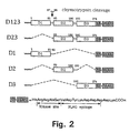
- FIG. 2 This figure shows a scheme depicting the structure of various soluble forms of uPAR to be employed in Example 1.
- Each construct was named on the basis of which of the three domains was still retained: D123 is the symbol for suPAR containing residues 1-274 and being secreted in a soluble form.
- Each construct contains at the 3' end a FLAGTM (SEQ ID NO:6) epitope which is employed for purification of the proteins onto a commercial antibody-affinity column.
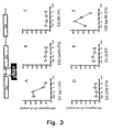
- FIG. 3 This figure shows the chemotactic response of THP-1 cells to fragments or mutants of soluble uPAR.
- migration toward the assay medium served as control (random cell migration) and is referred to as 100% migration. All experiments were performed in triplicate; the number of cells counted per high power field (ten fields for each condition) is expressed as percent'of the control values.
- the proteins were purified as described in the Methods section. Data points represent the mean of three independent experiments (+ SEM). The constructs used are depicted in Figure 2.
- Fig. 4 the chemotactic effect of synthetic peptides on THP-1 cells is shown.
- Peptides 1 and 2 show very potent activity, while peptide 3 or peptide-1 scrambled do not.
- Dose-dependent chemotactic response of THP-1 cells to peptide 1 (AVTYSRSRYLEC (SEQ ID NO:1), (aa 84-95) (panel A), peptide YTARLWGGTLLT (SEQ ID NO:2) (aa 302-313 of uPAR) (panel B), peptide (SRSRYLEC (SEQ ID NO:3), aa 88-95 of uPAR) (panel C) corresponding to the uPAR sequences of Roldan et al. (1990); panel D shows the chemotactic response to peptide TLVEYYSRASCR (SEQ ID NO:4), a scrambled version of peptide 1.
- Fig. 5 the stimulating effect of peptide 1 on the activity of Hck kinase of THP-1 cells is shown.
- THP-1 cells were metabolically labelled with [35S]-TransLabel, acid washed, and either mock-treated or treated with 0.1 nM peptide 1 (AVTYSRRYLEC, (SEQ ID NO:1)) for the indicated time at 37° C.
- Radiolabelled cell lysates were immunoprecipitated (Resnati et al., 1996) using an affinity-purified rabbit polyclonal anti-p56/p59hck antibody.
- the D1 region in the D12 and D123 constructs was deleted by substituting the NruI/NsiI fragment containing the D1 coding region, with a fragment generated by amplifying the uPAR cDNA with the primers FRA18 and D2.5'T and digested with the same enzymes.
- the D12 region in the D123 construct was deleted by substituting the NruI/NcoI fragment containing the D12 coding region, with a fragment generated by amplifying the uPAR cDNA with the primers FRA18 and D3.5'T and digesting with the same enzymes.
- All the mutant receptors were tagged at the carboxyterminus with the peptide sequence HRRASVDYKDDDDK (SEQ ID NO:5) which includes the protein kinase substrate and the FLAGTM epitope by inserting in the carboxy terminal ClaI site a linker made by annealing the two oligonucleotides K/FOcs and K/FOas. This linker was inserted into the ClaI site located at the carboxyterminus of all the constructs. All the recombinant coding regions were amplified with the primers NS1 and 46D, digested with NcoI and transferred to the eukaryotic expression vector pBNSEN digested with NcoI and EcoRV. Oligonucleotides (5'-3'):
- DMEM serum free medium
- DMEM serum free medium
- conditioned medium was collected and fresh medium added.
- Recombinant proteins were purified from the conditioned medium by passage over an anti-FLAGTM affinity column (M2 Affinity gel, Sigma). After washing with PBS, recombinant proteins were eluted with 0.1 M glycine pH 3.0.
- D1 (1-87) and D23 (88-274) were prepared by cleavage of D123 with chymotrypsin (Behrendt et al. 1991) followed by passage over the FLAGTM affinity column. D1(1-87) was recovered in the flow-through while the D23(88-274) containing the FLAGTM epitope was retained and later eluted with 0.1 M glycine pH 3.0.
- Chemotaxis analysis were performed using modified Boyden chambers containing polyvinylpyrrolidone polycarbonate filters (13 mm diameter, 5 mm pore size) coated with collagen type I (100 mg/ml in PBS pH 7.4).
- the coated filters were washed with medium supplemented with 0.2 % bovine serum albumin (BSA) and then placed in the Boyden apparatus. After acid wash, 2x10⁵ THP-1 cells suspended in serum-free medium were added above the filter in the Boyden chamber. Purified mutants were diluted in serum-free medium at the indicated concentrations and added below the filter in the lower chamber.
- the chambers were incubated in a humidified incubator at 37 °C in 5% CO2 in air for 90 minutes.
- Figure 2 shows a schematic representation of the structure of recombinant soluble uPAR and of various mutants generated.
- suPAR a soluble form of uPAR
- activity of suPAR requires chymotrypsin cleavage at Tyr87, between the N-terminal, ligand binding, domain D1 and domain D2+D3. This suggests that a unique conformation, uncovered by chymotrypsin cleavage, is required and must interact with a yet unidentified cell-surface receptor which might act as a transmembrane adaptor.
- the same mechanism applies to uPA binding to the surface-anchored uPAR.
- FIG. 2 shows the schematic representation of the three-domains entire suPAR (D123) (aa 1-274) and of the four deletion mutants constructed: D23 (aa 93-274), D1 (aa1-92), D2 (aa 93-191) and D3 (aa192-274).
- a kinase-site, followed by an in-frame FLAGTM epitope was inserted at the C terminus of each mutant. All the constructs were cloned in a modified form of the pBNSEN expression vector, COS7 cells transfected by the calcium phosphate co-precipitation method and secreted uPAR mutants protein purified by affinity chromatography.
- the purified D123 protein was digested with chymotrypsin (Resnati et al., 1996) and the corresponding products, D1 (aa 1-87) and D23 (88-274), were separated by an additional round of purification on FLAGTM antibody columns.
- the details of constructs preparation, the purification method and the system employed to assay chemotaxis are given in the above methods section.
- Figure 3 shows the results of the chemotaxis assays employing derivatives of suPAR D123 as chemoattractants. As predicted by previous results (Resnati et al., 1996), purified uncleaved DI23 was unable to stimulate chemotaxis in THP-1 cells, while chymotrypsin cleavage of this protein conferred a strong chemoattractant property (data not shown).
- SRSRY SEQ ID NO:7
- SEQ ID NO:7 Three human uPAR-related peptides were analyzed as inducers of chemotaxis on THP-1 cells: peptide 1 (AVTYSRSRYLEC (SEQ ID NO:1); amino acid sequence 84-95 of human uPAR) and its shorter analogue peptide 2 (SRSRYLEC (SEQ ID NO:3), amino acid sequence 88-95 of human uPAR) were tested as inducers of chemotaxis on THP-1 cells.
- AVTYSRSRYLEC SEQ ID NO:1
- SRSRYLEC SEQ ID NO:3
- SRSRY SEQ ID NO:7 motif.
- the third peptide was chosen in the carboxy terminal region of the uPAR cDNA sequence (YTARLWGGTLLT (SEQ ID NO:2), aa 301-313) and used in the assay as a negative control.
- An additional peptide was employed to control for the specificity of the effects mediated by the SRSRY (SEQ ID NO:7)-containing peptides.
- a scrambled version of peptide 1 (TLVEYYSRASCR (SEQ ID NO:4) was also tested under the same experimental conditions. Results of these experiments are shown in Fig. 4. Peptides 1 and 2 were able to elicit a dose-dependent chemotactic effect, with a maximal response at concentrations as low as 0.1 pM ( Figure 4, Panels A and C). On the contrary, when the assay was performed in the presence of similar doses of the control peptide 3, or of peptide TLVEYYSRASCR (SEQ ID NO:4) (peptide 1 in a scrambled sequence), no effect on cell migration was observed over the entire concentration range analyzed ( Figure 4, panels B and D). Thus, peptides containing the motif SRSRY (SEQ ID NO:7) of human uPAR have a potent chemotactic activity and must be responsible for uPAR induced migration.
- SRSRY motif SRSRY
- peptides represent novel chemotactic agents acting downstream of the uPA/uPAR interaction.
- Peptide 1 (AVTYSRSRYLEC (SEQ ID NO:1)) affects chemotaxis activating p56/p59hck tyrosine kinase activity in THP-1 cells
- the mixture of chymotrypsin fragments of suPAR like the uPA/uPAR interaction, induces chemotaxis by transiently activating the p56/p59hck protein kinase (Resnati et al., 1996). If human uPAR-related peptides containing the SRSRY (SEQ ID NO:7) motif induce a chemotactic response on THP-1 cells though the same mechanism exploited by the uPA/uPAR interaction (i.e. by binding to the unidentified adaptor), they should also activate p56/p59hck.
- SRSRY SEQ ID NO:7
- THP-1 cells were metabolically labelled with [35S]-TransLabel, acid washed, and either mock-treated or treated with 0.1 nM peptide 1 (AVTYSRSRYLEC (SEQ ID NO:1)) for the indicated time at 37° C.
- Radiolabelled cell lysates were immunoprecipitated (Resnati et al.,1996) using an affinity-purified rabbit polyclonal anti-p56/p59hck antibody. An aliquot from each immunoprecipitate was directly analyzed by SDS-PAGE to control that equal amount of p56/p59hck were present during in vitro kinase assay (Figure 5, panel A).
- Mammalian cells adhere on specially coated plastic plates, but do not adhere when plated onto plastic dishes commonly employed to grow bacteria. However, when those plates are coated with peptide 1 or 2 (or domain D1-1-92) or with recombinant forms of these peptides, they adhere because they express on their surface specific D1(1-92)-binding proteins. The adhesion can be measured.
- a positive control i.e. to set up the technique
- cells (106) exposing a high number of uPAR molecules are used and the panning is carried out on plates coated with a specific anti-uPAR antibody.
- a negative control the same cells but now not expressing uPAR are employed.
- the cells employed as negative controls are LB6, 32D, 3T3 fibroblasts, B-lymphoid cell lines and others.
- THP-1 cells, LB6/uPAR, 32D/uPAR and other expressing cell lines are employed.
- the same cells employed as negative controls (106) are used, and the plates are coated with domain D1(1-92) or domain D1(1-87) (control).
- the ability of the cells to bind to the plates is tested after 30-300 minutes incubation at 37°C by microscopic inspection, or by staining the plates with Coomassie Blue. The same can be done using different techniques.
- peptides 1- or 2-coated plates to specifically bind the THP-1 cells is the basis for the screening technique, as any compound capable of inhibiting this binding is a potential antiinflammatory agent, inhibiting chemotaxis.
- peptide 1 or peptide 2 can in fact inhibit adhesion of cells onto D1(1-92), while the scrambled or other peptides cannot.
- Such compounds therefore, can be tested as inhibitors of chemotaxis, employing the methods outlined in examples 1-3.
- Coating can also be carried out with other recombinant forms of the SRSRY (SEQ ID NO:7) sequence in which the epitope is synthesized in fusion with other proteins. Suitable proteins are ferritin (Sidoli et al. (1993)), thioredoxin, GST, beta galactosidase, human serum albumin.
- Inhibitors can be selected from phage display libraries of peptides, branched tripeptides combinatorial libraries, libraries of natural or of chemical compounds, and any other potentially exploitable collection of compounds.
Landscapes
- Health & Medical Sciences (AREA)
- Chemical & Material Sciences (AREA)
- Life Sciences & Earth Sciences (AREA)
- Organic Chemistry (AREA)
- Medicinal Chemistry (AREA)
- General Health & Medical Sciences (AREA)
- Nuclear Medicine, Radiotherapy & Molecular Imaging (AREA)
- Immunology (AREA)
- Veterinary Medicine (AREA)
- Public Health (AREA)
- Animal Behavior & Ethology (AREA)
- Pharmacology & Pharmacy (AREA)
- General Chemical & Material Sciences (AREA)
- Chemical Kinetics & Catalysis (AREA)
- Toxicology (AREA)
- Biophysics (AREA)
- Engineering & Computer Science (AREA)
- Bioinformatics & Cheminformatics (AREA)
- Biochemistry (AREA)
- Molecular Biology (AREA)
- Gastroenterology & Hepatology (AREA)
- Cell Biology (AREA)
- Genetics & Genomics (AREA)
- Zoology (AREA)
- Proteomics, Peptides & Aminoacids (AREA)
- Dermatology (AREA)
- Pain & Pain Management (AREA)
- Rheumatology (AREA)
- Medicines That Contain Protein Lipid Enzymes And Other Medicines (AREA)
- Peptides Or Proteins (AREA)
Abstract
Description
- The present invention relates to a method for preventing or activating the migration of cells in a mammal, in particular a human. The method comprises the use of synthetic peptides, or of defined recombinant soluble forms of the urokinase receptor to activate the recruitment of inflammatory cells in a mammal, by stimulating a cellular adaptor that mediates the chemotactic activity of uPA. This mechanism does not require the protease activity of urokinase. Actually it by-passes it by acting on a downstream step, namely the interaction of such a peptide or recombinant soluble form of urokinase receptor with a cellular adaptor. Additionally, the reagents produced are also useful to identify and isolate drugs that can inhibit said processes of cell recruitment, and hence may be employed in blocking the malignant phenotype of cancer and of aggressive hyper-or auto-inflammatory diseases.
- The response of the human body to noxic stimuli is dependent on the local production of inflammatory molecules as well as on the recruitment of specialized cells that migrate from the blood and the neighbouring tissue into the damaged site. These cells, neutrophils, monocyte-macrophages, lymphocytes and endothelial cells, respond to specific migratory stimuli and build up a defense response that results in the destruction and elimination of the nocive agent or organism. While essential and beneficial for the host defense, this type of response may also prove dangerous for the host or may hamper the success of some therapeutic or preventive approaches like transplantation and vaccination. In fact, a series of severe pathological entities exist which can be ascribed to excess or deficient migratory cell response. For example, immunodeficient patients fail to respond to infective agents and this may be due to inability of the cells to rush to the damaged or infected site. Excess of recruitment, on the other hand, may be responsive of destructive pathologies in which defense cells attack cells and functions of the host, as it happens for example in auto-immune diseases. Failure to properly respond to vaccination, moreover, may also depend on unwanted recruitment of host inflammatory cells that destroy the antigenic cells too quickly to promote an immunological response. The availability of a specific "adjuvant" to natural immunity cells, would be advantageous as it would produce a stronger antibody response.
- The malignancy of cancer cells mostly consists in the ability to invade and metastasize at a distance; in this process, however, non-malignant stromal host cells actively participate in establishing the malignant phenotype and hence the destructive and invasive phenotype. While the mechanisms involved are largely unknown, it is clear that stromal cells not only respond to stimuli arriving from the cancer cells but also signal to cancer cells through mechanisms of their own. In order to block the invasive phenotype of cancer, it is thus essential to act both on cancer and on stromal cells.
- The activity of urokinase (uPA) can be confined to the cell surface by the presence of a specific receptor (uPAR, CD87). uPA is a serine protease important in maintaining the fibrinolytic state of the body as it generates plasmin from plasminogen and hence prevents fibrin deposition. Plasmin is a broad spectrum protease that can destroy many proteins of the extracellular matrix and hence the inter-cellular and cell-to-extracellular matrix connections. UPA and uPAR have long been recognised as regulators of cell migration and hence to be important in inflammation and cancer invasion. In addition to functions connected to its proteolytic activity, uPA has other properties that do not require proteolytic activity, but simply the binding to the receptor. In fact, the proteolytic and the receptor binding activities are separated on the uPA molecule and can be assayed individually (receptor binding in the amino terminal fragment, proteolysis in the carboxy-terminal fragment). Among the functions that do not require the proteolytic moiety of uPA, are the stimulation of mitogenesis, cell migration, adhesion and, in particular, chemotaxis (Gudewicz and Bilboa, 1987; Gyetko et al., 1994; Resnati et al., 1996; Besser et al., 1996).
- The specific uPA receptor (uPAR) is a GPI-anchored plasma membrane protein endowed with a very high affinity (Kd of 0.1-1 nM) for uPA, pro-uPA and inhibited forms of uPA, like the uPA-PAI-1 complex (Fazioli and Blasi, 1994). In addition to uPA, uPAR also binds vitronectin with an about 10-20 nM affinity as well as integrins. Structurally, uPAR is formed by three repeats of about 90 amino acid residues connected by two linker regions (Danø, Blasi et al., 1990; Behrendt et al., 1991) which define functionally and structurally different domains: the amino terminal domain (D1) contains the uPA binding site, while the carboxyterminal region containing domains D2 and D3 binds vitronectin. However, the integrity of the three-domains structure is necessary for high affinity binding to uPA at least with the purified, soluble protein (Danø et al., 1994). To date, no information has been provided as to the function of the linker regions.
- uPAR is expressed in circulating blood cells, in particular monocytes, neutrophils and T-lymphocytes but not in erythrocytes or B-lymphocytes. In addition, uPAR is a target gene in lymphocytes and macrophage activation (CD87). Indeed, monocytes and monocyte-like cells (like HL60, U937) express uPAR, or are induced to overexpress uPAR by a variety of cytokines and other agents, like phorbol ester PMA, phytohemagglutinin, bacterial liposaccharide, TGFb1/vitamin D3, GM-CSF, IFNg, TNFa and others. In human T-lymphocytes, stimulation of the TCR/CD3 complex, lymphokines IL2, IL4, IL7 or the concomitant activation of the T cell receptor and integrins engagement, all induce uPAR expression (Nykjær et al., 1994; Bianchi et al., 1996). It is also noteworthy, that tumor infiltrating T-lymphocytes heavily express uPAR and some of the properties of the activated T-lymphocytes, in particular their migration through reconstituted basement membranes, appear to be at least in part uPA- and uPAR-dependent (Bianchi et al., 1996). In view of the important contribution of stromal cells to the invasiveness of cancer, it is also important to stress that macrophages present in human breast cancer and other cancers, where the level of uPAR production by the overall tumor importantly contributes to its malignancy, express high levels of uPAR (Brünner et al., 1996). The cooperation between stromal and cancer cells in cancer invasiveness, implies that high expression of uPAR by cancer or stromal cells, can affect cell prognosis possibly through different mechanisms depending on the overexpressing cell-type.
- Cooperation between cancer and stromal cells poses the problem of how uPAR expression on stromal cells influences the malignancy of cancer cells. Since the uPA/uPAR system has chemotactic activity, the production of uPAR by stromal cells can attract cancer cells through a chemokine like action. Indeed, chemokines produced by certain cells are anchored to a presentation molecule in the extracellular matrix and attract other cells having specific receptors (Schall and Bacon, 1996). The information available on the uPA/uPAR system supports this possibility.
- Stimulation of chemotaxis in monocyte-like cells, fibroblasts and some cancer cells requires the specific cell surface uPAR (CD87) which mediates the chemoattractant activity of uPA (Resnati et al., 1996). Chemotaxis by uPA does not require its protease activity, but only the occupancy of its receptor, and can be reproduced with the enzymatically inactive receptor binding moiety ATF (amino terminal fragment), pro-uPA and by chymotrypsin-cleaved soluble uPAR (Resnati et al., 1996). In mouse, the importance of this system in cell recruitment in vivo is shown by the fact that uPA is essential for the inflammatory response; indeed, mice lacking the uPA gene are highly defective in the response to bacterial and possibly other infections, and are incapable of recruiting T-lymphocytes and macrophages to the site of infection (Gyetko et al., 1996). In man, uPA and uPAR are induced during T cells activation and directly contribute to the in vitro migration of human T cells (Nykjær et al., 1994; Bianchi et al.,1996). Moreover, tumor infiltrating T-lymphocytes heavily express uPAR (Bianchi et al., 1996).
- uPA-dependent chemotaxis is mediated by the activation of src-family tyrosine kinases (i.e. Hck in monocytes and Src in fibroblasts). The mechanism involved requires that uPA modifies uPAR conformation such that it can bind to a still unidentified adaptor. Indeed, in cells lacking uPAR, a mixture of fragments generated by cleaving a soluble, recombinant uPAR with chymotrypsin, has a very potent chemoattractant activity (IC50 of 10-20 pM) (Resnati et al., 1996).
- In Boyden chamber-based assays, uPA or its derivatives induce migration through a chemotactic gradient in bovine adrenal capillary endothelial cells, keratinocytic cell lines, monocytes, monocyte-like cells, fibroblasts cell lines, and in neutrophils both in vitro and in vivo (Besser et al., 1996; Resnati et al., 1996). The involvement of uPAR in chemotaxis is not only a direct one, but may also be indirect by interfering with the chemotaxis induced by other chemokines. Exposure of human neutrophils to a chemotactic gradient of FMLP or MCP-1 chemokine localizes uPAR to the leading edge of the migrating cell; anti-uPAR antibodies ablate the chemotactic activity. The chemotactic effect of FMLP or MCP-1 does not require the proteolytic activity of uPA, but uPAR occupancy is absolutely required for chemotaxis as uPAR antibodies or antisense RNA expression totally abolishes FMLP or MCP-1 activity.
- The steps between the binding of chemokines to chemokine receptors and cell movements, are many and complex: signal transduction, reorganization of cytoskeleton, formation of focal adhesions, attachment and detachment from the substrate with pseudopodal extension and retraction to effect directional migration (Premack and Schall, 1996). The chemotactic activity of uPAR has many of the properties of chemokines. Interaction between uPAR and integrins has been observed in neutrophils: the leucocyte integrin CD11b/CD18, aMb2, also known as the complement receptor type-3 (CR3) has been shown to physically associate with uPAR. This interaction is reversible and correlates with cell shape. In resting cells, uPAR and CR3 are co-localized, but following spontaneous cell polarization and migration, the two receptors dissociate, CR3 concentrates in the uropods and uPAR in the lamellipodia of polarized cells. Since CR3 regulates cell adhesion, chemotaxis and cell migration into inflammatory sites, the interaction with uPAR may be central to all these functions and hence to the cell-recruitment action of uPA/uPAR. No information however is available as to whether this integrin (or others) is directly involved in mediating the uPAR chemotactic signal, i.e. whether this integrin is the so far unidentified adaptor.
- Activation of migration by uPAR occupancy is observed also in epithelial cell lines where uPAR associates with a protein kinase that can serine-phosphorylate two cytokeratins, CK18 and CK8. The enzyme responsible is possibly the protein kinase Cz, since the process is independent from Ca2+, resistant to PMA down modulation and since the kinase can be specifically immuno-recognized. In vivo, CK18 and CK8 are phosphorylated on serine and uPAR occupancy elicits a time-dependent increase in phosphorylation of CK8, cell shape changes and redistribution of cytokeratin filaments. Pro-uPA treatment caused rounding up of the cells and reduction of their diameter. In control cells, cytokeratins were distributed in a lattice array parallel to the surface of the coverslips; upon treatment with pro-uPA, little or no cytokeratins were observed in filament form. These data tie together the migratory effect of pro-uPA, the activation of protein kinase C and the cell shape changes. Tyrosine-phosphorylation of a non-characterized 35 kDa protein occurs upon binding of pro-uPA to U937 myeloid cells. In these cells, occupancy of uPAR promotes cell adhesion during PMA-induced differentiation. It has been shown that uPAR directly binds vitronectin and promotes cell adhesion. Moreover, uPAR directly interferes with the substrate recognition of adhesion receptors by forming complexes directly with the beta integrins, and hence promoting adhesion on vitronectin and inhibiting adhesion on fibronectin (Wei et al., 1996). Indeed, uPAR can be co-immunoprecipitated with anti-integrins IgG. The effect of uPAR on cell adhesion is also in line with its chemokine-like activity. A direct interaction of uPAR with integrins is suggested by their co-immunoprecipitation and by the finding of peptides that specifically inhibit the co-immunoprecipitation without interfering with the binding of uPA or vitronectin to uPAR (Wei et al., 1996). Therefore, integrins might represent the described adaptor suggested by Resnati et al. (1996). In this respect, the uPAR-like small molecules to be described in this application, have whatsoever no connection, either structural or conceptual, with the peptide identified by Wei et al. (1996).
- In monocytes and fibroblasts, occupancy of uPAR induces chemotaxis and the effect does not require extracellular proteolysis (Resnati et al., 1996). In this system, ATF binding causes a time-dependent and transient activation of a tyrosine kinase of the Src family, the p56/p58 Hck. In addition, uPAR itself associates with tyrosine kinases, through what appears to be a transmembrane-adaptor-mediated mechanism. Indeed, cells lacking uPAR, and therefore not responsive to ATF, are capable of chemotaxis when challenged with a mixture of chymotryptic fragments of soluble uPAR (chymotrypsin cleaves uPAR in two fragments between domain D1 and D2) (Resnati et al., 1996). This effect also occurs in fibroblasts derived from uPAR-/- mice. Therefore, uPAR can recognize a cell surface molecule which in turn is capable of mediating signal transduction. The occupancy of surface uPAR must in fact expose binding sites for the unidentified adaptor and hence transform uPAR from a receptor into a ligand. The role of tyrosine kinases in uPAR-dependent chemotaxis is also shown by the effect of tyrosine kinase inhibitors that prevent the chemotactic response to ATF, and by the failure of primary src-/- fibroblasts to respond to the soluble, cleaved uPAR (Resnati, Blasi and Fazioli, unpublished). Interestingly, while uPA binding to uPAR is strictly species-specific, human uPAR acts as a chemoattractant as efficiently on human as on murine cells. In particular, chymotrypsin-cleaved soluble uPAR has an IC50 of about 20 pM (Resnati et al., 1996).
- The uPA/uPAR system is involved in cancer cell invasiveness, and also in inflammation and other diseases (Danø et al., 1990). And in fact, the block of uPA/uPAR interaction by specific antagonists, or the reduction of uPAR synthesis by antisense gene expression leads to a drastic reduction of the migratory properties of cancer cells (Fazioli and Blasi, 1994). In human cancer, the expression level of the components of the system, uPA, PAI-1 and uPAR, directly relates to the malignancy of the disease and its prognosis (Brünner et al., 1996). In fact, many if not all solid cancers have already released metastatic cells which are found in the bone marrow. The presence of uPAR-positive metastatic cells at the time of diagnosis represents a sign of extreme likelihood of poor prognosis, while the presence of metastatic cells per se does not.
- On the basis of the above, uPAR appears to be a specific target for novel types of drugs that might interfere with the process of inflammation or of cancer invasiveness. Two different possibilities are open: to block uPAR by interfering with uPA binding (uPAR antagonists), or to prevent the direct transduction of a migratory signal, via interaction with other molecules, bypassing the step of the interaction between uPA and uPAR. Drugs preventing the binding of uPAR to the before mentioned adaptor should also prevent chemotaxis via uPAR. Therefore, antagonists of the uPAR/adaptor interaction should block or decrease the migration of cells and hence might be useful in diseases where this decrease or block is advantageous, like cancer, autoimmune and excess-inflammatory reactions.
- The recruitment of cells via the uPA-uPAR interaction suggests that drugs should be found which promote cell recruitment by binding to uPAR. And indeed, uPA derivatives like the amino terminal fragment ATF, pro-uPA or possibly also synthetic peptides that cover the uPAR-binding region of uPA are in fact capable of substituting for uPA in various functions not requiring uPA proteolytic activity (see for example, Fazioli and Blasi, 1994; Besser et al., 1996, Resnati et al., 1996). On the other hand, since uPA binding to uPAR causes a conformational change that transforms uPAR from a receptor for uPA into a ligand for a still unidentified membrane adaptor (Resnati, et al. 1996), an alternative possibility of enhancing cell recruitment would be to bypass the first step, i.e. the uPA-uPAR binding step, and stimulate cell migration by acting at the level of the unidentified adaptor with uPAR itself or specific uPAR-like agonists. These agonists might therefore be useful in diseases in which the recruitment of cells is blocked by genetic or acquired malfunctions upstream of the uPAR/adaptor interaction. Also, the availability of such drugs might be advantageous in connection with increasing the immunogenicity of various antigens, for example in vaccination, as it might act like an adjuvant by recruiting cells responsible of natural immunity.
- uPAR is an activation antigen in both T-lymphocytes and in monocytes (Nykjær et al., 1994; Bianchi et al., 1996), i.e. in cells capable of active migration and in which HIV can replicate. While CD3-positive circulating lymphocytes do not or only weakly express uPAR, CD8+ T lymphocytes of patients of AIDS and other viral diseases display a high expression of uPAR, and in several of these patients as many as 80% of their T-lymphocytes are high expressers (Nykjær et al., 1994). A fraction of persons remain uninfected by HIV despite multiple high-risk sexual exposure, and this is connected with the presence of HIV suppressive factors in their blood (Paxton et al., 1996). Several chemokines have been identified as HIV-suppressive factors produced by CD8+ T cells (Cocchi et al., 1996) and these chemokines represent novel targets for AIDS therapy. Soluble forms of uPAR exist in the blood and uPAR is in fact cleaved in cell lines and in cancer tissues. Since cleavage activates the chemotactic activity of soluble uPAR (Resnati et al., 1996), these soluble, cleaved forms might, like actual chemokines, also interfere with the infectivity or survival of HIV. If a cleaved form of soluble uPAR is also involved in HIV infectivity or resistance, it might represent a target for a novel type of therapy in cases of AIDS.
- Excessive fibrin deposits are observed in plasminogen-deficient, uPA-deficient and in the double uPAR/tPA-deficient mice and these mice are also deficient in skin wound healing (Carmeliet and Collen, 1996). Several mechanisms may be active in this phenomenon, including chemotaxis, since uPAR is localized at the leading edge of migrating keratinocytes during re-epithelization of mouse skin wounds. The importance of monocytes and macrophages in the process of wound healing makes it even more likely that this process might be augmented by a uPAR agonist capable of reproducing its chemoattractant property.
- Sofar, approaches directed to block the effect of uPAR in cell migration have been limited to the search of uPAR antagonists, i.e. drugs that prevent the binding of uPA to uPAR. In general, such drugs are also expected to inhibit cell surface proteolytic activity. However, binding of uPA to uPAR stimulates chemotaxis by transforming this molecule from a receptor into a ligand for an adaptor (Resnati et al., 1996). Therefore, the identification of a small molecule that can mimic the chemotactic activity of uPAR gives a new handle to control the uPAR-dependent reactions, independent of its occupancy by uPA. In relation to the present invention, such small molecules have been discovered and characterized. They act downstream of the uPA/uPAR interaction and therefore have no effect on cell surface proteolytic activity. These small molecules can be applied both in cases of deficient (as stimulators of the process), and in cases of excess cell recruitment and migration. In the latter case, the uPAR-like small molecules can be employed to identify suitable inhibitors of the uPAR-adaptor interaction.
- uPA is essential for inflammatory cell recruitment, binds uPAR and induces chemotaxis by direct signalling in a uPAR-dependent way. In this function uPAR interacts with an unidentified adaptor molecule, and in fact a mixture of chymotryptic fragments of soluble uPAR has chemoattracting activity. The present invention is based upon the discovery of the mechanism through which uPAR activates chemotaxis. First the region of uPAR inducing chemotaxis was identified. As shown in example 1, soluble fragments of uPAR were produced either by separation of chymotrypsin-cleaved uPAR, or by recombinant DNA technology, and were used in a chemotaxis assay to locate the region of uPAR responsible for the chemoattracting activity on THP-1 cells. Such a region (residues 88-92 of uPAR) was localized in the linker region between domain D1 and D2. In fact, the chemotactic activity was found either in domain D1 or D2, whenever they included this linker region: carboxyterminally of domain D1, or aminoterminally of domain D2. In example 2, the chemotactic activity of uPAR was reproduced using synthetic peptides including the 88-92 uPAR sequence of uPAR, on human THP-1 monocytoid cells (and murine LB6 fibroblastic cells). Synthetic molecules (
peptides 1 and 2) including the sequence SRSRY (ser-arg-ser-arg-tyr) (SEQ ID NO:7) had a very potent chemotactic activity at concentrations as low as 0.1 pM. Control peptides had no activity, in particular a peptide covering the carboxyterminal sequence deduced from uPAR cDNA, and a peptide having the same amino acid composition aspeptide 1 but a scrambled sequence. In example 3, we show thatpeptide 1 stimulates chemotaxis through the same mechanism employed by the ligand uPA, or by the mixture of chymotrypsin fragments of the soluble uPAR.Peptide 1, in fact, activates Hck tyrosine kinase of THP-1 cells with the same time-course and concentration dependence observed with the mixture of chymotrypsin fragments of soluble uPAR. Example 4 shows how to practically identify inhibitors of uPAR by employing a simple panning assay using uPAR mimicking peptides, i.e.peptide - Within the scope of the uPAR peptides according to the present invention are uPAR peptides derived from uPAR found in mammalian species, 'such as human, bovine, murine and rat.
- In the present context, the term "functional analogue" means a peptide with an amino acid sequence which is not identical to a chymotrypsin-cleaved suPAR peptide, but which has a substantially identical effect with respect to stimulating or increasing the chemotactic activity of a cell. This effect will typically be tested in an assay such as the one described in example 1. The increase in chemotactic activity in such an assay is preferably at least 100% of control, more preferably at least 200% of control, even more preferably at least 400% of control.
- Functional analogues of the human uPAR chemotactic activity according to the present invention are:









- Peptides having 80% sequence identity with any of the listed peptides and which exhibit an effect in the assay can also be used according to the invention. Thus, in one aspect, the present invention is directed to the use of a peptide having a sequence identity of 80% with any of the listed peptides for the manufacture of a medicament as claimed in
claims 9 to 11 and 15 to 17. - Another aspect of the present invention thus relates to a peptoid analogue of the peptide with the sequence SRSRY (SEQ ID NO:7) comprising one or more non-naturally occurring amino acids as listed in WO 94/28014.
- The peptoid analogues according to the invention can for example be prepared by using peptide synthesis methods, such as the method described by Merrifield. 1963. The synthesis may include any number of non-naturally occurring amino acids and optionally also including one or more naturally occurring amino acids in the preparation.The peptides and functional analogues of the invention can be added, in an effective amount, directly to cells or to an individual, such as a human patient, who harbours the cells which chemotactic activity can be stimulated or increased.
- A very important aspect of the present invention relates to peptides of the invention. Such peptides are a peptide with the sequence SRSRY (SEQ ID NO:7) or a functional analogue thereof as defined above and a peptide with the sequence AVTYSRSYLEC (SEQ ID NO:1) and a peptide with the sequence SRSRYLEC (SEQ ID NO:3).
- A particularly preferred embodiment of the present invention relates to a recombinant fusion protein, comprising one or more of the peptide species according to the invention and optionally additionally comprising one or more of the peptide species according to the invention.
- Functional analogues, according to the invention, which are prepared using peptide synthesis are within the scope of the present invention. Functional analogues, according to the invention, which are prepared by using other methods than peptide synthesis, such as recombinant expression, are also within the scope of the present invention.
- A further aspect of the present invention is the use of the inventive peptides for the manufacture of a medicament for stimulating or increasing a local inflammatory response. According to the present invention, the inventive chymotrypsin-cleaved fragment, or mixture of fragments, of uPAR, said fragment or mixture of fragments with the sequence SRSRY (SEQ ID NO: 7), or a functional analogue of said peptide, as defined above, may be used.
- In the present context, the term "stimulating or increasing local inflammatory response" means that one or more characteristics of an inflammatory response can be measured to be stimulated or increased. Such characteristics will typically be release of cytokines by T-lymphocytes or monocyte-macrophages, increase of chemotactic activity, ability to cause a local recruitment of lymphocytes/monocytes upon subcutaneous administration in a suitable laboratory animal, etc. In this particular case, the use of uPA-deficient mice is particularly favourable as these mice have a very low anti-inflammatory response. Methods to measure these activities are available routinely, and increases of such activities as mentioned of at least 50% are considered significant, preferably at least 80% even more preferably at least 100%.
Another important aspect of the present invention relates to the use of a peptide according to the present invention, in particular a peptide with the sequence SRSRY (SEQ ID NO:7) or a functional analogue of said peptide, as defined above, for the manufacture of a medicament for stimulating or increasing the chemotactic activity of a cell.
A further aspect of the present invention is the use of the inventive peptides for the manufacture of a medicament for stimulating or increasing wound healing: In particular, a peptide with the sequence SRSRY (SEQ ID NO:7) or a functional analogue of said peptide as defined above can be used according to the invention. - In the present context, the term " stimulating or increasing wound healing" means that at least one of the physiological characteristics or processes involved in healing of various types of wounds in an individual, such as a human, is substantially stimulated or increased. One way of quantifying such stimulation or increase in wound healing comprises measuring wound healing in a plasminogen-deficient or even a normal mouse, and identifying the time required for 50% healing. A reduction of the time of healing, upon addition of an effective amount of a composition comprising suPAR peptide, of at least 20%, preferably at least 50%, would represent a substantial wound healing stimulation.
- An important aspect of the present invention relates to the use of the inventive peptides for the manufacture of a medicament for stimulating the kinase activity of p56/p59hck. In particular a peptide with the sequence SRSRY (SEQ ID NO:7) or a functional analogue of said peptide, as defined above, can be used.
- In the present context, the term " stimulating the kinase activity" means substantially increasing the ability of p56/p59hck to phosphorylate a protein substrate. This will typically be measured using in vitro assays as the ones described in example 3. Quantification of such an effect might be obtained by cutting the bands from a gel such as the one showed in figure 5, and subsequently measuring the 32P-radioactivity, after normalization.
- A very important aspect of the present invention relates to a method of testing.whether a compound is capable of stimulating the binding between a chymotrypsin-cleaved soluble form of u-PAR according to the present invention and a cellular adaptor, said method comprising adding an effective amount of the compound to a test system comprising cells lacking endogenous u-PAR and measuring chemotactic activity; if chemotactic activity is present, then the compound is capable of stimulating said binding.
- In the present context the term "stimulating the binding" should be taken to mean increasing the binding in an assay, substantially resembling the above described, by at least 20%, preferably at least 30%, more preferably by at least 40%, even more preferably by at least 60%.
- The method of the present invention is suitable for the selection of a compound which is capable of stimulating the binding between a chymotrypsin-cleaved soluble form of uPAR and a cellular adaptor,
- A particularly important aspect of the present invention relates to a method of testing whether a compound is capable of blocking or inhibiting the binding between a chymotrypsin-cleaved soluble form of uPAR according to the present invention and a cellular adaptor, said method comprising adding chymotrypsin-cleaved soluble form of uPAR and an effective amount of the compound to a test system comprising cells lacking endogenous uPAR and measuring chemotactic activity. If chemotactic activity is inhibited or prevented, then the compound is capable of blocking said binding.
- In the present context the term "blocking or inhibiting the binding" should be taken to mean decreasing the binding in an assay, substantially resembling the above described, by at least 20%, preferably at least 30%, more preferably by at least 40%, even more preferably by at least 60%. The method of the present invention is suitable for the selection of a compound which is capable of blocking or inhibiting the binding between a chymotrypsin-cleaved soluble form of uPAR and a cellular adaptor.
- An important embodiment of the present invention is a compound according to the invention for,the treatment of cancer, autoimmune disease and/or hyper-inflammatory diseases. A compound which is capable of blocking or inhibiting the binding between a chymotrypsin-cleaved soluble form of uPAR and a cellular adaptor would according to the invention be administered to an individual, such as a human patient, in need thereof in a pharmaceutically acceptable form and in a pharmaceutically effective amount.
- A very important aspect of the present invention relates to a method of screening compounds for potential anti-inflammatory or anti-migratory properties. The method according to the invention comprises
- (a) coating a plastic surface which does not normally bind mammalian cells with an effective amount of a peptide according to the invention,
- (b) exposing selected mammalian cells to the coated surface for 30-300 minutes at 37 C, in the presence or absence of a potentially inhibitory compound, and
- (c) testing the ability of the cells to bind to the peptide coated surface by microscopic inspection or Coomassie Blue staining of the plates; if cellular binding is inhibited or prevented, then the compound is capable of blocking said binding and is a potential anti-inflammatory or anti-migratory agent.
-
- In a further aspect, the present invention relates to oligo nucleotides, in particular oligonucleotides encoding the peptides given by SEQ ID NO: 1 and SEQ ID NO: 3.
- Another aspect of the present invention relates to the use of an oligonucleotide according to the invention, for the recombinant expression of peptides for use in the screening of compounds according to the invention.
- Preferred embodiments of the present invention relate to use of oligonucleotides according to the invention, wherein the recombinant expression is performed in an expression system chosen from the group consisting of an E. coli expression system, a yeast expression system, a mammalian expression system or an insect cell expression system.
- When using the oligonucleotides according to the invention as the basis for preparing peptides according to the invention by performing recombinant expression, it is preferred to combine said oligo nucleotides with nucleic acid sequences, such as regulatory sequences, which enable the expression system of choice to express large amounts of peptide.
- A particularly important aspect of the present invention relates to the use of a eukaryotic vector expressing a nucleic acid sequence encoding an inventive peptide, in particular a peptide with the sequence SRSRY (SEQ ID NO: 7), or a functional analogue thereof, as defined above, for the manufacture of a medicament for stimulating or increasing anti-tumour immunity in autologous bone marrow transplantation treatment of an individual, such as a human patient, bearing tumours. The medicament is useful for modifying tumour cells by transfecting them with the vector.
- In the present context, the term "anti-tumor immunity" means the set of cellular and chemical reactions which an organism can organize with the aim of rejecting a tumor. In this respect, the production of cytokines and chemokines is of major importance as it will drive the recruitment and the functional activation of cells that can specifically attack the tumor cells and hence destroy it.
- Another important aspect of the present invention relates to the use of a peptide according to the invention, in particular a peptide with the sequence SRSRY (SEQ ID NO: 7), or a functional analogue thereof as defined above, for the manufacture of a medicament for stimulating or increasing cellular immunity in an individual, such as a human patient, who is immunodeficient.
- In the present context, the term "cellular immunity" means the recruitment and the functional activation of immune cells, such as T-lymphocytes, NK-cells, B-lymphocytes, macrophages etc.
- In the present context, the term "immunodeficient" means that the immune system of an individual is compromised and thus can be said to function at a sub-normal level with respect to at least some of the systems reactions.
- Yet another aspect of the present invention relates to the use of a human tumour cell line compatible on both the histotype and the HLA-A2 basis and modified so as to release the chemotactic region of the human or murine or bovine or rat uPAR, said chemotactic region being a fusion peptide or a domain of uPAR containing the SRSRY (SEQ ID NO: 7) peptide or a functional analogue thereof, as defined above, for the manufacture of an agent for vaccinating a subject, such as a human patient, suffering from tumours, with the aim of stimulating an anti-tumour T cell response.
- In a preferred embodiment of the present invention for the manufacture of the agent for vaccination autologous tumour cells are used.
A very important aspect of the present invention relates to exploiting the. cellular immunity of uPAR derivatives or peptides of the present invention to kill tumour cells, A tumour-bearing subject, such as a human patient may be injected with tumour-targeted cells modified to express the chemotactic region of uPAR from human, bovine, murine or rat, or any other uPAR having substantially the same properties,
wherein the target vector is further fused to said region, being a peptide or a domain of uPAR containing the SRSRY (SEQ ID NO: 7) peptide, or a functional analogue thereof; as defined above.
In the present context the term "uPAR derivatives" is used as defined in WO 90/12091, whereas the term "tumour-targeted cells" means cells that, naturally or because they are specially engineered, can be injected into an individual and will specifically interact with tumour cells of said individual. - Fig. 1. Part of this figure shows the sequence of
active peptides peptide 1 that was employed as a negative control employed in Example 2. Obviously the sequence SRSRYLEC (SEQ ID NO:3) is in common between thepeptides positions 88 and 92. Part B of this figure shows the sequence of the active peptide region in rat, mouse and bovine uPAR and its comparison with that of human uPAR. It also shows a preliminary consensus sequence that indicates that the fourth and fifth residues are always conserved, the first and second residues show a conservative change while the third residue appears to be more freely substituted. - Fig. 2. This figure shows a scheme depicting the structure of various soluble forms of uPAR to be employed in Example 1. Schematic representation of the soluble three-domain uPAR (D123) and the four deletion mutants. The number indicate amino acids residues according to the human uPAR sequence deduced from cDNA (Roldan et al., 1990) using as number one the first residue after the signal sequence. Each construct was named on the basis of which of the three domains was still retained: D123 is the symbol for suPAR containing residues 1-274 and being secreted in a soluble form. Each construct contains at the 3' end a FLAGTM (SEQ ID NO:6) epitope which is employed for purification of the proteins onto a commercial antibody-affinity column.
- Fig. 3. This figure shows the chemotactic response of THP-1 cells to fragments or mutants of soluble uPAR. For each set of experiments, migration toward the assay medium served as control (random cell migration) and is referred to as 100% migration. All experiments were performed in triplicate; the number of cells counted per high power field (ten fields for each condition) is expressed as percent'of the control values. The proteins were purified as described in the Methods section. Data points represent the mean of three independent experiments (+ SEM). The constructs used are depicted in Figure 2.
- Fig. 4. In this figure, the chemotactic effect of synthetic peptides on THP-1 cells is shown.
Peptides peptide 3 or peptide-1 scrambled do not. Dose-dependent chemotactic response of THP-1 cells to peptide 1 (AVTYSRSRYLEC (SEQ ID NO:1), (aa 84-95) (panel A), peptide YTARLWGGTLLT (SEQ ID NO:2) (aa 302-313 of uPAR) (panel B), peptide (SRSRYLEC (SEQ ID NO:3), aa 88-95 of uPAR) (panel C) corresponding to the uPAR sequences of Roldan et al. (1990); panel D shows the chemotactic response to peptide TLVEYYSRASCR (SEQ ID NO:4), a scrambled version ofpeptide 1. - Fig. 5. In this figure, the stimulating effect of
peptide 1 on the activity of Hck kinase of THP-1 cells is shown.THP-1 cells were metabolically labelled with [35S]-TransLabel, acid washed, and either mock-treated or treated with 0.1 nM peptide 1 (AVTYSRRYLEC, (SEQ ID NO:1)) for the indicated time at 37° C. Radiolabelled cell lysates were immunoprecipitated (Resnati et al., 1996) using an affinity-purified rabbit polyclonal anti-p56/p59hck antibody. - (A) An aliquot from each immunoprecipitate was directly analyzed by SDS-PAGE to control that equal amount of p56/p59hck were present during in vitro kinase assay.
- (B) The remainder of each immunoprecipitate was subjected to in vitro kinase assay in presence of 5-10 mCi of [g-32P]ATP (∼ 3000 Ci/mmol, Amersham) and 5 mg rabbit muscle enolase for 5 minutes at room temperature. Eluates were thus analyzed by SDS-PAGE autoradiography (7.5% acrylamide).
-
- Chemotactic response of human or murine cells stimulated with fragments of soluble uPAR or of soluble recombinant uPAR mutants
- Methods.
- In the following description, aminoacid residues are numbered according to the previously published uPAR cDNA sequence (Roldan et al., 1990). Mutant cDNA's encoding soluble uPA receptors were generated by Polymerase chain reaction (PCR). The following oligonucleotides were used for the various constructs:
- Construct D1(1-92): oligonucleotides FRA18 and D1.3'T.
- Construct D12 (1-191): oligonucleotides FRA18 and D2.3'T
- Construct D123 (1-274): oligonucleotides FRA18 and D3.3'T. The PCR-products were digested with BclI and ClaI, and cloned in Bluescript SK- (Stratagene) digested with BamHI and ClaI.
-
- To generate the constructs encoding D2 (93-191) and D23 (93-274) the D1 region in the D12 and D123 constructs was deleted by substituting the NruI/NsiI fragment containing the D1 coding region, with a fragment generated by amplifying the uPAR cDNA with the primers FRA18 and D2.5'T and digested with the same enzymes. To generate the construct encoding D3 (192-275), the D12 region in the D123 construct was deleted by substituting the NruI/NcoI fragment containing the D12 coding region, with a fragment generated by amplifying the uPAR cDNA with the primers FRA18 and D3.5'T and digesting with the same enzymes.
- All the mutant receptors were tagged at the carboxyterminus with the peptide sequence HRRASVDYKDDDDK (SEQ ID NO:5) which includes the protein kinase substrate and the FLAGTM epitope by inserting in the carboxy terminal ClaI site a linker made by annealing the two oligonucleotides K/FOcs and K/FOas. This linker was inserted into the ClaI site located at the carboxyterminus of all the constructs. All the recombinant coding regions were amplified with the primers NS1 and 46D, digested with NcoI and transferred to the eukaryotic expression vector pBNSEN digested with NcoI and EcoRV. Oligonucleotides (5'-3'):









- Expression and purification of recombinant proteins Semi-confluent COS7 cells were harvested in PBS containing 1 mM EDTA and washed twice with RPMI medium. 0.8 ml cell suspension (1-2 x 10⁷ cells/ml in RPMI) was transferred to electroporation cuvettes (0.4 cm, Bio-Rad) containing the plasmid DNA (30 µg, 1 mg/ml in water) and electroporated at 960 µF, 240 V in an electroporator (GenePulser, Bio-Rad).
After electroporation, cells were transferred to a T175 flask containing 30 ml of rich medium (DMEM with 10% FCS). The next morning, cells were washed three times with PBS and supplemented with 50 ml of serum free medium (DMEM containing 1 % Nutridoma NS, Boehringer Mannheim). Every 4-5 days, conditioned medium was collected and fresh medium added. Recombinant proteins were purified from the conditioned medium by passage over an anti-FLAGTM affinity column (M2 Affinity gel, Sigma). After washing with PBS, recombinant proteins were eluted with 0.1 M glycine pH 3.0. - Purified D1 (1-87) and D23 (88-274) were prepared by cleavage of D123 with chymotrypsin (Behrendt et al. 1991) followed by passage over the FLAGTM affinity column. D1(1-87) was recovered in the flow-through while the D23(88-274) containing the FLAGTM epitope was retained and later eluted with 0.1 M glycine pH 3.0.
- Chemotaxis analysis were performed using modified Boyden chambers containing polyvinylpyrrolidone polycarbonate filters (13 mm diameter, 5 mm pore size) coated with collagen type I (100 mg/ml in PBS pH 7.4). The coated filters were washed with medium supplemented with 0.2 % bovine serum albumin (BSA) and then placed in the Boyden apparatus. After acid wash, 2x10⁵ THP-1 cells suspended in serum-free medium were added above the filter in the Boyden chamber. Purified mutants were diluted in serum-free medium at the indicated concentrations and added below the filter in the lower chamber. The chambers were incubated in a humidified incubator at 37 °C in 5% CO2 in air for 90 minutes. Filters were removed, the upper surface scraped free of cells, fixed in methanol and stained with crystal violet. For each set of experiments, migration toward the assay medium served as control (random cell migration) and is referred to as 100% migration. All experiments were performed in triplicate; data are reported as number of cells counted for high power field (ten fields for each condition) and expressed as percentage of the control values. Data points represent the mean of three independent experiments (+ SEM). When cells of the fibroblast-type or cancer cells were employed for the assay, the time required for migration was higher and the Boyden chamber were usually incubated overnight.
- Figure 2 shows a schematic representation of the structure of recombinant soluble uPAR and of various mutants generated.
- Previous studies have shown that a soluble form of uPAR (suPAR) is a potent chemoattractant in different cell lines, also in cells lacking endogenous uPAR (Resnati, et al., 1996). However, activity of suPAR requires chymotrypsin cleavage at Tyr87, between the N-terminal, ligand binding, domain D1 and domain D2+D3. This suggests that a unique conformation, uncovered by chymotrypsin cleavage, is required and must interact with a yet unidentified cell-surface receptor which might act as a transmembrane adaptor. The same mechanism applies to uPA binding to the surface-anchored uPAR.
- In order to identify the region of uPAR endowed with chemoattractant activity, we first defined the domain of uPAR responsible for the signalling effect and thus for the interaction with the transmembrane adaptor. We engineered and purified different soluble uPAR mutants and analyzed them for the ability to stimulate chemotaxis on THP-1 cells. It is known that truncation of the carboxy-terminal domain results in the loss of the membrane-anchoring capacity and thus in the secretion of an otherwise active molecule (Masucci et al., 1991); to produce soluble forms of uPAR, the sequence corresponding to aa 275-313 (Roldan et al., 1990) was omitted in all the constructs. Fig. 2 shows the schematic representation of the three-domains entire suPAR (D123) (aa 1-274) and of the four deletion mutants constructed: D23 (aa 93-274), D1 (aa1-92), D2 (aa 93-191) and D3 (aa192-274). A kinase-site, followed by an in-frame FLAGTM epitope was inserted at the C terminus of each mutant. All the constructs were cloned in a modified form of the pBNSEN expression vector, COS7 cells transfected by the calcium phosphate co-precipitation method and secreted uPAR mutants protein purified by affinity chromatography. As a control, the purified D123 protein was digested with chymotrypsin (Resnati et al., 1996) and the corresponding products, D1 (aa 1-87) and D23 (88-274), were separated by an additional round of purification on FLAGTM antibody columns. The details of constructs preparation, the purification method and the system employed to assay chemotaxis are given in the above methods section.
- Figure 3 shows the results of the chemotaxis assays employing derivatives of suPAR D123 as chemoattractants. As predicted by previous results (Resnati et al., 1996), purified uncleaved DI23 was unable to stimulate chemotaxis in THP-1 cells, while chymotrypsin cleavage of this protein conferred a strong chemoattractant property (data not shown). When the products of chymotrypsin cleavage of D123 were purified and analyzed separately, only the D23 fragment (Figure 3, panel F) (residues 88-274) was able to elicit a dose-dependent chemotactic response, while D1 (1-87) (Figure 3, panel E) did not influence THP-1 cell migration. However when the domain D1 was produced by recombinant DNA technology (D1-92) was tested, it was unexpectedly found to induce a dose-dependent chemotactic effect (Figure 3, panel A). The sequence of soluble D1 comprised residues 88-92, while the chymotryptic fragment comprised only the sequence 1-87. Likewise, the recombinant D23 (93-274) failed to induce chemotaxis through the entire concentration range analyzed (Figure 3, panel B). It was noticed that the sequence 88-92 was missing in this mutant, while it was present in the chymotryptic D23(88-274) fragment. These findings overall indicate that the sequence from amino acid 88 to 92, SRSRY (SEQ ID NO:7), was responsible for the chemotactic effect on THP-1 cell migration. Accordingly, no effect on cell migration was observed when the assay was performed in the presence of comparable doses of purified domain D2(93-191) (Figure 3, panel C) and D3(192-274) (Figure 3, panel D), in which the sequence SRSRY was not present. This result was obtained not only with THP-1 cells, but also with other cells like murine 3T3 fibroblasts and murine LB6 cells (data not shown). Therefore the chemotactic effect of uPAR fragments is not species-specific, unlike the binding specificity of uPA to uPAR.
- To confirm the hypothesis that the sequence SRSRY (SEQ ID NO:7) of human uPAR was responsible for the uPAR-mediated signalling effect and thus for the interaction with one or more adaptors, another set of experiments was performed. Three human uPAR-related peptides were analyzed as inducers of chemotaxis on THP-1 cells: peptide 1 (AVTYSRSRYLEC (SEQ ID NO:1); amino acid sequence 84-95 of human uPAR) and its shorter analogue peptide 2 (SRSRYLEC (SEQ ID NO:3), amino acid sequence 88-95 of human uPAR) were tested as inducers of chemotaxis on THP-1 cells. Numbering of sequence positions follows that of Roldan et al. (1990), in which
amino acid residue 1 is the first residue after the signal peptide. The two above peptides both contain the SRSRY (SEQ ID NO:7) motif. The third peptide was chosen in the carboxy terminal region of the uPAR cDNA sequence (YTARLWGGTLLT (SEQ ID NO:2), aa 301-313) and used in the assay as a negative control. An additional peptide was employed to control for the specificity of the effects mediated by the SRSRY (SEQ ID NO:7)-containing peptides. More precisely, a scrambled version of peptide 1 (TLVEYYSRASCR (SEQ ID NO:4)) was also tested under the same experimental conditions. Results of these experiments are shown in Fig. 4.Peptides control peptide 3, or of peptide TLVEYYSRASCR (SEQ ID NO:4) (peptide 1 in a scrambled sequence), no effect on cell migration was observed over the entire concentration range analyzed (Figure 4, panels B and D). Thus, peptides containing the motif SRSRY (SEQ ID NO:7) of human uPAR have a potent chemotactic activity and must be responsible for uPAR induced migration. - These peptides represent novel chemotactic agents acting downstream of the uPA/uPAR interaction.
- The mixture of chymotrypsin fragments of suPAR, like the uPA/uPAR interaction, induces chemotaxis by transiently activating the p56/p59hck protein kinase (Resnati et al., 1996). If human uPAR-related peptides containing the SRSRY (SEQ ID NO:7) motif induce a chemotactic response on THP-1 cells though the same mechanism exploited by the uPA/uPAR interaction (i.e. by binding to the unidentified adaptor), they should also activate p56/p59hck. THP-1 cells were metabolically labelled with [35S]-TransLabel, acid washed, and either mock-treated or treated with 0.1 nM peptide 1 (AVTYSRSRYLEC (SEQ ID NO:1)) for the indicated time at 37° C. Radiolabelled cell lysates were immunoprecipitated (Resnati et al.,1996) using an affinity-purified rabbit polyclonal anti-p56/p59hck antibody. An aliquot from each immunoprecipitate was directly analyzed by SDS-PAGE to control that equal amount of p56/p59hck were present during in vitro kinase assay (Figure 5, panel A). The remainder of each immunoprecipitate was subjected to in vitro kinase assay in presence of 5-10 mCi of [g-32P]ATP (∼ 3000 Ci/mmol, Amersham) and 5 mg rabbit muscle enolase (substrate) for 5 minutes at room temperature. Eluates were thus analyzed by SDS-PAGE autoradiography (7.5% acrylamide). As shown in Fig. 5, panel B, an increase in autophosphorylation of p56/p59hck, as well as an increase of phosphorylation of the exogenous substrate, was observed after
peptide 1 stimulation. The effect was very rapid, since p56/p59hck stimulation was already evident after 2 minutes, reached a maximum after 10 minutes and returned to thebasal level 30 minutes after the addition of the peptide. No effect on p56/p59hck activity was observed when 0.1 nM of the corresponding scrambled peptide was used in the assay (data not shown). The time-course of the effect is identical to that observed with the addition of ATF or of chymotryptic uPAR fragments to THP-1 cells and reported previously (Resnati et al., 1996). - Mammalian cells adhere on specially coated plastic plates, but do not adhere when plated onto plastic dishes commonly employed to grow bacteria. However, when those plates are coated with
peptide 1 or 2 (or domain D1-1-92) or with recombinant forms of these peptides, they adhere because they express on their surface specific D1(1-92)-binding proteins. The adhesion can be measured. - As a positive control (i.e. to set up the technique), cells (106) exposing a high number of uPAR molecules are used and the panning is carried out on plates coated with a specific anti-uPAR antibody. As a negative control, the same cells but now not expressing uPAR are employed. The cells employed as negative controls are LB6, 32D, 3T3 fibroblasts, B-lymphoid cell lines and others. As positive controls, THP-1 cells, LB6/uPAR, 32D/uPAR and other expressing cell lines are employed.
- For the screening assay, the same cells employed as negative controls (106) are used, and the plates are coated with domain D1(1-92) or domain D1(1-87) (control). The ability of the cells to bind to the plates is tested after 30-300 minutes incubation at 37°C by microscopic inspection, or by staining the plates with Coomassie Blue. The same can be done using different techniques.
- The ability of peptides 1- or 2-coated plates to specifically bind the THP-1 cells is the basis for the screening technique, as any compound capable of inhibiting this binding is a potential antiinflammatory agent, inhibiting chemotaxis. For example,
peptide 1 orpeptide 2 can in fact inhibit adhesion of cells onto D1(1-92), while the scrambled or other peptides cannot. Such compounds, therefore, can be tested as inhibitors of chemotaxis, employing the methods outlined in examples 1-3. Coating can also be carried out with other recombinant forms of the SRSRY (SEQ ID NO:7) sequence in which the epitope is synthesized in fusion with other proteins. Suitable proteins are ferritin (Sidoli et al. (1993)), thioredoxin, GST, beta galactosidase, human serum albumin. - Inhibitors can be selected from phage display libraries of peptides, branched tripeptides combinatorial libraries, libraries of natural or of chemical compounds, and any other potentially exploitable collection of compounds.
-
- 1 Arienti, F., Sule-Suso, J. Belli, F., Mascheroni, L., Rivoltini, L., Melani, C., Maio, M., Cascinelli, N., Colombo, M.P., Parmiani, G. Limited antitumour T-cell response in melanoma patients vaccinated with interleukin-2 gene-transduced allogeneic melanoma cells. Human Gene Ther., 7, 1955-1963 (1996).
- 2 Behrendt, N., Ploug, M., Patthy, L., Houen, G., Blasi, F. & Danø, K. 1991. The ligand-binding domain of the cell surface receptor for urokinase-type plasminogen activator. J. Biol. Chem. 266, 7842-7847.
- 3 Besser, D., Verde, P., Nagamine, Y. and Blasi, F. 1996. Signal transduction and the uPA/uPAR system. Fibrinolysis, 10, 215-237.
- 4 Bianchi, E., Ferrero, E., Fazioli, F., Mangili, F., Wang, J., Bender, J.R., Blasi, F. and Pardi, R. 1996. Integrin-dependent induction of functional urokinase receptors in primary T lymphocytes. J. Clin. Invest., 98, 1133-1141.
- 5 Brünner, N., Holst-Hansen, C., Pedersen, A.N., Pyke, C., Høyer-Hansen, G., Foekens, J. and Danø, K. (1996). Urokinase plasminogen activator receptor in breast cancer. In "Breast Cancer: Adv. in Biol. and Therap.",
- 6 F. Calvo, M. Crepin and H. Magdelenat, Eds. John Libbey Eurotext, p. 201-207.
- 7 Cardoso, AA, Desjardin, E, Newman, W, Gerard, C and Sodrowski, J. 1996. CD4-induced interaction of primary HIV-1 gp120 glycoproteins with the chemokine receptor CCR-5. Nature, 384, 179-183.
- 8 Carmeliet, P. and Collen, D. 1996. Targeted gene manipulation and transfer of the plasminogen and coagulatiion system in mice. Fibrinolysis, 10, 195-214.
- 9 Cocchi, F., DeVico, A., Garzino-Demo, A., Cara, A., Gallo, R.C. and Lusso, P. 1996. The V3 domain of HIV-1 envelope gp120 glycoprotein is critical for chemokine-mediated blockade of infection. Nature Med., 2, 1244-1247.
- 10 Danø,K., Blasi, F., Roldan, A. et al. 1990. Urokinase-type plasminogen activator receptor. Intern. Patent Applic., WO 90/12091.
- 11 Danø,K., Behrendt,N., Brünner,N., Ellis,V., Ploug,M., and
Pyke,C. 1994.
Fibrinolysis 8, Suppl. 1, 189-203. - 12 Fazioli, F. and Blasi, F. 1994. Urokinase-type plasminogen activator and its receptor: a new target for anti-metastatic therapy? T. in Pharmacol. Sci., 15, 25-29.
- 13 Gudewicz, P.W. and Bilboa, N. 1987. Human urokinase-type plasminogen activator stimulates chemotaxis of human neutrophils. Biochem. Biophys. Res. Comm., 147, 1176-1181.
- 14 Gyetko, M.R., Chen, G.-H., McDonald, R.A., Goodman, R., Huffnagle, G.B., Wilkinson, C.C., Fuller, J.A. and Toews, G.B. 1996. Urokinase is required for the pulmonary inflammatory response to Cryptococcus neoformans. J. Clin. Invest., 97, 1818-1826.
- 15 Gyetko, M.R., Todd III, R.F., Wilkinson, C.C. and Sitrin, R.G. 1994. The urokinase receptor is required for human monocyte chemotaxis in vitro. J. Clin. Invest., 93, 1380-1387.
- 16 Magnani, P., Paganelli, P., Songini, C., Samuel, A., Sudati, F., Siccardi, A.G., Fazio, F. Pretargeted immunoscintigraphy in patients with medullary thyroid carcinoma. Brit. J. Cancer, 74, 825.831, (1996)
- 17 Masucci,M.-T., Pedersen,N. and Blasi,F. 1991. A ligand-binding soluble mutant form of the human urokinase plasminogen activator receptor. J. Biol. Chem., 266, 8655-8658.
- 18 Merrifield R. B., J. Am. Chem. Soc., 85, 2149, (1963)
- 19 Nykjær, A., Møller, B., Todd III, R.F., Christensen, T., Andreasen, P.A., Gliemann, J. and Petersen, C.M. 1994. Urokinase receptor. An activation antigen in human T lymphocytes. J. Immunol. 152, 505-516.
- 20 Premack, B.A. and Schall, T.J. 1996. Chemokine receptors: gateways to inflammation and infection. Nature Med., 2, 1174-1178.
- 21 Resnati, M., Guttinger, M., Valcamonica, S., Sidenius, N., Blasi, F. and Fazioli, F. 1996. Proteolytic cleavage of the urokinase receptor substitutes foir the agonist-induced chemotactic effect. EMBO J., 15, 1572-1582.
- 22 Roldan, A.L., Cubellis, M.V., Masucci, M.T., Behrendt, N., Lund, L.R., Danø, K., Appella, E., and Blasi, F. 1990. Cloning and expression of the receptor for human urokinase plasminogen activator, a central molecule in cell-surface plasmin-directed proteolysis. EMBO J. 9, 467-470.
- 23 Schall, T.J. and Bacon, K.B. 1996. Chemokines, leukocyte trafficking, and inflammation. Current Op. Immunol., 6, 865-873.
- 24 Sidoli, A., Tamborini, E. et.al. "Cloning, expression and immunological characterisation of recombinant Lolium perenne allergen Lol p II. "Journal of Biological Chemistry, 268:21819-21825(1993).
- 25 Wei, Y. et al. Science 273, 1551-1555 (1996).
-
-
- (1) GENERAL INFORMATION:
- (i) APPLICANT:
- (A) NAME: FONDAZIONE CENTRO SAN RAFFAELE DEL MONTE TABOR
- (B) STREET: Via Olgettina 60
- (C) CITY: Milano
- (E) COUNTRY: Italy
- (F) POSTAL CODE: 20132
- (A) NAME: UNIVERSITA' DEGLI STUDI DI MILANO
- (B) STREET: Via Festa del Perdono 7
- (C) CITY: Milano
- (E) COUNTRY: Italy
- (F) POSTAL CODE: 20122
- (A) NAME: Blasi, Francesco
- (B) STREET: Via Olgettina 60
- (C) CITY: Milano
- (E) COUNTRY: Italy
- (F) POSTAL CODE (ZIP): 20132
- (A) NAME: Fazioli, Francesca
- (B) STREET:
Piazza S. Francesco 1 - (C) CITY: Ancona
- (E) COUNTRY: Italy
- (F) POSTAL CODE (ZIP): 60121
- (A) NAME: Resnati, Massimo
- (B) STREET: Via Olgettina 60
- (C) CITY: Milano
- (E) COUNTRY: Italy
- (F) POSTAL CODE (ZIP): 20132
- (A) NAME: Sidenius, Nicolai
- (B) STREET: Via Olgettina 60
- (C) CITY: Milano
- (E) COUNTRY: Italy
- (F) POSTAL CODE (ZIP): 20132
- (ii) TITLE OF INVENTION: UPAR MIMICKING PEPTIDE
- (iii) NUMBER OF SEQUENCES: 28
- (iv) COMPUTER READABLE FORM:
- (A) MEDIUM TYPE: Floppy disk
- (B) COMPUTER: IBM PC compatible
- (C) OPERATING SYSTEM: PC-DOS/MS-DOS
- (D) SOFTWARE: PatentIn Release #1.0, Version #1.30 (EPO)
- (2) INFORMATION FOR SEQ ID NO: 1:
- (i) SEQUENCE CHARACTERISTICS:
- (A) LENGTH: 12 amino acids
- (B) TYPE: amino acid
- (C) STRANDEDNESS: single
- (D) TOPOLOGY: linear
- (ii) MOLECULE TYPE: peptide
- (xi) SEQUENCE DESCRIPTION: SEQ ID NO: 1:

- (2) INFORMATION FOR SEQ ID NO: 2:
- (i) SEQUENCE CHARACTERISTICS:
- (A) LENGTH: 12 amino acids
- (B) TYPE: amino acid
- (C) STRANDEDNESS: single
- (D) TOPOLOGY: linear
- (ii) MOLECULE TYPE: peptide
- (xi) SEQUENCE DESCRIPTION: SEQ ID NO: 2:

- (2) INFORMATION FOR SEQ ID NO: 3:
- (i) SEQUENCE CHARACTERISTICS:
- (A) LENGTH: 8 amino acids
- (B) TYPE: amino acid
- (C) STRANDEDNESS: single
- (D) TOPOLOGY: linear
- (ii) MOLECULE TYPE: peptide
- (xi) SEQUENCE DESCRIPTION: SEQ ID NO: 3:

- (2) INFORMATION FOR SEQ ID NO: 4:
- (i) SEQUENCE CHARACTERISTICS:
- (A) LENGTH: 12 amino acids
- (B) TYPE: amino acid
- (C) STRANDEDNESS: single
- (D) TOPOLOGY: linear
- (ii) MOLECULE TYPE: peptide
- (xi) SEQUENCE DESCRIPTION: SEQ ID NO: 4:

- (2) INFORMATION FOR SEQ ID NO: 5:
- (i) SEQUENCE CHARACTERISTICS:
- (A) LENGTH: 14 amino acids
- (B) TYPE: amino acid
- (C) STRANDEDNESS: single
- (D) TOPOLOGY: linear
- (ii) MOLECULE TYPE: peptide
- (xi) SEQUENCE DESCRIPTION: SEQ ID NO: 5:

- (2) INFORMATION FOR SEQ ID NO: 6:
- (i) SEQUENCE CHARACTERISTICS:
- (A) LENGTH: 6 amino acids
- (B) TYPE: amino acid
- (C) STRANDEDNESS: single
- (D) TOPOLOGY: linear
- (ii) MOLECULE TYPE: peptide
- (xi) SEQUENCE DESCRIPTION: SEQ ID NO: 6:

- (2) INFORMATION FOR SEQ ID NO: 7:
- (i) SEQUENCE CHARACTERISTICS:
- (A) LENGTH: 5 amino acids
- (B) TYPE: amino acid
- (C) STRANDEDNESS: single
- (D) TOPOLOGY: linear
- (ii) MOLECULE TYPE: peptide
- (xi) SEQUENCE DESCRIPTION: SEQ ID NO: 7:

- (2) INFORMATION FOR SEQ ID NO: 8:
- (i) SEQUENCE CHARACTERISTICS:
- (A) LENGTH: 5 amino acids
- (B) TYPE: amino acid
- (C) STRANDEDNESS: single
- (D) TOPOLOGY: linear
- (ii) MOLECULE TYPE: peptide
- (xi) SEQUENCE DESCRIPTION: SEQ ID NO: 8:

- (2) INFORMATION FOR SEQ ID NO: 9:
- (i) SEQUENCE CHARACTERISTICS:
- (A) LENGTH: 5 amino acids
- (B) TYPE: amino acid
- (C) STRANDEDNESS: single
- (D) TOPOLOGY: linear
- (ii) MOLECULE TYPE: peptide
- (xi) SEQUENCE DESCRIPTION: SEQ ID NO: 9:

- (2) INFORMATION FOR SEQ ID NO: 10:
- (i) SEQUENCE CHARACTERISTICS:
- (A) LENGTH: 5 amino acids
- (B) TYPE: amino acid
- (C) STRANDEDNESS: single
- (D) TOPOLOGY: linear
- (ii) MOLECULE TYPE: peptide
- (xi) SEQUENCE DESCRIPTION: SEQ ID NO: 10:

- (2) INFORMATION FOR SEQ ID NO: 11:
- (i) SEQUENCE CHARACTERISTICS:
- (A) LENGTH: 5 amino acids
- (B) TYPE: amino acid
- (C) STRANDEDNESS: single
- (D) TOPOLOGY: linear
- (ii) MOLECULE TYPE: peptide
- (xi) SEQUENCE DESCRIPTION: SEQ ID NO: 11:

- (2) INFORMATION FOR SEQ ID NO: 12:
- (i) SEQUENCE CHARACTERISTICS:
- (A) LENGTH: 5 amino acids
- (B) TYPE: amino acid
- (C) STRANDEDNESS: single
- (D) TOPOLOGY: linear
- (ii) MOLECULE TYPE: peptide
- (xi) SEQUENCE DESCRIPTION: SEQ ID NO: 12:

- (2) INFORMATION FOR SEQ ID NO: 13:
- (i) SEQUENCE CHARACTERISTICS:
- (A) LENGTH: 5 amino acids
- (B) TYPE: amino acid
- (C) STRANDEDNESS: single
- (D) TOPOLOGY: linear
- (ii) MOLECULE TYPE: peptide
- (xi) SEQUENCE DESCRIPTION: SEQ ID NO: 13:

- (2) INFORMATION FOR SEQ ID NO: 14:
- (i) SEQUENCE CHARACTERISTICS:
- (A) LENGTH: 5 amino acids
- (B) TYPE: amino acid
- (D) TOPOLOGY: linear
- (ii) MOLECULE TYPE: peptide
- (xi) SEQUENCE DESCRIPTION: SEQ ID NO: 14:

- (2) INFORMATION FOR SEQ ID NO: 15:
- (i) SEQUENCE CHARACTERISTICS:
- (A) LENGTH: 5 amino acids
- (B) TYPE: amino acid
- (C) STRANDEDNESS: single
- (D) TOPOLOGY: linear
- (ii) MOLECULE TYPE: peptide
- (xi) SEQUENCE DESCRIPTION: SEQ ID NO: 15:

- (2) INFORMATION FOR SEQ ID NO: 16:
- (i) SEQUENCE CHARACTERISTICS:
- (A) LENGTH: 5 amino acids
- (B) TYPE: amino acid
- (C) STRANDEDNESS: single
- (D) TOPOLOGY: linear
- (ii) MOLECULE TYPE: peptide
- (xi) SEQUENCE DESCRIPTION: SEQ ID NO: 16:

- (2) INFORMATION FOR SEQ ID NO: 17:
- (i) SEQUENCE CHARACTERISTICS:
- (A) LENGTH: 5 amino acids
- (B) TYPE: amino acid
- (C) STRANDEDNESS: single
- (D) TOPOLOGY: linear
- (ii) MOLECULE TYPE: peptide
- (xi) SEQUENCE DESCRIPTION: SEQ ID NO: 17:

- (2) INFORMATION FOR SEQ ID NO: 18:
- (i) SEQUENCE CHARACTERISTICS:
- (A) LENGTH: 5 amino acids
- (B) TYPE: amino acid
- (C) STRANDEDNESS: single
- (D) TOPOLOGY: linear
- (ii) MOLECULE TYPE: peptide
- (xi) SEQUENCE DESCRIPTION: SEQ ID NO: 18:

- (2) INFORMATION FOR SEQ ID NO: 19:
- (i) SEQUENCE CHARACTERISTICS:
- (A) LENGTH: 36 base pairs
- (B) TYPE: nucleic acid
- (C) STRANDEDNESS: single
- (D) TOPOLOGY: linear
- (ii) MOLECULE TYPE: other nucleic acid
- (A) DESCRIPTION: /desc = "DNA (synthetic)"
- (xi) SEQUENCE DESCRIPTION: SEQ ID NO: 19:

- (2) INFORMATION FOR SEQ ID NO: 20:
- (i) SEQUENCE CHARACTERISTICS:
- (A) LENGTH: 27 base pairs
- (B) TYPE: nucleic acid
- (C) STRANDEDNESS: single
- (D) TOPOLOGY: linear
- (ii) MOLECULE TYPE: other nucleic acid
- (A) DESCRIPTION: /desc = "DNA (synthetic)"
- (xi) SEOUENCE DESCRIPTION: SEO ID NO: 20:

- (2) INFORMATION FOR SEQ ID NO: 21:
- (i) SEQUENCE CHARACTERISTICS:
- (A) LENGTH: 27 base pairs
- (B) TYPE: nucleic acid
- (C) STRANDEDNESS: single
- (D) TOPOLOGY: linear
- (ii) MOLECULE TYPE: other nucleic acid
- (A) DESCRIPTION: /desc = "DNA (synthetic)"
- (xi) SEQUENCE DESCRIPTION: SEQ ID NO: 21:

- (2) INFORMATION FOR SEQ ID NO: 22:
- (i) SEQUENCE CHARACTERISTICS:
- (A) LENGTH: 27 base pairs
- (B) TYPE: nucleic acid
- (C) STRANDEDNESS: single
- (D) TOPOLOGY: linear
- (ii) MOLECULE TYPE: other nucleic acid
- (A) DESCRIPTION: /desc = "DNA (synthetic)"
- (xi) SEQUENCE DESCRIPTION: SEQ ID NO: 22:

- (2) INFORMATION FOR SEQ ID NO: 23:
- (i) SEQUENCE CHARACTERISTICS:
- (A) LENGTH: 29 base pairs
- (B) TYPE: nucleic acid
- (C) STRANDEDNESS: single
- (D) TOPOLOGY: linear
- (ii) MOLECULE TYPE: other nucleic acid
- (A) DESCRIPTION: /desc = "DNA (synthetic)"
- (xi) SEQUENCE DESCRIPTION: SEQ ID NO: 23:

- (2) INFORMATION FOR SEQ ID NO: 24:
- (i) SEQUENCE CHARACTERISTICS:
- (A) LENGTH: 56 base pairs
- (B) TYPE: nucleic acid
- (C) STRANDEDNESS: single
- (D) TOPOLOGY: linear
- (ii) MOLECULE TYPE: other nucleic acid
- (A) DESCRIPTION: /desc = "DNA (synthetic)"
- (xi) SEQUENCE DESCRIPTION: SEQ ID NO: 24:

- (2) INFORMATION FOR SEQ ID NO: 25:
- (i) SEQUENCE CHARACTERISTICS:
- (A) LENGTH: 42 base pairs
- (B) TYPE: nucleic acid
- (C) STRANDEDNESS: single
- (D) TOPOLOGY: linear
- (ii) MOLECULE TYPE: other nucleic acid
- (A) DESCRIPTION: /desc = "DNA (synthetic)"
- (xi) SEQUENCE DESCRIPTION: SEQ ID NO: 25:

- (2) INFORMATION FOR SEQ ID NO: 26:
- (i) SEQUENCE CHARACTERISTICS:
- (A) LENGTH: 42 base pairs
- (B) TYPE: nucleic acid
- (C) STRANDEDNESS: single
- (D) TOPOLOGY: linear
- (ii) MOLECULE TYPE: other nucleic acid
- (A) DESCRIPTION: /desc = "DNA (synthetic)"
- (xi) SEQUENCE DESCRIPTION: SEQ ID NO: 26:

- (2) INFORMATION FOR SEQ ID NO: 27:
- (i) SEQUENCE CHARACTERISTICS:
- (B) TYPE: nucleic acid
- (C) STRANDEDNESS: single
- (D) TOPOLOGY: linear
- (ii) MOLECULE TYPE: other nucleic acid
- (A) DESCRIPTION: /desc = "DNA (synthetic)"
- (xi) SEQUENCE DESCRIPTION: SEQ ID NO: 27:

- (2) INFORMATION FOR SEQ ID NO: 28:
- (i) SEQUENCE CHARACTERISTICS:
- (A) LENGTH: 24 base pairs
- (B) TYPE: nucleic acid
- (C) STRANDEDNESS: single
- (D) TOPOLOGY: linear
- (ii) MOLECULE TYPE: other nucleic acid
- (A) DESCRIPTION: /desc = "DNA (synthetic)"
- (xi) SEQUENCE DESCRIPTION: SEQ ID NO: 28:

-
Claims (27)
- The peptide with the sequence SRSRY (SEQ ID NO:7) or a functional analogue thereof which is selected from the following group: SRNRY, SRGRY, SQSRY, SQNRY, SQGRY, PRSRY, PRNRY, PRGRY, PQSRY, PQNRY, PGQRY.
- The peptide with the sequence AVTYSRSRYLEC (SEQ ID NO:1).
- The peptide with the sequence SRSRYLEC (SEQ ID NO:3).
- A recombinant fusion protein containing at least one of the peptides of claims 1-3.
- A nucleic acid molecule encoding a peptide according to claims 1-3.
- A eukaryotic vector carrying the nucleic acid molecule of claim 5.
- A cellular host bearing the vector of claim 6.
- The use of a peptide according to claims 1-3 for the manufacture of a medicament stimulating or increasing the chemotactic activity of a cell.
- Use of a peptide having a sequence identity of 80% with any of the peptides according to claim 1 for the manufacture of a medicament for stimulating or increasing the chemotactic activity of a cell.
- The use according to claim 8 or 9, wherein said medicament stimulates or increases a local inflammatory response.
- The use according to claim 8 or 9, wherein said medicament increases wound healing.
- The use of a peptide according to claims 1-3 for the manufacture of a medicament that stimulates the kinase activity of p56/p59hck.
- The use of a peptide according to claims 1-3 for the manufacture of a medicament for stimulating cellular immunity.
- The use of a peptide according to claims 1-3 for the manufacture of an anti-tumour agent.
- Use of a peptide having a sequence identity of 80% with any of the peptides according to claim 1 for the manufacture of a medicament for stimulating the kinase activity of p56/p59hck.
- Use of a peptide having a sequence identity of 80% with any of the peptides according to claim 1 for the manufacture of a medicament for stimulating cellular immunity.
- Use of a peptide having a sequence identity of 80% with any of the peptides according to claim 1 for the manufacture of a medicament for the treatment of cancer, autoimmune diseases and/or hyper-inflammatory diseases.
- The use of a eukaryotic vector according to claim 6 for the manufacture of a medicament for stimulating or increasing anti-tumour immunity in autologous bone marrow transplantation treatment.
- A method of identifying compounds able to modulate the binding between a peptide of any one of claims 1-3 and a cellular adaptor, which method comprises the steps of:(a) in vitro contacting said compound and a peptide according to any one of claims 1-3 with cells lacking endogenous u-PAR, and(b) measuring the chemotactic activity.
- A method according to claim 19, wherein said compounds are selected for their ability to stimulate or inhibit the chemotactic activity of cells lacking endogenous u-PAR.
- A method of screening anti-inflammatory or anti-migratory compounds, which comprises:(a) coating a plastic surface which does not normally bind mammalian cells with an effective amount of a peptide according to any of claims 1-3;(b) exposing selected mammalian cells to the coated surface for 30-300 minutes at 37°C, in the presence or absence of a candidate compound, and(c ) testing the ability of the cells to bind to the peptide coated surface by microscopic inspection or Coomassie Blue staining of the plates.
- A method according to claims 19, 20 or 21 for the identification of compounds useful for the treatment of cancer, autoimmune disease and/or hyperinflammatory diseases.
- The use of a nucleic acid molecule according to claim 5 for the recombinant expression of peptides for use in the screening of compounds according to claims 19-22.
- The use according to claim 23, wherein the recombinant expression is performed in an E. coli expression system.
- The use according to claim 23, wherein the recombinant expression is performed in a yeast expression system.
- The use according to claim 23, wherein the recombinant expression is performed in a mammalian expression system.
- The use according to claim 23, wherein the recombinant expression is performed in an insect cell expression system.
Priority Applications (1)
| Application Number | Priority Date | Filing Date | Title |
|---|---|---|---|
| DK98916982T DK0970106T3 (en) | 1997-03-20 | 1998-03-18 | uPAR-mimicking peptides |
Applications Claiming Priority (3)
| Application Number | Priority Date | Filing Date | Title |
|---|---|---|---|
| US4111297P | 1997-03-20 | 1997-03-20 | |
| US41112P | 1997-03-20 | ||
| PCT/EP1998/001547 WO1998042733A1 (en) | 1997-03-20 | 1998-03-18 | uPAR MIMICKING PEPTIDE |
Publications (2)
| Publication Number | Publication Date |
|---|---|
| EP0970106A1 EP0970106A1 (en) | 2000-01-12 |
| EP0970106B1 true EP0970106B1 (en) | 2004-09-15 |
Family
ID=21914807
Family Applications (1)
| Application Number | Title | Priority Date | Filing Date |
|---|---|---|---|
| EP98916982A Expired - Lifetime EP0970106B1 (en) | 1997-03-20 | 1998-03-18 | uPAR-MIMICKING PEPTIDES |
Country Status (9)
| Country | Link |
|---|---|
| US (1) | US6462170B1 (en) |
| EP (1) | EP0970106B1 (en) |
| AT (1) | ATE276268T1 (en) |
| AU (1) | AU7036398A (en) |
| CA (1) | CA2283637C (en) |
| DE (1) | DE69826255T2 (en) |
| DK (1) | DK0970106T3 (en) |
| ES (1) | ES2224378T3 (en) |
| WO (1) | WO1998042733A1 (en) |
Families Citing this family (3)
| Publication number | Priority date | Publication date | Assignee | Title |
|---|---|---|---|---|
| IL129332A (en) * | 1999-04-05 | 2006-12-10 | Hadasit Med Res Service | suPAR STIMULATING ACTIVITY OF tcuPA-MEDIATED FIBRINOLYSIS AND DIFFERENT USES THEREOF |
| EP1365797B1 (en) * | 2001-01-25 | 2007-05-02 | Fondazione Centro San Raffaele Del Monte Tabor | Antibodies modulating the upa/upar interaction and uses thereof |
| EP2189469B1 (en) * | 2004-11-18 | 2015-09-16 | The Board Of Trustees Of The University Of Illinois | Multicistronic siRNA constructs to inhibit tumors |
Family Cites Families (3)
| Publication number | Priority date | Publication date | Assignee | Title |
|---|---|---|---|---|
| CA1290267C (en) * | 1985-04-15 | 1991-10-08 | Lorin K. Johnson | Human anti-inflammatory phospholipase inhibitor protein |
| WO1994028014A2 (en) * | 1993-05-28 | 1994-12-08 | Chiron Corporation | Peptide inhibitors of urokinase receptor activity |
| WO1995007106A1 (en) * | 1993-09-10 | 1995-03-16 | The Regents Of The University Of California | A method for identifying mutagenic agents using sperm from transgenic mice |
-
1998
- 1998-03-18 US US09/381,244 patent/US6462170B1/en not_active Expired - Lifetime
- 1998-03-18 EP EP98916982A patent/EP0970106B1/en not_active Expired - Lifetime
- 1998-03-18 CA CA002283637A patent/CA2283637C/en not_active Expired - Fee Related
- 1998-03-18 ES ES98916982T patent/ES2224378T3/en not_active Expired - Lifetime
- 1998-03-18 DE DE69826255T patent/DE69826255T2/en not_active Expired - Lifetime
- 1998-03-18 WO PCT/EP1998/001547 patent/WO1998042733A1/en not_active Ceased
- 1998-03-18 DK DK98916982T patent/DK0970106T3/en active
- 1998-03-18 AU AU70363/98A patent/AU7036398A/en not_active Abandoned
- 1998-03-18 AT AT98916982T patent/ATE276268T1/en not_active IP Right Cessation
Also Published As
| Publication number | Publication date |
|---|---|
| US6462170B1 (en) | 2002-10-08 |
| ES2224378T3 (en) | 2005-03-01 |
| DE69826255T2 (en) | 2005-01-27 |
| DE69826255D1 (en) | 2004-10-21 |
| WO1998042733A1 (en) | 1998-10-01 |
| AU7036398A (en) | 1998-10-20 |
| CA2283637A1 (en) | 1998-10-01 |
| CA2283637C (en) | 2007-11-13 |
| EP0970106A1 (en) | 2000-01-12 |
| DK0970106T3 (en) | 2005-01-10 |
| ATE276268T1 (en) | 2004-10-15 |
Similar Documents
| Publication | Publication Date | Title |
|---|---|---|
| KR100554186B1 (en) | Intracellular Domain of HER-2 / neu Protein for the Prevention or Treatment of Malignant Tumors | |
| US6703221B1 (en) | Notch receptor ligands and uses thereof | |
| JP3577586B2 (en) | IL-17 homologous polypeptides and their therapeutic uses | |
| JP2002516103A (en) | Interleukin 21 and interleukin 22 | |
| US7906634B2 (en) | HEMCM42 nucleic acids | |
| JP2009131263A (en) | 50 human secreted proteins | |
| JP2002509432A (en) | Mammalian cytokine-like factor 7 | |
| JP2002500035A (en) | 36 human secreted proteins | |
| US6462170B1 (en) | UPAR mimicking peptide | |
| CA2456183C (en) | Il-6/ds-sil-6r fusion protein | |
| US7091178B2 (en) | Guanylate-binding protein | |
| WO1999021997A1 (en) | Viral encoded semaphorin protein receptor dna and polypeptides | |
| ES2315011T3 (en) | SEMAFORINE POLYPEPTIDES. | |
| JP2001502888A (en) | Nucleotide sequence encoding CD33-like protein | |
| JP2003514517A (en) | Human chemokine β-13 | |
| US7214497B2 (en) | Viral encoded semaphorin protein receptor DNA and polypeptides | |
| US6174689B1 (en) | Viral encoded semaphorin protein receptor DNA and polypeptides | |
| EP2258848B1 (en) | Il-17 homologous polypeptide and therapeutic uses thereof | |
| JP3621883B2 (en) | Semaphorin protein receptor DNA and polypeptide encoded by virus | |
| NZ507641A (en) | Nk cell activation inducing ligand (nail) dna and polypeptides and use thereof | |
| CA2387702A1 (en) | The prv-1 gene and use thereof | |
| US6562949B1 (en) | Antibodies to viral encoded semaphorin protein receptor polypeptides | |
| EP1518930B1 (en) | Methods and compositions for inhibiting neoplastic cell growth | |
| US20080280833A1 (en) | Therapeutic Peptides Derived from Urokinase Plasminogen Activator Receptor | |
| KR20030022293A (en) | Compositions and methods for the therapy and diagnosis of ovarian cancer |
Legal Events
| Date | Code | Title | Description |
|---|---|---|---|
| PUAI | Public reference made under article 153(3) epc to a published international application that has entered the european phase |
Free format text: ORIGINAL CODE: 0009012 |
|
| 17P | Request for examination filed |
Effective date: 19990825 |
|
| AK | Designated contracting states |
Kind code of ref document: A1 Designated state(s): AT BE CH DE DK ES FI FR GB GR IE IT LI LU MC NL PT SE |
|
| RAP1 | Party data changed (applicant data changed or rights of an application transferred) |
Owner name: UNIVERSITA' DEGLI STUDI DI MILANO Owner name: FONDAZIONE CENTRO SAN RAFFAELE DEL MONTE TABOR |
|
| 17Q | First examination report despatched |
Effective date: 20020827 |
|
| GRAP | Despatch of communication of intention to grant a patent |
Free format text: ORIGINAL CODE: EPIDOSNIGR1 |
|
| RTI1 | Title (correction) |
Free format text: UPAR-MIMICKING PEPTIDES |
|
| GRAS | Grant fee paid |
Free format text: ORIGINAL CODE: EPIDOSNIGR3 |
|
| GRAA | (expected) grant |
Free format text: ORIGINAL CODE: 0009210 |
|
| AK | Designated contracting states |
Kind code of ref document: B1 Designated state(s): AT BE CH DE DK ES FI FR GB GR IE IT LI LU MC NL PT SE |
|
| REG | Reference to a national code |
Ref country code: GB Ref legal event code: FG4D Ref country code: CH Ref legal event code: EP |
|
| REG | Reference to a national code |
Ref country code: CH Ref legal event code: NV Representative=s name: A. BRAUN, BRAUN, HERITIER, ESCHMANN AG PATENTANWAE |
|
| REG | Reference to a national code |
Ref country code: IE Ref legal event code: FG4D |
|
| REF | Corresponds to: |
Ref document number: 69826255 Country of ref document: DE Date of ref document: 20041021 Kind code of ref document: P |
|
| REG | Reference to a national code |
Ref country code: SE Ref legal event code: TRGR |
|
| PG25 | Lapsed in a contracting state [announced via postgrant information from national office to epo] |
Ref country code: GR Free format text: LAPSE BECAUSE OF FAILURE TO SUBMIT A TRANSLATION OF THE DESCRIPTION OR TO PAY THE FEE WITHIN THE PRESCRIBED TIME-LIMIT Effective date: 20041215 |
|
| REG | Reference to a national code |
Ref country code: ES Ref legal event code: FG2A Ref document number: 2224378 Country of ref document: ES Kind code of ref document: T3 |
|
| PG25 | Lapsed in a contracting state [announced via postgrant information from national office to epo] |
Ref country code: LU Free format text: LAPSE BECAUSE OF NON-PAYMENT OF DUE FEES Effective date: 20050318 |
|
| PG25 | Lapsed in a contracting state [announced via postgrant information from national office to epo] |
Ref country code: MC Free format text: LAPSE BECAUSE OF NON-PAYMENT OF DUE FEES Effective date: 20050331 |
|
| PLBE | No opposition filed within time limit |
Free format text: ORIGINAL CODE: 0009261 |
|
| STAA | Information on the status of an ep patent application or granted ep patent |
Free format text: STATUS: NO OPPOSITION FILED WITHIN TIME LIMIT |
|
| ET | Fr: translation filed | ||
| 26N | No opposition filed |
Effective date: 20050616 |
|
| PG25 | Lapsed in a contracting state [announced via postgrant information from national office to epo] |
Ref country code: PT Free format text: LAPSE BECAUSE OF NON-PAYMENT OF DUE FEES Effective date: 20050215 |
|
| REG | Reference to a national code |
Ref country code: CH Ref legal event code: PFA Owner name: FONDAZIONE CENTRO SAN RAFFAELE DEL MONTE TABOR Free format text: FONDAZIONE CENTRO SAN RAFFAELE DEL MONTE TABOR#VIA OLGETTINA, 60#20132 MILANO (IT) $ UNIVERSITA' DEGLI STUDI DI MILANO#VIA FESTA DEL PERDONO, 7#I-20122 MILAN (IT) -TRANSFER TO- FONDAZIONE CENTRO SAN RAFFAELE DEL MONTE TABOR#VIA OLGETTINA, 60#20132 MILANO (IT) $ UNIVERSITA' DEGLI STUDI DI MILANO#VIA FESTA DEL PERDONO, 7#I-20122 MILAN (IT) |
|
| PGFP | Annual fee paid to national office [announced via postgrant information from national office to epo] |
Ref country code: IE Payment date: 20100325 Year of fee payment: 13 Ref country code: ES Payment date: 20100324 Year of fee payment: 13 Ref country code: DK Payment date: 20100310 Year of fee payment: 13 Ref country code: CH Payment date: 20100325 Year of fee payment: 13 |
|
| PGFP | Annual fee paid to national office [announced via postgrant information from national office to epo] |
Ref country code: IT Payment date: 20100324 Year of fee payment: 13 Ref country code: FR Payment date: 20100402 Year of fee payment: 13 Ref country code: FI Payment date: 20100312 Year of fee payment: 13 |
|
| PGFP | Annual fee paid to national office [announced via postgrant information from national office to epo] |
Ref country code: GB Payment date: 20100325 Year of fee payment: 13 Ref country code: AT Payment date: 20100311 Year of fee payment: 13 |
|
| PGFP | Annual fee paid to national office [announced via postgrant information from national office to epo] |
Ref country code: NL Payment date: 20100315 Year of fee payment: 13 |
|
| PGFP | Annual fee paid to national office [announced via postgrant information from national office to epo] |
Ref country code: BE Payment date: 20100506 Year of fee payment: 13 |
|
| PGFP | Annual fee paid to national office [announced via postgrant information from national office to epo] |
Ref country code: SE Payment date: 20100312 Year of fee payment: 13 |
|
| BERE | Be: lapsed |
Owner name: *UNIVERSITA DEGLI STUDI DI MILANO Effective date: 20110331 Owner name: *FONDAZIONE CENTRO SAN RAFFAELE DEL MONTE TABOR Effective date: 20110331 |
|
| REG | Reference to a national code |
Ref country code: NL Ref legal event code: V1 Effective date: 20111001 |
|
| REG | Reference to a national code |
Ref country code: CH Ref legal event code: PL Ref country code: DK Ref legal event code: EBP |
|
| REG | Reference to a national code |
Ref country code: SE Ref legal event code: EUG |
|
| GBPC | Gb: european patent ceased through non-payment of renewal fee |
Effective date: 20110318 |
|
| PG25 | Lapsed in a contracting state [announced via postgrant information from national office to epo] |
Ref country code: AT Free format text: LAPSE BECAUSE OF NON-PAYMENT OF DUE FEES Effective date: 20110318 Ref country code: FI Free format text: LAPSE BECAUSE OF NON-PAYMENT OF DUE FEES Effective date: 20110318 |
|
| REG | Reference to a national code |
Ref country code: FR Ref legal event code: ST Effective date: 20111130 |
|
| PG25 | Lapsed in a contracting state [announced via postgrant information from national office to epo] |
Ref country code: BE Free format text: LAPSE BECAUSE OF NON-PAYMENT OF DUE FEES Effective date: 20110331 |
|
| REG | Reference to a national code |
Ref country code: IE Ref legal event code: MM4A |
|
| PG25 | Lapsed in a contracting state [announced via postgrant information from national office to epo] |
Ref country code: IE Free format text: LAPSE BECAUSE OF NON-PAYMENT OF DUE FEES Effective date: 20110318 Ref country code: LI Free format text: LAPSE BECAUSE OF NON-PAYMENT OF DUE FEES Effective date: 20110331 Ref country code: FR Free format text: LAPSE BECAUSE OF NON-PAYMENT OF DUE FEES Effective date: 20110331 Ref country code: CH Free format text: LAPSE BECAUSE OF NON-PAYMENT OF DUE FEES Effective date: 20110331 Ref country code: NL Free format text: LAPSE BECAUSE OF NON-PAYMENT OF DUE FEES Effective date: 20111001 |
|
| PG25 | Lapsed in a contracting state [announced via postgrant information from national office to epo] |
Ref country code: IT Free format text: LAPSE BECAUSE OF NON-PAYMENT OF DUE FEES Effective date: 20110318 Ref country code: GB Free format text: LAPSE BECAUSE OF NON-PAYMENT OF DUE FEES Effective date: 20110318 |
|
| REG | Reference to a national code |
Ref country code: ES Ref legal event code: FD2A Effective date: 20120424 |
|
| PG25 | Lapsed in a contracting state [announced via postgrant information from national office to epo] |
Ref country code: DK Free format text: LAPSE BECAUSE OF NON-PAYMENT OF DUE FEES Effective date: 20110331 |
|
| PG25 | Lapsed in a contracting state [announced via postgrant information from national office to epo] |
Ref country code: ES Free format text: LAPSE BECAUSE OF NON-PAYMENT OF DUE FEES Effective date: 20110319 |
|
| REG | Reference to a national code |
Ref country code: DE Ref legal event code: R082 Ref document number: 69826255 Country of ref document: DE Representative=s name: WEICKMANN & WEICKMANN, DE |
|
| PG25 | Lapsed in a contracting state [announced via postgrant information from national office to epo] |
Ref country code: SE Free format text: LAPSE BECAUSE OF NON-PAYMENT OF DUE FEES Effective date: 20110319 |
|
| REG | Reference to a national code |
Ref country code: DE Ref legal event code: R082 Ref document number: 69826255 Country of ref document: DE Representative=s name: WEICKMANN & WEICKMANN PATENT- UND RECHTSANWAEL, DE Effective date: 20130429 Ref country code: DE Ref legal event code: R082 Ref document number: 69826255 Country of ref document: DE Representative=s name: WEICKMANN & WEICKMANN PATENTANWAELTE - RECHTSA, DE Effective date: 20130429 Ref country code: DE Ref legal event code: R082 Ref document number: 69826255 Country of ref document: DE Representative=s name: PATENTANWAELTE WEICKMANN & WEICKMANN, DE Effective date: 20130429 Ref country code: DE Ref legal event code: R082 Ref document number: 69826255 Country of ref document: DE Representative=s name: WEICKMANN & WEICKMANN, DE Effective date: 20130429 Ref country code: DE Ref legal event code: R081 Ref document number: 69826255 Country of ref document: DE Owner name: OSPEDALE SAN RAFFAELE S.R.L, IT Free format text: FORMER OWNERS: FONDAZIONE CENTRO SAN RAFFAELE DEL MONTE TABOR, MAILAND/MILANO, IT; UNIVERSITA' DEGLI STUDI DI MILANO, MAILAND/MILAN, IT Effective date: 20130429 Ref country code: DE Ref legal event code: R081 Ref document number: 69826255 Country of ref document: DE Owner name: UNIVERSITA' DEGLI STUDI DI MILANO, IT Free format text: FORMER OWNERS: FONDAZIONE CENTRO SAN RAFFAELE DEL MONTE TABOR, MAILAND/MILANO, IT; UNIVERSITA' DEGLI STUDI DI MILANO, MAILAND/MILAN, IT Effective date: 20130429 Ref country code: DE Ref legal event code: R081 Ref document number: 69826255 Country of ref document: DE Owner name: OSPEDALE SAN RAFFAELE S.R.L, IT Free format text: FORMER OWNER: FONDAZIONE CENTRO SAN RAFFAELE , UNIVERSITA' DEGLI STUDI DI MILA, , IT Effective date: 20130429 Ref country code: DE Ref legal event code: R081 Ref document number: 69826255 Country of ref document: DE Owner name: UNIVERSITA' DEGLI STUDI DI MILANO, IT Free format text: FORMER OWNER: FONDAZIONE CENTRO SAN RAFFAELE , UNIVERSITA' DEGLI STUDI DI MILA, , IT Effective date: 20130429 |
|
| PGFP | Annual fee paid to national office [announced via postgrant information from national office to epo] |
Ref country code: DE Payment date: 20160321 Year of fee payment: 19 |
|
| REG | Reference to a national code |
Ref country code: DE Ref legal event code: R119 Ref document number: 69826255 Country of ref document: DE |
|
| PG25 | Lapsed in a contracting state [announced via postgrant information from national office to epo] |
Ref country code: DE Free format text: LAPSE BECAUSE OF NON-PAYMENT OF DUE FEES Effective date: 20171003 |