EP0966306B1 - Method of measuring physiological function - Google Patents
Method of measuring physiological function Download PDFInfo
- Publication number
- EP0966306B1 EP0966306B1 EP98903793.2A EP98903793A EP0966306B1 EP 0966306 B1 EP0966306 B1 EP 0966306B1 EP 98903793 A EP98903793 A EP 98903793A EP 0966306 B1 EP0966306 B1 EP 0966306B1
- Authority
- EP
- European Patent Office
- Prior art keywords
- agent
- wavelength
- bloodstream
- clearance
- icg
- Prior art date
- Legal status (The legal status is an assumption and is not a legal conclusion. Google has not performed a legal analysis and makes no representation as to the accuracy of the status listed.)
- Expired - Lifetime
Links
Images



Classifications
-
- A—HUMAN NECESSITIES
- A61—MEDICAL OR VETERINARY SCIENCE; HYGIENE
- A61K—PREPARATIONS FOR MEDICAL, DENTAL OR TOILETRY PURPOSES
- A61K49/00—Preparations for testing in vivo
- A61K49/001—Preparation for luminescence or biological staining
- A61K49/0013—Luminescence
- A61K49/0017—Fluorescence in vivo
- A61K49/0019—Fluorescence in vivo characterised by the fluorescent group, e.g. oligomeric, polymeric or dendritic molecules
- A61K49/0021—Fluorescence in vivo characterised by the fluorescent group, e.g. oligomeric, polymeric or dendritic molecules the fluorescent group being a small organic molecule
- A61K49/0032—Methine dyes, e.g. cyanine dyes
- A61K49/0034—Indocyanine green, i.e. ICG, cardiogreen
-
- A—HUMAN NECESSITIES
- A61—MEDICAL OR VETERINARY SCIENCE; HYGIENE
- A61K—PREPARATIONS FOR MEDICAL, DENTAL OR TOILETRY PURPOSES
- A61K49/00—Preparations for testing in vivo
- A61K49/001—Preparation for luminescence or biological staining
- A61K49/0013—Luminescence
- A61K49/0017—Fluorescence in vivo
- A61K49/0019—Fluorescence in vivo characterised by the fluorescent group, e.g. oligomeric, polymeric or dendritic molecules
- A61K49/0021—Fluorescence in vivo characterised by the fluorescent group, e.g. oligomeric, polymeric or dendritic molecules the fluorescent group being a small organic molecule
- A61K49/0041—Xanthene dyes, used in vivo, e.g. administered to a mice, e.g. rhodamines, rose Bengal
- A61K49/0043—Fluorescein, used in vivo
-
- A—HUMAN NECESSITIES
- A61—MEDICAL OR VETERINARY SCIENCE; HYGIENE
- A61K—PREPARATIONS FOR MEDICAL, DENTAL OR TOILETRY PURPOSES
- A61K49/00—Preparations for testing in vivo
- A61K49/001—Preparation for luminescence or biological staining
- A61K49/0013—Luminescence
- A61K49/0017—Fluorescence in vivo
- A61K49/005—Fluorescence in vivo characterised by the carrier molecule carrying the fluorescent agent
- A61K49/0056—Peptides, proteins, polyamino acids
Definitions
- the present invention is in the field of measuring physiological function of a group of body cells.
- CTC score is a compilation of laboratory data and clinical assessment of ascites and encephalopathy.
- ICG indocyanine green
- the ICG test has undergone an evolution in technology.
- blood was withdrawn from the subject at several times following an IV bolus injection.
- the blood samples were then processed spectrophotometrically to determine ICG concentration.
- A. W. Hemming, C. H. Scudamore, C. R. Shackleton, M. Pudek, and S. R. Erb "Indocyanine green clearance as a predictor of successful hepatic resection in cirrhotic patients," Am. J. Surg. 163, 515-518 (1992 ); P. Ott, S.
- This newer method employs transmitted light of two wavelengths measured throughout a finger to deduce ICG concentration.
- Both ear densitometry and the finger-piece method involve measuring absorption (or transmission) of light by ICG.
- a method of measuring renal physiological function of a group of kidney cells in a patient's body includes the step of selecting a detectable agent capable of emitting a measurable member comprising an electromagnetic emission.
- the agent is introduced into bloodstream which contacts the group of kidney cells.
- the emission is measured, and physiological function is determined based on measurement of the emission.
- kidney cell and/or organ function by measuring the blood pool clearance of a targeted agent, sometimes referred to herein as tracer.
- the kidney cell and/or organ function can be determined by the rate these cells remove the tracer from the bloodstream. Function can also be assessed by measuring the rate the cells of interest accumulate the tracer or convert it into an active or other form.
- the agent may be targeted to a group of kidney cells or organ which is a high capacity clearance system.
- the agent may contain a chromophore and/or fluorophore.
- blood pool clearance may be measured using a light source/photocell device that measures tissue absorbance or fluorescence in a non-target site, such as an ear lobe, finger, brain or retina. Accumulation of the tracer within the cells of interest can be assessed in a similar fashion. The detection of such accumulation is facilitated by using fluorophores which emit in the near infrared wavelengths since body tissues are relatively transparent at these wavelengths.
- the Agent may be introduced into the patient by any suitable method, including intravenous, intraperitoneal or subcutaneous injection or infusion, oral administration, transdermal absorption through the skin, or by inhalation.
- the present invention may be used for rapid bedside evaluation of biologic functions. For example, data on cause of hypercholesterolemia, as well as renal and hepatic function, may be obtained in less than sixty minutes at the bedside after a single intravenous injection.
- a patient may receive a bolus injection of a plurality (e.g., three) different compounds, each containing a different agent (e.g., fluorophore).
- a different agent e.g., fluorophore
- Glomerular filtration may be determined by clearance of a low molecular weight fluorescent agent such as fluorescein-poly-D-lysine or fluoresceininulin.
- Whether hypercholesterolemia is caused by poor LDL clearance may be determined by analyzing the clearance of fluorescent- labeled LDL. Hepatic function may be as assessed by measuring the clearance rate of a fluorescent-labeled asiaglycoprotein.
- the present invention includes fluorescence detection of an agent which is cleared from the bloodstream by the kidneys. Thus, assessment of renal function by in vivo fluorescence detection is encompassed within the invention.
- the invention can also be used to monitor the efficiency of hemodialysis.
- the clearance of a plurality of separate tracers may be determined simultaneously by selecting excitation wavelengths and filters for the emitted photons.
- the concentration/time curves may be analyzed in real time by a microprocessor.
- the resulting clearance rates may be calculated and displayed for immediate clinical impact.
- unlabeled competing compounds e.g., LDL, asialoglycoproteins
- a single blood sample may be analyzed for the concentration of these competing compounds and the results used to calculate a flux (micromoles/minute) through the clearance pathways.
- a non-invasive fluorescence detection system in accordance with the present invention was employed to continuously monitor dye removal from the vasculature. Differentiation between normal and abnormal organ function in a rat model was demonstrated for both liver and kidney.
- a fiber optic 10 transmited light from source 12 to ear 14.
- a second fiber optic 16 positioned near the ear 14 transmited the fluorescent light to a detector system 20.
- Two dyes were employed in this initial study. Indocyanine green is exclusively cleared from the blood stream by the liver, and was excited in vivo with laser light at 780 nm. The fluorescence signal was detected at 830 nm. A characteristic clearance curve of normal hepatic function was obtained.
- a nominal 785 nm collimated solid state laser source was employed (LaserMax Inc. model # LAS-300-780-5).
- an argon ion laser (Coherent Innova model 90) tuned to the 488 nm line was used. Either laser source was directed into the end of a 3.2 mm inner diameter glass fiber optic bundle 10 (Oriel #77526). The other end of this laser delivery bundle was placed approximately 2 cm from the rat ear 14 at an approximate 45° angle.
- a second similar fiber optic bundle 16 for use as the fluorescence detection conduit was placed approximately 1 cm from the ear 14 at a 30° angle.
- the exit end of the detection fiber bundle 16 was positioned at the focal length of a 20 mm focal length lens 18. The output light was thus collimated after exiting the bundle and passing through the lens.
- a narrow band interference filter 20 (IF) was the next element in the optics train (CVI Laser Corporation), allowing light of the appropriate wavelength to pass on to the detector 20.
- IF narrow band interference filter 20
- an 830 nm filter (10 nm FWHM) was used.
- a 518 nm filter (3 nm FWHM) was used.
- the detector 20 was a small silicon photodiode (UDT model PIN-10D) connected to a transimpedance amplifier (UDT model 101C).
- a digital voltmeter 22 (DVM) monitors the output signal.
- a subsequent voltage amplifier 24 (Tektronix AM-502) boosts the signal if needed.
- the amplifier output is connected to a National Instruments BNC-2080 breakout board, which is interfaced to a National Instruments DAQ-700 data acquisition board 26 (A/D).
- LabVIEW® data acquisition software in computer 28 collects the experimental raw data.
- the current method contrasts with the prior art methods which used radiolabeled tracers.
- the present method eliminates concerns about radioactivity and allows concurrent measurements of different parameters simply by rapid alteration of the excitation and emission wavelengths.
- the anesthetized rat was placed on its back and a midline ventral abdominal skin incision made extending from the xiphoid cartilage to approximately midway to the tail. A similar incision was then made in the abdominal muscles exposing the liver. The rat was repositioned and a bolster placed under the thorax to cause the liver to fall slightly forward and away from the diaphragm. The median and left lateral lobes of the liver were gently moved out of the abdominal cavity and placed onto a gauze pad wetted with saline. The two lobes were vertically raised and a 3-0 ligature placed around the isolated lobes but were not ligated at this point of the procedure. The lobes were replaced in the abdominal cavity and the bolster removed. The incision was closed with wound clips.
- ICG was administered to the animal via the exposed jugular vein as in the previous study.
- the clearance of the test article was monitored as before to determine the normal hepatic clearance of the ICG.
- the ligature around the two isolated lobes of the liver was tied securely to effect a partial hepatectomy.
- the animal was allowed to equilibrate for 20 minutes in this state.
- stage 3 The time dependence of fluorescence measured at the ear pre and post bolus injection of ICG for three rats is shown in Figure 3 .
- the data can be described in terms of three stages. Stage 1 consisted of approximately the first 30 seconds of data, which was gathered pre-bolus injection. These data were constant and represented the baseline value for the forthcoming experiment. The value of the baseline should be zero, since no fluorescence is occurring during this stage. Stage 2 occurred several seconds post-injection, the signal rapidly rose to a maximum as the dye reached the ear and equilibrated in the blood pool. In the third stage, the fluorescence signal decayed with time as the liver filtered the ICG out of the blood stream. Visually, the decay rates were similar for all three. After 15 minutes, approximately 90% of the initial signal was lost.
- the liver was then partially ablated by tightening the ligatures.
- the rat was allowed to equilibrate for ten minutes.
- another injection of 500 ⁇ L of 1mM ICG was given.
- the fluorescence time curve was measured and is also shown in Figure 5 .
- the capability of the liver to remove ICG from the blood pool was drastically altered, the fluorescence decay rate for the partially ablated liver was much slower than the normal.
- the liver was weighed and 44% of the liver was found to be ablated.
- the anesthetized rat was placed on the ventral surface and bilateral dorsoventral incisions were made in the abdominal cavity near to the coastal border of the thorax.
- the kidneys were freed of connective tissue and were gently pulled away from the abdomen by grasping the perirenal fat tissue.
- a single 3-0 ligature was placed around the renal vessels and ureter so as not to occlude collateral vessels. The ligatures were not tied at this point of the procedure.
- the steps of the invention may be repeated in order to determine if physiological function is changing.
- Indocyanine green is a dye which fluoresces at a wavelength of about 830 nm and was used to measure the physiological function of the liver. In order to measure the physiological function of the liver, a body portion was irradiated with light with a wavelength of about 780 nm. The physiological or hepatic function of liver cells was measured using the method described herein.
- Fluorescein labeled, succinylated poly-d-lysine is a dye which fluoresces at a wavelength of about 518 nm and was used to measure the physiological function of the kidneys.
- a body portion was irradiated with light with a wavelength of about 488 nm. Renal function was measured using the above described method of the invention. See Figure 2 .
- the dyes were intravenously injected.
- a body portion which included blood vessels near the surface of the skin, was irradiated with a laser or with infrared radiation.
- the claimed invention may also be used to evaluate hypercholesterolemia. Clearance rate measurements may allow the clinician to determine whether high serum cholesterol resulted from increased rate of LDL production or from decreased rate of LDL clearance, which may impact therapy.
- the claimed invention may also be used to measure cardiac output. The ability to concurrently measure cardiac function while also measuring hepatic and renal function may allow the clinician to draw preliminary conclusions about whether any observed changes in hepatic and renal functions were due to primary renal or hepatic disease or secondary to heart disease.
Landscapes
- Health & Medical Sciences (AREA)
- Life Sciences & Earth Sciences (AREA)
- Veterinary Medicine (AREA)
- Epidemiology (AREA)
- Biomedical Technology (AREA)
- Animal Behavior & Ethology (AREA)
- General Health & Medical Sciences (AREA)
- Public Health (AREA)
- Engineering & Computer Science (AREA)
- Proteomics, Peptides & Aminoacids (AREA)
- Investigating, Analyzing Materials By Fluorescence Or Luminescence (AREA)
- Medicines Containing Antibodies Or Antigens For Use As Internal Diagnostic Agents (AREA)
- Investigating Or Analysing Biological Materials (AREA)
- Investigating Or Analysing Materials By The Use Of Chemical Reactions (AREA)
- Measurement Of The Respiration, Hearing Ability, Form, And Blood Characteristics Of Living Organisms (AREA)
- Polysaccharides And Polysaccharide Derivatives (AREA)
- Peptides Or Proteins (AREA)
Description
- The present invention is in the field of measuring physiological function of a group of body cells.
- Current clinical practice for determining liver function includes deriving a CTC score, which is a compilation of laboratory data and clinical assessment of ascites and encephalopathy. D.A. Noe and R.C. Rock (eds), Laboratory Medicine, The Selection and Interpretation of clinical Laboratory Studies, Williams and Wilkins, 1994, Baltimore, MD, Chapter 5, Assessment of Organ Function, by D.A. Noe, p. 55-60, Chapter 19, Liver and Biliary Tract, by A.T. Blei, p. 363-379, Chapter 21, The Kidneys, by D.A. Oken and A.C. Schoolwerth, p. 401-410.
- Another test involves the use of indocyanine green (ICG). ICG is known to be exclusively cleared from the bloodstream by the liver. Thus a measurement of the ICG blood clearance time profile is directly related to liver function. J. Caesar, S. Shaldon, L. Chiandussi, L. Guevara, and Sheila Shaldon, L. Chiandussi, L. Guevara, and Sheila Sherlock, "The use of indocyanine green in the measurement of hepatic blood flow and as a test of hepatic function," Clin. Sci. 21, 43-57 (1961).
- The ICG test has undergone an evolution in technology. In its first incarnation, blood was withdrawn from the subject at several times following an IV bolus injection. The blood samples were then processed spectrophotometrically to determine ICG concentration. R. Jalan and P. C. Hayes, "Review article: quantitative tests of liver function," Aliment Pharmacol. Ther. 9, 263-270 (1995); A. W. Hemming, C. H. Scudamore, C. R. Shackleton, M. Pudek, and S. R. Erb, "Indocyanine green clearance as a predictor of successful hepatic resection in cirrhotic patients," Am. J. Surg. 163, 515-518 (1992); P. Ott, S. Keiding, and L. Bass, "Plasma elimination of indocyanine green in the intact pig after bolus injection and during constant infusion: comparison of spectrophotometry and high-pressure liquid chromatography for concentration analysis," Hepatology 18, 1504-1515 (1993). Subsequently, a non-invasive technique employing ear densitometry was developed. C. M. Leevy, F. Smith, J. Longueville, G. Paumgartner, and M. M. Howard, "Indocyanine green clearance as a test for hepatic function: Evaluation by dichromatic ear densitometry," Journal of Medicine 24, 10-27 (1993). Problems associated with the clinical development of this device recently led Japanese researchers to improve upon the ear densitometry technique. This newer method, termed the finger-piece method, employs transmitted light of two wavelengths measured throughout a finger to deduce ICG concentration. M. Kanda, K. Taniguchi, K. Awazu, Y. Ishigami, M. Masuzawa, and H. Abe, "Continuous monitoring of Cardiogreen removal by a diseased liver using an optical sensor," Proc. SPIE 904, 39-46 (1988); M. Nakayama, N. Kanaya, S. Fujita, and A. Namiki, "Effects of ephedrine on indocyanine green clearance during spinal anesthesia: Evaluation by the finger piece method," Anesth. Analg. 77, 947-949 (1993); N. Kanaya, H. Iwasaki, and A. Namiki, "Noninvasive ICG clearance test for estimating hepatic blood flow during halothane and isoflurane anaesthesia," Can. J. Anaesth. 42, 209-212 (1995); N. Kanaya, M. Nakayama, S. Fujita, and A. Namiki, "Comparison of the effects of sevoflurane, isoflurane and halothane on indocyanine green clearance," Br. J. Anaesth. 74, 164-167 (1995); S. Shimizu, W. Kamiike, N. Hatanaka, Y. Yoshida, K. Tagawa, M. Miyata, and H. Matsuda, "New method for measuring ICG Rmax with a clearance meter," World J. Surg. 19, 113-118 (1995).
- Both ear densitometry and the finger-piece method involve measuring absorption (or transmission) of light by ICG.
- Also of interest is that in vitro fluorometric determination of ICG in plasma has been demonstrated, B. Hollins, B. Noe, and J.M. Henderson, "Fluorometric determination of indocyanine green in plasma," Clin. Chem. 33, 765-768 (1987).
- Other references of general interest include:
- R.L. Sheridan, et al., "Burn depth estimation by indocyanine green fluorescence: Initial human trial," Journal of Burn Care & ); M.A. O'Leary, D.A. Boas, B. Chance, and A.G Yodh, "Reradiation and imaging of diffuse photon density waves using fluorescent inhomogeneities," Journal of Luminescence 60 & 61, 281-286 (1994); X. Li, B. Beauvoit, R. White, S. Nioka, B. Chance, and A. Yodh, "Tumor localization using fluorescence of indocyanine green (ICG) in rat models," Proc. SPIE 2389, 789-797 (1995).
- (A) BIOSIS Abstract No. 97:475263 & Scott et al., Annual Meeting of the American Society of Anesthesiologists, USA (October 18-22, 1997) Anesthesiology, 1997, 87(3), (B) CAS Abstract No. 118:76427 & Awazu et al., Yakuri to Chiryo, 1992, 20(suppl. 10), S2427-S2431, (C) Shinohara et al., Hepatology, 1996, 23(1), 137, (D) Mordon et al., Proc. SPIE-Int. Soc. Opt. Eng., 1995, 2391, 475-83 and (E) Tsai et al., Gastroenterological Journal of Taiwan, 1996, 13(2), 186-193 relate to in vivo studies in liver tissue using indocyanine green and photometric methods.
-
WO-A-97/06829 US-A-4 848 349 relate to the use of fluorescently labelled bile acid derivatives for visualising the gall bladder and neoplastic tissue. - Ishigami et al., Journal of Hepatology, 1993, 19(2), 232-240 relates to the use of indocyanine green (ICG) to measure hepatic function.
- There remains a need in the art for improved methods of measuring physiological function.
- In accordance with the present invention as defined by the appended claims, a method of measuring renal physiological function of a group of kidney cells in a patient's body, includes the step of selecting a detectable agent capable of emitting a measurable member comprising an electromagnetic emission. The agent is introduced into bloodstream which contacts the group of kidney cells. The emission is measured, and physiological function is determined based on measurement of the emission.
-
-
FIG. 1 is a schematic illustration of an in vivo fluorescence detection apparatus in accordance with one embodiment. -
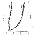
FIG. 2 graphically depicts the in vivo fluorescence time dependence after a bolus injection of FITC labeled poly-d-lysine in a single rat prekidney ligation (Normal) and post-kidney ligation (Ligated). The solid line is a single exponential fit to the measured data. (Excitation at 488 nm, emission monitored at 518 nm.) -
FIG. 3 graphically depicts in vivo fluorescence time dependence for three rats after a bolus injection of ICG. The solid lines are single exponential fits to the measured data. -
FIG. 4 graphically depicts the in vivo fluorescence time dependence of a succession of bolus injections in one rat. In chronological order: ICG (ICG-1), FITC only, saline only, ICG again (ICG-2). -
FIG. 5 graphically depicts the in vivo fluorescence time dependence after a bolus injection of ICG in a single rat pre-partial liver ablation (Normal) and post-partial liver ablation (Ablated). The solid lines are single exponential fits to the measured data. - In accordance with one embodiment of the present invention, a method is disclosed for determining kidney cell and/or organ function by measuring the blood pool clearance of a targeted agent, sometimes referred to herein as tracer. The kidney cell and/or organ function can be determined by the rate these cells remove the tracer from the bloodstream. Function can also be assessed by measuring the rate the cells of interest accumulate the tracer or convert it into an active or other form.
- The agent may be targeted to a group of kidney cells or organ which is a high capacity clearance system. The agent may contain a chromophore and/or fluorophore.
- For agents containing chromophores and/or fluorophores, blood pool clearance may be measured using a light source/photocell device that measures tissue absorbance or fluorescence in a non-target site, such as an ear lobe, finger, brain or retina. Accumulation of the tracer within the cells of interest can be assessed in a similar fashion. The detection of such accumulation is facilitated by using fluorophores which emit in the near infrared wavelengths since body tissues are relatively transparent at these wavelengths.
- The Agent may be introduced into the patient by any suitable method, including intravenous, intraperitoneal or subcutaneous injection or infusion, oral administration, transdermal absorption through the skin, or by inhalation.
- The present invention may be used for rapid bedside evaluation of biologic functions. For example, data on cause of hypercholesterolemia, as well as renal and hepatic function, may be obtained in less than sixty minutes at the bedside after a single intravenous injection. In accordance with one embodiment, a patient may receive a bolus injection of a plurality (e.g., three) different compounds, each containing a different agent (e.g., fluorophore).
- Glomerular filtration may be determined by clearance of a low molecular weight fluorescent agent such as fluorescein-poly-D-lysine or fluoresceininulin.
- Whether hypercholesterolemia is caused by poor LDL clearance may be determined by analyzing the clearance of fluorescent- labeled LDL. Hepatic function may be as assessed by measuring the clearance rate of a fluorescent-labeled asiaglycoprotein.
- The present invention includes fluorescence detection of an agent which is cleared from the bloodstream by the kidneys. Thus, assessment of renal function by in vivo fluorescence detection is encompassed within the invention. The invention can also be used to monitor the efficiency of hemodialysis.
- The clearance of a plurality of separate tracers may be determined simultaneously by selecting excitation wavelengths and filters for the emitted photons. The concentration/time curves may be analyzed in real time by a microprocessor. The resulting clearance rates may be calculated and displayed for immediate clinical impact. In cases where unlabeled competing compounds are present (e.g., LDL, asialoglycoproteins), a single blood sample may be analyzed for the concentration of these competing compounds and the results used to calculate a flux (micromoles/minute) through the clearance pathways.
- In order to demonstrate utility of the invention, a non-invasive fluorescence detection system in accordance with the present invention was employed to continuously monitor dye removal from the vasculature. Differentiation between normal and abnormal organ function in a rat model was demonstrated for both liver and kidney. With reference to
Figure 1 , afiber optic 10 transmited light fromsource 12 toear 14. Asecond fiber optic 16 positioned near theear 14 transmited the fluorescent light to adetector system 20. Two dyes were employed in this initial study. Indocyanine green is exclusively cleared from the blood stream by the liver, and was excited in vivo with laser light at 780 nm. The fluorescence signal was detected at 830 nm. A characteristic clearance curve of normal hepatic function was obtained. Upon ablation of a portion of the liver, the clearance curve was greatly extended as expected. FITC labeled, succinylated poly-d-lysine was excited in vivo with laser light at 488 nm. The fluorescence signal was detected at 518 nm. A characteristic clearance curve of normal renal function was obtained. Upon ligation of both kidneys, the clearance curve remained elevated and constant, indicating little if any clearance. SeeFigure 2 . - With the schematic apparatus for non-invasive in vivo detection of fluorescence shown in
Figure 1 , for ICG fluorescence detection, a nominal 785 nm collimated solid state laser source was employed (LaserMax Inc. model # LAS-300-780-5). For FITC fluorescence detection, an argon ion laser (Coherent Innova model 90) tuned to the 488 nm line was used. Either laser source was directed into the end of a 3.2 mm inner diameter glass fiber optic bundle 10 (Oriel #77526). The other end of this laser delivery bundle was placed approximately 2 cm from therat ear 14 at an approximate 45° angle. A second similarfiber optic bundle 16 for use as the fluorescence detection conduit was placed approximately 1 cm from theear 14 at a 30° angle. - The exit end of the
detection fiber bundle 16 was positioned at the focal length of a 20 mmfocal length lens 18. The output light was thus collimated after exiting the bundle and passing through the lens. A narrow band interference filter 20 (IF) was the next element in the optics train (CVI Laser Corporation), allowing light of the appropriate wavelength to pass on to thedetector 20. For the ICG fluorescence experiment, an 830 nm filter (10 nm FWHM) was used. For the FITC fluorescence experiment, a 518 nm filter (3 nm FWHM) was used. - The
detector 20 was a small silicon photodiode (UDT model PIN-10D) connected to a transimpedance amplifier (UDT model 101C). A digital voltmeter 22 (DVM) monitors the output signal. A subsequent voltage amplifier 24 (Tektronix AM-502) boosts the signal if needed. The amplifier output is connected to a National Instruments BNC-2080 breakout board, which is interfaced to a National Instruments DAQ-700 data acquisition board 26 (A/D). LabVIEW® data acquisition software incomputer 28 collects the experimental raw data. - The current method contrasts with the prior art methods which used radiolabeled tracers. The present method eliminates concerns about radioactivity and allows concurrent measurements of different parameters simply by rapid alteration of the excitation and emission wavelengths.
- The invention is further illustrated by the following examples, which are not intended to be limiting.
- For these in-vivo studies, normal Sprague-Dawley rats weighing ∼250 grams were first anesthetized with urethane (1.35g/kg) administered via intraperitoneal injection.
- After each animal had achieved the desired plane of anesthesia, a small (0.5cm) incision was made in the upper thorax exposing the left jugular vein. The lobe of the left ear was fixed to a glass microscope slide, and the incident laser light delivered from the fiber optic was centered on that ear. Data acquisition was then initiated, and a background reading of fluorescence was obtained prior to administration of the test article. Next, the dye (ICG for liver clearance assessment, FITC labeled poly-d-lysine for kidney clearance assessment) was administered via the jugular vein. The fluorescence signal immediately increased to a peak value. The signal decayed as a function of time as the dye presumably cleared from the bloodstream.
- The anesthetized rat was placed on its back and a midline ventral abdominal skin incision made extending from the xiphoid cartilage to approximately midway to the tail. A similar incision was then made in the abdominal muscles exposing the liver. The rat was repositioned and a bolster placed under the thorax to cause the liver to fall slightly forward and away from the diaphragm. The median and left lateral lobes of the liver were gently moved out of the abdominal cavity and placed onto a gauze pad wetted with saline. The two lobes were vertically raised and a 3-0 ligature placed around the isolated lobes but were not ligated at this point of the procedure. The lobes were replaced in the abdominal cavity and the bolster removed. The incision was closed with wound clips.
- ICG was administered to the animal via the exposed jugular vein as in the previous study. The clearance of the test article was monitored as before to determine the normal hepatic clearance of the ICG. After the normal clearance curve was obtained, the ligature around the two isolated lobes of the liver was tied securely to effect a partial hepatectomy. The animal was allowed to equilibrate for 20 minutes in this state. The ICG was next administered via the exposed jugular vein, and the clearance of the test article monitored. Clearance curves of normal versus partial hepatectomized animals were obtained for an n=3 sample.
- The time dependence of fluorescence measured at the ear pre and post bolus injection of ICG for three rats is shown in
Figure 3 . The data can be described in terms of three stages.Stage 1 consisted of approximately the first 30 seconds of data, which was gathered pre-bolus injection. These data were constant and represented the baseline value for the forthcoming experiment. The value of the baseline should be zero, since no fluorescence is occurring during this stage.Stage 2 occurred several seconds post-injection, the signal rapidly rose to a maximum as the dye reached the ear and equilibrated in the blood pool. In the third stage, the fluorescence signal decayed with time as the liver filtered the ICG out of the blood stream. Visually, the decay rates were similar for all three. After 15 minutes, approximately 90% of the initial signal was lost. - To verify that the measurement was indeed that of ICG fluorescence, the following control study was performed. A rat was injected, as above, with 500 µL of 1.41 mM ICG. A normal fluorescence time course was obtained and is labeled as ICG-1 in
Figure 4 . Then the same rat was injected with 500 µL of 1.41 mM fluorescein (Sigma, St. Louis, MO). As shown inFigure 4 , no fluorescence signal was detected. As a further check, 500 µL of saline solution (Baxter, Deerfield, IL) was injected into the same rat next. Again, no detectible signal was obtained. Finally, the rat was once again injected with 500 µL of 1.41 mM ICG, and a second "normal" curve was obtained. - To verify that these fluorescence decay curves were related to liver function, an experiment involving a partial liver ablation was performed. The partial liver ablation procedure is outlined above. Once the surgery was complete, and the ligatures for use in partially ablating the liver were ready, the rat was injected with 500 µL of 1 mM ICG solution. A normal fluorescence time course curve was obtained and is shown in
Figure 5 . - The liver was then partially ablated by tightening the ligatures. The rat was allowed to equilibrate for ten minutes. Next, another injection of 500 µL of 1mM ICG was given. The fluorescence time curve was measured and is also shown in
Figure 5 . The capability of the liver to remove ICG from the blood pool was drastically altered, the fluorescence decay rate for the partially ablated liver was much slower than the normal. Upon sacrifice, the liver was weighed and 44% of the liver was found to be ablated. - 500 mg (∼125 µmole equivalent Lysine) poly-d-lysine 4000 (Sigma P-0296) was dissolved in 10 ml 0.1 M Na2CO3, in a dark glass vial with magnetic stirring bar. 24.3 mg (62.5 µmoles) FITC (Fluorescein isothiocyanate, Sigma F-7250) was dissolved in 1.5 ml DMSO (dimethyl sulfoxide). At 25°C, with stirring, the FITC solution was slowly added to the poly-d-lysine solution. The reaction was allowed to proceed for 30 minutes at 25°C, then transferred to 4°C, and stirred 12 hrs. The fluorescein conjugate was separated from unbound FITC by gel filtration on Sephadex G-25, eluting with 0.9% (w/v) NaCl.
- 20 ml 0.9% NaCl containing ∼30 µmoles of the above conjugate were placed in a dark glass vial with magnetic stirring bar and pH electrode at 25°C; pH was raised to 9.5 by addition of 0.5 M NaOH. 500 mg of succinic anhydride was added slowly, with stirring, to this solution over a period of 30 minutes, maintaining pH 9.5-10.0 by the addition of 0.5 M NaOH. The pH was then allowed to fall to a stable value of 7.5, with a final volume of 27 ml. The reaction mixture was dialyzed vs. 0.9% (w/v) NaCl using a dialysis membrane with 3.5 kd cutoff, and the retained polymer conjugate, at -4.0 µM fluorescein concentration equivalent, was used directly for infusion.
- The anesthetized rat was placed on the ventral surface and bilateral dorsoventral incisions were made in the abdominal cavity near to the coastal border of the thorax. The kidneys were freed of connective tissue and were gently pulled away from the abdomen by grasping the perirenal fat tissue. A single 3-0 ligature was placed around the renal vessels and ureter so as not to occlude collateral vessels. The ligatures were not tied at this point of the procedure.
- Succinylated, fluorescein-labeled poly-d-lysine was administered via the exposed jugular vein. The clearance of the test article was monitored as before to determine the normal renal clearance of poly-d-lysine. After the normal clearance curve was obtained, the ligature was tied to effect a total (bilateral) nephrectomy. The animal was allowed to stabilize in this condition for 20 minutes. The test article was next administered via the exposed jugular vein and the clearance of the compound monitored. Clearance curves of normal versus total nephrectomized animals were obtained for an n=3 sample.
- The utility of non-invasive fluorescence detection to monitor liver or kidney function has been established.
- The steps of the invention may be repeated in order to determine if physiological function is changing.
- Indocyanine green is a dye which fluoresces at a wavelength of about 830 nm and was used to measure the physiological function of the liver. In order to measure the physiological function of the liver, a body portion was irradiated with light with a wavelength of about 780 nm. The physiological or hepatic function of liver cells was measured using the method described herein.
- Fluorescein labeled, succinylated poly-d-lysine is a dye which fluoresces at a wavelength of about 518 nm and was used to measure the physiological function of the kidneys. In order to measure the physiological function of the kidneys, a body portion was irradiated with light with a wavelength of about 488 nm. Renal function was measured using the above described method of the invention. See
Figure 2 . - The dyes were intravenously injected. A body portion, which included blood vessels near the surface of the skin, was irradiated with a laser or with infrared radiation.
- The claimed invention may also be used to evaluate hypercholesterolemia. Clearance rate measurements may allow the clinician to determine whether high serum cholesterol resulted from increased rate of LDL production or from decreased rate of LDL clearance, which may impact therapy. The claimed invention may also be used to measure cardiac output. The ability to concurrently measure cardiac function while also measuring hepatic and renal function may allow the clinician to draw preliminary conclusions about whether any observed changes in hepatic and renal functions were due to primary renal or hepatic disease or secondary to heart disease.
Claims (14)
- A method of measuring renal physiological function of a group of kidney cells in a patient's body comprising the steps of:a) selecting a detectable agent wherein said agent is a dye capable of fluorescing at a first wavelength upon being irradiated with light of a second wavelength, said agent being selectively removed from the bloodstream by said group of kidney cells, and wherein said emission occurs in said bloodstream;b) Introducing said agent into the bloodstream of the patient, which bloodstream contacts said group of kidney cells;c) irradiating a body portion of said patient's body through which said bloodstream passes with said light of said second wavelength, causing said agent to fluoresce at said first wavelength,d) measuring said emission from a body portion through which said bloodstream passes wherein said emission being measured is fluorescence of said agent at said first wavelength; ande) determining said renal function based on measurement of said emission.
- A method as defined by claim 1, wherein steps b) through e) are repeated to determine if renal physiological function changes.
- A method as defined by claim 1, wherein said agent is injected.
- A method as defined by claim 1, wherein said agent is intravenously injected.
- A method as defined by claim 1, wherein said body portion included blood vessels near a surface of skin of said patient.
- A method as defined by claim 1, wherein said agent comprises a fluorophore or chromophore.
- A method as defined by claim 6, wherein said agent comprises a fluorophore.
- A method as defined by claim 1, wherein said agent is fluorescein labeled, succinylated poly-d-lysine.
- A method as defined by claim 1, wherein said second wavelength is about 400-1200 nanometers.
- A method as defined by claim 1, wherein said second wavelength is about 488 nanometers.
- A method as defined by claim 1, wherein said body portion is irradiated with a laser.
- A method as defined by claim 1, wherein said body portion is irradiated with infrared radiation.
- A method as defined by claim 1, wherein said first wavelength is about 830 nanometers.
- A method as defined in claim 1, wherein said first wavelength is about 518 nanometers.
Priority Applications (1)
| Application Number | Priority Date | Filing Date | Title |
|---|---|---|---|
| EP05077066A EP1618899A3 (en) | 1997-03-13 | 1998-01-29 | Method of measuring physiological function |
Applications Claiming Priority (3)
| Application Number | Priority Date | Filing Date | Title |
|---|---|---|---|
| US816332 | 1991-12-31 | ||
| US08/816,332 US5928625A (en) | 1997-03-13 | 1997-03-13 | Method of measuring physiological function |
| PCT/US1998/001624 WO1998040106A1 (en) | 1997-03-13 | 1998-01-29 | Method of measuring physiological function |
Related Child Applications (2)
| Application Number | Title | Priority Date | Filing Date |
|---|---|---|---|
| EP05077066A Division EP1618899A3 (en) | 1997-03-13 | 1998-01-29 | Method of measuring physiological function |
| EP05077066A Division-Into EP1618899A3 (en) | 1997-03-13 | 1998-01-29 | Method of measuring physiological function |
Publications (2)
| Publication Number | Publication Date |
|---|---|
| EP0966306A1 EP0966306A1 (en) | 1999-12-29 |
| EP0966306B1 true EP0966306B1 (en) | 2015-07-08 |
Family
ID=25220304
Family Applications (2)
| Application Number | Title | Priority Date | Filing Date |
|---|---|---|---|
| EP98903793.2A Expired - Lifetime EP0966306B1 (en) | 1997-03-13 | 1998-01-29 | Method of measuring physiological function |
| EP05077066A Ceased EP1618899A3 (en) | 1997-03-13 | 1998-01-29 | Method of measuring physiological function |
Family Applications After (1)
| Application Number | Title | Priority Date | Filing Date |
|---|---|---|---|
| EP05077066A Ceased EP1618899A3 (en) | 1997-03-13 | 1998-01-29 | Method of measuring physiological function |
Country Status (6)
| Country | Link |
|---|---|
| US (1) | US5928625A (en) |
| EP (2) | EP0966306B1 (en) |
| JP (2) | JP2001514554A (en) |
| AU (1) | AU6046998A (en) |
| CA (1) | CA2283566C (en) |
| WO (1) | WO1998040106A1 (en) |
Cited By (1)
| Publication number | Priority date | Publication date | Assignee | Title |
|---|---|---|---|---|
| DE102024124894A1 (en) | 2024-08-30 | 2026-03-05 | Eagle Actuator Components Gmbh & Co. Kg | Housing for a sensor and sensor |
Families Citing this family (26)
| Publication number | Priority date | Publication date | Assignee | Title |
|---|---|---|---|---|
| US6228344B1 (en) * | 1997-03-13 | 2001-05-08 | Mallinckrodt Inc. | Method of measuring physiological function |
| US6280703B1 (en) * | 1997-03-13 | 2001-08-28 | Mallinckrodt Inc. | Simultaneous multimodal measurement of physiological function |
| US6592847B1 (en) | 1998-05-14 | 2003-07-15 | The General Hospital Corporation | Intramolecularly-quenched near infrared flourescent probes |
| US6083486A (en) * | 1998-05-14 | 2000-07-04 | The General Hospital Corporation | Intramolecularly-quenched near infrared fluorescent probes |
| US6132958A (en) * | 1999-05-27 | 2000-10-17 | The Rockefeller University | Fluorescent bead for determining the temperature of a cell and methods of use thereof |
| US6445945B1 (en) | 2000-06-26 | 2002-09-03 | André Arsenault | Non-invasive detection of endothelial dysfunction by blood flow measurement in opposed limbs using tracer injection |
| US20090209831A1 (en) * | 2000-06-28 | 2009-08-20 | Nexgen Medical Systems, Incorporated | Imaging methods for visualizing implanted living cells |
| US6440389B1 (en) * | 2000-07-19 | 2002-08-27 | The General Hospital Corporation | Fluorescent agents for real-time measurement of organ function |
| US6733744B1 (en) * | 2000-10-16 | 2004-05-11 | Mallinckrodt Inc. | Indole compounds as minimally invasive physiological function monitoring agents |
| US6656451B1 (en) * | 2000-10-16 | 2003-12-02 | Mallinckrodt, Inc. | Indole compounds as novel dyes for organ function monitoring |
| US7556797B2 (en) | 2000-10-16 | 2009-07-07 | Mallinckrodt Inc. | Minimally invasive physiological function monitoring agents |
| US6673334B1 (en) * | 2000-10-16 | 2004-01-06 | Mallinkcrodt, Inc. | Light sensitive compounds for instant determination of organ function |
| US6716413B1 (en) * | 2000-10-16 | 2004-04-06 | Mallinckrodt, Inc. | Indole compounds as tissue-specific exogenous optical agents |
| US7383076B2 (en) | 2000-11-27 | 2008-06-03 | The General Hospital Corporation | Fluorescence-mediated molecular tomography |
| US6542769B2 (en) | 2000-12-18 | 2003-04-01 | The General Hospital Corporation | Imaging system for obtaining quantative perfusion indices |
| US8337444B2 (en) * | 2001-05-22 | 2012-12-25 | Alfred E. Mann Institute For Biomedical Engineering At The University Of Southern California | Measurement of cardiac output and blood volume by non-invasive detection of indicator dilution for hemodialysis |
| US20040156782A1 (en) * | 2003-02-12 | 2004-08-12 | Akorn, Inc. | Methods of using indocyanine green (ICG) dye |
| ES2375395T3 (en) * | 2004-12-23 | 2012-02-29 | Mallinckrodt Llc | FLUORESCENT PIRAZINE DERIVATIVES AND METHOD OF USE OF THE SAME IN THE EVALUATION OF RENAL FUNCTION. |
| US8329143B2 (en) * | 2005-04-19 | 2012-12-11 | Indiana University Research And Technology Corporation | Method and apparatus for kidney function analysis |
| DE102007007738A1 (en) * | 2007-02-09 | 2008-08-14 | Südzucker AG Mannheim/Ochsenfurt | Color-labeled oligo- or polysaccharides |
| WO2009140026A2 (en) | 2008-04-18 | 2009-11-19 | Pharmacophotonics, Llc D/B/A Fast Diagnostics | Renal function analysis method and apparatus |
| US8591865B2 (en) | 2008-04-18 | 2013-11-26 | Pharmacophotonics, Inc. | Renal function analysis method and apparatus |
| US8956591B2 (en) | 2008-05-15 | 2015-02-17 | Osaka Prefectural Hospital Organization | Method for detecting cancer using ICG fluorescence method |
| GB0808777D0 (en) | 2008-05-15 | 2008-06-18 | Norgine Bv | Prognostic method |
| US10300151B2 (en) * | 2015-09-11 | 2019-05-28 | Board Of Regents, The University Of Texas System | Methods and systems for noninvasive fluorescence-based functional imaging of kidneys |
| US12440582B2 (en) | 2019-06-17 | 2025-10-14 | Board Of Regents, The University Of Texas System | Biothiol-activatable probe and method of use |
Family Cites Families (9)
| Publication number | Priority date | Publication date | Assignee | Title |
|---|---|---|---|---|
| IL84356A (en) * | 1986-11-05 | 1991-08-16 | Sumitomo Electric Industries | Liver function testing apparatus |
| JPS6417630A (en) * | 1987-07-13 | 1989-01-20 | Sumitomo Electric Industries | Liver function test apparatus |
| US4848349A (en) * | 1987-04-29 | 1989-07-18 | Sherman Igor A | Substance and method for measuring hepatic blood flow |
| JPH01129838A (en) * | 1987-11-13 | 1989-05-23 | Sumitomo Electric Ind Ltd | Liver function examination apparatus |
| CA1328018C (en) * | 1987-11-13 | 1994-03-22 | Masahiko Kanda | Liver function testing apparatus |
| US5301673A (en) * | 1991-11-18 | 1994-04-12 | Massachusetts General Hospital | Ambulatory clearance function monitor |
| JP3275159B2 (en) * | 1993-12-17 | 2002-04-15 | 日本光電工業株式会社 | Circulating blood volume measurement device |
| US5458128A (en) * | 1994-06-17 | 1995-10-17 | Polanyi; Michael | Method and apparatus for noninvasively measuring concentration of a dye in arterial blood |
| GB9517043D0 (en) * | 1995-08-19 | 1995-10-25 | Univ Birmingham | The use of bile acid derivatives |
-
1997
- 1997-03-13 US US08/816,332 patent/US5928625A/en not_active Expired - Lifetime
-
1998
- 1998-01-29 JP JP53955598A patent/JP2001514554A/en active Pending
- 1998-01-29 EP EP98903793.2A patent/EP0966306B1/en not_active Expired - Lifetime
- 1998-01-29 EP EP05077066A patent/EP1618899A3/en not_active Ceased
- 1998-01-29 CA CA002283566A patent/CA2283566C/en not_active Expired - Lifetime
- 1998-01-29 WO PCT/US1998/001624 patent/WO1998040106A1/en not_active Ceased
- 1998-01-29 AU AU60469/98A patent/AU6046998A/en not_active Abandoned
-
2007
- 2007-06-19 JP JP2007161427A patent/JP2007277576A/en active Pending
Non-Patent Citations (1)
| Title |
|---|
| YOSHITAKA ISHIGAMI ET AL: "Clinical applications of ICG Finger Monitor in patients with liver disease", JOURNAL OF HEPATOLOGY, vol. 19, no. 2, 1 January 1993 (1993-01-01), pages 232 - 240, XP055097652, ISSN: 0168-8278, DOI: 10.1016/S0168-8278(05)80577-X * |
Cited By (2)
| Publication number | Priority date | Publication date | Assignee | Title |
|---|---|---|---|---|
| DE102024124894A1 (en) | 2024-08-30 | 2026-03-05 | Eagle Actuator Components Gmbh & Co. Kg | Housing for a sensor and sensor |
| DE102024124894B4 (en) | 2024-08-30 | 2026-05-13 | Eagle Actuator Components Gmbh & Co. Kg | Housing for a sensor and sensor |
Also Published As
| Publication number | Publication date |
|---|---|
| AU6046998A (en) | 1998-09-29 |
| WO1998040106A1 (en) | 1998-09-17 |
| EP0966306A1 (en) | 1999-12-29 |
| EP1618899A3 (en) | 2006-02-01 |
| CA2283566C (en) | 2008-07-22 |
| CA2283566A1 (en) | 1998-09-17 |
| JP2007277576A (en) | 2007-10-25 |
| EP1618899A2 (en) | 2006-01-25 |
| US5928625A (en) | 1999-07-27 |
| JP2001514554A (en) | 2001-09-11 |
Similar Documents
| Publication | Publication Date | Title |
|---|---|---|
| CA2283566C (en) | Method of measuring physiological function | |
| EP1154802B1 (en) | Method of measuring physiological function with a dye like fluorescein-polyacrylic acid | |
| US6280703B1 (en) | Simultaneous multimodal measurement of physiological function | |
| US8865128B2 (en) | Folate targeted enhanced tumor and folate receptor positive tissue optical imaging technology | |
| Pogue et al. | Review of neurosurgical fluorescence imaging methodologies | |
| Dorshow et al. | Noninvasive fluorescence detection of hepatic and renal function | |
| US8858914B2 (en) | Folate targeted enhanced tumor and folate receptor positive tissue optical imaging technology | |
| US20110171136A1 (en) | Optical imaging probes | |
| EP1986701B1 (en) | Optical agents for use in surgery | |
| WO2008076467A2 (en) | Intraoperative imaging methods | |
| JPH06503487A (en) | Fluorescent probe used to measure oxygen concentration | |
| US8741263B2 (en) | Method and apparatus for kidney function analysis | |
| Yang et al. | Real time monitoring peripheral nerve function with ICG and BDA-ICG by NIR-II fluorescence imaging | |
| US20240066142A1 (en) | A upar (urokinase plasminogen activator receptor)-targeting conjugate | |
| Maréchal et al. | In Vivo Application of Intestinal pH Measurement Using 2, 7 ‘‐Bis (carboxyethyl)‐5, 6‐carboxyfluorescein (BCECF) Fluorescence Imaging | |
| Devoisselle et al. | Fluorescent characteristics and pharmacokinetic profiles of the fluorescent probe BCECF in various tissues: the role of blood content | |
| Soulie-Begu et al. | In vivo pharmacokinetic study of two fluorescein derivatives by fluorescence spectroscopy | |
| Dorshow et al. | Noninvasive fluorescence detection of physiological function | |
| WO2022252103A1 (en) | Use of methylene blue and fluorescein sodium double-staining in live cell imaging | |
| WO2022252104A1 (en) | Staining method for live-cell imaging | |
| Rescigno et al. | High-frequency perfusion fluorometry (HFPF): preliminary results on a new technique for assessing bronchial mucosa blood perfusion |
Legal Events
| Date | Code | Title | Description |
|---|---|---|---|
| PUAI | Public reference made under article 153(3) epc to a published international application that has entered the european phase |
Free format text: ORIGINAL CODE: 0009012 |
|
| 17P | Request for examination filed |
Effective date: 19990907 |
|
| AK | Designated contracting states |
Kind code of ref document: A1 Designated state(s): AT BE CH DE DK ES FI FR GB GR IE IT LI LU MC NL PT SE |
|
| RIN1 | Information on inventor provided before grant (corrected) |
Inventor name: JONES, WILLIAM, B. Inventor name: JOHNSON, MICHAEL, ANTHONY Inventor name: DUNCAN, JAMES, R. Inventor name: BURLEIGH, B., DANIEL Inventor name: BUGAJ, JOSEPH, E. Inventor name: DORSHOW, RICHARD, B. |
|
| 17Q | First examination report despatched |
Effective date: 20040317 |
|
| 17Q | First examination report despatched |
Effective date: 20040317 |
|
| RAP1 | Party data changed (applicant data changed or rights of an application transferred) |
Owner name: WASHINGTON UNIVERSITY Owner name: MALLINCKRODT LLC |
|
| GRAP | Despatch of communication of intention to grant a patent |
Free format text: ORIGINAL CODE: EPIDOSNIGR1 |
|
| INTG | Intention to grant announced |
Effective date: 20141203 |
|
| GRAS | Grant fee paid |
Free format text: ORIGINAL CODE: EPIDOSNIGR3 |
|
| GRAA | (expected) grant |
Free format text: ORIGINAL CODE: 0009210 |
|
| RAP1 | Party data changed (applicant data changed or rights of an application transferred) |
Owner name: MEDIBEACON, LLC Owner name: WASHINGTON UNIVERSITY |
|
| AK | Designated contracting states |
Kind code of ref document: B1 Designated state(s): AT BE CH DE DK ES FI FR GB GR IE IT LI LU MC NL PT SE |
|
| REG | Reference to a national code |
Ref country code: GB Ref legal event code: FG4D |
|
| REG | Reference to a national code |
Ref country code: DE Ref legal event code: R081 Ref document number: 69843414 Country of ref document: DE Owner name: MEDIBEACON INC. (N.D.G.D. STAATES DELAWARE), S, US Free format text: FORMER OWNERS: MALLINCKRODT, INC., ST. LOUIS, MO., US; WASHINGTON UNIVERSITY, 63130 ST. LOUIS, MO., US |
|
| REG | Reference to a national code |
Ref country code: AT Ref legal event code: REF Ref document number: 734866 Country of ref document: AT Kind code of ref document: T Effective date: 20150715 Ref country code: CH Ref legal event code: EP |
|
| REG | Reference to a national code |
Ref country code: IE Ref legal event code: FG4D |
|
| REG | Reference to a national code |
Ref country code: DE Ref legal event code: R096 Ref document number: 69843414 Country of ref document: DE |
|
| REG | Reference to a national code |
Ref country code: AT Ref legal event code: MK05 Ref document number: 734866 Country of ref document: AT Kind code of ref document: T Effective date: 20150708 |
|
| REG | Reference to a national code |
Ref country code: NL Ref legal event code: MP Effective date: 20150708 |
|
| REG | Reference to a national code |
Ref country code: FR Ref legal event code: PLFP Year of fee payment: 19 |
|
| PG25 | Lapsed in a contracting state [announced via postgrant information from national office to epo] |
Ref country code: GR Free format text: LAPSE BECAUSE OF FAILURE TO SUBMIT A TRANSLATION OF THE DESCRIPTION OR TO PAY THE FEE WITHIN THE PRESCRIBED TIME-LIMIT Effective date: 20151009 Ref country code: FI Free format text: LAPSE BECAUSE OF FAILURE TO SUBMIT A TRANSLATION OF THE DESCRIPTION OR TO PAY THE FEE WITHIN THE PRESCRIBED TIME-LIMIT Effective date: 20150708 |
|
| PG25 | Lapsed in a contracting state [announced via postgrant information from national office to epo] |
Ref country code: ES Free format text: LAPSE BECAUSE OF FAILURE TO SUBMIT A TRANSLATION OF THE DESCRIPTION OR TO PAY THE FEE WITHIN THE PRESCRIBED TIME-LIMIT Effective date: 20150708 Ref country code: SE Free format text: LAPSE BECAUSE OF FAILURE TO SUBMIT A TRANSLATION OF THE DESCRIPTION OR TO PAY THE FEE WITHIN THE PRESCRIBED TIME-LIMIT Effective date: 20150708 Ref country code: PT Free format text: LAPSE BECAUSE OF FAILURE TO SUBMIT A TRANSLATION OF THE DESCRIPTION OR TO PAY THE FEE WITHIN THE PRESCRIBED TIME-LIMIT Effective date: 20151109 Ref country code: AT Free format text: LAPSE BECAUSE OF FAILURE TO SUBMIT A TRANSLATION OF THE DESCRIPTION OR TO PAY THE FEE WITHIN THE PRESCRIBED TIME-LIMIT Effective date: 20150708 |
|
| REG | Reference to a national code |
Ref country code: DE Ref legal event code: R082 Ref document number: 69843414 Country of ref document: DE Representative=s name: MUELLER-BORE & PARTNER PATENTANWAELTE PARTG MB, DE Ref country code: DE Ref legal event code: R081 Ref document number: 69843414 Country of ref document: DE Owner name: MEDIBEACON INC. (N.D.G.D. STAATES DELAWARE), S, US Free format text: FORMER OWNERS: MEDIBEACON, LLC, ST. LOUIS, MO., US; WASHINGTON UNIVERSITY, ST. LOUIS, MO., US |
|
| REG | Reference to a national code |
Ref country code: DE Ref legal event code: R097 Ref document number: 69843414 Country of ref document: DE |
|
| REG | Reference to a national code |
Ref country code: FR Ref legal event code: CD Owner name: WASHINGTON UNIVERSITY, US Effective date: 20160318 Ref country code: FR Ref legal event code: CD Owner name: MEDIBEACON INC., US Effective date: 20160318 |
|
| PG25 | Lapsed in a contracting state [announced via postgrant information from national office to epo] |
Ref country code: IT Free format text: LAPSE BECAUSE OF FAILURE TO SUBMIT A TRANSLATION OF THE DESCRIPTION OR TO PAY THE FEE WITHIN THE PRESCRIBED TIME-LIMIT Effective date: 20150708 Ref country code: DK Free format text: LAPSE BECAUSE OF FAILURE TO SUBMIT A TRANSLATION OF THE DESCRIPTION OR TO PAY THE FEE WITHIN THE PRESCRIBED TIME-LIMIT Effective date: 20150708 |
|
| PLBE | No opposition filed within time limit |
Free format text: ORIGINAL CODE: 0009261 |
|
| STAA | Information on the status of an ep patent application or granted ep patent |
Free format text: STATUS: NO OPPOSITION FILED WITHIN TIME LIMIT |
|
| PG25 | Lapsed in a contracting state [announced via postgrant information from national office to epo] |
Ref country code: BE Free format text: LAPSE BECAUSE OF NON-PAYMENT OF DUE FEES Effective date: 20160131 |
|
| 26N | No opposition filed |
Effective date: 20160411 |
|
| PG25 | Lapsed in a contracting state [announced via postgrant information from national office to epo] |
Ref country code: LU Free format text: LAPSE BECAUSE OF FAILURE TO SUBMIT A TRANSLATION OF THE DESCRIPTION OR TO PAY THE FEE WITHIN THE PRESCRIBED TIME-LIMIT Effective date: 20160129 |
|
| REG | Reference to a national code |
Ref country code: CH Ref legal event code: PL |
|
| PG25 | Lapsed in a contracting state [announced via postgrant information from national office to epo] |
Ref country code: MC Free format text: LAPSE BECAUSE OF FAILURE TO SUBMIT A TRANSLATION OF THE DESCRIPTION OR TO PAY THE FEE WITHIN THE PRESCRIBED TIME-LIMIT Effective date: 20150708 |
|
| PG25 | Lapsed in a contracting state [announced via postgrant information from national office to epo] |
Ref country code: LI Free format text: LAPSE BECAUSE OF NON-PAYMENT OF DUE FEES Effective date: 20160131 Ref country code: CH Free format text: LAPSE BECAUSE OF NON-PAYMENT OF DUE FEES Effective date: 20160131 |
|
| REG | Reference to a national code |
Ref country code: IE Ref legal event code: MM4A |
|
| PG25 | Lapsed in a contracting state [announced via postgrant information from national office to epo] |
Ref country code: BE Free format text: LAPSE BECAUSE OF FAILURE TO SUBMIT A TRANSLATION OF THE DESCRIPTION OR TO PAY THE FEE WITHIN THE PRESCRIBED TIME-LIMIT Effective date: 20150708 |
|
| REG | Reference to a national code |
Ref country code: FR Ref legal event code: PLFP Year of fee payment: 20 |
|
| PG25 | Lapsed in a contracting state [announced via postgrant information from national office to epo] |
Ref country code: IE Free format text: LAPSE BECAUSE OF NON-PAYMENT OF DUE FEES Effective date: 20160129 |
|
| PGFP | Annual fee paid to national office [announced via postgrant information from national office to epo] |
Ref country code: DE Payment date: 20170125 Year of fee payment: 20 Ref country code: FR Payment date: 20170125 Year of fee payment: 20 |
|
| PGFP | Annual fee paid to national office [announced via postgrant information from national office to epo] |
Ref country code: GB Payment date: 20170127 Year of fee payment: 20 |
|
| PG25 | Lapsed in a contracting state [announced via postgrant information from national office to epo] |
Ref country code: NL Free format text: LAPSE BECAUSE OF FAILURE TO SUBMIT A TRANSLATION OF THE DESCRIPTION OR TO PAY THE FEE WITHIN THE PRESCRIBED TIME-LIMIT Effective date: 20150708 |
|
| REG | Reference to a national code |
Ref country code: DE Ref legal event code: R071 Ref document number: 69843414 Country of ref document: DE |
|
| REG | Reference to a national code |
Ref country code: GB Ref legal event code: PE20 Expiry date: 20180128 |
|
| PG25 | Lapsed in a contracting state [announced via postgrant information from national office to epo] |
Ref country code: GB Free format text: LAPSE BECAUSE OF EXPIRATION OF PROTECTION Effective date: 20180128 |