EP0932697B1 - Method for prediction of progression of normal and hyperplastic mammary cells to carcinoma - Google Patents
Method for prediction of progression of normal and hyperplastic mammary cells to carcinoma Download PDFInfo
- Publication number
- EP0932697B1 EP0932697B1 EP97912952A EP97912952A EP0932697B1 EP 0932697 B1 EP0932697 B1 EP 0932697B1 EP 97912952 A EP97912952 A EP 97912952A EP 97912952 A EP97912952 A EP 97912952A EP 0932697 B1 EP0932697 B1 EP 0932697B1
- Authority
- EP
- European Patent Office
- Prior art keywords
- tissue
- carcinoma
- loh
- normal
- chromosome
- Prior art date
- Legal status (The legal status is an assumption and is not a legal conclusion. Google has not performed a legal analysis and makes no representation as to the accuracy of the status listed.)
- Expired - Lifetime
Links
- 238000000034 method Methods 0.000 title claims description 27
- 201000009030 Carcinoma Diseases 0.000 title claims description 16
- 206010020718 hyperplasia Diseases 0.000 title claims description 6
- 230000002390 hyperplastic effect Effects 0.000 title claims description 6
- 210000004216 mammary stem cell Anatomy 0.000 title description 3
- 210000001519 tissue Anatomy 0.000 claims description 55
- 206010006187 Breast cancer Diseases 0.000 claims description 29
- 208000026310 Breast neoplasm Diseases 0.000 claims description 29
- 210000000481 breast Anatomy 0.000 claims description 21
- 210000000349 chromosome Anatomy 0.000 claims description 16
- 201000004933 in situ carcinoma Diseases 0.000 claims description 8
- 208000024312 invasive carcinoma Diseases 0.000 claims description 8
- 230000003211 malignant effect Effects 0.000 claims description 7
- 238000007894 restriction fragment length polymorphism technique Methods 0.000 claims description 5
- 201000008275 breast carcinoma Diseases 0.000 claims description 4
- 238000000137 annealing Methods 0.000 claims description 3
- 239000013611 chromosomal DNA Substances 0.000 claims 2
- 101710163270 Nuclease Proteins 0.000 claims 1
- 150000007523 nucleic acids Chemical class 0.000 claims 1
- 102000039446 nucleic acids Human genes 0.000 claims 1
- 108020004707 nucleic acids Proteins 0.000 claims 1
- 206010028980 Neoplasm Diseases 0.000 description 38
- 210000004027 cell Anatomy 0.000 description 26
- 108700028369 Alleles Proteins 0.000 description 25
- 108020004414 DNA Proteins 0.000 description 22
- 238000003752 polymerase chain reaction Methods 0.000 description 18
- 208000028715 ductal breast carcinoma in situ Diseases 0.000 description 15
- 208000037396 Intraductal Noninfiltrating Carcinoma Diseases 0.000 description 14
- 206010073094 Intraductal proliferative breast lesion Diseases 0.000 description 14
- 210000001766 X chromosome Anatomy 0.000 description 14
- 201000007273 ductal carcinoma in situ Diseases 0.000 description 14
- 201000011510 cancer Diseases 0.000 description 13
- 210000000981 epithelium Anatomy 0.000 description 13
- 230000000977 initiatory effect Effects 0.000 description 11
- 230000019975 dosage compensation by inactivation of X chromosome Effects 0.000 description 8
- 239000000499 gel Substances 0.000 description 8
- 108090000623 proteins and genes Proteins 0.000 description 8
- 108091008146 restriction endonucleases Proteins 0.000 description 8
- 230000009467 reduction Effects 0.000 description 7
- 238000003556 assay Methods 0.000 description 6
- 230000029087 digestion Effects 0.000 description 6
- 238000001531 micro-dissection Methods 0.000 description 6
- 239000013615 primer Substances 0.000 description 6
- 238000004458 analytical method Methods 0.000 description 5
- 230000011987 methylation Effects 0.000 description 5
- 238000007069 methylation reaction Methods 0.000 description 5
- 108010067770 Endopeptidase K Proteins 0.000 description 4
- 238000012408 PCR amplification Methods 0.000 description 4
- 238000002474 experimental method Methods 0.000 description 4
- 210000005075 mammary gland Anatomy 0.000 description 4
- YBYRMVIVWMBXKQ-UHFFFAOYSA-N phenylmethanesulfonyl fluoride Chemical compound FS(=O)(=O)CC1=CC=CC=C1 YBYRMVIVWMBXKQ-UHFFFAOYSA-N 0.000 description 4
- 238000012552 review Methods 0.000 description 4
- 210000003491 skin Anatomy 0.000 description 4
- 210000000130 stem cell Anatomy 0.000 description 4
- QKNYBSVHEMOAJP-UHFFFAOYSA-N 2-amino-2-(hydroxymethyl)propane-1,3-diol;hydron;chloride Chemical compound Cl.OCC(N)(CO)CO QKNYBSVHEMOAJP-UHFFFAOYSA-N 0.000 description 3
- 208000009458 Carcinoma in Situ Diseases 0.000 description 3
- 102100025064 Cellular tumor antigen p53 Human genes 0.000 description 3
- IAZDPXIOMUYVGZ-UHFFFAOYSA-N Dimethylsulphoxide Chemical compound CS(C)=O IAZDPXIOMUYVGZ-UHFFFAOYSA-N 0.000 description 3
- 108010080146 androgen receptors Proteins 0.000 description 3
- 231100000504 carcinogenesis Toxicity 0.000 description 3
- 238000006243 chemical reaction Methods 0.000 description 3
- RGWHQCVHVJXOKC-SHYZEUOFSA-J dCTP(4-) Chemical compound O=C1N=C(N)C=CN1[C@@H]1O[C@H](COP([O-])(=O)OP([O-])(=O)OP([O-])([O-])=O)[C@@H](O)C1 RGWHQCVHVJXOKC-SHYZEUOFSA-J 0.000 description 3
- 230000004069 differentiation Effects 0.000 description 3
- 201000010099 disease Diseases 0.000 description 3
- 208000037265 diseases, disorders, signs and symptoms Diseases 0.000 description 3
- 230000014509 gene expression Effects 0.000 description 3
- 230000002779 inactivation Effects 0.000 description 3
- 208000030776 invasive breast carcinoma Diseases 0.000 description 3
- 239000003550 marker Substances 0.000 description 3
- 230000002093 peripheral effect Effects 0.000 description 3
- 230000005855 radiation Effects 0.000 description 3
- 239000000243 solution Substances 0.000 description 3
- KCXVZYZYPLLWCC-UHFFFAOYSA-N EDTA Chemical compound OC(=O)CN(CC(O)=O)CCN(CC(O)=O)CC(O)=O KCXVZYZYPLLWCC-UHFFFAOYSA-N 0.000 description 2
- LFQSCWFLJHTTHZ-UHFFFAOYSA-N Ethanol Chemical compound CCO LFQSCWFLJHTTHZ-UHFFFAOYSA-N 0.000 description 2
- TWRXJAOTZQYOKJ-UHFFFAOYSA-L Magnesium chloride Chemical compound [Mg+2].[Cl-].[Cl-] TWRXJAOTZQYOKJ-UHFFFAOYSA-L 0.000 description 2
- 108091092878 Microsatellite Proteins 0.000 description 2
- 108700020796 Oncogene Proteins 0.000 description 2
- 230000001413 cellular effect Effects 0.000 description 2
- 230000034994 death Effects 0.000 description 2
- 231100000517 death Toxicity 0.000 description 2
- 238000002224 dissection Methods 0.000 description 2
- 238000013399 early diagnosis Methods 0.000 description 2
- 238000001962 electrophoresis Methods 0.000 description 2
- 230000002068 genetic effect Effects 0.000 description 2
- 230000003054 hormonal effect Effects 0.000 description 2
- 238000012744 immunostaining Methods 0.000 description 2
- 230000036210 malignancy Effects 0.000 description 2
- 239000000203 mixture Substances 0.000 description 2
- 230000035772 mutation Effects 0.000 description 2
- 229920002401 polyacrylamide Polymers 0.000 description 2
- 238000002360 preparation method Methods 0.000 description 2
- 230000002250 progressing effect Effects 0.000 description 2
- 230000002062 proliferating effect Effects 0.000 description 2
- 230000035755 proliferation Effects 0.000 description 2
- 238000007790 scraping Methods 0.000 description 2
- 238000012163 sequencing technique Methods 0.000 description 2
- 210000002536 stromal cell Anatomy 0.000 description 2
- 238000011282 treatment Methods 0.000 description 2
- 238000011269 treatment regimen Methods 0.000 description 2
- 102000036365 BRCA1 Human genes 0.000 description 1
- 108700020463 BRCA1 Proteins 0.000 description 1
- 101150072950 BRCA1 gene Proteins 0.000 description 1
- 208000005623 Carcinogenesis Diseases 0.000 description 1
- 102000003908 Cathepsin D Human genes 0.000 description 1
- 108090000258 Cathepsin D Proteins 0.000 description 1
- 208000001333 Colorectal Neoplasms Diseases 0.000 description 1
- 241000557626 Corvus corax Species 0.000 description 1
- 102000053602 DNA Human genes 0.000 description 1
- 239000003155 DNA primer Substances 0.000 description 1
- 102000001301 EGF receptor Human genes 0.000 description 1
- 108060006698 EGF receptor Proteins 0.000 description 1
- 102000009024 Epidermal Growth Factor Human genes 0.000 description 1
- 101800003838 Epidermal growth factor Proteins 0.000 description 1
- 108010010803 Gelatin Proteins 0.000 description 1
- 208000033640 Hereditary breast cancer Diseases 0.000 description 1
- 101000775732 Homo sapiens Androgen receptor Proteins 0.000 description 1
- 101000928259 Homo sapiens NADPH:adrenodoxin oxidoreductase, mitochondrial Proteins 0.000 description 1
- 101001012157 Homo sapiens Receptor tyrosine-protein kinase erbB-2 Proteins 0.000 description 1
- 206010064912 Malignant transformation Diseases 0.000 description 1
- 206010061309 Neoplasm progression Diseases 0.000 description 1
- 108091028043 Nucleic acid sequence Proteins 0.000 description 1
- 206010061535 Ovarian neoplasm Diseases 0.000 description 1
- 238000002944 PCR assay Methods 0.000 description 1
- 229920001213 Polysorbate 20 Polymers 0.000 description 1
- 102100030086 Receptor tyrosine-protein kinase erbB-2 Human genes 0.000 description 1
- 230000018199 S phase Effects 0.000 description 1
- 108010006785 Taq Polymerase Proteins 0.000 description 1
- 108700025716 Tumor Suppressor Genes Proteins 0.000 description 1
- 102000044209 Tumor Suppressor Genes Human genes 0.000 description 1
- 208000007097 Urinary Bladder Neoplasms Diseases 0.000 description 1
- 108700029631 X-Linked Genes Proteins 0.000 description 1
- 238000011226 adjuvant chemotherapy Methods 0.000 description 1
- 238000009098 adjuvant therapy Methods 0.000 description 1
- 238000011256 aggressive treatment Methods 0.000 description 1
- 238000007605 air drying Methods 0.000 description 1
- 230000004075 alteration Effects 0.000 description 1
- 230000019552 anatomical structure morphogenesis Effects 0.000 description 1
- 102000001307 androgen receptors Human genes 0.000 description 1
- 238000013459 approach Methods 0.000 description 1
- 238000000376 autoradiography Methods 0.000 description 1
- 210000002469 basement membrane Anatomy 0.000 description 1
- 238000009835 boiling Methods 0.000 description 1
- 208000030270 breast disease Diseases 0.000 description 1
- 230000036952 cancer formation Effects 0.000 description 1
- 230000001364 causal effect Effects 0.000 description 1
- 230000008859 change Effects 0.000 description 1
- 239000003153 chemical reaction reagent Substances 0.000 description 1
- 239000003795 chemical substances by application Substances 0.000 description 1
- 230000002759 chromosomal effect Effects 0.000 description 1
- 238000003776 cleavage reaction Methods 0.000 description 1
- 238000010367 cloning Methods 0.000 description 1
- 238000011109 contamination Methods 0.000 description 1
- 230000002596 correlated effect Effects 0.000 description 1
- 208000031513 cyst Diseases 0.000 description 1
- SUYVUBYJARFZHO-RRKCRQDMSA-N dATP Chemical compound C1=NC=2C(N)=NC=NC=2N1[C@H]1C[C@H](O)[C@@H](COP(O)(=O)OP(O)(=O)OP(O)(O)=O)O1 SUYVUBYJARFZHO-RRKCRQDMSA-N 0.000 description 1
- SUYVUBYJARFZHO-UHFFFAOYSA-N dATP Natural products C1=NC=2C(N)=NC=NC=2N1C1CC(O)C(COP(O)(=O)OP(O)(=O)OP(O)(O)=O)O1 SUYVUBYJARFZHO-UHFFFAOYSA-N 0.000 description 1
- HAAZLUGHYHWQIW-KVQBGUIXSA-N dGTP Chemical compound C1=NC=2C(=O)NC(N)=NC=2N1[C@H]1C[C@H](O)[C@@H](COP(O)(=O)OP(O)(=O)OP(O)(O)=O)O1 HAAZLUGHYHWQIW-KVQBGUIXSA-N 0.000 description 1
- NHVNXKFIZYSCEB-XLPZGREQSA-N dTTP Chemical compound O=C1NC(=O)C(C)=CN1[C@@H]1O[C@H](COP(O)(=O)OP(O)(=O)OP(O)(O)=O)[C@@H](O)C1 NHVNXKFIZYSCEB-XLPZGREQSA-N 0.000 description 1
- 230000003247 decreasing effect Effects 0.000 description 1
- 238000004925 denaturation Methods 0.000 description 1
- 230000036425 denaturation Effects 0.000 description 1
- 239000005547 deoxyribonucleotide Substances 0.000 description 1
- -1 deoxyribonucleotide triphosphates Chemical class 0.000 description 1
- 238000001514 detection method Methods 0.000 description 1
- 238000011161 development Methods 0.000 description 1
- 230000018109 developmental process Effects 0.000 description 1
- 238000003745 diagnosis Methods 0.000 description 1
- 230000000694 effects Effects 0.000 description 1
- 230000002255 enzymatic effect Effects 0.000 description 1
- 229940116977 epidermal growth factor Drugs 0.000 description 1
- 210000002919 epithelial cell Anatomy 0.000 description 1
- ZMMJGEGLRURXTF-UHFFFAOYSA-N ethidium bromide Chemical compound [Br-].C12=CC(N)=CC=C2C2=CC=C(N)C=C2[N+](CC)=C1C1=CC=CC=C1 ZMMJGEGLRURXTF-UHFFFAOYSA-N 0.000 description 1
- 229960005542 ethidium bromide Drugs 0.000 description 1
- 230000002349 favourable effect Effects 0.000 description 1
- 238000002594 fluoroscopy Methods 0.000 description 1
- 239000012634 fragment Substances 0.000 description 1
- 230000002496 gastric effect Effects 0.000 description 1
- 239000008273 gelatin Substances 0.000 description 1
- 229920000159 gelatin Polymers 0.000 description 1
- 235000019322 gelatine Nutrition 0.000 description 1
- 235000011852 gelatine desserts Nutrition 0.000 description 1
- 230000004077 genetic alteration Effects 0.000 description 1
- 239000003102 growth factor Substances 0.000 description 1
- 210000003780 hair follicle Anatomy 0.000 description 1
- 238000010438 heat treatment Methods 0.000 description 1
- 208000025581 hereditary breast carcinoma Diseases 0.000 description 1
- 238000011065 in-situ storage Methods 0.000 description 1
- 230000009545 invasion Effects 0.000 description 1
- 208000032839 leukemia Diseases 0.000 description 1
- 229910001629 magnesium chloride Inorganic materials 0.000 description 1
- 230000014759 maintenance of location Effects 0.000 description 1
- 230000036212 malign transformation Effects 0.000 description 1
- 238000009607 mammography Methods 0.000 description 1
- 230000008774 maternal effect Effects 0.000 description 1
- 210000004379 membrane Anatomy 0.000 description 1
- 239000012528 membrane Substances 0.000 description 1
- 208000037819 metastatic cancer Diseases 0.000 description 1
- 208000011575 metastatic malignant neoplasm Diseases 0.000 description 1
- 230000004048 modification Effects 0.000 description 1
- 238000012986 modification Methods 0.000 description 1
- 230000009826 neoplastic cell growth Effects 0.000 description 1
- 230000001613 neoplastic effect Effects 0.000 description 1
- 230000037311 normal skin Effects 0.000 description 1
- 230000003287 optical effect Effects 0.000 description 1
- 108700025694 p53 Genes Proteins 0.000 description 1
- 239000012188 paraffin wax Substances 0.000 description 1
- 230000008775 paternal effect Effects 0.000 description 1
- 230000037361 pathway Effects 0.000 description 1
- 239000008188 pellet Substances 0.000 description 1
- 102000054765 polymorphisms of proteins Human genes 0.000 description 1
- 235000010486 polyoxyethylene sorbitan monolaurate Nutrition 0.000 description 1
- 239000000256 polyoxyethylene sorbitan monolaurate Substances 0.000 description 1
- 238000004393 prognosis Methods 0.000 description 1
- 102000004169 proteins and genes Human genes 0.000 description 1
- 208000008128 pulmonary tuberculosis Diseases 0.000 description 1
- 239000011541 reaction mixture Substances 0.000 description 1
- 238000002271 resection Methods 0.000 description 1
- 230000007017 scission Effects 0.000 description 1
- 238000010186 staining Methods 0.000 description 1
- 239000012089 stop solution Substances 0.000 description 1
- 238000001356 surgical procedure Methods 0.000 description 1
- 238000012360 testing method Methods 0.000 description 1
- 238000002560 therapeutic procedure Methods 0.000 description 1
- 239000001226 triphosphate Substances 0.000 description 1
- 235000011178 triphosphate Nutrition 0.000 description 1
- 210000004881 tumor cell Anatomy 0.000 description 1
- 230000005740 tumor formation Effects 0.000 description 1
- 230000005751 tumor progression Effects 0.000 description 1
- 238000011144 upstream manufacturing Methods 0.000 description 1
- VBEQCZHXXJYVRD-GACYYNSASA-N uroanthelone Chemical compound C([C@@H](C(=O)N[C@H](C(=O)N[C@@H](CS)C(=O)N[C@@H](CC(N)=O)C(=O)N[C@@H](CS)C(=O)N[C@H](C(=O)N[C@@H]([C@@H](C)CC)C(=O)NCC(=O)N[C@@H](CC=1C=CC(O)=CC=1)C(=O)N[C@@H](CO)C(=O)NCC(=O)N[C@@H](CC(O)=O)C(=O)N[C@@H](CCCNC(N)=N)C(=O)N[C@@H](CS)C(=O)N[C@@H](CCC(N)=O)C(=O)N[C@@H]([C@@H](C)O)C(=O)N[C@@H](CCCNC(N)=N)C(=O)N[C@@H](CC(O)=O)C(=O)N[C@@H](CC(C)C)C(=O)N[C@@H](CCCNC(N)=N)C(=O)N[C@@H](CC=1C2=CC=CC=C2NC=1)C(=O)N[C@@H](CC=1C2=CC=CC=C2NC=1)C(=O)N[C@@H](CCC(O)=O)C(=O)N[C@@H](CC(C)C)C(=O)N[C@@H](CCCNC(N)=N)C(O)=O)C(C)C)[C@@H](C)O)NC(=O)[C@H](CO)NC(=O)[C@H](CC(O)=O)NC(=O)[C@H](CC(C)C)NC(=O)[C@H](CO)NC(=O)[C@H](CCC(O)=O)NC(=O)[C@@H](NC(=O)[C@H](CC=1NC=NC=1)NC(=O)[C@H](CCSC)NC(=O)[C@H](CS)NC(=O)[C@@H](NC(=O)CNC(=O)CNC(=O)[C@H](CC(N)=O)NC(=O)[C@H](CC(C)C)NC(=O)[C@H](CS)NC(=O)[C@H](CC=1C=CC(O)=CC=1)NC(=O)CNC(=O)[C@H](CC(O)=O)NC(=O)[C@H](CC=1C=CC(O)=CC=1)NC(=O)[C@H](CO)NC(=O)[C@H](CO)NC(=O)[C@H]1N(CCC1)C(=O)[C@H](CS)NC(=O)CNC(=O)[C@H]1N(CCC1)C(=O)[C@H](CC=1C=CC(O)=CC=1)NC(=O)[C@H](CO)NC(=O)[C@@H](N)CC(N)=O)C(C)C)[C@@H](C)CC)C1=CC=C(O)C=C1 VBEQCZHXXJYVRD-GACYYNSASA-N 0.000 description 1
- 210000003741 urothelium Anatomy 0.000 description 1
- 239000008096 xylene Substances 0.000 description 1
- 150000003738 xylenes Chemical class 0.000 description 1
Images


Classifications
-
- C—CHEMISTRY; METALLURGY
- C12—BIOCHEMISTRY; BEER; SPIRITS; WINE; VINEGAR; MICROBIOLOGY; ENZYMOLOGY; MUTATION OR GENETIC ENGINEERING
- C12Q—MEASURING OR TESTING PROCESSES INVOLVING ENZYMES, NUCLEIC ACIDS OR MICROORGANISMS; COMPOSITIONS OR TEST PAPERS THEREFOR; PROCESSES OF PREPARING SUCH COMPOSITIONS; CONDITION-RESPONSIVE CONTROL IN MICROBIOLOGICAL OR ENZYMOLOGICAL PROCESSES
- C12Q1/00—Measuring or testing processes involving enzymes, nucleic acids or microorganisms; Compositions therefor; Processes of preparing such compositions
- C12Q1/68—Measuring or testing processes involving enzymes, nucleic acids or microorganisms; Compositions therefor; Processes of preparing such compositions involving nucleic acids
- C12Q1/6809—Methods for determination or identification of nucleic acids involving differential detection
-
- C—CHEMISTRY; METALLURGY
- C12—BIOCHEMISTRY; BEER; SPIRITS; WINE; VINEGAR; MICROBIOLOGY; ENZYMOLOGY; MUTATION OR GENETIC ENGINEERING
- C12Q—MEASURING OR TESTING PROCESSES INVOLVING ENZYMES, NUCLEIC ACIDS OR MICROORGANISMS; COMPOSITIONS OR TEST PAPERS THEREFOR; PROCESSES OF PREPARING SUCH COMPOSITIONS; CONDITION-RESPONSIVE CONTROL IN MICROBIOLOGICAL OR ENZYMOLOGICAL PROCESSES
- C12Q1/00—Measuring or testing processes involving enzymes, nucleic acids or microorganisms; Compositions therefor; Processes of preparing such compositions
- C12Q1/68—Measuring or testing processes involving enzymes, nucleic acids or microorganisms; Compositions therefor; Processes of preparing such compositions involving nucleic acids
- C12Q1/6876—Nucleic acid products used in the analysis of nucleic acids, e.g. primers or probes
- C12Q1/6883—Nucleic acid products used in the analysis of nucleic acids, e.g. primers or probes for diseases caused by alterations of genetic material
- C12Q1/6886—Nucleic acid products used in the analysis of nucleic acids, e.g. primers or probes for diseases caused by alterations of genetic material for cancer
-
- C—CHEMISTRY; METALLURGY
- C12—BIOCHEMISTRY; BEER; SPIRITS; WINE; VINEGAR; MICROBIOLOGY; ENZYMOLOGY; MUTATION OR GENETIC ENGINEERING
- C12Q—MEASURING OR TESTING PROCESSES INVOLVING ENZYMES, NUCLEIC ACIDS OR MICROORGANISMS; COMPOSITIONS OR TEST PAPERS THEREFOR; PROCESSES OF PREPARING SUCH COMPOSITIONS; CONDITION-RESPONSIVE CONTROL IN MICROBIOLOGICAL OR ENZYMOLOGICAL PROCESSES
- C12Q2600/00—Oligonucleotides characterized by their use
- C12Q2600/112—Disease subtyping, staging or classification
-
- C—CHEMISTRY; METALLURGY
- C12—BIOCHEMISTRY; BEER; SPIRITS; WINE; VINEGAR; MICROBIOLOGY; ENZYMOLOGY; MUTATION OR GENETIC ENGINEERING
- C12Q—MEASURING OR TESTING PROCESSES INVOLVING ENZYMES, NUCLEIC ACIDS OR MICROORGANISMS; COMPOSITIONS OR TEST PAPERS THEREFOR; PROCESSES OF PREPARING SUCH COMPOSITIONS; CONDITION-RESPONSIVE CONTROL IN MICROBIOLOGICAL OR ENZYMOLOGICAL PROCESSES
- C12Q2600/00—Oligonucleotides characterized by their use
- C12Q2600/154—Methylation markers
Definitions
- This invention relates to prognostic methods for use in predicting the likelihood that a normal or hyperplastic mammary cell will progress to neoplasia.
- the methods enable oncologists to determine the degree of aggressiveness to be employed in treatment of breast cancer and benign, hyperplastic, breast conditions.
- Breast cancer is one of the most common cancers and is the third leading cause of death from cancers in the United States with an annual incidence of about 182,000 new cases and nearly 50,000 deaths.
- the mortality rate for breast cancer has remained unchanged since 1930. It has increased an average of 0.2% per year, but decreased in women under 65 years of age by an average of 0.3% per year.
- Preliminary data suggest that breast cancer mortality is beginning to decrease, probably as a result of an increase in the diagnosis of localized cancer and carcinoma in situ.
- tumor initiation occurs. Tumor initiation and the early stages of progression, do not necessarily render a cell malignant, in fact, the cell may appear histologically normal. To date, there has been no method of detecting mammary cells which have undergone tumor initiation but which are not yet neoplastic.
- breast carcinoma The earliest and most favorable form of breast carcinoma capable of being recognized is the intraepithelial, or noninvasive, stage. Its early detection has been greatly increased by mammography. In situ carcinoma is characterized by a malignant transformation of the epithelial cells that line the breast lobules and major lactiferous ducts but without invasion of these malignant cells beyond the investing basement membrane into the surrounding stroma. For review, see Frykberg et al. (1994) W.J.Surg. 18:45-57.
- LOH heterozygosity
- Tissue clonality can be assessed by analyzing the methylation status of polymorphic DNA markers on X-linked genes, since extensive de novo methylation is associated with their permanent inactivation.
- a stem cell has undergone tumor initiation prior to mammary gland differentiation, when subsequent hormonal changes induce extensive proliferation of the mammary tree, the initiated cell could populate a single localized region of the breast. This region (where all cells would have the same inactivated X-chromosome) would be at an increased cancer risk.
- X-chromosome inactivation was applied to understanding human breast morphogenesis by examining ducts and lobules of normal breast tissue. Since epithelium was not specifically isolated, in the studies performed by Noguchi et al. (1994), the microdissected normal breast tissue used in the previous study contained both stromal and epithelial components. Mammary epithelium and stroma might have arisen from different stem cells, the examples provided herein describe the results of a reevaluation of the clonality of normal breast epithelium.
- LOH at 3p24 is an early event for breast cancer initiation.
- LOH at this locus is useful as a marker for early diagnosis, prognosis, or for the cloning of a gene or genes involved in breast cancer initiation.
- the invention encompasses methods for determining the likelihood of various target tissues such as benign and normal breast tissues of progressing to carcinoma in situ and finally invasive carcinoma.
- Benign tissues are usually characterized as hyperplastic and are often termed cysts or proliferative disease without atypia.
- the normal breast tissue includes that adjacent to carcinoma tissue.
- tissue adjacent to a carcinoma is derived from the same stem cell as the carcinoma and thus may have undergone the initiating events leading to carcinoma. This is true despite an otherwise normal histological profile of the adjacent tissue.
- the adjacent tissue is that epithelial tissue immediately adjacent to the carcinoma cells.
- the methods encompass determining the LOH at chromosome 3p24 in the target tissues.
- LOH can be measured by any method known in the art. Preferably, the methods described herein are used. LOH analysis is performed on substantially isolated tissue. In the case of benign hyperplastic tissue it is readily isolated from adjacent normal tissue. Care should be taken in all cases to isolate the epithelial tissue from the surrounding stromal tissue. In the case of normal tissue adjacent to carcinoma tissue, microdissection techniques, such as those described herein, are preferred.
- the carcinoma tissue can be any breast carcinoma including, but not limited to, interstitial in situ carcinoma, ductal in situ carcinoma and invasive carcinoma.
- Exon 1 of the human androgen receptor gene has three Hhal and two HpaII restriction endonuclease sites within 100 bp 5' to a polymorphic CAG repeat region.
- the CAG repeat polymorphisms. were used to distinguish between the two X-chromosome alleles in informative females. Since methylation of the HhaI and HpaII restriction endonuclease sites was associated with lack of gene expression on the inactive X-chromosome (Allen et al. (1992)), cleavage with the methylation sensitive restriction endonucleases HhaI and HpaII, was used to distinguish between the active and inactive gene.
- DNA Prior to PCR amplification, DNA was digested with HhaI and HpaII. This restriction endonuclease digestion caused the restriction sites on the unmethylated active X-chromosome to be cleaved whereas the sites on the inactive X-chromosome remained intact.
- samples were amplified by two PCR reactions using nested primers specific for exon 1 of the androgen receptor gene.
- the two sets of oligonucleotide primers were designed to flank both of the methylation-sensitive restriction enzyme sites together with the CAG repeat. Therefore, PCR amplification yielded a product only from the uncleaved (inactive) X-chromosome. Therefore only one band was present in the gel.
- PCR amplification of DNA extracted from cells where either allele was randomly inactivated (or PCR amplification of an undigested DNA control) yielded products from both the active and inactive X-chromosome.
- X-inactivation analysis using the above scheme was performed according to the method described by Jacobs et al. (1992). Briefly, the DNA solution from each microdissected region was divided into two aliquots and subjected to either double digestion at 1-2 units each of HhaI and HpaII or mock digestion with the restriction enzyme buffer provided by the manufacturer and incubated for 90 min. at 37°C.
- the primary PCR was performed with outside primers 1A and 1B (1A:5'-GTGCGCGAAGTGATCCAGAA-3'; and 1B: 5'-TCTGGGACGCAACCTCTCTC-3') in a standard reaction volume of 25 T1 and consisted of 17 cycles at 940C for 1 min., 55°C for 1 min., and 72°C for 1.5 min.
- the secondary PCR was performed with inside primers 2A and 2B (2A, 5'-AGAGGCCGCGAGCGCAGCACCTC-3'; and 2B, 5'-GCTGTGAAGGTTGCTGTTCCTCAT-3'), 0.8 T1 to 1.0 T1 of the primary PCR product as the template, 2 TCi of [I- 32 P]dCTP, and consisted of 28 cycles at 94°C for 1 min., 66°C for 1 min., and 72°C for 1.5 min. For both PCR reactions, the initial denaturation and final elongation steps were lengthened to 2 min. and 3 min., respectively.
- Figure 1B represents the results obtained from the reduction mammaplasty case 2 where lobules 1 to 3 were on one section and lobules 4 to 6 were on one section from a different block; lanes 1 and 2 are from lobule 1, lanes 3 and 4 were from duct 2, lanes 5 and 6 were from lobule 3, lanes 7 and 8 were from lobule 4, lanes 9 and 10 were from lobule 5 and lanes 11 and 12 were from lobule 6.
- the size of the microdissected regions ranged from 0.08 to 0.45 mm 2 .
- the patches with a single inactivated X-chromosome could also be significantly larger since, in one reduction mammaplasty (case 2) and in the peripheral mastectomy tissue (case 3), only one hypermethylated allele in all of the dissected regions within a tissue block was found.
- LOH is considered an important marker for tumor initiation or progression.
- Tumor initiation is hypothesized to occur prior to mammary gland differentiation. If this is the case, one may be able to find genetic changes associated with early stages of carcinoma in morphologically normal mammary lobules adjacent to carcinoma.
- breast tissue may be clonal in nature, with different duct and lobule systems arising from different stem cells.
- the results presented below indicate that LOH on the short arm of chromosome 3 was detected frequently in the normal epithelium adjacent to breast cancer. This finding supports the breast tumor initiation theory.
- the selected areas of normal lobules, DCIS and invasive tumor were microdissected from individual hematoxylin-eosin stained, 4 micron thick section under light microscope. Essentially, the microdissection was performed as described in Example 1.
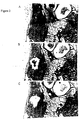
- Figure 2 depicts the steps of the microdissection technique. After carefully scraping away the surrounding tissue with a surgical scalpel, the isolated area was picked up using a new blade and transferred to an Eppendorf tube. DNA was isolated from the microdissected tissues according to the following procedure. Tissue was extracted with 100 T1 of xylenes (Fisher reagent grade) to remove the paraffin and treated with 100 T1 of 95% ethanol.
- the tissue pellet was digested in 20-50 T1 of solution containing 0.1 M Tris-HCl, pH 8, 1 mM EDTA, 0.5% Tween 20(Fisher) and 0.1 mg/ml proteinase K at 50°C overnight.
- the digestion mixture was heated at 95°C for 5 min. to inactivate the proteinase K, diluted to 100-500 T1 with a buffer comprising 10 mM Tris-HCl, pH 8, 1 mM EDTA buffer and stored at -20°C until use.
- PCR polymerase chain reaction
- PCR was processed for 45 cycles of denature (94°C for 1 min.), annealing (56-66°C for 1 min.) and chain extension (72°C for 1 min.).
- the annealing temperature for each pair of primers was listed in Table 2.
- 5 T1 of the PCR product was digested with the restriction endonuclease indicated in Table 2, and the digested fragments were separated on a 4% polyacrylamide gel.
- PCR was performed as above except 1 TCi [I- 32 P] dCTP was added to the reaction mixture, and the concentration of 4 deoxyribonucleotide triphosphates was reduced to 20 TM.
- 1 T1 of the PCR product was mixed with 3 T1 of sequencing stop solution (United States Biochemicals), denatured by heating and electrophoresed on a 6% sequencing gel.
- Each allele of the PCR product was measured by reading the optical density with a densitometer after photographing the ethidium bromide-stained gel with negative film or after exposing the X ray film to the gel.
- the ratios of the two alleles (the undigested allele over the digested, or the top allele over the bottom) for different tissues were counted. The results are presented in Table 2.
- RE stands for restriction endonuclease
- the DNA sequences are represented 5' to 3'
- RFLP stands for restriction fragment length polymorphism.
- the invasive tumor, DCIS or normal lobule samples in which the ratios of the two alleles relative to that for the normal skin show greater than 30% alteration were considered as LOH.
- Figure 4 depicts the LOH at locus 3p24 in normal lobule, DCIS and invasive tumor of sample H21(A) and H12(B). Note than in Figure 4A, skin and distal normal tissue are heterozygous at chromosome 3p24 (three bands on gel) whereas four different areas of adjacent normal tissue, DCIS and invasive carcinoma all show LOH at chromosome 3p24 (only one band on gel). In Figure 4B, the difference in density of the upper band in adjacent normal, DCIS and invasive carcinoma compared to skin indicates LOH in the test samples.
- the second potential artifact namely that the adjacent lobules contained cancerous cells was excluded by three approaches.
- photomicrographs of an adjacent section to the dissected lobules appeared to have completely normal histology.
- the DCIS and invasive components adjacent to the normal lobule had additional LOHs besides 3p24 which were not seen in the normal lobules (Table 7).
- DNA from the normal adjacent lobules of case H12 (Table 7) showed an LOH at 3p24 but not at 17p13.1 even though both the DCIS and invasive components had LOH's at both loci.
- Table 7 also summarizes the cases where the normal lobules had a LOH at 11p15.5 and 17p13.1 respectively, again showing that the DNA from the normal cells differed from that of the DCIS and invasive cancers.
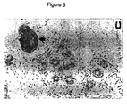
- a third method used to exclude the possibility that the normal lobules contained tumor cells was by immunostaining with antibody to erbB-2 protein.
- the invasive tumor and the DCIS components each had a least 30% of the cells immunopositive for erbB-2, there were no positive cells in the normal lobules.
- the results are depicted in Figure 3 and Table 8.
- the arrow points to the stained cells.
Landscapes
- Chemical & Material Sciences (AREA)
- Life Sciences & Earth Sciences (AREA)
- Organic Chemistry (AREA)
- Health & Medical Sciences (AREA)
- Proteomics, Peptides & Aminoacids (AREA)
- Zoology (AREA)
- Engineering & Computer Science (AREA)
- Analytical Chemistry (AREA)
- Wood Science & Technology (AREA)
- Genetics & Genomics (AREA)
- Immunology (AREA)
- Biotechnology (AREA)
- Physics & Mathematics (AREA)
- Microbiology (AREA)
- Molecular Biology (AREA)
- Biochemistry (AREA)
- Bioinformatics & Cheminformatics (AREA)
- General Engineering & Computer Science (AREA)
- General Health & Medical Sciences (AREA)
- Biophysics (AREA)
- Pathology (AREA)
- Hospice & Palliative Care (AREA)
- Oncology (AREA)
- Measuring Or Testing Involving Enzymes Or Micro-Organisms (AREA)
Description
| Clonality of Mammary Tissue Determined by X-Chromosome Inactivation | ||||
| Source of Tissue (case) | Tissue Block | Type of Mammary Tissue | Androgen Receptor Allele Lost After Digestion | Size of Dissected Area (mm2) |
| Reduction | A | lobule (1) | bottom | 0.33 |
| Mammaplasty | A | lobule (2) | top | 0.45 |
| (Case 1) | A | lobule (3) | top | 0.30 |
| Reduction | ||||
| Mammaplasty | A | lobule (1) | top | 0.30 |
| (Case 2) | A | duct(2) | top | 0.27 |
| A | lobule (3) | top | 0.21 | |
| B | lobule (4) | bottom | 0.11 | |
| B | lobule (5) | bottom | 0.17 | |
| B | lobule (6) | neither | 0.08 | |
| C | ducts | neither | 0.08 | |
| D | ducts | neither | 0.12 | |
| E | lobule | neither | 0.12 | |
| Peripheral | A | lobule | top | 0.55 |
| Mastectomy | A | lobule | top | 0.48 |
| (Case 3) | A | lobule | top | 0.50 |


| Incidence of LOH in Various Components of Breast Cancer | ||||
| Components | No. with LOH / Total Informative Cases (%) at | |||
| 3p24 (%) | 11p15.5 (%) | 13q13 (%) | 17p13.1 (%) | |
| Invasive | 10/21 (48) | 5/17 (29) | 10/14 (71) | 16/20 (80) |
| | 9/20 (45) | 2/18 (11) | 7/14 (50) | 14/20 (70) |
| | 6/17 (35) | 1/16 (6) | 0/11 (0) | 1/19 (5) |
| LOH at 3p24 In Normal Mammary Lobules Adjacent to Carcinoma | ||
| Case | Density Ratio of Upper DNA from Distant Normal Lobules | Allele to Lower Allele DNA from Adjacent Normal Lobules |
| H12 | 1.0/0.95 | 0.15/1.0 |
| H21 | 1.0/0.92 | 1.0/0.03 |
| H22 | 1.0/0.97 | 1.0/0.56 |
| H37 | 1.0/1.0 | 0.25/1.0 |
| Reproducibility of the PCR Assay for LOH | |
| Sample | Density ratio of upper allele to lower allele in adjacent lobules |
| H12 | 0.31/1.0 |
| 0.13/1.0 | |
| 0.42/1.0 | |
| 0.54/1.0 | |
| H21 | 1.0/0.05 |
| 1.0/0.03 | |
| H22 | 1.0/0.48 |
| 1.0/0.34 | |
| 1.0/0.19 | |
| 1.0/0.22 | |
| 1.0/0.56 | |
| H36 | 0.48/1.0 |
| 0.52/1.0 | |
| H37 | 1.0/0.02 |
| 1.0/0.03 | |
| 1.0/0.08 | |
| 1.0/0.14 | |
| H40 | 1.0/0.25 |
| 1.0/0.48 | |
| 1.0/0.47 | |
| 1.0/0.39 |
| LOH at Chromosome 3p24 in Individual Normal Lobules Adjacent to a Breast Carcinoma | |
| Lobule | Density ratio of upper allele to lower allele in adjacent |
| 1 | 1.0/0.05 |
| 2 | 1.0/0.10 |
| 3 | 1.0/0.05 |
| 4 | 1.0/0.05 |
| LOH at Various Chromosomal Loci in Cases with Chromosomes 3p24 LOH In Adjacent Normal Lobules | ||||||||||||
| 3p24 | 11p15.5 | 13q13 | 17p13.1 | |||||||||
| NA | DC | IN | NA | DC | IN | NA | DC | IN | NA | DC | IN | |
| H12 | Y | Y | Y | N | N | Y | N | Y | Y | |||
| H21 | Y | Y | Y | N | Y | Y | ||||||
| H37 | Y | Y | Y | Y | N | Y | Y | |||||
| H40 | Y | Y | Y | N | N | N | N | N | N | N | Y | Y |
| H5 | Y | Y | Y | N | Y | Y | ||||||
| H6 | N | N | N | N | Y | Y | Y | Y | Y |
| Comparison of p53 an erbB-2 Immunostaining in Various Components of Breast Cancer Tissue | |||
| % Immunopositive In: | |||
| Case | Normal Adjacent | DCIS Component | Invasive Component |
| p53 Immunopositive (%) | |||
| H12 | 0 | 1 | 1 |
| H40 | 0 | 10 | 10 |
| erbB-2 Immunopositive (%) | |||
| H22 | 0 | 60 | 30 |
| H37 | 0 | 10 | 5 |
| H40 | 0 | 70 | 30 |
Claims (9)
- A method for determining the likelihood of tissue in a human subject that is adjacent to breast carcinoma becoming malignant, comprising determining the loss of heterozygosity (LOH) at chromosome 3p24 in a sample of the tissue, and correlating the LOH at chromosome 3p24 in the sample with increased risk that the tissue in the human patient will become malignant.
- The method according to claim 1, wherein the tissue is benign hyperplastic tissue.
- The method according to claim 1, wherein the tissue is normal breast tissue.
- The method according to any preceding claim, wherein the carcinoma is selected from interstitial in situ carcinoma, ductal in situ carcinoma or invasive carcinoma.
- The method according to any preceding claim, further comprising the step of substantially isolating nonmalignant tissue in the sample from carcinoma present in the sample.
- The method according to any preceding claim, comprising amplifying chromosomal DNA from the 3p24 locus and determining whether the amplified DNA is polymorphic.
- The method according to claim 6, wherein the determining comprises analysing the amplified DNA for a restriction fragment length polymorphism (RFLP) or tetranucleotide.
- The method according to claim 7, wherein the analysing comprises digesting the amplified DNA with a restriction nuclease selected from the group consisting of HindIII and MspI.
- The method according to claim 6, wherein the amplifying comprises annealing a nucleic acid primer to the chromosomal DNA, wherein the primer is selected from the group consisting of:

Applications Claiming Priority (3)
| Application Number | Priority Date | Filing Date | Title |
|---|---|---|---|
| US734011 | 1996-10-18 | ||
| US08/734,011 US6686146B2 (en) | 1995-10-18 | 1996-10-18 | Prognostic methods for prediction of progression of normal and hyperplastic mammary cells to carcinoma |
| PCT/US1997/019457 WO1998017828A1 (en) | 1996-10-18 | 1997-10-17 | Method for prediction of progression of normal and hyperplastic mammary cells to carcinoma |
Publications (2)
| Publication Number | Publication Date |
|---|---|
| EP0932697A1 EP0932697A1 (en) | 1999-08-04 |
| EP0932697B1 true EP0932697B1 (en) | 2002-06-12 |
Family
ID=24949997
Family Applications (1)
| Application Number | Title | Priority Date | Filing Date |
|---|---|---|---|
| EP97912952A Expired - Lifetime EP0932697B1 (en) | 1996-10-18 | 1997-10-17 | Method for prediction of progression of normal and hyperplastic mammary cells to carcinoma |
Country Status (5)
| Country | Link |
|---|---|
| US (1) | US6686146B2 (en) |
| EP (1) | EP0932697B1 (en) |
| JP (1) | JP4145358B2 (en) |
| DE (1) | DE69713359T2 (en) |
| WO (1) | WO1998017828A1 (en) |
Families Citing this family (2)
| Publication number | Priority date | Publication date | Assignee | Title |
|---|---|---|---|---|
| US7056663B2 (en) * | 2001-03-23 | 2006-06-06 | California Pacific Medical Center | Prognostic methods for breast cancer |
| JP5151167B2 (en) * | 2007-01-31 | 2013-02-27 | 住友化学株式会社 | DNA methylation measurement method |
-
1996
- 1996-10-18 US US08/734,011 patent/US6686146B2/en not_active Expired - Lifetime
-
1997
- 1997-10-17 WO PCT/US1997/019457 patent/WO1998017828A1/en not_active Ceased
- 1997-10-17 JP JP51970098A patent/JP4145358B2/en not_active Expired - Lifetime
- 1997-10-17 DE DE69713359T patent/DE69713359T2/en not_active Expired - Lifetime
- 1997-10-17 EP EP97912952A patent/EP0932697B1/en not_active Expired - Lifetime
Also Published As
| Publication number | Publication date |
|---|---|
| US6686146B2 (en) | 2004-02-03 |
| DE69713359D1 (en) | 2002-07-18 |
| EP0932697A1 (en) | 1999-08-04 |
| DE69713359T2 (en) | 2003-01-09 |
| JP4145358B2 (en) | 2008-09-03 |
| WO1998017828A1 (en) | 1998-04-30 |
| US20020055096A1 (en) | 2002-05-09 |
| JP2001503259A (en) | 2001-03-13 |
Similar Documents
| Publication | Publication Date | Title |
|---|---|---|
| Tsai et al. | Contiguous patches of normal human mammary epithelium derived from a single stem cell: implications for breast carcinogenesis | |
| Lodygin et al. | Functional epigenomics identifies genes frequently silenced in prostate cancer | |
| Garcia et al. | Allelic loss of the PTEN region (10q23) in breast carcinomas of poor pathophenotype | |
| Eiriksdottir et al. | Mapping loss of heterozygosity at chromosome 13q: loss at 13q12-q13 is associated with breast tumour progression and poor prognosis | |
| Jovanovic et al. | Uteri of women with endometrial carcinoma contain a histopathological spectrum of monoclonal putative precancers, some with microsatellite instability | |
| Kytölä et al. | Comparative genomic hybridization identifies loss of 18q22-qter as an early and specific event in tumorigenesis of midgut carcinoids | |
| Millar et al. | Detailed methylation analysis of the glutathione S-transferase π (GSTP1) gene in prostate cancer | |
| Kinoshita et al. | Methylation of the androgen receptor minimal promoter silences transcription in human prostate cancer | |
| US20100330567A1 (en) | Dna markers for management of cancer | |
| Elo et al. | Three independently deleted regions at chromosome arm 16q in human prostate cancer: allelic loss at 16q24. 1–q24. 2 is associated with aggressive behaviour of the disease, recurrent growth, poor differentiation of the tumour and poor prognosis for the patient | |
| Suzuki et al. | Loss of heterozygosity in the tuberous sclerosis gene associated regions in adenocarcinoma of the lung accompanied by multiple atypical adenomatous hyperplasia | |
| Huang et al. | Alteration of RPL14 in squamous cell carcinomas and preneoplastic lesions of the esophagus | |
| Edwards et al. | Immunohistochemical expression of BRCA2 protein and allelic loss at the BRCA2 locus in prostate cancer | |
| Harada et al. | Clonality in Nevocellular and Melanoma: An Expression-Based Clonality Analysis at the X-Linked Genes by Polymerase Chain Reaction | |
| Tzao et al. | 5′ CpG island hypermethylation and aberrant transcript splicing both contribute to the inactivation of the FHIT gene in resected non-small cell lung cancer | |
| Xu et al. | Epithelioid trophoblastic tumor: comparative genomic hybridization and diagnostic DNA genotyping | |
| Rosenberg et al. | Detection of monoclonal microsatellite alterations in atypical breast hyperplasia. | |
| US6686146B2 (en) | Prognostic methods for prediction of progression of normal and hyperplastic mammary cells to carcinoma | |
| WO2005111232A2 (en) | Silencing of tumor-suppressive genes by cpg-methylation in prostate cancer | |
| Trzeciak et al. | Mutations, methylation and expression of CDKN2a/p16 gene in colorectal cancer and normal colonic mucosa | |
| EP2356252B1 (en) | Method for the urinary detection of bladder cancer | |
| Mead et al. | Frequent loss of heterozygosity in early non‐small cell lung cancers at chromosome 9p21 proximal to the CDKN2a gene | |
| Dominguez et al. | Clinicopathological characteristics of breast carcinomas with allelic loss in the p73 | |
| Mandai et al. | Expression of abnormal transcripts of the FHIT (fragile histidine triad) gene in ovarian carcinoma | |
| Arai et al. | The presence of candidate tumor suppressor gene loci at chromosome 3p for oral squamous cell carcinomas |
Legal Events
| Date | Code | Title | Description |
|---|---|---|---|
| PUAI | Public reference made under article 153(3) epc to a published international application that has entered the european phase |
Free format text: ORIGINAL CODE: 0009012 |
|
| 17P | Request for examination filed |
Effective date: 19990430 |
|
| AK | Designated contracting states |
Kind code of ref document: A1 Designated state(s): DE FR GB IT |
|
| RIN1 | Information on inventor provided before grant (corrected) |
Inventor name: SMITH, HELENE, S. Inventor name: LU, YOU Inventor name: DENG, GUOREN |
|
| GRAG | Despatch of communication of intention to grant |
Free format text: ORIGINAL CODE: EPIDOS AGRA |
|
| 17Q | First examination report despatched |
Effective date: 20010705 |
|
| GRAG | Despatch of communication of intention to grant |
Free format text: ORIGINAL CODE: EPIDOS AGRA |
|
| GRAG | Despatch of communication of intention to grant |
Free format text: ORIGINAL CODE: EPIDOS AGRA |
|
| GRAH | Despatch of communication of intention to grant a patent |
Free format text: ORIGINAL CODE: EPIDOS IGRA |
|
| GRAH | Despatch of communication of intention to grant a patent |
Free format text: ORIGINAL CODE: EPIDOS IGRA |
|
| GRAA | (expected) grant |
Free format text: ORIGINAL CODE: 0009210 |
|
| AK | Designated contracting states |
Kind code of ref document: B1 Designated state(s): DE FR GB IT |
|
| REG | Reference to a national code |
Ref country code: GB Ref legal event code: FG4D |
|
| REF | Corresponds to: |
Ref document number: 69713359 Country of ref document: DE Date of ref document: 20020718 |
|
| ET | Fr: translation filed | ||
| PLBE | No opposition filed within time limit |
Free format text: ORIGINAL CODE: 0009261 |
|
| STAA | Information on the status of an ep patent application or granted ep patent |
Free format text: STATUS: NO OPPOSITION FILED WITHIN TIME LIMIT |
|
| 26N | No opposition filed |
Effective date: 20030313 |
|
| REG | Reference to a national code |
Ref country code: FR Ref legal event code: PLFP Year of fee payment: 19 |
|
| REG | Reference to a national code |
Ref country code: FR Ref legal event code: PLFP Year of fee payment: 20 |
|
| PGFP | Annual fee paid to national office [announced via postgrant information from national office to epo] |
Ref country code: GB Payment date: 20161102 Year of fee payment: 20 Ref country code: DE Payment date: 20161101 Year of fee payment: 20 Ref country code: FR Payment date: 20161031 Year of fee payment: 20 |
|
| PGFP | Annual fee paid to national office [announced via postgrant information from national office to epo] |
Ref country code: IT Payment date: 20161031 Year of fee payment: 20 |
|
| REG | Reference to a national code |
Ref country code: DE Ref legal event code: R071 Ref document number: 69713359 Country of ref document: DE |
|
| REG | Reference to a national code |
Ref country code: GB Ref legal event code: PE20 Expiry date: 20171016 |
|
| PG25 | Lapsed in a contracting state [announced via postgrant information from national office to epo] |
Ref country code: GB Free format text: LAPSE BECAUSE OF EXPIRATION OF PROTECTION Effective date: 20171016 |