EP0922101B1 - Egf-like factor from chromaffin granules and glia cell-derived neurotrophic factor with survival-promoting activity on daergic neurons - Google Patents
Egf-like factor from chromaffin granules and glia cell-derived neurotrophic factor with survival-promoting activity on daergic neurons Download PDFInfo
- Publication number
- EP0922101B1 EP0922101B1 EP97940036A EP97940036A EP0922101B1 EP 0922101 B1 EP0922101 B1 EP 0922101B1 EP 97940036 A EP97940036 A EP 97940036A EP 97940036 A EP97940036 A EP 97940036A EP 0922101 B1 EP0922101 B1 EP 0922101B1
- Authority
- EP
- European Patent Office
- Prior art keywords
- chromaffin
- cells
- protein
- survival
- cultures
- Prior art date
- Legal status (The legal status is an assumption and is not a legal conclusion. Google has not performed a legal analysis and makes no representation as to the accuracy of the status listed.)
- Expired - Lifetime
Links
- 210000001366 chromaffin granule Anatomy 0.000 title claims description 110
- 210000002569 neuron Anatomy 0.000 title claims description 54
- 230000000694 effects Effects 0.000 title description 33
- 108010025020 Nerve Growth Factor Proteins 0.000 title description 8
- 102000007072 Nerve Growth Factors Human genes 0.000 title description 7
- 210000004498 neuroglial cell Anatomy 0.000 title description 6
- 239000003900 neurotrophic factor Substances 0.000 title description 6
- 102000004169 proteins and genes Human genes 0.000 claims description 96
- 108090000623 proteins and genes Proteins 0.000 claims description 95
- 210000004027 cell Anatomy 0.000 claims description 66
- 230000004083 survival effect Effects 0.000 claims description 42
- 230000001737 promoting effect Effects 0.000 claims description 27
- 239000003446 ligand Substances 0.000 claims description 12
- 230000011748 cell maturation Effects 0.000 claims description 8
- 230000035755 proliferation Effects 0.000 claims description 6
- 208000018737 Parkinson disease Diseases 0.000 claims description 4
- 208000015114 central nervous system disease Diseases 0.000 claims description 4
- 230000002093 peripheral effect Effects 0.000 claims description 4
- 208000033808 peripheral neuropathy Diseases 0.000 claims description 4
- 238000004519 manufacturing process Methods 0.000 claims description 3
- 208000027232 peripheral nervous system disease Diseases 0.000 claims description 3
- 239000008194 pharmaceutical composition Substances 0.000 claims description 3
- 208000024827 Alzheimer disease Diseases 0.000 claims description 2
- 206010012601 diabetes mellitus Diseases 0.000 claims description 2
- 102000052116 epidermal growth factor receptor activity proteins Human genes 0.000 claims description 2
- 230000002068 genetic effect Effects 0.000 claims description 2
- 208000015122 neurodegenerative disease Diseases 0.000 claims description 2
- 201000001119 neuropathy Diseases 0.000 claims description 2
- 230000007823 neuropathy Effects 0.000 claims description 2
- 230000004936 stimulating effect Effects 0.000 claims 2
- 108700015053 epidermal growth factor receptor activity proteins Proteins 0.000 claims 1
- 102000053171 Glial Fibrillary Acidic Human genes 0.000 description 28
- 101710193519 Glial fibrillary acidic protein Proteins 0.000 description 28
- 210000005046 glial fibrillary acidic protein Anatomy 0.000 description 28
- 108091000117 Tyrosine 3-Monooxygenase Proteins 0.000 description 27
- 102000048218 Tyrosine 3-monooxygenases Human genes 0.000 description 27
- 210000003737 chromaffin cell Anatomy 0.000 description 26
- OYIFNHCXNCRBQI-UHFFFAOYSA-N 2-aminoadipic acid Chemical compound OC(=O)C(N)CCCC(O)=O OYIFNHCXNCRBQI-UHFFFAOYSA-N 0.000 description 21
- 102000001301 EGF receptor Human genes 0.000 description 21
- 108060006698 EGF receptor Proteins 0.000 description 21
- 102000018233 Fibroblast Growth Factor Human genes 0.000 description 17
- 108050007372 Fibroblast Growth Factor Proteins 0.000 description 17
- 229940126864 fibroblast growth factor Drugs 0.000 description 17
- 238000011282 treatment Methods 0.000 description 16
- 238000010790 dilution Methods 0.000 description 15
- 239000012895 dilution Substances 0.000 description 15
- AAALVYBICLMAMA-UHFFFAOYSA-N 4,5-dianilinophthalimide Chemical compound C=1C=CC=CC=1NC=1C=C2C(=O)NC(=O)C2=CC=1NC1=CC=CC=C1 AAALVYBICLMAMA-UHFFFAOYSA-N 0.000 description 14
- VYFYYTLLBUKUHU-UHFFFAOYSA-N dopamine Chemical compound NCCC1=CC=C(O)C(O)=C1 VYFYYTLLBUKUHU-UHFFFAOYSA-N 0.000 description 14
- 238000002474 experimental method Methods 0.000 description 14
- 230000001228 trophic effect Effects 0.000 description 13
- 102000034615 Glial cell line-derived neurotrophic factor Human genes 0.000 description 12
- 108091010837 Glial cell line-derived neurotrophic factor Proteins 0.000 description 12
- FMGYKKMPNATWHP-UHFFFAOYSA-N Cyperquat Chemical compound C1=C[N+](C)=CC=C1C1=CC=CC=C1 FMGYKKMPNATWHP-UHFFFAOYSA-N 0.000 description 11
- 101800004564 Transforming growth factor alpha Proteins 0.000 description 11
- 102400001320 Transforming growth factor alpha Human genes 0.000 description 11
- LOKCTEFSRHRXRJ-UHFFFAOYSA-I dipotassium trisodium dihydrogen phosphate hydrogen phosphate dichloride Chemical compound P(=O)(O)(O)[O-].[K+].P(=O)(O)([O-])[O-].[Na+].[Na+].[Cl-].[K+].[Cl-].[Na+] LOKCTEFSRHRXRJ-UHFFFAOYSA-I 0.000 description 11
- 239000002953 phosphate buffered saline Substances 0.000 description 11
- 238000004113 cell culture Methods 0.000 description 10
- ODKNJVUHOIMIIZ-RRKCRQDMSA-N floxuridine Chemical compound C1[C@H](O)[C@@H](CO)O[C@H]1N1C(=O)NC(=O)C(F)=C1 ODKNJVUHOIMIIZ-RRKCRQDMSA-N 0.000 description 10
- 229960000961 floxuridine Drugs 0.000 description 10
- 239000003102 growth factor Substances 0.000 description 10
- 210000001259 mesencephalon Anatomy 0.000 description 10
- 230000000508 neurotrophic effect Effects 0.000 description 10
- QZAYGJVTTNCVMB-UHFFFAOYSA-N serotonin Chemical compound C1=C(O)C=C2C(CCN)=CNC2=C1 QZAYGJVTTNCVMB-UHFFFAOYSA-N 0.000 description 10
- 102000009024 Epidermal Growth Factor Human genes 0.000 description 9
- 101710098940 Pro-epidermal growth factor Proteins 0.000 description 9
- 239000003636 conditioned culture medium Substances 0.000 description 9
- 239000002609 medium Substances 0.000 description 9
- 238000000746 purification Methods 0.000 description 9
- 241000283690 Bos taurus Species 0.000 description 8
- AIXAANGOTKPUOY-UHFFFAOYSA-N carbachol Chemical compound [Cl-].C[N+](C)(C)CCOC(N)=O AIXAANGOTKPUOY-UHFFFAOYSA-N 0.000 description 8
- 229960004484 carbachol Drugs 0.000 description 8
- 230000004663 cell proliferation Effects 0.000 description 8
- 239000008187 granular material Substances 0.000 description 8
- 229940076279 serotonin Drugs 0.000 description 8
- HTTJABKRGRZYRN-UHFFFAOYSA-N Heparin Chemical compound OC1C(NC(=O)C)C(O)OC(COS(O)(=O)=O)C1OC1C(OS(O)(=O)=O)C(O)C(OC2C(C(OS(O)(=O)=O)C(OC3C(C(O)C(O)C(O3)C(O)=O)OS(O)(=O)=O)C(CO)O2)NS(O)(=O)=O)C(C(O)=O)O1 HTTJABKRGRZYRN-UHFFFAOYSA-N 0.000 description 7
- 241000700159 Rattus Species 0.000 description 7
- 229960003638 dopamine Drugs 0.000 description 7
- 230000014509 gene expression Effects 0.000 description 7
- 239000001963 growth medium Substances 0.000 description 7
- 229960002897 heparin Drugs 0.000 description 7
- 229920000669 heparin Polymers 0.000 description 7
- 238000003365 immunocytochemistry Methods 0.000 description 7
- 230000035800 maturation Effects 0.000 description 7
- 230000001404 mediated effect Effects 0.000 description 7
- 238000000034 method Methods 0.000 description 7
- GWCNJMUSWLTSCW-SFQUDFHCSA-N (e)-2-cyano-3-(3,4-dihydroxyphenyl)-n-(4-phenylbutyl)prop-2-enamide Chemical compound C1=C(O)C(O)=CC=C1\C=C(/C#N)C(=O)NCCCCC1=CC=CC=C1 GWCNJMUSWLTSCW-SFQUDFHCSA-N 0.000 description 6
- 108091032973 (ribonucleotides)n+m Proteins 0.000 description 6
- 239000006144 Dulbecco’s modified Eagle's medium Substances 0.000 description 6
- LFQSCWFLJHTTHZ-UHFFFAOYSA-N Ethanol Chemical compound CCO LFQSCWFLJHTTHZ-UHFFFAOYSA-N 0.000 description 6
- 229920002684 Sepharose Polymers 0.000 description 6
- FAPWRFPIFSIZLT-UHFFFAOYSA-M Sodium chloride Chemical compound [Na+].[Cl-] FAPWRFPIFSIZLT-UHFFFAOYSA-M 0.000 description 6
- 229930006000 Sucrose Natural products 0.000 description 6
- CZMRCDWAGMRECN-UGDNZRGBSA-N Sucrose Chemical compound O[C@H]1[C@H](O)[C@@H](CO)O[C@@]1(CO)O[C@@H]1[C@H](O)[C@@H](O)[C@H](O)[C@@H](CO)O1 CZMRCDWAGMRECN-UGDNZRGBSA-N 0.000 description 6
- 239000000203 mixture Substances 0.000 description 6
- 239000005720 sucrose Substances 0.000 description 6
- 102000004142 Trypsin Human genes 0.000 description 5
- 108090000631 Trypsin Proteins 0.000 description 5
- 150000003943 catecholamines Chemical class 0.000 description 5
- 238000005119 centrifugation Methods 0.000 description 5
- 210000002165 glioblast Anatomy 0.000 description 5
- 238000000338 in vitro Methods 0.000 description 5
- 230000001965 increasing effect Effects 0.000 description 5
- 230000007246 mechanism Effects 0.000 description 5
- 238000010899 nucleation Methods 0.000 description 5
- 238000002360 preparation method Methods 0.000 description 5
- 230000019491 signal transduction Effects 0.000 description 5
- 239000012588 trypsin Substances 0.000 description 5
- CSCPPACGZOOCGX-UHFFFAOYSA-N Acetone Chemical compound CC(C)=O CSCPPACGZOOCGX-UHFFFAOYSA-N 0.000 description 4
- TWRXJAOTZQYOKJ-UHFFFAOYSA-L Magnesium chloride Chemical compound [Mg+2].[Cl-].[Cl-] TWRXJAOTZQYOKJ-UHFFFAOYSA-L 0.000 description 4
- 150000001875 compounds Chemical class 0.000 description 4
- 229960003692 gamma aminobutyric acid Drugs 0.000 description 4
- 239000003112 inhibitor Substances 0.000 description 4
- 210000003463 organelle Anatomy 0.000 description 4
- 230000037361 pathway Effects 0.000 description 4
- 239000008188 pellet Substances 0.000 description 4
- 239000008363 phosphate buffer Substances 0.000 description 4
- 238000004007 reversed phase HPLC Methods 0.000 description 4
- 238000012552 review Methods 0.000 description 4
- 230000028327 secretion Effects 0.000 description 4
- 210000002966 serum Anatomy 0.000 description 4
- 230000000638 stimulation Effects 0.000 description 4
- UCSJYZPVAKXKNQ-HZYVHMACSA-N streptomycin Chemical compound CN[C@H]1[C@H](O)[C@@H](O)[C@H](CO)O[C@H]1O[C@@H]1[C@](C=O)(O)[C@H](C)O[C@H]1O[C@@H]1[C@@H](NC(N)=N)[C@H](O)[C@@H](NC(N)=N)[C@H](O)[C@H]1O UCSJYZPVAKXKNQ-HZYVHMACSA-N 0.000 description 4
- 231100000419 toxicity Toxicity 0.000 description 4
- 230000001988 toxicity Effects 0.000 description 4
- YFGBQHOOROIVKG-BHDDXSALSA-N (2R)-2-[[(2R)-2-[[2-[[2-[[(2S)-2-amino-3-(4-hydroxyphenyl)propanoyl]amino]acetyl]amino]acetyl]amino]-3-phenylpropanoyl]amino]-4-methylsulfanylbutanoic acid Chemical compound C([C@H](C(=O)N[C@H](CCSC)C(O)=O)NC(=O)CNC(=O)CNC(=O)[C@@H](N)CC=1C=CC(O)=CC=1)C1=CC=CC=C1 YFGBQHOOROIVKG-BHDDXSALSA-N 0.000 description 3
- 102000007299 Amphiregulin Human genes 0.000 description 3
- 108010033760 Amphiregulin Proteins 0.000 description 3
- 101100171060 Caenorhabditis elegans div-1 gene Proteins 0.000 description 3
- IAZDPXIOMUYVGZ-UHFFFAOYSA-N Dimethylsulphoxide Chemical compound CS(C)=O IAZDPXIOMUYVGZ-UHFFFAOYSA-N 0.000 description 3
- WSFSSNUMVMOOMR-UHFFFAOYSA-N Formaldehyde Chemical compound O=C WSFSSNUMVMOOMR-UHFFFAOYSA-N 0.000 description 3
- 102400000243 Leu-enkephalin Human genes 0.000 description 3
- 108010022337 Leucine Enkephalin Proteins 0.000 description 3
- 101710151321 Melanostatin Proteins 0.000 description 3
- 102400000988 Met-enkephalin Human genes 0.000 description 3
- 108010042237 Methionine Enkephalin Proteins 0.000 description 3
- 102400000064 Neuropeptide Y Human genes 0.000 description 3
- 108090000189 Neuropeptides Proteins 0.000 description 3
- 230000035578 autophosphorylation Effects 0.000 description 3
- 210000004556 brain Anatomy 0.000 description 3
- 239000000872 buffer Substances 0.000 description 3
- 210000003169 central nervous system Anatomy 0.000 description 3
- 239000000064 cholinergic agonist Substances 0.000 description 3
- 238000004587 chromatography analysis Methods 0.000 description 3
- 239000002299 complementary DNA Substances 0.000 description 3
- 230000006870 function Effects 0.000 description 3
- 230000002518 glial effect Effects 0.000 description 3
- 238000001727 in vivo Methods 0.000 description 3
- 238000011534 incubation Methods 0.000 description 3
- 230000006698 induction Effects 0.000 description 3
- 230000001939 inductive effect Effects 0.000 description 3
- URLZCHNOLZSCCA-UHFFFAOYSA-N leu-enkephalin Chemical compound C=1C=C(O)C=CC=1CC(N)C(=O)NCC(=O)NCC(=O)NC(C(=O)NC(CC(C)C)C(O)=O)CC1=CC=CC=C1 URLZCHNOLZSCCA-UHFFFAOYSA-N 0.000 description 3
- 230000003278 mimic effect Effects 0.000 description 3
- 210000001577 neostriatum Anatomy 0.000 description 3
- URPYMXQQVHTUDU-OFGSCBOVSA-N nucleopeptide y Chemical compound C([C@@H](C(=O)N[C@@H]([C@@H](C)CC)C(=O)N[C@@H](CC(N)=O)C(=O)N[C@@H](CC(C)C)C(=O)N[C@@H]([C@@H](C)CC)C(=O)N[C@@H]([C@@H](C)O)C(=O)N[C@@H](CCCNC(N)=N)C(=O)N[C@@H](CCC(N)=O)C(=O)N[C@@H](CCCNC(N)=N)C(=O)N[C@@H](CC=1C=CC(O)=CC=1)C(N)=O)NC(=O)[C@H](CC=1NC=NC=1)NC(=O)[C@H](CCCNC(N)=N)NC(=O)[C@H](CC(C)C)NC(=O)[C@H](C)NC(=O)[C@H](CO)NC(=O)[C@H](CC=1C=CC(O)=CC=1)NC(=O)[C@H](CC=1C=CC(O)=CC=1)NC(=O)[C@H](CCCNC(N)=N)NC(=O)[C@H](C)NC(=O)[C@H](CC(C)C)NC(=O)[C@H](CC(O)=O)NC(=O)[C@H](CCC(O)=O)NC(=O)[C@H](C)NC(=O)[C@H]1N(CCC1)C(=O)[C@H](C)NC(=O)[C@H](CC(O)=O)NC(=O)[C@H](CCC(O)=O)NC(=O)CNC(=O)[C@H]1N(CCC1)C(=O)[C@H](CC(N)=O)NC(=O)[C@H](CC(O)=O)NC(=O)[C@H]1N(CCC1)C(=O)[C@H](CCCCN)NC(=O)[C@H](CO)NC(=O)[C@H]1N(CCC1)C(=O)[C@@H](N)CC=1C=CC(O)=CC=1)C1=CC=C(O)C=C1 URPYMXQQVHTUDU-OFGSCBOVSA-N 0.000 description 3
- 238000003757 reverse transcription PCR Methods 0.000 description 3
- 239000011780 sodium chloride Substances 0.000 description 3
- 239000000243 solution Substances 0.000 description 3
- 238000010186 staining Methods 0.000 description 3
- 210000001519 tissue Anatomy 0.000 description 3
- QKNYBSVHEMOAJP-UHFFFAOYSA-N 2-amino-2-(hydroxymethyl)propane-1,3-diol;hydron;chloride Chemical compound Cl.OCC(N)(CO)CO QKNYBSVHEMOAJP-UHFFFAOYSA-N 0.000 description 2
- WEVYAHXRMPXWCK-UHFFFAOYSA-N Acetonitrile Chemical compound CC#N WEVYAHXRMPXWCK-UHFFFAOYSA-N 0.000 description 2
- CIWBSHSKHKDKBQ-JLAZNSOCSA-N Ascorbic acid Chemical compound OC[C@H](O)[C@H]1OC(=O)C(O)=C1O CIWBSHSKHKDKBQ-JLAZNSOCSA-N 0.000 description 2
- IJGRMHOSHXDMSA-UHFFFAOYSA-N Atomic nitrogen Chemical compound N#N IJGRMHOSHXDMSA-UHFFFAOYSA-N 0.000 description 2
- HEDRZPFGACZZDS-UHFFFAOYSA-N Chloroform Chemical compound ClC(Cl)Cl HEDRZPFGACZZDS-UHFFFAOYSA-N 0.000 description 2
- 108090000695 Cytokines Proteins 0.000 description 2
- 102000004127 Cytokines Human genes 0.000 description 2
- 108010092674 Enkephalins Proteins 0.000 description 2
- DHMQDGOQFOQNFH-UHFFFAOYSA-N Glycine Chemical compound NCC(O)=O DHMQDGOQFOQNFH-UHFFFAOYSA-N 0.000 description 2
- 101100118545 Holotrichia diomphalia EGF-like gene Proteins 0.000 description 2
- 241000282414 Homo sapiens Species 0.000 description 2
- URLZCHNOLZSCCA-VABKMULXSA-N Leu-enkephalin Chemical compound C([C@@H](C(=O)N[C@@H](CC(C)C)C(O)=O)NC(=O)CNC(=O)CNC(=O)[C@@H](N)CC=1C=CC(O)=CC=1)C1=CC=CC=C1 URLZCHNOLZSCCA-VABKMULXSA-N 0.000 description 2
- 229910019142 PO4 Inorganic materials 0.000 description 2
- 229930182555 Penicillin Natural products 0.000 description 2
- JGSARLDLIJGVTE-MBNYWOFBSA-N Penicillin G Chemical compound N([C@H]1[C@H]2SC([C@@H](N2C1=O)C(O)=O)(C)C)C(=O)CC1=CC=CC=C1 JGSARLDLIJGVTE-MBNYWOFBSA-N 0.000 description 2
- ISWSIDIOOBJBQZ-UHFFFAOYSA-N Phenol Chemical compound OC1=CC=CC=C1 ISWSIDIOOBJBQZ-UHFFFAOYSA-N 0.000 description 2
- RJKFOVLPORLFTN-LEKSSAKUSA-N Progesterone Chemical compound C1CC2=CC(=O)CC[C@]2(C)[C@@H]2[C@@H]1[C@@H]1CC[C@H](C(=O)C)[C@@]1(C)CC2 RJKFOVLPORLFTN-LEKSSAKUSA-N 0.000 description 2
- 102100036467 Protein delta homolog 1 Human genes 0.000 description 2
- 239000007983 Tris buffer Substances 0.000 description 2
- 230000009471 action Effects 0.000 description 2
- APKFDSVGJQXUKY-INPOYWNPSA-N amphotericin B Chemical compound O[C@H]1[C@@H](N)[C@H](O)[C@@H](C)O[C@H]1O[C@H]1/C=C/C=C/C=C/C=C/C=C/C=C/C=C/[C@H](C)[C@@H](O)[C@@H](C)[C@H](C)OC(=O)C[C@H](O)C[C@H](O)CC[C@@H](O)[C@H](O)C[C@H](O)C[C@](O)(C[C@H](O)[C@H]2C(O)=O)O[C@H]2C1 APKFDSVGJQXUKY-INPOYWNPSA-N 0.000 description 2
- 230000003321 amplification Effects 0.000 description 2
- 239000003080 antimitotic agent Substances 0.000 description 2
- FIVPIPIDMRVLAY-UHFFFAOYSA-N aspergillin Natural products C1C2=CC=CC(O)C2N2C1(SS1)C(=O)N(C)C1(CO)C2=O FIVPIPIDMRVLAY-UHFFFAOYSA-N 0.000 description 2
- 210000001130 astrocyte Anatomy 0.000 description 2
- 230000009286 beneficial effect Effects 0.000 description 2
- 230000000903 blocking effect Effects 0.000 description 2
- 230000008859 change Effects 0.000 description 2
- 239000000470 constituent Substances 0.000 description 2
- 238000001514 detection method Methods 0.000 description 2
- 238000000502 dialysis Methods 0.000 description 2
- 230000004069 differentiation Effects 0.000 description 2
- 230000029087 digestion Effects 0.000 description 2
- 239000012153 distilled water Substances 0.000 description 2
- 238000001493 electron microscopy Methods 0.000 description 2
- 230000008014 freezing Effects 0.000 description 2
- 238000007710 freezing Methods 0.000 description 2
- FIVPIPIDMRVLAY-RBJBARPLSA-N gliotoxin Chemical compound C1C2=CC=C[C@H](O)[C@H]2N2[C@]1(SS1)C(=O)N(C)[C@@]1(CO)C2=O FIVPIPIDMRVLAY-RBJBARPLSA-N 0.000 description 2
- 229940103893 gliotoxin Drugs 0.000 description 2
- 229930190252 gliotoxin Natural products 0.000 description 2
- 238000010348 incorporation Methods 0.000 description 2
- 230000005764 inhibitory process Effects 0.000 description 2
- NOESYZHRGYRDHS-UHFFFAOYSA-N insulin Chemical compound N1C(=O)C(NC(=O)C(CCC(N)=O)NC(=O)C(CCC(O)=O)NC(=O)C(C(C)C)NC(=O)C(NC(=O)CN)C(C)CC)CSSCC(C(NC(CO)C(=O)NC(CC(C)C)C(=O)NC(CC=2C=CC(O)=CC=2)C(=O)NC(CCC(N)=O)C(=O)NC(CC(C)C)C(=O)NC(CCC(O)=O)C(=O)NC(CC(N)=O)C(=O)NC(CC=2C=CC(O)=CC=2)C(=O)NC(CSSCC(NC(=O)C(C(C)C)NC(=O)C(CC(C)C)NC(=O)C(CC=2C=CC(O)=CC=2)NC(=O)C(CC(C)C)NC(=O)C(C)NC(=O)C(CCC(O)=O)NC(=O)C(C(C)C)NC(=O)C(CC(C)C)NC(=O)C(CC=2NC=NC=2)NC(=O)C(CO)NC(=O)CNC2=O)C(=O)NCC(=O)NC(CCC(O)=O)C(=O)NC(CCCNC(N)=N)C(=O)NCC(=O)NC(CC=3C=CC=CC=3)C(=O)NC(CC=3C=CC=CC=3)C(=O)NC(CC=3C=CC(O)=CC=3)C(=O)NC(C(C)O)C(=O)N3C(CCC3)C(=O)NC(CCCCN)C(=O)NC(C)C(O)=O)C(=O)NC(CC(N)=O)C(O)=O)=O)NC(=O)C(C(C)CC)NC(=O)C(CO)NC(=O)C(C(C)O)NC(=O)C1CSSCC2NC(=O)C(CC(C)C)NC(=O)C(NC(=O)C(CCC(N)=O)NC(=O)C(CC(N)=O)NC(=O)C(NC(=O)C(N)CC=1C=CC=CC=1)C(C)C)CC1=CN=CN1 NOESYZHRGYRDHS-UHFFFAOYSA-N 0.000 description 2
- 238000002955 isolation Methods 0.000 description 2
- 238000002372 labelling Methods 0.000 description 2
- 239000007788 liquid Substances 0.000 description 2
- 229910001629 magnesium chloride Inorganic materials 0.000 description 2
- 239000012528 membrane Substances 0.000 description 2
- 230000028161 membrane depolarization Effects 0.000 description 2
- 108020004999 messenger RNA Proteins 0.000 description 2
- 230000002297 mitogenic effect Effects 0.000 description 2
- 230000032405 negative regulation of neuron apoptotic process Effects 0.000 description 2
- 238000003199 nucleic acid amplification method Methods 0.000 description 2
- 229940049954 penicillin Drugs 0.000 description 2
- NBIIXXVUZAFLBC-UHFFFAOYSA-K phosphate Chemical compound [O-]P([O-])([O-])=O NBIIXXVUZAFLBC-UHFFFAOYSA-K 0.000 description 2
- 239000010452 phosphate Substances 0.000 description 2
- 230000003389 potentiating effect Effects 0.000 description 2
- 230000008569 process Effects 0.000 description 2
- XJMOSONTPMZWPB-UHFFFAOYSA-M propidium iodide Chemical compound [I-].[I-].C12=CC(N)=CC=C2C2=CC=C(N)C=C2[N+](CCC[N+](C)(CC)CC)=C1C1=CC=CC=C1 XJMOSONTPMZWPB-UHFFFAOYSA-M 0.000 description 2
- 239000012460 protein solution Substances 0.000 description 2
- 238000011002 quantification Methods 0.000 description 2
- 230000001105 regulatory effect Effects 0.000 description 2
- 239000012146 running buffer Substances 0.000 description 2
- 239000000523 sample Substances 0.000 description 2
- 230000003248 secreting effect Effects 0.000 description 2
- 239000013049 sediment Substances 0.000 description 2
- 239000012679 serum free medium Substances 0.000 description 2
- 230000011664 signaling Effects 0.000 description 2
- 210000000130 stem cell Anatomy 0.000 description 2
- 229960005322 streptomycin Drugs 0.000 description 2
- 239000000126 substance Substances 0.000 description 2
- 239000013589 supplement Substances 0.000 description 2
- JGVWCANSWKRBCS-UHFFFAOYSA-N tetramethylrhodamine thiocyanate Chemical compound [Cl-].C=12C=CC(N(C)C)=CC2=[O+]C2=CC(N(C)C)=CC=C2C=1C1=CC=C(SC#N)C=C1C(O)=O JGVWCANSWKRBCS-UHFFFAOYSA-N 0.000 description 2
- 239000003053 toxin Substances 0.000 description 2
- 231100000765 toxin Toxicity 0.000 description 2
- LENZDBCJOHFCAS-UHFFFAOYSA-N tris Chemical compound OCC(N)(CO)CO LENZDBCJOHFCAS-UHFFFAOYSA-N 0.000 description 2
- 238000012800 visualization Methods 0.000 description 2
- 238000005406 washing Methods 0.000 description 2
- XLYOFNOQVPJJNP-UHFFFAOYSA-N water Chemical compound O XLYOFNOQVPJJNP-UHFFFAOYSA-N 0.000 description 2
- BICGOULMPJLRQV-MYINAIGISA-N 1-[(2s,4s,5r)-2-bromo-4-hydroxy-5-(hydroxymethyl)oxolan-2-yl]pyrimidine-2,4-dione Chemical compound C1[C@H](O)[C@@H](CO)O[C@@]1(Br)N1C(=O)NC(=O)C=C1 BICGOULMPJLRQV-MYINAIGISA-N 0.000 description 1
- 210000002348 5-ht neuron Anatomy 0.000 description 1
- QTBSBXVTEAMEQO-UHFFFAOYSA-M Acetate Chemical compound CC([O-])=O QTBSBXVTEAMEQO-UHFFFAOYSA-M 0.000 description 1
- 101800001382 Betacellulin Proteins 0.000 description 1
- BTBUEUYNUDRHOZ-UHFFFAOYSA-N Borate Chemical compound [O-]B([O-])[O-] BTBUEUYNUDRHOZ-UHFFFAOYSA-N 0.000 description 1
- 102000004219 Brain-derived neurotrophic factor Human genes 0.000 description 1
- 108090000715 Brain-derived neurotrophic factor Proteins 0.000 description 1
- OYPRJOBELJOOCE-UHFFFAOYSA-N Calcium Chemical compound [Ca] OYPRJOBELJOOCE-UHFFFAOYSA-N 0.000 description 1
- 102000010792 Chromogranin A Human genes 0.000 description 1
- 108010038447 Chromogranin A Proteins 0.000 description 1
- 102000007345 Chromogranins Human genes 0.000 description 1
- 108010007718 Chromogranins Proteins 0.000 description 1
- 102000029816 Collagenase Human genes 0.000 description 1
- 108060005980 Collagenase Proteins 0.000 description 1
- 108010014303 DNA-directed DNA polymerase Proteins 0.000 description 1
- 102000016928 DNA-directed DNA polymerase Human genes 0.000 description 1
- 102100038606 Death-associated protein kinase 3 Human genes 0.000 description 1
- 206010012289 Dementia Diseases 0.000 description 1
- 108010015720 Dopamine beta-Hydroxylase Proteins 0.000 description 1
- 102100033156 Dopamine beta-hydroxylase Human genes 0.000 description 1
- 101150029707 ERBB2 gene Proteins 0.000 description 1
- 101150039808 Egfr gene Proteins 0.000 description 1
- 101800000155 Epiregulin Proteins 0.000 description 1
- 108090000379 Fibroblast growth factor 2 Proteins 0.000 description 1
- 102000003974 Fibroblast growth factor 2 Human genes 0.000 description 1
- 208000032612 Glial tumor Diseases 0.000 description 1
- 206010018338 Glioma Diseases 0.000 description 1
- WQZGKKKJIJFFOK-GASJEMHNSA-N Glucose Natural products OC[C@H]1OC(O)[C@H](O)[C@@H](O)[C@@H]1O WQZGKKKJIJFFOK-GASJEMHNSA-N 0.000 description 1
- SXRSQZLOMIGNAQ-UHFFFAOYSA-N Glutaraldehyde Chemical compound O=CCCCC=O SXRSQZLOMIGNAQ-UHFFFAOYSA-N 0.000 description 1
- 239000004471 Glycine Substances 0.000 description 1
- 235000010469 Glycine max Nutrition 0.000 description 1
- 244000068988 Glycine max Species 0.000 description 1
- 239000012981 Hank's balanced salt solution Substances 0.000 description 1
- 102000004877 Insulin Human genes 0.000 description 1
- 108090001061 Insulin Proteins 0.000 description 1
- 102100034343 Integrase Human genes 0.000 description 1
- XUIIKFGFIJCVMT-LBPRGKRZSA-N L-thyroxine Chemical compound IC1=CC(C[C@H]([NH3+])C([O-])=O)=CC(I)=C1OC1=CC(I)=C(O)C(I)=C1 XUIIKFGFIJCVMT-LBPRGKRZSA-N 0.000 description 1
- 241000700562 Myxoma virus Species 0.000 description 1
- 102000015336 Nerve Growth Factor Human genes 0.000 description 1
- 102000014413 Neuregulin Human genes 0.000 description 1
- 108050003475 Neuregulin Proteins 0.000 description 1
- 102400000058 Neuregulin-1 Human genes 0.000 description 1
- 108090000556 Neuregulin-1 Proteins 0.000 description 1
- 229930040373 Paraformaldehyde Natural products 0.000 description 1
- 229920001213 Polysorbate 20 Polymers 0.000 description 1
- 102100029837 Probetacellulin Human genes 0.000 description 1
- 102100025498 Proepiregulin Human genes 0.000 description 1
- 102000001253 Protein Kinase Human genes 0.000 description 1
- 101710119301 Protein delta homolog 1 Proteins 0.000 description 1
- 239000013614 RNA sample Substances 0.000 description 1
- 108010092799 RNA-directed DNA polymerase Proteins 0.000 description 1
- 241000700564 Rabbit fibroma virus Species 0.000 description 1
- 241000700157 Rattus norvegicus Species 0.000 description 1
- 101000606117 Rattus norvegicus Tyrosine 3-monooxygenase Proteins 0.000 description 1
- 102100030086 Receptor tyrosine-protein kinase erbB-2 Human genes 0.000 description 1
- 102000006382 Ribonucleases Human genes 0.000 description 1
- 108010083644 Ribonucleases Proteins 0.000 description 1
- 230000018199 S phase Effects 0.000 description 1
- 229940124639 Selective inhibitor Drugs 0.000 description 1
- -1 Sigma) Chemical compound 0.000 description 1
- 238000000692 Student's t-test Methods 0.000 description 1
- 102000004887 Transforming Growth Factor beta Human genes 0.000 description 1
- 108090001012 Transforming Growth Factor beta Proteins 0.000 description 1
- 101710162629 Trypsin inhibitor Proteins 0.000 description 1
- 229940122618 Trypsin inhibitor Drugs 0.000 description 1
- 208000004062 Tumor Virus Infections Diseases 0.000 description 1
- 108010067390 Viral Proteins Proteins 0.000 description 1
- 230000035508 accumulation Effects 0.000 description 1
- 238000009825 accumulation Methods 0.000 description 1
- 239000002253 acid Substances 0.000 description 1
- 230000001919 adrenal effect Effects 0.000 description 1
- 239000011543 agarose gel Substances 0.000 description 1
- 150000001412 amines Chemical class 0.000 description 1
- 238000004458 analytical method Methods 0.000 description 1
- 238000010171 animal model Methods 0.000 description 1
- 238000000137 annealing Methods 0.000 description 1
- 230000002927 anti-mitotic effect Effects 0.000 description 1
- 108010054176 apotransferrin Proteins 0.000 description 1
- 229960005070 ascorbic acid Drugs 0.000 description 1
- 235000010323 ascorbic acid Nutrition 0.000 description 1
- 239000011668 ascorbic acid Substances 0.000 description 1
- 210000003050 axon Anatomy 0.000 description 1
- 230000000035 biogenic effect Effects 0.000 description 1
- 230000004071 biological effect Effects 0.000 description 1
- 230000015572 biosynthetic process Effects 0.000 description 1
- 230000008499 blood brain barrier function Effects 0.000 description 1
- 210000001218 blood-brain barrier Anatomy 0.000 description 1
- 239000012888 bovine serum Substances 0.000 description 1
- 239000007853 buffer solution Substances 0.000 description 1
- 239000011575 calcium Substances 0.000 description 1
- 229910052791 calcium Inorganic materials 0.000 description 1
- 230000022131 cell cycle Effects 0.000 description 1
- 230000024245 cell differentiation Effects 0.000 description 1
- 239000006285 cell suspension Substances 0.000 description 1
- 230000001713 cholinergic effect Effects 0.000 description 1
- 229960002424 collagenase Drugs 0.000 description 1
- 230000001143 conditioned effect Effects 0.000 description 1
- 238000011109 contamination Methods 0.000 description 1
- 230000009089 cytolysis Effects 0.000 description 1
- 230000034994 death Effects 0.000 description 1
- 230000007850 degeneration Effects 0.000 description 1
- 230000018044 dehydration Effects 0.000 description 1
- 238000006297 dehydration reaction Methods 0.000 description 1
- 238000004925 denaturation Methods 0.000 description 1
- 230000036425 denaturation Effects 0.000 description 1
- 238000000432 density-gradient centrifugation Methods 0.000 description 1
- 239000003085 diluting agent Substances 0.000 description 1
- 201000010099 disease Diseases 0.000 description 1
- 208000037265 diseases, disorders, signs and symptoms Diseases 0.000 description 1
- BVTBRVFYZUCAKH-UHFFFAOYSA-L disodium selenite Chemical compound [Na+].[Na+].[O-][Se]([O-])=O BVTBRVFYZUCAKH-UHFFFAOYSA-L 0.000 description 1
- VHJLVAABSRFDPM-QWWZWVQMSA-N dithiothreitol Chemical compound SC[C@@H](O)[C@H](O)CS VHJLVAABSRFDPM-QWWZWVQMSA-N 0.000 description 1
- 231100000673 dose–response relationship Toxicity 0.000 description 1
- 239000003937 drug carrier Substances 0.000 description 1
- 229940121647 egfr inhibitor Drugs 0.000 description 1
- 238000000635 electron micrograph Methods 0.000 description 1
- 238000010218 electron microscopic analysis Methods 0.000 description 1
- 238000001962 electrophoresis Methods 0.000 description 1
- 210000002257 embryonic structure Anatomy 0.000 description 1
- ZMMJGEGLRURXTF-UHFFFAOYSA-N ethidium bromide Chemical compound [Br-].C12=CC(N)=CC=C2C2=CC=C(N)C=C2[N+](CC)=C1C1=CC=CC=C1 ZMMJGEGLRURXTF-UHFFFAOYSA-N 0.000 description 1
- 229960005542 ethidium bromide Drugs 0.000 description 1
- 238000011156 evaluation Methods 0.000 description 1
- 230000028023 exocytosis Effects 0.000 description 1
- 238000000605 extraction Methods 0.000 description 1
- 210000003754 fetus Anatomy 0.000 description 1
- 238000005194 fractionation Methods 0.000 description 1
- 239000012634 fragment Substances 0.000 description 1
- 239000012737 fresh medium Substances 0.000 description 1
- 230000003371 gabaergic effect Effects 0.000 description 1
- 108010074605 gamma-Globulins Proteins 0.000 description 1
- BTCSSZJGUNDROE-UHFFFAOYSA-N gamma-aminobutyric acid Chemical compound NCCCC(O)=O BTCSSZJGUNDROE-UHFFFAOYSA-N 0.000 description 1
- 239000011521 glass Substances 0.000 description 1
- 239000008103 glucose Substances 0.000 description 1
- ZDXPYRJPNDTMRX-UHFFFAOYSA-N glutamine Natural products OC(=O)C(N)CCC(N)=O ZDXPYRJPNDTMRX-UHFFFAOYSA-N 0.000 description 1
- ZJYYHGLJYGJLLN-UHFFFAOYSA-N guanidinium thiocyanate Chemical compound SC#N.NC(N)=N ZJYYHGLJYGJLLN-UHFFFAOYSA-N 0.000 description 1
- 238000010438 heat treatment Methods 0.000 description 1
- 210000004295 hippocampal neuron Anatomy 0.000 description 1
- 230000001771 impaired effect Effects 0.000 description 1
- 239000007943 implant Substances 0.000 description 1
- 230000005917 in vivo anti-tumor Effects 0.000 description 1
- 230000002779 inactivation Effects 0.000 description 1
- 230000000977 initiatory effect Effects 0.000 description 1
- 229940125396 insulin Drugs 0.000 description 1
- 150000002500 ions Chemical class 0.000 description 1
- 229950008325 levothyroxine Drugs 0.000 description 1
- 230000004807 localization Effects 0.000 description 1
- 230000010534 mechanism of action Effects 0.000 description 1
- 238000001000 micrograph Methods 0.000 description 1
- 239000003226 mitogen Substances 0.000 description 1
- 230000007659 motor function Effects 0.000 description 1
- 229940053128 nerve growth factor Drugs 0.000 description 1
- BPGXUIVWLQTVLZ-OFGSCBOVSA-N neuropeptide y(npy) Chemical compound C([C@@H](C(=O)N[C@@H]([C@@H](C)CC)C(=O)N[C@@H](CC(N)=O)C(=O)N[C@@H](CC(C)C)C(=O)N[C@@H]([C@@H](C)CC)C(=O)N[C@@H]([C@@H](C)O)C(=O)N[C@@H](CCCN=C(N)N)C(=O)N[C@@H](CCC(N)=O)C(=O)N[C@@H](CCCN=C(N)N)C(=O)N[C@@H](CC=1C=CC(O)=CC=1)C(O)=O)NC(=O)[C@H](CC=1N=CNC=1)NC(=O)[C@H](CCCN=C(N)N)NC(=O)[C@H](CC(C)C)NC(=O)[C@H](C)NC(=O)[C@H](CO)NC(=O)[C@H](CC=1C=CC(O)=CC=1)NC(=O)[C@H](CC=1C=CC(O)=CC=1)NC(=O)[C@H](CCCN=C(N)N)NC(=O)[C@H](C)NC(=O)[C@H](CC(C)C)NC(=O)[C@H](CC(O)=O)NC(=O)[C@H](CCC(O)=O)NC(=O)[C@H](C)NC(=O)[C@H]1N(CCC1)C(=O)[C@H](C)NC(=O)[C@H](CC(O)=O)NC(=O)[C@H](CCC(O)=O)NC(=O)CNC(=O)[C@H]1N(CCC1)C(=O)[C@H](CC(N)=O)NC(=O)[C@H](CC(O)=O)NC(=O)[C@H]1N(CCC1)C(=O)[C@H](CCCCN)NC(=O)[C@H](CO)NC(=O)[C@H]1N(CCC1)C(=O)[C@@H](N)CC=1C=CC(O)=CC=1)C1=CC=C(O)C=C1 BPGXUIVWLQTVLZ-OFGSCBOVSA-N 0.000 description 1
- 230000001414 neuropoietic effect Effects 0.000 description 1
- 229910052757 nitrogen Inorganic materials 0.000 description 1
- 238000012758 nuclear staining Methods 0.000 description 1
- 238000001543 one-way ANOVA Methods 0.000 description 1
- 229910000489 osmium tetroxide Inorganic materials 0.000 description 1
- 239000012285 osmium tetroxide Substances 0.000 description 1
- 229920002866 paraformaldehyde Polymers 0.000 description 1
- 230000010412 perfusion Effects 0.000 description 1
- 210000001428 peripheral nervous system Anatomy 0.000 description 1
- 208000028591 pheochromocytoma Diseases 0.000 description 1
- 108010055896 polyornithine Proteins 0.000 description 1
- 229920002714 polyornithine Polymers 0.000 description 1
- 235000010486 polyoxyethylene sorbitan monolaurate Nutrition 0.000 description 1
- 239000000256 polyoxyethylene sorbitan monolaurate Substances 0.000 description 1
- 239000013641 positive control Substances 0.000 description 1
- 230000003805 potassium influx Effects 0.000 description 1
- 239000000047 product Substances 0.000 description 1
- 229960003387 progesterone Drugs 0.000 description 1
- 239000000186 progesterone Substances 0.000 description 1
- 238000002731 protein assay Methods 0.000 description 1
- 108060006633 protein kinase Proteins 0.000 description 1
- 102000005962 receptors Human genes 0.000 description 1
- 108020003175 receptors Proteins 0.000 description 1
- 230000003830 regulated secretory pathway Effects 0.000 description 1
- 239000003161 ribonuclease inhibitor Substances 0.000 description 1
- 150000003839 salts Chemical class 0.000 description 1
- 238000009738 saturating Methods 0.000 description 1
- 230000000580 secretagogue effect Effects 0.000 description 1
- 210000004739 secretory vesicle Anatomy 0.000 description 1
- 230000000862 serotonergic effect Effects 0.000 description 1
- 230000013275 serotonin uptake Effects 0.000 description 1
- 229960001471 sodium selenite Drugs 0.000 description 1
- 239000011781 sodium selenite Substances 0.000 description 1
- 235000015921 sodium selenite Nutrition 0.000 description 1
- 238000004611 spectroscopical analysis Methods 0.000 description 1
- 239000006228 supernatant Substances 0.000 description 1
- 230000008093 supporting effect Effects 0.000 description 1
- 230000001629 suppression Effects 0.000 description 1
- 230000000946 synaptic effect Effects 0.000 description 1
- 238000003786 synthesis reaction Methods 0.000 description 1
- 238000012360 testing method Methods 0.000 description 1
- ZRKFYGHZFMAOKI-QMGMOQQFSA-N tgfbeta Chemical compound C([C@H](NC(=O)[C@H](C(C)C)NC(=O)CNC(=O)[C@H](CCC(O)=O)NC(=O)[C@H](CCCNC(N)=N)NC(=O)[C@H](CC(N)=O)NC(=O)[C@H](CC(C)C)NC(=O)[C@H]([C@@H](C)O)NC(=O)[C@H](CCC(O)=O)NC(=O)[C@H]([C@@H](C)O)NC(=O)[C@H](CC(C)C)NC(=O)CNC(=O)[C@H](C)NC(=O)[C@H](CO)NC(=O)[C@H](CCC(N)=O)NC(=O)[C@@H](NC(=O)[C@H](C)NC(=O)[C@H](C)NC(=O)[C@@H](NC(=O)[C@H](CC(C)C)NC(=O)[C@@H](N)CCSC)C(C)C)[C@@H](C)CC)C(=O)N[C@@H]([C@@H](C)O)C(=O)N[C@@H](C(C)C)C(=O)N[C@@H](CC=1C=CC=CC=1)C(=O)N[C@@H](C)C(=O)N1[C@@H](CCC1)C(=O)N[C@@H]([C@@H](C)O)C(=O)N[C@@H](CC(N)=O)C(=O)N[C@@H](CCC(O)=O)C(=O)N[C@@H](C)C(=O)N[C@@H](CC=1C=CC=CC=1)C(=O)N[C@@H](CCCNC(N)=N)C(=O)N[C@@H](C)C(=O)N[C@@H](CC(C)C)C(=O)N1[C@@H](CCC1)C(=O)N1[C@@H](CCC1)C(=O)N[C@@H](CCCNC(N)=N)C(=O)N[C@@H](CCC(O)=O)C(=O)N[C@@H](CCCNC(N)=N)C(=O)N[C@@H](CO)C(=O)N[C@@H](CCCNC(N)=N)C(=O)N[C@@H](CC(C)C)C(=O)N[C@@H](CC(C)C)C(O)=O)C1=CC=C(O)C=C1 ZRKFYGHZFMAOKI-QMGMOQQFSA-N 0.000 description 1
- 238000010257 thawing Methods 0.000 description 1
- 230000001225 therapeutic effect Effects 0.000 description 1
- 231100000331 toxic Toxicity 0.000 description 1
- 230000002588 toxic effect Effects 0.000 description 1
- 230000026683 transduction Effects 0.000 description 1
- 238000010361 transduction Methods 0.000 description 1
- 239000002753 trypsin inhibitor Substances 0.000 description 1
- 239000005483 tyrosine kinase inhibitor Substances 0.000 description 1
Images





Classifications
-
- C—CHEMISTRY; METALLURGY
- C07—ORGANIC CHEMISTRY
- C07K—PEPTIDES
- C07K14/00—Peptides having more than 20 amino acids; Gastrins; Somatostatins; Melanotropins; Derivatives thereof
- C07K14/435—Peptides having more than 20 amino acids; Gastrins; Somatostatins; Melanotropins; Derivatives thereof from animals; from humans
- C07K14/475—Growth factors; Growth regulators
-
- A—HUMAN NECESSITIES
- A61—MEDICAL OR VETERINARY SCIENCE; HYGIENE
- A61P—SPECIFIC THERAPEUTIC ACTIVITY OF CHEMICAL COMPOUNDS OR MEDICINAL PREPARATIONS
- A61P25/00—Drugs for disorders of the nervous system
-
- A—HUMAN NECESSITIES
- A61—MEDICAL OR VETERINARY SCIENCE; HYGIENE
- A61P—SPECIFIC THERAPEUTIC ACTIVITY OF CHEMICAL COMPOUNDS OR MEDICINAL PREPARATIONS
- A61P25/00—Drugs for disorders of the nervous system
- A61P25/14—Drugs for disorders of the nervous system for treating abnormal movements, e.g. chorea, dyskinesia
- A61P25/16—Anti-Parkinson drugs
-
- A—HUMAN NECESSITIES
- A61—MEDICAL OR VETERINARY SCIENCE; HYGIENE
- A61P—SPECIFIC THERAPEUTIC ACTIVITY OF CHEMICAL COMPOUNDS OR MEDICINAL PREPARATIONS
- A61P25/00—Drugs for disorders of the nervous system
- A61P25/28—Drugs for disorders of the nervous system for treating neurodegenerative disorders of the central nervous system, e.g. nootropic agents, cognition enhancers, drugs for treating Alzheimer's disease or other forms of dementia
-
- A—HUMAN NECESSITIES
- A61—MEDICAL OR VETERINARY SCIENCE; HYGIENE
- A61P—SPECIFIC THERAPEUTIC ACTIVITY OF CHEMICAL COMPOUNDS OR MEDICINAL PREPARATIONS
- A61P3/00—Drugs for disorders of the metabolism
- A61P3/08—Drugs for disorders of the metabolism for glucose homeostasis
- A61P3/10—Drugs for disorders of the metabolism for glucose homeostasis for hyperglycaemia, e.g. antidiabetics
-
- A—HUMAN NECESSITIES
- A61—MEDICAL OR VETERINARY SCIENCE; HYGIENE
- A61K—PREPARATIONS FOR MEDICAL, DENTAL OR TOILETRY PURPOSES
- A61K38/00—Medicinal preparations containing peptides
Definitions
- the present invention relates to chromaffin granule-derived EGF-like proteins that are involved in promoting survival of DAergic neurons, and to processes for their preparation.
- grafts of adrenal medullary tissue and chromaffin cells to the striatum of patients with Morbus Parkinson (PD) and animal models of the disease have pronounced therapeutic effects leading to the restoration of transmitters and motor functions (for reviews, see Freed, 1993; Fisher and Gage, 1993).
- PD Morbus Parkinson
- a common explanation for these effects has been that grafted chromaffin cells may compensate, by secreting dopamine (DA), for the substantial lack of this nigrostriatal transmitter in PD (Ehringer and Hornykiewicz, 1960).
- DA dopamine
- amounts of DA synthesized and released by chromaffin cells are small (approx.
- FGF-2 basic fibroblast growth factor
- the technical problem underlying the present invention is to provide new compounds capable of promoting survival of (mesencephalic) DAergic neurons, which can be used for the treatment of peripheral and/or CNS-disorders in man.
- These compounds can be the EGF-like activity from chromaffin granules as well as functional parts thereof or chemical compounds serving the same function which are small enough to cross the blood-brain barrier to manage that function.
- the present invention relates to a chromaffin granule-derived protein mediating selectively survival promoting activity on DAergic neurons.
- chromaffin granule-derived protein refers to single, defined proteins that, when applied singly or in combinations exert trophic, survival and differentiation promoting effects on DAergic neurons.
- selective survival promoting acitivity on DAergic neurons refers to a proteinaceous activity that may confer, by itself or in combination with other factors present in chromaffin granules, survival and differentiation upon DAergic neurons within the nanomolar range or below.
- the chromaffin granule-derived protein of the present invention is an epidermal growth factor receptor (EGFR)-ligand; i.e. the chromaffin granule-derived protein exhibits effects within a nanomolar range of concentrations.
- EGFR epidermal growth factor receptor
- the chromaffin granule-derived protein of the present invention is capable of promoting astroglial cell maturation.
- the expression "capable of astroglial cell maturation” means that within a culture system of embryonic mesencephalic cells, the protein increases the number of astroglial cells visualized by expression of proteins that are specific for this cell type.
- the chromaffin granule-derived protein is capable of inducing proliferation of non-DAergic cells, which are presumed to be glial progenitor cells.
- the expression "capable of proliferation of non-DAergic cells” means that expansion of number of presumed astroglial progenitor cells is the effect initially caused by the "chromaffin granule-derived protein". This initial effect is supposed to be the prerequisite for the promotion of survival by a factor secreted by the expanded number of astroglial cells.
- a further subject of the present invention relates to a process for the preparation of the above defined chromaffin granule-derived protein, comprising the steps of isolating chromaffin granules from chromaffin cells and extracting the aqueous-soluble protein content containing the chromaffin granule-derived protein, from the chromaffin granules in a buffer solution.
- isolated chromaffin granules from chromaffin cells comprises subcellular fractionation of chromaffin cell organelles by density gradient centrifugation at 1.7M sucrose.
- extracting the aqueous-soluble protein content containing the chromaffin granule-derived protein comprises lysis of the organelles obtained as a pellet of the 1.7M sucrose centrifugation in a 10 mM phosphate buffer at pH 7.0 following twenty minutes freezing at -80°C.
- a further subject of the present invention relates to a pharmaceutical composition
- a pharmaceutical composition comprising the above defined chromaffin granule-derived protein, optionally in association with a pharmaceutically acceptable carrier and/or diluent.
- the pharmaceutical composition may be used for the treatment of peripheral and/or CNS-disorders in man such a Parkinson's disease, Alzheimer's disease or other dementias, other neurodegenerative disorders of the central nervous system and peripheral neuropathies including diabetes, cisplatinium neuropathy or other genetic or acquired peripheral nerve diseases.
- a further subject of the present invention comprises the application of the above factor derived from chromaffin granules in conjunction with previously found neurotrophic molecules, such as neurotrophins, members of the TGF- ⁇ , fibroblast growth factor, and neuropoietic cytokine superfamilies.
- the serum-free medium used consisted of Dulbecco's modified Eagle's medium and Ham's F-12 (DMEM/F-12, BioWhittaker), including the N1 supplements (Bottenstein et al., 1980) apotrans-ferrin, insulin, L-thyroxine, sodium selenite and progesterone (Sigma, Deideshofen, Germany), and 2.5 mM glutamine (GIBCO, Eggenstein, Germany), 2.5 mg/ml bovine serum albumine (Sigma), 100 U/ml penicillin, 100 ⁇ g/ml streptomycin, and 0.25 ⁇ g/ml fungizone (BioWhittaker, Heidelberg, Germany).
- CMF Calcium-magnesium- free Hank's balanced salt solution
- horse serum trypsin
- FDUR 5-fluorodeoxyuridine
- AA ⁇ -aminoadipic acid
- DAPH 4,5-Dianilinophthalimide
- Tyrphostin B56 (Calbiochem, Bad Soden, Germany) were dissolved in DMSO to give a final concentration of 10 mM, protected from light and stored at 4°C until use.
- FDUR and AA were dissolved in PBS to give a final concentration of 1mM and stored in aliquots at -20°C.
- Spectrapor dialysis tubing (Roth, Düsseldorf, Germany) with a cutoff MW 3,500 was used.
- Growth factors rh FGF-2, rh TGF- ⁇ , rh EGF (IC Chemikalien, Ismaningen, Germany). Lyophylized growth factors were resuspended in DMEM containing 0.25% BSA and penicillin, streptomycin, and fungizone in the given concentration, to give a final concentration of 1 ⁇ g/ml. Aliquots of 100 ⁇ l were kept at -70°C until used.
- the chromaffin granules In order to purify large dense core vesicles, the chromaffin granules, the resuspended pellet (0.3 M sucrose, 10 mM phosphate, pH 7.0) was loaded on a 1.7 M sucrose cushion. The chromaffin granules were obtained as a sediment after centrifugation at 100,000 g for 90 min. The sediment was resuspended in 10 mM phosphate pH 7.0, frozen in liquid nitrogen and subsequently thawed, in order to lyze the vesicles and to extract the soluble protein content. Membrane fragments were collected by centrifugation at 100,000g for 30 min.
- the supernatent containing the soluble protein mixture from chromaffin granule was dialysed (cutoff 3,500 MW) over night against several batches of 100-fold excess of 10 mM phosphate buffer pH 7.0 to seperate catecholamines and other low molecular weight components from proteins.
- the protein solution was diluted to give a final protein concentration of 20 mg/ml. This protein solution was then sterile filtered (0.22 ⁇ M) and stored in aliquots at -70°C.
- the fractions collected were desalted by reversed phased chromatography (RP-HPLC) on Aquapore RP-300 columns, 7 micron, Applied Biosystems, using running buffer A (0.1 % TFA) and running buffer B (0.1 % TFA/90 % Acetonitril).
- RP-HPLC reversed phased chromatography
- the lyophilized samples were resuspended in 20 ⁇ l of 50 % Acetonitril and added to the medium at 5 ⁇ l per 2 ml Medium at each medium change. Heat inactivation was achieved by heating a protein sample three times to 90°C for 30 sec. An aliquot was digested with 0.1% trypsin at 37°C for one hour. Digestion was terminated by adding 100 ⁇ g disolved in 50 ⁇ l PBS of soybean trypsin inhibitor.
- Tissue culture Mesencephalic cell cultures were essentially established as described by Krieglstein and Unsicker (1994). In brief, the ventral midbrain floor was dissected from embryonic day (E) 14 Wistar rat fetuses of two litters (20-25 embryos) and collected in CMF. Tissue pieces were enzymatically dissociated using 0.25% trypsin (BioWhittaker) in CMF for 15 min at 37°C. After addition of an equal volume of ice-cold horse serum and 1mg DNAse, Cells were triturated with fire-polished and siliconized pasteur pipettes and subsequently washed with DMEM/F12.
- the single cell suspension (100 ⁇ l) was seeded on polyornithine (0.1 mg/ml in 15mM borate buffer, pH 8.4, Sigma)-laminin (5 ⁇ g/ml; Sigma) coated glass cover slips at a density of 200.000 cells/cm 2 .
- Coverslips were incubated in a humified 5% CO 2 /95% air atmosphere to allow cells to attach. After two hours coverslips were transferred to 24-well plates (Falcon) containing 750 ⁇ l medium. On the following day, and subsequently every three days, 500 ⁇ l of the medium was replaced and neurotrophic factors were added at the same time at the given concentrations.
- Bovine chromaffin cells were isolated by collagenase perfusion and digestion as previously described and enriched to > 95% purity employing Percoll gradient centrifugation (Unsicker et al., 1980; Bieger et al., 1995). Chromaffin cells were seeded at 200,000 cells/cm 2 on plastic culture flasks (Falcon; 5x10 6 cells per 25cm 2 ) and maintained in 5 ml of DMEM with N1 supplements for 40h. After washing of cells with prewarmed medium cells were exposed to 2ml DMEM/N1 containing the cholinergic agonist carbachol (10 -5 M) for 15 min, while control cultures were treated identically, but without secretagogue (cf.
- Conditioned medium from stimulated and unstimulated cells was stored in aliquots at -80°C to avoid repeated freezing and thawing and applied at 1:4 dilution to cultures of mesencephalic DAergic neurons.
- coverslips were stained with a monoclonal antibody to rat TH (1:200; Boehringer Mannheim; diluted in 1 % horse serum) or with an antibody against serotonin (1:50; DAKO) for 1 h at 37°C. Specific staining was anonymized using the anti-mouse Vectastain ABC kit in combination with DAB (Camon, Germany).
- GFAP glial fibrillary acidic protein
- BrdU was identified using anti-BrdU detection Kit I (Boehringer, Mannheim). BrdU/TH double detection was achieved by first applying the protocol for BrdU followed after another five washes with PBS and by the procedure for TH staining using TRITC anti-mouse-IgG as a secondary antibody. Nuclei were stained with propidium iodide (20 s, 0.1 ⁇ g/ml). Coverslips were mounted using Aquatex (Merck, Darmstadt).
- N-methyl-4-phenylpiridinium ion (MPP + ; RBI) was added at day 4 to mesencephalic cell cultures at a final concentration of 1 ⁇ M for a period of 48h, The toxin was then replaced by fresh culture medium for a final 48h period.
- Treatment with chromaffin granule protein started at 24h prior to the addition of toxin and lasted until the end of the experiment (day 8).
- cDNA was synthesized in a final volume of 20 ⁇ l with the following components: 2.5 ⁇ g of total RNA, 50 mM Tris-HCl (8.3), 75 mM KCI, 3 mM MgCl2, 10 mM dithiothreitol, 5 mM dNTPs, 25 mM oligo(dT) primer (Gibco), 20 units of RNase inhibitor (Boehringer) and 200 units SuperScript RNase H - reverse transcriptase (Gibco). The mixture was incubated at 37°C for 60 min.
- PCR was performed in a total volume of 25 ⁇ l containing cDNA (made from 625 ng of total RNA), 10 mM Tris-HCl (pH 8.8), 10 mMKCl, 2.5 mM MgCl 2 , 0.002% Tween 20, 0.2 mM dNTPs, 1 ⁇ M 5'primers, 1 ⁇ M 3'primers, and 1.5 units UITma DNA polymerase (Perkin Elmer) using the hot start technique according to manufacturers instructions, in a Perkin Elmer GeneAmp PCR system 9600.
- the amplification steps involved denaturation at 94°C for 1 min, annealing for 2 min at 60°C with GDNF primers, and extension at 72°C for 3 min.
- RNA isolated from B49 cells, the glial cell line from which GDNF was originally isolated was used as a positive control for GDNF amplification.
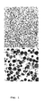
- Bovine chromaffin granules were prepared essentially following a protocol by Winkler and Smith (1975). The pellet obtained representing the granule fraction was analysed by electron microscopy in order to detect contaminations with other organelles. A representative micrograph of a chromaffin granule preparation is shown in Fig. 1. Electron microscopic analysis revealed that these preparations are essentially composed of large dense core granules, identified by their density and size of approximately 280 nm. Granules were ruptured using a low salt buffer, in order to elute only soluble proteins, which are not membrane attached. Extensive dialysis was used to dilute out catecholamines and other small components, resulting in a protein mixture carefully analyzed by Winkler et al. (1986). The prominent protein constituents of chromaffin granules are chromogranins A and B, enkephalins, neuropeptide Y, and dopamine- ⁇ -hydroxylase (not shown).
- Soluble chromaffin granule protein promotes in vitro survival of DAergic neurons
- chromaffin granule protein was treated with this mixture at a dilution of 1:20, starting treatment at day 1 in vitro (DIV 1).
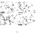
- Fig. 2 shows that protein from chromaffin granules clearly maintains more TH-positive neurons as compared to control cultures after eight days in culture. This effect was comparable to that seen by treating the cultures with a saturating concentration of FGF-2 (10 ng/ml) or TGF- ⁇ (20 ng/ml).
- DAergic activity is released from chromaffin cells by stimulation with carbachol
- chromaffin granules store and release a neurotrophic activity supporting midbrain DAergic neurons.
- Quantification of the neurotrophic effect of chromaffin granule protein showed highest survival promoting effects at a 1:20 dilution, and could not be further increased by using a dilution of 1:10.
- the IC 50 was reached at a dilution of 1:50 (Fig. 5).
- neuropeptide Y In order to investigate whether prominent components of chromaffin granules that are known to have some trophic activities (Unsicker and Stögbauer, 1992), can mimic the observed effects, neuropeptide Y, Leu- and Met-enkephalin were analysed for their capacity to promote survival of midbrain DAergic neurons. However, none of these neuropeptides had any survival promoting activity using concentrations up to 100 ng/ml (Fig. 6).
- DAergic neurons account for 5-10 % of the neuron population in the cell culture system employed as well as in the mesencephalon in vivo.
- Non-DAergic neurons are mainly GABAergic, but there are also few serotonergic neurons.
- To analyse the selectivity of the survival promoting effect of chromaffin granule protein its effect on the uptake of DA, GABA and serotonin was investigated, following an established protocol that was used to prove the selectivity of GDNF for DAergic neurons (Lin et al., 1993).
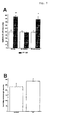
- 3 H-DA and 3 H-serotonin uptake, but not 3 H-GABA uptake were influenced by treatment of cultures with chromaffin granule protein.
- the increase in serotonin uptake was not due to a survival promoting effect of chromaffin granule protein, since the number of serotonin-positive neurons was not significantly altered (Fig. 7b).
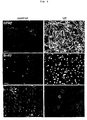
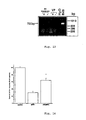
- Chromaffin granule protein-treated cultures showed a dramatic increase in GFAP-positive cells (Fig.8).
- BrdU was applied to cultures, allowed to be incorporated during S-phase of the cell cycle, and detected using a monoclonal antibody against BrdU.
- Control cultures showed incorporation of BrdU in nuclei of very few cells, whereas cultures treated with chromaffin granule protein showed abundant labelled nuclei (Fig. 8).
- Double-labeling with antibodies to BrdU and TH showed that TH + cells never incorporated BrdU (Fig. 8).
- Midbrain DAergic neuron cultures were set up as usually, and treatment with chromaffin granule protein (1:20) started 24h after seeding, for a period of 72 h (DIV1-DIV4). At this point, the entire culture medium was removed and replaced by serum-free medium, which was collected and renewed every 48h (DIV6 and DIV8). These media (stored at 4°C) were pooled and designated "chromaffin granule protein-conditioned medium (VP-CM)". Newly established midbrain DAergic neuron cultures were then treated with VP-CM, starting at DIV1, with two more changes of culture medium at DIV4 and DIV6, and finally processed for TH and GFAP immunocytochemistries at DIV8.
- chromaffin granule protein in a first step, elicits astroglial cell maturation leading to the production and/or release of a neurotrophic factor, which, in a second step, supports DAergic neuron survival.
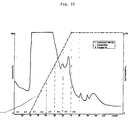
- Fig. 15 shows the chromatogram of the purification on Heparin Sepharose.
- the fractions were processed for TH and GFAP immunocytochemistry.
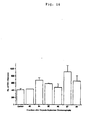
- Fig. 16 after purification on Heparin sepharose, fractions 4 and 7 caused a significant survival promoting effect.
- fraction 4 was able to induce maturation of glioblasts to become GFAP-positive astroglial cells.
- Proteins contained in fraction 4 were further purified using RP-HPLC.
- Fig. 17 shows the chromatogram of the purification.
- the fractions were processed for GFAP immunocytochemistry.
- fractions 7 to 9 were able to induce maturation of glioblasts to become GFAP-positive astroglial cells.
- the denotiation "+” means GFAP activity is detectable and the denotiation "-" means GFAP activity is not detectable.
- FGF-2, EGF, and TGF- ⁇ have been shown to both promote DAergic neuron survival and to increase cell proliferation in mesencephalic cell cultures (Otto and Unsicker, 1993b; Engele and Bohn, 1991; Knüsel et al., 1990).
- the chromaffin granule-derived activity is analyzed by specifically blocking its putative receptor pathway.
- a specific protein-tyrosine kinase inhibitor with selectivity for the EGF receptor (EGFR) signal transduction pathway is applied (Buchdunger et al. 1994). Having shown by the AA and FDUR experiments that the neurotrophic effect of the chromaffin granule protein is mediated by astrocytes (cf. Fig.
- astroglial cells it was screened for suppression of the number of astroglial cells.
- the protocol used was as follows: 24h after seeding mesencephalic cells and again after three days, chromaffin granule protein at a concentration of 1:50 was mixed with various concentrations of the EGFR signaling blocker DAPH and applied together with fresh medium. After a total of six days cultures were fixed and processed for GFAP immunocytochemistry, As shown in Figs. 11 and 12, DAPH at 10 ⁇ M concentration inhibited both the survival of DAergic neurons (Fig. 12, upper panel) and appearence of astroglial cell induced by the chromaffin granule activity.
- the inhibitor DAPH was chosen, because it has been shown that it does not affect signal transduction of FGF-2 or PDGF induced signaling. However, the compound DAPH inhibits not only the EGFR but also p185 c-erbB2 autophosphorylation (Buchdunger et al., 1994). To test which of the two protein kinases is involved in the chromaffin granule protein induced effects, Tyrphostin B56 which selectively inhibits the EGFR kinase autophosphorylation (Gazit et al., 1989), was used. At 10 ⁇ M Tyrphostin B56 successfully inhibited the chromaffin granule protein-induced appearence of astroglial cells (Fig. 12). This finding show furtheron that the chromaffin granule-derived protein is an EGFR ligand.
- the molecule induced by chromaffin granule protein is not GDNF
- RNA from mesencephalic cultures at DIV8 that had been treated with chromaffin granule protein.
- GDNF mRNA was clearly detectable in RNA samples from the B49 gliomal cell line, from which the protein has been isolated and cloned (Lin et al., 1993).
- no signal could be detected in chromaffin granule protein-treated cultures indicating that GDNF is unlikely to be the dopaminotrophic factor induced by chromaffin granule protein.
- Chromaffin granule protein protects DAergic neurons against MPP+ toxicity
- chromaffin granule protein Protein isolated and releasable from chromaffin granules promotes the survival of mesencephalic DAergic neurons in vitro and protects them from MPP+ toxicity.
- the neurotrophic effect is recruited in two steps. First, chromaffin granule protein elicits cell proliferation and astroglial cell maturation involving an EGFR ligand. Second, a glia cell-derived factor distinct from GDNF promotes survival of mesencephalic DAergic neurons.
- chromaffin cells grafted to the lesioned nigrostriatal system exert their beneficial functions through secreting biologically active molecule(s) distinct from biogenic amines. Chromaffin cells are known to synthesize, store and release neurotrophic factor(s) through the exocytotic pathway (Lachmund et al., 1994; Unsicker, 1993). Neither their number nor their molecular identity has been established as yet.
- FGF-2 the probably most intensely studied growth factor molecule in chromaffin cells (Stachowiak et al., 1994; Bieger et al., 1995) and potent trophic factor for developing and toxically impaired DAergic neurons in vitro and in vivo (Ferrari et al., 1989; Otto and Unsicker, 1990; 1993a, b), is not released from granules (Stachowiak et al., 1994; Bieger et al., 1995). Accordingly, FGF-2 must not be considered as a candidate for factors of the present invention.
- Chromaffin grafts to the brain receive de novo functional synaptic connections (Jousselin-Hosaja et al., 1994; Ortega et al., 1992). Moreover, grafted chromaffin cells have been shown to release their granular contents by exocytosis (Ortega et al., 1992). Exocytotic activity was reported to be most frequent in grafts to the striatum as compared to other grafting sites in the brain. It is likely therefore that catecholamines including DA and the molecule(s) causing the trophic effect would be co-released and could exert their actions near the graft site simultanously.
- the chromaffin granule-derived survival promoting effect on embryonic DAergic neurons two steps, (i) induction of cell proliferation and maturation of an increasing population of GFAP-positive astroglial cells, and (ii) stimulation of production and/or release of a dopaminotrophic factor from glial cells, can be involved. Therefore, the chromaffin granule-derived protein according to the present invention is a glial maturation and mitogenic factor. It may, however, act additionally through non-DAergic neurons.
- FGF-2 Several mitogenic growth factors, such as FGF-2, EGF, and TGF- ⁇ (Knusel et al., 1990; Casper et al., 1991; Engele and Bohn, 1991; Alexi and Hefti, 1993) have trophic effects on mesencephalic DAergic neurons which are accompanied by an increase in cell numbers, including astroglial cells. Chromaffin cells can synthesize FGF-2.
- DAPH the EGFR inhibitor employed, has been shown to inhibit the EGFR protein-tyrosine kinase in vitro with high selectivity and also have potent in vivo antitumor activity (Buchdunger et al., 1994). Therefore, that the factor from chromaffin granules is a ligand for the EGFR. Since EGF and TGF- ⁇ failed to mimic the effect of the granule factor on the in vivo maturation and proliferation of mesencephalic astroglial progenitors, it is conceivable that another EGFR ligand is stored in chromaffin cells.
- EGFR ligands comprising, in addition to EGF and TGF- ⁇ , amphiregulin (Shoyab et al., 1988, 1989), heparin-binding EGF (Higashiyama et al., 1991), betacellulin (Sasada et al., 1993; Shing et al., 1993), and the most recently identified epiregulin (Toyoda et al., 1995).
- Another potential member is Schwannoma-derived growth factor (Kimura et al., 1990), which is homologous with amphiregulin (see Lee DY et al., 1995, for review).
- VGF viral gene products
- Shope fibroma and myxoma viruses see Carpenter and Wahl, 1990, for a review
- p185c-erbB2 / HER2/neu proto-oncogene ligands as e.g.
- neuregulin, glial growth factor/heregulins (Marchionni et al., 1993) can be excluded as factor candidates, since Tyrphostin B56, which selectively inhibits the EGFR kinase (IC50 5.0 ⁇ M), but not the HER1-2-kinase autophosphorylation (IC50 > 500 ⁇ M; Gazit et al., 1989), fully blocked the effect of granule protein on mesencephalic glia.
- Another point relevant to the present invention concerns the sorting and release mechanisms of growth factors, especially in neurons. It has widely been assumed, but not proven previously, that growth factors are released via the constitutive pathway of secretion. Evidence is emerging now that this is not exclusively true. In the PC12 pheochromocytoma cell line, which resembles chromaffin cells in many respects, transfected BDNF and NT-4 seem to be primarily released via the regulated secretory pathway (Goodman and Hefti, 1994). In contrast, hippocampal neurons secrete transfected NGF by an unconventional mechanism, which involves depolarization and potassium influx, but not external calcium (Blöchl and Thoenen, 1995). Thus, the localization of a growth factor in chromaffin granules and its regulated secretion may not constitute an unusual mode of liberation for a growth factor.
- GDNF the most prominent dopaminotrophic molecule. Consistent with the results of the present invention that the dopaminotrophic effect of chromaffin granule protein involves a numerical expansion and maturation of astroglial cells is the fact that implant sites of chromaffin grafts exhibit pronounced accumulations of fibrous astroglia (Hansen et al., 1988).
Landscapes
- Health & Medical Sciences (AREA)
- Chemical & Material Sciences (AREA)
- Organic Chemistry (AREA)
- Engineering & Computer Science (AREA)
- Bioinformatics & Cheminformatics (AREA)
- Life Sciences & Earth Sciences (AREA)
- Biomedical Technology (AREA)
- Neurology (AREA)
- Neurosurgery (AREA)
- General Health & Medical Sciences (AREA)
- Medicinal Chemistry (AREA)
- Veterinary Medicine (AREA)
- Pharmacology & Pharmacy (AREA)
- General Chemical & Material Sciences (AREA)
- Animal Behavior & Ethology (AREA)
- Chemical Kinetics & Catalysis (AREA)
- Public Health (AREA)
- Nuclear Medicine, Radiotherapy & Molecular Imaging (AREA)
- Diabetes (AREA)
- Hospice & Palliative Care (AREA)
- Gastroenterology & Hepatology (AREA)
- Endocrinology (AREA)
- Hematology (AREA)
- Obesity (AREA)
- Psychology (AREA)
- Psychiatry (AREA)
- Toxicology (AREA)
- Zoology (AREA)
- Emergency Medicine (AREA)
- Biochemistry (AREA)
- Biophysics (AREA)
- Genetics & Genomics (AREA)
- Molecular Biology (AREA)
- Proteomics, Peptides & Aminoacids (AREA)
- Medicines That Contain Protein Lipid Enzymes And Other Medicines (AREA)
- Micro-Organisms Or Cultivation Processes Thereof (AREA)
- Pharmaceuticals Containing Other Organic And Inorganic Compounds (AREA)
- Peptides Or Proteins (AREA)
- Preparation Of Compounds By Using Micro-Organisms (AREA)
Description
| Results of the TH +-determination as shown in Fig. 16, as well as determination of GFAP+ inductive activity of fractions after purification of chromaffin granule protein on Heparin Sepharose | ||
| Fraction No. | TH + | GFAP + |
| | 100% | - |
| Fraction No. 3 | 105% | - |
| Fraction No. 4 | 165% | + |
| Fraction No. 5 | 140% | - |
| Fraction No. 6 | 110% | - |
| Fraction No. 7 | 210% | - |
| Fraction No. 8 | 150% | - |
| Results of the determination of GFAP+ inductive activity of fractions after purification of chromaffin granule protein using RP-HPLC | |
| Fraction No. | GFAP+ |
| Control | - |
| Fraction No. 7 | + |
| Fraction No. 8 | + |
| Fraction No. 9 | + |
| Fraction No. 10 | - |
| Fraction No. 11 | - |
| Fraction No. 12 | - |
| Fraction No. 13 | - |
Claims (4)
- Use of a chromaffin granule-derived protein factor receptor which is an epidermal grow (EGFR)- ligand, having a selective survival promoting activity in DAergic neurons, for stimulating astroglial cell maturation.
- The use of a chromaffin granule-derived protein according to claim 1 for stimulating proliferation of non-DAergic cells.
- Use of a chromaffin granule-derived protein as defined in claim 1 or 2 for the manufacture of a pharmaceutical composition for treating peripheral and/or CNS-disorders in man.
- The use according to claim 3, wherein the CNS-disorder is selected from the group consisting of Parkinson's disease, Alzheimer's Disease, a neurodegenerative disorder, a peripheral neuropathy including diabetes and cisplatinium neuropathy, and a genetic or acquired peripheral nerve disease.
Priority Applications (1)
| Application Number | Priority Date | Filing Date | Title |
|---|---|---|---|
| EP97940036A EP0922101B1 (en) | 1996-07-29 | 1997-07-28 | Egf-like factor from chromaffin granules and glia cell-derived neurotrophic factor with survival-promoting activity on daergic neurons |
Applications Claiming Priority (6)
| Application Number | Priority Date | Filing Date | Title |
|---|---|---|---|
| EP96112218 | 1996-07-29 | ||
| EP96112218 | 1996-07-29 | ||
| US2414396P | 1996-08-09 | 1996-08-09 | |
| US24143P | 1996-08-09 | ||
| EP97940036A EP0922101B1 (en) | 1996-07-29 | 1997-07-28 | Egf-like factor from chromaffin granules and glia cell-derived neurotrophic factor with survival-promoting activity on daergic neurons |
| PCT/EP1997/004087 WO1998004688A1 (en) | 1996-07-29 | 1997-07-28 | Egf-like factor from chromaffin granules and glia cell-derived neurotrophic factor with survival-promoting activity on daergic neurons |
Publications (2)
| Publication Number | Publication Date |
|---|---|
| EP0922101A1 EP0922101A1 (en) | 1999-06-16 |
| EP0922101B1 true EP0922101B1 (en) | 2003-12-17 |
Family
ID=56289784
Family Applications (1)
| Application Number | Title | Priority Date | Filing Date |
|---|---|---|---|
| EP97940036A Expired - Lifetime EP0922101B1 (en) | 1996-07-29 | 1997-07-28 | Egf-like factor from chromaffin granules and glia cell-derived neurotrophic factor with survival-promoting activity on daergic neurons |
Country Status (7)
| Country | Link |
|---|---|
| EP (1) | EP0922101B1 (en) |
| JP (1) | JP2000516457A (en) |
| AU (1) | AU4202897A (en) |
| CA (1) | CA2262884C (en) |
| DE (1) | DE69726870T2 (en) |
| ES (1) | ES2213221T3 (en) |
| WO (1) | WO1998004688A1 (en) |
Families Citing this family (3)
| Publication number | Priority date | Publication date | Assignee | Title |
|---|---|---|---|---|
| AU6262700A (en) * | 1999-06-09 | 2001-01-02 | Biopharm Gesellschaft Zur Biotechnologischen Entwicklung Und Zum Vertrieb Von Pharmaka Mbh | Nucleic acid encoding novel egf-like growth factors |
| CA2391885A1 (en) * | 1999-11-19 | 2001-05-25 | Curagen Corporation | Polypeptides and nucleic acids encoding same |
| CA2381065C (en) * | 2000-06-01 | 2007-06-05 | Japan Science And Technology Corporation | Method for enrichment and/or isolation of dopaminergic neurons |
Family Cites Families (2)
| Publication number | Priority date | Publication date | Assignee | Title |
|---|---|---|---|---|
| IL78197A (en) * | 1985-03-22 | 1991-07-18 | Genentech Inc | Nucleic acid encoding tgf-beta and its uses |
| WO1993006116A1 (en) * | 1991-09-20 | 1993-04-01 | Syntex-Synergen Neuroscience Joint Venture | Glial derived neurotrophic factor |
-
1997
- 1997-07-28 JP JP10508507A patent/JP2000516457A/en not_active Ceased
- 1997-07-28 WO PCT/EP1997/004087 patent/WO1998004688A1/en not_active Ceased
- 1997-07-28 AU AU42028/97A patent/AU4202897A/en not_active Abandoned
- 1997-07-28 DE DE69726870T patent/DE69726870T2/en not_active Expired - Fee Related
- 1997-07-28 CA CA002262884A patent/CA2262884C/en not_active Expired - Fee Related
- 1997-07-28 ES ES97940036T patent/ES2213221T3/en not_active Expired - Lifetime
- 1997-07-28 EP EP97940036A patent/EP0922101B1/en not_active Expired - Lifetime
Also Published As
| Publication number | Publication date |
|---|---|
| DE69726870D1 (en) | 2004-01-29 |
| DE69726870T2 (en) | 2004-10-28 |
| JP2000516457A (en) | 2000-12-12 |
| CA2262884A1 (en) | 1998-02-05 |
| WO1998004688A1 (en) | 1998-02-05 |
| CA2262884C (en) | 2004-06-08 |
| AU4202897A (en) | 1998-02-20 |
| EP0922101A1 (en) | 1999-06-16 |
| ES2213221T3 (en) | 2004-08-16 |
Similar Documents
| Publication | Publication Date | Title |
|---|---|---|
| Daadi et al. | Generation of tyrosine hydroxylase-producing neurons from precursors of the embryonic and adult forebrain | |
| AU715246B2 (en) | In vitro induction of dopaminergic cells | |
| Azmitia et al. | S-100B but not NGF, EGF, insulin or calmodulin is a CNS serotonergic growth factor | |
| Mallei et al. | Antidepressant treatments induce the expression of basic fibroblast growth factor in cortical and hippocampal neurons | |
| Bouvier et al. | Basic fibroblast growth factor increases division and delays differentiation of dopamine precursors in vitro | |
| FI120455B (en) | Method of having neural precursor cells to proliferate | |
| US7651853B2 (en) | Cultures of GFAP+ nestin+ cells that differentiate to neurons | |
| US9115208B2 (en) | Dopaminergic neuronal survival-promoting factors and uses thereof | |
| Cunningham et al. | Nerve growth factor released by transgenic astrocytes enhances the function of adrenal chromaffin cell grafts in a rat model of Parkinson's disease | |
| Casper et al. | Epidermal growth factor and basic fibroblast growth factor have independent actions on mesencephalic dopamine neurons in culture | |
| Gadient et al. | Effect of leukemia inhibitory factor (LIF) on the morphology and survival of cultured hippocampal neurons and glial cells | |
| Krieglstein et al. | Protein from chromaffin granules promotes survival of mesencephalic dopaminergic neurons by an EGF‐receptor ligand‐mediated mechanism | |
| CA2262884C (en) | Egf-like factor from chromaffin granules and glia cell-derived neurotrophic factor with survival-promoting activity on daergic neurons | |
| Gray et al. | Characterization of a neurotrophic factor produced by cultured astrocytes involved in the regulation of subcortical cholinergic neurons | |
| Chadi et al. | Analysis of trophic responses in lesioned brain: focus on basic fibroblast growth factor mechanisms | |
| Engele | Spatial and temporal growth factor influences on developing midbrain dopaminergic neurons | |
| Engele | Changing responsiveness of developing midbrain dopaminergic neurons for extracellular growth factors | |
| WO1998004688A9 (en) | Egf-like factor from chromaffin granules and glia cell-derived neurotrophic factor with survival-promoting activity on daergic neurons | |
| Garcia-Rill et al. | Fibroblast growth factor-induced increased survival of chollnergic mesopontine neurons in culture | |
| CA2383076A1 (en) | Dopaminergic neuronal survival-promoting factors and uses thereof | |
| Hagg et al. | Neurotrophic factors and CNS regeneration | |
| US20020197238A1 (en) | Platelet derived growth factor (PDGF)-derived neurospheres define a novel class of progenitor cells | |
| Patel et al. | Neurotrophic factors produced by astrocytes involved in the regulation of cholinergic neurons in the central nervous system | |
| Zhou et al. | A neurotrophic factor—SNTF—for serotonergic neurons | |
| AU2002240760A1 (en) | Dopaminergic neuronal survival-promoting factors and uses thereof |
Legal Events
| Date | Code | Title | Description |
|---|---|---|---|
| PUAI | Public reference made under article 153(3) epc to a published international application that has entered the european phase |
Free format text: ORIGINAL CODE: 0009012 |
|
| 17P | Request for examination filed |
Effective date: 19990210 |
|
| AK | Designated contracting states |
Kind code of ref document: A1 Designated state(s): DE ES FR GB IT |
|
| RAP3 | Party data changed (applicant data changed or rights of an application transferred) |
Owner name: BIOPHARM GESELLSCHAFT ZUR BIOTECHNOLOGISCHEN ENTWI |
|
| 17Q | First examination report despatched |
Effective date: 20010326 |
|
| GRAH | Despatch of communication of intention to grant a patent |
Free format text: ORIGINAL CODE: EPIDOS IGRA |
|
| GRAS | Grant fee paid |
Free format text: ORIGINAL CODE: EPIDOSNIGR3 |
|
| GRAA | (expected) grant |
Free format text: ORIGINAL CODE: 0009210 |
|
| AK | Designated contracting states |
Kind code of ref document: B1 Designated state(s): DE ES FR GB IT |
|
| REG | Reference to a national code |
Ref country code: GB Ref legal event code: FG4D |
|
| REF | Corresponds to: |
Ref document number: 69726870 Country of ref document: DE Date of ref document: 20040129 Kind code of ref document: P |
|
| REG | Reference to a national code |
Ref country code: ES Ref legal event code: FG2A Ref document number: 2213221 Country of ref document: ES Kind code of ref document: T3 |
|
| ET | Fr: translation filed | ||
| PLBE | No opposition filed within time limit |
Free format text: ORIGINAL CODE: 0009261 |
|
| STAA | Information on the status of an ep patent application or granted ep patent |
Free format text: STATUS: NO OPPOSITION FILED WITHIN TIME LIMIT |
|
| 26N | No opposition filed |
Effective date: 20040920 |
|
| PGFP | Annual fee paid to national office [announced via postgrant information from national office to epo] |
Ref country code: FR Payment date: 20050613 Year of fee payment: 9 |
|
| PGFP | Annual fee paid to national office [announced via postgrant information from national office to epo] |
Ref country code: ES Payment date: 20050630 Year of fee payment: 9 |
|
| PGFP | Annual fee paid to national office [announced via postgrant information from national office to epo] |
Ref country code: DE Payment date: 20050726 Year of fee payment: 9 |
|
| PGFP | Annual fee paid to national office [announced via postgrant information from national office to epo] |
Ref country code: GB Payment date: 20050727 Year of fee payment: 9 |
|
| PG25 | Lapsed in a contracting state [announced via postgrant information from national office to epo] |
Ref country code: GB Free format text: LAPSE BECAUSE OF NON-PAYMENT OF DUE FEES Effective date: 20060728 |
|
| PGFP | Annual fee paid to national office [announced via postgrant information from national office to epo] |
Ref country code: IT Payment date: 20060731 Year of fee payment: 10 |
|
| PG25 | Lapsed in a contracting state [announced via postgrant information from national office to epo] |
Ref country code: DE Free format text: LAPSE BECAUSE OF NON-PAYMENT OF DUE FEES Effective date: 20070201 |
|
| GBPC | Gb: european patent ceased through non-payment of renewal fee |
Effective date: 20060728 |
|
| REG | Reference to a national code |
Ref country code: FR Ref legal event code: ST Effective date: 20070330 |
|
| REG | Reference to a national code |
Ref country code: ES Ref legal event code: FD2A Effective date: 20060729 |
|
| PG25 | Lapsed in a contracting state [announced via postgrant information from national office to epo] |
Ref country code: ES Free format text: LAPSE BECAUSE OF NON-PAYMENT OF DUE FEES Effective date: 20060729 |
|
| PG25 | Lapsed in a contracting state [announced via postgrant information from national office to epo] |
Ref country code: FR Free format text: LAPSE BECAUSE OF NON-PAYMENT OF DUE FEES Effective date: 20060731 |
|
| PG25 | Lapsed in a contracting state [announced via postgrant information from national office to epo] |
Ref country code: IT Free format text: LAPSE BECAUSE OF NON-PAYMENT OF DUE FEES Effective date: 20070728 |