EP0917471B1 - Treatment and diagnosis of infections of gram positive cocci - Google Patents
Treatment and diagnosis of infections of gram positive cocci Download PDFInfo
- Publication number
- EP0917471B1 EP0917471B1 EP97930642A EP97930642A EP0917471B1 EP 0917471 B1 EP0917471 B1 EP 0917471B1 EP 97930642 A EP97930642 A EP 97930642A EP 97930642 A EP97930642 A EP 97930642A EP 0917471 B1 EP0917471 B1 EP 0917471B1
- Authority
- EP
- European Patent Office
- Prior art keywords
- immunogenic fragment
- abc transporter
- seq
- transporter protein
- sequence
- Prior art date
- Legal status (The legal status is an assumption and is not a legal conclusion. Google has not performed a legal analysis and makes no representation as to the accuracy of the status listed.)
- Expired - Lifetime
Links
- 238000003745 diagnosis Methods 0.000 title claims abstract description 20
- 238000011282 treatment Methods 0.000 title claims abstract description 14
- 208000015181 infectious disease Diseases 0.000 title claims description 30
- 239000012634 fragment Substances 0.000 claims abstract description 97
- 239000000427 antigen Substances 0.000 claims abstract description 92
- 108091007433 antigens Proteins 0.000 claims abstract description 84
- 102000036639 antigens Human genes 0.000 claims abstract description 84
- 230000002163 immunogen Effects 0.000 claims abstract description 77
- 108010006533 ATP-Binding Cassette Transporters Proteins 0.000 claims abstract description 58
- 102000005416 ATP-Binding Cassette Transporters Human genes 0.000 claims abstract description 58
- 230000001580 bacterial effect Effects 0.000 claims abstract description 39
- 238000000034 method Methods 0.000 claims abstract description 34
- 239000011230 binding agent Substances 0.000 claims abstract description 18
- 241001465754 Metazoa Species 0.000 claims abstract description 13
- 241000282414 Homo sapiens Species 0.000 claims abstract description 12
- 238000002405 diagnostic procedure Methods 0.000 claims abstract description 10
- 241000894006 Bacteria Species 0.000 claims description 35
- 238000009739 binding Methods 0.000 claims description 30
- 241000194031 Enterococcus faecium Species 0.000 claims description 22
- 108090000623 proteins and genes Proteins 0.000 claims description 20
- 102000004169 proteins and genes Human genes 0.000 claims description 20
- 241000194033 Enterococcus Species 0.000 claims description 13
- 241000194032 Enterococcus faecalis Species 0.000 claims description 12
- 150000007523 nucleic acids Chemical group 0.000 claims description 10
- 238000004519 manufacturing process Methods 0.000 claims description 9
- 102000053642 Catalytic RNA Human genes 0.000 claims description 8
- 108090000994 Catalytic RNA Proteins 0.000 claims description 8
- 108091092562 ribozyme Proteins 0.000 claims description 8
- 230000000692 anti-sense effect Effects 0.000 claims description 7
- 239000003814 drug Substances 0.000 claims description 7
- 238000001514 detection method Methods 0.000 claims description 6
- 238000006243 chemical reaction Methods 0.000 claims description 5
- 241001468179 Enterococcus avium Species 0.000 claims description 3
- 241000520130 Enterococcus durans Species 0.000 claims description 3
- 241000520134 Enterococcus mundtii Species 0.000 claims description 3
- 239000000032 diagnostic agent Substances 0.000 claims 2
- 229940039227 diagnostic agent Drugs 0.000 claims 2
- 108010078791 Carrier Proteins Proteins 0.000 claims 1
- 239000003795 chemical substances by application Substances 0.000 abstract description 16
- 230000003472 neutralizing effect Effects 0.000 abstract description 8
- 230000001225 therapeutic effect Effects 0.000 abstract description 6
- 238000002560 therapeutic procedure Methods 0.000 abstract description 4
- 241000295644 Staphylococcaceae Species 0.000 abstract description 2
- 230000002538 fungal effect Effects 0.000 abstract 1
- 150000001413 amino acids Chemical group 0.000 description 35
- 235000001014 amino acid Nutrition 0.000 description 28
- 206010040047 Sepsis Diseases 0.000 description 27
- 239000000499 gel Substances 0.000 description 21
- 239000000872 buffer Substances 0.000 description 18
- 235000018102 proteins Nutrition 0.000 description 18
- 210000002966 serum Anatomy 0.000 description 18
- 108010059993 Vancomycin Proteins 0.000 description 17
- 229960003165 vancomycin Drugs 0.000 description 17
- MYPYJXKWCTUITO-LYRMYLQWSA-N vancomycin Chemical compound O([C@@H]1[C@@H](O)[C@H](O)[C@@H](CO)O[C@H]1OC1=C2C=C3C=C1OC1=CC=C(C=C1Cl)[C@@H](O)[C@H](C(N[C@@H](CC(N)=O)C(=O)N[C@H]3C(=O)N[C@H]1C(=O)N[C@H](C(N[C@@H](C3=CC(O)=CC(O)=C3C=3C(O)=CC=C1C=3)C(O)=O)=O)[C@H](O)C1=CC=C(C(=C1)Cl)O2)=O)NC(=O)[C@@H](CC(C)C)NC)[C@H]1C[C@](C)(N)[C@H](O)[C@H](C)O1 MYPYJXKWCTUITO-LYRMYLQWSA-N 0.000 description 17
- MYPYJXKWCTUITO-UHFFFAOYSA-N vancomycin Natural products O1C(C(=C2)Cl)=CC=C2C(O)C(C(NC(C2=CC(O)=CC(O)=C2C=2C(O)=CC=C3C=2)C(O)=O)=O)NC(=O)C3NC(=O)C2NC(=O)C(CC(N)=O)NC(=O)C(NC(=O)C(CC(C)C)NC)C(O)C(C=C3Cl)=CC=C3OC3=CC2=CC1=C3OC1OC(CO)C(O)C(O)C1OC1CC(C)(N)C(O)C(C)O1 MYPYJXKWCTUITO-UHFFFAOYSA-N 0.000 description 17
- XLYOFNOQVPJJNP-UHFFFAOYSA-N water Chemical compound O XLYOFNOQVPJJNP-UHFFFAOYSA-N 0.000 description 15
- 239000012153 distilled water Substances 0.000 description 13
- 238000003119 immunoblot Methods 0.000 description 13
- 230000004044 response Effects 0.000 description 11
- 239000000047 product Substances 0.000 description 10
- 241000699670 Mus sp. Species 0.000 description 9
- 239000000243 solution Substances 0.000 description 9
- 108060003951 Immunoglobulin Proteins 0.000 description 7
- FAPWRFPIFSIZLT-UHFFFAOYSA-M Sodium chloride Chemical compound [Na+].[Cl-] FAPWRFPIFSIZLT-UHFFFAOYSA-M 0.000 description 7
- 239000007983 Tris buffer Substances 0.000 description 7
- 206010014665 endocarditis Diseases 0.000 description 7
- 102000018358 immunoglobulin Human genes 0.000 description 7
- 238000012163 sequencing technique Methods 0.000 description 7
- LENZDBCJOHFCAS-UHFFFAOYSA-N tris Chemical compound OCC(N)(CO)CO LENZDBCJOHFCAS-UHFFFAOYSA-N 0.000 description 7
- 108091003079 Bovine Serum Albumin Proteins 0.000 description 6
- DHMQDGOQFOQNFH-UHFFFAOYSA-N Glycine Chemical compound NCC(O)=O DHMQDGOQFOQNFH-UHFFFAOYSA-N 0.000 description 6
- 108010021625 Immunoglobulin Fragments Proteins 0.000 description 6
- 102000008394 Immunoglobulin Fragments Human genes 0.000 description 6
- ZMXDDKWLCZADIW-UHFFFAOYSA-N N,N-Dimethylformamide Chemical compound CN(C)C=O ZMXDDKWLCZADIW-UHFFFAOYSA-N 0.000 description 6
- 229940098773 bovine serum albumin Drugs 0.000 description 6
- 239000012530 fluid Substances 0.000 description 6
- RJQXTJLFIWVMTO-TYNCELHUSA-N Methicillin Chemical compound COC1=CC=CC(OC)=C1C(=O)N[C@@H]1C(=O)N2[C@@H](C(O)=O)C(C)(C)S[C@@H]21 RJQXTJLFIWVMTO-TYNCELHUSA-N 0.000 description 5
- 108700026244 Open Reading Frames Proteins 0.000 description 5
- MYPYJXKWCTUITO-KIIOPKALSA-N chembl3301825 Chemical compound O([C@@H]1[C@@H](O)[C@H](O)[C@@H](CO)O[C@H]1OC1=C2C=C3C=C1OC1=CC=C(C=C1Cl)[C@@H](O)[C@H](C(N[C@@H](CC(N)=O)C(=O)N[C@H]3C(=O)N[C@H]1C(=O)N[C@H](C(N[C@H](C3=CC(O)=CC(O)=C3C=3C(O)=CC=C1C=3)C(O)=O)=O)[C@H](O)C1=CC=C(C(=C1)Cl)O2)=O)NC(=O)[C@@H](CC(C)C)NC)[C@H]1C[C@](C)(N)C(O)[C@H](C)O1 MYPYJXKWCTUITO-KIIOPKALSA-N 0.000 description 5
- 229960003085 meticillin Drugs 0.000 description 5
- QRXMUCSWCMTJGU-UHFFFAOYSA-L (5-bromo-4-chloro-1h-indol-3-yl) phosphate Chemical compound C1=C(Br)C(Cl)=C2C(OP([O-])(=O)[O-])=CNC2=C1 QRXMUCSWCMTJGU-UHFFFAOYSA-L 0.000 description 4
- FWMNVWWHGCHHJJ-SKKKGAJSSA-N 4-amino-1-[(2r)-6-amino-2-[[(2r)-2-[[(2r)-2-[[(2r)-2-amino-3-phenylpropanoyl]amino]-3-phenylpropanoyl]amino]-4-methylpentanoyl]amino]hexanoyl]piperidine-4-carboxylic acid Chemical compound C([C@H](C(=O)N[C@H](CC(C)C)C(=O)N[C@H](CCCCN)C(=O)N1CCC(N)(CC1)C(O)=O)NC(=O)[C@H](N)CC=1C=CC=CC=1)C1=CC=CC=C1 FWMNVWWHGCHHJJ-SKKKGAJSSA-N 0.000 description 4
- 102000002260 Alkaline Phosphatase Human genes 0.000 description 4
- 108020004774 Alkaline Phosphatase Proteins 0.000 description 4
- 101100402709 Arabidopsis thaliana MRSA2 gene Proteins 0.000 description 4
- 108020004414 DNA Proteins 0.000 description 4
- 239000000020 Nitrocellulose Substances 0.000 description 4
- DBMJMQXJHONAFJ-UHFFFAOYSA-M Sodium laurylsulphate Chemical compound [Na+].CCCCCCCCCCCCOS([O-])(=O)=O DBMJMQXJHONAFJ-UHFFFAOYSA-M 0.000 description 4
- 241000191940 Staphylococcus Species 0.000 description 4
- 238000009640 blood culture Methods 0.000 description 4
- 238000005336 cracking Methods 0.000 description 4
- 238000001962 electrophoresis Methods 0.000 description 4
- 108020001507 fusion proteins Proteins 0.000 description 4
- 102000037865 fusion proteins Human genes 0.000 description 4
- 238000013507 mapping Methods 0.000 description 4
- 229920001220 nitrocellulos Polymers 0.000 description 4
- 238000002360 preparation method Methods 0.000 description 4
- 235000019333 sodium laurylsulphate Nutrition 0.000 description 4
- 238000012360 testing method Methods 0.000 description 4
- 238000002965 ELISA Methods 0.000 description 3
- 108010093031 Galactosidases Proteins 0.000 description 3
- PEDCQBHIVMGVHV-UHFFFAOYSA-N Glycerine Chemical compound OCC(O)CO PEDCQBHIVMGVHV-UHFFFAOYSA-N 0.000 description 3
- 239000004471 Glycine Substances 0.000 description 3
- OKKJLVBELUTLKV-UHFFFAOYSA-N Methanol Chemical compound OC OKKJLVBELUTLKV-UHFFFAOYSA-N 0.000 description 3
- 239000003242 anti bacterial agent Substances 0.000 description 3
- 102000005936 beta-Galactosidase Human genes 0.000 description 3
- 108010005774 beta-Galactosidase Proteins 0.000 description 3
- 210000004027 cell Anatomy 0.000 description 3
- 230000034994 death Effects 0.000 description 3
- 231100000517 death Toxicity 0.000 description 3
- 238000010790 dilution Methods 0.000 description 3
- 239000012895 dilution Substances 0.000 description 3
- 230000001900 immune effect Effects 0.000 description 3
- JPXMTWWFLBLUCD-UHFFFAOYSA-N nitro blue tetrazolium(2+) Chemical compound COC1=CC(C=2C=C(OC)C(=CC=2)[N+]=2N(N=C(N=2)C=2C=CC=CC=2)C=2C=CC(=CC=2)[N+]([O-])=O)=CC=C1[N+]1=NC(C=2C=CC=CC=2)=NN1C1=CC=C([N+]([O-])=O)C=C1 JPXMTWWFLBLUCD-UHFFFAOYSA-N 0.000 description 3
- 206010034674 peritonitis Diseases 0.000 description 3
- 230000001681 protective effect Effects 0.000 description 3
- 238000012216 screening Methods 0.000 description 3
- 239000011780 sodium chloride Substances 0.000 description 3
- 238000005406 washing Methods 0.000 description 3
- HRPVXLWXLXDGHG-UHFFFAOYSA-N Acrylamide Chemical compound NC(=O)C=C HRPVXLWXLXDGHG-UHFFFAOYSA-N 0.000 description 2
- 239000004160 Ammonium persulphate Substances 0.000 description 2
- 241000193388 Bacillus thuringiensis Species 0.000 description 2
- 238000001712 DNA sequencing Methods 0.000 description 2
- 206010014889 Enterococcal infections Diseases 0.000 description 2
- 102000002464 Galactosidases Human genes 0.000 description 2
- TWRXJAOTZQYOKJ-UHFFFAOYSA-L Magnesium chloride Chemical compound [Mg+2].[Cl-].[Cl-] TWRXJAOTZQYOKJ-UHFFFAOYSA-L 0.000 description 2
- 206010048723 Multiple-drug resistance Diseases 0.000 description 2
- KWYHDKDOAIKMQN-UHFFFAOYSA-N N,N,N',N'-tetramethylethylenediamine Chemical compound CN(C)CCN(C)C KWYHDKDOAIKMQN-UHFFFAOYSA-N 0.000 description 2
- 206010029803 Nosocomial infection Diseases 0.000 description 2
- 108091028043 Nucleic acid sequence Proteins 0.000 description 2
- 241001494479 Pecora Species 0.000 description 2
- 108020004511 Recombinant DNA Proteins 0.000 description 2
- 241000191967 Staphylococcus aureus Species 0.000 description 2
- ROOXNKNUYICQNP-UHFFFAOYSA-N ammonium persulfate Chemical compound [NH4+].[NH4+].[O-]S(=O)(=O)OOS([O-])(=O)=O ROOXNKNUYICQNP-UHFFFAOYSA-N 0.000 description 2
- 235000019395 ammonium persulphate Nutrition 0.000 description 2
- 238000004458 analytical method Methods 0.000 description 2
- 238000010171 animal model Methods 0.000 description 2
- 229940088710 antibiotic agent Drugs 0.000 description 2
- 230000000890 antigenic effect Effects 0.000 description 2
- 229940097012 bacillus thuringiensis Drugs 0.000 description 2
- 230000003796 beauty Effects 0.000 description 2
- 238000002512 chemotherapy Methods 0.000 description 2
- 238000010367 cloning Methods 0.000 description 2
- 125000000151 cysteine group Chemical group N[C@@H](CS)C(=O)* 0.000 description 2
- KAKKHKRHCKCAGH-UHFFFAOYSA-L disodium;(4-nitrophenyl) phosphate;hexahydrate Chemical compound O.O.O.O.O.O.[Na+].[Na+].[O-][N+](=O)C1=CC=C(OP([O-])([O-])=O)C=C1 KAKKHKRHCKCAGH-UHFFFAOYSA-L 0.000 description 2
- 229940079593 drug Drugs 0.000 description 2
- 210000002615 epidermis Anatomy 0.000 description 2
- 238000002474 experimental method Methods 0.000 description 2
- 238000011534 incubation Methods 0.000 description 2
- 208000032839 leukemia Diseases 0.000 description 2
- 230000002906 microbiologic effect Effects 0.000 description 2
- ZIUHHBKFKCYYJD-UHFFFAOYSA-N n,n'-methylenebisacrylamide Chemical compound C=CC(=O)NCNC(=O)C=C ZIUHHBKFKCYYJD-UHFFFAOYSA-N 0.000 description 2
- 108020004707 nucleic acids Proteins 0.000 description 2
- 102000039446 nucleic acids Human genes 0.000 description 2
- 229920002401 polyacrylamide Polymers 0.000 description 2
- 108090000765 processed proteins & peptides Proteins 0.000 description 2
- 238000012552 review Methods 0.000 description 2
- 238000002415 sodium dodecyl sulfate polyacrylamide gel electrophoresis Methods 0.000 description 2
- 239000006228 supernatant Substances 0.000 description 2
- 208000024891 symptom Diseases 0.000 description 2
- 230000009261 transgenic effect Effects 0.000 description 2
- 229960005486 vaccine Drugs 0.000 description 2
- DGVVWUTYPXICAM-UHFFFAOYSA-N β‐Mercaptoethanol Chemical compound OCCS DGVVWUTYPXICAM-UHFFFAOYSA-N 0.000 description 2
- QKNYBSVHEMOAJP-UHFFFAOYSA-N 2-amino-2-(hydroxymethyl)propane-1,3-diol;hydron;chloride Chemical compound Cl.OCC(N)(CO)CO QKNYBSVHEMOAJP-UHFFFAOYSA-N 0.000 description 1
- QFVHZQCOUORWEI-UHFFFAOYSA-N 4-[(4-anilino-5-sulfonaphthalen-1-yl)diazenyl]-5-hydroxynaphthalene-2,7-disulfonic acid Chemical compound C=12C(O)=CC(S(O)(=O)=O)=CC2=CC(S(O)(=O)=O)=CC=1N=NC(C1=CC=CC(=C11)S(O)(=O)=O)=CC=C1NC1=CC=CC=C1 QFVHZQCOUORWEI-UHFFFAOYSA-N 0.000 description 1
- 108091006112 ATPases Proteins 0.000 description 1
- 102000057290 Adenosine Triphosphatases Human genes 0.000 description 1
- 208000035143 Bacterial infection Diseases 0.000 description 1
- 206010051779 Bone marrow toxicity Diseases 0.000 description 1
- OBMZMSLWNNWEJA-XNCRXQDQSA-N C1=CC=2C(C[C@@H]3NC(=O)[C@@H](NC(=O)[C@H](NC(=O)N(CC#CCN(CCCC[C@H](NC(=O)[C@@H](CC4=CC=CC=C4)NC3=O)C(=O)N)CC=C)NC(=O)[C@@H](N)C)CC3=CNC4=C3C=CC=C4)C)=CNC=2C=C1 Chemical compound C1=CC=2C(C[C@@H]3NC(=O)[C@@H](NC(=O)[C@H](NC(=O)N(CC#CCN(CCCC[C@H](NC(=O)[C@@H](CC4=CC=CC=C4)NC3=O)C(=O)N)CC=C)NC(=O)[C@@H](N)C)CC3=CNC4=C3C=CC=C4)C)=CNC=2C=C1 OBMZMSLWNNWEJA-XNCRXQDQSA-N 0.000 description 1
- 241000283707 Capra Species 0.000 description 1
- 102000003846 Carbonic anhydrases Human genes 0.000 description 1
- 108090000209 Carbonic anhydrases Proteins 0.000 description 1
- 229930186147 Cephalosporin Natural products 0.000 description 1
- 208000035473 Communicable disease Diseases 0.000 description 1
- 108010047041 Complementarity Determining Regions Proteins 0.000 description 1
- 206010011409 Cross infection Diseases 0.000 description 1
- 206010014671 Endocarditis enterococcal Diseases 0.000 description 1
- 102000004190 Enzymes Human genes 0.000 description 1
- 108090000790 Enzymes Proteins 0.000 description 1
- 241000588724 Escherichia coli Species 0.000 description 1
- 241000233866 Fungi Species 0.000 description 1
- 229930182566 Gentamicin Natural products 0.000 description 1
- CEAZRRDELHUEMR-URQXQFDESA-N Gentamicin Chemical compound O1[C@H](C(C)NC)CC[C@@H](N)[C@H]1O[C@H]1[C@H](O)[C@@H](O[C@@H]2[C@@H]([C@@H](NC)[C@@](C)(O)CO2)O)[C@H](N)C[C@@H]1N CEAZRRDELHUEMR-URQXQFDESA-N 0.000 description 1
- WQZGKKKJIJFFOK-GASJEMHNSA-N Glucose Chemical compound OC[C@H]1OC(O)[C@H](O)[C@@H](O)[C@@H]1O WQZGKKKJIJFFOK-GASJEMHNSA-N 0.000 description 1
- 108010015899 Glycopeptides Proteins 0.000 description 1
- 102000002068 Glycopeptides Human genes 0.000 description 1
- 206010066476 Haematological malignancy Diseases 0.000 description 1
- 108010054477 Immunoglobulin Fab Fragments Proteins 0.000 description 1
- 102000001706 Immunoglobulin Fab Fragments Human genes 0.000 description 1
- 108010067060 Immunoglobulin Variable Region Proteins 0.000 description 1
- 102000017727 Immunoglobulin Variable Region Human genes 0.000 description 1
- 101710155913 Major envelope protein Proteins 0.000 description 1
- 208000037942 Methicillin-resistant Staphylococcus aureus infection Diseases 0.000 description 1
- 102000016943 Muramidase Human genes 0.000 description 1
- 108010014251 Muramidase Proteins 0.000 description 1
- 241001529936 Murinae Species 0.000 description 1
- 102000003505 Myosin Human genes 0.000 description 1
- 108060008487 Myosin Proteins 0.000 description 1
- 108010062010 N-Acetylmuramoyl-L-alanine Amidase Proteins 0.000 description 1
- 206010029155 Nephropathy toxic Diseases 0.000 description 1
- 206010033109 Ototoxicity Diseases 0.000 description 1
- 108010058846 Ovalbumin Proteins 0.000 description 1
- 239000002033 PVDF binder Substances 0.000 description 1
- 108090000526 Papain Proteins 0.000 description 1
- 229930182555 Penicillin Natural products 0.000 description 1
- JGSARLDLIJGVTE-MBNYWOFBSA-N Penicillin G Chemical compound N([C@H]1[C@H]2SC([C@@H](N2C1=O)C(O)=O)(C)C)C(=O)CC1=CC=CC=C1 JGSARLDLIJGVTE-MBNYWOFBSA-N 0.000 description 1
- 102000057297 Pepsin A Human genes 0.000 description 1
- 108090000284 Pepsin A Proteins 0.000 description 1
- 101710176384 Peptide 1 Proteins 0.000 description 1
- 108010065081 Phosphorylase b Proteins 0.000 description 1
- 239000004698 Polyethylene Substances 0.000 description 1
- 229920001213 Polysorbate 20 Polymers 0.000 description 1
- 239000004365 Protease Substances 0.000 description 1
- 206010037660 Pyrexia Diseases 0.000 description 1
- 241000700159 Rattus Species 0.000 description 1
- 206010038192 Red man syndrome Diseases 0.000 description 1
- 206010041925 Staphylococcal infections Diseases 0.000 description 1
- 241000191963 Staphylococcus epidermidis Species 0.000 description 1
- 206010056349 Subdiaphragmatic abscess Diseases 0.000 description 1
- 208000003144 Subphrenic Abscess Diseases 0.000 description 1
- 239000004098 Tetracycline Substances 0.000 description 1
- 101710162629 Trypsin inhibitor Proteins 0.000 description 1
- 229940122618 Trypsin inhibitor Drugs 0.000 description 1
- 238000001042 affinity chromatography Methods 0.000 description 1
- 125000000539 amino acid group Chemical group 0.000 description 1
- 229940126575 aminoglycoside Drugs 0.000 description 1
- 229960000723 ampicillin Drugs 0.000 description 1
- AVKUERGKIZMTKX-NJBDSQKTSA-N ampicillin Chemical compound C1([C@@H](N)C(=O)N[C@H]2[C@H]3SC([C@@H](N3C2=O)C(O)=O)(C)C)=CC=CC=C1 AVKUERGKIZMTKX-NJBDSQKTSA-N 0.000 description 1
- 238000009175 antibody therapy Methods 0.000 description 1
- 238000013459 approach Methods 0.000 description 1
- 238000003556 assay Methods 0.000 description 1
- 208000022362 bacterial infectious disease Diseases 0.000 description 1
- 238000009835 boiling Methods 0.000 description 1
- 231100000366 bone marrow toxicity Toxicity 0.000 description 1
- 210000004556 brain Anatomy 0.000 description 1
- UDSAIICHUKSCKT-UHFFFAOYSA-N bromophenol blue Chemical compound C1=C(Br)C(O)=C(Br)C=C1C1(C=2C=C(Br)C(O)=C(Br)C=2)C2=CC=CC=C2S(=O)(=O)O1 UDSAIICHUKSCKT-UHFFFAOYSA-N 0.000 description 1
- 239000007853 buffer solution Substances 0.000 description 1
- 229940124587 cephalosporin Drugs 0.000 description 1
- 150000001780 cephalosporins Chemical class 0.000 description 1
- 239000003153 chemical reaction reagent Substances 0.000 description 1
- 229960005091 chloramphenicol Drugs 0.000 description 1
- WIIZWVCIJKGZOK-RKDXNWHRSA-N chloramphenicol Chemical compound ClC(Cl)C(=O)N[C@H](CO)[C@H](O)C1=CC=C([N+]([O-])=O)C=C1 WIIZWVCIJKGZOK-RKDXNWHRSA-N 0.000 description 1
- 239000013611 chromosomal DNA Substances 0.000 description 1
- 230000001684 chronic effect Effects 0.000 description 1
- 239000013599 cloning vector Substances 0.000 description 1
- 230000001332 colony forming effect Effects 0.000 description 1
- 238000007796 conventional method Methods 0.000 description 1
- 238000001816 cooling Methods 0.000 description 1
- 238000011461 current therapy Methods 0.000 description 1
- 230000003247 decreasing effect Effects 0.000 description 1
- 238000011161 development Methods 0.000 description 1
- 238000000502 dialysis Methods 0.000 description 1
- 239000003085 diluting agent Substances 0.000 description 1
- 201000010099 disease Diseases 0.000 description 1
- 208000037265 diseases, disorders, signs and symptoms Diseases 0.000 description 1
- 239000003937 drug carrier Substances 0.000 description 1
- 238000002651 drug therapy Methods 0.000 description 1
- 239000012636 effector Substances 0.000 description 1
- 230000000694 effects Effects 0.000 description 1
- 230000006862 enzymatic digestion Effects 0.000 description 1
- 229940088598 enzyme Drugs 0.000 description 1
- 239000013604 expression vector Substances 0.000 description 1
- 230000027950 fever generation Effects 0.000 description 1
- 238000001502 gel electrophoresis Methods 0.000 description 1
- 230000002068 genetic effect Effects 0.000 description 1
- 229960002518 gentamicin Drugs 0.000 description 1
- 239000011521 glass Substances 0.000 description 1
- 230000036039 immunity Effects 0.000 description 1
- 229940127121 immunoconjugate Drugs 0.000 description 1
- 230000009851 immunogenic response Effects 0.000 description 1
- 230000005847 immunogenicity Effects 0.000 description 1
- 229940027941 immunoglobulin g Drugs 0.000 description 1
- 229940072221 immunoglobulins Drugs 0.000 description 1
- 238000001727 in vivo Methods 0.000 description 1
- 238000001802 infusion Methods 0.000 description 1
- 238000003780 insertion Methods 0.000 description 1
- 230000037431 insertion Effects 0.000 description 1
- 238000001990 intravenous administration Methods 0.000 description 1
- 238000002955 isolation Methods 0.000 description 1
- 238000011068 loading method Methods 0.000 description 1
- 210000004698 lymphocyte Anatomy 0.000 description 1
- 239000006166 lysate Substances 0.000 description 1
- 229960000274 lysozyme Drugs 0.000 description 1
- 239000004325 lysozyme Substances 0.000 description 1
- 235000010335 lysozyme Nutrition 0.000 description 1
- 229910001629 magnesium chloride Inorganic materials 0.000 description 1
- 239000003550 marker Substances 0.000 description 1
- 239000012528 membrane Substances 0.000 description 1
- 230000003278 mimic effect Effects 0.000 description 1
- 239000000203 mixture Substances 0.000 description 1
- 231100000417 nephrotoxicity Toxicity 0.000 description 1
- 230000007694 nephrotoxicity Effects 0.000 description 1
- 208000004235 neutropenia Diseases 0.000 description 1
- 210000000440 neutrophil Anatomy 0.000 description 1
- 230000003287 optical effect Effects 0.000 description 1
- 231100000262 ototoxicity Toxicity 0.000 description 1
- 229940092253 ovalbumin Drugs 0.000 description 1
- 229940055729 papain Drugs 0.000 description 1
- 235000019834 papain Nutrition 0.000 description 1
- 244000052769 pathogen Species 0.000 description 1
- 229940049954 penicillin Drugs 0.000 description 1
- 229940111202 pepsin Drugs 0.000 description 1
- 239000000546 pharmaceutical excipient Substances 0.000 description 1
- -1 polyethylene Polymers 0.000 description 1
- 229920000573 polyethylene Polymers 0.000 description 1
- 239000000256 polyoxyethylene sorbitan monolaurate Substances 0.000 description 1
- 235000010486 polyoxyethylene sorbitan monolaurate Nutrition 0.000 description 1
- 229920002981 polyvinylidene fluoride Polymers 0.000 description 1
- 230000006337 proteolytic cleavage Effects 0.000 description 1
- 238000000746 purification Methods 0.000 description 1
- 230000009257 reactivity Effects 0.000 description 1
- 238000011160 research Methods 0.000 description 1
- 230000002441 reversible effect Effects 0.000 description 1
- 206010040872 skin infection Diseases 0.000 description 1
- 210000004872 soft tissue Anatomy 0.000 description 1
- 241000894007 species Species 0.000 description 1
- 210000004988 splenocyte Anatomy 0.000 description 1
- 238000010186 staining Methods 0.000 description 1
- 239000000758 substrate Substances 0.000 description 1
- 229960002180 tetracycline Drugs 0.000 description 1
- 229930101283 tetracycline Natural products 0.000 description 1
- 235000019364 tetracycline Nutrition 0.000 description 1
- 150000003522 tetracyclines Chemical class 0.000 description 1
- 239000003053 toxin Substances 0.000 description 1
- 231100000765 toxin Toxicity 0.000 description 1
- 238000012546 transfer Methods 0.000 description 1
- 238000012250 transgenic expression Methods 0.000 description 1
- 239000002753 trypsin inhibitor Substances 0.000 description 1
- 210000001635 urinary tract Anatomy 0.000 description 1
- 239000013598 vector Substances 0.000 description 1
- 210000003462 vein Anatomy 0.000 description 1
- 238000005303 weighing Methods 0.000 description 1
- 238000001262 western blot Methods 0.000 description 1
Images

Classifications
-
- C—CHEMISTRY; METALLURGY
- C07—ORGANIC CHEMISTRY
- C07K—PEPTIDES
- C07K14/00—Peptides having more than 20 amino acids; Gastrins; Somatostatins; Melanotropins; Derivatives thereof
- C07K14/195—Peptides having more than 20 amino acids; Gastrins; Somatostatins; Melanotropins; Derivatives thereof from bacteria
- C07K14/305—Peptides having more than 20 amino acids; Gastrins; Somatostatins; Melanotropins; Derivatives thereof from bacteria from Micrococcaceae (F)
- C07K14/31—Peptides having more than 20 amino acids; Gastrins; Somatostatins; Melanotropins; Derivatives thereof from bacteria from Micrococcaceae (F) from Staphylococcus (G)
-
- A—HUMAN NECESSITIES
- A61—MEDICAL OR VETERINARY SCIENCE; HYGIENE
- A61P—SPECIFIC THERAPEUTIC ACTIVITY OF CHEMICAL COMPOUNDS OR MEDICINAL PREPARATIONS
- A61P31/00—Antiinfectives, i.e. antibiotics, antiseptics, chemotherapeutics
-
- A—HUMAN NECESSITIES
- A61—MEDICAL OR VETERINARY SCIENCE; HYGIENE
- A61P—SPECIFIC THERAPEUTIC ACTIVITY OF CHEMICAL COMPOUNDS OR MEDICINAL PREPARATIONS
- A61P31/00—Antiinfectives, i.e. antibiotics, antiseptics, chemotherapeutics
- A61P31/04—Antibacterial agents
-
- C—CHEMISTRY; METALLURGY
- C07—ORGANIC CHEMISTRY
- C07K—PEPTIDES
- C07K14/00—Peptides having more than 20 amino acids; Gastrins; Somatostatins; Melanotropins; Derivatives thereof
- C07K14/195—Peptides having more than 20 amino acids; Gastrins; Somatostatins; Melanotropins; Derivatives thereof from bacteria
- C07K14/315—Peptides having more than 20 amino acids; Gastrins; Somatostatins; Melanotropins; Derivatives thereof from bacteria from Streptococcus (G), e.g. Enterococci
-
- A—HUMAN NECESSITIES
- A61—MEDICAL OR VETERINARY SCIENCE; HYGIENE
- A61K—PREPARATIONS FOR MEDICAL, DENTAL OR TOILETRY PURPOSES
- A61K39/00—Medicinal preparations containing antigens or antibodies
Definitions
- the present invention concerns the treatment and diagnosis of infections of bacteria particularly of gram positive cocci, in particular enterococci.
- Enterococci are the second most common hospital-acquired infections in the USA. causing intra-abdominal abcesses, endocarditis, infections of the urinary tract and soft tissues and septicaemia (with a high mortality of 34-68%). They are now resistant to ampicillin, gentamicin and, increasingly, vancomycin. VRE (vancomycin-resistant enterococci) are currently untreatabie and are responsible for 14% of sepsis in intensive care units. There has been a 20 fold increase in VRE between 1989 and 1993 (CDC National Nosocomial Infection Surveillance; WHO Report, 1996).
- Staphylococccus aureus is one of the commonest causes of skin infection, septicaemia, osteomyetitis and endocarditis. 26% of S. aureus isolates in France ( 1989), and in the USA (1991) 38.3%, were multi-resistant MRSA (methicillin resistant S . aureus ). The MRSA is resistant to all antibiotics except vancomycin and mortality from MRSA septicaemia is 40%. However, the use of vancomycin is one of last resort since it may cause nephrotoxicity, ototoxicity, bone marrow toxicity and the red man syndrome. Transfer of vancomycin-resistance from a VRE to S . aureus has been demonstrated in the laboratory and clinically with certain species of staphylococcus. There is a significant possibility that this may occur with S.aureus in patients, and would lead to the creation of a bacterium untreatable by current therapies.
- a bacterial ABC transporter protein or an immunogenic fragment thereof for use in a method of treatment of the human or animal body.
- a bacterial ABC transporter protein or an immunogenic fragment thereof used in the manufacture of a medicament.
- ABC transporters are well known (Faith, M.J. and Kolter, R., 1993, Microbiological Reviews, 57 (4): 995-1017; Borenkamp, S.J. and St. Geme, J.W. III, 1994, Infection and Immunity 62 : 3320-3328 ). However, they have no known therapeutic application nor has one been suggested . Similarly, agents which neutralise their activity have not been proposed for a therapeutic application.
- ABC transporter protein or immunogenic fragment include their use as immunogens, for example as vaccines.
- Reference to ABC transporter proteins is to both importer and exporter proteins.
- Reference to immunogenic fragment is also reference to analogues of immunogenic fragments, particularly mimotopes (Geysen. H.M. et al , 1987, Journal of Immunological Methods, 102 :259-274; Geysen, H.M. et al ., 1988, J. Mol. Recognit., 1 (1): 32-41).
- a bacterium may be an entercoccus, for example selected from the group of E.faecium, E.faecalis , E.avium , E.gallinarium , E.durans , E.mundtii and E.casseflavus .
- the experimental section (below) describes particular enteroccal ABC transporter proteins having weights of 97 and 54kDa.
- the ABC transporter protein may be an enteroccal protein selected from the group of 97 and 54 kDa immunodominant conserved antigens.
- An immunogenic fragment may comprise an ATP binding site or a part thereof.
- Specific (therapeutically useful) epitopes have been determined to be displayed by the ATP binding sites of ABC transporters, and so they or neutralising agents specific to them may be used therapeutically. Similarly, agents which bind to them may be used diagnostically.
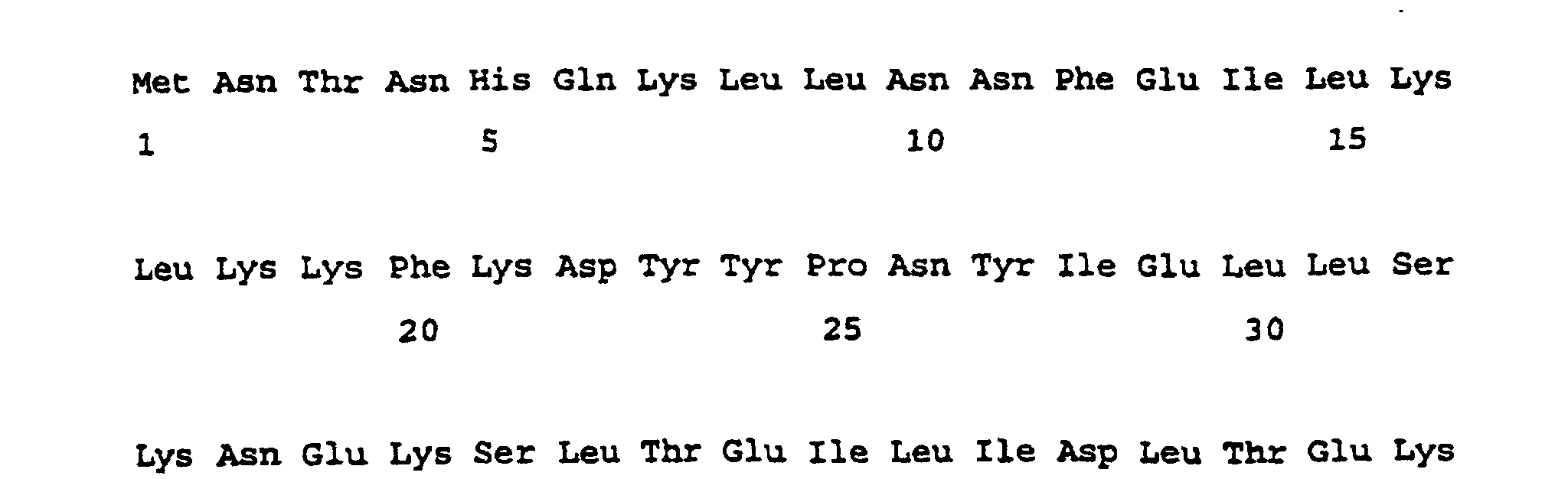
- An enteroccal immunogenic fragment may have the sequence of SEQ ID NO: 3, and is displayed by the ABC transporters of e.g. E.faecium and E.faecalis .
- An E.faecium immunogenic fragment may have the sequence of SEQ ID NO: 4.
- the immunogenic fragment may have the sequence of any one of SEQ ID NOs: 5-8.
- neutralising agents specific to a bacterial ABC transporter protein or immunogenic fragment thereof for use in a method of treatment of the human or animal body.
- a neutralising agent may be used in the manufacture of a medicament.
- Neutralising agents are well known and may include antibodies and antigen binding fragments thereof (Harlow, E. and Lane, D., "Antibodies - A Laboratory Mannual", Cold Spring Harbor Laboratory, Cold Spring Harbor Press, New York, 1988), ribozymes and antisense nucleic acid chains. Other neutralising agents will be readily apparent to one skilled in the art.
- Also provided according to the present invention is a method of treatment of the human or animal body comprising treating a patient with a bacterial ABC transporter protein or an immunogenic fragment thereof or a neutralising agent specific thereto according to the present invention.
- Medicaments according to the present invention may additionally comprise a pharmaceutically acceptable carrier, diluent or excipient (Remington's Pharmaceutical Sciences and US Pharmacopea, 1984, Mack Publishing Company, Easton, PA, USA).
- bacterial ABC transporter protein or immunogenic fragment thereof for use in a method of diagnosis of the human or animal body.
- the ABC transporter or immunogenic fragment thereof may be used in a method of diagnosis.
- the bacterium may be an enterococcus, for example selected from the group of E.faecium , E.faecalis , E.avium , E.gallinarium , E.durans , E.mundtii and E.casseflavus .
- the ABC transporter protein may be an enterocccal protein selected from the group of 97 and 54 kDa immunodominant conserved antigens.
- An immunogenic fragment may comprise an ATP binding site or a part thereof.
- the bacterium may be an enterocous, the immunogenic fragment having the sequence of SEQ ID NO: 3.
- the bacterium may be E.faecium , the immunogenic fragment having the sequence of SEQ ID NO: 4
- the bacterium may be E.faecium , the immunogenic fragment having the sequence of any one of SEQ ID NOs: 5-8.
- Also provided according to the present invention is a binding agent specific to a bacterial ABC transporter protein or immunogenic fragment thereof according to the present invention for use in a method of diagnosis of the human or animal body.
- Also provided according to the present invention is a binding agent specific to a bacterial ABC transporter protein or immunogenic fragment thereof according to the present invention used in a method of diagnosis.
- binding agent according to the present invention used in the manufacture of a diagnostic test kit.
- Binding agents include any agent capable of detecting the protein or immunogenic fragment and are well known, and include, for example, antibodies and antigen binding fragments thereof.
- Also provided according to the present invention is a method of diagnosis of the human or animal body comprising the use of a bacterial ABC transporter: or an immunogenic fragment thereof, or a binding agent specific thereto according to the present invention.
- a diagnostic test method for a bacterial ABC transporter protein or an immunogenic fragment thereof comprising the steps of:
- a sample from a patient may for example be patient dialysate or serum.
- the binding agent may comprise an antibody, the diagnostic test method comprising the steps of:
- a diagnostic test method for antibody specific against a bacterial ABC transporter protein or an immunogenic fragment thereof comprising the steps of:
- kits of parts for performing a diagnostic test are also provided.
- Also provided is a method of diagnosis comprising the use of a bacterial ABC transporter protein or an immunogenic fragment thereof or a binding agent specific thereto according to the present invention.
- the present inventors have now succeeded in isolating two immunodominant conserved enterococcal antigens and have found that, surprisingly, antibody specific to these antigens may be used to provide an effective therapy for enterococcal infection.
- the antigen may be for use in a method of treatment or diagnosis of the human or animal body.
- the antigen may be for use as an immunogen. Hence the antigen may be used to stimulate an immunogenic response in patients in order to protect them against infection from the bacteria.
- the antigen may for example be for use as a vaccine.
- the present invention also provides antibody or an antigen binding fragment thereof specific to an antigen according to the present invention.
- the antibody or an antigen binding fragment thereof may be for use in the diagnosis or treatment of infections of the enterococcus or staphylococcus.
- the use of antibody specific to the bacteria provides a novel and highly effective form of treatment for the infection.
- a major envelope protein antigen of molecular weight 53 kDa was defined in E. faecalis (and not in E. faecium ). and other common antigens of E. faecalis had molecular weights of 65, 63, 56, 49.5. 30 and 21 kDa. They also found that growth of E. faecalis in serum to mimic in vivo growth conditions in endocarditis patients altered the antigenic pattern, with only two major antigens of molecular weights 56 and 53 kDa reacting with sera from endocarditis patients. They suggested that these antigens may be of diagnostic potential. Burnie, J.P. et al . (1987, J. Clin.
- Pathol., 40 : 1149-1158 explored the role of immunoblotting in the diagnosis of culture-negative and enterococcal endocarditis. They found that in E . faecalis endocarditis there was a strong IgM response to E. faecalis bands of 112, 88-90 and 45-47 kDa, and a strong IgG response to 88-90 and 45-47 kDa bands. The 112 kDa antigen of E. faecalis was later used to develop an indirect enzyme-linked immunosorbent assay (ELISA) for the diagnosis of E. faecalis endocarditis (Bumie, J.P. and Clark, I., 1989, J. Immunol. Methods, 123 : 217-225). The three patients with E . faecium infection showing a strong IgG response to bands of 82-90 kDa.
- ELISA enzyme-linked immunosorbent assay
- the antibody may be a whole antibody or an antigen binding fragment thereof and may in general belong to any immunoglobulin class. Thus, for example, it may be an immunoglobulin M antibody or an immunoglobulin G antibody.
- the antibody or fragment may be of animal, for example, mammalian origin and may be for example of murine, rat, sheep or human origin. It may be a natural antibody or a fragment thereof, or, if desired, a recombinant antibody fragment, i.e. an antibody or antibody fragment which has been produced using recombinant DNA techniques.
- Particular recombinant antibodies or antibody fragments include, (1) those having an antigen binding site at least part of which is derived from a different antibody, for example those in which the hypervariable or complementarity determining regions of one antibody have been grafted into the variable framework regions of a second, different antibody (as described in, for example, European Patent Specification No 239400); (2) recombinant antibodies or fragments wherein non-Fv sequences have been substituted by non-Fv sequences from other, different antibodies (as described in, for example, European Patent Specification Nos 171469, 173494 and 194276); or (3) recombinant antibodies or fragments possessing substantially the structure of a natural immunoglobulin but wherein the hinge region has a different number of cysteine residues from that found in the natural immunoglobulin but wherein one or more cysteine residues in a surface pocket of the recombinant antibody or fragment is in the place of another amino acid residue present in the natural immunoglobulin (as described in, for example, International Patent
- the antibody or antibody fragment may be of polyclonal, monoclonal or recombinant origin. It may be specific for at least one epitope.
- Antigen binding antibody fragments include, for example, fragments derived by proteolytic cleavage of a whole antibody, such as F(ab')2, Fab' or Fab fragments, or fragments obtained by recombinant DNA techniques, for example Fv fragments (as described, for example, in International Patent Application No PCT/GB88/0747 (WO89/02465)).
- the antibodies according to the invention may be prepared using well-known immunological techniques employing the protein expressed during infection as antigen.
- any suitable host may be injected with the protein and the serum collected to yield the desired polyclonal antibody after appropriate purification and/or concentration (for example by affinity chromatography using the immobilised protein as the affinity medium).
- splenocytes or lymphocytes may be recovered from the protein-injected host and immortalised using for example the method of Kohler et al. (1976, Eur. J. Imtnuaol., 6 : 511), the resulting cells being segregated to obtain a single genetic line producing monoclonal antibodies.
- Antibody fragments may be produced using conventional techniques, for example, by enzymatic digestion with pepsin or papain. Where it is desired to produce recombinant antibodies according to the invention these may be produced using, for example, the methods described in European Patent Specification Nos 171469, 173494, 194276 and 239400.
- Antibodies according to the invention may be labelled with a detectable label or may be conjugated with an effector molecule, for example a drug e.g. an antibacterial agent or a toxin or an enzyme, using conventional procedures and the invention extends to such labelled antibodies or antibody conjugates.
- an effector molecule for example a drug e.g. an antibacterial agent or a toxin or an enzyme
- Such antibody may for example be expressed in transgenic animals, for example transgenic sheep and may be achieved using existing transgenic expression systems.
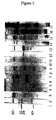
- Figure 1 shows pre- and post- VRE infection immunoblots for the IgM response of five patients (lanes 1 and 2, 3 and 4, 5 and 6, 7 and 8, 9 and 10 respectively).
- Lane 11 is a post-VRE infection immunoblot for the IgM response of one patient. All patients bad a septicaemia (Table 2);
- Figure 2 shows the IgG response of pre- and post- VRE infection patients.
- Lanes 1-15 are post-infection from all ten replicants and the pre-infection from five of the replicants (Table 2). Lanes are: 1 (post, patient 1), 2 (post, patient 2), 3 (post, patient 3), 4 (pre, patient 4), 5 (post, patient 4), 6 (pre, patient 5), 7 (post, patient 5), 8 (pre, patient 6), 9 (post, patient 6), 10 (pre, patient 7), 11 (post, patient 7), 12 (pre, patient 8), 13 (post, patient 8), 14 (post, patient 9), 15 (post, patient 10).
- Lane 16 is a Coomassie blue stained PVDF membrane of the VRE extract. Lane 17 contains molecular weight markers. Molecular weights of bands are as marked; and
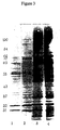
- Figure 3 shows sequential (lanes 1 and 2) IgM and sequential (lanes 3 and 4) IgG from a patient with a S . aureus septicaemia who recovered, blotted against the extract from the MRSA.
- Antibody from the peritoneal dialysate which bound to the expression product of the isolated clone s was separated from the expression product and bound to the 69 kDa MRSA antigen, showing that the same epitope was displayed by both the 69 kDa antigen and the isolated clone.
- the cloned MRSA DNA was then sequenced and putative amino acid sequences of two open reading frames (ORFs) derived and analysed. Analysis included comparison to sequence of the expression product derived by direct amino acid sequencing, comparison of the theoretical molecular weight to the observed expression product molecular weight and comparison to sequences of know proteins.
- Epitope mapping of SEQ ID NO: 10 identified 3 epitopes (SEQ ID NOs: 12-14; MRSA1a, MRSA1b and MRSA1c respectively) and epitope mapping of SEQ ID NO: 11 (termed MRSA2) identified 1 epitope (SEQ ID NO: 15; termed MRSA2a).
- Antibody specific against the antigens was found to be protective in animal models and therefore the antigens and agents which neutralise their function are therapeutically useful. Similarly, binding agents which specifically bind to the antigens may be diagnostically useful. Immunogenic fragments of the antigens may be used in place of the antigens themselves for diagnosis and therapy.
- VRE vancomycin resistant Enteroccus faecium
- Antibody from peritoneal dialysate which bound to the expression product of the isolated clone was separated from the expression product and bound to the 97kDa VRE antigen, showing that the same epitope was displayed by both the 97kDa antigen and the isolated clone.
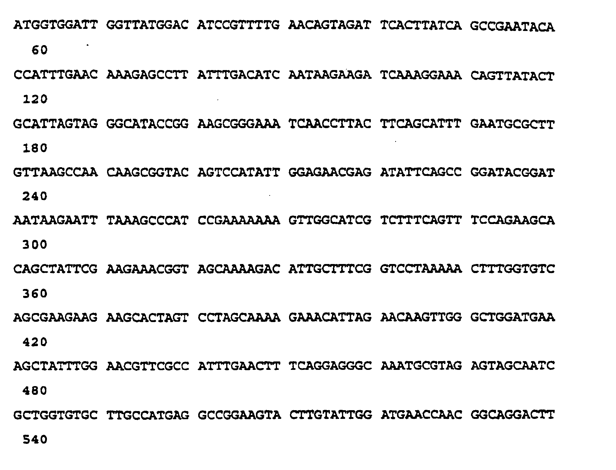
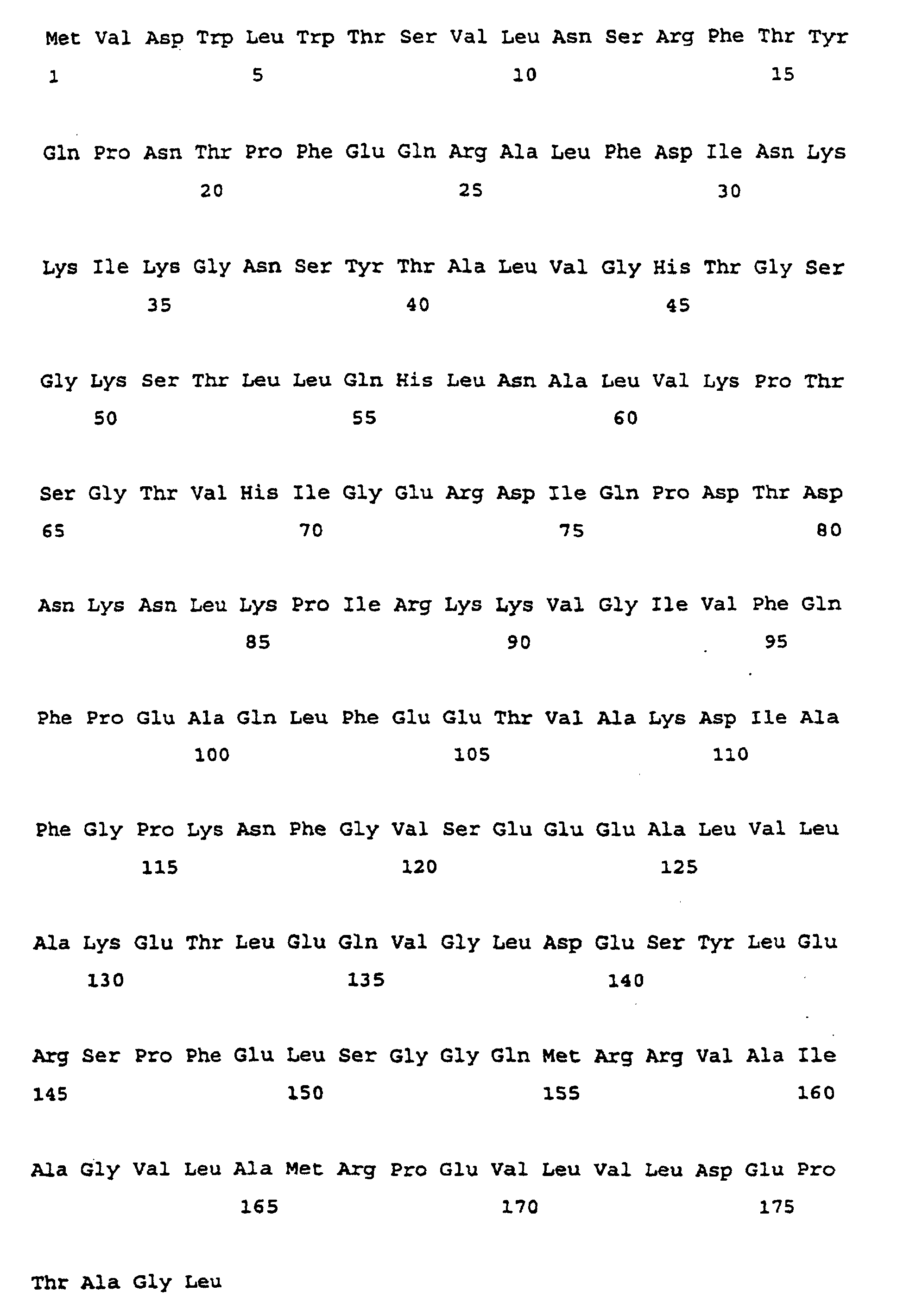
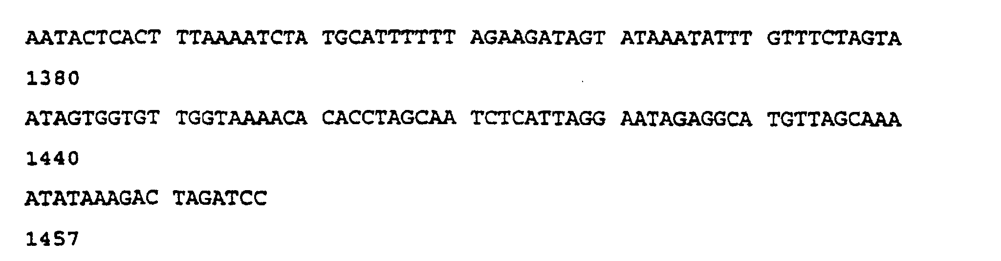
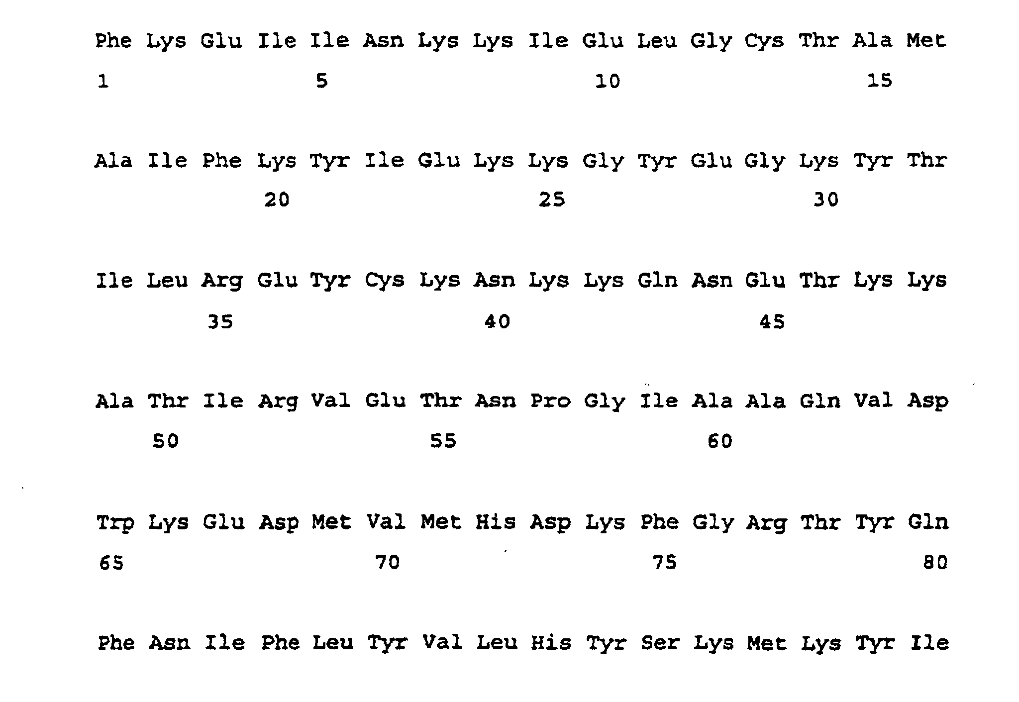
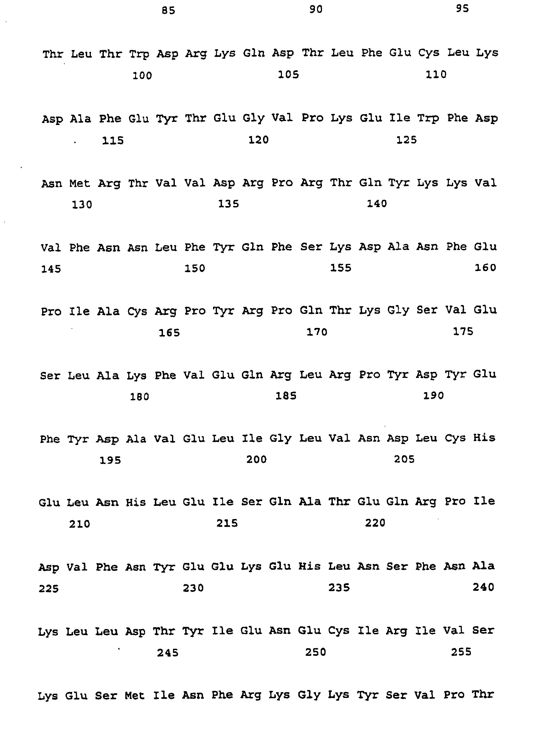
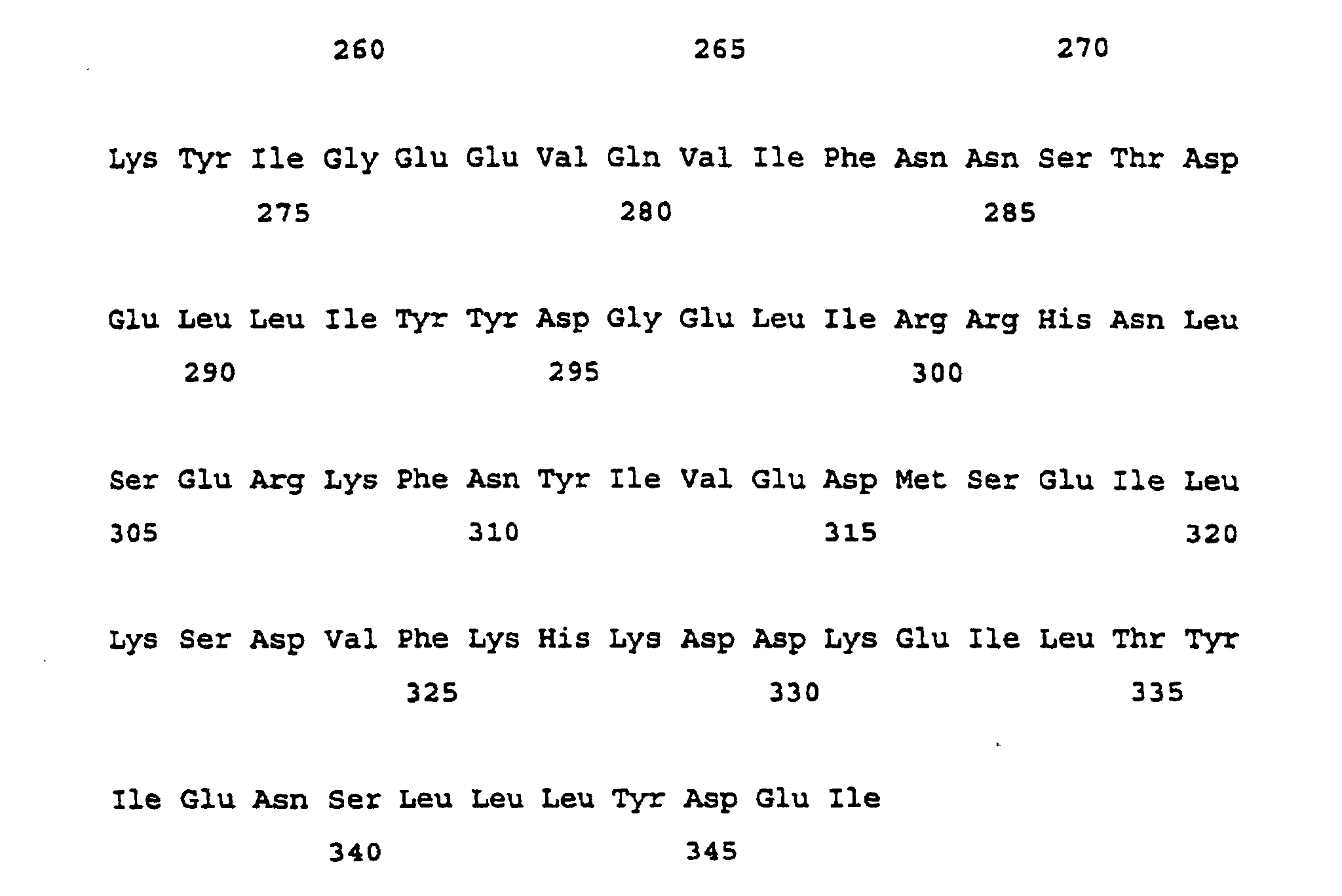
- the cloned VRE DNA was then sequenced and putative amino acid sequences of the six open reading frames (ORFs) derived and analysed. Analysis included comparison to sequence of the expression product derived by direct amino acid sequencing, comparison of the theoretical molecular weight to the observed expression product molecular weight and comparison to sequences of know proteins. This showed that only one of the putative amino acid sequences (SEQ ID NO: 2; coded for by DNA sequence SEQ ID NO: 1) could correspond to the expression product.
- Antigen was prepared by the following method. Using a clinical isolate of Vancomycin resistant enterococcus, an MRSA epidemic strain 16, an MRSA epidemic strain 2, the Oxford S.aureus and a clinical isolate of S.epidermis , 10 ml of brain heart infusion broth (Oxoid) was innoculated with the organism and grown up at 37 °C aerobically for 4 hours. This was added to 300 ml of the same broth and incubated on an orbital shaker at 37 °C for 24 hours. The culture was centrifuged at 2500 g for 15 minutes, resuspended in 10 ml of 10 mM Tris-HCl (pH 7.5) and repelleted as above.
- Oxoid brain heart infusion broth
- the organism was then fragmented in a hydraulic press (Enkoping, Sweden) at -20 °C, centrifuged at 13000 rpm for 30 minutes, and the supernatant (containing antigen) stored at -20 °C. Subsequently batches of antigen for the VRE and MRSA 16 were prepared by growing them at 30 °C and at 37 °C in the presence of 3 ⁇ g/ml Vancomycin.
- the sera collected had been submitted to the bacteriology, virology and biochemistry laboratories for routine testing. It was stored at -20 °C until required for immunoblotting.
- Group 1 septicaemia patients had signs and symptoms (e.g. pyrexia) consistent with infection at a time when VRE was isolated from blood cultures. These patients were neutropenic during admission secondary to chemotherapy for haematological malignancies (mostly leukaemias). Neutropenia was defined as a neutrophil count of ⁇ 1.0 x 10 9 /l. Pre- and post-infection serum were kept and tested in parallel. These were separated into survivors and non-survivors (those with positive blood cultures with a VRE who were unresponsive to chemotherapy and died).
- peritoneal dialysate was available from a single patient with peritonitis due to a VRE.
- Group 2 patients were considered to be colonized with VRE, in that at the time of isolation of these organisms the patients had no signs or symptoms of infection that could be attributed to VRE.
- This group was divided into Group 2a and Group 2b.
- Group 2a were patients all of whom would have been neutropenic at some point in their illnesses. Therefore, this subgroup will be referred to as colonized neutropenic patients.
- Group 2b patients were all renal or intensive care patients and none had been neutropenic. This subgroup will be referred to as colonized non-neutropenic patients.
- Post-colonization sera only was saved from Group 2 patients because colonization can be intermittent, and in any one patient it is never clear when exactly they first became colonized, or if perhaps they had been colonized during a previous admission.
- Group 3 patients were the control group for the VRE study.
- the serum had been routinely collected from pregnant women from the community attending antenatal clinics.
- the level of VRE in the community is low at present, and the majority of women should not have encountered it.
- Hospitalised patients were not used as a control group because of the possibility of them having been unknowingly colonized with VRE.
- Group 4 were patients with a septicaemia due to an MRSA, positive blood cultures who were treated with vancomycin therapy. Paired sera were available before and after the septicaemia. In addition, peritonea dialysate was available from a single patient with peritonitis due to an MRSA.
- Group 5 were patients colonized with an MRSA who showed no clinical signs of infection.
- Group 6 patients were the control group for the MRSA study.
- the serum had been routinely collected from pregnant women from the community attending antenatal clinics.
- Group 7 were patients with a septicaemia due to an MSSA (methicillin sensitive S.aureus ), positive blood cultures who were treated successfully. Paired sera were available before and after the septicaemia.
- MSSA methicillin sensitive S.aureus
- a 1.5 mm thick polyacrylamide gel was prepared in the vertical gel apparatus from a Protean II xi Cell (Biorad). A 10 % resolving gel was poured first and left to polymerize at room temperature for 60 minutes. A ten lane comb was placed on top of this and the stacking gel poured round it and on top of the separating gel. This was left for 30 minutes to polymerize. The following were used to make the gels:
- the antigen to be run on the gel was solubilised by boiling with cracking buffer and distilled water. This was VRE and MRSA epidemic strain 16 grown at 37 °C for all sera.
- Five antibody positive sera forom VRE septicaemia cases were tested against VRE grown at 30 °C and at 37 °C in the presence of Vancomycin (3 ⁇ g/ml).
- Two antibody positive sera from MRSA septicaemia patients were immunoblotted against MRSA 2 and the Oxford S.aureus .
- Eight further sere were immunoblotted against the antigen derived from S.epidermis and the MRSA epidemic strain 16 grown at 30 °C and the MRSA epidemic strain 16 grown at 37 °C in the presence of Vancomycin (3 ⁇ g/ml). Multiples of the following were boiled as the amount of protein in this ratio was found to give good results on immunoblotting: supernatant (containing antigen), 15 ⁇ l; distilled water, 10 ⁇ l; cracking buffer, 25 ⁇ l.
- Electrophoresis buffer was placed in the electrophoresis tank and in the cell above the gels. This formed the discontinous buffer system. Electrophoresis was carried out at a constant current of 40 mA per gel, with water cooling. It was run until the dye front had just run off the gel.
- the gel was carefully removed from between the glass plates and the stacking gel removed.
- the separating gel was then placed onto nitrocellulose paper (Biorad) and this was enveloped between filter paper.
- the nitrocellulose and filter paper had been presoaked in transblotting buffer. This was placed in the transblotting tank (Hoefer) which contained transblotting buffer. The current was run at the maximum for 45 minutes and the system was water cooled.
- the nitrocellulose was removed and left to block overnight in 3 % bovine serum albumin (BSA) at 4 °C.
- BSA bovine serum albumin
- the nitrocellulose paper was cut into strips reflecting the wells and each placed in separate compartments in an incubation container.
- BSA solution (3 %) was added to each strip and enough of the appropriate serum was added to this to give a 1:10 dilution. These were shaken at room temperature for 2 hours. The strips were then washed in washing solution for 30 minutes with the solution being changed every 6 minutes. Next, a 1:1000 dilution in 3 % BSA of anti-human immunoglobulin conjugated to alkaline phosphatase was added (Sigma).
- the main immunoblotting for serum from colonized or infected patients was for IgA, IgM and IgG.
- Alkaline phosphatase substrate buffer Alkaline phosphatase substrate buffer .
- Imtnunoblotting (Tables 1 and 2; Figures 1 and 2) demonstrated multiple antigenic bands of which four were immunodominant (97, 57, 54 and 40 kDa). These antigens were conserved between isolates and present when the strains were grown with and without vancomycin. IgG was produced against the 97 kDa band in 11 out of the 12 patients who survived a septicaemia and it was present in 12 of the neutropenic patients who did not develop a septicaemia. IgM was produced in 8 and IgA in 4 of the septicaemia patients. In the case of the 54 kDa antigen, 10 patients with a septicaemia made an IgM response and 5 an IgA response.
- the 97 and 54 kDa enterococcal antigens are immunodominant and may be used therapeutically and diagnostically. Further immunoblotting demonstrated the presence of the antigens when the VRE was grown at 30 °C and in the presence of Vancomycin at 37 °C.
- mice were injected with 200 ⁇ l of saline solution and 10 test mice were injected with 200 ⁇ l of antibody serum from a patient who had recovered from a septicaemia. 1 hour later the mice were each injected with 100 ⁇ l of 5 x 10 8 colony forming units (CFU) of vancomycin resistant E. faecium .
- CFU colony forming units
- mice Two sets of 30 female CD-1 mice weighing 22g each were infected via the tail vein with 3 x 10 9 /100 ⁇ l bolus of a vancomycin resistant E.faecium . Infected mice were subsequently injected with either (Group A) 200 ⁇ l of peritoneal dialysate fluid (as previously described) from a patient with CAPD peritonitis due to VRE, or with (Group B) 200 ⁇ l of normal fluid.
- Serum was obtained from two patients.
- Patient 1 had recovered from a subphrenic abscess which was treated by vancomycin.
- Patient 1 serum antibody was positive for both the 37 and 69 kDa antigens.
- Patient 2 was treated for an MRSA septicaemia with vancomycin.
- the patient 2 serum antibody was early preimmune and negative for the 37 and 69 kDa antigens.
- Vancomycin assays showed that patient 2 had a higher vancomycin level (30 ⁇ g/ml) than patient 1 (12 ⁇ g/ml).
- 20 CD-1 mice were given 1 x 10 7 of the MRSA epidemic strain '16' intravenously and 2 hours later two groups of 10 mice were each given 100 ml of the serum from either patient 1 or 2 as a single intravenous volume.
- Results show that serum antibody positive for both the 37 and 69 kDa antigens was protective against MRSA, with a mortality rate of 20% in the group treated with patient 1 (antibody positive for both the 37 and 69 kDa antigens) as compared to a mortality rate of 50% for the other group.
- Time Patient 1 Patient 2 Day 1 All Well All Well Days 2-6 All Well 1 death Day 7 2 deaths (20% mortality) 4 more deaths (50% mortality)
- a genomic library (one for each of vancomycin resistant Enterococcus faecium and multiply resistant Staphylococcus aureus ) was constructed in the expression/cloning vector lambda ZAP express, essentially as described by Young and Davies (1983, PNAS USA, 80 : 1194-1198). Chromosomal DNA from a clinical isolate was partially digested by Sau3a and fragments in the size range of 2 to 9 kbp were inserted into the vector, resulting in the production of p galactosidase fusion proteins.
- Each library was screened with antibody positive (37 and 69 kDa bands for MRSA or 54 and 97 kDa bands for VRE) peritoneal dialysate fluids (1 in 100 dilution) from patients with the respective infection. Positive clones were detected using alkaline phosphatase conjugated goat anti-human immuglobulin (IgG) (1 in 5,000) (Sigma, Poole UK). Lysogens were prepared from positive clones in Escherichia coli Y1089 according to Huynh, Young and Davies (1985, DNA cloning vol 1, a practical approach, IRL Press Oxford, p 49-78. Ed. D.M. Glover).
- the ⁇ galactosidase fusion proteins were detected by immunoblotting using respective peritoneal dialysate fluid (1 in 10) and a monoclonal antibody to p galactosidase (1 in 1,000).
- the epitope expressed by each of the positive clones was identified by antigen-selection as described by Lyon et al . (1986, PNAS USA, 83 : 2989-2993).
- the peritoneal dialysate fluid was affinity purified by hybridising with positive recombinant lambda plaques. The bound antibody was then eluted with glycine buffer pH 2.8, and used to immunoblot lysates of the relevant bacteria.
- PCR with T3 and T7 forward and reverse primers was used to amplify insert DNA from sera positive clones. This was subcloned into the TA Cloning System (version 1.3, Invitrogen Corporation, Oxon, UK) prior to DNA sequencing using the dideoxy termination method (sequence version 2.0 kit; United States Biochemical, Cambridge, UK). Initial sequencing reactions were performed using sequencing universal primers, the remaining sequence being determined using a primer walking strategy by progressively synthesising sequencing primers to generate new sequence data.
- Sequencing demonstrated a partial sequence in frame with the p galactosidase gene.
- the total insert size was 4.5. kb.
- the derived amino acid sequence produced a protein with two ATP- binding domains and a sequence homologous to the group of proteins which are ABC transporters (Fath and Kolter, 1993, Microbiological Reviews, 57 : 995-1017).
Landscapes
- Chemical & Material Sciences (AREA)
- Health & Medical Sciences (AREA)
- Organic Chemistry (AREA)
- General Health & Medical Sciences (AREA)
- Medicinal Chemistry (AREA)
- Life Sciences & Earth Sciences (AREA)
- Proteomics, Peptides & Aminoacids (AREA)
- Molecular Biology (AREA)
- Genetics & Genomics (AREA)
- Biophysics (AREA)
- Biochemistry (AREA)
- Gastroenterology & Hepatology (AREA)
- Veterinary Medicine (AREA)
- Public Health (AREA)
- Animal Behavior & Ethology (AREA)
- Pharmacology & Pharmacy (AREA)
- Nuclear Medicine, Radiotherapy & Molecular Imaging (AREA)
- General Chemical & Material Sciences (AREA)
- Chemical Kinetics & Catalysis (AREA)
- Oncology (AREA)
- Communicable Diseases (AREA)
- Peptides Or Proteins (AREA)
- Medicines That Contain Protein Lipid Enzymes And Other Medicines (AREA)
- Medicines Containing Antibodies Or Antigens For Use As Internal Diagnostic Agents (AREA)
- Pharmaceuticals Containing Other Organic And Inorganic Compounds (AREA)
- Measuring Or Testing Involving Enzymes Or Micro-Organisms (AREA)
- Saccharide Compounds (AREA)
- Medicines Containing Material From Animals Or Micro-Organisms (AREA)
- Cephalosporin Compounds (AREA)
Abstract
Description
| 30 % acrylamide / 0.8 % bisacrylamide | 25 ml |
| (Ultra pure Protogel, National diagnostics) | |
| Separating gel buffer (see below) | 14.06 |
| 10 % SDS | 0.75 ml |
| Distilled water | 35.2 |
| 10 % ammonium persulphate | 350 µl |
| TEMED | 37.5 µl |
| Tris | 24.22 g |
| Distilled water | 40 ml |
| 30 % acrylamide / 0.8 % bisacrylamide | 3.6 ml |
| Stacking gel buffer (see below) | 7.3 |
| 10 % SDS | 0.3 ml |
| Distilled water | 19 |
| 10 % ammonium persulphate | 300 µl |
| TEMED | 30 µl |
| |
6 .05 g |
| Distilled water | 90 ml |
| 20 |
4 |
| Stacking buffer | |
| 1 | |
| Distilled water | |
| 4 ml | |
| 2-mercaptoethanol | 0.4 ml |
| Glycerol | 0.6 ml |
| Tris | 18.96 g |
| Glycine | 12 g |
| SDS | 3 |
| Distilled water | |
| 1 litre |
| Tris | 9.07 g |
| Glycine | 43.2 g |
| Methanol | 750 |
| Distilled water | |
| 3 litre |
| Sodium chloride | 9 g |
| Tris | 1.2 |
| Distilled water | |
| 1 litre |
| Sodium chloride | 4.5 g |
| Tween 20 | 0.25 ml |
| Distilled water | 500 ml |
| Nitro-blue tetrazolium | 0.05 g |
| 70 % N,N- |
1 ml |
| BCIP | 0.05 g |
| 70 % N,N- |
1 ml |
| Tris | 1.12 g |
| Sodium chloride | 0.58 g |
| Magnesium chloride | 0.1 g |
| Distilled water | 100 ml |
| Antigen apparent molecular weight (kDa) | | | |||||
| neutropenic leukaemia n=22 | non-neutropenic renal n=12 | n=60 combined IgM, IgG and IgA | |||||
| IgM | IgG | IgA | | IgG | IgA | ||
| 97 | 4 | 12 | 0 | 1 | 0 | 2 | 3 |
| 57 | 1 | 2 | 0 | 0 | 1 | 0 | 7 |
| 54 | 4 | 3 | 1 | 1 | 0 | 0 | 4 |
| 40 | 0 | 4 | 0 | 1 | 1 | 0 | 1 |
| Antigen apparent MW (kDa) | | Dead (n=5) | ||||||||||||||
| IgM | IgG | IgA | IgM | igG | IgA | |||||||||||
| | C | C | ||||||||||||||
| 97 | 8 | 11 | 1 | 4 | 1 | 1 | 1 | 1 | 1 | 1 | ||||||
| 57 | 5 | 5 | 1 | 2 | 3 | 1 | 2 | 2 | ||||||||
| 54 | 10 | 2 | 1 | 5 | 5 | 1 | 2 | 1 | 1 | |||||||
| 40 | 4 | 1 | 4 | 1 | 2 | 2 |
| Antigen apparent mw (kDa) | | | | | ||||||||||
| Septicaemia survived (n=5) | septicaemia died (n=1) | colonized (n=3) | Control (n=10) | MSSA septicaemia survived (n=3) | ||||||||||
| IgM | IgG | IgM | IgG | IgM | IgG | IgM | | IgM | IgG | |||||
| 120 | 0 | 3 | 0 | 3 | 1 | 1 | 1 | 1 | 2 | 1 | 0 | 3 | 0 | 3 |
| 84 | 1 | 2 | 0 | 3 | 0 | 0 | 1 | 1 | 2 | 2 | 0 | 0 | 0 | 0 |
| 69 | 2 | 2 | 1 | 3 | 0 | 0 | 2 | 2 | 2 | 1 | 1 | 0 | 1 | 0 |
| 67 | 2 | 2 | 0 | 4 | 1 | 1 | 0 | 0 | 1 | 3 | 0 | 0 | 0 | 0 |
| 60 | 0 | 2 | 0 | 3 | 0 | 0 | 1 | 1 | 3 | 1 | 0 | 0 | 0 | 0 |
| 53 | 0 | 4 | 0 | 3 | 0 | 0 | 0 | 1 | 2 | 2 | 0 | 0 | 0 | 0 |
| 42 | 0 | 1 | 1 | 2 | 1 | 0 | 1 | 0 | 1 | 2 | 0 | 0 | 0 | 0 |
| 37 | 4 | 0 | 3 | 2 | 0 | 0 | 2 | 3 | 2 | 2 | 2 | 0 | 2 | 0 |
| 33 | 3 | 1 | 2 | 3 | 0 | 1 | 1 | 0 | 1 | 1 | 0 | 0 | 0 | 0 |
| 31 | 2 | 1 | 2 | 1 | 0 | 0 | 1 | 0 | 1 | 1 | 0 | 0 | 0 | 0 |
| | Patient | 1 | |
| | All Well | All Well | |
| Days 2-6 | | 1 | |
| Day | |||
| 7 | 2 deaths (20% mortality) | 4 more deaths (50% mortality) |
Positive Cocci







Claims (18)
- An immunogenic fragment of a bacterial ABC transporter protein for use in a method of treatment of the human or animal body, the bacterium being an enterococcus and the immunogenic fragment having the sequence of SEQ.ID.NO:3, or the bacterium being E.faecium and the immunogenic fragment having the sequence of any one of SEQ.ID Nos: 4-8.
- The use of a bacterial ABC transporter protein having the sequence of SEQ ID NOS: 1 and 2 or an immunogenic fragment thereof in the manufacture of a medicament for the treatment of infection by said bacterium.
- The use of antibodies specific to a bacterial ABC transporter protein having the sequence of SEQ ID NOS: 1 and 2 or immunogenic fragment thereof in the manufacture of a medicament for the treatment of infection due to said bacterium.
- The use according to either one of claims 2 and 3 of a bacterial ABC transporter protein or an immunogenic fragment thereof; or antibodies and antigen binding fragments thereof, specific to the bacterial ABC transporter protein or immunogenic fragment thereof, the bacterium being an enterococcus.
- The use according to either one of claims 2 and 3 of a bacterial ABC transporter protein having the sequence of SEQ. ID. NO: 1 or an immunogenic fragment thereof or ribozymes and nucleic acid chains specific to the bacterial ABC transporter protein or immunogenic fragment thereof, the bacterium being an enterococcus.
- The use according to claim 4 or claim 5 of a bacterial ABC transporter protein or an immunogenic fragment thereof; or antibodies, antigen binding fragments thereof, ribozymes and antisense nucleic acid chains specific to the bacterial ABC transporter protein or immunogenic fragment thereof, the bacterium being selected from one of the group E.faecium, E. faecalis, E. avium, E.gallinarium, E.durans, E.mundtii and E.casseflavus.
- The use according to any one of claims 4, 5 or 6 of a bacterial ABC transporter protein or an immunogenic fragment thereof; or antibodies, antigen binding fragments thereof, ribozymes and antisense nucleic acid chains specific to the bacterial ABC transporter protein or immunogenic fragment thereof, the ABC transporter protein being a 97 kDa immunodominant conserved antigen.
- The use according to any one of claims 2 to 7 of a bacterial ABC transporter protein or an immunogenic fragment thereof; or antibodies, antigen binding fragments thereof, ribozymes and antisense nucleic acid chains specific to the bacterial ABC transporter protein or immunogenic fragment thereof, the immunogenic fragment comprising an ATP binding site or a part thereof.
- The use according to any one of claims 2 to 8 of a bacterial ABC transporter protein or an immunogenic fragment thereof; or antibodies, antigen binding fragments thereof, ribozymes and antisense nucleic acid chains specific to the bacterial ABC transporter protein or immunogenic fragment thereof, the bacterium being an enterococcus and the immunogenic fragment having the sequence of SEQ.ID. NO: 3.
- The use according to claim 9 of a bacterial ABC transporter protein or an immunogenic fragment thereof; or antibodies, antigen binding fragments thereof, ribozymes and antisense nucleic acid chains specific to the bacterial ABC transporter protein or immunogenic fragment thereof, the bacterium being E.faecium and the immunogenic fragment having the sequence of SEQ.ID. NO: 4.
- The use according to any one of claims 2 to 8 of a bacterial ABC transporter protein or an immunogenic fragment thereof; or antibodies, antigen binding fragments thereof, ribozymes and antisense nucleic acid chains specific to the bacterial ABC transporter protein or immunogenic fragment thereof, the bacterium being E.faecium and the immunogenic fragment having the sequence of any one of SEQ ID Nos: 5-8.
- An immunogenic fragment of a bacterial ABC transporter protein for use in a method of diagnosis of the human or animal body, the bacterium being an enterococcus and the immunogenic fragment having the sequence of SEQ ID NO:3, or the bacterium being E.faecium and the immunogenic fragment having the sequence of any one of SEQ ID Nos: 4-8.
- The use of an immunogenic fragment of a bacterial ABC transporter protein for the manufacture of a diagnostic agent for use in diagnosis of infection by a bacterium, the bacterium being an enterococcus and the immunogenic fragment having the sequence of SEQ.ID. NO:3, or the bacterium being E.faecium and the immunogenic fragment having the sequence of any one of SEQ ID Nos: 4-8.
- The use of a binding agent specific to an immunogenic fragment of a bacterial ABC transporter protein for the manufacture of a diagnostic agent for use in diagnosis of infection by a bacterium of the human or animal body, the bacterium being an enterococcus and the immunogenic fragment having the sequence of SEQ ID NO: 3, or the bacterium being E.faecium and the immunogenic fragment having the sequence of any one of SEQ ID Nos: 4-8.
- The use of a binding agent specific to an immunogenic. fragment of a bacterial ABC transporter protein in the manufacture of a diagnostic test kit, the bacterium being an enterococcus and the immunogenic fragment having the sequence of SEQ ID NO: 3, or the bacterium being E.faecium and the immunogenic fragment having the sequence of any one of SEQ ID Nos: 4-8.
- A diagnostic test method for a bacterial ABC transporter protein or an immunogenic fragment thereof comprising the steps of:i) reacting a binding agent as defined in claim 14 with a sample from a patient;ii) detecting a reaction between the binding agent and antigen; andiii) correlating the detection of the reaction with the presence of the protein or an immunogenic fragment thereof.
- A diagnostic test method as defined in claim 15, the binding agent comprising an antibody, and comprising the steps of:i) reacting a binding agent according to claim 14 with a sample from a patient;ii) detecting an antibody-antigen binding reaction; andiii) correlating the detection of the antibody-antigen binding reaction with the presence of the protein or an immunogenic fragment thereof.
- A diagnostic test method for antibody specific against a bacterial ABC transporter protein or an immunogenic fragment thereof, comprising the steps of:i) reacting an immunogenic fragment of an ABC transporter protein with patient antisera, the immunogenic fragment being from an enterococcus and having the sequence SEQ ID NO:3, or the immunogenic fragment being from E.faecium and having the sequence of any one of SEQ ID NOs: 4-8;ii) detecting an antibody-antigen binding reaction; andiii) correlating the detection of the antibody-antigen binding reaction with the presence of antibody specific against the bacterial ABC . transporter protein or an immunogenic fragment thereof.
Applications Claiming Priority (3)
| Application Number | Priority Date | Filing Date | Title |
|---|---|---|---|
| GB9614274 | 1996-07-06 | ||
| GBGB9614274.0A GB9614274D0 (en) | 1996-07-06 | 1996-07-06 | Treatment and diagnosis of infections of gram positive cocci |
| PCT/GB1997/001830 WO1998001154A2 (en) | 1996-07-06 | 1997-07-07 | Treatment and diagnosis of infections of gram positive cocci |
Publications (2)
| Publication Number | Publication Date |
|---|---|
| EP0917471A2 EP0917471A2 (en) | 1999-05-26 |
| EP0917471B1 true EP0917471B1 (en) | 2005-04-20 |
Family
ID=10796521
Family Applications (1)
| Application Number | Title | Priority Date | Filing Date |
|---|---|---|---|
| EP97930642A Expired - Lifetime EP0917471B1 (en) | 1996-07-06 | 1997-07-07 | Treatment and diagnosis of infections of gram positive cocci |
Country Status (13)
| Country | Link |
|---|---|
| US (2) | US6544516B1 (en) |
| EP (1) | EP0917471B1 (en) |
| JP (1) | JP2001505534A (en) |
| AT (1) | ATE293456T1 (en) |
| AU (1) | AU717332B2 (en) |
| CA (1) | CA2259141C (en) |
| DE (1) | DE69733067T2 (en) |
| DK (1) | DK0917471T3 (en) |
| ES (1) | ES2238080T3 (en) |
| GB (1) | GB9614274D0 (en) |
| NZ (1) | NZ332991A (en) |
| PT (1) | PT917471E (en) |
| WO (1) | WO1998001154A2 (en) |
Families Citing this family (9)
| Publication number | Priority date | Publication date | Assignee | Title |
|---|---|---|---|---|
| US6610293B1 (en) * | 1997-06-16 | 2003-08-26 | The Henry M. Jackson Foundation For The Advancement Of Military Medicine | Opsonic and protective monoclonal and chimeric antibodies specific for lipoteichoic acid of gram positive bacteria |
| GB9806762D0 (en) * | 1998-03-31 | 1998-05-27 | Neutec Pharma Plc | Treatment and diagnosis of staphylococcal infections |
| EP1624064A3 (en) * | 1998-07-27 | 2006-05-10 | Microbial Technics Limited | Nucleic acids and proteins from streptococcus pneumoniae |
| WO2002057406A2 (en) * | 2001-01-17 | 2002-07-25 | Avestha Gengraine Technologies Pvt Ltd | Dna sequencing of abc transporter or multi drug resistant protein for better environment stress tolerant crops |
| CA2446294A1 (en) * | 2001-05-08 | 2002-11-14 | Yi Xu | Surface proteins from gram-positive bacteria having highly conserved motifs and antibodies that recognize them |
| GB0127983D0 (en) * | 2001-11-22 | 2002-01-16 | Neutec Pharma Plc | Treatment of micro-organism infection |
| US8729013B2 (en) | 2004-08-26 | 2014-05-20 | The University Of Western Ontario | Methods of inhibiting staphylobactin-mediated iron uptake in S. aureus |
| EP2450053B1 (en) * | 2010-11-05 | 2016-06-15 | Universitätsklinikum Freiburg | Novel antigen of enterococcal pathogens and use thereof as vaccine component for therapy and/or prophylaxis |
| EP2872535A1 (en) | 2012-07-16 | 2015-05-20 | Pfizer Inc. | Saccharides and uses thereof |
Family Cites Families (3)
| Publication number | Priority date | Publication date | Assignee | Title |
|---|---|---|---|---|
| US5747239A (en) * | 1990-02-16 | 1998-05-05 | United Biomedical, Inc. | Synthetic peptides specific for the detection of antibodies to HCV, diagnosis of HCV infection and preventions thereof as vaccines |
| WO1993006247A1 (en) * | 1991-09-16 | 1993-04-01 | Abbott Laboratories | Hepatitis c assay |
| GB9401689D0 (en) | 1994-01-28 | 1994-03-23 | Univ Manchester | Diagnosis and treatment of endocarditis |
-
1996
- 1996-07-06 GB GBGB9614274.0A patent/GB9614274D0/en active Pending
-
1997
- 1997-07-07 EP EP97930642A patent/EP0917471B1/en not_active Expired - Lifetime
- 1997-07-07 AU AU34522/97A patent/AU717332B2/en not_active Ceased
- 1997-07-07 NZ NZ332991A patent/NZ332991A/en not_active IP Right Cessation
- 1997-07-07 DK DK97930642T patent/DK0917471T3/en active
- 1997-07-07 US US09/214,307 patent/US6544516B1/en not_active Expired - Fee Related
- 1997-07-07 AT AT97930642T patent/ATE293456T1/en not_active IP Right Cessation
- 1997-07-07 PT PT97930642T patent/PT917471E/en unknown
- 1997-07-07 JP JP50494298A patent/JP2001505534A/en not_active Ceased
- 1997-07-07 ES ES97930642T patent/ES2238080T3/en not_active Expired - Lifetime
- 1997-07-07 WO PCT/GB1997/001830 patent/WO1998001154A2/en not_active Ceased
- 1997-07-07 DE DE69733067T patent/DE69733067T2/en not_active Expired - Lifetime
- 1997-07-07 CA CA002259141A patent/CA2259141C/en not_active Expired - Fee Related
-
2002
- 2002-01-25 US US10/054,968 patent/US6881410B2/en not_active Expired - Fee Related
Also Published As
| Publication number | Publication date |
|---|---|
| WO1998001154A2 (en) | 1998-01-15 |
| GB9614274D0 (en) | 1996-09-04 |
| US6881410B2 (en) | 2005-04-19 |
| WO1998001154A3 (en) | 1998-06-25 |
| JP2001505534A (en) | 2001-04-24 |
| AU3452297A (en) | 1998-02-02 |
| ATE293456T1 (en) | 2005-05-15 |
| DE69733067D1 (en) | 2005-05-25 |
| PT917471E (en) | 2005-07-29 |
| AU717332B2 (en) | 2000-03-23 |
| US20030119101A1 (en) | 2003-06-26 |
| NZ332991A (en) | 1999-06-29 |
| CA2259141C (en) | 2007-12-11 |
| DK0917471T3 (en) | 2005-06-06 |
| US6544516B1 (en) | 2003-04-08 |
| DE69733067T2 (en) | 2006-01-26 |
| ES2238080T3 (en) | 2005-08-16 |
| CA2259141A1 (en) | 1998-01-15 |
| EP0917471A2 (en) | 1999-05-26 |
Similar Documents
| Publication | Publication Date | Title |
|---|---|---|
| Seifert et al. | A unique serine-rich repeat protein (Srr-2) and novel surface antigen (ε) associated with a virulent lineage of serotype III Streptococcus agalactiae | |
| EP1423701B1 (en) | Cross-reactive monoclonal and polyclonal antibodies which recognize surface proteins from coagulase-negative staphylococci and staphylococcus aureus | |
| EA007409B1 (en) | Streptococcus antigen polypeptides, methods of producing and use thereof | |
| US5618533A (en) | Flagellin-based polypeptides for the diagnosis of lyme disease | |
| US7195763B2 (en) | Surface proteins from gram-positive bacteria having highly conserved motifs and antibodies that recognize them | |
| EP1540559B1 (en) | Bioinformatic method for identifying surface-anchored proteins from gram-positive bacteria and proteins obtained thereby | |
| JP3245298B2 (en) | Epitope region of pneumococcal surface protein A | |
| EP0695803A2 (en) | Epitopic regions of pneumococcal surface protein A | |
| EP0656014B1 (en) | Protein rib, a cell surface protein that confers immunity to many strains of the group b streptococcus; process for purification of the protein, reagent kit and pharmaceutical composition | |
| US6881410B2 (en) | Treatment of E. faecium infection | |
| Heckels et al. | The potential protective effect of immunization with outer-membrane protein I from Neisseria gonorrhoeae | |
| Swiatlo et al. | Choline‐binding proteins | |
| WO1994022443A1 (en) | Method for treating kawasaki syndrome | |
| EP1054895B1 (en) | Novel peptide diagnostic reagent and kit for detection of rickettsiosis | |
| US5556755A (en) | Method for detecting Branhamella catarrhalis | |
| US5476767A (en) | Isolated nucleic acid molecule coding for toxin associated with Kawasaki Syndrome and uses thereof | |
| WO1994022474A1 (en) | Method for treating kawasaki syndrome via immune modulation | |
| US5585465A (en) | Isolated toxin associated with Kawasaki syndrome | |
| CA2097912A1 (en) | Method of detecting methicillin-resistant staphylococcus aureus, novel peptides, and dnas encoding the same | |
| Anthony et al. | Gram-positive bacteria: an overview and summary of session | |
| CA1341574C (en) | Plasmid for production of membrane protein, bacterium containing same, monoclonal antibody therefore, and method for the idenification of haemophilus influenzae | |
| WO1995020648A1 (en) | Staphylococcus aureus strain atcc 55533 | |
| HK1008825B (en) | Protein rib, a cell surface protein that confers immunity to many strains of the group b streptococcus; process for purification of the protein, reagent kit and pharmaceutical composition | |
| MXPA98005424A (en) | Three synthetic peptides to be used in the vaccination and diagnosis of cisticercosis by taenia sol |
Legal Events
| Date | Code | Title | Description |
|---|---|---|---|
| PUAI | Public reference made under article 153(3) epc to a published international application that has entered the european phase |
Free format text: ORIGINAL CODE: 0009012 |
|
| 17P | Request for examination filed |
Effective date: 19981219 |
|
| AK | Designated contracting states |
Kind code of ref document: A2 Designated state(s): AT BE CH DE DK ES FI FR GB GR IE IT LI LU MC NL PT SE |
|
| 17Q | First examination report despatched |
Effective date: 20011109 |
|
| GRAP | Despatch of communication of intention to grant a patent |
Free format text: ORIGINAL CODE: EPIDOSNIGR1 |
|
| GRAS | Grant fee paid |
Free format text: ORIGINAL CODE: EPIDOSNIGR3 |
|
| GRAA | (expected) grant |
Free format text: ORIGINAL CODE: 0009210 |
|
| AK | Designated contracting states |
Kind code of ref document: B1 Designated state(s): AT BE CH DE DK ES FI FR GB GR IE IT LI LU MC NL PT SE |
|
| REG | Reference to a national code |
Ref country code: GB Ref legal event code: FG4D |
|
| REG | Reference to a national code |
Ref country code: CH Ref legal event code: EP |
|
| REG | Reference to a national code |
Ref country code: CH Ref legal event code: NV Representative=s name: ROTTMANN, ZIMMERMANN + PARTNER AG |
|
| REG | Reference to a national code |
Ref country code: IE Ref legal event code: FG4D |
|
| REF | Corresponds to: |
Ref document number: 69733067 Country of ref document: DE Date of ref document: 20050525 Kind code of ref document: P |
|
| REG | Reference to a national code |
Ref country code: DK Ref legal event code: T3 |
|
| REG | Reference to a national code |
Ref country code: SE Ref legal event code: TRGR |
|
| REG | Reference to a national code |
Ref country code: GR Ref legal event code: EP Ref document number: 20050401865 Country of ref document: GR |
|
| REG | Reference to a national code |
Ref country code: PT Ref legal event code: SC4A Effective date: 20050518 |
|
| REG | Reference to a national code |
Ref country code: ES Ref legal event code: FG2A Ref document number: 2238080 Country of ref document: ES Kind code of ref document: T3 |
|
| PLBE | No opposition filed within time limit |
Free format text: ORIGINAL CODE: 0009261 |
|
| STAA | Information on the status of an ep patent application or granted ep patent |
Free format text: STATUS: NO OPPOSITION FILED WITHIN TIME LIMIT |
|
| ET | Fr: translation filed | ||
| 26N | No opposition filed |
Effective date: 20060123 |
|
| PGFP | Annual fee paid to national office [announced via postgrant information from national office to epo] |
Ref country code: CH Payment date: 20090721 Year of fee payment: 13 |
|
| PGFP | Annual fee paid to national office [announced via postgrant information from national office to epo] |
Ref country code: BE Payment date: 20090805 Year of fee payment: 13 |
|
| PGFP | Annual fee paid to national office [announced via postgrant information from national office to epo] |
Ref country code: PT Payment date: 20100623 Year of fee payment: 14 Ref country code: MC Payment date: 20100628 Year of fee payment: 14 |
|
| PGFP | Annual fee paid to national office [announced via postgrant information from national office to epo] |
Ref country code: NL Payment date: 20100707 Year of fee payment: 14 Ref country code: IE Payment date: 20100712 Year of fee payment: 14 Ref country code: ES Payment date: 20100813 Year of fee payment: 14 |
|
| PGFP | Annual fee paid to national office [announced via postgrant information from national office to epo] |
Ref country code: SE Payment date: 20100708 Year of fee payment: 14 Ref country code: LU Payment date: 20100716 Year of fee payment: 14 Ref country code: IT Payment date: 20100717 Year of fee payment: 14 Ref country code: FR Payment date: 20100805 Year of fee payment: 14 Ref country code: FI Payment date: 20100712 Year of fee payment: 14 Ref country code: DE Payment date: 20100630 Year of fee payment: 14 Ref country code: AT Payment date: 20100712 Year of fee payment: 14 |
|
| PGFP | Annual fee paid to national office [announced via postgrant information from national office to epo] |
Ref country code: GR Payment date: 20100615 Year of fee payment: 14 Ref country code: GB Payment date: 20100707 Year of fee payment: 14 |
|
| BERE | Be: lapsed |
Owner name: *NEUTEC PHARMA P.L.C. Effective date: 20100731 |
|
| PGFP | Annual fee paid to national office [announced via postgrant information from national office to epo] |
Ref country code: DK Payment date: 20100712 Year of fee payment: 14 |
|
| REG | Reference to a national code |
Ref country code: CH Ref legal event code: PL |
|
| PG25 | Lapsed in a contracting state [announced via postgrant information from national office to epo] |
Ref country code: CH Free format text: LAPSE BECAUSE OF NON-PAYMENT OF DUE FEES Effective date: 20100731 Ref country code: LI Free format text: LAPSE BECAUSE OF NON-PAYMENT OF DUE FEES Effective date: 20100731 |
|
| PG25 | Lapsed in a contracting state [announced via postgrant information from national office to epo] |
Ref country code: BE Free format text: LAPSE BECAUSE OF NON-PAYMENT OF DUE FEES Effective date: 20100731 |
|
| REG | Reference to a national code |
Ref country code: PT Ref legal event code: MM4A Free format text: LAPSE DUE TO NON-PAYMENT OF FEES Effective date: 20120109 |
|
| REG | Reference to a national code |
Ref country code: NL Ref legal event code: V1 Effective date: 20120201 |
|
| PG25 | Lapsed in a contracting state [announced via postgrant information from national office to epo] |
Ref country code: MC Free format text: LAPSE BECAUSE OF NON-PAYMENT OF DUE FEES Effective date: 20110731 |
|
| REG | Reference to a national code |
Ref country code: SE Ref legal event code: EUG |
|
| REG | Reference to a national code |
Ref country code: DK Ref legal event code: EBP |
|
| GBPC | Gb: european patent ceased through non-payment of renewal fee |
Effective date: 20110707 |
|
| REG | Reference to a national code |
Ref country code: GR Ref legal event code: ML Ref document number: 20050401865 Country of ref document: GR Effective date: 20120202 |
|
| REG | Reference to a national code |
Ref country code: AT Ref legal event code: MM01 Ref document number: 293456 Country of ref document: AT Kind code of ref document: T Effective date: 20110707 |
|
| REG | Reference to a national code |
Ref country code: FR Ref legal event code: ST Effective date: 20120330 |
|
| REG | Reference to a national code |
Ref country code: IE Ref legal event code: MM4A |
|
| PG25 | Lapsed in a contracting state [announced via postgrant information from national office to epo] |
Ref country code: FR Free format text: LAPSE BECAUSE OF NON-PAYMENT OF DUE FEES Effective date: 20110801 Ref country code: DE Free format text: LAPSE BECAUSE OF NON-PAYMENT OF DUE FEES Effective date: 20120201 |
|
| REG | Reference to a national code |
Ref country code: DE Ref legal event code: R119 Ref document number: 69733067 Country of ref document: DE Effective date: 20120201 |
|
| PG25 | Lapsed in a contracting state [announced via postgrant information from national office to epo] |
Ref country code: NL Free format text: LAPSE BECAUSE OF NON-PAYMENT OF DUE FEES Effective date: 20120201 Ref country code: FI Free format text: LAPSE BECAUSE OF NON-PAYMENT OF DUE FEES Effective date: 20110707 Ref country code: PT Free format text: LAPSE BECAUSE OF NON-PAYMENT OF DUE FEES Effective date: 20120109 Ref country code: GR Free format text: LAPSE BECAUSE OF NON-PAYMENT OF DUE FEES Effective date: 20120202 Ref country code: IT Free format text: LAPSE BECAUSE OF NON-PAYMENT OF DUE FEES Effective date: 20110707 |
|
| PG25 | Lapsed in a contracting state [announced via postgrant information from national office to epo] |
Ref country code: GB Free format text: LAPSE BECAUSE OF NON-PAYMENT OF DUE FEES Effective date: 20110707 |
|
| PG25 | Lapsed in a contracting state [announced via postgrant information from national office to epo] |
Ref country code: DK Free format text: LAPSE BECAUSE OF NON-PAYMENT OF DUE FEES Effective date: 20110731 Ref country code: IE Free format text: LAPSE BECAUSE OF NON-PAYMENT OF DUE FEES Effective date: 20110707 |
|
| PG25 | Lapsed in a contracting state [announced via postgrant information from national office to epo] |
Ref country code: AT Free format text: LAPSE BECAUSE OF NON-PAYMENT OF DUE FEES Effective date: 20110707 |
|
| PG25 | Lapsed in a contracting state [announced via postgrant information from national office to epo] |
Ref country code: SE Free format text: LAPSE BECAUSE OF NON-PAYMENT OF DUE FEES Effective date: 20110708 |
|
| PG25 | Lapsed in a contracting state [announced via postgrant information from national office to epo] |
Ref country code: LU Free format text: LAPSE BECAUSE OF NON-PAYMENT OF DUE FEES Effective date: 20110707 |
|
| REG | Reference to a national code |
Ref country code: ES Ref legal event code: FD2A Effective date: 20130531 |
|
| PG25 | Lapsed in a contracting state [announced via postgrant information from national office to epo] |
Ref country code: ES Free format text: LAPSE BECAUSE OF NON-PAYMENT OF DUE FEES Effective date: 20110708 |