EP0901633B1 - Screening of agents for treatment of alzheimer's disease - Google Patents
Screening of agents for treatment of alzheimer's disease Download PDFInfo
- Publication number
- EP0901633B1 EP0901633B1 EP97923188A EP97923188A EP0901633B1 EP 0901633 B1 EP0901633 B1 EP 0901633B1 EP 97923188 A EP97923188 A EP 97923188A EP 97923188 A EP97923188 A EP 97923188A EP 0901633 B1 EP0901633 B1 EP 0901633B1
- Authority
- EP
- European Patent Office
- Prior art keywords
- tau
- filaments
- alzheimer
- disease
- agent
- Prior art date
- Legal status (The legal status is an assumption and is not a legal conclusion. Google has not performed a legal analysis and makes no representation as to the accuracy of the status listed.)
- Expired - Lifetime
Links
- 208000024827 Alzheimer disease Diseases 0.000 title claims abstract description 30
- 238000012216 screening Methods 0.000 title claims abstract description 6
- 102000013498 tau Proteins Human genes 0.000 claims abstract description 99
- 108010026424 tau Proteins Proteins 0.000 claims abstract description 99
- 229920002683 Glycosaminoglycan Polymers 0.000 claims abstract description 23
- 238000000034 method Methods 0.000 claims abstract description 15
- 150000001720 carbohydrates Chemical class 0.000 claims abstract description 8
- 238000006243 chemical reaction Methods 0.000 claims abstract description 5
- 238000012544 monitoring process Methods 0.000 claims abstract description 3
- HTTJABKRGRZYRN-UHFFFAOYSA-N Heparin Chemical compound OC1C(NC(=O)C)C(O)OC(COS(O)(=O)=O)C1OC1C(OS(O)(=O)=O)C(O)C(OC2C(C(OS(O)(=O)=O)C(OC3C(C(O)C(O)C(O3)C(O)=O)OS(O)(=O)=O)C(CO)O2)NS(O)(=O)=O)C(C(O)=O)O1 HTTJABKRGRZYRN-UHFFFAOYSA-N 0.000 claims description 27
- 229920000669 heparin Polymers 0.000 claims description 27
- 229960002897 heparin Drugs 0.000 claims description 27
- 102000001708 Protein Isoforms Human genes 0.000 claims description 20
- 108010029485 Protein Isoforms Proteins 0.000 claims description 20
- 229920002971 Heparan sulfate Polymers 0.000 claims description 18
- 239000003795 chemical substances by application Substances 0.000 claims description 11
- 150000001413 amino acids Chemical class 0.000 claims description 9
- 239000007993 MOPS buffer Substances 0.000 claims description 8
- MGSKVZWGBWPBTF-UHFFFAOYSA-N aebsf Chemical compound NCCC1=CC=C(S(F)(=O)=O)C=C1 MGSKVZWGBWPBTF-UHFFFAOYSA-N 0.000 claims description 8
- 230000004962 physiological condition Effects 0.000 claims description 6
- 238000001493 electron microscopy Methods 0.000 claims description 4
- 239000003153 chemical reaction reagent Substances 0.000 claims description 2
- 230000015572 biosynthetic process Effects 0.000 abstract description 17
- 238000000338 in vitro Methods 0.000 abstract description 8
- 230000000694 effects Effects 0.000 abstract description 5
- 230000002401 inhibitory effect Effects 0.000 abstract description 5
- 230000000903 blocking effect Effects 0.000 abstract description 3
- 238000001727 in vivo Methods 0.000 abstract description 3
- 230000002452 interceptive effect Effects 0.000 abstract description 2
- 238000011835 investigation Methods 0.000 abstract description 2
- 230000001225 therapeutic effect Effects 0.000 abstract description 2
- 229920001307 poly(hydroxymethylethylene hydroxymethyl formal) Polymers 0.000 abstract 1
- 102000029749 Microtubule Human genes 0.000 description 34
- 108091022875 Microtubule Proteins 0.000 description 34
- 210000004688 microtubule Anatomy 0.000 description 34
- 210000004556 brain Anatomy 0.000 description 18
- 238000010186 staining Methods 0.000 description 18
- FAPWRFPIFSIZLT-UHFFFAOYSA-M Sodium chloride Chemical compound [Na+].[Cl-] FAPWRFPIFSIZLT-UHFFFAOYSA-M 0.000 description 14
- 239000000872 buffer Substances 0.000 description 13
- 108090000623 proteins and genes Proteins 0.000 description 11
- 238000011534 incubation Methods 0.000 description 10
- 102000004169 proteins and genes Human genes 0.000 description 10
- 229940024606 amino acid Drugs 0.000 description 9
- 210000002569 neuron Anatomy 0.000 description 9
- 230000026731 phosphorylation Effects 0.000 description 9
- 238000006366 phosphorylation reaction Methods 0.000 description 9
- DVLFYONBTKHTER-UHFFFAOYSA-N 3-(N-morpholino)propanesulfonic acid Chemical compound OS(=O)(=O)CCCN1CCOCC1 DVLFYONBTKHTER-UHFFFAOYSA-N 0.000 description 8
- 229920002684 Sepharose Polymers 0.000 description 8
- 230000001419 dependent effect Effects 0.000 description 8
- 210000004027 cell Anatomy 0.000 description 7
- 239000011780 sodium chloride Substances 0.000 description 7
- LOKCTEFSRHRXRJ-UHFFFAOYSA-I dipotassium trisodium dihydrogen phosphate hydrogen phosphate dichloride Chemical compound P(=O)(O)(O)[O-].[K+].P(=O)(O)([O-])[O-].[Na+].[Na+].[Cl-].[K+].[Cl-].[Na+] LOKCTEFSRHRXRJ-UHFFFAOYSA-I 0.000 description 6
- 238000002372 labelling Methods 0.000 description 6
- 239000002953 phosphate buffered saline Substances 0.000 description 6
- 101000891579 Homo sapiens Microtubule-associated protein tau Proteins 0.000 description 5
- 238000002474 experimental method Methods 0.000 description 5
- 102000057063 human MAPT Human genes 0.000 description 5
- 230000003902 lesion Effects 0.000 description 5
- YBYRMVIVWMBXKQ-UHFFFAOYSA-N phenylmethanesulfonyl fluoride Chemical compound FS(=O)(=O)CC1=CC=CC=C1 YBYRMVIVWMBXKQ-UHFFFAOYSA-N 0.000 description 5
- 210000004899 c-terminal region Anatomy 0.000 description 4
- 235000014633 carbohydrates Nutrition 0.000 description 4
- 108010083213 heparitinsulfate lyase Proteins 0.000 description 4
- 230000003993 interaction Effects 0.000 description 4
- 239000008188 pellet Substances 0.000 description 4
- 210000001519 tissue Anatomy 0.000 description 4
- PEDCQBHIVMGVHV-UHFFFAOYSA-N Glycerine Chemical compound OCC(O)CO PEDCQBHIVMGVHV-UHFFFAOYSA-N 0.000 description 3
- 102000004243 Tubulin Human genes 0.000 description 3
- 108090000704 Tubulin Proteins 0.000 description 3
- 238000007792 addition Methods 0.000 description 3
- 239000012634 fragment Substances 0.000 description 3
- 210000001320 hippocampus Anatomy 0.000 description 3
- 230000006951 hyperphosphorylation Effects 0.000 description 3
- 238000003364 immunohistochemistry Methods 0.000 description 3
- 230000007137 neurofibrillary pathology Effects 0.000 description 3
- 210000002763 pyramidal cell Anatomy 0.000 description 3
- 238000002415 sodium dodecyl sulfate polyacrylamide gel electrophoresis Methods 0.000 description 3
- 239000006228 supernatant Substances 0.000 description 3
- 238000005406 washing Methods 0.000 description 3
- PVPBBTJXIKFICP-UHFFFAOYSA-N (7-aminophenothiazin-3-ylidene)azanium;chloride Chemical compound [Cl-].C1=CC(=[NH2+])C=C2SC3=CC(N)=CC=C3N=C21 PVPBBTJXIKFICP-UHFFFAOYSA-N 0.000 description 2
- JKMHFZQWWAIEOD-UHFFFAOYSA-N 2-[4-(2-hydroxyethyl)piperazin-1-yl]ethanesulfonic acid Chemical compound OCC[NH+]1CCN(CCS([O-])(=O)=O)CC1 JKMHFZQWWAIEOD-UHFFFAOYSA-N 0.000 description 2
- QFVHZQCOUORWEI-UHFFFAOYSA-N 4-[(4-anilino-5-sulfonaphthalen-1-yl)diazenyl]-5-hydroxynaphthalene-2,7-disulfonic acid Chemical compound C=12C(O)=CC(S(O)(=O)=O)=CC2=CC(S(O)(=O)=O)=CC=1N=NC(C1=CC=CC(=C11)S(O)(=O)=O)=CC=C1NC1=CC=CC=C1 QFVHZQCOUORWEI-UHFFFAOYSA-N 0.000 description 2
- 208000037259 Amyloid Plaque Diseases 0.000 description 2
- 206010012289 Dementia Diseases 0.000 description 2
- TWRXJAOTZQYOKJ-UHFFFAOYSA-L Magnesium chloride Chemical compound [Mg+2].[Cl-].[Cl-] TWRXJAOTZQYOKJ-UHFFFAOYSA-L 0.000 description 2
- 101710115937 Microtubule-associated protein tau Proteins 0.000 description 2
- 102100040243 Microtubule-associated protein tau Human genes 0.000 description 2
- 102000044589 Mitogen-Activated Protein Kinase 1 Human genes 0.000 description 2
- 108700015928 Mitogen-activated protein kinase 13 Proteins 0.000 description 2
- 229930012538 Paclitaxel Natural products 0.000 description 2
- 108091000080 Phosphotransferase Proteins 0.000 description 2
- 229920001213 Polysorbate 20 Polymers 0.000 description 2
- 108010076830 Thionins Proteins 0.000 description 2
- 239000007983 Tris buffer Substances 0.000 description 2
- 238000000429 assembly Methods 0.000 description 2
- 230000000712 assembly Effects 0.000 description 2
- 238000005119 centrifugation Methods 0.000 description 2
- 210000004292 cytoskeleton Anatomy 0.000 description 2
- 238000000635 electron micrograph Methods 0.000 description 2
- 239000000499 gel Substances 0.000 description 2
- 230000001537 neural effect Effects 0.000 description 2
- 210000002682 neurofibrillary tangle Anatomy 0.000 description 2
- 230000006911 nucleation Effects 0.000 description 2
- 238000010899 nucleation Methods 0.000 description 2
- 229960001592 paclitaxel Drugs 0.000 description 2
- 102000020233 phosphotransferase Human genes 0.000 description 2
- 239000000256 polyoxyethylene sorbitan monolaurate Substances 0.000 description 2
- 235000010486 polyoxyethylene sorbitan monolaurate Nutrition 0.000 description 2
- 230000008569 process Effects 0.000 description 2
- 230000001737 promoting effect Effects 0.000 description 2
- 238000000746 purification Methods 0.000 description 2
- 150000003839 salts Chemical class 0.000 description 2
- 239000000758 substrate Substances 0.000 description 2
- RCINICONZNJXQF-MZXODVADSA-N taxol Chemical compound O([C@@H]1[C@@]2(C[C@@H](C(C)=C(C2(C)C)[C@H](C([C@]2(C)[C@@H](O)C[C@H]3OC[C@]3([C@H]21)OC(C)=O)=O)OC(=O)C)OC(=O)[C@H](O)[C@@H](NC(=O)C=1C=CC=CC=1)C=1C=CC=CC=1)O)C(=O)C1=CC=CC=C1 RCINICONZNJXQF-MZXODVADSA-N 0.000 description 2
- LENZDBCJOHFCAS-UHFFFAOYSA-N tris Chemical compound OCC(N)(CO)CO LENZDBCJOHFCAS-UHFFFAOYSA-N 0.000 description 2
- FPJHWYCPAOPVIV-VOZMEZHOSA-N (2R,3S,4R,5R,6R)-6-[(2R,3R,4R,5R,6R)-5-acetamido-2-(hydroxymethyl)-6-methoxy-3-sulfooxyoxan-4-yl]oxy-4,5-dihydroxy-3-methoxyoxane-2-carboxylic acid Chemical compound CO[C@@H]1O[C@H](CO)[C@H](OS(O)(=O)=O)[C@H](O[C@@H]2O[C@H]([C@@H](OC)[C@H](O)[C@H]2O)C(O)=O)[C@H]1NC(C)=O FPJHWYCPAOPVIV-VOZMEZHOSA-N 0.000 description 1
- KIUKXJAPPMFGSW-DNGZLQJQSA-N (2S,3S,4S,5R,6R)-6-[(2S,3R,4R,5S,6R)-3-Acetamido-2-[(2S,3S,4R,5R,6R)-6-[(2R,3R,4R,5S,6R)-3-acetamido-2,5-dihydroxy-6-(hydroxymethyl)oxan-4-yl]oxy-2-carboxy-4,5-dihydroxyoxan-3-yl]oxy-5-hydroxy-6-(hydroxymethyl)oxan-4-yl]oxy-3,4,5-trihydroxyoxane-2-carboxylic acid Chemical compound CC(=O)N[C@H]1[C@H](O)O[C@H](CO)[C@@H](O)[C@@H]1O[C@H]1[C@H](O)[C@@H](O)[C@H](O[C@H]2[C@@H]([C@@H](O[C@H]3[C@@H]([C@@H](O)[C@H](O)[C@H](O3)C(O)=O)O)[C@H](O)[C@@H](CO)O2)NC(C)=O)[C@@H](C(O)=O)O1 KIUKXJAPPMFGSW-DNGZLQJQSA-N 0.000 description 1
- IHPYMWDTONKSCO-UHFFFAOYSA-N 2,2'-piperazine-1,4-diylbisethanesulfonic acid Chemical compound OS(=O)(=O)CCN1CCN(CCS(O)(=O)=O)CC1 IHPYMWDTONKSCO-UHFFFAOYSA-N 0.000 description 1
- HSTOKWSFWGCZMH-UHFFFAOYSA-N 3,3'-diaminobenzidine Chemical compound C1=C(N)C(N)=CC=C1C1=CC=C(N)C(N)=C1 HSTOKWSFWGCZMH-UHFFFAOYSA-N 0.000 description 1
- LVSPDZAGCBEQAV-UHFFFAOYSA-N 4-chloronaphthalen-1-ol Chemical compound C1=CC=C2C(O)=CC=C(Cl)C2=C1 LVSPDZAGCBEQAV-UHFFFAOYSA-N 0.000 description 1
- 241000283690 Bos taurus Species 0.000 description 1
- 108091003079 Bovine Serum Albumin Proteins 0.000 description 1
- UXVMQQNJUSDDNG-UHFFFAOYSA-L Calcium chloride Chemical compound [Cl-].[Cl-].[Ca+2] UXVMQQNJUSDDNG-UHFFFAOYSA-L 0.000 description 1
- 241000283707 Capra Species 0.000 description 1
- 229920001287 Chondroitin sulfate Polymers 0.000 description 1
- 229920000045 Dermatan sulfate Polymers 0.000 description 1
- KCXVZYZYPLLWCC-UHFFFAOYSA-N EDTA Chemical compound OC(=O)CN(CC(O)=O)CCN(CC(O)=O)CC(O)=O KCXVZYZYPLLWCC-UHFFFAOYSA-N 0.000 description 1
- 102000004190 Enzymes Human genes 0.000 description 1
- 108090000790 Enzymes Proteins 0.000 description 1
- 241000588724 Escherichia coli Species 0.000 description 1
- 108010010803 Gelatin Proteins 0.000 description 1
- 241000463231 Hemicordulia tau Species 0.000 description 1
- 102000043136 MAP kinase family Human genes 0.000 description 1
- 108091054455 MAP kinase family Proteins 0.000 description 1
- 239000007990 PIPES buffer Substances 0.000 description 1
- 102000001253 Protein Kinase Human genes 0.000 description 1
- 206010042618 Surgical procedure repeated Diseases 0.000 description 1
- 230000002159 abnormal effect Effects 0.000 description 1
- 230000006229 amino acid addition Effects 0.000 description 1
- BFNBIHQBYMNNAN-UHFFFAOYSA-N ammonium sulfate Chemical compound N.N.OS(O)(=O)=O BFNBIHQBYMNNAN-UHFFFAOYSA-N 0.000 description 1
- 229910052921 ammonium sulfate Inorganic materials 0.000 description 1
- 239000001166 ammonium sulphate Substances 0.000 description 1
- 235000011130 ammonium sulphate Nutrition 0.000 description 1
- 238000003556 assay Methods 0.000 description 1
- 230000001580 bacterial effect Effects 0.000 description 1
- 229960002685 biotin Drugs 0.000 description 1
- 239000011616 biotin Substances 0.000 description 1
- 229940098773 bovine serum albumin Drugs 0.000 description 1
- 239000001110 calcium chloride Substances 0.000 description 1
- 229910001628 calcium chloride Inorganic materials 0.000 description 1
- 125000003178 carboxy group Chemical group [H]OC(*)=O 0.000 description 1
- 150000001875 compounds Chemical class 0.000 description 1
- 230000002596 correlated effect Effects 0.000 description 1
- 210000000805 cytoplasm Anatomy 0.000 description 1
- 230000007850 degeneration Effects 0.000 description 1
- 238000012217 deletion Methods 0.000 description 1
- 230000037430 deletion Effects 0.000 description 1
- 229940079593 drug Drugs 0.000 description 1
- 239000003814 drug Substances 0.000 description 1
- 230000009881 electrostatic interaction Effects 0.000 description 1
- 238000010828 elution Methods 0.000 description 1
- DEFVIWRASFVYLL-UHFFFAOYSA-N ethylene glycol bis(2-aminoethyl)tetraacetic acid Chemical compound OC(=O)CN(CC(O)=O)CCOCCOCCN(CC(O)=O)CC(O)=O DEFVIWRASFVYLL-UHFFFAOYSA-N 0.000 description 1
- 239000008273 gelatin Substances 0.000 description 1
- 229920000159 gelatin Polymers 0.000 description 1
- 235000019322 gelatine Nutrition 0.000 description 1
- 235000011852 gelatine desserts Nutrition 0.000 description 1
- PCHJSUWPFVWCPO-UHFFFAOYSA-N gold Chemical compound [Au] PCHJSUWPFVWCPO-UHFFFAOYSA-N 0.000 description 1
- 239000010931 gold Substances 0.000 description 1
- 229910052737 gold Inorganic materials 0.000 description 1
- 229920002674 hyaluronan Polymers 0.000 description 1
- 229960003160 hyaluronic acid Drugs 0.000 description 1
- 238000012744 immunostaining Methods 0.000 description 1
- 238000000099 in vitro assay Methods 0.000 description 1
- 238000003780 insertion Methods 0.000 description 1
- 230000037431 insertion Effects 0.000 description 1
- 230000007154 intracellular accumulation Effects 0.000 description 1
- 229910001629 magnesium chloride Inorganic materials 0.000 description 1
- 230000007246 mechanism Effects 0.000 description 1
- 230000001404 mediated effect Effects 0.000 description 1
- 108020004999 messenger RNA Proteins 0.000 description 1
- 238000000386 microscopy Methods 0.000 description 1
- 239000003226 mitogen Substances 0.000 description 1
- 239000000203 mixture Substances 0.000 description 1
- 230000004048 modification Effects 0.000 description 1
- 238000012986 modification Methods 0.000 description 1
- 210000002241 neurite Anatomy 0.000 description 1
- 230000001575 pathological effect Effects 0.000 description 1
- 229940080469 phosphocellulose Drugs 0.000 description 1
- 229920002643 polyglutamic acid Polymers 0.000 description 1
- 108060006633 protein kinase Proteins 0.000 description 1
- 239000013049 sediment Substances 0.000 description 1
- 238000001179 sorption measurement Methods 0.000 description 1
- 230000004936 stimulating effect Effects 0.000 description 1
- 238000006467 substitution reaction Methods 0.000 description 1
- 239000000725 suspension Substances 0.000 description 1
- 238000012360 testing method Methods 0.000 description 1
- 238000011144 upstream manufacturing Methods 0.000 description 1
Images



Classifications
-
- G—PHYSICS
- G01—MEASURING; TESTING
- G01N—INVESTIGATING OR ANALYSING MATERIALS BY DETERMINING THEIR CHEMICAL OR PHYSICAL PROPERTIES
- G01N33/00—Investigating or analysing materials by specific methods not covered by groups G01N1/00 - G01N31/00
- G01N33/48—Biological material, e.g. blood, urine; Haemocytometers
- G01N33/50—Chemical analysis of biological material, e.g. blood, urine; Testing involving biospecific ligand binding methods; Immunological testing
- G01N33/68—Chemical analysis of biological material, e.g. blood, urine; Testing involving biospecific ligand binding methods; Immunological testing involving proteins, peptides or amino acids
- G01N33/6893—Chemical analysis of biological material, e.g. blood, urine; Testing involving biospecific ligand binding methods; Immunological testing involving proteins, peptides or amino acids related to diseases not provided for elsewhere
- G01N33/6896—Neurological disorders, e.g. Alzheimer's disease
-
- Y—GENERAL TAGGING OF NEW TECHNOLOGICAL DEVELOPMENTS; GENERAL TAGGING OF CROSS-SECTIONAL TECHNOLOGIES SPANNING OVER SEVERAL SECTIONS OF THE IPC; TECHNICAL SUBJECTS COVERED BY FORMER USPC CROSS-REFERENCE ART COLLECTIONS [XRACs] AND DIGESTS
- Y10—TECHNICAL SUBJECTS COVERED BY FORMER USPC
- Y10T—TECHNICAL SUBJECTS COVERED BY FORMER US CLASSIFICATION
- Y10T436/00—Chemistry: analytical and immunological testing
- Y10T436/14—Heterocyclic carbon compound [i.e., O, S, N, Se, Te, as only ring hetero atom]
- Y10T436/142222—Hetero-O [e.g., ascorbic acid, etc.]
- Y10T436/143333—Saccharide [e.g., DNA, etc.]
- Y10T436/144444—Glucose
Definitions
- This invention concerns screening agents for treatment of Alzheimer's disease, and concerns a method of screening an agent for potential use in the treatment of Alzheimer's disease.
- Alzheimer's disease is the leading cause of dementia in the elderly.
- the brains of patients with Alzheimer's disease contain characteristic structural lesions known as plaques and tangles. Plaques will not be considered here.
- Neurofibrillary tangles represent intracellular accumulations of paired helical filaments (PHFs), which are quite unlike any of the normal structural elements of the neuronal cytoskeleton. Tangles become extracellular on the death of the affected cell. PHFs are also found in abnormal neurites associated with neuritic plaques and as an extensive distribution of fine neurophil threads throughout affected regions of the brain. The extent of neurofibrillary degeneration found post mortem appears to provide the most reliable pathological correlate of the degree of dementia observed in life. These topics are discussed further in references 1 and 2.
- PHFs are made of microtubule-associated protein tau in a hyperphosphorylated state (3-8). Hyperphosphorylation of tau is known to result in its inability to bind to microtubules (9. 10) and is believed to precede PHF assembly (11). However, it is unclear whether hyperphosphorylation of tau is either necessary or sufficient for PHF formation. A major reason for this lack of understanding is that it has not hitherto been possible to form paired helical-like filaments for full-length tau protein either in vitro or in vivo.
- Tau protein consists in adult human brain of six isoforms, produced from a single gene by alternative mRNA splicing.
- the isoforms which range in size from 352 to 441 amino acids, contain towards the carboxyl terminus a tandem repeat region with three or four homologous stretches of 31 or 32 amino acids.
- the other isoforms are generated by the presence of a 29- or 58- amino-acid insertion in the amino-terminal half of the protein.
- the repeat region of tau represents the microtubule-binding domain, and the amino-terminal half of the protein appears to form an arm-like projection from the surface of the microtubule.
- tau protein When tau protein is in hyperphosphorylated state (PHF-tau) the protein is unable to bind to microtubules and assembles into PHFs or structural variants known as straight filaments (SFs).
- SFs straight filaments
- the repeat region of tau constitutes the core of the resulting filaments.
- WO 93/11231 describes in vitro assembly of recombinant tau constructs to give PHF-like filaments under non-physiological conditions. PHF-like filaments were only obtained with constructs containing essentially only the repeat region of tau, but not with full length tau protein. This is proposed as the basis for an in vitro test for drugs of possible use in treatment of Alzheimer's disease.
- the present invention is based on the discovery that suitable sulphated carbohydrates can induce in vitro assembly of tau protein into filaments like those in Alzheimer's disease under physiological conditions in an essentially quantitative manner. This discovery forms the basis of an in vitro assay or screen for agents that inhibit the assembly and so may have potential use in the treatment of Alzheimer's disease.
- the present invention thus provides a method of screening an agent for potential use in the treatment of Alzheimer's disease, comprising reacting, in the presence of the agent, tan protein with a suitable sulphated carbohydrate under appropriate conditions to form filaments, and monitoring for the presence of filaments.
- An agent which inhibits assembly of PHFs in vitro may also have an inhibiting effect in vivo and thus have potential therapeutic value in delaying the dementing effects of Alzheimer's disease.
- the invention can thus provide a screen to identify agents worthy of further investigation for use in the treatment of Alzheimer's disease.
- the tau protein may be the full length protein or a fragment thereof which forms filaments.
- Filament-forming fragments of tau generally include at least the portion thereof including three microtubule-binding repeats. Any of the 6 isoforms which occur in the adult human brain. as discussed above, may be used.
- the full length 3 repeat 381 amino acid isoform. which forms paired helical filaments very similar to the paired helical filaments found in the brain of patients with Alzheimer's disease as explained above, works very well and so is currently favoured.
- the tau protein may be modified in known manner by amino acid additions, substitutions and/or deletions that do not significantly affect filament formation.
- tau protein is used to cover all such proteins and fragments that form filaments under appropriate conditions.
- the currently preferred sulphated carbohydrate is sulphated glycosaminoglycan, conveniently heparin or heparan sulphate.
- the reaction may be carried out under physiological conditions, eg. by incubating the reagents in MOPS and AEBSF as described below. and provides quantitative results. Good results have been obtained by incubating with 30 mM MOPS. 1mM AEBSF, pH 7.4 at 30°C, with filament formation starting after 5 h and reaching a maximum after 48 h. Tau concentration is suitably at least 40 ⁇ M, with a tau/sulphated glycosaminoglycan molar ratio of approximately 4:1 giving good results.
- the presence (or otherwise) of filaments may be monitored by using electron microscopy or other appropriate methods.
- Tau protein isoforms (the three repeat-containing 381 amino acid isoform of human tau (htau37) and the four repeat-containing 441 amino acid isoform of human tau (htau40)) were expressed in E . coli , as described (28).
- frozen pellets from 10 L bacterial culture were resuspended in 240 ml of 50 mM Tris, pH 7.4, 0.1 mM EDTA, 0.1 mM DTT, 0.1 mM phenylmethylsulphonyl fluoride (PMSF) (buffer A) and sonicated for 5 x 1 min on ice.
- PMSF phenylmethylsulphonyl fluoride
- Tau-containing fractions were applied to a Mono S column equilibrated in buffer B and eluted using a salt gradient. Peak fractions were concentrated by speed vacuum and dialysed against 40mM Hepes, pH 7.4. 0.1 mM DTT, 0.1 mM PMSF. Tau purification was monitored by SDS-PAGE.
- htau37 and htau40 were incubated with heparin (20 ⁇ M, BDH) in 25 ⁇ l of 30 mM 3-[N-Morpholino]propane-sulphonic acid (MOPS), 1 mM [4-(2-aminoethyl)benzenesulphonylfluoride] (AEBSF, Calbiochem), pH 7.4. at 30°C for 48 h. Aliquots were removed and examined by electron microscopy, as described (7). In some cases. htau37 was phosphorylated with 5U/mi recombinant neuronal cdc2-like kinase (NCLK) (29), prior to the addition of heparin. Results are shown in Figure 1.
- NCLK neuronal cdc2-like kinase
- paired helical-like filaments started to form after approximately 5 h at 30°C in 30 mM MOPS, 1mM AEBSF, pH 7.4, reaching a maximum number after 48 h.
- Filament formation required a critical tau concentration, with the tau/sulphated glycosaminoglycan ratio being of crucial importance.
- Paired helical and straight filaments formed at tau concentrations above 40 ⁇ M, with a tau/sulphated glycosaminoglycan molar ratio of approximately 4:1.
- tau forms filaments in a glycosaminoglycan-dependent, but phosphorylation-independent manner. Further, tau can form filaments in vitro under physiological conditions in quantitative manner.
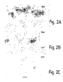
- Recombinant htau37 (80 ⁇ M) was incubated with heparin (20 ⁇ M) in 30 mM MOPS, 1 mM AEBSF, pH 7.4 at 30°C for 48 h. Following a 1 h centrifugation at 346,000 x g, the filament pellets were resuspended in 30 mM MOPS, 1 mM AEBSF, pH 7.4, and processed for immunoelectron microscopy, as described (7), using antibodies directed against the amino-terminus (antiserum 133), carboxy-terminus (antiserum 134) and microtubule-binding repeat region (antiserum 135) of tau.
- Results are shown in Figure 2.
- Gold labelling is seen with 133 and 134. but not with 135.
- the paired helical-like filaments were decorated by antibodies directed against the amino- and carboxy-termini of tau, but not by antibody against the microtubule-binding repeat region.
- microtubule-binding repeat region of tau is essential for sulphated glycosaminoglycan-induced filament formation. It is well known that three microtubule-binding repeats of tau form the core of the PHF from Alzheimer's disease brain (4, 15), thus supporting the evidence for a similar organisation of the two types of filament.
- the dimensions of tau filaments formed in the presence of sulphated glycosaminoglycans were similar to those of filaments from Alzheimer's disease brain, with a diameter of approximately 20nm for twisted and 15 nm for straight filaments and a crossing-over spacing of approximately 80nm for paired helical-like filaments, though their twist was in general less regular than in Alzheimer filaments.
- heparin-binding properties of tau were investigated following incubation with heparin-Sepharose.
- Tublin was assembled into microtubules by incubation with 20 ⁇ M taxol (Calbiochem) in assembly buffer (80 mM PIPES. 1 mM MgCl 2 , 1 mM EGTA, 1 mM DTT. 1 mM GTP, pH 6.8) for 15 min at 37°C.
- assembly buffer 80 mM PIPES. 1 mM MgCl 2 , 1 mM EGTA, 1 mM DTT. 1 mM GTP, pH 6.8
- Recombinant htau40 (4 ⁇ M, 0.18 mg/ml) was incubated with different concentrations of heparin (0, 2; 20, 100. 200. 500. 1000, 2000 ⁇ g/ml) in assembly buffer for 10 min at 37°C, added to 10 ⁇ M taxol-stabilised microtubules and incubated for a further 20 min.
- Antibody 10E4 (Seikagaku Corporation) was used at 1:250 and antibody AT8 (Innogenetics) at 1:500.
- Tissue sections were incubated overnight at 4°C with the first or second primary antibody in 50 mM Tris/HCl, pH 7.4, containing 0.2% Tween-20 (Tween-20 is a Trade Mark) and 150 mM NaCl. The sections were washed in phosphate-buffered saline (PBS) and.
- PBS phosphate-buffered saline
- tissue sections were incubated for 3 h at 37°C with 0, 2.5, 5 or 10 mU heparitinase (Seikagaku Corporation) in 0.5 ml 50 mM Hepes, pH 7.0, containing 100 mM NaCl, 1 mM CaCl 2 , 10 ⁇ g/ml bovine serum albumin and 1 mM PMSF.
- results are shown in Figure 4. 10E4 staining was in brown (lighter patches in the Figure) and AT8 staining in blue (darker patches in the Figure) (Nomarski optics). The section in (A) was counterstained with thionin, with no counterstaining in (B-F).
- FIG. 4A a subset of pyramidal cells was strongly immunoreactive with 10E4: these included cells with overt neurofibrillary tangles, as well as non-tangle-hearing cells. Double-labelling immunohistochemistry showed that nerve cells that stained diffusely with AT8 also stained with 10E4 ( Figures 4B-F). In Figures 4B-F, in most stained cells, the brown 10E4 and the blue AT8 staining were inter-mixed due to full double-labelling. However, in some nerve cells, the heparan sulphate staining was more extensive than the tau staining: pyramidal cells that are partially double-labelled are arrowed in Figures 4 C. D and F.
- tau and heparan sulphate immunoreactivities co-existed in nerve cells with overt neurofibrillary lesions and amyloid plaques were stained with heparan sulphate antibody.
- the immunostaining was specific, as the heparan sulphate staining was absent following heparitinase treatment of the tissue sections and the tau staining was absent following pre-adsorption of the antibody with phosphorylated tau.
- age-matched control brains only very small numbers of cells stained with 10E4 or AT8. This shows that heparan sulphate and hyperphosphorylated tau co-exist in nerve cells of the Alzheimer's disease brain at the earliest known stages of neurofibrillary pathology.
- tau protein and sulphated glycosaminoglycans may lead to the formation of the neurofibrillary lesions of Alzheimer's disease.
- the latter consist of PHFs containing hyperphosphorylated tau protein that is unable to bind to microtubules (3-6, 9, 10).
- Sulphated glycosaminoglycans are known to stimulate tau phosphorylation by a number of protein kinases in a substrate-dependent manner (12-14).
- a phosphorylation-independent interaction between full-length tau and sulphated glycosaminoglycans leads to the formation of filaments closely resembling those found in the Alzheimer's disease brain.
- nerve cells may trigger the hyperphosphorylation of tau, destabilise microtubules and induce assembly of PHFs, thus resulting in the neurofibrillary pathology of Alzheimer's disease. They also provide a simple assay for the identification of compounds that interfere with PHF formation.
Landscapes
- Life Sciences & Earth Sciences (AREA)
- Health & Medical Sciences (AREA)
- Engineering & Computer Science (AREA)
- Biomedical Technology (AREA)
- Hematology (AREA)
- Chemical & Material Sciences (AREA)
- Urology & Nephrology (AREA)
- Molecular Biology (AREA)
- Immunology (AREA)
- Biotechnology (AREA)
- Analytical Chemistry (AREA)
- Microbiology (AREA)
- Proteomics, Peptides & Aminoacids (AREA)
- Neurosurgery (AREA)
- Neurology (AREA)
- Food Science & Technology (AREA)
- Medicinal Chemistry (AREA)
- Physics & Mathematics (AREA)
- Cell Biology (AREA)
- Biochemistry (AREA)
- General Health & Medical Sciences (AREA)
- General Physics & Mathematics (AREA)
- Pathology (AREA)
- Investigating Or Analysing Biological Materials (AREA)
- Medicines That Contain Protein Lipid Enzymes And Other Medicines (AREA)
- Pharmaceuticals Containing Other Organic And Inorganic Compounds (AREA)
- Medicines Containing Material From Animals Or Micro-Organisms (AREA)
- Acyclic And Carbocyclic Compounds In Medicinal Compositions (AREA)
- Enzymes And Modification Thereof (AREA)
Abstract
Description
Claims (7)
- A method of screening an agent for potential use in the treatment of Alzheimer's disease, comprising reacting, in the presence of the agent. tau protein with a suitable sulphated carbohydrate under appropriate conditions to form filaments, and monitoring for the presence of filaments.
- A method according to claim 1, wherein the tau protein comprises the full length 3 repeat 381 amino acid isoform.
- A method according to claim 1 or 2, wherein the sulphated carbohydrate is a sulphated glycosaminoglycan.
- A method according to claim 3, wherein the sulphated glycosaminoglycan is heparin or heparan sulphate.
- A method according to any one of the preceding claims, wherein the reaction is carried out under physiological conditions.
- A method according to claim 5. wherein the reaction is carried out by incubating the reagents in MOPS and AEBSF.
- A method according to any one of the preceding claims, wherein the presence of filaments is monitored by using electron microscopy or other appropriate methods.
Applications Claiming Priority (3)
| Application Number | Priority Date | Filing Date | Title |
|---|---|---|---|
| GB9610829 | 1996-05-23 | ||
| GBGB9610829.5A GB9610829D0 (en) | 1996-05-23 | 1996-05-23 | Screening of agents for treatment of azlheimers disease |
| PCT/GB1997/001356 WO1997044669A1 (en) | 1996-05-23 | 1997-05-20 | Screening of agents for treatment of alzheimer's disease |
Publications (2)
| Publication Number | Publication Date |
|---|---|
| EP0901633A1 EP0901633A1 (en) | 1999-03-17 |
| EP0901633B1 true EP0901633B1 (en) | 2003-03-19 |
Family
ID=10794213
Family Applications (1)
| Application Number | Title | Priority Date | Filing Date |
|---|---|---|---|
| EP97923188A Expired - Lifetime EP0901633B1 (en) | 1996-05-23 | 1997-05-20 | Screening of agents for treatment of alzheimer's disease |
Country Status (10)
| Country | Link |
|---|---|
| US (1) | US6245575B1 (en) |
| EP (1) | EP0901633B1 (en) |
| JP (1) | JP2001503509A (en) |
| AT (1) | ATE235058T1 (en) |
| DE (1) | DE69719978T2 (en) |
| DK (1) | DK0901633T3 (en) |
| ES (1) | ES2194198T3 (en) |
| GB (1) | GB9610829D0 (en) |
| PT (1) | PT901633E (en) |
| WO (1) | WO1997044669A1 (en) |
Families Citing this family (6)
| Publication number | Priority date | Publication date | Assignee | Title |
|---|---|---|---|---|
| AU5757598A (en) * | 1996-12-13 | 1998-07-03 | Max-Planck-Gesellschaft Zur Forderung Der Wissenschaften E.V. | Novel methods for testing inhibitors of paired helical filaments and uses for treatment of alzheimer's disease |
| CA2504035A1 (en) | 2002-11-01 | 2004-05-21 | Proteotech, Inc. | Amyloid plaque formation by glycosaminoglycans |
| FR2888937B1 (en) * | 2005-07-21 | 2012-10-26 | Biomerieux Sa | METHOD OF DETECTING FCPA USING FCPA AGGREGATION AGENT AND FORM AGGREGATE CAPTURING AGENT |
| KR101095630B1 (en) * | 2009-06-17 | 2011-12-19 | 주식회사 이노파마스크린 | Methods of screening for substances that inhibit the interaction of beta amyloid peptides with VEF and inhibitors detected by |
| JP2013032295A (en) * | 2009-12-04 | 2013-02-14 | Kaneka Corp | Tau protein adsorbent material, and tau protein adsorption/removal system |
| EP3602046A1 (en) * | 2017-03-28 | 2020-02-05 | Janssen Vaccines & Prevention B.V. | Methods for detection of tau protein aggregation modulating compounds |
Family Cites Families (5)
| Publication number | Priority date | Publication date | Assignee | Title |
|---|---|---|---|---|
| US5164295A (en) * | 1991-03-06 | 1992-11-17 | The Upjohn Company | Method for identifying amyloid protein-extracellular matrix protein affinity altering compounds |
| EP0613560B2 (en) * | 1991-11-12 | 2006-06-21 | Prana Biotechnology Ltd | A method for assaying and treating alzheimer's disease |
| WO1993011231A1 (en) * | 1991-12-06 | 1993-06-10 | MAX-PLANCK-Gesellschaft zur Förderung der Wissenschaften e.V. | Tools for the diagnosis and treatment of alzheimer's disease |
| US5840294A (en) * | 1993-03-29 | 1998-11-24 | Queen's University At Kingston | Method for treating amyloidosis |
| US5643562A (en) * | 1993-03-29 | 1997-07-01 | Queen's University Of Kingston | Method for treating amyloidosis |
-
1996
- 1996-05-23 GB GBGB9610829.5A patent/GB9610829D0/en active Pending
-
1997
- 1997-05-20 WO PCT/GB1997/001356 patent/WO1997044669A1/en not_active Ceased
- 1997-05-20 DK DK97923188T patent/DK0901633T3/en active
- 1997-05-20 DE DE69719978T patent/DE69719978T2/en not_active Expired - Fee Related
- 1997-05-20 US US09/194,127 patent/US6245575B1/en not_active Expired - Fee Related
- 1997-05-20 JP JP54178697A patent/JP2001503509A/en not_active Ceased
- 1997-05-20 AT AT97923188T patent/ATE235058T1/en not_active IP Right Cessation
- 1997-05-20 PT PT97923188T patent/PT901633E/en unknown
- 1997-05-20 ES ES97923188T patent/ES2194198T3/en not_active Expired - Lifetime
- 1997-05-20 EP EP97923188A patent/EP0901633B1/en not_active Expired - Lifetime
Also Published As
| Publication number | Publication date |
|---|---|
| GB9610829D0 (en) | 1996-07-31 |
| US6245575B1 (en) | 2001-06-12 |
| DK0901633T3 (en) | 2003-06-16 |
| ATE235058T1 (en) | 2003-04-15 |
| DE69719978D1 (en) | 2003-04-24 |
| WO1997044669A1 (en) | 1997-11-27 |
| PT901633E (en) | 2003-07-31 |
| ES2194198T3 (en) | 2003-11-16 |
| JP2001503509A (en) | 2001-03-13 |
| EP0901633A1 (en) | 1999-03-17 |
| DE69719978T2 (en) | 2003-12-18 |
Similar Documents
| Publication | Publication Date | Title |
|---|---|---|
| King et al. | Differential assembly of human tau isoforms in the presence of arachidonic acid | |
| Arai et al. | Distinct isoforms of tau aggregated in neurons and glial cells in brains of patients with Pick's disease, corticobasal degeneration and progressive supranuclear palsy | |
| Castano et al. | Fibrillogenesis in Alzheimer's disease of amyloid β peptides and apolipoprotein E | |
| Reynolds et al. | Tau nitration occurs at tyrosine 29 in the fibrillar lesions of Alzheimer's disease and other tauopathies | |
| Tolnay et al. | Tau protein pathology in Alzheimer’s disease and related disorders | |
| Friedhoff et al. | Structure of tau protein and assembly into paired helical filaments | |
| Mirra et al. | Tau pathology in a family with dementia and a P301L mutation in tau | |
| Sahara et al. | Assembly of tau in transgenic animals expressing P301L tau: alteration of phosphorylation and solubility | |
| Bohrmann et al. | Self-assembly of β-amyloid 42 is retarded by small molecular ligands at the stage of structural intermediates | |
| Wischik et al. | Quantitative analysis of tau protein in paired helical filament preparations: implications for the role of tau protein phosphorylation in PHF assembly in Alzheimer's disease | |
| Smith et al. | Tau protein directly interacts with the amyloid β-protein precursor: implications for Alzheimer's disease | |
| Yen et al. | Fibrillogenesis of tau: insights from tau missense mutations in FTDP‐17 | |
| Crowther | Structural aspects of pathology in Alzheimer's disease | |
| Su et al. | Plaque biogenesis in brain aging and Alzheimer’s disease. II. Progressive transformation and developmental sequence of dystrophic neurites | |
| Sian et al. | Oligomerization of β-amyloid of the Alzheimer’s and the Dutch-cerebral-haemorrhage types | |
| De Garcini et al. | Tau factor polymers are similar to paired helical filaments of Alzheimer's disease | |
| Schlumpberger et al. | The prion domain of yeast Ure2p induces autocatalytic formation of amyloid fibers by a recombinant fusion protein | |
| Hulette et al. | Neuropathological features of frontotemporal dementia and parkinsonism linked to chromosome 17q21-22 (FTDP-17): Duke family 1684 | |
| US6245575B1 (en) | Screening of agents for treatment of alzheimer's disease | |
| Goedert et al. | Molecular dissection of the neurofibrillary lesions of Alzheimer's disease | |
| Ishizawa et al. | A double-labeling immunohistochemical study of tau exon 10 in Alzheimer’s disease, progressive supranuclear palsy and Pick’s disease | |
| J. Bonda et al. | A novel perspective on tau in Alzheimer's disease | |
| Iqbal et al. | Molecular pathology of Alzheimer neurofibrillary degeneration | |
| Dinoto et al. | Structural insights into Alzheimer filament assembly pathways based on site‐directed mutagenesis and S‐glutathionylation of three‐repeat neuronal Tau protein | |
| Pepys et al. | Molecular mechanisms of fibrillogenesis and the protective role of amyloid P component: two possible avenues for therapy |
Legal Events
| Date | Code | Title | Description |
|---|---|---|---|
| PUAI | Public reference made under article 153(3) epc to a published international application that has entered the european phase |
Free format text: ORIGINAL CODE: 0009012 |
|
| 17P | Request for examination filed |
Effective date: 19981031 |
|
| AK | Designated contracting states |
Kind code of ref document: A1 Designated state(s): AT BE CH DE DK ES FI FR GB GR IE IT LI LU MC NL PT SE |
|
| 17Q | First examination report despatched |
Effective date: 20010525 |
|
| GRAH | Despatch of communication of intention to grant a patent |
Free format text: ORIGINAL CODE: EPIDOS IGRA |
|
| GRAH | Despatch of communication of intention to grant a patent |
Free format text: ORIGINAL CODE: EPIDOS IGRA |
|
| GRAA | (expected) grant |
Free format text: ORIGINAL CODE: 0009210 |
|
| AK | Designated contracting states |
Designated state(s): AT BE CH DE DK ES FI FR GB GR IE IT LI LU MC NL PT SE |
|
| REG | Reference to a national code |
Ref country code: GB Ref legal event code: FG4D |
|
| REG | Reference to a national code |
Ref country code: SE Ref legal event code: TRGR |
|
| REG | Reference to a national code |
Ref country code: CH Ref legal event code: EP |
|
| REG | Reference to a national code |
Ref country code: IE Ref legal event code: FG4D |
|
| REF | Corresponds to: |
Ref document number: 69719978 Country of ref document: DE Date of ref document: 20030424 Kind code of ref document: P |
|
| REG | Reference to a national code |
Ref country code: DK Ref legal event code: T3 |
|
| REG | Reference to a national code |
Ref country code: GR Ref legal event code: EP Ref document number: 20030401928 Country of ref document: GR |
|
| REG | Reference to a national code |
Ref country code: CH Ref legal event code: NV Representative=s name: PATENTANWAELTE BREITER + WIEDMER AG |
|
| REG | Reference to a national code |
Ref country code: ES Ref legal event code: FG2A Ref document number: 2194198 Country of ref document: ES Kind code of ref document: T3 |
|
| ET | Fr: translation filed | ||
| PLBE | No opposition filed within time limit |
Free format text: ORIGINAL CODE: 0009261 |
|
| STAA | Information on the status of an ep patent application or granted ep patent |
Free format text: STATUS: NO OPPOSITION FILED WITHIN TIME LIMIT |
|
| 26N | No opposition filed |
Effective date: 20031222 |
|
| PGFP | Annual fee paid to national office [announced via postgrant information from national office to epo] |
Ref country code: AT Payment date: 20040608 Year of fee payment: 8 |
|
| PGFP | Annual fee paid to national office [announced via postgrant information from national office to epo] |
Ref country code: FI Payment date: 20040615 Year of fee payment: 8 |
|
| PGFP | Annual fee paid to national office [announced via postgrant information from national office to epo] |
Ref country code: LU Payment date: 20040616 Year of fee payment: 8 |
|
| PGFP | Annual fee paid to national office [announced via postgrant information from national office to epo] |
Ref country code: PT Payment date: 20040618 Year of fee payment: 8 |
|
| PGFP | Annual fee paid to national office [announced via postgrant information from national office to epo] |
Ref country code: MC Payment date: 20040621 Year of fee payment: 8 |
|
| PGFP | Annual fee paid to national office [announced via postgrant information from national office to epo] |
Ref country code: IE Payment date: 20040622 Year of fee payment: 8 |
|
| PGFP | Annual fee paid to national office [announced via postgrant information from national office to epo] |
Ref country code: GR Payment date: 20040625 Year of fee payment: 8 |
|
| PG25 | Lapsed in a contracting state [announced via postgrant information from national office to epo] |
Ref country code: FI Free format text: LAPSE BECAUSE OF NON-PAYMENT OF DUE FEES Effective date: 20050506 |
|
| PGFP | Annual fee paid to national office [announced via postgrant information from national office to epo] |
Ref country code: GB Payment date: 20050509 Year of fee payment: 9 |
|
| PG25 | Lapsed in a contracting state [announced via postgrant information from national office to epo] |
Ref country code: LU Free format text: LAPSE BECAUSE OF NON-PAYMENT OF DUE FEES Effective date: 20050520 Ref country code: IE Free format text: LAPSE BECAUSE OF NON-PAYMENT OF DUE FEES Effective date: 20050520 Ref country code: AT Free format text: LAPSE BECAUSE OF NON-PAYMENT OF DUE FEES Effective date: 20050520 |
|
| PG25 | Lapsed in a contracting state [announced via postgrant information from national office to epo] |
Ref country code: MC Free format text: LAPSE BECAUSE OF NON-PAYMENT OF DUE FEES Effective date: 20050531 |
|
| PGFP | Annual fee paid to national office [announced via postgrant information from national office to epo] |
Ref country code: FR Payment date: 20050614 Year of fee payment: 9 Ref country code: DK Payment date: 20050614 Year of fee payment: 9 |
|
| PGFP | Annual fee paid to national office [announced via postgrant information from national office to epo] |
Ref country code: NL Payment date: 20050616 Year of fee payment: 9 |
|
| PGFP | Annual fee paid to national office [announced via postgrant information from national office to epo] |
Ref country code: DE Payment date: 20050617 Year of fee payment: 9 |
|
| PGFP | Annual fee paid to national office [announced via postgrant information from national office to epo] |
Ref country code: CH Payment date: 20050620 Year of fee payment: 9 |
|
| PGFP | Annual fee paid to national office [announced via postgrant information from national office to epo] |
Ref country code: SE Payment date: 20050622 Year of fee payment: 9 Ref country code: ES Payment date: 20050622 Year of fee payment: 9 |
|
| PGFP | Annual fee paid to national office [announced via postgrant information from national office to epo] |
Ref country code: BE Payment date: 20050628 Year of fee payment: 9 |
|
| PG25 | Lapsed in a contracting state [announced via postgrant information from national office to epo] |
Ref country code: PT Free format text: LAPSE BECAUSE OF NON-PAYMENT OF DUE FEES Effective date: 20051121 |
|
| PG25 | Lapsed in a contracting state [announced via postgrant information from national office to epo] |
Ref country code: GR Free format text: LAPSE BECAUSE OF NON-PAYMENT OF DUE FEES Effective date: 20051205 |
|
| REG | Reference to a national code |
Ref country code: IE Ref legal event code: MM4A |
|
| PG25 | Lapsed in a contracting state [announced via postgrant information from national office to epo] |
Ref country code: GB Free format text: LAPSE BECAUSE OF NON-PAYMENT OF DUE FEES Effective date: 20060520 |
|
| PG25 | Lapsed in a contracting state [announced via postgrant information from national office to epo] |
Ref country code: SE Free format text: LAPSE BECAUSE OF NON-PAYMENT OF DUE FEES Effective date: 20060521 |
|
| PG25 | Lapsed in a contracting state [announced via postgrant information from national office to epo] |
Ref country code: ES Free format text: LAPSE BECAUSE OF NON-PAYMENT OF DUE FEES Effective date: 20060522 |
|
| PG25 | Lapsed in a contracting state [announced via postgrant information from national office to epo] |
Ref country code: LI Free format text: LAPSE BECAUSE OF NON-PAYMENT OF DUE FEES Effective date: 20060531 Ref country code: DK Free format text: LAPSE BECAUSE OF NON-PAYMENT OF DUE FEES Effective date: 20060531 Ref country code: CH Free format text: LAPSE BECAUSE OF NON-PAYMENT OF DUE FEES Effective date: 20060531 Ref country code: BE Free format text: LAPSE BECAUSE OF NON-PAYMENT OF DUE FEES Effective date: 20060531 |
|
| PGFP | Annual fee paid to national office [announced via postgrant information from national office to epo] |
Ref country code: IT Payment date: 20060531 Year of fee payment: 10 |
|
| PG25 | Lapsed in a contracting state [announced via postgrant information from national office to epo] |
Ref country code: NL Free format text: LAPSE BECAUSE OF NON-PAYMENT OF DUE FEES Effective date: 20061201 Ref country code: DE Free format text: LAPSE BECAUSE OF NON-PAYMENT OF DUE FEES Effective date: 20061201 |
|
| REG | Reference to a national code |
Ref country code: CH Ref legal event code: PL Ref country code: DK Ref legal event code: EBP |
|
| EUG | Se: european patent has lapsed | ||
| GBPC | Gb: european patent ceased through non-payment of renewal fee |
Effective date: 20060520 |
|
| NLV4 | Nl: lapsed or anulled due to non-payment of the annual fee |
Effective date: 20061201 |
|
| REG | Reference to a national code |
Ref country code: FR Ref legal event code: ST Effective date: 20070131 |
|
| REG | Reference to a national code |
Ref country code: ES Ref legal event code: FD2A Effective date: 20060522 |
|
| BERE | Be: lapsed |
Owner name: *MEDICAL RESEARCH COUNCIL Effective date: 20060531 |
|
| PG25 | Lapsed in a contracting state [announced via postgrant information from national office to epo] |
Ref country code: FR Free format text: LAPSE BECAUSE OF NON-PAYMENT OF DUE FEES Effective date: 20060531 |
|
| PG25 | Lapsed in a contracting state [announced via postgrant information from national office to epo] |
Ref country code: IT Free format text: LAPSE BECAUSE OF NON-PAYMENT OF DUE FEES Effective date: 20070520 |