EP0900379B1 - Receptor for underivatized, aqueous soluble beta(1-3)-glucan - Google Patents
Receptor for underivatized, aqueous soluble beta(1-3)-glucan Download PDFInfo
- Publication number
- EP0900379B1 EP0900379B1 EP97926398A EP97926398A EP0900379B1 EP 0900379 B1 EP0900379 B1 EP 0900379B1 EP 97926398 A EP97926398 A EP 97926398A EP 97926398 A EP97926398 A EP 97926398A EP 0900379 B1 EP0900379 B1 EP 0900379B1
- Authority
- EP
- European Patent Office
- Prior art keywords
- glucan
- underivatized
- aqueous soluble
- receptor
- binding
- Prior art date
- Legal status (The legal status is an assumption and is not a legal conclusion. Google has not performed a legal analysis and makes no representation as to the accuracy of the status listed.)
- Expired - Lifetime
Links
- 229920001503 Glucan Polymers 0.000 title claims abstract description 247
- 238000003556 assay Methods 0.000 claims abstract description 45
- 230000000694 effects Effects 0.000 claims abstract description 36
- 229930186217 Glycolipid Natural products 0.000 claims abstract description 35
- HVCOBJNICQPDBP-UHFFFAOYSA-N 3-[3-[3,5-dihydroxy-6-methyl-4-(3,4,5-trihydroxy-6-methyloxan-2-yl)oxyoxan-2-yl]oxydecanoyloxy]decanoic acid;hydrate Chemical compound O.OC1C(OC(CC(=O)OC(CCCCCCC)CC(O)=O)CCCCCCC)OC(C)C(O)C1OC1C(O)C(O)C(O)C(C)O1 HVCOBJNICQPDBP-UHFFFAOYSA-N 0.000 claims abstract description 34
- 150000001720 carbohydrates Chemical class 0.000 claims abstract description 33
- 230000003993 interaction Effects 0.000 claims abstract description 23
- 230000027455 binding Effects 0.000 claims description 148
- 238000009739 binding Methods 0.000 claims description 140
- 239000003795 chemical substances by application Substances 0.000 claims description 67
- WPIHMWBQRSAMDE-YCZTVTEBSA-N beta-D-galactosyl-(1->4)-beta-D-galactosyl-N-(pentacosanoyl)sphingosine Chemical compound CCCCCCCCCCCCCCCCCCCCCCCCC(=O)N[C@@H](CO[C@@H]1O[C@H](CO)[C@H](O[C@@H]2O[C@H](CO)[C@H](O)[C@H](O)[C@H]2O)[C@H](O)[C@H]1O)[C@H](O)\C=C\CCCCCCCCCCCCC WPIHMWBQRSAMDE-YCZTVTEBSA-N 0.000 claims description 40
- 230000004913 activation Effects 0.000 claims description 39
- 238000000034 method Methods 0.000 claims description 29
- 229920002498 Beta-glucan Polymers 0.000 claims description 27
- 230000002103 transcriptional effect Effects 0.000 claims description 26
- 230000001105 regulatory effect Effects 0.000 claims description 20
- 230000019491 signal transduction Effects 0.000 claims description 20
- 150000001875 compounds Chemical class 0.000 claims description 17
- 102000015280 CCAAT-Enhancer-Binding Protein-beta Human genes 0.000 claims description 12
- 108010064535 CCAAT-Enhancer-Binding Protein-beta Proteins 0.000 claims description 12
- 108010057466 NF-kappa B Proteins 0.000 claims description 11
- 102000003945 NF-kappa B Human genes 0.000 claims description 11
- WQZGKKKJIJFFOK-VFUOTHLCSA-N beta-D-glucose Chemical compound OC[C@H]1O[C@@H](O)[C@H](O)[C@@H](O)[C@@H]1O WQZGKKKJIJFFOK-VFUOTHLCSA-N 0.000 claims description 10
- 108020004414 DNA Proteins 0.000 claims description 9
- 230000007423 decrease Effects 0.000 claims description 9
- 239000003814 drug Substances 0.000 claims description 8
- 238000004519 manufacturing process Methods 0.000 claims description 6
- 108091034117 Oligonucleotide Proteins 0.000 claims description 5
- 239000000556 agonist Substances 0.000 claims description 4
- 239000005557 antagonist Substances 0.000 claims description 4
- 230000003278 mimic effect Effects 0.000 claims description 4
- JLCPHMBAVCMARE-UHFFFAOYSA-N [3-[[3-[[3-[[3-[[3-[[3-[[3-[[3-[[3-[[3-[[3-[[5-(2-amino-6-oxo-1H-purin-9-yl)-3-[[3-[[3-[[3-[[3-[[3-[[5-(2-amino-6-oxo-1H-purin-9-yl)-3-[[5-(2-amino-6-oxo-1H-purin-9-yl)-3-hydroxyoxolan-2-yl]methoxy-hydroxyphosphoryl]oxyoxolan-2-yl]methoxy-hydroxyphosphoryl]oxy-5-(5-methyl-2,4-dioxopyrimidin-1-yl)oxolan-2-yl]methoxy-hydroxyphosphoryl]oxy-5-(6-aminopurin-9-yl)oxolan-2-yl]methoxy-hydroxyphosphoryl]oxy-5-(6-aminopurin-9-yl)oxolan-2-yl]methoxy-hydroxyphosphoryl]oxy-5-(6-aminopurin-9-yl)oxolan-2-yl]methoxy-hydroxyphosphoryl]oxy-5-(6-aminopurin-9-yl)oxolan-2-yl]methoxy-hydroxyphosphoryl]oxyoxolan-2-yl]methoxy-hydroxyphosphoryl]oxy-5-(5-methyl-2,4-dioxopyrimidin-1-yl)oxolan-2-yl]methoxy-hydroxyphosphoryl]oxy-5-(4-amino-2-oxopyrimidin-1-yl)oxolan-2-yl]methoxy-hydroxyphosphoryl]oxy-5-(5-methyl-2,4-dioxopyrimidin-1-yl)oxolan-2-yl]methoxy-hydroxyphosphoryl]oxy-5-(5-methyl-2,4-dioxopyrimidin-1-yl)oxolan-2-yl]methoxy-hydroxyphosphoryl]oxy-5-(6-aminopurin-9-yl)oxolan-2-yl]methoxy-hydroxyphosphoryl]oxy-5-(6-aminopurin-9-yl)oxolan-2-yl]methoxy-hydroxyphosphoryl]oxy-5-(4-amino-2-oxopyrimidin-1-yl)oxolan-2-yl]methoxy-hydroxyphosphoryl]oxy-5-(4-amino-2-oxopyrimidin-1-yl)oxolan-2-yl]methoxy-hydroxyphosphoryl]oxy-5-(4-amino-2-oxopyrimidin-1-yl)oxolan-2-yl]methoxy-hydroxyphosphoryl]oxy-5-(6-aminopurin-9-yl)oxolan-2-yl]methoxy-hydroxyphosphoryl]oxy-5-(4-amino-2-oxopyrimidin-1-yl)oxolan-2-yl]methyl [5-(6-aminopurin-9-yl)-2-(hydroxymethyl)oxolan-3-yl] hydrogen phosphate Polymers Cc1cn(C2CC(OP(O)(=O)OCC3OC(CC3OP(O)(=O)OCC3OC(CC3O)n3cnc4c3nc(N)[nH]c4=O)n3cnc4c3nc(N)[nH]c4=O)C(COP(O)(=O)OC3CC(OC3COP(O)(=O)OC3CC(OC3COP(O)(=O)OC3CC(OC3COP(O)(=O)OC3CC(OC3COP(O)(=O)OC3CC(OC3COP(O)(=O)OC3CC(OC3COP(O)(=O)OC3CC(OC3COP(O)(=O)OC3CC(OC3COP(O)(=O)OC3CC(OC3COP(O)(=O)OC3CC(OC3COP(O)(=O)OC3CC(OC3COP(O)(=O)OC3CC(OC3COP(O)(=O)OC3CC(OC3COP(O)(=O)OC3CC(OC3COP(O)(=O)OC3CC(OC3COP(O)(=O)OC3CC(OC3COP(O)(=O)OC3CC(OC3CO)n3cnc4c(N)ncnc34)n3ccc(N)nc3=O)n3cnc4c(N)ncnc34)n3ccc(N)nc3=O)n3ccc(N)nc3=O)n3ccc(N)nc3=O)n3cnc4c(N)ncnc34)n3cnc4c(N)ncnc34)n3cc(C)c(=O)[nH]c3=O)n3cc(C)c(=O)[nH]c3=O)n3ccc(N)nc3=O)n3cc(C)c(=O)[nH]c3=O)n3cnc4c3nc(N)[nH]c4=O)n3cnc4c(N)ncnc34)n3cnc4c(N)ncnc34)n3cnc4c(N)ncnc34)n3cnc4c(N)ncnc34)O2)c(=O)[nH]c1=O JLCPHMBAVCMARE-UHFFFAOYSA-N 0.000 claims description 3
- 238000000338 in vitro Methods 0.000 claims 1
- 230000037361 pathway Effects 0.000 abstract description 14
- 238000002360 preparation method Methods 0.000 abstract description 14
- 238000012512 characterization method Methods 0.000 abstract description 6
- 230000016833 positive regulation of signal transduction Effects 0.000 abstract description 2
- 108020003175 receptors Proteins 0.000 description 96
- 102000005962 receptors Human genes 0.000 description 96
- OKKJLVBELUTLKV-UHFFFAOYSA-N Methanol Chemical compound OC OKKJLVBELUTLKV-UHFFFAOYSA-N 0.000 description 69
- 239000012528 membrane Substances 0.000 description 60
- 210000004027 cell Anatomy 0.000 description 59
- HEDRZPFGACZZDS-UHFFFAOYSA-N Chloroform Chemical compound ClC(Cl)Cl HEDRZPFGACZZDS-UHFFFAOYSA-N 0.000 description 44
- 210000000265 leukocyte Anatomy 0.000 description 41
- 108090000623 proteins and genes Proteins 0.000 description 21
- 102000004169 proteins and genes Human genes 0.000 description 20
- 239000012981 Hank's balanced salt solution Substances 0.000 description 18
- 239000008188 pellet Substances 0.000 description 18
- 239000000523 sample Substances 0.000 description 17
- CSCPPACGZOOCGX-UHFFFAOYSA-N Acetone Chemical compound CC(C)=O CSCPPACGZOOCGX-UHFFFAOYSA-N 0.000 description 15
- 210000000440 neutrophil Anatomy 0.000 description 14
- 238000000159 protein binding assay Methods 0.000 description 13
- JQWHASGSAFIOCM-UHFFFAOYSA-M sodium periodate Chemical compound [Na+].[O-]I(=O)(=O)=O JQWHASGSAFIOCM-UHFFFAOYSA-M 0.000 description 13
- 229920002307 Dextran Polymers 0.000 description 12
- 210000001616 monocyte Anatomy 0.000 description 12
- 239000000284 extract Substances 0.000 description 11
- 239000002953 phosphate buffered saline Substances 0.000 description 11
- 238000011282 treatment Methods 0.000 description 11
- FYGDTMLNYKFZSV-URKRLVJHSA-N (2s,3r,4s,5s,6r)-2-[(2r,4r,5r,6s)-4,5-dihydroxy-2-(hydroxymethyl)-6-[(2r,4r,5r,6s)-4,5,6-trihydroxy-2-(hydroxymethyl)oxan-3-yl]oxyoxan-3-yl]oxy-6-(hydroxymethyl)oxane-3,4,5-triol Chemical compound O[C@@H]1[C@@H](O)[C@H](O)[C@@H](CO)O[C@H]1OC1[C@@H](CO)O[C@@H](OC2[C@H](O[C@H](O)[C@H](O)[C@H]2O)CO)[C@H](O)[C@H]1O FYGDTMLNYKFZSV-URKRLVJHSA-N 0.000 description 10
- FAPWRFPIFSIZLT-UHFFFAOYSA-M Sodium chloride Chemical compound [Na+].[Cl-] FAPWRFPIFSIZLT-UHFFFAOYSA-M 0.000 description 10
- 238000002337 electrophoretic mobility shift assay Methods 0.000 description 10
- 238000005119 centrifugation Methods 0.000 description 9
- 238000011534 incubation Methods 0.000 description 9
- VLKZOEOYAKHREP-UHFFFAOYSA-N n-Hexane Chemical compound CCCCCC VLKZOEOYAKHREP-UHFFFAOYSA-N 0.000 description 9
- 230000009871 nonspecific binding Effects 0.000 description 9
- 230000009870 specific binding Effects 0.000 description 9
- 241001529936 Murinae Species 0.000 description 8
- 239000011780 sodium chloride Substances 0.000 description 8
- 229920001543 Laminarin Polymers 0.000 description 7
- 239000005717 Laminarin Substances 0.000 description 7
- 229930006000 Sucrose Natural products 0.000 description 7
- CZMRCDWAGMRECN-UGDNZRGBSA-N Sucrose Chemical compound O[C@H]1[C@H](O)[C@@H](CO)O[C@@]1(CO)O[C@@H]1[C@H](O)[C@@H](O)[C@H](O)[C@@H](CO)O1 CZMRCDWAGMRECN-UGDNZRGBSA-N 0.000 description 7
- 229940079593 drug Drugs 0.000 description 7
- 210000002540 macrophage Anatomy 0.000 description 7
- 239000005720 sucrose Substances 0.000 description 7
- 239000006228 supernatant Substances 0.000 description 7
- 238000012360 testing method Methods 0.000 description 7
- DBTMGCOVALSLOR-DEVYUCJPSA-N (2s,3r,4s,5r,6r)-4-[(2s,3r,4s,5r,6r)-3,5-dihydroxy-6-(hydroxymethyl)-4-[(2s,3r,4s,5s,6r)-3,4,5-trihydroxy-6-(hydroxymethyl)oxan-2-yl]oxyoxan-2-yl]oxy-6-(hydroxymethyl)oxane-2,3,5-triol Chemical compound O[C@@H]1[C@@H](O)[C@H](O)[C@@H](CO)O[C@H]1O[C@@H]1[C@@H](O)[C@H](O[C@H]2[C@@H]([C@@H](CO)O[C@H](O)[C@@H]2O)O)O[C@H](CO)[C@H]1O DBTMGCOVALSLOR-DEVYUCJPSA-N 0.000 description 6
- XKRFYHLGVUSROY-UHFFFAOYSA-N Argon Chemical compound [Ar] XKRFYHLGVUSROY-UHFFFAOYSA-N 0.000 description 6
- 101710112457 Exoglucanase Proteins 0.000 description 6
- DHMQDGOQFOQNFH-UHFFFAOYSA-N Glycine Chemical compound NCC(O)=O DHMQDGOQFOQNFH-UHFFFAOYSA-N 0.000 description 6
- HEMHJVSKTPXQMS-UHFFFAOYSA-M Sodium hydroxide Chemical compound [OH-].[Na+] HEMHJVSKTPXQMS-UHFFFAOYSA-M 0.000 description 6
- 150000004665 fatty acids Chemical class 0.000 description 6
- 239000003446 ligand Substances 0.000 description 6
- 102000037983 regulatory factors Human genes 0.000 description 6
- 108091008025 regulatory factors Proteins 0.000 description 6
- 102100040840 C-type lectin domain family 7 member A Human genes 0.000 description 5
- VYPSYNLAJGMNEJ-UHFFFAOYSA-N Silicium dioxide Chemical compound O=[Si]=O VYPSYNLAJGMNEJ-UHFFFAOYSA-N 0.000 description 5
- 230000004075 alteration Effects 0.000 description 5
- 230000003302 anti-idiotype Effects 0.000 description 5
- 108010059297 beta-glucan receptor Proteins 0.000 description 5
- 229940106189 ceramide Drugs 0.000 description 5
- 230000003247 decreasing effect Effects 0.000 description 5
- 229930182830 galactose Natural products 0.000 description 5
- 150000002254 galactosylceramides Chemical class 0.000 description 5
- 210000004698 lymphocyte Anatomy 0.000 description 5
- 239000000203 mixture Substances 0.000 description 5
- 230000002093 peripheral effect Effects 0.000 description 5
- 230000008569 process Effects 0.000 description 5
- 239000000047 product Substances 0.000 description 5
- 238000000746 purification Methods 0.000 description 5
- WWUZIQQURGPMPG-UHFFFAOYSA-N (-)-D-erythro-Sphingosine Natural products CCCCCCCCCCCCCC=CC(O)C(N)CO WWUZIQQURGPMPG-UHFFFAOYSA-N 0.000 description 4
- 108091006027 G proteins Proteins 0.000 description 4
- 102000030782 GTP binding Human genes 0.000 description 4
- 108091000058 GTP-Binding Proteins 0.000 description 4
- 230000004071 biological effect Effects 0.000 description 4
- 239000002158 endotoxin Substances 0.000 description 4
- 238000002290 gas chromatography-mass spectrometry Methods 0.000 description 4
- 108090001082 glucan-binding proteins Proteins 0.000 description 4
- 230000005764 inhibitory process Effects 0.000 description 4
- 229920006008 lipopolysaccharide Polymers 0.000 description 4
- 239000002245 particle Substances 0.000 description 4
- 210000002381 plasma Anatomy 0.000 description 4
- 239000011541 reaction mixture Substances 0.000 description 4
- 239000007787 solid Substances 0.000 description 4
- 150000003408 sphingolipids Chemical class 0.000 description 4
- WWUZIQQURGPMPG-KRWOKUGFSA-N sphingosine Chemical compound CCCCCCCCCCCCC\C=C\[C@@H](O)[C@@H](N)CO WWUZIQQURGPMPG-KRWOKUGFSA-N 0.000 description 4
- XLYOFNOQVPJJNP-UHFFFAOYSA-N water Substances O XLYOFNOQVPJJNP-UHFFFAOYSA-N 0.000 description 4
- DGVVWUTYPXICAM-UHFFFAOYSA-N β‐Mercaptoethanol Chemical compound OCCS DGVVWUTYPXICAM-UHFFFAOYSA-N 0.000 description 4
- 241000283690 Bos taurus Species 0.000 description 3
- KCXVZYZYPLLWCC-UHFFFAOYSA-N EDTA Chemical compound OC(=O)CN(CC(O)=O)CCN(CC(O)=O)CC(O)=O KCXVZYZYPLLWCC-UHFFFAOYSA-N 0.000 description 3
- LFQSCWFLJHTTHZ-UHFFFAOYSA-N Ethanol Chemical compound CCO LFQSCWFLJHTTHZ-UHFFFAOYSA-N 0.000 description 3
- PEDCQBHIVMGVHV-UHFFFAOYSA-N Glycerine Chemical compound OCC(O)CO PEDCQBHIVMGVHV-UHFFFAOYSA-N 0.000 description 3
- 239000004471 Glycine Substances 0.000 description 3
- 108010052285 Membrane Proteins Proteins 0.000 description 3
- 102000018697 Membrane Proteins Human genes 0.000 description 3
- 230000001464 adherent effect Effects 0.000 description 3
- 238000004458 analytical method Methods 0.000 description 3
- 239000000427 antigen Substances 0.000 description 3
- 108091007433 antigens Proteins 0.000 description 3
- 102000036639 antigens Human genes 0.000 description 3
- 229910052786 argon Inorganic materials 0.000 description 3
- 230000015572 biosynthetic process Effects 0.000 description 3
- 210000004369 blood Anatomy 0.000 description 3
- 239000008280 blood Substances 0.000 description 3
- 239000003153 chemical reaction reagent Substances 0.000 description 3
- 230000001419 dependent effect Effects 0.000 description 3
- 229940042399 direct acting antivirals protease inhibitors Drugs 0.000 description 3
- 238000000605 extraction Methods 0.000 description 3
- 239000012530 fluid Substances 0.000 description 3
- 150000002256 galaktoses Chemical class 0.000 description 3
- 150000004676 glycans Chemical class 0.000 description 3
- 150000002339 glycosphingolipids Chemical class 0.000 description 3
- 238000004128 high performance liquid chromatography Methods 0.000 description 3
- 230000016507 interphase Effects 0.000 description 3
- 230000001404 mediated effect Effects 0.000 description 3
- 210000005087 mononuclear cell Anatomy 0.000 description 3
- 210000002433 mononuclear leukocyte Anatomy 0.000 description 3
- 239000000137 peptide hydrolase inhibitor Substances 0.000 description 3
- 238000003359 percent control normalization Methods 0.000 description 3
- 210000005259 peripheral blood Anatomy 0.000 description 3
- 239000011886 peripheral blood Substances 0.000 description 3
- 239000012071 phase Substances 0.000 description 3
- 150000003904 phospholipids Chemical class 0.000 description 3
- 229920001282 polysaccharide Polymers 0.000 description 3
- 239000005017 polysaccharide Substances 0.000 description 3
- 150000003839 salts Chemical class 0.000 description 3
- 239000000741 silica gel Substances 0.000 description 3
- 229910002027 silica gel Inorganic materials 0.000 description 3
- PORPENFLTBBHSG-MGBGTMOVSA-N 1,2-dihexadecanoyl-sn-glycerol-3-phosphate Chemical compound CCCCCCCCCCCCCCCC(=O)OC[C@H](COP(O)(O)=O)OC(=O)CCCCCCCCCCCCCCC PORPENFLTBBHSG-MGBGTMOVSA-N 0.000 description 2
- ZIIUUSVHCHPIQD-UHFFFAOYSA-N 2,4,6-trimethyl-N-[3-(trifluoromethyl)phenyl]benzenesulfonamide Chemical compound CC1=CC(C)=CC(C)=C1S(=O)(=O)NC1=CC=CC(C(F)(F)F)=C1 ZIIUUSVHCHPIQD-UHFFFAOYSA-N 0.000 description 2
- IJRKANNOPXMZSG-SSPAHAAFSA-N 2-hydroxypropane-1,2,3-tricarboxylic acid;(2r,3s,4r,5r)-2,3,4,5,6-pentahydroxyhexanal Chemical compound OC[C@@H](O)[C@@H](O)[C@H](O)[C@@H](O)C=O.OC(=O)CC(O)(C(O)=O)CC(O)=O IJRKANNOPXMZSG-SSPAHAAFSA-N 0.000 description 2
- UMCMPZBLKLEWAF-BCTGSCMUSA-N 3-[(3-cholamidopropyl)dimethylammonio]propane-1-sulfonate Chemical compound C([C@H]1C[C@H]2O)[C@H](O)CC[C@]1(C)[C@@H]1[C@@H]2[C@@H]2CC[C@H]([C@@H](CCC(=O)NCCC[N+](C)(C)CCCS([O-])(=O)=O)C)[C@@]2(C)[C@@H](O)C1 UMCMPZBLKLEWAF-BCTGSCMUSA-N 0.000 description 2
- IJGRMHOSHXDMSA-UHFFFAOYSA-N Atomic nitrogen Chemical compound N#N IJGRMHOSHXDMSA-UHFFFAOYSA-N 0.000 description 2
- 241000972773 Aulopiformes Species 0.000 description 2
- 241000894006 Bacteria Species 0.000 description 2
- 108091003079 Bovine Serum Albumin Proteins 0.000 description 2
- YDNKGFDKKRUKPY-JHOUSYSJSA-N C16 ceramide Natural products CCCCCCCCCCCCCCCC(=O)N[C@@H](CO)[C@H](O)C=CCCCCCCCCCCCCC YDNKGFDKKRUKPY-JHOUSYSJSA-N 0.000 description 2
- 208000035473 Communicable disease Diseases 0.000 description 2
- 102000004127 Cytokines Human genes 0.000 description 2
- 108090000695 Cytokines Proteins 0.000 description 2
- 102000004190 Enzymes Human genes 0.000 description 2
- 108090000790 Enzymes Proteins 0.000 description 2
- 206010017533 Fungal infection Diseases 0.000 description 2
- 229920002527 Glycogen Polymers 0.000 description 2
- 230000004163 JAK-STAT signaling pathway Effects 0.000 description 2
- 102000043136 MAP kinase family Human genes 0.000 description 2
- 108091054455 MAP kinase family Proteins 0.000 description 2
- 229920000057 Mannan Polymers 0.000 description 2
- 241000699666 Mus <mouse, genus> Species 0.000 description 2
- 208000031888 Mycoses Diseases 0.000 description 2
- CRJGESKKUOMBCT-VQTJNVASSA-N N-acetylsphinganine Chemical compound CCCCCCCCCCCCCCC[C@@H](O)[C@H](CO)NC(C)=O CRJGESKKUOMBCT-VQTJNVASSA-N 0.000 description 2
- 108020005187 Oligonucleotide Probes Proteins 0.000 description 2
- 241000283973 Oryctolagus cuniculus Species 0.000 description 2
- 206010057249 Phagocytosis Diseases 0.000 description 2
- 102000011420 Phospholipase D Human genes 0.000 description 2
- 108090000553 Phospholipase D Proteins 0.000 description 2
- 102000015439 Phospholipases Human genes 0.000 description 2
- 108010064785 Phospholipases Proteins 0.000 description 2
- 239000004793 Polystyrene Substances 0.000 description 2
- 102000003923 Protein Kinase C Human genes 0.000 description 2
- 108090000315 Protein Kinase C Proteins 0.000 description 2
- 102000001788 Proto-Oncogene Proteins c-raf Human genes 0.000 description 2
- 108010029869 Proto-Oncogene Proteins c-raf Proteins 0.000 description 2
- 229920005654 Sephadex Polymers 0.000 description 2
- 239000012507 Sephadex™ Substances 0.000 description 2
- 102000014384 Type C Phospholipases Human genes 0.000 description 2
- 108010079194 Type C Phospholipases Proteins 0.000 description 2
- 239000012190 activator Substances 0.000 description 2
- VELGMVLNORPMAO-HMWOVMCASA-N beta-D-galactosyl-(1->3)-N-acetyl-beta-D-galactosaminyl-(1->4)-beta-D-galactosyl-(1->4)-beta-D-glucosyl-(1<->1')-N-stearoylsphingosine Chemical compound O[C@@H]1[C@@H](O)[C@H](OC[C@H](NC(=O)CCCCCCCCCCCCCCCCC)[C@H](O)\C=C\CCCCCCCCCCCCC)O[C@H](CO)[C@H]1O[C@H]1[C@H](O)[C@@H](O)[C@@H](O[C@H]2[C@@H]([C@@H](O[C@H]3[C@@H]([C@@H](O)[C@@H](O)[C@@H](CO)O3)O)[C@@H](O)[C@@H](CO)O2)NC(C)=O)[C@@H](CO)O1 VELGMVLNORPMAO-HMWOVMCASA-N 0.000 description 2
- 239000000872 buffer Substances 0.000 description 2
- ZVEQCJWYRWKARO-UHFFFAOYSA-N ceramide Natural products CCCCCCCCCCCCCCC(O)C(=O)NC(CO)C(O)C=CCCC=C(C)CCCCCCCCC ZVEQCJWYRWKARO-UHFFFAOYSA-N 0.000 description 2
- 150000001783 ceramides Chemical class 0.000 description 2
- 239000003638 chemical reducing agent Substances 0.000 description 2
- 108010047295 complement receptors Proteins 0.000 description 2
- 102000006834 complement receptors Human genes 0.000 description 2
- 230000009918 complex formation Effects 0.000 description 2
- 239000003599 detergent Substances 0.000 description 2
- DOIRQSBPFJWKBE-UHFFFAOYSA-N dibutyl phthalate Chemical compound CCCCOC(=O)C1=CC=CC=C1C(=O)OCCCC DOIRQSBPFJWKBE-UHFFFAOYSA-N 0.000 description 2
- 235000014113 dietary fatty acids Nutrition 0.000 description 2
- 208000037265 diseases, disorders, signs and symptoms Diseases 0.000 description 2
- VHJLVAABSRFDPM-QWWZWVQMSA-N dithiothreitol Chemical compound SC[C@@H](O)[C@H](O)CS VHJLVAABSRFDPM-QWWZWVQMSA-N 0.000 description 2
- 238000005516 engineering process Methods 0.000 description 2
- 210000003743 erythrocyte Anatomy 0.000 description 2
- 229930195729 fatty acid Natural products 0.000 description 2
- 239000000194 fatty acid Substances 0.000 description 2
- 238000000684 flow cytometry Methods 0.000 description 2
- 238000005194 fractionation Methods 0.000 description 2
- 230000006870 function Effects 0.000 description 2
- 239000000499 gel Substances 0.000 description 2
- 229940096919 glycogen Drugs 0.000 description 2
- 230000002209 hydrophobic effect Effects 0.000 description 2
- 230000002163 immunogen Effects 0.000 description 2
- 230000006698 induction Effects 0.000 description 2
- 208000015181 infectious disease Diseases 0.000 description 2
- 230000002401 inhibitory effect Effects 0.000 description 2
- -1 lactosyl ceramides Chemical class 0.000 description 2
- 208000032839 leukemia Diseases 0.000 description 2
- 150000002632 lipids Chemical class 0.000 description 2
- 238000005567 liquid scintillation counting Methods 0.000 description 2
- 238000000464 low-speed centrifugation Methods 0.000 description 2
- 239000011159 matrix material Substances 0.000 description 2
- 230000007246 mechanism Effects 0.000 description 2
- 239000002609 medium Substances 0.000 description 2
- 239000002032 methanolic fraction Substances 0.000 description 2
- 210000004493 neutrocyte Anatomy 0.000 description 2
- VVGIYYKRAMHVLU-UHFFFAOYSA-N newbouldiamide Natural products CCCCCCCCCCCCCCCCCCCC(O)C(O)C(O)C(CO)NC(=O)CCCCCCCCCCCCCCCCC VVGIYYKRAMHVLU-UHFFFAOYSA-N 0.000 description 2
- 239000002751 oligonucleotide probe Substances 0.000 description 2
- OIPPWFOQEKKFEE-UHFFFAOYSA-N orcinol Chemical compound CC1=CC(O)=CC(O)=C1 OIPPWFOQEKKFEE-UHFFFAOYSA-N 0.000 description 2
- 210000003024 peritoneal macrophage Anatomy 0.000 description 2
- 230000008782 phagocytosis Effects 0.000 description 2
- YBYRMVIVWMBXKQ-UHFFFAOYSA-N phenylmethanesulfonyl fluoride Chemical compound FS(=O)(=O)CC1=CC=CC=C1 YBYRMVIVWMBXKQ-UHFFFAOYSA-N 0.000 description 2
- 229920002223 polystyrene Polymers 0.000 description 2
- HHJTWTPUPVQKNA-PIIMIWFASA-N psychosine Chemical compound CCCCCCCCCCCCC\C=C\[C@@H](O)[C@@H](N)CO[C@@H]1O[C@H](CO)[C@H](O)[C@H](O)[C@H]1O HHJTWTPUPVQKNA-PIIMIWFASA-N 0.000 description 2
- HHJTWTPUPVQKNA-UHFFFAOYSA-N psychosine Natural products CCCCCCCCCCCCCC=CC(O)C(N)COC1OC(CO)C(O)C(O)C1O HHJTWTPUPVQKNA-UHFFFAOYSA-N 0.000 description 2
- 230000019254 respiratory burst Effects 0.000 description 2
- 235000019515 salmon Nutrition 0.000 description 2
- 238000013391 scatchard analysis Methods 0.000 description 2
- 238000007790 scraping Methods 0.000 description 2
- 238000004062 sedimentation Methods 0.000 description 2
- 238000000926 separation method Methods 0.000 description 2
- 210000002966 serum Anatomy 0.000 description 2
- 238000010898 silica gel chromatography Methods 0.000 description 2
- 150000003384 small molecules Chemical class 0.000 description 2
- 238000000527 sonication Methods 0.000 description 2
- 238000010561 standard procedure Methods 0.000 description 2
- 238000005556 structure-activity relationship Methods 0.000 description 2
- 230000001225 therapeutic effect Effects 0.000 description 2
- 238000004809 thin layer chromatography Methods 0.000 description 2
- TZCPCKNHXULUIY-RGULYWFUSA-N 1,2-distearoyl-sn-glycero-3-phosphoserine Chemical compound CCCCCCCCCCCCCCCCCC(=O)OC[C@H](COP(O)(=O)OC[C@H](N)C(O)=O)OC(=O)CCCCCCCCCCCCCCCCC TZCPCKNHXULUIY-RGULYWFUSA-N 0.000 description 1
- JWUBBDSIWDLEOM-UHFFFAOYSA-N 25-Hydroxycholecalciferol Natural products C1CCC2(C)C(C(CCCC(C)(C)O)C)CCC2C1=CC=C1CC(O)CCC1=C JWUBBDSIWDLEOM-UHFFFAOYSA-N 0.000 description 1
- JWUBBDSIWDLEOM-DCHLRESJSA-N 25-Hydroxyvitamin D3 Natural products C1(/[C@@H]2CC[C@@H]([C@]2(CCC1)C)[C@@H](CCCC(C)(C)O)C)=C/C=C1\C[C@@H](O)CCC1=C JWUBBDSIWDLEOM-DCHLRESJSA-N 0.000 description 1
- JWUBBDSIWDLEOM-NQZHSCJISA-N 25-hydroxy-3 epi cholecalciferol Chemical compound C1([C@@H]2CC[C@@H]([C@]2(CCC1)C)[C@@H](CCCC(C)(C)O)C)=CC=C1C[C@H](O)CCC1=C JWUBBDSIWDLEOM-NQZHSCJISA-N 0.000 description 1
- QTBSBXVTEAMEQO-UHFFFAOYSA-N Acetic acid Chemical group CC(O)=O QTBSBXVTEAMEQO-UHFFFAOYSA-N 0.000 description 1
- USFZMSVCRYTOJT-UHFFFAOYSA-N Ammonium acetate Chemical compound N.CC(O)=O USFZMSVCRYTOJT-UHFFFAOYSA-N 0.000 description 1
- 239000005695 Ammonium acetate Substances 0.000 description 1
- 108010039627 Aprotinin Proteins 0.000 description 1
- 208000023275 Autoimmune disease Diseases 0.000 description 1
- 101100289995 Caenorhabditis elegans mac-1 gene Proteins 0.000 description 1
- 108010028778 Complement C4 Proteins 0.000 description 1
- 241000699800 Cricetinae Species 0.000 description 1
- 229920002558 Curdlan Polymers 0.000 description 1
- 239000001879 Curdlan Substances 0.000 description 1
- 108020003215 DNA Probes Proteins 0.000 description 1
- 239000003298 DNA probe Substances 0.000 description 1
- 239000006144 Dulbecco’s modified Eagle's medium Substances 0.000 description 1
- 238000002965 ELISA Methods 0.000 description 1
- 229920001917 Ficoll Polymers 0.000 description 1
- 206010017523 Fungaemia Diseases 0.000 description 1
- JZNWSCPGTDBMEW-UHFFFAOYSA-N Glycerophosphorylethanolamin Natural products NCCOP(O)(=O)OCC(O)CO JZNWSCPGTDBMEW-UHFFFAOYSA-N 0.000 description 1
- ZWZWYGMENQVNFU-UHFFFAOYSA-N Glycerophosphorylserin Natural products OC(=O)C(N)COP(O)(=O)OCC(O)CO ZWZWYGMENQVNFU-UHFFFAOYSA-N 0.000 description 1
- 102000003886 Glycoproteins Human genes 0.000 description 1
- 108090000288 Glycoproteins Proteins 0.000 description 1
- 101001046686 Homo sapiens Integrin alpha-M Proteins 0.000 description 1
- 101000935040 Homo sapiens Integrin beta-2 Proteins 0.000 description 1
- 206010020751 Hypersensitivity Diseases 0.000 description 1
- 206010061218 Inflammation Diseases 0.000 description 1
- 102100022338 Integrin alpha-M Human genes 0.000 description 1
- 102100025390 Integrin beta-2 Human genes 0.000 description 1
- 241000222722 Leishmania <genus> Species 0.000 description 1
- GDBQQVLCIARPGH-UHFFFAOYSA-N Leupeptin Natural products CC(C)CC(NC(C)=O)C(=O)NC(CC(C)C)C(=O)NC(C=O)CCCN=C(N)N GDBQQVLCIARPGH-UHFFFAOYSA-N 0.000 description 1
- 241000124008 Mammalia Species 0.000 description 1
- 241000186359 Mycobacterium Species 0.000 description 1
- FOCMISOLVPZNSV-CYSIEEFGSA-N N-acetyl-beta-D-galactosaminyl-(1->4)-beta-D-galactosyl-(1->4)-beta-D-glucosyl-(1<->1')-N-stearoylsphingosine Chemical compound O[C@@H]1[C@@H](O)[C@H](OC[C@H](NC(=O)CCCCCCCCCCCCCCCCC)[C@H](O)\C=C\CCCCCCCCCCCCC)O[C@H](CO)[C@H]1O[C@H]1[C@H](O)[C@@H](O)[C@@H](O[C@H]2[C@@H]([C@@H](O)[C@@H](O)[C@@H](CO)O2)NC(C)=O)[C@@H](CO)O1 FOCMISOLVPZNSV-CYSIEEFGSA-N 0.000 description 1
- 206010028980 Neoplasm Diseases 0.000 description 1
- 206010063837 Reperfusion injury Diseases 0.000 description 1
- 239000006146 Roswell Park Memorial Institute medium Substances 0.000 description 1
- 240000004808 Saccharomyces cerevisiae Species 0.000 description 1
- 238000010266 Sephadex chromatography Methods 0.000 description 1
- 230000006044 T cell activation Effects 0.000 description 1
- 210000001744 T-lymphocyte Anatomy 0.000 description 1
- 241000606507 Talaromyces pinophilus Species 0.000 description 1
- 102000040945 Transcription factor Human genes 0.000 description 1
- 108091023040 Transcription factor Proteins 0.000 description 1
- 206010052779 Transplant rejections Diseases 0.000 description 1
- 238000002835 absorbance Methods 0.000 description 1
- 239000002253 acid Substances 0.000 description 1
- 239000002671 adjuvant Substances 0.000 description 1
- SHGAZHPCJJPHSC-YCNIQYBTSA-N all-trans-retinoic acid Chemical compound OC(=O)\C=C(/C)\C=C\C=C(/C)\C=C\C1=C(C)CCCC1(C)C SHGAZHPCJJPHSC-YCNIQYBTSA-N 0.000 description 1
- 208000026935 allergic disease Diseases 0.000 description 1
- 229940043376 ammonium acetate Drugs 0.000 description 1
- 235000019257 ammonium acetate Nutrition 0.000 description 1
- 230000002391 anti-complement effect Effects 0.000 description 1
- 230000005875 antibody response Effects 0.000 description 1
- 239000003146 anticoagulant agent Substances 0.000 description 1
- 229940127219 anticoagulant drug Drugs 0.000 description 1
- 108010008730 anticomplement Proteins 0.000 description 1
- 230000000890 antigenic effect Effects 0.000 description 1
- 239000004599 antimicrobial Substances 0.000 description 1
- 229960004405 aprotinin Drugs 0.000 description 1
- YZXBAPSDXZZRGB-DOFZRALJSA-N arachidonic acid Chemical class CCCCC\C=C/C\C=C/C\C=C/C\C=C/CCCC(O)=O YZXBAPSDXZZRGB-DOFZRALJSA-N 0.000 description 1
- 238000002820 assay format Methods 0.000 description 1
- 208000006673 asthma Diseases 0.000 description 1
- 210000003719 b-lymphocyte Anatomy 0.000 description 1
- 210000000941 bile Anatomy 0.000 description 1
- 210000000601 blood cell Anatomy 0.000 description 1
- 210000001124 body fluid Anatomy 0.000 description 1
- 229940098773 bovine serum albumin Drugs 0.000 description 1
- 210000004556 brain Anatomy 0.000 description 1
- 239000006227 byproduct Substances 0.000 description 1
- 201000011510 cancer Diseases 0.000 description 1
- 229910052799 carbon Inorganic materials 0.000 description 1
- 239000011203 carbon fibre reinforced carbon Substances 0.000 description 1
- 239000000969 carrier Substances 0.000 description 1
- 238000002659 cell therapy Methods 0.000 description 1
- 230000004715 cellular signal transduction Effects 0.000 description 1
- 108010085659 ceramide glycanase Proteins 0.000 description 1
- 210000001175 cerebrospinal fluid Anatomy 0.000 description 1
- 230000008859 change Effects 0.000 description 1
- 238000004587 chromatography analysis Methods 0.000 description 1
- 230000009137 competitive binding Effects 0.000 description 1
- 230000021615 conjugation Effects 0.000 description 1
- 238000011109 contamination Methods 0.000 description 1
- 229940078035 curdlan Drugs 0.000 description 1
- 235000019316 curdlan Nutrition 0.000 description 1
- 230000016396 cytokine production Effects 0.000 description 1
- 230000009089 cytolysis Effects 0.000 description 1
- 231100000599 cytotoxic agent Toxicity 0.000 description 1
- 239000002619 cytotoxin Substances 0.000 description 1
- 238000001514 detection method Methods 0.000 description 1
- 238000011161 development Methods 0.000 description 1
- 230000018109 developmental process Effects 0.000 description 1
- 229940041984 dextran 1 Drugs 0.000 description 1
- 238000000502 dialysis Methods 0.000 description 1
- 239000010432 diamond Substances 0.000 description 1
- OTKJDMGTUTTYMP-UHFFFAOYSA-N dihydrosphingosine Natural products CCCCCCCCCCCCCCCC(O)C(N)CO OTKJDMGTUTTYMP-UHFFFAOYSA-N 0.000 description 1
- LOKCTEFSRHRXRJ-UHFFFAOYSA-I dipotassium trisodium dihydrogen phosphate hydrogen phosphate dichloride Chemical compound P(=O)(O)(O)[O-].[K+].P(=O)(O)([O-])[O-].[Na+].[Na+].[Cl-].[K+].[Cl-].[Na+] LOKCTEFSRHRXRJ-UHFFFAOYSA-I 0.000 description 1
- 201000010099 disease Diseases 0.000 description 1
- 208000035475 disorder Diseases 0.000 description 1
- 238000010494 dissociation reaction Methods 0.000 description 1
- 230000005593 dissociations Effects 0.000 description 1
- 238000009826 distribution Methods 0.000 description 1
- 238000010828 elution Methods 0.000 description 1
- 210000002919 epithelial cell Anatomy 0.000 description 1
- DEFVIWRASFVYLL-UHFFFAOYSA-N ethylene glycol bis(2-aminoethyl)tetraacetic acid Chemical compound OC(=O)CN(CC(O)=O)CCOCCOCCN(CC(O)=O)CC(O)=O DEFVIWRASFVYLL-UHFFFAOYSA-N 0.000 description 1
- 230000007717 exclusion Effects 0.000 description 1
- 235000019387 fatty acid methyl ester Nutrition 0.000 description 1
- 238000000855 fermentation Methods 0.000 description 1
- 230000004151 fermentation Effects 0.000 description 1
- 239000012894 fetal calf serum Substances 0.000 description 1
- 235000013305 food Nutrition 0.000 description 1
- 230000002538 fungal effect Effects 0.000 description 1
- 244000053095 fungal pathogen Species 0.000 description 1
- 238000001502 gel electrophoresis Methods 0.000 description 1
- 238000005227 gel permeation chromatography Methods 0.000 description 1
- 238000001415 gene therapy Methods 0.000 description 1
- 150000002298 globosides Chemical class 0.000 description 1
- 108010008385 glycolipid receptor Proteins 0.000 description 1
- 229930182470 glycoside Natural products 0.000 description 1
- 210000000224 granular leucocyte Anatomy 0.000 description 1
- 230000011132 hemopoiesis Effects 0.000 description 1
- 150000002402 hexoses Chemical class 0.000 description 1
- 210000005260 human cell Anatomy 0.000 description 1
- 230000009610 hypersensitivity Effects 0.000 description 1
- 230000002519 immonomodulatory effect Effects 0.000 description 1
- 230000003053 immunization Effects 0.000 description 1
- 238000002649 immunization Methods 0.000 description 1
- 238000003018 immunoassay Methods 0.000 description 1
- 230000002766 immunoenhancing effect Effects 0.000 description 1
- 230000005847 immunogenicity Effects 0.000 description 1
- 238000002650 immunosuppressive therapy Methods 0.000 description 1
- 238000001727 in vivo Methods 0.000 description 1
- 230000002779 inactivation Effects 0.000 description 1
- 230000004054 inflammatory process Effects 0.000 description 1
- ZPNFWUPYTFPOJU-LPYSRVMUSA-N iniprol Chemical compound C([C@H]1C(=O)NCC(=O)NCC(=O)N[C@H]2CSSC[C@H]3C(=O)N[C@@H](CCCCN)C(=O)N[C@@H](C)C(=O)N[C@@H](CCCNC(N)=N)C(=O)N[C@H](C(N[C@H](C(=O)N[C@@H](CCCNC(N)=N)C(=O)N[C@@H](CC=4C=CC(O)=CC=4)C(=O)N[C@@H](CC=4C=CC=CC=4)C(=O)N[C@@H](CC=4C=CC(O)=CC=4)C(=O)N[C@@H](CC(N)=O)C(=O)N[C@@H](C)C(=O)N[C@@H](CCCCN)C(=O)N[C@@H](C)C(=O)NCC(=O)N[C@@H](CC(C)C)C(=O)N[C@@H](CSSC[C@H](NC(=O)[C@H](CC(O)=O)NC(=O)[C@H](CCC(O)=O)NC(=O)[C@H](C)NC(=O)[C@H](CO)NC(=O)[C@H](CCCCN)NC(=O)[C@H](CC=4C=CC=CC=4)NC(=O)[C@H](CC(N)=O)NC(=O)[C@H](CC(N)=O)NC(=O)[C@H](CCCNC(N)=N)NC(=O)[C@H](CCCCN)NC(=O)[C@H](C)NC(=O)[C@H](CCCNC(N)=N)NC2=O)C(=O)N[C@@H](CCSC)C(=O)N[C@@H](CCCNC(N)=N)C(=O)N[C@@H]([C@@H](C)O)C(=O)N[C@@H](CSSC[C@H](NC(=O)[C@H](CC=2C=CC=CC=2)NC(=O)[C@H](CC(O)=O)NC(=O)[C@H]2N(CCC2)C(=O)[C@@H](N)CCCNC(N)=N)C(=O)N[C@@H](CC(C)C)C(=O)N[C@@H](CCC(O)=O)C(=O)N2[C@@H](CCC2)C(=O)N2[C@@H](CCC2)C(=O)N[C@@H](CC=2C=CC(O)=CC=2)C(=O)N[C@@H]([C@@H](C)O)C(=O)NCC(=O)N2[C@@H](CCC2)C(=O)N3)C(=O)NCC(=O)NCC(=O)N[C@@H](C)C(O)=O)C(=O)N[C@@H](CCC(N)=O)C(=O)N[C@H](C(=O)N[C@@H](CC=2C=CC=CC=2)C(=O)N[C@H](C(=O)N1)C(C)C)[C@@H](C)O)[C@@H](C)CC)=O)[C@@H](C)CC)C1=CC=C(O)C=C1 ZPNFWUPYTFPOJU-LPYSRVMUSA-N 0.000 description 1
- 230000003834 intracellular effect Effects 0.000 description 1
- 208000012947 ischemia reperfusion injury Diseases 0.000 description 1
- 150000002597 lactoses Chemical class 0.000 description 1
- GDBQQVLCIARPGH-ULQDDVLXSA-N leupeptin Chemical compound CC(C)C[C@H](NC(C)=O)C(=O)N[C@@H](CC(C)C)C(=O)N[C@H](C=O)CCCN=C(N)N GDBQQVLCIARPGH-ULQDDVLXSA-N 0.000 description 1
- 108010052968 leupeptin Proteins 0.000 description 1
- 238000012417 linear regression Methods 0.000 description 1
- 239000007788 liquid Substances 0.000 description 1
- 210000004072 lung Anatomy 0.000 description 1
- 201000004792 malaria Diseases 0.000 description 1
- 239000000463 material Substances 0.000 description 1
- 102000006240 membrane receptors Human genes 0.000 description 1
- 108020004084 membrane receptors Proteins 0.000 description 1
- 230000002025 microglial effect Effects 0.000 description 1
- 238000012544 monitoring process Methods 0.000 description 1
- 230000000877 morphologic effect Effects 0.000 description 1
- PZGGVRFULLAFKR-UHFFFAOYSA-N n-[1-[3,4-dihydroxy-6-(hydroxymethyl)-5-[3,4,5-trihydroxy-6-(hydroxymethyl)oxan-2-yl]oxyoxan-2-yl]oxy-3-hydroxyoctadecan-2-yl]octadecanamide Chemical compound OC1C(O)C(OCC(NC(=O)CCCCCCCCCCCCCCCCC)C(O)CCCCCCCCCCCCCCC)OC(CO)C1OC1C(O)C(O)C(O)C(CO)O1 PZGGVRFULLAFKR-UHFFFAOYSA-N 0.000 description 1
- 150000002812 neutral glycosphingolipids Chemical class 0.000 description 1
- 229910052757 nitrogen Inorganic materials 0.000 description 1
- HEGSGKPQLMEBJL-RKQHYHRCSA-N octyl beta-D-glucopyranoside Chemical compound CCCCCCCCO[C@@H]1O[C@H](CO)[C@@H](O)[C@H](O)[C@H]1O HEGSGKPQLMEBJL-RKQHYHRCSA-N 0.000 description 1
- 229920001542 oligosaccharide Polymers 0.000 description 1
- 150000002482 oligosaccharides Chemical class 0.000 description 1
- 239000012074 organic phase Substances 0.000 description 1
- 244000052769 pathogen Species 0.000 description 1
- 230000007170 pathology Effects 0.000 description 1
- 108010091212 pepstatin Proteins 0.000 description 1
- FAXGPCHRFPCXOO-LXTPJMTPSA-N pepstatin A Chemical compound OC(=O)C[C@H](O)[C@H](CC(C)C)NC(=O)[C@H](C)NC(=O)C[C@H](O)[C@H](CC(C)C)NC(=O)[C@H](C(C)C)NC(=O)[C@H](C(C)C)NC(=O)CC(C)C FAXGPCHRFPCXOO-LXTPJMTPSA-N 0.000 description 1
- KHIWWQKSHDUIBK-UHFFFAOYSA-N periodic acid Chemical compound OI(=O)(=O)=O KHIWWQKSHDUIBK-UHFFFAOYSA-N 0.000 description 1
- WTJKGGKOPKCXLL-RRHRGVEJSA-N phosphatidylcholine Chemical compound CCCCCCCCCCCCCCCC(=O)OC[C@H](COP([O-])(=O)OCC[N+](C)(C)C)OC(=O)CCCCCCCC=CCCCCCCCC WTJKGGKOPKCXLL-RRHRGVEJSA-N 0.000 description 1
- 150000008104 phosphatidylethanolamines Chemical class 0.000 description 1
- 150000003905 phosphatidylinositols Chemical class 0.000 description 1
- 229920002401 polyacrylamide Polymers 0.000 description 1
- 229920000642 polymer Polymers 0.000 description 1
- 230000034190 positive regulation of NF-kappaB transcription factor activity Effects 0.000 description 1
- 239000002243 precursor Substances 0.000 description 1
- 230000002265 prevention Effects 0.000 description 1
- 102000004196 processed proteins & peptides Human genes 0.000 description 1
- 108090000765 processed proteins & peptides Proteins 0.000 description 1
- 230000035755 proliferation Effects 0.000 description 1
- 230000000069 prophylactic effect Effects 0.000 description 1
- 238000000163 radioactive labelling Methods 0.000 description 1
- 239000000018 receptor agonist Substances 0.000 description 1
- 229940044601 receptor agonist Drugs 0.000 description 1
- 239000002464 receptor antagonist Substances 0.000 description 1
- 229940044551 receptor antagonist Drugs 0.000 description 1
- 238000001525 receptor binding assay Methods 0.000 description 1
- 238000006268 reductive amination reaction Methods 0.000 description 1
- 230000002829 reductive effect Effects 0.000 description 1
- 238000009877 rendering Methods 0.000 description 1
- 239000011347 resin Substances 0.000 description 1
- 229920005989 resin Polymers 0.000 description 1
- 230000004044 response Effects 0.000 description 1
- 229930002330 retinoic acid Natural products 0.000 description 1
- 238000005464 sample preparation method Methods 0.000 description 1
- 230000028327 secretion Effects 0.000 description 1
- 239000000377 silicon dioxide Substances 0.000 description 1
- 239000000243 solution Substances 0.000 description 1
- 241000894007 species Species 0.000 description 1
- OTKJDMGTUTTYMP-ZWKOTPCHSA-N sphinganine Chemical compound CCCCCCCCCCCCCCC[C@@H](O)[C@@H](N)CO OTKJDMGTUTTYMP-ZWKOTPCHSA-N 0.000 description 1
- 150000003410 sphingosines Chemical class 0.000 description 1
- 239000007921 spray Substances 0.000 description 1
- 238000004659 sterilization and disinfection Methods 0.000 description 1
- 230000000638 stimulation Effects 0.000 description 1
- 238000003860 storage Methods 0.000 description 1
- 210000001179 synovial fluid Anatomy 0.000 description 1
- 230000036962 time dependent Effects 0.000 description 1
- 210000001519 tissue Anatomy 0.000 description 1
- 229960001727 tretinoin Drugs 0.000 description 1
- 238000000108 ultra-filtration Methods 0.000 description 1
- 238000005199 ultracentrifugation Methods 0.000 description 1
- 210000002700 urine Anatomy 0.000 description 1
- 230000035899 viability Effects 0.000 description 1
- 238000003260 vortexing Methods 0.000 description 1
- 239000003643 water by type Substances 0.000 description 1
- 230000029663 wound healing Effects 0.000 description 1
Images






Classifications
-
- G—PHYSICS
- G01—MEASURING; TESTING
- G01N—INVESTIGATING OR ANALYSING MATERIALS BY DETERMINING THEIR CHEMICAL OR PHYSICAL PROPERTIES
- G01N33/00—Investigating or analysing materials by specific methods not covered by groups G01N1/00 - G01N31/00
- G01N33/48—Biological material, e.g. blood, urine; Haemocytometers
- G01N33/50—Chemical analysis of biological material, e.g. blood, urine; Testing involving biospecific ligand binding methods; Immunological testing
- G01N33/53—Immunoassay; Biospecific binding assay; Materials therefor
- G01N33/569—Immunoassay; Biospecific binding assay; Materials therefor for microorganisms, e.g. protozoa, bacteria, viruses
- G01N33/56961—Plant cells or fungi
-
- G—PHYSICS
- G01—MEASURING; TESTING
- G01N—INVESTIGATING OR ANALYSING MATERIALS BY DETERMINING THEIR CHEMICAL OR PHYSICAL PROPERTIES
- G01N33/00—Investigating or analysing materials by specific methods not covered by groups G01N1/00 - G01N31/00
- G01N33/48—Biological material, e.g. blood, urine; Haemocytometers
- G01N33/50—Chemical analysis of biological material, e.g. blood, urine; Testing involving biospecific ligand binding methods; Immunological testing
- G01N33/53—Immunoassay; Biospecific binding assay; Materials therefor
- G01N33/566—Immunoassay; Biospecific binding assay; Materials therefor using specific carrier or receptor proteins as ligand binding reagents where possible specific carrier or receptor proteins are classified with their target compounds
-
- A—HUMAN NECESSITIES
- A61—MEDICAL OR VETERINARY SCIENCE; HYGIENE
- A61K—PREPARATIONS FOR MEDICAL, DENTAL OR TOILETRY PURPOSES
- A61K47/00—Medicinal preparations characterised by the non-active ingredients used, e.g. carriers or inert additives; Targeting or modifying agents chemically bound to the active ingredient
- A61K47/50—Medicinal preparations characterised by the non-active ingredients used, e.g. carriers or inert additives; Targeting or modifying agents chemically bound to the active ingredient the non-active ingredient being chemically bound to the active ingredient, e.g. polymer-drug conjugates
- A61K47/51—Medicinal preparations characterised by the non-active ingredients used, e.g. carriers or inert additives; Targeting or modifying agents chemically bound to the active ingredient the non-active ingredient being chemically bound to the active ingredient, e.g. polymer-drug conjugates the non-active ingredient being a modifying agent
- A61K47/56—Medicinal preparations characterised by the non-active ingredients used, e.g. carriers or inert additives; Targeting or modifying agents chemically bound to the active ingredient the non-active ingredient being chemically bound to the active ingredient, e.g. polymer-drug conjugates the non-active ingredient being a modifying agent the modifying agent being an organic macromolecular compound, e.g. an oligomeric, polymeric or dendrimeric molecule
- A61K47/61—Medicinal preparations characterised by the non-active ingredients used, e.g. carriers or inert additives; Targeting or modifying agents chemically bound to the active ingredient the non-active ingredient being chemically bound to the active ingredient, e.g. polymer-drug conjugates the non-active ingredient being a modifying agent the modifying agent being an organic macromolecular compound, e.g. an oligomeric, polymeric or dendrimeric molecule the organic macromolecular compound being a polysaccharide or a derivative thereof
-
- G—PHYSICS
- G01—MEASURING; TESTING
- G01N—INVESTIGATING OR ANALYSING MATERIALS BY DETERMINING THEIR CHEMICAL OR PHYSICAL PROPERTIES
- G01N33/00—Investigating or analysing materials by specific methods not covered by groups G01N1/00 - G01N31/00
- G01N33/48—Biological material, e.g. blood, urine; Haemocytometers
- G01N33/50—Chemical analysis of biological material, e.g. blood, urine; Testing involving biospecific ligand binding methods; Immunological testing
- G01N33/53—Immunoassay; Biospecific binding assay; Materials therefor
- G01N33/5308—Immunoassay; Biospecific binding assay; Materials therefor for analytes not provided for elsewhere, e.g. nucleic acids, uric acid, worms, mites
-
- G—PHYSICS
- G01—MEASURING; TESTING
- G01N—INVESTIGATING OR ANALYSING MATERIALS BY DETERMINING THEIR CHEMICAL OR PHYSICAL PROPERTIES
- G01N2400/00—Assays, e.g. immunoassays or enzyme assays, involving carbohydrates
- G01N2400/10—Polysaccharides, i.e. having more than five saccharide radicals attached to each other by glycosidic linkages; Derivatives thereof, e.g. ethers, esters
- G01N2400/12—Homoglycans, i.e. polysaccharides having a main chain consisting of one single sugar
- G01N2400/24—Homoglycans, i.e. polysaccharides having a main chain consisting of one single sugar beta-D-Glucans, i.e. having beta 1,n (n=3,4,6) linkages between saccharide units, e.g. xanthan
-
- G—PHYSICS
- G01—MEASURING; TESTING
- G01N—INVESTIGATING OR ANALYSING MATERIALS BY DETERMINING THEIR CHEMICAL OR PHYSICAL PROPERTIES
- G01N2405/00—Assays, e.g. immunoassays or enzyme assays, involving lipids
- G01N2405/08—Sphingolipids
- G01N2405/10—Glycosphingolipids, e.g. cerebrosides, gangliosides
-
- G—PHYSICS
- G01—MEASURING; TESTING
- G01N—INVESTIGATING OR ANALYSING MATERIALS BY DETERMINING THEIR CHEMICAL OR PHYSICAL PROPERTIES
- G01N2500/00—Screening for compounds of potential therapeutic value
Definitions
- aqueous soluble ⁇ (1-3)-glucan also known as PGG-Glucan or Betafectin®
- PGG-Glucan or Betafectin® aqueous soluble ⁇ (1-3)-glucan manufactured through a proprietary process. The biological activity of this molecule is clearly distinguishable from particulate or other soluble ⁇ -glucans.
- Numerous laboratories have reported direct induction of arachidonic acid metabolites (Czop et al ., J. Immunol. 141 (9):3170-3176 (1988)), cytokines (Abel and Czop, Intl. J. Immunopharmacol. 14 (8):1363-1373 (1992); Doita et al ., J. Leuk. Biol.
- ⁇ -glucans The biological activity of ⁇ -glucans is mediated through specific receptors located on target cells.
- receptors which bind particulate ⁇ -glucan preparations.
- receptors for particulate ⁇ -glucans e.g., zymosan-like particles
- Czop and colleagues Czop and Kay, J. Exp. Med. 173 :1511-1520 (1991); Szabo et al ., J. Biol. Chem. 270 :2145-2151 (1995)
- Goldman Immunology 63 (2):319-324 (1988); Exp. Cell. Res. 174 (2):481-490 (1988)).
- the leukocyte complement receptor 3 (CR3, also known as MAC 1 or CD11b/CD18) has been shown to have the capacity to bind both particulate and some soluble ⁇ -glucans, as well as other polysaccharides (Thornton et al ., J. Immunol. 156 :1235-1246 (1996)).
- a soluble aminated ⁇ -glucan preparation has been shown to bind to murine peritoneal macrophages (Konopski et al ., Biochim. Biophvs. Acta 1221 :61-65 (1994)), and a phosphorylated ⁇ -glucan derivative has been reported to bind to monocyte cell lines (Muller et al ., J. Immunol.
- EP 133 170 is drawn to a composition which incorporates a lactose derivative, for therapeutic or diagnostic use in connection with conditions due to bacteria, methods of treatment using the composition, processes for carrying out disinfection using the composition, and processes for the purification of acceptor structures of bacteria.
- U.S. 5,242,800 is drawn to lactosylceramide and the use as a receptor that binds pathogenic fungi.
- This invention pertains to the discovery that underivatized, aqueous soluble ⁇ (1,3)-glucan specifically binds to a novel receptor located on human leukocyte membranes (HLM).
- HLM human leukocyte membranes
- a radiolabeled underivatized, aqueous soluble ⁇ (1,3)-glucan was used to measure the binding of this ⁇ (1,3)-glucan to membrane receptors derived from human leukocytes as well as various murine and human cell lines.
- the receptor for underivatized, aqueous soluble ⁇ (1,3)-glucan shows specific and saturable binding to membranes and is highly selective for a subclass of soluble ⁇ glucans.
- the activity of the receptor for underivatized, aqueous soluble ⁇ (1-3)-glucan is activated through binding of an underivatized, aqueous soluble ⁇ (1-3)-glucan, whereby a signal transduction process is activated such that one or more transcriptional regulatory factors (e.g., from the NF- ⁇ B, NF-IL6 or jun/fos families) are activated.
- a signal transduction process is activated such that one or more transcriptional regulatory factors (e.g., from the NF- ⁇ B, NF-IL6 or jun/fos families) are activated.
- an appropriate activator or indicator of the signal pathway is activated by binding of underivatized, aqueous soluble ( ⁇ (1-3)-glucan to its receptor, and modulation of this binding can alter the corresponding signal transduction. Activation of these transcriptional regulatory factors can be used to measure the activation of the associated signal transduction pathway.
- Activation of the receptor can comprise, among others, an alteration in the receptor conformation, formation of a ligand-receptor complex, or alteration of the ligand-receptor complex.
- the activity of the receptor can be initiated by an agent which mimics the binding and activation ability of an underivatized, aqueous soluble ⁇ (1-3)-glucan.
- the transcriptional regulatory factor is activated as a result of ligand binding.
- the activity of the transcriptional regulatory factor is decreased, either partially or totally, by the binding of an agent to the receptor (and thus excludes the underivatized, aqueous soluble ⁇ (1-3)-glucan), but lacks the ability to activate the receptor.
- the invention also pertains to an assay for identifying agents which alter (e.g. increase or decrease) the binding of underivatized, aqueous soluble ⁇ (1-3)-glucan to its receptor.
- the assay comprises combining radiolabeled underivatized, aqueous soluble ⁇ (1-3)-glucan, the receptor for underivatized, aqueous soluble ⁇ (1-3)-glucan, and an agent to be tested, under conditions suitable for binding of underivatized, aqueous soluble ⁇ (1-3)-glucan to its receptor.
- the extent of binding of underivatized, aqueous soluble ⁇ (1-3)-glucan to its receptor in the presence of the agent to be tested is determined and compared with the extent of binding in the absence of the agent to be tested; a difference in the extent of binding indicates that the agent alters the binding of underivatized, aqueous soluble ⁇ (1-3)-glucan to its receptor.
- An increase in the extent of binding in the presence of the agent indicates that the agent enhances, i.e., prolongs or increases, binding or is an agonist of the receptor for underivatized, aqueous soluble ⁇ (1-3)-glucan.
- a decrease in the extent of binding in the presence of the agent indicates that the agent diminishes, i.e., shortens or decreases, binding or is an antagonist of the receptor for underivatized, aqueous soluble ⁇ (1-3)-glucan.
- the invention also relates to agents identified by assays described herein, and accordingly, relates to agonists and antagonists of underivatized, aqueous soluble ⁇ (1-3)-glucan activity.
- the present invention also pertains to a novel assay for identifying agents which alter (e.g., increase or decrease) the effect of underivatized, aqueous soluble ⁇ (1-3)-glucan on cellular signal transduction pathways, such as activation of transcriptional regulatory factors.
- This assay comprises combining underivatized, aqueous soluble ⁇ (1-3)-glucan, the receptor for underivatized, aqueous soluble ⁇ (1-3)-glucan and an agent to be tested under conditions in which binding of underivatized, aqueous soluble ⁇ (1-3)-glucan to its receptor occurs (i.e., conditions suitable for binding of underivatized, aqueous soluble ⁇ (1-3)-glucan to the receptor for underivatized, aqueous soluble ⁇ (1-3)-glucan).
- Binding of underivatized, aqueous soluble ⁇ (1-3)-glucan to its receptor activates the receptor, which in turn activates a signal transduction as exemplified or measured by a modulation of one or more transcriptional regulatory factors such as those from the NF- ⁇ B, NF-IL6 or jun/fos families.
- the extent of activation of the selected transcriptional regulatory factor in the presence of an agent to be tested is determined and compared with the extent of activation of the selected transcriptional regulatory factor in the absence of the agent to be tested; a difference in the extent of activation indicates that the agent alters the effect of underivatized, aqueous soluble ⁇ (1-3)-glucan on activation of the transcriptional regulatory factor.
- An increase in the activation of the transcriptional regulatory factor in the presence of the agent indicates that the agent enhances, i.e., prolongs or increases, the activation.
- a decrease in the activation of the transcriptional regulatory factor in the presence of the agent indicates that the agent diminishes, i.e., shortens or decreases, the activation.
- the assays and methods of the present invention can be used to identify agents and drugs for use in treatment of infectious disease, inflammation, autoimmune diseases, ischemia reperfusion injury, cancer, asthma and hypersensitivity disorders.
- the assays and methods described herein can also be used to identify drugs which prolong the underivatized, aqueous soluble ⁇ (1-3)-glucan effect, and therefore can be used in any therapeutic or prophylactic application in which underivatized, aqueous soluble ⁇ (1-3)-glucan can be used, such as for immunomodulation, hematopoiesis, prevention and treatment of infectious disease, platelet production, peripheral blood precursor cell mobilization and wound healing.
- These agents or drugs act to enhance the effects of underivatized, aqueous soluble ⁇ (1-3)-glucan by, for example, prolonging the binding of the glucan to its receptor or the effects thereof.
- Agents or drugs such as, but not limited to, peptides or small organic molecules can be designed with reference to the binding site for underivatized, aqueous soluble ⁇ (1-3)-glucan on the receptor for underivatized, aqueous soluble ⁇ (1-3)-glucan.
- agents or drugs can be designed to mimic the activity of the receptor binding site in that they bind underivatized, aqueous soluble ⁇ (1-3)-glucan, thus decreasing the amount of the ⁇ (1-3)-glucan which is available for binding to the receptor and decreasing the activation of downstream events such as signal transduction.
- An agonist may mimic the underivatized, aqueous soluble ⁇ (1-3)-glucan activity with respect to its binding and activation of the receptor for underivatized, aqueous soluble ⁇ (1-3)-glucan.
- the drug or agent can be designed to bind the receptor binding site, rendering it unavailable for binding by underivatized, aqueous soluble ⁇ (1-3)-glucan and may act as antagonist of underivatized, aqueous soluble ⁇ (1-3)-glucan binding activity.
- the work described herein has application to many areas. For example, it can be used in the monitoring of the underivatized, aqueous soluble ⁇ (1-3)-glucan manufacturing process and product characterization for commercial release, to measure ⁇ -glucans in fluids, to assess and determine structure-activity relationships of agents that interact with the receptor for the underivatized aqueous soluble ⁇ (1-3)-glucan. Additionally, this work has application to the targeted delivery of various agents, including drugs and small molecules, to receptor-positive cells such as peripheral polymorphonuclear leukocytes, monocytes, macrophages and epithelial cells. The results described herein can also be used in purification schemes to enrich for both receptor-positive cells and receptor-negative cells, as well as in the generation of anti-receptor antibodies for diagnostic purposes.
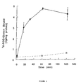
- Figure 1 is a graphic depiction of the time course of radioactively labeled underivatized, aqueous soluble ⁇ (1-3)-glucan binding to human leukocyte membranes. Total binding is indicated by a continuous line with closed circles and non-specific binding is indicated by a dashed line with open squares. The data points represent the mean ⁇ standard deviation of triplicate samples.
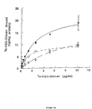
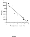
- Figures 2A and 2B show a graphic illustration of concentration-dependence of radioactively labeled underivatized, aqueous soluble ⁇ (1-3)-glucan binding to human leukocyte membranes.
- Figure 2A shows the total binding (continuous line, closed circles), non-specific binding (dashed line, open squares) and specific binding (dashed line, open diamonds) of varying concentrations of radioactively labeled underivatized, aqueous soluble ⁇ (1-3)-glucan. Data points represent the mean ⁇ standard deviation of triplicate samples.
- Figure 2B depicts a Scatchard analysis of the binding data.
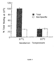
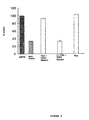
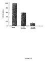
- Figure 3 is a graph of the effect of incubation temperature on the binding of radioactively labeled ⁇ (1-3)-glucan to human leukocyte membranes. Total binding is indicated by the shaded block, and non-specific binding is indicated by the open block. Data are expressed as a percent of the total binding at 37°C, and data points represent the mean ⁇ standard deviation of triplicate samples.
- Figure 4 is an autoradiograph of the time course for NF- ⁇ B activation by underivatized, aqueous soluble ⁇ (1-3)-glucan in the murine macrophage cell line BMC2.3, compared with a control and lipopolysaccharide.
- the lane for NF- ⁇ B is indicated with an arrow.
- Figure 5 is an autoradiograph of the time course of NF-IL6 activation by underivatized, aqueous soluble ⁇ (1-3)-glucan in the murine macrophage cell line BMC2.3 compared with control.
- the lane for NF-IL6 is indicated with an arrow.
- Figure 6 is a graph of the effect of exoglucanase treatment of underivatized, aqueous soluble ⁇ (1-3)-glucan on competitive binding.
- Figure 7 is a graph of the effect of NaOH treatment on the ability of underivatized, aqueous soluble ⁇ (1-3)-glucan to compete for binding.
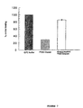
- Figure 8 is a graph of the effect on binding of treatment of human leukocyte membranes with glycine/pH 2.5 or 1 M NaCl. Solid columns indicate the saline control; stippled columns indicate underivatized, aqueous soluble ⁇ (1-3)-glucan sample.
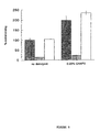
- Figure 9 is a graph of the effect on binding of pretreatment of human leukocyte membranes with CHAPS. Solid columns indicate the saline control; stippled columns indicate underivatized, aqueous soluble ⁇ (1-3)-glucan sample. Open columns indicate the dextran sample.
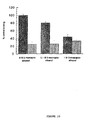
- Figure 10 is a graph of the effect of treatment of human leukocyte membranes with 2-mercaptoethanol on the binding of radioactively labelled underivatized, aqueous soluble ⁇ (1-3)-glucan to membranes. Solid columns indicate the saline control; stippled columns indicate underivatized, aqueous soluble ⁇ (1-3)-glucan sample.
- Figure 11 is a graph of the effect on specific binding of chloroform/methanol/HBSS extraction of human leukocyte membranes.
- Figure 12 is a graph of the effect of sodium periodate pretreatment of human leukocyte membranes on binding activity.
- Figure 13 is a graph of the effect of sodium periodate pretreatment of partially protein-depleted human leukocyte membranes on binding activity.
- Figure 14 is a graph of the comparison of underivatized, aqueous soluble ⁇ (1-3)-glucan and single helix competition of 3 H-underivatized, aqueous soluble ⁇ (1-3)-glucan binding in three assay formats.
- Underivatized, aqueous soluble ⁇ (1-3)-glucan or single helix glucan was used at designated concentrations to compete for 3 H-underivatized, aqueous soluble ⁇ (1-3)-glucan binding to lactosyl ceramide (LacCer) in a 96-well plate (triangles), or to lactosyl ceramide in reconstituted membranes (squares), or to human leukocyte membranes (circles).
- Open symbols represent underivatized, aqueous soluble ⁇ (1-3) glucan, and closed symbols represent single helix. Maximum competition was assigned at the highest concentration of underivatized, aqueous soluble ⁇ (1-3)-glucan used as competitor in each type of assay.
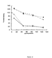
- Figure 15 is a graphic illustration of data showing that underivatized, aqueous soluble ⁇ (1-3) glucan and high molecular weight underivatized, aqueous soluble ⁇ (1-3) glucan (Network) compete more effectively for 3 H-underivatized, aqueous soluble ⁇ (1-3)-glucan binding to lactosyl ceramide in a 96-well plate assay than do single helix or laminarin.
- aqueous soluble ⁇ (1-3) glucan circles
- single helix closed squares
- laminarin crosses
- high molecular weight underivatized, aqueous soluble ⁇ (1-3)-glucan Network (hatched squares) were used at designated concentrations to compete for 3 H-underivatized, aqueous soluble ⁇ (1-3) glucan binding to lactosyl ceramide (bovine) in a 96-well plate assay.
- “% control binding” means the percent of binding with no competitor.
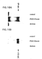
- Figures 16A and 16B are photographs showing that underivatized, aqueous soluble ⁇ (1-3)-glucan activates the nuclear transcription factors NF-kB and NF-IL6 in human neutrophils.
- Figure 16A shows the results for nuclear transcription factor NF-kB.
- Figure 16B shows the results for nuclear transcription factor NF-IL6.
- purified human neutrophils were incubated with or without underivatized, aqueous soluble ⁇ (1-3)-glucan (3 ⁇ g/ml) or dextran (3 ⁇ g/ml) for 60 minutes at 37°C.
- Nuclear extracts were prepared and protein/DNA complex formation assessed by electrophoretic mobility shift assay (EMSA).
- aqueous soluble ⁇ (1-3)-glucan increased the binding of nuclear extract proteins to an NF-kB or NF-IL6 specific 32P-labeled oligonucleotide probe relative to extracts from either control or dextran-treated neutrophils.
- the horizontal arrow indicates the location of the protein/DNA complex.
- This invention pertains to the discovery that underivatized, aqueous soluble ⁇ (1-3)-glucan (see WO-A-94/04163, the teachings of which are incorporated herein by reference) specifically binds to a novel receptor located on human leukocyte membranes (HLM). Underivatized, aqueous soluble ⁇ (1-3)-glucans are also described in U.S. Patent Nos. 5,322,841, 5,488,040 and 5,532,223.
- the receptor for underivatized, aqueous soluble ⁇ (1-3)-glucan is located primarily in human blood cells, specifically the neutrophil and mononuclear leukocytes (monocytes and lymphocytes).
- receptor is intended to encompass a traditional receptor molecule as well as a binding site; such a binding site can have an effect of its own or may induce or activate a second molecule or binding site to produce an effect (also known as "unmasking" of a second site; Sandberg et al ., Infect. Immun. 63 (7):2625-2631 (1995)).
- receptor is also intended to include compounds which have an affinity for underivatized, aqueous soluble ⁇ (1-3)-glucan; these compounds can be receptor mimics or receptor analogues, and can be isolated from a naturally-occurring source or can be chemically or synthetically produced.
- the receptor for underivatized, aqueous soluble ⁇ (1-3)-glucan exhibits selectivity for ⁇ (1-3)-glucans and particularly for underivatized, aqueous soluble ⁇ (1-3)-glucan in the triple helix conformation.
- a variety of polysaccharides were tested for their ability to compete for radioactively labeled underivatized, aqueous soluble ⁇ (1-3)-glucan binding.
- the receptor for underivatized, aqueous soluble ⁇ (1-3)-glucan distinguishes ⁇ (1-3)-glucans from non- ⁇ (1-3)-glucans such as dextran, mannan, glycogen and lipopolysaccharide (LPS) (Table 1).
- the underivatized, aqueous soluble ⁇ (1-3)-glucan sample represents non-specific binding.
- Aminated glucan was prepared by reductive amination of curdlan and provided by Dr. Rolph Seljelid (University of Troms ⁇ , Troms ⁇ , Norway).
- aqueous soluble ⁇ (1-3)-glucan was treated with exoglucanase, then used to compete with radioactively labeled underivatized, aqueous soluble ⁇ (1-3)-glucan binding.
- the anti-idiotype antibody was a rabbit monoclonal anti-idiotype antibody developed against a mouse monoclonal anti-laminarin antibody (Czop et al ., J. Immunol. 145 (3):995-1001 (1990)).
- Binding assays were carried out in the presence of 2-mercaptoethanol at the concentrations indicated in Figure 10 (0, 0.1 and 1 M). Similar results were obtained when the same concentrations of dithiothreitol (DTT) were used.
- DTT dithiothreitol
- Human leukocyte membranes were extracted with chloroform/methanol/HBSS (3:2:1) as described in the Examples.
- the protein-reduced fraction was used in a binding assay (non-oil/sucrose method) with the competitor indicated in Figure 11, underivatized, aqueous soluble ⁇ (1-3)-glucan or dextran, at 1 mg/ml final concentration.
- Human leukocyte membranes 500 ⁇ g protein were incubated with sodium periodate at concentrations indicated in Figure 12 (10 and 100 mM) for 30 minutes at room temperature, then centrifuged at 12,000 x g for 10 minutes, rinsed and used in a binding assay (non-oil/sucrose; see Figure 12). Protein-depleted membranes (derived from 1.87 mg human leukocyte membrane protein) were incubated in 0.1 M sodium periodate and treated as described above ( Figure 13) .
- the results show that the receptor is not affected by salt or low pH pretreatment of membranes and is enhanced by detergent pretreatment of membranes.
- the binding activity is decreased by sodium periodate treatment and by high concentrations of reducing agents.
- the binding site appears to be extractable with chloroform/methanol/water.
- the effect of sodium periodate on binding indicates the membrane target for underivatized, aqueous soluble ⁇ (1-3)-glucan may be a glycoconjugate, consistent with the observation that the binding moiety survives chloroform/methanol treatment.
- the carbohydrate portion of the glycoconjugate may be associated with a protein, a lipid, or both, as glycoconjugate receptors which are glycolipids or glycoproteins are known in the art (Sandberg et al ., Infect. Immun. 63 (7):2625-2631 (1995)).
- the distribution of the receptor for underivatized, aqueous soluble ⁇ (1-3)-glucan across various cell.lines and human tissues was determined by assessing the binding of radioactively labeled underivatized, aqueous soluble ⁇ (1-3)-glucan to membranes derived from a variety of cell types (see Table 2).
- the amount of radioactively labeled underivatized, aqueous soluble ⁇ (1-3)-glucan binding is expressed as mean ⁇ standard deviation in ng/mg membrane protein.
- the non-specific binding is defined as radioactively labeled underivatized, aqueous soluble ⁇ (1-3)-glucan bound in the presence of greater than 100-fold excess unlabeled underivatized, aqueous soluble ⁇ (1-3)-glucan.
- the specific binding is determined by (Total Binding) - (Non-Specific Binding), with the number in parenthesis in Table 2 representing the percent total binding.
- Quantitation of complement receptor 3 (CR3) was carried out by flow cytometry using fluorescently-labeled anti-CD11b, and data are expressed as mean channel fluorescence (MCF) in arbitrary fluorescence units; background fluorescence for isotype controls were subtracted.
- MCF mean channel fluorescence
- the neutrophil sample was greater than 95 percent neutrophils by histology.
- the mononuclear cell sample was approximately 40-50 percent monocytes by histology.
- the dominant cell type expressing receptors in human peripheral blood is the neutrophil, with mononuclear leukocytes (monocytes and lymphocytes) expressing less than 20% of the measured expression of neutrophils.
- the human monocytic cell line U937 does not bind detectable amounts of radioactively labeled underivatized, aqueous soluble ⁇ (1-3)-glucan, while the murine macrophage cell lines BMC2.3 (Dr. Kenneth Rock, Dana-Farber Cancer Institute, Boston, MA), RAW264.7 (ATCC) and P388D(1) (ATCC) express variable amounts of binding activity. When these three cell lines were assayed for CR3 expression by flow cytometry, the P388D(1) cells were found to be devoid of CR3.
- the binding activity of the radioactively labeled underivatized, aqueous soluble ⁇ (1-3)-glucan is sensitive to a ⁇ (1-3)-specific exoglucanase, indicating that the binding activity of the ligand is a ⁇ (1-3)-glucan.
- binding is not inhibited by either the anti-idiotype antibody of Czop (1990) or the anti-complement receptor 3 antibody (OKM1; Diamond et al ., J. Cell Biol. 120 :1031-1043 (1993)).
- This data along with the lack of inhibition by aminated glucan, indicates that the receptor for underivatized, aqueous soluble ⁇ (1-3)-glucan is a novel leukocyte carbohydrate receptor.
- Time course and concentration-dependence of radioactively labeled underivatized, aqueous soluble ⁇ (1-3)-glucan binding to human leukocyte membranes was determined by incubating 1 ⁇ g/ml of radioactively labeled underivatized, aqueous soluble ⁇ (1-3)-glucan with 2.5 mg/ml human leukocyte membranes in the presence or absence of unlabeled underivatized, aqueous soluble ⁇ (1-3)-glucan (1000 ⁇ g/ml) at 37°C for various times.
- Concentration-dependence of radioactively labeled underivatized, aqueous soluble ⁇ (1-3)-glucan binding to human leukocyte membranes was determined by incubating 2.2 mg/ml human leukocyte membranes with increasing concentrations of radioactively labeled underivatized, aqueous soluble ⁇ (1-3)-glucan in the presence (non-specific binding) or absence (total binding) of 50-fold excess unlabeled underivatized, aqueous soluble ⁇ (1-3)-glucan for 60 minutes at 37°C.
- Membrane-associated radioactivity was determined following centrifugation of the reaction mixture through a sucrose/oil density gradient as described in the Examples. Specific binding was calculated by subtracting non-specific binding from total binding.
- BMC2.3 cells were incubated with 3 ⁇ g/ml underivatized, aqueous soluble ⁇ (1-3)-glucan for various periods of time at 37°C.
- Nuclear extracts were prepared and incubated with 32 P-labeled DNA oligonucleotides specific for NF- ⁇ B or NF-IL6.
- the protein- 32 P DNA complexes were separated from unbound 32 P DNA by gel electrophoresis (electrophoretic mobility shift assay, EMSA).
- aqueous soluble ⁇ (1-3)-glucan 3 ⁇ g/ml
- dextran 3 ⁇ g/ml
- Nuclear extracts were prepared and protein/DNA complex formation assessed by electrophoretic mobility shift assay (EMSA).
- ESA electrophoretic mobility shift assay
- the particulate ⁇ -glucan receptor described by Czop (1990) is found on human monocytes and U937 cells. Underivatized, aqueous soluble ⁇ (1-3)-glucan binding is nominal on monocytes relative to peripheral polymorphonuclearcytes (PMN), and no binding was found on U937 cells. Furthermore, the anti-idiotype antibody generated by Czop (1990), which presumably occupies the glucan binding site, effectively inhibits glucan particle phagocytosis in monocytes but is ineffective in inhibiting underivatized, aqueous soluble ⁇ (1-3)-glucan binding.
- the receptor for aminated glucan present on murine peritoneal macrophages (Konopski et al ., (1994)) binds to their described aminated glucan at 4°C, whereas underivatized, aqueous soluble ⁇ (1-3)-glucan does not bind to its receptor at room temperature or below.
- samples of soluble aminated glucan did not compete for binding to the receptor for underivatized, aqueous soluble ⁇ (1-3)-glucan (Table 1).
- the ⁇ -glucan binding activity of CR3 as described by Thornton et al . (1996) is inhibited by monoclonal antibodies directed to the I-domain of CR3.
- One of these antibodies was OKM1, which inhibited glucan binding in the previously described system, but had no inhibitory effect on underivatized, aqueous soluble ⁇ (1-3)-glucan binding.
- Human peripheral blood PMN had five to ten-fold more specific underivatized, aqueous soluble ⁇ (1-3)-glucan binding relative to mononuclear cells, but both PMN and monocytes are known to express high levels of CR3.
- Particulate ⁇ -glucan receptors described by Goldman et al . can be inhibited by soluble glucans in the size range of 2,000-4,000 daltons, while underivatized, aqueous soluble ⁇ (1-3)-glucan binding is not inhibited by laminarin (molecular weight of approximately 5,000 daltons), or a single chain conformer of ⁇ -glucan of molecular weight 18,000 daltons.
- laminarin molecular weight of approximately 5,000 daltons
- the receptor of Goldman et al The receptor of Goldman et al .
- glycosphingolipids immobilized on a polystyrene 96-well plate exhibited significant specific binding to 3 H-underivatized, aqueous soluble ⁇ (1-3)-glucan, including lactosyl ceramide (LacCer) (from various sources), galactosyl ceramides (GalCer), globotriaosyl ceramide, and asialoganglioside-GM1 (see Table 3).
- lactosyl ceramide LacCer
- GalCer galactosyl ceramides
- globotriaosyl ceramide globotriaosyl ceramide
- asialoganglioside-GM1 see Table 3
- These compounds have in common a terminal galactose, which may therefore be involved in binding interactions between 3 H-underivatized, aqueous soluble ⁇ (1-3)-glucan and the glycosphingolipid.
- a terminal galactose alone is not
- LacCer While several fatty acid structures in LacCer can support binding of 3 H-underivatized, aqueous soluble glucan, as shown with HPLC fractionated LacCer from HLM in Table 4, the carbon-carbon double bond found in C18:1 sphingosine is apparently important for binding, since the commercially available dihydro-sphingosine LacCers do not bind.
- a glucan preparation comprising glucan that is primarily in the triple helix conformation and has a molecular weight of greater than one million, competes similarly to underivatized, aqueous soluble ⁇ (1-3)-glucan, while laminarin does not compete efficiently for 3 H-underivatized, aqueous soluble ⁇ (1-3)-glucan binding to LacCer in the 96-well plate assay (see Figure 15).
- an in vivo receptor for underivatized, aqueous soluble ⁇ (1-3)-glucan is LacCer.
- other compounds tested as described above also have an affinity for underivatized, aqueous soluble ⁇ (1-3)-glucan; these compounds are intended to be encompassed by the term "receptor" as described herein.
- these compounds can be used in place of the LacCer receptor in the assays described herein.
- terminal galactose is intended to encompass unmodified galactose as well as derivatized or modified galactose, such as sulphonated galactose. Accordingly, compounds having these characteristics are included in the present invention and are suitable for use in the methods described herein, along with other compounds having an affinity for underivatized, aqueous soluble ⁇ (1-3)-glucan.
- test samples can be included in a standard competition receptor binding assay to allow characterization of the molecule in terms of relative affinity for the receptor. This characterization will yield information with respect to some or all of the following characteristics: batch to batch quality, identification of product as a ⁇ (1,3)-glucan of a particular conformation and purity of the product.
- the receptor for underivatized, aqueous soluble ⁇ (1-3)-glucan to measure ⁇ -glucans in fluids.
- Test samples can be included in standard capture or competition assays and compared to a standard curve generated with a known ⁇ -glucan standard.
- This method can be used to measure soluble ⁇ -glucans in serum, plasma, urine, synovial fluid, cerebrospinal fluid, lung lavage, bile and other bodily fluids, as a diagnostic for fungemia. It can also be used to measure ⁇ -glucan levels in food manufacturing processes to test for fungal contamination, as well as to monitor yeast fermentation.
- Test samples can also be included in standard capture or competition assays and compared with a standard sample to elucidate structure-activity relationships. Alternatively, the test sample binding can be tested directly after radiolabeling. Samples can also be tested in an underivatized, aqueous soluble ⁇ (1-3)-glucan receptor-mediated assay to test for inhibition or stimulation of these functions. For example, such assays can be used in the development of polymer or small molecule receptor agonists or to develop a receptor antagonist to inhibit an inappropriate immunoenhancement, such as to prevent a transplant rejection which might occur as a result of an opportunistic fungal infection during immunosuppressive therapy.
- various agents can be conjugated (e.g., chemically conjugated, cross-linked or covalently bonded) to underivatized, aqueous soluble ⁇ (1-3)-glucan to produce a conjugate molecule which can be targeted to receptor-positive cells such as PMN and macrophages.
- Such a targeted delivery system can be used to enhance the delivery of agents such as antimicrobials for resistant intracellular pathogens (e.g., mycobacterium, leishmania, malaria), cytotoxins for receptor-positive leukemias, genes for gene therapy (e.g., for enhanced cytokine production or replacement for dysfunctional enzymes), or antigens for enhanced presentation and production of specific antibodies or T-cell activation.
- agents such as antimicrobials for resistant intracellular pathogens (e.g., mycobacterium, leishmania, malaria), cytotoxins for receptor-positive leukemias, genes for gene therapy (e.g., for enhanced cytokine production or replacement for dysfunctional enzymes), or antigens for enhanced presentation and production of specific antibodies or T-cell activation.
- the binding specificity of the underivatized, aqueous soluble ⁇ (1-3)-glucan to its receptor provides a method for purification of both receptor-positive and receptor-negative cells (i.e., cells which do not contain the receptor); for example, underivatized, aqueous soluble ⁇ (1-3)-glucan can be affixed to a solid matrix and used as an affinity matrix to positively select receptor-positive cells or to negatively select receptor-negative cells.
- anti-receptor antibodies can be used in place of the immobilized underivatized, aqueous soluble ⁇ (1-3)-glucan. Cells which are purified by this method can subsequently be expanded for use in cell therapy.
- the present invention makes possible the generation of anti-receptor antibodies for diagnostic purposes.
- Monoclonal or polyclonal antibodies can be produced using enriched or purified receptor preparations by standard techniques.
- the present invention also relates to antibodies which bind the receptor for underivatized, aqueous soluble ⁇ (1-3)-glucan.
- polyclonal and monoclonal antibodies which bind to the described receptor are within the scope of the invention.
- a mammal such as a mouse, hamster or rabbit, can be immunized with an immunogenic form of the receptor (i.e., an antigenic portion of the receptor which is capable of eliciting an antibody response).
- Techniques for conferring immunogenicity include, for example, conjugation to carriers or other techniques well known in the art.
- the antigen can be administered in the presence of an adjuvant, and the progress of immunization can be monitored by detection of antibody titers in plasma or serum. Standard ELISA or other immunoassays can be used with the immunogen as antigen to assess the levels of antibody.
- Antibodies to the glycolipid receptor described herein can be produced by standard techniques such as, for example, those described in Koscielak et al ( Immunochemistry 5 :441 (1968)), Hakomori (Methods of Enzymol. 28 :232-236 (1972)) and Marcus and Janis ( J. Immonol. 104 :1530 (1970). Such antibodies can be used to identify alterations in receptor-positive or receptor-negative cell populations which reflect disease pathology (e.g. response to cryptic fungal infection or leukemia).
- the activity of the receptor for underivatized, aqueous soluble ⁇ (1-3)-glucan is activated through binding of an underivatized, aqueous soluble ⁇ (1-3)-glucan, whereby a signal transduction pathway which is regulated by a transcriptional regulatory factor (e.g. from the NF- ⁇ B, NF-IL6 or jun/fos families) is activated.
- a transcriptional regulatory factor e.g. from the NF- ⁇ B, NF-IL6 or jun/fos families
- Activation of the receptor can comprise, among others, an alteration in the receptor conformation, formation of a ligand-receptor complex, or alteration of the ligand receptor complex.
- the activity of the receptor can be activated by an agent which mimics the binding and activation ability of an underivatized, aqueous soluble ⁇ (1-3)-glucan.
- the transcriptional regulatory factor is activated as a result of ligand binding.
- the activity of the transcriptional regulatory factor is decreased, either partially or totally, by the binding of an agent which binds the receptor (and thus excludes the underivatized, aqueous soluble ⁇ (1-3)-glucan), but lacks the ability to activate the receptor.
- the present invention also pertains to a novel assay for identifying agents which alter the effect of underivatized, aqueous soluble ⁇ (1-3)-glucan on signal transduction pathways such as activation of transcriptional regulatory factors.
- This assay comprises combining underivatized, aqueous soluble ⁇ (1-3)-glucan, the receptor for underivatized, aqueous soluble ⁇ (1-3)-glucan and an agent to be tested under conditions in which binding of underivatized, aqueous ⁇ (1-3)-glucan to its receptor occurs (i.e. conditions suitable for binding of underivatized, aqueous soluble ⁇ (1-3)-glucan to the receptor for underivatized, aqueous soluble ⁇ (1-3)-glucan.
- Binding of underivatized, aqueous soluble ⁇ (1-3)-glucan to its receptor activates the receptor, which in turn activates a signal transduction pathway as shown by transcriptional regulator factors such as those of the NF- ⁇ B, NF-IL6 or jun/fos families.
- Other signal transduction pathways which can be altered by the methods of the present invention include the ras/raf-1/MAP kinase pathway, the G-protein/phospholipase C/protein kinase C pathway, the JAK/STAT pathway, the phospholipase A pathway, G-protein/phospholipase D/phosphatidic acid pathway and the c-AMP-dependent pathway.
- an appropriate activator or indicator of the signal pathway is activated by binding of underivatized, aqueous ⁇ (1-3)-glucan to its receptor, and modulation of this binding can alter the corresponding signal transduction.
- the extent of activation of the selected transcriptional regulatory factor in the presence of an agent to be tested is determined (e.g., using radiolabeled DNA oligonucleotides specific for the transcriptional regulatory factor, as in the Examples) and compared with the extent of activation of the selected transcriptional regulatory factor in the absence of the agent to be tested; a difference in the extent of activation indicates that the agent alters the effect of underivatized, aqueous soluble ⁇ (1-3)-glucan on activation of the transcriptional regulatory factor.
- An increase in the activation of the transcriptional regulatory factor in the presence of the agent indicates that the agent enhances, i.e., prolongs or increases, the activation.
- a decrease in the activation of the transcriptional regulatory factor in the presence of the agent indicates that the agent diminishes, i.e., shortens or decreases, the activation.
- the invention also pertains to an assay or method for assessing the specificity of an interaction between a carbohydrate and a glycolipid.
- a selected carbohydrate and a selected glycolipid are combined under conditions suitable for interaction between the carbohydrate and glycolipid, and the extent of interaction between the carbohydrate and glycolipid is determined. This extent of interaction is compared with the extent of interaction between the selected glycolipid and selected carboydrate in the presence of another glycolipid or carbohydrate.
- the two are combined under appropriate conditions, and the extent of interaction is determined. Typically, the interaction will be binding.
- the extent of interaction is then compared with the extent of interaction in the presence of a selected competitor carbohydrate. In this way, the specificity of the interaction between the glycolipid and the selected carbohydrate can be compared with the interaction between the glycolipid and a competitor carbohydrate, and the relative specificities can be determined.
- the two are combined under appropriate conditions, and the extent of interaction is determined.
- the extent of interaction is then compared with the extent of interaction in the presence of a selected competitor glycolipid.
- the specificity of the interaction between the carbohydrate and the selected glycolipid can be compared with the interaction between the carbohydrate and a competitor glycolipid, and the relative specificities can be determined.
- the glycolipid is LacCer.
- the carbohydrate is underivatized, aqueous soluble ⁇ (1-3)-glucan.
- Lactosyl ceramide was purified from human leukocyte membranes as described below. All other sphingolipids, ceramides, and phospholipids were from Sigma Chemical Co. (St. Louis, MO), except lactosyl ceramide (porcine) which was from Matreya, Inc. (Pleasant Gap, PA). SOLVABLETM was from DuPont, NEN (Boston, MA). 96-well polystyrene plates were from Corning (New York).
- aqueous soluble ⁇ (1-3)-glucan (PGG-glucan, 17 mg; HPD0144, Alpha-Beta Technology, Worcester, MA) was incubated with NaIO 4 (225 mg; Sigma, St. Louis, MO) in sterile pyrogen-free (SPF) water for 72 hours at room temperature. The periodate was quenched by the addition of 50 mg glycerol. The underivatized, aqueous soluble ⁇ (1-3)-glucan was dialyzed against SPF water, and then reductively labeled with 100 mCi of NaB 3 H 4 (New England Nuclear, Boston, MA).
- Radioactively labeled underivatized, aqueous soluble ⁇ (1-3)-glucan was separated from tritiated low molecular weight by-products by dialysis (10 K cut-off) and ultrafiltration. Purity of the labeled product was assessed by gel permeation chromatography.
- Fresh whole blood was obtained from normal human volunteers using acid citrate dextrose as an anticoagulant. Following dextran sedimentation, cells were centrifuged (400 x g, 7 minutes) and resuspended in autologous plasma. Cells were then layered over a Ficoll gradient (Lymphocyte Separation Medium; Organon Teknika, Durham, NC) and centrifuged (400 x g, 30 minutes). Cells recovered from the pellet were hypotonically lysed to remove residual RBC. The remaining cells were greater than 95% neutrophils as judged by morphological criteria.
- Buffy coat cells from human donors (Red Cross, Dedham, MA) were incubated in 3% dextran for 15-20 minutes at room temperature to separate leukocytes from red cells.
- the leukocyte-rich supernatant was pelleted (500xg, 7 minutes) and washed once in ice cold phosphate buffered saline (PBS; Life Technologies, Grand Island, NY). All subsequent operations were performed at 4°C. The remaining red cells were removed by hypotonic lysis, and the leukocytes recovered by centrifugation (850xg, 7 minutes).
- the cell pellet was resuspended in approximately 3-4x volume PBS, and protease inhibitors were added (5 mM EDTA, 40 ⁇ g/ml aprotinin, 1 ⁇ M pepstatin A, 1 ⁇ g/ml leupeptin, 50 ⁇ M PMSF).
- the cells were disrupted by probe sonication (50 watts, 30 x 1 second pulses). Disruption of cells was monitored microscopically. Nuclei and remaining intact cells were removed by low speed centrifugation (700xg, 7 minutes). Occasionally, the low speed pellet was resonicated and subjected to another low speed centrifugation.
- Membrane pellets were resuspended in Hanks' balanced salt solution containing Ca ++ and Mg ++ (HBSS).
- Membrane protein was determined using the BCA or Comassie method (Pierce, Rockland, IL). Bovine serum albumin (Sigma, St. Louis, MO) was added to 1 mg/ml from a 10x stock, and the membranes stored in liquid nitrogen at 4-5 mg/ml. In some cases membranes were stored frozen without added protein; however, no change in binding was observed as a result of the different storage conditions.
- fresh human leukocytes were prepared from whole blood collected in acid citrate dextrose. Red blood cells were removed by dextran sedimentation and the leukocyte-rich layer was harvested and treated as described above. Finally, purified neutrophil and mononuclear leukocytes (including monocytes and lymphocytes) were prepared by resuspending the leukocyte-rich layer of cells in autologous plasma and layering the cells over Lymphocyte Separation Medium (LSM; Organontechnika) followed by centrifugation (700xg, 30 minutes). The neutrophil enriched pellet and the mononuclear cells present at the density interface were washed in ice cold PBS and membranes were prepared as described above. A stained Cytospin (Shandon) preparation indicated that the neutrophil preparation was greater than 95% pure, and that the mononuclear preparation contained approximately 40%-50% monocytes.
- LSM Lymphocyte Separation Medium
- Non-adherent cell lines were collected by centrifugation (500xg, 7 minutes), washed in ice cold PBS, and the membranes were prepared as described above.
- Lightly adherent cell lines were washed with ice cold PBS and removed by gentle scraping with a cell scraper (Costar). Greater than 90% of the cells maintained viability during this procedure as determined by dye exclusion.
- Cells were resuspended in PBS and protease inhibitors. Tightly adherent cells were washed with ice cold PBS, then incubated with PBS plus the protease inhibitors described above. Cells were then removed by scraping and collected by centrifugation (500xg, 7 minutes).
- Membranes from adherent cells were then prepared as described above.
- Membranes were diluted to 2 to 5 mg/ml in HBSS with and without 1 mg/ml BSA.
- the reaction mixture consisted of 280 ⁇ l membranes (2-4 mg/ml final concentration), 35 ⁇ l saline or test sample (in various concentrations), and 35 ⁇ l radioactively labeled underivatized, aqueous soluble ⁇ (1-3)-glucan (1 ⁇ g/ml final concentration). Binding was allowed to proceed for 60 to 120 minutes at 37°C.
- Human leukocyte membranes (5 mg protein/ml) prepared as described above were vortexed with 5 volumes of 3:2 chloroform/methanol (by volume) and centrifuged at 1,500 x g to separate phases. The upper aqueous and lower organic phases, were removed from the proteinaceous interphase, combined, and concentrated under a stream of Argon to approximately 50 ⁇ l. The residue was resuspended in HBSS, sonicated briefly, and centrifuged (12,000 x g, 10 minutes) to pellet membranes. The pellet was resuspended in 500 ⁇ l HBSS and 100 ⁇ l was used per 350 ⁇ l binding assay. Assays for protein using the BCA reagent (Pierce) indicated that approximately 85% of the protein was removed from the resuspended fraction.
- BCA reagent Pieris
- BMC2.3 cells in DMEM plus 10% fetal calf serum were incubated with 3 ⁇ g/ml underivatized, aqueous soluble ⁇ (1-3)-glucan for various lengths of time.
- Nuclear extracts were prepared by the method of Dignam et al . ( Nuc. Acid Res. 11 :1475-1489 (1983)). Nuclear extracts were then incubated with 32 P-labeled DNA oligonucleotide for twenty minutes at room temperature. The sequence of the oligonucleotide used as the probe was AGTTGAGGGGACTTTCCCAGGC (SEQ ID NO: 1). The reaction mixture was then separated on a 4% polyacrylamide gel and the results recorded autoradiographically.
- Human neutrophils (4.5 x 10 6 cells/ml; 10 ml total) purified as described above were incubated in RPMI + 10% FCS (Sigma Chemical Co.) for 30 minutes at 37°C. Underivatized, aqueous soluble ⁇ (1-3)-glucan or dextran (both at 3 ⁇ g/ml) were then added and incubation continued an additional 60 minutes at 37°C. None was added to control cells. At the end of the incubation, cells were washed in ice cold PBS + 20 mM EDTA and nuclear extracts prepared and EMSAs performed as described above.
- Lactosyl Ceramide (LacCer) from Human Leukocyte Membranes (HLM)
- HLM 15 mg protein
- HBSS HBSS
- chloroform 3 mL
- methanol 2 mL
- the upper layer and proteinaceous interphase were discarded.
- the lower layer was dried under a stream of argon, and resuspended in 200 ⁇ L chloroform/methanol (10:1).
- the acetone/methanol fraction from silica chromatography was applied to a DEAE-Sephadex A-25 column (1 mL resin in the acetate form, prepared in chloroform/methanol (1:2)). The column was washed with 60 mL chloroform/methanol (1:2). Neutral glycosphingolipids were contained in this fraction, which was dried and resuspended in chloroform/methanol (5:1).
- Membranes were prepared from the receptor-negative cell line, U937, and extracted with chloroform/methanol as described above. The upper and lower layers were combined after removal of the proteinaceous interphase and dried under a stream of argon with the sample to be assayed for binding (lactosyl ceramide or fractions from the purification).
- phospholipid mixture contained phosphatidylcholine (132 ⁇ g), phosphatidylethanolamine (112 ⁇ g), phosphatidylinositol (36 ⁇ g), and phosphatidylserine (56 ⁇ g).
- Assays were incubated at 37°C for one hour, after which time three 100 ⁇ L aliquots were removed and centrifuged in microfuge tubes at 12,000 x g for 5 minutes at room temperature. The supernatants were discarded and the pellets were rinsed with 150 ⁇ L of HBSS. Pellets were then dissolved in SOLVABLETM, scintillation fluid was added, and radioactivity was measured.
- Lactosyl ceramide or the sample to be measured was suspended in ethanol. Aliquots as specified were added in triplicate to the wells of a 96-well plate and air dried. The following components were premixed, then 100 ⁇ L was added to each well: saline (10 ⁇ L) or unlabelled, underivatized, aqueous soluble ⁇ (1-3)-glucan (10 ⁇ L of a 10 mg/ml stock), 3 H-underivatized, aqueous soluble ⁇ (1-3) glucan (10 ⁇ L of a 10 ⁇ g/ml stock), and HBSS (80 ⁇ L). Plates were incubated at 37°C for 1.5 to 2 hours, then supernatants were removed from each well and discarded. Wells were rinsed with HBSS (200 ⁇ L) and SOLVABLETM (100 ⁇ L) was added. The plate was incubated at 60°C for 5 minutes, then supernatants were removed and counted.
Landscapes
- Health & Medical Sciences (AREA)
- Life Sciences & Earth Sciences (AREA)
- Engineering & Computer Science (AREA)
- Immunology (AREA)
- Molecular Biology (AREA)
- Chemical & Material Sciences (AREA)
- Biomedical Technology (AREA)
- Hematology (AREA)
- Urology & Nephrology (AREA)
- Cell Biology (AREA)
- Microbiology (AREA)
- Medicinal Chemistry (AREA)
- General Health & Medical Sciences (AREA)
- General Physics & Mathematics (AREA)
- Analytical Chemistry (AREA)
- Biochemistry (AREA)
- Food Science & Technology (AREA)
- Biotechnology (AREA)
- Pathology (AREA)
- Physics & Mathematics (AREA)
- Tropical Medicine & Parasitology (AREA)
- Epidemiology (AREA)
- Mycology (AREA)
- Virology (AREA)
- Bioinformatics & Cheminformatics (AREA)
- Pharmacology & Pharmacy (AREA)
- Botany (AREA)
- Animal Behavior & Ethology (AREA)
- Public Health (AREA)
- Veterinary Medicine (AREA)
- Polysaccharides And Polysaccharide Derivatives (AREA)
- Peptides Or Proteins (AREA)
- Preparation Of Compounds By Using Micro-Organisms (AREA)
- Medicines That Contain Protein Lipid Enzymes And Other Medicines (AREA)
- Investigating Or Analysing Biological Materials (AREA)
- Medicinal Preparation (AREA)
- Measuring Or Testing Involving Enzymes Or Micro-Organisms (AREA)
- Pharmaceuticals Containing Other Organic And Inorganic Compounds (AREA)
Abstract
Description
| Effect of Various Incubation Conditions on 3H-PGG-Glucan Binding to Human Leukocyte Membranes | |||||
| Addition/Treatment | Concentration | Source | % Control Binding | %CV | |
| PGG-Glucan | 0.1 mg/ml | ABT | 22 | 5 | |
| | 1 mg/ | Sigma | 88 | 12 | |
| | 1 mg/ | Sigma | 88 | 5 | |
| Laminarin | 1.0 mg/ml | Calbiochem | 83 | 5 | |
| Glycogen | 0.1 mg/ml | Sigma | 134 | 20 | |
| Lipopolysaccharide | 0.1 mg/ml | Sigma | 89 | 16 | |
| Aminated Glucan | 0.1 mg/ml | R. Seljelid | 111 | 1 | |
| EDTA | 5mM | Sigma | 82 | 13 | |
| EGTA | 5mM | Sigma | 87 | 0.1 | |
| Exoglucanase | 0.26 U/ml | ABT | 92 | 5 | |
| | 10 µg/ | J. Czop | 88 | 7 | |
| anti-CR3 | 28 µg/ml | Becton-Dickinson | 123 | 19 |

| Specific binding of 3H-underivatized, aqueous soluble β(1-3)-glucan (PGG-Glucan) to various sphingolipids in the 96-well plate assay | |
| Sphingolipid | 3H-PGG-Glucan bound (ng/µg sphingolipid) |
| LacCer (HLM) | 3.14 ± 0.29 |
| LacCer (bovine) | 2.18 ± 0.39 |
| LacCer (porcine) | 2.35 ± 0.23 |
| GalCer I | 1.02 ± 0.32 |
| GalCer II | 3.90 ± 0.14 |
| Ceramides | 0.02 ± 0.01 |
| Sulfatides | 0.00 ± 0.01 |
| Sphingosine | 0.02 ± 0.00 |
| Psychosine | 0.09 ± 0.00 |
| Asialoganglioside-GM2 | 0.00 ± 0.01 |
| Globoside | 0.00 ± 0.01 |
| GlobotriaosylCer | 0.84 ± 0.13 |
| Glucocerebrosides (GlcCer) | 0.12 ± 0.03 |
| Asialoganglioside-GM1 | 0.50 ± 0.10 |
| Specific binding of 3H-underivatized, aqueous soluble β(1-3)-glucan (PGG-Glucan) to LacCer of defined structures in the 96-well plate assay | |||
| Fatty Acid | Long Chain Base | 3H-PGG-Glucan bound (ng/µg LacCer) | |
| LacCer (HLM) | C16:0 | C18:1 | 3.31 |
| C18:0 | C18:1 | 1.93 | |
| C24:2 | C18:1 | 2.01 | |
| C24:1 | C18:1 | 2.31 | |
| C22:0 | C18:1 | 3.57 | |
| C24:0 | C18:1 | 3.14 | |
| N-Palmitoyl-DL-dihydrolactocerebroside | C16:0 | C18:0 | 0.00 |
| N-Stearoyl-DL-dihydrolactocerebroside | C18:0 | C18:0 | 0.00 |
| N-Lignoceroyl-DL-dihydrolactocerebroside | C24:0 | C18:0 | 0.00 |
| Temperature dependence of 3H-underivatized, aqueous soluble β(1-3)-glucan (PGG-Glucan) specific binding to lactosyl ceramides | ||
| Temperature | 3H-PGG-Glucan bound (ng/µg LacCer) | |
| LacCer (HLM) | 37°C | 1.76 |
| 4°C | 0.00 | |
| LacCer (bovine) | 37°C | 2.50 |
| 4°C | 0.01 |
| Competition of 3H-underivatized, aqueous soluble β(1-3)-glucan (PGG-Glucan) binding to GalCer II | ||
| Ligand | Concentration (mg hexose/mL) | % control binding |
| none | - | 100 |
| PGG-glucan | 0.01 | 23.6 |
| 0.1 | 3.0 | |
| 1.0 | 1.4 | |
| SH | 0.01 | 46.1 |
| 0.1 | 10.7 |
Alternatively, membranes were centrifuged directly in their incubation tubes at 15,000xg for 4-5 minutes at 37°C, the pellet washed with HBSS, then pellets solubilized and radioactivity determined by liquid scintillation counting.
Claims (18)
- An assay for assessing the affinity of a carbohydrate for a glycolipid, comprising the steps of:thereby assessing the affinity of the carbohydrate for a glycolipid.a) combining a glycolipid and a carbohydrate under conditions suitable for interaction between the carbohydrate and the glycolipid;b) determining the affinity of the carbohydrate for the glycolipid; andc) comparing the affinity determined in (b) with the affinity in the presence of competitor carbohydrate or glycolipid,
- An assay for identifying an agent which alters the binding of a carbohydrate to a glycolipid, comprising the steps of:wherein a difference in the extent of binding of the carbohydrate to the glycolipid indicates that the agent alters the binding of the carbohydrate to the glycolipid.a) combining radiolabeled carbohydrate, glycolipid, and an agent to be tested, under conditions suitable for interaction of the carbohydrate and glycolipid;b) determining the extent of binding of the carbohydrate to the glycolipid; andc) comparing the extent of binding determined in step (b) with the extent of binding in the absence of the agent to be tested under conditions suitable for interaction of the carbohydrate and glycolipid,
- An assay according to Claim 2, wherein the agent is an antagonist or agonist of the receptor for underivatized, aqueous soluble P (1-3) - glucan.
- An assay according to any preceding claim, wherein the carbohydrate is underivatized, aqueous soluble β (1-3) - glucan.
- An assay according to any preceding claim, wherein the glycolipid is a receptor for underivatized, aqueous soluble P (1-3) - glucan.
- An assay according to Claim 5, wherein the glycolipid is lactosyl ceramide.
- An assay according to Claim 5 or 6, wherein the receptor is a compound having an affinity for underivatized, aqueous soluble β (1-3) - glucan or a receptor analogue or receptor mimic thereof.
- An assay for identifying an agent which has an affinity for the receptor for underivatized, aqueous soluble β (1-3) - glucan, comprising the steps of:wherein a decrease in the extent of binding of underivatized, aqueous soluble β (1-3) - glucan to the receptor for underivatized, aqueous soluble β (1-3) - glucan indicates that the agent has an affinity for the receptor for underivatized, aqueous soluble β (1-3) - glucan.a) combining radiolabeled underivatized, aqueous soluble P (1-3) - glucan, receptor for underivatized, aqueous soluble β (1-3) - glucan, and an agent to be tested, under conditions suitable for binding of underivatized, aqueous soluble β (1-3) - glucan to the receptor for underivatized, aqueous soluble β (1-3) - glucan;b) determining the extent of binding of underivatized, aqueous soluble β (1-3) - glucan to the receptor for underivatized, aqueous soluble β (1-3) - glucan; andc) comparing the extent of binding determined in step (b) with the extent of binding in the absence of the agent to be tested under conditions suitable for binding of underivatized, aqueous soluble β (1-3) - glucan to the receptor for underivatized, aqueous soluble β (1-3) - glucan,
- An assay for identifying an agent which alters the effect of underivatized, aqueous soluble β (1-3) - glucan on activation of a signal transduction pathway, said assay comprising the steps of:wherein a difference in the extent of activation of the signal transduction pathway indicates that the agent alters the effect of underivatized, aqueous soluble β (1-3)-glucan on activation of a signal transduction pathway.a) combining underivatized, aqueous soluble β (1-3) - glucan, receptor for underivatized, aqueous soluble β (1-3) - glucan, and an agent to be tested, under conditions suitable for binding of underivatized, aqueous soluble P (1-3) - glucan to the receptor for underivatized, aqueous soluble β (1-3)-glucan;b) determining the extent of activation of a signal transduction pathway; andc) comparing the extent of activation determined in step (b) with the extent of activation in the absence of the agent to be tested under conditions suitable for binding of underivatized, aqueous soluble β (1-3) - glucan to the receptor for underivatized, aqueous soluble β (1-3) - glucan,
- An assay according to Claim 9, wherein if the extent of activation is greater in the presence of the agent than in the absence of the agent, the agent enhances the effect of underivatized, aqueous soluble β (1-3) - glucan on activation of a signal transduction pathway; or
if the extent of activation is less in the presence of the agent than in the absence of the agent, the agent diminishes the effect of underivatized, aqueous soluble β (1-3) - glucan on activation of a signal transduction pathway. - An assay according to Claim 9 or 10, wherein the activation of the signal transduction pathway is measured by the modulation of at least one transcriptional regulatory factor.
- An assay according to Claim 11, wherein the transcriptional regulatory factor is selected from the group consisting of members of NF-κB, NF-IL6 or jun/fos families.
- An assay according to any one of Claims 9 to 12, wherein step (b) is carried out by 32P-labeled DNA oligonucleotides specific for the transcriptional regulatory factor.
- An assay according to any one of Claims 8 to 13, wherein the receptor is a compound having an affinity for underivatized, aqueous soluble β (1-3) - glucan; or
a receptor analogue or a receptor mimic thereof. - An assay according to any one of Claims 8 to 14, wherein the receptor is lactosyl ceramide.
- A method of altering the binding of a carbohydrate to a glycolipid. in vitro comprising combining a carbohydrate, a glycolipid, and an agent identified by an assay according to any one of Claims 2 to 7 under conditions suitable for binding of the carbohydrate to the glycolipid, whereby the agent alters binding of the carbohydrate to the glycolipid relative to binding in the absence of the agent.
- A conjugate molecule consisting of an underivatized, aqueous soluble β(1,3)-glucan, which is chemically conjugated, cross-linked, or covalently bonded to an agent, wherein the conjugate molecule binds to a receptor for underivatized, aqueous soluble β(1,3)-glucan in a cell containing the receptor.
- Use of an agent conjugated to an underivatized, aqueous soluble β(1-3)-glucan in the manufacture of a medicament for delivering an agent to a cell containing a receptor for underivatized, aqueous soluble β(1-3)-glucan.
Applications Claiming Priority (7)
| Application Number | Priority Date | Filing Date | Title |
|---|---|---|---|
| US63793496A | 1996-05-01 | 1996-05-01 | |
| US637934 | 1996-05-01 | ||
| US08/664,173 US6090938A (en) | 1996-05-01 | 1996-06-14 | Activation of signal transduction by underivatized, aqueous soluble . .beta(1-3)-Glucan |
| US664173 | 1996-06-14 | ||
| US797696 | 1997-01-31 | ||
| US08/797,696 US6110692A (en) | 1996-05-01 | 1997-01-31 | Receptor for underivatized aqueous soluble β(1-3)-glucan |
| PCT/US1997/007445 WO1997041439A1 (en) | 1996-05-01 | 1997-05-01 | RECEPTOR FOR UNDERIVATIZED, AQUEOUS SOLUBLE β(1-3)-GLUCAN |
Publications (2)
| Publication Number | Publication Date |
|---|---|
| EP0900379A1 EP0900379A1 (en) | 1999-03-10 |
| EP0900379B1 true EP0900379B1 (en) | 2004-04-21 |
Family
ID=27417615
Family Applications (1)
| Application Number | Title | Priority Date | Filing Date |
|---|---|---|---|
| EP97926398A Expired - Lifetime EP0900379B1 (en) | 1996-05-01 | 1997-05-01 | Receptor for underivatized, aqueous soluble beta(1-3)-glucan |
Country Status (10)
| Country | Link |
|---|---|
| US (2) | US6110692A (en) |
| EP (1) | EP0900379B1 (en) |
| JP (1) | JP3920349B2 (en) |
| KR (1) | KR100561660B1 (en) |
| AT (1) | ATE265046T1 (en) |
| AU (1) | AU718650B2 (en) |
| CA (1) | CA2253257C (en) |
| DE (1) | DE69728760T2 (en) |
| ES (1) | ES2223079T3 (en) |
| WO (1) | WO1997041439A1 (en) |
Families Citing this family (19)
| Publication number | Priority date | Publication date | Assignee | Title |
|---|---|---|---|---|
| US6084092A (en) * | 1997-01-31 | 2000-07-04 | The Collaborative Group, Ltd. | β(1-3)-glucan diagnostic assays |
| US6110692A (en) | 1996-05-01 | 2000-08-29 | The Collaborative Group, Ltd. | Receptor for underivatized aqueous soluble β(1-3)-glucan |
| US6413715B2 (en) * | 1997-01-31 | 2002-07-02 | The Collaborative Group | β(1-3)-glucan diagnostic assays |
| AU1401999A (en) * | 1997-12-12 | 1999-07-05 | Alpha-Beta Technology, Inc. | Activation of transcription factor complex by beta(1-3) glucan |
| JP2002523522A (en) * | 1998-08-26 | 2002-07-30 | ザ コラボレイティブ グループ,リミテッド. | Non-derivatized water-soluble beta- (1,3) -glucan receptor |
| NO20014256D0 (en) * | 2001-09-03 | 2001-09-03 | Bjoern Kristiansen | Preparation of immunostimulatory compound |
| US7514085B2 (en) | 2004-07-16 | 2009-04-07 | Medimush A/S | Immune modulating compounds from fungi |
| MX2007013725A (en) | 2005-05-05 | 2008-04-09 | Sensient Flavors Inc | Production of beta-glucans and mannans. |
| US9072776B2 (en) | 2005-06-15 | 2015-07-07 | Glycanova As | Anti-cancer combination treatment and kit-of-parts |
| US20090005340A1 (en) * | 2005-06-15 | 2009-01-01 | Medimush A/S | Bioactive Agents Produced By Submerged Cultivation of a Basidiomycete Cell |
| US9610303B2 (en) * | 2006-06-15 | 2017-04-04 | Biothera, Inc. | Glucan preparations |
| WO2008027581A1 (en) * | 2006-09-01 | 2008-03-06 | University Of Louisville | Mcrp antagonists and their uses |
| WO2008027580A2 (en) * | 2006-09-01 | 2008-03-06 | University Of Louisville | PARTICULATE β-GLUCAN COMPOSITIONS FOR REGULATING DENDRITIC CELLS |
| EP2446018B1 (en) * | 2009-06-26 | 2017-01-18 | Charles River Laboratories, Inc. | Heat-treated limulus amebocyte lysates |
| US20110293784A1 (en) | 2010-05-28 | 2011-12-01 | Anja Wittke | Milk-based nutritional compositions |
| WO2013006795A2 (en) | 2011-07-07 | 2013-01-10 | Humanitas International Foundation | Antiviral compositions and methods of their use |
| JP6893594B2 (en) | 2014-07-10 | 2021-06-23 | ハイバーセル,インク. | Β-Glucan in combination with antineoplastic agents that affect the tumor microenvironment |
| JP6887378B2 (en) | 2014-11-06 | 2021-06-16 | バイオセラ,インク. | Beta-glucan methods and compositions that affect the intratumoral microenvironment |
| WO2018156888A1 (en) | 2017-02-24 | 2018-08-30 | Biothera Pharmaceuticals, Inc. | Beta glucan immunopharmacodynamics |
Citations (4)
| Publication number | Priority date | Publication date | Assignee | Title |
|---|---|---|---|---|
| EP0133170A2 (en) * | 1983-07-15 | 1985-02-13 | Ab Symbicom | A compound and a composition for therapeutic or diagnostic use and a method for therapeutic treatment |
| JPH02250834A (en) * | 1989-03-24 | 1990-10-08 | Snow Brand Milk Prod Co Ltd | Therapeutic agent for demyelinating diseases |
| US5242800A (en) * | 1990-01-30 | 1993-09-07 | The United States Of America As Represented By The Secretary Of The Department Of Health And Human Services | Receptor for pathogenic fungi |
| JPH0853488A (en) * | 1994-08-12 | 1996-02-27 | Daikin Ind Ltd | 6'-fluorolactosylceramide, intermediate thereof and method for producing the same |
Family Cites Families (16)
| Publication number | Priority date | Publication date | Assignee | Title |
|---|---|---|---|---|
| US5089479A (en) * | 1988-11-28 | 1992-02-18 | Krivan Howard C | Adhesion of mycoplasma pneumoniae and mycoplasma hominus to sulfatide |
| US5225330A (en) * | 1988-08-01 | 1993-07-06 | The United States Of America As Represented By The Department Of Health And Human Services | Diagnostic kit and diagnostic method utilizing carbohydrate receptors |
| US5217715A (en) * | 1988-08-01 | 1993-06-08 | The United States Of America As Represented By The Department Of Health And Human Services | Carbohydrate receptor for bacteria and method for use thereof |
| US5032401A (en) * | 1989-06-15 | 1991-07-16 | Alpha Beta Technology | Glucan drug delivery system and adjuvant |
| US5622939A (en) * | 1992-08-21 | 1997-04-22 | Alpha-Beta Technology, Inc. | Glucan preparation |
| US5488040A (en) | 1989-09-08 | 1996-01-30 | Alpha-Beta Technology, Inc. | Use of neutral soluble glucan preparations to stimulate platelet production |
| EP0490995A1 (en) * | 1989-09-08 | 1992-06-24 | Alpha Beta Technology | Method for producing soluble glucans |
| JP2944709B2 (en) | 1990-06-21 | 1999-09-06 | 生化学工業株式会社 | (1 → 3) -β-D-glucan measuring agent |
| US5156948A (en) | 1990-07-20 | 1992-10-20 | Christensen Dale A | Method and kit for diagnosis of diseases |
| DE69221879T2 (en) | 1991-03-14 | 1998-01-29 | Seikagaku Kogyo K.K., Tokio/Tokyo | REAGENT FOR DETERMINING (1-3) -BETA-GLUCAN |
| JPH04346791A (en) | 1991-05-21 | 1992-12-02 | Taito Kk | Monoclonal antibody |
| EP0549102B1 (en) | 1991-12-25 | 1996-07-03 | Maruha Corporation | Beta-glucans detection reagents and methods of detecting beta-glucans |
| JP3524120B2 (en) | 1992-05-08 | 2004-05-10 | 生化学工業株式会社 | Pretreatment agent, pretreatment method, measurement method using pretreated sample, measurement kit, and sample determination method |
| JP3322700B2 (en) | 1992-09-14 | 2002-09-09 | 生化学工業株式会社 | Endotoxin-specific measuring agent for solid-phase reaction |
| JP3793573B2 (en) | 1994-09-01 | 2006-07-05 | 生化学工業株式会社 | (1 → 3) -β-D-glucan binding protein, antibody recognizing it and use thereof |
| US6110692A (en) | 1996-05-01 | 2000-08-29 | The Collaborative Group, Ltd. | Receptor for underivatized aqueous soluble β(1-3)-glucan |
-
1997
- 1997-01-31 US US08/797,696 patent/US6110692A/en not_active Expired - Lifetime
- 1997-05-01 AT AT97926398T patent/ATE265046T1/en active
- 1997-05-01 EP EP97926398A patent/EP0900379B1/en not_active Expired - Lifetime
- 1997-05-01 AU AU31171/97A patent/AU718650B2/en not_active Expired
- 1997-05-01 DE DE69728760T patent/DE69728760T2/en not_active Expired - Lifetime
- 1997-05-01 WO PCT/US1997/007445 patent/WO1997041439A1/en not_active Ceased
- 1997-05-01 KR KR1019980708877A patent/KR100561660B1/en not_active Expired - Lifetime
- 1997-05-01 ES ES97926398T patent/ES2223079T3/en not_active Expired - Lifetime
- 1997-05-01 CA CA002253257A patent/CA2253257C/en not_active Expired - Lifetime
- 1997-05-01 JP JP53926197A patent/JP3920349B2/en not_active Expired - Fee Related
-
1998
- 1998-10-30 US US09/183,676 patent/US6630310B1/en not_active Expired - Fee Related
Patent Citations (4)
| Publication number | Priority date | Publication date | Assignee | Title |
|---|---|---|---|---|
| EP0133170A2 (en) * | 1983-07-15 | 1985-02-13 | Ab Symbicom | A compound and a composition for therapeutic or diagnostic use and a method for therapeutic treatment |
| JPH02250834A (en) * | 1989-03-24 | 1990-10-08 | Snow Brand Milk Prod Co Ltd | Therapeutic agent for demyelinating diseases |
| US5242800A (en) * | 1990-01-30 | 1993-09-07 | The United States Of America As Represented By The Secretary Of The Department Of Health And Human Services | Receptor for pathogenic fungi |
| JPH0853488A (en) * | 1994-08-12 | 1996-02-27 | Daikin Ind Ltd | 6'-fluorolactosylceramide, intermediate thereof and method for producing the same |
Non-Patent Citations (5)
| Title |
|---|
| DATABASE BIOSIS abstract no. PREV199090089214 * |
| DATABASE BIOSIS abstract no. PREV1992020777 * |
| DATABASE WPI Week 199046, Derwent World Patents Index; Class B03, AN 1990-345663 * |
| DATABASE WPI Week 199618, Derwent World Patents Index; Class B03, AN 1996-175725 * |
| Wakshull, et al, Immunopharmacology vol. 41, 1999, p. 89-107 * |
Also Published As
| Publication number | Publication date |
|---|---|
| WO1997041439A1 (en) | 1997-11-06 |
| US6630310B1 (en) | 2003-10-07 |
| AU3117197A (en) | 1997-11-19 |
| AU718650B2 (en) | 2000-04-20 |
| JP3920349B2 (en) | 2007-05-30 |
| DE69728760D1 (en) | 2004-05-27 |
| KR20000065194A (en) | 2000-11-06 |
| JP2000512004A (en) | 2000-09-12 |
| KR100561660B1 (en) | 2006-11-30 |
| CA2253257A1 (en) | 1997-11-06 |
| US6110692A (en) | 2000-08-29 |
| CA2253257C (en) | 2007-11-13 |
| EP0900379A1 (en) | 1999-03-10 |
| ATE265046T1 (en) | 2004-05-15 |
| ES2223079T3 (en) | 2005-02-16 |
| DE69728760T2 (en) | 2005-03-31 |
Similar Documents
| Publication | Publication Date | Title |
|---|---|---|
| US6630310B1 (en) | Assay for binding between carbohydrate and glycolipid | |
| AU740158B2 (en) | Beta(1-3)-glucan diagnostic assays | |
| US6362010B1 (en) | Methods and compositions for release of CD3430 cells from affinity matrices | |
| US6090938A (en) | Activation of signal transduction by underivatized, aqueous soluble . .beta(1-3)-Glucan | |
| Lee et al. | Clonal restriction of the immune response to phosphorylcholine | |
| AU687219B2 (en) | Compositions for neutralization of lipopolysaccharides | |
| US6413715B2 (en) | β(1-3)-glucan diagnostic assays | |
| JPH06507245A (en) | Assay for inhibitors of leukocyte adhesion | |
| Ilangumaran et al. | Integration of mycobacterial lipoarabinomannans into glycosylphosphatidylinositol-rich domains of lymphomonocytic cell plasma membranes | |
| Symington et al. | Intracellular localization of lactosylceramide, the major human neutrophil glycosphingolipid. | |
| EP0282482A1 (en) | Biochemical reagent. | |
| Yodoi et al. | Regulatory role of IgE-binding factors from rat T lymphocytes. V. The carbohydrate moieties in IgE-potentiating factors and IgE-suppressive factors. | |
| Tahri-Jouti et al. | Specific binding of lipopolysaccharides to mouse macrophages—I. Characteristics of the interaction and inefficiency of the polysaccharide region | |
| El‐Samalouti et al. | Detection of Lipopolysaccharide (LPS)‐Binding Membrane Proteins by Immuno‐Coprecipitation with LPS and Anti‐LPS Antibodies | |
| Cain et al. | Solubilization of glycosyl-phosphatidylinositol-anchored proteins in quiescent and stimulated neutrophils | |
| Ketis et al. | Control of high affinity lectin binding to an integral membrane glycoprotein in lipid bilayers | |
| Karlsson et al. | Different glycosphingolipid composition in human neutrophil subcellular compartments | |
| Mukherjee et al. | Organization and mobility of CD11b/CD18 and targeting of superoxide on the surface of degranulated human neutrophils | |
| Campbell et al. | Functional reassembly of lymphocyte lentil lectin receptor glycoproteins into lipid bilayer vesicles | |
| Mamoune et al. | CD45 autoantibodies mediate neutralization of activated T cells from lupus patients through anergy or apoptosis | |
| EP1105733A1 (en) | Receptor for underivatized, aqueous soluble beta-(1,3)-glucan | |
| Carlson | Solubilization and partial characterization of an activity regulating complement activation from membranes of raji and tonsil cells |
Legal Events
| Date | Code | Title | Description |
|---|---|---|---|
| PUAI | Public reference made under article 153(3) epc to a published international application that has entered the european phase |
Free format text: ORIGINAL CODE: 0009012 |
|
| 17P | Request for examination filed |
Effective date: 19981120 |
|
| AK | Designated contracting states |
Kind code of ref document: A1 Designated state(s): AT BE CH DE DK ES FI FR GB GR IE IT LI LU MC NL PT SE |
|
| RTI1 | Title (correction) |
Free format text: RECEPTOR FOR UNDERIVATIZED, AQUEOUS SOLUBLE BETA(1-3)-GLUCAN |
|
| 17Q | First examination report despatched |
Effective date: 20010308 |
|
| GRAP | Despatch of communication of intention to grant a patent |
Free format text: ORIGINAL CODE: EPIDOSNIGR1 |
|
| GRAS | Grant fee paid |
Free format text: ORIGINAL CODE: EPIDOSNIGR3 |
|
| GRAA | (expected) grant |
Free format text: ORIGINAL CODE: 0009210 |
|
| AK | Designated contracting states |
Kind code of ref document: B1 Designated state(s): AT BE CH DE DK ES FI FR GB GR IE IT LI LU MC NL PT SE |
|
| PG25 | Lapsed in a contracting state [announced via postgrant information from national office to epo] |
Ref country code: FI Free format text: LAPSE BECAUSE OF FAILURE TO SUBMIT A TRANSLATION OF THE DESCRIPTION OR TO PAY THE FEE WITHIN THE PRESCRIBED TIME-LIMIT Effective date: 20040421 |
|
| REG | Reference to a national code |
Ref country code: GB Ref legal event code: FG4D |
|
| REG | Reference to a national code |
Ref country code: CH Ref legal event code: EP |
|
| PG25 | Lapsed in a contracting state [announced via postgrant information from national office to epo] |
Ref country code: LU Free format text: LAPSE BECAUSE OF NON-PAYMENT OF DUE FEES Effective date: 20040501 |
|
| PG25 | Lapsed in a contracting state [announced via postgrant information from national office to epo] |
Ref country code: IE Free format text: LAPSE BECAUSE OF NON-PAYMENT OF DUE FEES Effective date: 20040504 |
|
| REG | Reference to a national code |
Ref country code: IE Ref legal event code: FG4D |
|
| REF | Corresponds to: |
Ref document number: 69728760 Country of ref document: DE Date of ref document: 20040527 Kind code of ref document: P |
|
| PG25 | Lapsed in a contracting state [announced via postgrant information from national office to epo] |
Ref country code: MC Free format text: LAPSE BECAUSE OF NON-PAYMENT OF DUE FEES Effective date: 20040531 |
|
| PG25 | Lapsed in a contracting state [announced via postgrant information from national office to epo] |
Ref country code: SE Free format text: LAPSE BECAUSE OF FAILURE TO SUBMIT A TRANSLATION OF THE DESCRIPTION OR TO PAY THE FEE WITHIN THE PRESCRIBED TIME-LIMIT Effective date: 20040721 Ref country code: GR Free format text: LAPSE BECAUSE OF FAILURE TO SUBMIT A TRANSLATION OF THE DESCRIPTION OR TO PAY THE FEE WITHIN THE PRESCRIBED TIME-LIMIT Effective date: 20040721 Ref country code: DK Free format text: LAPSE BECAUSE OF FAILURE TO SUBMIT A TRANSLATION OF THE DESCRIPTION OR TO PAY THE FEE WITHIN THE PRESCRIBED TIME-LIMIT Effective date: 20040721 |
|
| REG | Reference to a national code |
Ref country code: CH Ref legal event code: PUE Owner name: BIOPOLYMER ENGINEERING PHARMACEUTICAL, INC. Free format text: THE COLLABORATIVE GROUP, LTD.#50 EAST LOOP ROAD#STONY BROOK, NY 11790 (US) -TRANSFER TO- BIOPOLYMER ENGINEERING PHARMACEUTICAL, INC.#445 ETNA STREET SUITE 58#ST. PAUL, MN 55106 (US) Ref country code: CH Ref legal event code: PUE Owner name: THE COLLABORATIVE GROUP, LTD. Free format text: ALPHA-BETA TECHNOLOGY, INC.#ONE INNOVATION DRIVE#WORCESTER, MA 01605 (US) -TRANSFER TO- THE COLLABORATIVE GROUP, LTD.#50 EAST LOOP ROAD#STONY BROOK, NY 11790 (US) |
|
| ET | Fr: translation filed | ||
| REG | Reference to a national code |
Ref country code: ES Ref legal event code: PC2A |
|
| REG | Reference to a national code |
Ref country code: IE Ref legal event code: MM4A |
|
| REG | Reference to a national code |
Ref country code: ES Ref legal event code: FG2A Ref document number: 2223079 Country of ref document: ES Kind code of ref document: T3 |
|
| PLBE | No opposition filed within time limit |
Free format text: ORIGINAL CODE: 0009261 |
|
| STAA | Information on the status of an ep patent application or granted ep patent |
Free format text: STATUS: NO OPPOSITION FILED WITHIN TIME LIMIT |
|
| 26N | No opposition filed |
Effective date: 20050124 |
|
| PG25 | Lapsed in a contracting state [announced via postgrant information from national office to epo] |
Ref country code: PT Free format text: LAPSE BECAUSE OF NON-PAYMENT OF DUE FEES Effective date: 20040921 |
|
| REG | Reference to a national code |
Ref country code: FR Ref legal event code: PLFP Year of fee payment: 20 |
|
| PGFP | Annual fee paid to national office [announced via postgrant information from national office to epo] |
Ref country code: BE Payment date: 20160324 Year of fee payment: 20 |
|
| PGFP | Annual fee paid to national office [announced via postgrant information from national office to epo] |
Ref country code: NL Payment date: 20160510 Year of fee payment: 20 |
|
| PGFP | Annual fee paid to national office [announced via postgrant information from national office to epo] |
Ref country code: CH Payment date: 20160511 Year of fee payment: 20 Ref country code: GB Payment date: 20160427 Year of fee payment: 20 Ref country code: DE Payment date: 20160426 Year of fee payment: 20 Ref country code: ES Payment date: 20160412 Year of fee payment: 20 |
|
| PGFP | Annual fee paid to national office [announced via postgrant information from national office to epo] |
Ref country code: IT Payment date: 20160524 Year of fee payment: 20 Ref country code: FR Payment date: 20160412 Year of fee payment: 20 Ref country code: AT Payment date: 20160425 Year of fee payment: 20 |
|
| REG | Reference to a national code |
Ref country code: DE Ref legal event code: R071 Ref document number: 69728760 Country of ref document: DE |
|
| REG | Reference to a national code |
Ref country code: NL Ref legal event code: MK Effective date: 20170430 |
|
| REG | Reference to a national code |
Ref country code: CH Ref legal event code: PL |
|
| REG | Reference to a national code |
Ref country code: GB Ref legal event code: PE20 Expiry date: 20170430 |
|
| REG | Reference to a national code |
Ref country code: AT Ref legal event code: MK07 Ref document number: 265046 Country of ref document: AT Kind code of ref document: T Effective date: 20170501 |
|
| PG25 | Lapsed in a contracting state [announced via postgrant information from national office to epo] |
Ref country code: GB Free format text: LAPSE BECAUSE OF EXPIRATION OF PROTECTION Effective date: 20170430 |
|
| REG | Reference to a national code |
Ref country code: ES Ref legal event code: FD2A Effective date: 20180508 |
|
| PG25 | Lapsed in a contracting state [announced via postgrant information from national office to epo] |
Ref country code: ES Free format text: LAPSE BECAUSE OF EXPIRATION OF PROTECTION Effective date: 20170502 |