EP0897287B1 - A method and a device for recording mechanical oscillations in soft biological tissues - Google Patents
A method and a device for recording mechanical oscillations in soft biological tissues Download PDFInfo
- Publication number
- EP0897287B1 EP0897287B1 EP97908149A EP97908149A EP0897287B1 EP 0897287 B1 EP0897287 B1 EP 0897287B1 EP 97908149 A EP97908149 A EP 97908149A EP 97908149 A EP97908149 A EP 97908149A EP 0897287 B1 EP0897287 B1 EP 0897287B1
- Authority
- EP
- European Patent Office
- Prior art keywords
- tissue
- testing end
- biological tissue
- under investigation
- mechanical
- Prior art date
- Legal status (The legal status is an assumption and is not a legal conclusion. Google has not performed a legal analysis and makes no representation as to the accuracy of the status listed.)
- Expired - Lifetime
Links
Images

Classifications
-
- A—HUMAN NECESSITIES
- A61—MEDICAL OR VETERINARY SCIENCE; HYGIENE
- A61B—DIAGNOSIS; SURGERY; IDENTIFICATION
- A61B9/00—Instruments for examination by percussion; Pleximeters
- A61B9/005—Electric apparatus for detecting reflex action, e.g. monitoring depth of anaesthesia
-
- A—HUMAN NECESSITIES
- A61—MEDICAL OR VETERINARY SCIENCE; HYGIENE
- A61B—DIAGNOSIS; SURGERY; IDENTIFICATION
- A61B5/00—Measuring for diagnostic purposes; Identification of persons
- A61B5/103—Measuring devices for testing the shape, pattern, colour, size or movement of the body or parts thereof, for diagnostic purposes
-
- A—HUMAN NECESSITIES
- A61—MEDICAL OR VETERINARY SCIENCE; HYGIENE
- A61B—DIAGNOSIS; SURGERY; IDENTIFICATION
- A61B5/00—Measuring for diagnostic purposes; Identification of persons
- A61B5/45—For evaluating or diagnosing the musculoskeletal system or teeth
- A61B5/4519—Muscles
Definitions
- the present invention relates to medical diagnostics devices and is intended for repeated non-invasive monitoring of mechanical properties of soft biological tissues.
- the method and device are used to assess the efficiency of massage procedures and the effect of physiotherapeutic treatment on biological tissues, to assess quantitatively the extent of pathological processes, to determine the genetic characteristics of organs, to monitor the tone of biological tissues undergoing surgical operations, to estimate the condition of soft tissues in accordance with the needs of forensic medicine, including measurements on the scene of accident.
- the reasons for using the mechanical properties of soft biological tissues as a source of information for assessing the functional state of tissues and organs are the following:
- the intensity of metabolic processes taking place in biological tissues depends on both the internal and external environmental influences, which bring about changes in the mechanical properties of the tissues.
- Physical activity for example, causes hypertrophy of skeletal muscles, whereas inactivity causes atrophy.
- the muscular tone undergoes significant changes.
- the muscular tone is characterized by means of the stiffness and dempferity properties of muscles. The stiffness depends on the intramuscular pressure, whereas the dempferity varies according to elasticity properties of the morphological structures of muscles.
- the human support-motor system has developed in such a way that the rotation process of a body part round the axis of a joint always has two antagonistic muscle groups involved.
- the agonists create a torque in relation to the axis of the joint, which is necessary for initiating the motion, while at the same time the antagonists are stretched. From the point of view of movement energetics and moving in general it is important what amount of mechanical energy is needed to stretch the antagonists.
- This mechanical energy in its turn comprises two parts: first, the constraining force which depends on the antagonist's tonicity, the area of its cross-section, and its change in length, and. second, the resisting force, caused by the dempferity properties of the antagonist with the same change in length, which depends on the velocity of stretching.
- the tone of skeletal muscles depends on the intensity of efferent innervation, on the other, on the cellular tone.
- the co-influence of both can cause an increase in the mechanical tension of the envelopes or facias of the organs.
- the elasticity of the collagen fibres of muscle envelopes in its turn affects the ability of biological tissues to dissipate mechanical energy.
- changes in the mechanical properties of biological tissues can cause traumas or pathological processes.
- data about changes in the mechanical properties of skeletal muscles enable us to estimate the efficiency of surgical operations, physiotherapeutic procedures, massage, rehabilitation gymnastics and drug treatment. From what was said above we can conclude that the mechanical properties of biological tissues can yield important information about the functional ability of the tissues, thus making it possible to predict the results concurrent with changes in the mechanical properties of biological tissues.
- the one closest in design to the present invention is the device for measuring biomechanical characteristics of biological tissues (Author's Certificate of the USSR No. 1782537, A 61 B 5/05, G 01 N 3/30, A.Vain, L.-H.Humal, 1992).
- the device consists of a gripped frame, into which a pivoting two-shouldered lever is attached. To the end of one shoulder two electromechanical transducers and a wheel-shaped testing end are fastened, and to the end of the other shoulder the drive of the testing end together with its controller.
- the device is equipped with an elastic element situated between the frame and the two-shouldered lever, and a recorder.
- the aim of the present invention is to present a method and a device for recording oscillations of soft biological tissues, so that the resulting graph of oscillations of the biological tissue under investigation would make it possible to calculate the characteristics of the mechanical properties of the biological tissue, to increase the accuracy of the measuring process, to increase the field of possible applications, to shorten the time period necessary for testing and to raise the accuracy of the measurement data (repeatability).
- the present invention makes it possible to carry out the monitoring of a soft biological tissue repeatedly, in an undisturbed and non-invasive way.
- the biological tissue under investigation is externally mechanically influenced by means of the testing end of the device and the mechanical response of the above-mentioned tissue is subsequently recorded as a graph representing the evoked oscillations of the tissue.
- an inflexible plane means - joining marker - is fastened onto the biological tissue in order to designate the area under investigation and connect the testing end with the tissue without causing any harm to the latter. After that the testing end is inflexibly connected to the inflexible plane means for the time it takes to influence the tissue mechanically and register its mechanical response.
- the device for recording the oscillations of soft biological tissues - myometer - consists of a supplementary inflexible plane means for designating the surface area of the soft biological tissue under investigation and for providing an inflexible continuous connection of the testing end of the myometer with the soft biological tissue under investigation without harming the tissue.
- the length of the testing end is adjustable by means of, for instance, a bayonet joint. The mass of the testing end does not change during the adjustment process.
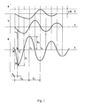
- Fig.1 is a graph of damping oscillations of soft biological tissue.
- Fig.2 is a view of the device for recording the oscillations of soft biological tissues - the myometer.
- t 1 - time denoting the initial moment of the mechanical impact on the tissue, produced by the testing end drive
- t 2 - time when the testing end drive is switched off and the biological tissue begins to oscillate
- t 3 initial moment of the mechanical influencing of the biological tissue caused by the force of elastic deformation of the biological tissue on the testing end
- t 4 the stoppage of upward movement of the testing end, the initial moment of the second period of the oscillation
- t k - working cycle of the testing end drive t' d - duration of the first mechanical influence on the soft biological tissue
- t' v - time during which the soft biological tissue restores its initial shape after the first deformation caused by the first mechanical influence
- an inflexible plane means - joining marker - is attached to the surface of the above-mentioned area (by means of, e.g., vacuum, adhesive or some other binding) for marking on the surface of the biological tissue the area under investigation and for connecting the testing end to the tissue without harming the biological tissue.
- the testing end of the device is connected inflexibly to the above-mentioned joining marker for the time necessary for producing an external mechanical influence and recording the mechanical response of the biological tissue.
- adhesive, vacuum or some other mechanical coupling is used.
- the testing end of the device On the area to which the joining marker is attached, an external mechanical influence is exerted by means of the testing end of the device. After delivering the external force impulse, the testing end will maintain its inflexible connection with the joining marker as well as with the area of the soft biological tissue under investigation, repeating the oscillations of the soft biological tissue in the process of restoring the situation before its elastic deformation.
- the testing end When the measuring procedure is terminated, the testing end is disconnected from the joining marker. To perform measurements on the next area under investigation, the testing end is connected to the next joining marker.
- the measuring procedure described above is of short duration, and therefore neither affects the metabolic processes of the tissues under investigation nor causes neurological reactions of the subject under investigation.
- the joining markers can be removed.
- the graph of damping oscillations of a soft biological tissue, recorded by means of the method described above will contain valuable information, on the basis of which the characteristics of the mechanical properties of the tissue can be calculated, e.g. the oscillation period and the measure of damping of the oscillation, its logarithmic decrement, etc.
- the device for recording the oscillations of soft biological tissues includes the frame 1 with the grip, the pivotable double-armed lever 2 together with the electromechanical transducer 3, the shutter 4, the electromechanical pickup 5, the testing end 6, the pivot 7, the drive of the testing end, which includes the solenoid 8 and the armature 9, the control panel 10, the switch 11 for controlling the measurement process and the inflexible plane means for marking on the tissue the area under investigation and for establishing a permanent and inflexible connection of the testing end with the tissue under investigation - the joining marker 12.
- the investigator places the testing end 6 on the joining marker 12, previously attached to the biological tissue by means of, e. g., adhesive, vacuum or some other mechanical coupling.
- the shutter 4 is moved close to the electromechanical pickup 5 by turning the frame of the device.
- the electric current is switched on to the solenoid 8 for a previously fixed time period and during this time period the armature 9 is pulled into the solenoid 8 by its electromagnetic field, in result of which the testing end exerts to the tissue under investigation a mechanical impact through the joining marker.
- the testing end drive is switched off and the biological tissue together with the joining marker and the testing end perform damping oscillations, the characteristics of which are dependent on the elasticity properties of the soft biological tissue under investigation.
- the oscillations are recorded and the necessary mechanical characteristics of the tissue are calculated, the variant of calculations is indicated on the control panel 10 designed as a numerical indicator.
- the initial state of the device is restored and the procedure can be repeated immediately.
- the measuring procedure can be completely computer-controlled. This makes it possible to record the oscillations of biological tissues quickly and repeatedly.
- the length of the testing end 6 is adjustable by means of, for instance, a bayonet joint.
- the mass of the testing end does not change during the adjustment process.
- the measurements are presented by the graph of damping oscillations of the biological tissue under investigation, which reflects the nature of the short-time (some ms duration) deformation of the tissue (the mechanical characteristics of the deformation) and the nature of the subsequent oscillation of the biological tissue under investigation (which includes such mechanical characteristics as the stiffness and the dempferity properties). It takes the computer-controlled device not more than 0.5 seconds to produce one complete oscillation graph, after that the device is ready for repeating the measuring procedure.
Landscapes
- Health & Medical Sciences (AREA)
- Life Sciences & Earth Sciences (AREA)
- Medical Informatics (AREA)
- Molecular Biology (AREA)
- Veterinary Medicine (AREA)
- Biophysics (AREA)
- Public Health (AREA)
- Engineering & Computer Science (AREA)
- Biomedical Technology (AREA)
- Heart & Thoracic Surgery (AREA)
- General Health & Medical Sciences (AREA)
- Physics & Mathematics (AREA)
- Surgery (AREA)
- Animal Behavior & Ethology (AREA)
- Dentistry (AREA)
- Pathology (AREA)
- Oral & Maxillofacial Surgery (AREA)
- Anesthesiology (AREA)
- Measuring And Recording Apparatus For Diagnosis (AREA)
- Ultra Sonic Daignosis Equipment (AREA)
Abstract
Description
- The present invention relates to medical diagnostics devices and is intended for repeated non-invasive monitoring of mechanical properties of soft biological tissues. The method and device are used to assess the efficiency of massage procedures and the effect of physiotherapeutic treatment on biological tissues, to assess quantitatively the extent of pathological processes, to determine the genetic characteristics of organs, to monitor the tone of biological tissues undergoing surgical operations, to estimate the condition of soft tissues in accordance with the needs of forensic medicine, including measurements on the scene of accident.
- The reasons for using the mechanical properties of soft biological tissues as a source of information for assessing the functional state of tissues and organs are the following:
The intensity of metabolic processes taking place in biological tissues depends on both the internal and external environmental influences, which bring about changes in the mechanical properties of the tissues.
Physical activity, for example, causes hypertrophy of skeletal muscles, whereas inactivity causes atrophy. As a result of various neurological diseases and traumas the muscular tone undergoes significant changes. The muscular tone is characterized by means of the stiffness and dempferity properties of muscles. The stiffness depends on the intramuscular pressure, whereas the dempferity varies according to elasticity properties of the morphological structures of muscles. The human support-motor system has developed in such a way that the rotation process of a body part round the axis of a joint always has two antagonistic muscle groups involved. The agonists create a torque in relation to the axis of the joint, which is necessary for initiating the motion, while at the same time the antagonists are stretched. From the point of view of movement energetics and moving in general it is important what amount of mechanical energy is needed to stretch the antagonists. This mechanical energy in its turn comprises two parts: first, the constraining force which depends on the antagonist's tonicity, the area of its cross-section, and its change in length, and. second, the resisting force, caused by the dempferity properties of the antagonist with the same change in length, which depends on the velocity of stretching. On the one hand, the tone of skeletal muscles depends on the intensity of efferent innervation, on the other, on the cellular tone. The co-influence of both can cause an increase in the mechanical tension of the envelopes or facias of the organs. The elasticity of the collagen fibres of muscle envelopes in its turn affects the ability of biological tissues to dissipate mechanical energy.
Thus changes in the mechanical properties of biological tissues can cause traumas or pathological processes. At the same time, data about changes in the mechanical properties of skeletal muscles enable us to estimate the efficiency of surgical operations, physiotherapeutic procedures, massage, rehabilitation gymnastics and drug treatment.
From what was said above we can conclude that the mechanical properties of biological tissues can yield important information about the functional ability of the tissues, thus making it possible to predict the results concurrent with changes in the mechanical properties of biological tissues. - Various devices and methods have been developed for ascertaining the mechanical properties of soft biological tissues.
The common disadvantage of the methods used until now has been that either these methods themselves cause changes to the mechanical properties of the tissue under investigation or the measuring procedure lasts so long that the subject under investigation manages to change the mechanical properties of the tissue voluntarily during the procedure. Take for instance the device for measuring muscular tone which includes two cuffs. Upon one cuff an acceleration transducer is attached, while on the other cuff the mechanical impacts are produced (Author's certificate of the USSR No. 150573, A61B 5/05, Fedorov V.L., Talysev F.M. 1961). In this case the cuff fastening causes the amount of blood in the muscle to increase, which brings about corresponding changes in the muscular tone. The results of measurements depend on how long the cuffs have been fastened to the muscle. - From among the known methods for measuring the mechanical properties of soft biological tissues, the one most similar to the present invention is the so-called method of damping oscillations, the essence of which lies in subjecting the biological tissue under investigation to external mechanical impact and subsequently recording the mechanical response of the tissue as a graph of its damping oscillations (Fenn W.G., Garwey P.H. , J. Clin., Invest. 1934, 13, Pp. 383-397; Vajn A., Metod zatuhajuscih kolebanij pri diagnostike funkcional'nogo sostojanija skeletnyh mysc. Sb. naucnyh trudov. Metody vibracionnoj diagnostiki reologiceskih harakteristik mjagkih materialov i biologiceskih tkanej. - Gorkij, 1989. Pp. 116-125.).
- Several devices have been designed and constructed with the aim of using the method of damping oscillations, however, due to inadequacies in their construction the results of measurements have been metrologically unreliable. The main shortcomings have been the unstable construction of the mechanism used for producing the mechanical impact to the biological tissue and deficiencies of the system used for recording the mechanical response of the biological tissue to the influence. The graph of the recorded damping oscillations obtained by means of these devices either included additional information about the elasticity of the parts of the device itself, or the signal of the device was too weak to provide metrologically reliable data about the characteristics of the mechanical properties of the biological tissues investigated.
- A method and a device have been invented for non-invasive ultrasonic monitoring of the fluctuations of the properties of living tissue related to its viscoelasticity (
US patent No 4, 580,574, A 61B 10/00, B.Gavish, 1986). There is also another device for measuring the mechanical properties of soft biological tissues. ( Author's Certificate of the USSR No. 1517939, A 61B 5/10, Godin E.A., Cernys V.A. and Stengol'd E.S., 1988). - The common shortcomings of the above-mentioned two are that during the measuring procedure the subject under investigation can voluntarily change the muscular tone, since during the measuring procedure the subject is in a fixed permanent connection with the measuring apparatus; b) the fixed position of peripheral biological tissues in the measuring apparatus sets certain limitations to the possibilities of carrying out a test; c) repeated measurements provide a low level of accuracy.
- Of all the known devices, the one closest in design to the present invention is the device for measuring biomechanical characteristics of biological tissues (Author's Certificate of the USSR No. 1782537, A 61
B 5/05, G 01N 3/30, A.Vain, L.-H.Humal, 1992). The device consists of a gripped frame, into which a pivoting two-shouldered lever is attached. To the end of one shoulder two electromechanical transducers and a wheel-shaped testing end are fastened, and to the end of the other shoulder the drive of the testing end together with its controller. The device is equipped with an elastic element situated between the frame and the two-shouldered lever, and a recorder. - Among the deficiencies of the prototype first of all the presence of the same above-mentioned elastic element must be mentioned, which sets certain limits to the recording of such oscillation frequencies which are lower than the oscillation frequency of the above-mentioned elastic element, thus making signal processing more complicated, which reduces the accuracy of measurements. Also it is difficult to find for the elastic element of the device an elastic material with such stable mechanical properties which would last for a long exploitation period.
- Secondly, in case an elastic element is used it is important to what extent the elastic element has been deformed before the mechanical impulse is produced by the drive of the testing end. In case the deformation of the elastic element as well as the force used to create the initial pressure of the testing end against the biological tissue are of a small order, and the mechanical impulse created by the drive is of a relatively great magnitude, the testing end may lose its contact with the biological tissue under investigation during the oscillation process after the switch-off of the drive of the testing end. So, as a consequence of systematic errors, the resulting graph of oscillations and its analysis will become valueless.
- Thirdly, it has been established that in case the initial pressure of the elastic element is of a relatively great magnitude, then the mechanical impulse produced by the drive of the testing end can neither cause any significant deformation of the biological tissue nor evoke any noticeable damping oscillations when the drive of the testing end is switched off after the mechanical impulse has been performed. In addition to this, the creepability and relaxation properties of biological tissues will further complicate the measuring procedure and reduce its accuracy, when the initial pressure of a great magnitude is used.
- The aim of the present invention is to present a method and a device for recording oscillations of soft biological tissues, so that the resulting graph of oscillations of the biological tissue under investigation would make it possible to calculate the characteristics of the mechanical properties of the biological tissue, to increase the accuracy of the measuring process, to increase the field of possible applications, to shorten the time period necessary for testing and to raise the accuracy of the measurement data (repeatability). The present invention makes it possible to carry out the monitoring of a soft biological tissue repeatedly, in an undisturbed and non-invasive way.
- The essence of the present method lies in the following: the biological tissue under investigation is externally mechanically influenced by means of the testing end of the device and the mechanical response of the above-mentioned tissue is subsequently recorded as a graph representing the evoked oscillations of the tissue. But before exerting the above-mentioned external mechanical impact by the testing end of the device, as distinct from any other known methods, an inflexible plane means - joining marker - is fastened onto the biological tissue in order to designate the area under investigation and connect the testing end with the tissue without causing any harm to the latter. After that the testing end is inflexibly connected to the inflexible plane means for the time it takes to influence the tissue mechanically and register its mechanical response.
- The device for recording the oscillations of soft biological tissues - myometer - consists of a supplementary inflexible plane means for designating the surface area of the soft biological tissue under investigation and for providing an inflexible continuous connection of the testing end of the myometer with the soft biological tissue under investigation without harming the tissue. The length of the testing end is adjustable by means of, for instance, a bayonet joint. The mass of the testing end does not change during the adjustment process.
- Fig.1 is a graph of damping oscillations of soft biological tissue.
- Fig.2 is a view of the device for recording the oscillations of soft biological tissues - the myometer.
- Fig.1
t1 - time, denoting the initial moment of the mechanical impact on the tissue, produced by the testing end drive;
t2 - time, when the testing end drive is switched off and the biological tissue begins to oscillate;
t3 - initial moment of the mechanical influencing of the biological tissue caused by the force of elastic deformation of the biological tissue on the testing end;
t4 - the stoppage of upward movement of the testing end, the initial moment of the second period of the oscillation;
tk - working cycle of the testing end drive;
t'd - duration of the first mechanical influence on the soft biological tissue;
t'v - time during which the soft biological tissue restores its initial shape after the first deformation caused by the first mechanical influence;
a1 - peak acceleration caused by the mechanical influence on the soft biological tissue;
a2 - peak acceleration of the testing end emerging as a result of the force of elasticity of the soft biological tissue, evoked by the mechanical influence on the tissue;
v - graph of the velocity of the testing end;
s - trajectory of the testing end;
Δs - static deformation of the soft biological tissue, caused by the weight of the testing end.
'The area ABC is equal to the deformation velocity of the soft biological tissue vd. - At first it is established by way of palpation which area of the biological tissue under investigation is most convenient for testing, e.g. a tendon of a peripheral muscle. Before exerting external mechanical influence onto the biological tissue an inflexible plane means - joining marker - is attached to the surface of the above-mentioned area (by means of, e.g., vacuum, adhesive or some other binding) for marking on the surface of the biological tissue the area under investigation and for connecting the testing end to the tissue without harming the biological tissue. After that the testing end of the device is connected inflexibly to the above-mentioned joining marker for the time necessary for producing an external mechanical influence and recording the mechanical response of the biological tissue. To connect the testing end with the joining marker, adhesive, vacuum or some other mechanical coupling is used. On the area to which the joining marker is attached, an external mechanical influence is exerted by means of the testing end of the device. After delivering the external force impulse, the testing end will maintain its inflexible connection with the joining marker as well as with the area of the soft biological tissue under investigation, repeating the oscillations of the soft biological tissue in the process of restoring the situation before its elastic deformation. When the measuring procedure is terminated, the testing end is disconnected from the joining marker. To perform measurements on the next area under investigation, the testing end is connected to the next joining marker. The measuring procedure described above is of short duration, and therefore neither affects the metabolic processes of the tissues under investigation nor causes neurological reactions of the subject under investigation. When the measuring procedure is terminated, the joining markers can be removed.
- The graph of damping oscillations of a soft biological tissue, recorded by means of the method described above will contain valuable information, on the basis of which the characteristics of the mechanical properties of the tissue can be calculated, e.g. the oscillation period and the measure of damping of the oscillation, its logarithmic decrement, etc.
- The device for recording the oscillations of soft biological tissues, Fig. 2, includes the frame 1 with the grip, the pivotable double-
armed lever 2 together with theelectromechanical transducer 3, theshutter 4, theelectromechanical pickup 5, the testing end 6, thepivot 7, the drive of the testing end, which includes the solenoid 8 and the armature 9, thecontrol panel 10, theswitch 11 for controlling the measurement process and the inflexible plane means for marking on the tissue the area under investigation and for establishing a permanent and inflexible connection of the testing end with the tissue under investigation - the joiningmarker 12. - Holding the device by its grip, the investigator places the testing end 6 on the joining
marker 12, previously attached to the biological tissue by means of, e. g., adhesive, vacuum or some other mechanical coupling. After pressing theswitch 11 theshutter 4 is moved close to theelectromechanical pickup 5 by turning the frame of the device. When theshutter 4 covers theelectromechanical pickup 5, the electric current is switched on to the solenoid 8 for a previously fixed time period and during this time period the armature 9 is pulled into the solenoid 8 by its electromagnetic field, in result of which the testing end exerts to the tissue under investigation a mechanical impact through the joining marker. When the above-mentioned time period ends, the testing end drive is switched off and the biological tissue together with the joining marker and the testing end perform damping oscillations, the characteristics of which are dependent on the elasticity properties of the soft biological tissue under investigation. The oscillations are recorded and the necessary mechanical characteristics of the tissue are calculated, the variant of calculations is indicated on thecontrol panel 10 designed as a numerical indicator. After pressing theswitch 11 the initial state of the device is restored and the procedure can be repeated immediately. The measuring procedure can be completely computer-controlled. This makes it possible to record the oscillations of biological tissues quickly and repeatedly. - The length of the testing end 6 is adjustable by means of, for instance, a bayonet joint. The mass of the testing end does not change during the adjustment process.
- The measurements are presented by the graph of damping oscillations of the biological tissue under investigation, which reflects the nature of the short-time (some ms duration) deformation of the tissue (the mechanical characteristics of the deformation) and the nature of the subsequent oscillation of the biological tissue under investigation (which includes such mechanical characteristics as the stiffness and the dempferity properties). It takes the computer-controlled device not more than 0.5 seconds to produce one complete oscillation graph, after that the device is ready for repeating the measuring procedure.
- The application of the method and device described above makes it possible
- to increase the accuracy of the measuring procedure, replacing the elastic element used in the construction of the prototype by a non-invasive inflexible connection of the testing end to the area of the soft biological tissue under investigation, using the joining marker described above;
- to measure the characteristics of the soft biological tissue under investigation in optional position of the tissue;
- to expand the field of applications of the device using the possibility to change the length and position of the testing end;
- as a result of the soft biological tissue under investigation being inflexibly connected with the testing end of the device, to use a wide range of deformation velocities of the tissues, without any pain sensations to the subject or changes in blood circulation of the tissues under investigation, at the same time the stability and lack of distortions of the resulting oscillation graphs is secured by the permanent contact of the testing end with the tissue under investigation;
- to shorten the testing time by previously fastening the necessary amount of joining markers to the area of the soft biological tissue under investigation;
- to raise the accuracy of the measuring data and to guarantee repeatability of the procedure.
Claims (3)
- Method for recording mechanical damped natural oscillations of soft biological tissues by exerting on the soft biological tissue an external mechanical impact by means of the testing end (6) of the below-described device and the subsequent recording of the mechanical response of the biological tissue as a graph of damped natural oscillations of the tissue, which is characterized by that before exerting the external mechanical impact on the biological tissue by the testing end (6) of the device, an inflexible plane means (12) is fastened onto the biological tissue in order to mark the area under investigation and connect the testing end with the tissue causing no harm to the latter, after that the testing end is inflexibly connected to the inflexible plane means for the time necessary for influencing the tissue mechanically and for recording the mechanical response of the biological tissue, after which the tissue under investigation is given an external mechanical impact during some milliseconds, ending with quick release of the testing end from its drive without causing any harm to the biological tissue or changes in metabolic processes of the tissue or neurological reactions of the subject under investigation.
- A device for recording natural oscillations of soft biological tissues - the myometer - which consists of a frame (1) with a grip, onto which are attached a pivoting double-armed lever (2) with an electromechanical transducer (3) and shutter (4), an electromechanical pickup (5), a testing end (6), a pivot (7) of the testing end drive (8, 9), a switch (11) for controlling the measuring process and the recorder, and which is characterized by that it includes a supplementary inflexible plane means (12) for marking the area under investigation and for connecting the testing end of the device to the tissue permanently and inflexibly without causing any harm to the biological tissue or changes in metabolic processes of the tissue or neurological reactions of the subject under investigation.
- The device according to claim 2, which is characterized in that the length of the testing end (6) is adjustable, while the mass of the testing end remains constant in the adjustment process, by means of, for instance, a bayonet joint.
Applications Claiming Priority (3)
| Application Number | Priority Date | Filing Date | Title |
|---|---|---|---|
| EE9600015 | 1996-03-27 | ||
| EE9600015A EE03374B1 (en) | 1996-03-27 | 1996-03-27 | Method and device for recording soft biological tissue self-oscillation - myometer |
| PCT/EE1997/000001 WO1997035521A1 (en) | 1996-03-27 | 1997-03-21 | A method and a device for recording mechanical oscillations in soft biological tissues |
Publications (2)
| Publication Number | Publication Date |
|---|---|
| EP0897287A1 EP0897287A1 (en) | 1999-02-24 |
| EP0897287B1 true EP0897287B1 (en) | 2002-06-12 |
Family
ID=8161671
Family Applications (1)
| Application Number | Title | Priority Date | Filing Date |
|---|---|---|---|
| EP97908149A Expired - Lifetime EP0897287B1 (en) | 1996-03-27 | 1997-03-21 | A method and a device for recording mechanical oscillations in soft biological tissues |
Country Status (7)
| Country | Link |
|---|---|
| US (1) | US6132385A (en) |
| EP (1) | EP0897287B1 (en) |
| JP (1) | JP3597201B2 (en) |
| AU (1) | AU2022997A (en) |
| DE (1) | DE69713320T2 (en) |
| EE (1) | EE03374B1 (en) |
| WO (1) | WO1997035521A1 (en) |
Families Citing this family (28)
| Publication number | Priority date | Publication date | Assignee | Title |
|---|---|---|---|---|
| US6839661B2 (en) * | 2000-12-15 | 2005-01-04 | Medispectra, Inc. | System for normalizing spectra |
| US7530975B2 (en) * | 2002-09-06 | 2009-05-12 | Massachusetts Institute Of Technology | Measuring properties of an anatomical body |
| US7833189B2 (en) | 2005-02-11 | 2010-11-16 | Massachusetts Institute Of Technology | Controlled needle-free transport |
| US20060258986A1 (en) * | 2005-02-11 | 2006-11-16 | Hunter Ian W | Controlled needle-free transport |
| US20060247586A1 (en) | 2005-04-08 | 2006-11-02 | Voegele James W | Intra-abdominal storage device |
| US8357085B2 (en) | 2009-03-31 | 2013-01-22 | Ethicon Endo-Surgery, Inc. | Devices and methods for providing access into a body cavity |
| EP2077877B1 (en) * | 2006-09-01 | 2019-12-11 | Massachusetts Institute of Technology | Needle-free injector device with autoloading capability |
| US20080262391A1 (en) * | 2007-04-23 | 2008-10-23 | Mark Peter Ottensmeyer | Instrument for measuring the mechanical properties of vocal tissues |
| US8801635B2 (en) * | 2008-10-03 | 2014-08-12 | Hlz Innovation, Llc | Adjustable pneumatic supporting surface |
| US9737334B2 (en) | 2009-03-06 | 2017-08-22 | Ethicon Llc | Methods and devices for accessing a body cavity |
| US8353824B2 (en) | 2009-03-31 | 2013-01-15 | Ethicon Endo-Surgery, Inc. | Access method with insert |
| US20100249521A1 (en) | 2009-03-31 | 2010-09-30 | Shelton Iv Frederick E | Access Device Including Retractor And Insert |
| US20110028793A1 (en) * | 2009-07-30 | 2011-02-03 | Ethicon Endo-Surgery, Inc. | Methods and devices for providing access into a body cavity |
| US9265461B2 (en) * | 2009-09-01 | 2016-02-23 | Massachusetts Institute Of Technology | Identification techniques and device for testing the efficacy of beauty care products and cosmetics |
| US8758271B2 (en) | 2009-09-01 | 2014-06-24 | Massachusetts Institute Of Technology | Nonlinear system identification techniques and devices for discovering dynamic and static tissue properties |
| US9474540B2 (en) | 2009-10-08 | 2016-10-25 | Ethicon-Endo-Surgery, Inc. | Laparoscopic device with compound angulation |
| US20110143310A1 (en) * | 2009-12-15 | 2011-06-16 | Hunter Ian W | Lorentz-Force Actuated Cleaning Device |
| US8562592B2 (en) | 2010-05-07 | 2013-10-22 | Ethicon Endo-Surgery, Inc. | Compound angle laparoscopic methods and devices |
| US9226760B2 (en) | 2010-05-07 | 2016-01-05 | Ethicon Endo-Surgery, Inc. | Laparoscopic devices with flexible actuation mechanisms |
| US8460337B2 (en) | 2010-06-09 | 2013-06-11 | Ethicon Endo-Surgery, Inc. | Selectable handle biasing |
| EE05601B2 (en) | 2010-12-31 | 2015-06-15 | As Myoton | Device and method for simultaneous real-time measurement of parameters of state of mechanical stress, elasticity, dynamic stiffness, creepability and relaxation time of mechanical stress of soft biological tissue and a computer program product |
| CA2870390C (en) * | 2012-04-16 | 2019-09-17 | Myoton As | Device and method for the non-invasive measurement of state of tension, biomechanical and viscoelastic properties of surfaces of soft biological tissues |
| GB201302093D0 (en) | 2013-02-06 | 2013-03-20 | Myoton As | System and Method for Non-invasive Measurement of Soft Biological Tissue |
| CA2964805C (en) * | 2014-10-17 | 2023-04-04 | La Trobe University | A screening test for detection of deep vein thrombosis |
| CN106037823B (en) * | 2016-08-17 | 2019-01-15 | 奉节县吐祥镇中心卫生院 | A kind of novel suspended type medical treatment hand hammer |
| GB201904010D0 (en) | 2019-03-22 | 2019-05-08 | Univ Tartu | Method and device for measuring or determining at least one biomechanical parameter of soft biological tissues |
| WO2022076579A1 (en) * | 2020-10-06 | 2022-04-14 | Impact Biosystems, Inc. | Systems, devices, and methods for measurement and treatment of tissue with force control and feedback |
| US20250176952A1 (en) * | 2023-12-04 | 2025-06-05 | Cervalia, Inc. | Force mediated mechanical testing device for the characterization of soft tissues |
Family Cites Families (11)
| Publication number | Priority date | Publication date | Assignee | Title |
|---|---|---|---|---|
| US3200814A (en) * | 1963-03-12 | 1965-08-17 | Ellis R Taylor | Apparatus for measuring reflexes, tremors and the like |
| FR2359411A1 (en) * | 1976-07-20 | 1978-02-17 | Oreal | APPARATUS FOR MEASURING THE MECHANICAL CHARACTERISTICS OF A MATERIAL SUCH AS SKIN |
| DE2741338A1 (en) * | 1977-09-14 | 1979-03-22 | Industrie Automation Gmbh & Co | DEVICE FOR DETERMINING THE NATURAL MECHANICAL FREQUENCY OF BLOOD VESSELS, TENDONS, BONES OR SIMILAR ORGANS IN A BODY |
| US4444205A (en) * | 1980-05-31 | 1984-04-24 | University Of Strathclyde | Apparatus for assessing joint mobility |
| DE3215498A1 (en) * | 1982-04-26 | 1983-10-27 | Siemens AG, 1000 Berlin und 8000 München | PERCUSSION INSTRUMENT |
| IL69471A (en) | 1983-08-12 | 1987-07-31 | Benjamin Gavish | Method and device for non-invasively monitoring the instantaneous fluctuations in the viscoelastic properties of a soft tissue |
| GB8431323D0 (en) * | 1984-12-12 | 1985-01-23 | Warin C C | Investigating muscles/joints |
| SU1782537A1 (en) * | 1989-06-29 | 1992-12-23 | Univ Tartusky | Device for measuring bio mechanical characteristics of biological tissues |
| DE4017251A1 (en) * | 1990-05-29 | 1991-12-05 | Phywe Systeme Gmbh | DEVICE FOR GENERATING TACTICAL REASONS BY VIBRATING A PUSH TO BE PUT ON THE SKIN OF A PEOPLE |
| JP3306133B2 (en) * | 1992-11-30 | 2002-07-24 | オリンパス光学工業株式会社 | Medical equipment |
| US5471996A (en) * | 1993-09-30 | 1995-12-05 | Charlotte-Mecklenburg Hospital Authority | Apparatus and method for measuring abduction strength of a patient's thumb |
-
1996
- 1996-03-27 EE EE9600015A patent/EE03374B1/en not_active IP Right Cessation
-
1997
- 1997-03-21 DE DE69713320T patent/DE69713320T2/en not_active Expired - Lifetime
- 1997-03-21 JP JP53395497A patent/JP3597201B2/en not_active Expired - Fee Related
- 1997-03-21 AU AU20229/97A patent/AU2022997A/en not_active Abandoned
- 1997-03-21 WO PCT/EE1997/000001 patent/WO1997035521A1/en not_active Ceased
- 1997-03-21 EP EP97908149A patent/EP0897287B1/en not_active Expired - Lifetime
- 1997-03-21 US US09/155,219 patent/US6132385A/en not_active Expired - Lifetime
Also Published As
| Publication number | Publication date |
|---|---|
| EE9600015A (en) | 1997-10-15 |
| US6132385A (en) | 2000-10-17 |
| JP3597201B2 (en) | 2004-12-02 |
| EE03374B1 (en) | 2001-04-16 |
| DE69713320D1 (en) | 2002-07-18 |
| EP0897287A1 (en) | 1999-02-24 |
| JP2001502557A (en) | 2001-02-27 |
| WO1997035521A1 (en) | 1997-10-02 |
| AU2022997A (en) | 1997-10-17 |
| DE69713320T2 (en) | 2003-02-13 |
Similar Documents
| Publication | Publication Date | Title |
|---|---|---|
| US6132385A (en) | Method and a device for recording mechanical oscillations in soft biological tissues | |
| AU2011351911B2 (en) | Device and method for real-time measurement of parameters of mechanical stress state and biomechanical properties of soft biological tissue | |
| CN103124518B (en) | For non-invasively and optionally measure the method and apparatus of the biomechanics of surface skeletal, contraction and viscoelastic property | |
| Bardorfer et al. | Upper limb motion analysis using haptic interface | |
| US5195532A (en) | Apparatus for producing a stimulation by vibration of a tappet which is put on a human's skin | |
| JPH05500461A (en) | Biofeedback device that monitors muscle movement | |
| US20100228156A1 (en) | Dexterity device | |
| JP4866343B2 (en) | Method and apparatus for measuring water (edema) | |
| JPH07506998A (en) | Reflection measurement equipment and methods | |
| CA1270908A (en) | Methods and apparatus for investigating muscles and/or joints | |
| EP3941336B1 (en) | Method for measuring or determining at least one biomechanical parameter of soft biological tissues | |
| JP2009297206A (en) | Measuring system for noninvasively evaluating damaged state of human muscle | |
| Cornu et al. | Muscle elastic properties during wrist flexion and extension in healthy sedentary subjects and volley-ball players | |
| Kroemer | Human muscle strength: definition, generation and measurement | |
| Hsu et al. | A novel and clinically feasible instrument for quantifying upper Limb muscle tone and motor function via indirect measure methods | |
| Hinoki et al. | An application of strain-gage-type instruments to the analysis of the equilibrium function in human subjects and animals | |
| Weiss-Lambrou et al. | The effect of differing feedback conditions on grip strength: a pilot study | |
| RU2082312C1 (en) | Apparatus for determining viscoelastic properties of soft tissues | |
| DUICU et al. | EXPERIMENTAL MODEL FOR IN VIVO TESTING OF MUSCLE RELAXANT DRUGS | |
| Marchese et al. | M Department Veterans Affairs of | |
| Sepehri et al. | Optimization in measurement of MVC and active stiffness in wrist using the quick release method | |
| CZ18589U1 (en) | System for measuring reaction time of animals, particularly horses |
Legal Events
| Date | Code | Title | Description |
|---|---|---|---|
| PUAI | Public reference made under article 153(3) epc to a published international application that has entered the european phase |
Free format text: ORIGINAL CODE: 0009012 |
|
| 17P | Request for examination filed |
Effective date: 19981023 |
|
| AK | Designated contracting states |
Kind code of ref document: A1 Designated state(s): DE FI FR GB IT SE |
|
| GRAG | Despatch of communication of intention to grant |
Free format text: ORIGINAL CODE: EPIDOS AGRA |
|
| 17Q | First examination report despatched |
Effective date: 20010705 |
|
| GRAG | Despatch of communication of intention to grant |
Free format text: ORIGINAL CODE: EPIDOS AGRA |
|
| GRAG | Despatch of communication of intention to grant |
Free format text: ORIGINAL CODE: EPIDOS AGRA |
|
| GRAH | Despatch of communication of intention to grant a patent |
Free format text: ORIGINAL CODE: EPIDOS IGRA |
|
| RIN1 | Information on inventor provided before grant (corrected) |
Inventor name: VAIN, ARVED |
|
| GRAH | Despatch of communication of intention to grant a patent |
Free format text: ORIGINAL CODE: EPIDOS IGRA |
|
| GRAA | (expected) grant |
Free format text: ORIGINAL CODE: 0009210 |
|
| AK | Designated contracting states |
Kind code of ref document: B1 Designated state(s): DE FI FR GB IT SE |
|
| REG | Reference to a national code |
Ref country code: GB Ref legal event code: FG4D |
|
| REF | Corresponds to: |
Ref document number: 69713320 Country of ref document: DE Date of ref document: 20020718 |
|
| ET | Fr: translation filed | ||
| PLBE | No opposition filed within time limit |
Free format text: ORIGINAL CODE: 0009261 |
|
| STAA | Information on the status of an ep patent application or granted ep patent |
Free format text: STATUS: NO OPPOSITION FILED WITHIN TIME LIMIT |
|
| 26N | No opposition filed |
Effective date: 20030313 |
|
| REG | Reference to a national code |
Ref country code: GB Ref legal event code: 732E |
|
| REG | Reference to a national code |
Ref country code: FR Ref legal event code: TP |
|
| PGFP | Annual fee paid to national office [announced via postgrant information from national office to epo] |
Ref country code: FI Payment date: 20100319 Year of fee payment: 14 |
|
| PGFP | Annual fee paid to national office [announced via postgrant information from national office to epo] |
Ref country code: GB Payment date: 20100325 Year of fee payment: 14 |
|
| PGFP | Annual fee paid to national office [announced via postgrant information from national office to epo] |
Ref country code: FR Payment date: 20100421 Year of fee payment: 14 |
|
| PGFP | Annual fee paid to national office [announced via postgrant information from national office to epo] |
Ref country code: IT Payment date: 20100330 Year of fee payment: 14 Ref country code: DE Payment date: 20100331 Year of fee payment: 14 |
|
| PGFP | Annual fee paid to national office [announced via postgrant information from national office to epo] |
Ref country code: SE Payment date: 20100325 Year of fee payment: 14 |
|
| REG | Reference to a national code |
Ref country code: SE Ref legal event code: EUG |
|
| GBPC | Gb: european patent ceased through non-payment of renewal fee |
Effective date: 20110321 |
|
| PG25 | Lapsed in a contracting state [announced via postgrant information from national office to epo] |
Ref country code: FI Free format text: LAPSE BECAUSE OF NON-PAYMENT OF DUE FEES Effective date: 20110321 |
|
| REG | Reference to a national code |
Ref country code: FR Ref legal event code: ST Effective date: 20111130 |
|
| PG25 | Lapsed in a contracting state [announced via postgrant information from national office to epo] |
Ref country code: DE Free format text: LAPSE BECAUSE OF NON-PAYMENT OF DUE FEES Effective date: 20111001 Ref country code: FR Free format text: LAPSE BECAUSE OF NON-PAYMENT OF DUE FEES Effective date: 20110331 |
|
| REG | Reference to a national code |
Ref country code: DE Ref legal event code: R119 Ref document number: 69713320 Country of ref document: DE Effective date: 20111001 |
|
| PG25 | Lapsed in a contracting state [announced via postgrant information from national office to epo] |
Ref country code: IT Free format text: LAPSE BECAUSE OF NON-PAYMENT OF DUE FEES Effective date: 20110321 Ref country code: GB Free format text: LAPSE BECAUSE OF NON-PAYMENT OF DUE FEES Effective date: 20110321 |
|
| PG25 | Lapsed in a contracting state [announced via postgrant information from national office to epo] |
Ref country code: SE Free format text: LAPSE BECAUSE OF NON-PAYMENT OF DUE FEES Effective date: 20110322 |