EP0882229B1 - Elisa testsystem for detecting substances interfering with the binding between ig e and its receptor - Google Patents
Elisa testsystem for detecting substances interfering with the binding between ig e and its receptor Download PDFInfo
- Publication number
- EP0882229B1 EP0882229B1 EP96944649A EP96944649A EP0882229B1 EP 0882229 B1 EP0882229 B1 EP 0882229B1 EP 96944649 A EP96944649 A EP 96944649A EP 96944649 A EP96944649 A EP 96944649A EP 0882229 B1 EP0882229 B1 EP 0882229B1
- Authority
- EP
- European Patent Office
- Prior art keywords
- ige
- receptor
- fcεriα
- binding
- elisa
- Prior art date
- Legal status (The legal status is an assumption and is not a legal conclusion. Google has not performed a legal analysis and makes no representation as to the accuracy of the status listed.)
- Expired - Lifetime
Links
Images


Classifications
-
- G—PHYSICS
- G01—MEASURING; TESTING
- G01N—INVESTIGATING OR ANALYSING MATERIALS BY DETERMINING THEIR CHEMICAL OR PHYSICAL PROPERTIES
- G01N33/00—Investigating or analysing materials by specific methods not covered by groups G01N1/00 - G01N31/00
- G01N33/48—Biological material, e.g. blood, urine; Haemocytometers
- G01N33/50—Chemical analysis of biological material, e.g. blood, urine; Testing involving biospecific ligand binding methods; Immunological testing
- G01N33/5005—Chemical analysis of biological material, e.g. blood, urine; Testing involving biospecific ligand binding methods; Immunological testing involving human or animal cells
- G01N33/5008—Chemical analysis of biological material, e.g. blood, urine; Testing involving biospecific ligand binding methods; Immunological testing involving human or animal cells for testing or evaluating the effect of chemical or biological compounds, e.g. drugs, cosmetics
-
- G—PHYSICS
- G01—MEASURING; TESTING
- G01N—INVESTIGATING OR ANALYSING MATERIALS BY DETERMINING THEIR CHEMICAL OR PHYSICAL PROPERTIES
- G01N33/00—Investigating or analysing materials by specific methods not covered by groups G01N1/00 - G01N31/00
- G01N33/48—Biological material, e.g. blood, urine; Haemocytometers
- G01N33/50—Chemical analysis of biological material, e.g. blood, urine; Testing involving biospecific ligand binding methods; Immunological testing
- G01N33/53—Immunoassay; Biospecific binding assay; Materials therefor
- G01N33/564—Immunoassay; Biospecific binding assay; Materials therefor for pre-existing immune complex or autoimmune disease, i.e. systemic lupus erythematosus, rheumatoid arthritis, multiple sclerosis, rheumatoid factors or complement components C1-C9
-
- G—PHYSICS
- G01—MEASURING; TESTING
- G01N—INVESTIGATING OR ANALYSING MATERIALS BY DETERMINING THEIR CHEMICAL OR PHYSICAL PROPERTIES
- G01N33/00—Investigating or analysing materials by specific methods not covered by groups G01N1/00 - G01N31/00
- G01N33/48—Biological material, e.g. blood, urine; Haemocytometers
- G01N33/50—Chemical analysis of biological material, e.g. blood, urine; Testing involving biospecific ligand binding methods; Immunological testing
- G01N33/68—Chemical analysis of biological material, e.g. blood, urine; Testing involving biospecific ligand binding methods; Immunological testing involving proteins, peptides or amino acids
- G01N33/6854—Immunoglobulins
-
- G—PHYSICS
- G01—MEASURING; TESTING
- G01N—INVESTIGATING OR ANALYSING MATERIALS BY DETERMINING THEIR CHEMICAL OR PHYSICAL PROPERTIES
- G01N2800/00—Detection or diagnosis of diseases
- G01N2800/24—Immunology or allergic disorders
Definitions
- the invention relates to a test system by ELISA (enzyme-linked immunosorbent assay). It concerns an ELISA system to detect biochemical entities in very minute amounts and in the presence of structurally very similar compounds, which are differentiated by means of monospecific antibodies, such as immunoglobulins of various species.
- ELISA enzyme-linked immunosorbent assay
- the antigen which is to be detected, or a specific antibody which binds to the respective antigen is bound to a solid phase (microtiter plate) by hydrophobic interactions: the protein interacts with the solid phase, usually a polystyrene surface at high pH. Although this bond is responsible for all the consecutive steps in the procedure, it remains the weakest bridge to the assay support, the ELISA plate.
- a catching antibody is used. This antibody binds to the polystyrene plate and exposes the high affinity hyper-variable region toward the incoming antigen. The antigen is then detected by a second antibody, which is labelled directly or indirectly (via biotin/avidin) with an enzyme. This enzyme will cleave a chromogenic substrate, which itself is converted from the leucoform to the chromoform and thus visualizes the presence of the antigen in question. But even catching antibodies may affect a given protein by changing its conformation. This is demonstrated by many examples of therapeutic antibodies whose mode of action is the blocking of an active site on, or the alteration of, a biospecific molecule.
- EP-A-0 507 586 discloses a homogeneous immunoassay for immunoa labelled second globulins employing a biotinglated first antigen, antigen and an oridin coated solid surface.
- JP 93/02 18920 discloses on IgE assay in which Fc ⁇ RI-IgE binding is determined.
- the present invention concerns an assay for screening of substances interfering with the attachment of human IgE to its high affinity receptor Fc ⁇ RI ⁇ , and/or of substances capable of detaching already bound IgE from this receptor according to claim 1, which is devoid of the above disadvantages. It comprises reacting a solution of a biotinylated Fc ⁇ RI ⁇ receptor with IgE, transferring the resultant binding reaction mixture to a streptavidin-coated plate and quantifying by means of an appropriate enzyme-labeled antibody.
- biotinylated IgE-receptor with IgE in the presence or absence of interfering or inhibitory substances, such as non-biotinylated IgE-receptor, at well-defined molar concentrations, temperature, pH and salt conditions, thereby eliminating the structural influence of solid phases, and transferring after a defined incubation time an aliquot of the binding reaction mixture to a streptavidin-coated microtiter plate, where the biotinylated IgE-receptor/IgE complex is quantified by means of an appropriate enzyme-labeled antibody, such as horse radish peroxidase (POD)-labeled antibody against human IgE.
- interfering or inhibitory substances such as non-biotinylated IgE-receptor
- biotinylated IgE-receptor is first reacted with IgE a sample possibly containing interfering or inhibitory substances, such as non-biotinylated IgE-receptor, at well-defined molar concentrations, temperature, pH and salt conditions, thereby eliminating the structural influence of solid phases.
- an aliquot of the binding reaction mixture is transferred to a streptavidin-coated microtiter plate, where the biotinylated IgE-receptor/IgE complex is trapped and quantified by means of an appropriate enzyme-labeled antibody, such as horse radish peroxidase (POD)-labeled antibody against human IgE.
- an appropriate enzyme-labeled antibody such as horse radish peroxidase (POD)-labeled antibody against human IgE.
- both molecules float freely in solution and thus are not influenced by the structural modification of solid phases. They receive their conformation by the given pH and salt concentration and exert their maximal binding affinity. After a given incubation time, selected according to the results of appropriate equilibrium measurements, the receptor/ligand complex has to be deprived of all other compounds.
- the receptor is therefore biotinylated prior to the above reaction and an aliquot of the reaction is transferred to a streptavidin-coated microtiter plate.
- the binding of streptavidin with the plate surface is preferably made covalent.
- a microtiter plate (such as a DNA-Bind plate) chemically modified with e.g. a N-oxysuccinimide ester coating is used, which reacts with nucleophiles such as primary amines under formation of a covalent bond. This bond is stronger than 10 -15 moles per liter.
- a chemically modified biotin such as NHS-LC-Biotin is used, which comprises an extended spacer arm of approximately 22.4 ⁇ in length. This long chain analogue reduces steric hindrances associated with the binding of four biotinylated molecules on one streptavidin molecule.
- the target of biotinylation on the IgE-receptor molecule is thereby well defined and interference with the ligand is avoided.
- the binding affinity of biotin to streptavidin is also known to be 10 -15 moles per liter. Thereby the first two steps of the binding cascade are established and reach from the bottom of the plate to streptavidin to the biotinylated receptor. The affinity constants are decreasing from the bottom to the top. At this stage the reactants, which have found their partners in solution under appropriate conditions, are trapped by the means of the streptavidin plate, and the bound IgE is then detected with an appropriate enzyme-labeled antibody, such as POD-labeled antibody against human IgE.
- an appropriate enzyme-labeled antibody such as POD-labeled antibody against human IgE.
- the affinity cascade now reaches from ⁇ 10 -15 to 10 -15 to 10 -10 to 10 -12 for the anti-IgE antibody toward IgE. Any interference of biochemical material and especially of IgE-receptor binding inhibitors will occur at the point with the lowest affinity, which resides between the IgE and the receptor (10 -10 ), and not at the point of attachment of the assay to the plate. This would be read as a false positive result - an artifact of the system.
- the above provides the basis for the detection of biomolecules other than IgE but which are also binding to Fc ⁇ RI ⁇ .
- the prerequisite therefor is the abolishment of the binding of IgE to the receptor in order to keep the receptor site free for other molecules.
- This is achieved by means of an anti-IgE antibody as BSW17 which binds to the receptor-binding site of the IgE and thus renders the molecule non-effective, whereby even in the presence of IgE other biomolecules can bind to the IgE-binding site of the receptor (the hybridoma cell line producing monoclonal antibody BSW 17 has been deposited on December 18, 1996 with the European Collection of Animal Cell Cultures (ECACC), Porton Down, U.K.
- the invention concerns an ELISA-type assay for finding inhibitors of the binding of human IgE to its high affinity receptor, the ⁇ -chain of Fc ⁇ RI (Fc ⁇ RI ⁇ ), especially substances inhibiting IgE-mediated early phase response in allergic reactions.
- the assay mimics natural conditions of binding of IgE to its receptor, i.e. all binding partners interact in solution instead of one of them being immobilized on a solid support.
- biotinylated soluble Fc ⁇ RI ⁇ chain interacts with free, soluble IgE at a defined molar ratio (1 : 2.5) in the presence of inhibitory or interfering substance to reach thermodynamic equilibrium within a three-hours incubation period at 37°C.
- the IgE/Fc ⁇ RI complex formed under these conditions is captured via the biotin-linker coupled to the Fc ⁇ RI ⁇ chain on ELISA plates modified by covalently-linked streptavidin.
- the amount of captured complex is determined by identifying bound IgE via a peroxidase-linked anti-human IgE antibody preparation, such as anti-human IgE ( ⁇ -chain-specific) peroxidase conjugate , e.g. Sigma no.
- the concentrations of the binding partners in primary screening are chosen to obtain an optimal signal to noise ratio under nonsaturating conditions.
- the K d of the IgE/Fc ⁇ RI complex formed is found to be 4 nM.
- the maximally tolerated dose of DMSO in the test solution is 5 % and therefore, at a concentration of pure compound of 50 ⁇ M competitive inhibitors with a K d of 30-10 ⁇ M can be easily detected in primary screening at a cut-off level of 30 % inhibition.
- a monoclonal antibody recognizing free as well as receptor-bound IgE and inhibiting its binding to the receptor or releasing it from the complex, respectively, such as BSW17 is tested in 5 different concentrations on every screening plate.
- the throughput of test samples in the above assay can be very high (>1000 samples/day) and the sensitivity is also high, as compounds which competitively inhibit the binding reaction with a K d of 10 ⁇ M are easily detected by a signal reduction of 50%.
- the soluble extracellular part of the ⁇ -chain, the IgE-binding subunit of Fc ⁇ RI is available through recombinant DNA technology and hence the assay for high capacity screening of inhibitors of the IgE/Fc ⁇ RI interaction could be established cell-free.
- the assay is designed for the interaction of binding partners to occur in solution.
- the ⁇ -chain is biotinylated using an extended spacer arm for chemical coupling and streptavidin is covalently bound to the ELISA plate via amino groups.
- the binding reactions between IgE and its high affinity receptor, Fc ⁇ RI ⁇ , and the interference of test compound are performed in a separate microtiter plate to reach the equilibrium in solution during an extended incubation period at 37°C.
- To favor conditions for inhibitory compounds to be active they are added to and preincubated with the ⁇ -chain prior to the addition of IgE.
- the residual IgE/ ⁇ -chain complex formed in the presence of test compound is then captured on the streptavidin plate during a short incubation period at room temperature or alternatively, after an overnight incubation at 4°C.
- the amount of captured complex is determined by a peroxidase-catalyzed colorimetric reaction using an anti-human IgE antibody preparation with covalently coupled enzyme.
- the consecutive steps can be done under normal conditions: five washes (Biomek) of the plate with PBST and tapping onto a paper towel in order to free the plate from residual moisture, addition of 360 ⁇ l of 0.5 M Tris-Cl pH 8 and incubation for 1 hour at room temperature. This step will inactivate residual free ester groups on the plate surface. Then, 200 ⁇ l of blocking solution (2 % BSA in PBS, 0.05 % Tween 20) is added into each well and kept at room temperature for 30 minutes. Then the plate is washed once more as described and stored in a sealed plastic box at 4°C.
- a given amount of receptor solution (Fc ⁇ RI ⁇ , extracellular portion, recombinant, produced in insect cells) is concentrated by ultrafiltration over Amicon YM 3/25 to give 1.5 ml at a concentration of 2.2 mg protein per ml.
- This solution is dialyzed with a Pierce Slide-A Lyzer against three times 1 liter of 100 mM sodium carbonate pH 8.5 within four hours at 4°C.
- the biotinylation is done by adding 23 ⁇ l of a solution of 4.8 mg of Biotin II Pierce in 120 ⁇ l of DMF and the vial is tumbled for 15 minutes at room temperature. After another addition of 23 ⁇ l of the biotin solution the whole device is transferred to 4°C and tumbling continued for 1.5 hours. Then the reaction is stopped by addition of 150 ⁇ l of 1 M Tris-Cl pH 8 and a further incubation for 10 minutes at room temperature.
- This sample is loaded onto a FPLC Superdex 75 [16 x 1200 mm] in PBS 0.02 % sodium azide at 4°C.
- the flow-rate is 2 ml/min, the fraction size is 2.6 ml.
- the fractions containing adequately biotinylated receptor are pooled and concentrated over an Amicon YM 3/43 to approximately 4 ml.
- After determination of the protein concentration an equal volume of glycerol is added. 100 ⁇ l aliquots of this solution are stored at -80°C. 50 ⁇ l of fraction aliquots are placed into each well of a streptavidin-covalent-plate and kept at room temperature for one hour.
- the plate is washed with PBST and 50 ⁇ l of IgE [20 nM] are added to each well and incubated for one hour at room temperature. After a subsequent wash with PBST, 100 ⁇ l of anti-IgE-POD [1 : 1000] are added to each well and the plate is further incubated for one hour. Then the plate is washed again and 100 ⁇ l of TMB substrate are added. After 20 minutes of incubation at room temperature the reaction is stopped by addition of 100 ⁇ l of 4 N H 2 SO 4 and the plate is read at 450 nm.
- a high molecular weight pool is detected containing highly biotinylated but aggregated material, which binds to the plate but consequently is inactive in binding to IgE.
- the appropriate IgE-binding competent material is found at the expected elution position of 32 kDa. Remaining reactants are seen in a low molecular weight fraction as well.
- IgG BSW17 is pipetted into well A4-A8 at 12 nM in two-fold dilutions. These are the dilution plates. Then the working solution for the receptor is prepared by diluting the stock solution to 600 pM in incubation buffer. 50 ⁇ l of this solution is distributed to each well of a new round bottom plate (incubation plate) and 50 ⁇ l of the respective dilution plate is transferred to this plate and mixed with the receptor five times. The plate is kept for one hour at 37°C covered with an adhesive foil and thereafter 50 ⁇ l of IgE B 11 at a concentration of 1500 pM is pipetted into each well of the incubation plate and mixed five times.
- the respective concentrations are as follows: 5 % DMSO, 50 ⁇ M substance, 600 mM NaOH, 200 pM receptor, 500 pM IgE; 4; 2; 1; 0.5; and 0.25 nM IgG BSW17.
- the plate is sealed with an adhesive foil and incubated for three hours at 37°C. Thereafter 100 ⁇ l are transferred from the incubation plate into the streptavidin covalent plate which is kept overnight at 4°C. After a five times wash 100 ⁇ l of anti IgE-POD [1:2000] are added and the plate is incubated for another hour at room temperature. The plate is washed again five times and 100 ⁇ l of TMB-substrate are added. After 20 minutes incubation at room temperature the reaction is stopped by addition of 100 ⁇ l of 4 N H 2 SO 4 and the plate is read at 450/690 nm.
- Figure 2 depicts the functional properties of the Fc ⁇ RI ⁇ receptor at 20 nM IgE.
- the receptor concentration of 200 pM chosen for the screening assay represents the high endpoint of the exponential phase of the curve. Previous experiments showed that the molar binding ratio for IgE is 1 : 2.5 and hence 500 pM was chosed as the assay concentration for IgE.
- the IgG BSW17 standard provides a measure for the sensitivity of the assay system as well as a gauge for the determination of the IC50.
- TMB stock solution 2 mg TMB/ml ethanol, filtered through a glass sinter filter no. 2; TMB working solution; 100 ml citrate buffer pH 5 plus 2.2 ml TMB stock solution plus 560 ⁇ l 0.3 % H 2 O 2 :
- this assay method is useful for the differential analysis between autoimmune disorders and classical allergies, i.e. allergies triggered by exogenous factors.
- Urticaria is a common disorder characterized by the eruption of transitory, itchy skin swellings that are frequently associated with the occurrence of debilitating and potentially life-threatening angioedema.
- accurate data on the prevalence of urticaria are not available, it is estimated that 15 % to 32 % of the general population experience urticarial/angio-edematous symptoms during their life time. In about 25 % of these patients the initially sporadic character of this condition is followed by a chronic disease period with symptoms of frequent but unpredictable occurrence.
- CU chronic urticaria
- IgG autoreactivity against Fc ⁇ RI ⁇ is of central pathophysiological importance for CU and furthermore is a selective marker identifying an autoimmune-mediated sub-entity of this disease. It is hence of great interest to develop strategies that allow the reliable and fast detection and quantification of such serum autoantibodies.
- the gene segment encoding the extracellular portion of human Pc ⁇ RI ⁇ is cloned into the baculovirus vector pVL941 (Baculogold Transfection Kit no. 21100 K, Baculovirus Vector PVL1392/1393 no. 21201 P, PharMingen, D-22335 Hamburg, Germany).
- Recombinant baculovirus is generated in insect cells using the Baculogold transfection kit (PharMingen) according to the manufacturer's instructions.
- the soluble receptor molecule is purified from supernatants of infected insect cells by a two step chromatography protocol.
- the material is immunoaffinity-purified on a Sepharose 4B (Pharmacia Biotech) - coupled mouse anti-human Fc ⁇ RI ⁇ mAb (E. Fiebiger et al., J. Clin. Invest. 96 [1995] 2606-2612).
- Final purification is achieved by anion exchange column chromatography.
- the purification product migrates as a single 32 kD band as judged by silver staining of a denaturing polyacrylamide gel.
- Solid-phase immobilization of soluble Fc ⁇ RI ⁇ on ELISA plates leads to a significant reduction of its capacity to bind IgE. This effect is most likely due to changes in the tertiary structure of rsFc ⁇ RI ⁇ induced by the hydrophobic interactions of the protein with the solid phase matrix. Therefore, conventional ELISA conditions are inappropriate for the detection of serum IgG anti-Fc ⁇ RI ⁇ autoantibodies.
- an ELISA system is established that allows the binding of autoantibodies to native, non-denatured rsFc ⁇ RI ⁇ in a liquid phase. Such a strategy mimics the natural binding conditions of serum IgG anti-Fc ⁇ RI ⁇ autoantibodies with their target in vivo.
- a defined molar concentration of biotinylated rsFc ⁇ RI ⁇ is allowed to react with diluted serum specimens, an aliquot of this reaction mixture is then transferred to a streptavidin-bound ELISA plate and the binding of IgG anti-Fc ⁇ RI ⁇ /rsFc ⁇ RI ⁇ complexes is monitored using peroxidase-coupled anti-human IgG antibodies.
- Serum IgE binds rsFc ⁇ RI ⁇ and therefore can (I) competitively inhibit autoantibody-binding to rsFc ⁇ RI ⁇ and/or (II) allow the binding of naturally occurring IgG anti-IgE antibodies to rsFc ⁇ RI ⁇ .
- sera are preincubated with mouse mAbs directed against the Fc part of human IgE prior to their exposure to biotinylated rsFc ⁇ RI ⁇ .
- Using the anti-IgE mAb BSW17 a complete blockage of serum IgE binding to rsFc ⁇ RI ⁇ is observed.
- Serum dilutions (1 : 100; 1 : 400) are incubated with 2 nM mAb BSW17 for at least 3 hours at room temperature. Thereafter, half of the sample volume is removed and incubated with non-biotinylated rsFc ⁇ RI ⁇ overnight at 4°C. All samples are then reacted with 5 nM biotinylated rsFc ⁇ RI ⁇ for 3 hours at 37°C. 100 ⁇ l aliquots of these reaction mixtures are transferred to ELISA plates covalently coupled to streptavidin and incubated for 1 hour at room temperature. After several rounds of washings, plates are reacted with peroxidase-coupled anti-human IgE. The amount of plate-bound enzyme was evaluated using TMB.
- mAb BSW17 blocks the interference of serum IgE with soluble recombinant Fc ⁇ RI ⁇ .
- the anti-human IgE mAb BSW17 was tested for its ability to block IgE binding to rsFc ⁇ RI ⁇ .
- Serum from atopic dermatitis (AD; containing >10 4 U IgE) and from chronic urticaria (CU) patients were analyzed for their IgE and IgG reactivity with rsFc ⁇ RI ⁇ in the presence or absence of mAb BSW17.
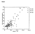
- IgG anti-Fc ⁇ RI ⁇ autoreactivity in 85 patients suffering from chronic urticaria and 20 atopic dermatitis patients versus 31 healthy controls was evaluated. Results are shown in Figure 8. IgG anti-Fc ⁇ RI ⁇ autoreactivity is found predominantly if not exclusively in the group of CU patients.
Landscapes
- Health & Medical Sciences (AREA)
- Life Sciences & Earth Sciences (AREA)
- Engineering & Computer Science (AREA)
- Immunology (AREA)
- Hematology (AREA)
- Biomedical Technology (AREA)
- Chemical & Material Sciences (AREA)
- Urology & Nephrology (AREA)
- Molecular Biology (AREA)
- Medicinal Chemistry (AREA)
- General Health & Medical Sciences (AREA)
- Microbiology (AREA)
- Biotechnology (AREA)
- Pathology (AREA)
- General Physics & Mathematics (AREA)
- Food Science & Technology (AREA)
- Cell Biology (AREA)
- Physics & Mathematics (AREA)
- Analytical Chemistry (AREA)
- Biochemistry (AREA)
- Bioinformatics & Cheminformatics (AREA)
- Tropical Medicine & Parasitology (AREA)
- Proteomics, Peptides & Aminoacids (AREA)
- Rehabilitation Therapy (AREA)
- Rheumatology (AREA)
- Toxicology (AREA)
- Investigating Or Analysing Biological Materials (AREA)
- Peptides Or Proteins (AREA)
- Pyrane Compounds (AREA)
- Medicines That Contain Protein Lipid Enzymes And Other Medicines (AREA)
- Other Investigation Or Analysis Of Materials By Electrical Means (AREA)
- Preparation Of Compounds By Using Micro-Organisms (AREA)
- Transition And Organic Metals Composition Catalysts For Addition Polymerization (AREA)
- Medicines Containing Plant Substances (AREA)
Abstract
Description
In conventional ELISA systems either the antigen which is to be detected, or a specific antibody which binds to the respective antigen, is bound to a solid phase (microtiter plate) by hydrophobic interactions: the protein interacts with the solid phase, usually a polystyrene surface at high pH. Although this bond is responsible for all the consecutive steps in the procedure, it remains the weakest bridge to the assay support, the ELISA plate. Strong detergents at higher concentrations such as 0.05 % are able to diminish the amount of reagent bound to the plate and can even abolish binding totally. The consecutive steps in ELISA technique, such as the binding of an antigen to the solid phase-bound antibody and further binding of a second antibody, occur with an affinity of approximately 10-12 to 10-10 mol per liter. One can view this as being similar to an inverse binding cascade from the bottom of the plate to the top, as with a pyramide standing on its top.
Another concern is the intramolecular event upon binding. A protein such as a cell-receptor or an antibody behaves very flexibly according to its polypeptide structure, which forms a complex architecture in solution. This explains its high specificity and selectivity to the ligands which are to bind to it in vivo. Enzymatic activities for example are entirely dependent upon the proper formation of the active site pocket, which itself remains flexible in order to engulf the substrate and release the product. Cell-receptors such as Fc εRIα react with an affinity for IgE in vivo of 10-10 mol per liter provided the respective active site is properly exposed. Most proteins react to binding to a given surface with a dramatic change of their tertiary structure, i.e. they unfold, refold, hide their active site or change their conformation in such a way that their reactivitv toward a given ligand is altered or even cancelled. In order to circumvent this disadvantage, in conventional ELISA systems a catching antibody is used. This antibody binds to the polystyrene plate and exposes the high affinity hyper-variable region toward the incoming antigen. The antigen is then detected by a second antibody, which is labelled directly or indirectly (via biotin/avidin) with an enzyme. This enzyme will cleave a chromogenic substrate, which itself is converted from the leucoform to the chromoform and thus visualizes the presence of the antigen in question. But even catching antibodies may affect a given protein by changing its conformation. This is demonstrated by many examples of therapeutic antibodies whose mode of action is the blocking of an active site on, or the alteration of, a biospecific molecule. EP-A-0 507 586 discloses a homogeneous immunoassay for immunoa labelled second globulins employing a biotinglated first antigen, antigen and an oridin coated solid surface. JP 93/02 18920 discloses on IgE assay in which FcεRI-IgE binding is determined.
During the reaction of the receptor with its ligand (IgE) in solution, both molecules float freely in solution and thus are not influenced by the structural modification of solid phases. They receive their conformation by the given pH and salt concentration and exert their maximal binding affinity. After a given incubation time, selected according to the results of appropriate equilibrium measurements, the receptor/ligand complex has to be deprived of all other compounds. The receptor is therefore biotinylated prior to the above reaction and an aliquot of the reaction is transferred to a streptavidin-coated microtiter plate. In order to build up an affinity cascade from the bottom of the microtiter plate to the top of the reactants, the binding of streptavidin with the plate surface is preferably made covalent. For example, a microtiter plate (such as a DNA-Bind plate) chemically modified with e.g. a N-oxysuccinimide ester coating is used, which reacts with nucleophiles such as primary amines under formation of a covalent bond. This bond is stronger than 10-15 moles per liter. For the next step, the biotinylation of the receptor, a chemically modified biotin, such as NHS-LC-Biotin is used, which comprises an extended spacer arm of approximately 22.4 Å in length. This long chain analogue reduces steric hindrances associated with the binding of four biotinylated molecules on one streptavidin molecule. The target of biotinylation on the IgE-receptor molecule is thereby well defined and interference with the ligand is avoided. The binding affinity of biotin to streptavidin is also known to be 10-15 moles per liter. Thereby the first two steps of the binding cascade are established and reach from the bottom of the plate to streptavidin to the biotinylated receptor. The affinity constants are decreasing from the bottom to the top. At this stage the reactants, which have found their partners in solution under appropriate conditions, are trapped by the means of the streptavidin plate, and the bound IgE is then detected with an appropriate enzyme-labeled antibody, such as POD-labeled antibody against human IgE. The affinity cascade now reaches from <10-15 to 10-15 to 10-10 to 10-12 for the anti-IgE antibody toward IgE. Any interference of biochemical material and especially of IgE-receptor binding inhibitors will occur at the point with the lowest affinity, which resides between the IgE and the receptor (10-10), and not at the point of attachment of the assay to the plate. This would be read as a false positive result - an artifact of the system.
More particularly, the invention concerns an ELISA-type assay for finding inhibitors of the binding of human IgE to its high affinity receptor, the α-chain of FcεRI (FcεRIα), especially substances inhibiting IgE-mediated early phase response in allergic reactions. The assay mimics natural conditions of binding of IgE to its receptor, i.e. all binding partners interact in solution instead of one of them being immobilized on a solid support. In the binding reaction biotinylated soluble FcεRIα chain interacts with free, soluble IgE at a defined molar ratio (1 : 2.5) in the presence of inhibitory or interfering substance to reach thermodynamic equilibrium within a three-hours incubation period at 37°C. The IgE/FcεRI complex formed under these conditions is captured via the biotin-linker coupled to the FcεRIα chain on ELISA plates modified by covalently-linked streptavidin. The amount of captured complex is determined by identifying bound IgE via a peroxidase-linked anti-human IgE antibody preparation, such as anti-human IgE (ε-chain-specific) peroxidase conjugate, e.g. Sigma no. A-9667. The concentrations of the binding partners in primary screening are chosen to obtain an optimal signal to noise ratio under nonsaturating conditions. In this experimental setting the Kd of the IgE/FcεRI complex formed is found to be 4 nM. The maximally tolerated dose of DMSO in the test solution is 5 % and therefore, at a concentration of pure compound of 50 µM competitive inhibitors with a Kd of 30-10 µM can be easily detected in primary screening at a cut-off level of 30 % inhibition. As a standard control a monoclonal antibody recognizing free as well as receptor-bound IgE and inhibiting its binding to the receptor or releasing it from the complex, respectively, such as BSW17, is tested in 5 different concentrations on every screening plate.
Some of the advantages of this assay in comparison to standard ELISA systems where the receptor or antigen first will be attached to a solid support with either a catching antibody or by high pH (9.6) are the following:
Further, the soluble extracellular part of the α-chain, the IgE-binding subunit of FcεRI, is available through recombinant DNA technology and hence the assay for high capacity screening of inhibitors of the IgE/FcεRI interaction could be established cell-free. To mimic the natural binding reaction of IgE to the α-chain the assay is designed for the interaction of binding partners to occur in solution. Instead of directly immobilizing one binding partner on a solid support by non-covalent adsorption techniques, e.g. the α-chain by hydrophobic interactions on the surface of ELISA plates, an interaction which is highly susceptible to detergent-like substances, a more stable form of fixation to the solid medium is evaluated. The affinity of biotin for streptavidin is orders of magnitude higher (Kd approximately 10-15 M) than that of IgE for FcεRI. Therefore, inhibitors of the IgE/FcεRI interaction are unlikely to disturb the interaction of biotin and streptavidin. Additionally, the latter is insensitive to extremes of pH. Hence, this interaction was chosen to capture the IgE/α-chain complex on the surface of ELISA plates. For this purpose, the α-chain is biotinylated using an extended spacer arm for chemical coupling and streptavidin is covalently bound to the ELISA plate via amino groups. The binding reactions between IgE and its high affinity receptor, FcεRIα, and the interference of test compound are performed in a separate microtiter plate to reach the equilibrium in solution during an extended incubation period at 37°C. To favor conditions for inhibitory compounds to be active, they are added to and preincubated with the α-chain prior to the addition of IgE. The residual IgE/α-chain complex formed in the presence of test compound is then captured on the streptavidin plate during a short incubation period at room temperature or alternatively, after an overnight incubation at 4°C. The amount of captured complex is determined by a peroxidase-catalyzed colorimetric reaction using an anti-human IgE antibody preparation with covalently coupled enzyme.
A high molecular weight pool is detected containing highly biotinylated but aggregated material, which binds to the plate but consequently is inactive in binding to IgE.
The appropriate IgE-binding competent material is found at the expected elution position of 32 kDa. Remaining reactants are seen in a low molecular weight fraction as well.
The plate is sealed with an adhesive foil and incubated for three hours at 37°C. Thereafter 100 µl are transferred from the incubation plate into the streptavidin covalent plate which is kept overnight at 4°C. After a five times wash 100 µl of anti IgE-POD [1:2000] are added and the plate is incubated for another hour at room temperature. The plate is washed again five times and 100 µl of TMB-substrate are added. After 20 minutes incubation at room temperature the reaction is stopped by addition of 100 µl of 4 N H2SO4 and the plate is read at 450/690 nm.
- B11 =
- recombinant human IgE monoclonal antibody (protein concentration 1850 µg/ml; molecular weight 188 kDa)
- BSA =
- bovine serum albumin
- BSW17 =
- IgG monoclonal antibody directed against the CH3 epitope of native IgE
(protein concentration 2.3 mg/ml;
molecular weight 150 kDa) - CU =
- chronic urticaria
- DMF =
- dimethylformamide
- DMSO =
- dimethylsulfoxide
- EDTA =
- ethylene diamine tetraacetic acid
- ELISA =
- enzyme-linked immunosorbent assay
- FCS =
- fetal calf serum
- FPLC =
- forced pressure liquid chromatography
- IgE =
- immunoglobulin E
- LC =
- long chain spacer arm
- mAb =
- monoclonal antibody
- Mwt =
- molecular weight
- PBS =
- phosphate-buffered saline
- PBST =
- phosphate-buffered saline /
Tween 20 - POD =
- horse radish peroxydase
- TMB =
- tetramethylbenzidine
- WB =
- Western blot
- = IgE binding
I) Serum specimens are reacted with an anti-IgE mAb and thereafter incubated with biotinylated rsFcεRIα: II) IgE is blocked, α-chain specific IgG can bind to its antigen; III) IgG anti- Fc εRIα/rsFcεRIαbiot complexes bind to the streptavidin-coated plate and are detected with enzyme-coupled anti-human IgG. This embodiment falls outside the scope of the invention.
Sera (diluted 1 : 15) from atopic patients (AD) and from chronic urticaria patients (CU) were analyzed for anti-IgG and anti-IgE reactivity with rsFcεRIα in the presence (closed bars) or absence of the anti-IgE mAb BSW17 (dotted bars). y-axis shows dilution-corrected optical density (OD x 1000).

560 µl 0.3 % H2O2:
10 mM EDTA (3.7 g per liter);
Urticaria is a common disorder characterized by the eruption of transitory, itchy skin swellings that are frequently associated with the occurrence of debilitating and potentially life-threatening angioedema. Although accurate data on the prevalence of urticaria are not available, it is estimated that 15 % to 32 % of the general population experience urticarial/angio-edematous symptoms during their life time. In about 25 % of these patients the initially sporadic character of this condition is followed by a chronic disease period with symptoms of frequent but unpredictable occurrence. If widespread wheals tend to appear daily or almost daily for at least six weeks the disease is termed chronic urticaria (CU). Taken together, CU is a frequent disease afflicting a considerable proportion of the population in the Western hemisphere.
Although it is a well known fact that histamine release from mast cells is of central pathophysiological relevance for the elicitation of CU, the causative agent(s) that mediate mast cell activation remained obscure until very recently. The existence of autoantibodies directed against the high affinity IgE receptor FcεRI in the serum of CU patients was shown. These autoantibodies were found to cross-link FcεRI on basophils and mast cells, resulting in the exocytosis of histamine by these cells. Using recombinant soluble FcεRIα as a reaction target for autoantibodies, it was found that about 37 % of CU sera exhibited IgG autoreactivity against FcεRIα. No IgG anti-FcεRIα reactivity was observed in the serum of atopic dermatitis patients or healthy control individuals. Therefore, IgG autoreactivity against FcεRIα is of central pathophysiological importance for CU and furthermore is a selective marker identifying an autoimmune-mediated sub-entity of this disease. It is hence of great interest to develop strategies that allow the reliable and fast detection and quantification of such serum autoantibodies.
A further indication of the urgent need for an appropriate screening system comes from the fact that up to now laborious and cost-intensive searches for certain implicated, but not unequivocally proven, etiopathogenetic conditions, e.g. bacterial or fungal infections, hormonal dysregulation, psychological factors, neoplasms, and intolerance to certain food activities, arc performed in most CU patients. It is obvious that such laboratory, physical, and psychological examinations, which often require long-lasting hospitalization of the patients, cause enormous financial expense. Thus, the quick and easy detection of disease-eliciting autoantibodies helps to drastically reduce the requirement for diagnostic procedures that are unrevealing in most patients.
Results obtained indicate that the ability to block ELISA reactivity by high concentrations of rsFcεRIα can be used to discriminate IgG anti-FcεRIα autoantibody-containing and non-containing serum samples. Thus, besides the OD value reflecting the IgG-binding to the ELISA plates, the ability to block this reactivity by non-labeled rsFcεRIα is a valuable indicator for the presence of IgG anti-FcεRIα autoantibodies (Figure 6).
Claims (3)
- Assay for screening of substances interfering with the attachment of human IgE to its high affinity receptor FcεRIα, and/or of substances capable of detaching already bound IgE from this receptor, comprising reacting, in a sample possibly containing such substances , dissolved biotinylated FcεRIα receptor with IgE , at well-defined molar concentrations, temperature, pH and salt conditions, and transferring after a defined incubation time an aliquot of the resultant binding reaction mixture to a streptavidin-coated microtiter plate, where the biotinylated FcεRIα-receptor/IgE complex is quantified by means of an appropriate enzyme-labeled antibody against human IgE.
- Method for the differential analysis between autoimmune disorders and classical allergies, comprising determination, in a serum sample from a patient, which has been preincubated with anti-IgE antibody of the presence or absence of autoreactive antibody against FcεRIα receptor according to the assay defined in claim 1.
- An assay according to claim 1 or 2 wherein the antibody for quantifying is based on the antibody against human IgE which is antibody BSW17 produced by the hybridoma cell line deposited with the ECACC under the provisions of the Budapest treaty on December 18, 1996 under Deposit number 96121916.
Applications Claiming Priority (3)
| Application Number | Priority Date | Filing Date | Title |
|---|---|---|---|
| GB9526599 | 1995-12-28 | ||
| GBGB9526599.7A GB9526599D0 (en) | 1995-12-28 | 1995-12-28 | Elisa test system |
| PCT/EP1996/005824 WO1997024617A1 (en) | 1995-12-28 | 1996-12-23 | Elisa test system |
Publications (2)
| Publication Number | Publication Date |
|---|---|
| EP0882229A1 EP0882229A1 (en) | 1998-12-09 |
| EP0882229B1 true EP0882229B1 (en) | 2003-03-19 |
Family
ID=10786124
Family Applications (1)
| Application Number | Title | Priority Date | Filing Date |
|---|---|---|---|
| EP96944649A Expired - Lifetime EP0882229B1 (en) | 1995-12-28 | 1996-12-23 | Elisa testsystem for detecting substances interfering with the binding between ig e and its receptor |
Country Status (12)
| Country | Link |
|---|---|
| US (1) | US6258549B1 (en) |
| EP (1) | EP0882229B1 (en) |
| JP (1) | JP2000502793A (en) |
| AT (1) | ATE235054T1 (en) |
| AU (1) | AU1305897A (en) |
| CY (1) | CY2470B1 (en) |
| DE (1) | DE69626860T2 (en) |
| DK (1) | DK0882229T3 (en) |
| ES (1) | ES2195037T3 (en) |
| GB (1) | GB9526599D0 (en) |
| PT (1) | PT882229E (en) |
| WO (1) | WO1997024617A1 (en) |
Families Citing this family (10)
| Publication number | Priority date | Publication date | Assignee | Title |
|---|---|---|---|---|
| US5945294A (en) | 1996-11-26 | 1999-08-31 | Heska Corporation | Method to detect IgE |
| AU769954B2 (en) * | 1996-11-26 | 2004-02-12 | Heska Corporation | Method to detect IgE |
| US7759133B2 (en) * | 1998-12-22 | 2010-07-20 | Alk-Abello A/S | Method of detecting and/or quantifying a specific IgE antibody in a liquid sample |
| AU2002326751A1 (en) * | 2001-08-13 | 2003-03-03 | Ige Therapeutics, Inc. | Immunoglobulin e vaccines and methods of use thereof |
| US20060154372A1 (en) * | 2004-12-21 | 2006-07-13 | Arter Thomas C | Providing additional motion in assays |
| US20080206855A1 (en) * | 2005-05-06 | 2008-08-28 | Kalypsys, Inc. | Automated Compound Screening Using Gel-Permeation Matrix and Pin-Based Sample Transfer |
| US20110256641A1 (en) * | 2010-04-19 | 2011-10-20 | Michael Ling | Methods and Systems for Detecting Free IgE |
| CN110358738B (en) * | 2019-08-22 | 2021-08-13 | 中国食品药品检定研究院 | A kind of method for stable determination of anti-IgE antibody drug biological activity |
| CN110716054A (en) * | 2019-09-11 | 2020-01-21 | 天津医科大学 | A kit for quantitative detection of serum cytophilic immunoglobulin E |
| CN110716055A (en) * | 2019-09-11 | 2020-01-21 | 天津医科大学 | Allergen specificity immune globulin E pathogenic activity detection kit |
Family Cites Families (8)
| Publication number | Priority date | Publication date | Assignee | Title |
|---|---|---|---|---|
| US4935339A (en) * | 1985-05-07 | 1990-06-19 | Nichols Institute Diagnostics | Delayed solid phase immunologic assay |
| US4962035A (en) | 1987-12-01 | 1990-10-09 | President And Fellows Of Harvard College | DNA encoding IgE receptor alpha-subunit or fragment thereof |
| US5639660A (en) | 1988-02-24 | 1997-06-17 | Hoffmann-La Roche Inc. | Polypeptide and DNA sequence corresponding to the human receptor with high affinity for IgE |
| JPH05133952A (en) | 1991-04-03 | 1993-05-28 | Syntex Usa Inc | Immunoassay of immunoglobulins |
| DK137992D0 (en) * | 1992-11-13 | 1992-11-13 | Alk Lab As | PROCEDURE FOR DETERMINING AN IMMUNOLOGICALLY ACTIVE SUBSTANCE IN A TEST USING A LABELING SUBSTANCE |
| JPH0772150A (en) * | 1993-09-02 | 1995-03-17 | Tonen Corp | IgE measurement method |
| EP0733207B1 (en) * | 1993-12-10 | 1997-08-27 | Genentech, Inc. | Methods for diagnosis of allergy and screening of anti-allergy therapeutics |
| US5962634A (en) * | 1994-07-08 | 1999-10-05 | Thomas Jefferson University | IgE antagonists |
-
1995
- 1995-12-28 GB GBGB9526599.7A patent/GB9526599D0/en active Pending
-
1996
- 1996-12-23 US US09/101,053 patent/US6258549B1/en not_active Expired - Fee Related
- 1996-12-23 PT PT96944649T patent/PT882229E/en unknown
- 1996-12-23 DE DE69626860T patent/DE69626860T2/en not_active Expired - Fee Related
- 1996-12-23 AU AU13058/97A patent/AU1305897A/en not_active Abandoned
- 1996-12-23 AT AT96944649T patent/ATE235054T1/en not_active IP Right Cessation
- 1996-12-23 WO PCT/EP1996/005824 patent/WO1997024617A1/en not_active Ceased
- 1996-12-23 EP EP96944649A patent/EP0882229B1/en not_active Expired - Lifetime
- 1996-12-23 DK DK96944649T patent/DK0882229T3/en active
- 1996-12-23 JP JP9524017A patent/JP2000502793A/en active Pending
- 1996-12-23 ES ES96944649T patent/ES2195037T3/en not_active Expired - Lifetime
-
2004
- 2004-08-10 CY CY0400061A patent/CY2470B1/en unknown
Also Published As
| Publication number | Publication date |
|---|---|
| GB9526599D0 (en) | 1996-02-28 |
| PT882229E (en) | 2003-07-31 |
| AU1305897A (en) | 1997-07-28 |
| US6258549B1 (en) | 2001-07-10 |
| CY2470B1 (en) | 2005-06-03 |
| DK0882229T3 (en) | 2003-07-14 |
| EP0882229A1 (en) | 1998-12-09 |
| ATE235054T1 (en) | 2003-04-15 |
| JP2000502793A (en) | 2000-03-07 |
| WO1997024617A1 (en) | 1997-07-10 |
| DE69626860T2 (en) | 2003-12-04 |
| ES2195037T3 (en) | 2003-12-01 |
| DE69626860D1 (en) | 2003-04-24 |
Similar Documents
| Publication | Publication Date | Title |
|---|---|---|
| JP2726793B2 (en) | Immunoassay | |
| EP0615129B1 (en) | Methods for selectively detecting perinuclear anti-neutrophil cytoplasmic antibody of ulcerative colitis or primary sclerosing cholangitis | |
| JP3542808B2 (en) | Cell counting immunoassay | |
| EP2906600A1 (en) | Monoclonal antibodies and detection methods for enzymes that confer resistance to phosphinothricin-n-acetyl-transferase | |
| US6258549B1 (en) | Elisa screening method for inhibitors of human IgE binding to the high affinity receptor, FcεRIα | |
| Wang et al. | Comparison of eight commercial kits for quantitation of antineutrophil cytoplasmic antibodies (ANCA) | |
| US5741654A (en) | Immunoassay for the detection of human autoantibodies | |
| JPH1078435A (en) | Rise of sensitivity in immunochemical measurement for material to be inspected | |
| EP0585346A1 (en) | Hla typing | |
| AU771535B2 (en) | Method of diagnosing Sjogren's syndrome | |
| JP4438455B2 (en) | Method for measuring free human immunoglobulin light chain and kit | |
| CA2476979A1 (en) | Assay for anti-ingap antibodies | |
| CA2329948A1 (en) | Solution-phase elisa | |
| EP0398292B1 (en) | Diagnostic composition for rheumatoid arthritis | |
| KR940008091B1 (en) | Immunological Detection of Ligands | |
| JP3309900B2 (en) | Method for detecting anti-FKBP12 autoantibody | |
| RU2197736C1 (en) | Reagent for immunoenzyme analysis and method of immunoenzyme testing of specific antibody in opisthorchiasis caused by hepatic trematode opisthorchis felineus | |
| Muñoz et al. | Quantitation of secretory group V phospholipase A2 in human tissues by sandwich enzyme-linked immunosorbent assay | |
| US20020004247A1 (en) | Assay method | |
| WO2001044499A9 (en) | Modified labeled complement components for immunoassays | |
| CA2130486A1 (en) | Method and kit for the determination of antibodies | |
| Coppo et al. | A solid phase enzyme immunoassay for the measurement of urinary albumin and the detection of microalbuminuria | |
| JP2023542587A (en) | Compositions and methods for diagnosing primary biliary cirrhosis | |
| JPH10332692A (en) | Immunological assay for human MCP-3 | |
| Falcioni et al. | A simple and sensitive fluorometric immunoassay for the measurement of immunoglobulins in culture medium of mitogen-stimulated lymphocytes |
Legal Events
| Date | Code | Title | Description |
|---|---|---|---|
| PUAI | Public reference made under article 153(3) epc to a published international application that has entered the european phase |
Free format text: ORIGINAL CODE: 0009012 |
|
| 17P | Request for examination filed |
Effective date: 19980606 |
|
| AK | Designated contracting states |
Kind code of ref document: A1 Designated state(s): AT BE CH DE DK ES FI FR GB GR IE IT LI LU MC NL PT SE |
|
| 17Q | First examination report despatched |
Effective date: 20010611 |
|
| RAP1 | Party data changed (applicant data changed or rights of an application transferred) |
Owner name: NOVARTIS-ERFINDUNGEN VERWALTUNGSGESELLSCHAFT M.B. Owner name: NOVARTIS AG |
|
| GRAG | Despatch of communication of intention to grant |
Free format text: ORIGINAL CODE: EPIDOS AGRA |
|
| RTI1 | Title (correction) |
Free format text: ELISA TESTSYSTEM FOR DETECTING SUBSTANCES INTERFERING WITH THE BINDING BETWEEN IG E AND ITS RECEPTOR |
|
| RTI1 | Title (correction) |
Free format text: ELISA TESTSYSTEM FOR DETECTING SUBSTANCES INTERFERING WITH THE BINDING BETWEEN IG E AND ITS RECEPTOR |
|
| RTI1 | Title (correction) |
Free format text: ELISA TESTSYSTEM FOR DETECTING SUBSTANCES INTERFERING WITH THE BINDING BETWEEN IG E AND ITS RECEPTOR |
|
| GRAG | Despatch of communication of intention to grant |
Free format text: ORIGINAL CODE: EPIDOS AGRA |
|
| GRAH | Despatch of communication of intention to grant a patent |
Free format text: ORIGINAL CODE: EPIDOS IGRA |
|
| GRAH | Despatch of communication of intention to grant a patent |
Free format text: ORIGINAL CODE: EPIDOS IGRA |
|
| GRAA | (expected) grant |
Free format text: ORIGINAL CODE: 0009210 |
|
| AK | Designated contracting states |
Designated state(s): AT BE CH DE DK ES FI FR GB GR IE IT LI LU MC NL PT SE |
|
| REG | Reference to a national code |
Ref country code: GB Ref legal event code: FG4D |
|
| REG | Reference to a national code |
Ref country code: CH Ref legal event code: EP |
|
| REG | Reference to a national code |
Ref country code: IE Ref legal event code: FG4D |
|
| REF | Corresponds to: |
Ref document number: 69626860 Country of ref document: DE Date of ref document: 20030424 Kind code of ref document: P |
|
| REG | Reference to a national code |
Ref country code: DK Ref legal event code: T3 |
|
| REG | Reference to a national code |
Ref country code: SE Ref legal event code: TRGR |
|
| REG | Reference to a national code |
Ref country code: GR Ref legal event code: EP Ref document number: 20030402192 Country of ref document: GR |
|
| ET | Fr: translation filed | ||
| PLBE | No opposition filed within time limit |
Free format text: ORIGINAL CODE: 0009261 |
|
| STAA | Information on the status of an ep patent application or granted ep patent |
Free format text: STATUS: NO OPPOSITION FILED WITHIN TIME LIMIT |
|
| 26N | No opposition filed |
Effective date: 20031222 |
|
| PGFP | Annual fee paid to national office [announced via postgrant information from national office to epo] |
Ref country code: NL Payment date: 20081203 Year of fee payment: 13 Ref country code: MC Payment date: 20081127 Year of fee payment: 13 Ref country code: LU Payment date: 20081230 Year of fee payment: 13 Ref country code: IE Payment date: 20081216 Year of fee payment: 13 Ref country code: DK Payment date: 20081212 Year of fee payment: 13 Ref country code: CH Payment date: 20081230 Year of fee payment: 13 |
|
| PGFP | Annual fee paid to national office [announced via postgrant information from national office to epo] |
Ref country code: PT Payment date: 20081222 Year of fee payment: 13 Ref country code: FI Payment date: 20081212 Year of fee payment: 13 Ref country code: AT Payment date: 20081211 Year of fee payment: 13 |
|
| PGFP | Annual fee paid to national office [announced via postgrant information from national office to epo] |
Ref country code: SE Payment date: 20081205 Year of fee payment: 13 Ref country code: IT Payment date: 20081220 Year of fee payment: 13 |
|
| PGFP | Annual fee paid to national office [announced via postgrant information from national office to epo] |
Ref country code: FR Payment date: 20081212 Year of fee payment: 13 Ref country code: ES Payment date: 20090120 Year of fee payment: 13 |
|
| PGFP | Annual fee paid to national office [announced via postgrant information from national office to epo] |
Ref country code: DE Payment date: 20081219 Year of fee payment: 13 |
|
| PGFP | Annual fee paid to national office [announced via postgrant information from national office to epo] |
Ref country code: GR Payment date: 20081113 Year of fee payment: 13 Ref country code: GB Payment date: 20081217 Year of fee payment: 13 |
|
| PGFP | Annual fee paid to national office [announced via postgrant information from national office to epo] |
Ref country code: BE Payment date: 20090119 Year of fee payment: 13 |
|
| BERE | Be: lapsed |
Owner name: *NOVARTIS A.G. Effective date: 20091231 |
|
| REG | Reference to a national code |
Ref country code: PT Ref legal event code: MM4A Free format text: LAPSE DUE TO NON-PAYMENT OF FEES Effective date: 20100623 |
|
| REG | Reference to a national code |
Ref country code: NL Ref legal event code: V1 Effective date: 20100701 |
|
| EUG | Se: european patent has lapsed | ||
| PG25 | Lapsed in a contracting state [announced via postgrant information from national office to epo] |
Ref country code: PT Free format text: LAPSE BECAUSE OF NON-PAYMENT OF DUE FEES Effective date: 20100623 Ref country code: MC Free format text: LAPSE BECAUSE OF NON-PAYMENT OF DUE FEES Effective date: 20100701 |
|
| REG | Reference to a national code |
Ref country code: CH Ref legal event code: PL |
|
| REG | Reference to a national code |
Ref country code: DK Ref legal event code: EBP |
|
| GBPC | Gb: european patent ceased through non-payment of renewal fee |
Effective date: 20091223 |
|
| PG25 | Lapsed in a contracting state [announced via postgrant information from national office to epo] |
Ref country code: FI Free format text: LAPSE BECAUSE OF NON-PAYMENT OF DUE FEES Effective date: 20091223 Ref country code: AT Free format text: LAPSE BECAUSE OF NON-PAYMENT OF DUE FEES Effective date: 20091223 |
|
| REG | Reference to a national code |
Ref country code: FR Ref legal event code: ST Effective date: 20100831 |
|
| REG | Reference to a national code |
Ref country code: IE Ref legal event code: MM4A |
|
| PG25 | Lapsed in a contracting state [announced via postgrant information from national office to epo] |
Ref country code: NL Free format text: LAPSE BECAUSE OF NON-PAYMENT OF DUE FEES Effective date: 20100701 Ref country code: LI Free format text: LAPSE BECAUSE OF NON-PAYMENT OF DUE FEES Effective date: 20091231 Ref country code: IE Free format text: LAPSE BECAUSE OF NON-PAYMENT OF DUE FEES Effective date: 20091223 Ref country code: FR Free format text: LAPSE BECAUSE OF NON-PAYMENT OF DUE FEES Effective date: 20091231 Ref country code: CH Free format text: LAPSE BECAUSE OF NON-PAYMENT OF DUE FEES Effective date: 20091231 Ref country code: BE Free format text: LAPSE BECAUSE OF NON-PAYMENT OF DUE FEES Effective date: 20091231 |
|
| PG25 | Lapsed in a contracting state [announced via postgrant information from national office to epo] |
Ref country code: DE Free format text: LAPSE BECAUSE OF NON-PAYMENT OF DUE FEES Effective date: 20100701 |
|
| PG25 | Lapsed in a contracting state [announced via postgrant information from national office to epo] |
Ref country code: GB Free format text: LAPSE BECAUSE OF NON-PAYMENT OF DUE FEES Effective date: 20091223 |
|
| PG25 | Lapsed in a contracting state [announced via postgrant information from national office to epo] |
Ref country code: DK Free format text: LAPSE BECAUSE OF NON-PAYMENT OF DUE FEES Effective date: 20100104 |
|
| REG | Reference to a national code |
Ref country code: ES Ref legal event code: FD2A Effective date: 20110324 |
|
| PG25 | Lapsed in a contracting state [announced via postgrant information from national office to epo] |
Ref country code: IT Free format text: LAPSE BECAUSE OF NON-PAYMENT OF DUE FEES Effective date: 20091223 |
|
| PG25 | Lapsed in a contracting state [announced via postgrant information from national office to epo] |
Ref country code: LU Free format text: LAPSE BECAUSE OF NON-PAYMENT OF DUE FEES Effective date: 20091223 |
|
| PG25 | Lapsed in a contracting state [announced via postgrant information from national office to epo] |
Ref country code: SE Free format text: LAPSE BECAUSE OF NON-PAYMENT OF DUE FEES Effective date: 20091224 |
|
| PG25 | Lapsed in a contracting state [announced via postgrant information from national office to epo] |
Ref country code: ES Free format text: LAPSE BECAUSE OF NON-PAYMENT OF DUE FEES Effective date: 20110310 |
|
| PG25 | Lapsed in a contracting state [announced via postgrant information from national office to epo] |
Ref country code: ES Free format text: LAPSE BECAUSE OF NON-PAYMENT OF DUE FEES Effective date: 20091224 |