EP0881301B1 - Method for assaying vital sample - Google Patents
Method for assaying vital sample Download PDFInfo
- Publication number
- EP0881301B1 EP0881301B1 EP96941190A EP96941190A EP0881301B1 EP 0881301 B1 EP0881301 B1 EP 0881301B1 EP 96941190 A EP96941190 A EP 96941190A EP 96941190 A EP96941190 A EP 96941190A EP 0881301 B1 EP0881301 B1 EP 0881301B1
- Authority
- EP
- European Patent Office
- Prior art keywords
- adp
- reaction
- enzyme
- assay
- sample
- Prior art date
- Legal status (The legal status is an assumption and is not a legal conclusion. Google has not performed a legal analysis and makes no representation as to the accuracy of the status listed.)
- Expired - Lifetime
Links
- 238000000034 method Methods 0.000 title claims abstract description 96
- 238000006243 chemical reaction Methods 0.000 claims abstract description 154
- 229910001425 magnesium ion Inorganic materials 0.000 claims abstract description 50
- 108010029731 6-phosphogluconolactonase Proteins 0.000 claims abstract description 36
- 102100031126 6-phosphogluconolactonase Human genes 0.000 claims abstract description 36
- 108010018962 Glucosephosphate Dehydrogenase Proteins 0.000 claims abstract description 36
- WQZGKKKJIJFFOK-GASJEMHNSA-N Glucose Natural products OC[C@H]1OC(O)[C@H](O)[C@@H](O)[C@@H]1O WQZGKKKJIJFFOK-GASJEMHNSA-N 0.000 claims abstract description 35
- 239000008103 glucose Substances 0.000 claims abstract description 35
- 238000006911 enzymatic reaction Methods 0.000 claims abstract description 28
- 239000007788 liquid Substances 0.000 claims abstract description 17
- 229910001437 manganese ion Inorganic materials 0.000 claims abstract description 17
- 229910001429 cobalt ion Inorganic materials 0.000 claims abstract description 16
- 150000003839 salts Chemical class 0.000 claims abstract description 13
- 150000002500 ions Chemical class 0.000 claims abstract description 12
- 102000005548 Hexokinase Human genes 0.000 claims abstract description 11
- 108700040460 Hexokinases Proteins 0.000 claims abstract description 11
- 230000001419 dependent effect Effects 0.000 claims abstract description 10
- 239000010941 cobalt Substances 0.000 claims abstract description 6
- GUTLYIVDDKVIGB-UHFFFAOYSA-N cobalt atom Chemical compound [Co] GUTLYIVDDKVIGB-UHFFFAOYSA-N 0.000 claims abstract description 6
- FYYHWMGAXLPEAU-UHFFFAOYSA-N Magnesium Chemical compound [Mg] FYYHWMGAXLPEAU-UHFFFAOYSA-N 0.000 claims abstract description 5
- 239000011777 magnesium Substances 0.000 claims abstract description 5
- 102000004190 Enzymes Human genes 0.000 claims description 77
- 108090000790 Enzymes Proteins 0.000 claims description 77
- 239000003153 chemical reaction reagent Substances 0.000 claims description 73
- PEDCQBHIVMGVHV-UHFFFAOYSA-N Glycerine Chemical compound OCC(O)CO PEDCQBHIVMGVHV-UHFFFAOYSA-N 0.000 claims description 72
- DDRJAANPRJIHGJ-UHFFFAOYSA-N creatinine Chemical compound CN1CC(=O)NC1=N DDRJAANPRJIHGJ-UHFFFAOYSA-N 0.000 claims description 64
- 239000000758 substrate Substances 0.000 claims description 57
- CVSVTCORWBXHQV-UHFFFAOYSA-N creatine Chemical compound NC(=[NH2+])N(C)CC([O-])=O CVSVTCORWBXHQV-UHFFFAOYSA-N 0.000 claims description 50
- JLVVSXFLKOJNIY-UHFFFAOYSA-N Magnesium ion Chemical compound [Mg+2] JLVVSXFLKOJNIY-UHFFFAOYSA-N 0.000 claims description 45
- XSQUKJJJFZCRTK-UHFFFAOYSA-N Urea Chemical compound NC(N)=O XSQUKJJJFZCRTK-UHFFFAOYSA-N 0.000 claims description 40
- 229940109239 creatinine Drugs 0.000 claims description 33
- 229960003624 creatine Drugs 0.000 claims description 25
- 239000006046 creatine Substances 0.000 claims description 25
- 239000004202 carbamide Substances 0.000 claims description 20
- 229960001231 choline Drugs 0.000 claims description 19
- OEYIOHPDSNJKLS-UHFFFAOYSA-N choline Chemical compound C[N+](C)(C)CCO OEYIOHPDSNJKLS-UHFFFAOYSA-N 0.000 claims description 17
- 102000004420 Creatine Kinase Human genes 0.000 claims description 13
- 108010042126 Creatine kinase Proteins 0.000 claims description 13
- WHUUTDBJXJRKMK-VKHMYHEASA-N L-glutamic acid Chemical compound OC(=O)[C@@H](N)CCC(O)=O WHUUTDBJXJRKMK-VKHMYHEASA-N 0.000 claims description 12
- WAEMQWOKJMHJLA-UHFFFAOYSA-N Manganese(2+) Chemical compound [Mn+2] WAEMQWOKJMHJLA-UHFFFAOYSA-N 0.000 claims description 12
- XLJKHNWPARRRJB-UHFFFAOYSA-N cobalt(2+) Chemical compound [Co+2] XLJKHNWPARRRJB-UHFFFAOYSA-N 0.000 claims description 11
- 102000057621 Glycerol kinases Human genes 0.000 claims description 10
- 229930195714 L-glutamate Natural products 0.000 claims description 10
- 108700016170 Glycerol kinases Proteins 0.000 claims description 8
- 102000011420 Phospholipase D Human genes 0.000 claims description 8
- 108090000553 Phospholipase D Proteins 0.000 claims description 8
- 108010046334 Urease Proteins 0.000 claims description 8
- UFTFJSFQGQCHQW-UHFFFAOYSA-N triformin Chemical compound O=COCC(OC=O)COC=O UFTFJSFQGQCHQW-UHFFFAOYSA-N 0.000 claims description 8
- 239000004367 Lipase Substances 0.000 claims description 7
- 102100039611 Glutamine synthetase Human genes 0.000 claims description 5
- 108090001060 Lipase Proteins 0.000 claims description 5
- 102000004882 Lipase Human genes 0.000 claims description 5
- 108020002326 glutamine synthetase Proteins 0.000 claims description 5
- 235000019421 lipase Nutrition 0.000 claims description 5
- 102000002745 Choline Kinase Human genes 0.000 claims description 4
- 108010018888 Choline kinase Proteins 0.000 claims description 4
- 108090000371 Esterases Proteins 0.000 claims description 4
- CKLJMWTZIZZHCS-REOHCLBHSA-N L-aspartic acid Chemical compound OC(=O)[C@@H](N)CC(O)=O CKLJMWTZIZZHCS-REOHCLBHSA-N 0.000 claims description 4
- ATBOMIWRCZXYSZ-XZBBILGWSA-N [1-[2,3-dihydroxypropoxy(hydroxy)phosphoryl]oxy-3-hexadecanoyloxypropan-2-yl] (9e,12e)-octadeca-9,12-dienoate Chemical compound CCCCCCCCCCCCCCCC(=O)OCC(COP(O)(=O)OCC(O)CO)OC(=O)CCCCCCC\C=C\C\C=C\CCCCC ATBOMIWRCZXYSZ-XZBBILGWSA-N 0.000 claims description 4
- AWUCVROLDVIAJX-UHFFFAOYSA-N alpha-glycerophosphate Natural products OCC(O)COP(O)(O)=O AWUCVROLDVIAJX-UHFFFAOYSA-N 0.000 claims description 4
- KPGXRSRHYNQIFN-UHFFFAOYSA-N 2-oxoglutaric acid Chemical compound OC(=O)CCC(=O)C(O)=O KPGXRSRHYNQIFN-UHFFFAOYSA-N 0.000 claims description 3
- 102100036475 Alanine aminotransferase 1 Human genes 0.000 claims description 3
- 108010082126 Alanine transaminase Proteins 0.000 claims description 3
- 108010003415 Aspartate Aminotransferases Proteins 0.000 claims description 3
- 102000004625 Aspartate Aminotransferases Human genes 0.000 claims description 3
- 229960003767 alanine Drugs 0.000 claims description 3
- WTJKGGKOPKCXLL-RRHRGVEJSA-N phosphatidylcholine Chemical compound CCCCCCCCCCCCCCCC(=O)OC[C@H](COP([O-])(=O)OCC[N+](C)(C)C)OC(=O)CCCCCCCC=CCCCCCCCC WTJKGGKOPKCXLL-RRHRGVEJSA-N 0.000 claims description 3
- LDVVTQMJQSCDMK-UHFFFAOYSA-N 1,3-dihydroxypropan-2-yl formate Chemical compound OCC(CO)OC=O LDVVTQMJQSCDMK-UHFFFAOYSA-N 0.000 claims description 2
- QNAYBMKLOCPYGJ-UHFFFAOYSA-N D-alpha-Ala Natural products CC([NH3+])C([O-])=O QNAYBMKLOCPYGJ-UHFFFAOYSA-N 0.000 claims description 2
- 102000005133 Glutamate 5-kinase Human genes 0.000 claims description 2
- 108700023479 Glutamate 5-kinases Proteins 0.000 claims description 2
- QNAYBMKLOCPYGJ-UWTATZPHSA-N L-Alanine Natural products C[C@@H](N)C(O)=O QNAYBMKLOCPYGJ-UWTATZPHSA-N 0.000 claims description 2
- QNAYBMKLOCPYGJ-REOHCLBHSA-N L-alanine Chemical compound C[C@H](N)C(O)=O QNAYBMKLOCPYGJ-REOHCLBHSA-N 0.000 claims description 2
- -1 choline ester Chemical class 0.000 claims description 2
- RHYBFKMFHLPQPH-UHFFFAOYSA-N N-methylhydantoin Chemical compound CN1CC(=O)NC1=O RHYBFKMFHLPQPH-UHFFFAOYSA-N 0.000 claims 6
- 239000000126 substance Substances 0.000 abstract description 5
- 230000008033 biological extinction Effects 0.000 abstract 1
- 238000003556 assay Methods 0.000 description 113
- BAWFJGJZGIEFAR-NNYOXOHSSA-N NAD zwitterion Chemical compound NC(=O)C1=CC=C[N+]([C@H]2[C@@H]([C@H](O)[C@@H](COP([O-])(=O)OP(O)(=O)OC[C@@H]3[C@H]([C@@H](O)[C@@H](O3)N3C4=NC=NC(N)=C4N=C3)O)O2)O)=C1 BAWFJGJZGIEFAR-NNYOXOHSSA-N 0.000 description 67
- 229940088598 enzyme Drugs 0.000 description 67
- 229950006238 nadide Drugs 0.000 description 67
- 239000000523 sample Substances 0.000 description 53
- TWRXJAOTZQYOKJ-UHFFFAOYSA-L Magnesium chloride Chemical compound [Mg+2].[Cl-].[Cl-] TWRXJAOTZQYOKJ-UHFFFAOYSA-L 0.000 description 38
- 239000012472 biological sample Substances 0.000 description 32
- QKNYBSVHEMOAJP-UHFFFAOYSA-N 2-amino-2-(hydroxymethyl)propane-1,3-diol;hydron;chloride Chemical compound Cl.OCC(N)(CO)CO QKNYBSVHEMOAJP-UHFFFAOYSA-N 0.000 description 25
- 238000010521 absorption reaction Methods 0.000 description 21
- 239000007853 buffer solution Substances 0.000 description 21
- 230000003287 optical effect Effects 0.000 description 21
- XJLXINKUBYWONI-DQQFMEOOSA-N [[(2r,3r,4r,5r)-5-(6-aminopurin-9-yl)-3-hydroxy-4-phosphonooxyoxolan-2-yl]methoxy-hydroxyphosphoryl] [(2s,3r,4s,5s)-5-(3-carbamoylpyridin-1-ium-1-yl)-3,4-dihydroxyoxolan-2-yl]methyl phosphate Chemical compound NC(=O)C1=CC=C[N+]([C@@H]2[C@H]([C@@H](O)[C@H](COP([O-])(=O)OP(O)(=O)OC[C@@H]3[C@H]([C@@H](OP(O)(O)=O)[C@@H](O3)N3C4=NC=NC(N)=C4N=C3)O)O2)O)=C1 XJLXINKUBYWONI-DQQFMEOOSA-N 0.000 description 19
- 229910001629 magnesium chloride Inorganic materials 0.000 description 19
- 230000000694 effects Effects 0.000 description 18
- 238000012207 quantitative assay Methods 0.000 description 18
- 239000007864 aqueous solution Substances 0.000 description 17
- 239000012490 blank solution Substances 0.000 description 17
- 238000011534 incubation Methods 0.000 description 17
- FAPWRFPIFSIZLT-UHFFFAOYSA-M Sodium chloride Chemical compound [Na+].[Cl-] FAPWRFPIFSIZLT-UHFFFAOYSA-M 0.000 description 15
- XLYOFNOQVPJJNP-UHFFFAOYSA-N water Substances O XLYOFNOQVPJJNP-UHFFFAOYSA-N 0.000 description 14
- 238000005259 measurement Methods 0.000 description 13
- CIWBSHSKHKDKBQ-JLAZNSOCSA-N Ascorbic acid Chemical compound OC[C@H](O)[C@H]1OC(=O)C(O)=C1O CIWBSHSKHKDKBQ-JLAZNSOCSA-N 0.000 description 10
- 239000011541 reaction mixture Substances 0.000 description 10
- 108010066906 Creatininase Proteins 0.000 description 9
- QGZKDVFQNNGYKY-UHFFFAOYSA-N Ammonia Chemical compound N QGZKDVFQNNGYKY-UHFFFAOYSA-N 0.000 description 8
- 230000003247 decreasing effect Effects 0.000 description 8
- 108020005115 Pyruvate Kinase Proteins 0.000 description 7
- 102000013009 Pyruvate Kinase Human genes 0.000 description 7
- 239000011780 sodium chloride Substances 0.000 description 7
- 239000000872 buffer Substances 0.000 description 6
- 239000003638 chemical reducing agent Substances 0.000 description 6
- 108010029444 creatinine deiminase Proteins 0.000 description 6
- 229940040461 lipase Drugs 0.000 description 6
- 239000000243 solution Substances 0.000 description 6
- 102000003855 L-lactate dehydrogenase Human genes 0.000 description 5
- 108700023483 L-lactate dehydrogenases Proteins 0.000 description 5
- 102000004316 Oxidoreductases Human genes 0.000 description 5
- 108090000854 Oxidoreductases Proteins 0.000 description 5
- 241001115170 Pyrococcus furiosus DSM 3638 Species 0.000 description 5
- 108010011939 Pyruvate Decarboxylase Proteins 0.000 description 5
- 229940072107 ascorbate Drugs 0.000 description 5
- 235000010323 ascorbic acid Nutrition 0.000 description 5
- 239000011668 ascorbic acid Substances 0.000 description 5
- 229930027945 nicotinamide-adenine dinucleotide Natural products 0.000 description 5
- 108020002663 Aldehyde Dehydrogenase Proteins 0.000 description 4
- 102000005369 Aldehyde Dehydrogenase Human genes 0.000 description 4
- 108091000080 Phosphotransferase Proteins 0.000 description 4
- LCTONWCANYUPML-UHFFFAOYSA-M Pyruvate Chemical compound CC(=O)C([O-])=O LCTONWCANYUPML-UHFFFAOYSA-M 0.000 description 4
- 229910021529 ammonia Inorganic materials 0.000 description 4
- 239000000203 mixture Substances 0.000 description 4
- 102000020233 phosphotransferase Human genes 0.000 description 4
- 229910000028 potassium bicarbonate Inorganic materials 0.000 description 4
- 239000011736 potassium bicarbonate Substances 0.000 description 4
- TYJJADVDDVDEDZ-UHFFFAOYSA-M potassium hydrogencarbonate Chemical compound [K+].OC([O-])=O TYJJADVDDVDEDZ-UHFFFAOYSA-M 0.000 description 4
- 229910021380 Manganese Chloride Inorganic materials 0.000 description 3
- GLFNIEUTAYBVOC-UHFFFAOYSA-L Manganese chloride Chemical compound Cl[Mn]Cl GLFNIEUTAYBVOC-UHFFFAOYSA-L 0.000 description 3
- 229910019142 PO4 Inorganic materials 0.000 description 3
- 102000019280 Pancreatic lipases Human genes 0.000 description 3
- 108050006759 Pancreatic lipases Proteins 0.000 description 3
- ONIBWKKTOPOVIA-UHFFFAOYSA-N Proline Natural products OC(=O)C1CCCN1 ONIBWKKTOPOVIA-UHFFFAOYSA-N 0.000 description 3
- SGMZJAMFUVOLNK-UHFFFAOYSA-M choline chloride Chemical compound [Cl-].C[N+](C)(C)CCO SGMZJAMFUVOLNK-UHFFFAOYSA-M 0.000 description 3
- 238000010828 elution Methods 0.000 description 3
- 230000002255 enzymatic effect Effects 0.000 description 3
- 239000011565 manganese chloride Substances 0.000 description 3
- 244000005700 microbiome Species 0.000 description 3
- BOPGDPNILDQYTO-NNYOXOHSSA-N nicotinamide-adenine dinucleotide Chemical compound C1=CCC(C(=O)N)=CN1[C@H]1[C@H](O)[C@H](O)[C@@H](COP(O)(=O)OP(O)(=O)OC[C@@H]2[C@H]([C@@H](O)[C@@H](O2)N2C3=NC=NC(N)=C3N=C2)O)O1 BOPGDPNILDQYTO-NNYOXOHSSA-N 0.000 description 3
- 229940116369 pancreatic lipase Drugs 0.000 description 3
- NBIIXXVUZAFLBC-UHFFFAOYSA-K phosphate Chemical compound [O-]P([O-])([O-])=O NBIIXXVUZAFLBC-UHFFFAOYSA-K 0.000 description 3
- 239000010452 phosphate Substances 0.000 description 3
- 239000008363 phosphate buffer Substances 0.000 description 3
- 150000003904 phospholipids Chemical class 0.000 description 3
- 230000035484 reaction time Effects 0.000 description 3
- 239000000725 suspension Substances 0.000 description 3
- CXONXVMMINSQBV-NNYOXOHSSA-N (2r,3r,4s,5r)-5-[[[[(2r,3s,4r,5r)-5-(6-aminopurin-9-yl)-3,4-dihydroxyoxolan-2-yl]methoxy-hydroxyphosphoryl]oxy-hydroxyphosphoryl]oxymethyl]-2-(3-carbamothioylpyridin-1-ium-1-yl)-4-hydroxyoxolan-3-olate Chemical compound NC(=S)C1=CC=C[N+]([C@H]2[C@@H]([C@H](O)[C@@H](COP(O)(=O)OP(O)(=O)OC[C@@H]3[C@H]([C@@H](O)[C@@H](O3)N3C4=NC=NC(N)=C4N=C3)O)O2)[O-])=C1 CXONXVMMINSQBV-NNYOXOHSSA-N 0.000 description 2
- KJTLQQUUPVSXIM-ZCFIWIBFSA-M (R)-mevalonate Chemical compound OCC[C@](O)(C)CC([O-])=O KJTLQQUUPVSXIM-ZCFIWIBFSA-M 0.000 description 2
- HWKRAUXFMLQKLS-UHFFFAOYSA-N 2-oxidanylidenepropanoic acid Chemical compound CC(=O)C(O)=O.CC(=O)C(O)=O HWKRAUXFMLQKLS-UHFFFAOYSA-N 0.000 description 2
- AGNTUZCMJBTHOG-UHFFFAOYSA-N 3-[3-(2,3-dihydroxypropoxy)-2-hydroxypropoxy]propane-1,2-diol Chemical compound OCC(O)COCC(O)COCC(O)CO AGNTUZCMJBTHOG-UHFFFAOYSA-N 0.000 description 2
- KPVQNXLUPNWQHM-RBEMOOQDSA-N 3-acetylpyridine adenine dinucleotide Chemical compound CC(=O)C1=CC=C[N+]([C@H]2[C@@H]([C@H](O)[C@@H](COP([O-])(=O)OP(O)(=O)OC[C@@H]3[C@H]([C@@H](O)[C@@H](O3)N3C4=NC=NC(N)=C4N=C3)O)O2)O)=C1 KPVQNXLUPNWQHM-RBEMOOQDSA-N 0.000 description 2
- RXGJTUSBYWCRBK-UHFFFAOYSA-M 5-methylphenazinium methyl sulfate Chemical compound COS([O-])(=O)=O.C1=CC=C2[N+](C)=C(C=CC=C3)C3=NC2=C1 RXGJTUSBYWCRBK-UHFFFAOYSA-M 0.000 description 2
- 108090000531 Amidohydrolases Proteins 0.000 description 2
- 102000004092 Amidohydrolases Human genes 0.000 description 2
- NLXLAEXVIDQMFP-UHFFFAOYSA-N Ammonia chloride Chemical compound [NH4+].[Cl-] NLXLAEXVIDQMFP-UHFFFAOYSA-N 0.000 description 2
- BPYKTIZUTYGOLE-IFADSCNNSA-N Bilirubin Chemical compound N1C(=O)C(C)=C(C=C)\C1=C\C1=C(C)C(CCC(O)=O)=C(CC2=C(C(C)=C(\C=C/3C(=C(C=C)C(=O)N\3)C)N2)CCC(O)=O)N1 BPYKTIZUTYGOLE-IFADSCNNSA-N 0.000 description 2
- 229910021580 Cobalt(II) chloride Inorganic materials 0.000 description 2
- KJTLQQUUPVSXIM-UHFFFAOYSA-N DL-mevalonic acid Natural products OCCC(O)(C)CC(O)=O KJTLQQUUPVSXIM-UHFFFAOYSA-N 0.000 description 2
- 101710088194 Dehydrogenase Proteins 0.000 description 2
- 102100039329 Glycerol kinase 2 Human genes 0.000 description 2
- 101710181608 Glycerol kinase 2 Proteins 0.000 description 2
- MHAJPDPJQMAIIY-UHFFFAOYSA-N Hydrogen peroxide Chemical compound OO MHAJPDPJQMAIIY-UHFFFAOYSA-N 0.000 description 2
- 102000004157 Hydrolases Human genes 0.000 description 2
- 108090000604 Hydrolases Proteins 0.000 description 2
- 241000192130 Leuconostoc mesenteroides Species 0.000 description 2
- 102000003960 Ligases Human genes 0.000 description 2
- 108090000364 Ligases Proteins 0.000 description 2
- DRBBFCLWYRJSJZ-UHFFFAOYSA-N N-phosphocreatine Chemical compound OC(=O)CN(C)C(=N)NP(O)(O)=O DRBBFCLWYRJSJZ-UHFFFAOYSA-N 0.000 description 2
- DTBNBXWJWCWCIK-UHFFFAOYSA-N Phosphoenolpyruvic acid Natural products OC(=O)C(=C)OP(O)(O)=O DTBNBXWJWCWCIK-UHFFFAOYSA-N 0.000 description 2
- 101710163410 Probable glycerol kinase Proteins 0.000 description 2
- AUNGANRZJHBGPY-SCRDCRAPSA-N Riboflavin Chemical compound OC[C@@H](O)[C@@H](O)[C@@H](O)CN1C=2C=C(C)C(C)=CC=2N=C2C1=NC(=O)NC2=O AUNGANRZJHBGPY-SCRDCRAPSA-N 0.000 description 2
- 240000004808 Saccharomyces cerevisiae Species 0.000 description 2
- 235000014680 Saccharomyces cerevisiae Nutrition 0.000 description 2
- UIIMBOGNXHQVGW-UHFFFAOYSA-M Sodium bicarbonate Chemical compound [Na+].OC([O-])=O UIIMBOGNXHQVGW-UHFFFAOYSA-M 0.000 description 2
- IQFYYKKMVGJFEH-XLPZGREQSA-N Thymidine Chemical compound O=C1NC(=O)C(C)=CN1[C@@H]1O[C@H](CO)[C@@H](O)C1 IQFYYKKMVGJFEH-XLPZGREQSA-N 0.000 description 2
- 102000004357 Transferases Human genes 0.000 description 2
- 108090000992 Transferases Proteins 0.000 description 2
- WEXDNVMJQPCPEE-UHFFFAOYSA-N [5-(3-carbamoylpyridin-1-ium-1-yl)-3,4-dihydroxyoxolan-2-yl]methyl [[3,4-dihydroxy-5-(6-oxo-3h-purin-9-yl)oxolan-2-yl]methoxy-hydroxyphosphoryl] phosphate;sodium Chemical compound [Na].NC(=O)C1=CC=C[N+](C2C(C(O)C(COP([O-])(=O)OP(O)(=O)OCC3C(C(O)C(O3)N3C4=C(C(N=CN4)=O)N=C3)O)O2)O)=C1 WEXDNVMJQPCPEE-UHFFFAOYSA-N 0.000 description 2
- OIRDTQYFTABQOQ-KQYNXXCUSA-N adenosine Chemical compound C1=NC=2C(N)=NC=NC=2N1[C@@H]1O[C@H](CO)[C@@H](O)[C@H]1O OIRDTQYFTABQOQ-KQYNXXCUSA-N 0.000 description 2
- 238000004458 analytical method Methods 0.000 description 2
- 229940009098 aspartate Drugs 0.000 description 2
- 238000010876 biochemical test Methods 0.000 description 2
- 210000004027 cell Anatomy 0.000 description 2
- 210000001175 cerebrospinal fluid Anatomy 0.000 description 2
- 230000008878 coupling Effects 0.000 description 2
- 238000010168 coupling process Methods 0.000 description 2
- 238000005859 coupling reaction Methods 0.000 description 2
- 229930195712 glutamate Natural products 0.000 description 2
- VMGAPWLDMVPYIA-HIDZBRGKSA-N n'-amino-n-iminomethanimidamide Chemical compound N\N=C\N=N VMGAPWLDMVPYIA-HIDZBRGKSA-N 0.000 description 2
- FSVCQIDHPKZJSO-UHFFFAOYSA-L nitro blue tetrazolium dichloride Chemical compound [Cl-].[Cl-].COC1=CC(C=2C=C(OC)C(=CC=2)[N+]=2N(N=C(N=2)C=2C=CC=CC=2)C=2C=CC(=CC=2)[N+]([O-])=O)=CC=C1[N+]1=NC(C=2C=CC=CC=2)=NN1C1=CC=C([N+]([O-])=O)C=C1 FSVCQIDHPKZJSO-UHFFFAOYSA-L 0.000 description 2
- RECVMTHOQWMYFX-UHFFFAOYSA-N oxygen(1+) dihydride Chemical compound [OH2+] RECVMTHOQWMYFX-UHFFFAOYSA-N 0.000 description 2
- 210000002381 plasma Anatomy 0.000 description 2
- 235000015497 potassium bicarbonate Nutrition 0.000 description 2
- 229940086066 potassium hydrogencarbonate Drugs 0.000 description 2
- BOLDJAUMGUJJKM-LSDHHAIUSA-N renifolin D Natural products CC(=C)[C@@H]1Cc2c(O)c(O)ccc2[C@H]1CC(=O)c3ccc(O)cc3O BOLDJAUMGUJJKM-LSDHHAIUSA-N 0.000 description 2
- 238000011218 seed culture Methods 0.000 description 2
- 210000002966 serum Anatomy 0.000 description 2
- 235000008170 thiamine pyrophosphate Nutrition 0.000 description 2
- 239000011678 thiamine pyrophosphate Substances 0.000 description 2
- 210000002700 urine Anatomy 0.000 description 2
- QIJRTFXNRTXDIP-UHFFFAOYSA-N (1-carboxy-2-sulfanylethyl)azanium;chloride;hydrate Chemical compound O.Cl.SCC(N)C(O)=O QIJRTFXNRTXDIP-UHFFFAOYSA-N 0.000 description 1
- BFXUWDKAQDARCA-DKWTVANSSA-N (2s)-2-aminobutanedioic acid;azane Chemical compound [NH4+].OC(=O)[C@@H](N)CC([O-])=O BFXUWDKAQDARCA-DKWTVANSSA-N 0.000 description 1
- UKAUYVFTDYCKQA-UHFFFAOYSA-N -2-Amino-4-hydroxybutanoic acid Natural products OC(=O)C(N)CCO UKAUYVFTDYCKQA-UHFFFAOYSA-N 0.000 description 1
- OWEGMIWEEQEYGQ-UHFFFAOYSA-N 100676-05-9 Natural products OC1C(O)C(O)C(CO)OC1OCC1C(O)C(O)C(O)C(OC2C(OC(O)C(O)C2O)CO)O1 OWEGMIWEEQEYGQ-UHFFFAOYSA-N 0.000 description 1
- ZIIUUSVHCHPIQD-UHFFFAOYSA-N 2,4,6-trimethyl-N-[3-(trifluoromethyl)phenyl]benzenesulfonamide Chemical compound CC1=CC(C)=CC(C)=C1S(=O)(=O)NC1=CC=CC(C(F)(F)F)=C1 ZIIUUSVHCHPIQD-UHFFFAOYSA-N 0.000 description 1
- MSWZFWKMSRAUBD-IVMDWMLBSA-N 2-amino-2-deoxy-D-glucopyranose Chemical compound N[C@H]1C(O)O[C@H](CO)[C@@H](O)[C@@H]1O MSWZFWKMSRAUBD-IVMDWMLBSA-N 0.000 description 1
- FZIPCQLKPTZZIM-UHFFFAOYSA-N 2-oxidanylpropane-1,2,3-tricarboxylic acid Chemical compound OC(=O)CC(O)(C(O)=O)CC(O)=O.OC(=O)CC(O)(C(O)=O)CC(O)=O FZIPCQLKPTZZIM-UHFFFAOYSA-N 0.000 description 1
- OOXNYFKPOPJIOT-UHFFFAOYSA-N 5-(3-bromophenyl)-7-(6-morpholin-4-ylpyridin-3-yl)pyrido[2,3-d]pyrimidin-4-amine;dihydrochloride Chemical compound Cl.Cl.C=12C(N)=NC=NC2=NC(C=2C=NC(=CC=2)N2CCOCC2)=CC=1C1=CC=CC(Br)=C1 OOXNYFKPOPJIOT-UHFFFAOYSA-N 0.000 description 1
- 108091006112 ATPases Proteins 0.000 description 1
- 108010092060 Acetate kinase Proteins 0.000 description 1
- 102000057290 Adenosine Triphosphatases Human genes 0.000 description 1
- 108010076278 Adenosine kinase Proteins 0.000 description 1
- 102100032534 Adenosine kinase Human genes 0.000 description 1
- 102000002281 Adenylate kinase Human genes 0.000 description 1
- 108020000543 Adenylate kinase Proteins 0.000 description 1
- QGZKDVFQNNGYKY-UHFFFAOYSA-O Ammonium Chemical compound [NH4+] QGZKDVFQNNGYKY-UHFFFAOYSA-O 0.000 description 1
- 108010020366 Arginine kinase Proteins 0.000 description 1
- 108010055400 Aspartate kinase Proteins 0.000 description 1
- HOPVGFKDVOOCHD-UHFFFAOYSA-N Benzoylcholine Chemical compound C[N+](C)(C)CCOC(=O)C1=CC=CC=C1 HOPVGFKDVOOCHD-UHFFFAOYSA-N 0.000 description 1
- DWRXFEITVBNRMK-UHFFFAOYSA-N Beta-D-1-Arabinofuranosylthymine Natural products O=C1NC(=O)C(C)=CN1C1C(O)C(O)C(CO)O1 DWRXFEITVBNRMK-UHFFFAOYSA-N 0.000 description 1
- 239000002126 C01EB10 - Adenosine Substances 0.000 description 1
- DTONNMHNCUNUPF-UHFFFAOYSA-N CC1=NC=C(C(=C1O)C=O)CO.N1=C(C)C(O)=C(C=O)C(CO)=C1 Chemical compound CC1=NC=C(C(=C1O)C=O)CO.N1=C(C)C(O)=C(C=O)C(CO)=C1 DTONNMHNCUNUPF-UHFFFAOYSA-N 0.000 description 1
- UXVMQQNJUSDDNG-UHFFFAOYSA-L Calcium chloride Chemical compound [Cl-].[Cl-].[Ca+2] UXVMQQNJUSDDNG-UHFFFAOYSA-L 0.000 description 1
- 108020004827 Carbamate kinase Proteins 0.000 description 1
- VEXZGXHMUGYJMC-UHFFFAOYSA-M Chloride anion Chemical compound [Cl-] VEXZGXHMUGYJMC-UHFFFAOYSA-M 0.000 description 1
- NBSCHQHZLSJFNQ-GASJEMHNSA-N D-Glucose 6-phosphate Chemical compound OC1O[C@H](COP(O)(O)=O)[C@@H](O)[C@H](O)[C@H]1O NBSCHQHZLSJFNQ-GASJEMHNSA-N 0.000 description 1
- AUNGANRZJHBGPY-UHFFFAOYSA-N D-Lyxoflavin Natural products OCC(O)C(O)C(O)CN1C=2C=C(C)C(C)=CC=2N=C2C1=NC(=O)NC2=O AUNGANRZJHBGPY-UHFFFAOYSA-N 0.000 description 1
- 229920002271 DEAE-Sepharose Polymers 0.000 description 1
- 102100036238 Dihydropyrimidinase Human genes 0.000 description 1
- 101150096839 Fcmr gene Proteins 0.000 description 1
- 108090000156 Fructokinases Proteins 0.000 description 1
- 102000003793 Fructokinases Human genes 0.000 description 1
- 229930091371 Fructose Natural products 0.000 description 1
- RFSUNEUAIZKAJO-ARQDHWQXSA-N Fructose Chemical compound OC[C@H]1O[C@](O)(CO)[C@@H](O)[C@@H]1O RFSUNEUAIZKAJO-ARQDHWQXSA-N 0.000 description 1
- 239000005715 Fructose Substances 0.000 description 1
- 102000048120 Galactokinases Human genes 0.000 description 1
- 108700023157 Galactokinases Proteins 0.000 description 1
- 241000193385 Geobacillus stearothermophilus Species 0.000 description 1
- VFRROHXSMXFLSN-UHFFFAOYSA-N Glc6P Natural products OP(=O)(O)OCC(O)C(O)C(O)C(O)C=O VFRROHXSMXFLSN-UHFFFAOYSA-N 0.000 description 1
- 108030006685 Glucosamine kinases Proteins 0.000 description 1
- XYZZKVRWGOWVGO-UHFFFAOYSA-N Glycerol-phosphate Chemical compound OP(O)(O)=O.OCC(O)CO XYZZKVRWGOWVGO-UHFFFAOYSA-N 0.000 description 1
- 108010041921 Glycerolphosphate Dehydrogenase Proteins 0.000 description 1
- 102000000587 Glycerolphosphate Dehydrogenase Human genes 0.000 description 1
- 239000006173 Good's buffer Substances 0.000 description 1
- 102000001554 Hemoglobins Human genes 0.000 description 1
- 108010054147 Hemoglobins Proteins 0.000 description 1
- VEXZGXHMUGYJMC-UHFFFAOYSA-N Hydrochloric acid Chemical compound Cl VEXZGXHMUGYJMC-UHFFFAOYSA-N 0.000 description 1
- 108010008604 L-alpha-glycerol-phosphate oxidase Proteins 0.000 description 1
- ODKSFYDXXFIFQN-BYPYZUCNSA-N L-arginine Chemical compound OC(=O)[C@@H](N)CCCN=C(N)N ODKSFYDXXFIFQN-BYPYZUCNSA-N 0.000 description 1
- 229930064664 L-arginine Natural products 0.000 description 1
- 235000014852 L-arginine Nutrition 0.000 description 1
- UKAUYVFTDYCKQA-VKHMYHEASA-N L-homoserine Chemical compound OC(=O)[C@@H](N)CCO UKAUYVFTDYCKQA-VKHMYHEASA-N 0.000 description 1
- GUBGYTABKSRVRQ-PICCSMPSSA-N Maltose Natural products O[C@@H]1[C@@H](O)[C@H](O)[C@@H](CO)O[C@@H]1O[C@@H]1[C@@H](CO)OC(O)[C@H](O)[C@H]1O GUBGYTABKSRVRQ-PICCSMPSSA-N 0.000 description 1
- PWHULOQIROXLJO-UHFFFAOYSA-N Manganese Chemical compound [Mn] PWHULOQIROXLJO-UHFFFAOYSA-N 0.000 description 1
- RBRYKEHUSCTGLF-QHTZZOMLSA-N N.OC(=O)[C@@H](N)CCC(N)=O.OC(=O)[C@@H](N)CCC(O)=O Chemical compound N.OC(=O)[C@@H](N)CCC(N)=O.OC(=O)[C@@H](N)CCC(O)=O RBRYKEHUSCTGLF-QHTZZOMLSA-N 0.000 description 1
- 102100023515 NAD kinase Human genes 0.000 description 1
- 108010084634 NADP phosphatase Proteins 0.000 description 1
- ACFIXJIJDZMPPO-NNYOXOHSSA-J NADPH(4-) Chemical compound C1=CCC(C(=O)N)=CN1[C@H]1[C@H](O)[C@H](O)[C@@H](COP([O-])(=O)OP([O-])(=O)OC[C@@H]2[C@H]([C@@H](OP([O-])([O-])=O)[C@@H](O2)N2C3=NC=NC(N)=C3N=C2)O)O1 ACFIXJIJDZMPPO-NNYOXOHSSA-J 0.000 description 1
- 229910004619 Na2MoO4 Inorganic materials 0.000 description 1
- 239000007832 Na2SO4 Substances 0.000 description 1
- 229910020350 Na2WO4 Inorganic materials 0.000 description 1
- 229910021586 Nickel(II) chloride Inorganic materials 0.000 description 1
- 108010064785 Phospholipases Proteins 0.000 description 1
- 102000015439 Phospholipases Human genes 0.000 description 1
- 102000001253 Protein Kinase Human genes 0.000 description 1
- 108010070648 Pyridoxal Kinase Proteins 0.000 description 1
- 102100038517 Pyridoxal kinase Human genes 0.000 description 1
- 108010053763 Pyruvate Carboxylase Proteins 0.000 description 1
- 108010042687 Pyruvate Oxidase Proteins 0.000 description 1
- 102100039895 Pyruvate carboxylase, mitochondrial Human genes 0.000 description 1
- 102000048125 Riboflavin kinases Human genes 0.000 description 1
- 108010060059 Sarcosine Oxidase Proteins 0.000 description 1
- 102000008118 Sarcosine oxidase Human genes 0.000 description 1
- 229920002684 Sepharose Polymers 0.000 description 1
- PMZURENOXWZQFD-UHFFFAOYSA-L Sodium Sulfate Chemical compound [Na+].[Na+].[O-]S([O-])(=O)=O PMZURENOXWZQFD-UHFFFAOYSA-L 0.000 description 1
- VMHLLURERBWHNL-UHFFFAOYSA-M Sodium acetate Chemical compound [Na+].CC([O-])=O VMHLLURERBWHNL-UHFFFAOYSA-M 0.000 description 1
- QAOWNCQODCNURD-UHFFFAOYSA-L Sulfate Chemical compound [O-]S([O-])(=O)=O QAOWNCQODCNURD-UHFFFAOYSA-L 0.000 description 1
- 102000006601 Thymidine Kinase Human genes 0.000 description 1
- 108020004440 Thymidine kinase Proteins 0.000 description 1
- 102100033451 Thyroid hormone receptor beta Human genes 0.000 description 1
- 239000013504 Triton X-100 Substances 0.000 description 1
- 229920004890 Triton X-100 Polymers 0.000 description 1
- LEHOTFFKMJEONL-UHFFFAOYSA-N Uric Acid Chemical compound N1C(=O)NC(=O)C2=C1NC(=O)N2 LEHOTFFKMJEONL-UHFFFAOYSA-N 0.000 description 1
- TVWHNULVHGKJHS-UHFFFAOYSA-N Uric acid Natural products N1C(=O)NC(=O)C2NC(=O)NC21 TVWHNULVHGKJHS-UHFFFAOYSA-N 0.000 description 1
- YBCVMFKXIKNREZ-UHFFFAOYSA-N acoh acetic acid Chemical compound CC(O)=O.CC(O)=O YBCVMFKXIKNREZ-UHFFFAOYSA-N 0.000 description 1
- 230000003213 activating effect Effects 0.000 description 1
- 239000013543 active substance Substances 0.000 description 1
- 229960005305 adenosine Drugs 0.000 description 1
- 235000004279 alanine Nutrition 0.000 description 1
- BVCZEBOGSOYJJT-UHFFFAOYSA-N ammonium carbamate Chemical compound [NH4+].NC([O-])=O BVCZEBOGSOYJJT-UHFFFAOYSA-N 0.000 description 1
- MSWZFWKMSRAUBD-UHFFFAOYSA-N beta-D-galactosamine Natural products NC1C(O)OC(CO)C(O)C1O MSWZFWKMSRAUBD-UHFFFAOYSA-N 0.000 description 1
- IQFYYKKMVGJFEH-UHFFFAOYSA-N beta-L-thymidine Natural products O=C1NC(=O)C(C)=CN1C1OC(CO)C(O)C1 IQFYYKKMVGJFEH-UHFFFAOYSA-N 0.000 description 1
- 239000001110 calcium chloride Substances 0.000 description 1
- 229910001628 calcium chloride Inorganic materials 0.000 description 1
- 229940041514 candida albicans extract Drugs 0.000 description 1
- 125000001951 carbamoylamino group Chemical group C(N)(=O)N* 0.000 description 1
- KXDHJXZQYSOELW-UHFFFAOYSA-N carbonic acid monoamide Natural products NC(O)=O KXDHJXZQYSOELW-UHFFFAOYSA-N 0.000 description 1
- 239000006285 cell suspension Substances 0.000 description 1
- 239000007795 chemical reaction product Substances 0.000 description 1
- 239000003795 chemical substances by application Substances 0.000 description 1
- 229910017052 cobalt Inorganic materials 0.000 description 1
- GVPFVAHMJGGAJG-UHFFFAOYSA-L cobalt dichloride Chemical compound [Cl-].[Cl-].[Co+2] GVPFVAHMJGGAJG-UHFFFAOYSA-L 0.000 description 1
- KTVIXTQDYHMGHF-UHFFFAOYSA-L cobalt(2+) sulfate Chemical compound [Co+2].[O-]S([O-])(=O)=O KTVIXTQDYHMGHF-UHFFFAOYSA-L 0.000 description 1
- 239000005515 coenzyme Substances 0.000 description 1
- 238000004040 coloring Methods 0.000 description 1
- ARUVKPQLZAKDPS-UHFFFAOYSA-L copper(II) sulfate Chemical compound [Cu+2].[O-][S+2]([O-])([O-])[O-] ARUVKPQLZAKDPS-UHFFFAOYSA-L 0.000 description 1
- 229910000366 copper(II) sulfate Inorganic materials 0.000 description 1
- 229960001305 cysteine hydrochloride Drugs 0.000 description 1
- 238000004925 denaturation Methods 0.000 description 1
- 230000036425 denaturation Effects 0.000 description 1
- 230000001627 detrimental effect Effects 0.000 description 1
- 238000000502 dialysis Methods 0.000 description 1
- 235000014113 dietary fatty acids Nutrition 0.000 description 1
- 108091022884 dihydropyrimidinase Proteins 0.000 description 1
- 238000010790 dilution Methods 0.000 description 1
- 239000012895 dilution Substances 0.000 description 1
- ZPWVASYFFYYZEW-UHFFFAOYSA-L dipotassium hydrogen phosphate Chemical compound [K+].[K+].OP([O-])([O-])=O ZPWVASYFFYYZEW-UHFFFAOYSA-L 0.000 description 1
- 229910000396 dipotassium phosphate Inorganic materials 0.000 description 1
- 239000012153 distilled water Substances 0.000 description 1
- 238000003366 endpoint assay Methods 0.000 description 1
- 238000007824 enzymatic assay Methods 0.000 description 1
- 229930195729 fatty acid Natural products 0.000 description 1
- 239000000194 fatty acid Substances 0.000 description 1
- 150000004665 fatty acids Chemical class 0.000 description 1
- 238000005558 fluorometry Methods 0.000 description 1
- 229930182830 galactose Natural products 0.000 description 1
- 238000002523 gelfiltration Methods 0.000 description 1
- 229960002442 glucosamine Drugs 0.000 description 1
- 238000010438 heat treatment Methods 0.000 description 1
- 150000002402 hexoses Chemical class 0.000 description 1
- 108010071598 homoserine kinase Proteins 0.000 description 1
- 229910052588 hydroxylapatite Inorganic materials 0.000 description 1
- 230000005764 inhibitory process Effects 0.000 description 1
- 230000002452 interceptive effect Effects 0.000 description 1
- 230000003834 intracellular effect Effects 0.000 description 1
- BAUYGSIQEAFULO-UHFFFAOYSA-L iron(2+) sulfate (anhydrous) Chemical compound [Fe+2].[O-]S([O-])(=O)=O BAUYGSIQEAFULO-UHFFFAOYSA-L 0.000 description 1
- 229910000359 iron(II) sulfate Inorganic materials 0.000 description 1
- UJKDYMOBUGTJLZ-RUCXOUQFSA-N ksc605q1h Chemical compound OC(=O)[C@@H](N)CCC(O)=O.OC(=O)[C@@H](N)CCC(O)=O UJKDYMOBUGTJLZ-RUCXOUQFSA-N 0.000 description 1
- 235000019626 lipase activity Nutrition 0.000 description 1
- 238000004811 liquid chromatography Methods 0.000 description 1
- 229910052748 manganese Inorganic materials 0.000 description 1
- 239000011572 manganese Substances 0.000 description 1
- 235000002867 manganese chloride Nutrition 0.000 description 1
- 229940099607 manganese chloride Drugs 0.000 description 1
- SQQMAOCOWKFBNP-UHFFFAOYSA-L manganese(II) sulfate Chemical compound [Mn+2].[O-]S([O-])(=O)=O SQQMAOCOWKFBNP-UHFFFAOYSA-L 0.000 description 1
- 229910000357 manganese(II) sulfate Inorganic materials 0.000 description 1
- 230000000813 microbial effect Effects 0.000 description 1
- NJTGANWAUPEOAX-UHFFFAOYSA-N molport-023-220-454 Chemical compound OCC(O)CO.OCC(O)CO NJTGANWAUPEOAX-UHFFFAOYSA-N 0.000 description 1
- 125000000896 monocarboxylic acid group Chemical group 0.000 description 1
- QMMRZOWCJAIUJA-UHFFFAOYSA-L nickel dichloride Chemical compound Cl[Ni]Cl QMMRZOWCJAIUJA-UHFFFAOYSA-L 0.000 description 1
- XYJRXVWERLGGKC-UHFFFAOYSA-D pentacalcium;hydroxide;triphosphate Chemical compound [OH-].[Ca+2].[Ca+2].[Ca+2].[Ca+2].[Ca+2].[O-]P([O-])([O-])=O.[O-]P([O-])([O-])=O.[O-]P([O-])([O-])=O XYJRXVWERLGGKC-UHFFFAOYSA-D 0.000 description 1
- 239000008055 phosphate buffer solution Substances 0.000 description 1
- 229930029653 phosphoenolpyruvate Natural products 0.000 description 1
- 239000000843 powder Substances 0.000 description 1
- 238000002360 preparation method Methods 0.000 description 1
- 108060006633 protein kinase Proteins 0.000 description 1
- 238000000746 purification Methods 0.000 description 1
- 238000012205 qualitative assay Methods 0.000 description 1
- 239000012429 reaction media Substances 0.000 description 1
- 235000019192 riboflavin Nutrition 0.000 description 1
- 229960002477 riboflavin Drugs 0.000 description 1
- 239000002151 riboflavin Substances 0.000 description 1
- 108091000042 riboflavin kinase Proteins 0.000 description 1
- 239000012488 sample solution Substances 0.000 description 1
- WTGQALLALWYDJH-AKTDCHNFSA-N scopolamine hydrobromide Chemical compound Br.C1([C@@H](CO)C(=O)OC2C[C@@H]3N([C@@H](C2)[C@H]2[C@@H]3O2)C)=CC=CC=C1 WTGQALLALWYDJH-AKTDCHNFSA-N 0.000 description 1
- 229910000030 sodium bicarbonate Inorganic materials 0.000 description 1
- 239000011684 sodium molybdate Substances 0.000 description 1
- TVXXNOYZHKPKGW-UHFFFAOYSA-N sodium molybdate (anhydrous) Chemical compound [Na+].[Na+].[O-][Mo]([O-])(=O)=O TVXXNOYZHKPKGW-UHFFFAOYSA-N 0.000 description 1
- 229910052938 sodium sulfate Inorganic materials 0.000 description 1
- XMVONEAAOPAGAO-UHFFFAOYSA-N sodium tungstate Chemical compound [Na+].[Na+].[O-][W]([O-])(=O)=O XMVONEAAOPAGAO-UHFFFAOYSA-N 0.000 description 1
- 238000003756 stirring Methods 0.000 description 1
- 229910001631 strontium chloride Inorganic materials 0.000 description 1
- AHBGXTDRMVNFER-UHFFFAOYSA-L strontium dichloride Chemical compound [Cl-].[Cl-].[Sr+2] AHBGXTDRMVNFER-UHFFFAOYSA-L 0.000 description 1
- 239000006228 supernatant Substances 0.000 description 1
- 239000010414 supernatant solution Substances 0.000 description 1
- 239000004094 surface-active agent Substances 0.000 description 1
- 229960002363 thiamine pyrophosphate Drugs 0.000 description 1
- AYEKOFBPNLCAJY-UHFFFAOYSA-O thiamine pyrophosphate Chemical compound CC1=C(CCOP(O)(=O)OP(O)(O)=O)SC=[N+]1CC1=CN=C(C)N=C1N AYEKOFBPNLCAJY-UHFFFAOYSA-O 0.000 description 1
- YXVCLPJQTZXJLH-UHFFFAOYSA-N thiamine(1+) diphosphate chloride Chemical compound [Cl-].CC1=C(CCOP(O)(=O)OP(O)(O)=O)SC=[N+]1CC1=CN=C(C)N=C1N YXVCLPJQTZXJLH-UHFFFAOYSA-N 0.000 description 1
- 229940104230 thymidine Drugs 0.000 description 1
- NVLXSBKBVKDYQG-UHFFFAOYSA-M trimethyl-[2-(2-methylbenzoyl)oxyethyl]azanium;iodide Chemical compound [I-].CC1=CC=CC=C1C(=O)OCC[N+](C)(C)C NVLXSBKBVKDYQG-UHFFFAOYSA-M 0.000 description 1
- 229940055835 triptone Drugs 0.000 description 1
- ZJHHPAUQMCHPRB-UHFFFAOYSA-N urea urea Chemical compound NC(N)=O.NC(N)=O ZJHHPAUQMCHPRB-UHFFFAOYSA-N 0.000 description 1
- 229940116269 uric acid Drugs 0.000 description 1
- 239000012138 yeast extract Substances 0.000 description 1
- NWONKYPBYAMBJT-UHFFFAOYSA-L zinc sulfate Chemical compound [Zn+2].[O-]S([O-])(=O)=O NWONKYPBYAMBJT-UHFFFAOYSA-L 0.000 description 1
- 229910000368 zinc sulfate Inorganic materials 0.000 description 1
- 239000011686 zinc sulphate Substances 0.000 description 1
Images






Classifications
-
- C—CHEMISTRY; METALLURGY
- C12—BIOCHEMISTRY; BEER; SPIRITS; WINE; VINEGAR; MICROBIOLOGY; ENZYMOLOGY; MUTATION OR GENETIC ENGINEERING
- C12Q—MEASURING OR TESTING PROCESSES INVOLVING ENZYMES, NUCLEIC ACIDS OR MICROORGANISMS; COMPOSITIONS OR TEST PAPERS THEREFOR; PROCESSES OF PREPARING SUCH COMPOSITIONS; CONDITION-RESPONSIVE CONTROL IN MICROBIOLOGICAL OR ENZYMOLOGICAL PROCESSES
- C12Q1/00—Measuring or testing processes involving enzymes, nucleic acids or microorganisms; Compositions therefor; Processes of preparing such compositions
- C12Q1/48—Measuring or testing processes involving enzymes, nucleic acids or microorganisms; Compositions therefor; Processes of preparing such compositions involving transferase
-
- C—CHEMISTRY; METALLURGY
- C12—BIOCHEMISTRY; BEER; SPIRITS; WINE; VINEGAR; MICROBIOLOGY; ENZYMOLOGY; MUTATION OR GENETIC ENGINEERING
- C12Q—MEASURING OR TESTING PROCESSES INVOLVING ENZYMES, NUCLEIC ACIDS OR MICROORGANISMS; COMPOSITIONS OR TEST PAPERS THEREFOR; PROCESSES OF PREPARING SUCH COMPOSITIONS; CONDITION-RESPONSIVE CONTROL IN MICROBIOLOGICAL OR ENZYMOLOGICAL PROCESSES
- C12Q1/00—Measuring or testing processes involving enzymes, nucleic acids or microorganisms; Compositions therefor; Processes of preparing such compositions
- C12Q1/527—Measuring or testing processes involving enzymes, nucleic acids or microorganisms; Compositions therefor; Processes of preparing such compositions involving lyase
Definitions
- This invention relates to a method for determining ADP contained in a liquid sample by means of an enzymatic reaction, which comprises reacting the sample at 15 to 45°C at least in the presence of glucose, ADP-dependent hexokinase (ADP-HK), oxidized NAD(P) [oxidized nicotinamide adenine dinucleotide(phosphate)], glucose-6-phosphate dehydrogenase (G6PDH), and one or more ion releasing salt selected from the group consisiting of magnesium, cobalt and manganese ions and determining the ADP contained in the sample together with the AMP resulting from the reaction based on the amount of the reduced NAD (P) [reduced nicotinamide adenine dinucleotide (phosphate)] yielded.
- ADP-HK ADP-dependent hexokinase
- NAD(P) oxidized nicotinamide adenine dinucleotide(phosphat
- the present invention further relates to a method for determining an enzyme for generating ADP or substrate thereof in a liquid sample by means of an enzymatic reaction, which comprises reacting the sample containing the enzyme for generating ADP or substrate thereof in the liquid sample at 15 to 45°C at least in the presence of a reaction reagent involving in the reaction based on the enzyme for generating ADP and the substrate thereof, ATP, glucose, ADP-HK, oxidized NAD (P), G6PDH and one or more ion releasing salt of magnesium ion, cobalt ion or manganese ion, and determining the enzyme for generating ADP or the substrate thereof contained in the sample together with the AMP resulting from the reaction based on the amount of the reduced NAD (P) yielded.
- the invention is applicable in the clinical chemistry.
- the method has advantageous for sample and exact assay of the ADP in the sample such as serum, plasma, urine and cerebrospinal fluid, by means of an amount of generation of or increase in the reduced
- Methods for asssaying ADP in biological sample or enzyme, which generate ADP, or substrate thereof have known. These include a method using combination of the enzyme and other dehydrogenase or oxidase for the reaction product from substrate and the enzyme (for example, in case of enzymatic reaction with glycerol and glycerokinase, a method wherein glycerokinase is reacted with glycerol in the presence of ATP to generate ADP and glycerol phosphate, which is assayed by an action of glycerol phosphate dehydrogenase or glycerol phosphate oxidase) and a method assyaing ADP which is generated by enzymatic action.
- a method using combination of the enzyme and other dehydrogenase or oxidase for the reaction product from substrate and the enzyme for example, in case of enzymatic reaction with glycerol and glycerokinase,
- a method for assaying ADP which is generated by enzymatic reaction, includes an assay by means of liquid chromatography. This has disadvantage due to complicated chromatographic operation.
- Enzymatic assay methods of ADP which has good operability have known (Japan. Pta. Unexam. Publ. 7-8297). These include : a method measuring decreased reduced NAD (NADH + H + ) using pyruvate kinase [PK (EC 2. 7. 1. 40)] and lactate dehydrogenase [LDH (EC 1. 2. 3. 3)] (decreased method. Reaction 1); a method using pyruvate kinase and pyruvate oxidase (oxidasemethod.
- Reaction 2 a method measuring increase reduced NAD (P)[NAD (P) H + H + ] using pyruvate kinase, pyruvate decarboxylase and aldehyde dehydrogenase (increased method, Reaction 3).
- PEP phosphoenolpyruvate.
- Pi phosphate.
- PDC pyruvate decarboxylase.
- A1DH aldehyde dehydrogenase, ans TPP: thiamine pyrophosphate.
- reaction reagents which involve in the reaction including ADP generating enzyme and its substrate, are used in the presence of the substrate of the enzyme and the ATP, or in case that the biological sample is a liquid specimen for an assay of amount of substrate, reaction reagents, which involve in the reaction including ADP generating enzyme and its substrate, are used in the presence of the enzyme, which generate ADP by a reaction with the substrate, and ATP.
- the said liquid specimen is treated with the said reaction reagents.
- the reaction proceeds based on a reaction involving in glucose, ADP-HK, oxidized NAD (P), G6PDH and magnesium ion cobalt ion or manganese ion, to generate AMP from ADP, simultaneously to generate reduced NAD (P) by a reducing reaction of the oxidized NAD (P).
- oxidized NAD P
- G6PDH magnesium ion cobalt ion or manganese ion
- An object of the present invention is to provide a method for assaying ADP in a biological sample comprising measuring the said ADP as a generated and increased amount of reduced NAD (P) by using glucose, ADP-HK, oxidized NAD (P), G6PDH and magnesium ion, conbalt ion or manganese ion.
- Another object of the present invention is to provide a method for simple and precise assay of an enzyme, which generates ADP, or its substrate in a biological sample to be assayed, comprising using reaction reagents which involve in a reaction beased on the enzyme, which generates ADP, and its substrate, and reacting the ADP generating enzyme and substrate thereof in the presense of ATP, glucose, ADP-HK oxidized NAD (P), G6PDH and magnesium ion, cobalt ion or manganese ion.
- the present invention has been completed by the knowledge hereinabove.
- the present invnetion relates to a method for determining ADP contained in a liquid sample by means of an enzymatic reaction, which comprises reacting the sample at 15 to 45°C at least in the presence of glucose, ADP-HK, oxidized NAD (P), G6PDH and one or more ion releasing salt selected from the group consisting of magnesium, cobalt and manganese ions, and determining the ADP contained in the sample together with the AMP resulting from the reaction based on the amount of the reduced NAD (P) yielded.
- the present invention further relates to a method for determining an enzyme for generating ADP or substrat thereof in a liquid sample by means of an enzymatic reaction, which comprises reacting the sample containing the enzyme for generating ADP or substrate thereof in the liquid sample at 15 to 45°C at least in the presence of a reaction reagent involving in the reaction based on the enzyme for generating ADP and the substrate thereof, ATP, glucose, ADP-HK, oxidized NAD (P), G6PDH and one or more ion releasing salt of magnesium ion, cobalt ion or manganese ion, and determining the enzyme for generating ADP or the substrate thereof contained in the sample together with the AMP resulting from the reaction based on the amount of the reduced NAD (P) yielded.
- the enzymatic reactions which generate the reduced NAD (P) from the oxidized NAD (P) in the presence of glucose, ADP-HK, oxidized NAD (P), G6PDH and one or more ion releasing salt selected from the group consisting of magnesium, cobalt and manganese ions used in the present invention, are shown in the following Reactions 4 and 5.
- ADP-HK of the preent invention is an enzyme ADP-HK which uses at least glucose as a substrate and consumes ADP to generate glucose-6-phosphate and AMP as shown in the above Reaction 4, and is not limited thereto.
- Example of ADP-HK producing microorganism is a super thermophile pyrococcus furiosus DSM3638, which has deposited in Deutsche Sammlung von Mikroorganismen und Zellkulturen GmbH (DSM) and mentioned in the DSM catalogue (1993), and can be obtainable by any person.
- the super thermophile ADP-HK is a preferable example of the enzyme.
- G6PDH used in the enzymatic reaction of the above Reaction 5 is distributed in the market (Boehringer Mannheim Inc: Leuconostoc mesenteroides. Sigma Corp: Baker's yeast, Bacillus strearothermophilus and Leuconostoc mesenteroides), and is easily obtainable.
- the enzymatic reaction catalyzed by ADP-HK, the ion releaasing salt of magnesium ion can be replaced by one or more ion releasing salt of cobalt or manganese.
- Example of the salt includes chloride or sulfate, and is preferably magnesium chloride, cobalt chloride or manganese chloride, but is not limited to these examples.
- Oxidized NAD which is a coenzyme used in the enzymatic reaction catalyzed by the above G6PDH, as exemplified in Reaction 5, includes oxidized thio-NAD (P), oxidized 3-acetyl NAD (P) and oxidized deamino NAD (P), but is not limited within these examples.
- the amount of ADP-HK G6PDH, glucose, oxidized NAD (P) and magnesium ion, cobalt ion or manganese ion used can be the amount for good process of the enzymatic reaction. It can be adjusted depending on the types of substance in the specimen to be assayed, contents in the specimen, types of coupling enzymatic reaction, reaction time and temperature. Examples of concentration of ADP-HK and G6PDH are 0.1-100 U/ml, preferably 1-50 U/ml. Concentration of glucose and oxidized NAD (P) can be sufficient level for enzymatic reaction. Example of glucose concentration is 0.5-100 mM, preferably 1-50 mM.
- Example of oxidized NAD (P) concentration is 0.5-50 mM, preferably 1-10 mM.
- Example of magnesium ion, cobalt ion or manganese ion is 0.1-50 mM, preferably 0.5-10 mM.
- the method of the present invention proceeds with using preferable buffer solution which does not show detrimental action to the enzymatic reaction.
- Example thereof is Tris-HCl buffer solution, phosphate buffer solution, mono- or di-ethanolamine buffer solution or Good's buffer solution.
- Assay procedures are not limited. Examples thereof are end-point asay method and rate assay method.
- sample solution to be assayed are biological sample which contains ADP (existed or generated), and are specimens such as serum, plasma, urine and cerebrospinal fluid.
- the specimen used in the assay may be 5-200 ⁇ l.
- Reaction temperature can be proceeded at 15-45 °C preferably at 20°C-40 °C.
- the reaction time in the end point assay is 1-60 min., preferably 1-10 min.
- the rate assay method the measurement is performed within a time for linear reaction, preferably in the interval of 2-3 min.
- ADP in the specimen is converted to AMP which is accompanied by generation of the reduced NAD (P).
- the reduced NAD (P) can be assayed by various methods, usually optical absorption, which is prefereably for simple and precise measurement. Wave length for measurement can be selected depending on the type of reduced NAD (P).
- Reduced NAD (P), reduced 3-acetyl-NAD (P) and reduced deamino-NAD(P) can be measured at 340 nm, and reduced thio-NAD (P) is measured at 405 nm.
- Another method for measurement of the reduced NAD (P) includes that formazan is generated by an action of electro acceptor such as phenazine methosulfate (PMS) or diaphorase (EC 1. 6. 4. 3) by using tetrazorium salt such as indonitro tetrazorium (INT) or nitroblue tetrazorium (NTB), and the said formazan is colorimetrically measured.
- Reduced NAD (P) is also measured by fluorometry.
- the specimen as a biological sample of the present invention is used for assay of ADP in the biological sample, or assay of substrate for enzyme which generates ADP in the biological sample or assay of the enzyme activity in the biological sample.
- the substrate of kinase synthetase, hydratase or carboxylase in the specimen can be converted to ADP by an action of kinase synthetase, hydratase or carboxylase.
- the enzyme activity or substrate thereof, which is introduced to ADP, in the specimen can be assayed as a generated amount of the reduced NAD (P) by coupling with these enzyme reactions and the enzyme reaction of Reactions 4 and 5.
- P reduced NAD
- examples of substrates and enzyme used are shown. Either the substrate or the enzyme activity can be measured by using either the enzyme or the substrate as a reaction reagent (ATP is not an objective for the assay).
- reaction using the reaction reagents involved in the reaction is illustrated in the following.
- ADP which is generated by the enzymatic reaction
- the reaction reagents involved in the reaction mean the components shown in the left part of the reaction scheme and in the part of arrow.
- the reaction reagents in an assay of urea in the biological sample include urea amidohydrolase, water, magnesium ion (magnesium chloride) and potassium hydrogen carbonate.
- the reaction reagents in an assay of urea amidohydrolase activity in the biological sample include urea, water, magnesium ion (magnesium chloride) and potassium hydrogen carbonate. In these reactions, water is replaced by water in the reaction medium.
- the reactions using the reaction reagents involved in the reactions are illustarted in the following.
- consecutive enzymatic reactions in case that a component in the left side of the reacton is identical with that in the right side of the reaction, those components are involved in the reaction, but those are apparently not necessary to be added.
- creatine which is generated by an enzymatic reaction, can be assayed by measuring ADP generated in the reaction 1 or in the reaction 2.
- the reaction reagents involved in the reaction are creatinine amidohydrolase, water, creatine kinase (CK) and magnesium ion for the reaction 1, or creatinine amidohydrolase, water, creatine amidohydrolase, urea, ureido amidohydrolase and magnesium ion for the reaction 2.
- the reaction using the reaction reagents involved in the reactions are illustrated in the foillowing.
- ADP generated by an action of enzyme is measured.
- the reaction reagents for assay of creatinine in the biological sample are creatinine deiminase, water and N-methylhydantoinase.
- the reaction reagents for assay of creatinine deiminase activity in the biological sample are creatinine, water and N-methylhydantoinase.
- reaction using the reaction reagent involved in the reaction is illustrated in the following.
- ADP which is generated by the enzymatic reaction
- the reaction reagents used for assaying creatine are creatine kinase and magnesium ion.
- the reaction reagnets used for assay of creatine kinase activity in the biological sample are creatine and magnesium ion.
- Creatine in the reaction 9 can be the creatine generated in the above reaction 7.
- the reaction scheme is shown in the following, and generated ADP by the enzymatic action is measured.
- the reagents used are glycerol kinase and magnesium ion.
- Glycerol in the above reaction can be the glycerol liberated from triglyceride or mono- or di-glyceride by an action of lipase or pancreatic lipase, to which activating agent Co-lipase is added, or the glycerol liberated from phosphatidylglycerol by an action of phospholipase D.
- the reaction is preferable for assay of triglyceride and assay of pancreatic lipase activity using synthetic substrate of triglyceride or diglyceride in the biochemical test of the specimen.
- the reagents used for an assay of triglyceride in the biological sample are lipase, water, glycerol kinase and magnesium ion.
- the reagents used for assaying pancreatic lipase are synthetic substrate such as triglyceride or diglyceride, water, glycerol kinase, magnesium ion. Co-lipase and surface active agent for solvilizing the synthetic substrate.
- reaction scheme In the assay of choline or choline kinase, the reaction scheme is shown in the following. In this reaction, enzymatically generated ADP can be measured.
- Choline in the reaction may be a liberated choline from phosphatidylcholine (component of phospholipid) by an action of phospholipase D, and from synthetic substrate of choline ester such as benzoil choline or o-toluoylcholine by an action of choline esterase.
- the method is preferable for assay of choline esterase activity in an assay of phospholipid component in the biochemical test using synthetic sybstrate.
- reaction reagents used for the assay of phospholipid component are phospholipase D, water and magnesium ion
- reagents used for the assay of choline esterase activity are synthetic substrate such as benzoyl choline or o-tolyoylcholine, water and magnesium ion.
- reaction schemes in which the components involved in the said reaction are used as the reaction reagents, is illustrated in the following.
- ADP genenrated by the enzymatic reaction can be measured.
- reaction reagents of the components involved in the reaction of the present invention are obvious in view of the above and below illustrated reaction schemes. Further, these can preferably be prepared optionally depending on the sample to be assayed, and are not limited to the reagents described in the specification.
- ammonia to be assayed can be generated by a reaction with urea and urea amidohydrolase as illustrated in the Reaction 6, or by a reaction with creatinine and creatinine amidohydrolase as illustrated in the reaction 2 of the Reaction 7, or by a previous step of the reaction with creatinine and creatinine deiminase as illustrated in the Reaction 8. Ammonia in these reactions is not limited in its origin.
- the above reaction is preferably used in an assay of GOT activity.
- the above reaction is preferable for use of an assay of an assay of GPT activity.
- amount of ATP added is for example 0.1-50 mM, preferably 0.5-10 mM.
- Enzyme added for an assay of substrate is, for example, 1-100 U/ml.
- Substrate to be added in an assay of enzyme activity is, for example 1-100 mM.
- Concentration of magnesium ion is, for example, 0.1-50 mM as magnesium chloride, preferably 0.5-10 mM, however excess use is not excluded unless inhibition in the reaction occurs.
- reaction may be coupled with the Reactions 4 and 5 or may not be coupled. Preferable reaction proceed in couple of these reactions.
- reaction proceeds accroding to the Reaction 38 as follows.
- the method includes measurement of decreased amount of reuduced NAD (P).
- P reuduced NAD
- the assay method of the present invention is :
- the method of the present invention can be illustrated as follows.
- creatine is used as substrate which results to provide reagents for assay with low price.
- the assay involves in oxidase method.
- the reaction is affected by the reducing substance (ascorbate) to result unexpect assay.
- reducing substance ascorbate
- sarcosine oxidase reacts with proline in a sample, consequently the reaction is affected by proline in the specimen.
- the reaction consists of the oxidase method, which is affected by reducing substance (ascorbate) in the specimen and result of assay may be inexact.
- the method of the present invention is illustrated by the following reactions.
- the method of the present invention is an increased method of reduced NAD (P) and is not influenced by the reducing substance in the specimen.
- the method is the oxidase method, which is influenced by a reducing substance (ascorbate), and produces inexact result.
- the method of the present invention is illustrated as follows.
- the method of the present invention is the increased NAD (P) method, which is not influenced by a reducing substance.
- the method is the decreased method of NAD.
- the method has disadvantages as follows.
- the method is influenced by lactate dehydrogenase in the specimen which indicate impossible to proceed exact assay.
- the method of the present invention is shown as follows.
- the present method is the increased reduced NAD (P).
- the method has no disadvantages as like in the conventional reduced method. Furthermore, it needs not to remove lactate dehydrogenase in the specimen. As a result, the present method shows superior result.
- the method is the decreased method of NAD.
- the method has disadvantages as follows.
- the method is influenced by lactate dehydrogenase in the specimen which indicates impossible to proceed exact assay.
- the method of the present invention is shown as follows.
- the present method is the increased reduced NAD (P).
- the method has no disadvantages as like in the conventional reduced method. Furthermore, it needs not to remove lactate dehydrogenase in the specimen. As a result, the present method shows superior result.
- Tris-HCl buffer solution pH 7.5
- glucose 2 mM
- ADP 2 mM MgCl 2 5 U/ml
- G6PDH 0.025 % NBT (nitrotetrazolium blue)
- NADP nitrotetrazolium blue
- Triton X-100 5 U/ml diaphorase (NADPH)
- a unit of enzyme is defined as an amount of enzyme which generates 1 ⁇ mole of reduced NADP in 1 min. at 37 °C as shown in the following equaltion.
- Enzyme unit U / ml As - Ab ⁇ 0.795 ⁇ dilution rate of enzyme
- Liquid medium 500 ml containing the above culture composition was poured into ten 500 ml Erlenmeyer flasks and sterilized at 120°C for 20 minutes. A suspension of Pyrococcus furiosus DSM3638, 10 ml, was inoculated thereto, and the medium was cultured with stirring at 95°C for 20 hours to prepare seed culture. The liquid medium 200 lit./300 lit. tank containing the above culture composition was sterilized, and the seed culture was inoculated thereto, then the inoculated medium was cultured with agitating at 95°C for 15 hours to obtain 5 mU/ml cultured liquid 200 lit.
- the obtained cultured liquid 200 lit. was centrifuged.
- the obtained microbial cells was washed with 20 mM Tris-HCl buffer (pH 7.5) containing 0.9 % NaCl.
- the washed cells was suspended in 20 mM Tris-HCl buffer (pH 7.5) to adjust a volume 2 lit.
- the suspension was sonicated by using sonicator (KUBOTA Inc., INSONATOR 201M) at 180 W for 30 minutes to obtain sonicated cell suspension.
- the sonicated suspension was centrifuged at 8000 rpm for 30 minutes to obtain supernatant solution 1.8 lit. (enzyme activity 980 U).
- the supernatant was dialyzed by using dialysis tube against Tris-HCl buffer (pH 7.5) 8 lit. at 5°C for overnight.
- the dialyzate was applied in the column of DEAE-Sepharose FF (Pharmacia Inc.) 200 ml (2.6 ⁇ 38 cm) buffered with 10 mM Tris-HCl buffer (pH 7.5), and was eluted by linear gradient elution of 0-1 mole NaCl.
- Active fraction 950 U was eluted at the concentration of 0.08-0.1 mole of NaCl.
- NaCl was dissolved to 4 M in the obtained active fraction, and the solution was applied in the column of Phenyl-Sepharose FF (Pharmacia Inc.) 200 ml (2.6 ⁇ 38 cm) buffered with 4 M NaCl, and was eluted by linear gradient elution of 4-0 mole NaCl.
- the active fraction (900 U) was obtained in 0.02-0.07 mole NaCl concentraion.
- the thus obtained active fraction was dialyzed against 10 mM Tris-HCl buffer (pH 7.5) 8 lit. at 5°C for overnight.
- the dialyzate was applied in the column of hydroxyapatite (Pentax Inc.) 100 ml (2.6 ⁇ 19 cm) buffered with 10 mM Tris-HCl buffer (pH 7.5), and was eluted by linear gradient elution of 0-0.5 mole phosphate buffer (pH 7.5).
- Active fraction (850 U) was eluted at the concentration of 0.02-0.03 mole of phosphate buffer.
- the thus obtained enzyme solution was lyophilized to obtain enzyme powder 5 mg (170 U/mg).
- Tris-HCl buffer solution pH 7.5
- glucose 5 U/ml
- G6PDH 1 mM NADP 2 mM MgCl 2 5 U/ml
- ADP-HK 50 mM Tris-HCl buffer solution (pH 7.5) 20 mM glucose 5 U/ml G6PDH 1 mM NADP 2 mM MgCl 2 5 U/ml ADP-HK
- ADP aqueous solution was prepared at concentration of 2.6 mM, 5.2 mM, 7.8 mM, 10.4 mM and 13 mM to prepare ADP sample.
- ADP sample 20 ⁇ l was added to the reagents for assay 1 ml, and the optical absorption at 340 nm was measured after incubation at 37°C for 5 minutes with a control of the blank solution. As shown in Fig. 1 (horizontal axis: shown as final concentraiton). ADP could be assayed quantitatively.
- ADP aqueous solution was prepared at concentration of 2.3 mM, 4.6 mM, 6.9 mM, 9.2 mM and 11.5 mM to prepare ADP sample.
- ADP sample 20 ⁇ l was added to the reagents for assay 1 ml, and the optical absorption at 340 nm was measured after incubation at 37 °C for 5 minutes with a control of the blank solution. As shown in Fig. 2 , ADP could be assayed quantitatively.
- ADP aqueous solution was prepared at concentration of 2.5 mM, 5.0 mM, 7.5 mM, 10.0 mM and 12.5 mM to prepare ADP sample.
- ADP sample ⁇ l was added to the reagents for assay 1 ml, and the optical absorption at 340 nm was measured after incubation at 37 °C for 5 minutes with a control of the blank solution. As shown in Fig. 3 , ADP could be assayed quantitatively.
- ADP aqueous solution was prepared at concentration of 2.8 mM, 5.6 mM, 8.4 mM,11.2 mM and 14 mM to prepare ADP sample.
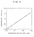
- ADP sample 20 ⁇ l was added to the reagents for assay 1 ml, and the optical absorption at 340 nm was measured after incubation at 20°C for 5 minutes with a control of the blank solution. As shown in Fig. 4 . ADP could be assayed quantitatively.
- ADP aqueous solution was prepared at concentration of 2.7 mM, 5.4 mM, 8.1 mM, 10.8 mM and 13.5 mM to prepare ADP sample.
- ADP sample 20 ⁇ l was added to the reagents for assay 1 ml, and the optical absorption at 340 nm was measured after incubation at 40°C for 5 minutes with a control of the blank solution. As shown in Fig. 5 , ADP could be assayed quantitatively.
- Urea aqueous solution was prepared at concentration of 2.6 mM, 5.2 mM, 7.8 mM, 10.4 mM and 13 mM to prepare urea sample.
- Urea sample 20 ⁇ l was added to the reagents for assay 1 ml, and the optical absorption at 340 nm was measured after incubation at 37°C for 5 minutes with a control of the blank solution. As shown in Fig. 6 , urea could be assayed quantitatively.
- Tris-HCl buffer solution pH 7.5
- U/ml creatinine amide hydrolase 5 U/ml creatine kinase 2 mM ATP 20 mM glucose 5 U/ml G6PDH 1 mM NADP 2 mM MgCl 2 5 U/ml ADP-HK
- Creatinine aqueous solution was prepared at concentration of 2.6 mM, 5.2 mM, 7.8 mM, 10.4 mM and 13 mM to prepare creatinine sample. Creatinine sample 20 ⁇ l was added to the reagents for assay 1 ml, and the optical absorption at 340 nm was measured after incubation at 37°C for 5 minutes with a control of the blank solution. As shown in Fig. 7 , creatinine could be assayed quantitatively.
- Tris-HCl buffer solution pH 7.5
- creatinine deiminase 10 U/ml N-methyl hydantoinase 2 mM ATP
- glucose 5 U/ml
- G6PDH 1 mM NADP 2 mM MgCl 2 5 U/ml
- ADP-HK 50 mM Tris-HCl buffer solution (pH 7.5) 10 U/ml creatinine deiminase 10 U/ml N-methyl hydantoinase 2 mM ATP 20 mM glucose 5 U/ml G6PDH 1 mM NADP 2 mM MgCl 2 5 U/ml ADP-HK
- Creatinine aqueous solution was prepared at concentration of 2.7 mM, 5.3 mM, 7.9 mM, 10.5 mM and 13 mM to prepare creatinine sample. Creatinine sample 20 ⁇ l was added to the reagents for assay 1 ml, and the optical absorption at 340 nm was measured after incubation at 37°C for 5 minutes with a control of the blank solution. As shown in Fig. 8 , creatinine could be assayed quantitatively.
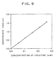
- Creatine aqueous solution was prepared at concentration of 2.6 mM, 5.2 mM, 7.8 mM, 10.4 mM and 13 mM to prepare creatine sample.
- Creatine sample 20 ⁇ l was added to the reagents for assay 1 ml, and the optical absorption at 340 nm was measured after incubation at 37°C for 5 minutes with a control of the blank solution. As shown in Fig. 9 , creatine could be assayed quantitatively.
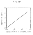
- Glycerol aqueous solution was prepared at concentration of 2.6 mM, 5.2 mM, 7.8 mM, 10.4 mM and 13 mM to prepare glycerol Sample.
- Glycerol sample 20 ⁇ l was added to the reagents for assay 1 ml, and the optical absorption at 340 nm was measured after incubation at 37°C for 5 minutes with a control of the blank solution.
- glycerol could be assayed quantitatively.
- Liberated glycerol from triglycerol by an action of lipase, or that from phosphatidylglycerol by an action of phospholipase D could be assayed.
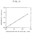
- Choline hydrochloride aqueous solution was prepared at concentration of 2.6 mM, 5.2 mM, 7.8 mM, 10.4 mM and 13 mM to prepare choline hydrochloride sample.
- Choline hydrochloride sample 20 ⁇ l was added to the reagents for assay 1 ml, and the optical absorption at 340 nm was measured after incubation at 37°C for 5 minutes with a control of the blank solution.
- choline could be assayed quantitatively.
- Liberated choline from phospholipase by an action of phospholipase D could be assayed.
- Tris-HCl buffer solution pH 7.5
- glutamine synthetase 10 mM NH 4 Cl 2 mM ATP
- glucose 5 U/ml G6PDH 1 mM NADP 2 mM MgCl 2 5 U/ml ADP-HK
- L-glutamate aqueous solution was prepared at concentration of 2.6 mM, 5.2 mM, 7.8 mM, 10.4 mM and 13 mM to prepare L-glutamate sample.
- L-glutamate sample 20 ⁇ l was added to the reagents for assay 1 ml, and the optical absorption at 340 nm was measured after incubation at 37°C for 5 minutes with a control of the blank solution. As shown in Fig. 12 , L-glutamate could be assayed quantitatively.
- Liberated L-glutamate by an action of aspartate transferase (GOT) or alanine transferase (GPT) could be assayed.
- Urea aqueous solution was prepared at concentration of 2.3 mM, 4.6 mM, 6.9 mM, 9.2 mM and 11.5 mM to prepare urea sample.
- Urea sample 20 ⁇ l was added to the reagents for assay 1 ml, and the optical absorption at 340 nm was measured after incubation at 37°C for 5 minutes with a control of the blank solution. As shown in Fig. 13 , urea could be assayed quantitatively.
- Tris-HCl buffer solution pH 7.5
- U/ml creatinine amide hydrolase 5 U/ml creatine kinase 2 mM ATP 20 mM glucose 5 U/ml G6PDH 1 mM NADP 2 mM MnCl 2 10 U/ml ADP-HK
- Creatinine aqueous solution was prepared at concentration of 2.5 mM. 5.0 mM, 7.5 mM, 10.0 mM and 12.5 mM to prepare creatinine sample. Creatinine sample 20 ⁇ l was added to the reagents for assay 1 ml, and the optical absorption at 340 nm was measured after incubation at 37 °C for 5 minutes with a control of the blank solution. As shown in Fig. 14 , creatinine could be assayed quantitatively.
- Creatine aqueous solution was prepared at concentration of 2.8 mM, 5.6 mM, 8.4 mM, 11.2 mM and 14 mM to prepare creatine sample.
- Creatine sample 20 ⁇ l was added to the reagents for assay 1 ml, and the optical absorption at 340 nm was measured after incubation at 20°C for 5 minutes with a control of the blank solution. As shown in Fig. 15 , creatine could be assayed quantitatively.
- Glycerol aqueous solution was prepared at concentration of 2.7 mM, 5.4 mM, 8.1 mM. 10.8 mM and 13.5 mM to prepare glycerol sample.
- Glycerol sample 20 ⁇ l was added to the reagents for assay 1 ml, and the optical absorption at 340 nm was measured after incubation at 40 °C for 5 minutes with a control of the blank solution.
- glycerol could be assayed quantitatively.
- Liberated glycerol from triglycerol by an action of lipase, or that from phosphatidylglycerol by an action of phospholipase D could be assayed.
- ADO aqueous solution was prepared at concentration of 0.5 mM, 10 mM and 15 mM to prepare ADO sample. After the reagent for assay 1 ml was pre-heated at 50°C for 5 minutes, ADO sample 20 ⁇ l was added therein, and the optical absorption at 340 nm was measured after incubation at 50°C for 5 minutes with a control of the reagent blank solution. As shown in Fig. 17 . ADP could not be assayed quantitatively.
- the assay limit is high. Since the reduced NAD(P), in which the molecular absorption coefficient is clearly defined, is assayed, reliability of assay data is high. Further, the assay method of the present invention has advantages without influence of the reducing active substance in the specimen. Consequently, according to the present invention, ADP in the biological sample or the enzyme, which generates ADP, and substrate thereof in the biological sample can be assayed with simply and precisely.
Landscapes
- Chemical & Material Sciences (AREA)
- Organic Chemistry (AREA)
- Life Sciences & Earth Sciences (AREA)
- Zoology (AREA)
- Wood Science & Technology (AREA)
- Proteomics, Peptides & Aminoacids (AREA)
- Health & Medical Sciences (AREA)
- Engineering & Computer Science (AREA)
- Microbiology (AREA)
- Biochemistry (AREA)
- Physics & Mathematics (AREA)
- Molecular Biology (AREA)
- Biotechnology (AREA)
- Biophysics (AREA)
- Analytical Chemistry (AREA)
- Immunology (AREA)
- Bioinformatics & Cheminformatics (AREA)
- General Engineering & Computer Science (AREA)
- General Health & Medical Sciences (AREA)
- Genetics & Genomics (AREA)
- Measuring Or Testing Involving Enzymes Or Micro-Organisms (AREA)
- Sampling And Sample Adjustment (AREA)
Abstract
Description
- This invention relates to a method for determining ADP contained in a liquid sample by means of an enzymatic reaction, which comprises reacting the sample at 15 to 45°C at least in the presence of glucose, ADP-dependent hexokinase (ADP-HK), oxidized NAD(P) [oxidized nicotinamide adenine dinucleotide(phosphate)], glucose-6-phosphate dehydrogenase (G6PDH), and one or more ion releasing salt selected from the group consisiting of magnesium, cobalt and manganese ions and determining the ADP contained in the sample together with the AMP resulting from the reaction based on the amount of the reduced NAD (P) [reduced nicotinamide adenine dinucleotide (phosphate)] yielded. The present invention further relates to a method for determining an enzyme for generating ADP or substrate thereof in a liquid sample by means of an enzymatic reaction, which comprises reacting the sample containing the enzyme for generating ADP or substrate thereof in the liquid sample at 15 to 45°C at least in the presence of a reaction reagent involving in the reaction based on the enzyme for generating ADP and the substrate thereof, ATP, glucose, ADP-HK, oxidized NAD (P), G6PDH and one or more ion releasing salt of magnesium ion, cobalt ion or manganese ion, and determining the enzyme for generating ADP or the substrate thereof contained in the sample together with the AMP resulting from the reaction based on the amount of the reduced NAD (P) yielded. The invention is applicable in the clinical chemistry. The method has advantageous for sample and exact assay of the ADP in the sample such as serum, plasma, urine and cerebrospinal fluid, by means of an amount of generation of or increase in the reduced NAD (P).
- Methods for asssaying ADP in biological sample or enzyme, which generate ADP, or substrate thereof have known. These include a method using combination of the enzyme and other dehydrogenase or oxidase for the reaction product from substrate and the enzyme (for example, in case of enzymatic reaction with glycerol and glycerokinase, a method wherein glycerokinase is reacted with glycerol in the presence of ATP to generate ADP and glycerol phosphate, which is assayed by an action of glycerol phosphate dehydrogenase or glycerol phosphate oxidase) and a method assyaing ADP which is generated by enzymatic action.
- However, in the method of assay by combining with other dehydrogenase or oxidase, a combination of each enzyme should be changed depending on the enzyme for generating ADP. This is not suitable for general use, furthermore there are possibilities of no enzymes with preferable combination for substrate or enzyme.
- A method for assaying ADP, which is generated by enzymatic reaction, includes an assay by means of liquid chromatography. This has disadvantage due to complicated chromatographic operation.
- Enzymatic assay methods of ADP which has good operability have known (Japan. Pta. Unexam. Publ. 7-8297). These include : a method measuring decreased reduced NAD (NADH + H+) using pyruvate kinase [PK (EC 2. 7. 1. 40)] and lactate dehydrogenase [LDH (EC 1. 2. 3. 3)] (decreased method. Reaction 1); a method using pyruvate kinase and pyruvate oxidase (oxidasemethod. Reaction 2); and a method measuring increase reduced NAD (P)[NAD (P) H + H+] using pyruvate kinase, pyruvate decarboxylase and aldehyde dehydrogenase (increased method, Reaction 3).
- The reactions of these methods are shown below.
- In the equation ; PEP: phosphoenolpyruvate. Pi: phosphate. PDC: pyruvate decarboxylase. A1DH: aldehyde dehydrogenase, ans TPP: thiamine pyrophosphate.


- In the decreased method of reduced NAD (NADH + H+) shown in Reaction 1, previously alliquot amount of reduced NAD should be added in the reaction mixture, and after termination of the reaction, residual amount of reduced NAD in the reaction mixture is measured. Consequently, this decreased method has number of problems as follows.
- (1) Assay value is not exact in case of small amount of components for assay.
- (2) Uper limit of an amount of measurable components is restricted by an amount of added reduced NAD in the reaction mixture.
- (3) Amount of addition of the reduced NAD in the reaction mixture before assay should be controlled depending on the type of spectrophotometer for measurement of reduced NAD.
- (4) Reduced NAD in the reagent for analysis is unstable.
Quantitative assay of pyruvate by means of coloring indicator reagent of hydrogen peroxide, which is generated from the reaction by using pyruvate kinase and pyruvate as shown in Reaction 2, has known. - (5) The method is affected by an interfering substance (reduced substance) (such as uric acid and ascorbate) or color substance (bilirubin, hemoglobin, etc.) in the biological samples. Consequently, result of assay is not so exact and is not satisfactory.
As shown in the reaction 3, the increased method of reduced NAD (P) using pyruvate kinase, pyruvate decarboxylase and aldehyde dehydrogenase is used to apply the reverse reaction of pyruvate kinase. - (6) Optimum pH of the reverse reaction is pH 7.5.
Optimum pH of pyruvate decarboxylase is pH 6.0-6.4 and that of aldehyde dehydrogenase is pH 9.0. Optimum pH of these enzymes is different. This resulted to determine optimum pH of the reaction 3. - (7) Since Km value of pyruvate decarboxylase for pyruvate is slightly hight as 3.6-30 mM, large amount of substrate, which is also expensive, should be used for generating pyruvate.
- (8) Large amount of enzyme is required for shortage of the reaction time. These result indicates that the method of the reaction 3 makes to complicate for operation.
Intracellular ADP dependent hexokinase, which may be used in the present invention, in super thermophile pyrococcus furiosus DSM 3638 has known [J. Biol. Chem., 269, 17537-17541, (1994), Dutch Patent Open No.9400 836 ]. However, no physicochemical and biochemical properties is reported. Moreover the enzyme is neither isolated nor purified. Furthermore growth temperature of the microorganism strain is 90°C-105°C. Since the optimum pH of the enzyme is above 90°C, assay of enzyme activity is performed at 50°C for considering heat stability of G6PDH. which is originated from yeast. This indicates that the assay of enzyme action is performed in the different optimum temperature of ADP-HK, and the reaction condition is completely different from the assay condition of general clinical diagonosis for analysis of biological specimens in view of the optimum temperature of ADP-HK. - (6) If the reaction as like, in this prior reference, is performed at 50°C, it is not preferable in the clinical chemistry for exact assay due to denature of the combination of the enzymes.
- (7) Turbidity of the reaction mixture occurs due to thermal denaturation of the biological components.
- We have cultured a strain of Pyrococcus furiosus DSM3638, purified ADK-HK, measured its optimum temperature and determined to 80°C-100°C. Further, we have found that relative activity of the enzyme at 37°C was about 10 % as cpmpared to at 100°C. Consequently, it has been considered that the exact enzyme reaction at 37°C using the enzyme might be impossible. However, quite unexpectedly, as a result of the enzyme reaction at 37 °C, we have found that ADP in the specimen such as biological composition could exctly and simply be assayed as an increased amount and generation of reduced NAD (P) at 15°C- 45°C, which included the experimented temperature at 37°C with a general reaction temperature for determination of biological sample.
- We have found a method for assaying the ADP in a biological sample, or the enzyme which generated ADP in the biolopgical sample or its substrate by using the enzymatic reaction hereinbelow illustrated (Reactions 4 and 5). For example, in case that the biological sample is a liquid specimen for assaying enzymatic activity, which generates ADP, reaction reagents, which involve in the reaction including ADP generating enzyme and its substrate, are used in the presence of the substrate of the enzyme and the ATP, or in case that the biological sample is a liquid specimen for an assay of amount of substrate, reaction reagents, which involve in the reaction including ADP generating enzyme and its substrate, are used in the presence of the enzyme, which generate ADP by a reaction with the substrate, and ATP. The said liquid specimen is treated with the said reaction reagents. The reaction proceeds based on a reaction involving in glucose, ADP-HK, oxidized NAD (P), G6PDH and magnesium ion cobalt ion or manganese ion, to generate AMP from ADP, simultaneously to generate reduced NAD (P) by a reducing reaction of the oxidized NAD (P). We have found that the quantitative assay of ADP generating enzyme or its sybstrate in the biological sample based on the assay of the generated reduced NAD (P) was quite useful, and that these enzymatic reaction could be applied generally to the enzyme, which generate ADP, or its substrate in the biological sample, and completed the present invention.
- An object of the present invention is to provide a method for assaying ADP in a biological sample comprising measuring the said ADP as a generated and increased amount of reduced NAD (P) by using glucose, ADP-HK, oxidized NAD (P), G6PDH and magnesium ion, conbalt ion or manganese ion.
- Another object of the present invention is to provide a method for simple and precise assay of an enzyme, which generates ADP, or its substrate in a biological sample to be assayed, comprising using reaction reagents which involve in a reaction beased on the enzyme, which generates ADP, and its substrate, and reacting the ADP generating enzyme and substrate thereof in the presense of ATP, glucose, ADP-HK oxidized NAD (P), G6PDH and magnesium ion, cobalt ion or manganese ion.
- The present invention has been completed by the knowledge hereinabove. The present invnetion relates to a method for determining ADP contained in a liquid sample by means of an enzymatic reaction, which comprises reacting the sample at 15 to 45°C at least in the presence of glucose, ADP-HK, oxidized NAD (P), G6PDH and one or more ion releasing salt selected from the group consisting of magnesium, cobalt and manganese ions, and determining the ADP contained in the sample together with the AMP resulting from the reaction based on the amount of the reduced NAD (P) yielded. The present invention further relates to a method for determining an enzyme for generating ADP or substrat thereof in a liquid sample by means of an enzymatic reaction, which comprises reacting the sample containing the enzyme for generating ADP or substrate thereof in the liquid sample at 15 to 45°C at least in the presence of a reaction reagent involving in the reaction based on the enzyme for generating ADP and the substrate thereof, ATP, glucose, ADP-HK, oxidized NAD (P), G6PDH and one or more ion releasing salt of magnesium ion, cobalt ion or manganese ion, and determining the enzyme for generating ADP or the substrate thereof contained in the sample together with the AMP resulting from the reaction based on the amount of the reduced NAD (P) yielded.
- The enzymatic reactions, which generate the reduced NAD (P) from the oxidized NAD (P) in the presence of glucose, ADP-HK, oxidized NAD (P), G6PDH and one or more ion releasing salt selected from the group consisting of magnesium, cobalt and manganese ions used in the present invention, are shown in the following Reactions 4 and 5.


- The present invention will be explained in details as follows.
- ADP-HK of the preent invention is an enzyme ADP-HK which uses at least glucose as a substrate and consumes ADP to generate glucose-6-phosphate and AMP as shown in the above Reaction 4, and is not limited thereto. Example of ADP-HK producing microorganism is a super thermophile pyrococcus furiosus DSM3638, which has deposited in Deutsche Sammlung von Mikroorganismen und Zellkulturen GmbH (DSM) and mentioned in the DSM catalogue (1993), and can be obtainable by any person. The super thermophile ADP-HK is a preferable example of the enzyme.
- G6PDH used in the enzymatic reaction of the above Reaction 5 is distributed in the market (Boehringer Mannheim Inc: Leuconostoc mesenteroides. Sigma Corp: Baker's yeast, Bacillus strearothermophilus and Leuconostoc mesenteroides), and is easily obtainable.
- In the Reaction 4, the enzymatic reaction catalyzed by ADP-HK, the ion releaasing salt of magnesium ion can be replaced by one or more ion releasing salt of cobalt or manganese. Example of the salt includes chloride or sulfate, and is preferably magnesium chloride, cobalt chloride or manganese chloride, but is not limited to these examples.
- Oxidized NAD (P), which is a coenzyme used in the enzymatic reaction catalyzed by the above G6PDH, as exemplified in Reaction 5, includes oxidized thio-NAD (P), oxidized 3-acetyl NAD (P) and oxidized deamino NAD (P), but is not limited within these examples.
- In the present invention, the amount of ADP-HK G6PDH, glucose, oxidized NAD (P) and magnesium ion, cobalt ion or manganese ion used can be the amount for good process of the enzymatic reaction. It can be adjusted depending on the types of substance in the specimen to be assayed, contents in the specimen, types of coupling enzymatic reaction, reaction time and temperature. Examples of concentration of ADP-HK and G6PDH are 0.1-100 U/ml, preferably 1-50 U/ml. Concentration of glucose and oxidized NAD (P) can be sufficient level for enzymatic reaction. Example of glucose concentration is 0.5-100 mM, preferably 1-50 mM. Example of oxidized NAD (P) concentration is 0.5-50 mM, preferably 1-10 mM. Example of magnesium ion, cobalt ion or manganese ion is 0.1-50 mM, preferably 0.5-10 mM.
- The method of the present invention proceeds with using preferable buffer solution which does not show detrimental action to the enzymatic reaction. Example thereof is Tris-HCl buffer solution, phosphate buffer solution, mono- or di-ethanolamine buffer solution or Good's buffer solution. Assay procedures are not limited. Examples thereof are end-point asay method and rate assay method. Examples of sample solution to be assayed are biological sample which contains ADP (existed or generated), and are specimens such as serum, plasma, urine and cerebrospinal fluid. The specimen used in the assay may be 5-200 µl. Reaction temperature can be proceeded at 15-45 °C preferably at 20°C-40 °C. The reaction time in the end point assay is 1-60 min., preferably 1-10 min. In the rate assay method, the measurement is performed within a time for linear reaction, preferably in the interval of 2-3 min.
- in the reaction of the present invention, ADP in the specimen is converted to AMP which is accompanied by generation of the reduced NAD (P). The reduced NAD (P) can be assayed by various methods, usually optical absorption, which is prefereably for simple and precise measurement. Wave length for measurement can be selected depending on the type of reduced NAD (P). Reduced NAD (P), reduced 3-acetyl-NAD (P) and reduced deamino-NAD(P) can be measured at 340 nm, and reduced thio-NAD (P) is measured at 405 nm. Another method for measurement of the reduced NAD (P) includes that formazan is generated by an action of electro acceptor such as phenazine methosulfate (PMS) or diaphorase (EC 1. 6. 4. 3) by using tetrazorium salt such as indonitro tetrazorium (INT) or nitroblue tetrazorium (NTB), and the said formazan is colorimetrically measured. Reduced NAD (P) is also measured by fluorometry.
- The specimen as a biological sample of the present invention is used for assay of ADP in the biological sample, or assay of substrate for enzyme which generates ADP in the biological sample or assay of the enzyme activity in the biological sample. In the assay of substrate for an enzyme which genenrate ADP in the biological sample or the assay of the enzyme activity, especially in the assay of the enzyme which genenrates ADP or the substrate thereof in the presence of ATP, the substrate of kinase synthetase, hydratase or carboxylase in the specimen can be converted to ADP by an action of kinase synthetase, hydratase or carboxylase. Accordingly, the enzyme activity or substrate thereof, which is introduced to ADP, in the specimen can be assayed as a generated amount of the reduced NAD (P) by coupling with these enzyme reactions and the enzyme reaction of Reactions 4 and 5. In Table 1, examples of substrates and enzyme used are shown. Either the substrate or the enzyme activity can be measured by using either the enzyme or the substrate as a reaction reagent (ATP is not an objective for the assay).
Table 1 Reaction No. Substrate Enzyme 6 urea urea amidohydrolase (EC 3. 5. 1. 45) 7 creatinine creatinine amidohydrolase (EC 3. 5. 2. 10) creatine kinase (EC 2. 7. 3. 2) creatine amidohydrolase (EC 3. 5. 3. 3) urea amidohydrolase (EC 3. 5. 1. 45) 8 creatinine creatinine deiminase (EC 3. 5. 4. 21) N-methylhydantoinase (EC 3. 5. 2. 14) 9 creatine creatine kinase (EC 2. 7. 3. 2) 10 glycerol glycerol kinase (EC 2. 7. 1. 30) 11 choline choline kinase (EC 2. 7. 1. 32) 12 hexose hexokinase (EC 2. 7. 1. 1) 13 fructose fructokinase (EC 2. 7. 1. 4) 14 galactose galactokinase (EC 2. 7. 1. 6) 15 glucosamine glucosamine kinase (EC 2. 7. 1. 8) 16 adenosine adenosine kinase (EC 2. 7. 1. 20) 17 thymidine thymidine kinase (EC 2. 7. 1. 21) 18 NAD NAD kinase (EC 2. 7. 1. 23) 19 riboflavin riboflavin kinase (EC 2. 7. 1. 26) 20 pyridoxal pyridoxal kinase (EC 2. 7. 1. 35) 21 mevalonate mevalonate klinase (EC 2. 7. 1. 36) 22 protein protein kinase (EC 2. 7. 1. 37) 23 homoserine homoserine kinase (EC 2. 7. 1. 39) 24 pyruvate pyruvate kinase (EC 2. 7. 1. 40) 25 pyruvate pyruvate carboxylase (EC 6. 4. 1. 1) 26 acetate acetate kinase (EC 2. 7. 2. 1) 27 ammonia carbamate kinase (EC 2. 7. 2. 2) 28 L-arginine arginine kinase (EC 2. 7. 3. 3) 29 L-glutamate ammonia glutamine synthetase (EC 6. 3. 1. 2) 30 L-glutamate glutamate kinase (EC 2. 7. 2. 11) 31 L-aspartate ammonia aspartate synthetatse (EC 6. 3. 1. 4) 32 L-aspartate aspartate kinase (EC 2. 7. 2. 4) 33 ATP myokinase (EC 2. 7. 4. 3) 34 ATP ATPase (EC 3. 6. 1. 4) 35 citrate citrate kinase (EC 4. 1. 3. 8) 36 L-aspartate aspartate aminotransferase(GOT) (EC 2. 6. 1. 1) glutamine synthetase (EC 6. 3. 1. 2) 37 L-alanine alanine aminotransferase (GPT) α-ketoglutarate (EC 2. 6. 1. 2) glutamine synthetase (EC 6. 3. 1. 2) - These reactions are illustrated as follows.
- In the asasy of urea or urea amidohydrolase in the specimen, a reaction using the reaction reagents involved in the reaction is illustrated in the following. In this assay, ADP, which is generated by the enzymatic reaction, is measured. The reaction reagents involved in the reaction mean the components shown in the left part of the reaction scheme and in the part of arrow. The reaction reagents in an assay of urea in the biological sample include urea amidohydrolase, water, magnesium ion (magnesium chloride) and potassium hydrogen carbonate. The reaction reagents in an assay of urea amidohydrolase activity in the biological sample include urea, water, magnesium ion (magnesium chloride) and potassium hydrogen carbonate. In these reactions, water is replaced by water in the reaction medium.

- In the assay of creatinine or creatinine amidohydrolase in the biological sample, the reactions using the reaction reagents involved in the reactions are illustarted in the following. In such the consecutive enzymatic reactions, in case that a component in the left side of the reacton is identical with that in the right side of the reaction, those components are involved in the reaction, but those are apparently not necessary to be added.
- In the reaction ① and ② hereinbelow illustrated, and in the assay of creatinine or creatinine amidohydrolase in the biological sample, creatine, which is generated by an enzymatic reaction, can be assayed by measuring ADP generated in the reaction ① or in the reaction ②.
- In an assay of creatinine in the biological sample, the reaction reagents involved in the reaction are creatinine amidohydrolase, water, creatine kinase (CK) and magnesium ion for the reaction ①, or creatinine amidohydrolase, water, creatine amidohydrolase, urea, ureido amidohydrolase and magnesium ion for the reaction ②.

- In the assay of creatinine or creatinine deiminase in the biological sample, the reaction using the reaction reagents involved in the reactions are illustrated in the foillowing. In this reaction, ADP generated by an action of enzyme is measured. The reaction reagents for assay of creatinine in the biological sample are creatinine deiminase, water and N-methylhydantoinase. The reaction reagents for assay of creatinine deiminase activity in the biological sample are creatinine, water and N-methylhydantoinase.

- In the assay of creatine or kinase in the biological sample, the reaction using the reaction reagent involved in the reaction is illustrated in the following. In this reaction, ADP, which is generated by the enzymatic reaction, is measured. The reaction reagents used for assaying creatine are creatine kinase and magnesium ion. The reaction reagnets used for assay of creatine kinase activity in the biological sample are creatine and magnesium ion.

- Creatine in the reaction 9 can be the creatine generated in the above reaction 7.
- In the assay of glycerol or glycerol kinase (GK) in the biological sample, the reaction scheme is shown in the following, and generated ADP by the enzymatic action is measured. In the reaction, the reagents used are glycerol kinase and magnesium ion.

- Glycerol in the above reaction can be the glycerol liberated from triglyceride or mono- or di-glyceride by an action of lipase or pancreatic lipase, to which activating agent Co-lipase is added, or the glycerol liberated from phosphatidylglycerol by an action of phospholipase D.
- The reaction is preferable for assay of triglyceride and assay of pancreatic lipase activity using synthetic substrate of triglyceride or diglyceride in the biochemical test of the specimen.
- For example, the reagents used for an assay of triglyceride in the biological sample are lipase, water, glycerol kinase and magnesium ion. The reagents used for assaying pancreatic lipase are synthetic substrate such as triglyceride or diglyceride, water, glycerol kinase, magnesium ion. Co-lipase and surface active agent for solvilizing the synthetic substrate.
- In the assay of choline or choline kinase, the reaction scheme is shown in the following. In this reaction, enzymatically generated ADP can be measured.

- Choline in the reaction may be a liberated choline from phosphatidylcholine (component of phospholipid) by an action of phospholipase D, and from synthetic substrate of choline ester such as benzoil choline or o-toluoylcholine by an action of choline esterase.
- The method is preferable for assay of choline esterase activity in an assay of phospholipid component in the biochemical test using synthetic sybstrate.
- The reaction reagents used for the assay of phospholipid component are phospholipase D, water and magnesium ion, and the reagents used for the assay of choline esterase activity are synthetic substrate such as benzoyl choline or o-tolyoylcholine, water and magnesium ion.
- In the assay of substrate or enzyme which generate ADP in the biological sample, the reaction schemes, in which the components involved in the said reaction are used as the reaction reagents, is illustrated in the following. In these reaction, ADP genenrated by the enzymatic reaction can be measured.
- In the assay method of the present invention, the reaction reagents of the components involved in the reaction of the present invention are obvious in view of the above and below illustrated reaction schemes. Further, these can preferably be prepared optionally depending on the sample to be assayed, and are not limited to the reagents described in the specification.





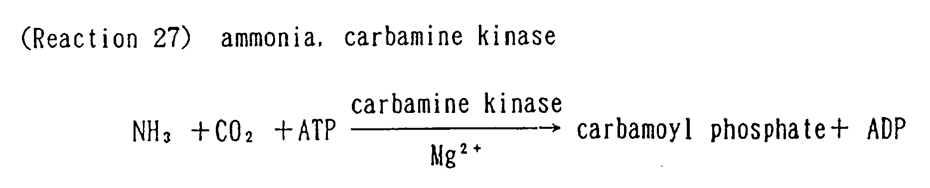


- In the above reaction, ammonia to be assayed can be generated by a reaction with urea and urea amidohydrolase as illustrated in the Reaction 6, or by a reaction with creatinine and creatinine amidohydrolase as illustrated in the reaction ② of the Reaction 7, or by a previous step of the reaction with creatinine and creatinine deiminase as illustrated in the Reaction 8. Ammonia in these reactions is not limited in its origin.



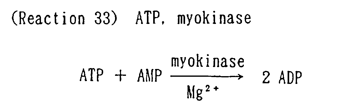


- The above reaction is preferably used in an assay of GOT activity.


- The above reaction is preferable for use of an assay of an assay of GPT activity.
- In these reactions, amount of ATP added is for example 0.1-50 mM, preferably 0.5-10 mM. Enzyme added for an assay of substrate is, for example, 1-100 U/ml. Substrate to be added in an assay of enzyme activity is, for example 1-100 mM. In the reagents other than these substrates and enzyme, components which are consumed by reaction with the substrate are equal to 2-fold of the amount of the above substrates used. Concentration of magnesium ion is, for example, 0.1-50 mM as magnesium chloride, preferably 0.5-10 mM, however excess use is not excluded unless inhibition in the reaction occurs.
- These reaction may be coupled with the Reactions 4 and 5 or may not be coupled. Preferable reaction proceed in couple of these reactions.
- In the prior known assay method of urea, reaction proceeds accroding to the Reaction 38 as follows.

- As shown in Reaction 38, the method includes measurement of decreased amount of reuduced NAD (P). In this reaction, there are many disadvantages as follows.
- (8) Assay result may be incorrect in case of small amount of components to be assayed.
- (9) The upper limit of measurable components is restricted depending upon the added reduced NAD (P) in the reaction (narrow range of the measurement).
- (10) Amount of reduced NAD (P) in the reaction mixture before assay should be changed depending on the type of spectrophotometer which is used for measurement of the reduced NAD (P).
- (11) Reduced NAD (P) in the reagent is unstable.
- (12) Application to dry chemistry is impossible.
- (13) The reaction consists of a method for measurement of ammonium in its principle, and ammonia in the specimen or reagents must be removed previously.
- The assay method of the present invention is :


- As shown in the above reactions 39, 40 and 41, since the reaction is the increased method of reduced NAD (P), there are no disadvantages of the decreased method as well as no need for removal of ammonia. Therefore the present method is superior.
- Prior known conventional assay method of creatine kinase is illustrated as follows.

- In this reaction system, a substrate creatine phosphate is quite expensive which leads to expensive reagents in total.
- The method of the present invention can be illustrated as follows.


- As shown in the reactions 43, 44 and 45, creatine is used as substrate which results to provide reagents for assay with low price.
- Conventional known method for creatinine assay is illustrated as follows.

- As shown in the above reaction scheme 46, the assay involves in oxidase method. The reaction is affected by the reducing substance (ascorbate) to result unexpect assay. Further, sarcosine oxidase reacts with proline in a sample, consequently the reaction is affected by proline in the specimen.
- The methods of the present invention can be illustrated as follows.


- As shown in the reactions 47, 48 and 49, and the reactions 50, 51 and 52, these are increased method of the reduced NAD (P). The reactions are not affected by the reducing substance (ascorbate) and proline.
- Consequently assay method of triglyceride is shown as follows.

- As shown in the above reaction scheme 53, the reaction consists of the oxidase method, which is affected by reducing substance (ascorbate) in the specimen and result of assay may be inexact.
- The method of the present invention is illustrated by the following reactions.


- As shown in the above reactions 54, 55 and 56, the method of the present invention is an increased method of reduced NAD (P) and is not influenced by the reducing substance in the specimen.
- In replace of triglyceride of the above reaction 54 to diglyceride or monoglyceride, in which the reaction proceeds by consuming 2 moles or 1 mole of water to liberate 2 moles or 1 mole of fatty acid, the result is identical with the aboves.
- Conventional assay method of phosphatidylcholine is illustrated as follows.

- As shown in the above reaction scheme 57, the method is the oxidase method, which is influenced by a reducing substance (ascorbate), and produces inexact result.
- The method of the present invention is illustrated as follows.


- As shown in the reactions 58, 59 and 60, the method of the present invention is the increased NAD (P) method, which is not influenced by a reducing substance.
- Conventional assay method of aspartate aminotransferase (GOT) is illustrated in the reaction 61.

- As shown in the reaction sheme 61, the method is the decreased method of NAD. The method has disadvantages as follows.
- (14) Assay result is incorrect in case of small amount of components to be assayed.
- (15) The upper limit of measurable components is restricted depending on the added reduced NAD in the reaction (narrow range of the measurement).
- (16) Amount of reduced NAD in the reaction mixture before assay has to be changed depending on the type of spectrophotometer used for measurement of the reduced NAD.
- (17) Reduced NAD in the reagent is unstable.
- (18) Application to dry chemistry is impossible.
- Further the method is influenced by lactate dehydrogenase in the specimen which indicate impossible to proceed exact assay.
- The method of the present invention is shown as follows.
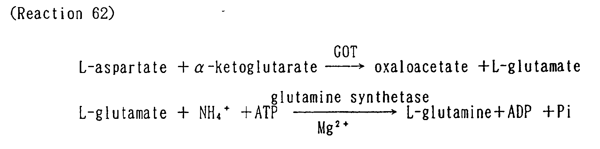


- As shown in the reactions 62, 63 and 64, the present method is the increased reduced NAD (P). The method has no disadvantages as like in the conventional reduced method. Furthermore, it needs not to remove lactate dehydrogenase in the specimen. As a result, the present method shows superior result.
- Prior known conventional alanine aminotransferase (GPT) is illustrated as follows.

- As shown in the above reaction scheme 65, the method is the decreased method of NAD. The method has disadvantages as follows.
- (19) Assay result is incorrect in case of small amount of components to be assayed.
- (20) The upper limit of measurable components is restricted depending on the added reduced NAD in the reaction (narrow range of the measurement).
- (21) Amount of reduced NAD in the reaction mixture before assay has to be changed depending on the type of spectrophotometer used for measurement of the reduced NAD.
- (22) Reduced NAD in the reagent is unstable.
- (23) Application to dry chemistry is impossible.
- Further the method is influenced by lactate dehydrogenase in the specimen which indicates impossible to proceed exact assay.
- The method of the present invention is shown as follows.


- As shown in the above reactions 66, 67 and 68, the present method is the increased reduced NAD (P). The method has no disadvantages as like in the conventional reduced method. Furthermore, it needs not to remove lactate dehydrogenase in the specimen. As a result, the present method shows superior result.
- The methods for assay of enzyme, which generate ADP in the specimen, or its substrate of the present invention are explained in the following examples but are not limited within these examples.
-
-
Fig. 1 : Standard curve for assaying ADP using magnesium ion at 37°C in this invention. -
Fig. 2 : Standard curve for assaying ADP using cobalt ion at 37 °C in this invention. -
Fig. 3 : Standard curve for assaying ADP using manganese ion at 37°C in this invention. -
Fig. 4 : Standard curve for assaying ADP using magnesium ion at 20°C in this invention. -
Fig. 5 : Standard curve for assaying ADP using magnesium ion at 40°C in this invention. -
Fig. 6 : Standard curve for assaying urea uaing magnesium ion at 37°C in this invention. -
Fig. 7 : Standard curve for assaying creatinine using magnesium ion at 37°C in this invention. -
Fig. 8 : Standard curve for assaying creatinine using magnesium ion at 37°C in this invention. -
Fig. 9 : Standard curve for assaying creatine uaing magnesium ion at 37°C in this invention. -
Fig. 10 : Standard curve for assaying glycerol using magnesium ion at 37°C in this invention. -
Fig. 11 : Standard curve for assaying choline using magnesium ion at 37°C in this invention. -
Fig. 12 : Standard curve for assaying glutamate using magnesium ion at 37°C in this invention. -
Fig. 13 : Standard curve for assaying urea using cobalt ion at 37°C in this invention. -
Fig. 14 : Standard curve for assaying creatinine using manganese ion at 37°C in this invention. -
Fig. 15 : Standard curve for assaying creatine using magnesium ion at 20°C in this invention. -
Fig. 16 : Standard curve for assaying glycerol using magnesium ion at 40°C in this invention. -
Fig. 17 : Qualitative assay curve for assaying ADP at 50°C in place of this invention. - The present invention is explained in details by examples and ref rential examples, but the present invention are not limited within these examples and referential examples.
- Assay method of enzyme activity of ADP-HK
-
50 mM Tris-HCl buffer solution (pH 7.5) 20 mM glucose 2 mM ADP 2 mM MgCl2 5 U/ml G6PDH 0.025 % NBT (nitrotetrazolium blue) 1 mM NADP 1 % Triton X-100 5 U/ml diaphorase (NADPH) - After pre-heating the reagent for assay at 37 °C for 1 minute, enzyme solution 0.02 ml was added and the reaction mixture was reacted for 10 minutes. The reaction was terminated by adding 0.1N HCl 2 ml, and the optical density of the reaction mixture was measured at 550 nm using optical path 1.0 cm within 5 minutes (As). Optical absorption of the blank was measured using distilled water 0.02 ml in place of the enzyme solution (Ab). Difference in the absorption in use of enzyme solution (As) and the blank thereof (Ab). i.e. (As-Ab), is used to measure enzyme activity. A unit of enzyme is defined as an amount of enzyme which generates 1 µ mole of reduced NADP in 1 min. at 37 °C as shown in the following equaltion.

-
0.1 % Yeast extract 0.5 % triptone 0.72 % maltose 2.39 % NaCl 0.4 % Na2SO4 0.07 % KCl 0.02 % NaHCO3 0.01 % KBr 0.03 % H3BO4 1.08 % MgCl2 0.15 % CaCl2 0.0025 % SrCl2 0.025 % NH4Cl 0.014 % K2HPO4 0.1 % CH3COONa 0.0015 % N (COOH)3 0.0005 % MnSO4 0.0014 % FeSO4 0.0002 % NiCl2 0.0001 % CoSO4 0.0001 % ZnSO4 0.00001 % CuSO4 0.000001 % Na2WO4 0.000001 % Na2MoO4 0.1 % cysteine hydrochloride - Liquid medium (pH 7) 500 ml containing the above culture composition was poured into ten 500 ml Erlenmeyer flasks and sterilized at 120°C for 20 minutes. A suspension of Pyrococcus furiosus DSM3638, 10 ml, was inoculated thereto, and the medium was cultured with stirring at 95°C for 20 hours to prepare seed culture. The liquid medium 200 lit./300 lit. tank containing the above culture composition was sterilized, and the seed culture was inoculated thereto, then the inoculated medium was cultured with agitating at 95°C for 15 hours to obtain 5 mU/ml cultured liquid 200 lit.
- The obtained cultured liquid 200 lit. was centrifuged. The obtained microbial cells was washed with 20 mM Tris-HCl buffer (pH 7.5) containing 0.9 % NaCl. The washed cells was suspended in 20 mM Tris-HCl buffer (pH 7.5) to adjust a volume 2 lit. The suspension was sonicated by using sonicator (KUBOTA Inc., INSONATOR 201M) at 180 W for 30 minutes to obtain sonicated cell suspension.
- The sonicated suspension was centrifuged at 8000 rpm for 30 minutes to obtain supernatant solution 1.8 lit. (enzyme activity 980 U). The supernatant was dialyzed by using dialysis tube against Tris-HCl buffer (pH 7.5) 8 lit. at 5°C for overnight. The dialyzate was applied in the column of DEAE-Sepharose FF (Pharmacia Inc.) 200 ml (2.6× 38 cm) buffered with 10 mM Tris-HCl buffer (pH 7.5), and was eluted by linear gradient elution of 0-1 mole NaCl. Active fraction (950 U) was eluted at the concentration of 0.08-0.1 mole of NaCl. NaCl was dissolved to 4 M in the obtained active fraction, and the solution was applied in the column of Phenyl-Sepharose FF (Pharmacia Inc.) 200 ml (2.6× 38 cm) buffered with 4 M NaCl, and was eluted by linear gradient elution of 4-0 mole NaCl.
- The active fraction (900 U) was obtained in 0.02-0.07 mole NaCl concentraion. The thus obtained active fraction was dialyzed against 10 mM Tris-HCl buffer (pH 7.5) 8 lit. at 5°C for overnight. The dialyzate was applied in the column of hydroxyapatite (Pentax Inc.) 100 ml (2.6 × 19 cm) buffered with 10 mM Tris-HCl buffer (pH 7.5), and was eluted by linear gradient elution of 0-0.5 mole phosphate buffer (pH 7.5). Active fraction (850 U) was eluted at the concentration of 0.02-0.03 mole of phosphate buffer. The thus obtained enzyme solution was lyophilized to obtain enzyme powder 5 mg (170 U/mg).
- Biochemical properties of ADP-HK was as follows.
-
- (1) Enzyme action
Enzyme action using glucose as a substrate is shown as follows.
- (2) Molecular weight
Molecular weight of ADP-HK measured by gel-filtration using TSK-G3000SW XL(0.75 × 30 cm)(Toso Corp.) was 100000 ± 5000. - (3) Optimum pH : pH 6.0 - 7.0 (phosphate buffer).
- (4) Optimum temperature : 80 - 100°C. Consequently, the microorganism was defined as super thermophile ADP-HK.
-
50 mM Tris-HCl buffer solution (pH 7.5) 20 mM glucose 5 U/ml G6PDH 1 mM NADP 2 mM MgCl2 5 U/ml ADP-HK - ADP aqueous solution was prepared at concentration of 2.6 mM, 5.2 mM, 7.8 mM, 10.4 mM and 13 mM to prepare ADP sample. ADP sample 20 µl was added to the reagents for assay 1 ml, and the optical absorption at 340 nm was measured after incubation at 37°C for 5 minutes with a control of the blank solution. As shown in
Fig. 1 (horizontal axis: shown as final concentraiton). ADP could be assayed quantitatively. -
50 mM Tris-HCl buffer solution (pH 7.5) 20 mM glucose 5 U/ml G6PDH 1 mM NADP 2 mM CoCl2 10 U/ml ADP-HK - ADP aqueous solution was prepared at concentration of 2.3 mM, 4.6 mM, 6.9 mM, 9.2 mM and 11.5 mM to prepare ADP sample. ADP sample 20 µl was added to the reagents for assay 1 ml, and the optical absorption at 340 nm was measured after incubation at 37 °C for 5 minutes with a control of the blank solution. As shown in
Fig. 2 , ADP could be assayed quantitatively. -
50 mM Tris-HCl buffer solution (pH 7.5) 20 mM glucose 5 U/ml G6PDH 1 mM NADP 2 mM MnCl2 10 U/ml ADP-HK - ADP aqueous solution was prepared at concentration of 2.5 mM, 5.0 mM, 7.5 mM, 10.0 mM and 12.5 mM to prepare ADP sample. ADP sample µl was added to the reagents for assay 1 ml, and the optical absorption at 340 nm was measured after incubation at 37 °C for 5 minutes with a control of the blank solution. As shown in
Fig. 3 , ADP could be assayed quantitatively. -
50 mM Tris-HCl buffer solution (pH 7.5) 20 mM glucose 5 U/ml G6PDH 1 mM NADP 2 mM MgCl2 10 U/ml ADP-HK - ADP aqueous solution was prepared at concentration of 2.8 mM, 5.6 mM, 8.4 mM,11.2 mM and 14 mM to prepare ADP sample. ADP sample 20 µl was added to the reagents for assay 1 ml, and the optical absorption at 340 nm was measured after incubation at 20°C for 5 minutes with a control of the blank solution. As shown in
Fig. 4 . ADP could be assayed quantitatively. -
50 mM Tris-HCl buffer solution (pH 7.5) 20 mM glucose 5 U/ml G6PDH 1 mM NADP 2 mM MgCl2 10 U/ml ADP-HK - ADP aqueous solution was prepared at concentration of 2.7 mM, 5.4 mM, 8.1 mM, 10.8 mM and 13.5 mM to prepare ADP sample. ADP sample 20 µl was added to the reagents for assay 1 ml, and the optical absorption at 340 nm was measured after incubation at 40°C for 5 minutes with a control of the blank solution. As shown in
Fig. 5 , ADP could be assayed quantitatively. -
50 mM Tris-HCl buffer solution (pH 7.5) 30 U/ml urea amidelyase 2 mM ATP 10 mM KCl 8 mM KHCO3 20 mM glucose 5 U/ml G6PDH 1 mM NADP 2 mM MgCl2 5 U/ml ADP-HK - Urea aqueous solution was prepared at concentration of 2.6 mM, 5.2 mM, 7.8 mM, 10.4 mM and 13 mM to prepare urea sample. Urea sample 20 µl was added to the reagents for assay 1 ml, and the optical absorption at 340 nm was measured after incubation at 37°C for 5 minutes with a control of the blank solution. As shown in
Fig. 6 , urea could be assayed quantitatively. -
50 mM Tris-HCl buffer solution (pH 7.5) 100 U/ml creatinine amide hydrolase 5 U/ml creatine kinase 2 mM ATP 20 mM glucose 5 U/ml G6PDH 1 mM NADP 2 mM MgCl2 5 U/ml ADP-HK - Creatinine aqueous solution was prepared at concentration of 2.6 mM, 5.2 mM, 7.8 mM, 10.4 mM and 13 mM to prepare creatinine sample. Creatinine sample 20 µl was added to the reagents for assay 1 ml, and the optical absorption at 340 nm was measured after incubation at 37°C for 5 minutes with a control of the blank solution. As shown in
Fig. 7 , creatinine could be assayed quantitatively. -
50 mM Tris-HCl buffer solution (pH 7.5) 10 U/ml creatinine deiminase 10 U/ml N-methyl hydantoinase 2 mM ATP 20 mM glucose 5 U/ml G6PDH 1 mM NADP 2 mM MgCl2 5 U/ml ADP-HK - Creatinine aqueous solution was prepared at concentration of 2.7 mM, 5.3 mM, 7.9 mM, 10.5 mM and 13 mM to prepare creatinine sample. Creatinine sample 20 µl was added to the reagents for assay 1 ml, and the optical absorption at 340 nm was measured after incubation at 37°C for 5 minutes with a control of the blank solution. As shown in
Fig. 8 , creatinine could be assayed quantitatively. -
50 mM Tris-HCl buffer solution (pH 7.5) 5 U/ml creatine kinase 2 mM ATP 20 mM glucose 5 U/ml G6PDH 1 mM NADP 2 mM MgCl2 5 U/ml ADP-HK - Creatine aqueous solution was prepared at concentration of 2.6 mM, 5.2 mM, 7.8 mM, 10.4 mM and 13 mM to prepare creatine sample. Creatine sample 20 µl was added to the reagents for assay 1 ml, and the optical absorption at 340 nm was measured after incubation at 37°C for 5 minutes with a control of the blank solution. As shown in
Fig. 9 , creatine could be assayed quantitatively. -
50 mM Tris-HCl buffer solution (pH 7.5) 5 U/ml glycerol kinase 2 mM ATP 20 mM glucose 5 U/ml G6PDH 1 mM NADP 2 mM MgCl2 5 U/ml ADP-HK - Glycerol aqueous solution was prepared at concentration of 2.6 mM, 5.2 mM, 7.8 mM, 10.4 mM and 13 mM to prepare glycerol Sample. Glycerol sample 20 µl was added to the reagents for assay 1 ml, and the optical absorption at 340 nm was measured after incubation at 37°C for 5 minutes with a control of the blank solution. As shown in
Fig. 10 , glycerol could be assayed quantitatively. Liberated glycerol from triglycerol by an action of lipase, or that from phosphatidylglycerol by an action of phospholipase D could be assayed. -
50 mM Tris-HCl buffer solution (pH 7.5) 5 U/ml choline kinase 2 mM ATP 20 mM glucose 5 U/ml G6PDH 1 mM NADP 2 mM MgCl2 5 U/ml ADP-HK - Choline hydrochloride aqueous solution was prepared at concentration of 2.6 mM, 5.2 mM, 7.8 mM, 10.4 mM and 13 mM to prepare choline hydrochloride sample. Choline hydrochloride sample 20 µl was added to the reagents for assay 1 ml, and the optical absorption at 340 nm was measured after incubation at 37°C for 5 minutes with a control of the blank solution. As shown in
Fig. 11 , choline could be assayed quantitatively. Liberated choline from phospholipase by an action of phospholipase D could be assayed. -
50 mM Tris-HCl buffer solution (pH 7.5) 10 U/ml glutamine synthetase 10 mM NH4Cl 2 mM ATP 20 mM glucose 5 U/ml G6PDH 1 mM NADP 2 mM MgCl2 5 U/ml ADP-HK - L-glutamate aqueous solution was prepared at concentration of 2.6 mM, 5.2 mM, 7.8 mM, 10.4 mM and 13 mM to prepare L-glutamate sample. L-glutamate sample 20 µl was added to the reagents for assay 1 ml, and the optical absorption at 340 nm was measured after incubation at 37°C for 5 minutes with a control of the blank solution. As shown in
Fig. 12 , L-glutamate could be assayed quantitatively. Liberated L-glutamate by an action of aspartate transferase (GOT) or alanine transferase (GPT) could be assayed. -
50 mM Tris-HCl buffer solution (pH 7.5) 30 U/ml urea amidelyase 2 mM ATP 10 mM KCl 8 mM KHCO3 20 mM glucose 5 U/ml G6PDH 1 mM NADP 2 mM CoCl2 10 U/ml ADP-HK - Urea aqueous solution was prepared at concentration of 2.3 mM, 4.6 mM, 6.9 mM, 9.2 mM and 11.5 mM to prepare urea sample. Urea sample 20 µl was added to the reagents for assay 1 ml, and the optical absorption at 340 nm was measured after incubation at 37°C for 5 minutes with a control of the blank solution. As shown in
Fig. 13 , urea could be assayed quantitatively. -
50 mM Tris-HCl buffer solution (pH 7.5) 100 U/ml creatinine amide hydrolase 5 U/ml creatine kinase 2 mM ATP 20 mM glucose 5 U/ml G6PDH 1 mM NADP 2 mM MnCl2 10 U/ml ADP-HK - Creatinine aqueous solution was prepared at concentration of 2.5 mM. 5.0 mM, 7.5 mM, 10.0 mM and 12.5 mM to prepare creatinine sample. Creatinine sample 20 µl was added to the reagents for assay 1 ml, and the optical absorption at 340 nm was measured after incubation at 37 °C for 5 minutes with a control of the blank solution. As shown in
Fig. 14 , creatinine could be assayed quantitatively. -
50 mM Tris-HCl buffer solution (pH 7.5) 5 U/ml creatine kinase 5 U/ml creatine kinase 2 mM ATP 20 mM glucose 5 U/ml G6PDH 1 mM NADP 2 mM MgCl2 20 U/ml ADP-HK - Creatine aqueous solution was prepared at concentration of 2.8 mM, 5.6 mM, 8.4 mM, 11.2 mM and 14 mM to prepare creatine sample. Creatine sample 20 µl was added to the reagents for assay 1 ml, and the optical absorption at 340 nm was measured after incubation at 20°C for 5 minutes with a control of the blank solution. As shown in
Fig. 15 , creatine could be assayed quantitatively. -
50 mM Tris-HCl buffer solution (pH 7.5) 5 U/ml glycerol kinase 2 mM ATP 20 mM glucose 5 U/ml G6PDH 1 mM NADP 2 mM MgCl2 5 U/ml ADP-HK - Glycerol aqueous solution was prepared at concentration of 2.7 mM, 5.4 mM, 8.1 mM. 10.8 mM and 13.5 mM to prepare glycerol sample. Glycerol sample 20 µl was added to the reagents for assay 1 ml, and the optical absorption at 340 nm was measured after incubation at 40 °C for 5 minutes with a control of the blank solution. As shown in
Fig. 16 , glycerol could be assayed quantitatively. Liberated glycerol from triglycerol by an action of lipase, or that from phosphatidylglycerol by an action of phospholipase D could be assayed. -
50 mM Tris-HCl buffer solution (pH 7.5) 20 mM glucose 0.5 U/ml G6PDH 1 mM NADP 2 mM MgCl2 5 U/ml ADP-HK - ADO aqueous solution was prepared at concentration of 0.5 mM, 10 mM and 15 mM to prepare ADO sample. After the reagent for assay 1 ml was pre-heated at 50°C for 5 minutes, ADO sample 20 µl was added therein, and the optical absorption at 340 nm was measured after incubation at 50°C for 5 minutes with a control of the reagent blank solution. As shown in
Fig. 17 . ADP could not be assayed quantitatively. - In the method of the present invention, since the biological sample is assayed based on the generated amount of reduced NAD(P), the assay limit is high. Since the reduced NAD(P), in which the molecular absorption coefficient is clearly defined, is assayed, reliability of assay data is high. Further, the assay method of the present invention has advantages without influence of the reducing active substance in the specimen. Consequently, according to the present invention, ADP in the biological sample or the enzyme, which generates ADP, and substrate thereof in the biological sample can be assayed with simply and precisely.
Claims (20)
- A method for determining ADP contained in a liquid sample by means of an enzymatic reaction, which comprises reacting the sample at 15 to 45 °C at least in the presence of glucose, ADP-dependent hexokinase, oxidized NAD (P), glucose-6-phosphate dehydrogenase, and one or more ion releasing salt selected from the group consisting of magnesium, cobalt and manganese ions, and determining the ADP contained in the sample based on the amount of the reduced NAD (P) yielded.
- A method according to claim 1 wherein ADP is existed or generated ADP in the sample.
- A method according to claim 1 wherein ADP-dependent hexokinase is super thermophile ADP-dependent hexokinase.
- A method according to claim 1 wherein temperature for reaction is 20 - 40 °C.
- A method according to claim 1 wherein concentration of ADP-dependent hexokinase and glucose-6-phosphate dehydrogenase is 0.1-100 U/ml; concentration of glucose is 0.5-100 mM; concentratioin of oxidized NAD(P) is 5-50 mM; and concentration of ion releasing salt is 0.1 - 50 mM.
- A method for determining an enzyme for generating ADP or substrate thereof in a liquid sample by means of an enzymatic reaction, which comprises reacting the sample containing the enzyme for generating ADP or substrate thereof in the liquid sample at 15 to 45°C at least in the presence of a reaction reagent involving in the reaction based on the enzyme for generating ADP and the substrate thereof. ATP, glucose, ADP-dependent hexokinase, oxidized NAD(P), glucose-6-phosphate dehydrogenase and one or more ion releasing salt of magnesium ion, cobalt ion or manganese ion, and determining the enzyme for generating ADP or the substrate thereof contained in the sample together with the AMP resulting from the reaction based on the amount of the reduced NAD (P) yielded.
- A method according to claim 6 wherein the enzyme for generating ADP is urea amidohydrolase and the substrate thereof is urea.
- A method according to claim 6 wherein the enzyme for generating ADP is creatine kinase and the substrate thereof is creatine.
- A method according to claim 8 wherein creatine is the creatine liberated from creatinine by an action of craetininase.
- A method according to claim 6 wherein the enzyme for generating ADP is N-methylhydantoinase and the substrate thereof is N-methylhydantoin.
- A method according to claim 10 wherein N-methylhydantoin is the N-methylhydantoin liberated from creatinine by an action of craetinine deiminase.
- A method according to claim 6 wherein the enzyme for generating ADP is glycerol kinase and the substrate thereof is glycerol.
- A method according to claim 12 wherein glycerol is the glycerol liberated from triglyceride, diglyceride or monoglyceride by an action of lipase or the glycerol liberated from phosphatidylglycerol by an action of phospholipase D.
- A method according to claim 6 wherein the enzyme for generating ADP is choline kinase and the substrate thereof is choline.
- A method according to claim 14 wherein choline is the choline liberated from phosphatidylcholine by an action of phospholipase D or the choline liberated from choline ester by an action of choline esterase.
- A method according to claim 6 wherein the enzyme for generating ADP is glutamine synthetase or glutamate kinase and the substrate thereof is L-glutamate.
- A method according to claim 16 wherein L-glutamate is the L-glutamate liberated from L-aspartate and α-ketoglutarate by an action of aspartate aminotransferase.
- A method according to claim 16 wherein L-glutamate is the L-glutamate liberated from L-alanine and α-ketoglutarate by an action of alanine aminotransferase.
- A method according to claim 6 wherein ADP-dependent hexokinase is super thermophile ADP-dependent hexokinase.
- A method according to claim 5 wherein temperature for reaction is 20 - 40 °C.
Applications Claiming Priority (5)
| Application Number | Priority Date | Filing Date | Title |
|---|---|---|---|
| JP340482/95 | 1995-12-27 | ||
| JP34048295 | 1995-12-27 | ||
| JP3035296 | 1996-02-19 | ||
| JP30352/96 | 1996-02-19 | ||
| PCT/JP1996/003577 WO1997024456A1 (en) | 1995-12-27 | 1996-12-06 | Method for assaying vital sample |
Publications (3)
| Publication Number | Publication Date |
|---|---|
| EP0881301A1 EP0881301A1 (en) | 1998-12-02 |
| EP0881301A4 EP0881301A4 (en) | 2004-12-08 |
| EP0881301B1 true EP0881301B1 (en) | 2008-07-02 |
Family
ID=26368681
Family Applications (1)
| Application Number | Title | Priority Date | Filing Date |
|---|---|---|---|
| EP96941190A Expired - Lifetime EP0881301B1 (en) | 1995-12-27 | 1996-12-06 | Method for assaying vital sample |
Country Status (6)
| Country | Link |
|---|---|
| US (1) | US5916761A (en) |
| EP (1) | EP0881301B1 (en) |
| AT (1) | ATE399879T1 (en) |
| DE (1) | DE69637583D1 (en) |
| ES (1) | ES2306453T3 (en) |
| WO (1) | WO1997024456A1 (en) |
Families Citing this family (11)
| Publication number | Priority date | Publication date | Assignee | Title |
|---|---|---|---|---|
| CA2291912A1 (en) | 1998-12-11 | 2000-06-11 | Kyowa Medex Co., Ltd. | Method and reagent for quantitative determination of 1,5-anhydroglucitol |
| WO2003044222A1 (en) * | 2001-11-21 | 2003-05-30 | Unitika Ltd. | Atp measurement method allowing visual judgement and reagent therefor |
| EP1517996A4 (en) * | 2002-05-01 | 2005-10-26 | Polymer Technology Systems Inc | Test strip and method for determining concentration of creatinine in a body fluid |
| US7338775B1 (en) | 2005-02-09 | 2008-03-04 | Myriad Genetics, Inc. | Enzyme assay and use thereof |
| JP5843072B2 (en) * | 2010-04-30 | 2016-01-13 | 日東紡績株式会社 | Method for measuring specific substance and kit for measuring specific substance |
| WO2012050536A1 (en) | 2010-10-15 | 2012-04-19 | Biorealis, S.R.O. | Method of the adenosine diphosphate quantitative determination |
| US10472665B2 (en) | 2014-09-26 | 2019-11-12 | Asahi Kasei Pharma Corporation | Measuring method and composition using kinase |
| CN107974485B (en) * | 2016-10-25 | 2024-01-16 | 爱科来株式会社 | Ammonia quantitative methods, quantitative kits, test strips and ammonia quantitative devices |
| JP7328739B2 (en) * | 2017-08-10 | 2023-08-17 | アークレイ株式会社 | A method for efficiently performing a glutamine synthetase reaction, a method for quantifying ammonia using the method, a reagent for glutamine synthetase reaction, and a reagent kit for quantifying ammonia containing the same |
| WO2020080249A1 (en) | 2018-10-19 | 2020-04-23 | 旭化成ファーマ株式会社 | Enzymatic measurement method and reagent for enzymatic measurement |
| CN120082629B (en) * | 2025-03-14 | 2026-04-17 | 湖南师范大学 | Reagent for detecting creatine kinase, kit and application |
Family Cites Families (12)
| Publication number | Priority date | Publication date | Assignee | Title |
|---|---|---|---|---|
| US4189536A (en) * | 1977-11-09 | 1980-02-19 | Hycel, Inc. | Reagent systems, enzymatic assays and compositions therefor |
| US4226935A (en) * | 1978-08-07 | 1980-10-07 | W. R. Grace & Co. | Enzymatic diagnostic composition |
| JPS5599190A (en) * | 1979-01-25 | 1980-07-28 | Mitsubishi Petrochem Co Ltd | Preparation of heat-resistant hexokinase |
| US4247633A (en) * | 1979-04-05 | 1981-01-27 | Pabst Brewing Company | Reagent for colorimetric determination of creative phosphokinase |
| IT1220938B (en) * | 1979-07-10 | 1990-06-21 | Grace W R & Co | Enzymatic diagnostic compsns. |
| US4394449A (en) * | 1980-02-13 | 1983-07-19 | Modrovich Ivan Endre | Stabilization of coenzymes in aqueous solution |
| JPS56164797A (en) * | 1980-05-21 | 1981-12-17 | Toyo Jozo Co Ltd | Improved method of determining components in samples |
| CA1173768A (en) * | 1980-10-09 | 1984-09-04 | Kazuhiko Nagata | Adenylate kinase and process for the production thereof |
| CA1223638A (en) * | 1983-05-05 | 1987-06-30 | Graham Davis | Assay systems utilising more than one enzyme |
| DD291846A5 (en) * | 1990-01-26 | 1991-07-11 | Adw Ddr | METHOD FOR SENSITIVELY DETERMINING SUBSTANCES WITH A BIOSENSOR |
| JP2818696B2 (en) * | 1991-01-25 | 1998-10-30 | 財団法人野田産業科学研究所 | Highly sensitive quantitative method using NADH kinase |
| NL9400836A (en) * | 1994-05-20 | 1996-01-02 | Rijkslandbouwhogeschool | Stable ADP-dependent kinases, and preparation and use thereof |
-
1996
- 1996-12-06 EP EP96941190A patent/EP0881301B1/en not_active Expired - Lifetime
- 1996-12-06 US US09/077,390 patent/US5916761A/en not_active Expired - Lifetime
- 1996-12-06 ES ES96941190T patent/ES2306453T3/en not_active Expired - Lifetime
- 1996-12-06 WO PCT/JP1996/003577 patent/WO1997024456A1/en not_active Ceased
- 1996-12-06 AT AT96941190T patent/ATE399879T1/en not_active IP Right Cessation
- 1996-12-06 DE DE69637583T patent/DE69637583D1/en not_active Expired - Lifetime
Also Published As
| Publication number | Publication date |
|---|---|
| EP0881301A4 (en) | 2004-12-08 |
| DE69637583D1 (en) | 2008-08-14 |
| ATE399879T1 (en) | 2008-07-15 |
| ES2306453T3 (en) | 2008-11-01 |
| EP0881301A1 (en) | 1998-12-02 |
| WO1997024456A1 (en) | 1997-07-10 |
| US5916761A (en) | 1999-06-29 |
Similar Documents
| Publication | Publication Date | Title |
|---|---|---|
| US5916761A (en) | Method for assaying vital sample | |
| US3929581A (en) | Quantitative determination of blood ammonia | |
| US20020119507A1 (en) | Determination method of biological component and reagent kit used therefor | |
| US4491631A (en) | Assay method for lipid component, assay composition, and process for production of enzyme used therefor | |
| US4767712A (en) | Assay method using nad synthetase and a process for production of the enzyme | |
| JP4155415B2 (en) | Measuring method of biological sample | |
| EP0437373B1 (en) | High sensitive assay method of L-carnitine and composition for an assay | |
| US4816393A (en) | Nucleoside triphosphate-dependent 1-methylhydantoinase, a process for obtaining it and the use thereof | |
| JP3049217B2 (en) | How to measure ADP | |
| JP4140929B2 (en) | Method for measuring 1,5AG or ADP | |
| US4596772A (en) | Reagent for assaying cholinesterase | |
| US5206146A (en) | Assay method using NAD synthetase | |
| JP3885977B2 (en) | Method for measuring biological samples | |
| US6046018A (en) | Highly sensitive method for assaying chiro-inositol and compositions for the assay | |
| EP0525836B1 (en) | Assay method using novel NAD synthetase | |
| JP2818696B2 (en) | Highly sensitive quantitative method using NADH kinase | |
| JP2001252095A (en) | Method for measuring trace components and composition used therefor | |
| JP3586485B2 (en) | Determination method of pyruvic acid and reagent for the determination | |
| JPH10225300A (en) | Highly sensitive determination of glucose or adp | |
| EP0266905B1 (en) | Reagents for assay of gamma-glutamyl-transpeptidase | |
| JP4029609B2 (en) | Biological component measurement method and reagent kit used therefor | |
| EP0019875B1 (en) | Method for assaying fatty acids | |
| JP4001658B2 (en) | Kinase | |
| JP3586737B2 (en) | Methods for measuring biological substances | |
| JP2980811B2 (en) | Determination method of ammonium ion |
Legal Events
| Date | Code | Title | Description |
|---|---|---|---|
| PUAI | Public reference made under article 153(3) epc to a published international application that has entered the european phase |
Free format text: ORIGINAL CODE: 0009012 |
|
| 17P | Request for examination filed |
Effective date: 19980605 |
|
| AK | Designated contracting states |
Kind code of ref document: A1 Designated state(s): AT BE CH DE DK ES FI FR GB GR IE IT LI LU MC NL PT SE |
|
| RAP1 | Party data changed (applicant data changed or rights of an application transferred) |
Owner name: ASAHI KASEI KABUSHIKI KAISHA |
|
| RAP1 | Party data changed (applicant data changed or rights of an application transferred) |
Owner name: ASAHI KASEI PHARMA CORPORATION |
|
| RIC1 | Information provided on ipc code assigned before grant |
Ipc: 7C 12Q 1/50 B Ipc: 7C 12Q 1/58 B Ipc: 7C 12Q 1/34 B Ipc: 7C 12Q 1/61 B Ipc: 7C 12Q 1/46 B Ipc: 7C 12Q 1/48 B Ipc: 7C 12Q 1/54 A |
|
| A4 | Supplementary search report drawn up and despatched |
Effective date: 20041025 |
|
| 17Q | First examination report despatched |
Effective date: 20070709 |
|
| GRAP | Despatch of communication of intention to grant a patent |
Free format text: ORIGINAL CODE: EPIDOSNIGR1 |
|
| GRAS | Grant fee paid |
Free format text: ORIGINAL CODE: EPIDOSNIGR3 |
|
| GRAA | (expected) grant |
Free format text: ORIGINAL CODE: 0009210 |
|
| AK | Designated contracting states |
Kind code of ref document: B1 Designated state(s): AT BE CH DE DK ES FI FR GB GR IE IT LI LU MC NL PT SE |
|
| REG | Reference to a national code |
Ref country code: GB Ref legal event code: FG4D |
|
| REG | Reference to a national code |
Ref country code: CH Ref legal event code: EP |
|
| REF | Corresponds to: |
Ref document number: 69637583 Country of ref document: DE Date of ref document: 20080814 Kind code of ref document: P |
|
| REG | Reference to a national code |
Ref country code: IE Ref legal event code: FG4D |
|
| REG | Reference to a national code |
Ref country code: ES Ref legal event code: FG2A Ref document number: 2306453 Country of ref document: ES Kind code of ref document: T3 |
|
| PG25 | Lapsed in a contracting state [announced via postgrant information from national office to epo] |
Ref country code: NL Free format text: LAPSE BECAUSE OF FAILURE TO SUBMIT A TRANSLATION OF THE DESCRIPTION OR TO PAY THE FEE WITHIN THE PRESCRIBED TIME-LIMIT Effective date: 20080702 |
|
| NLV1 | Nl: lapsed or annulled due to failure to fulfill the requirements of art. 29p and 29m of the patents act | ||
| PG25 | Lapsed in a contracting state [announced via postgrant information from national office to epo] |
Ref country code: PT Free format text: LAPSE BECAUSE OF FAILURE TO SUBMIT A TRANSLATION OF THE DESCRIPTION OR TO PAY THE FEE WITHIN THE PRESCRIBED TIME-LIMIT Effective date: 20081202 |
|
| PG25 | Lapsed in a contracting state [announced via postgrant information from national office to epo] |
Ref country code: FI Free format text: LAPSE BECAUSE OF FAILURE TO SUBMIT A TRANSLATION OF THE DESCRIPTION OR TO PAY THE FEE WITHIN THE PRESCRIBED TIME-LIMIT Effective date: 20080702 Ref country code: AT Free format text: LAPSE BECAUSE OF FAILURE TO SUBMIT A TRANSLATION OF THE DESCRIPTION OR TO PAY THE FEE WITHIN THE PRESCRIBED TIME-LIMIT Effective date: 20080702 |
|
| PG25 | Lapsed in a contracting state [announced via postgrant information from national office to epo] |
Ref country code: BE Free format text: LAPSE BECAUSE OF FAILURE TO SUBMIT A TRANSLATION OF THE DESCRIPTION OR TO PAY THE FEE WITHIN THE PRESCRIBED TIME-LIMIT Effective date: 20080702 |
|
| PG25 | Lapsed in a contracting state [announced via postgrant information from national office to epo] |
Ref country code: DK Free format text: LAPSE BECAUSE OF FAILURE TO SUBMIT A TRANSLATION OF THE DESCRIPTION OR TO PAY THE FEE WITHIN THE PRESCRIBED TIME-LIMIT Effective date: 20080702 |
|
| PLBE | No opposition filed within time limit |
Free format text: ORIGINAL CODE: 0009261 |
|
| STAA | Information on the status of an ep patent application or granted ep patent |
Free format text: STATUS: NO OPPOSITION FILED WITHIN TIME LIMIT |
|
| 26N | No opposition filed |
Effective date: 20090403 |
|
| PG25 | Lapsed in a contracting state [announced via postgrant information from national office to epo] |
Ref country code: MC Free format text: LAPSE BECAUSE OF NON-PAYMENT OF DUE FEES Effective date: 20081231 |
|
| REG | Reference to a national code |
Ref country code: CH Ref legal event code: PL |
|
| REG | Reference to a national code |
Ref country code: IE Ref legal event code: MM4A |
|
| PG25 | Lapsed in a contracting state [announced via postgrant information from national office to epo] |
Ref country code: LI Free format text: LAPSE BECAUSE OF NON-PAYMENT OF DUE FEES Effective date: 20081231 Ref country code: IE Free format text: LAPSE BECAUSE OF NON-PAYMENT OF DUE FEES Effective date: 20081206 Ref country code: CH Free format text: LAPSE BECAUSE OF NON-PAYMENT OF DUE FEES Effective date: 20081231 |
|
| PG25 | Lapsed in a contracting state [announced via postgrant information from national office to epo] |
Ref country code: SE Free format text: LAPSE BECAUSE OF FAILURE TO SUBMIT A TRANSLATION OF THE DESCRIPTION OR TO PAY THE FEE WITHIN THE PRESCRIBED TIME-LIMIT Effective date: 20081002 |
|
| PG25 | Lapsed in a contracting state [announced via postgrant information from national office to epo] |
Ref country code: LU Free format text: LAPSE BECAUSE OF NON-PAYMENT OF DUE FEES Effective date: 20081206 |
|
| PG25 | Lapsed in a contracting state [announced via postgrant information from national office to epo] |
Ref country code: GR Free format text: LAPSE BECAUSE OF FAILURE TO SUBMIT A TRANSLATION OF THE DESCRIPTION OR TO PAY THE FEE WITHIN THE PRESCRIBED TIME-LIMIT Effective date: 20081003 |
|
| PGFP | Annual fee paid to national office [announced via postgrant information from national office to epo] |
Ref country code: FR Payment date: 20101224 Year of fee payment: 15 |
|
| PGFP | Annual fee paid to national office [announced via postgrant information from national office to epo] |
Ref country code: IT Payment date: 20101218 Year of fee payment: 15 |
|
| PGFP | Annual fee paid to national office [announced via postgrant information from national office to epo] |
Ref country code: DE Payment date: 20101130 Year of fee payment: 15 |
|
| PGFP | Annual fee paid to national office [announced via postgrant information from national office to epo] |
Ref country code: ES Payment date: 20110112 Year of fee payment: 15 |
|
| REG | Reference to a national code |
Ref country code: FR Ref legal event code: ST Effective date: 20120831 |
|
| REG | Reference to a national code |
Ref country code: DE Ref legal event code: R119 Ref document number: 69637583 Country of ref document: DE Effective date: 20120703 |
|
| PG25 | Lapsed in a contracting state [announced via postgrant information from national office to epo] |
Ref country code: DE Free format text: LAPSE BECAUSE OF NON-PAYMENT OF DUE FEES Effective date: 20120703 |
|
| PG25 | Lapsed in a contracting state [announced via postgrant information from national office to epo] |
Ref country code: IT Free format text: LAPSE BECAUSE OF NON-PAYMENT OF DUE FEES Effective date: 20111206 |
|
| PG25 | Lapsed in a contracting state [announced via postgrant information from national office to epo] |
Ref country code: FR Free format text: LAPSE BECAUSE OF NON-PAYMENT OF DUE FEES Effective date: 20120102 |
|
| REG | Reference to a national code |
Ref country code: ES Ref legal event code: FD2A Effective date: 20130703 |
|
| PG25 | Lapsed in a contracting state [announced via postgrant information from national office to epo] |
Ref country code: ES Free format text: LAPSE BECAUSE OF NON-PAYMENT OF DUE FEES Effective date: 20111207 |
|
| PGFP | Annual fee paid to national office [announced via postgrant information from national office to epo] |
Ref country code: GB Payment date: 20131204 Year of fee payment: 18 |
|
| GBPC | Gb: european patent ceased through non-payment of renewal fee |
Effective date: 20141206 |
|
| PG25 | Lapsed in a contracting state [announced via postgrant information from national office to epo] |
Ref country code: GB Free format text: LAPSE BECAUSE OF NON-PAYMENT OF DUE FEES Effective date: 20141206 |