EP0872262B1 - Device for non-invasive treatment of biological tissue - Google Patents
Device for non-invasive treatment of biological tissue Download PDFInfo
- Publication number
- EP0872262B1 EP0872262B1 EP98106818A EP98106818A EP0872262B1 EP 0872262 B1 EP0872262 B1 EP 0872262B1 EP 98106818 A EP98106818 A EP 98106818A EP 98106818 A EP98106818 A EP 98106818A EP 0872262 B1 EP0872262 B1 EP 0872262B1
- Authority
- EP
- European Patent Office
- Prior art keywords
- ultrasonic
- treatment
- intervertebral disc
- transducer
- transducers
- Prior art date
- Legal status (The legal status is an assumption and is not a legal conclusion. Google has not performed a legal analysis and makes no representation as to the accuracy of the status listed.)
- Expired - Lifetime
Links
Images


Classifications
-
- A—HUMAN NECESSITIES
- A61—MEDICAL OR VETERINARY SCIENCE; HYGIENE
- A61N—ELECTROTHERAPY; MAGNETOTHERAPY; RADIATION THERAPY; ULTRASOUND THERAPY
- A61N7/00—Ultrasound therapy
- A61N7/02—Localised ultrasound hyperthermia
-
- A—HUMAN NECESSITIES
- A61—MEDICAL OR VETERINARY SCIENCE; HYGIENE
- A61B—DIAGNOSIS; SURGERY; IDENTIFICATION
- A61B17/00—Surgical instruments, devices or methods
- A61B17/00234—Surgical instruments, devices or methods for minimally invasive surgery
- A61B2017/00238—Type of minimally invasive operation
- A61B2017/00261—Discectomy
Definitions
- the present invention relates to a device for non-invasive treatment of biological tissue, whereby the treatment aim at changing or degenerating said tissue.
- the intervertebral disc is comparable to other non-vascularized biological tissue. It has a central nucleus pulposus with a gelatinous character and a surrounding stiffer anulus.
- the matrix in the disc includes several proteins with different rates of turn over and energy demand.
- HIFU high intensity focused ultrasound
- the advantage of ultrasound as a generator of energy, compared to e.g. electromagnetic fields, is that ultrasound is a non-invasive method to generate tissue heating at depths. Focusing of the ultrasound and placing the focus inside the tissue to be treated, makes the heating effect to develop at the desired location instead of at the skin and the tissue in between (Lele 1980). If the transducer surface is spherical, the transducer has a fixed focus (Lele 1980). It is also possible to achieve a flexible focus by means of phased array (Diederich 1991, Ebbini 1988, Ebbini 1991 and Holmer 198?).
- Prior art also includes a number of patent specifications relating to methods and devices wherein ultrasound is utilized for various therapeutic purposes without any type of surgical operations on the patient.
- One such patent specification is US-A-5 435 311 relating to an "ultrasound therapeutic system” for treatment of e.g. malignant tumours or various types of calculi such as gallstone, kidney stone, etc..
- the device of US-A-4 787 394 relating to an "ultrasound therapy apparatus" has a similar purpose.
- the object of the present invention has been to provide a device for non-invasive treatment of back problems. This is arrived at according to the invention by providing a device with the features of claim 1.
- intervertebral discs preferably nucleus pulposus
- this treatment can be carried through non-invasively, which means that one does not have to insert foreign objects into the body and obviates thereby the risks and additional costs this might incur.
- the device illustrated in the drawings is intended for treatment of biological tissues in the form of intervertebral discs 1, preferably nucleus pulposus 1a.
- the device comprises a treatment transducer 2 which includes two ultrasonic transducers 3 and 4 (so called therapeutic transducers). These are arranged to each transmit an ultrasonic field 3a and 4a respectively, such that these meet to define a focal area 5.
- said focal area 5 is located in the intervertebral disc 1, preferably nucleus pulposus 1a, to be treated for heating the tissue therein to such temperature that the tissue in the focal area 5 degenerates, whereby the pressure in the intervertebral disc 1 and thus, the pressure against the spinal cord 6, is reduced.
- the ultrasonic transducers 3, 4 are provided to transmit ultrasonic fields 3a, 4a which to not heat biological tissue outside the focal area such that it degenerates.
- the ultrasound effect is distributed over larger areas and therefore, a lower power per transducer can be used. This results in that eventual heat increases in the skin are minimized for the power to be put in the intervertebral disc 1.
- the power and frequency of the ultrasonic fields 3a, 4a, the temperature of the tissue in the focal area 5 and the transmitting time of the ultrasonic fields 3a, 4a may vary depending on various factors, but the frequency should lie within the range of 0,5 - 2,5 MHz, the temperature of the tissue in the focal area within the range of 45-80°C and the total transmitting time of the ultrasonic fields 3a, 4a within the interval of 5-60 minutes.
- the ultrasonic transducers 3, 4 can be arranged to transmit ultrasonic fields 3a, 4a, the focal area 5 of which is adaptable relative to the space M between the end plates 7, 8 surrounded by the vertebrae 9, 10 and which surround the intervertebral disc 1, such that said focal area 5 can lie between the end plates 7, 8 without heating thereof to tissue-degenerating temperatures.
- a focal area 5 with such adapted extension can be obtained by providing the ultrasonic transducers 3 and 4 respectively, with means 11, preferably in the form of transducer elements with concave spherical transducer surface.
- means 11, preferably in the form of transducer elements with concave spherical transducer surface preferably in the form of transducer elements with concave spherical transducer surface.
- the ultrasonic transducers 3, 4 may also include means 12 for, if required, displacing the focal area 5, adapted preferably as mentioned to the space M between the end plates 7, 8, to the intervertebral disc 1, preferably nucleus pulposus 1a.
- These means 12 can be transducer elements of the phased array type.
- the intervertebral disc 1 For being able to locate to focal area 5 in the intervertebral disc 1, preferably nucleus pulposus 1a, it may some-times - depending on the relative positions of the ultrasonic transducers 3, 4 and the intervertebral disc 1 - be advantageous or necessary to shrink or reduce the extention of one or both the ultrasonic fields 3a, 4a for avoiding heating of tissue outside the intervertebral disc 1 which may not be heated to tissue-degenerating temperatures.
- This can be achieved while the ultrasonic transducers 3, 4 have a plurality of means 12, preferably said transducers of phased array type, which momentarily can be deactivated or put out of operation for reducing the extension of the ultrasonic field 3a and/or 4a.
- the ultrasonic transducers 3, 4 can preferably be located relative to the intervertebral disc 1 so that they transmit ultrasonic fields 3a and 4a which together define a focal area 5 with a substantially planar extension and which are situated substantially in parallel with and in a plane P wherein the intervertebral disc 1 is located.
- the ultrasonic transducers 3, 4 can be arranged to transmit ultrasonic fields 3a, 4a with a focal area 5 the extension of which can be varied for adaptation to the size of the intervertebral disc 1 and/or nucleus pulposus 1a.
- a diagnostic device 13 may include at least one ultrasonic transducer 14 which is provided, prior to the ultrasound treatment, to transmit an ultrasonic field 14a for registrering the location of the intervertebral disc 1.
- the position of the ultrasonic transducers 3, 4 relative to the ultrasonic transducer 14 are also known and in this way, their positions relative to the intervertebral disc 1 can be determined.
- the ultrasonic transducers 3, 4 can be arranged to be controlled depending on information registered in the diagnostic device 13 regarding the current position of the intervertebral disc 1, preferably nucleus pulposus 1a, so that the focal area 5 is moved to coincide with the intervertebral disc 1, preferably nucleus pulposus 1a.
- the positions of the ultrasonic transducers 3, 4 can e.g. be controlled by changing their positions relative to their attachments. The control can be determined by a computer.
- Said control of the ultrasonic transducers 3, 4 can be carried through by means of a control device 15 either automatically or by a surgeon marking on a screen (not shown) a point/an area in nucleus pulposus 1a in which the focal area 5 is desired.
- a computer (not shown) can determine the necessary parameters in the ultrasonic transducers 3, 4 such that the focal area 5 becomes correct.
- a reading device 16 can be provided for registering displacements of the intervertebral disc 1 relative to the ultrasonic transducers 3 and 4 which occur when the patient moves during treatment.
- a setting device 17 can be provided to automatically control the ultrasonic transducers 3, 4 to set so that the focal area 5 again will lie in the intervertebral disc 1, preferably nucleus pulposus 1a, after said displacement.
- thermo-supervising device 18 for supervising the temperature in the intervertebral disc 1 during treatment.
- This temperature -supervising device 18 may cooperate with a control unit 19 which is provided to control the ultrasonic transducers 3, 4 such that undesired temperature changes in the intervertebral disc 1 are prevented during treatment.
- the temperature-supervising device 18 may cooperate with the ultrasonic transducer 14 of the diagnostic device 13 for supervising or monitoring the temperature in the intervertebral disc 1 by means of ultrasound.
- the ultrasonic transducers 3, 4 are preferably arranged obliquely behind the spine on opposite sides of the spinal cord 6, so that they can transmit their ultrasonic fields 3a, 4a in a direction beside said spinal cord 6 on opposite sides thereof and so that they meet in front thereof in order to together define the focal area 5 in the intervertebral disc 1, preferably nucleus pulposus 1a.
- a treatment table 20 can be used, which has a support surface 21 for the patient 22.
- This support surface 21 can be curved or settable into a curved shape such that the space M between the vertebrae 9, 10 at the intervertebral disc 1 to be treated, increases when the patient 22 rests with his or her back against said support surface 21.
- the ultrasonic transducers 3, 4 and 14 can be mounted on a frame 23, whereby the mutual positions of said transducers 3, 4 and 14 can be known.
- Said frame 23 with the transducers 3, 4 and 14 is preferably displaceable relative to the treatment table 20 and the patient 22 for setting the transducers 3, 4 and 14 relative to an intervertebral disc 1, preferably nucleus pulposus 1a, to be treated, in a patient 22 on the treatment table 20.
- a gasvoid liquid in a liquid container 24 can be located between the transducers and the adjacent skin of the patient 22.
- Air between the transducers 3, 4 and 14, the liquid container 24 and adjacent skin can for the same purpose be removed by locating a gel 25 schematically illustrated with broken lines.
- the frame 23 and the patient 22 and eventually the ultrasonic transducers 3, 4 are oriented separately so that the angle of incidence of the therapeutic transducers 3, 4 is correct.
- the ultrasonic fields 3a, 4a of the ultrasonic transducers 3, 4 lie in the same plane P as the intervertebral disc 1 to be treated, and so that the therapeutic transducers 3, 4 are directed towards the patient 22 from behind.
- a point in the nucleus pulposus 1a is marked in which the focal area 5 (a so called movable focus which is displaced by means of the abovementioned means 12) for the therapeutic transducers 3, 4 shall be located.
- a computer then establish the requirements for the various transducer elements in the therapeutic transducers 3, 4.
- adjacent bones can be marked as zones which the ultrasonic fields 3a, 4a are not allowed to reach.
- Said computer may e.g. deactivate the transducer elements which are closest and reduces thereby the size of the ultrasonic field 3a or 4a in question. This reduction of the ultrasonic field 3a and/or 4a can be compensated with a longer treatment time and/or treatment effect.
- the "fixed focus" mentioned in the flow chart is the focal area obtained by means of the abovementioned means 11.
- ultrasonic transducers instead of ultrasonic transducers, other therapeutic and/or diagnostic transducers, transmitters or applicators, e.g. transducers or transmitters of electromagnetic radiation, can be used in the device described above.
- Drugs can be used in connection with and/or after treatment with the abovementioned device, namely pressure reducing/volume decreasing drugs, e.g. steroids or corticonTM, and/or dehydrating drugs, e.g. impuganTM, and/or antiinflammatory drugs, e.g. voltarenTM.
- pressure reducing/volume decreasing drugs e.g. steroids or corticonTM
- dehydrating drugs e.g. impuganTM
- antiinflammatory drugs e.g. voltarenTM.
- dehydrating drugs By means of the dehydrating drugs, it is possible to improve the removal of fluid from the treatment area in connection with and/or after treatment with ultrasound.
- antiinflammatory drugs it is possible to oppose inflammations in the treatment area in connection with and/or after treatment with ultrasound.
- the diagnostic device 13 can operate with something else than ultrasound.
- the patient 22 and treatment equipment can be positioned otherwise.
- the patient 22 can e.g. be lying on his or her face and the treatment equipment be mounted on an arm permitting location thereof in a treatment position.
Landscapes
- Health & Medical Sciences (AREA)
- Engineering & Computer Science (AREA)
- Biomedical Technology (AREA)
- Nuclear Medicine, Radiotherapy & Molecular Imaging (AREA)
- Radiology & Medical Imaging (AREA)
- Life Sciences & Earth Sciences (AREA)
- Animal Behavior & Ethology (AREA)
- General Health & Medical Sciences (AREA)
- Public Health (AREA)
- Veterinary Medicine (AREA)
- Thermotherapy And Cooling Therapy Devices (AREA)
- Surgical Instruments (AREA)
- Materials For Medical Uses (AREA)
- Laser Surgery Devices (AREA)
Abstract
Description
- The present invention relates to a device for non-invasive treatment of biological tissue, whereby the treatment aim at changing or degenerating said tissue.
- The lifetime prevalence of sciatica is 40 percent and 1-2 percent of the population will develop nerve compression by a lumbar disc herniation requiring invasive (surgical) treatment, usually during the fourth and fifth decades of life. Surgical removal of discal hernia, discectomy, has been performed for almost seven decades. The diagnostic procedures going from myelography using oil to nonionic contrastmedia over to computerized tomography (CT) and magnetic resonance imaging (MRI) has led to a rapid increase of the number of spinal operations based on diversity of surgical procedures.
- There are four necessary prerequisites for intervention of disc herniation: a/ functionally incapacitating pain in the leg extending below the knee with a nerve root distribution, b/ nerve root tension signs with or without neurological abnormalities, c/ failure of clinical improvement after four to eight weeks, d/ diagnostic confirmation (e.g. through imaging study).
- The introduction of non-surgical or minimal invasive surgical procedures started 1964 with chymopapain and chemonucleolysis which brought about the era of percutaneous treatment for herniated discs. The clinical success of chymopapain with good results in 60-75 percent led to an extensive use. Unfortunately, it had an anaplylaxis rate estimated to be about 1 percent. The enzyme polymerizes the long chains of proteoglycans in the nucleus pulposus with subsequent loss or water binding capacity. This causes reduction in the volume and pressure of the nucleus and the herniated fragment, eventually explaining the immediate relief of sciatica in patients following chemanucleolysis. Most authors agree that relief of leg pain after chymopapain is less frequent than after surgery. Several new methods using percutaneous techniques have evolved since 1975, using endoscopic equipment and recently also non-endoscopic technique with an automated percutaneous suction of tissue by means of a probe. Recently, laser (electromagnetic) radiation created by external stimulation of a laser medium, has been used. For directing the laser radiation to the treatment area however, a probe is required, i.e. the method is invasive.
- All these methods are to a certain extent invasive (require surgical operations) and problems such as disc infection and nerve root injury are encountered although less frequently than after open surgery.
- The intervertebral disc is comparable to other non-vascularized biological tissue. It has a central nucleus pulposus with a gelatinous character and a surrounding stiffer anulus. The matrix in the disc includes several proteins with different rates of turn over and energy demand.
- There is today no non-invasive treatment that will affect the disc in such a manner that it will reduce its volume and pressure in the nucleus and thereby possibly diminish a discal hernia.
- Attempts to achieve tissue degeneration with high intensity focused ultrasound (HIFU) have been going on for several years in cancer research projects (Bush 1993, Billard 1990, Lele 1980, Linke 1973, Sibille 1993). The advantage of ultrasound as a generator of energy, compared to e.g. electromagnetic fields, is that ultrasound is a non-invasive method to generate tissue heating at depths. Focusing of the ultrasound and placing the focus inside the tissue to be treated, makes the heating effect to develop at the desired location instead of at the skin and the tissue in between (Lele 1980). If the transducer surface is spherical, the transducer has a fixed focus (Lele 1980). It is also possible to achieve a flexible focus by means of phased array (Diederich 1991, Ebbini 1988, Ebbini 1991 and Holmer 198?).
- Prior art also includes a number of patent specifications relating to methods and devices wherein ultrasound is utilized for various therapeutic purposes without any type of surgical operations on the patient. One such patent specification is US-A-5 435 311 relating to an "ultrasound therapeutic system" for treatment of e.g. malignant tumours or various types of calculi such as gallstone, kidney stone, etc.. The device of US-A-4 787 394 relating to an "ultrasound therapy apparatus", has a similar purpose.
- Other devices and methods utilizing ultrasound for a corresponding therapeutic purpose, are described in e.g. US-A-5 327 884 and US-A-5 501 655.
- Similar tissue degeneration as with ultrasound can also be made with other methods, such as percutaneous laser discectomy and percutaneous radio-frequency coagulation by means of cauterizing instruments (Buchalt 1992, Troussier 1995), but these methods are invasive.
- The object of the present invention has been to provide a device for non-invasive treatment of back problems. This is arrived at according to the invention by providing a device with the features of
claim 1. - By providing the device with said features, it is possible to treat intervertebral discs, preferably nucleus pulposus, in a lenient manner, i.e. without degenerating tissue outside the intervertebral disc and this treatment can be carried through non-invasively, which means that one does not have to insert foreign objects into the body and obviates thereby the risks and additional costs this might incur.
- The invention will be further described below with reference to the accompanying drawings, in which
- fig. 1 schematically illustrates a device according to the invention during treatment of an intervertebral disc, whereby a treatment table is shown in cross-section;
- fig. 2 illustrates an enlarged part of the device of fig. 1;
- fig. 3 schematically illustrates a treatment transducer forming part of the device of fig. 1 and its ultrasonic field during treatment of the intervertebral disc;
- fig. 4 is a flow chart according to which the treatment of the intervertebral disc is carried through by means of the device according to the invention; and
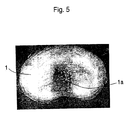
- fig. 5 is a picture of an intervertebral disc treated with the device according to the invention.
-
- The device illustrated in the drawings is intended for treatment of biological tissues in the form of
intervertebral discs 1, preferably nucleus pulposus 1a. For this purpose, the device comprises atreatment transducer 2 which includes twoultrasonic transducers 3 and 4 (so called therapeutic transducers). These are arranged to each transmit anultrasonic field focal area 5. During treatment, saidfocal area 5 is located in theintervertebral disc 1, preferably nucleus pulposus 1a, to be treated for heating the tissue therein to such temperature that the tissue in thefocal area 5 degenerates, whereby the pressure in theintervertebral disc 1 and thus, the pressure against thespinal cord 6, is reduced. - The
ultrasonic transducers 3, 4 are provided to transmitultrasonic fields ultrasonic transducers 3, 4, the ultrasound effect is distributed over larger areas and therefore, a lower power per transducer can be used. This results in that eventual heat increases in the skin are minimized for the power to be put in theintervertebral disc 1. - The power and frequency of the
ultrasonic fields focal area 5 and the transmitting time of theultrasonic fields ultrasonic fields - The
ultrasonic transducers 3, 4 can be arranged to transmitultrasonic fields focal area 5 of which is adaptable relative to the space M between theend plates vertebrae intervertebral disc 1, such that saidfocal area 5 can lie between theend plates - A
focal area 5 with such adapted extension can be obtained by providing theultrasonic transducers 3 and 4 respectively, withmeans 11, preferably in the form of transducer elements with concave spherical transducer surface. Hereby, a larger or better focussing is achieved and thefocal area 5 is given a substantially planar extension, as is shown in fig. 3. - The
ultrasonic transducers 3, 4 may also includemeans 12 for, if required, displacing thefocal area 5, adapted preferably as mentioned to the space M between theend plates intervertebral disc 1, preferably nucleus pulposus 1a. These means 12 can be transducer elements of the phased array type. - For being able to locate to
focal area 5 in theintervertebral disc 1, preferably nucleus pulposus 1a, it may some-times - depending on the relative positions of theultrasonic transducers 3, 4 and the intervertebral disc 1 - be advantageous or necessary to shrink or reduce the extention of one or both theultrasonic fields intervertebral disc 1 which may not be heated to tissue-degenerating temperatures. This can be achieved while theultrasonic transducers 3, 4 have a plurality ofmeans 12, preferably said transducers of phased array type, which momentarily can be deactivated or put out of operation for reducing the extension of theultrasonic field 3a and/or 4a. - The
ultrasonic transducers 3, 4 can preferably be located relative to theintervertebral disc 1 so that they transmitultrasonic fields focal area 5 with a substantially planar extension and which are situated substantially in parallel with and in a plane P wherein theintervertebral disc 1 is located. - The
ultrasonic transducers 3, 4 can be arranged to transmitultrasonic fields focal area 5 the extension of which can be varied for adaptation to the size of theintervertebral disc 1 and/or nucleus pulposus 1a. - A
diagnostic device 13 may include at least oneultrasonic transducer 14 which is provided, prior to the ultrasound treatment, to transmit anultrasonic field 14a for registrering the location of theintervertebral disc 1. The position of theultrasonic transducers 3, 4 relative to theultrasonic transducer 14 are also known and in this way, their positions relative to theintervertebral disc 1 can be determined. Theultrasonic transducers 3, 4 can be arranged to be controlled depending on information registered in thediagnostic device 13 regarding the current position of theintervertebral disc 1, preferably nucleus pulposus 1a, so that thefocal area 5 is moved to coincide with theintervertebral disc 1, preferably nucleus pulposus 1a. The positions of theultrasonic transducers 3, 4 can e.g. be controlled by changing their positions relative to their attachments. The control can be determined by a computer. - Said control of the
ultrasonic transducers 3, 4 can be carried through by means of acontrol device 15 either automatically or by a surgeon marking on a screen (not shown) a point/an area in nucleus pulposus 1a in which thefocal area 5 is desired. A computer (not shown) can determine the necessary parameters in theultrasonic transducers 3, 4 such that thefocal area 5 becomes correct. - A
reading device 16 can be provided for registering displacements of theintervertebral disc 1 relative to theultrasonic transducers 3 and 4 which occur when the patient moves during treatment. Asetting device 17 can be provided to automatically control theultrasonic transducers 3, 4 to set so that thefocal area 5 again will lie in theintervertebral disc 1, preferably nucleus pulposus 1a, after said displacement. - There might also be a non-invasive temperature-
supervising device 18 for supervising the temperature in theintervertebral disc 1 during treatment. This temperature -supervisingdevice 18 may cooperate with acontrol unit 19 which is provided to control theultrasonic transducers 3, 4 such that undesired temperature changes in theintervertebral disc 1 are prevented during treatment. - The temperature-supervising
device 18 may cooperate with theultrasonic transducer 14 of thediagnostic device 13 for supervising or monitoring the temperature in theintervertebral disc 1 by means of ultrasound. - The
ultrasonic transducers 3, 4 are preferably arranged obliquely behind the spine on opposite sides of thespinal cord 6, so that they can transmit theirultrasonic fields spinal cord 6 on opposite sides thereof and so that they meet in front thereof in order to together define thefocal area 5 in theintervertebral disc 1, preferably nucleus pulposus 1a. - During treatment, a treatment table 20 can be used, which has a
support surface 21 for thepatient 22. Thissupport surface 21 can be curved or settable into a curved shape such that the space M between thevertebrae intervertebral disc 1 to be treated, increases when thepatient 22 rests with his or her back against saidsupport surface 21. - The
ultrasonic transducers frame 23, whereby the mutual positions of saidtransducers frame 23 with thetransducers patient 22 for setting thetransducers intervertebral disc 1, preferably nucleus pulposus 1a, to be treated, in a patient 22 on the treatment table 20. - To avoid disturbances of the
ultrasonic fields transducers liquid container 24 can be located between the transducers and the adjacent skin of thepatient 22. - Air between the
transducers liquid container 24 and adjacent skin can for the same purpose be removed by locating agel 25 schematically illustrated with broken lines. - Treatment by means of the device described above is shown in the flow chart of fig. 4, whereby the diagnosis is already made, i.e. it is already determined which
intervertebral disc 1 should be treated. - By means of the "ultrasound picture" obtained from the
diagnostic device 13, theframe 23 and thepatient 22 and eventually theultrasonic transducers 3, 4 are oriented separately so that the angle of incidence of thetherapeutic transducers 3, 4 is correct. Hereby, it is achieved that theultrasonic fields ultrasonic transducers 3, 4 lie in the same plane P as theintervertebral disc 1 to be treated, and so that thetherapeutic transducers 3, 4 are directed towards the patient 22 from behind. On the abovementioned screen, a point in thenucleus pulposus 1a is marked in which the focal area 5 (a so called movable focus which is displaced by means of the abovementioned means 12) for thetherapeutic transducers 3, 4 shall be located. A computer then establish the requirements for the various transducer elements in thetherapeutic transducers 3, 4. Eventually, adjacent bones can be marked as zones which theultrasonic fields - Said computer may e.g. deactivate the transducer elements which are closest and reduces thereby the size of the
ultrasonic field ultrasonic field 3a and/or 4a can be compensated with a longer treatment time and/or treatment effect. The "fixed focus" mentioned in the flow chart is the focal area obtained by means of theabovementioned means 11. - It should also be mentioned that instead of ultrasonic transducers, other therapeutic and/or diagnostic transducers, transmitters or applicators, e.g. transducers or transmitters of electromagnetic radiation, can be used in the device described above.
- Drugs can be used in connection with and/or after treatment with the abovementioned device, namely pressure reducing/volume decreasing drugs, e.g. steroids or corticon™, and/or dehydrating drugs, e.g. impugan™, and/or antiinflammatory drugs, e.g. voltaren™.
- By means of the pressure reducing/volume decreasing drugs, it is possible, in connection with and/or after treatment with ultrasound, to improve the pressure reduction/volume decrease in nucleus pulposus obtained thereby.
- By means of the dehydrating drugs, it is possible to improve the removal of fluid from the treatment area in connection with and/or after treatment with ultrasound.
- By means of said antiinflammatory drugs, it is possible to oppose inflammations in the treatment area in connection with and/or after treatment with ultrasound.
- The invention is not limited to the device described above and illustrated in the drawings, but may vary within the scope of the following claims. Thus, the
diagnostic device 13 can operate with something else than ultrasound. - Instead of the patient 22 lying on his back on a treatment table 20, whereby the treatment equipment is situated beneath his or her back, the
patient 22 and treatment equipment can be positioned otherwise. Thus, the patient 22 can e.g. be lying on his or her face and the treatment equipment be mounted on an arm permitting location thereof in a treatment position. -
- Billard BE, Hynynen K and Roemer R B, 1990, Effects of physical parameters on high temperature ultrasound hyperhtermia. Ultrasound in Med. & Biol., 16 (4), 409-420
- Buchelt M, Kutschera H-P, Katterschafka T, Kiss H, Schneider B and Ullrich R, 1992, Erb.YAG and Hol.YAG Laser Ablation of Meniscus and Intervertebral discs. Lasers in Surgery and Medicine, 12, 375-381
- Bush N L, Rivens I, ter Haar G R and Bamber J C, 1993, Acoustic properties of lesions generated with an ultrasound therapy system. Ultrasound in Med & Biol, 19 (9), 789-801
- Diederich C J and Hynynen K, 1991, The feasibility of using electrically focused ultrasound arrays to induce deep hyperthermia via body cavities. IEEE transactions on ultrasonics, ferroelectrics, and frequency control, 38 (3), 207-219
- Ebbini E S and Cain C A, 1991, A Spherical-Section Ultrasound Phased Array Applicator for Deep Localized Hyperthermia. IEEE Transactions on Biomedical Engineering, 38 (7), 634-643
- Ebbini E S, Umemura S-I, Ibbini M and Cain C A, 1988, A cylindrical-section ultrasound phased-array applicator for hyperthermia cancer therapy. IEEE transactions on ultrasonics, ferroelectrics, and frequency control, 35 (5), 561-572
- Holmer N-G and Lindström K, 198?, Från A-mode till Phased array i Diagnostiskt ultraljud-Grunderna, editerad av Holmer, s 65-67
- Lele P P, 1980, Induction of deep, local hyperthermia by ultrasound and electromagnetic fields. -Problems & choices. Radiation and Environmental Biophysics, 17, 205-217
- Linke C A, Carstensen E L, Frizzell L A, Elbadawi A and Fridd C W, 1973, Localized tissue destruction by high-intensity focused ultrasound. Arch Surg, 107 (6), 887-891
- Sibille A, Prat F, Chapelon J-Y, El Fadil F A, Henry L, Theillère Y, Ponchon T and Cathignol D, 1993, Extracorporeal ablation of liver tissues by high intensity focused ultrasound. Oncology, 50, 375-379
- Troussier B, Lebas J F, Chirossel J P, Peoc'h M, Grand S, Leviel J L and Phelip X, 1995, Percutaneous Intradiscal Radio-Frequency Thermocoagulation - A Cadaveric Study. Spine, 20 (15), 1713-1718
-
Claims (9)
- Device for non-invasive treatment of the tissue of an intervertebral disc (1), whereby the treatment aims at changing or degenerating said tissue, said device comprising a treatment transducer (2) comprising at least two ultrasonic transducers (3, 4) having means (11), preferably in the form of transducer elements with a concave spherical transducer surface, for transmitting ultrasonic fields (3a, 4a) with a focal area (5) which has a substantially planar extension which is adaptable relative to a space (M) between end plates (7, 8) of a vertebrae (9, 10) surrounding the intervertebral disc (1) such that said focal area (5) lies between said end plates (7, 8),
the ultrasonic transducers (3, 4) of the treatment transducer (2) further including means (12), preferably transducer elements of the phased array type, for displacing said focal area (5) and for varying the extension of said focal area (5),
the ultrasonic transducers (3, 4) being located such that said focal area (5) is placeable substantially parallel to a plane (P) in which the intervertebral disc (1) is located. - Device according to claim 1, wherein the ultrasonic transducers (3, 4) of the treatment transducer (2) are capable of transmitting ultrasonic waves having a frequency of 0,5 - 2,5 MHz.
- Device according to any preceding claim, wherein the ultrasonic transducers (3, 4) of the treatment transducer (2) are capable of transmitting ultrasonic fields (3a, 4a) which in the focal area (5) heat the tissue to a temperature of 45-80°C.
- Device according to any preceding claim, further comprising means for deactivating one or more of said transducers or put out of operation for reducing the extension of the ultrasonic fields (3a, 4a) for preventing said ultrasonic fields from heating biological tissues outside the intervertebral disc (1) to tissue-degenerating temperatures.
- Device according to any preceding claim, further comprising
a non-invasive temperature-supervising device (18), preferably by means of ultrasound, to supervise the temperature in the intervertebral disc (1) during treatment, and
a control unit for controlling the ultrasonic transducers (3, 4) of the treatment transducer (2) such that undesired temperature changes in the intervertebral disc (1) are prevented. - Device according to any preceding claim, further comprising
a diagnostic device (13) for registering the location of the intervertebral disc (1) relative to the ultrasonic transducers (3, 4) of the treatment transducer (2), and
the ultrasonic transducers (3, 4) of the treatment transducer (2) are arranged to be controlled depending on information registered by the diagnostic device (13) so that the focal area (5) of their ultrasonic fields (3a, 4a) is moved to coincide with the intervertebral disc (1). - Device according to claim 6, wherein the diagnostic device (13) includes an ultrasonic transducer (14) which transmits an ultrasonic field for registering the location of the intervertebral disc (1) relative to the treatment transducer (2).
- Device according to any preceding claim, further comprising
a reading device (16) for registering displacements of the intervertebral disc (1) relative to the ultrasonic transducers (3, 4) of the treatment transducer (2) due to patient movements during treatment, and
a setting device (17) for controlling the ultrasonic transducers (3, 4) of the treatment transducer (2) so that the focal area (5) of the ultrasonic fields (3a, 4a) is capable of lying in the intervertebral disc (1) after displacement thereof. - Device according to claim 7,
the ultrasonic transducers (3, 4) of the treatment transducer (2) and the diagnostic device (13) having an ultrasonic transducer (14) are mounted on a frame (23) relative to a treatment table (20), whereby the mutual positions between said transducers (3, 4, 14) are known, wherein
the frame (23) is displaceable relative to a patient (22) for setting said transducers (3, 4, 14) relative to an intervertebral disc (1) of the patient (22).
Applications Claiming Priority (2)
| Application Number | Priority Date | Filing Date | Title |
|---|---|---|---|
| SE9701449A SE518490C2 (en) | 1997-04-18 | 1997-04-18 | Device for non-invasive treatment of biological tissue |
| SE9701449 | 1997-04-18 |
Publications (3)
| Publication Number | Publication Date |
|---|---|
| EP0872262A2 EP0872262A2 (en) | 1998-10-21 |
| EP0872262A3 EP0872262A3 (en) | 1999-11-10 |
| EP0872262B1 true EP0872262B1 (en) | 2004-09-29 |
Family
ID=20406625
Family Applications (1)
| Application Number | Title | Priority Date | Filing Date |
|---|---|---|---|
| EP98106818A Expired - Lifetime EP0872262B1 (en) | 1997-04-18 | 1998-04-15 | Device for non-invasive treatment of biological tissue |
Country Status (7)
| Country | Link |
|---|---|
| US (1) | US6254553B1 (en) |
| EP (1) | EP0872262B1 (en) |
| JP (1) | JP4078492B2 (en) |
| AT (1) | ATE277673T1 (en) |
| DE (1) | DE69826550T2 (en) |
| ES (1) | ES2229408T3 (en) |
| SE (1) | SE518490C2 (en) |
Cited By (1)
| Publication number | Priority date | Publication date | Assignee | Title |
|---|---|---|---|---|
| WO2025219450A1 (en) * | 2024-04-16 | 2025-10-23 | Universitat Pompeu Fabra | System for spinal treatment comprising an adjustable stretcher and method for optimally configuring said system |
Families Citing this family (60)
| Publication number | Priority date | Publication date | Assignee | Title |
|---|---|---|---|---|
| EP1124614A1 (en) | 1998-10-27 | 2001-08-22 | Fraunhofer-Gesellschaft Zur Förderung Der Angewandten Forschung E.V. | Method and device for controlling a targeted thermal deposition into a material |
| US6508774B1 (en) * | 1999-03-09 | 2003-01-21 | Transurgical, Inc. | Hifu applications with feedback control |
| SE518763C2 (en) * | 2000-07-17 | 2002-11-19 | Ultrazonix Dnt Ab | Device for non-invasive ultrasound treatment of disk disease |
| SE518764C2 (en) | 2000-07-17 | 2002-11-19 | Ultrazonix Dnt Ab | Device for mini-invasive ultrasound treatment of disk disease |
| US20100081893A1 (en) * | 2008-09-19 | 2010-04-01 | Physiosonics, Inc. | Acoustic palpation using non-invasive ultrasound techniques to identify and localize tissue eliciting biological responses and target treatments |
| WO2002072201A1 (en) * | 2001-03-08 | 2002-09-19 | The Regents Of The University Of California | Transcutaneous spine trauma and disorders treatment using ultrasonically induced confined heat zone |
| US6673023B2 (en) * | 2001-03-23 | 2004-01-06 | Stryker Puerto Rico Limited | Micro-invasive breast biopsy device |
| US20020138091A1 (en) * | 2001-03-23 | 2002-09-26 | Devonrex, Inc. | Micro-invasive nucleotomy device and method |
| US20020138021A1 (en) * | 2001-03-23 | 2002-09-26 | Devonrex, Inc. | Micro-invasive tissue removal device |
| US20030013972A1 (en) * | 2001-05-29 | 2003-01-16 | Makin Inder Raj. S. | Treatment of lung lesions using ultrasound |
| US20030069569A1 (en) * | 2001-08-29 | 2003-04-10 | Burdette Everette C. | Ultrasound device for treatment of intervertebral disc tissue |
| SE520857C2 (en) | 2002-01-15 | 2003-09-02 | Ultrazonix Dnt Ab | Device with both therapeutic and diagnostic sensors for mini-invasive ultrasound treatment of an object, where the therapeutic sensor is thermally insulated |
| EP1465701A4 (en) | 2002-01-15 | 2008-08-13 | Univ California | SYSTEM AND METHOD FOR DIRECTIONAL ULTRASONIC THERAPY OF SKELETAL JOINTS |
| SE520858C2 (en) * | 2002-01-15 | 2003-09-02 | Ultrazonix Dnt Ab | Device with both therapeutic and diagnostic sensors for non-invasive ultrasound treatment of an object |
| EP1503685B1 (en) | 2002-01-23 | 2012-10-31 | The Regents of The University of California | Implantable thermal treatment apparatus |
| US6780161B2 (en) * | 2002-03-22 | 2004-08-24 | Fmd, Llc | Apparatus for extracorporeal shock wave lithotripter using at least two shock wave pulses |
| US6969392B2 (en) * | 2002-05-01 | 2005-11-29 | Nevmet Corporation | Multiportal device and method for percutaneous surgery |
| US7118576B2 (en) * | 2002-05-15 | 2006-10-10 | Nevmet Corporation | Multiportal device with linked cannulae and method for percutaneous surgery |
| US7118577B2 (en) * | 2002-05-16 | 2006-10-10 | Nevmet Corporation | Multiportal device with linked segmented cannulae and method for percutaneous surgery |
| US8613744B2 (en) | 2002-09-30 | 2013-12-24 | Relievant Medsystems, Inc. | Systems and methods for navigating an instrument through bone |
| US8808284B2 (en) | 2008-09-26 | 2014-08-19 | Relievant Medsystems, Inc. | Systems for navigating an instrument through bone |
| US7258690B2 (en) | 2003-03-28 | 2007-08-21 | Relievant Medsystems, Inc. | Windowed thermal ablation probe |
| US6907884B2 (en) | 2002-09-30 | 2005-06-21 | Depay Acromed, Inc. | Method of straddling an intraosseous nerve |
| US8361067B2 (en) | 2002-09-30 | 2013-01-29 | Relievant Medsystems, Inc. | Methods of therapeutically heating a vertebral body to treat back pain |
| JP2005052523A (en) * | 2003-08-07 | 2005-03-03 | Teijin Ltd | Ultrasound irradiation device that promotes natural retraction of intervertebral disc herniation |
| US8406845B2 (en) * | 2004-09-01 | 2013-03-26 | University Of Tennessee Research Foundation | Method and apparatus for imaging tracking |
| US7771418B2 (en) * | 2005-03-09 | 2010-08-10 | Sunnybrook Health Sciences Centre | Treatment of diseased tissue using controlled ultrasonic heating |
| US8801701B2 (en) * | 2005-03-09 | 2014-08-12 | Sunnybrook Health Sciences Centre | Method and apparatus for obtaining quantitative temperature measurements in prostate and other tissue undergoing thermal therapy treatment |
| JP2009508970A (en) | 2005-09-21 | 2009-03-05 | ザ リージェンツ オブ ザ ユニバーシティ オブ カリフォルニア | System, composition and method for local imaging and treatment of pain |
| ITVR20060113A1 (en) * | 2006-06-07 | 2008-01-07 | Giglio Antonio Del | DEVICE FOR THE TREATMENT OF ADIPOSE SUBCUTANEOUS FABRIC BY NON-FOICALIZED AND OPPOSED SHOCKWAVES |
| US8088084B2 (en) * | 2007-03-06 | 2012-01-03 | The Cleveland Clinic Foundation | Method and apparatus for repair of intervertebral discs |
| US10183183B2 (en) | 2007-04-13 | 2019-01-22 | Acoustic Medsystems, Inc. | Acoustic applicators for controlled thermal modification of tissue |
| JP5688022B2 (en) | 2008-09-26 | 2015-03-25 | リリーバント メドシステムズ、インコーポレイテッド | System and method for guiding an instrument through the interior of a bone |
| US10028753B2 (en) | 2008-09-26 | 2018-07-24 | Relievant Medsystems, Inc. | Spine treatment kits |
| RU2417800C1 (en) * | 2009-10-28 | 2011-05-10 | Максимильян Григорьевич Кравчик | Method of noninvasive treatment of morphologoanatomic pathologies of intervertebral disks |
| US9707413B2 (en) | 2010-03-09 | 2017-07-18 | Profound Medical Inc. | Controllable rotating ultrasound therapy applicator |
| US11027154B2 (en) | 2010-03-09 | 2021-06-08 | Profound Medical Inc. | Ultrasonic therapy applicator and method of determining position of ultrasonic transducers |
| RU2436606C1 (en) * | 2010-04-28 | 2011-12-20 | Максимильян Григорьевич Кравчик | Method of treating inflammatory-degenerative spine and intervertebral disk diseases |
| RU2452454C1 (en) * | 2010-09-24 | 2012-06-10 | Валерий Викторович Педдер | Apparatus for non-contact ultrasonic biotissue treatment |
| US9408624B2 (en) | 2011-03-31 | 2016-08-09 | Isis Innovation Limited | Intervertebral disc treatment apparatus |
| RU2479301C2 (en) * | 2011-05-27 | 2013-04-20 | Валерий Викторович Педдер | Apparatus for treating lymphedema in breast cancer patients |
| EP2760545B1 (en) * | 2011-09-26 | 2019-04-24 | Profound Medical Inc. | System for control and monitoring of conformal thermal therapy |
| US10390877B2 (en) | 2011-12-30 | 2019-08-27 | Relievant Medsystems, Inc. | Systems and methods for treating back pain |
| US20150209551A1 (en) | 2012-08-15 | 2015-07-30 | Everette C. Burdette | Mri compatible ablation catheter system incorporating directional high-intensity ultrasound for treatment |
| US10588691B2 (en) | 2012-09-12 | 2020-03-17 | Relievant Medsystems, Inc. | Radiofrequency ablation of tissue within a vertebral body |
| AU2013337680B2 (en) | 2012-11-05 | 2019-04-18 | Relievant Medsystems, Inc. | Systems and methods for creating curved paths through bone and modulating nerves within the bone |
| US9724151B2 (en) | 2013-08-08 | 2017-08-08 | Relievant Medsystems, Inc. | Modulating nerves within bone using bone fasteners |
| US20150231417A1 (en) * | 2014-02-20 | 2015-08-20 | Warsaw Orthopedic, Inc. | Spinal surgery system and method |
| WO2016097867A2 (en) | 2014-12-19 | 2016-06-23 | Université Pierre Et Marie Curie (Paris 6) | Implantable ultrasound generating treating device for brain treatment, apparatus comprising such device and method implementing such device |
| CA3017916C (en) | 2016-03-11 | 2023-11-07 | Sorbonne Universite | External ultrasound generating treating device for spinal cord and spinal nerves treatment, apparatus comprising such device and method implementing such device |
| WO2017153798A1 (en) | 2016-03-11 | 2017-09-14 | Université Pierre Et Marie Curie (Paris 6) | Implantable ultrasound generating treating device for spinal cord and/or spinal nerve treatment, apparatus comprising such device and method |
| RU2642253C2 (en) * | 2016-05-20 | 2018-01-24 | Федеральное государственное бюджетное образовательное учреждение высшего образования "Чеченский государственный университет" | Method of conservative treatment of osteochondrosis |
| US11576677B2 (en) * | 2017-12-28 | 2023-02-14 | Cilag Gmbh International | Method of hub communication, processing, display, and cloud analytics |
| CN109999376B (en) * | 2019-03-19 | 2021-06-29 | 深圳市声科生物医学研究院 | HIFU equipment control system and nondestructive temperature measurement method thereof |
| WO2021050767A1 (en) | 2019-09-12 | 2021-03-18 | Relievant Medsystems, Inc. | Systems and methods for tissue modulation |
| WO2021234214A1 (en) * | 2020-05-18 | 2021-11-25 | Innotia Eesti Oü | System for ultrasound treatment combined with uvc teatment |
| AU2021306313A1 (en) | 2020-07-10 | 2023-03-02 | Relievant Medsystems, Inc. | Vertebral denervation in conjunction with vertebral fusion |
| US12082876B1 (en) | 2020-09-28 | 2024-09-10 | Relievant Medsystems, Inc. | Introducer drill |
| EP4268150A4 (en) | 2020-12-22 | 2024-12-18 | Relievant Medsystems, Inc. | PREDICTION OF CANDIDATES FOR SPINAL NEUROMODULATION |
| US12433668B1 (en) | 2021-11-08 | 2025-10-07 | Relievant Medsystems, Inc. | Impedance stoppage mitigation during radiofrequency tissue ablation procedures |
Family Cites Families (14)
| Publication number | Priority date | Publication date | Assignee | Title |
|---|---|---|---|---|
| US4513749A (en) * | 1982-11-18 | 1985-04-30 | Board Of Trustees Of Leland Stanford University | Three-dimensional temperature probe |
| US4813402A (en) * | 1986-02-19 | 1989-03-21 | Siemens Aktiengesellschaft | Coupling member for a shock wave therapy device |
| US4787394A (en) | 1986-04-24 | 1988-11-29 | Kabushiki Kaisha Toshiba | Ultrasound therapy apparatus |
| US5435311A (en) | 1989-06-27 | 1995-07-25 | Hitachi, Ltd. | Ultrasound therapeutic system |
| US5242439A (en) * | 1990-01-12 | 1993-09-07 | Laserscope | Means for inserting instrumentation for a percutaneous diskectomy using a laser |
| JPH03251240A (en) * | 1990-02-28 | 1991-11-08 | Toshiba Corp | Ultrasonic medical treatment device |
| WO1992015272A1 (en) * | 1991-03-06 | 1992-09-17 | Risaasen Borge | Operation table |
| US5291890A (en) * | 1991-08-29 | 1994-03-08 | General Electric Company | Magnetic resonance surgery using heat waves produced with focussed ultrasound |
| DE4207463C2 (en) * | 1992-03-10 | 1996-03-28 | Siemens Ag | Arrangement for the therapy of tissue with ultrasound |
| WO1993019705A1 (en) | 1992-03-31 | 1993-10-14 | Massachusetts Institute Of Technology | Apparatus and method for acoustic heat generation and hyperthermia |
| DE4238645C1 (en) * | 1992-11-16 | 1994-05-05 | Siemens Ag | Therapeutic ultrasonic applicator for urogenital area - has ultrasonic waves focussed onto working zone defined by envelope curve with two perpendicular main axes |
| US5307812A (en) | 1993-03-26 | 1994-05-03 | General Electric Company | Heat surgery system monitored by real-time magnetic resonance profiling |
| US5433739A (en) * | 1993-11-02 | 1995-07-18 | Sluijter; Menno E. | Method and apparatus for heating an intervertebral disc for relief of back pain |
| US5571147A (en) * | 1993-11-02 | 1996-11-05 | Sluijter; Menno E. | Thermal denervation of an intervertebral disc for relief of back pain |
-
1997
- 1997-04-18 SE SE9701449A patent/SE518490C2/en not_active IP Right Cessation
-
1998
- 1998-04-15 EP EP98106818A patent/EP0872262B1/en not_active Expired - Lifetime
- 1998-04-15 ES ES98106818T patent/ES2229408T3/en not_active Expired - Lifetime
- 1998-04-15 DE DE69826550T patent/DE69826550T2/en not_active Expired - Lifetime
- 1998-04-15 AT AT98106818T patent/ATE277673T1/en not_active IP Right Cessation
- 1998-04-16 US US09/061,547 patent/US6254553B1/en not_active Expired - Fee Related
- 1998-04-20 JP JP10916298A patent/JP4078492B2/en not_active Expired - Fee Related
Cited By (1)
| Publication number | Priority date | Publication date | Assignee | Title |
|---|---|---|---|---|
| WO2025219450A1 (en) * | 2024-04-16 | 2025-10-23 | Universitat Pompeu Fabra | System for spinal treatment comprising an adjustable stretcher and method for optimally configuring said system |
Also Published As
| Publication number | Publication date |
|---|---|
| SE9701449L (en) | 1999-01-08 |
| EP0872262A2 (en) | 1998-10-21 |
| DE69826550D1 (en) | 2004-11-04 |
| DE69826550T2 (en) | 2005-11-17 |
| JPH10295718A (en) | 1998-11-10 |
| SE9701449D0 (en) | 1997-04-18 |
| US6254553B1 (en) | 2001-07-03 |
| SE518490C2 (en) | 2002-10-15 |
| ES2229408T3 (en) | 2005-04-16 |
| JP4078492B2 (en) | 2008-04-23 |
| EP0872262A3 (en) | 1999-11-10 |
| ATE277673T1 (en) | 2004-10-15 |
Similar Documents
| Publication | Publication Date | Title |
|---|---|---|
| EP0872262A2 (en) | Device for non-invasive treatment of biological tissue | |
| AU2001271208B2 (en) | Device for mini-invasive ultrasound treatment of disc disease | |
| US6613005B1 (en) | Systems and methods for steering a focused ultrasound array | |
| US10898254B2 (en) | Implantable thermal treatment method and apparatus | |
| US5643179A (en) | Method and apparatus for ultrasonic medical treatment with optimum ultrasonic irradiation control | |
| KR100970304B1 (en) | Devices for micro-invasive ultrasound therapy of the treatment target by heat-insulated transducer | |
| EP1796545B1 (en) | Focused ultrasound system for surrounding a body tissue mass | |
| EP1301243B1 (en) | Device for non-invasive ultrasound treatment of disc disease | |
| KR20040075350A (en) | Device for non-invasive ultrasound treatment of an object | |
| Persson et al. | Ultrasound nucleolysis: an in vitro study | |
| Watkin et al. | High intensity focused ultrasound: applications in urology |
Legal Events
| Date | Code | Title | Description |
|---|---|---|---|
| PUAI | Public reference made under article 153(3) epc to a published international application that has entered the european phase |
Free format text: ORIGINAL CODE: 0009012 |
|
| AK | Designated contracting states |
Kind code of ref document: A2 Designated state(s): AT BE CH DE ES FR GB IT LI NL SE |
|
| AX | Request for extension of the european patent |
Free format text: AL;LT;LV;MK;RO;SI |
|
| PUAL | Search report despatched |
Free format text: ORIGINAL CODE: 0009013 |
|
| AK | Designated contracting states |
Kind code of ref document: A3 Designated state(s): AT BE CH CY DE DK ES FI FR GB GR IE IT LI LU MC NL PT SE |
|
| AX | Request for extension of the european patent |
Free format text: AL;LT;LV;MK;RO;SI |
|
| 17P | Request for examination filed |
Effective date: 20000502 |
|
| AKX | Designation fees paid |
Free format text: AT BE CH DE ES FR GB IT LI NL SE |
|
| RAP1 | Party data changed (applicant data changed or rights of an application transferred) |
Owner name: ULTRAZONIX DNT AB |
|
| 17Q | First examination report despatched |
Effective date: 20030509 |
|
| GRAP | Despatch of communication of intention to grant a patent |
Free format text: ORIGINAL CODE: EPIDOSNIGR1 |
|
| GRAS | Grant fee paid |
Free format text: ORIGINAL CODE: EPIDOSNIGR3 |
|
| GRAA | (expected) grant |
Free format text: ORIGINAL CODE: 0009210 |
|
| AK | Designated contracting states |
Kind code of ref document: B1 Designated state(s): AT BE CH DE ES FR GB IT LI NL SE |
|
| REG | Reference to a national code |
Ref country code: GB Ref legal event code: FG4D |
|
| REG | Reference to a national code |
Ref country code: CH Ref legal event code: EP |
|
| REF | Corresponds to: |
Ref document number: 69826550 Country of ref document: DE Date of ref document: 20041104 Kind code of ref document: P |
|
| REG | Reference to a national code |
Ref country code: SE Ref legal event code: TRGR |
|
| REG | Reference to a national code |
Ref country code: CH Ref legal event code: NV Representative=s name: A. BRAUN, BRAUN, HERITIER, ESCHMANN AG PATENTANWAE |
|
| REG | Reference to a national code |
Ref country code: ES Ref legal event code: FG2A Ref document number: 2229408 Country of ref document: ES Kind code of ref document: T3 |
|
| PLBE | No opposition filed within time limit |
Free format text: ORIGINAL CODE: 0009261 |
|
| STAA | Information on the status of an ep patent application or granted ep patent |
Free format text: STATUS: NO OPPOSITION FILED WITHIN TIME LIMIT |
|
| ET | Fr: translation filed | ||
| 26N | No opposition filed |
Effective date: 20050630 |
|
| REG | Reference to a national code |
Ref country code: CH Ref legal event code: PFA Owner name: ULTRAZONIX DNT AB Free format text: ULTRAZONIX DNT AB#KROSSVERKSGATAN 3#216 16 LIMHAMN (SE) -TRANSFER TO- ULTRAZONIX DNT AB#KROSSVERKSGATAN 3#216 16 LIMHAMN (SE) |
|
| PGFP | Annual fee paid to national office [announced via postgrant information from national office to epo] |
Ref country code: AT Payment date: 20090427 Year of fee payment: 12 |
|
| PGFP | Annual fee paid to national office [announced via postgrant information from national office to epo] |
Ref country code: BE Payment date: 20090427 Year of fee payment: 12 |
|
| PGFP | Annual fee paid to national office [announced via postgrant information from national office to epo] |
Ref country code: CH Payment date: 20090416 Year of fee payment: 12 |
|
| BERE | Be: lapsed |
Owner name: *ULTRAZONIX DNT A.B. Effective date: 20100430 |
|
| REG | Reference to a national code |
Ref country code: CH Ref legal event code: PL |
|
| PG25 | Lapsed in a contracting state [announced via postgrant information from national office to epo] |
Ref country code: AT Free format text: LAPSE BECAUSE OF NON-PAYMENT OF DUE FEES Effective date: 20100415 |
|
| PG25 | Lapsed in a contracting state [announced via postgrant information from national office to epo] |
Ref country code: LI Free format text: LAPSE BECAUSE OF NON-PAYMENT OF DUE FEES Effective date: 20100430 Ref country code: CH Free format text: LAPSE BECAUSE OF NON-PAYMENT OF DUE FEES Effective date: 20100430 |
|
| PG25 | Lapsed in a contracting state [announced via postgrant information from national office to epo] |
Ref country code: BE Free format text: LAPSE BECAUSE OF NON-PAYMENT OF DUE FEES Effective date: 20100430 |
|
| PGFP | Annual fee paid to national office [announced via postgrant information from national office to epo] |
Ref country code: SE Payment date: 20110414 Year of fee payment: 14 Ref country code: DE Payment date: 20110421 Year of fee payment: 14 Ref country code: FR Payment date: 20110510 Year of fee payment: 14 Ref country code: ES Payment date: 20110426 Year of fee payment: 14 |
|
| PGFP | Annual fee paid to national office [announced via postgrant information from national office to epo] |
Ref country code: GB Payment date: 20110421 Year of fee payment: 14 Ref country code: NL Payment date: 20110426 Year of fee payment: 14 |
|
| PGFP | Annual fee paid to national office [announced via postgrant information from national office to epo] |
Ref country code: IT Payment date: 20110422 Year of fee payment: 14 |
|
| REG | Reference to a national code |
Ref country code: NL Ref legal event code: V1 Effective date: 20121101 |
|
| REG | Reference to a national code |
Ref country code: SE Ref legal event code: EUG |
|
| GBPC | Gb: european patent ceased through non-payment of renewal fee |
Effective date: 20120415 |
|
| REG | Reference to a national code |
Ref country code: FR Ref legal event code: ST Effective date: 20121228 |
|
| PG25 | Lapsed in a contracting state [announced via postgrant information from national office to epo] |
Ref country code: GB Free format text: LAPSE BECAUSE OF NON-PAYMENT OF DUE FEES Effective date: 20120415 |
|
| REG | Reference to a national code |
Ref country code: DE Ref legal event code: R119 Ref document number: 69826550 Country of ref document: DE Effective date: 20121101 |
|
| PG25 | Lapsed in a contracting state [announced via postgrant information from national office to epo] |
Ref country code: SE Free format text: LAPSE BECAUSE OF NON-PAYMENT OF DUE FEES Effective date: 20120416 Ref country code: FR Free format text: LAPSE BECAUSE OF NON-PAYMENT OF DUE FEES Effective date: 20120430 Ref country code: IT Free format text: LAPSE BECAUSE OF NON-PAYMENT OF DUE FEES Effective date: 20120415 |
|
| PG25 | Lapsed in a contracting state [announced via postgrant information from national office to epo] |
Ref country code: NL Free format text: LAPSE BECAUSE OF NON-PAYMENT OF DUE FEES Effective date: 20121101 |
|
| REG | Reference to a national code |
Ref country code: ES Ref legal event code: FD2A Effective date: 20130715 |
|
| PG25 | Lapsed in a contracting state [announced via postgrant information from national office to epo] |
Ref country code: ES Free format text: LAPSE BECAUSE OF NON-PAYMENT OF DUE FEES Effective date: 20120416 |
|
| PG25 | Lapsed in a contracting state [announced via postgrant information from national office to epo] |
Ref country code: DE Free format text: LAPSE BECAUSE OF NON-PAYMENT OF DUE FEES Effective date: 20121101 |