EP0869976B1 - Design of hormone-like antibodies with agonistic and antagonistic functions - Google Patents
Design of hormone-like antibodies with agonistic and antagonistic functions Download PDFInfo
- Publication number
- EP0869976B1 EP0869976B1 EP96939791A EP96939791A EP0869976B1 EP 0869976 B1 EP0869976 B1 EP 0869976B1 EP 96939791 A EP96939791 A EP 96939791A EP 96939791 A EP96939791 A EP 96939791A EP 0869976 B1 EP0869976 B1 EP 0869976B1
- Authority
- EP
- European Patent Office
- Prior art keywords
- trka
- mab
- antibody
- ngf
- cells
- Prior art date
- Legal status (The legal status is an assumption and is not a legal conclusion. Google has not performed a legal analysis and makes no representation as to the accuracy of the status listed.)
- Expired - Lifetime
Links
Images





Classifications
-
- A—HUMAN NECESSITIES
- A61—MEDICAL OR VETERINARY SCIENCE; HYGIENE
- A61K—PREPARATIONS FOR MEDICAL, DENTAL OR TOILETRY PURPOSES
- A61K51/00—Preparations containing radioactive substances for use in therapy or testing in vivo
- A61K51/02—Preparations containing radioactive substances for use in therapy or testing in vivo characterised by the carrier, i.e. characterised by the agent or material covalently linked or complexing the radioactive nucleus
- A61K51/04—Organic compounds
- A61K51/08—Peptides, e.g. proteins, carriers being peptides, polyamino acids, proteins
- A61K51/10—Antibodies or immunoglobulins; Fragments thereof, the carrier being an antibody, an immunoglobulin or a fragment thereof, e.g. a camelised human single domain antibody or the Fc fragment of an antibody
- A61K51/1045—Antibodies or immunoglobulins; Fragments thereof, the carrier being an antibody, an immunoglobulin or a fragment thereof, e.g. a camelised human single domain antibody or the Fc fragment of an antibody against animal or human tumor cells or tumor cell determinants
-
- C—CHEMISTRY; METALLURGY
- C07—ORGANIC CHEMISTRY
- C07K—PEPTIDES
- C07K14/00—Peptides having more than 20 amino acids; Gastrins; Somatostatins; Melanotropins; Derivatives thereof
- C07K14/435—Peptides having more than 20 amino acids; Gastrins; Somatostatins; Melanotropins; Derivatives thereof from animals; from humans
- C07K14/705—Receptors; Cell surface antigens; Cell surface determinants
- C07K14/71—Receptors; Cell surface antigens; Cell surface determinants for growth factors; for growth regulators
-
- C—CHEMISTRY; METALLURGY
- C07—ORGANIC CHEMISTRY
- C07K—PEPTIDES
- C07K16/00—Immunoglobulins [IG], e.g. monoclonal or polyclonal antibodies
- C07K16/18—Immunoglobulins [IG], e.g. monoclonal or polyclonal antibodies against material from animals or humans
- C07K16/28—Immunoglobulins [IG], e.g. monoclonal or polyclonal antibodies against material from animals or humans against receptors, cell surface antigens or cell surface determinants
- C07K16/2863—Immunoglobulins [IG], e.g. monoclonal or polyclonal antibodies against material from animals or humans against receptors, cell surface antigens or cell surface determinants against receptors for growth factors, growth regulators
-
- A—HUMAN NECESSITIES
- A61—MEDICAL OR VETERINARY SCIENCE; HYGIENE
- A61K—PREPARATIONS FOR MEDICAL, DENTAL OR TOILETRY PURPOSES
- A61K38/00—Medicinal preparations containing peptides
-
- C—CHEMISTRY; METALLURGY
- C07—ORGANIC CHEMISTRY
- C07K—PEPTIDES
- C07K2317/00—Immunoglobulins specific features
- C07K2317/70—Immunoglobulins specific features characterized by effect upon binding to a cell or to an antigen
- C07K2317/77—Internalization into the cell
Definitions
- the invention relates to a method of designing agonistic and/or antagonistic antibodies for any hormone receptor more specifically antibodies which are capable of blocking nerve growth factor (NGF) binding and antibodies which can mimic NGF binding to its receptor.
- NGF nerve growth factor
- the TrkA receptor is a 140 kDa transmembrane glycoprotein with tyrosine kinase activity that functions as the Nerve Growth Factor (NGF) receptor.
- NGF Nerve Growth Factor
- p75 receptor a p75 receptor whose signaling function is unclear.
- Homo or heterodimers or oligomers of TrkA and p75 bind NGF with higher affinity (Jing, S. et al. (1992) Neuron, 9 : 1067-1079) suggesting that specific receptor conformations may play specific functions.
- NGF promotes the differentiation of certain neuronal cells, is mitogenic for TrkA-transfected fibroblasts, and allows survival in serum-deprived conditions for both cell types.
- Activation of the tyrosine kinase activity of TrkA via NGF binding leads to receptor trans- and auto-tyrosine phosphorylation (PY), and PY of second messengers including phosphatidylinositol-3 kinase (PI-3 kinase).
- PI-3 kinase is involved in protein trafficking and endocytosis of ligand-receptor complexes (reviewed by Kaplan, D.R. et al. (1994) J. Neurobiol., 25: 1404-1417). Since microinjection of NGF into cells does not result in NGF biological signals, cell surface receptor ligation and internalization of TrkA or NGF-TrkA complexes must mediate these effects.
- TrkA like most kinase growth factor receptors, signals through receptor oligomerization.
- monovalent TrkA-binding agents are antagonistic or have no biological effects (LeSterrorism, L. et al.(1995) J. Biol. Chem., 270 :6564-6569)
- bivalent receptor-binding agents such as NGF (a homodimer) or antibodies can be agonistic.
- NGF a homodimer
- mAb anti-receptor monoclonal antibodies
- an agonistic or antagonistic anti-human TrkA mAb which recognizes the NGF docking site.
- Such antibodies may be used for the diagnosis, treatment or prevention of neurological diseases, neuromas and neoplastic tumors which express TrkA receptors. Also these antibodies may be used to develop and screen for pharmaceutical agents which are agonistic or antagonistic by binding to the TrkA receptors.
- One aim of the present invention is to report the development and characterization of an agonistic anti-human TrkA mAb 5C3 which recognizes at least one NGF docking site.
- This MAb 5C3 was used to characterize the pattern of TrkA protein expression in normal human brain, and the NGF binding features of the receptor.
- MAb 5C3 behaves like NGF in bioassays, and monomeric 5C3 F abs retained binding and functional agonistic activity.
- MAb 5C3 will be useful to identify the NGF docking site on TrkA and possibly as a pharmacological lead in the development of small mimetics.
- an antibody or functional fragment thereof which binds to at least the TrkA receptor under physiological conditions, and wherein the binding to the receptor at least partially mimics or inhibits nerve growth factor biological activity.
- a method of screening pharmacological agents which are capable to mimic or inhibit nerve growth factor biological activity which comprises using the antibody of the present invention to screen for pharmacological agents capable of binding to the complementary determining region of the antibody, wherein the screened pharmacological agents can mimic or inhibit nerve growth factor biological activity.
- an antibody having CDR-like domains of hormones wherein the antibody or functional fragment thereof binds to at least TrkA receptor under physiological conditions, and wherein the binding to the receptor at least partially mimics or inhibits nerve growth factor biological activity.
- a pharmaceutical composition for the treatment of neurological diseases, neuromas and neoplastic tumors which express TrkA receptors which comprises an effective amount of an antibody of the present invention or a functional fragment thereof in association with a pharmaceutically acceptable carrier.
- a pharmaceutical composition for the targeting of pharmaceutical agents to tissues of the central and/or peripheral nervous system which comprises an effective amount of an antibody of the present invention or a functional fragment thereof coupled to a pharmaceutical agent in association with a pharmaceutically acceptable carrier.
- the pharmaceutical agent may be selected from the group consisting of radioligands, nucleic acid molecules, toxins, growth factors and gangliosides.
- TrkA docking site which binds to the antibody of the present invention.
- the docking site of the present invention for screening pharmacological agents which are capable to mimic or inhibit nerve growth factor biological activity and said antibody biological activity.
- Monoclonal antibody (mAb) 5C3 directed against human p140 TrkA is a structural and functional mimic of NGF and an artificial receptor agonist.
- MAb 5C3 binds in the Nerve Growth Factor (NGF) docking site, and like NGF it promotes TrkA internalization; TrkA and phosphatidylinositol-3 kinase tyrosine phosphorylation; and increased transformation of TrkA-expressing fibroblasts. More importantly, mAb 5C3 protects human TrkA-expressing cells from apoptotic death in serum-free media. Interestingly, agonistic activity is observed with monomeric F ab 5C3 fragments.
- the affinity of mAb 5C3 is ⁇ 2 nM and was used to study features of ligand binding by TrkA and the distribution of TrkA protein in normal human brain.
- mice Female Balb/c mice were immunized with human TrkA, and splenocytes fused to SP2/0 myelomas. Hybridomas were screened by differential binding between untransfected and TrkA-transfected cells using a Fluorescent Activated Cell ScannerTM (FACScan) (Becton Dickinson, CA). MAb 5C3 was identified and subcloned 3 times.
- FACScan Fluorescent Activated Cell ScannerTM
- Rat anti-mouse IgG ( ⁇ mIgG) (Sigma, Saint Louis, MO), anti-phosphotyrosine mAb 4G10 (UBI, Lake Placid, NY), and anti-PI-3 kinase polyclonal serum (UBI) were purchased, mouse anti-rat p75 mAb MC192 ascites were a gift from P. Barker, and anti-p65 mAb 87.92.6 was grown in the laboratory.
- MAb 5C3 was purified (1 mg/ml) with Protein G-SepharoseTM (Sigma) and digested with papain (10 ⁇ g/ml) (GIBCO, Toronto, Ontario). F abs were repurified on KappaLock-SepharoseTM (UBI) and Protein G-SepharoseTM and dialyzed against phosphate buffered saline (PBS). All products were characterized by SDS-PAGE under non-reducing or reducing conditions (100 mM 2-mercaptoethanol) to >98% purity. Control F abs from anti-rat p75 mAb MC192 were similarly prepared.
- Mouse SP2/0 myelomas, mouse R1.1 and EL4 thymomas, mouse NIH-3T3 fibroblasts, mouse 2B4 T cell hybridomas, NGF responsive-rat PC12 pheochromocytoma cells, human Jurkat T lymphomas, and human HeLa fibroblasts were used.
- NIH-3T3 cells transfected either with human p140trkA cDNA (E25 cells), with p75 cDNA (Z91 cells), or p75 and p140trkA cDNAs (R7 cells) (Jing, S. et al. (1992) Neuron, 9: 1067-1079).
- the trk negative rat B104 neuronal cell line (expressing endogenous rat p75), and B104 transfected with human trkA cDNA (4-3.6 cells, expressing human TrkA and rat p75) were kindly provided by Dr. E. Bogenmann (Bogenmann, E. et al. (1995) Oncogene , 10 : 1915-1925).
- PC12 cells transfected with human trkA cDNA were obtained from Dr. Kaplan. All cells were cultured in RPMI media supplemented with 5-10% Fetal Bovine Serum (FBS) and antibiotics (GIBCO). Transfectants were added the appropriate drug selection.
- the indicated cell lines expressing human TrkA (hTrkA), rat TrkA (rTrkA) and/or p75 were analyzed by surface immunofluorocytometry with mAb 5C3 versus control mIgG. Transient transfections (48 hrs) were done by electroporation of cDNAs. Relative intensities of staining are indicated as +++++ (high staining) or - (no staining) (see Fig. 1). Saturating doses of mAb were used, and differences represent receptor number. Other cells tested include wild type NIH-3T3, Jurkat, R1.1, EL4, 2B4, and HeLa cells which are all negative.
- rat PC12 expressing rat TrkA and rat p75
- rat B104 parental cells of 4-3.6, expressing rat p75
- Z91 NIH-3T3 transfected with p75
- wild type NIH-3T3, or NIH-3T3 cells transiently transfected with human trkB, rat trkA , or rat trkB cDNA did not bind mAb 5C3.
- mAb 5C3 is specific for human TrkA, and co-expression of rat or human p75 does not interfere with binding.
- mAb 5C3 concentration of mAb 5C3 required to saturate TrkA receptors was determined in E25 cells by testing increasing amounts of antibody in FACScan assays (Fig. 1A). Similar analysis with mAb 5C3 F abs demonstrated that specificity and saturability were similar to that obtained with intact mAb but that 3-fold lower F ab protein concentrations were required (Fig. 1B). Since the molecular weight of 5C3 F ab is 3-fold lower than 5C3 IgG ( ⁇ 50 versus ⁇ 150 kDa), equimolar concentrations were required for saturation.
- E25 cells expressing human TrkA were analyzed by indirect immunofluorescence in a FACScan with various doses of mAb 5C3 or 5C3 F abs to assess ligand concentrations that achieve receptor saturation.
- mAb 5C3 doses 0.02 ⁇ g/ml (thick line, c); 0.2 ⁇ g/ml (thin line, b); 2 ⁇ g/ml (dotted line, a).
- mIgG at 2 ⁇ g/ml (crossed line, d) was used.
- Fig. 1B 5C3 F abs doses: 0.007 ⁇ g/ml (thick line, c); 0.07 ⁇ g/ml (thin line, b); 0.7 ⁇ g/ml (dotted line, a).
- background fluorescence 192 F ab at 0.7 ⁇ g/ml (crossed line, d) was used.
- Increased fluorescence intensity reflect increased staining by mAb 5C3 or 5C3 F abs .
- the areas under the curves represent the total number of cells acquired for each sample (constant 5000 cells). Histogram heterogeneity is due to individual cell receptor density.
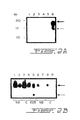
- Fig. 2A Lane 1, Jurkat; lane 2, PC12; lane 3, NIH-3T3; lane 4, R1.1; lane 5, Z91; lane 6, E25. Thick arrow, p140 TrkA; thin arrow, p110.
- Fig. 2B Dissected human brain tissues: nucleus basalis (NB; lanes 1, 2, 6, 7), and cortex (Fig. 2C; lanes 3, 4, 8, 9) were compared to E25 cells (lane 5; 2x10 5 cell equivalents).
- 33x10 6 cells/ml were detergent solubilized (lysis buffer 2% NP-40TM, 150 mM NaCl, 50 mM Tris-Glycine, 10 mM NaF, 50 ⁇ M Na 3 VO 4 , 30 mM Na Pyrophosphate 10 mM benzamidine, 20 mM iodoacetamide, pH 7.8) supplemented with protease inhibitors (2 ⁇ g/ml soybean trypsin inhibitor, 10 ⁇ g/ml aprotinin, 5 mM PMSF, and 10 ⁇ g/ml leupeptin) for 30 min at 4 °C, followed by a 15 minute centrifugation at 14,000 g. Cleared supernatants were analyzed by SDS-PAGE directly (whole cell lysates) or after immunoprecipitation.
- Cell lysates were prepared in Laemmli electrophoresis sample buffer and analyzed by SDS-PAGE under reducing (100 mM 2-mercaptoethanol) or non-reducing conditions. Prestained protein markers (GIBCO) were used as reference. Protein concentrations were quantitated by the biuret assay (Bio-Rad, Melville, NY), and by parallel Coomassie blueTM staining of SDS-PAGE gels. For western blotting samples were electrotransfered to PVDF (Xymotech Biosystems, Mt.
- HRP horseradish peroxidase
- HRP-G ⁇ R horseradish peroxidase conjugated goat anti-rabbit IgG
- HRP-G ⁇ M goat anti-mouse IgG
- ECL enhanced chemiluminescence
- MAb 5C3 was 125 I-labeled by the Iodogen (Pierce) method (Harlow, E., Lane, D. (1988) A Laboratory Manual. Cold Spring Harbour Laboratory Publishing. Chapter 9 :332-333) to a specific activity of 1.8 mCi/mg.
- 125 I[5C3] was repurified from free 125 I with SephadexTM G25 columns (15x1 cm) to >96% trichloroacetic acid precipitable incorporation. Binding studies were performed with serial dilutions of 125 I [5C3] on 0.5x10 6 E25 or 4-3.6 cells (and their respective controls NIH-3T3 and B104 cells) for 1 hour at 4°C.
- Cell-associated 125 I[5C3] and free 125 I[5C3] were counted after washing unbound ligand.
- Parallel 125 I[NGF] (70 mCi/mg) (NEN-DuPont, Mississauga, Ont.) binding assays were performed as control.
- Competition of 125 I[5C3] binding was done in binding assays in the presence of unlabeled mAb 5C3 (100-fold molar excess), or unlabeled NGF (500-fold molar excess).
- 125 I[NGF] binding was performed by first incubating cells with excess unlabeled mAb 5C3, NGF, mAb 87.92.6 or vehicle binding buffer for 30 min at 4°C. 125 I[NGF] was then added to a final saturating concentration of ⁇ 1 nM, the mixtures incubated for an additional 45 min at 4°C, cells were washed and cell-associated 125 I[NGF] were determined.
- the docking site of mAb 5C3 was identified within the juxtamembrane/IgG2 domain of human TrkA receptors.
- TrkA-binding agents (0.01 ⁇ g mAb 5C3; 2 nM NGF) or controls (mIgG; HBSS) for 20 min either at 37°C (internalization permissive temperature) or at 4°C (internalization non-permissive temperature): After washing, cells were processed for surface TrkA immunofluorescence with mAb 5C3 primary and FITC-G ⁇ mIgG secondary as above, and analyzed by FACScan.
- TrkA surface immunostaining was performed on 4-3.6 cells with mAb 5C3 after the indicated treatments, and measured by FACScan analysis. Data is presented as % staining ⁇ sem, with reference to control vehicle treatment (100%) as per the following formula: (Treated sample staining - mIgG background staining) x 100% (maximum staining - mIgG background staining)
- mAb 5C3 inhibited ⁇ 60% of 125 I[NGF] binding to E25 cells.
- background binding was assessed by blocking with 5 ⁇ M NGF (100% inhibition), and maximal binding was assessed with binding buffer vehicle only (0% inhibition) or by using irrelevant binding mAb 87.92.6 (Table 3).
- MAb 5C3 blocks NGF binding to TrkA TREATMENT % NGF BINDING mAb 5C3 39.3 ⁇ 7.4 mAb 87.92.6 100 NGF (5 ⁇ M) 0
- E25 cells expressing TrkA (but not p75 receptors) were incubated with 125 I[NGF] in the presence of the indicated agents.
- 125 I[NGF] binding after treatment with mAb 87.92.6 was identical to treatment with vehicle binding buffer. Assays were done 3 times in duplicate. Data is expressed as % binding ⁇ standard deviation (sd), where mAb 87.92.6 is maximum and 5 ⁇ M NGF is background binding as per the formula: (test - background) * 100 % (maximum - background)
- the proliferative/survival profile of the cells was quantitated using the tetrazolium salt reagent (MTT, Sigma) 48-72 hours after plating as initially described by T. Mosmann (Hansen, M.B. et al.(1989) Jour. Immunol. Meth ., 119: 203-210).
- Optical density readings of MTT were done in an EIA Plate Reader Model 2550TM (Bio-Rad) at 600 nm with the blanks subtracted. Assays were repeated at least 5 times in quadruplicates.
- E25 cells were plated in a 25% serum-containing 0.35% soft agar mixture in the presence of either mIgG control (0.5 ⁇ g/ml), mAb 5C3 (0.5 ⁇ g/ml), or NGF (2 nM). Conditions were replenished every 3 days and foci were counted after two weeks.
- Human brain tissue was obtained from six males (age 71.7 ⁇ 4.6) without signs of neurologic or psychiatric disorders. Tissue blocks were prepared (mean time post-mortem 16.2 ⁇ 3.5 hrs) and stored at -80°C. Twenty ⁇ m thick cryostat sections were fixed (4% paraformaldehyde, 0.1 M phosphate, pH 7.4; 1 hour at 4°C), and rinsed in PBS for 1 hour at 4°C. Immunocytochemistry was performed using avidin-biotin complex (Vectastain EliteTM kit, Vector Labs) as described (Hsu, S-M. et al. (1981) J. Histochem. Cytochem. , 29 : 577-580).
- Primary mAb 5C3 was used either as a 1:1000-1:4000 dilution of ascites or a 1:4 dilution of serum free-media culture supernatant. Where indicated, 0.5% nickel ammonium sulfate was used to amplify the signal in the DAB revelation step. Some sections were also stained with cresyl violet to facilitate the cytoarchitectural analysis. Negative controls were performed without primary antibody or with normal mouse IgG as primary and in all cases yielded no detectable immunolabeling.
- MAb 5C3 was used to map TrkA protein expression by immunocytochemistry of normal adult human brains.
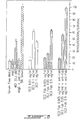
- the striatum, the basal forebrain and the brainstem exhibited the strongest immunostaining, whereas only weak staining could be detected in the cerebral cortex and hippocampal formation (Fig. 3).
- Figs. 3C, 3D, and 3E illustrate high power photomicrographs of TrkA-containing neurons in the nucleus basalis (Fig. 3C), the putamen (Fig. 3D), and the CA4 sub-field of the hippocampus (Fig. 3E).
- the perinuclear area displayed particular strong concentration of DAB precipitate (small arrows) often in granules. Labeled proximal processes could also be observed (arrows).
- Figs. 3A and 3C All sectors of the basal nucleus contained large TrkA-positive neurons (Figs. 3A and 3C), most of them in groups embedded in a dense network of overlapping stained processes (Fig. 3A).
- the cells had heterogeneous shapes, ranging from complex multipolar to fusiform.
- Fig. 3D shows typical labeled multipolar neurons that displayed strong granular immunoreactivity around the nucleus and in proximal processes. Moreover, numerous puncta and varicose fiber fragments were observed in these areas. The globus pallidus and the claustrum were mostly negative except for varicose fibers. Similarly, the interstitial elements and fiber bundles did not contain reactive fibers whereas the internal capsule displayed some labeled punctas and fibers particularly near the putamen and caudate nucleus.
- the hippocampal formation showed weak immunostaining located principally in scattered fibers and puncta in the stratum granulosum of the dentate gyrus, as well as in the strati oriens and pyramidal of the Ammon's horn.
- some weakly stained perikarya could be observed in the stratum pyramidal of the CA2 and CA3 sub-f ields of the Ammon's Horn and in the hilus of the dentate gyrus (CA4 sub-field, Fig. 3E).
- the perikarya of these neurons was relatively large in size, of ovoid to pyramidal shape, and bearing one prominent apical and radial dendritic process.
- the immunoreactivity appeared like in other stained cell types of the brain as small granular patches of precipitate located principally near the nuclear envelope and in some cases within the cytoplasm (Fig. 3E).
- TrkA immunoreactivity appeared more discrete. At high magnification, immunoreactive puncta and fiber fragments without a particular pattern of distribution are observed in all layers, but the laminae III-VI appeared more stained than superficial ones (Fig. 3H). Occasional, weakly staining, medium-sized perikarya were observed in layer IV (Fig. 3H).
- TrkA staining In the brainstem TrkA staining is also detected.
- the pontine nuclei contained numerous immunoreactive medium sized globular perikarya and fibers between the pontocerebellar fibers (Fig. 3F).
- the reticular formation also displayed strong immunoreactivity for TrkA principally located in fiber networks (Fig. 3G). Some large neurons of bipolar or multipolar shape are also stained. No TrkA immunostaining was observed in the cerebellum.
- TrkA phosphotyrosinylation increased significantly over basal levels after short treatment with mAb 5C3 or with NGF (Fig. 4).
- E25 cells were untreated (lane 1) or treated with mAb 5C3 (lane 2), NGF (lane 3), or mIgG (lane 4) for 15 min. at 37°C.
- Whole cell lysates were resolved in a 8% SDS-PAGE under reducing conditions and immunoblotted with anti-phosphotyrosine mAb 4G10.
- a parallel gel under non-reducing conditions immunoblotted with mAb 5C3 (not shown) controlled for M r and equal loading of TrkA on all samples.
- E25 or 4-3.6 cells were untreated or treated with saturating concentrations of mAb 5C3 or NGF for 15 min. at 37°C.
- Whole cell lysates, or anti-PY immunoprecipitates were resolved by SDS-PAGE under reducing conditions, western transferred, immunoblotted with anti-phosphotyrosine ( ⁇ PY) mAb 4G10, and developed using ECL techniques.
- NGF treatment causes the transformation and an increase in anchorage-independent growth of TrkA-expressing E25 cells.
- MAb 5C3 caused a ⁇ 2-fold increase in the number and the size of foci as compared with mIgG treated cells (Table 5).
- MAb 5C3-induced anchorage-independent growth TREATMENT AVERAGE NUMBER OF FOCl a TYPICAL CELLS/FOCl a FOLD INCREASE IN FOCl b mlgG 416 ⁇ 45 ⁇ 24 1 ⁇ 0.11 mAb 5C3 806 ⁇ 178 >48 1.9 ⁇ 0.22 NGF 676 ⁇ 51 ⁇ 24 1.6 ⁇ 0.08
- TrkA Agonistic ligands of TrkA protect receptor-expressing cells from death in serum-free media (SFM). Both NGF and mAb 5C3 and 5C3 fragments or derivatives increased the number of surviving/proliferating E25 fibroblastoid cells (Fig. 6) and 4-3.6 cells (Fig. 8).
- TrkA ligands were also afforded to neuronal 4-3.6 cells.
- TrkA ligands were dose-dependent, although high dose antibody inhibition is sometimes seen (e.g. 1 mg/ml of mAb 5C3).
- DNA was prepared from serum-free cultured cells which showed a typical apoptotic fragmentation ladder. The DNA ladder was not seen in preparations from cells cultured in the presence of mAb 5C3 or NGF.
- MAb 5C3 F abs afforded protection from apoptotic death to E25 cells (Fig. 6) and 4-3.6 cells in serum-free media. Moreover, anti-phosphotyrosine western blots revealed that cells treated with 5C3 F abs had increased TrkA tyrosine phosphorylation similar to increases obtained with whole mAb 5C3.
- Monomeric 5C3 F ab protection was dose-dependent. However, equivalent or better protective effects were achieved when F abs were externally cross-linked with G ⁇ mF ab antibodies. Specificity controls included those described in the previous section for whole mAb 5C3, plus 192 F abs which had no protective activity in E25 cells.
- mAb 5C3 is specific for human TrkA, functions in FACScan immunofluorescence analysis, immunoprecipitation, western blot analysis, and immunocytochemistry. Moreover, mAb 5C3 is a structural and functional mimic of NGF.
- TrkA-binding agents Aberrant expression of trkA mRNA and NGF responsiveness have been correlated with neurodegenerative disorders and neoplastic malignancy. Hence, TrkA-binding agents will be useful clinical tools in diagnosis, prognosis and perhaps treatment of these diseases. Indeed, mAb 5C3 binding is a positive prognostic marker for certain human neoplasias.
- TrkA expression in neuroblastoma Neuroblastomas Number Positive Mixed Negative Group 1 60 38 17 5 Group 2 53 13 5 35 TrkA expression in other malignant tumors Malignant tumor N 42 TrkA-Pos Central nervous system tumors 6 0 Rhabdomyosarcomas 5 0 Primitive neuroectodermal tumors 6 0 Ewing's sarcomas 2 0 Wilm's tumors 6 1 Osteosarcomas 4 0 Melanomas 5 0 Breast carcinomas 5 0 Lung carcinomas 3 0 TrkA detection by immunocytochemistry, RT-PCR and western blot IMMUNOCYTO. N RT-PCR Pos N West. Pos Group 1 15 15 2 2 5C3 Pos 5C3 Neg 4 4 2 1 5C3 Mixed 1 1 10 10 Group 2 11 11 11 11 5C3 Pos 5C3 Neg 11 4 9 5 5C3 Mixed 2 2 0 0 0
- MAb 5C3 recognizes a disulfide-stabilized domain of TrkA and an extracellular epitope with these characteristics appears to be the NGF docking site.
- Cross-blocking studies indicated that mAb 5C3 and NGF can reciprocally block each other's binding to TrkA, further suggesting that the docking site of 5C3 may be similar to NGF.
- sequence comparison of both ligands revealed interesting homology between CDRs of mAb 5C3 and the variable turn regions of NGF. Since most CDRs are ⁇ -turns (Sibanda, B.L. et al. (1989) J. Mol.
- mAb 5C3 was more efficient at blocking NGF binding than vice versa. Only ⁇ 25% of the mAb 5C3 binding sites on E25 cells were blocked by saturating doses of NGF. This data suggest that not all TrkA receptors in these cell lines bind NGF. It is unlikely that affinity considerations can account for these observations, as both ligands have roughly comparable K d for TrkA (mAb 5C3 K d ⁇ 2 nM versus NGF K d 0.7 nM (Jing, S. et al. (1992) Neuron , 9 : 1067-1079)) and the affinity of mAb 5C3 was unchanged in the presence of NGF.
- TrkA receptors exist at equilibrium where ⁇ 25% are in an NGF binding conformation (e.g. dimers) and the rest are in a non-NGF binding conformation;
- specific post-translational modifications of TrkA receptors allow for NGF binding (iii) expression of other membrane proteins (e.g. p75 or unknown proteins) induce or favor the NGF binding conformation of TrkA.
- mAb 5C3 Absence of mAb 5C3 binding to rat TrkA is intriguing. Binding by mAb 5C3 to rat TrkA was expected because of the homology between mAb 5C3 CDRs and the variable loops of NGF, particularly since NGF from one species does bind to TrkAs from other species. MAb 5C3 is a binding and structural mimic of NGF, with enhanced human receptor specificity. Remodeling and mutating of the CDRs of mAb 5C3 will yield a pan-TrkA binding mAb. Further, analysis of the epitope of mAb 5C3 on TrkA revealed differences in the docking site of human and rat TrkAs. This information will be useful in screening receptor-binding analogs.
- mAb 5C3 NGF bioassays were performed using trkA transfected fibroblast and neuronal cells.
- Functional mimicry by mAb 5C3 included TrkA internalization, TrkA tyrosine phosphorylation, PI-3 kinase phosphorylation, increased anchorage-independent growth and proliferation/survival of cells in serum-free media.
- mAb 5C3 is agonistic.
- TrkA receptor turnover or internalization is induced by NGF binding.
- MAb 5C3 increased the internalization of TrkA, as measured by loss of cell surface receptors.
- TrkA NGF ligation of TrkA causes receptor activation and autophosphorylation.
- MAb 5C3 induced TrkA tyrosine phosphorylation to a similar degree.
- Agonism in the absence of NGF suggests that TrkA dimerization and/or internalization are the required signaling event, rather than the formation of NGF-TrkA complexes.
- mAb 5C3-TrkA is the functional signal transducing complex.
- TrkA Ligand-induced tyrosine phosphorylation of the intracellular domain of TrkA allows for the recruitment of substrates and the activation of cytosolic proteins and nuclear oncoproteins.
- MAb 5C3 induces the tyrosine phosphorylation of proteins of M r 60, 85 and 95 kDa.
- the 85 kDa protein was identified as PI-3 kinase, whose activation correlates with the actions of growth factors and oncogenes.
- NGF stimulates neuronal survival and differentiation, and the proliferation of non-neuronal cells.
- NGF-activated TrkA induces transformation and morphological changes in fibroblast cells.
- MAb 5C3 caused similar increases in anchorage-independent growth and foci formation in soft agar.
- mAb 5C3 can positively modulate the growth of TrkA-expressing cells.
- the size of the mAb 5C3-induced foci were larger on average than NGF-induced foci.
- Monomeric 5C3 F abs protected E25 and 4-3.6 cells from apoptotic death. When F abs were externally cross-linked using anti-F ab antibodies, a heightened response occurred. Since growth factor receptor activation requires bivalent binding, the monomeric 5C3 F abs must have retained the ability to induce TrkA oligomerization. This could be explained in the following 3 ways: (i) F abs are relatively large molecules capable of aggregation; (ii) 5C3 F ab binding could cause conformational changes in TrkA which induce receptor-receptor interactions; and (iii) monomeric 5C3 F abs bind to two receptor molecules in a bivalent manner. The last possibility could occur by two CDRs binding to two different TrkAs. Homology of mAb 5C3 CDRs to NGF turn regions, and experiments using small recombinant antibody analogs support the last explanation.
- MAb 5C3 is the first report of an agonistic anti-neurotrophin receptor mAb and will be useful in studies of TrkA biology and for drug development. Antineoplastic effects with mAb 5C3 may be achieved either through terminal differentiation, anti-body-dependent cell cytotoxicity, or by the delivery of toxins or radionuclides. Furthermore, the structure of this mAb may be useful in designing peptidic and non-peptidic TrkA binding agents (Saragovi, H.U. et al. (1991) Science , 253 : 792-795). Small non-peptidic agonists of TrkA should be useful pharmacological agents for the treatment of neurodegenerative diseases.
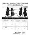
- mice were injected subcutaneously (right abdominal side) with 2X10 6 human TrkA expressing tumor cells. Two days post-injections tumors in all mice had begun to form. Mice were randomized prior to treatment. A total of four intraperitoneal injections of 100 micrograms each on the left side were then administered over a 12 day period with control mouse IgG or mAb 5C3 (Fig. 10).
- the mAb 5C3 dramatically reduced the primary tumor weight with no observable metastatic invasion.
- a small fibrotic mass was localized at the site of injection in mAb 5C3 treated mice.
- IgG treated mice had large, vascularized tumor masses, which metastasized to the liver, peritoneum gut and spleen. All animals had similar body weights ( ⁇ 30 grams).
- TREATMENT PRIMARY TUMOR WEIGHT (mg) METASTASIS WEIGHT (mg) 5C3 mAb 50 ⁇ 20 (fibrotic) NONE mouse IgG 800 ⁇ 250 350 ⁇ 20
- Mab 5C3 and its derivatives for the diagnosis, prognosis and localization of tumors
- Complementarity Determining regions (according to Kabat, Sequences of Proteins of Immunological Interest, 4th ed. Bethesda, MD, U.S. Dept. of Health and Human Services. Public Health, NIH 1987)
- Antibodies have 6 complementarity determining regions (CDRs) that combine in specific ways to create direct contacts with their antigen. Molecular cloning, recombination, mutagenesis and modeling studies of mAb 5C3 variable region indicated that 3 or less of its CDRs are relevant for binding TrkA. These were named CDR1, CDR2, and CDR3. Region CDR1 is connected to CDR2 by a 15 amino acid linker; CDR2 is connected to CDR3 by a 30 amino acid linker. Their secondary structures have been analyzed.
- variable domains of mAb 5C3 heavy (VH) and light (LH) chains were cloned and sequenced.
- the predicted variable domain amino acid sequences are shown above.
- CDRs hypervariable complementarity determining regions
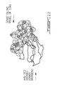
- FIG. 11 An example of the model of the structure of the CDRs (Insight IITM, Biosym, CA) within the backbone of the variable domains is shown in Fig. 11.
- mAb 5C3 is a TrkA agonist.
- Previous functional studies in vitro demonstrated that mAb 5C3 can: (i) induce tyrosine phosphorylation of TrkA, and PI3-kinase; (ii) increase transformation of TrkA-expressing fibroblasts.
- Shown above are also experiments where the 5C3 and its derivatives protect cells from apoptotic death in serum-free media to the same degree as NGF does.
- Monovalent Fabs of 5C3 obtained after papain digestion are also agonistic, especially when externally cross-linked by anti-Fabs.
- a smaller fragment of mAb 5C3 called CDR(R) also protects cells from apoptosis (Fig. 8).
- CDR(R) for recombinant CDR was developed as a replacement for mAb 5C3: it is approximately five times smaller than the 25 kDa NGF molecule and is still agonistic.
- CDR(R) is composed of 3 selected CDRs (out of 6 possible ones) linked by long. spacer regions. Preliminary studies have suggested that actually only 2 of the 3 CDRs are relevant for binding to TrkA. Further, it is expected that even smaller fragments can be designed, e.g. upon removal of the linker regions.
- Fig. 8 4-3.6 cells transfected with human TrkA were cultured in serum-free-media (SFM) to induce apoptosis (Fig. 8). The indicated agents were added to the cells and cell viability was measured after 48-72 h by the MTT assay. Proliferation was standardized to normal growth conditions (5% serum). NGF, mAb 5C3, 5C3 Fabs, and CDR(R) (recombinant 5C3 CDR analog) protect from apoptosis, but mouse IgG (control) does not.
- SFM serum-free-media
- the mAb 5C3 of the present invention and its derivatives induce differentiation/neuritogenesis of human TrkA-expressing cells.
- PC12 cells expressing rat TrkA
- PC12 cells transfected with and expressing human trkA cDNA were cultured with various TrkA binding agents or controls (Fig. 9) as indicated NGF caused the differentiation of wild type PC12 and PC12 transfectants, whereas SC3 and CDR(R) only induced differentiation of PC12 transfectants.
- Ligand doses are as shown in Fig. 8.
Landscapes
- Health & Medical Sciences (AREA)
- Life Sciences & Earth Sciences (AREA)
- Chemical & Material Sciences (AREA)
- Immunology (AREA)
- Proteomics, Peptides & Aminoacids (AREA)
- Organic Chemistry (AREA)
- General Health & Medical Sciences (AREA)
- Medicinal Chemistry (AREA)
- Biochemistry (AREA)
- Biophysics (AREA)
- Genetics & Genomics (AREA)
- Molecular Biology (AREA)
- Optics & Photonics (AREA)
- Veterinary Medicine (AREA)
- Oncology (AREA)
- Pharmacology & Pharmacy (AREA)
- Epidemiology (AREA)
- Animal Behavior & Ethology (AREA)
- Public Health (AREA)
- Physics & Mathematics (AREA)
- Cell Biology (AREA)
- Toxicology (AREA)
- Zoology (AREA)
- Gastroenterology & Hepatology (AREA)
- Medicines Containing Antibodies Or Antigens For Use As Internal Diagnostic Agents (AREA)
- Peptides Or Proteins (AREA)
- Medicines That Contain Protein Lipid Enzymes And Other Medicines (AREA)
- Preparation Of Compounds By Using Micro-Organisms (AREA)
Abstract
Description
| Surface phenotyping with mAb 5C3 | |
| CELLS | 5C3 BINDING |
| E25 (hTrkA) | +++++ |
| R7 (hTrkA/p75) | +++ |
| Z91 (p75) | - |
| 4-3.6 (hTrkA/p75) | +++ |
| B104 (p75) | - |
| PC12 (rTrkA/p75) transient NIH-3T3 transfections | - |
| htrkA cDNA | ++ |
| htrkB cDNA | - |
| rtrkB cDNA | - |
| rtrkA cDNA | - |


| mAb 5C3-induced TrkA receptor internalization | ||
| TREATMENT | TEMP.(°C) | % 5C3 STAINING |
| NGF (2 nM) | 4°C | 83 ± 2.0 |
| 37°C | 75 ± 3.6 | |
| 5C3 (0.01 µg/ml) | 4°C | 96 ± 9.0 |
| 37°C | 77 ± 5.5 |
| MAb 5C3 blocks NGF binding to TrkA | |
| TREATMENT | % NGF BINDING |
| mAb 5C3 | 39.3 ± 7.4 |
| mAb 87.92.6 | 100 |
| NGF (5 µM) | 0 |
| TrkA tyrosine phosphorylation by mAb 5C3 | ||
| TREATMENT | E25 cells | 4-3.6 cells |
| mAb 5C3 | 2.7 ± 0.6 | 3.4 ± 1.5 |
| NGF | 6.5 ± 1.3 | 3.8 ± 0.8 |
| MAb 5C3-induced anchorage-independent growth | |||
| TREATMENT | AVERAGE NUMBER OF FOCla | TYPICAL CELLS/FOCla | FOLD INCREASE IN FOClb |
| mlgG | 416 ± 45 | ~24 | 1 ± 0.11 |
| mAb 5C3 | 806 ± 178 | >48 | 1.9 ± 0.22 |
| NGF | 676 ± 51 | ~24 | 1.6 ± 0.08 |
| TrkA expression in neuroblastoma | ||||
| Neuroblastomas | Number | Positive | Mixed | |
| Group | ||||
| 1 | 60 | 38 | 17 | 5 |
| | 53 | 13 | 5 | 35 |
| TrkA expression in other malignant tumors | ||
| Malignant tumor | N=42 | TrkA-Pos |
| Central | 6 | 0 |
| | 5 | 0 |
| | 6 | 0 |
| Ewing's | 2 | 0 |
| Wilm's | 6 | 1 |
| | 4 | 0 |
| | 5 | 0 |
| | 5 | 0 |
| | 3 | 0 |
| TrkA detection by immunocytochemistry, RT-PCR and western blot | ||||
| IMMUNOCYTO. | N | RT-PCR Pos | N | West. |
| Group | ||||
| 1 | 15 | 15 | 2 | 2 |
| 5C3 | ||||
| 5C3 Neg | ||||
| 4 | 4 | 2 | 1 | |
| 5C3 Mixed | 1 | 1 | 10 | 10 |
| | 11 | 11 | 11 | 11 |
| 5C3 | ||||
| 5C3 Neg | ||||
| 11 | 4 | 9 | 5 | |
| 5C3 Mixed | 2 | 2 | 0 | 0 |
| TREATMENT | PRIMARY TUMOR WEIGHT (mg) | METASTASIS WEIGHT (mg) |
| | 50 ± 20 (fibrotic) | NONE |
| mouse IgG | 800 ± 250 | 350 ± 20 |
| Biodistribution of 99mTc-[TrkA ligands] in mice. | |||
| 99mTc-[5C3] | |||
| Ligand | % id/g | T/ | |
| 1 | tumor | 1.25 | 1 |
| 2 | blood | 0.1 | 13 |
| 3 | muscle | 0.06 | 20 |
| 4 | heart | 0.10 | 13 |
| 5 | lung | 0.17 | 7.30 |
| 6 | liver | 0.61 | 2.10 |
| 7 | spleen | 0.13 | 9.42 |
| 8 | kidney | 1.48 | 0.9 |
| 9 | large bowel | 2.2 | 0.7 |




- (A) NAME: SARAGOVI, H. Uri
- (B) STREET: 1100 Dr. Penfield, Apt. 718
- (C) CITY: Montreal
- (D) STATE: Quebec
- (E) COUNTRY: Canada
- (F) POSTAL CODE (ZIP): H3A 1A8
- (A) NAME: LESAUTEUR, Lynne
- (B) STREET: 210 Coronation
- (C) CITY: Rosemere
- (D) STATE: Quebec
- (E) COUNTRY: Canada
- (F) POSTAL CODE (ZIP): J7A 1P3





Claims (16)
- A monoclonal antibody or functional fragment thereof raised against human p140 TrkA and which binds to TrkA receptor under physiological conditions wherein the amino acids sequences of the variable domains of the light and heavy chains are as defined herein by SEQ ID NO:1 and SEQ ID NO:2, respectively.
- A method of screening pharmacological agents which are capable to mimic or inhibit nerve growth factor biological activity, which comprises using the antibody of claim 1 to screen for pharmacological agents capable of binding to the complementary determining region of said antibody.
- Use of the antibody of claim 1, for the manufacture of a medicament for the in vivo inhibition of nerve growth factor binding to TrkA receptor or the internalization or downmodulation of said receptor.
- The use of claim 3, wherein the medicament is for use in inhibiting tumor growth in situ.
- The use of claim 3, wherein the medicament is for use in the treatment or prevention of neurological diseases, neuromas and neoplastic tumors which express TrkA receptors.
- The use of the antibody of claim 1, for mapping hormone-receptor interactive sites and receptor domain-function correlation in vitro.
- The use of the antibody of claim 1, for mapping TrkA docking site in vitro.
- The use of claims 6 and 7, for screening in vitro pharmacological agents which bind to said hormone-receptor interactive sites.
- The use of the antibody of claim 1, for inducing in vitro partial nerve growth factor-like responses in cells expressing TrkA.
- The use of claim 9, wherein said responses are selected from differentiation/neuritogenesis function and trophic function.
- A pharmaceutical composition for the treatment of neurological diseases, neuromas and neoplastic tumors which express TrkA receptors, which comprises an effective amount of an antibody of claim 1 or a functional fragment thereof in association with a pharmaceutically acceptable carrier.
- A method for the prognosis or diagnosis of human tumor which comprises:immunocytochemistry of tumors outside of the body using the antibody of claim 1 and/or functional fragments thereof.
- A pharmaceutical composition for the targeting of pharmaceutical agents to tissues of the central and/or peripheral nervous system, which comprises an effective amount of an antibody of claim 1 or a functional fragment thereof coupled to a pharmaceutical agent in association with a pharmaceutically acceptable carrier.
- The pharmaceutical composition of claim 13, wherein the pharmaceutical agent is selected from the group consisting of radioligands, nucleic acid molecules, toxins, growth factors and gangliosides.
- A TrkA docking site as defined herein by SEQ ID NO: 9 which binds to the antibody of claim 1.
- Use of the docking site of claim 15 for screening pharmacological agents which are capable to mimic or inhibit nerve growth factor biological activity and said antibody biological activity.
Applications Claiming Priority (3)
| Application Number | Priority Date | Filing Date | Title |
|---|---|---|---|
| GB9525180 | 1995-12-08 | ||
| GBGB9525180.7A GB9525180D0 (en) | 1995-12-08 | 1995-12-08 | Design of hormone-like antibodies with agonistic and antagonistic fuctions |
| PCT/CA1996/000815 WO1997021732A1 (en) | 1995-12-08 | 1996-12-06 | Design of hormone-like antibodies with agonistic and antagonistic functions |
Publications (2)
| Publication Number | Publication Date |
|---|---|
| EP0869976A1 EP0869976A1 (en) | 1998-10-14 |
| EP0869976B1 true EP0869976B1 (en) | 2004-02-25 |
Family
ID=10785168
Family Applications (1)
| Application Number | Title | Priority Date | Filing Date |
|---|---|---|---|
| EP96939791A Expired - Lifetime EP0869976B1 (en) | 1995-12-08 | 1996-12-06 | Design of hormone-like antibodies with agonistic and antagonistic functions |
Country Status (7)
| Country | Link |
|---|---|
| US (1) | US6610500B1 (en) |
| EP (1) | EP0869976B1 (en) |
| AT (1) | ATE260299T1 (en) |
| AU (1) | AU7689496A (en) |
| DE (1) | DE69631686T2 (en) |
| GB (1) | GB9525180D0 (en) |
| WO (1) | WO1997021732A1 (en) |
Families Citing this family (46)
| Publication number | Priority date | Publication date | Assignee | Title |
|---|---|---|---|---|
| CA2212315A1 (en) * | 1997-08-05 | 1999-02-05 | Mcgill University | Pharmaceutical composition with neurotrophic-like biological activity |
| FR2776661B1 (en) * | 1998-03-26 | 2002-04-05 | Centre Nat Rech Scient | IMMUNOREACTIVE POLYPEPTIDE OF THE NGF TRKA RECEPTOR AND USES |
| GB9807781D0 (en) * | 1998-04-09 | 1998-06-10 | Univ Bristol | Therapeutic agent |
| US6365154B1 (en) * | 1998-09-28 | 2002-04-02 | Smithkline Beecham Corporation | Tie2 agonist antibodies |
| ATE406907T1 (en) * | 1998-10-28 | 2008-09-15 | Cornell Res Foundation Inc | METHODS FOR REGULATION OF ANGIOGENESIS AND VASCULAR INTEGRITY USING TRK RECEPTOR LIGANDS BDNF, NT-3 AND NT-4 |
| US20040120891A1 (en) | 1998-12-21 | 2004-06-24 | Craig Hill | Compounds for intracellular delivery of therapeutic moieties to nerve cells |
| US6652864B1 (en) | 1998-12-21 | 2003-11-25 | Asilomar Pharmaceuticals, Inc. | Compounds for intracellular delivery of therapeutic moieties to nerve cells |
| IT1306704B1 (en) | 1999-05-26 | 2001-10-02 | Sirs Societa Italiana Per La R | MONOCLONAL ANTIBODIES AND ITS SYNTHETIC OR BIOTECHNOLOGICAL DERIVATIVES ENABLE TO ACT AS ANTAGONIST MOLECULES FOR NGF. |
| FR2807660A1 (en) * | 2000-04-13 | 2001-10-19 | Warner Lambert Co | Use of a nerve growth factor antagonist for the manufacture of a medicament for the prevention or treatment of chronic visceral pain |
| IL153375A0 (en) * | 2000-06-22 | 2003-07-06 | Genentech Inc | Agonist anti-trk-c monoclonal antibodies |
| EP1343531A2 (en) * | 2000-12-21 | 2003-09-17 | McGILL UNIVERSITY | Conjugates of antibodies and anticancer drugs |
| US8034904B2 (en) * | 2002-06-14 | 2011-10-11 | Immunogen Inc. | Anti-IGF-I receptor antibody |
| US7538195B2 (en) | 2002-06-14 | 2009-05-26 | Immunogen Inc. | Anti-IGF-I receptor antibody |
| UA80447C2 (en) | 2002-10-08 | 2007-09-25 | Methods for treating pain by administering nerve growth factor antagonist and opioid analgesic | |
| EP1556083B1 (en) | 2002-10-08 | 2011-02-02 | Rinat Neuroscience Corp. | Methods for treating post-surgical pain by admisnistering an antibody against nerve growth factor and compositions containing the same |
| US7255860B2 (en) | 2002-10-08 | 2007-08-14 | Rinat Neuroscience Corp. | Methods for treating post-surgical pain by administering an anti-nerve growth factor antagonist antibody |
| US9498530B2 (en) | 2002-12-24 | 2016-11-22 | Rinat Neuroscience Corp. | Methods for treating osteoarthritis pain by administering a nerve growth factor antagonist and compositions containing the same |
| US7569364B2 (en) | 2002-12-24 | 2009-08-04 | Pfizer Inc. | Anti-NGF antibodies and methods using same |
| EP2270048B1 (en) | 2002-12-24 | 2015-11-11 | Rinat Neuroscience Corp. | Anti-NGF antibodies and methods using same |
| KR20050111598A (en) | 2003-02-19 | 2005-11-25 | 리나트 뉴로사이언스 코퍼레이션 | Methods for treating pain by administering a nerve growth factor antagonist and an nsaid and compositions containing the same |
| PL2248899T3 (en) | 2003-03-19 | 2015-10-30 | Biogen Ma Inc | NOGO receptor binding protein |
| EP1500399A1 (en) * | 2003-07-24 | 2005-01-26 | Institut Pasteur | Active or passive immunization against proapoptotic neurotrophins for the treatment or prevention of neurodegenerative deseases |
| AU2005243247B2 (en) | 2004-04-07 | 2012-03-01 | Regents Of The University Of Minnesota | Methods for treating bone cancer pain by administering a nerve growth factor antagonist |
| KR101239542B1 (en) | 2004-06-24 | 2013-03-07 | 바이오겐 아이덱 엠에이 인코포레이티드 | Treatment of conditions involving demyelination |
| ITRM20050332A1 (en) * | 2005-06-24 | 2006-12-25 | Lay Line Genomics Spa | USE OF MOLECULES ABLE TO BLOCK TRKA'S ACTIVITY TO STRENGTHEN THE EFFECTS OF ANALGESIC OPPIACEI ON PAIN. |
| CA2902070A1 (en) | 2005-07-08 | 2007-01-18 | Biogen Idec Ma Inc. | Sp35 antibodies and uses thereof |
| WO2008013782A2 (en) | 2006-07-24 | 2008-01-31 | Biogen Idec Ma Inc. | Methods for promoting myelination, neuronal survival and oligodendrocyte differentiation via administration of sp35 or trka antagonists |
| AU2009211340B2 (en) | 2008-02-04 | 2013-09-12 | Lay Line Genomics S.P.A. | Anti-TrkA antibodies and derivatives thereof |
| JP2011527572A (en) | 2008-07-09 | 2011-11-04 | バイオジェン・アイデック・エムエイ・インコーポレイテッド | Composition comprising a LINGO antibody or fragment |
| WO2012024650A2 (en) | 2010-08-19 | 2012-02-23 | Abbott Laboratories | Anti-ngf antibodies and their use |
| US9884909B2 (en) | 2010-12-01 | 2018-02-06 | Alderbio Holdings Llc | Anti-NGF compositions and use thereof |
| US9539324B2 (en) | 2010-12-01 | 2017-01-10 | Alderbio Holdings, Llc | Methods of preventing inflammation and treating pain using anti-NGF compositions |
| NZ611076A (en) | 2010-12-01 | 2015-09-25 | Alderbio Holdings Llc | Anti-ngf compositions and use thereof |
| US9078878B2 (en) | 2010-12-01 | 2015-07-14 | Alderbio Holdings Llc | Anti-NGF antibodies that selectively inhibit the association of NGF with TrkA, without affecting the association of NGF with p75 |
| US9067988B2 (en) | 2010-12-01 | 2015-06-30 | Alderbio Holdings Llc | Methods of preventing or treating pain using anti-NGF antibodies |
| US11214610B2 (en) | 2010-12-01 | 2022-01-04 | H. Lundbeck A/S | High-purity production of multi-subunit proteins such as antibodies in transformed microbes such as Pichia pastoris |
| MX381731B (en) | 2012-05-14 | 2025-03-13 | Biogen Ma Inc | Leucine-rich repeat-containing immunoglobulin domain (LINGO-2) receptor-interacting protein antagonists for the treatment of conditions involving motor neurons. |
| JP6629069B2 (en) | 2012-06-06 | 2020-01-15 | ゾエティス・エルエルシー | Canine anti-NGF antibody and method thereof |
| EP2674439B1 (en) | 2012-06-13 | 2017-02-01 | Rottapharm Biotech S.r.l. | Anti-TrkA antibodies, derivatives and uses thereof |
| JP2018504400A (en) | 2015-01-08 | 2018-02-15 | バイオジェン・エムエイ・インコーポレイテッドBiogen MA Inc. | LINGO-1 antagonist and use for treatment of demyelinating disorders |
| AR114110A1 (en) | 2018-02-28 | 2020-07-22 | Lilly Co Eli | ANTI-TRKA ANTIBODY |
| MX2020009526A (en) | 2018-03-12 | 2020-10-28 | Zoetis Services Llc | Anti-ngf antibodies and methods thereof. |
| MX2021000644A (en) | 2018-07-18 | 2021-08-19 | Manzanita Pharmaceuticals Inc | Conjugates for delivering an anti-cancer agent to nerve cells, methods of use and methods of making thereof. |
| JP2025503539A (en) * | 2021-12-28 | 2025-02-04 | 4ビー テクノロジーズ(スーヂョウ) リミテッド | TrkA antibody and its applications |
| EP4716738A1 (en) * | 2023-05-23 | 2026-04-01 | 4B Technologies (Shanghai) Co., Ltd | Trka antibody and use thereof |
| US20250122291A1 (en) | 2023-10-13 | 2025-04-17 | Cephalon Llc | Anti-trka antibodies and uses thereof |
Citations (1)
| Publication number | Priority date | Publication date | Assignee | Title |
|---|---|---|---|---|
| EP0471205A1 (en) * | 1990-08-15 | 1992-02-19 | E.R. SQUIBB & SONS, INC. | Monoclonal antibodies which bind TRK proto-oncogene protein |
Family Cites Families (2)
| Publication number | Priority date | Publication date | Assignee | Title |
|---|---|---|---|---|
| DE3909799A1 (en) | 1989-03-24 | 1990-09-27 | Behringwerke Ag | MONOCLONAL ANTIBODIES (MAK) AGAINST TUMOR ASSOCIATED ANTIGENS, THEIR PRODUCTION AND USE |
| US5753225A (en) * | 1993-12-03 | 1998-05-19 | The Regents Of The University Of California | Antibodies that mimic actions of neurotrophins |
-
1995
- 1995-12-08 GB GBGB9525180.7A patent/GB9525180D0/en active Pending
-
1996
- 1996-12-06 EP EP96939791A patent/EP0869976B1/en not_active Expired - Lifetime
- 1996-12-06 WO PCT/CA1996/000815 patent/WO1997021732A1/en not_active Ceased
- 1996-12-06 DE DE69631686T patent/DE69631686T2/en not_active Expired - Fee Related
- 1996-12-06 AT AT96939791T patent/ATE260299T1/en not_active IP Right Cessation
- 1996-12-06 AU AU76894/96A patent/AU7689496A/en not_active Abandoned
- 1996-12-06 US US09/091,071 patent/US6610500B1/en not_active Expired - Lifetime
Patent Citations (1)
| Publication number | Priority date | Publication date | Assignee | Title |
|---|---|---|---|---|
| EP0471205A1 (en) * | 1990-08-15 | 1992-02-19 | E.R. SQUIBB & SONS, INC. | Monoclonal antibodies which bind TRK proto-oncogene protein |
Also Published As
| Publication number | Publication date |
|---|---|
| DE69631686T2 (en) | 2004-12-02 |
| WO1997021732A1 (en) | 1997-06-19 |
| ATE260299T1 (en) | 2004-03-15 |
| AU7689496A (en) | 1997-07-03 |
| EP0869976A1 (en) | 1998-10-14 |
| DE69631686D1 (en) | 2004-04-01 |
| US6610500B1 (en) | 2003-08-26 |
| GB9525180D0 (en) | 1996-02-07 |
Similar Documents
| Publication | Publication Date | Title |
|---|---|---|
| US6610500B1 (en) | Design of hormone-like antibodies with agonistic and antagonistic functions | |
| US20230242650A1 (en) | Monoclonal antibodies to fibroblast growth factor receptor 2 | |
| LeSauteur et al. | Potent human p140-TrkA agonists derived from an anti-receptor monoclonal antibody | |
| US6099841A (en) | Hepatocyte growth factor receptor agonists and uses thereof | |
| Holmes et al. | Identification of heregulin, a specific activator of p185erbB2 | |
| US6656465B2 (en) | Antibodies that mimic actions of neurotrophins | |
| AU2004231742A2 (en) | Nogo-receptor antagonists for the treatment of conditions involving amyloid plaques | |
| HK40045902A (en) | Monoclonal antibodies to fibroblast growth factor receptor 2 | |
| HK1249053B (en) | Monoclonal antibodies to fibroblast growth factor receptor 2 | |
| HK1207974B (en) | Monoclonal antibodies to fibroblast growth factor receptor 2 | |
| HK1160012B (en) | Monoclonal antibodies to fibroblast growth factor receptor 2 |
Legal Events
| Date | Code | Title | Description |
|---|---|---|---|
| PUAI | Public reference made under article 153(3) epc to a published international application that has entered the european phase |
Free format text: ORIGINAL CODE: 0009012 |
|
| 17P | Request for examination filed |
Effective date: 19980702 |
|
| AK | Designated contracting states |
Kind code of ref document: A1 Designated state(s): AT BE CH DE DK ES FI FR GB GR IE IT LI LU MC NL PT SE |
|
| 17Q | First examination report despatched |
Effective date: 20010615 |
|
| GRAH | Despatch of communication of intention to grant a patent |
Free format text: ORIGINAL CODE: EPIDOS IGRA |
|
| GRAS | Grant fee paid |
Free format text: ORIGINAL CODE: EPIDOSNIGR3 |
|
| GRAA | (expected) grant |
Free format text: ORIGINAL CODE: 0009210 |
|
| AK | Designated contracting states |
Kind code of ref document: B1 Designated state(s): AT BE CH DE DK ES FI FR GB GR IE IT LI LU MC NL PT SE |
|
| PG25 | Lapsed in a contracting state [announced via postgrant information from national office to epo] |
Ref country code: NL Free format text: LAPSE BECAUSE OF FAILURE TO SUBMIT A TRANSLATION OF THE DESCRIPTION OR TO PAY THE FEE WITHIN THE PRESCRIBED TIME-LIMIT Effective date: 20040225 Ref country code: LI Free format text: LAPSE BECAUSE OF FAILURE TO SUBMIT A TRANSLATION OF THE DESCRIPTION OR TO PAY THE FEE WITHIN THE PRESCRIBED TIME-LIMIT Effective date: 20040225 Ref country code: FI Free format text: LAPSE BECAUSE OF FAILURE TO SUBMIT A TRANSLATION OF THE DESCRIPTION OR TO PAY THE FEE WITHIN THE PRESCRIBED TIME-LIMIT Effective date: 20040225 Ref country code: ES Free format text: LAPSE BECAUSE OF FAILURE TO SUBMIT A TRANSLATION OF THE DESCRIPTION OR TO PAY THE FEE WITHIN THE PRESCRIBED TIME-LIMIT Effective date: 20040225 Ref country code: CH Free format text: LAPSE BECAUSE OF FAILURE TO SUBMIT A TRANSLATION OF THE DESCRIPTION OR TO PAY THE FEE WITHIN THE PRESCRIBED TIME-LIMIT Effective date: 20040225 Ref country code: BE Free format text: LAPSE BECAUSE OF FAILURE TO SUBMIT A TRANSLATION OF THE DESCRIPTION OR TO PAY THE FEE WITHIN THE PRESCRIBED TIME-LIMIT Effective date: 20040225 Ref country code: AT Free format text: LAPSE BECAUSE OF FAILURE TO SUBMIT A TRANSLATION OF THE DESCRIPTION OR TO PAY THE FEE WITHIN THE PRESCRIBED TIME-LIMIT Effective date: 20040225 |
|
| REG | Reference to a national code |
Ref country code: GB Ref legal event code: FG4D |
|
| REG | Reference to a national code |
Ref country code: CH Ref legal event code: EP |
|
| REG | Reference to a national code |
Ref country code: IE Ref legal event code: FG4D |
|
| REF | Corresponds to: |
Ref document number: 69631686 Country of ref document: DE Date of ref document: 20040401 Kind code of ref document: P |
|
| PG25 | Lapsed in a contracting state [announced via postgrant information from national office to epo] |
Ref country code: SE Free format text: LAPSE BECAUSE OF FAILURE TO SUBMIT A TRANSLATION OF THE DESCRIPTION OR TO PAY THE FEE WITHIN THE PRESCRIBED TIME-LIMIT Effective date: 20040525 Ref country code: GR Free format text: LAPSE BECAUSE OF FAILURE TO SUBMIT A TRANSLATION OF THE DESCRIPTION OR TO PAY THE FEE WITHIN THE PRESCRIBED TIME-LIMIT Effective date: 20040525 Ref country code: DK Free format text: LAPSE BECAUSE OF FAILURE TO SUBMIT A TRANSLATION OF THE DESCRIPTION OR TO PAY THE FEE WITHIN THE PRESCRIBED TIME-LIMIT Effective date: 20040525 |
|
| NLV1 | Nl: lapsed or annulled due to failure to fulfill the requirements of art. 29p and 29m of the patents act | ||
| REG | Reference to a national code |
Ref country code: CH Ref legal event code: PL |
|
| PGFP | Annual fee paid to national office [announced via postgrant information from national office to epo] |
Ref country code: DE Payment date: 20041130 Year of fee payment: 9 |
|
| PGFP | Annual fee paid to national office [announced via postgrant information from national office to epo] |
Ref country code: GB Payment date: 20041201 Year of fee payment: 9 |
|
| ET | Fr: translation filed | ||
| PG25 | Lapsed in a contracting state [announced via postgrant information from national office to epo] |
Ref country code: LU Free format text: LAPSE BECAUSE OF NON-PAYMENT OF DUE FEES Effective date: 20041206 Ref country code: IE Free format text: LAPSE BECAUSE OF NON-PAYMENT OF DUE FEES Effective date: 20041206 |
|
| PGFP | Annual fee paid to national office [announced via postgrant information from national office to epo] |
Ref country code: FR Payment date: 20041208 Year of fee payment: 9 |
|
| PG25 | Lapsed in a contracting state [announced via postgrant information from national office to epo] |
Ref country code: MC Free format text: LAPSE BECAUSE OF NON-PAYMENT OF DUE FEES Effective date: 20041231 |
|
| PLBE | No opposition filed within time limit |
Free format text: ORIGINAL CODE: 0009261 |
|
| STAA | Information on the status of an ep patent application or granted ep patent |
Free format text: STATUS: NO OPPOSITION FILED WITHIN TIME LIMIT |
|
| 26N | No opposition filed |
Effective date: 20041126 |
|
| REG | Reference to a national code |
Ref country code: IE Ref legal event code: MM4A |
|
| PG25 | Lapsed in a contracting state [announced via postgrant information from national office to epo] |
Ref country code: IT Free format text: LAPSE BECAUSE OF NON-PAYMENT OF DUE FEES Effective date: 20051206 Ref country code: GB Free format text: LAPSE BECAUSE OF NON-PAYMENT OF DUE FEES Effective date: 20051206 |
|
| PG25 | Lapsed in a contracting state [announced via postgrant information from national office to epo] |
Ref country code: DE Free format text: LAPSE BECAUSE OF NON-PAYMENT OF DUE FEES Effective date: 20060701 |
|
| GBPC | Gb: european patent ceased through non-payment of renewal fee |
Effective date: 20051206 |
|
| PG25 | Lapsed in a contracting state [announced via postgrant information from national office to epo] |
Ref country code: FR Free format text: LAPSE BECAUSE OF NON-PAYMENT OF DUE FEES Effective date: 20060831 |
|
| REG | Reference to a national code |
Ref country code: FR Ref legal event code: ST Effective date: 20060831 |
|
| PG25 | Lapsed in a contracting state [announced via postgrant information from national office to epo] |
Ref country code: PT Free format text: LAPSE BECAUSE OF NON-PAYMENT OF DUE FEES Effective date: 20040725 |