EP0868505B1 - In vitro chondrogenic induction of human mesenchymal stem cells - Google Patents
In vitro chondrogenic induction of human mesenchymal stem cells Download PDFInfo
- Publication number
- EP0868505B1 EP0868505B1 EP96940480A EP96940480A EP0868505B1 EP 0868505 B1 EP0868505 B1 EP 0868505B1 EP 96940480 A EP96940480 A EP 96940480A EP 96940480 A EP96940480 A EP 96940480A EP 0868505 B1 EP0868505 B1 EP 0868505B1
- Authority
- EP
- European Patent Office
- Prior art keywords
- cells
- stem cells
- mesenchymal stem
- insulin
- chemically defined
- Prior art date
- Legal status (The legal status is an assumption and is not a legal conclusion. Google has not performed a legal analysis and makes no representation as to the accuracy of the status listed.)
- Expired - Lifetime
Links
- 210000002901 mesenchymal stem cell Anatomy 0.000 title claims abstract description 91
- 238000000338 in vitro Methods 0.000 title claims abstract description 29
- 230000002648 chondrogenic effect Effects 0.000 title abstract description 9
- 230000006698 induction Effects 0.000 title abstract description 5
- 238000000034 method Methods 0.000 claims abstract description 38
- 230000022159 cartilage development Effects 0.000 claims abstract description 23
- 239000000203 mixture Substances 0.000 claims abstract description 21
- 210000001612 chondrocyte Anatomy 0.000 claims abstract description 17
- 210000000130 stem cell Anatomy 0.000 claims abstract description 14
- 210000004027 cell Anatomy 0.000 claims description 114
- NOESYZHRGYRDHS-UHFFFAOYSA-N insulin Chemical compound N1C(=O)C(NC(=O)C(CCC(N)=O)NC(=O)C(CCC(O)=O)NC(=O)C(C(C)C)NC(=O)C(NC(=O)CN)C(C)CC)CSSCC(C(NC(CO)C(=O)NC(CC(C)C)C(=O)NC(CC=2C=CC(O)=CC=2)C(=O)NC(CCC(N)=O)C(=O)NC(CC(C)C)C(=O)NC(CCC(O)=O)C(=O)NC(CC(N)=O)C(=O)NC(CC=2C=CC(O)=CC=2)C(=O)NC(CSSCC(NC(=O)C(C(C)C)NC(=O)C(CC(C)C)NC(=O)C(CC=2C=CC(O)=CC=2)NC(=O)C(CC(C)C)NC(=O)C(C)NC(=O)C(CCC(O)=O)NC(=O)C(C(C)C)NC(=O)C(CC(C)C)NC(=O)C(CC=2NC=NC=2)NC(=O)C(CO)NC(=O)CNC2=O)C(=O)NCC(=O)NC(CCC(O)=O)C(=O)NC(CCCNC(N)=N)C(=O)NCC(=O)NC(CC=3C=CC=CC=3)C(=O)NC(CC=3C=CC=CC=3)C(=O)NC(CC=3C=CC(O)=CC=3)C(=O)NC(C(C)O)C(=O)N3C(CCC3)C(=O)NC(CCCCN)C(=O)NC(C)C(O)=O)C(=O)NC(CC(N)=O)C(O)=O)=O)NC(=O)C(C(C)CC)NC(=O)C(CO)NC(=O)C(C(C)O)NC(=O)C1CSSCC2NC(=O)C(CC(C)C)NC(=O)C(NC(=O)C(CCC(N)=O)NC(=O)C(CC(N)=O)NC(=O)C(NC(=O)C(N)CC=1C=CC=CC=1)C(C)C)CC1=CN=CN1 NOESYZHRGYRDHS-UHFFFAOYSA-N 0.000 claims description 41
- 239000008188 pellet Substances 0.000 claims description 38
- 102000004877 Insulin Human genes 0.000 claims description 31
- 108090001061 Insulin Proteins 0.000 claims description 31
- 229960003957 dexamethasone Drugs 0.000 claims description 27
- UREBDLICKHMUKA-CXSFZGCWSA-N dexamethasone Chemical compound C1CC2=CC(=O)C=C[C@]2(C)[C@]2(F)[C@@H]1[C@@H]1C[C@@H](C)[C@@](C(=O)CO)(O)[C@@]1(C)C[C@@H]2O UREBDLICKHMUKA-CXSFZGCWSA-N 0.000 claims description 26
- 230000008569 process Effects 0.000 claims description 23
- 229940125396 insulin Drugs 0.000 claims description 19
- 239000003795 chemical substances by application Substances 0.000 claims description 17
- 102000046299 Transforming Growth Factor beta1 Human genes 0.000 claims description 14
- 101800002279 Transforming growth factor beta-1 Proteins 0.000 claims description 14
- XEEYBQQBJWHFJM-UHFFFAOYSA-N Iron Chemical compound [Fe] XEEYBQQBJWHFJM-UHFFFAOYSA-N 0.000 claims description 12
- CIWBSHSKHKDKBQ-JLAZNSOCSA-N Ascorbic acid Chemical compound OC[C@H](O)[C@H]1OC(=O)C(O)=C1O CIWBSHSKHKDKBQ-JLAZNSOCSA-N 0.000 claims description 8
- 229910052742 iron Inorganic materials 0.000 claims description 6
- 230000015572 biosynthetic process Effects 0.000 claims description 5
- 239000000919 ceramic Substances 0.000 claims description 5
- 239000007758 minimum essential medium Substances 0.000 claims description 5
- 239000012679 serum free medium Substances 0.000 claims description 5
- 108090000723 Insulin-Like Growth Factor I Proteins 0.000 claims description 4
- 229940072107 ascorbate Drugs 0.000 claims description 4
- 235000010323 ascorbic acid Nutrition 0.000 claims description 4
- 239000011668 ascorbic acid Substances 0.000 claims description 4
- 238000012258 culturing Methods 0.000 claims description 3
- 230000001939 inductive effect Effects 0.000 claims description 2
- 102000013275 Somatomedins Human genes 0.000 claims 2
- 210000001185 bone marrow Anatomy 0.000 description 35
- 239000002609 medium Substances 0.000 description 32
- 210000000988 bone and bone Anatomy 0.000 description 22
- 241000283973 Oryctolagus cuniculus Species 0.000 description 15
- 230000004069 differentiation Effects 0.000 description 15
- 210000001519 tissue Anatomy 0.000 description 14
- 238000002360 preparation method Methods 0.000 description 11
- 210000000845 cartilage Anatomy 0.000 description 10
- 239000004026 insulin derivative Substances 0.000 description 9
- 238000010186 staining Methods 0.000 description 9
- 210000003743 erythrocyte Anatomy 0.000 description 8
- 238000000926 separation method Methods 0.000 description 8
- 102000000503 Collagen Type II Human genes 0.000 description 7
- 108010041390 Collagen Type II Proteins 0.000 description 7
- 239000006144 Dulbecco’s modified Eagle's medium Substances 0.000 description 7
- 108010081368 Isophane Insulin Proteins 0.000 description 7
- 102000005237 Isophane Insulin Human genes 0.000 description 7
- LEMUFSYUPGXXCM-JNEQYSBXSA-N caninsulin Chemical compound [Zn].C([C@@H](C(=O)N[C@@H](CC(C)C)C(=O)N[C@H]1CSSC[C@H]2C(=O)N[C@H](C(=O)N[C@@H](CO)C(=O)N[C@H](C(=O)N[C@H](C(N[C@@H](CO)C(=O)N[C@@H](CC(C)C)C(=O)N[C@@H](CC=3C=CC(O)=CC=3)C(=O)N[C@@H](CCC(N)=O)C(=O)N[C@@H](CC(C)C)C(=O)N[C@@H](CCC(O)=O)C(=O)N[C@@H](CC(N)=O)C(=O)N[C@@H](CC=3C=CC(O)=CC=3)C(=O)N[C@@H](CSSC[C@H](NC(=O)[C@H](C(C)C)NC(=O)[C@H](CC(C)C)NC(=O)[C@H](CC=3C=CC(O)=CC=3)NC(=O)[C@H](CC(C)C)NC(=O)[C@H](C)NC(=O)[C@H](CCC(O)=O)NC(=O)[C@H](C(C)C)NC(=O)[C@H](CC(C)C)NC(=O)[C@H](CC3N=CN=C3)NC(=O)[C@H](CO)NC(=O)CNC1=O)C(=O)NCC(=O)N[C@@H](CCC(O)=O)C(=O)N[C@@H](CCCNC(N)=N)C(=O)NCC(=O)N[C@@H](CC=1C=CC=CC=1)C(=O)N[C@@H](CC=1C=CC=CC=1)C(=O)N[C@@H](CC=1C=CC(O)=CC=1)C(=O)N[C@@H]([C@@H](C)O)C(=O)N1[C@@H](CCC1)C(=O)N[C@@H](CCCCN)C(=O)N[C@@H](C(C)O)C(O)=O)C(=O)N[C@@H](CC(N)=O)C(O)=O)=O)CSSC[C@@H](C(N2)=O)NC(=O)[C@H](CCC(N)=O)NC(=O)[C@H](CCC(O)=O)NC(=O)[C@H](C(C)C)NC(=O)[C@@H](NC(=O)CN)[C@@H](C)CC)[C@@H](C)CC)[C@@H](C)O)NC(=O)[C@H](CCC(N)=O)NC(=O)[C@H](CC(N)=O)NC(=O)[C@@H](NC(=O)[C@@H](N)CC=1C=CC=CC=1)C(C)C)C1C=NC=N1 LEMUFSYUPGXXCM-JNEQYSBXSA-N 0.000 description 7
- 230000000694 effects Effects 0.000 description 7
- 238000002513 implantation Methods 0.000 description 7
- 238000001727 in vivo Methods 0.000 description 7
- 210000001772 blood platelet Anatomy 0.000 description 6
- 210000002808 connective tissue Anatomy 0.000 description 6
- 238000004519 manufacturing process Methods 0.000 description 6
- 102000004169 proteins and genes Human genes 0.000 description 6
- 108090000623 proteins and genes Proteins 0.000 description 6
- 210000002966 serum Anatomy 0.000 description 6
- 239000000243 solution Substances 0.000 description 6
- 239000000725 suspension Substances 0.000 description 6
- KCXVZYZYPLLWCC-UHFFFAOYSA-N EDTA Chemical compound OC(=O)CN(CC(O)=O)CCN(CC(O)=O)CC(O)=O KCXVZYZYPLLWCC-UHFFFAOYSA-N 0.000 description 5
- 208000006735 Periostitis Diseases 0.000 description 5
- 230000000975 bioactive effect Effects 0.000 description 5
- 210000004369 blood Anatomy 0.000 description 5
- 239000008280 blood Substances 0.000 description 5
- 238000004113 cell culture Methods 0.000 description 5
- 229960001484 edetic acid Drugs 0.000 description 5
- 229910052588 hydroxylapatite Inorganic materials 0.000 description 5
- XYJRXVWERLGGKC-UHFFFAOYSA-D pentacalcium;hydroxide;triphosphate Chemical compound [OH-].[Ca+2].[Ca+2].[Ca+2].[Ca+2].[Ca+2].[O-]P([O-])([O-])=O.[O-]P([O-])([O-])=O.[O-]P([O-])([O-])=O XYJRXVWERLGGKC-UHFFFAOYSA-D 0.000 description 5
- 210000003460 periosteum Anatomy 0.000 description 5
- 239000000523 sample Substances 0.000 description 5
- NLXLAEXVIDQMFP-UHFFFAOYSA-N Ammonia chloride Chemical compound [NH4+].[Cl-] NLXLAEXVIDQMFP-UHFFFAOYSA-N 0.000 description 4
- 108010049931 Bone Morphogenetic Protein 2 Proteins 0.000 description 4
- 102100024506 Bone morphogenetic protein 2 Human genes 0.000 description 4
- 241000906034 Orthops Species 0.000 description 4
- 230000009471 action Effects 0.000 description 4
- 210000001789 adipocyte Anatomy 0.000 description 4
- 239000011324 bead Substances 0.000 description 4
- 210000004207 dermis Anatomy 0.000 description 4
- 238000002474 experimental method Methods 0.000 description 4
- 238000005194 fractionation Methods 0.000 description 4
- 239000003102 growth factor Substances 0.000 description 4
- 239000001963 growth medium Substances 0.000 description 4
- 210000003958 hematopoietic stem cell Anatomy 0.000 description 4
- 238000002955 isolation Methods 0.000 description 4
- 229950003937 tolonium Drugs 0.000 description 4
- HNONEKILPDHFOL-UHFFFAOYSA-M tolonium chloride Chemical compound [Cl-].C1=C(C)C(N)=CC2=[S+]C3=CC(N(C)C)=CC=C3N=C21 HNONEKILPDHFOL-UHFFFAOYSA-M 0.000 description 4
- SQDAZGGFXASXDW-UHFFFAOYSA-N 5-bromo-2-(trifluoromethoxy)pyridine Chemical compound FC(F)(F)OC1=CC=C(Br)C=N1 SQDAZGGFXASXDW-UHFFFAOYSA-N 0.000 description 3
- 108091003079 Bovine Serum Albumin Proteins 0.000 description 3
- 229920001287 Chondroitin sulfate Polymers 0.000 description 3
- 102000010834 Extracellular Matrix Proteins Human genes 0.000 description 3
- 108010037362 Extracellular Matrix Proteins Proteins 0.000 description 3
- 101000976075 Homo sapiens Insulin Proteins 0.000 description 3
- 229920000288 Keratan sulfate Polymers 0.000 description 3
- JGSARLDLIJGVTE-MBNYWOFBSA-N Penicillin G Chemical group N([C@H]1[C@H]2SC([C@@H](N2C1=O)C(O)=O)(C)C)C(=O)CC1=CC=CC=C1 JGSARLDLIJGVTE-MBNYWOFBSA-N 0.000 description 3
- 108010007568 Protamines Proteins 0.000 description 3
- 102000007327 Protamines Human genes 0.000 description 3
- HCHKCACWOHOZIP-UHFFFAOYSA-N Zinc Chemical compound [Zn] HCHKCACWOHOZIP-UHFFFAOYSA-N 0.000 description 3
- APKFDSVGJQXUKY-INPOYWNPSA-N amphotericin B Chemical compound O[C@H]1[C@@H](N)[C@H](O)[C@@H](C)O[C@H]1O[C@H]1/C=C/C=C/C=C/C=C/C=C/C=C/C=C/[C@H](C)[C@@H](O)[C@@H](C)[C@H](C)OC(=O)C[C@H](O)C[C@H](O)CC[C@@H](O)[C@H](O)C[C@H](O)C[C@](O)(C[C@H](O)[C@H]2C(O)=O)O[C@H]2C1 APKFDSVGJQXUKY-INPOYWNPSA-N 0.000 description 3
- 230000008901 benefit Effects 0.000 description 3
- 239000000872 buffer Substances 0.000 description 3
- 229940059329 chondroitin sulfate Drugs 0.000 description 3
- 210000002744 extracellular matrix Anatomy 0.000 description 3
- 235000019197 fats Nutrition 0.000 description 3
- 239000012091 fetal bovine serum Substances 0.000 description 3
- 210000003559 hypertrophic chondrocyte Anatomy 0.000 description 3
- 238000003364 immunohistochemistry Methods 0.000 description 3
- 238000012744 immunostaining Methods 0.000 description 3
- 239000007943 implant Substances 0.000 description 3
- PBGKTOXHQIOBKM-FHFVDXKLSA-N insulin (human) Chemical compound C([C@@H](C(=O)N[C@@H](CC(C)C)C(=O)N[C@H]1CSSC[C@H]2C(=O)N[C@H](C(=O)N[C@@H](CO)C(=O)N[C@H](C(=O)N[C@H](C(N[C@@H](CO)C(=O)N[C@@H](CC(C)C)C(=O)N[C@@H](CC=3C=CC(O)=CC=3)C(=O)N[C@@H](CCC(N)=O)C(=O)N[C@@H](CC(C)C)C(=O)N[C@@H](CCC(O)=O)C(=O)N[C@@H](CC(N)=O)C(=O)N[C@@H](CC=3C=CC(O)=CC=3)C(=O)N[C@@H](CSSC[C@H](NC(=O)[C@H](C(C)C)NC(=O)[C@H](CC(C)C)NC(=O)[C@H](CC=3C=CC(O)=CC=3)NC(=O)[C@H](CC(C)C)NC(=O)[C@H](C)NC(=O)[C@H](CCC(O)=O)NC(=O)[C@H](C(C)C)NC(=O)[C@H](CC(C)C)NC(=O)[C@H](CC=3NC=NC=3)NC(=O)[C@H](CO)NC(=O)CNC1=O)C(=O)NCC(=O)N[C@@H](CCC(O)=O)C(=O)N[C@@H](CCCNC(N)=N)C(=O)NCC(=O)N[C@@H](CC=1C=CC=CC=1)C(=O)N[C@@H](CC=1C=CC=CC=1)C(=O)N[C@@H](CC=1C=CC(O)=CC=1)C(=O)N[C@@H]([C@@H](C)O)C(=O)N1[C@@H](CCC1)C(=O)N[C@@H](CCCCN)C(=O)N[C@@H]([C@@H](C)O)C(O)=O)C(=O)N[C@@H](CC(N)=O)C(O)=O)=O)CSSC[C@@H](C(N2)=O)NC(=O)[C@H](CCC(N)=O)NC(=O)[C@H](CCC(O)=O)NC(=O)[C@H](C(C)C)NC(=O)[C@@H](NC(=O)CN)[C@@H](C)CC)[C@@H](C)CC)[C@@H](C)O)NC(=O)[C@H](CCC(N)=O)NC(=O)[C@H](CC(N)=O)NC(=O)[C@@H](NC(=O)[C@@H](N)CC=1C=CC=CC=1)C(C)C)C1=CN=CN1 PBGKTOXHQIOBKM-FHFVDXKLSA-N 0.000 description 3
- KXCLCNHUUKTANI-RBIYJLQWSA-N keratan Chemical compound CC(=O)N[C@@H]1[C@@H](O)C[C@@H](COS(O)(=O)=O)O[C@H]1O[C@@H]1[C@@H](O)[C@H](O[C@@H]2[C@H](O[C@@H](O[C@H]3[C@H]([C@@H](COS(O)(=O)=O)O[C@@H](O)[C@@H]3O)O)[C@H](NC(C)=O)[C@H]2O)COS(O)(=O)=O)O[C@H](COS(O)(=O)=O)[C@@H]1O KXCLCNHUUKTANI-RBIYJLQWSA-N 0.000 description 3
- 210000000265 leukocyte Anatomy 0.000 description 3
- 239000011159 matrix material Substances 0.000 description 3
- 230000037361 pathway Effects 0.000 description 3
- 210000005259 peripheral blood Anatomy 0.000 description 3
- 239000011886 peripheral blood Substances 0.000 description 3
- 210000002381 plasma Anatomy 0.000 description 3
- 229940048914 protamine Drugs 0.000 description 3
- 238000007920 subcutaneous administration Methods 0.000 description 3
- 239000006228 supernatant Substances 0.000 description 3
- 239000011701 zinc Substances 0.000 description 3
- 229910052725 zinc Inorganic materials 0.000 description 3
- 102000002260 Alkaline Phosphatase Human genes 0.000 description 2
- 108020004774 Alkaline Phosphatase Proteins 0.000 description 2
- APKFDSVGJQXUKY-KKGHZKTASA-N Amphotericin-B Natural products O[C@H]1[C@@H](N)[C@H](O)[C@@H](C)O[C@H]1O[C@H]1C=CC=CC=CC=CC=CC=CC=C[C@H](C)[C@@H](O)[C@@H](C)[C@H](C)OC(=O)C[C@H](O)C[C@H](O)CC[C@@H](O)[C@H](O)C[C@H](O)C[C@](O)(C[C@H](O)[C@H]2C(O)=O)O[C@H]2C1 APKFDSVGJQXUKY-KKGHZKTASA-N 0.000 description 2
- 108010049955 Bone Morphogenetic Protein 4 Proteins 0.000 description 2
- 102100024505 Bone morphogenetic protein 4 Human genes 0.000 description 2
- 101001011741 Bos taurus Insulin Proteins 0.000 description 2
- 108010022452 Collagen Type I Proteins 0.000 description 2
- 102000012422 Collagen Type I Human genes 0.000 description 2
- 102000030746 Collagen Type X Human genes 0.000 description 2
- 108010022510 Collagen Type X Proteins 0.000 description 2
- WQZGKKKJIJFFOK-GASJEMHNSA-N Glucose Natural products OC[C@H]1OC(O)[C@H](O)[C@@H](O)[C@@H]1O WQZGKKKJIJFFOK-GASJEMHNSA-N 0.000 description 2
- 229920002683 Glycosaminoglycan Polymers 0.000 description 2
- ZRALSGWEFCBTJO-UHFFFAOYSA-N Guanidine Chemical compound NC(N)=N ZRALSGWEFCBTJO-UHFFFAOYSA-N 0.000 description 2
- 102000014429 Insulin-like growth factor Human genes 0.000 description 2
- MIJPAVRNWPDMOR-ZAFYKAAXSA-N L-ascorbic acid 2-phosphate Chemical compound OC[C@H](O)[C@H]1OC(=O)C(OP(O)(O)=O)=C1O MIJPAVRNWPDMOR-ZAFYKAAXSA-N 0.000 description 2
- 108010077977 Lente Insulin Proteins 0.000 description 2
- UIIMBOGNXHQVGW-UHFFFAOYSA-M Sodium bicarbonate Chemical compound [Na+].OC([O-])=O UIIMBOGNXHQVGW-UHFFFAOYSA-M 0.000 description 2
- FAPWRFPIFSIZLT-UHFFFAOYSA-M Sodium chloride Chemical compound [Na+].[Cl-] FAPWRFPIFSIZLT-UHFFFAOYSA-M 0.000 description 2
- 102000004338 Transferrin Human genes 0.000 description 2
- 108090000901 Transferrin Proteins 0.000 description 2
- 102000004887 Transforming Growth Factor beta Human genes 0.000 description 2
- 108090001012 Transforming Growth Factor beta Proteins 0.000 description 2
- 239000007983 Tris buffer Substances 0.000 description 2
- 102000004142 Trypsin Human genes 0.000 description 2
- 108090000631 Trypsin Proteins 0.000 description 2
- 238000002835 absorbance Methods 0.000 description 2
- 238000010521 absorption reaction Methods 0.000 description 2
- 230000004913 activation Effects 0.000 description 2
- 230000001464 adherent effect Effects 0.000 description 2
- SHGAZHPCJJPHSC-YCNIQYBTSA-N all-trans-retinoic acid Chemical compound OC(=O)\C=C(/C)\C=C\C=C(/C)\C=C\C1=C(C)CCCC1(C)C SHGAZHPCJJPHSC-YCNIQYBTSA-N 0.000 description 2
- 229960003942 amphotericin b Drugs 0.000 description 2
- 229940009098 aspartate Drugs 0.000 description 2
- 238000010322 bone marrow transplantation Methods 0.000 description 2
- IXIBAKNTJSCKJM-BUBXBXGNSA-N bovine insulin Chemical compound C([C@@H](C(=O)N[C@@H](CC(C)C)C(=O)N[C@H]1CSSC[C@H]2C(=O)N[C@@H](C)C(=O)N[C@@H](CO)C(=O)N[C@H](C(=O)N[C@H](C(N[C@@H](CO)C(=O)N[C@@H](CC(C)C)C(=O)N[C@@H](CC=3C=CC(O)=CC=3)C(=O)N[C@@H](CCC(N)=O)C(=O)N[C@@H](CC(C)C)C(=O)N[C@@H](CCC(O)=O)C(=O)N[C@@H](CC(N)=O)C(=O)N[C@@H](CC=3C=CC(O)=CC=3)C(=O)N[C@@H](CSSC[C@H](NC(=O)[C@H](C(C)C)NC(=O)[C@H](CC(C)C)NC(=O)[C@H](CC=3C=CC(O)=CC=3)NC(=O)[C@H](CC(C)C)NC(=O)[C@H](C)NC(=O)[C@H](CCC(O)=O)NC(=O)[C@H](C(C)C)NC(=O)[C@H](CC(C)C)NC(=O)[C@H](CC=3NC=NC=3)NC(=O)[C@H](CO)NC(=O)CNC1=O)C(=O)NCC(=O)N[C@@H](CCC(O)=O)C(=O)N[C@@H](CCCNC(N)=N)C(=O)NCC(=O)N[C@@H](CC=1C=CC=CC=1)C(=O)N[C@@H](CC=1C=CC=CC=1)C(=O)N[C@@H](CC=1C=CC(O)=CC=1)C(=O)N[C@@H]([C@@H](C)O)C(=O)N1[C@@H](CCC1)C(=O)N[C@@H](CCCCN)C(=O)N[C@@H](C)C(O)=O)C(=O)N[C@@H](CC(N)=O)C(O)=O)=O)CSSC[C@@H](C(N2)=O)NC(=O)[C@H](CCC(N)=O)NC(=O)[C@H](CCC(O)=O)NC(=O)[C@H](C(C)C)NC(=O)[C@@H](NC(=O)CN)[C@@H](C)CC)C(C)C)NC(=O)[C@H](CCC(N)=O)NC(=O)[C@H](CC(N)=O)NC(=O)[C@@H](NC(=O)[C@@H](N)CC=1C=CC=CC=1)C(C)C)C1=CN=CN1 IXIBAKNTJSCKJM-BUBXBXGNSA-N 0.000 description 2
- 230000010261 cell growth Effects 0.000 description 2
- 239000000515 collagen sponge Substances 0.000 description 2
- 150000001875 compounds Chemical class 0.000 description 2
- 238000009833 condensation Methods 0.000 description 2
- 230000005494 condensation Effects 0.000 description 2
- 210000001608 connective tissue cell Anatomy 0.000 description 2
- 238000012217 deletion Methods 0.000 description 2
- 230000037430 deletion Effects 0.000 description 2
- 230000002255 enzymatic effect Effects 0.000 description 2
- 210000003414 extremity Anatomy 0.000 description 2
- 210000002950 fibroblast Anatomy 0.000 description 2
- 239000003862 glucocorticoid Substances 0.000 description 2
- 239000008103 glucose Substances 0.000 description 2
- 230000012010 growth Effects 0.000 description 2
- 210000004349 growth plate Anatomy 0.000 description 2
- 238000003306 harvesting Methods 0.000 description 2
- 230000001969 hypertrophic effect Effects 0.000 description 2
- 230000002163 immunogen Effects 0.000 description 2
- 238000011534 incubation Methods 0.000 description 2
- 108010067471 inhibin A Proteins 0.000 description 2
- 239000007924 injection Substances 0.000 description 2
- 238000002347 injection Methods 0.000 description 2
- 239000006249 magnetic particle Substances 0.000 description 2
- 230000003458 metachromatic effect Effects 0.000 description 2
- 230000004048 modification Effects 0.000 description 2
- 238000012986 modification Methods 0.000 description 2
- 230000000921 morphogenic effect Effects 0.000 description 2
- 230000007935 neutral effect Effects 0.000 description 2
- 238000011580 nude mouse model Methods 0.000 description 2
- 201000008482 osteoarthritis Diseases 0.000 description 2
- 230000002188 osteogenic effect Effects 0.000 description 2
- 239000004033 plastic Substances 0.000 description 2
- 239000011148 porous material Substances 0.000 description 2
- 239000000047 product Substances 0.000 description 2
- 230000008439 repair process Effects 0.000 description 2
- 229930002330 retinoic acid Natural products 0.000 description 2
- 239000002356 single layer Substances 0.000 description 2
- 239000011780 sodium chloride Substances 0.000 description 2
- 238000002415 sodium dodecyl sulfate polyacrylamide gel electrophoresis Methods 0.000 description 2
- 241000894007 species Species 0.000 description 2
- 230000004936 stimulating effect Effects 0.000 description 2
- UCSJYZPVAKXKNQ-HZYVHMACSA-N streptomycin Chemical compound CN[C@H]1[C@H](O)[C@@H](O)[C@H](CO)O[C@H]1O[C@@H]1[C@](C=O)(O)[C@H](C)O[C@H]1O[C@@H]1[C@@H](NC(N)=N)[C@H](O)[C@@H](NC(N)=N)[C@H](O)[C@H]1O UCSJYZPVAKXKNQ-HZYVHMACSA-N 0.000 description 2
- 210000002536 stromal cell Anatomy 0.000 description 2
- 239000013589 supplement Substances 0.000 description 2
- 238000001356 surgical procedure Methods 0.000 description 2
- ZRKFYGHZFMAOKI-QMGMOQQFSA-N tgfbeta Chemical compound C([C@H](NC(=O)[C@H](C(C)C)NC(=O)CNC(=O)[C@H](CCC(O)=O)NC(=O)[C@H](CCCNC(N)=N)NC(=O)[C@H](CC(N)=O)NC(=O)[C@H](CC(C)C)NC(=O)[C@H]([C@@H](C)O)NC(=O)[C@H](CCC(O)=O)NC(=O)[C@H]([C@@H](C)O)NC(=O)[C@H](CC(C)C)NC(=O)CNC(=O)[C@H](C)NC(=O)[C@H](CO)NC(=O)[C@H](CCC(N)=O)NC(=O)[C@@H](NC(=O)[C@H](C)NC(=O)[C@H](C)NC(=O)[C@@H](NC(=O)[C@H](CC(C)C)NC(=O)[C@@H](N)CCSC)C(C)C)[C@@H](C)CC)C(=O)N[C@@H]([C@@H](C)O)C(=O)N[C@@H](C(C)C)C(=O)N[C@@H](CC=1C=CC=CC=1)C(=O)N[C@@H](C)C(=O)N1[C@@H](CCC1)C(=O)N[C@@H]([C@@H](C)O)C(=O)N[C@@H](CC(N)=O)C(=O)N[C@@H](CCC(O)=O)C(=O)N[C@@H](C)C(=O)N[C@@H](CC=1C=CC=CC=1)C(=O)N[C@@H](CCCNC(N)=N)C(=O)N[C@@H](C)C(=O)N[C@@H](CC(C)C)C(=O)N1[C@@H](CCC1)C(=O)N1[C@@H](CCC1)C(=O)N[C@@H](CCCNC(N)=N)C(=O)N[C@@H](CCC(O)=O)C(=O)N[C@@H](CCCNC(N)=N)C(=O)N[C@@H](CO)C(=O)N[C@@H](CCCNC(N)=N)C(=O)N[C@@H](CC(C)C)C(=O)N[C@@H](CC(C)C)C(O)=O)C1=CC=C(O)C=C1 ZRKFYGHZFMAOKI-QMGMOQQFSA-N 0.000 description 2
- 239000012581 transferrin Substances 0.000 description 2
- 229960001727 tretinoin Drugs 0.000 description 2
- LENZDBCJOHFCAS-UHFFFAOYSA-N tris Chemical compound OCC(N)(CO)CO LENZDBCJOHFCAS-UHFFFAOYSA-N 0.000 description 2
- 239000012588 trypsin Substances 0.000 description 2
- 150000002266 vitamin A derivatives Chemical class 0.000 description 2
- JIAARYAFYJHUJI-UHFFFAOYSA-L zinc dichloride Chemical compound [Cl-].[Cl-].[Zn+2] JIAARYAFYJHUJI-UHFFFAOYSA-L 0.000 description 2
- 108091032973 (ribonucleotides)n+m Proteins 0.000 description 1
- WEEMDRWIKYCTQM-UHFFFAOYSA-N 2,6-dimethoxybenzenecarbothioamide Chemical compound COC1=CC=CC(OC)=C1C(N)=S WEEMDRWIKYCTQM-UHFFFAOYSA-N 0.000 description 1
- XZKIHKMTEMTJQX-UHFFFAOYSA-N 4-Nitrophenyl Phosphate Chemical compound OP(O)(=O)OC1=CC=C([N+]([O-])=O)C=C1 XZKIHKMTEMTJQX-UHFFFAOYSA-N 0.000 description 1
- 108010067219 Aggrecans Proteins 0.000 description 1
- 102000016284 Aggrecans Human genes 0.000 description 1
- 241000486679 Antitype Species 0.000 description 1
- 241000283690 Bos taurus Species 0.000 description 1
- 125000001433 C-terminal amino-acid group Chemical group 0.000 description 1
- 206010053567 Coagulopathies Diseases 0.000 description 1
- 102000008186 Collagen Human genes 0.000 description 1
- 108010035532 Collagen Proteins 0.000 description 1
- 244000265913 Crataegus laevigata Species 0.000 description 1
- 102000004127 Cytokines Human genes 0.000 description 1
- 108090000695 Cytokines Proteins 0.000 description 1
- 102000008857 Ferritin Human genes 0.000 description 1
- 108050000784 Ferritin Proteins 0.000 description 1
- 238000008416 Ferritin Methods 0.000 description 1
- 102000001554 Hemoglobins Human genes 0.000 description 1
- 108010054147 Hemoglobins Proteins 0.000 description 1
- HTTJABKRGRZYRN-UHFFFAOYSA-N Heparin Chemical compound OC1C(NC(=O)C)C(O)OC(COS(O)(=O)=O)C1OC1C(OS(O)(=O)=O)C(O)C(OC2C(C(OS(O)(=O)=O)C(OC3C(C(O)C(O)C(O3)C(O)=O)OS(O)(=O)=O)C(CO)O2)NS(O)(=O)=O)C(C(O)=O)O1 HTTJABKRGRZYRN-UHFFFAOYSA-N 0.000 description 1
- 241000282412 Homo Species 0.000 description 1
- 101000875027 Homo sapiens Collagen alpha-1(X) chain Proteins 0.000 description 1
- 241001202975 Isophanes Species 0.000 description 1
- CKLJMWTZIZZHCS-REOHCLBHSA-N L-aspartic acid Chemical group OC(=O)[C@@H](N)CC(O)=O CKLJMWTZIZZHCS-REOHCLBHSA-N 0.000 description 1
- OYHQOLUKZRVURQ-HZJYTTRNSA-N Linoleic acid Chemical compound CCCCC\C=C/C\C=C/CCCCCCCC(O)=O OYHQOLUKZRVURQ-HZJYTTRNSA-N 0.000 description 1
- 108010072582 Matrilin Proteins Proteins 0.000 description 1
- 102000055008 Matrilin Proteins Human genes 0.000 description 1
- 102000018697 Membrane Proteins Human genes 0.000 description 1
- 108010052285 Membrane Proteins Proteins 0.000 description 1
- 241001465754 Metazoa Species 0.000 description 1
- 241000699666 Mus <mouse, genus> Species 0.000 description 1
- 241000699660 Mus musculus Species 0.000 description 1
- CHJJGSNFBQVOTG-UHFFFAOYSA-N N-methyl-guanidine Natural products CNC(N)=N CHJJGSNFBQVOTG-UHFFFAOYSA-N 0.000 description 1
- 206010028980 Neoplasm Diseases 0.000 description 1
- 229930182555 Penicillin Natural products 0.000 description 1
- 239000004743 Polypropylene Substances 0.000 description 1
- 108010005991 Pork Regular Insulin Proteins 0.000 description 1
- 101710093543 Probable non-specific lipid-transfer protein Proteins 0.000 description 1
- LCTONWCANYUPML-UHFFFAOYSA-M Pyruvate Chemical compound CC(=O)C([O-])=O LCTONWCANYUPML-UHFFFAOYSA-M 0.000 description 1
- 108020004511 Recombinant DNA Proteins 0.000 description 1
- VMHLLURERBWHNL-UHFFFAOYSA-M Sodium acetate Chemical compound [Na+].CC([O-])=O VMHLLURERBWHNL-UHFFFAOYSA-M 0.000 description 1
- 108010061168 Ultralente Insulin Proteins 0.000 description 1
- 239000008351 acetate buffer Substances 0.000 description 1
- 229940024606 amino acid Drugs 0.000 description 1
- 125000000539 amino acid group Chemical group 0.000 description 1
- 150000001413 amino acids Chemical class 0.000 description 1
- 235000019270 ammonium chloride Nutrition 0.000 description 1
- 210000004102 animal cell Anatomy 0.000 description 1
- 238000010171 animal model Methods 0.000 description 1
- 230000003367 anti-collagen effect Effects 0.000 description 1
- 239000002543 antimycotic Substances 0.000 description 1
- CKLJMWTZIZZHCS-REOHCLBHSA-L aspartate group Chemical class N[C@@H](CC(=O)[O-])C(=O)[O-] CKLJMWTZIZZHCS-REOHCLBHSA-L 0.000 description 1
- 238000003556 assay Methods 0.000 description 1
- 230000003190 augmentative effect Effects 0.000 description 1
- 210000003651 basophil Anatomy 0.000 description 1
- 210000000601 blood cell Anatomy 0.000 description 1
- 238000009583 bone marrow aspiration Methods 0.000 description 1
- 210000002798 bone marrow cell Anatomy 0.000 description 1
- 229940098773 bovine serum albumin Drugs 0.000 description 1
- 230000005779 cell damage Effects 0.000 description 1
- 239000006285 cell suspension Substances 0.000 description 1
- 108091092328 cellular RNA Proteins 0.000 description 1
- 230000001413 cellular effect Effects 0.000 description 1
- 238000005119 centrifugation Methods 0.000 description 1
- 230000008859 change Effects 0.000 description 1
- 238000006243 chemical reaction Methods 0.000 description 1
- 230000035602 clotting Effects 0.000 description 1
- 229920001436 collagen Polymers 0.000 description 1
- 238000011161 development Methods 0.000 description 1
- 230000018109 developmental process Effects 0.000 description 1
- 238000009792 diffusion process Methods 0.000 description 1
- 238000007865 diluting Methods 0.000 description 1
- SWSQBOPZIKWTGO-UHFFFAOYSA-N dimethylaminoamidine Natural products CN(C)C(N)=N SWSQBOPZIKWTGO-UHFFFAOYSA-N 0.000 description 1
- 229940042399 direct acting antivirals protease inhibitors Drugs 0.000 description 1
- 239000006185 dispersion Substances 0.000 description 1
- 238000009826 distribution Methods 0.000 description 1
- 230000002500 effect on skin Effects 0.000 description 1
- 210000002889 endothelial cell Anatomy 0.000 description 1
- 210000003979 eosinophil Anatomy 0.000 description 1
- 239000000284 extract Substances 0.000 description 1
- 210000003754 fetus Anatomy 0.000 description 1
- MHMNJMPURVTYEJ-UHFFFAOYSA-N fluorescein-5-isothiocyanate Chemical compound O1C(=O)C2=CC(N=C=S)=CC=C2C21C1=CC=C(O)C=C1OC1=CC(O)=CC=C21 MHMNJMPURVTYEJ-UHFFFAOYSA-N 0.000 description 1
- 238000000799 fluorescence microscopy Methods 0.000 description 1
- 239000012909 foetal bovine serum Substances 0.000 description 1
- 238000009472 formulation Methods 0.000 description 1
- 239000012737 fresh medium Substances 0.000 description 1
- 239000011521 glass Substances 0.000 description 1
- 229930195712 glutamate Chemical group 0.000 description 1
- WHUUTDBJXJRKMK-VKHMYHEASA-L glutamate group Chemical group N[C@@H](CCC(=O)[O-])C(=O)[O-] WHUUTDBJXJRKMK-VKHMYHEASA-L 0.000 description 1
- 239000008187 granular material Substances 0.000 description 1
- 210000004524 haematopoietic cell Anatomy 0.000 description 1
- 210000001564 haversian system Anatomy 0.000 description 1
- 230000011132 hemopoiesis Effects 0.000 description 1
- 229920000669 heparin Polymers 0.000 description 1
- 229960002897 heparin Drugs 0.000 description 1
- 210000001624 hip Anatomy 0.000 description 1
- 210000004394 hip joint Anatomy 0.000 description 1
- 238000011540 hip replacement Methods 0.000 description 1
- HNDVDQJCIGZPNO-UHFFFAOYSA-N histidine Natural products OC(=O)C(N)CC1=CN=CN1 HNDVDQJCIGZPNO-UHFFFAOYSA-N 0.000 description 1
- 125000000487 histidyl group Chemical group [H]N([H])C(C(=O)O*)C([H])([H])C1=C([H])N([H])C([H])=N1 0.000 description 1
- 102000044123 human COL10A1 Human genes 0.000 description 1
- 210000005260 human cell Anatomy 0.000 description 1
- 238000009396 hybridization Methods 0.000 description 1
- 230000028993 immune response Effects 0.000 description 1
- 238000002991 immunohistochemical analysis Methods 0.000 description 1
- 229910052500 inorganic mineral Inorganic materials 0.000 description 1
- 230000003993 interaction Effects 0.000 description 1
- 230000008611 intercellular interaction Effects 0.000 description 1
- BAUYGSIQEAFULO-UHFFFAOYSA-L iron(2+) sulfate (anhydrous) Chemical compound [Fe+2].[O-]S([O-])(=O)=O BAUYGSIQEAFULO-UHFFFAOYSA-L 0.000 description 1
- 229910000359 iron(II) sulfate Inorganic materials 0.000 description 1
- 229940006445 isophane insulin Drugs 0.000 description 1
- 210000000629 knee joint Anatomy 0.000 description 1
- 238000013150 knee replacement Methods 0.000 description 1
- 210000000982 limb bud Anatomy 0.000 description 1
- 108700041430 link Proteins 0.000 description 1
- OYHQOLUKZRVURQ-IXWMQOLASA-N linoleic acid Natural products CCCCC\C=C/C\C=C\CCCCCCCC(O)=O OYHQOLUKZRVURQ-IXWMQOLASA-N 0.000 description 1
- 235000020778 linoleic acid Nutrition 0.000 description 1
- 238000011068 loading method Methods 0.000 description 1
- 210000004698 lymphocyte Anatomy 0.000 description 1
- 238000012423 maintenance Methods 0.000 description 1
- 230000036210 malignancy Effects 0.000 description 1
- 239000000463 material Substances 0.000 description 1
- 238000010297 mechanical methods and process Methods 0.000 description 1
- 230000005226 mechanical processes and functions Effects 0.000 description 1
- 239000012913 medium supplement Substances 0.000 description 1
- 108020004999 messenger RNA Proteins 0.000 description 1
- 230000005012 migration Effects 0.000 description 1
- 238000013508 migration Methods 0.000 description 1
- 239000011707 mineral Substances 0.000 description 1
- 235000010755 mineral Nutrition 0.000 description 1
- 210000001616 monocyte Anatomy 0.000 description 1
- 238000004264 monolayer culture Methods 0.000 description 1
- 210000003205 muscle Anatomy 0.000 description 1
- 230000003387 muscular Effects 0.000 description 1
- 210000000440 neutrophil Anatomy 0.000 description 1
- 238000011587 new zealand white rabbit Methods 0.000 description 1
- 235000015097 nutrients Nutrition 0.000 description 1
- 210000000963 osteoblast Anatomy 0.000 description 1
- 210000004409 osteocyte Anatomy 0.000 description 1
- 230000001575 pathological effect Effects 0.000 description 1
- 230000035515 penetration Effects 0.000 description 1
- 229940049954 penicillin Drugs 0.000 description 1
- 229940056360 penicillin g Drugs 0.000 description 1
- 239000000137 peptide hydrolase inhibitor Substances 0.000 description 1
- 239000008363 phosphate buffer Substances 0.000 description 1
- -1 polypropylene Polymers 0.000 description 1
- 229920001155 polypropylene Polymers 0.000 description 1
- 230000000291 postprandial effect Effects 0.000 description 1
- 238000001556 precipitation Methods 0.000 description 1
- 239000002243 precursor Substances 0.000 description 1
- 239000013630 prepared media Substances 0.000 description 1
- 238000012545 processing Methods 0.000 description 1
- 230000035755 proliferation Effects 0.000 description 1
- 230000002035 prolonged effect Effects 0.000 description 1
- 238000000746 purification Methods 0.000 description 1
- 230000009257 reactivity Effects 0.000 description 1
- 230000008929 regeneration Effects 0.000 description 1
- 238000011069 regeneration method Methods 0.000 description 1
- 238000011160 research Methods 0.000 description 1
- 230000000717 retained effect Effects 0.000 description 1
- 210000001995 reticulocyte Anatomy 0.000 description 1
- 230000028327 secretion Effects 0.000 description 1
- 229940000207 selenious acid Drugs 0.000 description 1
- MCAHWIHFGHIESP-UHFFFAOYSA-N selenous acid Chemical compound O[Se](O)=O MCAHWIHFGHIESP-UHFFFAOYSA-N 0.000 description 1
- 210000003491 skin Anatomy 0.000 description 1
- 239000001632 sodium acetate Substances 0.000 description 1
- 235000017281 sodium acetate Nutrition 0.000 description 1
- 235000017557 sodium bicarbonate Nutrition 0.000 description 1
- 229910000030 sodium bicarbonate Inorganic materials 0.000 description 1
- 239000001509 sodium citrate Substances 0.000 description 1
- NLJMYIDDQXHKNR-UHFFFAOYSA-K sodium citrate Chemical compound O.O.[Na+].[Na+].[Na+].[O-]C(=O)CC(O)(CC([O-])=O)C([O-])=O NLJMYIDDQXHKNR-UHFFFAOYSA-K 0.000 description 1
- 159000000000 sodium salts Chemical class 0.000 description 1
- 210000004872 soft tissue Anatomy 0.000 description 1
- 239000008174 sterile solution Substances 0.000 description 1
- 210000001562 sternum Anatomy 0.000 description 1
- 229960005322 streptomycin Drugs 0.000 description 1
- 229960002385 streptomycin sulfate Drugs 0.000 description 1
- 239000000126 substance Substances 0.000 description 1
- 238000006467 substitution reaction Methods 0.000 description 1
- 239000000758 substrate Substances 0.000 description 1
- 230000009885 systemic effect Effects 0.000 description 1
- 238000012360 testing method Methods 0.000 description 1
- 230000001225 therapeutic effect Effects 0.000 description 1
- 238000002560 therapeutic procedure Methods 0.000 description 1
- 210000002303 tibia Anatomy 0.000 description 1
- 230000000451 tissue damage Effects 0.000 description 1
- 231100000827 tissue damage Toxicity 0.000 description 1
- 230000017423 tissue regeneration Effects 0.000 description 1
- 238000012546 transfer Methods 0.000 description 1
- 238000002054 transplantation Methods 0.000 description 1
- 230000035899 viability Effects 0.000 description 1
- 238000005406 washing Methods 0.000 description 1
- 238000001262 western blot Methods 0.000 description 1
- 239000011592 zinc chloride Substances 0.000 description 1
- 235000005074 zinc chloride Nutrition 0.000 description 1
Images



Classifications
-
- C—CHEMISTRY; METALLURGY
- C12—BIOCHEMISTRY; BEER; SPIRITS; WINE; VINEGAR; MICROBIOLOGY; ENZYMOLOGY; MUTATION OR GENETIC ENGINEERING
- C12N—MICROORGANISMS OR ENZYMES; COMPOSITIONS THEREOF; PROPAGATING, PRESERVING, OR MAINTAINING MICROORGANISMS; MUTATION OR GENETIC ENGINEERING; CULTURE MEDIA
- C12N5/00—Undifferentiated human, animal or plant cells, e.g. cell lines; Tissues; Cultivation or maintenance thereof; Culture media therefor
- C12N5/06—Animal cells or tissues; Human cells or tissues
- C12N5/0602—Vertebrate cells
- C12N5/0652—Cells of skeletal and connective tissues; Mesenchyme
- C12N5/0655—Chondrocytes; Cartilage
-
- C—CHEMISTRY; METALLURGY
- C12—BIOCHEMISTRY; BEER; SPIRITS; WINE; VINEGAR; MICROBIOLOGY; ENZYMOLOGY; MUTATION OR GENETIC ENGINEERING
- C12N—MICROORGANISMS OR ENZYMES; COMPOSITIONS THEREOF; PROPAGATING, PRESERVING, OR MAINTAINING MICROORGANISMS; MUTATION OR GENETIC ENGINEERING; CULTURE MEDIA
- C12N2500/00—Specific components of cell culture medium
- C12N2500/05—Inorganic components
- C12N2500/10—Metals; Metal chelators
- C12N2500/20—Transition metals
- C12N2500/24—Iron; Fe chelators; Transferrin
- C12N2500/25—Insulin-transferrin; Insulin-transferrin-selenium
-
- C—CHEMISTRY; METALLURGY
- C12—BIOCHEMISTRY; BEER; SPIRITS; WINE; VINEGAR; MICROBIOLOGY; ENZYMOLOGY; MUTATION OR GENETIC ENGINEERING
- C12N—MICROORGANISMS OR ENZYMES; COMPOSITIONS THEREOF; PROPAGATING, PRESERVING, OR MAINTAINING MICROORGANISMS; MUTATION OR GENETIC ENGINEERING; CULTURE MEDIA
- C12N2500/00—Specific components of cell culture medium
- C12N2500/30—Organic components
- C12N2500/34—Sugars
-
- C—CHEMISTRY; METALLURGY
- C12—BIOCHEMISTRY; BEER; SPIRITS; WINE; VINEGAR; MICROBIOLOGY; ENZYMOLOGY; MUTATION OR GENETIC ENGINEERING
- C12N—MICROORGANISMS OR ENZYMES; COMPOSITIONS THEREOF; PROPAGATING, PRESERVING, OR MAINTAINING MICROORGANISMS; MUTATION OR GENETIC ENGINEERING; CULTURE MEDIA
- C12N2500/00—Specific components of cell culture medium
- C12N2500/30—Organic components
- C12N2500/36—Lipids
-
- C—CHEMISTRY; METALLURGY
- C12—BIOCHEMISTRY; BEER; SPIRITS; WINE; VINEGAR; MICROBIOLOGY; ENZYMOLOGY; MUTATION OR GENETIC ENGINEERING
- C12N—MICROORGANISMS OR ENZYMES; COMPOSITIONS THEREOF; PROPAGATING, PRESERVING, OR MAINTAINING MICROORGANISMS; MUTATION OR GENETIC ENGINEERING; CULTURE MEDIA
- C12N2500/00—Specific components of cell culture medium
- C12N2500/30—Organic components
- C12N2500/38—Vitamins
-
- C—CHEMISTRY; METALLURGY
- C12—BIOCHEMISTRY; BEER; SPIRITS; WINE; VINEGAR; MICROBIOLOGY; ENZYMOLOGY; MUTATION OR GENETIC ENGINEERING
- C12N—MICROORGANISMS OR ENZYMES; COMPOSITIONS THEREOF; PROPAGATING, PRESERVING, OR MAINTAINING MICROORGANISMS; MUTATION OR GENETIC ENGINEERING; CULTURE MEDIA
- C12N2500/00—Specific components of cell culture medium
- C12N2500/90—Serum-free medium, which may still contain naturally-sourced components
-
- C—CHEMISTRY; METALLURGY
- C12—BIOCHEMISTRY; BEER; SPIRITS; WINE; VINEGAR; MICROBIOLOGY; ENZYMOLOGY; MUTATION OR GENETIC ENGINEERING
- C12N—MICROORGANISMS OR ENZYMES; COMPOSITIONS THEREOF; PROPAGATING, PRESERVING, OR MAINTAINING MICROORGANISMS; MUTATION OR GENETIC ENGINEERING; CULTURE MEDIA
- C12N2501/00—Active agents used in cell culture processes, e.g. differentation
- C12N2501/10—Growth factors
- C12N2501/15—Transforming growth factor beta (TGF-β)
-
- C—CHEMISTRY; METALLURGY
- C12—BIOCHEMISTRY; BEER; SPIRITS; WINE; VINEGAR; MICROBIOLOGY; ENZYMOLOGY; MUTATION OR GENETIC ENGINEERING
- C12N—MICROORGANISMS OR ENZYMES; COMPOSITIONS THEREOF; PROPAGATING, PRESERVING, OR MAINTAINING MICROORGANISMS; MUTATION OR GENETIC ENGINEERING; CULTURE MEDIA
- C12N2501/00—Active agents used in cell culture processes, e.g. differentation
- C12N2501/10—Growth factors
- C12N2501/155—Bone morphogenic proteins [BMP]; Osteogenins; Osteogenic factor; Bone inducing factor
-
- C—CHEMISTRY; METALLURGY
- C12—BIOCHEMISTRY; BEER; SPIRITS; WINE; VINEGAR; MICROBIOLOGY; ENZYMOLOGY; MUTATION OR GENETIC ENGINEERING
- C12N—MICROORGANISMS OR ENZYMES; COMPOSITIONS THEREOF; PROPAGATING, PRESERVING, OR MAINTAINING MICROORGANISMS; MUTATION OR GENETIC ENGINEERING; CULTURE MEDIA
- C12N2501/00—Active agents used in cell culture processes, e.g. differentation
- C12N2501/30—Hormones
- C12N2501/38—Hormones with nuclear receptors
- C12N2501/39—Steroid hormones
-
- C—CHEMISTRY; METALLURGY
- C12—BIOCHEMISTRY; BEER; SPIRITS; WINE; VINEGAR; MICROBIOLOGY; ENZYMOLOGY; MUTATION OR GENETIC ENGINEERING
- C12N—MICROORGANISMS OR ENZYMES; COMPOSITIONS THEREOF; PROPAGATING, PRESERVING, OR MAINTAINING MICROORGANISMS; MUTATION OR GENETIC ENGINEERING; CULTURE MEDIA
- C12N2533/00—Supports or coatings for cell culture, characterised by material
- C12N2533/50—Proteins
- C12N2533/54—Collagen; Gelatin
Definitions
- the present invention relates to the field of methods and compositions for directing mesenchymal progenitor cells cultivated in vitro to differentiate into specific cell lineage pathways, and particularly to such directed lineage induction prior to, or at the time of, their implantation into a recipient or host for the therapeutic treatment of pathologic conditions in humans and other species.
- MSCs Mesenchymal stem cells
- the specific differentiation pathway which these cells enter depends upon various influences from mechanical influences and/or endogenous bioactive factors, such as growth factors, cytokines, and/or local microenvironmental conditions established by host tissues.
- MSCs In prenatal organisms, the differentiation of MSCs into specialized connective tissue cells is well established; for example embryonic chick, mouse or human limb bud mesenchymal cells differentiate into cartilage, bone and other connective tissues (Caplan AI, In: 39th Annual Symposium of the Society for Developmental Biology , ed by S. Subtelney and U Abbott, pp 3768.
- MSCs in post-natal organisms has not been widely studied with the objective of showing the differentiation of post-embryonic cells into several mesodermal phenotypes.
- the few studies which have been done involve the formation of bone and cartilage by bone marrow cells following their encasement in diffusion chambers and in vivo transplantation (Ashton et al ., Clin Orthop Rel Res, 151 :294-307, 1980; Bruder et al., Bone Mineral , 11 :141-151, 1990).
- human mesenchymal stem cells when associated in a three-dimensional format they can be induced to commit and differentiate along the chondrogenic pathway when contacted in vitro with certain chondroinductive agents or factors.
- the three dimensional format is critical to the in vitro chondrogenesis of the invention and the cells are preferably condensed together, for example, as a packed or pelleted cell mass. This in vitro process is believed to recapitulate that which occurs in vivo and can be used to define the molecular events that are important in the process of chondrogenesis.
- the invention provides a composition for the in vitro chondrogenesis of human mesenchymal precursor cells and the in vitro formation of human chondrocytes therefrom, which composition comprises isolated human mesenchymal stem cells in a three dimensional format and at least one chondroinductive agent in contact therewith.
- the mesenchymal stem cells are preferably isolated, culture expanded human mesenchymal stem cells in a chemically defined serum-free environment and are condensed into close proximity, such as in the form of a three dimensional cell mass, e . g . packed cells or a centrifugal cell pellet.
- the chondroinductive agent is preferably selected, individually or in combination, from the group consisting of (i) a glucocorticoid such as dexamethasone; (ii) a member of the transforming growth factor- ⁇ superfamily such as a bone morphogenic protein (preferably BMP-2 or BMP-4), TGF- ⁇ 1, inhibin A or chondrogenic stimulating activity factor; (iii) a component of the collagenous extracellular matrix such as collagen I (particularly in the form of a gel); and (iv) a vitamin A analog such as retinoic acid. Particularly preferred is the combination of dexamethasone and TGF- ⁇ 1.
- a glucocorticoid such as dexamethasone
- a member of the transforming growth factor- ⁇ superfamily such as a bone morphogenic protein (preferably BMP-2 or BMP-4), TGF- ⁇ 1, inhibin A or chondrogenic stimulating activity factor
- a component of the collagenous extracellular matrix such as collagen I (particularly
- the invention also provides a process for producing chondrocytes from mesenchymal stem cells by contacting mesenchymal stem cells with a chondroinductive agent in vitro where the stem cells are associated in a three dimensional format.
- the invention also provides a process for inducing chondrogenesis in mesenchymal stem cells by contacting mesenchymal stem cells with a chondroinductive agent in vitro where the stem cells are associated in a three dimensional format.
- the mesenchymal stem cells are preferably isolated, culture expanded human mesenchymal stem cells in a chemically defined serum-free environment and are condensed into close proximity, such as in the form of a three dimensional cell mass, e.g. packed cells or a centrifugal cell pellet.
- the contacting preferably comprises culturing a pellet of human mesenchymal precursor cells in a chemically defined serum-free medium which comprises (1) a chemically defined minimum essential medium; (2) ascorbate or an analog thereof; (3) an iron source; (4) insulin or an insulin-like growth factor; and (5) at least one chondroinductive agent or factor.
- the above methods can also preferably comprise steps where the cells are cultured with the chondroinductive composition and thereafter placed in a rigid porous vessel, such as a ceramic cube.
- MSCs can be isolated as non-cultured, non-homogeneous preparations, such as by density gradient fractionation, from tissue such as bone marrow, blood (including peripheral blood), periosteum and dermis, and other tissues which have mesodermal origins.
- tissue such as bone marrow, blood (including peripheral blood), periosteum and dermis, and other tissues which have mesodermal origins.
- human mesenchymal stem cell preparations can be isolated from tissue, particularly bone marrow, so as to be substantially free of other types of cells in the marrow. It is contemplated that the isolated fractionation preparation will comprise cells of which at least about 90%, and preferably at least about 95%, are human mesenchymal stem cells.
- the sequence of events that occur in the induction of chondrogenesis and production of chondrocytes in the above in vitro methods resembles that of chondrogenesis in embryonic limb formation. Since all components of the system are defined, the system can be used as a valuable research tool for studies of the effects of growth factors etc. on the progression of chondrogenesis. It is also applicable to studies of the molecular control of mammalian chondrogenesis from progenitor cells.
- This invention has multiple uses and advantages.
- One such advantage lies in the ability to direct and accelerate MSC differentiation prior to implantation back into autologous hosts.
- MSCs which are directed in vitro to become chondrogenic cells will synthesize cartilage matrix at an implant site more rapidly and uniformly than MSCs which must first be recruited into the lineage and then progress through the key differentiation steps.
- Such an ex vivo treatment also provides for uniform and controlled application of bioactive factors to purified MSCs, leading to uniform lineage commitment and differentiation. In vivo availability of endogenous bioactive factors cannot be as readily assured or controlled.
- a pretreatment step such as is disclosed herein circumvents this.
- chondroinductive agent or “chondroinductive factor” refers to any natural or synthetic, organic or inorganic chemical or biochemical compound or combination or mixture of compounds, or any mechanical or other physical device, container, influence or force that can be applied to human mesenchymal stem cells which are in a three dimensional format so as to effect their in vitro chondrogenic induction or the production of chondrocytes.
- the chondroinductive agent is preferably selected, individually or in combination, from the group consisting of (i) a glucocorticoid such as dexamethasone; (ii) a member of the transforming growth factor- ⁇ superfamily such as a bone morphogenic protein (preferably BMP-2 or BMP-4), TGF- ⁇ 1, inhibin A or chondrogenic stimulating activity factor (CSA); (iii) a component of the collagenous extracellular matrix such as collagen I (particularly in the form of a gel); and (iv) a vitamin A analog such as retinoic acid.
- a glucocorticoid such as dexamethasone
- a member of the transforming growth factor- ⁇ superfamily such as a bone morphogenic protein (preferably BMP-2 or BMP-4), TGF- ⁇ 1, inhibin A or chondrogenic stimulating activity factor (CSA)
- CSA chondrogenic stimulating activity factor
- a component of the collagenous extracellular matrix such as collagen I (particularly in the form
- the term "chemically defined medium” refers to a maintenance, growth or culture medium in which the composition of the invention can undergo in vitro chondrogenesis, particularly in accordance with the methods of the invention, and includes a minimum essential medium, ascorbate or an analog thereof, an iron source and insulin or an insulin-like growth factor.
- minimum essential medium refers to any serum-free animal cell culture preparation or medium of known composition which will support the viability of human mesenchymal stem cells in vitro .
- examples are any of the Eagle's based media, i.e. , Dulbecco's Modified Eagle's Medium (DMEM); Iscove's Modified Eagle's Medium, alpha Modified Eagle's Medium, and also McCoy's 5A and BGJ b (Fitton-Jackson Modification).
- iron source refers to any species that will release the reduced, ferric, form of iron to the medium, including but not limited to transferrin, FeSO 4 or ferritin.
- insulin refers to any of the various insulins that are known. Insulins are divided into three categories according to promptness, duration and intensity of action following subcutaneous administration, i.e. , as mentioned above, rapid, intermediate or long-acting. Crystalline regular insulin is prepared by precipitation in the presence of zinc chloride and modified forms have been developed to alter the pattern of activity.
- Protamine zinc insulin (PZI) is the result of the reaction of insulin and zinc with the basic protein, protamine, to form a protein complex which dissolves and is absorbed more slowly than crystalline regular insulin but is highly reliable for absorption at a steady rate.
- Isophane is a modified crystalline protamine zinc insulin whose effects are comparable to a mixture of predominantly regular insulin with a lesser portion of protamine zinc insulin.
- the extended and prompt insulin-zinc suspensions are also contemplated for use in the invention.
- the insulin can be, for example, of human bovine, ovine or other animal origin or can be a recombinant product.
- Human insulin is now widely available as a result of its production by recombinant DNA techniques; in theory it should be slightly less immunogenic than purified porcine insulin, which in turn should be less immunogenic than bovine insulin.
- Bovine insulin differs from human insulin by three amino acid residues, whereas porcine differs from human insulin by only one amino acid at the carboxyl-terminus of the ⁇ -chain. However, when highly purified, all three insulins have a relatively low, but measurable, capacity to stimulate the immune response.
- Short- or rapid-acting insulins are simply solutions of regular, crystalline zinc insulin (insulin injection) dissolved in a buffer at neutral pH. These have the most rapid onset of action but the shortest duration, i.e. , glucose levels reach a low point within 20-30 minutes and return to baseline in about 2-3 hours.
- NPH insulin neutral protamine Hagedorn
- Lente insulin insulin zinc suspension
- NPH insulin is a suspension of insulin in a complex with zinc and protamine in a phosphate buffer.
- Lente insulin is a mixture of crystallized (Ultralente) and amorphous (Semilente) insulins in an acetate buffer, which minimizes the solubility of insulin.
- the preparations have similar pharmacokinetic profiles.
- Ultralente insulin extended insulin zinc suspension
- protamine zinc insulin suspension are long-acting insulins; they have a very slow onset and a prolonged (“flat”) peak of action. These insulins are advocated to provide a low basal concentration of insulin throughout the day.
- insulin is also contemplated to encompass insulin analogs.
- Recent development of insulin that have altered rates of absorption have raised interest. Insulin with aspartate and glutamate substituted at positions B9 and B27, respectively, crystallizes poorly and has been termed "monomeric insulin". This insulin is absorbed more rapidly from subcutaneous depots and thus may be useful in meeting postprandial demands. By contrast, other insulin analogs tend to crystallize at the site of injection and are absorbed more slowly. Insulins with enhanced potency have been produced by substitution of aspartate for histidine at position B10 and by modification of the carboxyl-terminal residues of the B chain.
- the human mesenchymal stem cells isolated and purified as described here can be derived, for example, from bone marrow, blood, dermis or periosteum. When obtained from bone marrow this can be marrow from a number of different sources, including plugs of femoral head cancellous bone pieces, obtained from patients with degenerative joint disease during hip or knee replacement surgery, or from aspirated marrow obtained from normal donors and oncology patients who have marrow harvested for future bone marrow transplantation. The harvested marrow is then prepared for cell culture.
- the isolation process involves the use of a specially prepared medium that contains agents which allow for not only mesenchymal stem cell growth without differentiation, but also for the direct adherence of only the mesenchymal stem cells to the plastic or glass surface of the culture vessel.
- Bone marrow is the soft tissue occupying the medullary cavities of long bones, some haversian canals, and spaces between trabeculae of cancellous or spongy bone. Bone marrow is of two types: red, which is found in all bones in early life and in restricted locations in adulthood ( i . e . in the spongy bone) and is concerned with the production of blood cells ( i.e. hematopoiesis) and hemoglobin (thus, the red color); and yellow, which consists largely of fat cells (thus, the yellow color) and connective tissue.
- red which is found in all bones in early life and in restricted locations in adulthood ( i . e . in the spongy bone) and is concerned with the production of blood cells ( i.e. hematopoiesis) and hemoglobin (thus, the red color); and yellow, which consists largely of fat cells (thus, the yellow color) and connective tissue.
- bone marrow is a complex tissue comprised of hematopoietic cells, including the hematopoietic stem cells, and red and white blood cells and their precursors; and a group of cells including mesenchymal stem cells, fibroblasts, reticulocytes, adipocytes, and endothelial cells which contribute to the connective tissue network called "stroma".
- stroma a group of cells including mesenchymal stem cells, fibroblasts, reticulocytes, adipocytes, and endothelial cells which contribute to the connective tissue network called "stroma”.
- stroma a group of cells including mesenchymal stem cells, fibroblasts, reticulocytes, adipocytes, and endothelial cells which contribute to the connective tissue network called "stroma”.
- stroma a group of cells including mesenchymal stem cells, fibroblasts, reticulocytes, adipocytes, and endothelial cells which contribute to
- pluripotent stromal stem cells or mesenchymal stem cells
- mesenchymal stem cells have the ability to generate into several different types of cell lines (i.e. osteocytes, chondrocytes, adipocytes, etc.) upon activation, depending upon the influence of a number of bioactive factors.
- the mesenchymal stem cells are present in the tissue in very minute amounts with a wide variety of other cells ( i . e . erythrocytes, platelets, neutrophils, lymphocytes, monocytes, eosinophils, basophils, adipocytes, etc.).
- a process has been developed for isolating and purifying human mesenchymal stem cells from tissue prior to differentiation and then culture expanding the mesenchymal stem cells to produce a valuable tool for musculoskeletal therapy.
- the objective of such manipulation is to greatly increase the number of mesenchymal stem cells and to utilize these cells to redirect and/or reinforce the body's normal reparative capacity.
- the mesenchymal stem cells are expanded to great numbers and applied to areas of connective tissue damage to enhance or stimulate in vivo growth for regeneration and/or repair, to improve implant adhesion to various prosthetic devices through subsequent activation and differentiation, or enhance hemopoietic cell production, etc.
- DMEM-LG Dulbecco's Modified Eagle's Medium-Low Glucose
- the commercial formulation is supplemented with 3700 mg/l of sodium bicarbonate and 10 ml/l of 100x antibiotic-antimycotic containing 10,000 units of penicillin (base), 10,000 ⁇ g of streptomycin (base) and 25 ⁇ g of amphotericin B/ml utilizing penicillin G (sodium salt), streptomycin sulfate, and amphotericin B as FUNGIZONE® in 0.85% saline.
- the medium described above is made up and stored in 90 ml per 100 ml or 450 ml per 500 ml bottles at 4 °C until ready to use.
- 10 ml or 50 ml of fetal bovine serum (from selected lots) is added to the bottles of media to give a final volume of 10% serum.
- the medium is warmed to 37 °C prior to use.
- BGJ b medium Gibco, Grand Island, NY
- 10% fetal bovine serum J.R. Scientific, Woodland, CA, or other suppliers
- This medium which was also a "Complete Medium” contained factors which also stimulated mesenchymal stem cell growth without differentiation and allowed for the selective attachment through specific protein binding sites, etc. of only the mesenchymal stem cells to the plastic surfaces of Petri dishes.
- the complete medium can be utilized in a number of different isolation processes depending upon the specific type of initial harvesting processes used in order to prepare the harvested bone marrow for cell culture separation.
- the marrow was added to the complete medium and vortexed to form a dispersion which was then centrifuged to separate the marrow cells from bone pieces, etc.
- the marrow cells (consisting predominantly of red and white blood cells, and a very minute amount of mesenchymal stem cells, etc.) were then dissociated into single cells by sequentially passing the complete medium containing the marrow cells through syringes fitted with a series of 16, 18, and 20 gauge needles.
- the single cell suspension (which was made up of approximately 50-100 x 10 6 nucleated cells) was then subsequently plated in 100 mm dishes for the purpose of selectively separating and/or isolating the mesenchymal stem cells from the remaining cells found in the suspension.
- the marrow stem cells (which contained little or no bone chips but a great deal of blood) were added to the complete medium and fractionated with Percoll (Sigma, St. Louis, MO) gradients.
- the Percoll gradients separated a large percentage of the red blood cells and the mononucleate hematopoietic cells from the low density platelet fraction which contained the marrow-derived mesenchymal stem cells.
- the platelet fraction which contained approximately 30-50 x 10 6 cells was made up of an undetermined amount of platelets, 30-50 x 10 6 nucleated cells, and only about 50-500 mesenchymal stem cells depending upon the age of the marrow donor.
- the low density platelet fraction was then plated in the Petri dish for selective separation based upon cell adherence.
- the marrow cells obtained from either the cancellous bone or iliac aspirate (i.e. the primary cultures) were grown in complete medium and allowed to adhere to the surface of the Petri dishes for one to seven days according to the conditions set forth in Example 1 below. Since minimal cell attachment was observed after the third day, three days was chosen as the standard length of time at which the non-adherent cells were removed from the cultures by replacing the original complete medium with fresh complete medium. Subsequent medium changes were performed every four days until the culture dishes became confluent which normally required 14-21 days This represented a 10 3 -10 4 fold increase in the number of undifferentiated human mesenchymal stem cells.
- the cells were then detached from the culture dishes utilizing a releasing agent such as trypsin with EDTA (ethylene diaminetetra-acetic acid) (0.25% trypsin, 1mM EDTA (1X), Gibco, Grand Island, NY).
- a releasing agent such as trypsin with EDTA (ethylene diaminetetra-acetic acid) (0.25% trypsin, 1mM EDTA (1X), Gibco, Grand Island, NY).
- trypsin with EDTA ethylene diaminetetra-acetic acid
- trypsin, 1mM EDTA (1X) 0.25% trypsin, 1mM EDTA (1X), Gibco, Grand Island, NY
- MSCs can be isolated as non-cultured, non-homogeneous preparations, such as by density gradient fractionation, from tissue such as bone marrow, blood (including peripheral blood), periosteum and dermis, and other tissues which have mesodermal origins.
- tissue such as bone marrow, blood (including peripheral blood), periosteum and dermis, and other tissues which have mesodermal origins.
- human mesenchymal stem cell preparations can be isolated from tissue, particularly bone marrow, so as to be substantially free of other types of cells in the marrow. It is contemplated that the isolated fractionation preparation will comprise cells of which at least about 90%, and preferably at least about 95%, are human mesenchymal stem cells.
- Marrow in femoral head cancellous bone pieces is obtained from patients with degenerative joint disease during hip or knee joint replacement surgery.
- marrow is also obtained by iliac aspirate from normal donors and oncology patients who are having marrow harvested for future bone marrow transplantation. All of the oncology patients have malignancies unrelated to the stromal cells and their stromal cells express normal karyotype.
- the bone marrow is aspirated from several sites from the sternum, rib and iliac crest under sterile working conditions. Aspiration is slow to avoid clotting in the syringe. Multiple aspiration sites from the bone with one or two skin penetration sites provides high nucleated cell counts contaminated with relatively low volume of diluting peripheral blood.
- the syringe is equipped with a conventional sternal aspiration needle, 12 gauge bone marrow aspiration trocar needle or trephine needle used for bone marrow harvesting. Twenty-five ml. of bone marrow is harvested into heparinized syringes 91000 units/liter of sterile saline).
- the human bone marrow is then transferred to a 50 ml. centrifuge tube and centrifuged at low speed to yield a cell pellet. Fat and plasma are removed from the centrifuge tube by aspiration.
- the cell pellet is resuspended in a sterile solution containing 20 mM Tris base and 0.7% ammonium chloride. The pH is adjusted to 7.2 and the suspension is then centrifuged at low speed to yield a cell pellet.
- the Tris NH 4 Cl solution is aspirated from the cell pellet and the pellet is resuspended in 10 ml of DMEM medium. The resuspended pellet is carefully layered onto a 50 ml tube containing 35 ml. of 70% PercollTM.
- the tube is centrifuged at 460 x g for 15 minutes.
- the upper 25% of the gradient or 12.5 ml of the Percoll gradient containing mesenchymal stem cells, platelets and other cells is harvested with a pipet.
- This fraction is transferred to a 50 ml centrifuge tube to which 25 ml of medium has been added.
- the tube is inverted several times to suspend the cells and then recentrifuged at low speed to yield a cell pellet. This process is repeated twice with fresh medium.
- the human bone marrow sample is then concentrated to remove plasma and cleared of red blood cells either by NH 4 Cl treatment as described above or by passage of the samples over a LeukosorbTM filter contained in a syringe cartridge filter removing fat, red blood cells and plasma.
- the cell fraction retained by the filter is eluted from the filter using a buffer containing sodium citrate.
- the MSC enriched cells which elute from the filter are then further enriched by passage over an hydroxyapatite column which preferentially binds MSCs.
- the syringe filter eluate containing red blood cell depleted bone marrow is passed over a syringe filled with hydroxyapatite.
- the hydroxyapatite used in this example is obtained from Interpore Corp.
- IP200 Porous hydroxyapatite granules having a minimum pore size of 200 micrometers and a maximum pore size of up to 500 micrometers are used.
- the cells are loaded into the syringe containing hydroxyapatite in a sterile transfer step. The cells are allowed to bind for 15 minutes and buffer present in the cells allowed to flow through. The syringe is then washed one time with 15 ml. of medium (DMEM).
- DMEM medium
- the base of the syringe which is threaded is unscrewed and the implant material pushed out of the syringe with the plunger for further processing or for direct intraoperative application to a graft site.
- a monoclonal antibody separation is then performed as follows.
- Dynabeads M-450 (Dynal (r) Inc. Lake Success, NY) are coupled to anti-MSC monoclonal antibodies having ATCC Accession Number HB 10743, HB 10744 and HB 10745 by incubating antibody with secondary antibody coated Dynabeads (2.0 ⁇ g anti-MSC antibody/mg Dynabead) in PBS for 30 minutes at 4°C.
- a bead solution contain 1 x 10 7 Dynabeads/ml. is used.
- Antibody is incubated.
- the Dynabeads are collected by placing the solution containing beads and antibodies into a Magnetic Particle Concentrator (Dynal MPC).
- the supernatant is removed while the Dynabeads are kept on the wall of the test tube with the magnet. Dynabeads are cleared of free antibody by washing 5 times with PBS. After the last wash, the Dynabeads are collected and the supernatant is removed. To the 80 ml. of Dynabeads is added 35 ml. of heparinized bone marrow. The cells are incubated with the Dynabeads for 15 minutes with shaking. The Dynabeads with attached MSCs are then collected using the Dynal MPC. The supernatant is removed and the magnetic particles washed concentration with PBS. Approximately 200 x 10 6 cells are collected on the Dynabeads. The cells are detached from the beads by incubating beads in 50 ml. of a solution containing 1% EDTA. The EDTA solution is removed from the cells by centrifugation at low speed and results in a cell pellet suitable for use in the invention.
- the present invention contemplates that the creation of a precartilage condensation in vitro promotes chondrogenesis in mesenchymal progenitor cells derived from postnatal bone marrow, where such populations contain either stem or progenitor cells for chondrocytes. This was accomplished using the pellet culture system, which was developed for use with isolated growth plate cells (Kato et al., PNAS, 85: 9552-9556, 1988; Ballock and Reddi, J Cell Biol, 126 :1311-1318, 1994) and has also been used to maintain expression of the cartilage phenotype of chondrocytes placed in culture (Solursh, J Cell Biochem, 45 :258-260, 1991).
- Bone marrow-derived mesenchymal progenitor cells were harvested from New Zealand white rabbit tibia or iliac crest by aspiration into a syringe containing 3000 U heparin. These cells were plated at 20 million/100 mm dish in DMEM containing 10% foetal bovine serum and grown for 14 days at 37°C in 5% CO 2 , with medium changes every four days. A serum screen was first done to identify lots of serum that support the proliferation of these cells and produce the most bone and cartilage of first passage cells in the in vivo ceramic cube assay referenced above.
- the supplement consists of DMEM with insulin (6.25 ⁇ g/ml), transferrin (6.25 ⁇ g/ml), selenious acid (6.25 ⁇ g/ml), linoleic acid (1.25 ⁇ g/ml) and bovine serum albumin (5.35 ⁇ g/ml), (concentrations given are final).
- pyruvate (1 mM)
- ascorbate-2 phosphate 50 ⁇ g/ml
- DEX dexamethasone
- the culture conditions were identical to those used for the rabbit cells. Within 24 hours of incubation, the cells formed a pellet. Medium changes were carried out every 2 days. When pellets were harvested at time points to 21 days, the alkaline phosphatase activity of each pellet was determined by incubation with p-nitrophenyl phosphate and determination of the absorbance at 405 nm. The absorbance values obtained in a typical experiment for pellets incubated +/- DEX increased three to five-fold during the first 14 days of culture and remained at the elevated level until day 21. For histological and immunohistochemical analyses, the pellets were frozen in OCT and 5 ⁇ m sections were cut.
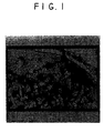
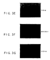
- Toluidine blue staining and immunohistochemistry were done, the latter with antibodies to extracellular matrix components ( Figures 2 and 3) including: anti-collagen types I, II and X, and anti-glycosaminoglycan antibodies 3-B-3, 7-D-4 (chondroitin sulfate), and 5-D-4 (keratan sulfate). Reactivity was detected with either FITC-linked secondary antibodies and fluorescence microscopy or alkaline phosphatase-linked antibodies and substrate. Pellets were also extracted in 4M guanidine, 20 mM sodium acetate containing protease inhibitors and subjected to immunolocalization after separation by SDS-PAGE and Western blotting.
- the +DEX pellets contained obvious metachromatic staining around a region of internally located cells, which had the appearance of hypertrophic chondrocytes. Those cells on the periphery of the pellets remained flat and did not show metachromasia.
- the +DEX pellets resembled a ball of hypertrophic chondrocytes.
- the -DEX defined medium pellets all shrunk in size and in many cases fell apart by 21 days in culture. No obvious hypertrophic cells were evident in any -DEX pellet.
- the above demonstrates a culture system in which rabbit and human bone marrow-derived mesenchymal progenitor cells differentiate into hypertrophic chondrocytes.
- the sequence of events resembles that of chondrogenesis in embryonic limb formation. Since all components are defined, the system can be used for studies of the effects of growth factors etc. on the progression of chondrogenesis. It is also applicable to studies of the molecular control of mammalian chondrogenesis from progenitor cells.
- This system utilizes a postnatal source and adds to the data concerning bone marrow-derived progenitor cells.
- In vitro systems have been used by others to show that these cell populations have osteogenic and adipocytic potential; we demonstrate here that at least a sub-set of that population has chondrogenic potential.
- This system will facilitate the exploration of the control of chondrogenesis and may lead to an understanding of what factors are required to promote this process in vivo. This has clinical applicability for cartilage repair.
- Rabbit marrow-derived mesenchymal cells were cultured as described in Example 1, with either (i) the addition of TGF- ⁇ 1 (10 ng/ml) or (ii) the addition of TGF- ⁇ 1 (10 ng/ml) and the deletion of dexamethasone.
- Human marrow-derived mesenchymal cells were cultured as described in Example 1, with either (i) the addition of TGF- ⁇ 1 (10 ng/ml) or (ii) the addition of TGF- ⁇ 1 (10 ng/ml) and the deletion of dexamethasone.
- Rat marrow-derived mesenchymal cells were obtained and pelleted as described in Example 1.
- the culture media were modified as follows. Rat marrow-derived mesenchymal cells were cultured as described in Example 1, with the addition of BMP-2 at 10 ng/ml and 100 ng/ml in the presence or absence of dexamethasone (10 -7 M). Differentiation of the MSCs into chondrocytes was observed in the presence of TGF- ⁇ 1 with dexamethasone, but not in its absence.
Landscapes
- Health & Medical Sciences (AREA)
- Engineering & Computer Science (AREA)
- Life Sciences & Earth Sciences (AREA)
- Biomedical Technology (AREA)
- Wood Science & Technology (AREA)
- Zoology (AREA)
- Organic Chemistry (AREA)
- Bioinformatics & Cheminformatics (AREA)
- Genetics & Genomics (AREA)
- Chemical & Material Sciences (AREA)
- Rheumatology (AREA)
- Biotechnology (AREA)
- Microbiology (AREA)
- Biochemistry (AREA)
- General Engineering & Computer Science (AREA)
- General Health & Medical Sciences (AREA)
- Cell Biology (AREA)
- Micro-Organisms Or Cultivation Processes Thereof (AREA)
- Medicines Containing Material From Animals Or Micro-Organisms (AREA)
- Materials For Medical Uses (AREA)
Abstract
Description
Claims (20)
- A composition for the in vitro chondrogenesis of human mesenchymal precursor cells and the in vitro formation of human chondrocytes therefrom, which composition comprises isolated human mesenchymal stem cells condensed into close proximity as packed cells and at least one chondroinductive agent in contact therewith, wherein the chondroinductive agent is a combination of dexamethasone and TGF-β1.
- The composition of claim 1 wherein the mesenchymal stem cells are isolated, culture expanded human mesenchymal stem cells
- The composition of claim 1 wherein the mesenchymal stem cells are in a chemically defined serum-free environment.
- The composition of claim 1 wherein the packed cells are a centrifugal cell pellet.
- A process for producing chondrocytes from mesenchymal stem cells by contacting mesenchymal stem cells with a chondroinductive agent in vitro wherein the stem cells are condensed into close proximity as packed cells, wherein the chondroinductive agent is a combination of dexamethasone and TGF-β1.
- The process of claim 5 wherein the mesenchymal stem cells are isolated, culture expanded human mesenchymal stem cells.
- The process of claim 5 or 6 wherein the mesenchymal stem cells are in a chemically defined serum-free environment.
- The process of claim 5 wherein the packed cells are a centrifugal cell pellet.
- The process of claim 5 wherein the step of contacting comprises culturing a pellet of human mesenchymal stem cells in a chemically defined serum-free medium.
- The process of claim 9 wherein the chemically defined serum-free medium comprises (1) a chemically defined minimum essential medium; (2) ascorbate or an analog thereof; (3) an iron source; (4) insulin or an insulin-like growth factor.
- The method of claim 5 wherein the cells are cultured with the chondroinductive composition and thereafter placed in a rigid porous vessel.
- The method of claim 11 wherein the rigid porous vessel is a ceramic cube.
- A process for inducing chondrogenesis in mesenchymal stem cells by contacting mesenchymal stem cells with a chondroinductive agent in vitro wherein the stem cells are condensed into close proximity as packed cells, wherein the chondroinductive agent is a combination of dexamethasone and TGF-β1.
- The process of claim 13 wherein the mesenchymal stem cells are isolated, culture expanded human mesenchymal stem cells.
- The process of claim 13 or 14 wherein the mesenchymal stem cells are in a chemically defined serum-free environment.
- The process of claim 13 wherein the packed cells are a centrifugal cell pellet.
- The process of claim 13 wherein the step of contacting comprises culturing a pellet of human mesenchymal stem cells in a chemically defined serum-free medium.
- The process of claim 13 wherein the chemically defined serum-free medium comprises (1) a chemically defined minimum essential medium; (2) ascorbate or an analog thereof; (3) an iron source; (4) insulin or an insulin-like growth factor.
- The method of claim 13 wherein the cells are cultured with the chondroinductive composition and thereafter placed in a rigid porous vessel.
- The method of claim 19 wherein the rigid porous vessel is a ceramic cube.
Applications Claiming Priority (3)
| Application Number | Priority Date | Filing Date | Title |
|---|---|---|---|
| US686695P | 1995-11-16 | 1995-11-16 | |
| US6866P | 1995-11-16 | ||
| PCT/US1996/018371 WO1997018299A1 (en) | 1995-11-16 | 1996-11-15 | In vitro chondrogenic induction of human mesenchymal stem cells |
Publications (3)
| Publication Number | Publication Date |
|---|---|
| EP0868505A1 EP0868505A1 (en) | 1998-10-07 |
| EP0868505A4 EP0868505A4 (en) | 2002-07-17 |
| EP0868505B1 true EP0868505B1 (en) | 2005-02-02 |
Family
ID=21723005
Family Applications (1)
| Application Number | Title | Priority Date | Filing Date |
|---|---|---|---|
| EP96940480A Expired - Lifetime EP0868505B1 (en) | 1995-11-16 | 1996-11-15 | In vitro chondrogenic induction of human mesenchymal stem cells |
Country Status (11)
| Country | Link |
|---|---|
| US (1) | US5908784A (en) |
| EP (1) | EP0868505B1 (en) |
| JP (1) | JP3781433B2 (en) |
| AT (1) | ATE288480T1 (en) |
| AU (1) | AU713280B2 (en) |
| CA (1) | CA2237890C (en) |
| DE (1) | DE69634303T2 (en) |
| DK (1) | DK0868505T3 (en) |
| ES (1) | ES2238077T3 (en) |
| PT (1) | PT868505E (en) |
| WO (1) | WO1997018299A1 (en) |
Cited By (1)
| Publication number | Priority date | Publication date | Assignee | Title |
|---|---|---|---|---|
| US8945535B2 (en) | 2005-09-20 | 2015-02-03 | Zimmer Orthobiologics, Inc. | Implant for the repair of a cartilage defect and method for manufacturing the implant |
Families Citing this family (145)
| Publication number | Priority date | Publication date | Assignee | Title |
|---|---|---|---|---|
| US5736396A (en) * | 1995-01-24 | 1998-04-07 | Case Western Reserve University | Lineage-directed induction of human mesenchymal stem cell differentiation |
| ATE295875T1 (en) | 1996-03-01 | 2005-06-15 | Isotis Nv | METHOD FOR PRODUCING BONE IN VITRO |
| EP0920490A2 (en) * | 1996-07-25 | 1999-06-09 | Genzyme Corporation | Chondrocyte media formulations and culture procedures |
| JP4531135B2 (en) * | 1996-12-06 | 2010-08-25 | オシリス セラピューティクス,インコーポレイテッド | Improved chondrogenic differentiation of human mesenchymal stem cells |
| ATE218896T1 (en) | 1997-07-16 | 2002-06-15 | Isotis Nv | DEVICE FOR BONE TREATMENT CONSISTING OF DEGRADABLE THERMOPLASTIC COPOLYESTER AND CULTURED CELLS |
| EP0896825B1 (en) | 1997-08-14 | 2002-07-17 | Sulzer Innotec Ag | Composition and device for in vivo cartilage repair comprising nanocapsules with osteoinductive and/or chondroinductive factors |
| DE19755023C2 (en) * | 1997-12-11 | 2000-03-09 | Celanese Chem Europe Gmbh | Catalyst and process for making vinyl acetate |
| WO2000005344A1 (en) * | 1998-07-24 | 2000-02-03 | The Carnegie Institution Of Washington | METHOD FOR MAINTENANCE AND PROPAGATION OF GERMLINE STEM CELLS USING MEMBERS OF THE TGF-β FAMILY OF GROWTH FACTORS |
| DE69937888T2 (en) * | 1998-07-31 | 2009-01-02 | Genzyme Corp., Cambridge | Process for the preparation of mesenchymal stem cells |
| DE19842802A1 (en) * | 1998-09-18 | 2000-03-23 | Christoph Geissmaier | Culture system for stimulating the growth and differentiation of cartilage and/or bone cells comprises a combination of undifferentiated cells and differentiating and/or differentiated cells |
| US7001746B1 (en) | 1999-01-29 | 2006-02-21 | Artecel Sciences, Inc. | Methods and compositions for the differentiation of human preadipocytes into adipocytes |
| US6197061B1 (en) * | 1999-03-01 | 2001-03-06 | Koichi Masuda | In vitro production of transplantable cartilage tissue cohesive cartilage produced thereby, and method for the surgical repair of cartilage damage |
| IL145002A0 (en) | 1999-03-10 | 2002-06-30 | Univ Pittsburgh | Adipose-derived stem cells and lattices |
| US20030082152A1 (en) * | 1999-03-10 | 2003-05-01 | Hedrick Marc H. | Adipose-derived stem cells and lattices |
| US20050076396A1 (en) * | 1999-03-10 | 2005-04-07 | Katz Adam J. | Adipose-derived stem cells and lattices |
| US6777231B1 (en) * | 1999-03-10 | 2004-08-17 | The Regents Of The University Of California | Adipose-derived stem cells and lattices |
| US6429013B1 (en) * | 1999-08-19 | 2002-08-06 | Artecel Science, Inc. | Use of adipose tissue-derived stromal cells for chondrocyte differentiation and cartilage repair |
| US7078232B2 (en) | 1999-08-19 | 2006-07-18 | Artecel, Inc. | Adipose tissue-derived adult stem or stromal cells for the repair of articular cartilage fractures and uses thereof |
| US6555374B1 (en) | 1999-08-19 | 2003-04-29 | Artecel Sciences, Inc. | Multiple mesodermal lineage differentiation potentials for adipose tissue-derived stromal cells and uses thereof |
| EP1229940B1 (en) * | 1999-11-15 | 2014-05-14 | Piramal Healthcare (Canada) Limited | Temperature-controlled and ph-dependant self-gelling biopolymeric aqueous solution |
| US20030158302A1 (en) * | 1999-12-09 | 2003-08-21 | Cyric Chaput | Mineral-polymer hybrid composition |
| WO2001041822A1 (en) * | 1999-12-09 | 2001-06-14 | Biosyntech Canada Inc. | Mineral-polymer hybrid composition |
| WO2001048147A1 (en) * | 1999-12-28 | 2001-07-05 | Iso Tis N.V. | Cell culture medium containing growth factors and l-glutamine |
| US7582292B2 (en) | 2000-02-26 | 2009-09-01 | Artecel, Inc. | Adipose tissue derived stromal cells for the treatment of neurological disorders |
| PT1261694E (en) | 2000-02-26 | 2008-04-03 | Artecel Inc | Pleuripotent stem cells generated from adipose tissue-derived stromal cells and uses thereof |
| EP1918366A1 (en) | 2000-02-26 | 2008-05-07 | Artecel, Inc. | Pleuripotent stem cells generated from adipose tissue-derived stromal cells and uses thereof |
| US7182781B1 (en) | 2000-03-02 | 2007-02-27 | Regeneration Technologies, Inc. | Cervical tapered dowel |
| ES2353061T5 (en) | 2000-04-25 | 2014-04-07 | Osiris Therapeutics, Inc. | Joint repair using mesenchymal stem cells |
| CN1471412B8 (en) * | 2000-06-29 | 2016-06-15 | 生物合成技术加拿大公司 | For repairing and regeneration of cartilage and the composition of other tissue and the purposes of said composition |
| DE60125973D1 (en) * | 2000-11-15 | 2007-02-22 | Biosyntech Canada Inc | METHOD FOR RECOVERING A DAMAGED BAND DISC |
| US7311905B2 (en) | 2002-02-13 | 2007-12-25 | Anthrogenesis Corporation | Embryonic-like stem cells derived from post-partum mammalian placenta, and uses and methods of treatment using said cells |
| EP2206772A3 (en) | 2000-12-06 | 2010-08-04 | Robert J. Hariri | Method of collecting placental stem cells |
| EP2336299A1 (en) * | 2001-02-14 | 2011-06-22 | Anthrogenesis Corporation | Post-partum mammalian placenta, its use and placental stem cells therefrom |
| CA2437957C (en) | 2001-02-14 | 2014-04-22 | Robert J. Hariri | Renovation and repopulation of decellularized tissues and cadaveric organs by stem cells |
| US20030036194A1 (en) * | 2001-07-06 | 2003-02-20 | Chunhui Xu | Mesenchymal cells derived from human embryonic stem cells |
| US7476257B2 (en) | 2001-09-15 | 2009-01-13 | Rush University Medical Center | Methods to engineer stratified cartilage tissue |
| US20030165473A1 (en) * | 2001-11-09 | 2003-09-04 | Rush-Presbyterian-St. Luke's Medical Center | Engineered intervertebral disc tissue |
| US7585670B2 (en) | 2001-12-07 | 2009-09-08 | Cytori Therapeutics, Inc. | Automated methods for isolating and using clinically safe adipose derived regenerative cells |
| US20030109038A1 (en) * | 2001-12-07 | 2003-06-12 | Thies R. Scott | Chondrocyte precursors derived from human embryonic stem cells |
| US20050008626A1 (en) * | 2001-12-07 | 2005-01-13 | Fraser John K. | Methods of using adipose tissue-derived cells in the treatment of cardiovascular conditions |
| US8404229B2 (en) | 2001-12-07 | 2013-03-26 | Cytori Therapeutics, Inc. | Methods of using adipose derived stem cells to treat acute tubular necrosis |
| US7514075B2 (en) * | 2001-12-07 | 2009-04-07 | Cytori Therapeutics, Inc. | Systems and methods for separating and concentrating adipose derived stem cells from tissue |
| US20050095228A1 (en) | 2001-12-07 | 2005-05-05 | Fraser John K. | Methods of using regenerative cells in the treatment of peripheral vascular disease and related disorders |
| US8105580B2 (en) | 2001-12-07 | 2012-01-31 | Cytori Therapeutics, Inc. | Methods of using adipose derived stem cells to promote wound healing |
| US7595043B2 (en) * | 2001-12-07 | 2009-09-29 | Cytori Therapeutics, Inc. | Method for processing and using adipose-derived stem cells |
| US9597395B2 (en) | 2001-12-07 | 2017-03-21 | Cytori Therapeutics, Inc. | Methods of using adipose tissue-derived cells in the treatment of cardiovascular conditions |
| US20060204556A1 (en) * | 2001-12-07 | 2006-09-14 | Cytori Therapeutics, Inc. | Cell-loaded prostheses for regenerative intraluminal applications |
| KR100811995B1 (en) * | 2001-12-07 | 2008-03-10 | 사이토리 테라퓨틱스, 인크. | Systems and methods for treating patients with treated lipoasperate cells |
| US7651684B2 (en) | 2001-12-07 | 2010-01-26 | Cytori Therapeutics, Inc. | Methods of using adipose tissue-derived cells in augmenting autologous fat transfer |
| US7771716B2 (en) * | 2001-12-07 | 2010-08-10 | Cytori Therapeutics, Inc. | Methods of using regenerative cells in the treatment of musculoskeletal disorders |
| JP2005518828A (en) * | 2001-12-20 | 2005-06-30 | マクロポア インコーポレイテッド | System and method for treating a patient using a collagen-rich substance extracted from adipose tissue |
| US7700090B2 (en) | 2002-02-13 | 2010-04-20 | Anthrogenesis Corporation | Co-culture of placental stem cells and stem cells from a second source |
| ATE408666T1 (en) * | 2002-05-28 | 2008-10-15 | Toyo Boseki | METHOD OF CULTURE, STORAGE AND INDUCE DIFFERENTIATION OF CELLS AND DEVICE FOR USE IN SUCH METHOD, AND METHOD OF USE THEREOF. |
| US7622562B2 (en) | 2002-06-26 | 2009-11-24 | Zimmer Orthobiologics, Inc. | Rapid isolation of osteoinductive protein mixtures from mammalian bone tissue |
| CA2493083C (en) * | 2002-07-16 | 2012-11-06 | Bio Syntech Canada Inc. | Composition for cytocompatible, injectable, self-gelling chitosan solutions for encapsulating and delivering live cells or biologically active factors |
| KR101042448B1 (en) | 2002-11-26 | 2011-06-16 | 안트로제네시스 코포레이션 | Cell Therapy, Cell Therapy Unit, and Therapeutic Method Using The Same |
| EP2263698A1 (en) * | 2003-04-01 | 2010-12-22 | United States of America Dempartment of Veteran's Affairs | Stem-cell, precursor cell, or target cell-based treatment of multi-organ failure and renal dysfunction |
| SG146691A1 (en) | 2003-10-08 | 2008-10-30 | Vet Stem Inc | Methods of preparing and using stem cell compositions and kits comprising the same |
| WO2005039662A2 (en) * | 2003-10-10 | 2005-05-06 | Kw2 Implantattechnologie Gmbh | Cartilage regeneration by generation of chondrons under high concentrations of magnesium |
| JP2006000059A (en) * | 2004-06-17 | 2006-01-05 | Two Cells Co Ltd | Method for promoting proliferation and differentiation of animal cell by using extracellular substrate |
| US8182806B2 (en) * | 2004-09-07 | 2012-05-22 | Johnson Lanny L | Synovial villi for use with tissue engineering |
| US20060051328A1 (en) * | 2004-09-07 | 2006-03-09 | Johnson Lanny L | Mobilization of cells via physical means |
| US7785582B2 (en) * | 2004-09-07 | 2010-08-31 | Johnson Lanny L | Use of synovium and omentum for tissue engineering |
| US8697139B2 (en) | 2004-09-21 | 2014-04-15 | Frank M. Phillips | Method of intervertebral disc treatment using articular chondrocyte cells |
| CA2512667A1 (en) * | 2005-01-07 | 2006-07-07 | Takahiro Ochiya | Human hepatocyte-like cells and uses thereof |
| US20080140451A1 (en) * | 2005-01-10 | 2008-06-12 | Cytori Therapeutics, Inc. | Devices and Methods for Monitoring, Managing, and Servicing Medical Devices |
| JP2008545703A (en) | 2005-05-27 | 2008-12-18 | バイアセル インコーポレーティッド | Treatment of ischemia using stem cells |
| US7531355B2 (en) | 2005-07-29 | 2009-05-12 | The Regents Of The University Of California | Methods and compositions for smooth muscle reconstruction |
| EP2796544B1 (en) | 2005-09-09 | 2019-04-03 | Duke University | Tissue engineering methods and compositions |
| US8518349B2 (en) | 2005-09-12 | 2013-08-27 | Lanny Johnson | Use of autologous sediment from fluid aspirates as vehicles for drug delivery |
| US7927630B2 (en) * | 2005-09-12 | 2011-04-19 | Johnson Lanny L | Use of autologous sediment from fluid aspirates as vehicles for drug delivery |
| JP4958094B2 (en) * | 2005-09-22 | 2012-06-20 | 国立大学法人広島大学 | Homogeneity identification method of mesenchymal stem cells, homogeneous mesenchymal stem cells obtained by using the method |
| CA2624925C (en) | 2005-10-13 | 2014-09-30 | Anthrogenesis Corporation | Immunomodulation using placental stem cells |
| US20090149421A1 (en) * | 2005-11-04 | 2009-06-11 | Bio Syntech Canada Inc. | Gel formation of polyelectrolyte aqueous solutions by thermally induced changes in ionization state |
| ES2708933T3 (en) | 2005-12-29 | 2019-04-12 | Celularity Inc | Populations of placental stem cells |
| WO2007089798A2 (en) | 2006-01-30 | 2007-08-09 | University Of Virginia Patent Foundation | Methods of preparing and characterizing mesenchymal stem cell aggregates and uses thereof |
| PL2548951T3 (en) | 2006-03-23 | 2016-08-31 | Pluristem Ltd | Methods for cell expansion and uses of cells and conditioned media produced thereby for therapy |
| WO2007139551A1 (en) * | 2006-05-30 | 2007-12-06 | Cytori Therapeutics, Inc. | Systems and methods for manipulation of regenerative cells from adipose tissue |
| WO2007146123A2 (en) * | 2006-06-09 | 2007-12-21 | Anthrogenesis Corporation | Placental niche and use thereof to culture stem cells |
| WO2008013863A2 (en) * | 2006-07-26 | 2008-01-31 | Cytori Therapeutics, Inc. | Generation of adipose tissue and adipocytes |
| JP5013584B2 (en) * | 2006-08-30 | 2012-08-29 | 株式会社日立製作所 | Chondrocyte culture method and chondrocyte |
| JP2010505849A (en) * | 2006-10-06 | 2010-02-25 | ユニバーシティ オブ バージニア パテント ファウンデーション | Methods and compositions useful for diabetic wound healing |
| JP2010507392A (en) * | 2006-10-23 | 2010-03-11 | アントフロゲネシス コーポレーション | Methods and compositions for treating bone defects in placental cell populations |
| KR101569168B1 (en) | 2007-02-12 | 2015-11-13 | 안트로제네시스 코포레이션 | Treatment of inflammatory diseases using placental stem cells |
| US7632340B2 (en) * | 2007-03-07 | 2009-12-15 | Hamilton Beach Brands, Inc. | Air purifier for removing particles or contaminants from air |
| US20100291042A1 (en) | 2007-05-03 | 2010-11-18 | The Brigham And Women's Hospital, Inc. | Multipotent stem cells and uses thereof |
| TWM322542U (en) * | 2007-05-23 | 2007-11-21 | Universal Scient Ind Co Ltd | Testing machine |
| US9200253B1 (en) | 2007-08-06 | 2015-12-01 | Anthrogenesis Corporation | Method of producing erythrocytes |
| KR101738323B1 (en) | 2007-09-19 | 2017-05-19 | 플루리스템 리미티드 | Adherent cells from adipose or placenta tissues and use thereof in therapy |
| CA2700617C (en) | 2007-09-28 | 2018-11-06 | Celgene Cellular Therapeutics | Tumor suppression using human placental perfusate and human placenta-derived intermediate natural killer cells |
| WO2009142770A2 (en) * | 2008-05-23 | 2009-11-26 | The Regents Of The University Of California | Compositions and methods for generating musculoskeletal tissue |
| EP2303293A1 (en) | 2008-05-27 | 2011-04-06 | Pluristem Ltd. | Methods of treating inflammatory colon diseases |
| WO2010021993A1 (en) * | 2008-08-19 | 2010-02-25 | Cytori Therapeutics, Inc. | Methods of using adipose tissue-derived cells in the treatment of the lymphatic system and malignant disease |
| DK2330889T3 (en) | 2008-08-20 | 2017-01-30 | Anthrogenesis Corp | Improved cell composition and process for making the same |
| JP5950577B2 (en) | 2008-08-20 | 2016-07-13 | アンスロジェネシス コーポレーション | Treatment of stroke using isolated placental cells |
| HRP20130812T1 (en) | 2008-08-22 | 2013-09-30 | Anthrogenesis Corporation | Methods and compositions for treatment of bone defects with placental cell populations |
| SG190659A1 (en) | 2008-09-02 | 2013-06-28 | Pluristem Ltd | Adherent cells from placenta tissue and use thereof in therapy |
| EP2182055A1 (en) | 2008-10-14 | 2010-05-05 | Heinrich-Heine-Universität Düsseldorf | Human cord blood derived unrestricted somatic stem cells (USSC) |
| US20100098739A1 (en) * | 2008-10-20 | 2010-04-22 | University Of Virginia Patent Foundation | Compositions and methods for modular soft tissue repair |
| CN102282252B (en) | 2008-11-19 | 2017-07-04 | 人类起源公司 | amnion derived adherent cells |
| US9192695B2 (en) * | 2008-11-20 | 2015-11-24 | Allosource | Allografts combined with tissue derived stem cells for bone healing |
| WO2010127310A1 (en) | 2009-05-01 | 2010-11-04 | Cytori Therapeutics, Inc. | Systems, methods and compositions for optimizing tissue and cell enriched grafts |
| US8586360B2 (en) | 2009-07-02 | 2013-11-19 | Anthrogenesis Corporation | Method of producing erythrocytes without feeder cells |
| US9081008B2 (en) | 2009-12-04 | 2015-07-14 | James Sherley | Detecting and counting tissue—specific stem cells and uses thereof |
| WO2011087779A2 (en) | 2009-12-22 | 2011-07-21 | National Cheng Kung University | Cell tissue gel containing collagen and hyaluronan |
| ES2646750T3 (en) | 2010-01-26 | 2017-12-15 | Anthrogenesis Corporation | Treatment of bone-related cancers using placental stem cells |
| IL250234B2 (en) | 2010-04-07 | 2023-10-01 | Anthrogenesis Llc | Creating blood vessels using placental stem cells |
| MX2012011543A (en) | 2010-04-08 | 2013-05-06 | Anthrogenesis Corp | Treatment of sarcoidosis using placental stem cells. |
| EP2590695B1 (en) | 2010-07-09 | 2024-11-20 | GID BIO, Inc. | Apparatus and methods relating to collecting and processing human biological material containing adipose |
| US9206387B2 (en) | 2010-07-09 | 2015-12-08 | The Gid Group, Inc. | Method and apparatus for processing adipose tissue |
| WO2013106655A1 (en) | 2012-01-11 | 2013-07-18 | The Gid Group, Inc. | Method for processing adipose tissue and processing apparatus |
| US9296984B2 (en) | 2010-07-09 | 2016-03-29 | The Gid Group, Inc. | Tissue processing apparatus and method for processing adipose tissue |
| KR20180099915A (en) | 2010-07-13 | 2018-09-05 | 안트로제네시스 코포레이션 | Methods of generating natural killer cells |
| US20120208757A1 (en) | 2010-10-07 | 2012-08-16 | Excel Med, Llc | Use of hyaluronan for promoting angiogenesis |
| EP4663241A3 (en) | 2010-10-08 | 2026-03-25 | Mesoblast International Sàrl | Enhanced msc preparations |
| US8895291B2 (en) | 2010-10-08 | 2014-11-25 | Terumo Bct, Inc. | Methods and systems of growing and harvesting cells in a hollow fiber bioreactor system with control conditions |
| AU2011352036A1 (en) | 2010-12-31 | 2013-07-18 | Anthrogenesis Corporation | Enhancement of placental stem cell potency using modulatory RNA molecules |
| WO2012127320A1 (en) | 2011-03-22 | 2012-09-27 | Pluristem Ltd. | Methods for treating radiation or chemical injury |
| CN104220081A (en) | 2011-06-01 | 2014-12-17 | 人类起源公司 | Treatment of pain using placental stem cells |
| WO2013055476A1 (en) | 2011-09-09 | 2013-04-18 | Anthrogenesis Corporation | Treatment of amyotrophic lateral sclerosis using placental stem cells |
| BR112015004003B1 (en) | 2012-09-06 | 2020-05-19 | The Gid Group, Inc. | apparatus for processing human biological material containing fibrous tissue and method for processing adipose tissue |
| AU2014215458A1 (en) | 2013-02-05 | 2015-08-13 | Anthrogenesis Corporation | Natural killer cells from placenta |
| US10351910B2 (en) | 2013-02-20 | 2019-07-16 | Pluristem Ltd | Gene and protein expression properties of adherent stromal cells cultured in 3D |
| KR102312720B1 (en) | 2013-03-15 | 2021-10-13 | 알로소스 | Cell repopulated collagen matrix for soft tissue repair and regeneration |
| WO2015035221A1 (en) | 2013-09-05 | 2015-03-12 | The Gid Group, Inc. | Tissue processing apparatus and method for processing adipose tissue |
| JP6612227B2 (en) | 2013-11-16 | 2019-11-27 | テルモ ビーシーティー、インコーポレーテッド | Cell growth in bioreactors |
| US11008547B2 (en) | 2014-03-25 | 2021-05-18 | Terumo Bct, Inc. | Passive replacement of media |
| CN115531551A (en) | 2014-07-29 | 2022-12-30 | 黄玲惠 | Cell tissue gel containing collagen and hyaluronic acid |
| WO2016049421A1 (en) | 2014-09-26 | 2016-03-31 | Terumo Bct, Inc. | Scheduled feed |
| HUP1500218A2 (en) | 2015-05-08 | 2016-11-28 | Deltabio 2000 Kft | Method and composition for treatment of orthopedic diseases articular cortilage injuries and particularly to dysphoria |
| WO2017004592A1 (en) | 2015-07-02 | 2017-01-05 | Terumo Bct, Inc. | Cell growth with mechanical stimuli |
| WO2017205667A1 (en) | 2016-05-25 | 2017-11-30 | Terumo Bct, Inc. | Cell expansion |
| US11104874B2 (en) | 2016-06-07 | 2021-08-31 | Terumo Bct, Inc. | Coating a bioreactor |
| US11685883B2 (en) | 2016-06-07 | 2023-06-27 | Terumo Bct, Inc. | Methods and systems for coating a cell growth surface |
| WO2018044791A1 (en) | 2016-08-30 | 2018-03-08 | Lifecell Corporation | Systems and methods for medical device control |
| USD851777S1 (en) | 2017-01-30 | 2019-06-18 | Lifecell Corporation | Canister-type device for tissue processing |
| US11655454B1 (en) * | 2017-03-23 | 2023-05-23 | Elite IP, LLC | Method and apparatus for improved mesenchymal stem cell harvesting |
| US11624046B2 (en) | 2017-03-31 | 2023-04-11 | Terumo Bct, Inc. | Cell expansion |
| US12234441B2 (en) | 2017-03-31 | 2025-02-25 | Terumo Bct, Inc. | Cell expansion |
| WO2018184028A2 (en) | 2017-03-31 | 2018-10-04 | Terumo Bct, Inc. | Cell expansion |
| US11732233B2 (en) | 2017-07-18 | 2023-08-22 | Gid Bio, Inc. | Adipose tissue digestion system and tissue processing method |
| AU2018372631B2 (en) | 2017-11-22 | 2025-06-05 | Mesoblast International Sarl | Cellular compositions and methods of treatment I |
| GB2619893A (en) | 2021-03-23 | 2023-12-20 | Terumo Bct Inc | Cell capture and expansion |
| US12209689B2 (en) | 2022-02-28 | 2025-01-28 | Terumo Kabushiki Kaisha | Multiple-tube pinch valve assembly |
| USD1099116S1 (en) | 2022-09-01 | 2025-10-21 | Terumo Bct, Inc. | Display screen or portion thereof with a graphical user interface for displaying cell culture process steps and measurements of an associated bioreactor device |
Family Cites Families (7)
| Publication number | Priority date | Publication date | Assignee | Title |
|---|---|---|---|---|
| US5226914A (en) * | 1990-11-16 | 1993-07-13 | Caplan Arnold I | Method for treating connective tissue disorders |
| US5486359A (en) * | 1990-11-16 | 1996-01-23 | Osiris Therapeutics, Inc. | Human mesenchymal stem cells |
| US5197985A (en) * | 1990-11-16 | 1993-03-30 | Caplan Arnold I | Method for enhancing the implantation and differentiation of marrow-derived mesenchymal cells |
| US5646001A (en) * | 1991-03-25 | 1997-07-08 | Immunivest Corporation | Affinity-binding separation and release of one or more selected subset of biological entities from a mixed population thereof |
| US5436151A (en) * | 1992-04-03 | 1995-07-25 | Regents Of The University Of Minnesota | Method for culturing human hematopoietic stem cells in vitro |
| US5466572A (en) * | 1992-09-03 | 1995-11-14 | Systemix, Inc. | High speed flow cytometric separation of viable cells |
| US5736396A (en) * | 1995-01-24 | 1998-04-07 | Case Western Reserve University | Lineage-directed induction of human mesenchymal stem cell differentiation |
-
1996
- 1996-11-15 WO PCT/US1996/018371 patent/WO1997018299A1/en not_active Ceased
- 1996-11-15 CA CA2237890A patent/CA2237890C/en not_active Expired - Fee Related
- 1996-11-15 DE DE69634303T patent/DE69634303T2/en not_active Expired - Lifetime
- 1996-11-15 DK DK96940480T patent/DK0868505T3/en active
- 1996-11-15 JP JP51911497A patent/JP3781433B2/en not_active Expired - Fee Related
- 1996-11-15 AU AU77351/96A patent/AU713280B2/en not_active Ceased
- 1996-11-15 PT PT96940480T patent/PT868505E/en unknown
- 1996-11-15 AT AT96940480T patent/ATE288480T1/en active
- 1996-11-15 EP EP96940480A patent/EP0868505B1/en not_active Expired - Lifetime
- 1996-11-15 US US08/749,484 patent/US5908784A/en not_active Expired - Lifetime
- 1996-11-15 ES ES96940480T patent/ES2238077T3/en not_active Expired - Lifetime
Cited By (1)
| Publication number | Priority date | Publication date | Assignee | Title |
|---|---|---|---|---|
| US8945535B2 (en) | 2005-09-20 | 2015-02-03 | Zimmer Orthobiologics, Inc. | Implant for the repair of a cartilage defect and method for manufacturing the implant |
Also Published As
| Publication number | Publication date |
|---|---|
| CA2237890A1 (en) | 1997-05-22 |
| ATE288480T1 (en) | 2005-02-15 |
| US5908784A (en) | 1999-06-01 |
| DE69634303T2 (en) | 2006-01-05 |
| JP2000516802A (en) | 2000-12-19 |
| AU713280B2 (en) | 1999-11-25 |
| PT868505E (en) | 2005-06-30 |
| AU7735196A (en) | 1997-06-05 |
| CA2237890C (en) | 2011-03-29 |
| JP3781433B2 (en) | 2006-05-31 |
| EP0868505A1 (en) | 1998-10-07 |
| DK0868505T3 (en) | 2005-06-06 |
| WO1997018299A1 (en) | 1997-05-22 |
| EP0868505A4 (en) | 2002-07-17 |
| DE69634303D1 (en) | 2005-03-10 |
| ES2238077T3 (en) | 2005-08-16 |
Similar Documents
| Publication | Publication Date | Title |
|---|---|---|
| US5908784A (en) | In vitro chondrogenic induction of human mesenchymal stem cells | |
| AU742638B2 (en) | Improved chondrogenic differentiation of human mesenchymal stem cells | |
| AU756411B2 (en) | Isolation of bone and cartilage precursor cells | |
| Caterson et al. | Human marrow-derived mesenchymal progenitor cells: isolation, culture expansion, and analysis of differentiation | |
| US5226914A (en) | Method for treating connective tissue disorders | |
| US5197985A (en) | Method for enhancing the implantation and differentiation of marrow-derived mesenchymal cells | |
| US6761887B1 (en) | Alginate layer system for chondrogenic differentiation of human mesenchymal stem cells | |
| EP0805853B1 (en) | Lineage-directed induction of human mesenchymal stem cell differentiation | |
| KR100489248B1 (en) | Isolation and culture-expansion methods of mesenchymal stem/progenitor cells from umbilical cord blood, and differentiation method of umbilical cord blood-derived mesenchymal stem/progenitor cells into various mesenchymal tissues | |
| US20020045260A1 (en) | Method of isolating mesenchymal stem cells | |
| WO1996023059A9 (en) | Lineage-directed induction of human mesenchymal stem cell differentiation | |
| WO2007119213A2 (en) | A culture medium and pharmaceutical composition for regenerating cartilage tissue, a method, uses and products related thereto | |
| CN113692282A (en) | Bioactive substance composition, serum-free culture medium containing composition and application of serum-free culture medium | |
| EP3385372B1 (en) | Method for manufacturing mesenchymal cell line derived from vertebrate animal adipose tissue | |
| WO2000029552A1 (en) | Alginate layer system for chondrogenic differentiation of human mesenchymal stem cells | |
| US20030026786A1 (en) | Chondrogenic differentiation of human mesenchymal stem cells | |
| EP1856245A2 (en) | A composition for creating an artificial bone-marrow like environment and uses thereof | |
| WO1998033889A1 (en) | Adipocyte culture | |
| MXPA97005612A (en) | Induction directed by lineage of the differentiation of human mesenquimatosas cells of ori |
Legal Events
| Date | Code | Title | Description |
|---|---|---|---|
| PUAI | Public reference made under article 153(3) epc to a published international application that has entered the european phase |
Free format text: ORIGINAL CODE: 0009012 |
|
| 17P | Request for examination filed |
Effective date: 19980508 |
|
| AK | Designated contracting states |
Kind code of ref document: A1 Designated state(s): AT BE CH DE DK ES FR GB GR IE IT LI LU MC NL PT SE |
|
| A4 | Supplementary search report drawn up and despatched |
Effective date: 20020603 |
|
| AK | Designated contracting states |
Kind code of ref document: A4 Designated state(s): AT BE CH DE DK ES FR GB GR IE IT LI LU MC NL PT SE |
|
| 17Q | First examination report despatched |
Effective date: 20030820 |
|
| GRAP | Despatch of communication of intention to grant a patent |
Free format text: ORIGINAL CODE: EPIDOSNIGR1 |
|
| GRAS | Grant fee paid |
Free format text: ORIGINAL CODE: EPIDOSNIGR3 |
|
| GRAA | (expected) grant |
Free format text: ORIGINAL CODE: 0009210 |
|
| AK | Designated contracting states |
Kind code of ref document: B1 Designated state(s): AT BE CH DE DK ES FR GB GR IE IT LI LU MC NL PT SE |
|
| REG | Reference to a national code |
Ref country code: GB Ref legal event code: FG4D |
|
| REG | Reference to a national code |
Ref country code: CH Ref legal event code: EP |
|
| REG | Reference to a national code |
Ref country code: IE Ref legal event code: FG4D |
|
| REF | Corresponds to: |
Ref document number: 69634303 Country of ref document: DE Date of ref document: 20050310 Kind code of ref document: P |
|
| REG | Reference to a national code |
Ref country code: SE Ref legal event code: TRGR |
|
| REG | Reference to a national code |
Ref country code: DK Ref legal event code: T3 |
|
| REG | Reference to a national code |
Ref country code: GR Ref legal event code: EP Ref document number: 20050401464 Country of ref document: GR |
|
| REG | Reference to a national code |
Ref country code: PT Ref legal event code: SC4A Free format text: AVAILABILITY OF NATIONAL TRANSLATION Effective date: 20050502 |
|
| REG | Reference to a national code |
Ref country code: CH Ref legal event code: NV Representative=s name: FIAMMENGHI-FIAMMENGHI |
|
| REG | Reference to a national code |
Ref country code: ES Ref legal event code: FG2A Ref document number: 2238077 Country of ref document: ES Kind code of ref document: T3 |
|
| PG25 | Lapsed in a contracting state [announced via postgrant information from national office to epo] |
Ref country code: MC Free format text: LAPSE BECAUSE OF NON-PAYMENT OF DUE FEES Effective date: 20051130 |
|
| PLBE | No opposition filed within time limit |
Free format text: ORIGINAL CODE: 0009261 |
|
| STAA | Information on the status of an ep patent application or granted ep patent |
Free format text: STATUS: NO OPPOSITION FILED WITHIN TIME LIMIT |
|
| 26N | No opposition filed |
Effective date: 20051103 |
|
| ET | Fr: translation filed | ||
| PGFP | Annual fee paid to national office [announced via postgrant information from national office to epo] |
Ref country code: NL Payment date: 20101111 Year of fee payment: 15 Ref country code: IE Payment date: 20101123 Year of fee payment: 15 Ref country code: DK Payment date: 20101110 Year of fee payment: 15 Ref country code: AT Payment date: 20101112 Year of fee payment: 15 |
|
| PGFP | Annual fee paid to national office [announced via postgrant information from national office to epo] |
Ref country code: PT Payment date: 20101108 Year of fee payment: 15 Ref country code: LU Payment date: 20101123 Year of fee payment: 15 Ref country code: CH Payment date: 20101123 Year of fee payment: 15 |
|
| PGFP | Annual fee paid to national office [announced via postgrant information from national office to epo] |
Ref country code: BE Payment date: 20101117 Year of fee payment: 15 Ref country code: SE Payment date: 20101112 Year of fee payment: 15 Ref country code: IT Payment date: 20101126 Year of fee payment: 15 Ref country code: GR Payment date: 20101119 Year of fee payment: 15 |
|
| PGFP | Annual fee paid to national office [announced via postgrant information from national office to epo] |
Ref country code: ES Payment date: 20101124 Year of fee payment: 15 |
|
| REG | Reference to a national code |
Ref country code: PT Ref legal event code: MM4A Free format text: LAPSE DUE TO NON-PAYMENT OF FEES Effective date: 20120515 |
|
| BERE | Be: lapsed |
Owner name: *CASE WESTERN RESERVE UNIVERSITY Effective date: 20111130 |
|
| REG | Reference to a national code |
Ref country code: NL Ref legal event code: V1 Effective date: 20120601 |
|
| REG | Reference to a national code |
Ref country code: CH Ref legal event code: PL |
|
| REG | Reference to a national code |
Ref country code: DK Ref legal event code: EBP |
|
| REG | Reference to a national code |
Ref country code: SE Ref legal event code: EUG |
|
| REG | Reference to a national code |
Ref country code: GR Ref legal event code: ML Ref document number: 20050401464 Country of ref document: GR Effective date: 20120605 |
|
| PG25 | Lapsed in a contracting state [announced via postgrant information from national office to epo] |
Ref country code: CH Free format text: LAPSE BECAUSE OF NON-PAYMENT OF DUE FEES Effective date: 20111130 Ref country code: NL Free format text: LAPSE BECAUSE OF NON-PAYMENT OF DUE FEES Effective date: 20120601 Ref country code: LI Free format text: LAPSE BECAUSE OF NON-PAYMENT OF DUE FEES Effective date: 20111130 |
|
| REG | Reference to a national code |
Ref country code: IE Ref legal event code: MM4A |
|
| PG25 | Lapsed in a contracting state [announced via postgrant information from national office to epo] |
Ref country code: BE Free format text: LAPSE BECAUSE OF NON-PAYMENT OF DUE FEES Effective date: 20111130 Ref country code: IT Free format text: LAPSE BECAUSE OF NON-PAYMENT OF DUE FEES Effective date: 20111115 Ref country code: PT Free format text: LAPSE BECAUSE OF NON-PAYMENT OF DUE FEES Effective date: 20120515 Ref country code: GR Free format text: LAPSE BECAUSE OF NON-PAYMENT OF DUE FEES Effective date: 20120605 |
|
| PG25 | Lapsed in a contracting state [announced via postgrant information from national office to epo] |
Ref country code: SE Free format text: LAPSE BECAUSE OF NON-PAYMENT OF DUE FEES Effective date: 20111116 Ref country code: DK Free format text: LAPSE BECAUSE OF NON-PAYMENT OF DUE FEES Effective date: 20111130 Ref country code: IE Free format text: LAPSE BECAUSE OF NON-PAYMENT OF DUE FEES Effective date: 20111115 |
|
| REG | Reference to a national code |
Ref country code: AT Ref legal event code: MM01 Ref document number: 288480 Country of ref document: AT Kind code of ref document: T Effective date: 20111115 |
|
| PG25 | Lapsed in a contracting state [announced via postgrant information from national office to epo] |
Ref country code: AT Free format text: LAPSE BECAUSE OF NON-PAYMENT OF DUE FEES Effective date: 20111115 |
|
| PG25 | Lapsed in a contracting state [announced via postgrant information from national office to epo] |
Ref country code: LU Free format text: LAPSE BECAUSE OF NON-PAYMENT OF DUE FEES Effective date: 20111115 |
|
| REG | Reference to a national code |
Ref country code: ES Ref legal event code: FD2A Effective date: 20130605 |
|
| PG25 | Lapsed in a contracting state [announced via postgrant information from national office to epo] |
Ref country code: ES Free format text: LAPSE BECAUSE OF NON-PAYMENT OF DUE FEES Effective date: 20111116 |
|
| REG | Reference to a national code |
Ref country code: FR Ref legal event code: PLFP Year of fee payment: 20 |
|
| PGFP | Annual fee paid to national office [announced via postgrant information from national office to epo] |
Ref country code: DE Payment date: 20151118 Year of fee payment: 20 Ref country code: GB Payment date: 20151111 Year of fee payment: 20 |
|
| PGFP | Annual fee paid to national office [announced via postgrant information from national office to epo] |
Ref country code: FR Payment date: 20151123 Year of fee payment: 20 |
|
| REG | Reference to a national code |
Ref country code: DE Ref legal event code: R071 Ref document number: 69634303 Country of ref document: DE |
|
| REG | Reference to a national code |
Ref country code: GB Ref legal event code: PE20 Expiry date: 20161114 |
|
| PG25 | Lapsed in a contracting state [announced via postgrant information from national office to epo] |
Ref country code: GB Free format text: LAPSE BECAUSE OF EXPIRATION OF PROTECTION Effective date: 20161114 |