EP0861268B1 - Inhibitory and non-inhibitory antigen binding polypeptides against human p450 enzymes - Google Patents
Inhibitory and non-inhibitory antigen binding polypeptides against human p450 enzymes Download PDFInfo
- Publication number
- EP0861268B1 EP0861268B1 EP96939561A EP96939561A EP0861268B1 EP 0861268 B1 EP0861268 B1 EP 0861268B1 EP 96939561 A EP96939561 A EP 96939561A EP 96939561 A EP96939561 A EP 96939561A EP 0861268 B1 EP0861268 B1 EP 0861268B1
- Authority
- EP
- European Patent Office
- Prior art keywords
- human
- metabolism
- antigen binding
- monoclonal antibody
- compound
- Prior art date
- Legal status (The legal status is an assumption and is not a legal conclusion. Google has not performed a legal analysis and makes no representation as to the accuracy of the status listed.)
- Expired - Lifetime
Links
Images




Classifications
-
- C—CHEMISTRY; METALLURGY
- C12—BIOCHEMISTRY; BEER; SPIRITS; WINE; VINEGAR; MICROBIOLOGY; ENZYMOLOGY; MUTATION OR GENETIC ENGINEERING
- C12N—MICROORGANISMS OR ENZYMES; COMPOSITIONS THEREOF; PROPAGATING, PRESERVING, OR MAINTAINING MICROORGANISMS; MUTATION OR GENETIC ENGINEERING; CULTURE MEDIA
- C12N9/00—Enzymes; Proenzymes; Compositions thereof; Processes for preparing, activating, inhibiting, separating or purifying enzymes
- C12N9/0004—Oxidoreductases (1.)
- C12N9/0071—Oxidoreductases (1.) acting on paired donors with incorporation of molecular oxygen (1.14)
- C12N9/0077—Oxidoreductases (1.) acting on paired donors with incorporation of molecular oxygen (1.14) with a reduced iron-sulfur protein as one donor (1.14.15)
-
- C—CHEMISTRY; METALLURGY
- C07—ORGANIC CHEMISTRY
- C07K—PEPTIDES
- C07K16/00—Immunoglobulins [IG], e.g. monoclonal or polyclonal antibodies
- C07K16/40—Immunoglobulins [IG], e.g. monoclonal or polyclonal antibodies against enzymes
-
- C—CHEMISTRY; METALLURGY
- C12—BIOCHEMISTRY; BEER; SPIRITS; WINE; VINEGAR; MICROBIOLOGY; ENZYMOLOGY; MUTATION OR GENETIC ENGINEERING
- C12N—MICROORGANISMS OR ENZYMES; COMPOSITIONS THEREOF; PROPAGATING, PRESERVING, OR MAINTAINING MICROORGANISMS; MUTATION OR GENETIC ENGINEERING; CULTURE MEDIA
- C12N2799/00—Uses of viruses
- C12N2799/02—Uses of viruses as vector
- C12N2799/021—Uses of viruses as vector for the expression of a heterologous nucleic acid
- C12N2799/023—Uses of viruses as vector for the expression of a heterologous nucleic acid where the vector is derived from a poxvirus
-
- C—CHEMISTRY; METALLURGY
- C12—BIOCHEMISTRY; BEER; SPIRITS; WINE; VINEGAR; MICROBIOLOGY; ENZYMOLOGY; MUTATION OR GENETIC ENGINEERING
- C12N—MICROORGANISMS OR ENZYMES; COMPOSITIONS THEREOF; PROPAGATING, PRESERVING, OR MAINTAINING MICROORGANISMS; MUTATION OR GENETIC ENGINEERING; CULTURE MEDIA
- C12N2799/00—Uses of viruses
- C12N2799/02—Uses of viruses as vector
- C12N2799/021—Uses of viruses as vector for the expression of a heterologous nucleic acid
- C12N2799/026—Uses of viruses as vector for the expression of a heterologous nucleic acid where the vector is derived from a baculovirus
Definitions
- the cytochrome P450 enzymes play an important role in metabolizing xenobiotics and certain endobiotics.
- the P450s exist in multiple isozymic forms which direct the metabolic flow of individual substrates.
- Compounds which are metabolized by the cytochrome P450 enzymes include various drugs, carcinogens, mutagens, pesticides, steroids, fatty acids, bile acids and prostaglandins.
- Cytochrome P450 metabolism of xenobiotics can result in detoxification of toxic compounds by their conjugation into excretable forms or can result in activation of compounds into metabolites that are toxic, mutagenic, or carcinogenic. Many steroids are deactivated by cytochrome P450-catalyzed oxidation.
- the human cytochrome P450 3A family consists of several isozymes. Cytochome P450 3A4 is the most abundant of the P450 3A enzymes in the human liver, but its expression levels in the liver and other tissues vary. Enzymatically active human P450 3A4 has been produced from cloned cDNA expressed in a baculovirus expression system. Buters et al ., Drug Metab. Dispos. 22: 688 (1994). The nucleotide sequence of cytochrome P450 3A3 is 98% identical to the P450 3A4 nucleotide sequence.
- 3A3 is in fact 3A4 and the minor sequence variations between 3A3 and 3A4 are due to sequencing errors.
- 3A3 and 3A4 will be referred to separately throughout the specification, with the recognition that further research may demonstrate these two enzymes to actually be the same.
- Approximately one fourth of individuals have the P450 3A5 enzyme, and when expressed in the liver, its level is lower than that of the P450 3A4 enzyme.
- P450 3A5 has not been characterized as well as the 3A4 enzyme. It has been shown that P450 3A5 and 3A4 have similar, but not identical, substrate specificities. Id.
- Cytochrome P450 3A4 and 3A3 are very important members of the P450 class of enzymes.
- the human P450, 3A4 and 3A3 assume an especially important place among the human P450s because they metabolize a large variety of drugs, steroids and carcinogens. Id. Cytochromes P450 3A3 and 3A4 are considered the most important P450s for a wide range of high molecular weight substrates which include many of the known clinically useful drugs, such as tranquilizers, anti-depressants, immunosuppressants and anti-cancer drugs. A partial list of the P450 3A substrates appears in Table 1 below.
- Cytochrome P450 2E1 plays a major role in the metabolism of a variety of low molecular weight compounds including environmental chemicals, and carcinogens such as short-chain dialkylnitrosamines, benzene, styrene, halomethanes, vinyl halides, ethyl carbamate, and small vinyl monomers. Guengerich et al., Chem. Res. Toxicol. 4: 168 (1991). The human 2E1 also metabolizes clinically useful drugs such as the anesthetic chlorzoxazone and the analgesic acetaminophen as well as caffeine.
- the rat 2E1 nucleotide sequence is 75% homologous to the human 2E1 and the amino acid sequences of rat and human are 78% homologous. See Song, et al. J. Biol. Chem., 261:16689-16697, 1986. A partial list of the P450 2E1 substrates appears in Table 2.
- Cytochrome P450s are a paradigm for multi-isozymic systems whose activity may result in metabolic products with opposing physiological and pathological consequences.
- the large multiplicity of P450 forms, their differing structure and function, their often poorly defined substrate and product specificity, and their heterogeneous distribution in vivo make difficult the determination of the quantitative contribution of each P450 to the metabolism of specific substrates.
- a thorough understanding of the different P450 isozymes, their specificity, regulation, and distribution is crucial to designing more effective drugs, evaluating the modes of action of drugs, carcinogens and environmental chemicals, and developing inhibitors of carcinogens and other toxic chemicals. Individuals sensitive to particular chemicals could be characterized based on their cytochrome P450 makeup, permitting administration of the most effective drugs and toxicity inhibitors.
- Monoclonal antibodies which inhibit cytochrome P450 3A3, 3A4, and 3A5 and P450 2E1 would be powerful analytical tools because the cytochrome P450 3A enzymes play an important role in metabolizing a wide variety of xenobiotics and endobiotics, including many large molecular weight compounds, and P450 2E1 plays an important role in metabolizing many small molecular weight chemicals and carcinogens.
- cytochrome P450 3A enzymes play an important role in metabolizing a wide variety of xenobiotics and endobiotics, including many large molecular weight compounds
- P450 2E1 plays an important role in metabolizing many small molecular weight chemicals and carcinogens.
- In order to produce specific monoclonal antibodies there must be a large enough supply of purified cytochrome P450s to immunize for the production of antibodies.
- the invention relates to antigen binding polypeptides that specifically bind to human cytochrome P450 3A3, 3A4 and 3A5, and that specifically inhibit the enzyme activity of human cytochrome P450 3A3, 3A4 and 3A5.
- the invention also relates to antigen binding polypeptides that specifically bind to human cytochrome P450 3A3 and 3A4.
- These antigen binding polypeptides of the invention include monoclonal antibodies, antibody fragments and single chain antigen binding polypeptides with the P450-recognition properties described above.
- the invention further relates to a method for determining the contribution of human cytochrome P450 3A3, 3A4, or 3A5 to the metabolism of a compound, comprising the steps of:
- the invention further relates to a method for determining the contribution of cytochrome P450 3A3, 3A4, and 3A5 to the metabolism of a compound by mammalian cells, microsomes, or tissue, comprising the steps of:
- Figure 1 shows the inhibitory effect of monoclonal antibody 3-29-9 on p-nitroanisole metabolism.
- Monoclonal antibody 3-29-9 and P450 were pre-incubated at 37°C for 5 minutes and diluted with KPi buffer. Incubations were carried out for 20 min and terminated with dichloromethane (DCM). An internal standard, 4-nitrophenol was added. Nitrophenol (DO) and 4-nitrophenol were derivatized for gas chromatography-mass spectroscopy.
- Percentage of control in the formation of nitrophenol by 3A3, 3A4, 3A5 human livery microsomes (HLM), and dexamethasane-induced rat liver microsomes (RLM) is expressed as [(metabolite formed/internal standard) with 3-29-9]/[(metabolite formed/internal standard) without 3-29-9].
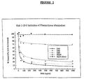
- Figure 2 shows monoclonal antibody 3-29-9 inhibition of phenanthrene metabolism.
- P450 and monoclonal antibody 3-29-9 were preincubated in KPi buffer for 5 minutes. Phenanthrene, NADPH and KPi were added and incubated for 25 minutes, and the reaction was terminated with DCM.
- B[a]P 9,10-diol was added as an internal standard. Extracts were dried down and analyzed by reversed-phase HPLC. Percentage of control was calculated as described with respect to Figure 1.
- Figures 3A and 3B show monoclonal antibody 3-29-9 inhibition of diazepam conversion to temazepam (TMZ) and N-desmethyldiazepam (NDZ).
- TMZ diazepam
- NDZ N-desmethyldiazepam
- P450 and monoclonal antibodies were preincubated for 5 min and diazepam and NADPH were added. Incubations were carried out for 20 min and terminated with DCM; 2-oxoquanzepam (OQZ) was added as an internal standard. Extracts were dried and dissolved in 50% methanol for HPLC analysis. Percentage of control was calculated as described in the description of Figure 1.
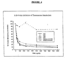
- Figure 4 shows inhibition by Mab 3-29-9 of testosterone conversion to the 6 ⁇ -OH metabolite.
- P450s and monoclonal antibodies were preincubated for 20 minutes.
- Testosterone, NADPH and KPi were added and incubated for 20 minutes.
- Metabolites were extracted with DCM and analyzed by HPLC. Percentage of control was calculated as described with respect to Figure 1.
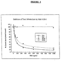
- Figure 5 shows inhibition of taxol metabolism by monoclonal antibody 3-29-9.
- P450 was preincubated with monoclonal antibody 3-29-9 for 5 min at 37°C.
- Taxol, NADPH and KPi were added and incubated for 30 minutes. Incubation was terminated with DCM, and B[ a ]P 9,10-diol, an internal standard, was added.
- the extracts were analyzed by HPLC. Percentage of control was calculated as described with respect to Figure 1.
- Figure 6 shows monoclonal antibody 3-29-9 inhibition of cyclosporin metabolism.
- P450 was preincubated in KPi with Mab 3-29-9 for 5 min at 37°C. Cyclosporin and NADPH were added, and incubations were carried out for 20 minutes. Extracts were analyzed by HPLC. Percentage of control was calculated as described above.
- the present inventors have determined that, by using recombinantly produced human cytochrome P450 3A4, a monoclonal antibody can be produced that specifically inhibits the enzyme activity of human cytochrome P450 3A3, 3A4, and 3A5.
- Monoclonal antibodies which specifically inhibit these human enzyme activities have not been isolated previously; nor has there been any indication that such specific inhibition was attainable.
- a monoclonal antibody which specifically binds to human P450 3A3 and 3A4 but not to 3A5 can be produced.
- antigen binding polypeptides of the invention include (A)' a "half antibody” molecule, i.e ., a single heavy:light chain pair, and (B) an antibody fragment, such as the univalent fragments Fab and Fab', the divalent fragment F(ab') 2 , and a single or double chain Fv fragment.
- a fragment of the present invention possesses the characteristic specificity of one of the aforementioned monoclonal antibodies; that is, for binding human P450 3A3, 3A4 and 3A5 (antibody 3-29-9), for human 3A3 and 3A4 (antibody 275-1-2).
- an antigen binding polypeptide within the present invention is a non-peptide "mimetic," i.e ., a compound that mimics an epitope binding site of 3-29-9 or 275-1-2 or 1-73-18 or 2-106-12, but that is water-soluble, resistant to proteolysis, and non-immunogenic.
- Conformationally restricted, cyclic organic peptides which mimic any of these antibodies can be produced in accordance with known methods described, for example, by Saragovi, et al., Science 253: 792 (1991).
- fragments within the invention can be obtained from a monoclonal antibody, produced as described above, by methods that include digestion with enzymes such as pepsin or papain and/or cleavage of disulfide bonds by chemical reduction.
- monoclonal antibody fragments encompassed by the present invention can be synthesized using an automated peptide synthesizer such as those supplied commercially by Applied Biosystems, Multiple Peptide Systems and others, or they may be produced manually, using techniques well known in the art. See Geysen et al., J. Immunol. Methods 102: 259 (1978). Direct determination of the amino acid sequences of the variable regions of the heavy and light chains of the monoclonal antibodies according to the invention can be carried out using conventional techniques.
- a fragment according to the present invention can be an Fv fragment.
- An Fv fragment of an antibody is made up of the variable region of the heavy chain (Vh) of an antibody and the variable region of the light chain of an antibody (Vl). Proteolytic cleavage of an antibody can produce double chain Fv fragments in which the Vh and Vl regions remain non-covalently associated and retain antigen binding capacity.
- Double chain Fv fragments also can be produced by recombinant expression methods well known in the art. See Skerra et al., Science 240: 1038 (1988), and King et al., Biochemical J. 290: 723 (1991). Briefly, the amino acid sequence of the variable regions of the heavy and light chains of the monoclonal antibodies according to the invention can be obtained by direct amino acid sequencing using methods well known to those in the art. From this amino acid sequence, synthetic genes can be designed which code for these variable regions and they can both be inserted into an expression vector. Two polypeptides can be expressed simultaneously from a mammalian or bacterial host, resulting in formation of an active Fv fragment.
- An antigen binding polypeptide of the present invention also can be a single-chain molecule or so-called "single chain antigen binding polypeptide," a phrase used in this description to denote a linear polypeptide that binds antigen with specificity and that comprises variable or hypervariable regions from the heavy and light chain chains of an antibody.
- Single chain antigen binding polypeptides that retain an antigen-binding capacity that is characteristic of the present invention can be produced by conventional methodology.
- the Vh and Vl regions of the Fv fragment can be covalently joined and stabilized by the insertion of a disulfide bond. See Glockshuber, et al ., Biochemistry 1362 (1990).
- Vh and Vl regions can be joined by the insertion of a peptide linker.
- a gene encoding the Vh, Vl and peptide linker sequences can be constructed and expressed using a recombinant expression vector. See Colcher, et al ., J. Nat'l Cancer Inst. 82: 1191 (1990).
- Amino acid sequences comprising hypervariable regions from the Vh and Vl antibody chains can also be constructed using disulfide bonds or peptide linkers, as described herein.
- the present invention relates to monoclonal antibodies and fragments thereof that specifically inhibit the enzyme activity of human cytochrome P450 3A3, 3A4 and 3A5.
- the invention also relates to monoclonal antibodies that specifically bind to human P450 3A3 and 3A4.
- An example of the antibodies contemplated by the present invention is a monoclonal antibody, designated "3-29-9," that inhibits the activity of human cytochrome P450 3A3, 3A4 and 3A5.
- a further example of the antibodies contemplated by the present invention is the monoclonal antibody 275-1-2, which specifically binds to cytochrome P450 3A3 and 3A4.
- Monoclonal antibody 3-29-9 is specific for human cytochrome P450 3A3, 3A4, and 3A5 because it does not cross react with other recombinantly-expressed human P450s and does not cross react with various recombinantly-expressed rat and mouse cytochrome P450s.
- monoclonal antibody 275-1-2 is specific for P450 3A3 and 3A4 and does not cross react with other recombinantly-expressed human, rat or mouse P450s.
- the present inventors have discovered that monoclonal antibody 3-29-9 strongly inhibits the enzyme activity of human cytochrome P450 3A, 3A4 and 3A5.
- Antibody 3-29-9 inhibited the metabolism of phenanthrene by purified P450 3A3, 3A4 and 3A5 and human liver microsomes. Antibody 3-29-9 did not inhibit metabolism of phenanthrene by rat liver microsomes. Antibody 3-29-9 inhibited the metabolism of diazepam by purified 3A3, 3A4 and 3A5 and also inhibited diazepam metabolism by human liver microsomes. Antibody 3-29-9 did not inhibit metabolism of diazepam by rat liver microsomes. Taxol metabolism by purified P450 3A3 and 3A4 and by human liver microsomes was inhibited by antibody 3-29-9. Metabolism of taxol by rat liver microsomes was not affected by antibody 3-29-9.
- the metabolism of cyclosporin by purified 3A4 was inhibited by antibody 3-29-9.
- the metabolism of testosterone by purified P450 3A3, 3A4, 3A5, and by human liver microsomes was inhibited by antibody 3-29-9.
- the metabolism of p-nitroanisole by 3A3, 4 and 5 was inhibited by antibody 3-29-9, but the inhibition was moderate. But p-nitroanisole is considered a poor substrate for P450 3A3 and 3A4.
- the antigen binding polypeptides according to this invention include any polypeptide, natural or synthetic, that has the binding specificity of monoclonal antibody 3-29-9, 1-73-18, 275-1-1 or 2-106-12.
- One antigen binding polypeptide according to the invention binds to an epitope on human P450 3A3, 3A4 and 3A5 and specifically inhibits the enzyme activity of human P450 3A3, 3A4, and 3A5.
- Another antigen binding polypeptide according to the invention specifically binds to an epitope on human P450 3A3 and 3A4.
- an antigen binding polypeptide of the present invention binds to P450 3A3, 3A4 and 3A5 in such a way as to inhibit the enzyme activity of P450 3A3, 3A4, and 3A5.
- other antigen binding polypeptides of the invention bind to human P450 3A3 and 3A4, but do not inhibit P450 enzyme activity.
- Monoclonal antibodies can be produced in various ways using techniques well understood by those having ordinary skill in the art. Details of these techniques are described in ANTIBODIES: A LABORATORY MANUAL 726, Cold Spring Harbor Publications (1988). The production of quantities of recombinant human P450 3A4 sufficient for immunization can be carried out using a recombinant expression system, such as the baculovirus system used to express human P450 3A4, which is described in Buters et al ., Drug Metab. Dispos. 22: 688 (1994). This publication also describes purification procedures for the recombinantly-produced human P450 3A4.
- monoclonal antibodies according to the present invention are prepared by immunizing BALB/c mice intraperitoneally with purified recombinantly expressed P450 3A4 with a series of booster immunizations. Several days after the final immunization, a mouse is sacrificed, its spleen removed and the spleen cells are fused with myeloma cells. Hybrids are screened using a solid phase enzyme-linked immunosorbent assay and P450 3A4 as antigen. Antibody-positive wells are re-screened and those hybridomas of interest are cloned at least three times.
- the present invention relates to a method of determining the contribution of human P450 3A3, 3A4 and 3A5 to the metabolism of compounds.
- “Compound” is used here to denote any xenobiotic, such as a drug, carcinogen, pesticide or other industrial or environmental chemical, or any endobiotic, such as a steroid hormone.
- the term “metabolism” is used to indicate the processing a compound into a metabolite or metabolites by the action of an enzyme or enzymes, where the chemical structure of the metabolite(s) is different from the compound prior to its metabolism.
- human P450 3A3 and 3A4 are responsible for the metabolism of a wide variety of xenobiotics and endobiotics, such as drugs, carcinogens and steroid hormones. Metabolism of compounds by P450 3A3, 3A4 and 3A5 can be monitored and quantified using standard techniques to measure chemical metabolites. See, for example, Buters et al., Drug Metab. Dispos. 22: 688 (1994).
- the antigen binding polypeptides of the invention can be used to determine the contribution of human cytochrome P450 3A3, 3A4 and 3A5 to the metabolism of a compound by quantifying the inhibition of P450-catalyzed chemical metabolism by an antigen binding polypeptide of the invention which inhibits the activity of human P450 3A3, 3A4 and 3A5.
- This method can be used with purified P450 enzymes to determine their substrate specificity and contribution to the metabolism of specific compounds.
- the inhibition of compound metabolism by cell, tissue or microsomal preparations by the antigen binding polypeptides of the invention can be quantified.
- the samples to be analyzed -- isolated P450s, cells, tissue or microsomes -- are contacted with increasing amounts of an antigen binding polypeptide of the invention and the compound to be tested is added and its metabolism quantified. Another sample is not treated with the antigen binding polypeptide as a control.
- the inhibition of compound metabolism by an antigen binding polypeptide of the invention is directly proportional to the contribution of the P450 3A3, 3A4 and 3A5 to the metabolism of the compound tested.
- the methods of determining the contribution of P450 to the metabolism of compounds of the present invention have a number of applications. These methods can be used to identify potential drug/drug interaction problems. If two different drugs are metabolized by the same P450 enzyme, the two drugs may compete with each other, leading to possible toxicity and/or reduced drug effectiveness. These quantification methods of the present invention can also be used to design drugs that utilize particular P450 enzymes so as to minimize drug/drug interactions and to maximize drug effectiveness.
- an antigen binding polypeptide with the binding specificity of monoclonal antibody 275-1-2 can be used to screen for the presence and amount of P450 3A3 and 3A4 in an individual.
- Any and all tissues and organs believed relevant to P450 metabolism can be examined, e.g., tissues of known carcinogen susceptibility.
- Human cDNA for P450 3A4 was constructed into a baculovirus vector. SF9 insect cells were infected with the virus which expressed the P-450 3A4. See Buters et al., Drug Metab. Dispos. 22: 688 (1994). Purification of the P450 3A4 was accomplished by a series of extraction and column chromatography procedures which are fully described in Buters, supra.
- cDNAs coding for different isozymes were constructed into vaccinia virus vectors. See Aoyama et al., Proc. Nat'l Acad. Sci. USA 87: 4790 (1990), and Battula et al., Proc Nat'l Acad. Sci. USA 84: 4073 (1987).
- the enzymes were expressed by infecting tk- or Hep G2 cells for 24-48 hours.
- the cell lysates were used as the source of P450 and the P450 content determined by spectral analysis. Cell lysates from infected tk- cells were used in ELISA and Western blotting, and cell lysates from Hep G2 cells were used for metabolic studies.
- mice Two female Balb/c mice were immunized by intraperitoneal injection weekly for three weeks with 10 ⁇ g of purified baculovirus expressed human 3A4 protein emulsified in 0.2 ml complete Freund's adjuvant for the first week, then with incomplete Freund's adjuvant thereafter. Three days after the third injection, one mouse was sacrificed and the spleen removed. The fusion of myeloma cells with primed, dissociated spleen cells was carried out essentially as described in Park et al., Biochem. Pharmacol . 35: 2859 (1986). Cells were diluted with HAT medium and dispensed into tissue culture plates.
- ELISA analysis was made of 24 hybridoma clones that were selected from more than 700 clones and examined for their binding to baculovirus expressed P450 3A4. Due to the low yield of highly purified expressed protein, partially purified baculovirus expressed 3A4 was used for purposes of screening hybrids, and lysate from cells infected with wild type baculovirus was also used which went through the same partial purification scheme to check for specificity of the antibodies. As the hybrids began to grow, the spent media from each individual well with hybrid growth was screened for the presence of antibody to 3A4 as described above. Each individual well on the 96 well plates which had cell growth was assigned a number. Each positive well was re-screened and then cloned using complete media with 10% HCF.
- Hybrids of interest were cloned at least three times. Cloned hybrids producing monoclonal antibody were grown in flasks containing serum-free media (UltraDOMA-Biowhittaker) at a concentration of 5x10 5 /ml for 3-5 days. Cells were removed by centrifugation and the resulting supernatant was concentrated with a Filtron Macrosep concentrator (mol. wt. cutoff 30,000). Concentrated monoclonal antibodies were dialyzed in PBS (pH 7.2). Ascites fluid was prepared as previously described in Park et al . Biochem. Pharmacol. 2859 (1986).
- Antibody binding was detected using alkaline phosphate conjugated goat anti-mouse antibodies. ELISA results are expressed as optical density (O.D.) values.
- the monoclonal antibody 275-1-2 shows strong Western Blot activity towards baculovirus-expressed human 3A4 and vaccinia expressed human 3A3 and 3A4, but did not recognize human 3A5. Thus monoclonal antibody 275-1-2 detects a common epitope in human 3A3 and 3A4 which is not present in 3A5 and thus monoclonal antibody 275-1-2 can distinguish between the closely related human 3A3, 3A4 and 3A5.
- the monoclonal antibody 275-1-2 also detected P450 3A3 and 3A4 in three different samples of human liver with one of the livers giving a second weaker band.
- the monoclonal antibody 275-1-2 detected two bands in the liver of dexamethasone treated rats. These are likely P450 3A1 and 3A2 of rat.
- the inhibitory monoclonal antibody 3-29-9 did not yield a Western Blot with any of the above samples. Vaccinia or baculovirus wild type proteins and human P450 2E1 were not recognized in Western Blotting with monoclonal antibody 275-1-2 or monoclonal antibody 3-29-9.
- Table 3 shows the cross reactivity of monoclonal antibody 3-29-9 and monoclonal antibody 275-1-2 with a variety of vaccinia expressed and baculovirus-expressed human P450s as measured by the ELISA assay.
- Monoclonal antibody 3-29-9 also showed cross reactivity with vaccinia virus expressed human 3A4, 3A3, and to a lesser extent human 3A5.
- Monoclonal antibody 275-1-2 showed cross reactivity with human 3A3, 3A4 and only negligibly with human 3A5, which gave slightly greater cross reactivity than the wild type.
- Both monoclonal antibody 3-29-9 and monoclonal antibody 275-1-2 showed no cross reactivity with a variety of vaccinia expressed human P450s other than 3A3, 3A4, and 3A5.
- Table 4 shows that the monoclonal antibody 3-29-9 and monoclonal antibody 275-1-2 do not cross react with vaccinia expressed rat 3A1, rat 2A1, rat 4A1, rat 4A3, rat 2B1, mouse 1A1, and mouse 1A2.
- the ELISA O.D. values are the average of two experiments. There is slight cross reactivity of monoclonal antibody 275-1-2 with dexamethasone-induced rat liver microsomes.
- the monoclonal antibody 3-29-9 in a concentration ranging from 10 to 1000 ⁇ g, was pre-incubated with 10 - 50 pmol of P450 3A4, 3A3, 3A5, or human microsomes or dexamethasone-induced rat liver microsomes in 100 ⁇ l of 50 mM potassium phosphate buffer (pH 7.4) at 37°C for five minutes. The mixture was then diluted with buffer to 1 ml final volume.
- Test compounds dissolved in 10 ⁇ l of methanol 250 ⁇ M of testosterone, 80 ⁇ M of diazepam, 100 ⁇ M of taxol, 10 ⁇ M of cyclosporin, 100 ⁇ M of phenanthrene or 150 ⁇ M of nitroanisole, as final concentration
- NADPH 1 mM
- Anti-lysozyme monoclonal antibody HyHel or monoclonal antibody 1-68-11 against rat 2C11 in amount equivalent to that of the 3A4 monoclonal antibody were used as controls for nonspecific binding.
- Extracts of products were dissolved in methanol and analyzed immediately by reversed phase HPLC. Metabolites formed were identified by comparing their retention times with authentic standards. Metabolically formed nitrophenol (D 0 ) from nitroanisole and internal standard (D 4 -nitrophenol) were further derivatized and analyzed by GC-MS as follows: The sample was concentrated to 100 ⁇ l and derivatized with t-butyldimethysilytrifluoroacetamide containing 1% t-butyldimethysislyl chloride.
- the derivatized phenols were quantified by GC-MS using an Alltech AT-1 column (30 m x .25 mm; 1.0 ⁇ m film thickness) and a Hewlett Packard 5971 Mass Selective Detector. Samples were injected at 50°C, ramped at 30°C/min to 150°C followed by 10°C/min to 280°C. The derivatized nitrophenols eluted at 16.3 min and were quantitated by integrating the peak areas at masses 253 and 257. For achieving higher recoveries of cyclosporin and its metabolites, a modified procedure was utilized.
- the HPLC was performed on a Hewlett-Packard Mode HP1050 liquid chromatograph equipped with an HP Model 1050 autosampler, a ternary solvent delivery system and a multiple-wavelength detector, all controlled by Hewlett Packard HPLC 2D ChemStation software installed a Compaq Prolinea 4/66 personal computer.
- Diazepam (DZ) and its metabolites were separated on a Zorbax SB-C18 column (4.6 mm x 15 cm, MAC-MOD Analytical Inc., Chadds Ford, PA) and eluted isocratically with acetonitrile/methanol/water (10:40:50, v/v/v) at a flow rate of 1 ml/min and a detection of 232 nm.
- Cyclosporin and metabolites were injected onto an UltrasphereTM-octyl column (5 ⁇ m, 25 cm x 4.6 mm, ALTEX).
- the metabolites of taxol, testosterone, or phenanthrene were separated and identified as previously described. See Harris et al., Cancer Res. 54: 4026 (1994); Hanioka et al. (1990), supra; and Shou et al., Cancer Lett. 83: 305 (1994).
- Phenanthrene is known to be metabolized by P450 3A3, 3A4 and 3A5 and by microsomes from dexamethasone-treated rats and by human liver microsomes.
- monoclonal antibody 3-29-9 inhibited phenanthrene metabolism catalyzed by P450 3A4 by 97%.
- P450 3A3 catalyzed metabolism was inhibited by 86% and P450 3A5 catalyzed metabolism was inhibited by about 78%.
- Human liver microsomes were inhibited by 30%, which indicates that human P450 3A3, 3A4 and 3A5 contribute not less than 30% to the metabolism of phenanthrene in human liver.
- rat liver microsomes from dexamethasone treated rats no inhibition by monoclonal antibody 3-29-9 was observed. This is consistent with the fact that monoclonal antibody 3-29-9 does not cross react with dexamethasone induced P450 3A1/2 in rat liver.
- the determination of 30% inhibition in human liver by monoclonal antibody 3-29-9 demonstrates the value of a monoclonal antibody for determining the quantitative role of an individual P450 in a tissue containing a variety of P450s.
- Diazepam is one of the most common clinically used drugs in the world. Diazepam is converted by P450 3A3 and 3A4 to the metabolite TMZ by hydroxylation of the ring and to the metabolite NDZ by demethylation. As shown in Figure 3A and B, monoclonal antibody 3-29-9 is a strong inhibitor of both human P450 3A3 and 3A4 catalyzed diazepam metabolism.
- TMZ and NDZ The formation of the two major diazepam metabolites TMZ and NDZ by P450 3A4 was inhibited by monoclonal antibody 3-29-9 by 92% and 87%, respectively, and metabolite formation by P450 3A3 was inhibited by approximately 80% for TMZ and 48% for NDZ. Diazepam metabolism catalyzed by 3A5 was inhibited by monoclonal antibody 3-29-9 by 73% for TMZ and by 52% for NDZ.
- the difference in inhibition of TMZ and NDZ formation in human liver by monoclonal antibody 3-29-9 indicates that there are at least two types of P450 enzymes in liver responsible for diazepam metabolism.
- One type is P450 3A3, 3A4 and 3A5 and the other has not been identified.
- Human Cytochrome P450 3A4 metabolizes a large diverse number of steroids, a prototype of which is testosterone.
- monoclonal antibody 3-29-9 was a potent inhibitor of human P450 3A3, 3A4, and 3A5 catalyzed conversion of testosterone to 6 ⁇ -hydroxy testosterone. Similar inhibition was observed for the formation of 15 ⁇ -OH testosterone (data not shown).
- the addition of monoclonal antibody 3-29-9 to human liver microsomes inhibited 6 ⁇ OH formation more than 90%, indicating that P450 3A3, 3A4 and 3A5 are essentially the sole P450s responsible for testosterone metabolism by human liver.
- Specific activities for 3A3, 3A4, 3A5, HLM and MLM in the formation of 6 ⁇ -OH testosterone, without monoclonal antibody, 3-29-9 were 25, 25, 10, 2.0 and 65, respectively.
- Taxol is known to be metabolized by the human P450 3A4 system and is a very important compound currently used in the therapy of certain kinds of cancer. Taxol phenol has been characterized as a major product of taxol metabolism by human P450 3A4. As shown in Figure 5, Taxol conversion to phenol by human P450 3A3 and 3A4 was inhibited by the monoclonal antibody 3-29-9 by about 90%. Human liver microsome metabolism of taxol was inhibited by monoclonal antibody 3-29-9 by 84% which indicates clearly the very dominant and important roles of P450 3A3 and 3A4 for taxol metabolism in human liver. Thus, 84% of taxol metabolism in human liver is due to P450 3A3 and 3A4. The liver microsomes obtained from pregnenolone-alpha-carbonitrile-treated rats showed total resistance to inhibition by monoclonal antibody 3-29-9 and retained 97% of their usual activity in the presence of the monoclonal antibody.
- Cyclosporin is a very important drug for suppressing the immune response and is crucial for organ transplants. It has a relatively high molecular weight. As shown in Figure 6, the HPLC analysis of cyclosporin metabolism shows that the formation of all four metabolites of cyclosporin by P450 3A4 was inhibited to approximately the same extent by monoclonal antibody 3-29-9. This inhibition ranged from 70%-85% for the formation of each of the four metabolites. Thus, the metabolism of a very large molecule such as cyclosporin is catalyzed by P450 3A4 and is inhibited by monoclonal antibody 3-29-9.
Landscapes
- Chemical & Material Sciences (AREA)
- Health & Medical Sciences (AREA)
- Life Sciences & Earth Sciences (AREA)
- Organic Chemistry (AREA)
- Genetics & Genomics (AREA)
- General Health & Medical Sciences (AREA)
- Biochemistry (AREA)
- Medicinal Chemistry (AREA)
- Molecular Biology (AREA)
- Engineering & Computer Science (AREA)
- Bioinformatics & Cheminformatics (AREA)
- Wood Science & Technology (AREA)
- Zoology (AREA)
- General Engineering & Computer Science (AREA)
- Microbiology (AREA)
- Biotechnology (AREA)
- Biomedical Technology (AREA)
- Immunology (AREA)
- Biophysics (AREA)
- Proteomics, Peptides & Aminoacids (AREA)
- Peptides Or Proteins (AREA)
- Preparation Of Compounds By Using Micro-Organisms (AREA)
- Investigating Or Analysing Biological Materials (AREA)
Abstract
Description
- The cytochrome P450 enzymes (P450s) play an important role in metabolizing xenobiotics and certain endobiotics. The P450s exist in multiple isozymic forms which direct the metabolic flow of individual substrates. Gelboin, Pharmacol. Rev. 45: 413 (1993). Compounds which are metabolized by the cytochrome P450 enzymes include various drugs, carcinogens, mutagens, pesticides, steroids, fatty acids, bile acids and prostaglandins. Gonzalez, loc. cit. 40: 243 (1989). Cytochrome P450 metabolism of xenobiotics can result in detoxification of toxic compounds by their conjugation into excretable forms or can result in activation of compounds into metabolites that are toxic, mutagenic, or carcinogenic. Many steroids are deactivated by cytochrome P450-catalyzed oxidation.
- The human cytochrome P450 3A family consists of several isozymes. Cytochome P450 3A4 is the most abundant of the P450 3A enzymes in the human liver, but its expression levels in the liver and other tissues vary. Enzymatically active human P450 3A4 has been produced from cloned cDNA expressed in a baculovirus expression system. Buters et al., Drug Metab. Dispos. 22: 688 (1994). The nucleotide sequence of cytochrome P450 3A3 is 98% identical to the P450 3A4 nucleotide sequence. Guengerich et al., "The Importance of Cytochrome P450 3A Enzymes in Drug Metabolism," in ASSESSMENT OF THE USE OF SINGLE CYTOCHROME P450 ENZYMES IN DRUG RESEARCH 161-186 (Springer Verlag 1994). P450 3A3 has been cloned and expressed, and its enzyme activity is nearly identical to that of 3A4. Schuetz, et al., Hepatology, 18:1254-62, 1993; Molowa, et al., Proc. Natl. Acad. Sci. U.S.A. 83:5311-5315, 1986. Some researchers believe that 3A3 is in fact 3A4 and the minor sequence variations between 3A3 and 3A4 are due to sequencing errors. 3A3 and 3A4 will be referred to separately throughout the specification, with the recognition that further research may demonstrate these two enzymes to actually be the same. Approximately one fourth of individuals have the P450 3A5 enzyme, and when expressed in the liver, its level is lower than that of the P450 3A4 enzyme. Id. P450 3A5 has not been characterized as well as the 3A4 enzyme. It has been shown that P450 3A5 and 3A4 have similar, but not identical, substrate specificities. Id.
- Cytochrome P450 3A4 and 3A3 are very important members of the P450 class of enzymes. The human P450, 3A4 and 3A3 assume an especially important place among the human P450s because they metabolize a large variety of drugs, steroids and carcinogens. Id. Cytochromes P450 3A3 and 3A4 are considered the most important P450s for a wide range of high molecular weight substrates which include many of the known clinically useful drugs, such as tranquilizers, anti-depressants, immunosuppressants and anti-cancer drugs. A partial list of the P450 3A substrates appears in Table 1 below.
P450 3A SUBSTRATES epipodophyllotoxins sulfoxide tamoxifen cyclosporin teniposide (S)-warfarin erythromycin etoposide α-naphthoflavone benzphetamine midazolam phenanthrene triacetyloleandomycin taxol benzo[a]pyrene nifedipine FL-506 sulfentamil cocaine vinblastine codeine cortisol afentanil imipramine testosterone vindesine dapsone aflatoxin B1 amiodarone aminochrysene lidocaine mephenytoin 1-nitropyrene amphetamine digitoxin alfentanil benzo[a]pyrene 7, 8-diol - Cytochrome P450 2E1 plays a major role in the metabolism of a variety of low molecular weight compounds including environmental chemicals, and carcinogens such as short-chain dialkylnitrosamines, benzene, styrene, halomethanes, vinyl halides, ethyl carbamate, and small vinyl monomers. Guengerich et al., Chem. Res. Toxicol. 4: 168 (1991). The human 2E1 also metabolizes clinically useful drugs such as the anesthetic chlorzoxazone and the analgesic acetaminophen as well as caffeine. The rat 2E1 nucleotide sequence is 75% homologous to the human 2E1 and the amino acid sequences of rat and human are 78% homologous. See Song, et al. J. Biol. Chem., 261:16689-16697, 1986. A partial list of the P450 2E1 substrates appears in Table 2.
SUBSTRATES FOR 2E1 Chlorzoxazone vinyl bromide Acetaminophen vinyl carbamate Acrylonitrile ethyl Carbamate p-Nitrophenol CC14 N-Nitrosodimethylamine (NDMA) CHC13 N-Nitrodiethylamine (NDEA) 1,2-Dichloropropane Enflurane CH3CC13 N-Nitrosonornicotine (NNN) Tricholoroethylene 1,1-Dimethylformamide Benzene Stryrene Eicosatetraenoic acid Caffeine N,N-Dimethylformamide 1,3-Butadiene Ethylene dibromide Furan Ethylene dichloride p-Nitroanisole vinyl chloride Sevoflurane, Isoflurane & Methoxyflurane 1,1,2,2-Tetrafluoro-1-(2,2,2-trifluoroethoxy)-ethane (HFE) - Many current methods for assessing the contribution of specific P450 enzymes to the metabolism of various chemicals are not effective. Chemical enzyme inhibitors are non-specific. Comparison of metabolism rates in different tissue preparations is not useful because this method does not distinguish between P450 isozymes. Reconstitution of catalytic activities using purified enzyme preparations does not provide information about enzyme activity in vivo. Inhibition of enzyme activity with antibodies to cytochrome P450s has also been reported, see Guengerich et al. (1991), supra, but the production of monoclonal antibodies to human P450s has been greatly hindered by the inability to obtain human P450s in amounts sufficient for immunization. Barnes et al. reported monoclonal antibodies made against human liver microsomes or semi-purified human P450s. These antibodies recognized a 53kDa band on a Western blot, but there was no characterization of whether these antibodies recognize 3A3, 3A4, 3A5 or other human P450s. In addition, there was no investigation of inhibition of cytochrome P450 enzyme activity by these antibodies. Barnes et al., Proc. Nat'l Acad. Sci. USA 84: 4073 (1987). Beaune et al. obtained monoclonal antibodies against a mixture of purified human P450s, but the precise identity of the P450 isozymes with which these antibodies cross react is unclear. This report also did not discuss antibody inhibition of enzyme activity. Beaune et al., Biochem. Pharmacol. 34: 3547 (1985). Park et al., "Preparation and characterization of monoclonal antibodies to pregnenolone 16-α-carbonitrile inducible rat liver cytochrome P450," Biochem. Pharmacol. 35: 2859 (1986), reported monoclonal antibodies against rat P450 3A1/2. The structure and function of the rat 3A1/2 enzymes are similar, but not identical to the human P450 3A3 or 3A4 enzymes. Park et al. did not investigate antibody-mediated inhibition of rat 3A1/2 enzyme activity.
- Guengerich et al., Chem. Res. Toxicol. 4: 168 (1991), reported on a polyclonal antibody against human P450 2E1 which inhibited the metabolism of 2E1 substrates in human liver microsomes. A monoclonal antibody against rat cytochrome P450 2E1 was prepared by Ko et al., Cancer Res. 47: 3101 (1987), and was found to inhibit P450 2E1-mediated metabolism of certain chemicals in rat microsomes. Ko et al. also isolated a monoclonal antibody against rat P450 2E1 which recognized P450 2E1 on a Western blot but did not inhibit enzyme activity.
- Cytochrome P450s are a paradigm for multi-isozymic systems whose activity may result in metabolic products with opposing physiological and pathological consequences. The large multiplicity of P450 forms, their differing structure and function, their often poorly defined substrate and product specificity, and their heterogeneous distribution in vivo make difficult the determination of the quantitative contribution of each P450 to the metabolism of specific substrates.
- A need therefore exists for reagents that can identify and inhibit certain human cytochrome P450 enzymes specifically. A thorough understanding of the different P450 isozymes, their specificity, regulation, and distribution is crucial to designing more effective drugs, evaluating the modes of action of drugs, carcinogens and environmental chemicals, and developing inhibitors of carcinogens and other toxic chemicals. Individuals sensitive to particular chemicals could be characterized based on their cytochrome P450 makeup, permitting administration of the most effective drugs and toxicity inhibitors. Monoclonal antibodies which inhibit cytochrome P450 3A3, 3A4, and 3A5 and P450 2E1 would be powerful analytical tools because the cytochrome P450 3A enzymes play an important role in metabolizing a wide variety of xenobiotics and endobiotics, including many large molecular weight compounds, and P450 2E1 plays an important role in metabolizing many small molecular weight chemicals and carcinogens. In order to produce specific monoclonal antibodies, there must be a large enough supply of purified cytochrome P450s to immunize for the production of antibodies.
- It therefore is an object of the present invention to provide reagents which can specifically identify and inhibit human cytochrome P450 3A3, 3A4 and 3A5.
- It is also an object of the invention to provide monoclonal antibodies and other antigen binding polypeptides which can specifically identify and inhibit human P450 3A3, 3A4, and 3A5.
- It is a further object of the invention to provide methods utilizing antigen binding polypeptides that can determine the contribution of human P450 3A3, 3A4, 3A5 to the metabolism of xenobiotics and endobiotics in samples comprising one or more human cytochrome P450 enzymes and in samples of various cells, microsomes, and tissues.
- Thus, the invention relates to antigen binding polypeptides that specifically bind to human cytochrome P450 3A3, 3A4 and 3A5, and that specifically inhibit the enzyme activity of human cytochrome P450 3A3, 3A4 and 3A5. The invention also relates to antigen binding polypeptides that specifically bind to human cytochrome P450 3A3 and 3A4. These antigen binding polypeptides of the invention include monoclonal antibodies, antibody fragments and single chain antigen binding polypeptides with the P450-recognition properties described above.
- The invention further relates to a method for determining the contribution of human cytochrome P450 3A3, 3A4, or 3A5 to the metabolism of a compound, comprising the steps of:
- (a) contacting one or more samples comprising human
cytochrome P450 3A3, 3A4, 3A5, or any combination thereof, with
- (i) varying amounts of an antigen binding polypeptide of the invention that specifically binds to human cytochrome P450 3A3, 3A4 and 3A5, and that specifically inhibits the enzyme activity of human cytochrome P450 3A3, 3A4 and 3A5; and
- (ii) said compound; and then
- (b) measuring the metabolism of said compound by the one or more samples of step (a), whereby the contribution of cytochrome P450 3A3, 3A4, or 3A5 to the metabolism of said compound is proportional to the inhibition of said metabolism by said antigen binding polypeptide.
-
- The invention further relates to a method for determining the contribution of cytochrome P450 3A3, 3A4, and 3A5 to the metabolism of a compound by mammalian cells, microsomes, or tissue, comprising the steps of:
- (a) contacting one or more samples comprising mammalian
cells, microsomes, or tissue with
- (i) varying amounts of an antigen binding polypeptide of the invention that specifically binds to human cytochrome P450 3A3, 3A4 and 3A5, and that specifically inhibits the enzyme activity of human cytochrome P450 3A3, 3A4 and 3A5; and
- (ii) said compound; and then
- (b) measuring the metabolism of said compound by the one or more samples of step (a), whereby the contribution of cytochrome P450 3A3, 3A4, or 3A5 to the metabolism of said compound by mammalian cells, microsomes, or tissue is proportional to the inhibition of said metabolism by said antigen binding polypeptide.
-
- Figure 1 shows the inhibitory effect of monoclonal antibody 3-29-9 on p-nitroanisole metabolism. Monoclonal antibody 3-29-9 and P450 were pre-incubated at 37°C for 5 minutes and diluted with KPi buffer. Incubations were carried out for 20 min and terminated with dichloromethane (DCM). An internal standard, 4-nitrophenol was added. Nitrophenol (DO) and 4-nitrophenol were derivatized for gas chromatography-mass spectroscopy. Percentage of control in the formation of nitrophenol by 3A3, 3A4, 3A5 human livery microsomes (HLM), and dexamethasane-induced rat liver microsomes (RLM) is expressed as [(metabolite formed/internal standard) with 3-29-9]/[(metabolite formed/internal standard) without 3-29-9].
- Figure 2 shows monoclonal antibody 3-29-9 inhibition of phenanthrene metabolism. P450 and monoclonal antibody 3-29-9 were preincubated in KPi buffer for 5 minutes. Phenanthrene, NADPH and KPi were added and incubated for 25 minutes, and the reaction was terminated with DCM. B[a]
P 9,10-diol was added as an internal standard. Extracts were dried down and analyzed by reversed-phase HPLC. Percentage of control was calculated as described with respect to Figure 1. - Figures 3A and 3B show monoclonal antibody 3-29-9 inhibition of diazepam conversion to temazepam (TMZ) and N-desmethyldiazepam (NDZ). P450 and monoclonal antibodies were preincubated for 5 min and diazepam and NADPH were added. Incubations were carried out for 20 min and terminated with DCM; 2-oxoquanzepam (OQZ) was added as an internal standard. Extracts were dried and dissolved in 50% methanol for HPLC analysis. Percentage of control was calculated as described in the description of Figure 1.
- Figure 4 shows inhibition by Mab 3-29-9 of testosterone conversion to the 6β-OH metabolite. P450s and monoclonal antibodies were preincubated for 20 minutes. Testosterone, NADPH and KPi were added and incubated for 20 minutes. Metabolites were extracted with DCM and analyzed by HPLC. Percentage of control was calculated as described with respect to Figure 1.
- Figure 5 shows inhibition of taxol metabolism by monoclonal antibody 3-29-9. P450 was preincubated with monoclonal antibody 3-29-9 for 5 min at 37°C. Taxol, NADPH and KPi were added and incubated for 30 minutes. Incubation was terminated with DCM, and B[a]
P 9,10-diol, an internal standard, was added. The extracts were analyzed by HPLC. Percentage of control was calculated as described with respect to Figure 1. - Figure 6 shows monoclonal antibody 3-29-9 inhibition of cyclosporin metabolism. P450 was preincubated in KPi with Mab 3-29-9 for 5 min at 37°C. Cyclosporin and NADPH were added, and incubations were carried out for 20 minutes. Extracts were analyzed by HPLC. Percentage of control was calculated as described above.
- The present inventors have determined that, by using recombinantly produced human cytochrome P450 3A4, a monoclonal antibody can be produced that specifically inhibits the enzyme activity of human cytochrome P450 3A3, 3A4, and 3A5.
- Monoclonal antibodies which specifically inhibit these human enzyme activities have not been isolated previously; nor has there been any indication that such specific inhibition was attainable. By the same token, it also has been discovered that a monoclonal antibody which specifically binds to human P450 3A3 and 3A4 but not to 3A5 can be produced.
- Again, these binding specificities were not presaged by any structure/function relationship or other information available heretofore.
- Based on knowledge concerning these monoclonal antibodies, it also is possible to prepare a range of antigen binding polypeptides that possess the characteristic, highly desirable P450-recognition properties described above. Examples of antigen binding polypeptides of the invention include (A)' a "half antibody" molecule, i.e., a single heavy:light chain pair, and (B) an antibody fragment, such as the univalent fragments Fab and Fab', the divalent fragment F(ab')2, and a single or double chain Fv fragment. A fragment of the present invention possesses the characteristic specificity of one of the aforementioned monoclonal antibodies; that is, for binding human P450 3A3, 3A4 and 3A5 (antibody 3-29-9), for human 3A3 and 3A4 (antibody 275-1-2).
- Also illustrative of an antigen binding polypeptide within the present invention is a non-peptide "mimetic," i.e., a compound that mimics an epitope binding site of 3-29-9 or 275-1-2 or 1-73-18 or 2-106-12, but that is water-soluble, resistant to proteolysis, and non-immunogenic. Conformationally restricted, cyclic organic peptides which mimic any of these antibodies can be produced in accordance with known methods described, for example, by Saragovi, et al., Science 253: 792 (1991).
- In accordance with the present invention, fragments within the invention can be obtained from a monoclonal antibody, produced as described above, by methods that include digestion with enzymes such as pepsin or papain and/or cleavage of disulfide bonds by chemical reduction. Alternatively, monoclonal antibody fragments encompassed by the present invention can be synthesized using an automated peptide synthesizer such as those supplied commercially by Applied Biosystems, Multiple Peptide Systems and others, or they may be produced manually, using techniques well known in the art. See Geysen et al., J. Immunol. Methods 102: 259 (1978). Direct determination of the amino acid sequences of the variable regions of the heavy and light chains of the monoclonal antibodies according to the invention can be carried out using conventional techniques.
- As noted, a fragment according to the present invention can be an Fv fragment. An Fv fragment of an antibody is made up of the variable region of the heavy chain (Vh) of an antibody and the variable region of the light chain of an antibody (Vl). Proteolytic cleavage of an antibody can produce double chain Fv fragments in which the Vh and Vl regions remain non-covalently associated and retain antigen binding capacity.
- Double chain Fv fragments also can be produced by recombinant expression methods well known in the art. See Skerra et al., Science 240: 1038 (1988), and King et al., Biochemical J. 290: 723 (1991). Briefly, the amino acid sequence of the variable regions of the heavy and light chains of the monoclonal antibodies according to the invention can be obtained by direct amino acid sequencing using methods well known to those in the art. From this amino acid sequence, synthetic genes can be designed which code for these variable regions and they can both be inserted into an expression vector. Two polypeptides can be expressed simultaneously from a mammalian or bacterial host, resulting in formation of an active Fv fragment.
- An antigen binding polypeptide of the present invention also can be a single-chain molecule or so-called "single chain antigen binding polypeptide," a phrase used in this description to denote a linear polypeptide that binds antigen with specificity and that comprises variable or hypervariable regions from the heavy and light chain chains of an antibody. Single chain antigen binding polypeptides that retain an antigen-binding capacity that is characteristic of the present invention can be produced by conventional methodology. The Vh and Vl regions of the Fv fragment can be covalently joined and stabilized by the insertion of a disulfide bond. See Glockshuber, et al., Biochemistry 1362 (1990). Alternatively, the Vh and Vl regions can be joined by the insertion of a peptide linker. A gene encoding the Vh, Vl and peptide linker sequences can be constructed and expressed using a recombinant expression vector. See Colcher, et al., J. Nat'l Cancer Inst. 82: 1191 (1990). Amino acid sequences comprising hypervariable regions from the Vh and Vl antibody chains can also be constructed using disulfide bonds or peptide linkers, as described herein.
- In one aspect, therefore, the present invention relates to monoclonal antibodies and fragments thereof that specifically inhibit the enzyme activity of human cytochrome P450 3A3, 3A4 and 3A5. The invention also relates to monoclonal antibodies that specifically bind to human P450 3A3 and 3A4. An example of the antibodies contemplated by the present invention is a monoclonal antibody, designated "3-29-9," that inhibits the activity of human cytochrome P450 3A3, 3A4 and 3A5. A further example of the antibodies contemplated by the present invention is the monoclonal antibody 275-1-2, which specifically binds to cytochrome P450 3A3 and 3A4.
- Monoclonal antibody 3-29-9 is specific for human cytochrome P450 3A3, 3A4, and 3A5 because it does not cross react with other recombinantly-expressed human P450s and does not cross react with various recombinantly-expressed rat and mouse cytochrome P450s. Similarly, monoclonal antibody 275-1-2 is specific for P450 3A3 and 3A4 and does not cross react with other recombinantly-expressed human, rat or mouse P450s. The present inventors have discovered that monoclonal antibody 3-29-9 strongly inhibits the enzyme activity of human cytochrome P450 3A, 3A4 and 3A5.
- Antibody 3-29-9 inhibited the metabolism of phenanthrene by purified P450 3A3, 3A4 and 3A5 and human liver microsomes. Antibody 3-29-9 did not inhibit metabolism of phenanthrene by rat liver microsomes. Antibody 3-29-9 inhibited the metabolism of diazepam by purified 3A3, 3A4 and 3A5 and also inhibited diazepam metabolism by human liver microsomes. Antibody 3-29-9 did not inhibit metabolism of diazepam by rat liver microsomes. Taxol metabolism by purified P450 3A3 and 3A4 and by human liver microsomes was inhibited by antibody 3-29-9. Metabolism of taxol by rat liver microsomes was not affected by antibody 3-29-9. The metabolism of cyclosporin by purified 3A4 was inhibited by antibody 3-29-9. The metabolism of testosterone by purified P450 3A3, 3A4, 3A5, and by human liver microsomes was inhibited by antibody 3-29-9. The metabolism of p-nitroanisole by 3A3, 4 and 5 was inhibited by antibody 3-29-9, but the inhibition was moderate. But p-nitroanisole is considered a poor substrate for P450 3A3 and 3A4.
- The antigen binding polypeptides according to this invention include any polypeptide, natural or synthetic, that has the binding specificity of monoclonal antibody 3-29-9, 1-73-18, 275-1-1 or 2-106-12. One antigen binding polypeptide according to the invention binds to an epitope on human P450 3A3, 3A4 and 3A5 and specifically inhibits the enzyme activity of human P450 3A3, 3A4, and 3A5. Another antigen binding polypeptide according to the invention specifically binds to an epitope on human P450 3A3 and 3A4.
- Preferably, an antigen binding polypeptide of the present invention binds to P450 3A3, 3A4 and 3A5 in such a way as to inhibit the enzyme activity of P450 3A3, 3A4, and 3A5. Preferably other antigen binding polypeptides of the invention bind to human P450 3A3 and 3A4, but do not inhibit P450 enzyme activity.
- Monoclonal antibodies can be produced in various ways using techniques well understood by those having ordinary skill in the art. Details of these techniques are described in ANTIBODIES: A LABORATORY MANUAL 726, Cold Spring Harbor Publications (1988). The production of quantities of recombinant human P450 3A4 sufficient for immunization can be carried out using a recombinant expression system, such as the baculovirus system used to express human P450 3A4, which is described in Buters et al., Drug Metab. Dispos. 22: 688 (1994). This publication also describes purification procedures for the recombinantly-produced human P450 3A4.
- Generally, monoclonal antibodies according to the present invention are prepared by immunizing BALB/c mice intraperitoneally with purified recombinantly expressed P450 3A4 with a series of booster immunizations. Several days after the final immunization, a mouse is sacrificed, its spleen removed and the spleen cells are fused with myeloma cells. Hybrids are screened using a solid phase enzyme-linked immunosorbent assay and P450 3A4 as antigen. Antibody-positive wells are re-screened and those hybridomas of interest are cloned at least three times.
- In another preferred embodiment, the present invention relates to a method of determining the contribution of human P450 3A3, 3A4 and 3A5 to the metabolism of compounds. "Compound" is used here to denote any xenobiotic, such as a drug, carcinogen, pesticide or other industrial or environmental chemical, or any endobiotic, such as a steroid hormone. The term "metabolism" is used to indicate the processing a compound into a metabolite or metabolites by the action of an enzyme or enzymes, where the chemical structure of the metabolite(s) is different from the compound prior to its metabolism. As noted, human P450 3A3 and 3A4 are responsible for the metabolism of a wide variety of xenobiotics and endobiotics, such as drugs, carcinogens and steroid hormones. Metabolism of compounds by P450 3A3, 3A4 and 3A5 can be monitored and quantified using standard techniques to measure chemical metabolites. See, for example, Buters et al., Drug Metab. Dispos. 22: 688 (1994).
- In particular, the antigen binding polypeptides of the invention can be used to determine the contribution of human cytochrome P450 3A3, 3A4 and 3A5 to the metabolism of a compound by quantifying the inhibition of P450-catalyzed chemical metabolism by an antigen binding polypeptide of the invention which inhibits the activity of human P450 3A3, 3A4 and 3A5. This method can be used with purified P450 enzymes to determine their substrate specificity and contribution to the metabolism of specific compounds. To determine the contribution of P450 3A3, 3A4 and 3A5 to compound metabolism by different tissues, the inhibition of compound metabolism by cell, tissue or microsomal preparations by the antigen binding polypeptides of the invention can be quantified. Thus, the samples to be analyzed -- isolated P450s, cells, tissue or microsomes -- are contacted with increasing amounts of an antigen binding polypeptide of the invention and the compound to be tested is added and its metabolism quantified. Another sample is not treated with the antigen binding polypeptide as a control. The inhibition of compound metabolism by an antigen binding polypeptide of the invention is directly proportional to the contribution of the P450 3A3, 3A4 and 3A5 to the metabolism of the compound tested.
- The methods of determining the contribution of P450 to the metabolism of compounds of the present invention have a number of applications. These methods can be used to identify potential drug/drug interaction problems. If two different drugs are metabolized by the same P450 enzyme, the two drugs may compete with each other, leading to possible toxicity and/or reduced drug effectiveness. These quantification methods of the present invention can also be used to design drugs that utilize particular P450 enzymes so as to minimize drug/drug interactions and to maximize drug effectiveness.
- It is known that different people have different P450 profiles. This phenomenon is commonly known as polymorphism for a particular trait. These differences lead to varying susceptibility to xenobiotics, including drugs. Knowledge of an individual's P450 profile can be used to design drug treatment plans that will have maximum effectiveness. Screening individuals for the presence and quantity of certain cytochrome P450s can be accomplished by using techniques such as Western blotting, which are well known in the art. In particular, an antigen binding polypeptide of the invention with the binding specificity of monoclonal antibody 2-106-12 can be used to screen for the presence and amount of human P450 2E1 in an individual. In particular, an antigen binding polypeptide with the binding specificity of monoclonal antibody 275-1-2 can be used to screen for the presence and amount of P450 3A3 and 3A4 in an individual. Any and all tissues and organs believed relevant to P450 metabolism can be examined, e.g., tissues of known carcinogen susceptibility.
- Without further elaboration, it is believed that one skilled in the art can, using the preceding description, utilize the present invention to its fullest extent. The following examples are merely illustrative and not limitative of the remainder of the disclosure in any way whatsoever.
- Human cDNA for P450 3A4 was constructed into a baculovirus vector. SF9 insect cells were infected with the virus which expressed the P-450 3A4. See Buters et al., Drug Metab. Dispos. 22: 688 (1994). Purification of the P450 3A4 was accomplished by a series of extraction and column chromatography procedures which are fully described in Buters, supra.
- cDNAs coding for different isozymes were constructed into vaccinia virus vectors. See Aoyama et al., Proc. Nat'l Acad. Sci. USA 87: 4790 (1990), and Battula et al., Proc Nat'l Acad. Sci. USA 84: 4073 (1987). The enzymes were expressed by infecting tk- or Hep G2 cells for 24-48 hours. The cell lysates were used as the source of P450 and the P450 content determined by spectral analysis. Cell lysates from infected tk- cells were used in ELISA and Western blotting, and cell lysates from Hep G2 cells were used for metabolic studies.
- Two female Balb/c mice were immunized by intraperitoneal injection weekly for three weeks with 10µg of purified baculovirus expressed human 3A4 protein emulsified in 0.2 ml complete Freund's adjuvant for the first week, then with incomplete Freund's adjuvant thereafter. Three days after the third injection, one mouse was sacrificed and the spleen removed. The fusion of myeloma cells with primed, dissociated spleen cells was carried out essentially as described in Park et al., Biochem. Pharmacol. 35: 2859 (1986). Cells were diluted with HAT medium and dispensed into tissue culture plates.
- ELISA analysis was made of 24 hybridoma clones that were selected from more than 700 clones and examined for their binding to baculovirus expressed P450 3A4. Due to the low yield of highly purified expressed protein, partially purified baculovirus expressed 3A4 was used for purposes of screening hybrids, and lysate from cells infected with wild type baculovirus was also used which went through the same partial purification scheme to check for specificity of the antibodies. As the hybrids began to grow, the spent media from each individual well with hybrid growth was screened for the presence of antibody to 3A4 as described above. Each individual well on the 96 well plates which had cell growth was assigned a number. Each positive well was re-screened and then cloned using complete media with 10% HCF. Hybrids of interest were cloned at least three times. Cloned hybrids producing monoclonal antibody were grown in flasks containing serum-free media (UltraDOMA-Biowhittaker) at a concentration of 5x10⁵/ml for 3-5 days. Cells were removed by centrifugation and the resulting supernatant was concentrated with a Filtron Macrosep concentrator (mol. wt. cutoff 30,000). Concentrated monoclonal antibodies were dialyzed in PBS (pH 7.2). Ascites fluid was prepared as previously described in Park et al. Biochem. Pharmacol. 2859 (1986).
- Of the 24 hybridomas tested, 15 yielded antibodies that were positive for specific binding to P450 3A4 by the ELISA test, six were high-to-moderate positive, and nine were moderately or negligibly positive. A positive sample is defined as one giving an ELISA value three times greater than the ELISA background value using as antigen proteins expressed from baculovirus containing no P450 cDNA. Of the 24 hybridomas, 13 were subcloned and one of the hybridoma clones, monoclonal antibody 275-1-2, yielded antibodies that gave an especially strong Western Blot. Seven of the hybridomas produced monoclonal antibody which were tested for inhibition of P450 3A4 catalyzed testosterone metabolism. Of all the hybridoma clones tested, monoclonal antibody 3-29-9 strongly inhibited, by 85%, human P450 3A4-catalyzed testosterone oxidation.
- Western Blot analysis using monoclonal antibody 3-29-9 and monoclonal antibody 275-1-2 was performed. Antibody binding to baculovirus-expressed human P450 3A4, human P450 2E1, wild type control, vaccinia expressed human P450 3A3, 3A4, and 3A5, human liver microsomes, dexamethasone-induced rat liver microsomes and vaccinia-expressed rat 3A1 was measured. Proteins were electrophoretically separated on SDS-polyacrylamide gels (10%), transferred to nitrocellulose, then incubated with monoclonal antibody in culture or ascites fluid. Western blotting was performed according to the method in Ko et al., Cancer Res. 42: 3101-3109 (1987). Antibody binding was detected using alkaline phosphate conjugated goat anti-mouse antibodies. ELISA results are expressed as optical density (O.D.) values. The monoclonal antibody 275-1-2 shows strong Western Blot activity towards baculovirus-expressed human 3A4 and vaccinia expressed human 3A3 and 3A4, but did not recognize human 3A5. Thus monoclonal antibody 275-1-2 detects a common epitope in human 3A3 and 3A4 which is not present in 3A5 and thus monoclonal antibody 275-1-2 can distinguish between the closely related human 3A3, 3A4 and 3A5. The monoclonal antibody 275-1-2 also detected P450 3A3 and 3A4 in three different samples of human liver with one of the livers giving a second weaker band. The monoclonal antibody 275-1-2 detected two bands in the liver of dexamethasone treated rats. These are likely P450 3A1 and 3A2 of rat. The inhibitory monoclonal antibody 3-29-9 did not yield a Western Blot with any of the above samples. Vaccinia or baculovirus wild type proteins and human P450 2E1 were not recognized in Western Blotting with monoclonal antibody 275-1-2 or monoclonal antibody 3-29-9.
- Table 3 shows the cross reactivity of monoclonal antibody 3-29-9 and monoclonal antibody 275-1-2 with a variety of vaccinia expressed and baculovirus-expressed human P450s as measured by the ELISA assay. Monoclonal antibodies used were from culture fluid or serum-free media (10µg/ml). The O.D. values are the average from two experiments (vaccinia virus = vv; baculovirus = bv; P450 = 2-5 pmol/well).
CROSS-REACTIVITY OF MAb 3-29-9 AND MAb 275-1-2 to HUMAN 3A3/4 WITH RECOMBINANTLY-EXPRESSED HUMAN P-450 O.D. O.D. MAb MAb 3-29-9 275-1 P450 bv3A4 0.89 1.34 bv2E1 0.05 0.07 bvWild 0.09 0.13 vv3A4 0.31 0.56 vv3A3 0.41 0.71 vv3A5 0.24 0.18 vvWild 0.09 0.15 vv2C8 0.08 0.15 vv2C9 0.09 0.16 vv2B6 0.08 0.14 vv2E1 0.09 0.17 vv1A2 0.07 0.16 - Both monoclonal antibody 3-29-9 and 275-1-2 made against baculovirus-expressed human 3A4 recognized baculovirus expressed human 3A4 but did not recognize baculo expressed human 2E1. Monoclonal antibody 3-29-9 also showed cross reactivity with vaccinia virus expressed human 3A4, 3A3, and to a lesser extent human 3A5. Monoclonal antibody 275-1-2 showed cross reactivity with human 3A3, 3A4 and only negligibly with human 3A5, which gave slightly greater cross reactivity than the wild type. Both monoclonal antibody 3-29-9 and monoclonal antibody 275-1-2 showed no cross reactivity with a variety of vaccinia expressed human P450s other than 3A3, 3A4, and 3A5. These included human 2C8, 2C9, 2B6, 2E1, and 1A2. This lack of cross reactivity of monoclonal antibody 3-29-9 and monoclonal antibody 275-1-2 with other major human P450s indicate that these new monoclonal antibody are of high specificity.
- Table 4 shows that the monoclonal antibody 3-29-9 and monoclonal antibody 275-1-2 do not cross react with vaccinia expressed rat 3A1, rat 2A1, rat 4A1, rat 4A3, rat 2B1, mouse 1A1, and mouse 1A2. Monoclonal antibodies were from serum-free medium (10µg/ml) (bv = baculovirus; vv = vaccinia virus; P450 = 2.5 pmol/well). The ELISA O.D. values are the average of two experiments. There is slight cross reactivity of monoclonal antibody 275-1-2 with dexamethasone-induced rat liver microsomes.
CROSS-REACTIVITY OF MAbs 3-29-9 AND 275-1-2 AGAINST RAT AND MOUSE EXPRESSED P450S BY ELISA O.D. O.D. MAb MAb 3-29-9 275-1-2 P450 bv h2E1 0.03 0.07 bv h3A4 0.41 0.62 bv Wild 0.05 0.05 DEX MICS 0.02 0.24 vv r3A1 0.03 0.11 vv r2A1 0.03 0.15 vv r4A1 0.02 0.05 vv r4A3 0.02 0.07 vv Wild 0.02 0.06 vv m1A1 0.03 0.16 vv m1A2 0.02 0.05 vv r2B1 0.02 0.06 - The monoclonal antibody 3-29-9, in a concentration ranging from 10 to 1000 µg, was pre-incubated with 10 - 50 pmol of P450 3A4, 3A3, 3A5, or human microsomes or dexamethasone-induced rat liver microsomes in 100 µl of 50 mM potassium phosphate buffer (pH 7.4) at 37°C for five minutes. The mixture was then diluted with buffer to 1 ml final volume. Test compounds dissolved in 10 µl of methanol (250 µM of testosterone, 80 µM of diazepam, 100 µM of taxol, 10 µM of cyclosporin, 100 µM of phenanthrene or 150 µM of nitroanisole, as final concentration) were added and the reaction was initiated by addition of NADPH (1 mM) at 37°C. Anti-lysozyme monoclonal antibody HyHel or monoclonal antibody 1-68-11 against rat 2C11 in amount equivalent to that of the 3A4 monoclonal antibody were used as controls for nonspecific binding. Reactions were followed for 20 to 30 minutes, and were terminated with 5 volumes of dichloromethane (DCM), except for the metabolism of cyclosporin. 6β-OH-progesterone, OQZ and deuterated nitrophenol were used respectively as internal standards in metabolism of testosterone, diazepam and nitroanisole, and B[a]
P 9,10-diol was used as an internal standard in metabolism of taxol, cyclosporin and phenanthrene for quantification of the products formed. See Hanioka et al., Protein Eng'rg 3: 571 (1990); Yang, J. Liquid Chromatogr. 16: 2605 (1993); Harris et al., Cancer Res. 54: 4026 (1994); Prueksaritanonte et al., Biochem. Pharmacol. 43: 1977 (1992); and Shou et al., Cancer Lett. 83: 305 (1994). - Extracts of products were dissolved in methanol and analyzed immediately by reversed phase HPLC. Metabolites formed were identified by comparing their retention times with authentic standards. Metabolically formed nitrophenol (D0) from nitroanisole and internal standard (D4-nitrophenol) were further derivatized and analyzed by GC-MS as follows: The sample was concentrated to 100 µl and derivatized with t-butyldimethysilytrifluoroacetamide containing 1% t-butyldimethysislyl chloride. The derivatized phenols were quantified by GC-MS using an Alltech AT-1 column (30 m x .25 mm; 1.0 µm film thickness) and a Hewlett Packard 5971 Mass Selective Detector. Samples were injected at 50°C, ramped at 30°C/min to 150°C followed by 10°C/min to 280°C. The derivatized nitrophenols eluted at 16.3 min and were quantitated by integrating the peak areas at masses 253 and 257. For achieving higher recoveries of cyclosporin and its metabolites, a modified procedure was utilized. Three milliliters of a mixture of 20% acetonitrile, 30% methanol and 5% zinc sulfate in water were added to precipitate the proteins. Samples were centrifuged at 3000 rpm in a Sorvall RT600 centrifuge (Du Pont Co., Wilmington, DE) after the addition of B[a]
P 9,10-diol (internal standard) and the supernatant was loaded onto Sep-Pak cartridge C18 (Millipore Corp., Milford, MA) washed with 2 ml of water, and eluted with 4 ml of methanol. The eluent was evaporated to dryness under a stream of nitrogen and the residue was dissolved in 50% methanol in water for further HPLC analysis. - The HPLC was performed on a Hewlett-Packard Mode HP1050 liquid chromatograph equipped with an HP Model 1050 autosampler, a ternary solvent delivery system and a multiple-wavelength detector, all controlled by Hewlett Packard HPLC2D ChemStation software installed a Compaq Prolinea 4/66 personal computer.
- Diazepam (DZ) and its metabolites (temazepam (TMZ) and desmethyldiazepam (NDZ)) were separated on a Zorbax SB-C18 column (4.6 mm x 15 cm, MAC-MOD Analytical Inc., Chadds Ford, PA) and eluted isocratically with acetonitrile/methanol/water (10:40:50, v/v/v) at a flow rate of 1 ml/min and a detection of 232 nm. Cyclosporin and metabolites were injected onto an Ultrasphere™-octyl column (5 µm, 25 cm x 4.6 mm, ALTEX). The elution of metabolites was used with a 65:30:5 mixture of diluted phosphoric acid (pH = 3):acetonitrile:tetrohydrofuran for 5 min, and a 23 min linear gradient to 60:35:5 and 12 min to 38:57:5 at a flow rate of 1 ml/min and a detection of 230 nm. The metabolites of taxol, testosterone, or phenanthrene were separated and identified as previously described. See Harris et al., Cancer Res. 54: 4026 (1994); Hanioka et al. (1990), supra; and Shou et al., Cancer Lett. 83: 305 (1994).
- Different molecular weight substrates were used to examine the possibility that the monoclonal antibody 3-29-9 inhibition of P450 3A3 and 3A4 was limited to substrates of unique molecular size which could reflect a unique binding of the substrate or the monoclonal antibody with the P450 3A4 epitope. However, monoclonal antibody 3-29-9 inhibited the metabolism of substrates with very diverse molecular sizes and characteristics.
- As shown in Figure 1, monoclonal antibody 3-29-9 inhibited P450 3A3 catalyzed p-nitroanisole metabolism by 45% and by P450 3A4 by 60%. Metabolism of p-nitroanisole by P450 3A5 was inhibited by only 26%. Specific activities (nmol/min, nmol of P450 in the formation of nitrophenol) for 3A4, 3A3, 3A5, HLM and 2E1 without monoclonal antibody 3-29-9 were 0.20, 0.21, 0.14, 4.93, and 7.80, respectively. The compound p-nitroanisole is not considered a good substrate for 3A3 and 3A4; accordingly, it may be uniquely insensitive to inhibition. The metabolism of p-nitroanisole by rat microsomes induced for P450 3A1/3A2 activity by dexamethasone was not inhibited by the monoclonal antibody 3-29-9. Thus, monoclonal antibody does not cross react by inhibition of rat P450 3A1/2. Human P450 2E1, used as a control, is an enzyme highly active in the metabolism of p-nitroanisole and was not inhibited by monoclonal antibody 3-29-9. Human liver microsomes display metabolic activity towards p-nitroanisole. However, monoclonal antibody 3-29-9 did not inhibit p-nitroanisole metabolism in human liver. These results suggest that p-nitroanisole metabolism in human liver is a function of P450s other than P450 3A3 and 3A4 and 3A5.
- The metabolism of phenanthrene by P450 3A3, 3A4 and 3A5 and its inhibition by the monoclonal antibody 3-29-9 was examined. Phenanthrene is known to be metabolized by P450 3A3, 3A4 and 3A5 and by microsomes from dexamethasone-treated rats and by human liver microsomes. As shown in Figure 2, monoclonal antibody 3-29-9 inhibited phenanthrene metabolism catalyzed by P450 3A4 by 97%. P450 3A3 catalyzed metabolism was inhibited by 86% and P450 3A5 catalyzed metabolism was inhibited by about 78%. Human liver microsomes were inhibited by 30%, which indicates that human P450 3A3, 3A4 and 3A5 contribute not less than 30% to the metabolism of phenanthrene in human liver. In rat liver microsomes from dexamethasone treated rats, no inhibition by monoclonal antibody 3-29-9 was observed. This is consistent with the fact that monoclonal antibody 3-29-9 does not cross react with dexamethasone induced P450 3A1/2 in rat liver. The determination of 30% inhibition in human liver by monoclonal antibody 3-29-9 demonstrates the value of a monoclonal antibody for determining the quantitative role of an individual P450 in a tissue containing a variety of P450s.
- Diazepam is one of the most common clinically used drugs in the world. Diazepam is converted by P450 3A3 and 3A4 to the metabolite TMZ by hydroxylation of the ring and to the metabolite NDZ by demethylation. As shown in Figure 3A and B, monoclonal antibody 3-29-9 is a strong inhibitor of both human P450 3A3 and 3A4 catalyzed diazepam metabolism. The formation of the two major diazepam metabolites TMZ and NDZ by P450 3A4 was inhibited by monoclonal antibody 3-29-9 by 92% and 87%, respectively, and metabolite formation by P450 3A3 was inhibited by approximately 80% for TMZ and 48% for NDZ. Diazepam metabolism catalyzed by 3A5 was inhibited by monoclonal antibody 3-29-9 by 73% for TMZ and by 52% for NDZ. Specific activities for the formation of TMZ (nmol/min, nmol of P450) for 3A3, 3A4, 3A5, HLM and dexamethosone induced RLM, without monoclonal antibody 3-29-9, were 20.7, 15.4, 19.6, 9.2 and 50, and in the formation of NDZ were 4.1, 2.7, 2.1, 2.0 and 8.5, respectively. Examination of the metabolism of diazepam by human liver microsomes shows that TMZ metabolite formation is largely due to P450 3A3, 3A4 and 3A5 since monoclonal antibody 3-29-9 added to human liver microsomes diazepam to TMZ conversion by 75% and NDZ by 27%. The difference in inhibition of TMZ and NDZ formation in human liver by monoclonal antibody 3-29-9 indicates that there are at least two types of P450 enzymes in liver responsible for diazepam metabolism. One type is P450 3A3, 3A4 and 3A5 and the other has not been identified.
- Human Cytochrome P450 3A4 metabolizes a large diverse number of steroids, a prototype of which is testosterone. As shown in Figure 4, monoclonal antibody 3-29-9 was a potent inhibitor of human P450 3A3, 3A4, and 3A5 catalyzed conversion of testosterone to 6β-hydroxy testosterone. Similar inhibition was observed for the formation of 15β-OH testosterone (data not shown). The addition of monoclonal antibody 3-29-9 to human liver microsomes inhibited 6β OH formation more than 90%, indicating that P450 3A3, 3A4 and 3A5 are essentially the sole P450s responsible for testosterone metabolism by human liver. Specific activities for 3A3, 3A4, 3A5, HLM and MLM in the formation of 6β-OH testosterone, without monoclonal antibody, 3-29-9 were 25, 25, 10, 2.0 and 65, respectively.
- Taxol is known to be metabolized by the human P450 3A4 system and is a very important compound currently used in the therapy of certain kinds of cancer. Taxol phenol has been characterized as a major product of taxol metabolism by human P450 3A4. As shown in Figure 5, Taxol conversion to phenol by human P450 3A3 and 3A4 was inhibited by the monoclonal antibody 3-29-9 by about 90%. Human liver microsome metabolism of taxol was inhibited by monoclonal antibody 3-29-9 by 84% which indicates clearly the very dominant and important roles of P450 3A3 and 3A4 for taxol metabolism in human liver. Thus, 84% of taxol metabolism in human liver is due to P450 3A3 and 3A4. The liver microsomes obtained from pregnenolone-alpha-carbonitrile-treated rats showed total resistance to inhibition by monoclonal antibody 3-29-9 and retained 97% of their usual activity in the presence of the monoclonal antibody.
- Cyclosporin is a very important drug for suppressing the immune response and is crucial for organ transplants. It has a relatively high molecular weight. As shown in Figure 6, the HPLC analysis of cyclosporin metabolism shows that the formation of all four metabolites of cyclosporin by P450 3A4 was inhibited to approximately the same extent by monoclonal antibody 3-29-9. This inhibition ranged from 70%-85% for the formation of each of the four metabolites. Thus, the metabolism of a very large molecule such as cyclosporin is catalyzed by P450 3A4 and is inhibited by monoclonal antibody 3-29-9.
- All seventeen monoclonal antibodies were tested in ELISA for their binding activity with recombinant antigens of different P450 isoforms. The concentration of immunoglobulins used in each series of the experiment was : 10.0, 1.0 and 0.1 µg per ml. No binding occurred between any of the 17 monocional antibodies with baculo-expressed human 3A4 or with vaccinia-expressed human 1A2, 2C8, 2C9, 3A3, 3A4, and 3A5. As shown in Table 5, monoclonal antibodies 1-73-18 and 1-42-4 reacted with the homologous h2E1 antigen but did not bind any of the heterologous expressed antigens. ELISA results are expressed as O.D. values.
Claims (9)
- An antigen binding polypeptide that specifically binds to human cytochrome P450 3A3, 3A4 and 3A5, and that specifically inhibits the enzyme activity of human cytochrome P450 3A3, 3A4 and 3A5.
- An antigen binding polypeptide according to claim 1 which is a monoclonal antibody or a fragment thereof.
- An antigen binding polypeptide according to claim 1 which is a single chain antigen binding polypeptide.
- A method for determining the contribution of human cytochrome P450 3A3, 3A4, or 3A5 to the metabolism of a compound, comprising the steps ofwhereby the contribution of cytochrome P450 3A3, 3A4, or 3A5 to the metabolism of said compound is proportional to the inhibition of said metabolism by said antigen binding polypeptide.(a) contacting one or more samples comprising human cytochrome P450 3A3, 3A4, 3A5, or any combination thereof, with(i) varying amounts of the antigen binding polypeptide of claim 1; and(ii) said compound; and then(b) measuring the metabolism of said compound by the one or more samples of step (a),
- A method for determining the contribution of cytochrome P450 3A3, 3A4, and 3A5 to the metabolism of a compound by mammalian cells, microsomes, or tissue, comprising the steps ofwhereby the contribution of cytochrome P450 3A3, 3A4, or 3A5 to the metabolism of said compound by mammalian cells, microsomes, or tissue is proportional to the inhibition of said metabolism by said antigen binding polypeptide.(a) contacting one or more samples comprising mammalian cells, microsomes, or tissue with(i) varying amounts of the antigen binding polypeptide of claim 1; and(ii) said compound; and then(b) measuring the metabolism of said compound by the one or more samples of step (a),
- The method of claim 4, wherein the compound is a drug, steroid hormones, or carcinogen.
- The method of claim 5, wherein the compound is a drug, steroid hormones, or carcinogen.
- The method of claim 6 wherein the compound is p-nitroanisole, phenanthrene, diazepam, taxol, cyclosporin, or testosterone.
- The method of claim 7 wherein the compound is p-nitroanisole, phenanthrene, diazepam, taxol, cyclosporin, or testosterone.
Applications Claiming Priority (3)
| Application Number | Priority Date | Filing Date | Title |
|---|---|---|---|
| US559808 | 1995-11-17 | ||
| US08/559,808 US5939530A (en) | 1995-11-17 | 1995-11-17 | Inhibitory and non-inhibitory antigen binding polypeptides against human P450 enzymes |
| PCT/US1996/017781 WO1997019112A1 (en) | 1995-11-17 | 1996-11-15 | Inhibitory and non-inhibitory antigen binding polypeptides against human p450 enzymes |
Publications (2)
| Publication Number | Publication Date |
|---|---|
| EP0861268A1 EP0861268A1 (en) | 1998-09-02 |
| EP0861268B1 true EP0861268B1 (en) | 2002-10-02 |
Family
ID=24235111
Family Applications (1)
| Application Number | Title | Priority Date | Filing Date |
|---|---|---|---|
| EP96939561A Expired - Lifetime EP0861268B1 (en) | 1995-11-17 | 1996-11-15 | Inhibitory and non-inhibitory antigen binding polypeptides against human p450 enzymes |
Country Status (6)
| Country | Link |
|---|---|
| US (1) | US5939530A (en) |
| EP (1) | EP0861268B1 (en) |
| AU (1) | AU724468B2 (en) |
| CA (2) | CA2238014C (en) |
| DE (1) | DE69624128T2 (en) |
| WO (1) | WO1997019112A1 (en) |
Families Citing this family (11)
| Publication number | Priority date | Publication date | Assignee | Title |
|---|---|---|---|---|
| DE19721113A1 (en) * | 1996-05-20 | 1997-11-27 | Sumitomo Chemical Co | Diagnostic kit |
| US6335428B1 (en) | 1998-07-23 | 2002-01-01 | The United States Of America As Represented By The Department Of Health And Human Services | Agents that bind to and inhibit human cytochrome P450 1A2 |
| US6323325B1 (en) | 1998-07-23 | 2001-11-27 | The United States Of America As Represented By The Department Of Health And Human Services | Agents that bind to and inhibit human cytochrome P450 2A6 |
| US6908738B2 (en) | 1999-02-12 | 2005-06-21 | National Institute Of Health | Agents that bind to and inhibit human cytochrome P450 2C19 |
| US6623960B1 (en) | 1999-02-12 | 2003-09-23 | The United States Of America As Represented By The Department Of Health And Human Services | Agents that bind to and inhibit human cytochrome P450 2C8, 2C9, 2C18 and 2C19 |
| US6924359B1 (en) * | 1999-07-01 | 2005-08-02 | Yale University | Neovascular-targeted immunoconjugates |
| DE60042410D1 (en) * | 1999-08-05 | 2009-07-30 | Merck & Co Inc | Determination of human cytochrome P450 3A4 (CYP3A4) |
| US6489094B2 (en) * | 2000-05-31 | 2002-12-03 | Pfizer Inc. | Method and device for drug-drug interaction testing sample preparation |
| US7262018B2 (en) * | 2001-09-10 | 2007-08-28 | National Institutes Of Health | Agents that bind to and inhibit human cytochrome P450 2C19 |
| US20030219802A1 (en) * | 2002-03-08 | 2003-11-27 | The Regents Of The University Of Michigan | Diagnosis and treatment of osteosarcoma |
| WO2024223797A1 (en) | 2023-04-28 | 2024-10-31 | Institut National de la Santé et de la Recherche Médicale | Use of cyp3a4 inhibitors for the treatment of hepatitis d virus (hdv) infections |
Family Cites Families (1)
| Publication number | Priority date | Publication date | Assignee | Title |
|---|---|---|---|---|
| US4946778A (en) * | 1987-09-21 | 1990-08-07 | Genex Corporation | Single polypeptide chain binding molecules |
-
1995
- 1995-11-17 US US08/559,808 patent/US5939530A/en not_active Expired - Lifetime
-
1996
- 1996-11-15 DE DE69624128T patent/DE69624128T2/en not_active Expired - Lifetime
- 1996-11-15 AU AU76701/96A patent/AU724468B2/en not_active Expired
- 1996-11-15 CA CA002238014A patent/CA2238014C/en not_active Expired - Lifetime
- 1996-11-15 WO PCT/US1996/017781 patent/WO1997019112A1/en not_active Ceased
- 1996-11-15 CA CA2629643A patent/CA2629643C/en not_active Expired - Lifetime
- 1996-11-15 EP EP96939561A patent/EP0861268B1/en not_active Expired - Lifetime
Also Published As
| Publication number | Publication date |
|---|---|
| CA2238014C (en) | 2008-08-12 |
| AU7670196A (en) | 1997-06-11 |
| DE69624128T2 (en) | 2005-03-24 |
| US5939530A (en) | 1999-08-17 |
| EP0861268A1 (en) | 1998-09-02 |
| CA2629643A1 (en) | 1997-05-29 |
| AU724468B2 (en) | 2000-09-21 |
| CA2629643C (en) | 2013-01-08 |
| DE69624128D1 (en) | 2002-11-07 |
| CA2238014A1 (en) | 1997-05-29 |
| WO1997019112A1 (en) | 1997-05-29 |
Similar Documents
| Publication | Publication Date | Title |
|---|---|---|
| Edwards et al. | Development of a comprehensive panel of antibodies against the major xenobiotic metabolising forms of cytochrome P450 in humans | |
| Mei et al. | Role of a potent inhibitory monoclonal antibody to cytochrome P-450 3A4 in assessment of human drug metabolism | |
| Gelboin et al. | Inhibitory and non-inhibitory monoclonal antibodies to human cytochrome P450 3A3/4 | |
| US5939530A (en) | Inhibitory and non-inhibitory antigen binding polypeptides against human P450 enzymes | |
| Yang et al. | Inhibitory monoclonal antibody to human cytochrome P450 2B6 | |
| US6060253A (en) | Agents that bind to and inhibit human cytochrome P450 2D6 | |
| Schulz-Utermoehl et al. | Polymorphic debrisoquine 4-hydroxylase activity in the rat is due to differences in CYP2D2 expression | |
| Yang et al. | Eight inhibitory monoclonal antibodies define the role of individual P-450s in human liver microsomal diazepam, 7-ethoxycoumarin, and imipramine metabolism | |
| Wang et al. | Inhibitory anti-peptide antibody against human CYP3A4 | |
| Krausz et al. | Monoclonal antibodies specific and inhibitory to human cytochromes P450 2C8, 2C9, and 2C19 | |
| Sai et al. | An inhibitory monoclonal antibody to human cytochrome P450 2A6 defines its role in the metabolism of coumarin, 7-ethoxycoumarin and 4-nitroanisole in human liver | |
| EP1206530B1 (en) | Measuring human cytochrome P4503A4 (CYP3A4) | |
| US7682800B2 (en) | Agents that bind to and inhibit human cytochrome P450 2C19 | |
| US6623960B1 (en) | Agents that bind to and inhibit human cytochrome P450 2C8, 2C9, 2C18 and 2C19 | |
| US6335428B1 (en) | Agents that bind to and inhibit human cytochrome P450 1A2 | |
| US6323325B1 (en) | Agents that bind to and inhibit human cytochrome P450 2A6 | |
| US6908738B2 (en) | Agents that bind to and inhibit human cytochrome P450 2C19 | |
| Krausz et al. | Inhibitory monoclonal antibodies to human cytochrome P450 2D6 | |
| Fujino et al. | Studies on the mechanism of monoclonal antibody inhibition of enzyme activity of phenobarbital-induced cytochrome P-450 | |
| Park et al. | Monoclonal antibodies to rat liver microsomal cytochrome b5 | |
| US7015008B2 (en) | Antibodies for human cytochrome P450 2D6 | |
| JP2002233378A (en) | Antibodies that recognize human cytochrome P450 molecular species |
Legal Events
| Date | Code | Title | Description |
|---|---|---|---|
| PUAI | Public reference made under article 153(3) epc to a published international application that has entered the european phase |
Free format text: ORIGINAL CODE: 0009012 |
|
| 17P | Request for examination filed |
Effective date: 19980615 |
|
| AK | Designated contracting states |
Kind code of ref document: A1 Designated state(s): DE FR GB |
|
| 17Q | First examination report despatched |
Effective date: 20010725 |
|
| GRAG | Despatch of communication of intention to grant |
Free format text: ORIGINAL CODE: EPIDOS AGRA |
|
| GRAG | Despatch of communication of intention to grant |
Free format text: ORIGINAL CODE: EPIDOS AGRA |
|
| GRAH | Despatch of communication of intention to grant a patent |
Free format text: ORIGINAL CODE: EPIDOS IGRA |
|
| GRAH | Despatch of communication of intention to grant a patent |
Free format text: ORIGINAL CODE: EPIDOS IGRA |
|
| GRAA | (expected) grant |
Free format text: ORIGINAL CODE: 0009210 |
|
| AK | Designated contracting states |
Kind code of ref document: B1 Designated state(s): DE FR GB |
|
| REG | Reference to a national code |
Ref country code: GB Ref legal event code: FG4D |
|
| REF | Corresponds to: |
Ref document number: 69624128 Country of ref document: DE Date of ref document: 20021107 |
|
| ET | Fr: translation filed | ||
| RAP2 | Party data changed (patent owner data changed or rights of a patent transferred) |
Owner name: THE UNITED STATES OF AMERICA,AS REPRESENTED BY THE |
|
| PLBE | No opposition filed within time limit |
Free format text: ORIGINAL CODE: 0009261 |
|
| STAA | Information on the status of an ep patent application or granted ep patent |
Free format text: STATUS: NO OPPOSITION FILED WITHIN TIME LIMIT |
|
| 26N | No opposition filed |
Effective date: 20030703 |
|
| REG | Reference to a national code |
Ref country code: FR Ref legal event code: PLFP Year of fee payment: 20 |
|
| PGFP | Annual fee paid to national office [announced via postgrant information from national office to epo] |
Ref country code: DE Payment date: 20151127 Year of fee payment: 20 Ref country code: GB Payment date: 20151127 Year of fee payment: 20 |
|
| PGFP | Annual fee paid to national office [announced via postgrant information from national office to epo] |
Ref country code: FR Payment date: 20151117 Year of fee payment: 20 |
|
| REG | Reference to a national code |
Ref country code: DE Ref legal event code: R071 Ref document number: 69624128 Country of ref document: DE |
|
| REG | Reference to a national code |
Ref country code: GB Ref legal event code: PE20 Expiry date: 20161114 |
|
| PG25 | Lapsed in a contracting state [announced via postgrant information from national office to epo] |
Ref country code: GB Free format text: LAPSE BECAUSE OF EXPIRATION OF PROTECTION Effective date: 20161114 |