Diabetes is a systemic disease characterized by
hyperglycemia, hyperlipidemia, and hyperaminoacidemia. It
is caused by a decrease in the secretion or activity of
insulin or both and a decrease in the metabolic clearance
and utilization of glucose and fatty acids. Diabetes is
associated frequently with specific lesions of the
microcirculation, neuropathic disorders, and a
predisposition to atherosclerosis. The diabetic syndromes
represent a diverse clinical spectrum manifesting a variety
of genetic, immunologic, biochemical, and epidemiologic
expressions for which precise etiologies have yet to be
established.
Diabetes mellitus is an extremely prevalent disease
striking approximately 16 million people in the Unites
States alone with an estimated 100 million affected
individuals worldwide. This year (1997) 625,000 people in
the US will be diagnosed with diabetes, amounting to
approximately one person every minute of every day.
Four major types of diabetes have been defined:
insulin-dependent diabetes mellitus (IDDM or Type I), non-insulin-dependent
diabetes mellitus (NIDDM or Type II),
gestational diabetes mellitus (GDM), and diabetes secondary
to other conditions. Type I characteristically presents
with prominent diabetes symptoms and extreme hyperglycemia.
Type II can be diagnosed by the presence of the classical
signs and symptoms of diabetes together with unequivocal
hyperglycemia (blood glucose greater than 200 mg/dl); by
fasting plasma glucose (FPG) greater than or equal to 140
mg/dl; or by venous plasma glucose greater than or equal to
200 mg/dl at 2 hours after a 75-g oral glucose challenge.
Criteria for GDM vary but require an oral glucose challenge
and measurement of post-load plasma glucose.
Insulin-dependent diabetes mellitus (Type I) is usually
characterized by a relatively abrupt onset of symptoms,
although present evidence suggests that its
evolution may involve a longer antecedent period of
autoimmune damage to the pancreatic β cells. Islet-cell
antibodies are usually present at the onset of symptoms.
Genetic factors related to histocompatibility antigens
(human lymphocyte antigens) DR3 and DR4 are associated with
the predisposition to humoral or cytotoxic immune activity
directed against islet tissues. Environmental factors,
particularly certain viral agents such as Coxsackie B,
mumps, and rubella, are postulated to play an etiologic role
in predisposed individuals.
Failure of insulin secretory capacity causes ketosis, a
clinical feature that differentiates insulin dependent from
non-insulin dependent forms of diabetes. Patients with
insulin dependent (Type I) disease depend on insulin
injection to prevent ketosis and to sustain health. Acute
onset Type I diabetes occurs primarily in young patients,
but a slower onset form may be seen at any age.
Noninsulin-dependent diabetes mellitus (Type II) is
recognized as having a strong genetic basis, as evidenced by
studies of identical twins and by familial transmission of
diabetes in an autosomal dominant inheritance pattern.
There is, however, no association with human lymphocyte
antigen distributions, and islet-cell antibody is not
present in the majority of patients initially diagnosed with
NIDDM. Fasting and stimulated insulin responses vary
considerably in patients with Type II disease. In some,
insulin may be required to correct hyperglycemia. Patients
with Type II disease are ketosis-resistant under normal
conditions, but may exhibit ketosis during severe stress or
infection. Environmental factors, such as obesity, play a
role in inducing or worsening glucose tolerance in these
patients. Within the Type II diabetic population is a
subgroup of latent Type I subjects characterized by the
presence of islet-cell antibodies, slowly progressive β-cell
deterioration, and a long latency period.
While not necessarily mutually exclusive, pathophysiologic
alterations are generally of two forms in patients
with Type II diabetes: (1) a decreased insulin secretory
response to the glucose stimulus characterized by reduced
acute insulin release, and decreased total insulin secretion
relative to the blood glucose concentration; or (2) reduced
sensitivity or responsiveness to insulin related to
decreased insulin-receptor activity or to pre- or postreceptor
defects. The latter conditions involve a
disturbance in glucose transport across cell membranes, a
decrease in cellular processing of substrate resulting from
reduced action of the insulin-receptor complex and its
mediator systems within the cell, or a reduction in the
delivery of hormone or substrate to metabolically active
tissues.
Two major sites of insulin resistance are defined in
Type II diabetes mellitus. In the fasting state, the
overproduction of glucose identifies the liver as a site of
insulin resistance. This is reflected in an increased
hepatic glucose output in the postabsorptive state leading
to a rise in fasting plasma glucose concentrations. This
increase in hepatic glucose output is suppressed by insulin
to a variable extent. In more severe diabetes, the ability
of insulin to inhibit hepatic glucose output is reduced. In
the fed state, the primary site of insulin resistance is
muscle, demonstrated by a 50% reduction in glucose uptake by
peripheral tissues of patients with diabetes during insulinglucose
clamp studies. Muscle is the primary tissue that
requires insulin for glucose uptake.
Type II diabetes, which occurs in 80 to 85% of the
diabetic population, is usually encountered in adults, but
may occur in young patients. Approximately 80% of Type II
patients are obese, although the percentages have ranged
from 60 to 90% in some studies. Patients with Type II
diabetes may respond to weight reduction, dietary
management, exercise, and medications. Insulin injections
are used for those patients who require exogenous insulin
during periods of hyperglycemia.
Type II diabetes mellitus is currently treated by a
combination of behavioral, physical, dietary, and
pharmacologic interventions. Specific diabetic meal
planning and a graded exercise program are the first
modalities instituted in all patients diagnosed with
diabetes. This approach rarely succeeds given the extensive
behavioral and societal changes that must be rigorously
adhered to indefinitely. Indeed, diet therapy alone is
effective in controlling blood glucose in less than 10% of
patients.
The sulfonylurea class of compounds have been a
foundation in the treatment of Type II diabetes mellitus for
decades. However, between 20 - 30% of patients will fail to
have glycemic improvement when this agent is started and an
additional 10% of patients each year will become non-responders.
Furthermore, an additional 5 - 10% will develop
secondary failure each year such that after 10 years of
therapy, less than 30% of those initially treated with
sulfonylureas will have adequate control of blood glucose.
Finally, these agents as a class, cause significant
increases in weight that tend to exacerbate insulin
resistance and hyperglycemia.
Metformin (a biguanide) and acarbose (an α-glucosidase
inhibitor), also improve glycemic control but both are less
effective than sulfonylureas. Furthermore, both compounds
are associated with gastrointestinal side effects that may
limit compliance. Recently, troglitazone, a
thiazolidinedion has been approved for the treatment of
NIDDM.
When oral therapy fails, the vast majority of patients
require parenteral treatment with insulin. Although highly
effective, insulin usage requires subcutaneous injections
and refrigeration and is associated with an increased
incidence of hypoglycemic events. Appropriate therapy
invariably leads to weight gain and hyperinsulinemia, a
circumstance that may contribute to further insulin
resistance, resulting in greater changes in the
abnormalities associated with the Metabolic Syndrome.
The Metabolic Syndrome or Syndrome X, along with
impaired glucose homeostasis, includes: obesity;
hypertension; and hypertriglyceridemia and reduced high
density lipoprotein-cholesterol. For many years, a common
link between these abnormalities has been sought. Several
mechanisms have been proposed as the underlying etiology of
the Metabolic Syndrome including hyperinsulinemia, abnormal
regulation of muscle blood flow, excess sympathetic tone,
excess circulating free fatty acids and abnormal lipolysis,
and changes in baroreceptor function. All of these changes
are tied to insulin resistance which itself can be explained
by an abnormality in skeletal muscle glucose utilization or
extraction. Analogous to the Metabolic Syndrome, the
specific cause of myogenic insulin resistance is not fully
understood and has been explained as one or several
abnormalities. These include impaired delivery of glucose
to the muscle, abnormal glucose transport into the tissue,
and aberrant intracellular utilization of glucose. In the
early phases of impaired glucose tolerance, hyperinsulinemia
compensates for insulin resistance and glucose homeostasis
is maintained. Over time, a reduction in the ability of the
pancreatic islets to sustain levels of insulin secretory
capacity that are capable of overcoming insulin resistance
leads to the pathognomonic finding of NIDDM: hyperglycemia.
Given the large percentage of patients with NIDDM who
are not effectively treated on current modalities or oral
therapy, there is intense interest in compounds aimed
against novel cellular and molecular targets that improve
parameters of the Metabolic Syndrome and glycemic control.
Thiazolidinediones are the first new class of drug to treat
diabetes in more than 25 years. Although the glycemic
efficacy of these compounds are limited and some concerns
regarding overall safety still exist, there is, nonetheless,
a great deal of excitement over their potential use. Both
health care practitioners and patients understand that a
vast need exists for new oral therapies in the prevention
and treatment of glucose metabolism deterioration.
Sympathetic outflow has been the target of several
anti-hypertensive medications. Many of these agents (α-methyldopa,
guanabenz, guanfacine, moxonidine and clonidine)
have been proposed to act via central α2-adrenergic
receptors thereby decreasing CNS sympathetic outflow.
Although clonidine has potent α2-adrenergic activity, its
anti-hypertensive efficacy as well as the efficacy of
moxonidine has been more closely correlated to its ability
to activate a separate family of central nervous system
receptors, the imidazoline-1 (I1) receptor.
Although both moxonidine and clonidine may reduce blood
pressure via the I1 receptor, the potent α-adrenergic
activity of clonidine likely diminishes its ability to alter
glucose metabolism. That is, peripheral α-adrenergic
stimulation leading to changes in perfusion of insulinresponsive
tissues may counterbalance any beneficial effect
of decreases in central sympathetic outflow. A significant
portion of the side effect profile of clonidine is related
to peripheral α-adrenergic activity such as sedation. In
contrast, moxonidine has a significantly higher affinity for
the I1 receptor relative to the α2 receptor. With lower α-adrenergic
activity, moxonidine may represent a new
therapeutic class of hypoglycemic or anti-hyperglycemic
agents that elicit changes in glucose metabolism by
selective central I1 activation. Indeed, several lines of
pre-clinical evidence support the hypothesis that moxonidine
can improve hyperglycemia in Type II diabetes mellitus.
In EP 0 689 837, the use of moxonidine and its nontoxic
acid salts for the treatment of glucose metabolism
disturbances accompanied by hyperglycemia is described; that
is, treatment of the hyperglycemic sequela.
It has now been discovered that certain substituted
aminopyrimidines as defined below, and the pharmaceutically
acceptable salts and solvates thereof can be used to inhibit
glucose metabolism deterioration in mammals having glucose
metabolism abnormalities and mammals at risk for developing
glucose metabolism abnormalities, prior to the onset of
hyperglycemia, thereby preventing or delaying the onset of
glucose metabolism abnormalities, and preventing or delaying
deterioration in glucose metabolism if a glucose metabolism
abnormality exists; that is, treatment of one or more
etiologies causing, or at risk for causing glucose
metabolism deterioration.
Other objects, features and advantages of the present
invention will become apparent from the subsequent
description and the appended claims.
The present invention provides a method of inhibiting
glucose metabolism deterioration in a mammal having a
glucose metabolism abnormality or at risk for developing a
glucose metabolism abnormality (treating one or more
etiologies of glucose metabolism deterioration) prior to the
onset of hyperglycemia comprising administering to said
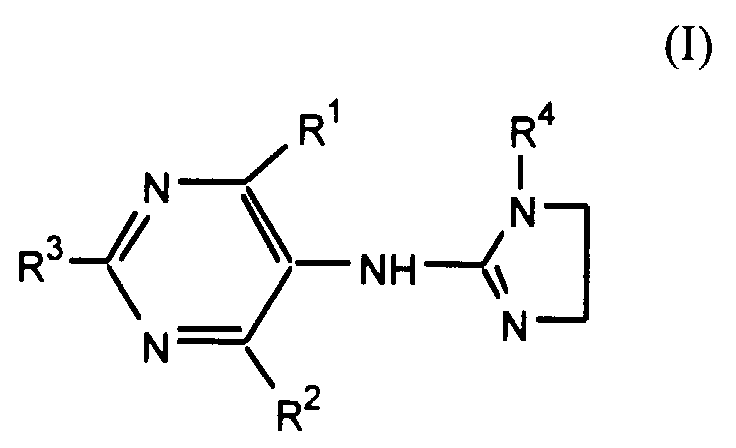
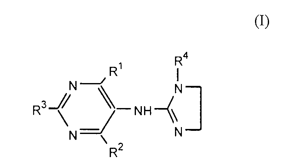
mammal an effective dose of a substituted aminopyrimidine,
or a pharmaceutically acceptable salt or solvate thereof,
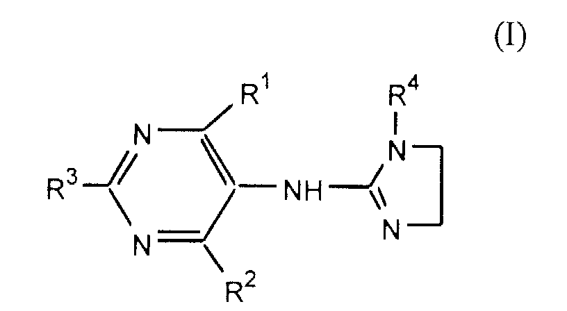
having the formula:
wherein
provided that R
1, R
2 and R
3 are not all hydrogen at the
same time;
The method of this invention is practiced by
administering to a mammal having a glucose metabolism
abnormality or at risk for developing a glucose metabolism
abnormality an effective dose of a substituted
aminopyrimidine of Formula I or a pharmaceutically
acceptable salt or solvate thereof, preferably in a
pharmaceutical formulation. It is necessary that the mammal
being treated have functioning pancreatic beta cells.
The compound 4-chloro-N- (imidazoline-2-ylidene) -6-methoxy-2-methyl-5-pyrimidineamine
(moxonidine) is known and
described in U.S. Patent No. 4,323,570 which is incorporated
herein by reference in its entirety, and is the preferred
compound of the present invention.
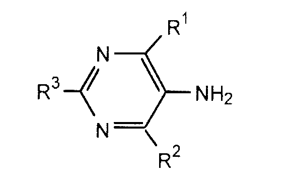
The compounds of Formula I are prepared by reacting the
corresponding 5-aminopyrimidines of Formula II
wherein R
1, R
2 and R
3 are as defined above for Formula
I with 1-acyl-imidazolidin-2-ones of Formula III
wherein R
4 is formyl, acetyl, propionyl, or butyryl.
Compounds of Formulas II and III are well-known or are
synthesized in accordance with known methods of preparation.
The term "halogen" means chloro, bromo, fluoro and
iodo. Among these halogens, chloro and bromo are preferred
and most preferred is chloro.
The cycloalkyl substituents which are particularly
suitable are cyclopropyl, cyclobutyl, or cyclopentyl, and
C1-C4 alkyl derivatives thereof.
The aminopyrimidines substituted by alkyl or cycloalkyl
groups in the 2 position are synthesized from the
correspondingly substituted imino ethers. Starting with the
nitriles, amidines are cyclized with malonic esters to form
the hydroxypyrimidine derivatives. The desired substituted
halogenated aminopyrimidines are formed by halogenation with
exchange of hydroxy groups and the introduction of the amino
group over the nitrile group.
If aminopyrimidines substituted in the 2 and 4 position
are desired, synthesis begins by halogenation of the
corresponding 6-uracils and introduction of the amino group
as previously described.
The reaction between the aminopyrimidines and the
acylimidazolidinones can be carried out in the presence of
phosphorous oxychloride between about 20° C. and the boiling
point of the phosphorous oxychloride. The reaction takes
between 3 and 70 hours. It is preferred that an excess of
the phosphorous compound be used so that it also serves as a
solvent. However, inert organic solvents or mixtures
thereof may also be used. Most preferable, in the event
that phosphorous oxychloride is used alone, is a reaction of
time of approximately 50 hours at temperatures between 50°
and 100° C. After removing the excess phosphorous
oxychloride with water or by means of an aqueous alkaline
medium (e.g. sodium carbonate solution or lye), the reaction
product is converted to the acyl derivatives, preferably at
reduced temperature.
For the compounds in which R4 is hydrogen, the acyl
group can be split off by the use of mineral acids, organic
acids, and/or alkaline reagents. Advantageously, acetic
acid, lye, sodium carbonate, alkali alcoholates or amines
may be used. It is especially preferred to remove the
acetyl group by heating with water or aliphatic alcohols.
Methanol is particularly useful for this purpose. This
means of removal is particularly gentle and, for that
reason, desirable.
It is also possible to convert the compounds wherein R4
is hydrogen to the acyl form by reacting in a known manner
with a recognized acylating agent. In this manner, new 1-acylimidazolines
are obtained. Typical acylating agents are
acid anhydrides and acid chlorides in the presence of bases
such as pyridine. In this manner, such acyl groups as
formyl, acetyl, propionyl, or butyryl may be introduced.
The corresponding alkoxy and alkylthio compounds can
also be obtained from the halogenated compounds by
substitution of the halogen atoms under suitable reaction
conditions. The halogen substituents on the pyrimidine
radical can be converted to alkoxy groups by the use of
alcohols or alcoholates, preferably at a temperature between
O° C. and the boiling point of the alcohol or alcoholates.
Depending upon the reaction conditions, one or more alkoxy
groups can be substituted for a corresponding number of
halogen atoms.
If desired, the reaction can be carried out so that, by
a suitable selection of the conditions (temperature,
reaction time, concentration of reactants, etc.) at least
one halogen atom bonded to the pyrimidine radical is still
available. If this is done, the alkylthio compounds
according to the invention can be prepared. Moreover, under
more extreme conditions of higher temperature, longer
reaction time, and higher alcohol or alcoholate
concentration, additional halogen atoms can be converted.
Also, separation of the R4 acyl group is possible.
The reactions in question are carried out either at
room temperature or higher temperatures, preferably in
alcohols or diluted alcohol solutions. Suitable alcohols
(and alcoholates) are these having 1 to 4 carbon atoms,
especially methanol and ethanol.
It is also possible to produce compounds of Formula I
wherein R1 is halogen, hydrogen, alkoxy, alkylthio, or alkyl
with 1 to 4 carbon atoms or a cycloalkyl group with 3 to 5
carbon atoms; R2 is alkoxy having 1 to 4 carbon atoms; R3 is
hydrogen, alkoxy, alkylthio, or alkyl having 1 to 4 carbon
atoms, or a cycloalkyl group having 3 to 5 carbon atoms; and
R4 is hydrogen. To do so, the compounds of Formula I
wherein R1 is a halogen or hydrogen atom, or an alkylthio or
alkyl group having 1 to 4 carbon atoms, or a cycloalkyl
group with 3 to 5 carbon atoms; R2 is halogen; R3 is
hydrogen, halogen, or an alkylthio or alkyl group with 1 to
4 carbon atoms, or a cycloalkyl group with 3 to 5 carbon
atoms; and R4 is acyl or hydrogen, are used as starting
materials.
The introduction of alkoxy groups is also possible in
accordance with the above-described method if there are
alkylthio substituents present particularly in the case of
those compounds wherein R1 is halogen, R2 is either
alkylthio or cycloalkyl, and R3 is hydrogen, alkyl with 1 to
4 carbon atoms, or cycloalkyl, the halogen substituent can
be substituted by the corresponding alkoxy group having 1 to
4 carbon atoms in the presence of alcohols or alcoholates at
temperatures of between 0° C. and the boiling point of the
alcohol or alcoholate used.
The conversion of halogen substituents on the
pyrimidine radical to alkylthio groups is carried out in a
manner analogous to that described for the substitution of
alkoxy groups for the halogens. Alkylmercaptans are used
and, it has been found, the preferred temperature range is
from 20° C. to the boiling temperature of the solvent. The
method is operable as low as O° C. As in the case of the
alkoxy introduction, leaving at least one halogen atom
bonded to the pyrimidine radical and available for synthesis
of other compounds of this invention is useful. It permits
an exchange in the presence of alkoxy substituents. The
additional halogens can be reacted under more extreme
conditions of temperature reaction time and alkali
mercaptide concentration. The preferred material is a
suspension of sodium methyl mercaptide in toluene.
In the foregoing manner, compounds having alkylthio
substituents are preferably obtained from the compounds of
Formula I wherein R1 and R3 are halogen, hydrogen, or alkyl
or alkoxy with 1 to 4 carbon atoms or a cycloalkyl group
having 3 to 5 carbon atoms; R2 is halogen, and R4 is
hydrogen or an aliphatic acyl group. The compounds can be
obtained by substituting the halogen substituents with the
desired radicals.
By a suitable selection of reaction conditions, some or
all of R1, R2, and R3 may be halogen atoms and such
compounds can be used for further substitution by alkoxy or
alkylthio groups in any desired order or combination.
The compounds for Formula I can also be prepared by
reacting the
S-alkylisothiouronium salts of Formula IV
with ethylene diamine in a known manner, where R
5 is alkyl,
preferably methyl and X is halogen. One method for carrying
out this reaction is by heating the two reactants for
several hours in solvents, such as alcohol. Methanol is
particularly preferred. In this reaction, the
S-methylisothiouronium
salts are preferred.
The starting compounds for the preparation of the
compounds of Formula IV can be obtained by known methods
from the corresponding 5-aminopyrimidine derivatives of
Formula II
It is only necessary to start from those compounds wherein
R
1, R
2, and R
3 represent the appropriate desired
substituents.
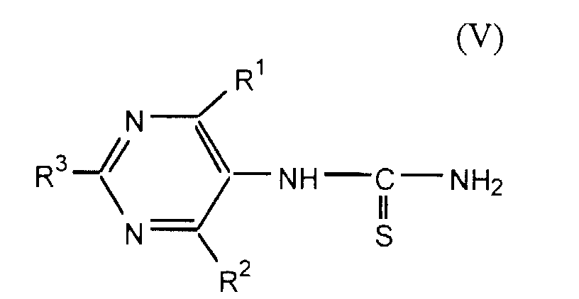
The compounds of Formula II can be reacted with
benzoylisothiocyanate to form the corresponding benzoyl
thioureas. These can be hydrolized to the pyrimidyl
thioureas of Formula V
The benzoylisothiocyanate is obtained by the reaction
between ammonium thiocyanate and benzoylchloride. The
compounds of Formula V can then be reacted with methyliodide
or other similar methylating agents to form the
S-methylisothiouronium
salts of Formula IV.
The pyrimidyl benzoylthioureas are preferably prepared
by the reaction of ammonium thiocyanate, benzoylchloride and
aminopyrimidine in a heated solvent, preferably boiling
acetone. The thiourea derivatives obtained are then
hydrolized in order to separate the benzoyl radical. The
hydrolization can take place, for example, in the presence
of bases and, if necessary, with heating. Lye is
particularly suitable for this purpose. The substituted
thioureas are suspended, for example in acetone, and
converted by methylating agents (preferably methyl iodide)
to isothiouroniumiodide. The reaction is preferably carried
out in boiling acetone.
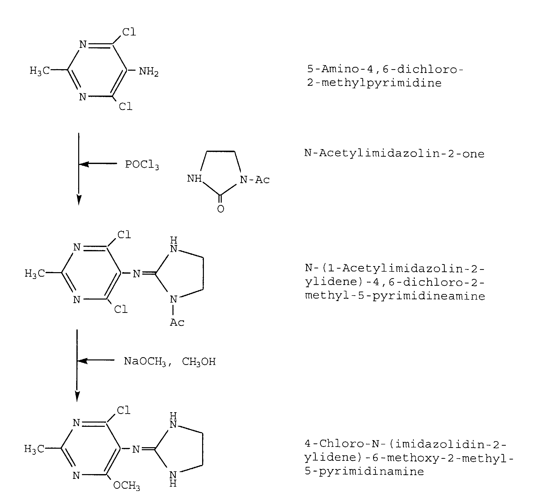
Moxonidine, 4-chloro-
N- (imidazoline-2-ylidene) -6-methoxy-2-methyl-5-pyrimidineamine,
is prepared generally
as disclosed in U.S. Patent 4,323,570. Preferably, 4-chloro-
N-
(imidazoline-2-ylidene) -6-methoxy-2-methyl-5-pyrimidineamine
is prepared as follows.
N-acetylimidazoline-2-one is prepared by reacting
acetic anhydride with 2-imidazolidone at room temperature.
The reaction mixture is heated to between 80 °C and 100 °C
for 90 minutes and then cooled to from about 10 °C to about
-10 °C to afford N-acetylimidazoline-2-one.
The first intermediate, 4,6-dihydroxy-2-methylpyrimidinamine,
is synthesized by preparing sodium
ethoxide in situ from sodium and anhydrous ethanol under a
nitrogen blanket. Acetamidine hydrochloride and diethyl
malonate are added and the reaction mixture heated to
boiling for 2 to 5 hours to afford 4,6-dihydroxy-2-methylpyrimidine.
The second intermediate, 4,6-dihydroxy-2-methyl-5-nitropyrimidine,
is then synthesized by slowly adding 4,6-dihydroxy-2-methylpyrimidine
to a reaction mixture of fuming
nitric acid in acetic acid. Once addition of 4, 6-dihydroxy-2-methylpyrimidine
is complete, the reaction
mixture is stirred for one-half to 2 hours to afford 4,6-dihydroxy-2-methyl-5-nitropyrimidine.
Following the nitration, phosphorous oxychloride
(POCl3) and 4,6-dihydroxy-2-methyl-5-nitropyrimidine are
combined with stirring. To this mixture, diethylaniline is
added dropwise at a rate so that the reaction mixture
temperature is maintained below about 40 °C. After the
addition is complete, the reaction mixture is refluxed for
one to three hours and then distilled under a vacuum to
afford the third intermediate, 4,6-dichloro-2-methyl-5-nitropyrimidine.
The third intermediate, 4,6-dichloro-2-methyl-5-nitropyrimidine
is hydrogenated over Raney-Ni as a 10% to
30% solution in toluene to afford the corresponding
compound, 4,6-dichloro-2-methyl-5-aminopyrimidine, as a
fourth intermediate.
The fifth intermediate, N-(l-acetylimidazolin-2-ylidene)
-4,6-dichloro-5-pyrimidinamine, is then prepared by
combining phosphorous oxychloride, N-acetylimidazolin-2-one
and 5-amino-4,6-dichloro-2-methylpyrimidine, and heating to
boiling over from 2 to 4 hours, and then cooling, with
stirring to room temperature.
The final product, 4-chloro-N- (imidazolin-2-ylidene) -6-methoxy-2-methyl-5-pyrimidinamine
is synthesized by first
preparing sodium methoxide in situ from anhydrous methanol
and sodium. The fifth intermediate, N-(1-acetylimidazolin-2-ylidene)
-4,6-dichloro-2-methyl-5-pyrimidinamine, is added
and the reaction mixture brought to a boil. From 15 minutes
to 1 hour after the reaction mixture is brought to a boil,
further sodium methoxide is added and the reaction mixture
is maintained at a boil for from 15 minutes to 1 hour to
afford 4-chloro-N- (imidazolin-2-ylidene) -6-methoxy-2-methyl-5-pyrimidinamine.
Work-up of the several intermediates are carried out by
standard techniques well-known to those skilled in the art.
The various reactants and reagents used in this synthesis
are commercially available or readily prepared from
commercially available material by standard methods well-known
to those skilled in the art.
It will be appreciated that the compounds of the
present invention may be isolated per se or may be converted
to an acid addition salt using conventional methods. As
mentioned above, the invention includes pharmaceutically
acceptable salts of the compounds of Formula I. The
substituted aminopyrimidines of Formula I can react with any
of a number of inorganic and organic acids, to form a
pharmaceutically acceptable salt. Acids commonly employed
to form acid addition salts are inorganic acids such as
hydrochloric acid, hydrobromic acid, hydroiodic acid,
sulfuric acid, phosphoric acid, and the like, and organic
acids such as p-toluene-sulfonic, methanesulfonic acid,
oxalic acid, p-bromophenyl-sulfonic acid, carbonic acid,
succinic acid, citric acid, benzoic acid, acetic acid, and
the like. Examples of such pharmaceutically acceptable
salts thus are the sulfate pyrosulfate, bisulfate, sulfite,
bisulfite, phosphate, monohydrogenphosphate,
dihydrogenphosphate, metaphosphate, pyrophosphate, chloride,
bromide, iodide, acetate, propionate, decanoate, caprylate,
acrylate, formate, isobutyrate, caproate, heptanoate,
propiolate, oxalate, malonate, succinate, suberate,
sebacate, fumarate, maleate, butyne-1,4-dioate, hexyne-1,6-dioate,
benzoate, chlorobenzoate, methylbenzoate,
dinitrobenzoate, hydroxybenzoate, methoxybenzoate,
phthalate, sulfonate, xylenesulfonate, phenylacetate,
phenylpropionate, phenylbutyrate, citrate, lactate, gamma-hydroxybutyrate,
glycollate, tartrate, methanesulfonate,
propanesulfonate, naphthalene-1-sulfonate, naphthalene-2-sulfonate,
mandelate, and the like. Preferred
pharmaceutically acceptable acid addition salts are those
formed with mineral acids such as hydrochloric acid,
hydrobromic acid and sulfuric acid.
In addition, the substituted aminopyrimidines of
Formula I, or their pharmaceutically acceptable salts may
form solvates with water or common organic solvents such as
ethanol. These solvates are also contemplated as useful in
the methods of this invention.
The acid addition salts are typically formed by
reacting a compound of Formula I with an equimolar or excess
amount of acid. The reactants are generally combined in a
mutual solvent. The salt normally precipitates out of
solution within about one hour to 10 days and can be
isolated by filtration or the solvent can be stripped off by
conventional means.
The pharmaceutically acceptable salts generally have
enhanced solubility characteristics compared to the compound
from which they are derived, and thus are often more
amenable to formulation.
As noted above, the compounds of Formula I,
particularly moxonidine, and the pharmaceutically acceptable
salts and solvates thereof, are useful for inhibiting
glucose metabolism deterioration in mammals having glucose
metabolism abnormalities and at risk for developing glucose
metabolism abnormalities, prior to the onset of
hyperglycemia, thereby preventing, delaying or remitting
glucose metabolism deterioration in the presence of a
glucose metabolism abnormality etiology. Non-limiting
examples of such glucose metabolism abnormality etiologies
include, impaired glucose tolerance, insulin resistance,
circulating islet-cell antibodies, hyperinsulinemia,
hypoinsulinemia (in the presence of functioning beta cells),
increased hepatic glucose output, and obesity.
The term "impaired glucose tolerance" means individuals
whose plasma glucose concentrations lie between normal and
values diagnostic of frank diabetes. Plasma glucose
concentrations in such individuals may progress to overt
diabetes over variable time periods.
Impaired glucose tolerance can be determined by an oral
glucose tolerance test. The World Health Organization
classification defines impaired glucose tolerance with a
two-hour plasma glucose concentration between 140 and 200
mg/dl (7.8 to 11.1 mmol/L) after a standard glucose
challenge in persons with a fasting glucose concentration of
less than 140 mg/dl (7.8 mmol/L). The National Diabetes
Data Group and World Health Organization classifications
differ in that nondiagnostic or unclassifiable oral glucose
tolerance test values occur in the National Diabetes Data
Group system but not in that of the World Health
Organization. Under standardized conditions of testing, the
two-hour value alone may be adequate for diagnosis.
"Glucose metabolism abnormality" means one or more
conditions or disorders that cause, or tend to cause, or if
left uncompensated will cause blood glucose levels to be
greater than normal. Generally, such blood glucose levels
are a result of insulin deficiency or resistance of the
body's cells to the action of insulin. Blood glucose levels
in the range that causes a clinical diagnosis of diabetes
are frankly hyperglycemic, while values above normal, and
below frankly hyperglycemic are indeterminate.
The term "at risk for developing glucose metabolism
abnormalities" means individuals with a previous abnormality
of glucose metabolism, and individuals with a potential
abnormality of glucose metabolism.
The term "previous abnormality of glucose metabolism"
means individuals with previous diabetes or impaired glucose
metabolism who regained normal glucose metabolism. Most
frequently, individuals in this class include patients with
former gestational diabetes, impaired glucose tolerance, or
Type II diabetes whose glucose metabolic status has returned
to normal. These individuals are at increased risk for
developing diabetes.
The term "potential abnormality of glucose metabolism"
means individuals at substantially greater risk for
developing a glucose metabolism abnormality and/or diabetes
than the general population. The category includes first-degree
relatives of patients with diabetes, an identical co-twin
of a patient with Type I or Type II diabetes, human-lymphocyte-antigen-identical
sibling, or an individual with
circulating antibodies directed against the islet-cell
(ICA). Patients in this latter group may demonstrate islet-cell
antibodies in sera for some years before progressing
toward overt diabetes. Also included are obese individuals
and individuals who have delivered a large infant (nine
pounds or greater).
As described above, insulin-dependent (Type I) diabetes
mellitus is characterized by complete or nearly complete
insulin deficiency, and the need for insulin replacement
therapy. The metabolic disturbances (abnormalities) that
characterize non-insulin-dependent (Type II) diabetes
mellitus occur less acutely than those in Type I disease,
and insulin replacement generally is not needed in the early
stages. Initially, diet therapy alone may be sufficient.
Later, oral agents or insulin may be required.
The central role of insulin resistance in the
pathogenesis of Type II diabetes mellitus is widely
accepted. Insulin resistance is defined as the subnormal
biologic response to a given concentration of insulin.
Given the pleiotropic actions of insulin on glucose, lipid,
and protein metabolism, multiple defects could result in
hyperinsulinemia or insulin resistance. Insulinemia,
especially levels of fasting insulin, correlate surprisingly
well (at the level of 0.6-0.8) with more sophisticated
measures of insulin resistance such as the euglycemic clamp
or the minimal model.
Individuals who develop impaired glucose tolerance from
normal glucose tolerance demonstrate that insulin resistance
preceeds a defect in insulin secretion. Recent studies
suggest a two-step process, the first being insulin
resistance and the second being β-cell failure. Loss of
pulsatile insulin secretion has also been suggested as the
first lesion leading to Type II diabetes mellitus.
Chronic hyperglycemia is believed detrimental to
insulin secretion and may also induce insulin resistance.
This gluco-toxicity may perpetuate the diabetic state and
eventually lead to a permanent loss of β-cell function.
Elevated urinary albumin excretion occurs early in the
course of Type II diabetes mellitus, in persons with
impaired glucose tolerance, and in nondiabetic offspring of
Type II diabetes mellitus subjects.
Impaired glucose tolerance (IGT) is defined as a
glycemic response to the standard 75-g oral glucose
challenge that is intermediate between the normal and
diabetic criteria established by the U.S. National Diabetes
Data Group, i.e., venous plasma or capillary whole blood
glucose concentration 140-199 mg/dl at 2 hours after a
glucose load. The importance of impaired glucose tolerance
as a prognostic category for development of Type II diabetes
is generally accepted. Those patients in this group have
higher fasting blood glucose levels, that is, between 125
and 140 mg/dl have the greatest risk of developing Type II
diabetes mellitus.
Genetic susceptibility to Type II diabetes is likely to
include separate but interrelated genes for insulin
resistance, obesity, fat distribution, and other specific
gene defects. Some of these have been identified, others
are still unknown. Major behavioral factors include dietary
calories and fat, physical inactivity, and weight gain.
Cultural and psychosocial factors may operate through diet,
activity, and weight gain, and may also operate
independently. It appears that these behavioral factors can
operate not only to start the cascade but also as promoters
during progression to Type II diabetes mellitus.
Weight gain is the major mechanism etiologically
associated with insulin resistance. Insulin resistance may
be a feedback mechanism to prevent further weight gain above
that determined by some set point. Rapid weight gain
increases insulin resistance in subjects with
hyperandrogenism, insulin resistance, and acanthosis
nigricans. On the other hand, weight loss in severely obese
subjects appears to deter progression from IGT to Type II
diabetes mellitus.
Insulin responses increase during the progression of
glucose intolerance from normal to impaired. However, there
is a progressive fall in insulin secretion as glucose levels
rise, either by unmasking a primary defect in insulin
secretion, from "pancreatic exhaustion," from glucose
toxicity, or from a combination of these defects.
Prospective studies of persons with IGT have identified a
decrease in insulin levels after a glucose challenge
(consistent with a secretory defect) as a predictor of the
development of Type II diabetes mellitus.
Unlike Type I diabetes, in which glucose metabolism
abnormalities are thought to result primarily from absolute
insulin deficiency caused by loss of β-cell mass, Type II
diabetes is recognized as a heterogeneous disorder having
one or more of three defects (glucose metabolism
abnormalities). These include abnormalities in insulin
secretion, reduced sensitivity to insulin (peripheral
resistance), and excessive hepatic glucose production in the
fasted state (although this third category may be a
consequence of the first two).
The failure of many patients with mild-to-moderate
hyperglycemia to develop ketoacidosis has long indicated the
presence of endogenous insulin. Investigations have focused
on four aspects of insulin secretion: 1) Fasted serum
insulin concentrations; 2) Serum immunoreactive insulin
profiles immediately after an intravenous glucose challenge,
called the early-phase response; 3) Insulin secretion
patterns after the early phase, called the late-phase
response; and 4) Sum of the insulin secretion from the two
phases, or total insulin secretion
Characteristically, the early phase of insulin
secretion following administration of glucose or food is
decreased or absent in patients and can be considered a
"marker" for early Type II diabetes. In some cases,
therapeutic measures such as weight reduction and physical
exercise may improve Type II diabetes, however, the
abnormalities in early-phase insulin secretion remains.
Insulin secretory defects also include aberrations in
the pulsatile nature of secretion; and subnormal, normal, or
elevated serum total insulin responses, which are
insufficient to reduce ambient hyperglycemia. Also, in most
obese (nondiabetic or diabetic) individuals, basal and
stimulated insulin concentrations (and secretion rates) are
elevated. This suggests that both a defect in the β-cell
responsiveness to the glucose signal and a peripheral
insensitivity or resistance to insulin contribute to the
pathogenesis of Type II diabetes.
Insulin resistance exists when a normal serum insulin
concentration produces a subnormal response or when elevated
insulin is required to produce a normal response.
Peripheral insulin resistance accounts for most increases in
postprandial blood glucose concentrations since 70 to 90% of
ingested glucose is disposed of in the periphery. Causes of
insulin resistance include obesity; diabetes due to
antibodies to the insulin receptor and other conditions
related to receptor dysfunction; abnormal "insulin" secreted
by the pancreas; hyperproinsulinemia due to a failure of
cleavage of the connecting peptide from proinsulin; mutant
insulin receptor as a result of aberration in the insulin
receptor gene; humoral insulin antagonists; physiologic and
endocrinopathic hypersecretion of the counterregulatory
hormones-glucagon, epinephrine, cortisol, and growth
hormone; immunologic insulin antagonists; antibodies to
animal insulins and miscellaneous; free fatty acids.
All of the conditions which cause insulin resistance
are characterized by single or combined defects that occur
before, during, or after the insulin-receptor interaction
(prereceptor, receptor, or postreceptor stage).
Patients with Type II diabetes showed reduced glucose
disposal rates, demonstrating a tissue defect in the
handling of glucose. Adipocytes from persons with impaired
glucose tolerance or Type II diabetes bind less insulin than
adipocytes from normal subjects. Diminished insulin binding
is due to a decrease in the number of insulin receptors
rather than to lessened binding affinity. The principal
tissue responsible for peripheral insulin insensitivity is
not fat but skeletal muscle. Decreased insulin binding in
obesity results from a reduced number of binding sites.
Deficient cellular or tissue responses to insulin contribute
to hyperglycemia.
In patients with Type II diabetes, hepatic glucose
output is approximately 0.5 mg/kg/min greater than normal,
corresponding to a difference of 50 g of glucose after 24
hours in a 70-kg person. The liver of a normal individual
produces glucose at a rate of approximately 2.2 mg/kg/min.
A number of studies have indicated a close correlation
between hepatic glucose output and fasting hyperglycemia in
patients with Type II diabetes. Since moderate fasting
hyperinsulinemia (usually in the range of 20 to 50 µU/ml) is
frequently found in Type II diabetes, its failure to reduce
hepatic glucose output indicates the presence of hepatic
insulin resistance. Hepatic glucose output is more than 90%
suppressed in patients with Type II disease when serum
insulin concentrations are elevated to 100 µU/ml or more.
While the biochemistry of increased glucose flux from
the liver is not well understood, certain data and
hypotheses are worthy of mention. First, elevated glucagon
concentrations are consistently found in Type II diabetes.
It has been suggested that the failed suppression of
hyperglucagonemia and the consequent increased hepatic
glucose output in Type II diabetes could be related to the
loss of the normal pulsatile bursts of insulin.
Further, increased conversion of glucose to lactate by
the glycolytic (Cori) cycle could occur in skeletal muscle
because glucose is not converted to glycogen in skeletal
muscle, thus resulting in recycling of the 3-carbon
substrate to the liver. In the liver, lactate is converted
to glucose then recycled to the muscle, where it is
reconverted to 3-carbon fragments, then recirculated and so
on. Energy to sustain this sequence of events is supplied
by oxidation of fatty acids in the liver and could explain
the increased free-fatty-acid concentrations associated with
hyperglycemia in Type II diabetes. Specifically, in the
Hales-Randle cycle, free fatty acids are taken up and
oxidized within the muscle cells. Increased free-fatty-acid
oxidation inhibits glycolysis and glucose uptake, actions
which oppose the effect of insulin action.
Excessive hepatic glucose output is the most important
contributor to the basal hyperglycemia observed in Type II
diabetes. Patients with impaired glucose tolerance have
either no or subtle increase in hepatic glucose output since
they have normal fasting blood glucose concentrations. In
these patients, the degree of insulin deficiency reduces
glucose uptake in muscle.
Hyperglycemia resulting from the interaction of
impaired insulin secretion, peripheral resistance to
insulin, and increased hepatic glucose output may be toxic
to the β-cell and result in further worsening of diabetes.
In addition to Type I and Type II, a subclass termed
Other Types of diabetes mellitus is recognized. This Other
Types subclass is heterogeneous and includes specific
disorders such as pancreatic disease, hyperendocrinopathies,
chemical-agent - or drug-induced diabetes, or genetic
syndromes associated with glucose intolerance, each of which
comprises a glucose metabolism abnormality.
In clinical practice, the suspicion of a glucose
metabolism abnormality and those at risk for developing a
glucose metabolism abnormality is gleaned from individual
medical history and physical findings. Symptoms such as
fatigue, thirst, polyuria, weight loss, and recurrent
infection are frequent clues. A family history of diabetes,
obesity, unfavorable obstetrical experiences, premature
atherosclerosis, or neuropathic disorders are indicators.
Urine glucose testing is frequently performed as a screening
test, but is not acceptable for diagnostic purposes. A
positive urine test is usually a helpful indicator, but may
give misleading results during pregnancy because of
increased renal blood flow or in the presence of renal
glycosuria. Patients with high renal glucose thresholds or
elderly patients with decreased renal blood flow may not
have glucosuria in spite of elevated blood glucose
concentrations.
Diagnostic criteria for diabetes are used for
ascertaining a glucose metabolism abnormality and
individuals at risk for developing a glucose metabolism
abnormality. These diagnostic criteria are based on venous
plasma glucose values using glucose analytical methods.
Plasma (or serum) is preferred over whole blood for glucose
determinations because there is less interference with
equipment, differences in hematocrit are eliminated, and
many nonglucose-reducing substances are removed. Capillary
blood, which provides values similar to arterial whole blood
glucose concentrations, approximates venous plasma.
Automated equipment is used to process venous plasma for
determination of glucose concentration with either
ferricyanide, hexokinase, glucose oxidase, or orthotoluidine
reagents. To interpret laboratory test results correctly,
clinicians must have information concerning the methods used
in their referral laboratory and be aware of the
laboratory's normal range of values.
In normal subjects, the currently generally accepted
upper normal limit of fasting plasma glucose is 115 mg/dl
(6.4 mmol/L). Diabetes can be diagnosed reliably when
fasting plasma glucose concentrations are greater than or
equal to 140 mg/dl (7.8 mmol/L) (i.e. hyperglycemic). An
aid in diagnosis is to determine the glycosylated hemoglobin
concentration. If the glycosylated hemoglobin concentration
is greater than normal, the diagnosis must be confirmed by
an elevated ambient glucose concentration preceding the
fasting plasma glucose determination; otherwise, tests for
an elevated fasting plasma glucose should be done on two
occasions to eliminate the possibility of error. The
advantages of measuring the fasting plasma glucose
concentration to test for diabetes are that age, previous
caloric intake, or activity does not affect plasma glucose
and conditions for obtaining a proper blood sample are
standardized. With few exceptions, fasting plasma glucose
values correlate well with the rise in plasma glucose
concentrations observed after a meal or glucose challenge.
A two-hour post-oral glucose load plasma glucose
concentration of greater than or equal to 200 mg/dl (11.1
mmol/L) may be indicative of diabetes if certain precautions
have been observed. Because glucose utilization is impaired
in persons consuming low-carbohydrate or weight-reduction
diets, the post-oral glucose load glucose test should be
performed in those consuming unrestricted diets providing
greater than 150 g of carbohydrate daily for at least three
days. Additional precautionary measures include eliminating
drugs that reduce glucose tolerance, having individuals
maintain normal physical activity, and avoiding the test in
ill or stressed persons; a specific test meal has not been
adequately validated for diagnostic screening. The test is
most reliably performed using a standard oral glucose load.
In patients with liver or kidney disease, partial
gastrectomy, or thyrotoxicosis, an elevated postprandial
plasma glucose may be observed in the absence of diabetes.
Conversely, in an obviously healthy patient with a family
history of diabetes, the combination of a normal fasting
plasma glucose and an abnormal elevation of postglucose
values (greater than or equal to 200 mg/dl; 11.1 mmol/L) may
be indicative of glucose metabolism abnormalities.
Subsequent testing, corroborated by glycohemoglobin
determinations, should be performed to verify the diagnosis
in this situation.
The oral glucose tolerance test is the most sensitive
test for the diagnosis of diabetes. Without scrupulous care
in standardizing the test and preparing the patient,
however, the oral glucose tolerance test can be misleading
since many factors can interfere with normal glucose
tolerance and result in a hyperglycemic curve. This test is
not required for diagnostic purposes when the fasting plasma
glucose is unequivocally elevated, or if blood sugars are
repeatedly greater than 200 mg/dl in the presence of
hyperglycemic symptoms.
Before the oral glucose tolerance test, it is
recommended that the patient receive a balanced diet
providing carbohydrate intake greater than 150 g daily for a
minimum of three days while maintaining normal daily
activities. Carbohydrate restriction or bed rest impairs
glucose tolerance. Acute illness; stress; or the use of
drugs such as 5,5-diphenyl-2,4-imidazolidinedione,
anovulatory agents, diuretics, and glucocorticoids can
adversely influence the results.
The oral glucose tolerance test is performed in the
morning after a 10- to 16-hour fast. A glucose load of 75 g
is given in 300 ml of flavored beverage and consumed within
five minutes after the fasting blood sample is drawn. The
patient remains at rest and does not smoke or consume
fluids, especially caffeine, during the procedure. Blood
samples are drawn at 30-minute intervals for two hours using
tubes containing sodium fluoride (30 mg/5 ml blood). Plasma
is separated by centrifugation within four hours and frozen
if the glucose analysis is not performed promptly.
The oral glucose tolerance test adopted by the World
Health Organization requires only two plasma glucose values:
fasting and two hours after a 75-g glucose load. The
intervening samples at 0.5, 1, and 1.5 hours are not used.
The scheme provides a simple and accurate method for
obtaining diagnostic criteria. Both sets of diagnostic
criteria specify that a fasting plasma glucose concentration
greater than or equal to 140 mg/dl (7.8 mmol/L) and a two-hour
plasma glucose concentration greater than or equal to
200 mg/dl (11.1 mmol/L) are indicative of diabetes mellitus
in the nonpregnant adult. The latter value represents the
concentration observed in the bimodal distribution of two-hour
plasma glucose concentrations in epidemiologic studies,
and is the glucose concentration at which characteristic
microvascular lesions such as diabetic retinopathy appear.
The diagnosis of impaired glucose tolerance is based on
the oral glucose tolerance test after the fasting plasma
glucose concentration has been determined to be less than
140 mg/dl (7.8 mmol/L), as in the criteria of the U.S.
National Diabetes Data Group and the World Health
Organization. Individuals having a fasting plasma glucose
concentration between 115 and 139 mg/dl (6.4-7.8 mmol/L) or
from 140 to 200 mg/dl (7.8 to 11.1 mmol/L) at two hours are
classified as having impaired glucose tolerance.
Gestational diabetes is diagnosed using criteria
established by O'Sullivan and Mahan, "Criteria For the Oral
Glucose Tolerance Test in Pregnancy," Diabetes, 13, 278-285
(1964). Glycosylated hemoglobin determinations are not
useful in the detection of this condition. The values for
fasting plasma glucose and plasma glucose concentrations are
lower in the gestational diabetes criteria than in those for
diabetes mellitus in the nonpregnant adult because of ties
to fetal risks. Modifications of the glucose tolerance test
given during pregnancy conform to their recommendations.
With the same precautions, the patient receives 100 g of
glucose, and blood samples are drawn at one-hour intervals
for three hours after administration of the glucose load.
The World Health Organization Expert Committee has
recommended that the diagnostic procedures and criteria for
pregnant women remain the same as those for all adults.
Screening for gestational diabetes is recommended at
the outset of pregnancy for those at risk for developing
diabetes and everyone should be tested at 24 to 28 weeks. A
practical screening procedure for plasma glucose measurement
is to administer a 50-g glucose load and obtain a blood
sample at one hour in the absence of fasting. A test is
positive if the plasma glucose concentration is greater than
or equal to 150 mg/dl (8.3 mmol/L). - If the screening test
is positive, the oral glucose tolerance test is performed
using the preparation and precautions described in Freinkel
et al., Diabetes Care, 3, 499-501 (1980).
Classic symptoms of diabetes are present in 95% of
children with diabetes. Since their random plasma glucose
concentration exceeds 200 mg/dl (11.1 mmol/L), the oral
glucose tolerance test is seldom required for diagnosis.
When the oral glucose tolerance test is required, it is
performed under the same standardized procedures as those
for adults, except the glucose load is modified to 1.75 g/kg
of ideal body weight up to 75 g. The recommended diagnostic
criteria are the same as those for adult patients, except
that both fasting (greater than 140 mg/dl; 7.8 mmol/L) and
postload plasma glucose concentrations (greater than 200
mg/dl; 11.1 mmol/L) must be elevated for a definitive
diagnosis of diabetes. Impaired glucose tolerance in
children is diagnosed if the fasting plasma glucose
concentration is less than 140 mg/dl (7.8 mmol/L) and the
two-hour value is greater than 140 mg/dl (7.8 mmol/L) even
if one of the postload values exceeds 200 mg/dl (11.1
mmol/L).
Further details of the diagnostic criteria and test
procedures are set forth in the National Diabetes Data
Group: Classification and diagnosis of diabetes mellitus
and other categories of glucose intolerance. Diabetes 1979;
28: 1039-1057 and World Health Organization Expert
Committee: Second report on diabetes mellitus. Technical
report series No. 646. Geneva, Switzerland, 1980.
The rapid intravenous glucose tolerance test has been
used primarily as an investigative tool and generally is not
recommended for clinical use. This procedure has the
advantage of greater reproducibility than the oral glucose
tolerance test. (For details, see Soeldner JS: The
intravenous glucose tolerance test, in Fajans SS, Sussman KE
[eds]: Diabetes Mellitus: Diagnosis and Treatment. New
York, American Diabetes Association Inc., 1971, vol. 3, pp.
107-110). The intravenous glucose tolerance test also may
be used for patients unable to complete an oral glucose
tolerance test because of vomiting and for those in whom the
oral test is not interpretable.
The connecting peptide (C-peptide) radioimmunoassay is
useful in certain conditions when information is needed
regarding endogenous insulin secretion, Recent Advances in
Obesity and Diabetes Research, Raven Press, New York, 1984,
pp. 139-150. The C-peptide is cleaved from the pro-insulin
molecule and is packaged with insulin in secretory granules
so that both are released during the insulin-secretion
process. Insulin antibodies in the blood of patients
treated with exogenous insulin preclude the use of
radioimmunoassay for the circulating insulin hormone.
Partial suppression of insulin secretion by administration
of exogenous insulin can be shown by C-peptide
radioimmunoassay. Using C-peptide determinations, two
cohorts of Type I diabetes have been described: stable
insulin-dependent patients with C-peptide response to
glucose or glucagon stimulation, and labile patients in whom
C-peptide is absent. In Type II diabetes, C-peptide may be
normal or reduced, depending on insulin secretory responses
which may change during the course of treatment. Randel, J.
Clin. Endocrinol. Metab., 57, 1198-1206 (1983). C-peptide
levels are low or absent in patients with factitious
hypoglycemia due to surreptitious insulin administration.
Glycosylated hemoglobin determination is not
recommended as a criterion for the diagnosis of diabetes
because of a lack of glycohemoglobin standards, difficulty
in quality control, and overlapping values for patients with
diabetes, and nondiabetic individuals. It may be used,
however, to confirm the diagnosis. Currently, glycosylated
hemoglobin concentrations are measured in patients with Type
I diabetes primarily to assess glycemic control over periods
of 6 to 12 weeks. Glycosylated hemoglobin (HA1C) is clearly
tied to complications in Type I diabetes. N. Eng. J. Med.
329 (14) 977-986 (1993).
In severe stress, such as acute illness or injury,
vascular catastrophe, or major surgery, a marked rise in
catecholamine concentrations occurs, following by
stimulation of adrenocorticol hormone secretion. These
counterregulatory hormones, together with growth hormone and
glucagon, reduce the tissue sensitivity to insulin and
increase hepatic glucose production while epinephrine
decreases insulin secretory release. The consequence is
abnormal elevation of glucose concentrations in the fasting
a well as in the fed state. This condition may persist for
several weeks, as demonstrated in postmyocardial infarction
studies. Testing for diabetes will yield unreliable results
until recovery from the stressful event has occurred.
Liver disease is associated with an abnormal elevation
of blood glucose values after a glucose challenge. The
liver is also associated with hyperglycemia and to failure
of glycogen storage, an important source of glucose during
fasting. The finding of a normal fasting plasma glucose
concentration in patients with liver disease in whom the
postglucose values are abnormal has been termed hepatic
dysfunctional hyperglycemia. In reversible liver disorders,
such as hepatitis, the glucose tolerance returns to normal
upon recovery from hepatic impairment. Diabetes mellitus is
diagnosed in the presence of chronic liver disease when the
fasting plasma glucose value is abnormally elevated.
Renal disease also may be associated with impaired
glucose tolerance, a condition termed "pseudodiabetes or
uremia." Several factors contribute to this abnormality;
the most significant appears to be a decrease in tissue
sensitivity to insulin. Severe potassium depletion seen in
chronic renal disease, despite normal serum levels, also has
been cited as a cause of impaired glucose tolerance in
uremia and clearly causes failure of insulin secretion.
Diabetes mellitus is diagnosed in these patients when the
fasting plasma glucose concentration is elevated or, in some
situations, when diabetic retinopathy is present and nodular
glomerulosclerosis is seen on renal biopsy.
A number of commonly used drugs may induce diabetes,
among them diuretics used frequently in the management of
hypertension. Prospective studies have shown a 7% greater
prevalence of diabetes in patients receiving thiazide than
in hypertensive patients treated without these agents.
Diabetic ketoacidosis and nonketotic hyperosmolar coma have
been observed in patients receiving diuretics. Although the
underlying mechanism of this form of diabetes is unclear,
the leading consideration is depletion of intracellular
potassium (not always reflected in serum values).
Administration of potassium may ameliorate glucose
intolerance induced by thiazides although thiazides inhibit
insulin release even when potassium is taken into account.
In patients with known diabetes, the impairment of
carbohydrate intolerance is improved by increasing the
dosage of drug or other diabetes care agent. Other
conditions associated with potasium depletion, such as
hyperaldosteronism, have been marked by glucose intolerance,
which is improved by potassium administration.
Another form of drug-induced disease is glucocorticoid-induced
diabetes, which occurs in 14% of patients who
receive glucocorticoid therapy as treatment for a variety of
diseases. This adverse effect is usually observed with
prolonged oral use of these agents, and also has been noted
with extensive topical use of steroids in dermatologic
disorders. The factors involved in glucose intolerance are
thought to involve a reduced affinity of insulin for its
tissue receptor where insulin action is initiated, together
with an increase in hepatic glucose production through
stimulation of gluconeogenesis. This form of diabetes is
infrequently associated with ketonuria and acidosis.
Cessation of steroid therapy is usually accompanied by
improvement in glucose tolerance, but occasionally insulin
requiring diabetes persists. The latter may occur in
patients in whom undetected diabetes was unmasked by steroid
therapy. The oral contraceptive steroids may exhibit a mild
diabetogenic effect, although this influence appears to be
no significant diabetogenic effects associated with other
contraceptive preparations.
A rare but important form of diabetes, termed insulin
resistance with acanthosis nigricans, has been described in
which alterations in the function of the insulin receptor is
the predominant abnormality. Two types of disorders are
included in this category. Type A is usually observed in
young women with features of virilization, polycystic
ovaries, and hyperandrogenism. In these patients, reduced
insulin-receptor activity or postreceptor defects with
normal insulin binding result in impaired insulin action and
insulin resistance. Type B insulin resistance is caused by
the production of antibodies to the insulin receptor. The
insulin-receptor antibody, which blocks access of insulin to
the insulin receptor, results from an autoimmune-type
response seen in patients with systemic lupus erythematosus
or other autoimmune states. In early stages of this
disorder, or in tissue cultures, this antibody may function
as an insulin agonist, in which its coupling with the
receptor may produce insulin effects resulting in severe or
even fatal hypoglycemia. A frequent effect, however, is
intense hyperglycemia and insulin resistance resulting from
the interference of insulin binding to tissue receptors.
Diagnosis of this condition can be established by the
demonstration of insulin-receptor antibody in the patient's
serum.
Nearly one-third of patient with diabetes are over 60
years of age. The increasing prevalence of diabetes may be
related, in part, to the increasing longevity of the
population. Early studies have demonstrated a gradual
decrease in glucose tolerance with advancing age. The basis
for impairment of postglucose curves has been shown to be
related to the reduced tissue sensitivity to insulin rather
than to a decrease in insulin availability. These data,
however, are not applicable to the diagnosis of diabetes in
the elderly. The current diagnostic criteria of the
National Diabetes Data Group and the World Health
Organization apply to all age groups and are higher than
values for the age-related gradients previously used in the
interpretation of the oral glucose tolerance test in the
elderly.
Among the elderly, the classic symptoms of diabetes are
not always manifest. Clinical presentations are often those
related to complications of the disease, such as vascular
disorders or neuropathic syndromes. To arrive at the
correct diagnosis, physicians must be alert to the clinical
signs of diabetes that may appear in various organ systems
of the elderly. Early diagnosis is needed to apply
appropriate treatment and therefore to forestall
hyperglycemia and the progression of diabetic complications
and improve the general health, well being, and vigor of
these patients.
Recommended for diagnosis of diabetes in the elderly is
a fasting plasma glucose concentration greater than or equal
to 140 mg/dl (7.8 mmol/L). The sedentary or infirm elderly
patient whose dietary intake is often faulty is not a
suitable candidate for glucose tolerance testing. Treatment
for these patients should be conservative if there is a risk
of hypoglycemia, with standards for glycemic control raised
by 30 or 40 mg/dl (1.7 or 2.2 mmol/L) above those for the
middle-aged, nonpregnant adult diabetic population.
Type II diabetes differs from Type I diabetes not only
in its genetic features-high concordance rate in identical
twins and absence of association with human leukocyte
antigens (HLA) types-but in other important respects. The
most striking is that β-cell destruction does not occur
acutely in Type II diabetes; indeed, the pancreatic islets
can be microscopically indistinguishable from those of
normal subjects and contain normal or near-normal amounts of
insulin. Insulin is present in the circulation and, in the
nonobese, the insulin response to a glucose load is
preserved, although the peak concentration may be delayed.
Early Type II diabetes does not seem to result from insulin
deficiency but from sluggish responsiveness, perhaps the
result of a defect in the secretory control of insulin and
from resistance to its action. This defect, whatever its
cause, is strongly influenced by genetic factors.
Presence of diabetes in a family member is an
established risk factor for diabetes, particularly Type II.
In addition to genetics, diet, such as high-fat, low-carbohydrate
intake, physical inactivity, obesity, and low
birth weight are all generally recognized as risk factors
for diabetes.
By the term "effective dose" is meant an amount of a
substituted aminopyrimidine of Formula I, preferably 4-chloro-N-
(imidazoline-2-ylidene) -6-methoxy-2-methyl-5-pyrimidineamine,
or a pharmaceutically acceptable salt or
solvate thereof, sufficient to afford the desired
therapeutic or prophylactic effect. The term "treating" as
used herein includes therapeutically or prophylactically
administering a substituted aminopyrimidine of Formula I,
preferably moxonidine to a mammal to prevent or inhibit the
onset of a glucose metabolism abnormality or inhibit
glucose metabolism deterioration once the condition is
established, and prior to the onset of hyperglycemia. Also
contemplated as within the scope of the present invention is
concomitant treatment with diabetes care agents including
oral hypoglycemic agents, insulin or insulin derivatives,
such as lispro. The need and dosage of such diabetes care
agents would be readily determined by the attending
physician as circumstances dictate.
The compounds of the present invention are putative I1-imidazoline
ligands demonstrating substantial selectivity
for I1 receptors over α2 adrenergic receptors. In
saturation binding experiments in bovine rostral
ventrolateral medulla (bovine RVLM), moxonidine demonstrates
a selectivity value (Ki at α2 sites in µM/Ki sites in µM) of
greater than 20 and preferably greater than 30 X, where Ki
is the inhibitory affinity constant. Of course, Ki is
inversely proportional to affinity, so lower Ki values
indicate higher affinity. Thus, the higher the selectivity
value, the more selective the compound. In contract,
clonidine's selectivity value in bovine RVLM is less than 4.
See Ernsberger et al., J. Pharmacol. Exp. Ther., 264, 172-182
(1993) for details on experimental protocol and results.
As used herein, the term "mammal" means the Mammalia
class of higher vertebrates. The term "mammal" includes,
but is not limited to, a human. The dose of compound to be
administered, in general, is from about 0.01 to about 15.0
mg/day; as usual, the daily dose may be administered in a
single bolus, or in divided doses, depending on the judgment
of the physician in charge of the case. A more preferred
range of doses is from about 0.02 to about 10.0 mg/day;
other dosage ranges which may be preferred in certain
circumstances are from about 0.05 to about 5.0 mg/day; from
about 0.1 to about 2.0 mg/day; from about 0.05 to about 1.0
mg/day; and a particularly preferred range is from about
0.05 to about 1.5 mg/day. It will be understood that the
dose for a given patient is always to be set by the judgment
of the attending physician, and that the dose is subject to
modification based on the size of the patient, the lean or
fat nature of the patient, the characteristics of the
particular compound (freebase or salt) chosen, the severity
of the patient's symptoms and psychological factors which
may affect the patient's physiological responses, as well as
concomitant treatment with other diabetes care agents.
Pharmaceuticals are substantially always formulated
into pharmaceutical dosage forms, in order to provide an
easily controllable dosage of the drug, and to give the
patient an elegant and easily handled product. The present
compounds are susceptible to formulation into the
conventional pharmaceutical dosage forms, including
capsules, tablets, inhalants, injectable parenteral
solutions and suppositories. Usually the oral dosage forms,
particularly tablets and capsules, are most convenient for
the patient and are usually preferred. Liquid suspensions
of pharmaceuticals, formerly popular, have now become less
popular and are seldom used but the present compounds are
entirely amenable to such products should they be desired.
While it is possible to administer a compound of
Formula I, preferably 4-chloro-N- (imidazoline-2-ylidene) -6-methoxy-2-methyl-5-pyrimidineamine
directly, it is
preferably employed in the form of a pharmaceutical
formulation comprising a pharmaceutically acceptable
carrier, diluent or excipient and the compound or a
pharmaceutically acceptable salt or solvate thereof. Such
formulations will contain from about 0.01 percent to about
99.9 percent by weight of the compound.
In making the formulations of the present invention,
the active ingredient will usually be mixed with at least
one carrier, or diluted by at least one carrier, or enclosed
within a carrier which may be in the form of a capsule,
sachet, paper or other container using conventional
techniques and procedures for the preparing of
pharmaceutical formulations. When the carrier serves as a
diluent, it may be a solid, semi-solid or liquid material
which acts as a vehicle, excipient or medium for the active
ingredient. Thus, the formulations can be in the form of
tablets, granules, pills, powders, lozenges, sachets,
cachets, emulsions, solutions, syrups, suspensions and soft
and hard gelatin capsules.
Examples of suitable carriers, diluents and excipients
include lactose, dextrose, sucrose, sorbitol, mannitol,
starches, gum acacia, calcium phosphate, alginates, liquid
paraffin, calcium silicate, microcrystalline cellulose,
polyvinylpyrrolidone, cellulose, tragcanth, gelatin, syrup,
methylcellulose, methyl- and propyl-hydroxybenzoates,
vegetable oils, such as olive oil, injectable organic esters
such as ethyl oleate, talc, magnesium stearate, water and
mineral oil. The formulations may also include wetting
agents, lubricating, emulsifying and suspending agents,
preserving agents, sweetening agents, perfuming agents,
stabilizing agents or flavoring agents.
The compounds of the present invention can be
formulated in unit dosage formulations comprising a dose
between about 0.01 mg and about 15 mg. Preferably the
compound is in the form of a pharmaceutically acceptable
salt.
The compounds of Formula I can be administered by a
variety of routes including oral, rectal, transdermal,
subcutaneous, intravenous, intramuscular, and intranasal.
The compound is preferably formulated prior to
administration as a pharmaceutical formulation comprising an
effective amount of a compound of Formula I preferably
moxonidine, or a pharmaceutically acceptable salt or solvate
thereof in association with a pharmaceutically acceptable
carrier, diluent or excipient therefor.
By "pharmaceutically acceptable" it is meant the
carrier, diluent or excipient must be compatible with the
other ingredients of the formulation and not deleterious to
the recipient thereof.
The present pharmaceutical formulations are prepared by
known procedures using well-known and readily available
ingredients. The compositions of this invention may be
formulated so as to provide quick, sustained, or delayed
release of the active ingredient after administration to the
patient by employing procedures well-known in the art. In
making the compositions of the present invention, the active
ingredient will usually be admixed with a carrier, or
diluted by a carrier, or enclosed within a carrier which may
be in the form of a capsule, sachet, paper or other
container. When the carrier serves as a diluent, it may be
a solid, semi-solid or liquid material which acts as a
vehicle, excipient or medium for the active ingredient.
Thus, the compositions can be in the form of tablets, pills,
powders, lozenges, sachets, cachets, elixirs, suspensions,
emulsions, solutions, syrups, aerosols, (as a solid or in a
liquid medium), soft and hard gelatin capsules,
suppositories, sterile injectable solutions, sterile
packaged powders, and the like.
The activity of the compounds of the present invention
was determined by testing the efficacy in vivo in Zucker
diabetic rats.
The Zucker diabetic fatty rat (ZDF-DRT-fa) is a classic
rodent model of diabetes associated with obesity and insulin
resistance, with tissue specific defects in insulin
sensitivity similar to human patients with type II diabetes
mellitus. This animal model at six weeks of age is
normoglycemic, but glucose intolerant (Clark JB, Palmer CJ,
and Shaw WN: The diabetic Zucker fatty rat, Proceeding of
the Society for Experimental Bioloqy and Medicine 173, 68-75,
1983; and Peterson RG, Shaw WN, Neel M, Little LA,
Eichburg J: Zucker diabetic fatty rat as a model for non-insulin
dependent diabetes mellitus. ILAR News 32:16-19,
1990.) Between seven and eight weeks of age, plasma glucose
levels rise as the pancreatic beta cells lose their ability
to secrete sufficient insulin to compensate for insulin
resistance. In an attempt to maintain plasma glucose in
normal range, the pancreas is forced to secrete increasing
amount of insulin, resulting in hyperinsulinemia (Bue JM and
Shaw WN: ZDF-Drt-fa rat: A new model with marked
susceptibility to dietary manipulation. Diabetes 40:865A,
1991). The ZDF rat is usually studied between eight and
nine weeks of age when it is hyperglycemic,
hyperinsulinemic, and hyperlipemic, in an attempt to halt
further decline of pancreatic function. The present study
was carried out in six weeks old animals (prediabetic). The
purpose of this study was to determine whether moxonidine
treatment will prevent or delay onset of diabetes in the six
weeks old prediabetic ZDF rat.
A number of interventions are capable of impacting the
disease process in the ZDF rat, including agents that
enhance peripheral insulin sensitivity (thiazolidinediones,
TZDs), agents that act at the level of the pancreas glucagon
like insulinotropic peptide (GLP-1), agents that affect
intestinal absorption of glucose (acarbose), and agents with
poorly defined mechanisms (metformin). These interventions
also are effective in human NIDDM patients. As in humans,
weight loss also improves insulin sensitivity.
To determine the impact of treatment with the
substituted aminopyrimidines of Formula I on the progression
of disease 6 week-old ZDF animals were grouped into the
following categories, with 5 animals assigned to each group.
- Group 1
- Control (5008 chow ad libitum)
- Group 2
- Moxonidine (10 mg/kg/day)
- Group 3
- Pair fed to correct for decrease in food
consumption in 10 mg/kg/day group
Rats were housed one per plastic cage with bedding and
had free access to water and Purina Formulalab 5008 Chow
(except pair fed group). Because previous studies had
indicated a reduction in food consumption with moxonidine
treatment, body weight and food consumption were monitored
daily, and a group of control animals was pair fed to
moxonidine treated group. Blood samples were collected from
the tail vein. Plasma glucose and insulin levels are
monitored weekly.
Plasma glucose was measured by coupled hexokinase
method on the IL Monarch System (Instrumentation Laboratory,
Lexington, MA). Plasma insulin was determined with
radioimmunoassay kit from Diagnostics Products Corporation
(San Diego, CA) using rat insulin as a standard.
Plasma glucose and plasma insulin were monitored
weekly. The results of these
in vivo experiments are set
forth below in Tables 1 and 2. The value in parenthesis
following the plasma glucose value and plasma insulin value
is the standard error of the mean.
| Effect of Moxonidine on Plasma Glucose (mg/dl) |
| Moxonidine |
| Day | Control | 10 mg/kg | Pair fed |
| 0 | 141.3 (3.9) | 145.6 (2.4) | 148.3 (2.4) |
| 7 | 141.1 (10.8) | 148.1 (13.8) | 129.4 (5.0) |
| 14 | 320.2 (38.8) | 166.1 (11.2) | 132.3 (11.5) |
| 21 | 436.8 (40.9) | 164.2 (8.0) | 245.1 (35.9) |
| 28 | 555.6 (19.8) | 168.0 (12.6) | 473.1 (16.7) |
| 35 | 574.5 (18.8) | 191.6 (8.2) | 489.6 (9.1) |
| 42 | 565.0 (18.3) | 179.8 (6.1) | 481.4 (11.9) |
| 49 | 536.6 (12.4) | 152.1 (8.7) | 482.0 (9.2) |
| 63 | 541.3 (24.7) | 168.4 (12.6) | 531.0 (24.9) |
| Effects of Moxonidine on Plasma Insulin (ng/ml) |
| Moxonidine |
| Day | Control | 10 mg/kg | Pair fed |
| 0 | 8.4 (1.1) | 10.9 (0.9) | 9.9 (1.3) |
| 7 | 19.0 (0.3) | 9.3 (1.0) | 14.1 (2.5) |
| 14 | 18.1 (1.5) | 12.7 (1.4) | 14.8 (2.5) |
| 21 | 13.8 (2.2) | 17.8 (1.2) | 20.5 (2.2) |
| 28 | 8.5 (1.8) | 23.4 (1.8) | 15.3 (3.3) |
| 35 | 6.3 (0.8) | 24.8 (1.4) | 12.2 (2.0) |
| 42 | 4.8 (0.5) | 20.6 (0.7) | 6.8 (0.9) |
The above data clearly demonstrates moxonidine's impact
in preventing deterioration of glucose metabolism in a
mammal having a genetically predisposed glucose metabolism
abnormality in the absence of hyperglycemia.
A further study was carried out as described above,
except that moxonidine treatment was reduced to 6.4
mg/kg/day for two days, followed by resumption of moxonidine
treatment at 10 mg/kg/day for 13 days, and then stopping
moxonidine treatment. Data on plasma glucose in mg/dl over
the treatment period in days is reported below in Table 3.
| Moxonidine Effect on Plasma Glucose (mg/dl) |
| Treatment period (days) | Control | Control sem | Mox. | Mox sem | pr fed | pr fed sem | Comment |
| 0 | 140.4 | 3.817853 | 141.5333 | 4.100705 | 141.1 | 3.993244 | mox @ 10 m/k/d |
| 7 | 187.4889 | 9.105252 | 152.8917 | 10.69197 | 101.8222 | 1.67174 |
| 14 | 299.3694 | 46.70882 | 166.2528 | 5.754693 | 255.9278 | 34.40768 |
| 21 | 405 | 58.78127 | 172.1556 | 17.00969 | 204.9278 | 18.56692 | Reduced mox dose to 6.4 m/k/d |
| 23 | 443.4333 | 27.22072 | 254.0889 | 38.84799 | 317.8556 | 48.35806 | mox @ 10 m/k/d |
| 28 | 459.6389 | 29.02029 | 251.4833 | 20.28488 | 422.4944 | 51.87904 |
| 36 | 501.4239 | 20.33956 | 242.5944 | 42.08123 | 442.55 | 26.80212 | stopped mox treatment |
| 42 | 556.2778 | 21.15415 | 265.9556 | 42.19204 | 549.1667 | 25.56191 | 1 wk after mox is removed |
| 49 | 595.8 | 13.16921 | 410.425 | 46.99505 | 601.35 | 20.43749 | 2 wks after mox is removed |
The above data, Tables 1, 2 and 3, clearly demonstrates
moxonidine's impact on preventing deterioration of glucose
metabolism by intervening at one or more of the etiological
cause(s) of glucose metabolism abnormalities.