EP0843820B1 - Elisa serodiagnosis of pig pleuropneumonia serotypes 1, 9 and 11 - Google Patents
Elisa serodiagnosis of pig pleuropneumonia serotypes 1, 9 and 11 Download PDFInfo
- Publication number
- EP0843820B1 EP0843820B1 EP96921847A EP96921847A EP0843820B1 EP 0843820 B1 EP0843820 B1 EP 0843820B1 EP 96921847 A EP96921847 A EP 96921847A EP 96921847 A EP96921847 A EP 96921847A EP 0843820 B1 EP0843820 B1 EP 0843820B1
- Authority
- EP
- European Patent Office
- Prior art keywords
- kit
- pleuropneumoniae
- serum
- antigen
- pigs
- Prior art date
- Legal status (The legal status is an assumption and is not a legal conclusion. Google has not performed a legal analysis and makes no representation as to the accuracy of the status listed.)
- Expired - Lifetime
Links
- 241000204031 Mycoplasma Species 0.000 title description 15
- 206010035664 Pneumonia Diseases 0.000 title description 15
- 201000006509 pleuropneumonia Diseases 0.000 title description 15
- 239000000427 antigen Substances 0.000 claims description 66
- 102000036639 antigens Human genes 0.000 claims description 66
- 108091007433 antigens Proteins 0.000 claims description 66
- 241000606748 Actinobacillus pleuropneumoniae Species 0.000 claims description 54
- 210000002966 serum Anatomy 0.000 claims description 47
- 241000282887 Suidae Species 0.000 claims description 45
- 238000000034 method Methods 0.000 claims description 38
- 238000002965 ELISA Methods 0.000 claims description 34
- 244000144980 herd Species 0.000 claims description 34
- 239000000243 solution Substances 0.000 claims description 33
- 241000122975 Actinobacillus pleuropneumoniae serovar 1 Species 0.000 claims description 28
- 108010001336 Horseradish Peroxidase Proteins 0.000 claims description 17
- 239000013641 positive control Substances 0.000 claims description 17
- 238000003556 assay Methods 0.000 claims description 15
- 239000013642 negative control Substances 0.000 claims description 15
- 230000001580 bacterial effect Effects 0.000 claims description 13
- 230000000087 stabilizing effect Effects 0.000 claims description 12
- ISWSIDIOOBJBQZ-UHFFFAOYSA-N Phenol Chemical compound OC1=CC=CC=C1 ISWSIDIOOBJBQZ-UHFFFAOYSA-N 0.000 claims description 11
- 239000002158 endotoxin Substances 0.000 claims description 11
- 229920006008 lipopolysaccharide Polymers 0.000 claims description 11
- 244000052769 pathogen Species 0.000 claims description 9
- 230000001717 pathogenic effect Effects 0.000 claims description 9
- 238000009007 Diagnostic Kit Methods 0.000 claims description 8
- OHDRQQURAXLVGJ-HLVWOLMTSA-N azane;(2e)-3-ethyl-2-[(e)-(3-ethyl-6-sulfo-1,3-benzothiazol-2-ylidene)hydrazinylidene]-1,3-benzothiazole-6-sulfonic acid Chemical compound [NH4+].[NH4+].S/1C2=CC(S([O-])(=O)=O)=CC=C2N(CC)C\1=N/N=C1/SC2=CC(S([O-])(=O)=O)=CC=C2N1CC OHDRQQURAXLVGJ-HLVWOLMTSA-N 0.000 claims description 8
- 238000002360 preparation method Methods 0.000 claims description 8
- 239000002953 phosphate buffered saline Substances 0.000 claims description 7
- 238000000605 extraction Methods 0.000 claims description 6
- 239000000287 crude extract Substances 0.000 claims description 5
- 239000007787 solid Substances 0.000 claims description 5
- 102000003992 Peroxidases Human genes 0.000 claims description 4
- LOKCTEFSRHRXRJ-UHFFFAOYSA-I dipotassium trisodium dihydrogen phosphate hydrogen phosphate dichloride Chemical compound P(=O)(O)(O)[O-].[K+].P(=O)(O)([O-])[O-].[Na+].[Na+].[Cl-].[K+].[Cl-].[Na+] LOKCTEFSRHRXRJ-UHFFFAOYSA-I 0.000 claims description 4
- 230000003053 immunization Effects 0.000 claims description 4
- 239000000203 mixture Substances 0.000 claims description 4
- 108040007629 peroxidase activity proteins Proteins 0.000 claims description 4
- 239000000758 substrate Substances 0.000 claims description 4
- RTKIYNMVFMVABJ-UHFFFAOYSA-L thimerosal Chemical compound [Na+].CC[Hg]SC1=CC=CC=C1C([O-])=O RTKIYNMVFMVABJ-UHFFFAOYSA-L 0.000 claims description 4
- 229940033663 thimerosal Drugs 0.000 claims description 4
- 108060003951 Immunoglobulin Proteins 0.000 claims description 3
- 238000005119 centrifugation Methods 0.000 claims description 3
- 102000018358 immunoglobulin Human genes 0.000 claims description 3
- 229940072221 immunoglobulins Drugs 0.000 claims description 3
- 238000004806 packaging method and process Methods 0.000 claims description 3
- 230000009870 specific binding Effects 0.000 claims description 3
- 241000124008 Mammalia Species 0.000 claims description 2
- 238000012800 visualization Methods 0.000 claims description 2
- 102000004190 Enzymes Human genes 0.000 claims 2
- 108090000790 Enzymes Proteins 0.000 claims 2
- 238000012360 testing method Methods 0.000 description 29
- 241001465754 Metazoa Species 0.000 description 20
- 241000282898 Sus scrofa Species 0.000 description 16
- 239000000872 buffer Substances 0.000 description 16
- 208000015181 infectious disease Diseases 0.000 description 13
- WSFSSNUMVMOOMR-UHFFFAOYSA-N Formaldehyde Chemical compound O=C WSFSSNUMVMOOMR-UHFFFAOYSA-N 0.000 description 12
- 230000035945 sensitivity Effects 0.000 description 11
- 238000011534 incubation Methods 0.000 description 8
- 230000004044 response Effects 0.000 description 8
- 239000008346 aqueous phase Substances 0.000 description 7
- 238000009826 distribution Methods 0.000 description 7
- 238000002156 mixing Methods 0.000 description 7
- 239000000725 suspension Substances 0.000 description 7
- 238000005406 washing Methods 0.000 description 7
- 230000000007 visual effect Effects 0.000 description 6
- 229920001817 Agar Polymers 0.000 description 5
- 206010070834 Sensitisation Diseases 0.000 description 5
- 239000008272 agar Substances 0.000 description 5
- 239000013256 coordination polymer Substances 0.000 description 5
- 230000037029 cross reaction Effects 0.000 description 5
- 230000003287 optical effect Effects 0.000 description 5
- 230000008313 sensitization Effects 0.000 description 5
- 230000000405 serological effect Effects 0.000 description 5
- 241000894006 Bacteria Species 0.000 description 4
- 238000004458 analytical method Methods 0.000 description 4
- 239000007853 buffer solution Substances 0.000 description 4
- 238000006243 chemical reaction Methods 0.000 description 4
- 239000012153 distilled water Substances 0.000 description 4
- 229940079593 drug Drugs 0.000 description 4
- 239000003814 drug Substances 0.000 description 4
- XLYOFNOQVPJJNP-UHFFFAOYSA-N water Chemical compound O XLYOFNOQVPJJNP-UHFFFAOYSA-N 0.000 description 4
- QTBSBXVTEAMEQO-UHFFFAOYSA-M Acetate Chemical compound CC([O-])=O QTBSBXVTEAMEQO-UHFFFAOYSA-M 0.000 description 3
- 241000606731 Actinobacillus suis Species 0.000 description 3
- 238000008157 ELISA kit Methods 0.000 description 3
- 241000588724 Escherichia coli Species 0.000 description 3
- 241000606807 Glaesserella parasuis Species 0.000 description 3
- FAPWRFPIFSIZLT-UHFFFAOYSA-M Sodium chloride Chemical compound [Na+].[Cl-] FAPWRFPIFSIZLT-UHFFFAOYSA-M 0.000 description 3
- 230000000890 antigenic effect Effects 0.000 description 3
- 230000000295 complement effect Effects 0.000 description 3
- 238000001514 detection method Methods 0.000 description 3
- 201000010099 disease Diseases 0.000 description 3
- 208000037265 diseases, disorders, signs and symptoms Diseases 0.000 description 3
- 238000011049 filling Methods 0.000 description 3
- 150000004676 glycans Chemical class 0.000 description 3
- 238000002955 isolation Methods 0.000 description 3
- 230000003902 lesion Effects 0.000 description 3
- 239000007788 liquid Substances 0.000 description 3
- 238000004519 manufacturing process Methods 0.000 description 3
- 239000012071 phase Substances 0.000 description 3
- 229920001282 polysaccharide Polymers 0.000 description 3
- 239000005017 polysaccharide Substances 0.000 description 3
- 229920000136 polysorbate Polymers 0.000 description 3
- 238000000746 purification Methods 0.000 description 3
- 238000009589 serological test Methods 0.000 description 3
- 238000003307 slaughter Methods 0.000 description 3
- HQFLTUZKIRYQSP-UHFFFAOYSA-N 3-ethyl-2h-1,3-benzothiazole-6-sulfonic acid Chemical compound OS(=O)(=O)C1=CC=C2N(CC)CSC2=C1 HQFLTUZKIRYQSP-UHFFFAOYSA-N 0.000 description 2
- 101710098119 Chaperonin GroEL 2 Proteins 0.000 description 2
- 230000006978 adaptation Effects 0.000 description 2
- 239000000969 carrier Substances 0.000 description 2
- 229940061607 dibasic sodium phosphate Drugs 0.000 description 2
- 238000007865 diluting Methods 0.000 description 2
- BNIILDVGGAEEIG-UHFFFAOYSA-L disodium hydrogen phosphate Chemical compound [Na+].[Na+].OP([O-])([O-])=O BNIILDVGGAEEIG-UHFFFAOYSA-L 0.000 description 2
- 238000011156 evaluation Methods 0.000 description 2
- 239000000284 extract Substances 0.000 description 2
- 238000002649 immunization Methods 0.000 description 2
- 102000004169 proteins and genes Human genes 0.000 description 2
- 108090000623 proteins and genes Proteins 0.000 description 2
- 238000011160 research Methods 0.000 description 2
- 239000011780 sodium chloride Substances 0.000 description 2
- 230000006641 stabilisation Effects 0.000 description 2
- 238000011105 stabilization Methods 0.000 description 2
- 229960005486 vaccine Drugs 0.000 description 2
- DGVVWUTYPXICAM-UHFFFAOYSA-N β‐Mercaptoethanol Chemical compound OCCS DGVVWUTYPXICAM-UHFFFAOYSA-N 0.000 description 2
- 241001478285 Actinobacillus pleuropneumoniae serovar 2 Species 0.000 description 1
- 241000178944 Actinobacillus pleuropneumoniae serovar 5 Species 0.000 description 1
- 241001258963 Actinobacillus pleuropneumoniae serovar 7 Species 0.000 description 1
- KRKNYBCHXYNGOX-UHFFFAOYSA-K Citrate Chemical compound [O-]C(=O)CC(O)(CC([O-])=O)C([O-])=O KRKNYBCHXYNGOX-UHFFFAOYSA-K 0.000 description 1
- 238000012286 ELISA Assay Methods 0.000 description 1
- 241000283973 Oryctolagus cuniculus Species 0.000 description 1
- 101710116435 Outer membrane protein Proteins 0.000 description 1
- 241000606860 Pasteurella Species 0.000 description 1
- 241000606856 Pasteurella multocida Species 0.000 description 1
- 208000037581 Persistent Infection Diseases 0.000 description 1
- 239000004793 Polystyrene Substances 0.000 description 1
- 230000001154 acute effect Effects 0.000 description 1
- 230000004520 agglutination Effects 0.000 description 1
- 239000003242 anti bacterial agent Substances 0.000 description 1
- 229940088710 antibiotic agent Drugs 0.000 description 1
- 239000004599 antimicrobial Substances 0.000 description 1
- 230000005540 biological transmission Effects 0.000 description 1
- 210000004369 blood Anatomy 0.000 description 1
- 239000008280 blood Substances 0.000 description 1
- 238000010241 blood sampling Methods 0.000 description 1
- 230000000711 cancerogenic effect Effects 0.000 description 1
- 231100000315 carcinogenic Toxicity 0.000 description 1
- 210000004027 cell Anatomy 0.000 description 1
- 210000002421 cell wall Anatomy 0.000 description 1
- 239000003795 chemical substances by application Substances 0.000 description 1
- 230000001684 chronic effect Effects 0.000 description 1
- 239000011248 coating agent Substances 0.000 description 1
- 238000000576 coating method Methods 0.000 description 1
- 238000011109 contamination Methods 0.000 description 1
- 239000013078 crystal Substances 0.000 description 1
- 238000011161 development Methods 0.000 description 1
- 238000000502 dialysis Methods 0.000 description 1
- 238000010790 dilution Methods 0.000 description 1
- 239000012895 dilution Substances 0.000 description 1
- 238000004090 dissolution Methods 0.000 description 1
- 230000008029 eradication Effects 0.000 description 1
- 239000000499 gel Substances 0.000 description 1
- 238000003306 harvesting Methods 0.000 description 1
- 230000028993 immune response Effects 0.000 description 1
- 230000002163 immunogen Effects 0.000 description 1
- 229940027941 immunoglobulin g Drugs 0.000 description 1
- 238000011081 inoculation Methods 0.000 description 1
- 239000002198 insoluble material Substances 0.000 description 1
- 238000009533 lab test Methods 0.000 description 1
- 239000000463 material Substances 0.000 description 1
- 239000012528 membrane Substances 0.000 description 1
- 244000005700 microbiome Species 0.000 description 1
- 238000012986 modification Methods 0.000 description 1
- 230000004048 modification Effects 0.000 description 1
- 229940045641 monobasic sodium phosphate Drugs 0.000 description 1
- 229910000403 monosodium phosphate Inorganic materials 0.000 description 1
- 235000019799 monosodium phosphate Nutrition 0.000 description 1
- 238000007254 oxidation reaction Methods 0.000 description 1
- 229940051027 pasteurella multocida Drugs 0.000 description 1
- 229920002223 polystyrene Polymers 0.000 description 1
- 239000011148 porous material Substances 0.000 description 1
- 238000004321 preservation Methods 0.000 description 1
- 230000002265 prevention Effects 0.000 description 1
- 230000002685 pulmonary effect Effects 0.000 description 1
- 210000002345 respiratory system Anatomy 0.000 description 1
- 238000005070 sampling Methods 0.000 description 1
- 238000002791 soaking Methods 0.000 description 1
- AJPJDKMHJJGVTQ-UHFFFAOYSA-M sodium dihydrogen phosphate Chemical compound [Na+].OP(O)([O-])=O AJPJDKMHJJGVTQ-UHFFFAOYSA-M 0.000 description 1
- 239000007790 solid phase Substances 0.000 description 1
- 241000894007 species Species 0.000 description 1
- 239000000126 substance Substances 0.000 description 1
- 239000006228 supernatant Substances 0.000 description 1
- 230000009261 transgenic effect Effects 0.000 description 1
Images
Classifications
-
- G—PHYSICS
- G01—MEASURING; TESTING
- G01N—INVESTIGATING OR ANALYSING MATERIALS BY DETERMINING THEIR CHEMICAL OR PHYSICAL PROPERTIES
- G01N33/00—Investigating or analysing materials by specific methods not covered by groups G01N1/00 - G01N31/00
- G01N33/48—Biological material, e.g. blood, urine; Haemocytometers
- G01N33/50—Chemical analysis of biological material, e.g. blood, urine; Testing involving biospecific ligand binding methods; Immunological testing
- G01N33/53—Immunoassay; Biospecific binding assay; Materials therefor
- G01N33/569—Immunoassay; Biospecific binding assay; Materials therefor for microorganisms, e.g. protozoa, bacteria, viruses
- G01N33/56911—Bacteria
-
- Y—GENERAL TAGGING OF NEW TECHNOLOGICAL DEVELOPMENTS; GENERAL TAGGING OF CROSS-SECTIONAL TECHNOLOGIES SPANNING OVER SEVERAL SECTIONS OF THE IPC; TECHNICAL SUBJECTS COVERED BY FORMER USPC CROSS-REFERENCE ART COLLECTIONS [XRACs] AND DIGESTS
- Y10—TECHNICAL SUBJECTS COVERED BY FORMER USPC
- Y10S—TECHNICAL SUBJECTS COVERED BY FORMER USPC CROSS-REFERENCE ART COLLECTIONS [XRACs] AND DIGESTS
- Y10S435/00—Chemistry: molecular biology and microbiology
- Y10S435/975—Kit
Definitions
- the invention relates to stable kits for the accurate, rapid and sensitive assay of A. pleuropneumoniae serotypes 1, 9 and 11 antibodies in pig serum for swine pleuropneumonia serodiagnosis.
- Actinobacillus pleuropneumoniae is known as one of the most pathogenic agent of the respiratory tract of swine. Swine pleuropneumonia is still an important problem in large swine operations, causing serious economic losses in this industry. Since the presence of A. pleuropneumoniae is often unnoticed in chronically infected herds, the identification of carrier animals is a main concern. Following a stressful situation, several clinically fatal cases may arise in a given herd. Infection in swine can be fatal but animals surviving the infection frequently become carriers. Detection of chronically infected carriers is crucial since those animals act as reservoirs of infection. Since the infection is often unnoticed, serology become a useful tool for the detection of chronic infection. Several studies indicate that it is possible to control or eliminate the infection in certain herds based on the serological results.
- a saline extract of boiled-formalinized whole cells of A. pleuropneumoniae (also called crude extract) is presently used, in some laboratories, as the antigen for ELISA serodiagnosis (Goyette G. et al., 1986, Int. Pig. Vet. Soc. Proc., 9 :258). Standardization of the assay is complicated as variations are noticed between extracts.
- Serology which is used to identify animals that have developed an immune response to specific pathogens, is an important tool in disease management and prevention of A. pleuropneumoniae infection in pigs.
- the importance of serological testing is further emphasized by the lack of a vaccine that reliably prevents infection.
- the demand of pigs from A. pleuropneumoniae sero-negative herds is increasing, especially from producers whose herds have experienced acute outbreaks of the disease and who have decided to "eradicate" A. pleuropneumoniae, buying only sero-negative animals (coming from sero-negative herds) for the replacement.
- a successful eradication program depends mostly on the accuracy and reliability of the serological tests used to identify A.
- Radacovici et al. utilize phenol extractions of a crude extract of A. pleuropneumoniae serotype 1 to recover LPS with long 0-chains in the aqueous phase to be used as antigens in an ELISA.
- the antigen used in the Elisa assay of Radacovici et al. is not very stable.
- Healthy carrier pigs may be responsible for the transmission of the disease.
- the absence of clinical signs and/or lesions at the slaughter-house does not implicate necessarily the absence of the infection.
- antibodies can usually be detected in 10-15 days. Some animals will remain serologically positive for a few months, but most will be positive for a long period of time; once more, it will depend on the test used.
- One aim of the present invention is to provide a kit for the accurate, rapid and sensitive assay of antibodies against A. pleuropneumoniae serotypes 1, 9 and 11 in a sample.
- Another aim of the present invention is to provide an ELISA diagnostic kit for A. pleuropneumoniae to be used for A. pleuropneumoniae serodiagnosis while remaining in the field.
- the novelty and originality of the ELISA diagnostic kit of the present invention resides in the particular combination of a novel purification method of the antigen to be used and a novel sensitization and stabilization methods of the plates of the kit.
- kits of the present invention differs from the ELISA method of the prior art for the determination of A. pleuropneumoniae antibodies.
- the antigen is fixed to the plates in a PBS buffer and the plates are immediately used after the antigen fixation is completed.
- the prior art method may include a computerized reading protocol for the determination of the antibodies in the samples as described by Trottier, Y.L. et al. (1992, J. Clin. Microbiol. , 30 :46-53).
- the kits of the present invention mainly differ in that the antigen is purified using a higher concentration of phenol and the antigen fixation procedure is different.
- the purified antigen is resuspended in a PBS-EDTA buffer which is then added to each well of the plate. After a 18h incubation, a HRP buffer is added to each well.
- the antibodies in the samples are determined visually by adding a chromogen, preferably TMBlueTM.
- the kits of the present invention when compared to the prior art ELISA method, demonstrate a relative sensitivity and a relative specificity of 100%.
- an ELISA diagnostic kit for the assay of A. pleuropneumoniae serotypes 1, 9 or 11 antibodies in the serum of pigs comprising in separate packaging, at least one of the following:
- the ELISA diagnostic kits of the present invention may further comprise the following: e) a substrate which allow the visualization of the detectably labeled conjugate.
- the solution used to stabilized the antigen once fixed to the plate comprises HRP stabilizing solution.
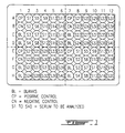
- Fig. 1 illustrates the layout of the plate of the preferred kit of the present invention.
- kits of the present invention are novel in that they allow for a simple and fast testing in the field where the animals are. These kits are sufficiently stable that they have a shelf life of at least 25 weeks.
- the antigen was purified according to a novel procedure which allows for an increased sensitivity.
- kits of the present invention are based essentially on the particular combination of a novel method of purification of lipopolysaccharide antigen from a reference strain of A. pleuropneumoniae serotype 1 antigen and a novel coating and stabilization of the antigen on the surface of the plate using a solution containing HRP stabilizing solution.
- the kit in accordance with the present invention is very specific, sensitive and stable.
- the test in accordance with the kit of the present invention consists in the determination of the presence or absence of anti- A. pleuropneumoniae antibodies in the serum of pigs for the serodiagnosis of A. pleuropneumoniae serotypes 1, 9 and 11.
- the test essentially consists in the following steps:
- the preferred kit of the present invention comprises the following items:
- Fig. 1 illustrates the layout of the plate of the preferred kit of the present invention, where 40 different sera are analyzed.
- the international reference strain, Shope 4074 (American Type Culture Collection, 12301 Parklawn Drive, Rockville, MD 20852 USA, ATCC accession number 27088), of A. pleuropneumoniae serotype 1 was used for the antigen production.
- the strain was kept lyophilized.
- PPLO Denssion Laboratories, Detroit, MI
- the content of a vial was resuspended in one ml of PPLO (Difco Laboratories, Detroit, MI) liquid medium and inoculated to exhaustion on a PPLO agar plate.
- the plate was incubated aerobically for 24 hours at 37°C.
- a few colonies were resuspended in 5 ml of PPLO liquid medium.
- PPLO plates were inoculated at confluence with a sterile swab, these plates were then incubated 6 hours at 37°C.
- the bacterial growth was harvested by adding 3.0 ml of phosphate-buffered saline (PBS, pH 7.4) (Oxoid Ltd., Basingstoke, England) containing 0.5% (vol/vol) of formaldehyde (Fisher Scientific, Fair Lawn, NJ) to each gel plates.
- PBS phosphate-buffered saline
- the bacterial suspension obtained was placed in a sterile bottle and was allowed to stand overnight at 4°C.
- the optical density was adjusted to 5.0 with a solution of PBS-0.5% formaldehyde.
- the suspensions were separated in sterile screw cap vials and boiled 60 min. Then the suspensions were centrifuged at 12,000 x g for 30-40 min., 4°C. The supernatants were collected and filtered on a 0.22 ⁇ m pore size filter (Millipore Corp., Bedford, MA).
- the antigen was purified according to the following procedure.
- Sera from several pigs were obtained at the slaughter from a specific pathogen free herd; the sera were mixed and thimerosal (sold by Sigma, St-Louis, MO 14508, U.S.A., catalogue #T-5125) was added to obtain a final concentration of 0.01%. No history of A. pleuropneumoniae was ever reported for this herd since at least four years. The sera were tested using the ELISA technique against all the A. pleuropneumoniae serotypes by the pleuropneumonia laboratory of the Veterinary Medicine Faculty of University of Montreal.
- strain A. pleuropneumoniae serotype 1 (strain 4074; ATCC 27088) was used for the bacterial production.
- the content of a vial was resuspended in one ml of PPLO (Difco Laboratories, Detroit, MI) liquid medium and inoculated to exhaustion on two PPLO agar plates. The plates were incubated aerobically for 18 hours at 37°C.
- the bacterial production for the immunization of pigs was effected according to the following procedure.
- the pigs were immunized intravenously with 0.5 ml of the bacterial suspension every three weeks and this until the ELISA titer give a value superior or equal to 1.0 with the serum diluted 1/200.
- the pigs were bled and the serum of each animal was mixed together.
- the plates are used immediately after sensitization, the incubation period are of one hour and ABTS (2,2-azino-bis(3-ethyl-benzthiazoline-6-sulfonic acid) is used as a chromogen.
- the plates are treated with HRP (horseradish peroxidase) conjugate stabilizing solution (sold by Calbiochem-Novabiochem Corporation, La Jolla, CA 92039, U.S.A., catalogue #516534) after sensitization, the incubation period are of 15 min. and TMBLueTM is used as a chromogen.
- the HRP conjugate stabilizing solution is diluted to a 1:5 ratio using PBS-EDTA buffer at pH 7.3.
- the ELISA consists in:
- the plates are treated with HRP conjugate stabilizing solution.
- the content of the wells are emptied and 100 ⁇ l of the diluted HRP conjugate stabilizing solution is added to each well.
- the plates are kept at 4°C until usage.
- the optical density was read at 410 nm using an automated plate reader (MR5000TM, Dynatech Laboratories Inc.).
- the aim of this test series consists in verifying the reproducibility of the antigen bounding at the bottom of the wells of the plate.
- the variation in the antigen attachment between the wells of a same plate was determined.
- Each of the 96 wells of each three plates was sensitized with 10 ⁇ g of A. pleuropneumoniae serotype 1 antigen, the plates were used immediately after their sensitization according to the above-mentioned ELISA-ABTS procedure.
- the control sera were distributed in the three plates.
- the positive control serum was used in 16 wells of each three plates and the negative control serum in 17 wells.
- Each of the six field controls were distributed in eight wells of each plates. The results are presented in Table 1.
- the specificity of the kit of the present invention comes from a purified lipopolysaccharide antigen from a reference strain of A. pleuropneumoniae serotype 1.
- the kit allows for the detection of anti- A. pleuropneumoniae antibodies in the serum of pigs and to determine whether or not the pigs have developed antibodies.
- several animals of a herd must be verified to determine the status of the herd.
- the presence of at least one positive serum means that at least one A. pleuropneumoniae serotypes 1, 9 or 11 is present in the herd.
- all the sera analyzed are negative, and if the sampling is representative of the herd, this indicates that the pigs analyzed have not developed antibodies against A.
- pleuropneumoniae serotypes 1, 9 or 11 and that during the blood sampling, the herd has not come in contact with the bacteria. Nevertheless, due to the limit of the serological testing, it is recommended to repeat the testing after a few weeks, the more analysis are negatives higher is the probability that the herd is free of A. pleuropneumoniae serotypes 1, 9 or 1. Following a first series of analysis, in cases where only weak positive and negative sera are observed, no status can be certified. It is then recommended to repeat the testing and to compare the results with those of a reference laboratory.
- the antigen specificity was verified using serum of pigs infected experimentally with strains of A. pleuropneumoniae of different serotypes or other bacteria.
- the ELISA method was used with ABTS as a chromogen.
- the antigen gave positive reactions with antisera against A. pleuropneumoniae serotypes 1, 9 and 11 and negative reactions for serotypes 2, 3, 4, 5 and 7.
- the sera from pigs inoculated with a strain of different serotypes of Actinobacillus pleuropneumoniae, Haemophilus parasuis, Pasteurella multocida, Escherichia coli, or A. suis gave negative results with the purified antigen. The results are presented in Table 2.
- pleuropneumoniae serotype 1 (strain 4074) against different sera of pigs inoculated experimentally Sera from pigs inoculated with Optical density
- A. pleuropneumoniae serotype 1 (strain Shoe 4074) 0.69 A. pleuropneumoniae serotype 2 (strain 4226) 0.03 A. pleuropneumoniae serotype 3 (strain 1421) 0.01 A. pleuropneumoniae serotype 3 (strain 89-92A13) 0.04 A. pleuropneumoniae serotype 4 (strain M62) 0.04 A.
- pleuropneumoniae serotype 5 (strain K17) 0.05 A. pleuropneumoniae serotype 7 (strain WF83) 0.02 A. pleuropneumoniae serotype 9 (strain CVJ 13261) 0.58 A. pleuropneumoniae serotype 11 (strain 56153) 0.87 Actinobacillus suis 0.02 Haemophilus parasuis 0.02 Pasteurella multicida 0.02 Escherichia coli 0.02
- ELISA-TMBLueTM results of the purified antigen A. pleuropneumoniae serotype 1 (strain 4074) against reference sera Serum used Optical density Visual Response CN 0.063 0 CP 1.884 3+ EP1 2.097 4+ EP2 0.995 2+ EN1 0.139 0 EN2 0.046 0 ED1 0.515 1+ ED2 0.450 1+
- the CP, EP1 and EP2 sera gave positive responses.
- the serum CP came from a pool of sera obtained pursuant to the inoculation of two five weeks old piglets with the reference strain of A. pleuropneumoniae.
- the EP1 and EP2 sera were obtained from animals of a herd where clinical cases caused by A. pleuropneumoniae serotype 1 is frequently observed.
- the CN serum is the negative control serum which came from a pool of sera of a herd specific pathogen free where no history of A. pleuropneumoniae was ever reported for this herd since at least four years.
- the EN1 and EN2 sera came from two pigs from a herd of no history of A. pleuropneumoniae and lesions at the slaughter.
- the preferred method for the antigen attachment consists in diluting 150 ⁇ l of antigen in 75 ⁇ l PBS-EDTA buffer, pH 7.3; 100 ⁇ l of this solution is distributed in each wells of the plates (96-well NuncTM plates). The plates are incubated 18 hours at 4°C. Then, the plates are washed three times with PBS-Tween TM-20 buffer. The content of the plates is emptied and 100 ⁇ l of diluted HRP conjugate stabilizing solution (1:5 with PBS-EDTA buffer at pH 7.3) is added to each well. The plates are kept at 4°C.
- kits In addition to the three controls included in the kit, eight additional sera are used for the stability study of the kit. These additional sera are kept at -20°C and a new aliquot is used during each assay. During each assay, a complete kit is used, the kit is validated with the different controls included with each kit. As demonstrated in Table 4 below, the kit was first kept up to 25 weeks. Stability of the kit A. pleuropneumoniae serotype 1, 9 and 11 visual test Sera Number of weeks at 4°C 0 4 7 12 20 25 Positives (4+,3+,2+) 5 5 5 5 5 5 5 Weak positives (1+) 1 1 1 1 1 1 Negatives (0+) 5 5 5 5 5 5 5 5
- kits were prepared. A total of 20 different sera obtained from the serology laboratory of the Veterinary Medicine Faculty of University of Montreal, a kit as well as an aliquot of each serum undiluted were given to three different user. Identical results were obtained by all of these users (Table 5). Evaluation of the kit A. pleuropneumoniae serotype 1, 9 and 11 visual test by three different users Sera User #1 batch User #2 User #3 Positives (4+,3+,2+) 6 5 6 Weak positives (1+) 1 2 2 2 Negatives (0+) 16 16 15
- kits were prepared in three different batches. Each batch of kits was used with different sera (Table 6). A total of 295 sera, the status of which was determined by the pleuropneumonia serology laboratory of the Veterinary Medicine Faculty of the University of Montreal, were evaluated. The 192 negative sera classified as negative by the pleuropneumonia serology laboratory were confirmed as negative with the kit of the present invention. Within the sera classified as positive by the pleuropneumonia serology laboratory, 15 sera gave a 1+ response and the remaining 88 sera gave a 2-4+ response with the kit of the present invention (Table 6). Sensitivity and specificity of the kit A. pleuropneumoniae serotype 1, 9 and 11 visual test during three different assays Results with the kit Results with the "golden test" Positive Negative 4-3+ 45 0 2+ 43 0 1+ 15 0 0 0 192
- the determination of the sensitivity and the specificity of a test is effected either by using animal populations of status clearly identified as “infected” or as "healthy” or by comparing the test results with a reference test or a test referred to as a "golden” test.
- the two methods were used.
- sera of well defined status 13 sera from pigs infected experimentally with strains of A. pleuropneumoniae of different serotypes or with different bacteria were used. Thereafter, eight additional sera of pigs with well defined status were used; a serum from a pig infected experimentally with A. pleuropneumoniae serotype 1, a serum from a pool of sera of specific pathogen free pigs and six sera of pigs from different herds with a well defined status.
- the ELISA kit of the present invention gave a positive response only with sera from pigs infected experimentally or naturally with A. pleuropneumoniae serotype 1, serotype 9 or serotype 11.
- the ELISA kit of the present invention gave negative results for sera of pigs infected experimentally with A. pleuropneumoniae serotypes 2, 3, 4, 5 and 7, H. parasuis, P. multicida, E. coli, or A. suis as well as for two pool of sera from pigs of specific pathogen free herds.
- the ED1 and ED2 sera came from pigs with no clinical signs of pleuropneumonia but where evidence of infection with A. pleuropneumoniae serotype 1 was reported for this herd.
- These sera gave a positive response with the kit of the present invention.
- the EN1 and EN2 sera which came from a herd with no history of pleuropneumonia, were considered as negative.
- the ELISA kit of the present invention is stable at 4°C for at least 25 weeks. The stability study is still in progress.
- the pleuropneumonia laboratory was considered as the "golden test” and the positive limit was established so that a maximum of correlation between the two tests is obtained.
- This pleuropneumonia laboratory analyzes between 30,000 and 40,000 sera of pigs per year and this since more than 12 years.
- the method used by this laboratory consists in a standardized ELISA technique to determine the presence of antibodies, which method was established by comparing different antigens (Gottschalk, M. et al., 1994, Vet. Microbiol., 42 :91-104).
- This laboratory has stopped using the complement fixation test due to its lack of sensitivity and specificity. Since this laboratory receives numerous serum samples, the results obtained by this laboratory were considered as a reference.
- the status of the sera, negative or positive, obtained by this laboratory was thus well certified.
- the specificity and the sensitivity of the kit was evaluated using 295 pig sera. A correlation of 100% was obtained. All the sera classified as negative by the reference serology laboratory were also determined as negative by the kit of the present invention. All the sera classified as positive by the reference laboratory were also confirmed as positive using the kit of the present invention.
- the kit of the present invention differs from the ELISA method used by the reference serology laboratory.
- the antigen is fixed to the plates in a PBS buffer and the plates are immediately used after the antigen fixation is completed.
- This reference serology laboratory utilizes a computerized reading protocol for the determination of the antibodies in the samples (Trottier, Y.L. et al., 1992, J. Clin. Microbiol., 30 :46-53).
- the kit of the present invention differs in that the antigen is purified using a higher concentration of phenol and the antigen fixation is different. Indeed, the purified antigen is resuspended in a PBS-EDTA buffer which is then added to each well of the plate. After a 18h incubation, a HRP buffer is added to each well.
- the antibodies in the samples are determined visually by adding a chromogen, preferably TMBlueTM.
- the kit of the present invention when compared to the standardized ELISA method, demonstrate a relative sensitivity and a relative specificity of 100%.
- the kit of the present invention is faster to use than the ELISA-ABTS method of the prior art.
- the determination of A. pleuropneumoniae serotypes 1, 9 or 11 antibodies using the kit of the present invention can be effected in less than one hour whereas a minimum of three hours should is required for the ELISA-ABTS method in addition to one night of incubation for the antigen attachment step. Further, ABTS is considered as a potentially carcinogenic and has very little stability.
- the kit of the present invention is easily used and provide rapid results.
- the kit of the present invention may be used in the field where the animals are kept and do not require laboratory skills since only simple steps are performed. This kit was demonstrated to give highly reliable and reproducible results and this independently of the users.
- the results obtained for the A. pleuropneumoniae serotypes 1, 9 and 11 samples are equivalent to the results obtained by the pleuropneumonia laboratory.
- the kit of the present invention is highly advantageous compared to the now available laboratory test with respect to its rapidity, reliability, sensitivity, specificity, stability and cost.
Landscapes
- Health & Medical Sciences (AREA)
- Life Sciences & Earth Sciences (AREA)
- Immunology (AREA)
- Engineering & Computer Science (AREA)
- Urology & Nephrology (AREA)
- Hematology (AREA)
- Biomedical Technology (AREA)
- Chemical & Material Sciences (AREA)
- Molecular Biology (AREA)
- Medicinal Chemistry (AREA)
- Physics & Mathematics (AREA)
- Cell Biology (AREA)
- Tropical Medicine & Parasitology (AREA)
- Biotechnology (AREA)
- Food Science & Technology (AREA)
- Virology (AREA)
- Microbiology (AREA)
- Analytical Chemistry (AREA)
- Biochemistry (AREA)
- General Health & Medical Sciences (AREA)
- General Physics & Mathematics (AREA)
- Pathology (AREA)
- Peptides Or Proteins (AREA)
- Medicines Containing Antibodies Or Antigens For Use As Internal Diagnostic Agents (AREA)
- Measuring Or Testing Involving Enzymes Or Micro-Organisms (AREA)
Description
- The invention relates to stable kits for the accurate, rapid and sensitive assay of A. pleuropneumoniae
serotypes - Actinobacillus pleuropneumoniae is known as one of the most pathogenic agent of the respiratory tract of swine. Swine pleuropneumonia is still an important problem in large swine operations, causing serious economic losses in this industry. Since the presence of A. pleuropneumoniae is often unnoticed in chronically infected herds, the identification of carrier animals is a main concern. Following a stressful situation, several clinically fatal cases may arise in a given herd. Infection in swine can be fatal but animals surviving the infection frequently become carriers. Detection of chronically infected carriers is crucial since those animals act as reservoirs of infection. Since the infection is often unnoticed, serology become a useful tool for the detection of chronic infection. Several studies indicate that it is possible to control or eliminate the infection in certain herds based on the serological results.
- Various serological assays for A. pleuropneumoniae have been described. Among others, the complement fixation test (CFT), the enzyme-linked immunosorbent assay (ELISA); (Goyette G. et al., 1986, Int. Pig. Vet. Soc. Proc., 9:258) and the 2-mercapto-ethanol tube agglutination test (Mittal, K. et al., 1984, Am. J. Vet. Res., 45:715-719) have been used. Out of the different assays, the ELISA is often the most useful since it is faster and easier to perform. On the other hand, up to now, the results obtained suggested the use of a more purified antigenic preparation in order to improve the specificity of the test.
- A saline extract of boiled-formalinized whole cells of A. pleuropneumoniae (also called crude extract) is presently used, in some laboratories, as the antigen for ELISA serodiagnosis (Goyette G. et al., 1986, Int. Pig. Vet. Soc. Proc., 9:258). Standardization of the assay is complicated as variations are noticed between extracts.
- Using different antigen preparations, cross-reactions among serotypes and with other bacterial species were also reported (Bossé, J. et al., 1990, Can. J. Vet. Res., 54:427-431). Although the capsular polysaccharide (CPS) of A. pleuropneumoniae has been shown to be responsible for serotype specificity (Inzana, T. and Mathison, T., 1987, Infect. Immun., 55:1580-1587), the difficulty of obtaining pure CPS in large quantity precludes its utilization for serodiagnostic purposes. The CPS were very unstable and were fixed with difficulty to the walls of the polystyrene plate used in the ELISA assay (Perry, B. et al., 1990, Sero. Immunol. Infect. Dis., 4:299-308).
- Serology, which is used to identify animals that have developed an immune response to specific pathogens, is an important tool in disease management and prevention of A. pleuropneumoniae infection in pigs. The importance of serological testing is further emphasized by the lack of a vaccine that reliably prevents infection. The demand of pigs from A. pleuropneumoniae sero-negative herds is increasing, especially from producers whose herds have experienced acute outbreaks of the disease and who have decided to "eradicate" A. pleuropneumoniae, buying only sero-negative animals (coming from sero-negative herds) for the replacement. A successful eradication program depends mostly on the accuracy and reliability of the serological tests used to identify A. pleuropneumoniae infected pigs. Nevertheless, interpretation of serology should be done cautiously. A test that is not sensitive will not detect all infected herds or animals (false negative results) and one that is not specific will erroneously condemn some non-infected animals (false positive results).
- Presently, it is impossible to serologically distinguish the infection caused by strains of A. pleuropneumoniae
serotype 1 from that caused by strains of A. pleuropneumoniaeserotype 9 and/or 11. This is due mainly to the O chain from the lipopolysaccharide (LPS), which presents common antigenic determinants. The antigenic specificity of A. pleuropneumoniae mainly comes from the capsular polysaccharides, which are not very immunogenic. Purified capsular polysaccharides antigens are very difficult to obtain and, in addition, contamination with non-specific antigen are very common. The distinction between these serotypes (1-9-11) necessitate bacterial isolation. The isolation of the bacteria from chronically infected animals is a time consuming and low sensitive method. - An Elisa assay for the serodiagnosis of Actinobacillus pleuropneumoniae have been used by Radacovici et al. (Vet. Microbiol. vol. 39, pages 219-230, April 1994). Radacovici et al. utilize phenol extractions of a crude extract of
A. pleuropneumoniae serotype 1 to recover LPS with long 0-chains in the aqueous phase to be used as antigens in an ELISA. However, the antigen used in the Elisa assay of Radacovici et al. is not very stable. - The use of antibiotics is mainly useful to control the mortality, but it has no real benefit on pigs with chronic pleuropneumonia. Treated animals often continue to carry the organism and can be a source of infection for other animals. In addition, an increasing number of strains resistant to different antimicrobials has been observed in the last years in Quebec (Nadeau, M. and Higgins, R., 1991, Bulletin épidémiologique, 2:4-5).
- There are some cross-reactions among serotypes; for example:
serotypes serotypes serotypes - Healthy carrier pigs may be responsible for the transmission of the disease. The absence of clinical signs and/or lesions at the slaughter-house does not implicate necessarily the absence of the infection.
- Following infection, antibodies can usually be detected in 10-15 days. Some animals will remain serologically positive for a few months, but most will be positive for a long period of time; once more, it will depend on the test used.
- The proportion of seropositive sows as well as their titers tended to decrease with age.
- Isolation of A. pleuropneumoniae from apparently healthy carrier pigs is difficult; it probably should be used as a complement to the serology in conflictive cases.
- The development of better serological tests is a necessity because the infection still has an economic impact on the swine industry and the current vaccines are not effective.
- To date, there exist no stable kit for the effective serodiagnosis of pig pleuropneumonia in the field.
- It would be highly desirable to be provided with a kit for readily determining the presence of antibodies against A. pleuropneumoniae serotypes 1, 9 and 11 in a serum sample.
- It would be highly desirable to be provided with such an ELISA diagnostic kit for A. pleuropneumoniae which could be used for A. pleuropneumoniae serodiagnosis while remaining in the field.
- One aim of the present invention is to provide a kit for the accurate, rapid and sensitive assay of antibodies against A. pleuropneumoniae serotypes 1, 9 and 11 in a sample.
- Another aim of the present invention is to provide an ELISA diagnostic kit for A. pleuropneumoniae to be used for A. pleuropneumoniae serodiagnosis while remaining in the field. The novelty and originality of the ELISA diagnostic kit of the present invention resides in the particular combination of a novel purification method of the antigen to be used and a novel sensitization and stabilization methods of the plates of the kit.
- The kits of the present invention differs from the ELISA method of the prior art for the determination of A. pleuropneumoniae antibodies. In the prior art method, the antigen is fixed to the plates in a PBS buffer and the plates are immediately used after the antigen fixation is completed. The prior art method may include a computerized reading protocol for the determination of the antibodies in the samples as described by Trottier, Y.L. et al. (1992, J. Clin. Microbiol., 30:46-53). The kits of the present invention mainly differ in that the antigen is purified using a higher concentration of phenol and the antigen fixation procedure is different. Indeed, the purified antigen is resuspended in a PBS-EDTA buffer which is then added to each well of the plate. After a 18h incubation, a HRP buffer is added to each well. The antibodies in the samples are determined visually by adding a chromogen, preferably TMBlue™. The kits of the present invention, when compared to the prior art ELISA method, demonstrate a relative sensitivity and a relative specificity of 100%.
- In accordance with one embodiment of the present invention there is provided an ELISA diagnostic kit for the assay of A. pleuropneumoniae serotypes 1, 9 or 11 antibodies in the serum of pigs comprising in separate packaging, at least one of the following:
- a) a solid support having bound thereto a purified
lipopolysaccharide
A. pleuropneumoniae serotype 1 antigen for a specific binding to anti-A. pleuropneumoniae serotypes 1, 9 or 11 antibodies present in the serum of pigs, wherein the bound antigen is stored in a solution which keeps said bound antigen stable for 25 weeks at 4°C; - b) serum from pigs experimentally inoculated with
a strain of
A. pleuropneumoniae serotypes 1 to serve as a positive control; - c) pig serum from a specific pathogen free herd to serve as a negative control; and
- d) a detectably labeled conjugate which bind to pigs antibodies bound to the plate of a).
-
- The ELISA diagnostic kits of the present invention may further comprise the following:
e) a substrate which allow the visualization of the detectably labeled conjugate. - In accordance with another embodiment of the present invention there is provided a method for the preparation of the kit, which comprises the steps of:
- a) purifying lipopolysaccharide
A. pleuropneumoniae serotype 1 antigen by phenol extraction and centrifugation of said antigen bacterial crude extract; - b) fixing the antigen of step a) to a solid support and stabilizing said fixed antigen by storing it in a solution which keeps the fixed antigen stable for 25 weeks at 4°C;
- c) immunizing mammals with a strain of
A. pleuropneumoniae serotype 1 and collecting serum to serve as positive control sera; and - d) collecting sera from A. pleuropneumoniae free herds to serve as negative control sera.
-
- In accordance with the present invention, the solution used to stabilized the antigen once fixed to the plate comprises HRP stabilizing solution.
- Fig. 1 illustrates the layout of the plate of the preferred kit of the present invention.
- The kits of the present invention are novel in that they allow for a simple and fast testing in the field where the animals are. These kits are sufficiently stable that they have a shelf life of at least 25 weeks. The antigen was purified according to a novel procedure which allows for an increased sensitivity.
- The kits of the present invention are based essentially on the particular combination of a novel method of purification of lipopolysaccharide antigen from a reference strain of
A. pleuropneumoniae serotype 1 antigen and a novel coating and stabilization of the antigen on the surface of the plate using a solution containing HRP stabilizing solution. The kit in accordance with the present invention is very specific, sensitive and stable. The test in accordance with the kit of the present invention consists in the determination of the presence or absence of anti-A. pleuropneumoniae antibodies in the serum of pigs for the serodiagnosis of A. pleuropneumoniae serotypes 1, 9 and 11. - The test essentially consists in the following steps:
- a) A plate of 96 wells sensitized with the specific
antigen of
A. pleuropneumoniae serotype 1 is washed with a PBS-Tween™-20 buffer solution. The antigen-coated plates are previously stabilized using HRP stabilizing solution. - b) A serum sample of each pigs from the tested herds is distributed in two sensitized wells of step a). During this first incubation, the anti-A. pleuropneumoniae serotypes 1, 9 and 11 antibodies, if present in the sera, will bind to the antigen attached to the plate or solid phase bound.
- c) The plate is washed to remove from the wells any unbound material. A peroxidase-anti-IgG conjugate is added to each well, preferably obtained from Jackson Immuno Research Laboratories (catalogue #114-035-003). This conjugate binds to any IgG which would have bound to the antigen attached to the plate in step b). If the pig serum did not contain any anti-A. pleuropneumoniae serotypes 1, 9 and 11 antibodies, the conjugate will remain free or in suspension and will be eliminated during this washing step.
- d) The presence of immobilized peroxidase within the bound conjugate is revealed by the addition of a chromogen substrate TMBLue™ (sold by Transgenic Science Inc., Milford' MA 01757, U.S.A., catalogue #TM-102). If the conjugate is present, there will be an oxidation reaction and a blue color will appear.
-
- The preferred kit of the present invention comprises the following items:
- 1- Five 96-well plates (Nunc™, sold by Gibco, Burlington, Ontario, Canada, L7P lAl) sensitized and stabilized with the purified antigen in accordance with the present invention.
- 2- Positive control; five vials containing each
0.1 ml of lyophilized serum from pigs experimentally
inoculated with a strain of
A. pleuropneumoniae serotype 1. - 3- Negative control; five vials containing each 0.1 ml of lyophilized pig serum from a specific pathogen free herd.
- 4- Weak positive control; five vials containing
each 0.1 ml of lyophilized serum from pigs
experimentally inoculated with a strain of
A. pleuropneumoniae serotype 1. - 5- Conjugate; five vials of pig anti-IgG immunoglobulins coupled to peroxidase. Each vial contains 1.2 ml of lyophilized conjugate.
- 6- TMBLue™; five vials containing each 10 ml of TMBLue™.
-
- Fig. 1 illustrates the layout of the plate of the preferred kit of the present invention, where 40 different sera are analyzed. The wells are identified as follows:
BL = Blanks, PBS-Tween"-20 buffer solution (4 wells) CP = Positive control,item # 2 above (4 wells) CN = Negative control,item # 3 above (4 wells) CPFA = Weak positive control,item # 4 above (4 wells) S1 to S40 = serum to be analyzed, 2 wells for each serum. - Add the following to 3 L of distilled water,
- 52.59 g of sodium chloride;
- 1.47 g of monobasic sodium phosphate;
- 7.02 g dibasic sodium phosphate;
- 1.5 ml of Tween™-20.
-
- Mix well until a complete dissolution is achieved. Verify the pH, which should be about 7.30 ± 0.05, if different adjust the pH using dibasic sodium phosphate. This solution has a shelf life of 1 week when kept at 4°C. The buffer solution should always be brought to room temperature before being used in the test.
- The international reference strain, Shope 4074 (American Type Culture Collection, 12301 Parklawn Drive, Rockville, MD 20852 USA, ATCC accession number 27088), of
A. pleuropneumoniae serotype 1 was used for the antigen production. The strain was kept lyophilized. - The content of a vial was resuspended in one ml of PPLO (Difco Laboratories, Detroit, MI) liquid medium and inoculated to exhaustion on a PPLO agar plate. The plate was incubated aerobically for 24 hours at 37°C. A few colonies were resuspended in 5 ml of PPLO liquid medium. PPLO plates were inoculated at confluence with a sterile swab, these plates were then incubated 6 hours at 37°C. After the incubation, the bacterial growth was harvested by adding 3.0 ml of phosphate-buffered saline (PBS, pH 7.4) (Oxoid Ltd., Basingstoke, England) containing 0.5% (vol/vol) of formaldehyde (Fisher Scientific, Fair Lawn, NJ) to each gel plates.
- The bacterial suspension obtained was placed in a sterile bottle and was allowed to stand overnight at 4°C. The optical density was adjusted to 5.0 with a solution of PBS-0.5% formaldehyde. The suspensions were separated in sterile screw cap vials and boiled 60 min. Then the suspensions were centrifuged at 12,000 x g for 30-40 min., 4°C. The supernatants were collected and filtered on a 0.22 µm pore size filter (Millipore Corp., Bedford, MA).
- The antigen was purified according to the following procedure.
- Preparing the phenol solution by mixing 90 g of phenol crystals with 100 ml of distilled water.
- Mixing an equal volume of the phenol solution with an equal volume of the crude extract in Corex™ tubes, mixing by inversion and let stands for 30 min. at room temperature.
- Centrifuged at 12,000 x g, 4°C for 30 min.
- After the first centrifugation, two phases were obtained with an interface of insoluble material. The aqueous phase was collected with a Pasteur™ pipette and the volume was measured in a graduated cylinder.
- Mixing an equal volume of the aqueous phase (first extraction) with an equal volume of the phenol phase in Corex™ tubes, mixing by inversion and let stands for 30 min. at room temperature.
- Centrifuged at 12,000 x g, 4°C for 30 min.
- The aqueous phase was collected and the volume was measured in a graduated cylinder.
- Mixing an equal volume of the aqueous phase (second extraction) with an equal volume of the phenol phase in Corex™ tubes, mixing by inversion and let stands for 30 min. at room temperature.
- During this period, prepare the dialysis membrane by soaking in distilled water for a sufficient period of time.
- Centrifuged at 12,000 x g, 4°C for 30 min.
- The aqueous phase was collected and the volume was measured in a graduated cylinder.
- The aqueous phase (third extraction) was dialyzed against 3 x 12 L of distilled water to remove traces of phenol, do not dialyze for more than 24 hours.
- Sera from several pigs were obtained at the slaughter from a specific pathogen free herd; the sera were mixed and thimerosal (sold by Sigma, St-Louis, MO 14508, U.S.A., catalogue #T-5125) was added to obtain a final concentration of 0.01%. No history of A. pleuropneumoniae was ever reported for this herd since at least four years. The sera were tested using the ELISA technique against all the A. pleuropneumoniae serotypes by the pleuropneumonia laboratory of the Veterinary Medicine Faculty of University of Montreal.
- The strain A. pleuropneumoniae serotype 1 (strain 4074; ATCC 27088) was used for the bacterial production. The content of a vial was resuspended in one ml of PPLO (Difco Laboratories, Detroit, MI) liquid medium and inoculated to exhaustion on two PPLO agar plates. The plates were incubated aerobically for 18 hours at 37°C.
- The bacterial production for the immunization of pigs was effected according to the following procedure.
- Collecting a few colonies isolated with a sterile swab and resuspending them in a PPLO broth.
- 5 PPLO agar plates were inoculated at confluence with the broth and sterile swabs. One Mueller-Hinton agar plate was inoculated with the remaining broth to serve as a negative control. One PPLO agar plate was inoculated to exhaustion.
- These plates were then incubated 18 hours at 37°C. One plate is used for serotyping.
- After the incubation, harvesting the bacterial growth by adding 3.0 ml of PBS-0.5% formaldehyde to each plate, and mixed with a hockey stick made of sterile Pasteur™ pipette and recovering the suspension with a pipette.
- The bacterial suspension obtained was placed in a sterile bottle, mixed well and incubated 18 hours at room temperature.
- The optical density was read at 540 nm and adjusted to 1.0 with a solution of PBS-0.5% formaldehyde.
- The solution was kept at 4°C until usage or for a maximum of one week.
- For the immunization, four five-week old piglets were obtained from a specific pathogen free herd. No history of A. pleuropneumoniae was ever reported for this herd since at least four years and no pulmonary lesions were observed at the slaughter. At their arrival, the general state of health of the piglets is verified. The piglets are fed on demand with introductory fattening food for pigs (15/30 CO-OP). After a few days of adaptation, a blood sample is taken from each animal. The sera were tested using the ELISA technique against all the A. pleuropneumoniae serotypes by the pleuropneumonia laboratory of the Veterinary Medicine Faculty of University of Montreal. The sera were negative for all serotypes.
- The pigs were immunized intravenously with 0.5 ml of the bacterial suspension every three weeks and this until the ELISA titer give a value superior or equal to 1.0 with the serum diluted 1/200. The pigs were bled and the serum of each animal was mixed together.
- For evaluating the efficiency and reliability of the kits of the present invention, two methods of ELISA were used. In the first method, the plates are used immediately after sensitization, the incubation period are of one hour and ABTS (2,2-azino-bis(3-ethyl-benzthiazoline-6-sulfonic acid) is used as a chromogen. In the second method, the plates are treated with HRP (horseradish peroxidase) conjugate stabilizing solution (sold by Calbiochem-Novabiochem Corporation, La Jolla, CA 92039, U.S.A., catalogue #516534) after sensitization, the incubation period are of 15 min. and TMBLue™ is used as a chromogen. The HRP conjugate stabilizing solution is diluted to a 1:5 ratio using PBS-EDTA buffer at pH 7.3.
- The ELISA consists in:
-
- Diluting in 150 µl of antigen in 75 ml of PBS-EDTA buffer, pH 7.3.
- Add 100 µl of antigen to each well.
- Seal the plate with an acetate sheet.
- Incubate overnight at 4°C.
- For the evaluation of the kit, for the stability assays as well as for the visual assays, the plates are treated with HRP conjugate stabilizing solution. The content of the wells are emptied and 100 µl of the diluted HRP conjugate stabilizing solution is added to each well. The plates are kept at 4°C until usage.
-
- Recovering the plate and emptying its content.
- Filling each well with PBS-Tween"-20 buffer.
- Emptying the plate content.
- Repeat these steps four times.
- Shake off 2-3 times on an absorbing paper to remove any washing solution excess.
-
- The sera are diluted 1/200 in PBS-Tween™-20 buffer and distributed in the amount of 100 µl to each well.
- Gently shake the plate to ensure the distribution of the samples at the bottom of the wells. Cover the plate with an acetate sheet.
- Let the plate stand for one hour at room temperature for ELISA using ABTS or for 15 min. between 18°C and 22°C for ELISA using TMBLue™.
-
- Recovering the plate and emptying its content.
- Filling each well with PBS-Tween™-20 buffer.
- Emptying the plate content.
- Repeat these steps four times.
- Shake off 2-3 times on an absorbing paper to remove any washing solution excess.
-
- The conjugate consists in horseradish peroxidase-labeled immunoglobulin G fraction of rabbit antiserum raised against porcine IgG (Jackson Immuno Research Laboratories Inc., catalogue #114-035-003). The conjugate is used at a final dilution of 1/6000. The conjugate is distributed in the amount of 100 µl to each well of the plate.
- Gently shake the plate to ensure the distribution of the samples at the bottom of the wells. Cover the plate with an acetate sheet.
- Let the plate stand at room temperature for one hour for ELISA using ABTS or for 15 min. for ELISA using TMBLue™.
-
- Recovering the plate and emptying its content.
- Filling each well with PBS-Tween™-20 buffer.
- Emptying the plate content.
- Repeat these steps four times.
- Shake off 2-3 times on an absorbing paper to remove any washing solution excess.
- This was used only to validate the kit of the present invention or to get a spectrophotometer value.
-
- The reaction was visualized using 2 mM H2O2 and 0.4 mM ABTS (2,2-azino-bis(3-ethylbenzthiazoline-6-sulfonic acid) (Sigma Chemical) in 50 mM citrate solution (pH 4.0). Add 100 µl of this citrate-ABTS solution to each well of the plate.
- Gently shake the plate to ensure the distribution of the samples at the bottom of the wells.
- Let the plate stand for 30 min. at room temperature (between 18°C and 22°C).
- The optical density was read at 410 nm using an automated plate reader (MR5000™, Dynatech Laboratories Inc.).
- The results were calculated according to the following procedure.
- 1- The values of the eight wells BL (blank) were
verified:
- A value inferior to 0.08 indicates a valid test, continue the reading.
- A value superior to 0.08 indicates an invalid test, repeat the test with a new kit or contact the kit manufacturer.
- 2- The mean values of the negative and positive
controls are calculated as follows (see Fig. 1
for identification of wells):
-
-
- Add 100 µl of TMBLue™ to each well of the plate.
- Gently shake the plate to ensure the distribution of the samples at the bottom of the wells.
- Let the plate stand for 5 min. at room temperature (between 18°C and 22°C).
- The visual reading was effected without any reading instruments as follows:
- The results were calculated according to the following procedure.
- 1- The color of the eight wells BL (blanks) and of the negative controls is verified; it should be colorless.
- 2- The color of the positive controls is verified; it should be dark blue.
- 3- The color of the CPFA (weak positive control) is verified; it should be light blue.
- 4- The answer of each sample should be quantified
as follows:
- 0 = colorless well or slightly bluish
- 1+ = well of a light blue color
- 2+ = well of a blue color
- 3+ = well of a dark blue color
- 4+ = well of a dark blue color
-
- The reading with a spectrophotometer was effected and calculated as described in section 8a above.
- The aim of this test series consists in verifying the reproducibility of the antigen bounding at the bottom of the wells of the plate. The variation in the antigen attachment between the wells of a same plate was determined. Each of the 96 wells of each three plates was sensitized with 10 µg of
A. pleuropneumoniae serotype 1 antigen, the plates were used immediately after their sensitization according to the above-mentioned ELISA-ABTS procedure. The control sera were distributed in the three plates. The positive control serum was used in 16 wells of each three plates and the negative control serum in 17 wells. Each of the six field controls were distributed in eight wells of each plates. The results are presented in Table 1. The wells which had not receive any serum, the blanks, demonstrated a variation of 10% between the three plates, whereas the wells which received a serum demonstrated variations, interplate and intraplate, inferior to 8%.Reproducibility of antigen attachment between wells of a same plate or of different plates Plate # 1 Plate # 2Plate # 3Serum Mean Standard deviation Deviation % Mean Standard deviation Deviation % Mean Standard deviation Deviation % B 0.0554 0.0078 14.02 0.0499 0.0065 12.97 0.0450 0.0081 17.96 CN 0.1151 0.0067 5.84 0.1206 0.0054 4.49 0.1086 0.0046 4.24 CP 1.0933 0.0424 3.87 1.1591 0.0553 4.77 1.1420 0.0428 3.75 EP1 1.3182 0.0423 3.21 1.3488 0.0114 0.85 1.3978 0.0324 232 EP2 1.0588 0.0766 7.23 1.0795 0.0421 3.90 1.1157 0.0192 1.72 EN1 0.3877 0.0241 6.21 0.4295 0.0340 7.91 0.4147 0.0165 3.98 EN2 0.1312 0.0074 5.65 0.1367 0.0048 3.48 0.1363 0.0038 2.81 ED1 0.6470 0.0147 2.28 0.6782 0.0239 3.52 0.6778 0.0200 2.95 ED2 0.5372 0.0267 4.97 0.6012 0.0185 3.07 0.6000 0.0243 4.05 Mean of three plates Mean Standard deviation Deviation % 0.0501 0.0052 10.37 0.1147 0.0060 524 1.1198 0.0347 3.10 1.3549 0.0402 2.97 1.0847 0.0288 2.65 0.4106 0.0212 5.17 0.1347 0.0031 229 0.6677 0.0179 2.68 0.5794 0.0366 6.32 - The specificity of the kit of the present invention comes from a purified lipopolysaccharide antigen from a reference strain of
A. pleuropneumoniae serotype 1. The kit allows for the detection of anti-A. pleuropneumoniae antibodies in the serum of pigs and to determine whether or not the pigs have developed antibodies. During an analysis, several animals of a herd must be verified to determine the status of the herd. The presence of at least one positive serum means that at least one A. pleuropneumoniae serotypes 1, 9 or 11 is present in the herd. When all the sera analyzed are negative, and if the sampling is representative of the herd, this indicates that the pigs analyzed have not developed antibodies against A. pleuropneumoniae serotypes 1, 9 or 11 and that during the blood sampling, the herd has not come in contact with the bacteria. Nevertheless, due to the limit of the serological testing, it is recommended to repeat the testing after a few weeks, the more analysis are negatives higher is the probability that the herd is free of A. pleuropneumoniae serotypes 1, 9 or 1. Following a first series of analysis, in cases where only weak positive and negative sera are observed, no status can be certified. It is then recommended to repeat the testing and to compare the results with those of a reference laboratory. - Firstly, the antigen specificity was verified using serum of pigs infected experimentally with strains of A. pleuropneumoniae of different serotypes or other bacteria. The ELISA method was used with ABTS as a chromogen. The antigen gave positive reactions with antisera against A. pleuropneumoniae serotypes 1, 9 and 11 and negative reactions for
serotypes ELISA results of the purified antigen A. pleuropneumoniae serotype 1 (strain 4074) against different sera of pigs inoculated experimentally Sera from pigs inoculated with Optical density A. pleuropneumoniae serotype 1 (strain Shoe 4074) 0.69 A. pleuropneumoniae serotype 2 (strain 4226) 0.03 A. pleuropneumoniae serotype 3 (strain 1421) 0.01 A. pleuropneumoniae serotype 3 (strain 89-92A13) 0.04 A. pleuropneumoniae serotype 4 (strain M62) 0.04 A. pleuropneumoniae serotype 5 (strain K17) 0.05 A. pleuropneumoniae serotype 7 (strain WF83) 0.02 A. pleuropneumoniae serotype 9 (strain CVJ 13261) 0.58 A. pleuropneumoniae serotype 11 (strain 56153) 0.87 Actinobacillus suis 0.02 Haemophilus parasuis 0.02 Pasteurella multicida 0.02 Escherichia coli 0.02 - Secondly, the plates were treated with HRP after the sensitivity with the antigen and the ELISA-TMBLue™ technique was used (Table 3).
ELISA-TMBLue™ results of the purified antigen A. pleuropneumoniae serotype 1 (strain 4074) against reference sera Serum used Optical density Visual Response CN 0.063 0 CP 1.884 3+ EP1 2.097 4+ EP2 0.995 2+ EN1 0.139 0 EN2 0.046 0 ED1 0.515 1+ ED2 0.450 1+ - The CP, EP1 and EP2 sera gave positive responses. The serum CP came from a pool of sera obtained pursuant to the inoculation of two five weeks old piglets with the reference strain of A. pleuropneumoniae. The EP1 and EP2 sera were obtained from animals of a herd where clinical cases caused by
A. pleuropneumoniae serotype 1 is frequently observed. The CN serum is the negative control serum which came from a pool of sera of a herd specific pathogen free where no history of A. pleuropneumoniae was ever reported for this herd since at least four years. The EN1 and EN2 sera came from two pigs from a herd of no history of A. pleuropneumoniae and lesions at the slaughter. These sera give a negative response in an ELISA. The ED1 and ED2 sera give weak positive responses in an ELISA. These sera came from two pigs from a herd chronically infected withA. pleuropneumo niae serotype 1. - During the preliminary trials, different techniques of antigen attachment, different buffers as well as different preservation methods of the plates were evaluated. The preferred method for the antigen attachment consists in diluting 150 µl of antigen in 75 µl PBS-EDTA buffer, pH 7.3; 100 µl of this solution is distributed in each wells of the plates (96-well Nunc™ plates). The plates are incubated 18 hours at 4°C. Then, the plates are washed three times with PBS-Tween ™-20 buffer. The content of the plates is emptied and 100 µl of diluted HRP conjugate stabilizing solution (1:5 with PBS-EDTA buffer at pH 7.3) is added to each well. The plates are kept at 4°C. In addition to the three controls included in the kit, eight additional sera are used for the stability study of the kit. These additional sera are kept at -20°C and a new aliquot is used during each assay. During each assay, a complete kit is used, the kit is validated with the different controls included with each kit. As demonstrated in Table 4 below, the kit was first kept up to 25 weeks.
Stability of the kit A. pleuropneumoniae serotype Sera Number of weeks at 4°C 0 4 7 12 20 25 Positives (4+,3+,2+) 5 5 5 5 5 5 Weak positives (1+) 1 1 1 1 1 1 Negatives (0+) 5 5 5 5 5 5 - Three complete kits were prepared. A total of 20 different sera obtained from the serology laboratory of the Veterinary Medicine Faculty of University of Montreal, a kit as well as an aliquot of each serum undiluted were given to three different user. Identical results were obtained by all of these users (Table 5).
Evaluation of the kit A. pleuropneumoniae serotype Sera User # 1 batch User # 2User # 3Positives (4+,3+,2+) 6 5 6 Weak positives (1+) 1 2 2 Negatives (0+) 16 16 15 - Finally in order to verify the sensitivity and the specificity of the kit of the present invention, several complete kits were prepared in three different batches. Each batch of kits was used with different sera (Table 6). A total of 295 sera, the status of which was determined by the pleuropneumonia serology laboratory of the Veterinary Medicine Faculty of the University of Montreal, were evaluated. The 192 negative sera classified as negative by the pleuropneumonia serology laboratory were confirmed as negative with the kit of the present invention. Within the sera classified as positive by the pleuropneumonia serology laboratory, 15 sera gave a 1+ response and the remaining 88 sera gave a 2-4+ response with the kit of the present invention (Table 6).
Sensitivity and specificity of the kit A. pleuropneumoniae serotype Results with the kit Results with the "golden test" Positive Negative 4-3+ 45 0 2+ 43 0 1+ 15 0 0 0 192 - The determination of the sensitivity and the specificity of a test is effected either by using animal populations of status clearly identified as "infected" or as "healthy" or by comparing the test results with a reference test or a test referred to as a "golden" test. In accordance with the present invention, the two methods were used. As sera of well defined status, 13 sera from pigs infected experimentally with strains of A. pleuropneumoniae of different serotypes or with different bacteria were used. Thereafter, eight additional sera of pigs with well defined status were used; a serum from a pig infected experimentally with
A. pleuropneumoniae serotype 1, a serum from a pool of sera of specific pathogen free pigs and six sera of pigs from different herds with a well defined status. - The ELISA kit of the present invention gave a positive response only with sera from pigs infected experimentally or naturally with
A. pleuropneumoniae serotype 1,serotype 9 orserotype 11. The ELISA kit of the present invention gave negative results for sera of pigs infected experimentally with A. pleuropneumoniae serotypes 2, 3, 4, 5 and 7, H. parasuis, P. multicida, E. coli, or A. suis as well as for two pool of sera from pigs of specific pathogen free herds. The ED1 and ED2 sera came from pigs with no clinical signs of pleuropneumonia but where evidence of infection withA. pleuropneumoniae serotype 1 was reported for this herd. These sera gave a positive response with the kit of the present invention. The EN1 and EN2 sera which came from a herd with no history of pleuropneumonia, were considered as negative. - The ELISA kit of the present invention is stable at 4°C for at least 25 weeks. The stability study is still in progress.
- The pleuropneumonia laboratory was considered as the "golden test" and the positive limit was established so that a maximum of correlation between the two tests is obtained. This pleuropneumonia laboratory analyzes between 30,000 and 40,000 sera of pigs per year and this since more than 12 years. The method used by this laboratory consists in a standardized ELISA technique to determine the presence of antibodies, which method was established by comparing different antigens (Gottschalk, M. et al., 1994, Vet. Microbiol., 42:91-104). This laboratory has stopped using the complement fixation test due to its lack of sensitivity and specificity. Since this laboratory receives numerous serum samples, the results obtained by this laboratory were considered as a reference. The status of the sera, negative or positive, obtained by this laboratory was thus well certified. The specificity and the sensitivity of the kit was evaluated using 295 pig sera. A correlation of 100% was obtained. All the sera classified as negative by the reference serology laboratory were also determined as negative by the kit of the present invention. All the sera classified as positive by the reference laboratory were also confirmed as positive using the kit of the present invention.
- The kit of the present invention differs from the ELISA method used by the reference serology laboratory. For the reference serology method, the antigen is fixed to the plates in a PBS buffer and the plates are immediately used after the antigen fixation is completed. This reference serology laboratory utilizes a computerized reading protocol for the determination of the antibodies in the samples (Trottier, Y.L. et al., 1992, J. Clin. Microbiol., 30:46-53). The kit of the present invention differs in that the antigen is purified using a higher concentration of phenol and the antigen fixation is different. Indeed, the purified antigen is resuspended in a PBS-EDTA buffer which is then added to each well of the plate. After a 18h incubation, a HRP buffer is added to each well. The antibodies in the samples are determined visually by adding a chromogen, preferably TMBlue™.
- The kit of the present invention, when compared to the standardized ELISA method, demonstrate a relative sensitivity and a relative specificity of 100%. The kit of the present invention is faster to use than the ELISA-ABTS method of the prior art. The determination of A. pleuropneumoniae serotypes 1, 9 or 11 antibodies using the kit of the present invention can be effected in less than one hour whereas a minimum of three hours should is required for the ELISA-ABTS method in addition to one night of incubation for the antigen attachment step. Further, ABTS is considered as a potentially carcinogenic and has very little stability.
- The kit of the present invention is easily used and provide rapid results. The kit of the present invention may be used in the field where the animals are kept and do not require laboratory skills since only simple steps are performed. This kit was demonstrated to give highly reliable and reproducible results and this independently of the users. The results obtained for the A. pleuropneumoniae serotypes 1, 9 and 11 samples are equivalent to the results obtained by the pleuropneumonia laboratory. The kit of the present invention is highly advantageous compared to the now available laboratory test with respect to its rapidity, reliability, sensitivity, specificity, stability and cost.
- While the invention has been described in connection with specific embodiments thereof, it will be understood that it is capable of further modifications and this application is intended to cover any variations, uses, or adaptations of the invention following, in general, the principles of the invention and including such departures from the present disclosure as come within known or customary practice within the art to which the invention pertains and as may be applied to the essential features hereinbefore set forth, and as follows in the scope of the appended claims.
Claims (14)
- An ELISA diagnostic kit for the assay of A. pleuropneumoniae serotypes 1, 9 or 11 antibodies in the serum of pigs comprising in separate packaging, at leasta) a solid support having bound thereto a purified lipopolysaccharide A. pleuropneumoniae serotype 1 antigen for a specific binding to anti-A. pleuropneumoniae serotypes 1, 9 or 11 antibodies present in the serum of pigs, wherein the bound antigen is stored in a phosphate buffered saline pH 7.2 solution, containing at least BSA and 0,1 % thimerosal, which keeps said bound antigen stable for 25 weeks at 4°C;b) serum from pigs experimentally inoculated with a strain of A. pleuropneumoniae serotypes 1 to serve as a positive control;c) pig serum from a specific pathogen free herd to serve as a negative control; andd) a detectably labeled conjugate which bind to pigs antibodies bound to the plate of a).
- The kit of claim 1, which further comprises the following:
e) a substrate which allow the visualization of the detectably labeled conjugate. - The kit of claim 2, wherein said detectably labeled conjugate comprises an enzyme label.
- The kit of claim 3, wherein said substrate is a composition for providing a colorimetric, fluorimetric or chemiluminescent signal in the presence of said enzyme label.
- The kit of claim 3, wherein said detectably labeled conjugate comprises pig anti-IgG immunoglobulins coupled to peroxidase.
- The kit of claim 4, wherein said colorimetric composition is TMBLue™.
- The kit of claim 6, wherein any of said positive or negative control serum is lyophilized serum.
- The kit of claim 7, wherein said serum is in the amount of about 0.1 ml of lyophilized serum.
- The kit of claim 1, wherein said solid support is a 96-well plate.
- An ELISA diagnostic kit for the assay of A. pleuropneumoniae serotypes 1, 9 or 11 antibodies in the serum of pigs,comprising in separate packaging, at least :a) a plate having bound thereto a purified lipopolysaccharide A. pleuropneumoniae serotype 1 antigen for a specific binding to anti-A. pleuropneumoniae serotypes 1, 9 or 11 antibodies present in the serum of pigs, wherein bound antigen is stored in a phosphate buffered saline pH 7.2 solution, containing at least BSA and 0,1 % thimerosal, which keeps said bound antigen stable for 25 weeks ac 4°C ;b) a positive control vial of serum from pigs experimentally inoculated with a strain of A. pleuropneumoniae serotypes 1, 9 or 11;c) a negative control vials of pig serum from A. pleuropneumoniae free herd;d) a weak positive control vials of serum from pigs experimentally inoculated with a strain of A. pleuropneumoniae serotypes 1, 9 or 11;e) a conjugate vial of pig anti-IgG immunoglobulins coupled to peroxidase; andf) a colorimetric composition consisting of TMBLue™ or ABTS.
- A method for the preparation of the kit of claim 1, which comprises the steps of:a) purifying lipopolysaccharide A. pleuropneumoniae serotype 1 antigen by phenol extraction and centrifugation of said antigen bacterial crude extract;b) fixing the antigen of step a) to a solid support and stabilizing said fixed antigen by storing it in a phosphate buffered saline pH7.2 solution containing at least BSA and 0,1 % thimerosal, which keeps the fixed antigen stable for 25 weeks at 4°C;c) immunizing mammals with a strain of A. pleuropneumoniae serotype 1 and collecting serum to serve as positive control sera; andd) collecting sera from A. pleuropneumoniae free herds to serve as negative control sera.
- The ELISA diagnostic kit of claim 1 or 10, wherein said solution is a horseradish peroxidase (HRP) stabilizing solution.
- The method of claim 11, wherein said solution is a horseradish peroxidase (HRP) stabilizing solution.
- The kit of claim 12, or the method of claim 11, wherein said solution is the solution sold by Calbiochem-Novabiochem Corporation, La Jolla, CA 92039, USA, catalogue 516534.
Applications Claiming Priority (3)
| Application Number | Priority Date | Filing Date | Title |
|---|---|---|---|
| US50148995A | 1995-07-12 | 1995-07-12 | |
| US501489 | 1995-07-12 | ||
| PCT/CA1996/000459 WO1997003361A1 (en) | 1995-07-12 | 1996-07-09 | Elisa serodiagnosis of pig pleuropneumonia serotypes 1, 9 and 11 |
Publications (2)
| Publication Number | Publication Date |
|---|---|
| EP0843820A1 EP0843820A1 (en) | 1998-05-27 |
| EP0843820B1 true EP0843820B1 (en) | 2001-03-14 |
Family
ID=23993772
Family Applications (1)
| Application Number | Title | Priority Date | Filing Date |
|---|---|---|---|
| EP96921847A Expired - Lifetime EP0843820B1 (en) | 1995-07-12 | 1996-07-09 | Elisa serodiagnosis of pig pleuropneumonia serotypes 1, 9 and 11 |
Country Status (7)
| Country | Link |
|---|---|
| US (1) | US6218195B1 (en) |
| EP (1) | EP0843820B1 (en) |
| AU (1) | AU6296196A (en) |
| CA (1) | CA2226378C (en) |
| DE (1) | DE69612099T2 (en) |
| ES (1) | ES2157446T3 (en) |
| WO (1) | WO1997003361A1 (en) |
Families Citing this family (7)
| Publication number | Priority date | Publication date | Assignee | Title |
|---|---|---|---|---|
| CA2227448C (en) * | 1995-07-26 | 2008-02-05 | Universite De Montreal | Elisa serodiagnosis of pig pleuropneumonia serotypes 5a and 5b |
| WO1997046883A1 (en) * | 1996-06-03 | 1997-12-11 | Universite De Montreal | Elisa serodiagnosis of pig pleuropneumonia serotypes 4 and 7 |
| GB0312747D0 (en) * | 2003-06-04 | 2003-07-09 | Imp College Innovations Ltd | Products and methods |
| CN101858912B (en) * | 2010-06-03 | 2013-07-17 | 中华人民共和国上海出入境检验检疫局 | Method for united typing detection of porcine contagious pleuropneumonia antibody and kit |
| CN104297485B (en) * | 2014-10-23 | 2016-06-22 | 中华人民共和国上海出入境检验检疫局 | The monoclonal antibody of specific bond Actinobacillus pleuropneumoniae serum 7-type antigen and test kit |
| CN111295094A (en) | 2017-10-09 | 2020-06-16 | 泰尔茂比司特生物技术有限公司 | Freeze-drying container and method of using freeze-drying container |
| WO2020185916A1 (en) | 2019-03-14 | 2020-09-17 | Terumo Bct Biotechnologies, Llc | Lyophilization container fill fixture, system and method of use |
Family Cites Families (2)
| Publication number | Priority date | Publication date | Assignee | Title |
|---|---|---|---|---|
| US4281061A (en) * | 1979-07-27 | 1981-07-28 | Syva Company | Double antibody for enhanced sensitivity in immunoassay |
| JPH02176464A (en) | 1988-12-28 | 1990-07-09 | Fujirebio Inc | Bonded material of medicament and protein |
-
1996
- 1996-07-09 EP EP96921847A patent/EP0843820B1/en not_active Expired - Lifetime
- 1996-07-09 WO PCT/CA1996/000459 patent/WO1997003361A1/en not_active Ceased
- 1996-07-09 DE DE69612099T patent/DE69612099T2/en not_active Expired - Lifetime
- 1996-07-09 ES ES96921847T patent/ES2157446T3/en not_active Expired - Lifetime
- 1996-07-09 CA CA002226378A patent/CA2226378C/en not_active Expired - Lifetime
- 1996-07-09 AU AU62961/96A patent/AU6296196A/en not_active Abandoned
-
1999
- 1999-08-10 US US09/371,218 patent/US6218195B1/en not_active Expired - Lifetime
Also Published As
| Publication number | Publication date |
|---|---|
| DE69612099D1 (en) | 2001-04-19 |
| CA2226378C (en) | 2006-06-06 |
| CA2226378A1 (en) | 1997-01-30 |
| EP0843820A1 (en) | 1998-05-27 |
| AU6296196A (en) | 1997-02-10 |
| DE69612099T2 (en) | 2001-11-08 |
| ES2157446T3 (en) | 2001-08-16 |
| US6218195B1 (en) | 2001-04-17 |
| WO1997003361A1 (en) | 1997-01-30 |
Similar Documents
| Publication | Publication Date | Title |
|---|---|---|
| Winslow et al. | Evaluation of a commercial enzyme-linked immunosorbent assay for detection of immunoglobulin M antibody in diagnosis of human leptospiral infection | |
| Carlone et al. | Multicenter comparison of levels of antibody to the Neisseria meningitidis group A capsular polysaccharide measured by using an enzyme-linked immunosorbent assay | |
| Grauballe et al. | Optimized enzyme‐linked immunosorbent assay for detection of human and bovine rotavirus in stools: Comparison with electron‐microscopy, immunoelectro‐osmophoresis, and fluorescent antibody techniques | |
| Uldum et al. | Enzyme immunoassay for detection of immunoglobulin M (IgM) and IgG antibodies to Mycoplasma pneumoniae | |
| Teysseire et al. | Comparison of Western immunoblotting and microimmunofluorescence for diagnosis of Mediterranean spotted fever | |
| Carlsson et al. | Enzyme-linked immunosorbent assay for immunological diagnosis of human tularemia | |
| Zrein et al. | Recombinant antigen-based enzyme immunoassay for screening of Treponema pallidum antibodies in blood bank routine | |
| Joens et al. | Enzyme-linked immunosorbent assay for detection of antibody to Treponema hyodysenteriae antigens | |
| US6270985B1 (en) | ELISA serodiagnosis of pig pleuropneumonia serotypes 5a and 5b | |
| Stevens et al. | Antibody responses to Brucella abortus 2308 in cattle vaccinated with B. abortus RB51 | |
| Van Aert et al. | A comparative study of ELISA and other methods for the detection of Brucella antibodies in bovine sera | |
| US6218195B1 (en) | ELISA serodiagnosis of pig pleuropneumonia serotypes 1,9, and 11 | |
| US6350584B1 (en) | Elisa serodiagnosis of pig pleuropneumoniae serotype 2 | |
| Gottschalk et al. | Evaluation of long chain lipopolysaccharides (LC-LPS) of Actinobacillus pleuropneumoniae serotype 5 for the serodiagnosis of swine pleuropneumonia | |
| WO1997046883A1 (en) | Elisa serodiagnosis of pig pleuropneumonia serotypes 4 and 7 | |
| Nguyen et al. | Rapid method for detection of Coxiella burnetii antibodies using high-density particle agglutination | |
| Hirschberg et al. | Enzyme-linked immunosorbent assay for detection of Mycoplasma pneumoniae specific immunoglobulin M | |
| Surujballi et al. | An indirect enzyme linked immunosorbent assay for the detection of bovine antibodies to multiple Leptospira serovars | |
| Jacobs et al. | A 168-kilodalton protein of Mycoplasma pneumoniae used as antigen in a dot enzyme-linked immunosorbent assay | |
| Karlsson et al. | Application and usefulness of enzyme immunoassay for diagnosis of Salmonella typhimurium infection | |
| Andersen et al. | Measurement of antibodies against meningococcal capsular polysaccharides B and C in enzyme-linked immunosorbent assays: towards an improved surveillance of meningococcal disease | |
| Keysary et al. | Use of enzyme-linked immunosorbent assay techniques with cross-reacting human sera in diagnosis of murine typhus and spotted fever | |
| Wasilauskas et al. | Determination of bacterial meningitis: a retrospective study of 80 cerebrospinal fluid specimens evaluated by four in vitro methods | |
| Iacono-Connors et al. | Enzyme-linked immunosorbent assay using a recombinant baculovirus-expressed Bacillus anthracis protective antigen (PA): measurement of human anti-PA antibodies | |
| Sørensen et al. | Aujeszky's disease: blocking ELISA for the detection of serum antibodies |
Legal Events
| Date | Code | Title | Description |
|---|---|---|---|
| PUAI | Public reference made under article 153(3) epc to a published international application that has entered the european phase |
Free format text: ORIGINAL CODE: 0009012 |
|
| 17P | Request for examination filed |
Effective date: 19980109 |
|
| AK | Designated contracting states |
Kind code of ref document: A1 Designated state(s): DE ES FR NL |
|
| 17Q | First examination report despatched |
Effective date: 19991006 |
|
| GRAG | Despatch of communication of intention to grant |
Free format text: ORIGINAL CODE: EPIDOS AGRA |
|
| GRAG | Despatch of communication of intention to grant |
Free format text: ORIGINAL CODE: EPIDOS AGRA |
|
| GRAH | Despatch of communication of intention to grant a patent |
Free format text: ORIGINAL CODE: EPIDOS IGRA |
|
| GRAH | Despatch of communication of intention to grant a patent |
Free format text: ORIGINAL CODE: EPIDOS IGRA |
|
| GRAA | (expected) grant |
Free format text: ORIGINAL CODE: 0009210 |
|
| AK | Designated contracting states |
Kind code of ref document: B1 Designated state(s): DE ES FR NL |
|
| ET | Fr: translation filed | ||
| REF | Corresponds to: |
Ref document number: 69612099 Country of ref document: DE Date of ref document: 20010419 |
|
| REG | Reference to a national code |
Ref country code: ES Ref legal event code: FG2A Ref document number: 2157446 Country of ref document: ES Kind code of ref document: T3 |
|
| PLBE | No opposition filed within time limit |
Free format text: ORIGINAL CODE: 0009261 |
|
| STAA | Information on the status of an ep patent application or granted ep patent |
Free format text: STATUS: NO OPPOSITION FILED WITHIN TIME LIMIT |
|
| 26N | No opposition filed | ||
| PGFP | Annual fee paid to national office [announced via postgrant information from national office to epo] |
Ref country code: NL Payment date: 20100730 Year of fee payment: 15 |
|
| PGFP | Annual fee paid to national office [announced via postgrant information from national office to epo] |
Ref country code: DE Payment date: 20100809 Year of fee payment: 15 |
|
| PGFP | Annual fee paid to national office [announced via postgrant information from national office to epo] |
Ref country code: FR Payment date: 20110801 Year of fee payment: 16 |
|
| PGFP | Annual fee paid to national office [announced via postgrant information from national office to epo] |
Ref country code: ES Payment date: 20110829 Year of fee payment: 16 |
|
| REG | Reference to a national code |
Ref country code: NL Ref legal event code: V1 Effective date: 20120201 |
|
| PG25 | Lapsed in a contracting state [announced via postgrant information from national office to epo] |
Ref country code: DE Free format text: LAPSE BECAUSE OF NON-PAYMENT OF DUE FEES Effective date: 20120201 |
|
| REG | Reference to a national code |
Ref country code: DE Ref legal event code: R119 Ref document number: 69612099 Country of ref document: DE Effective date: 20120201 |
|
| PG25 | Lapsed in a contracting state [announced via postgrant information from national office to epo] |
Ref country code: NL Free format text: LAPSE BECAUSE OF NON-PAYMENT OF DUE FEES Effective date: 20120201 |
|
| REG | Reference to a national code |
Ref country code: FR Ref legal event code: ST Effective date: 20130329 |
|
| PG25 | Lapsed in a contracting state [announced via postgrant information from national office to epo] |
Ref country code: FR Free format text: LAPSE BECAUSE OF NON-PAYMENT OF DUE FEES Effective date: 20120731 |
|
| REG | Reference to a national code |
Ref country code: ES Ref legal event code: FD2A Effective date: 20131022 |
|
| PG25 | Lapsed in a contracting state [announced via postgrant information from national office to epo] |
Ref country code: ES Free format text: LAPSE BECAUSE OF NON-PAYMENT OF DUE FEES Effective date: 20120710 |