EP0842427B1 - Screening assay for k+ channel agonists - Google Patents
Screening assay for k+ channel agonists Download PDFInfo
- Publication number
- EP0842427B1 EP0842427B1 EP96933513A EP96933513A EP0842427B1 EP 0842427 B1 EP0842427 B1 EP 0842427B1 EP 96933513 A EP96933513 A EP 96933513A EP 96933513 A EP96933513 A EP 96933513A EP 0842427 B1 EP0842427 B1 EP 0842427B1
- Authority
- EP
- European Patent Office
- Prior art keywords
- growth
- minoxidil
- fcs
- test
- cells
- Prior art date
- Legal status (The legal status is an assumption and is not a legal conclusion. Google has not performed a legal analysis and makes no representation as to the accuracy of the status listed.)
- Expired - Lifetime
Links
- 239000000556 agonist Substances 0.000 title claims abstract description 16
- 238000007423 screening assay Methods 0.000 title 1
- 210000004027 cell Anatomy 0.000 claims abstract description 46
- 238000012360 testing method Methods 0.000 claims abstract description 40
- 239000000126 substance Substances 0.000 claims abstract description 34
- 210000002950 fibroblast Anatomy 0.000 claims abstract description 27
- 230000000694 effects Effects 0.000 claims abstract description 25
- 210000002966 serum Anatomy 0.000 claims abstract description 23
- 239000002647 aminoglycoside antibiotic agent Substances 0.000 claims abstract description 13
- 230000002297 mitogenic effect Effects 0.000 claims abstract description 12
- 230000004044 response Effects 0.000 claims abstract description 8
- ZFMITUMMTDLWHR-UHFFFAOYSA-N Minoxidil Chemical group NC1=[N+]([O-])C(N)=CC(N2CCCCC2)=N1 ZFMITUMMTDLWHR-UHFFFAOYSA-N 0.000 claims description 67
- 229960003632 minoxidil Drugs 0.000 claims description 67
- 239000003226 mitogen Substances 0.000 claims description 21
- 238000010998 test method Methods 0.000 claims description 16
- 108090000723 Insulin-Like Growth Factor I Proteins 0.000 claims description 9
- 102000004218 Insulin-Like Growth Factor I Human genes 0.000 claims description 9
- 238000012258 culturing Methods 0.000 claims description 6
- 238000000034 method Methods 0.000 abstract description 12
- 230000012010 growth Effects 0.000 description 41
- 108091006146 Channels Proteins 0.000 description 30
- UCSJYZPVAKXKNQ-HZYVHMACSA-N streptomycin Chemical compound CN[C@H]1[C@H](O)[C@@H](O)[C@H](CO)O[C@H]1O[C@@H]1[C@](C=O)(O)[C@H](C)O[C@H]1O[C@@H]1[C@@H](NC(N)=N)[C@H](O)[C@@H](NC(N)=N)[C@H](O)[C@H]1O UCSJYZPVAKXKNQ-HZYVHMACSA-N 0.000 description 20
- 239000002609 medium Substances 0.000 description 19
- 238000002474 experimental method Methods 0.000 description 16
- 230000010261 cell growth Effects 0.000 description 12
- 230000000638 stimulation Effects 0.000 description 11
- 230000003779 hair growth Effects 0.000 description 10
- 229960005322 streptomycin Drugs 0.000 description 10
- JLRGJRBPOGGCBT-UHFFFAOYSA-N Tolbutamide Chemical compound CCCCNC(=O)NS(=O)(=O)C1=CC=C(C)C=C1 JLRGJRBPOGGCBT-UHFFFAOYSA-N 0.000 description 9
- 229960005371 tolbutamide Drugs 0.000 description 9
- 230000017363 positive regulation of growth Effects 0.000 description 8
- 201000004384 Alopecia Diseases 0.000 description 7
- 101710126338 Apamin Proteins 0.000 description 7
- 239000006144 Dulbecco’s modified Eagle's medium Substances 0.000 description 7
- LOKCTEFSRHRXRJ-UHFFFAOYSA-I dipotassium trisodium dihydrogen phosphate hydrogen phosphate dichloride Chemical compound P(=O)(O)(O)[O-].[K+].P(=O)(O)([O-])[O-].[Na+].[Na+].[Cl-].[K+].[Cl-].[Na+] LOKCTEFSRHRXRJ-UHFFFAOYSA-I 0.000 description 7
- 229960004580 glibenclamide Drugs 0.000 description 7
- ZNNLBTZKUZBEKO-UHFFFAOYSA-N glyburide Chemical compound COC1=CC=C(Cl)C=C1C(=O)NCCC1=CC=C(S(=O)(=O)NC(=O)NC2CCCCC2)C=C1 ZNNLBTZKUZBEKO-UHFFFAOYSA-N 0.000 description 7
- YVIIHEKJCKCXOB-STYWVVQQSA-N molport-023-276-178 Chemical compound C([C@H](NC(=O)[C@H](CCC(N)=O)NC(=O)[C@H](CCC(N)=O)NC(=O)[C@@H]1CSSC[C@H]2C(=O)N[C@@H](CCCCN)C(=O)N[C@@H](C)C(=O)N3CCC[C@H]3C(=O)N[C@@H](CCC(O)=O)C(=O)N[C@H](C(=O)N[C@@H](C)C(=O)N[C@H](C(N[C@@H](CSSC[C@H](N)C(=O)N[C@@H](CC(N)=O)C(=O)N2)C(=O)N[C@@H](C)C(=O)N[C@@H](CCCNC(N)=N)C(=O)N[C@@H](CCCNC(N)=N)C(=O)N1)=O)CC(C)C)[C@@H](C)O)C(N)=O)C1=CNC=N1 YVIIHEKJCKCXOB-STYWVVQQSA-N 0.000 description 7
- 239000002953 phosphate buffered saline Substances 0.000 description 7
- IAZDPXIOMUYVGZ-UHFFFAOYSA-N Dimethylsulphoxide Chemical compound CS(C)=O IAZDPXIOMUYVGZ-UHFFFAOYSA-N 0.000 description 6
- 230000003389 potentiating effect Effects 0.000 description 6
- 239000003104 tissue culture media Substances 0.000 description 6
- 230000003676 hair loss Effects 0.000 description 5
- 230000009467 reduction Effects 0.000 description 5
- LFQSCWFLJHTTHZ-UHFFFAOYSA-N Ethanol Chemical compound CCO LFQSCWFLJHTTHZ-UHFFFAOYSA-N 0.000 description 4
- ZDXPYRJPNDTMRX-VKHMYHEASA-N L-glutamine Chemical compound OC(=O)[C@@H](N)CCC(N)=O ZDXPYRJPNDTMRX-VKHMYHEASA-N 0.000 description 4
- 229930182816 L-glutamine Natural products 0.000 description 4
- 229930182555 Penicillin Natural products 0.000 description 4
- JGSARLDLIJGVTE-MBNYWOFBSA-N Penicillin G Chemical compound N([C@H]1[C@H]2SC([C@@H](N2C1=O)C(O)=O)(C)C)C(=O)CC1=CC=CC=C1 JGSARLDLIJGVTE-MBNYWOFBSA-N 0.000 description 4
- 238000000692 Student's t-test Methods 0.000 description 4
- 244000309466 calf Species 0.000 description 4
- 238000004113 cell culture Methods 0.000 description 4
- 230000002500 effect on skin Effects 0.000 description 4
- 210000004209 hair Anatomy 0.000 description 4
- 210000003780 hair follicle Anatomy 0.000 description 4
- 238000000338 in vitro Methods 0.000 description 4
- 230000005764 inhibitory process Effects 0.000 description 4
- 229940049954 penicillin Drugs 0.000 description 4
- 239000011550 stock solution Substances 0.000 description 4
- 108090000695 Cytokines Proteins 0.000 description 3
- 102000004127 Cytokines Human genes 0.000 description 3
- 208000012868 Overgrowth Diseases 0.000 description 3
- 230000004663 cell proliferation Effects 0.000 description 3
- 239000003102 growth factor Substances 0.000 description 3
- 230000031774 hair cycle Effects 0.000 description 3
- 230000002401 inhibitory effect Effects 0.000 description 3
- 230000008569 process Effects 0.000 description 3
- 210000003491 skin Anatomy 0.000 description 3
- 210000001519 tissue Anatomy 0.000 description 3
- 230000003698 anagen phase Effects 0.000 description 2
- 206010068168 androgenetic alopecia Diseases 0.000 description 2
- 239000005557 antagonist Substances 0.000 description 2
- 238000004166 bioassay Methods 0.000 description 2
- 210000004207 dermis Anatomy 0.000 description 2
- 230000002900 effect on cell Effects 0.000 description 2
- 208000024963 hair loss Diseases 0.000 description 2
- 238000001727 in vivo Methods 0.000 description 2
- 239000006210 lotion Substances 0.000 description 2
- 239000000463 material Substances 0.000 description 2
- 230000001404 mediated effect Effects 0.000 description 2
- 239000000203 mixture Substances 0.000 description 2
- 230000035755 proliferation Effects 0.000 description 2
- 230000002829 reductive effect Effects 0.000 description 2
- 238000011160 research Methods 0.000 description 2
- 230000003797 telogen phase Effects 0.000 description 2
- 241000124008 Mammalia Species 0.000 description 1
- 241001465754 Metazoa Species 0.000 description 1
- 241000283984 Rodentia Species 0.000 description 1
- 230000002159 abnormal effect Effects 0.000 description 1
- 230000009471 action Effects 0.000 description 1
- 230000032683 aging Effects 0.000 description 1
- 208000004631 alopecia areata Diseases 0.000 description 1
- 210000004102 animal cell Anatomy 0.000 description 1
- 239000003242 anti bacterial agent Substances 0.000 description 1
- 229940088710 antibiotic agent Drugs 0.000 description 1
- 238000003556 assay Methods 0.000 description 1
- 210000000270 basal cell Anatomy 0.000 description 1
- 210000002469 basement membrane Anatomy 0.000 description 1
- 230000000903 blocking effect Effects 0.000 description 1
- 230000003778 catagen phase Effects 0.000 description 1
- 230000007541 cellular toxicity Effects 0.000 description 1
- 230000008859 change Effects 0.000 description 1
- 210000004748 cultured cell Anatomy 0.000 description 1
- 230000001086 cytosolic effect Effects 0.000 description 1
- 208000037265 diseases, disorders, signs and symptoms Diseases 0.000 description 1
- 208000035475 disorder Diseases 0.000 description 1
- 239000001963 growth medium Substances 0.000 description 1
- 239000007952 growth promoter Substances 0.000 description 1
- 210000004919 hair shaft Anatomy 0.000 description 1
- 210000003128 head Anatomy 0.000 description 1
- 238000011065 in-situ storage Methods 0.000 description 1
- 230000000977 initiatory effect Effects 0.000 description 1
- 239000011159 matrix material Substances 0.000 description 1
- 238000005259 measurement Methods 0.000 description 1
- 210000004379 membrane Anatomy 0.000 description 1
- 239000012528 membrane Substances 0.000 description 1
- 230000017066 negative regulation of growth Effects 0.000 description 1
- 229920003023 plastic Polymers 0.000 description 1
- 239000004033 plastic Substances 0.000 description 1
- 230000034918 positive regulation of cell growth Effects 0.000 description 1
- 230000001373 regressive effect Effects 0.000 description 1
- 230000000284 resting effect Effects 0.000 description 1
- 230000002441 reversible effect Effects 0.000 description 1
- 210000004918 root sheath Anatomy 0.000 description 1
- 210000004761 scalp Anatomy 0.000 description 1
- 238000012216 screening Methods 0.000 description 1
- 230000019491 signal transduction Effects 0.000 description 1
- 239000000243 solution Substances 0.000 description 1
- 230000004936 stimulating effect Effects 0.000 description 1
- 239000006228 supernatant Substances 0.000 description 1
- 239000013589 supplement Substances 0.000 description 1
- 239000000725 suspension Substances 0.000 description 1
- 230000009466 transformation Effects 0.000 description 1
- 210000003135 vibrissae Anatomy 0.000 description 1
Images


Classifications
-
- G—PHYSICS
- G01—MEASURING; TESTING
- G01N—INVESTIGATING OR ANALYSING MATERIALS BY DETERMINING THEIR CHEMICAL OR PHYSICAL PROPERTIES
- G01N33/00—Investigating or analysing materials by specific methods not covered by groups G01N1/00 - G01N31/00
- G01N33/48—Biological material, e.g. blood, urine; Haemocytometers
- G01N33/50—Chemical analysis of biological material, e.g. blood, urine; Testing involving biospecific ligand binding methods; Immunological testing
- G01N33/68—Chemical analysis of biological material, e.g. blood, urine; Testing involving biospecific ligand binding methods; Immunological testing involving proteins, peptides or amino acids
- G01N33/6872—Intracellular protein regulatory factors and their receptors, e.g. including ion channels
-
- G—PHYSICS
- G01—MEASURING; TESTING
- G01N—INVESTIGATING OR ANALYSING MATERIALS BY DETERMINING THEIR CHEMICAL OR PHYSICAL PROPERTIES
- G01N2500/00—Screening for compounds of potential therapeutic value
- G01N2500/10—Screening for compounds of potential therapeutic value involving cells
Definitions
- the invention relates to a method of testing, in particular to a method of testing substances (such as potential hair growth actives), or compositions containing such substances, for their mitogenic ability or ability to potentiate other mitogens.
- substances such as potential hair growth actives
- compositions containing such substances for their mitogenic ability or ability to potentiate other mitogens.
- the hair follicle is composed of an epithelial component, the matrix and outer root sheath enclosing the hair shaft and a dermal component, the dermal papilla within the bulb. Hair growth is effected by the division of the hair follicle basal cells and in the mammal, this is cyclical. Three distinct stages of hair growth can be identified, namely:
- the initiation of a new anagen stage is revealed by rapid proliferation in the germ, expansion of the dermal papilla and elaboration of basement membrane components.
- the hair cycle is then repeated many times until, as a consequence of the onset of male pattern baldness, most of the hair follicles spend an increasing proportion of their time in the telogen stage, and the hairs produced become finer, shorter, and less visible; this is known as terminal to vellus transformation.
- Loss of hair on the human head is a natural process often associated with advancing age. Baldness occurring in young people, particularly men, can give the impression that age is advancing faster than it really is.
- Baldness can also result from a disorder of the skin known as Alopecia areata.
- K + channel blockers have been shown to inhibit mitogenesis in vitro, mitogens increase cytosolic membrane K + channel permability, K + channel openers stimulate hair growth in vivo and the Ras/Raf signal transduction pathway induces K + channel activity.
- Minoxidil does potentiate the growth of NIH 3T3 fibroblasts (a common cell line) in vitro, but only in the absence of aminoglycoside antibiotics. Whilst not wishing to be bound by any theory, it is believed that the experiments of the prior art were carried out in the presence of aminoglycoside antibiotics, and those aminoglycoside antibiotics (such as streptomycin) had an inhibiting effect on K + channel activity, as suggested by Takeuchi and Wangemann (Hearing Research 67 (1993) 13-19).
- Minoxidil causes significant stimulation of proliferation of NIH 3T3 fibroblasts maintained in medium supplemented with foetal calf serum (FCS). Minoxidil can therefore be said to "potentiate” the mitogenic effects of the serum. We have found that this potentiation is an effect which is significant and observable so as to provide a rapid, convenient bioassay for substances thought to have an analogous mode of action to Minoxidil, namely suspected K + channel agonists.
- FCS foetal calf serum
- Minoxidil potentiates, to a significant observable degree, the mitogenic effects of the individual mitogens IGF-1 and PDGF, when fibroblasts are cultured in medium containing one of these actives in the presence of a low mitogen serum substitute instead of foetal calf serum.
- cell sera such as foetal calf serum are typically very complex "cocktails" of many different mitogens, cytokines and other components.
- test now described enables the key components of complex growth media to be defined, and their effects quantified individually and precisely for the purposes of isolating suitable mitogens in the context of, for example, identification of hair growth actives. Equally, if a known individual mitogen is used, to supplement the medium, rather than a test mitogen, the test can thus be utilised to assay instead for the presence of a K + channel agonist, through the measurement of potentiation of mitogenesis observed for a test K + channel agonist.
- the present invention provides, in a first aspect, a test method for assessing the activity of a substance as a K + channel agonist, comprising:
- the present invention provides a test method for assessing the mitogenic activity of a substance comprising:
- the present invention provides a test method for assessing the activity of a substance as a K + channel agonist comprising:
- NIH 3T3 fibroblasts are initially maintained in streptomycin-free Dulbecco's modified Eagle's medium (DMEM) supplemented with 10% FCS and 2mM L-glutamine, in 75 cm 2 tissue culture flasks at 37°C in an atmosphere of 5% CO 2 and 95% air.
- DMEM Dulbecco's modified Eagle's medium
- DMEM is available from FLOW Laboratory, Rickmansworth, Herts. L-glutamine and FCS are available from Gibco, Trident House, Paisley.
- cells Prior to testing, cells are passaged and plated out to give 10 4 cells per well in 24 well multi-well plates. Cells are then incubated for 24 hours with DMEM supplemented with 10% FCS to aid attachment, then washed 3 times with phosphate-buffered saline (PBS).
- PBS phosphate-buffered saline
- the techniques described herein can be employed to test substances (such as potential hair growth promoters) or compositions containing them for their ability to act as K + channel agonists.
- the cells cultured as described above are transferred to an experimental medium containing a level of FCS that supports sub-optimal levels of fibroblast cell growth. This is so as to maximise any observed potentiation of mitogenesis encountered.
- a typical level of FCS that supports a sub-optimal level of cell growth is around 5%.
- the test substance e.g. 10 ⁇ g/ml Minoxidil
- cell growth/proliferation assessed, e.g. by cell counting (although the change in any physical, chemical or biochemical property of the cultured cells can be employed to assess the response of a test substance).
- NIH 3T3 fibroblasts are initially cultured as described under "Initial Cell Culture” above, and then transferred to an experimental medium in which FCS is replaced with a low mitogen serum substitute.
- a suitable such substitute is Controlled Process Serum Replacement - 2 (CPSR-2) available from sigma Chemical Co., Dorset.
- CPSR-2 Controlled Process Serum Replacement - 2
- the suitable level of low mitogen serum substitute is, similarly, around 5%.
- the test substance e.g. IGF-1 at 10 ng/ml or PDGF at 25 ng/ml
- cell growth/proliferation assessed by suitable means as described above.
- Tissue culture plastics were obtained from Becton Dickson, Between Towns Road, Cowley, Oxford OX4 3LY NIH 3T3 fibroblasts were obtained from the European collection of animal cell cultures, Porton Down, Salisbury, Wilts SP4 0JG.
- Dulbecco's modified Eagle's medium was obtained from Flow Laboratories, Rickmansworth, Herts, WD3 1PQ.
- L-glutamine, penicillin/streptomycin, IGF-1 and foetal calf serum were from Gibco, Trident House, PO Box 35, Renfew Road, Paisley PA3 LEF.
- Minoxidil was a gift from Unilever Research, Sharnbrook, Bedford. All other chemicals were obtained from Sigma Chemical Co. Ltd, Fancy Road, Poole, Dorset and were of the highest grade available.
- NIH 3T3 fibroblasts were initially maintained in Dulbecco's modified Eagle's medium supplemented with 10% FCS and 2mM L-glutamine, in 75cm 2 tissue culture flasks at 37°C in an atmosphere of 5% CO 2 /95% air. Antibiotics were not added unless specified. Medium was changed every three days, and passages were carried out twice a week before cells reached confluence. Prior to experiments, cells were passaged and plated out to give 10 4 cells per well in 24 well multi-well plates. Cells were incubated for 24 hours with DMEM supplemented with 10% FCS to aid attachment, then washed 3 times with PBS and the experimental medium was added. Each experiment was carried out in triplicate.
- a stock solution of Minoxidil was prepared by adding 10 mg of Minoxidil to 10mls of tissue culture medium and then gently shaking this solution at room temperature for 2-3 hours until all the Minoxidil was dissolved. This was then diluted in tissue culture medium and used at a final concentration of 10 ⁇ g/ml (0.05 m/M).
- Tolbutamide was made as a 410 mM stock solution in ethanol, which was then diluted in the tissue culture medium to give a final concentration of 5 mM or 1mM.
- Glibenclamide was made as a stock solution of 10mM in DMSO. This was diluted in tissue culture medium and diluted to give a final concentration of 2mM.
- Apamin stock solution of 250 ⁇ M was made up in PBS and diluted in tissue culture medium to give a final concentration of 100 ⁇ M. Each experiment was carried out in triplicate. Control experiments were supplemented with either ethanol, DMSO or PBS as appropriate. Medium was changed after three days.
- CPSR-2 a mitogen free serum substitute to replace FCS.
- medium containing CPSR-2 was additionally supplemented with either IGF-1 at 10 ng/ml or PDGF at 25 ng/ml.
- Tissue culture medium was removed from all wells and the cells were washed with PBS (Ca 2+ and Mg 2+ free). Cells were incubated with 0.5 mls of trypsin-EDTA per well until they became detached, then 0.5 mls of 10% FCS supplemented medium was added.
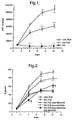
- FIG. 1 shows that, in the presence of 10% FCS, fibroblasts showed a typical growth curve and reached confluence after 10 days in culture. When fibroblasts were maintained in the presence of 5% FCS they showed a much slower rate of growth and by 7 days had produced only half the number of cells found in wells containing 10% FCS. When fibroblasts were maintained in the absence of serum there was no increase in cell numbers.
- Minoxidil significantly stimulated the growth of 3T3 fibroblasts maintained under sub-optimal conditions
- further experiments were carried out to determine whether the Minoxidil stimulated growth was mediated via K + channels.
- K + channel that Minoxidil is thought to activate is unknown at present, we looked at a number of different K + channel blockers to see if they could block the Minoxidil potentiation of the mitogenic effects of FCS on 3T3 fibroblasts.
- Tolbutamide which specifically blocks ATP-sensitive K + channels (Cook N.S. (1988). TiPS.9.21-28) used at a concentration of 1mM, caused a slight reduction in the Minoxidil-stimulated increase in cell numbers but this did not achieve statistical significance (Fig. 3A). When the concentration was increased to 5mM (Fig. 3B) there was a significant reduction (p ⁇ 0.001) of the Minoxidil stimulation though growth was still significantly greater than with 5% FCS alone (p ⁇ 0.01). This therefore indicates that Minoxidil is acting as a K + channel agonist.
- TEA a weak non-selective blocker of most types of K + channels (Cook, 1988), when used at 2mM gave a slight but not statistically significant reduction in the Minoxidil stimulation of growth (Fig. 3C). But when tolbutamide (1mM) and TEA (2mM) were used together, there was a significant (p ⁇ 0.01) inhibition of Minoxidil stimulated cell growth (Fig.3D). This, moreover, was not significantly different to that obtained in 5% FCS alone, which showed that the Minoxidil potentiation had been completely blocked. This again indicates that Minoxidil is acting as a K + channel agonist. Control experiments showed that in the absence of Minoxidil, neither tolbutamide (5mM) nor TEA (2mM) alone or used together had any effect on cell numbers.
- FIGURES
- Fig. 1 Effect of serum concentration on the growth of 3T3 fibroblasts. Growth is significantly reduced with 5% FCS and in serum free conditions when compared to growth in 10% FCS. Values are expressed as cell numbers per well and are shown as mean ⁇ s.e.m. of 3 experiments performed in triplicate. Statistical significance relative to growth in 10% FCS was determined using Student's t-test. ***p ⁇ 0.001.
- Fig. 2 Effect of Minoxidil (10 ⁇ g/ml) on the growth of 3T3 fibroblasts. In the absence of aminoglycoside antibiotics Minoxidil gives significant stimulation over growth in 5% FCS alone. In the presence of penicillin 100U/ml and streptomycin 100 ⁇ g/ml, Minoxidil initially inhibits growth but gives slight stimulation after 10 days. Values are expressed as a percentage of the control growth in 10% FCS and are shown as mean ⁇ s.e.m. of 5 experiments performed in triplicate. Statistical significance was determined using Student's t-test. ***p ⁇ 0.001 Significance compared to growth in 5% FCS ⁇ p ⁇ 0.01 Significance compared to growth in 5% FCS plus penicillin/streptomycin.
- Fig. 3 Effects of a tolbutamide and TEA on the Minoxidil stimulation of growth of 3T3 fibroblasts maintained in 5% FCS supplemented medium.
- A with 1mM tolbutamide there is no significant effect on the stimulation of growth by Minoxidil.
- B At 5mM tolbutamide gives a significant (p ⁇ 0.001) reduction in the Minoxidil stimulation of growth.
- C TEA at 2mM has no significant effect on the Minoxidil stimulation of growth.
- Fig. 4 Effects of glibenclamide and apamin on the Minoxidil stimulation of NIH 3T3 fibroblasts maintained in 5% FCS supplemented medium.
- A with 1 ⁇ M glibenclamide there is no significant effect on the stimulation of growth by Minoxidil.
- B Apamin at 10 nM also has no significant effect on the Minoxidil stimulation.
- C Glibenclamide (1 ⁇ M) and apamin (10 nM) used together significantly reduced the Minoxidil stimulation (p ⁇ 0.001). Values are expressed as a percentage of the control growth in 10% FCS after 7 days culture and shown as mean ⁇ s.e.m. of 3 experiments performed in triplicate. Significance is shown compared to both growth in 5% FCS alone and growth in 5% FCS plus Minoxidil.
- Fig. 5 Effects of Minoxidil, EGF-1 and PDGF on the growth of NIH 3T3 fibroblasts maintained in 5% CPSR-2. Values are expressed as a percentage of the control growth in 10% FCS after 7 days culture. Minoxidil has no effect in 5% CPSR -2 alone. Addition of IGF-1 (10 ng/ml) or PDGF (25 ng/ml) gives significant stimulation over growth in 5% CPSR-2 alone (p ⁇ 0.001). Minoxidil gives significant stimulation when added with IGF-1 or PDGF over growth with the cytokine alone (p ⁇ 0.001). ***p ⁇ 0.001 Significance compared to growth in 5% CPSR-2. ⁇ p ⁇ 0.001 Significance compared to growth in 5% CPSR-2 plus IGF-1. ⁇ p ⁇ 0.001 Significance compared to growth in 5% CPSR-2 plus PDGF.
Landscapes
- Life Sciences & Earth Sciences (AREA)
- Health & Medical Sciences (AREA)
- Engineering & Computer Science (AREA)
- Molecular Biology (AREA)
- Chemical & Material Sciences (AREA)
- Urology & Nephrology (AREA)
- Biomedical Technology (AREA)
- Cell Biology (AREA)
- Hematology (AREA)
- Immunology (AREA)
- Physics & Mathematics (AREA)
- General Physics & Mathematics (AREA)
- Biotechnology (AREA)
- Food Science & Technology (AREA)
- Medicinal Chemistry (AREA)
- Proteomics, Peptides & Aminoacids (AREA)
- Analytical Chemistry (AREA)
- Biochemistry (AREA)
- General Health & Medical Sciences (AREA)
- Microbiology (AREA)
- Pathology (AREA)
- Medicines That Contain Protein Lipid Enzymes And Other Medicines (AREA)
- Cosmetics (AREA)
- Crystals, And After-Treatments Of Crystals (AREA)
- Pharmaceuticals Containing Other Organic And Inorganic Compounds (AREA)
- Medicines Containing Antibodies Or Antigens For Use As Internal Diagnostic Agents (AREA)
- Measuring Or Testing Involving Enzymes Or Micro-Organisms (AREA)
Abstract
Description
***p<0.001.
***p<0.001 Significance compared to growth in 5% FCS
†† p<0.01 Significance compared to growth in 5% FCS plus penicillin/streptomycin.
Statistical significance was determined using Student's t-test.
*p<0.05 Significance compared to growth in 5% FCS.
**p<0.01 Significance compared to growth in 5% FCS.
***ep<0.001 Significance compared to growth in 5% FCS.
††† p<0.001 Significance compared to growth in 5% FCS plus Minoxidil
*p<0.05 Significance compared to growth in 5% FCS.
**p<0.01 Significance compared to growth in 5% FCS.
***p<0.001 Significance compared to growth in 5% FCS. †††p<0.001 Significance compared to growth in 5% FCS plus Minoxidil.
***p<0.001 Significance compared to growth in 5% CPSR-2.
··· p<0.001 Significance compared to growth in 5% CPSR-2 plus IGF-1.
†††p<0.001 Significance compared to growth in 5% CPSR-2 plus PDGF.
Claims (11)
- A test method for assessing the activity of a substance as a K+ channel agonist, comprising:(i) culturing cells of a cell line containing K+ channels in a test medium substantially free of aminoglycoside antibiotics and containing (a) serum and (b) the test substance;(ii) assessing the response of the cells to the test substance.
- A test method according to claim 1, in which the serum is FCS.
- A test method according to claim 2, in which the level of FCS in the test medium is about 5%.
- A test method for assessing the mitogenic activity of a substance comprising:(i) culturing cells of a cell line containing K+ channels in a test medium substantially free of aminoglycoside antibiotics and containing (a) a K+ channel agonist, (b) a low mitogen serum substitute and (c) the test substance;(ii) assessing the response of the cells to the test substance.
- A test method for assessing the activity of a substance as a K+ channel agonist, comprising:(i) culturing cells of a cell line containing K+ channels in a test medium substantially free of aminoglycoside antibiotics and containing (a) known individual mitogen, (b) a low mitogen serum substitute and (c) the test substance;(ii) assessing the response of the cells to the test substance.
- A test method according to claim 4 or claim 5, in which the low mitogen serum substitute is CPSR-2.
- A test method according to claim 6, in which the level of CPSR-2 in the medium is about 5%.
- A test method according to claim 4, in which the K+ channel agonist is Minoxidil.
- A test method according to claim 5, in which the known individual mitogen is IGF-1 or PDGF.
- A test method according to any one of the preceding claims in which the cell line is a fibroblast cell line.
- A test method according to claim 10, in which the fibroblasts are NIH-3T3 fibroblasts.
Applications Claiming Priority (3)
| Application Number | Priority Date | Filing Date | Title |
|---|---|---|---|
| GBGB9520411.1A GB9520411D0 (en) | 1995-10-06 | 1995-10-06 | Method of testing |
| GB9520411 | 1995-10-06 | ||
| PCT/GB1996/002442 WO1997014036A1 (en) | 1995-10-06 | 1996-10-04 | Screening assay for k+ channel agonists |
Publications (2)
| Publication Number | Publication Date |
|---|---|
| EP0842427A1 EP0842427A1 (en) | 1998-05-20 |
| EP0842427B1 true EP0842427B1 (en) | 2003-01-22 |
Family
ID=10781868
Family Applications (1)
| Application Number | Title | Priority Date | Filing Date |
|---|---|---|---|
| EP96933513A Expired - Lifetime EP0842427B1 (en) | 1995-10-06 | 1996-10-04 | Screening assay for k+ channel agonists |
Country Status (7)
| Country | Link |
|---|---|
| US (1) | US5837225A (en) |
| EP (1) | EP0842427B1 (en) |
| AT (1) | ATE231614T1 (en) |
| AU (1) | AU7221596A (en) |
| DE (1) | DE69625912D1 (en) |
| GB (1) | GB9520411D0 (en) |
| WO (1) | WO1997014036A1 (en) |
Families Citing this family (3)
| Publication number | Priority date | Publication date | Assignee | Title |
|---|---|---|---|---|
| GB9909792D0 (en) | 1998-12-04 | 1999-06-23 | Cambridge Bioclinical Limited | Potassium channel activators and their use |
| EP2590623A1 (en) | 2010-07-06 | 2013-05-15 | Unilever PLC | Malvidin for promoting hair growth |
| JP2013529671A (en) | 2010-07-06 | 2013-07-22 | ユニリーバー・ナームローゼ・ベンノートシヤープ | Use of glabranin to stimulate hair growth |
Family Cites Families (2)
| Publication number | Priority date | Publication date | Assignee | Title |
|---|---|---|---|---|
| JPH04504173A (en) * | 1989-03-06 | 1992-07-23 | ザ リージェンツ オブ ザ ユニバーシティ オブ カリフォルニア | Immunoadsorbent assays and treatments |
| US5620892A (en) * | 1993-09-07 | 1997-04-15 | Bristol-Myers Squibb Company | Strain of Saccharomyces cerevisiae expressing the gene encoding potassium transporter minK |
-
1995
- 1995-10-06 GB GBGB9520411.1A patent/GB9520411D0/en active Pending
-
1996
- 1996-04-05 US US08/628,463 patent/US5837225A/en not_active Expired - Fee Related
- 1996-10-04 AT AT96933513T patent/ATE231614T1/en not_active IP Right Cessation
- 1996-10-04 DE DE69625912T patent/DE69625912D1/en not_active Expired - Lifetime
- 1996-10-04 EP EP96933513A patent/EP0842427B1/en not_active Expired - Lifetime
- 1996-10-04 WO PCT/GB1996/002442 patent/WO1997014036A1/en not_active Ceased
- 1996-10-04 AU AU72215/96A patent/AU7221596A/en not_active Abandoned
Non-Patent Citations (7)
| Title |
|---|
| BERND A.: "Uptake kinetics and the effect of Minoxidil on human keratinocyte cultures", ARZNEIM.-FORSCH./DRUG RES., vol. 40, no. 4, 1990, pages 413 - 416 * |
| KATSUOKA K.: "effects of epidermal growth factor, fibroblast growth factor, minoxidil and hydrocortisone on growth kinetics in human hair bulb papilla cells and root sheath fibroblasts cultured in vitro", ARCH. DERMATOL. RES., vol. 279, 1987, pages 247 - 279 * |
| MURAD S. ET AL.: "Suppression of Fibroblast proliferation and lysyl hydroxylase activity by Mnoxidil", THE JOURNAL OF BIOLOGICAL CHEMISTRY, vol. 262, no. 25, 1987, pages 11973 - 11978 * |
| NOMURA K. ET AL.: "Aminoglycoside blockade of Ca2+-Activated K+ channel from rat brain synaptosomal membrane incorporated into planar bilayers", J. MEMBRANE. BIOL., vol. 115, 1990, pages 241 - 251 * |
| NORRIS W.D. ET AL.: "The effects of Minoxidil on endothelial cells in vitro", CELL BIOLOGY INTERNATIONAL REPORTS, vol. 13, no. 6, 1989, pages 555 - 562 * |
| O'KEFFE E. ET AL.: "Minoxidil: inhibition of proliferation of keratinocytes in Vitro", THE JOURNAL OF INVESTIGATIVE DERMATOLOGY, vol. 97, no. 3, 1991, pages 534 - 536 * |
| OOSAWA Y.ET AL.: "Voltage-dependent aminoglycoside blockade of the sarcosplasmic reticulum K+ channel", AM. J. PHYSIOL., vol. 250, no. 19, - 1986, pages C361 - C364 * |
Also Published As
| Publication number | Publication date |
|---|---|
| WO1997014036A1 (en) | 1997-04-17 |
| ATE231614T1 (en) | 2003-02-15 |
| DE69625912D1 (en) | 2003-02-27 |
| GB9520411D0 (en) | 1995-12-06 |
| AU7221596A (en) | 1997-04-30 |
| US5837225A (en) | 1998-11-17 |
| EP0842427A1 (en) | 1998-05-20 |
Similar Documents
| Publication | Publication Date | Title |
|---|---|---|
| Hawke et al. | Myogenic satellite cells: physiology to molecular biology | |
| Taylor et al. | Cyclosporin A prolongs human hair growth in vitro | |
| Mundy et al. | Resorbing bone is chemotactic for monocytes | |
| Zalin | The role of hormones and prostanoids in the in vitro proliferation and differentiation of human myoblasts | |
| Olutoye et al. | Lower cytokine release by fetal porcine platelets: a possible explanation for reduced inflammation after fetal wounding | |
| Zhang et al. | Epidermal growth factor promotes proliferation and migration of follicular outer root sheath cells via Wnt/β-catenin signaling | |
| Botchkarev et al. | p53 Involvement in the control of murine hair follicle regression | |
| Finkenzeller et al. | In vitro angiogenesis properties of endothelial progenitor cells: a promising tool for vascularization of ex vivo engineered tissues | |
| US6025151A (en) | Uses for compounds which reduce c-jun gene expression | |
| Rapoport et al. | Induction of refractoriness to thyrotropin stimulation in cultured thyroid cells. Dependence on new protein synthesis. | |
| Froger-Gaillard et al. | Growth-promoting effects of acidic and basic fibroblast growth factor on rabbit articular chondrocytes aging in culture | |
| Nishimura et al. | Glucose-mediated alteration of cellular function in human periodontal ligament cells | |
| CA2032177C (en) | Method of testing hair in vitro growth | |
| Xiong et al. | Interleukin-1β is differentially expressed by human dermal papilla cells in response to PKC activation and is a potent inhibitor of human hair follicle growth in organ culture | |
| EP2904095A1 (en) | Sebocyte cell culturing and methods of use | |
| Sraer et al. | Prostaglandin synthesis by rat isolated glomeruli and glomerular cultured cells | |
| Bak et al. | Boehmite enhances hair follicle growth via stimulation of dermal papilla cells by upregulating β‐catenin signalling | |
| Sanders et al. | In the absence of streptomycin, minoxidil potentiates the mitogenic effects of fetal calf serum, insulin-like growth factor 1, and platelet-derived growth factor on NIH 3T3 fibroblasts in a K+ channel-dependent fashion | |
| US5837225A (en) | Test methods for assessing K+ channel agonist or mitogenic activity | |
| Staiano‐Coico et al. | Sodium‐N‐butyrate induces cytoskeletal rearrangements and formation of cornified envelopes in cultured adult human keratinocytes | |
| US5885766A (en) | Method of screening of substances for their effect on the expression of mediators of inflammation in a hair follicle | |
| Misler et al. | Dependence on multivalent cations of quantal release of transmitter induced by black widow spider venom | |
| Hampton et al. | Expression of the chemokines, monocyte chemotactic protein (MCP)-1 and MCP-2 in endometrium of normal women and Norplant® users, does not support a central role in macrophage infiltration into endometrium | |
| Lacroix et al. | Supplementation with a complex of active nutrients improved dermal and epidermal characteristics in skin equivalents generated from fibroblasts from young or aged donors | |
| Hardy et al. | Either chick embryo dermis or retinoid‐treated mouse dermis can initiate glandular morphogenesis from mammalian epidermal tissue |
Legal Events
| Date | Code | Title | Description |
|---|---|---|---|
| PUAI | Public reference made under article 153(3) epc to a published international application that has entered the european phase |
Free format text: ORIGINAL CODE: 0009012 |
|
| 17P | Request for examination filed |
Effective date: 19971223 |
|
| AK | Designated contracting states |
Kind code of ref document: A1 Designated state(s): AT BE CH DE DK ES FR GB IT LI NL |
|
| 17Q | First examination report despatched |
Effective date: 20000704 |
|
| GRAG | Despatch of communication of intention to grant |
Free format text: ORIGINAL CODE: EPIDOS AGRA |
|
| GRAG | Despatch of communication of intention to grant |
Free format text: ORIGINAL CODE: EPIDOS AGRA |
|
| GRAG | Despatch of communication of intention to grant |
Free format text: ORIGINAL CODE: EPIDOS AGRA |
|
| GRAH | Despatch of communication of intention to grant a patent |
Free format text: ORIGINAL CODE: EPIDOS IGRA |
|
| GRAH | Despatch of communication of intention to grant a patent |
Free format text: ORIGINAL CODE: EPIDOS IGRA |
|
| GRAA | (expected) grant |
Free format text: ORIGINAL CODE: 0009210 |
|
| AK | Designated contracting states |
Kind code of ref document: B1 Designated state(s): AT BE CH DE DK ES FR GB IT LI NL |
|
| PG25 | Lapsed in a contracting state [announced via postgrant information from national office to epo] |
Ref country code: NL Free format text: LAPSE BECAUSE OF FAILURE TO SUBMIT A TRANSLATION OF THE DESCRIPTION OR TO PAY THE FEE WITHIN THE PRESCRIBED TIME-LIMIT Effective date: 20030122 Ref country code: LI Free format text: LAPSE BECAUSE OF FAILURE TO SUBMIT A TRANSLATION OF THE DESCRIPTION OR TO PAY THE FEE WITHIN THE PRESCRIBED TIME-LIMIT Effective date: 20030122 Ref country code: IT Free format text: LAPSE BECAUSE OF FAILURE TO SUBMIT A TRANSLATION OF THE DESCRIPTION OR TO PAY THE FEE WITHIN THE PRESCRIBED TIME-LIMIT;WARNING: LAPSES OF ITALIAN PATENTS WITH EFFECTIVE DATE BEFORE 2007 MAY HAVE OCCURRED AT ANY TIME BEFORE 2007. THE CORRECT EFFECTIVE DATE MAY BE DIFFERENT FROM THE ONE RECORDED. Effective date: 20030122 Ref country code: FR Free format text: LAPSE BECAUSE OF FAILURE TO SUBMIT A TRANSLATION OF THE DESCRIPTION OR TO PAY THE FEE WITHIN THE PRESCRIBED TIME-LIMIT Effective date: 20030122 Ref country code: CH Free format text: LAPSE BECAUSE OF FAILURE TO SUBMIT A TRANSLATION OF THE DESCRIPTION OR TO PAY THE FEE WITHIN THE PRESCRIBED TIME-LIMIT Effective date: 20030122 Ref country code: BE Free format text: LAPSE BECAUSE OF FAILURE TO SUBMIT A TRANSLATION OF THE DESCRIPTION OR TO PAY THE FEE WITHIN THE PRESCRIBED TIME-LIMIT Effective date: 20030122 Ref country code: AT Free format text: LAPSE BECAUSE OF FAILURE TO SUBMIT A TRANSLATION OF THE DESCRIPTION OR TO PAY THE FEE WITHIN THE PRESCRIBED TIME-LIMIT Effective date: 20030122 |
|
| REG | Reference to a national code |
Ref country code: GB Ref legal event code: FG4D |
|
| REG | Reference to a national code |
Ref country code: CH Ref legal event code: EP |
|
| REF | Corresponds to: |
Ref document number: 69625912 Country of ref document: DE Date of ref document: 20030227 Kind code of ref document: P |
|
| PG25 | Lapsed in a contracting state [announced via postgrant information from national office to epo] |
Ref country code: DK Free format text: LAPSE BECAUSE OF FAILURE TO SUBMIT A TRANSLATION OF THE DESCRIPTION OR TO PAY THE FEE WITHIN THE PRESCRIBED TIME-LIMIT Effective date: 20030422 |
|
| PG25 | Lapsed in a contracting state [announced via postgrant information from national office to epo] |
Ref country code: DE Free format text: LAPSE BECAUSE OF FAILURE TO SUBMIT A TRANSLATION OF THE DESCRIPTION OR TO PAY THE FEE WITHIN THE PRESCRIBED TIME-LIMIT Effective date: 20030423 |
|
| NLV1 | Nl: lapsed or annulled due to failure to fulfill the requirements of art. 29p and 29m of the patents act | ||
| PG25 | Lapsed in a contracting state [announced via postgrant information from national office to epo] |
Ref country code: ES Free format text: LAPSE BECAUSE OF FAILURE TO SUBMIT A TRANSLATION OF THE DESCRIPTION OR TO PAY THE FEE WITHIN THE PRESCRIBED TIME-LIMIT Effective date: 20030730 |
|
| REG | Reference to a national code |
Ref country code: CH Ref legal event code: PL |
|
| PLBE | No opposition filed within time limit |
Free format text: ORIGINAL CODE: 0009261 |
|
| STAA | Information on the status of an ep patent application or granted ep patent |
Free format text: STATUS: NO OPPOSITION FILED WITHIN TIME LIMIT |
|
| EN | Fr: translation not filed | ||
| 26N | No opposition filed |
Effective date: 20031023 |
|
| PGFP | Annual fee paid to national office [announced via postgrant information from national office to epo] |
Ref country code: GB Payment date: 20050923 Year of fee payment: 10 |
|
| GBPC | Gb: european patent ceased through non-payment of renewal fee |
Effective date: 20061004 |
|
| PG25 | Lapsed in a contracting state [announced via postgrant information from national office to epo] |
Ref country code: GB Free format text: LAPSE BECAUSE OF NON-PAYMENT OF DUE FEES Effective date: 20061004 |