EP0829724A1 - Assay for detecting collagen degradation - Google Patents
Assay for detecting collagen degradation Download PDFInfo
- Publication number
- EP0829724A1 EP0829724A1 EP96202596A EP96202596A EP0829724A1 EP 0829724 A1 EP0829724 A1 EP 0829724A1 EP 96202596 A EP96202596 A EP 96202596A EP 96202596 A EP96202596 A EP 96202596A EP 0829724 A1 EP0829724 A1 EP 0829724A1
- Authority
- EP
- European Patent Office
- Prior art keywords
- collagen
- amino acids
- directed
- antibody
- distance
- Prior art date
- Legal status (The legal status is an assumption and is not a legal conclusion. Google has not performed a legal analysis and makes no representation as to the accuracy of the status listed.)
- Withdrawn
Links
- 230000011382 collagen catabolic process Effects 0.000 title claims abstract description 18
- 238000003556 assay Methods 0.000 title description 7
- 102000008186 Collagen Human genes 0.000 claims abstract description 55
- 108010035532 Collagen Proteins 0.000 claims abstract description 55
- 229920001436 collagen Polymers 0.000 claims abstract description 54
- 150000001413 amino acids Chemical class 0.000 claims abstract description 36
- 239000007857 degradation product Substances 0.000 claims abstract description 18
- 210000002966 serum Anatomy 0.000 claims abstract description 16
- 210000004369 blood Anatomy 0.000 claims abstract description 9
- 239000008280 blood Substances 0.000 claims abstract description 9
- 210000002381 plasma Anatomy 0.000 claims abstract description 6
- 206010036790 Productive cough Diseases 0.000 claims abstract description 3
- 210000003802 sputum Anatomy 0.000 claims abstract description 3
- 208000024794 sputum Diseases 0.000 claims abstract description 3
- 239000012472 biological sample Substances 0.000 claims abstract 3
- 238000000034 method Methods 0.000 claims description 24
- 210000000988 bone and bone Anatomy 0.000 claims description 6
- 102000004169 proteins and genes Human genes 0.000 claims description 4
- 108090000623 proteins and genes Proteins 0.000 claims description 4
- 238000009007 Diagnostic Kit Methods 0.000 claims description 3
- 239000000463 material Substances 0.000 claims 2
- 108010029483 alpha 1 Chain Collagen Type I Proteins 0.000 claims 1
- 239000000523 sample Substances 0.000 claims 1
- 238000003018 immunoassay Methods 0.000 abstract description 5
- 238000004113 cell culture Methods 0.000 abstract description 2
- 238000001514 detection method Methods 0.000 abstract description 2
- 239000012634 fragment Substances 0.000 description 16
- 238000004132 cross linking Methods 0.000 description 14
- LCYXYLLJXMAEMT-SAXRGWBVSA-N Pyridinoline Chemical compound OC(=O)[C@@H](N)CCC1=C[N+](C[C@H](O)CC[C@H](N)C([O-])=O)=CC(O)=C1C[C@H](N)C(O)=O LCYXYLLJXMAEMT-SAXRGWBVSA-N 0.000 description 13
- YSMODUONRAFBET-UHNVWZDZSA-N erythro-5-hydroxy-L-lysine Chemical group NC[C@H](O)CC[C@H](N)C(O)=O YSMODUONRAFBET-UHNVWZDZSA-N 0.000 description 12
- LCWXJXMHJVIJFK-UHFFFAOYSA-N Hydroxylysine Natural products NCC(O)CC(N)CC(O)=O LCWXJXMHJVIJFK-UHFFFAOYSA-N 0.000 description 10
- KDXKERNSBIXSRK-UHFFFAOYSA-N Lysine Natural products NCCCCC(N)C(O)=O KDXKERNSBIXSRK-UHFFFAOYSA-N 0.000 description 10
- 239000004472 Lysine Substances 0.000 description 10
- YSMODUONRAFBET-UHFFFAOYSA-N delta-DL-hydroxylysine Natural products NCC(O)CCC(N)C(O)=O YSMODUONRAFBET-UHFFFAOYSA-N 0.000 description 10
- QJHBJHUKURJDLG-UHFFFAOYSA-N hydroxy-L-lysine Natural products NCCCCC(NO)C(O)=O QJHBJHUKURJDLG-UHFFFAOYSA-N 0.000 description 10
- 210000004899 c-terminal region Anatomy 0.000 description 5
- 208000037265 diseases, disorders, signs and symptoms Diseases 0.000 description 5
- 208000035475 disorder Diseases 0.000 description 5
- ZAHDXEIQWWLQQL-IHRRRGAJSA-N Deoxypyridinoline Chemical compound OC(=O)[C@@H](N)CCCC[N+]1=CC(O)=C(C[C@H](N)C([O-])=O)C(CC[C@H](N)C(O)=O)=C1 ZAHDXEIQWWLQQL-IHRRRGAJSA-N 0.000 description 4
- 102000004669 Protein-Lysine 6-Oxidase Human genes 0.000 description 4
- 108010003894 Protein-Lysine 6-Oxidase Proteins 0.000 description 4
- 210000000845 cartilage Anatomy 0.000 description 4
- SPIZPNKPOATXFB-UWVGGRQHSA-N (2s)-6-amino-2-[[[(1s)-1-carboxypentyl]amino]-hydroxyamino]hexanoic acid Chemical compound CCCC[C@@H](C(O)=O)NN(O)[C@H](C(O)=O)CCCCN SPIZPNKPOATXFB-UWVGGRQHSA-N 0.000 description 3
- 102000004190 Enzymes Human genes 0.000 description 3
- 108090000790 Enzymes Proteins 0.000 description 3
- 230000015572 biosynthetic process Effects 0.000 description 3
- 238000009833 condensation Methods 0.000 description 3
- 230000005494 condensation Effects 0.000 description 3
- 210000002808 connective tissue Anatomy 0.000 description 3
- 108010013977 delta-hydroxylysylnorleucine Proteins 0.000 description 3
- -1 hydroxylysine aldehydes Chemical class 0.000 description 3
- 108090000765 processed proteins & peptides Proteins 0.000 description 3
- 210000001519 tissue Anatomy 0.000 description 3
- 210000002700 urine Anatomy 0.000 description 3
- ZCGMEDNXACWKIR-MRLBHPIUSA-N (2s,5r)-6-amino-2-[2-[[(1s,4r)-5-amino-1-carboxy-4-hydroxypentyl]amino]-2-[(1s)-1-carboxypentyl]hydrazinyl]-5-hydroxyhexanoic acid Chemical compound NC[C@H](O)CC[C@@H](C(O)=O)NN([C@@H](CCCC)C(O)=O)N[C@@H](CC[C@@H](O)CN)C(O)=O ZCGMEDNXACWKIR-MRLBHPIUSA-N 0.000 description 2
- HSJKGGMUJITCBW-UHFFFAOYSA-N 3-hydroxybutanal Chemical compound CC(O)CC=O HSJKGGMUJITCBW-UHFFFAOYSA-N 0.000 description 2
- QTBSBXVTEAMEQO-UHFFFAOYSA-N Acetic acid Chemical compound CC(O)=O QTBSBXVTEAMEQO-UHFFFAOYSA-N 0.000 description 2
- 102000012422 Collagen Type I Human genes 0.000 description 2
- 108010022452 Collagen Type I Proteins 0.000 description 2
- 102000000503 Collagen Type II Human genes 0.000 description 2
- 108010041390 Collagen Type II Proteins 0.000 description 2
- 102000001187 Collagen Type III Human genes 0.000 description 2
- 108010069502 Collagen Type III Proteins 0.000 description 2
- PMMYEEVYMWASQN-DMTCNVIQSA-N Hydroxyproline Chemical compound O[C@H]1CN[C@H](C(O)=O)C1 PMMYEEVYMWASQN-DMTCNVIQSA-N 0.000 description 2
- ONIBWKKTOPOVIA-BYPYZUCNSA-N L-Proline Chemical compound OC(=O)[C@@H]1CCCN1 ONIBWKKTOPOVIA-BYPYZUCNSA-N 0.000 description 2
- LRHPLDYGYMQRHN-UHFFFAOYSA-N N-Butanol Chemical compound CCCCO LRHPLDYGYMQRHN-UHFFFAOYSA-N 0.000 description 2
- ONIBWKKTOPOVIA-UHFFFAOYSA-N Proline Natural products OC(=O)C1CCCN1 ONIBWKKTOPOVIA-UHFFFAOYSA-N 0.000 description 2
- 229920002678 cellulose Polymers 0.000 description 2
- 239000001913 cellulose Substances 0.000 description 2
- DDRJAANPRJIHGJ-UHFFFAOYSA-N creatinine Chemical compound CN1CC(=O)NC1=N DDRJAANPRJIHGJ-UHFFFAOYSA-N 0.000 description 2
- PMMYEEVYMWASQN-UHFFFAOYSA-N dl-hydroxyproline Natural products OC1C[NH2+]C(C([O-])=O)C1 PMMYEEVYMWASQN-UHFFFAOYSA-N 0.000 description 2
- HNDVDQJCIGZPNO-UHFFFAOYSA-N histidine Natural products OC(=O)C(N)CC1=CN=CN1 HNDVDQJCIGZPNO-UHFFFAOYSA-N 0.000 description 2
- 229960002591 hydroxyproline Drugs 0.000 description 2
- 239000003550 marker Substances 0.000 description 2
- 230000035800 maturation Effects 0.000 description 2
- 230000001404 mediated effect Effects 0.000 description 2
- 102000004196 processed proteins & peptides Human genes 0.000 description 2
- 239000000047 product Substances 0.000 description 2
- FGMPLJWBKKVCDB-UHFFFAOYSA-N trans-L-hydroxy-proline Natural products ON1CCCC1C(O)=O FGMPLJWBKKVCDB-UHFFFAOYSA-N 0.000 description 2
- 230000007306 turnover Effects 0.000 description 2
- 230000002485 urinary effect Effects 0.000 description 2
- DQJBXDBKQNYIGP-WLUIXCMPSA-N (2S,10S)-2,10-diamino-5-[[[(5S)-5-amino-5-carboxy-2-hydroxypentyl]amino]methyl]undec-5-enedioic acid Chemical compound N[C@@H](CCCC=C(CC[C@H](N)C(O)=O)CNCC(O)CC[C@H](N)C(O)=O)C(O)=O DQJBXDBKQNYIGP-WLUIXCMPSA-N 0.000 description 1
- 201000001320 Atherosclerosis Diseases 0.000 description 1
- 208000006386 Bone Resorption Diseases 0.000 description 1
- 206010005949 Bone cancer Diseases 0.000 description 1
- 208000020084 Bone disease Diseases 0.000 description 1
- 206010065687 Bone loss Diseases 0.000 description 1
- 206010007572 Cardiac hypertrophy Diseases 0.000 description 1
- 239000011665 D-biotin Substances 0.000 description 1
- 102000010834 Extracellular Matrix Proteins Human genes 0.000 description 1
- 108010037362 Extracellular Matrix Proteins Proteins 0.000 description 1
- 206010016654 Fibrosis Diseases 0.000 description 1
- 206010020772 Hypertension Diseases 0.000 description 1
- 206010028980 Neoplasm Diseases 0.000 description 1
- 208000010191 Osteitis Deformans Diseases 0.000 description 1
- 208000001132 Osteoporosis Diseases 0.000 description 1
- 208000027868 Paget disease Diseases 0.000 description 1
- 230000001594 aberrant effect Effects 0.000 description 1
- 229960000583 acetic acid Drugs 0.000 description 1
- 150000001299 aldehydes Chemical class 0.000 description 1
- 238000005882 aldol condensation reaction Methods 0.000 description 1
- 206010003246 arthritis Diseases 0.000 description 1
- 239000011324 bead Substances 0.000 description 1
- 239000013060 biological fluid Substances 0.000 description 1
- 210000004204 blood vessel Anatomy 0.000 description 1
- 230000024279 bone resorption Effects 0.000 description 1
- 239000000872 buffer Substances 0.000 description 1
- 210000000748 cardiovascular system Anatomy 0.000 description 1
- 230000015556 catabolic process Effects 0.000 description 1
- 238000006243 chemical reaction Methods 0.000 description 1
- 238000006482 condensation reaction Methods 0.000 description 1
- 229940109239 creatinine Drugs 0.000 description 1
- 238000006731 degradation reaction Methods 0.000 description 1
- 230000008021 deposition Effects 0.000 description 1
- 238000002405 diagnostic procedure Methods 0.000 description 1
- 239000003085 diluting agent Substances 0.000 description 1
- 229940079593 drug Drugs 0.000 description 1
- 239000003814 drug Substances 0.000 description 1
- 238000007812 electrochemical assay Methods 0.000 description 1
- 230000002255 enzymatic effect Effects 0.000 description 1
- 238000006911 enzymatic reaction Methods 0.000 description 1
- 230000005284 excitation Effects 0.000 description 1
- 210000002744 extracellular matrix Anatomy 0.000 description 1
- 230000004761 fibrosis Effects 0.000 description 1
- 238000007421 fluorometric assay Methods 0.000 description 1
- 239000012362 glacial acetic acid Substances 0.000 description 1
- 238000004128 high performance liquid chromatography Methods 0.000 description 1
- 108010020375 histidinohydroxylysinonorleucine Proteins 0.000 description 1
- 230000001900 immune effect Effects 0.000 description 1
- 238000002649 immunization Methods 0.000 description 1
- 238000010324 immunological assay Methods 0.000 description 1
- 210000004185 liver Anatomy 0.000 description 1
- 230000004807 localization Effects 0.000 description 1
- 208000027202 mammary Paget disease Diseases 0.000 description 1
- 208000010125 myocardial infarction Diseases 0.000 description 1
- 238000002414 normal-phase solid-phase extraction Methods 0.000 description 1
- 201000008482 osteoarthritis Diseases 0.000 description 1
- 238000007833 oxidative deamination reaction Methods 0.000 description 1
- 230000037361 pathway Effects 0.000 description 1
- 230000003239 periodontal effect Effects 0.000 description 1
- 210000002345 respiratory system Anatomy 0.000 description 1
- 206010039073 rheumatoid arthritis Diseases 0.000 description 1
- 239000002002 slurry Substances 0.000 description 1
- 239000000758 substrate Substances 0.000 description 1
- 238000000108 ultra-filtration Methods 0.000 description 1
- 230000036325 urinary excretion Effects 0.000 description 1
- 230000029663 wound healing Effects 0.000 description 1
Images


Classifications
-
- G—PHYSICS
- G01—MEASURING; TESTING
- G01N—INVESTIGATING OR ANALYSING MATERIALS BY DETERMINING THEIR CHEMICAL OR PHYSICAL PROPERTIES
- G01N33/00—Investigating or analysing materials by specific methods not covered by groups G01N1/00 - G01N31/00
- G01N33/48—Biological material, e.g. blood, urine; Haemocytometers
- G01N33/50—Chemical analysis of biological material, e.g. blood, urine; Testing involving biospecific ligand binding methods; Immunological testing
- G01N33/68—Chemical analysis of biological material, e.g. blood, urine; Testing involving biospecific ligand binding methods; Immunological testing involving proteins, peptides or amino acids
- G01N33/6887—Chemical analysis of biological material, e.g. blood, urine; Testing involving biospecific ligand binding methods; Immunological testing involving proteins, peptides or amino acids from muscle, cartilage or connective tissue
-
- G—PHYSICS
- G01—MEASURING; TESTING
- G01N—INVESTIGATING OR ANALYSING MATERIALS BY DETERMINING THEIR CHEMICAL OR PHYSICAL PROPERTIES
- G01N2800/00—Detection or diagnosis of diseases
- G01N2800/10—Musculoskeletal or connective tissue disorders
- G01N2800/105—Osteoarthritis, e.g. cartilage alteration, hypertrophy of bone
-
- Y—GENERAL TAGGING OF NEW TECHNOLOGICAL DEVELOPMENTS; GENERAL TAGGING OF CROSS-SECTIONAL TECHNOLOGIES SPANNING OVER SEVERAL SECTIONS OF THE IPC; TECHNICAL SUBJECTS COVERED BY FORMER USPC CROSS-REFERENCE ART COLLECTIONS [XRACs] AND DIGESTS
- Y10—TECHNICAL SUBJECTS COVERED BY FORMER USPC
- Y10S—TECHNICAL SUBJECTS COVERED BY FORMER USPC CROSS-REFERENCE ART COLLECTIONS [XRACs] AND DIGESTS
- Y10S435/00—Chemistry: molecular biology and microbiology
- Y10S435/975—Kit
Definitions
- the invention relates to a method for detecting collagen degradation. More specifically, it relates to a method for quantitating high molecular weight crosslinked collagen fragments in blood, plasma, serum, sputum or cell cultures.
- Collagen proteins in connective tissues constitute a three-dimensional network with crosslinks.
- the crosslinks are formed after deposition of the collagen molecule in the extracellular matrix.
- Different types of crosslinks between collagen molecules are known.
- lysino-norleucine-type difunctional crosslinks, pyridinoline-type trifunctional crosslinks, and pyrrole-type crosslinks are formed.
- Aberrant degradation of collagen is an indication of disorders of connective tissues.
- Disorders of bone tissue comprise osteoporosis, Paget's disease, bone tumours, and drug-related bone loss.
- disorders of cartilage tissue occur in arthritis (such as osteoarthritis and rheumatoid arthritis), and disorders of the cardiovascular system are prominent in atherosclerosis, aneurisms, hypertension, myocardial infarction and hypertrophy.
- disorders of the respiratory system and liver such as fibrosis, of the teeth and periodontal tissues, of skin and wound healing, and in tumours involve changes in collagen turnover.
- telopeptide sequences corresponding to the crosslinking site are based on serum/plasma/blood or urinary levels of pyridinoline crosslinks (free or still containing residual amino acids), or telopeptide sequences corresponding to the crosslinking site.
- US-A-5,140,103 discloses a method for measuring collagen degradation by quantitating the concentration of a C-terminal collagen type II or III telopeptide containing a pyridinoline-type crosslink by immunological, electrochemical or fluorometric assays.
- WO-92/21698 describes a method of determining bone resorption by means of antibodies to type I collagen telopeptides.
- WO-95/08115 discloses a method for assaying collagen fragments by means of antibodies against crosslinking sites of collagen.
- WO-91/10141 describes the use of antibodies to detect collagen-derived crosslinks in biological fluids as a diagnostic method of bone diseases.
- a disadvantage of these known methods for assaying collagen degradation is that the detection means (e.g. antibodies) recognise an epitope that includes the crosslink, or an epitope close to the crosslinking site.
- these methods depend on the nature and structure of the crosslink, or the amino acid sequence close to the crosslinking site.
- Collagen fragments containing other types of crosslinks are not necessarily detected and thus not quantitated, which leads to a less reliable and/or less sensitive result.
- most of these methods require a relatively insensitive and laborious competition-type immunoassay that is mostly only applicable to urine, and not to serum/plasma/blood.
- Disadvantages of urinary assays are the need to correct for the urine volume based on creatinine levels, diurnal variation in urinary excretion rate and the need to collect urine samples.
- crosslinks in collagen is a basic feature of maturation of the tissue.
- the crosslinks can be intramolecular, linking two collagen ⁇ -chains within the same molecule, or intermolecular, between chains in different molecules. Irrespective of the exact structure of the crosslink, by definition they involve two or more ⁇ -chains.
- crosslink formation The chemistry and biology of crosslink formation have been reviewed extensively.
- the basic mechanism of crosslinking is oxidative deamination of the ⁇ -amino groups of lysine or hydroxylysine residues, catalysed by lysyl oxidase.
- the lysine or hydroxylysine aldehydes formed undergo condensation reactions with lysine or hydroxylysine into the difunctional crosslinks (dehydro)-lysinonorleucine, -hydroxylysinonorleucine, and -dihydroxylysinonorleucine.
- Other difunctional crosslinks involve aldol condensation of two aldehydes, and pyrrole-type crosslinks.
- trifunctional crosslinks connecting three ⁇ -chains may result: for instance, hydroxylysinonorleucine and dihydroxylysinonorleucine are converted into the pyridinoline crosslinks lysylpyridinoline (LP) and hydroxylysylpyridinoline (HP), respectively.
- the difunctional crosslink hydroxylysinonorleucine may also undergo condensation with histidine to form the trifunctional crosslink histidinohydroxylysinonorleucine.
- the aldol product of two hydroxylysine aldehydes can undergo condensation with hydroxylysine resulting in hydroxymerodesmosine, or with histidine leading to dehydroaldolhistidine. The latter can undergo additional condensation with lysine or hydroxylysine to produce the tetrafunctional crosslinks dehydro-histidino-merodesmosine and dehydro-histidino-hydroxymerodesmosine, respectively.
- crosslink formation depends more on tissue localisation than on the type of collagen. For instance, crosslinks based on lysine aldehydes predominate in skin. The crosslinks formed via the hydroxylysine pathway predominate in bone and cartilage. The di-, tri- or tetrafunctional crosslinks are reproducibly located at predefined positions in the collagen molecule, probably due to the selectivity of lysyl oxidase.
- difunctional hydroxylysinonorleucine and dihydroxylysinonorleucine and their trifunctional pyridinoline maturation products (LP and HP, respectively) in bone and cartilage (type I and II collagen, respectively) involve the lysine or hydroxylysine at position 87 in the triple helix and the hydroxylysine residue in the C-terminal telopeptide.
- these crosslinks are formed between lysine or hydroxylysine at position 930 of the triple helix and the hydroxylysine residue in the N-terminal telopeptide (see Fig. 2).
- crosslinks are homotypic.
- crosslinks of type III collagen are homotypic.
- Homotypic crosslinks in type I collagen may involve the ⁇ 1 (I)- as well as the ⁇ 2 (I)-chain.
- heterotypic crosslinks, connecting different collagen molecules have been reported: crosslinks between type I and III collagen in blood vessels, between type II and IX, type II and XI, and type II and III collagen in cartilage, between type I and V in bone, and between type I and II in intervertebral disc.
- the enzymatic crosslinks are very diverse in nature, occur at specific locations in the molecule, and connect two or more collagen ⁇ -chains from the same or different types of collagen (homo- and heterotypic crosslinks).
- collagen degradation can be measured effectively in plasma, serum or blood by a sandwich-type of immunoassay using epitope ; further removed from the crosslinking site.
- the invention is concerned with a method of assaying collagen degradation products as defined in the appending claims; the method is generally applicable to crosslinked collagen fragments irrespective of the exact nature and structure of the crosslinking site.
- the invention also pertains to antibodies and diagnostic kits for carrying such a method.
- telopeptides usually comprise about 15 amino acids), but also large peptides, up to 165 amino acids, from the triple helical region (Fig. 2).
- high-molecular weight collagen fragments can be detected by ⁇ -chain epitopes residing in the triple helical region, irrespective of the type of crosslink.
- ⁇ -chain epitopes residing in the triple helical region, irrespective of the type of crosslink.
- a sandwich-type of assay with one antibody recognising an epitope in one ⁇ -chain, and a second antibody recognising another ⁇ -chain (which may be the same type of ⁇ -chain), or even on the same ⁇ -chain as explained below is proposed.
- the two or more epitopes for the antibodies can be present on the degradation product at different locations as depicted in Fig. 3.
- the epitopes suitable for the present method are located at a distance of up to 165 amino acids from a collagen chain crosslink site.
- a distance of an epitope to a crosslink site of a particular number of amino acids is understood as that number of amino acids along the amino acid chain from the distal amino acid of the epitope to the lysine or hydroxylysine residue of either a telopeptide crosslink site or a triple helical region crosslink site, the latter being located at about 100 amino acids distance from the collagen chain ends (see Figs. 4 and 5).
- An especially preferred distance from the crosslink sites is up to 100 amino acids, especially 5-100 or even 10-90 amino acids.
- One of the epitopes should be present on a chain having a telopeptide crosslink site. It may be present on the crosslink site itself and even comprise part of the other chains linked by the crosslink. If the first epitope is present on the telopeptide crosslink, the second epitope may reside on the same chain, as depicted in Fig. 3, variations 3 and 5. The second epitope may, however, also be present on another chain (Fig. 3, variations 2, 8 and 9). If the first epitope is at some distance (e.g.
- the second epitope should be present on another ⁇ -chain, in order to selectively detect degradation products that contain a crosslink (Fig. 3, variations 1, 4, 6 and 7).
- the epitopes may be the same as well (Fig. 3, variation 7).
- a very suitable combination of epitopes (and antibodies) for the sandwich assay is a first epitope on a collagen within 165 amino acids from a telopeptide crosslink, and a second epitope on another collagen chain within 165 amino acids from a triple helical region crosslink; this combination recognises most of the degradation products that are of diagnostic interest. If the telopeptide crosslink is N-terminal, the helical region crosslink is at the 930 or equivalent position (Fig. 3, variations 1, 2, 4, 9), whereas if the telopeptide crosslink is C-terminal, the helical region crosslink is at the 87 or equivalent position (not depicted in Fig. 3, but similarly applicable).
- the antibodies according to the invention can be produced by well-known methods.
- the antibodies are monoclonal antibodies generated by immunisation with either natural collagen degradation products having molecular weights of up to 50 kD, or with synthetic peptides containing at least the epitope of interest.
- One of said antibodies is preferably immobilised in a known way, e.g. by physical or covalent binding to spheres, beads, sticks, plate walls, and the like, or immobilisable by aspecific reaction with a further, immobilised antibody.
- Another of the other of said antibodies is preferably detectable by being provided with a detectable label, such as a fluorescence marker, a radioisotope, an enzyme, an avidine-biotine marker or the like, or by being recognised by a further, labelled antibody.
- a detectable label such as a fluorescence marker, a radioisotope, an enzyme, an avidine-biotine marker or the like, or by being recognised by a further, labelled antibody.
- the invention further provides novel antibodies as described above, directed at an epitope which is at some distance, up to 165 amino acids, in particular 5-150 amino acids, more in particular 10-100 amino acids, from a crosslink site.
- the invention also provides a diagnostic kit containing at least one of such antibodies, optionally two of said antibodies, one being immobilised or immobilisable, the other being labelled or detectable.
- the kit may further contain conventional means for carrying out a sandwich-type immunoassay, such as enzyme substrates in case of enzyme labels, diluents, buffers, filters, etc.
- Hydrolysed fractions were dried, reconstituted in 500 ⁇ l 6N HCl, 200 ⁇ l glacial acetic acid, 200 ⁇ l CF-1 cellulose slurry, and 800 ⁇ l n-butanol, and subjected to solid phase extraction with CF-1 cellulose according to Black et al, (Anal. Biochem. 1988, 169 : 197-203).
- HPLChromatography was used for quantitating amounts of pyridinoline crosslinks, which were detected by their fluoresence at 295/400 nm (excitation/emission wavelengths) (see Black et al. above).
- pyridinoline crosslinks were detected in 0-10, 10-30, and 30-50 kD fractions.
- About half of the total serum concentration of pyridinoline (approx. 1.5 nM) had a molecular weight between 10 and 30 kD.
- About 25% had a molecular weight less than 10 kD. The other 25% had molecular weight between 30 and 50 kD (Fig. 1).
Landscapes
- Life Sciences & Earth Sciences (AREA)
- Health & Medical Sciences (AREA)
- Engineering & Computer Science (AREA)
- Biomedical Technology (AREA)
- Molecular Biology (AREA)
- Chemical & Material Sciences (AREA)
- Urology & Nephrology (AREA)
- Hematology (AREA)
- Immunology (AREA)
- Biotechnology (AREA)
- Microbiology (AREA)
- Cell Biology (AREA)
- Proteomics, Peptides & Aminoacids (AREA)
- Food Science & Technology (AREA)
- Medicinal Chemistry (AREA)
- Physics & Mathematics (AREA)
- Analytical Chemistry (AREA)
- Biochemistry (AREA)
- General Health & Medical Sciences (AREA)
- General Physics & Mathematics (AREA)
- Pathology (AREA)
- Peptides Or Proteins (AREA)
Abstract
Description
- The invention relates to a method for detecting collagen degradation. More specifically, it relates to a method for quantitating high molecular weight crosslinked collagen fragments in blood, plasma, serum, sputum or cell cultures.
- Collagen proteins in connective tissues constitute a three-dimensional network with crosslinks. The crosslinks are formed after deposition of the collagen molecule in the extracellular matrix. Different types of crosslinks between collagen molecules are known. As a result from enzymatic reactions, lysino-norleucine-type difunctional crosslinks, pyridinoline-type trifunctional crosslinks, and pyrrole-type crosslinks are formed. Aberrant degradation of collagen is an indication of disorders of connective tissues. Disorders of bone tissue comprise osteoporosis, Paget's disease, bone tumours, and drug-related bone loss. Disorders of cartilage tissue occur in arthritis (such as osteoarthritis and rheumatoid arthritis), and disorders of the cardiovascular system are prominent in atherosclerosis, aneurisms, hypertension, myocardial infarction and hypertrophy. Similarly, disorders of the respiratory system and liver, such as fibrosis, of the teeth and periodontal tissues, of skin and wound healing, and in tumours involve changes in collagen turnover.
- Known methods to determine turnover of connective tissues, in particular bone, are based on serum/plasma/blood or urinary levels of pyridinoline crosslinks (free or still containing residual amino acids), or telopeptide sequences corresponding to the crosslinking site. For example US-A-5,140,103 discloses a method for measuring collagen degradation by quantitating the concentration of a C-terminal collagen type II or III telopeptide containing a pyridinoline-type crosslink by immunological, electrochemical or fluorometric assays. WO-92/21698 describes a method of determining bone resorption by means of antibodies to type I collagen telopeptides. WO-95/08115 discloses a method for assaying collagen fragments by means of antibodies against crosslinking sites of collagen. Similarly, WO-91/10141 describes the use of antibodies to detect collagen-derived crosslinks in biological fluids as a diagnostic method of bone diseases.
- A disadvantage of these known methods for assaying collagen degradation is that the detection means (e.g. antibodies) recognise an epitope that includes the crosslink, or an epitope close to the crosslinking site. Thus, these methods depend on the nature and structure of the crosslink, or the amino acid sequence close to the crosslinking site. Collagen fragments containing other types of crosslinks are not necessarily detected and thus not quantitated, which leads to a less reliable and/or less sensitive result. More importantly, most of these methods require a relatively insensitive and laborious competition-type immunoassay that is mostly only applicable to urine, and not to serum/plasma/blood. Disadvantages of urinary assays are the need to correct for the urine volume based on creatinine levels, diurnal variation in urinary excretion rate and the need to collect urine samples.
- The formation of crosslinks in collagen is a basic feature of maturation of the tissue. The crosslinks can be intramolecular, linking two collagen α-chains within the same molecule, or intermolecular, between chains in different molecules. Irrespective of the exact structure of the crosslink, by definition they involve two or more α-chains.
- The chemistry and biology of crosslink formation have been reviewed extensively. The basic mechanism of crosslinking is oxidative deamination of the α-amino groups of lysine or hydroxylysine residues, catalysed by lysyl oxidase. The lysine or hydroxylysine aldehydes formed undergo condensation reactions with lysine or hydroxylysine into the difunctional crosslinks (dehydro)-lysinonorleucine, -hydroxylysinonorleucine, and -dihydroxylysinonorleucine. Other difunctional crosslinks involve aldol condensation of two aldehydes, and pyrrole-type crosslinks.
- Subsequently, trifunctional crosslinks connecting three α-chains may result: for instance, hydroxylysinonorleucine and dihydroxylysinonorleucine are converted into the pyridinoline crosslinks lysylpyridinoline (LP) and hydroxylysylpyridinoline (HP), respectively. The difunctional crosslink hydroxylysinonorleucine may also undergo condensation with histidine to form the trifunctional crosslink histidinohydroxylysinonorleucine. Furthermore, the aldol product of two hydroxylysine aldehydes can undergo condensation with hydroxylysine resulting in hydroxymerodesmosine, or with histidine leading to dehydroaldolhistidine. The latter can undergo additional condensation with lysine or hydroxylysine to produce the tetrafunctional crosslinks dehydro-histidino-merodesmosine and dehydro-histidino-hydroxymerodesmosine, respectively.
- The type of crosslink formation depends more on tissue localisation than on the type of collagen. For instance, crosslinks based on lysine aldehydes predominate in skin. The crosslinks formed via the hydroxylysine pathway predominate in bone and cartilage. The di-, tri- or tetrafunctional crosslinks are reproducibly located at predefined positions in the collagen molecule, probably due to the selectivity of lysyl oxidase. For instance, difunctional hydroxylysinonorleucine and dihydroxylysinonorleucine and their trifunctional pyridinoline maturation products (LP and HP, respectively) in bone and cartilage (type I and II collagen, respectively) involve the lysine or hydroxylysine at
position 87 in the triple helix and the hydroxylysine residue in the C-terminal telopeptide. Furthermore, these crosslinks are formed between lysine or hydroxylysine atposition 930 of the triple helix and the hydroxylysine residue in the N-terminal telopeptide (see Fig. 2). - In type II collagen, composed of three identical α1(II)-chains, by definition crosslinks are homotypic. Similarly, crosslinks of type III collagen (composed of three α1(III)-chains) are homotypic. Homotypic crosslinks in type I collagen may involve the α1(I)- as well as the α2(I)-chain. Also heterotypic crosslinks, connecting different collagen molecules have been reported: crosslinks between type I and III collagen in blood vessels, between type II and IX, type II and XI, and type II and III collagen in cartilage, between type I and V in bone, and between type I and II in intervertebral disc.
- In summary, the enzymatic crosslinks are very diverse in nature, occur at specific locations in the molecule, and connect two or more collagen α-chains from the same or different types of collagen (homo- and heterotypic crosslinks).
- It has been found according to the invention that collagen degradation can be measured effectively in plasma, serum or blood by a sandwich-type of immunoassay using epitope ; further removed from the crosslinking site. The invention is concerned with a method of assaying collagen degradation products as defined in the appending claims; the method is generally applicable to crosslinked collagen fragments irrespective of the exact nature and structure of the crosslinking site. The invention also pertains to antibodies and diagnostic kits for carrying such a method.
- It was found by HPLC analysis of normal serum that pyridinoline crosslinks are present in collagen degradation products with relatively high molecular weight (cf. Example 1; Fig. 1). About 25, 50 and 25% have molecular weights between 0-10, 10-30, and 30-50 kD, respectively. Assuming an average weight per amino acid of 100 Dalton, half of the collagen degradation products in serum contain between 100 and 300 amino acids. One quarter of the fragments comprise 300-500 amino acids, while another quarter represents fragments with less than 100 amino acids. This implies that collagen degradation products present in blood can be as large as 500 amino acids. Thus, on average the three α-chains of the pyridinoline crosslink-containing degradation products are as large as up to 500/3 = 165 amino acids. Thus, these fragments contain not only (part of) the collagen telopeptide (telopeptides usually comprise about 15 amino acids), but also large peptides, up to 165 amino acids, from the triple helical region (Fig. 2).
- According to the invention high-molecular weight collagen fragments can be detected by α-chain epitopes residing in the triple helical region, irrespective of the type of crosslink. Inasmuch as two, three or four α-chains are present in these collagen degradation products in serum, a sandwich-type of assay with one antibody recognising an epitope in one α-chain, and a second antibody recognising another α-chain (which may be the same type of α-chain), or even on the same α-chain as explained below, is proposed. The two or more epitopes for the antibodies can be present on the degradation product at different locations as depicted in Fig. 3.
- The epitopes suitable for the present method are located at a distance of up to 165 amino acids from a collagen chain crosslink site. A distance of an epitope to a crosslink site of a particular number of amino acids is understood as that number of amino acids along the amino acid chain from the distal amino acid of the epitope to the lysine or hydroxylysine residue of either a telopeptide crosslink site or a triple helical region crosslink site, the latter being located at about 100 amino acids distance from the collagen chain ends (see Figs. 4 and 5).
- An especially preferred distance from the crosslink sites (whether telopeptide or triple helical) is up to 100 amino acids, especially 5-100 or even 10-90 amino acids.
- One of the epitopes should be present on a chain having a telopeptide crosslink site. It may be present on the crosslink site itself and even comprise part of the other chains linked by the crosslink. If the first epitope is present on the telopeptide crosslink, the second epitope may reside on the same chain, as depicted in Fig. 3,
variations variations variations variations - The antibodies according to the invention can be produced by well-known methods. Preferably the antibodies are monoclonal antibodies generated by immunisation with either natural collagen degradation products having molecular weights of up to 50 kD, or with synthetic peptides containing at least the epitope of interest.
- One of said antibodies is preferably immobilised in a known way, e.g. by physical or covalent binding to spheres, beads, sticks, plate walls, and the like, or immobilisable by aspecific reaction with a further, immobilised antibody.
- Another of the other of said antibodies is preferably detectable by being provided with a detectable label, such as a fluorescence marker, a radioisotope, an enzyme, an avidine-biotine marker or the like, or by being recognised by a further, labelled antibody.
- The invention further provides novel antibodies as described above, directed at an epitope which is at some distance, up to 165 amino acids, in particular 5-150 amino acids, more in particular 10-100 amino acids, from a crosslink site. The invention also provides a diagnostic kit containing at least one of such antibodies, optionally two of said antibodies, one being immobilised or immobilisable, the other being labelled or detectable. The kit may further contain conventional means for carrying out a sandwich-type immunoassay, such as enzyme substrates in case of enzyme labels, diluents, buffers, filters, etc.
-
- Fig. 1. Molecular weight distribution, on a molar basis, of collagen degradation products containing pyridinoline crosslinks in normal human serum.
- Fig. 2. Schematic representation of collagen degradation products present in serum, based on findings presented in Fig. 1, and illustrated for collagen types I, II and III. Fragments containing trifunctional crosslinks (e.g. pyridinolines) contain all 3 α-chains (A1, A2, and B; C1, C2, and D). Fragments with difunctional crosslinks are composed of 2 α-chains (A2 and B; C2 and D). The maximal length of each peptide chain is on average 165 amino acids (α-chains B and D and A1, A2, C1 and C2; some 15 thereof represent the N- or C-terminal telopeptide; the remaining α-chain of 150 (or less) amino acids α-chain are derived from the triple helical region). Italic numbers refer to residue numbers of the triple helix. Although the triple helix region fragments (B and D are depicted with the
crosslink site - Fig. 3. Principle of sandwich-type assay of high-molecular weight crosslinked collagen degradation products in serum, plasma or blood, illustrated with degradation products composed of two or three α-chains crosslinked at
residue 930 of the triple helix and the N-terminal telopeptide, originating from homotypically crosslinked collagens type I, II or III. The antibody recognising the telopeptide (situations residue 87 and the C-terminal telopeptide of these collagens, as well as to heterotypically crosslinked fragments derived from combinations of collagens such as type I and II, I and III, I and V, II and III, II and IX, and II and XI.
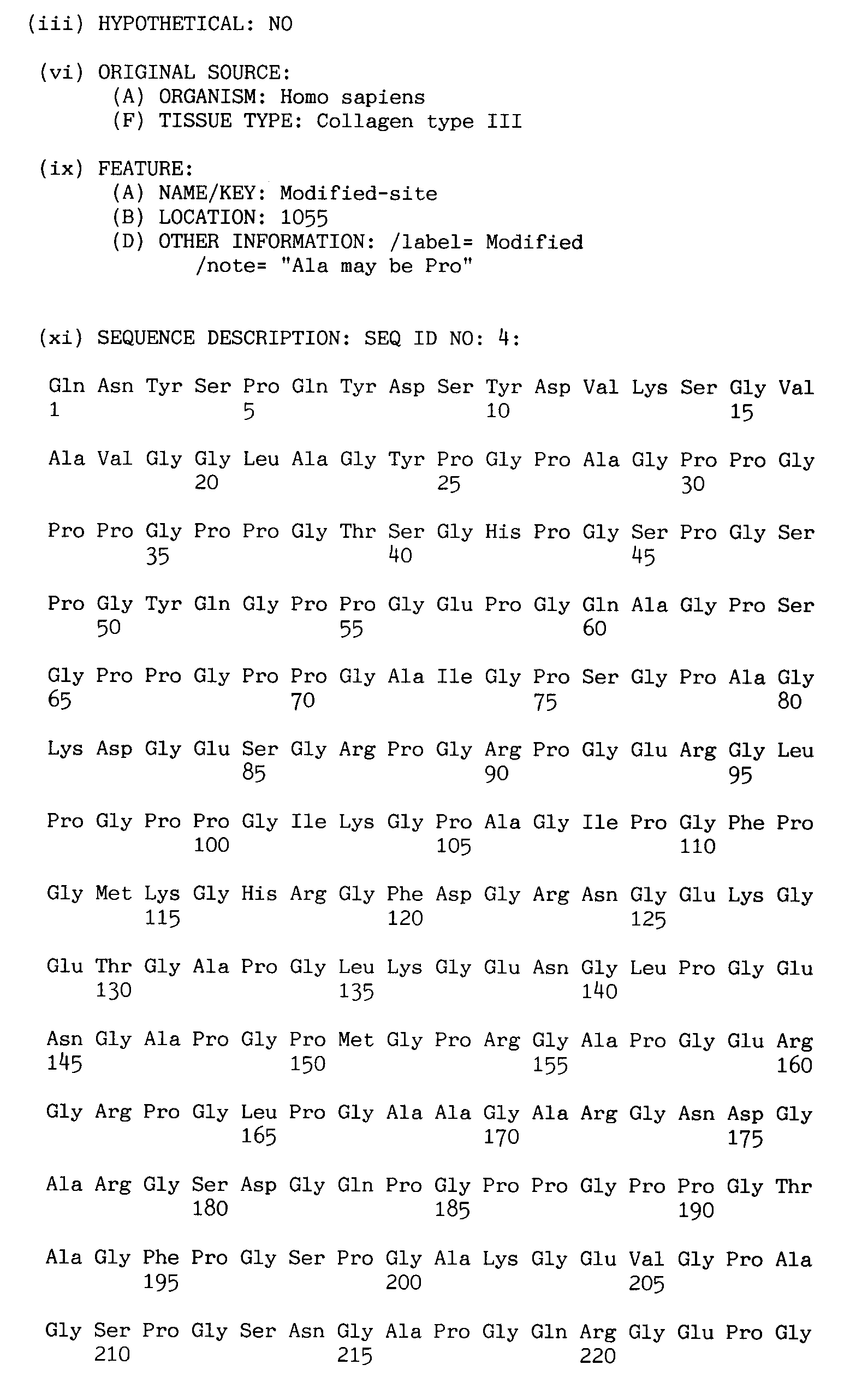
One of the two antibodies drawn in each of the 9 possible situations (panel A) serves as a catching antibody. The other antibody, that may be identical to the first, is used to detect the collagen degradation fragments, by whatever means, in a sandwich assay (see panel B). Antibodies directed against any epitope in situations 1-9 may serve the purpose of developing a sensitive and convenient assay to detect or/and quantitate collagen degradation products in serum, plasma or blood or other medium. - Fig. 4. Sequences of collagen types I, II and III proteins, with possible sites of epitopes based on crosslinking residue in triple helical region. The underlined parts represent epitopes based on crosslinking residues in triple helical region; double underlined are residues involved in lysyl oxidase-mediated crosslinking. P represents proline or hydroxyproline, K represents lysine or hydroxylysine (optionally glycosylated).
- Fig. 5: Sequences of collagen types I, II and III proteins, with possible sites of epitopes based on crosslinking residue in telopeptides. The underlined parts represent epitopes based on crosslinking residues in telopeptides; double underlined are residues involved in lysyl oxidase-mediated crosslinking. P represents proline or hydroxyproline, K represents lysine or hydroxylysine (optionally glycosylated).
- Normal human serum was separated into fractions with different molecular weight by ultrafiltration: serum (10 ml) was passed through cut off-filters of 100, 50, 30, and 10 kD (Centriprep, Grace BV, Amicon Division, Capelle aan de IJssel, The Netherlands. Fractions (0.5 ml) with molecular weight of <10 kD, 10-30 kD, 30-50 kD, and 50-100 kD were dried, reconstituted in 250 µl 6 N HCl, and hydrolysed at 108°C for 16 hours. Hydrolysed fractions were dried, reconstituted in 500 µl 6N HCl, 200 µl glacial acetic acid, 200 µl CF-1 cellulose slurry, and 800 µl n-butanol, and subjected to solid phase extraction with CF-1 cellulose according to Black et al, (Anal. Biochem. 1988, 169: 197-203). HPLChromatography was used for quantitating amounts of pyridinoline crosslinks, which were detected by their fluoresence at 295/400 nm (excitation/emission wavelengths) (see Black et al. above).
Substantial amounts of pyridinoline crosslinks (the sum of hydroxylysylpyridinoline and lysylpyridinoline) were detected in 0-10, 10-30, and 30-50 kD fractions. About half of the total serum concentration of pyridinoline (approx. 1.5 nM) had a molecular weight between 10 and 30 kD. About 25% had a molecular weight less than 10 kD. The other 25% had molecular weight between 30 and 50 kD (Fig. 1).




Claims (11)
- Method for assaying collagen degradation products in a biological sample, characterised by contacting said sample, or a fraction thereof containing protein material, with a first antibody directed at an epitope present on a collagen molecule at a distance of up to 165 amino acids from a collagen telopeptide crosslink site, and a second antibody directed at an epitope present on a collagen molecule at a distance of up to 165 amino acids from a collagen crosslink site.
- Method according to claim 1, wherein one of said antibodies is detectable and the other of said antibodies is immobilised.
- Method according to claim 1 or 2, wherein said second antibody is directed at an epitope present on a collagen molecule at a distance of from 5 to 150 amino acids from a collagen triple helical region crosslink site.
- Method according to claim 1 or 2, wherein said second antibody is directed at an epitope present on a collagen molecule at a distance of 5 to 165 amino acids from a collagen telopeptide crosslink site.
- Method according to claim 4, wherein said first antibody and said second 0antibody are directed at an identical epitope present on a collagen molecule at a distance of from 5 to 165 amino acids from a collagen telopeptide crosslink site.
- Method according to any one of claims 1-4, wherein said first antibody is directed at an epitope present on a collagen telopeptide crosslink site, and said second antibody is directed at an epitope present on a collagen molecule at a distance of 5 to 165 amino acids from a collagen crosslink site.
- Method according to any one of claims 1-5, wherein said first and/or second antibody is directed at an epitope at a distance of 5 to 100 amino acids from said collagen crosslink site.
- Method according to any one of the previous claims, wherein at least one of said first antibody and said second antibody is directed at an epitope present on the collagen α1(I), α2(I), α1(II) or α1(III) chain or any other collagen such as type IV, V, VI, IX, X or XI.
- Method according to any one of the previous claims, wherein said protein material has molecular weights between 3 kD and 50 kD, in particular between 5 kD and 40 kD.
- Method according to any one of the previous claims, wherein said biological sample is derived from blood, serum, plasma or sputum.
- Diagnostic kit for assaying bone degradation products, containing one or more antibodies directed at an epitope present on a collagen molecule, at least one of said antibodies being directed at an epitope located at a distance of up to 165 amino acids from a collagen telopeptide crosslink site, and/or at least one of said antibodies being directed at an epitope located at a distance of 5 to 165 amino acids from a collagen crosslink site.
Priority Applications (3)
| Application Number | Priority Date | Filing Date | Title |
|---|---|---|---|
| EP96202596A EP0829724A1 (en) | 1996-09-17 | 1996-09-17 | Assay for detecting collagen degradation |
| JP9268200A JPH10185915A (en) | 1996-09-17 | 1997-09-16 | Assay for collagen decomposition |
| US08/931,820 US6010863A (en) | 1996-09-17 | 1997-09-16 | Assay for collagen degradation |
Applications Claiming Priority (1)
| Application Number | Priority Date | Filing Date | Title |
|---|---|---|---|
| EP96202596A EP0829724A1 (en) | 1996-09-17 | 1996-09-17 | Assay for detecting collagen degradation |
Publications (1)
| Publication Number | Publication Date |
|---|---|
| EP0829724A1 true EP0829724A1 (en) | 1998-03-18 |
Family
ID=8224388
Family Applications (1)
| Application Number | Title | Priority Date | Filing Date |
|---|---|---|---|
| EP96202596A Withdrawn EP0829724A1 (en) | 1996-09-17 | 1996-09-17 | Assay for detecting collagen degradation |
Country Status (3)
| Country | Link |
|---|---|
| US (1) | US6010863A (en) |
| EP (1) | EP0829724A1 (en) |
| JP (1) | JPH10185915A (en) |
Cited By (6)
| Publication number | Priority date | Publication date | Assignee | Title |
|---|---|---|---|---|
| WO1998026286A3 (en) * | 1996-12-09 | 1998-08-13 | Osteometer Biotech As | Sandwich assays for collagen type i fragments |
| WO1999006840A1 (en) * | 1997-07-31 | 1999-02-11 | Metra Biosystems, Inc. | Collagen-peptide assay method |
| WO2000058733A3 (en) * | 1999-03-29 | 2001-02-15 | Isis Innovation | Assay for testing the efficacity of a therapy |
| US6300083B1 (en) | 1996-08-22 | 2001-10-09 | Osteometer Biotech A/S | Assaying D-amino acids in body fluids |
| CN109709341A (en) * | 2019-01-17 | 2019-05-03 | 浙江大学 | The biomarker and purposes of the second hypotype of osteoarthritis |
| EP2208073B1 (en) * | 2007-11-05 | 2020-01-15 | Nordic Bioscience A/S | Biochemical markers for cvd risk assessment |
Families Citing this family (6)
| Publication number | Priority date | Publication date | Assignee | Title |
|---|---|---|---|---|
| WO2002029409A2 (en) * | 2000-10-03 | 2002-04-11 | Rowett Research Institute | Method of assaying pyrrole-containing biological compounds |
| US20020177223A1 (en) * | 2001-03-12 | 2002-11-28 | Ogle Mathew F. | Methods and compositions for crosslinking tissue |
| US20040049351A1 (en) * | 2002-08-28 | 2004-03-11 | Matson Robert S. | Immunosorbent assay in microarray format |
| EP2238455A4 (en) * | 2008-01-18 | 2011-03-02 | Vatrix Medical Inc | Diagnostic biomarkers for vascular aneurysm |
| EP2902413B1 (en) * | 2012-09-26 | 2019-07-17 | FUJIFILM Corporation | Polypeptide, scaffold composition, composition for cartilage tissue restoration, composition for cartilage cell culture, and composition for promoting glycosaminoglycan production |
| US20240003906A1 (en) * | 2020-04-16 | 2024-01-04 | Nordic Bioscience A/S | Biomarker of Fibrosis |
Citations (4)
| Publication number | Priority date | Publication date | Assignee | Title |
|---|---|---|---|---|
| WO1988008980A1 (en) * | 1987-05-08 | 1988-11-17 | Farmos-Yhtymä Oy | Type iii collagen degradation assay |
| EP0401370A1 (en) * | 1988-02-19 | 1990-12-12 | Fuji Yakuhin Kogyo Kabushiki Kaisha | Enzyme immunoassay according to sandwich method of human iv-type collagen |
| EP0505210A2 (en) * | 1991-03-20 | 1992-09-23 | Orion-Yhtymà Oy | Assay for measuring the degradation of type I collagen |
| WO1995008115A1 (en) * | 1993-09-17 | 1995-03-23 | Osteometer Biotech As | A method of assaying collagen fragments in body fluids, a test kit and means for carrying out the method and use of the method to diagnose the presence of disorders associated with the metabolism of collagen |
Family Cites Families (1)
| Publication number | Priority date | Publication date | Assignee | Title |
|---|---|---|---|---|
| US4973666A (en) * | 1987-11-06 | 1990-11-27 | Washington Research Foundation | Peptide fragments containing HP and LP cross-links |
-
1996
- 1996-09-17 EP EP96202596A patent/EP0829724A1/en not_active Withdrawn
-
1997
- 1997-09-16 US US08/931,820 patent/US6010863A/en not_active Expired - Fee Related
- 1997-09-16 JP JP9268200A patent/JPH10185915A/en active Pending
Patent Citations (4)
| Publication number | Priority date | Publication date | Assignee | Title |
|---|---|---|---|---|
| WO1988008980A1 (en) * | 1987-05-08 | 1988-11-17 | Farmos-Yhtymä Oy | Type iii collagen degradation assay |
| EP0401370A1 (en) * | 1988-02-19 | 1990-12-12 | Fuji Yakuhin Kogyo Kabushiki Kaisha | Enzyme immunoassay according to sandwich method of human iv-type collagen |
| EP0505210A2 (en) * | 1991-03-20 | 1992-09-23 | Orion-Yhtymà Oy | Assay for measuring the degradation of type I collagen |
| WO1995008115A1 (en) * | 1993-09-17 | 1995-03-23 | Osteometer Biotech As | A method of assaying collagen fragments in body fluids, a test kit and means for carrying out the method and use of the method to diagnose the presence of disorders associated with the metabolism of collagen |
Cited By (7)
| Publication number | Priority date | Publication date | Assignee | Title |
|---|---|---|---|---|
| US6300083B1 (en) | 1996-08-22 | 2001-10-09 | Osteometer Biotech A/S | Assaying D-amino acids in body fluids |
| WO1998026286A3 (en) * | 1996-12-09 | 1998-08-13 | Osteometer Biotech As | Sandwich assays for collagen type i fragments |
| WO1999006840A1 (en) * | 1997-07-31 | 1999-02-11 | Metra Biosystems, Inc. | Collagen-peptide assay method |
| US5972623A (en) * | 1997-07-31 | 1999-10-26 | Metra Biosystems, Inc. | Collagen-peptide assay method |
| WO2000058733A3 (en) * | 1999-03-29 | 2001-02-15 | Isis Innovation | Assay for testing the efficacity of a therapy |
| EP2208073B1 (en) * | 2007-11-05 | 2020-01-15 | Nordic Bioscience A/S | Biochemical markers for cvd risk assessment |
| CN109709341A (en) * | 2019-01-17 | 2019-05-03 | 浙江大学 | The biomarker and purposes of the second hypotype of osteoarthritis |
Also Published As
| Publication number | Publication date |
|---|---|
| JPH10185915A (en) | 1998-07-14 |
| US6010863A (en) | 2000-01-04 |
Similar Documents
| Publication | Publication Date | Title |
|---|---|---|
| AU716309B2 (en) | Screening of combinatorial peptide libraries for selection of peptide ligand useful in affinitypurification of target proteins | |
| US5585277A (en) | Screening method for identifying ligands for target proteins | |
| JP2886728B2 (en) | Bone resorption analysis based on peptide released during collagen degradation | |
| EP0675903B1 (en) | Bone resorption assay | |
| WO1994028166A1 (en) | Fluorescence energy transfer substrates | |
| US6010863A (en) | Assay for collagen degradation | |
| CA2262403A1 (en) | Biomarkers and targets for diagnosis, prognosis and management of prostate disease | |
| JP2023503653A5 (en) | ||
| Dr. Kamel et al. | Multiple molecular forms of pyridinolines cross‐links excreted in human urine evaluated by chromatographic and immunoassay methods | |
| JPH08509294A (en) | Immunoassay for detecting collagen or collagen fragments | |
| US6110689A (en) | Method of assaying collagen fragments in body fluids, a test kit and means for carrying out the method and use of the method to diagnose the presence of disorders associated with the metabolism of collagen | |
| JP2575296B2 (en) | Synthetic standards for immunoassays | |
| EP0944833B1 (en) | Sandwich assays for collagen fragments | |
| AU603911B2 (en) | Nuclear antigen la | |
| JP4130505B2 (en) | Elimination of diagnostic method interference by peptides consisting of D-amino acids | |
| WO2007124593A1 (en) | Branched peptide amplification and uses thereof | |
| EP0787301B1 (en) | Estimation of the fragmentation pattern of collagen in body fluids and the diagnosis of disorders associated with the metabolism of collagen | |
| WO1998011436A1 (en) | Non-specific affinity enhancement to identify combinatorial library members | |
| AU2008255631B2 (en) | Fluorescent protein particles | |
| Akiyama et al. | Comparisons of evolutionarily distinct fibronectins: evidence for the origin of plasma and fibroblast cellular fibronectins from a single gene | |
| EP1485718B1 (en) | Method for monitoring collagen type ii degradation in cartilage | |
| US20050153383A1 (en) | Synthetic and recombinant substrates for the detecion of the von willebrand factor-cleaving protease | |
| DE19919121A1 (en) | Multimeric cytomegalovirus antigen useful as an immunoassay reagent, and which comprises at least two p52 peptide sequences | |
| JP2002098698A (en) | DNA binding protein assay | |
| JP2002187899A (en) | Peptides having binding properties to luciferase |
Legal Events
| Date | Code | Title | Description |
|---|---|---|---|
| PUAI | Public reference made under article 153(3) epc to a published international application that has entered the european phase |
Free format text: ORIGINAL CODE: 0009012 |
|
| AK | Designated contracting states |
Kind code of ref document: A1 Designated state(s): AT BE CH DE DK ES FI FR GB GR IE IT LI LU NL PT SE |
|
| 17P | Request for examination filed |
Effective date: 19980917 |
|
| AKX | Designation fees paid |
Free format text: AT BE CH DE DK ES FI FR GB GR IE IT LI LU NL PT SE |
|
| RBV | Designated contracting states (corrected) |
Designated state(s): AT BE CH DE DK ES FI FR GB GR IE IT LI LU NL PT SE |
|
| 17Q | First examination report despatched |
Effective date: 20020718 |
|
| STAA | Information on the status of an ep patent application or granted ep patent |
Free format text: STATUS: THE APPLICATION IS DEEMED TO BE WITHDRAWN |
|
| 18D | Application deemed to be withdrawn |
Effective date: 20050401 |