EP0815231B1 - Use of neuronal apoptosis inhibitor protein (naip) - Google Patents

Use of neuronal apoptosis inhibitor protein (naip) Download PDF

Info

Publication number
EP0815231B1
EP0815231B1 EP97902522A EP97902522A EP0815231B1 EP 0815231 B1 EP0815231 B1 EP 0815231B1 EP 97902522 A EP97902522 A EP 97902522A EP 97902522 A EP97902522 A EP 97902522A EP 0815231 B1 EP0815231 B1 EP 0815231B1
Authority
EP
European Patent Office
Prior art keywords
naip
nucleic acid
seq
cell
sequence
Prior art date
Legal status (The legal status is an assumption and is not a legal conclusion. Google has not performed a legal analysis and makes no representation as to the accuracy of the status listed.)
Expired - Lifetime
Application number
EP97902522A
Other languages
German (de)
French (fr)
Other versions
EP0815231A2 (en
Inventor
Robert G. Korneluk
Alexander E. Mackenzie
Natalie Roy
George Robertson
Katsu Tamai
Current Assignee (The listed assignees may be inaccurate. Google has not performed a legal analysis and makes no representation or warranty as to the accuracy of the list.)
University of Ottawa
Original Assignee
University of Ottawa
Priority date (The priority date is an assumption and is not a legal conclusion. Google has not performed a legal analysis and makes no representation as to the accuracy of the date listed.)
Filing date
Publication date
Application filed by University of Ottawa filed Critical University of Ottawa
Priority to EP05106334A priority Critical patent/EP1609864A3/en
Publication of EP0815231A2 publication Critical patent/EP0815231A2/en
Application granted granted Critical
Publication of EP0815231B1 publication Critical patent/EP0815231B1/en
Anticipated expiration legal-status Critical
Expired - Lifetime legal-status Critical Current

Links

Images

Classifications

    • CCHEMISTRY; METALLURGY
    • C07ORGANIC CHEMISTRY
    • C07KPEPTIDES
    • C07K14/00Peptides having more than 20 amino acids; Gastrins; Somatostatins; Melanotropins; Derivatives thereof
    • C07K14/435Peptides having more than 20 amino acids; Gastrins; Somatostatins; Melanotropins; Derivatives thereof from animals; from humans
    • C07K14/46Peptides having more than 20 amino acids; Gastrins; Somatostatins; Melanotropins; Derivatives thereof from animals; from humans from vertebrates
    • C07K14/47Peptides having more than 20 amino acids; Gastrins; Somatostatins; Melanotropins; Derivatives thereof from animals; from humans from vertebrates from mammals
    • AHUMAN NECESSITIES
    • A61MEDICAL OR VETERINARY SCIENCE; HYGIENE
    • A61PSPECIFIC THERAPEUTIC ACTIVITY OF CHEMICAL COMPOUNDS OR MEDICINAL PREPARATIONS
    • A61P25/00Drugs for disorders of the nervous system
    • AHUMAN NECESSITIES
    • A61MEDICAL OR VETERINARY SCIENCE; HYGIENE
    • A61PSPECIFIC THERAPEUTIC ACTIVITY OF CHEMICAL COMPOUNDS OR MEDICINAL PREPARATIONS
    • A61P31/00Antiinfectives, i.e. antibiotics, antiseptics, chemotherapeutics
    • A61P31/12Antivirals
    • AHUMAN NECESSITIES
    • A61MEDICAL OR VETERINARY SCIENCE; HYGIENE
    • A61PSPECIFIC THERAPEUTIC ACTIVITY OF CHEMICAL COMPOUNDS OR MEDICINAL PREPARATIONS
    • A61P43/00Drugs for specific purposes, not provided for in groups A61P1/00-A61P41/00
    • AHUMAN NECESSITIES
    • A61MEDICAL OR VETERINARY SCIENCE; HYGIENE
    • A61PSPECIFIC THERAPEUTIC ACTIVITY OF CHEMICAL COMPOUNDS OR MEDICINAL PREPARATIONS
    • A61P9/00Drugs for disorders of the cardiovascular system
    • CCHEMISTRY; METALLURGY
    • C07ORGANIC CHEMISTRY
    • C07KPEPTIDES
    • C07K14/00Peptides having more than 20 amino acids; Gastrins; Somatostatins; Melanotropins; Derivatives thereof
    • C07K14/435Peptides having more than 20 amino acids; Gastrins; Somatostatins; Melanotropins; Derivatives thereof from animals; from humans
    • C07K14/46Peptides having more than 20 amino acids; Gastrins; Somatostatins; Melanotropins; Derivatives thereof from animals; from humans from vertebrates
    • C07K14/47Peptides having more than 20 amino acids; Gastrins; Somatostatins; Melanotropins; Derivatives thereof from animals; from humans from vertebrates from mammals
    • C07K14/4701Peptides having more than 20 amino acids; Gastrins; Somatostatins; Melanotropins; Derivatives thereof from animals; from humans from vertebrates from mammals not used
    • C07K14/4747Apoptosis related proteins
    • CCHEMISTRY; METALLURGY
    • C07ORGANIC CHEMISTRY
    • C07KPEPTIDES
    • C07K16/00Immunoglobulins [IG], e.g. monoclonal or polyclonal antibodies
    • C07K16/18Immunoglobulins [IG], e.g. monoclonal or polyclonal antibodies against material from animals or humans
    • CCHEMISTRY; METALLURGY
    • C12BIOCHEMISTRY; BEER; SPIRITS; WINE; VINEGAR; MICROBIOLOGY; ENZYMOLOGY; MUTATION OR GENETIC ENGINEERING
    • C12QMEASURING OR TESTING PROCESSES INVOLVING ENZYMES, NUCLEIC ACIDS OR MICROORGANISMS; COMPOSITIONS OR TEST PAPERS THEREFOR; PROCESSES OF PREPARING SUCH COMPOSITIONS; CONDITION-RESPONSIVE CONTROL IN MICROBIOLOGICAL OR ENZYMOLOGICAL PROCESSES
    • C12Q1/00Measuring or testing processes involving enzymes, nucleic acids or microorganisms; Compositions therefor; Processes of preparing such compositions
    • C12Q1/68Measuring or testing processes involving enzymes, nucleic acids or microorganisms; Compositions therefor; Processes of preparing such compositions involving nucleic acids
    • C12Q1/6876Nucleic acid products used in the analysis of nucleic acids, e.g. primers or probes
    • C12Q1/6883Nucleic acid products used in the analysis of nucleic acids, e.g. primers or probes for diseases caused by alterations of genetic material
    • GPHYSICS
    • G01MEASURING; TESTING
    • G01NINVESTIGATING OR ANALYSING MATERIALS BY DETERMINING THEIR CHEMICAL OR PHYSICAL PROPERTIES
    • G01N33/00Investigating or analysing materials by specific methods not covered by groups G01N1/00 - G01N31/00
    • G01N33/48Biological material, e.g. blood, urine; Haemocytometers
    • G01N33/50Chemical analysis of biological material, e.g. blood, urine; Testing involving biospecific ligand binding methods; Immunological testing
    • G01N33/53Immunoassay; Biospecific binding assay; Materials therefor
    • G01N33/575Immunoassay; Biospecific binding assay; Materials therefor for cancer
    • G01N33/5758Immunoassay; Biospecific binding assay; Materials therefor for cancer involving compounds serving as markers for tumours, cancers or neoplasias, e.g. cellular determinants, receptors, heat shock/stress proteins, A-protein, oligosaccharides or metabolites
    • AHUMAN NECESSITIES
    • A01AGRICULTURE; FORESTRY; ANIMAL HUSBANDRY; HUNTING; TRAPPING; FISHING
    • A01KANIMAL HUSBANDRY; AVICULTURE; APICULTURE; PISCICULTURE; FISHING; REARING OR BREEDING ANIMALS, NOT OTHERWISE PROVIDED FOR; NEW BREEDS OF ANIMALS
    • A01K2217/00Genetically modified animals
    • A01K2217/05Animals comprising random inserted nucleic acids (transgenic)
    • AHUMAN NECESSITIES
    • A01AGRICULTURE; FORESTRY; ANIMAL HUSBANDRY; HUNTING; TRAPPING; FISHING
    • A01KANIMAL HUSBANDRY; AVICULTURE; APICULTURE; PISCICULTURE; FISHING; REARING OR BREEDING ANIMALS, NOT OTHERWISE PROVIDED FOR; NEW BREEDS OF ANIMALS
    • A01K2227/00Animals characterised by species
    • A01K2227/10Mammal
    • A01K2227/105Murine
    • AHUMAN NECESSITIES
    • A61MEDICAL OR VETERINARY SCIENCE; HYGIENE
    • A61KPREPARATIONS FOR MEDICAL, DENTAL OR TOILETRY PURPOSES
    • A61K38/00Medicinal preparations containing peptides
    • AHUMAN NECESSITIES
    • A61MEDICAL OR VETERINARY SCIENCE; HYGIENE
    • A61KPREPARATIONS FOR MEDICAL, DENTAL OR TOILETRY PURPOSES
    • A61K48/00Medicinal preparations containing genetic material which is inserted into cells of the living body to treat genetic diseases; Gene therapy
    • CCHEMISTRY; METALLURGY
    • C12BIOCHEMISTRY; BEER; SPIRITS; WINE; VINEGAR; MICROBIOLOGY; ENZYMOLOGY; MUTATION OR GENETIC ENGINEERING
    • C12QMEASURING OR TESTING PROCESSES INVOLVING ENZYMES, NUCLEIC ACIDS OR MICROORGANISMS; COMPOSITIONS OR TEST PAPERS THEREFOR; PROCESSES OF PREPARING SUCH COMPOSITIONS; CONDITION-RESPONSIVE CONTROL IN MICROBIOLOGICAL OR ENZYMOLOGICAL PROCESSES
    • C12Q2600/00Oligonucleotides characterized by their use
    • C12Q2600/136Screening for pharmacological compounds
    • CCHEMISTRY; METALLURGY
    • C12BIOCHEMISTRY; BEER; SPIRITS; WINE; VINEGAR; MICROBIOLOGY; ENZYMOLOGY; MUTATION OR GENETIC ENGINEERING
    • C12QMEASURING OR TESTING PROCESSES INVOLVING ENZYMES, NUCLEIC ACIDS OR MICROORGANISMS; COMPOSITIONS OR TEST PAPERS THEREFOR; PROCESSES OF PREPARING SUCH COMPOSITIONS; CONDITION-RESPONSIVE CONTROL IN MICROBIOLOGICAL OR ENZYMOLOGICAL PROCESSES
    • C12Q2600/00Oligonucleotides characterized by their use
    • C12Q2600/156Polymorphic or mutational markers
    • CCHEMISTRY; METALLURGY
    • C12BIOCHEMISTRY; BEER; SPIRITS; WINE; VINEGAR; MICROBIOLOGY; ENZYMOLOGY; MUTATION OR GENETIC ENGINEERING
    • C12QMEASURING OR TESTING PROCESSES INVOLVING ENZYMES, NUCLEIC ACIDS OR MICROORGANISMS; COMPOSITIONS OR TEST PAPERS THEREFOR; PROCESSES OF PREPARING SUCH COMPOSITIONS; CONDITION-RESPONSIVE CONTROL IN MICROBIOLOGICAL OR ENZYMOLOGICAL PROCESSES
    • C12Q2600/00Oligonucleotides characterized by their use
    • C12Q2600/158Expression markers
    • GPHYSICS
    • G01MEASURING; TESTING
    • G01NINVESTIGATING OR ANALYSING MATERIALS BY DETERMINING THEIR CHEMICAL OR PHYSICAL PROPERTIES
    • G01N2510/00Detection of programmed cell death, i.e. apoptosis

Definitions

  • This invention relates in general to the function of the NAIP inhibitor protein in apoptosis and more particularly to the use of NAIP proteins and nucleic acids to characterize NAIP, identify compounds which modulate NAIP, and treat conditions affected by changes in NAIP levels.
  • Apoptosis is a morphologically distinct form of programmed cell death that is important in the normal development and maintenance of multicellular organisms. Dysregulation of apoptosis can take the form of inappropriate suppression of cell death, as occurs in the development of some cancers, or in a failure to control the extent of cell death, as is believed to occur in acquired immunodeficiency and certain neurodegenerative disorders, such as spinal muscular atrophy (SMA).
  • SMA spinal muscular atrophy
  • baculoviruses encode proteins that are termed inhibitors of apoptosis proteins (IAPs) because they inhibit the apoptosis that would otherwise occur when insect cells are infected by the virus. These proteins are thought to work in a manner that is independent of other viral proteins.
  • the baculovirus IAP genes include sequences encoding a ring zinc finger-like motif (RZF), which may be involved in DNA binding, and two N-terminal domains that consist of a 70 amino acid repeat motif termed a BIR domain (Baculovirus IAP Repeat).
  • WO 9612016 published prior to the international filing date but later than the priority date of the present invention, discloses the gene encoding NAIP and the corresponding amino acid sequence of 1232 amino acids in length. Use of the sequence information for detection of NAIP sequences and for diagnosis and treatment of SMA is also disclosed.
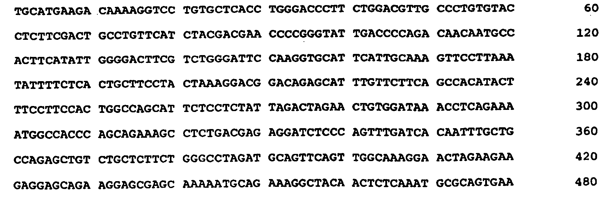
  • the present invention relates to a novel Neuronal Apoptosis Inhibitor Protein (NAIP) sequence and a NAIP fragment with anti-apoptotic activities, which comprise nucleotides 3734 to 3886 (exon 14a) and 4139 to 4503 (exon 17) of SEQ ID NO: 21 or nucleotides 3838 to 3990 (exon 14a) and nucleotides 4243 to 4605 (exon 17) of SEQ ID NO: 23.
  • NAIP Neuronal Apoptosis Inhibitor Protein
  • the present invention provides a substantially pure nucleic acid having at least 90% nucleic acid sequence identity to the full length Neuronal Apoptosis Inhibitor Protein (NAIP) nucleic acid sequence of SEQ ID NO: 21 and including nucleotides 3734 to 3886 (exon 14a) and 4139 to 4503 (exon 17) of SEQ ID NO: 21 or having at least 90% nucleic acid sequence identity to the full length NAIP nucleic acid sequence of SEQ ID NO: 23 and including nucleotides 3838 to 3990 of SEQ ID NO:23 and nucleotides 4243 to 4605 of SEQ ID 23 said nucleic acid encoding a NAIP polypeptide that inhibits apoptosis.
  • NAIP Neuronal Apoptosis Inhibitor Protein
  • the present invention also provides a substantially pure nucleic acid having the sequence of SEQ ID NO:21 or degenerate variants thereof and encode a polypeptide having the amino acid sequence of SEQ ID NO:22, wherein said polypeptide is capable of inhibiting apoptosis.
  • nucleic acid is operably linked to a regulatory sequence for expression of said NAIP polypeptide and said regulatory sequence comprises a promoter.
  • the promoter is a constitutive promoter and is inducible by one or more external agents or is cell-type specific.
  • the present invention provides a vector comprising the nucleic acid of the present invention, wherein the vector is capable of directing expression of a NAIP polypeptide that is capable of inhibiting apoptosis.
  • the invention also provides a host cell that contains the above described vector.
  • the present invention provides a substantially pure mammalian NAIP polypeptide encoded by the nucleic acid sequence of the present invention.
  • the present invention also provides a therapeutic composition comprising an effective amount of NAIP polypeptide of the invention in a physiologically acceptable carrier.
  • the present invention also provides an in vitro method of obtaining a NAIP polypeptide.
  • the method comprises the following steps of:
  • the invention also provides an in vitro method of identifying a NAIP nucleic acid in a mammalian cell.
  • the method comprises the following steps of:
  • the present invention provides an in vitro method of isolating a NAIP nucleic acid of the invention or a portion thereof.
  • the method comprises amplifying by PCR the NAIP nucleic acid, or portion thereof, using oligonucleotide primers wherein said primers;
  • Also provided by the present invention is an in vitro method of identifying a compound that modulates apoptosis.
  • the method comprises contacting in vitro a cell expressing a NAIP polypeptide with a candidate compound and monitoring expression of a NAIP nucleic acid of the invention by detecting a nucleic acid sequence comprising nucleotides 3838 to 3990 of SEQ ID NO:23 or nucleotides 4243 to 4605 of SEQ ID NO:23.
  • An alteration in the level of said expression of said nucleic acid indicates the presence of a compound which modulates apoptosis.
  • the present invention also provides a neuronal apoptosis inhibitor protein (NAIP) nucleic acid of the invention or a polypeptide encoded by said nucleic acid sequence, for use as a medicament.
  • NAIP neuronal apoptosis inhibitor protein
  • NAIP nucleic acid of the invention or a polypeptide encoded by said nucleic acid sequence, for the manufacture of a medicament for the treatment of spinal muscular atrophy, ALS, AIDS, a neurodegenerative disease, a myelodysplastic syndrome or an ischemic injury .
  • the invention also provides a NAIP antisense molecule comprising nucleic acid that is complimentary to nucleotides 3734 to 3886 (exon 14a) or to nucleotides 4139 to 4503 (exon 17) of SEQ ID NO: 21 or to nucleotides 3838 to 3990 of SEQ ID NO:23 or nucleotides 4243 to 4605 of SEQ ID 23 and use of this antisense molecule as a medicament.
  • the invention features a substantially pure nucleic acid molecule, such as a genomic, cDNA, antisense DNA, RNA, or a synthetic nucleic acid molecule, that encodes or corresponds to a mammalian NAIP polypeptide of the invention.
  • This nucleic acid may be incorporated into a vector.
  • a vector may be in a cell, such as a mammalian, yeast, nematode, or bacterial cell.
  • the nucleic acid may also be incorporated into a non-human transgenic animal or embryo thereof.
  • the nucleic acid molecule is a human NAIP nucleic acid.
  • the NAIP gene is a human NAIP gene.
  • the cell is a transformed cell.
  • the nucleic acid sequence includes the cDNA sequences encoding exons 14a and 17.
  • the sequence includes exons 1-14, 14a, and 15-17.
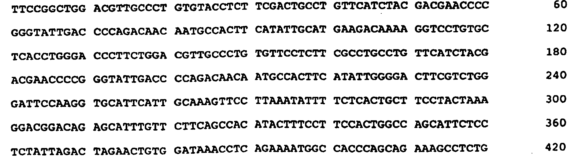
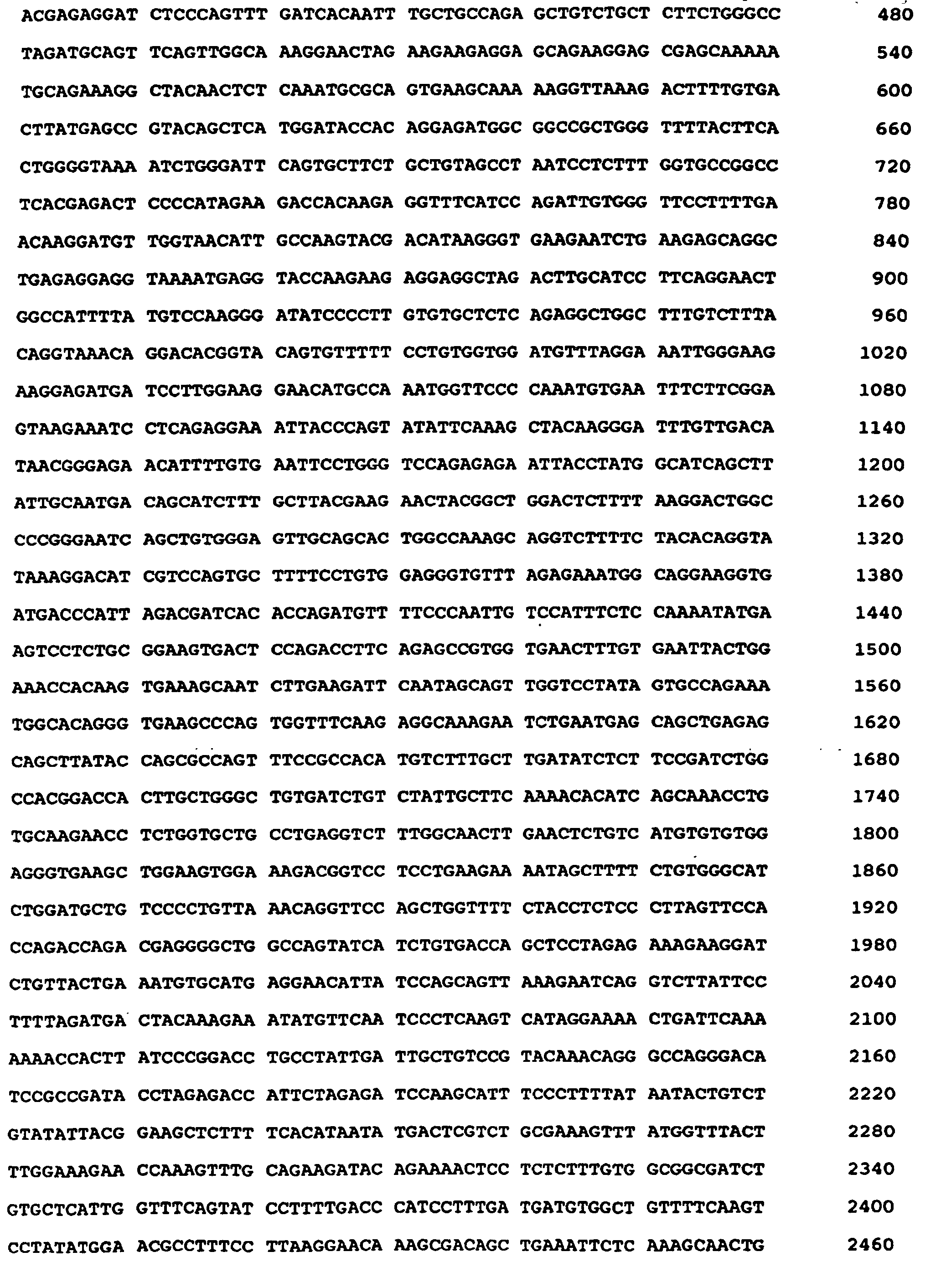
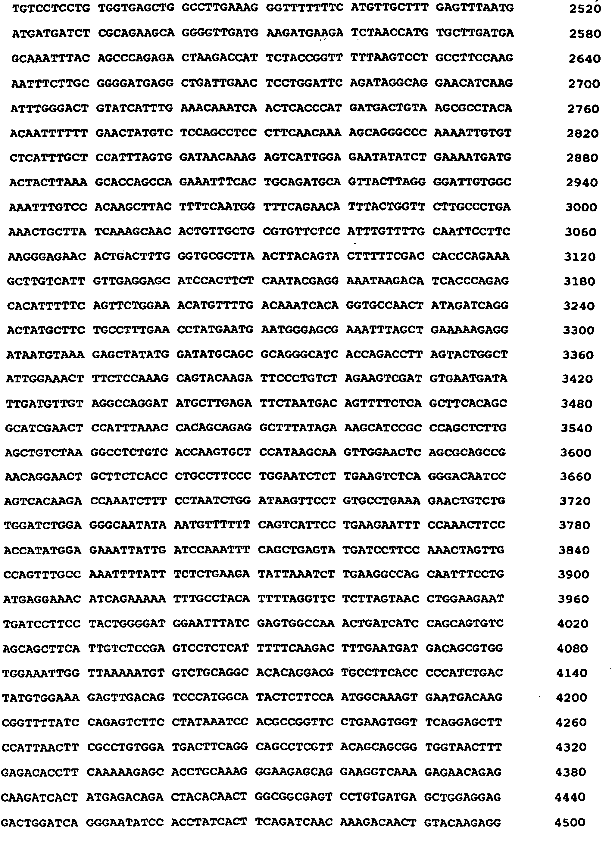
  • the sequence also includes the complete 5' and 3' untranslated regions of the NAIP gene and is represented as Seq. ID No. 2, 21, or 23, most preferably, as in Seq. ID No. 21.
  • the nucleic acid is a purified nucleotide sequence comprising genomic DNA, cDNA, mRNA, anti-sense DNA or other DNA substantially identical to the cDNA sequences of Seq. ID No. 2, 21, or 23 corresponding to the cDNA sequences of the invention. Most preferably exons 1 to 14 and 1 4a to 17 are as described in Seq. ID No. 21.
  • the invention features nucleic acid sequences substantially identical to the sequences shown in Fig. 21, or fragments thereof, wherein the fragments comprise nucleotides 3734 to 3886 (exon 14a) and 4139 to 4503 (exon 17) of SEQ ID NO: 21 or nucleotides 3838 to 3990 (exon 14a) and nucleotides 4243 to 4605 (exon 17) of SEQ ID NO: 23 and encode a polypeptide that inhibits apoptosis.
  • the invention also features RNA which is encoded by the DNA described herein.
  • the RNA is mRNA.
  • the RNA is antisense RNA that is complementary to the coding strand of NAIP.
  • the NAIP encoding nucleic acid comprises at least the 3 BIR domains of a NAIP sequence provided herein (e.g., nucleotides 1-1360 of the NAIP sequence provided in Fig. 6), but lacks at least some of the sequences encoding the carboxy terminus of the NAIP polypeptide.
  • at least 30 nucleic acids are deleted from the region of the NAIP gene between nucleic acids 1360 (i.e., the end of the BIR domains) 4607 (i.e., the end of the coding sequence) of the NAIP sequence shown in Fig. 6, Seq. ID No. 21. More preferably, at least 100 nucleotides are deleted, and even more preferably at least 1000 nucleotides are deleted. In the most preferred embodiment, up to 3247 nucleotides are deleted. Preferably, the deletion results in a statistically significant increase in the anti-apoptotic activity of the encoded protein on one of the assays provided herein.
  • the invention features a substantially pure DNA which includes a promoter capable of expressing or activating the expression of the NAIP gene or fragments according to the invention in a cell susceptible to apoptosis.
  • the NAIP gene is human NAIP or fragments according to the invention, as described above.
  • the promoter is the promoter native to the NAIP gene. Additionally, transcriptional and translational regulatory regions are, preferably, those native to a NAIP gene.
  • the invention provides transgenic cell lines including the NAIP nucleic acids of the invention.
  • the transgenic cells of the invention are preferably cells that are altered in their apoptotic response.
  • the transgenic mammalian cell is a fibroblast, neuronal cell, a pulmonary cell, a renal cell, a lymphocyte cell, a glial cell, a myocardial cell, an embryonic stem cell, or an insect cell.
  • the neuron is a motor neuron and the lymphocyte is a CD4+ T cell.
  • transgenic cell having a transgene encoding a NAIP polypeptide or antisense nucleic acid of the invention.
  • the transgene is integrated into the genome of the cell in a way that allows for expression.
  • the level of expression in the cell is sufficient to alter the level of apoptosis.
  • the transgene is in a motor neuron or a myocardial cell.
  • the invention features a non-human transgenic animal, preferably a mammal, more preferably a rodent, and most preferably a mouse, having a NAIP gene as described above inserted into the genome (mutant or wild-type), or a knockout of the NAIP gene in the genome, or both.
  • a non-human transgenic animal expressing NAIP antisense nucleic acid of the invention is also included.
  • the non-human transgenic animals may express either an increased or a decreased amount of NAIP polypeptide, depending on the construct used and the nature of the genomic alteration.
  • utilizing a nucleic acid molecule that encodes a NAIP of the invention to engineer a knockout mutation in a NAIP gene would generate a non-human animal with decreased expression of either all or part of the corresponding NAIP polypeptide.
  • inserting exogenous copies of a NAIP gene of the invention into the genome, preferably under the control of active regulatory and promoter elements, would lead to increased expression or the corresponding NAIP polypeptide.
  • the invention features an in-vitro method of detecting a NAIP gene in a cell by detecting the NAIP gene, or a portion thereof (which is greater than 9 nucleotides, and preferably greater than 18 nucleotides in length), with a preparation of genomic DNA from the cell.
  • the NAIP gene and the genomic DNA are brought into contact under conditions that allow for hybridization (and therefore, detection) of nucleic acid sequences in the cell that are at least 50% identical to the DNA encoding the NAIP polypeptides.
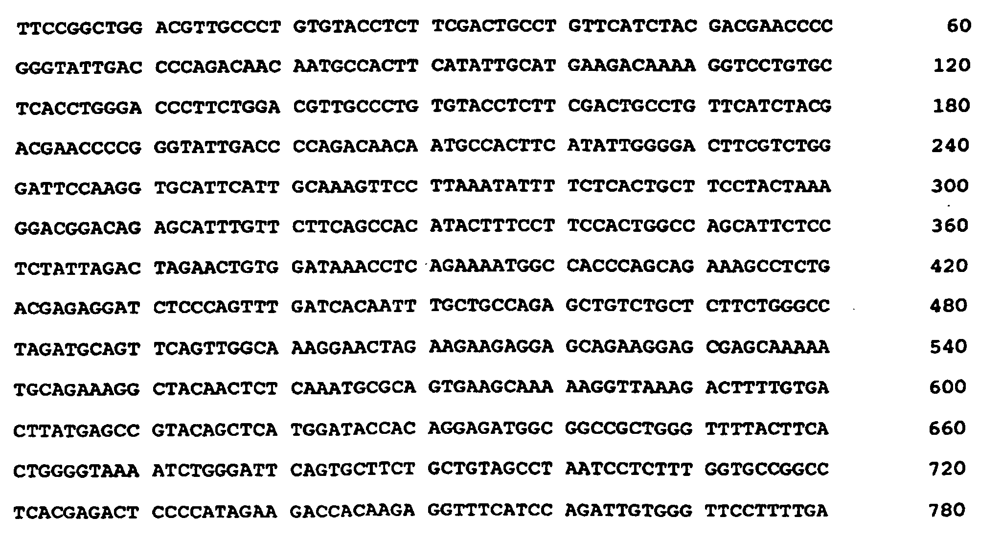
  • the nucleic acid used comprises at least a part of exon 14a or exon 17, as provided in Figs. 6 and 7.
  • the invention features a method of producing a NAIP polypeptide in vitro.
  • this method involves providing a cell with nucleic acid encoding all or part of a NAIP polypeptide (which is positioned for expression in the cell), culturing the cell under conditions that allow for expression of the nucleic acid, and isolating the NAIP polypeptide.
  • the NAIP polypeptide is expressed by DNA that is under the control of a constitutive or inducible promotor.
  • the promotor may be a native or heterologous promotor.
  • the nucleic acid comprises exon 14a or exon 17. Most preferably the nucleic acid is the nucleic acid shown in either Fig. 6 or Fig. 7. Most preferably, it is the sequence of Fig. 6.
  • the invention features substantially pure mammalian NAIP polypeptide.
  • the polypeptide includes an amino acid sequence that is substantially identical to one of the amino acid sequences shown in any one of Figs. 6 or 7.
  • the polypeptide is the human NAIP polypeptide of Fig. 6.
  • Fragments including at least two BIR domains, as provided herein, are also a part of the invention.
  • the fragment has at least three BIR domains.
  • polypeptides encoded by the nucleic acids described above having deletions between nucleic acids 1360 and the end of the gene are a part of the invention.
  • the NAIP fragments according to the invention included those NAIP fragments comprising at least 15 sequential amino acids of Seq. ID No. 22 or 24.
  • the fragment includes at least a portion of exon 14a or exon 17.
  • the invention features a recombinant mammalian polypeptide derived from NAIP that is capable of modulating apoptosis.
  • the polypeptide may include at least two BIR domains as defined herein, preferably three BIR domains.
  • the NAIP amino acid sequence differs from the NAIP sequences of Figs. 6 or 7 by only conservative substitutions or differs from the sequences encoded by the nucleic acids of Seq. ID Nos. 1 2, 21 or 23 by deletions of amino acids carboxy terminal to the BIR domains.
  • the recombinant protein decreases apoptosis relative to a control by at least 5%, more preferably by 25%.
  • the invention features an in vitro method of identifying a compound that modulates apoptosis.
  • the method includes contacting in vitro a cell expressing or capable of expressing a NAIP polypeptide of the present invention with a candidate compound, and monitoring the expression of the NAIP nucleic acid of the present invention by detecting a nucleic acid sequence comprising nucleotides 3838 to 3990 or nucleotides 4243 to 4605 of SEQ ID 23.
  • An alteration in the level of expression of the NAIP nucleic acid indicates the presence of a compound which modulates apoptosis.
  • the compound may be an inhibitor or an enhancer of apoptosis.
  • the mammalian cell is a myocardial cell, a fibroblast, a neuronal cell, a glial cell, a lymphocyte (T cell or B cell), or an insect cell.
  • the invention features methods of detecting compounds that modulate apoptosis using the interaction trap technology and NAIP polypeptides of the present invention, or fragments of the present invention, as a component of the bait.
  • the compound being tested as a modulator of apoptosis is also a polypeptide.
  • NAIP nucleic acid and polypeptides of the invention may be used in the diagnosis of a cell proliferation disease, or an increased likelihood of such a disease, e.g., a NAIP nucleic acid probe or NAIP antibody may be used.
  • the disease is a cancer of the central nervous system.
  • the disease is selected from the group consisting of neuroblastoma, meningioma, glialblastoma, astracystoma, neuroastrocytoma, promyelocytic leukemia, a HeLa-type carcinoma, chronic myelogenous leukemia (preferably using xiap or hiap-2 related probes), lymphoblastic leukemia (preferably using a xiap related probe), Burkitt's lymphoma, colorectal adenocarcinoma, lung carcinoma, and melanoma.
  • a diagnosis is indicated by a 2-fold increase in expression or activity, more preferably, at least a 10-fold increase in expression or activity.
  • NAIP nucleic acid and polypeptides of the invention may be also be used for the preparation of a medicament for the treatment of deleterious levels ofapoptosis.
  • a therapeutically effective amount of NAIP protein, NAIP nucleic acid, or a compound which enhances NAIP activity levels in a form which allows delivery to the cells which are undergoing more apoptosis than is therapeutically desirable is appropriate.
  • the cell having deleterious levels of apoptosis is a myocardial cell in a patient diagnosed with a cardiac condition.
  • apoptosis may be induced in a cell by administering to the cell a negative regulator of the NAIP-dependent anti-apoptotic pathway.
  • the negative regulator may be, but is not limited to, a NAIP polypeptide fragment of the invention or purified NAIP specific antibody.
  • the negative regulator may also be a NAIP antisense RNA molecule of the invention.
  • a mammalian NAIP of the present invention may serve as an active ingredient in a therapeutic composition.
  • This composition depending on the NAIP or fragment of the invention included, may be used to modulate apoptosis and thereby treat any condition that is caused by a disturbance in apoptosis.
  • another aspect of the invention described herein includes the compounds of the invention in a pharmaceutically acceptable carrier.
  • a NAIP nucleic acid or polypeptide of the present invention may be used to modulate apoptosis. Furthermore, a NAIP nucleic acid or polypeptide may be used in the discovery and/or manufacture of a medicament for the modulation of apoptosis.
  • NAIP gene is meant a gene encoding a polypeptide having at least exon 14a or exon 17 Figs. 6 or 7, or the sequence of Fig. 5, Seq. ID No. 1, wherein at least 10 carboxy-terminal nucleic acids have been deleted to enhance activity, as described above.
  • the NAIP gene encodes a polypeptide which is capable of inhibiting apoptosis.
  • a NAIP gene is a nucleic acid having 90% or greater nucleotide sequence identity to the NAIP amino acid encoding sequences of Figs. 6 or 7.
  • the NAIP gene encodes a polypeptide that inhibits apoptosis.
  • the region of sequence over which identity is measured is a region including nucleic acid encoding exon 14a or exon 17.
  • Mammalian NAIP genes include nucleotide sequences isolated from any mammalian source. Preferably, the mammal is a human.
  • NAIP gene is meant to encompass any NAIP gene, which is characterized by its ability to modulate apoptosis and encodes a polypeptide that has 90% amino acid sequence identity with the NAIP polypeptides shown in Figs. 6 and 7.
  • NAIP protein or "NAIP polypeptide” is meant a polypeptide, or fragment according to the invention, encoded by a NAIP gene as described above.
  • the cell population is selected from a group including T cells, neuronal cells, fibroblasts, myocardial cells, or any other cell line known to undergo apoptosis in a laboratory setting (e.g., the baculovirus infected insect cells).
  • inhibiting apoptosis is meant any decrease in the number of cells which undergo apoptosis relative to an untreated control.
  • the decrease is at least 25%, more preferably the decrease is 50%, and most preferably the decrease is at least one-fold.
  • polypeptide any chain of more than two amino acids, regardless of posttranslational modification such as glycosylation or phosphorylation.
  • substantially identical is meant a polypeptide or nucleic acid exhibiting at least 90% identity to a reference amino acid or nucleic acid sequence.
  • Sequence identity is typically measured using sequence analysis software with the default parameters specified therein (e.g., Sequence Analysis Software Package of the Genetics Computer Group, University of Wisconsin Biotechnology Center, 1710 University Avenue, Madison, WI 53705). This software program matches similar sequences by assigning degrees of homology to various substitutions, deletions, and other modifications. Conservative substitutions typically include substitutions within the following groups: glycine, alanine, valine, isoleucine, leucine; aspartic acid, glutamic acid, asparagine, glutamine; serine, threonine; lysine, arginine; and phenylalanine, tyrosine.
  • substantially pure polypeptide is meant a polypeptide that has been separated from the components that naturally accompany it.
  • the polypeptide is substantially pure when it is at least 60%, by weight, free from the proteins and naturally-occurring organic molecules with which it is naturally associated.
  • the polypeptide is a NAIP polypeptide that is at least 75%, more preferably at least 90%, and most preferably at least 99%, by weight, pure.
  • a substantially pure NAIP polypeptide may be obtained, for example, by extraction from a natural source (e.g. a fibroblast, neuronal cell, or lymphocyte) by expression of a recombinant nucleic acid encoding a NAIP polypeptide, or by chemically synthesizing the protein. Purity can be measured by any appropriate method, e.g., by column chromatography, polyacrylamide gel electrophoresis, or HPLC analysis.
  • substantially pure polypeptides include those derived from eukaryotic organisms but synthesized in E. coli or other prokaryotes.
  • substantially pure DNA is meant DNA that is free of the genes which, in the naturally-occurring genome of the organism from which the DNA of the invention is derived, flank the gene.
  • the term therefore includes, for example, a recombinant DNA which is incorporated into a vector; into an autonomously replicating plasmid or virus; or into the genomic DNA of a prokaryote or eukaryote; or which exists as a separate molecule (e.g., a cDNA or a genomic or cDNA fragment produced by PCR or restriction endonuclease digestion) independent of other sequences. It also includes a recombinant DNA which is part of a hybrid gene encoding additional polypeptide sequence.
  • transformed cell is meant a cell into which (or into an ancestor of which) has been introduced, by means of recombinant DNA techniques, a DNA molecule encoding (as used herein) a NAIP polypeptide.
  • transgene any piece of DNA which is inserted by artifice into a cell, and becomes part of the genome of the organism which develops from that cell.
  • a transgene may include a gene which is partly or entirely heterologous (i.e., foreign) to the transgenic organism, or may represent a gene homologous to an endogenous gene of the organism.
  • transgenic any cell which includes a DNA sequence which is inserted by artifice into a cell and becomes part of the genome of the organism which develops from that cell.
  • the transgenic organisms are generally transgenic mammalian (e.g., rodents such as rats or mice) and the DNA (transgene) is inserted by artifice into the nuclear genome.
  • transformation is meant any method for introducing foreign molecules into a cell. Lipofection, calcium phosphate precipitation, retroviral delivery, electroporation, and biolistic transformation are just a few of the teachings which may be used.
  • biolistic transformation is a method for introducing foreign molecules into a cell using velocity driven nucroprojectiles such as tungsten or gold particles. Such velocity-driven methods originate from pressure bursts which include, but are not limited to, helium-driven, air-driven, and gunpowder-driven techniques.
  • Biolistic transformation may be applied to the transformation or transfection of a wide variety of cell types and intact tissues including, without limitation, intracellular organelles (e.g., and mitochondria and chloroplasts), bacteria, yeast, fungi, algae, animal tissue, and cultured cells.
  • intracellular organelles e.g., and mitochondria and chloroplasts
  • bacteria e.g., and mitochondria and chloroplasts
  • yeast e.g., and mitochondria and chloroplasts
  • fungi e.g., and algae, animal tissue, and cultured cells.
  • positioned for expression is meant that the DNA molecule is positioned adjacent to a DNA sequence which directs transcription and translation of the sequence (i.e., facilitates the production of, e.g., a NAIP polypeptide, a recombinant protein or a RNA molecule).
  • reporter gene a gene whose expression may be assayed; such genes include, without limitation, glucuronidase (GUS), luciferase, chloramphenicol transacetylase (CAT), and ⁇ -galactosidase, and green fluorescent protein (GFP).
  • GUS glucuronidase
  • CAT chloramphenicol transacetylase
  • GFP green fluorescent protein
  • promoter minimal sequence sufficient to direct transcription. Also included in the invention are those promoter elements which are sufficient to render promoter-dependent gene expression controllable for cell type-specific, tissue-specific or inducible by external signals or agents; such elements may be located in the 5' or 3' regions of the native gene.
  • operably linked is meant that a gene and one or more regulatory sequences are connected in such a way as to permit gene expression when the appropriate molecules (e.g., transcriptional activator proteins are bound to the regulatory sequences).
  • conserved region is meant any stretch of six or more contiguous amino acids exhibiting at least 30%, preferably 50%, and most preferably 70% amino acid sequence identity between two or more of the NAIP family members, (e.g., between human NAIP and murine NAIP).
  • carboxy terminal amino acids of NAIP is meant the amino acids of carboxy terminal to the three BIR domains of the NAIP gene.
  • amino acids encoded beyond nucleic acid 1360 of Seq. ID. No. 21 are carboxy terminal.
  • detectably-labelled any means for marking and identifying the presence of a molecule, e.g., an oligonucleotide probe or primer, a gene or fragment thereof, or a cDNA molecule.
  • Methods for detectably-labelling a molecule are well known in the art and include, without limitation, radioactive labelling (e.g., with an isotope such as 32 P or 35 S) and nonradioactive labelling (e.g., chemiluminescent labelling, e.g., fluorescein labelling).
  • antisense as used herein in reference to nucleic acids, is meant a nucleic acid sequence, regardless of length, that is complementary to the coding strand of a gene.
  • purified antibody is meant antibody which is at least 60%, by weight, free from proteins and naturally occurring organic molecules with which it is naturally associated. Preferably, the preparation is at least 75%, more preferably 90%, and most preferably at least 99%, by weight, antibody, e.g., a NAIP specific antibody.
  • a purified antibody may be obtained, for example, by affinity chromatography using recombinantly-produced protein or conserved motif peptides and standard techniques.
  • telomere sequence ID No. 2 telomere sequence ID No. 2
  • NAIP neurodegenerative phenotypes
  • NAIP nucleotide 3' UTR.
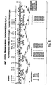
  • the complete NAIP gene shown in Fig. 6, contains 18 exons (1 to 14, and 14a to 17) and spans an estimated 90 kb of genomic DNA. (Other intermediate sequences obtained are shown in Figs. 5 and 7).
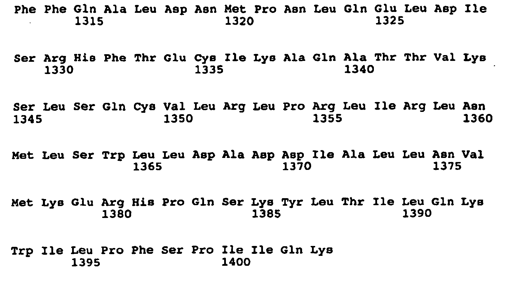
  • the NAIP coding region spans 4212 nucleotides resulting in a predicted gene product of 1404 amino acids (Seq. ID No. 22).
  • the total length of the NAIP gene spans 6228 nucleotides (Seq. ID No. 21) with a 395 nucleotide 5' UTR and a 1621 nucleotide 3' UTR.
  • Sequence ID No.2 allows one skilled in the art to develop probes and primers for the identification of homologous sequences and for the identification of mutations within the DNA. Both 5' and 3' regions may also prove useful as encoding binding sites for agents which may up or down-regulate the gene further delineating the NAIP pathway and function.
  • the sequences identified as Seq. ID No. 2 and 23 are also useful for protein expression in appropriate vectors and hosts to produce NAIP and study its function as well as to develop antibodies. Sequencing of the PAC 125D9 154 kb, which was identified as a likely site of the SMA gene, resulted in the identification of the NAIP sequence shown in Fig. 5, Seq. ID No. 1.
  • exon 14a An additional coding sequence, exon 14a, has since been identified and is provided herewith.
  • the NAIP DNA sequence containing exon 14a appears to be a predominant gene isoform which is not deleted or mutated in SMA patients.
  • the techniques and primers used for the isolation and application of exon 14a from the human fetal spinal cord cDNA libraries was as described for the identification of the other exons and detailed in Example 4. Additional screening of cDNA libraries combined with analysis of PAC 125D9 genomic DNA sequence has resulted in the identification of a novel 3' end of NAIP which includes additional exon 17 sequence.
  • the characteristics of the cloned NAIP gene sequence may be analyzed by introducing the sequence into various cell types or using in vitro extracellular systems. The function of the NAIP may then be examined under different physiological conditions.
  • the NAIP DNA sequence may be manipulated in studies to understand the expression of the gene and gene product. Alternatively, cell lines may be produced which overexpress the gene product allowing purification of NAIP for biochemical characterization, large-scale production, antibody production, and patient therapy.
  • eukaryotic and prokaryotic expression systems may be generated in which the NAIP gene sequence is introduced into a plasmid or other vector which is then introduced into living cells. Constructs in which the NAIP cDNA sequence containing the entire open reading frame inserted in the correct orientation into an expression plasmid may be used for protein expression. Alternatively, portions of the sequence, including wild-type or mutant NAIP sequences, may be inserted.
  • Prokaryotic and eukaryotic expression systems allow various important functional domains of the protein to be recovered as fusion proteins and then used for binding, structural and functional studies and also for the generation of appropriate antibodies. If a NAIP increases apoptosis, it may be desirable to express that protein under control of an inducible promotor.
  • Typical expression vectors contain promoters that direct the synthesis of large amounts of mRNA corresponding to the gene. They may also include sequences allowing for their autonomous replication within the host organism, sequences that encode genetic traits that allow cells containing the vectors to be selected, and sequences that increase the efficiency with which the mRNA is translated. Some vectors contain selectable markers such as neomycin resistance that permit isolation of cells by growing them under selective conditions. Stable long-term vectors may be maintained as freely replicating entities by using regulatory elements of viruses. Cell lines may also be produced which have integrated the vector into the genomic DNA and in this manner the gene product is produced on a continuous basis.
  • expression vectors in bacteria require the insertion of the NAIP sequence into an expression vector, usually a bacterial plasmid.
  • This plasmid vector contains several elements such as sequences encoding a selectable marker that assures maintenance of the vector in the cell, a controllable transcriptional promoter (ie , lac) which upon induction can produce large amounts of mRNA from the cloned gene, translational control sequences and a polylinker to simplify insertion of the gene in the correct orientation within the vector.
  • a controllable transcriptional promoter ie , lac
  • the expression vector plasmid contains a fragment of the E.coli chromosome containing the lac promoter and the neighboring lacZ gene.
  • RNA polymerase In the presence of the lactose analog IPTG, RNA polymerase normally transcribes the lacZ gene producing lacZ mRNA which is translated into the encoded protein, ⁇ -galactosidase.
  • the lacZ gene can be cut out of the expression vector with restriction enzymes and replaced by NAIP gene sequence.
  • this resulting plasmid is transfected into E.coli , addition of IPTG and subsequent transcription from the lac promoter produces NAIP mRNA, which is translated into NAIP.
  • the appropriate expression vector containing the NAIP gene is constructed it is introduced into an appropriate E.coli strain by transformation techniques including calcium phosphate transfection, DEAE-dextran transfection, electroporation, microinjection, protoplast fusion and liposome-mediated transfection.
  • the host cell which may be transfected with the vector of this invention may be selected from the group consisting of E.coli, pseudomonas, bacillus subtillus , or other bacili , other bacteria, yeast, fungi, insect (using baculoviral vectors for expression), mouse or other animal or human tissue cells.
  • Mammalian cells can also be used to express the NAIP protein using a vaccinia virus expression system.
  • T7 late-promoter expression system In vitro expression of proteins encoded by cloned DNA is also possible using the T7 late-promoter expression system.
  • This system depends on the regulated expression of T7 RNA polymerase which is an enzyme encoded in the DNA of bacteriophage T7.
  • the T7 RNA polymerase transcribes DNA beginning within a specific 23-bp promotor sequence called the T7 late promoter. Copies of the T7 late promoter are located at several sites on the T7 genome, but none is present in E.coli chromosomal DNA.
  • T7 RNA polymerase catalyzes transcription of viral genes but not of E.coli genes.
  • E.coli cells are first engineered to carry the gene encoding T7 RNA polymerase next to the lac promoter. In the presence of IPTG, these cells transcribe the T7 polymerase gene at a high rate and synthesize abundant amounts of T7 RNA polymerase. These cells are then transformed with plasmid vectors that carry a copy of the T7 late promoter protein. When IPTG is added to the culture medium containing these transformed E.coli cells , large amounts of T7 RNA polymerase are produced. The polymerase then binds to the T7 late promoter on the plasmid expression vectors, catalyzing transcription of the inserted cDNA at a high rate.
  • E.coli Since each E.coli cell contains many copies of the expression vector, large amounts of mRNA corresponding to the cloned cDNA can be produced in this system and the resulting protein can be radioactively labelled. Plasmid vectors containing late promoters and the corresponding RNA polymerases from related bacteriophages such as T3, T5 and SP6 may also be used for in vitro production of proteins from cloned DNA. E.coli can also be used for expression by infection with M13 Phage mGPI-2. E.coli vectors can also be used with phage lambda regulatory sequences, by fusion protein vectors, by maltose-binding protein fusions, and by glutathione-S-trahsferase fusion proteins.
  • a preferred expression system is the baculovirus system using, for example, the vector pBacPAK9, which is available from Clontech (Palo Alto, CA). If desired, this system may be used in conjunction with other protein expression techniques, for example, the myc tag approach described by Evan et al. (Mol. Cell Biol.5:3610-3616, 1985).
  • Eukaryotic expression systems permit appropriate post-translational modifications to expressed proteins. This allows for studies of the NAIP gene and gene product including determination of proper expression and post-translational modifications for biological activity, identifying regulatory elements located in the 5' region of the NAIP gene and their role in tissue regulation of protein expression. It also permits the production of large amounts of normal and mutant proteins for isolation and purification, to use cells expressing NAIP as a functional assay system for antibodies generated against the protein, to test the effectiveness of pharmacological agents or as a component of a signal transduction system, to study the function of the normal complete protein, specific portions of the protein, or of naturally occurring polymorphisms and artificially produced mutated proteins.
  • NAIP DNA sequence can be altered using procedures such as restriction enzyme digestion, DNA polymerase fill-in, exonuclease deletion, terminal deoxynucleotide transferase extension, ligation of synthetic or cloned DNA sequences and site-directed sequence alteration using specific oligonucleotides together with PCR.
  • a NAIP may be produced by a stably-transfected mammalian cell line.
  • a number of vectors suitable for stable transfection of mammalian cells are available to the public, e.g., see Pouwels et al. ( supra ), as are methods for constructing such cell lines (see e.g., Ausubel et al. ( supra ).
  • cDNA encoding a NAIP is cloned into an expression vector that includes the dihydrofolate reductase (DHFR) gene.
  • DHFR dihydrofolate reductase
  • Integration of the plasmid and, therefore, integration of the NAIP-encoding gene into the host cell chromosome is selected for by inclusion of 0.01-300 ⁇ M methotrexate in the cell culture medium (as described, Ausubel et al., supra ). This dominant selection can be accomplished in most cell types. Recombinant protein expression can be increased by DHFR-mediated amplification of the transfected gene.
  • DHFR-containing expression vectors are pCVSEII-DHFR and pAdD26SV(A) (described in Ausubel et al:, supra ).
  • the host cells described above or, preferably, a DHFR-deficient CHO cell line are among those most preferred for DHFR selection of a stably-transfected cell line or DHFR-mediated gene amplification.
  • the recombinant protein is expressed, it is isolated by, for example, affinity chromatography.
  • an anti-NAIP antibody which may be produced by the methods described herein, can be attached to a column and used to isolate the NAIP protein. Lysis and fractionation ofNAIP-harboring cells prior to affinity chromatography may be performed by standard methods (see e.g., Ausubel et al., supra ).
  • the recombinant protein can, if desired, be purified further by e.g., by high performance liquid chromatography (HPLC; e.g., see Fisher, Laboratory Techniques In Biochemistry And Molecular Biology , Work and Burdon, Eds., Elsevier, 1980).
  • Polypeptides of the invention can also be produced by chemical synthesis (e.g., by the methods described in Solid Phase Peptide Synthesis , 2nd ed., 1984 The Pierce Chemical Co., Rockford, IL). These general techniques of polypeptide expression and purification can also be used to produce and isolate useful NAIP fragments or analogs, as described herein.
  • the NAIP protein may be produced in a prokaryotic host (e.g., E. coli ) or in a eukaryotic host (e.g., S. cerevisiae , insect cells such as Sf21 cells, or mammalian cells such as COS-1, NIH 3T3, or HeLa cells). These cells are publically available, for example, from the American Type Culture Collection, Rockville, MD; see also Ausubel et al., Current Protocols in Molecular Biology , John Wiley & Sons, New York, NY, 1994).
  • a prokaryotic host e.g., E. coli
  • a eukaryotic host e.g., S. cerevisiae , insect cells such as Sf21 cells, or mammalian cells such as COS-1, NIH 3T3, or HeLa cells.
  • transduction and the choice of expression vehicle will depend on the host system selected. Transformation and transfection methods are described, e.g., in Ausubel et al. ( supra ), and expression vehicles may be chosen from those provided, e.g. in Cloning Vectors: A Laboratory Manual (P.H. Pouwels et al., 1985, Supp. 1987).
  • NAIP a protein which functions under normal conditions to protect cells against apoptosis
  • expression plasmids alone or encoding nearly full length NAIP or Bcl-2 a protein which functions under normal conditions to protect cells against apoptosis
  • Bcl-2 a protein which functions under normal conditions to protect cells against apoptosis
  • NAIP is expressed in at least the following tissues: motor neurons, myocardial cells, liver, placenta and CNS.
  • the BIR domains of NAIP appear to be both necessary and sufficient for NAIP biological activity. Surprisingly, we have reason to believe carboxy terminal deletions of NAIP amino acids actually enhances inhibition of apoptosis by NAIP. Deletions may be up to the end of the last NAIP BIR domain (i.e., the third), but need not delete the entire region carboxy terminal to the third BIR domains.
  • NAIP nucleophilicity factor
  • fragments of NAIP or fusion proteins containing defined portions or all of the NAIP protein can be synthesized in bacteria by expression of corresponding DNA sequences in a suitable cloning vehicle. Fusion proteins are commonly used as a source of antigen for producing antibodies. Two widely used expression systems for E.coli are lacZ fusions using the pUR series of vectors and trpE fusions using the pATH vectors. The protein can then be purified, coupled to a carrier protein and mixed with Freund's adjuvant (to help stimulate the antigenic response by the rabbits) and injected into rabbits or other laboratory animals. Alternatively, protein can be isolated from NAIP expressing cultured cells.
  • the rabbits or other laboratory animals are then bled and the sera isolated.
  • the sera can be used directly or purified prior to use, by various methods including affinity chromatography employing Protein A-Sepharose, Antigen Sepharose, Anti-mouse-Ig-Sepharose.
  • the sera can then be used to probe protein extracts from tissues run on a polyacrylamide gel to identify the NAIP protein.
  • synthetic peptides can be made to the antigenic portions of the protein and used to innoculate the animals.
  • a NAIP coding sequence i.e., amino acid fragments shown in Seq. ID Nos. 22 and 24
  • GST glutathione S-transferase
  • the fusion protein can be purified on glutathione-Sepharose beads, eluted with glutathione, and cleaved with thrombin (at the engineered cleavage site), and purified to the degree required to successfully immunize rabbits.
  • Antibody titres are monitored by Western blot and immunoprecipitation analyses using the thrombin-cleaved NAIP fragment of the GST-NAIP fusion protein. Immune sera are affinity purified using CNBr-Sepharose-coupled NAIP protein. Antiserum specificity is determined using a panel of unrelated GST proteins (including GSTp53, Rb, HPV-16 E6, and E6-AP) and GST-trypsin (which was generated by PCR using known sequences).
  • monoclonal NAIP antibodies may be produced by culturing cells actively expressing the protein or isolated from tissues.
  • the cell extracts, or recombinant protein extracts, containing the NAIP protein may for example, be injected in Freund's adjuvant into mice. After being injected, the mice spleens may be removed and resuspended in phosphate buffered saline (PBS).
  • PBS phosphate buffered saline
  • the spleen cells serve as a source of lymphocytes, some of which are producing antibody of the appropriate specificity. These are then fused with a permanently growing myeloma partner cells, and the products of the fusion are plated into a number of tissue culture wells in the presence of a selective agent such as HAT.
  • the wells are then screened by ELISA to identify those containing cells making binding antibody. These are then plated and after a period of growth, these wells are again screened to identify antibody-producing cells. Several cloning procedures are carried out until over 90% of the wells contain single clones which are positive for antibody production. From this procedure a stable line of clones which produce the antibody is established.
  • the monoclonal antibody can then be purified by affinity chromatography using Protein A Sepharose, ion-exchange chromatography, as well as variations and combinations of these techniques. Truncated versions of monoclonal antibodies may also be produced by recombinant methods in which plasmids are generated which express the desired monoclonal antibody fragment(s) in a suitable host.
  • peptides corresponding to relatively unique hydrophilic regions of NAIP may be generated and coupled to keyhole limpet hemocyanin (KLH) through an introduced C-terminal lysine.
  • KLH keyhole limpet hemocyanin
  • Antiserum to each of these peptides is similarly affinity purified on peptides conjugated to BSA, and specificity is tested by ELISA and Western blotting using peptide conjugates, and by Western blotting and immunoprecipitation using NAIP expressed as a GST fusion protein.
  • monoclonal antibodies may be prepared using the NAIP proteins described above and standard hybridoma technology (see, e.g., Kohler et al., Nature 256:495, 1975; Kohler et al., Eur. J. Immunol. 6:511, 1976; Kohler et al., Eur. J. Immunol. 6:292, 1976; Hammerling et al., In Monoclonal Antibodies and T Cell Hybridomas , Elsevier, New York, NY, 1981; Ausubel et al., supra ). Once produced, monoclonal antibodies are also tested for specific NAIP recognition by Western blot or immunoprecipitation analysis (by the methods described in Ausubel et al., supra ).
  • Antibodies that specifically recognize NAIP (or fragments of NAIP), such as those described herein containing one or more BIR domains are considered useful in the invention. They may, for example, be used in an immunoassay to monitor NAIP expression levels or to determine the subcellular location of a NAIP or NAIP fragment produced by a mammal. Antibodies that inhibit NAIP described herein may be especially useful in inducing apoptosis in cells undergoing undesirable proliferation.
  • antibodies are produced using NAIP sequence that does not reside within highly conserved regions, and that appears likely to be antigenic, as analyzed by criteria such as those provided by the Peptide structure program (Genetics Computer Group Sequence Analysis Package, Program Manual for the GCG Package, Version 7, 1991) using the algorithm of Jameson and Wolf (CABIOS 4:181, 1988).
  • Peptide structure program Genetics Computer Group Sequence Analysis Package, Program Manual for the GCG Package, Version 7, 1991
  • Jameson and Wolf CABIOS 4:181, 1988.
  • These fragments can be generated by standard techniques, e.g. by the PCR, and cloned into the pGEX expression vector (Ausubel et al., supra ). Fusion proteins are expressed in E. coli and purified using a glutathione agarose affinity matrix as described in Ausubel et al. ( supra ).
  • two or three fusions are generated for each protein, and each fusion is injected into at least two rabbits.
  • Antisera are raised by injections in series, preferably including at least three booster injections.
  • Antibodies to NAIP may be used, as noted above, to detect NAIP or inhibit the protein.
  • the antibodies coupled to compounds for diagnostic and/or therapeutic uses such as radionucleotides for imaging and therapy and liposomes for the targeting of compounds to a specific tissue location.
  • in situ hybridization is a method which may be used to detect the expression of the NAIP gene.
  • In situ hybridization relies upon the hybridization of a specifically labelled nucleic acid probe to the cellular RNA in individual cells or tissues. Therefore, it allows the identification of mRNA within intact tissues, such as the brain.
  • oligonucleotides or cloned nucleotide (RNA or DNA) fragments corresponding to unique portions of the NAIP gene are used to detect specific mRNA species, e.g., in the brain.
  • a rat is anesthetized and transcardially perfused with cold PBS, followed by perfusion with a formaldehyde solution.
  • the brain or other tissues is then removed, frozen in liquid nitrogen, and cut into thin micron sections.
  • the sections are placed on slides and incubated in proteinase K. Following rinsing in DEP, water and ethanol, the slides are placed in prehybridization buffer.
  • a radioactive probe corresponding to the primer is made by nick translation and incubated with the sectioned brain tissue. After incubation and air drying, the labelled areas are visualized by autoradiography. Dark spots on the tissue sample indicate hybridization of the probe with NAIP mRNA which demonstrates the expression of the protein.
  • NAIP cDNAs may be used to facilitate the identification of molecules that increase or decrease NAIP expression.
  • candidate molecules are added, in varying concentration, to the culture medium of cells expressing NAIP mRNA.
  • NAIP expression is then measured, for example, by Northern blot analysis (Ausubel et al., supra ) using a NAIP cDNA, or cDNA or RNA fragment, as a hybridization probe.
  • the level of NAIP expression in the presence of the candidate molecule is compared to the level of NAIP expression in the absence of the candidate molecule, all other factors (e.g. cell type and culture conditions) being equal.
  • the effect of candidate molecules on NAIP-mediated apoptosis may, instead, be measured at the level of translation by using the general approach described above with standard protein detection techniques, such as Western blotting or immunoprecipitation with a NAIP-specific antibody (for example, the NAIP antibody described herein).
  • standard protein detection techniques such as Western blotting or immunoprecipitation with a NAIP-specific antibody (for example, the NAIP antibody described herein).
  • Compounds that modulate the level ofNAIP may be purified, or substantially purified, or may be one component of a mixture of compounds such as an extract or supernatant obtained from cells (Ausubel et al., supra ).
  • NAIP expression is tested against progressively smaller subsets of the compound pool (e.g., produced by standard purification techniques such as HPLC or FPLC) until a single compound or minimal number of effective compounds is demonstrated to modulate NAIP expression.
  • Compounds may also be screened for their ability to modulate NAIP apoptosis inhibiting activity.
  • the degree of apoptosis in the presence of a candidate compound is compared to the degree of apoptosis in its absence, under equivalent conditions.
  • the screen may begin with a pool of candidate compounds, from which one or more useful modulator compounds are isolated in a step-wise fashion.
  • Apoptosis activity may be measured by any standard assay, for example, those described herein.
  • Another method for detecting compounds that modulate the activity of NAIPs is to screen for compounds that interact physically with a given NAIP polypeptide. These compounds may be detected by adapting interaction trap expression systems known in the art. These systems detect protein interactions using a transcriptional activation assay and are generally described by Gyuris et al. (Cell 75:791-803, 1993) and Field et al., Nature 340:245-246, 1989), and are commercially available from Clontech (Palo Alto, CA). In addition, PCT Publication WO 95/28497 describes an interaction trap assay in which proteins involved in apoptosis, by virtue of their interaction with Bcl-2, are detected. A similar method may be used to identify proteins and other compounds that interact with NAIP.
  • Compounds or molecules that function as modulators of NAIP-mediated cell death may include peptide and non-peptide molecules such as those present in cell extracts, mammalian serum, or growth medium in which mammalian cells have been cultured.
  • a molecule that promotes an increase in NAIP expression or NAIP activity is considered particularly useful in the invention; such a molecule may be used, for example, as a therapeutic to increase cellular levels of NAIP and thereby exploit the ability of NAIP polypeptides to inhibit apoptosis.
  • a molecule that decreases NAIP activity may be used to decrease cellular proliferation. This would be advantageous in the treatment of neoplasms or other cell proliferative diseases.
  • Molecules that are found, by the methods described above, to effectively modulate NAIP gene expression or polypeptide activity may be tested further in animal models. If they continue to function successfully in an in vivo setting, they may be used as therapeutics to either inhibit or enhance apoptosis, as appropriate.
  • Therapies may be designed to circumvent or overcome an NAIP gene defect or inadequate NAIP gene expression, and thus moderate and possibly prevent apoptosis.
  • the NAIP gene is expressed in the liver, myocardium, and placenta, as well as in the CNS.
  • therapies may be targeted at tissue other than the brain, such as the liver, myocardium, and any other tissues subsequently demonstrated to express NAIP.
  • Treatment or prevention of apoptosis can be accomplished by replacing mutant or insufficient NAIP protein with normal protein, by modulating the function of mutant protein, or by delivering normal NAIP protein to the appropriate cells.
  • the biological pathway of the NAIP protein has been completely understood, it may also be possible to modify the pathophysiologic pathway (e.g., a signal transduction pathway) in which the protein participates in order to correct the physiological defect.
  • Gene therapy is another potential therapeutic approach in which normal copies of the NAIP gene are introduced into selected tissues to successfully code for normal and abundant protein in affected cell types.
  • the gene must be delivered to those cells in a form in which it can be taken up and code for sufficient protein to provide-effective function.
  • it may be possible to prevent apoptosis by introducing another copy of the homologous gene bearing a second mutation in that gene or to alter the mutation, or use another gene to block any negative effect.
  • Transducing retroviral vectors can be used for somatic cell gene therapy especially because of their high efficiency of infection and stable integration and expression.
  • the targeted cells however must be able to divide and the expression of the levels of normal protein should be high.
  • the full length NAIP gene, or portions thereof can be cloned into a retroviral vector and driven from its endogenous promoter or from the retroviral long terminal repeat or from a promoter specific for the target cell type of interest (such as neurons).
  • Other viral vectors which can be used include adeno-associated virus, vaccinia virus, bovine papilloma virus, or a herpes virus such as Epstein-Barr virus.
  • Gene transfer could also be achieved using non-viral means requiring infection in vitro . This would include calcium phosphate, DEAE dextran, electroporation, and protoplast fusion. Liposomes may also be potentially beneficial for delivery of DNA into a cell. Although these methods are available, many of these are lower efficiency.
  • Antisense based strategies can be employed to explore NAIP gene function and as a basis for therapeutic drug design. The principle is based on the hypothesis that sequence-specific suppression of gene expression can be achieved by intracellular hybridization between mRNA and a complementary antisense species. The formation of a hybrid RNA duplex may then interfere with the processing/transport/translation and/or stability of the target NAIP mRNA.
  • Antisense strategies may use a variety of approaches including the use of antisense oligonucleotides, injection of antisense RNA and transfection of antisense RNA expression vectors.
  • Antisense effects can be induced by control (sense) sequences, however, the extent of phenotypic changes are highly variable. Phenotypic effects induced by antisense effects are based on changes in criteria such as protein levels, protein activity measurement, and target mRNA levels.
  • Transplantation of normal genes into the affected cells of a patient can also be useful therapy.
  • normal NAIP is transferred into a cultivatable cell type, either exogenously or endogenously to the patient. These cells are then injected serotologically into the targeted tissue(s).
  • Retroviral vectors may be used as a gene transfer delivery system for a therapeutic NAIP gene construct.
  • Retroviral vectors are particularly well developed and have been used in clinical settings (Rosenberg et al., N. Engl. J. Med 323:370, 1990; Anderson et al., U.S. Patent No. 5,399,346).
  • Non-viral approaches may also be employed for the introduction of therapeutic DNA into cells otherwise predicted to undergo apoptosis.
  • NAIP may be introduced into a neuron or a T cell by lipofection (Feigner et al., Proc. Natl. Acad. Sci. USA 84:7413, 1987; Ono et al., Neurosci. Lett. 117:259, 1990; Brigham et al., Am. J. Med. Sci.
  • the therapeutic NAIP DNA construct is preferably applied to the site of the predicted apoptosis event (for example, by injection). However, it may also be applied to tissue in the vicinity of the predicted apoptosis event or to a blood vessel supplying the cells predicted to undergo apoptosis.
  • NAIP cDNA expression can be directed from any suitable promoter (e.g., the human cytomegalovirus (CMV), simian virus 40 (SV40), or metallothionein promoters), and regulated by any appropriate mammalian regulatory element.
  • CMV human cytomegalovirus
  • SV40 simian virus 40
  • metallothionein promoters e.g., metallothionein promoters
  • enhancers known to preferentially direct gene expression in neural cells, T cells, or B cells may be used to direct NAIP expression.
  • the enhancers used could include, without limitation, those that are characterized as tissue- or cell-specific in their expression.
  • regulation may be mediated by the cognate regulatory sequences or, if desired, by regulatory sequences derived from a heterologous source, including any of the promoters or regulatory elements described above.
  • NAIP gene therapy is accomplished by direct administration of the NAIP mRNA or antisense NAIP mRNA to a cell that is expected to undergo apoptosis.
  • the mRNA may be produced and isolated by any standard technique, but is most readily produced by in vitro transcription using a NAIP cDNA under the control of a high efficiency promoter (e.g., the T7 promoter).
  • Administration of NAIP antisense or mRNA to cells mRNA can be carried out by any of the methods for direct nucleic acid administration described above.
  • the production of NAIP protein by any gene therapy approach will result in cellular levels of NAIP that are at least equivalent to the normal, cellular level of NAIP in an unaffected cell. Treatment by any NAIP-mediated gene therapy approach may be combined with more traditional therapies.
  • NAIP protein recombinant NAIP protein
  • administration of recombinant NAIP protein either directly to the site of a predicted apoptosis event (for example, by injection) or systemically (for example, by any conventional recombinant protein administration technique).
  • the dosage of NAIP depends on a number of factors, including the size and health of the individual patient, but, generally, between [0.1 mg and 100 mg] inclusive are administered per day to an adult in any pharmaceutically acceptable formulation.
  • a NAIP protein, gene, or modulator may be administered within a pharmaceutically-acceptable diluent, carrier, or excipient, in unit dosage form.
  • Conventional pharmaceutical practice may be employed to provide suitable formulations or compositions to administer NAIP to patients suffering from a disease that is caused by excessive apoptosis. Administration may begin before the patient is symptomatic. Any appropriate route of administration may be employed, for example, administration may be parenteral, intravenous, intraarterial, subcutaneous, intramuscular, intracranial, intraorbital, ophthalmic, intraventricular, intracapsular, intraspinal, intracisternal, intraperitoneal, intranasal, aerosol, by suppositories, or oral administration.
  • Therapeutic formulations may be in the form of liquid solutions or suspensions; for oral administration, formulations may be in the form of tablets or capsules; and for intranasal formulations, in the form of powders, nasal drops, or aerosols.
  • Formulations for parenteral administration may, for example, contain excipients, sterile water, or saline, polyalkylene glycols such as polyethylene glycol, oils of vegetable origin, or hydrogenated napthalenes.
  • Biocompatible, biodegradable lactide polymer, lactide/glycolide copolymer, or polyoxyethylene-polyoxypropylene copolymers may be used to control the release of the compounds.
  • Other potentially useful parenteral delivery systems for NAIP modulatory compounds include ethylene-vinyl acetate copolymer particles, osmotic pumps, implantable infusion systems, and liposomes.
  • Formulations for inhalation may contain excipients, for example, lactose, or may be aqueous solutions containing, for example, polyoxyethylene-9-lauryl ether, glycocholate and deoxycholate, or may be oily solutions for administration in the form of nasal drops, or as a gel.
  • treatment with a NAIP protein, gene, or modulatory compound may be combined with more traditional therapies for the disease such as surgery, steroid therapy, or chemotherapy for autoimmune disease; antiviral therapy for AIDS; and tissue plasminogen activator (TPA) for ischemic injury.
  • therapies for the disease such as surgery, steroid therapy, or chemotherapy for autoimmune disease; antiviral therapy for AIDS; and tissue plasminogen activator (TPA) for ischemic injury.
  • NAIP polypeptides and nucleic acid sequences find diagnostic use in the detection or monitoring of conditions involving aberrant levels of apoptosis. For example, decrease expression of NAIP may be correlated with enhanced apoptosis in humans (see XII, below). Accordingly, a decrease or increase in the level of NAIP production may provide an indication of a deleterious condition.
  • Levels of NAIP expression may be assayed by any standard technique. For example, NAIP expression in a biological sample (e.g., a biopsy) may be monitored by standard Northern blot analysis or may be aided by PCR (see, e.g., Ausubel et al., supra ; PCR Technology; Principles and Applications for DNA Amplification , H.A. Ehrlich, Ed. Stockton Press, NY; Yap et al. Nucl. Acids. Res. 19:4294, 1991).
  • a biological sample obtained from a patient may be analyzed for one or more mutations in the NAIP sequences using a mismatch detection approach.
  • these techniques involve PCR amplification of nucleic acid from the patient sample, followed by identification of the mutation (i.e., mismatch) by either altered hybridization, aberrant electrophoretic gel migration, binding or cleavage mediated by mismatch binding proteins, or direct nucleic acid sequencing. Any of these techniques may be used to facilitate mutant NAIP detection, and each is well known in the art; examples of particular techniques are described, without limitation, in Orita et al., Proc. Natl. Acad. Sci. USA 86:2766-2770, 1989; Sheffield et al., Proc. Natl. Acad. Sci. USA 86:232-236, 1989).
  • immunoassays are used to detect or monitor NAIP protein in a biological sample.
  • NAIP specific polyclonal or monoclonal antibodies produced as described above may be used in any standard immunoassay format (e.g., ELISA, Western blot, or RIA) to measure NAIP polypeptide levels. These levels would be compared to wild-type NAIP levels, with a decrease in NAIP production indicating a condition involving increased apoptosis. Examples of immunoassays are described, e.g., in Ausubel et al., supra . Immunohistochemical techniques may also be utilized for NAIP detection.
  • a tissue sample may be obtained from a patient, sectioned, and stained for the presence of NAIP using an anti-NAIP antibody and any standard detection system (e.g., one which includes a secondary antibody conjugated to horseradish peroxidase).
  • any standard detection system e.g., one which includes a secondary antibody conjugated to horseradish peroxidase.
  • a combined diagnostic method may be employed that begins with an evaluation of NAIP protein production (for example, by immunological techniques or the protein truncation test (Hogerrorst et al., Nature Genetics 10:208-212, 1995) and also includes a nucleic acid-based detection technique designed to identify more subtle NAIP mutations (for example, point mutations).
  • a number of mismatch detection assays are available to those skilled in the art, and any preferred technique may be used. Mutations in NAIP may be detected that either result in loss of NAIP expression or loss of NAIP biological activity.
  • NAIP biological activity is measured as anti-apoptotic activity using any appropriate apoptosis assay system (for example, those described herein).
  • Mismatch detection assays also provide an opportunity to diagnose a NAIP-mediated predisposition to diseases caused by inappropriate apoptosis. For example, a patient heterozygous for a NAIP mutation may show no clinical symptoms and yet possess a higher than normal probability of developing one or more types ofneurodegenerative, myelodysplastic or having severe sequelae to an ischemic event. Given this diagnosis, a patient may take precautions to minimize their exposure to adverse environmental factors (for example, UV exposure or chemical mutagens) and to carefully monitor their medical condition (for example, through frequent physical examinations). This type of NAIP diagnostic approach may also be used to detect NAIP mutations in prenatal screens.
  • the NAIP diagnostic assays described above may be carried out using any biological sample (for example, any biopsy sample or other tissue) in which NAIP is normally expressed. Identification of a mutant NAIP gene may also be assayed using these sources for test samples.
  • a NAIP mutation particularly as part of a diagnosis for predisposition to NAIP-associated degenerative disease, may be tested using a DNA sample from any cell, for example, by mismatch detection techniques.
  • the DNA sample is subjected to PCR amplification prior to analysis.
  • any of the above therapies may be administered before the occurrence of the disease phenotype.
  • the therapies may be provided to a patient who is HIV positive but does not yet show a diminished T cell count or other overt signs of AIDS.
  • compounds shown to increase NAIP expression or NAIP biological activity may be administered by any standard dosage and route of administration (see above).
  • gene therapy using a NAIP expression construct may be undertaken to reverse or prevent the cell defect prior to the development of the degenerative disease.
  • compositions and methods described may be used to reduce or diagnose the disorders described herein in any mammal, for example, humans, domestic pets, or livestock. Where a nonhuman mammal is treated or diagnosed, the NAIP polypeptide, nucleic acid, or antibody employed is preferably specific for that species.
  • Standard techniques such as the polymerase chain reaction (PCR) and DNA hybridization, may be used to clone additional NAIP homologues in other species.
  • Southern blots of murine genomic DNA hybridized at low stringency with probes specific for human NAIP reveal bands that correspond to NAIP and/or related family members. Thus, additional NAIP sequences may be readily identified using low stringency hybridization. Examples of murine and human NAIPspecific primers, which may be used to clone additional genes by RT-PCR.
  • NAIP ability of NAIP to modulate apoptosis can be defined in in vitro systems in which alterations of apoptosis can be detected.
  • Mammalian expression constructs carrying NAIP cDNAs which are either full-length or truncated, can be introduced into cell lines such as CHO, NIH 3T3, HL60, Rat-1, or Jurkat cells.
  • SF21 insect cells may be used, in which case the NAIP gene is preferentially expressed using an insect heat shock promotor.
  • apoptosis can be induced by standard methods, which include serum withdrawal, or application of staurosporine, menadione (which induces apoptosis via free radical formation), or anti-Fas antibodies.
  • cells are cultured under the same conditions as those induced to undergo apoptosis, but either not transfected, or transfected with a vector that lacks a NAIP insert.
  • the ability of each NAIP construct to inhibit apoptosis upon expression can be quantified by calculating the survival index of the cells, i.e., the ratio of surviving transfected cells to surviving control cells.
  • apoptosis assays are also provided in the following references.
  • Assays for apoptosis in lymphocytes are disclosed by: Li et al., "Induction of apoptosis in uninfected lymphocytes by HIV-1 Tat protein", Science 268:429-431, 1995; Gibellini et al., "Tat-expressing Jurkat cells show an increased resistance to different apoptotic stimuli, including acute human immunodeficiency virus-type 1 (HIV-1) infection", Br. J. Haematol. 89:24-33, 1995; Martin et al., "HIV-1 infection of human CD4 + T cells in vitro . Differential induction of apoptosis in these cells.” J.
  • Characterization of NAIP genes provides information that is necessary for a NAIP knockout animal model to be developed by homologous recombination.
  • the model is a mammalian animal, most preferably a mouse.
  • an animal model of NAIP overproduction may be generated by integrating one or more NAIP sequences into the genome, according to standard transgenic techniques.
  • a replacement-type targeting vector which would be used to create a knockout model, can be constructed using an isogenic genomic clone, for example, from a mouse strain such as 129/Sv (Stratagene Inc., LaJolla, CA).
  • the targeting vector will be introduced into a suitably-derived line of embryonic stem (ES) cells by electroporation to generate ES cell lines that carry a profoundly truncated form of a NAIP.
  • ES embryonic stem
  • the targeted cell lines will be injected into a mouse blastula stage embryo. Heterozygous offspring will be interbred to homozygosity.
  • Knockout mice would provide the means, in vivo , to screen for therapeutic compounds that modulate apoptosis via an NAIP-dependent pathway. Making such mice may require use of loxP sites due to the multiple copies of NAIP on the chromosome (see Sauer and Henderson, Nucleic Aids Res. 17: 147-61 (1989)).
  • Example 1 Expression of NAIP in Rat-1, CHO and HeLa pooled stable lines and adenovirus infected cells analysed by Western blotting and immunofluorescence.
  • NAIP construct tagged with the myc epitope (I) MTG-SP3.7, a 2.5 kb Bsu36I/SalI fragment of NAIP cloned into Bluescript and (ii) Bsu36I/XhoI cut MTG-SE1.7, the expression vector pcDNA3 containing a 300 bp myc epitope and a 1.7 kb fragment of NAIP were ligated.
  • HeLa, CHO and Rat-1 cells were transfected by lipofection (Gibco BRL) with 8 ⁇ g DNA and G418 resistant transformants were selected by maintaining the cells in 250 ⁇ g/ml, 400 ⁇ g/ml and 800 ⁇ g/ml G418 respectively.
  • Western blot analysis was performed using mouse anti-human myc monoclonal antibody (Ellison, M.J. and Hochstrasser, M.J. 1991) or rabbit anti-human NAIP (E1.0) polyclonal antibody.
  • mouse anti-human myc monoclonal antibody Ellison, M.J. and Hochstrasser, M.J. 1991
  • rabbit anti-human NAIP E1.0
  • rabbits were immunized with purified bacterial produced fusion protein in complete Freunds adjuvant. Serum was pre-cleared with GST protein and anti-NAIP immunoglobin purified with immobilized GST-NAIP fusion proteins.
  • cells were grown on glass slides, fixed with formaldehyde for 10 minutes, incubated with anti-NAIP (1:200) or anti-myc (1:20) in PBS, 0.3% Triton X-100TM for 1 hour followed by incubation with secondary antisera, FITC-labelled donkey anti-rabbit immunoglobulin (Amersham), biotinylated goat anti-mouse immunoglobulin (Amersham) and streptavidin Texas-RedTM (Amersham).
  • CHO or Rat-1 cells were treated with menadione for 1.5 hours, washed 5 times in PBS and maintained in normal media.
  • serum deprivation assays cells were washed 5 times in PBS and maintained in media with 0% fetal calf serum.
  • HeLa cells were treated with 20 units/ml TNF- ⁇ in combination with 30 g/ml cyclohexamide for 17 hours. Apoptosis was assayed for each trigger by propidium iodide staining. Adenovirus infected cells were subjected to triggers 36 hours post infection.
  • LacZ expression was confirmed histochemically by 5-bromo-4-chloro-3-indoyl- ⁇ -D-galactoside (X-gal) as described in Ellison, M.J. and Hochstrasser, M.J. 1991. Transcription of PIAN was determined by in situ hybridization using the DIG labelled sense oligonucleotide following the manufacturers protocol (Boehringer Mannheim).
  • the human Bcl-2 clone pB4 was digested with EcoRI and ligated into the EcoRI site of pcDNA3.
  • adenovirus assays an adenovirus encoding LacZ, antisense NAIP (NAIP) or vector alone with no insert were utilized as controls.
  • Bcl-2 was utilized as a positive control and pcDNA alone as a negative control in cell line assays.
  • Cell viability was determined by trypan blue exclusion. Date are presented as averages of three independently derived transfected pools or infections.
  • the 40G1 CATT subloci demonstrated linkage disequilibrium and therefore a PAC contiguous array containing the CATT region was constructed.
  • This PAC contig array comprised 9 clones and extended approximately 400 kb.
  • Genetic analysis combined with the physical mapping data indicated that the 40G 1 CATT subloci marker which showed the greatest disequilibrium with SMA was duplicated and was localized at the extreme centromeric of the critical SMA interval. Consequently the 154 kb PAC clone 125D9 which contained within 10 kb of its centromeric end the SMA interval defining CMS allele 9 and extended telemetrically to incorporate the 40G1 CATT sublocus was chosen for further examination.
  • Two genomic libraries were constructed by performing complete and partial (average insert size 5 kb) Sau3A1 on PAC 125D9 and cloning the restricted products into BamH1 digested Bluescript plasmids. Genomic sequencing was conducted on both termini of 200 clones from the 5 kb insert partial Sau3A1 library in the manner of (Chen et al., 1993) permitting the construction of contiguous and overlapping genomic clones covering most of the PAC. This proved instrumental in the elucidation of the neuronal apoptosis inhibitor protein gene structure.
  • PAC 125D9 is cleaved into 30 kb centromeric and 125 kb telomeric fragments by a NotI site (which was later shown to bisect exon 7 of the PAC 125D9 at the beginning of the apoptosis inhibitor domain.
  • the NotI PAC fragments were isolated by preparative PFGE and used separately to probe fetal brain cDNA libraries. Physical mapping and sequencing of the NotI site region was also undertaken to assay for the presence of a CpG island, an approach which rapidly detected coding sequences.
  • the PAC 125D9 was also used as a template in an exon trapping system resulting in the identification of the exons contained in the neuronal apoptosis inhibitor protein gene.
  • the multipronged approach in addition to the presence of transcripts identified previously by hybridization by clones from the cosmid array (such as, GA1 and L7), resulted in the rapid identification of six cDNA clones contained in neuronal apoptosis inhibitor protein gene.
  • the clones were arranged, where possible, into overlapping arrays. Chimerism was excluded on a number of occasions by detection of co-linearity of the cDNA clone termini with sequences from clones derived from the PAC 125D9 partial Sau3A1 genomic library.
  • a human fetal spinal cord cDNA library was probed with the entire genomic DNA insert of cosmid 250B6 containing one of the 5 CATT subloci. This resulted in a detection of a 2.2 kb transcript referred to as GA1. Further probings of fetal brain libraries with the contiguous cosmid inserts (cosmids 40G1) as well as single copy subclones isolated from such cosmids were undertaken. A number of transcripts were obtained including one termed L7. No coding region was detected for L7 probably due to the fact that a substantial portion of the clone contained unprocessed heteronuclear RNA.
  • the remaining gaps in the cDNA were completed and the final 3' extension was achieved by probing a fetal brain library with two trapped exons.
  • a physical map of the cDNA with overlapping clones was prepared. The entire cDNA sequence is shown in Table 1 and contains 18 exons (1 to 14a and 14 to 17). The amino acid sequence starts with methionine which corresponds to the nucleotide triplet ATG.
  • Coding sequences from the PACs were isolated by the exon amplification procedure as described by Church et al. (1994). PACs were digested with BamHI or BamHI and BglII and subcloned into pSPL3. Pooled clones of each PAC were transfected into COS-1 cells. After a 24h transfection total RNA was extracted. Exons were cloned into pAMP10 (Gibco, BRL) and sequenced utilizing primer SD2 (GTG AAC TGC ACT GTG ACA AGC TGC).
  • DNA sequencing was conducted on an ABI 373A automated DNA sequencer. Two commercial human fetal brain cDNA libraries in lambda gt (Stratagene) and lambda ZAP (Clontech) were used for candidate transcript isolation. The Northern blot was commercially acquired (Clontech) and probing was performed using standard methodology.
  • primers used in the paper for PCR were selected for T m s of 60°C and can be used with the following conditions: 30 cycles of 94°C, 60s; 60°C, 60s; 72°C, 90s.
  • PCR primer mappings are as referred to in the figure legends and text. Primer sequences are as follows:
  • the NAIP and SMN tel genes are in a tail to tail (5'-->3':3' ⁇ --5') orientation, spanning 56 kb and 28 kb of genomic DNA, respectively.
  • the gene BTF2p44 exists in a number of copies on 5q13.1 10 ; exons 11-16 of one BTF2P44 copy occupy the most 5' eleven kb of the PAC insert followed by an 11 kb interval before NAIP exon 2.
  • the first NAIP exon as originally reported 3 is not present in this PAC and may have been a heteronuclear artifact.
  • NAIP cDNA (6228 bp with an ORF of 4212 bp) was also elucidated by cDNA sequencing and comparison with PAC sequence, comprising 17 exons encoding a predicted 156 kDa protein of 1403 amino acids (data not shown).
  • a novel NAIP exon 14 between the original exon 14 and 15 was identified.
  • the original exon 17 has been replaced by a novel exon which contains the stop codon, a 1.6 kb 3' UTR region and the polyadenylation consensus site (AATAAA) identified by 3' RACE. No new protein domains are found in the NAIP gene.
  • type 1 SMA chromosomes A rigorous definition of how far deletions extend on type 1 SMA chromosomes is central to our understanding of disease pathogenesis. If the genotype most frequently observed on type 1 SMA chromosomes (i.e. absence of NAIP exons 4 and 5 as well as SMN tel exons 7 and 8) are the result of a single event, then our sequencing suggests a minimal deletion size of 60 kb. The high deletion frequency on type 1 SMA chromosomes of the CATT-40G1 14 , (which maps between NAIP exon 7 and 8) is consistent with such a deletion.
  • SMN tel maps the intact NAIP locus and clinically relevant SMN tel to a 100 kb region which contains those microsatellite polymorphisms that are preferentially deleted on the significant majority of type 1 SMA chromosomes (i.e. CATT-40G1 14 C272 15 or AG-1 16,17 ).
  • CATT-40G1 14 C272 15 or AG-1 16,17 The absence of any protein coding sequence, other than NAIP and SMN in this interval, focuses attention on these two genes as the key modulators of type 1 SMA.
  • SMN tel absence acts as the primary neurotoxic insult 19 with NAIP depletion/absence leading to an attenuated apoptotic resistance 5,6 , exacerbating motor neuron attrition.
  • Presence of additional SMN cen may also act to modulate the course of the disease 20 .
  • sequence presented here should help in the study of transcriptional control elements for both genes, possibly facilitating the formulation of genetic therapies for this devastating neuromuscular disease.
  • Partial Sau3A1 (selected for 3-5kb) BamHI, EcoRI, HindIII, PstI, SstI, XbaI and EcoRV libraries) were made from the PAC125D9 insert and sequenced using a transposon-based methodology (TN1000 Gold Biotechnology 10 ). Subcloning of a large number of inserts into the commercially supplied pMOB plasmid was found to be problematic, therefore pUC18 and pBluescript SK were used. In general, fewer than 10% of clones had transposons in the vector region. E. coli lysate was employed as sequencing template using our modified heat soaked protocol 21 .
  • Sequencing was from the TN1000 transposon randomly inserted into the target DNA, using primers of opposite orientation (5'-ATA TAA ACA ACGAAT TAT CTC C-3'; 5'-GTA TTA TAA TCA ATA AGTTAT ACC-3'), generating approximately 1 kb of sequence with a 5 bp overlap, easily spanning 300bp Alu repeats.
  • Our approach permitted sequencing of inserts as large as 14 kb.
  • Raw DNA sequence data generated by our automated sequencers (ABI 373 and ABI 373A) were processed and assembled in parallel by the Sequencher 3.0 program (Gene Codes Inc.); and the GAP4 program from the Staden package 27 .
  • the edited results were automatically converted into GCG file formats 22 and placed in a separate database for searches by outside users using our e-mail server at smafasta@mgcheo.med.uottawa.ca. GRAIL 28 and Blast 29 searches were employed to screen for protein coding sequence and the PROSITE Protein database 24 was used to search for protein domains.
  • NAIP sequence information Using the identified NAIP sequence information, a full length 3.7 kb NAIP construct tagged with the myc epitope (I) MTG-SP3.7, a 2.5 kb Bsu36I/SalI fragment of NAIP cloned into Bluescript and (ii) Bsu36I/XhoI cut MTG-SE1.7, the expression vector pcDNA3 containing a 300 bp myc epitope and a 1.7 kb fragment of NAIP were ligated.
  • HeLa, CHO and Rat-1 cells were transfected by lipofection (Gibco BRL) with 8 ⁇ g DNA and G418 resistant transformants were selected by maintaining the cells in 250 ⁇ g/ml, 400 ⁇ g/ml and 800 ⁇ g/ml G418 respectively.
  • CHO cells Following infection with the recombinant adenoviruses, CHO cells were induced to undergo apoptosis by serum deprivation with survival rates of 48% (no insert), 51% (LacZ) and 45% (antisense NAIP) at 48 hours (Fig. 1a). In contrast, CHO cells infected with adenovirus expressing NAIP demonstrate 78-83% survival. NAIP also induced survival in stably transfected CHO pools, albeit slightly less than that seen in adenovirus infected cells: 44% of the vector transfectants and 65% of the NAIP transfectants survived at 48 hours (Fig. 1b).
  • Immunofluorescence is a technique which localizes proteins within a cell by light microscopy by the use of antibodies specific for a desired protein and a fluorescence microscope. Dyes can be chemically coupled to antibodies directed against purified antibodies specific for a desired protein. This flourescent dye-antibody complex when added to permeabilized cells or tissue sections binds to the desired antigen-antibody which lights up when illuminated by the exciting wavelength. Fluorescent antibodies may also be microinjected into cultured cells for visualization.
  • CY-3 a dye which emits red light
  • CY-3 a dye which emits red light
  • NAIP a novel protein with homology to baculoviral inhibitor of apoptosis
  • the ability of the polyclonal antibody to detect NAIP was confirmed by immunofluorescence of cells transfected with myc tagged NAIP employed both the anti-NAIP and anti-Myc antibodies, as well as western blot analysis on protein extracts of these cells (Fig. 1).
  • Western blotting technique proteins are run on polyacrylamide gel and then transferred onto nitrocellulose membranes. These membranes are then incubated in the presence of the antibody (primary), then following washing are incubated to a secondary antibody which is used for detection of the protein-primary antibody complex. Following repeated washing, the entire complex is visualized using colorimetric or chemiluminescent methods.
  • a protein of the expected molecular weight was detected by both antibodies in western blots and their cellular co-localization demonstrated by immunofluorescence.
  • Sections of human spinal cord stained with anti-NAIP showed strong immunoreactivity in the cytoplasm of the anterior horn cells and intermediolateral neurons (Figs. 3a and 3b).
  • NAIP reactivity was observed in the ventral roots which contain motor axons but not the dorsal roots comprised of sensory axons (Figs. 3c and 3d).
  • the observation of motor neuron staining correlates well with a role for the protein in the pathogenesis of SMA.
  • the presence of NAIP in intermediolateral neurons which are not reported to be affected in SMA, implies heterogeneity in the apoptotic pathways between the two classes of neurons.
  • the invention includes any protein which is substantially identical to a mammalian NAIP polypeptides provided in Figs. 6 and 7, Seq. ID NOS: 22 and 24); such homologs include other substantially pure naturally-occurring mammalian NAIP proteins as well as allelic variants; natural mutants; induced mutants; DNA sequences which encode proteins and also hybridize to the NAIP DNA sequences of Figs. 6 and 7, (Seq. ID NOS: 21 and 23) under high stringency conditions or, less preferably, under low stringency conditions (e.g., washing at 2X SSC at 400C with a probe length of at least 40 nucleotides); and proteins specifically bound by antisera directed to a NAIP polypeptide.
  • the term also includes chimeric polypeptides that include a NAIP portion. The sequence of Seq. ID No. 1 and the IAP proteins are specifically excluded.
  • the invention further includes analogs of any naturally-occurring NAIP polypeptide.
  • Analogs can differ from the naturally-occurring NAIP protein by amino acid sequence differences, by post-translational modifications, or by both.
  • Analogs of the invention will generally exhibit at least 85%, more preferably 90%, and most preferably 95% or even 99% identity with all or part of a naturally occurring NAIP amino acid sequence.
  • the length of sequence comparison is at least 15 amino acid residues, preferably at least 25 amino acid residues, and more preferably more than 35 amino acid residues.
  • Modifications include in vivo and in vitro chemical derivatization of polypeptides, e.g., acetylation, carboxylation, phosphorylation, or glycosylation; such modifications may occur during polypeptide synthesis or processing or following treatment with isolated modifying enzymes.
  • Analogs can also differ from the naturally-occurring NAIP polypeptide by alterations in primary sequence.
  • fragment means at least 20 contiguous amino acids, preferably at least 30 contiguous amino acids, more preferably at least 50 contiguous amino acids, and most preferably at least 60 to 80 or more contiguous amino acids. Fragments of NAIP polypeptides can be generated by methods known to those skilled in the art or may result from normal protein processing (e.g., removal of amino acids from the nascent polypeptide that are not required for biological activity or removal of amino acids by alternative mRNA splicing or alternative protein processing events).
  • Preferable fragments or analogs according to the invention are those which facilitate specific detection of a NAIP nucleic acid or amino acid sequence in a sample to be diagnosed.
  • Particularly useful NAIP fragments for this purpose include, without limitation, the amino acid fragments shown in Table 2.

Landscapes

  • Health & Medical Sciences (AREA)
  • Chemical & Material Sciences (AREA)
  • Life Sciences & Earth Sciences (AREA)
  • Organic Chemistry (AREA)
  • General Health & Medical Sciences (AREA)
  • Engineering & Computer Science (AREA)
  • Proteomics, Peptides & Aminoacids (AREA)
  • Medicinal Chemistry (AREA)
  • Molecular Biology (AREA)
  • Genetics & Genomics (AREA)
  • Biochemistry (AREA)
  • Immunology (AREA)
  • Biophysics (AREA)
  • Zoology (AREA)
  • Analytical Chemistry (AREA)
  • Bioinformatics & Cheminformatics (AREA)
  • Biomedical Technology (AREA)
  • Animal Behavior & Ethology (AREA)
  • Wood Science & Technology (AREA)
  • Urology & Nephrology (AREA)
  • Veterinary Medicine (AREA)
  • Hematology (AREA)
  • Gastroenterology & Hepatology (AREA)
  • Public Health (AREA)
  • Pharmacology & Pharmacy (AREA)
  • Nuclear Medicine, Radiotherapy & Molecular Imaging (AREA)
  • General Chemical & Material Sciences (AREA)
  • Chemical Kinetics & Catalysis (AREA)
  • Toxicology (AREA)
  • Pathology (AREA)
  • Microbiology (AREA)
  • Physics & Mathematics (AREA)
  • Biotechnology (AREA)
  • Cell Biology (AREA)
  • General Physics & Mathematics (AREA)
  • Food Science & Technology (AREA)
  • General Engineering & Computer Science (AREA)
  • Heart & Thoracic Surgery (AREA)
  • Neurosurgery (AREA)
  • Oncology (AREA)

Abstract

The invention provides NAIP nucleic acid and sequences. Also provided are anti-NAIP antibodies and methods for modulating apoptosis and detecting compounds which modulate apoptosis.

Description

Field of the Invention
This invention relates in general to the function of the NAIP inhibitor protein in apoptosis and more particularly to the use of NAIP proteins and nucleic acids to characterize NAIP, identify compounds which modulate NAIP, and treat conditions affected by changes in NAIP levels.
Background of the invention
Apoptosis is a morphologically distinct form of programmed cell death that is important in the normal development and maintenance of multicellular organisms. Dysregulation of apoptosis can take the form of inappropriate suppression of cell death, as occurs in the development of some cancers, or in a failure to control the extent of cell death, as is believed to occur in acquired immunodeficiency and certain neurodegenerative disorders, such as spinal muscular atrophy (SMA).
Childhood spinal muscular atrophies are neurodegenerative disorders characterized by progressive spinal cord motor neuron depletion and are among the most common autosomal recessive disorders (Dubowitz, V. 1978, Brooke, M.A. 1986). Type I SMA is the most frequent inherited cause of death in infancy. The loss of motor neurons in SMA, has led to suggestions that an inappropriate continuation or reactivation of normally occurring motor neuron apoptosis may underlie the disorder (Sarnat, H.B. 1992). NAIP, a gene associated with SMA, has been mapped to human chromosome 5q13.1
Some baculoviruses encode proteins that are termed inhibitors of apoptosis proteins (IAPs) because they inhibit the apoptosis that would otherwise occur when insect cells are infected by the virus. These proteins are thought to work in a manner that is independent of other viral proteins. The baculovirus IAP genes include sequences encoding a ring zinc finger-like motif (RZF), which may be involved in DNA binding, and two N-terminal domains that consist of a 70 amino acid repeat motif termed a BIR domain (Baculovirus IAP Repeat).
Roy et al (Cell, vol. 80, no. 1, 13 January 1995, NA US pages 167-178) discloses a sequence encoding NAIP of 5502 nucleotides in length. The function of NAIP as an apoptosis inhibitor and in the development of spinal muscular atrophies (SMA) is also disclosed.
WO 9612016, published prior to the international filing date but later than the priority date of the present invention, discloses the gene encoding NAIP and the corresponding amino acid sequence of 1232 amino acids in length. Use of the sequence information for detection of NAIP sequences and for diagnosis and treatment of SMA is also disclosed.
Liston et al (Nature, vol. 379, no. 6563, 25 January 1996, pages 349-353), published prior to the international filing dated but later than the priority date of the present invention, is concerned with the testing of sequences homologous to the sequence encoding NAIP for antiapoptotic activity.
Summary of the Invention
The present invention relates to a novel Neuronal Apoptosis Inhibitor Protein (NAIP) sequence and a NAIP fragment with anti-apoptotic activities, which comprise nucleotides 3734 to 3886 (exon 14a) and 4139 to 4503 (exon 17) of SEQ ID NO: 21 or nucleotides 3838 to 3990 (exon 14a) and nucleotides 4243 to 4605 (exon 17) of SEQ ID NO: 23.
The present invention provides a substantially pure nucleic acid having at least 90% nucleic acid sequence identity to the full length Neuronal Apoptosis Inhibitor Protein (NAIP) nucleic acid sequence of SEQ ID NO: 21 and including nucleotides 3734 to 3886 (exon 14a) and 4139 to 4503 (exon 17) of SEQ ID NO: 21 or having at least 90% nucleic acid sequence identity to the full length NAIP nucleic acid sequence of SEQ ID NO: 23 and including nucleotides 3838 to 3990 of SEQ ID NO:23 and nucleotides 4243 to 4605 of SEQ ID 23 said nucleic acid encoding a NAIP polypeptide that inhibits apoptosis.
The present invention also provides a substantially pure nucleic acid having the sequence of SEQ ID NO:21 or degenerate variants thereof and encode a polypeptide having the amino acid sequence of SEQ ID NO:22, wherein said polypeptide is capable of inhibiting apoptosis.
Preferably the nucleic acid is operably linked to a regulatory sequence for expression of said NAIP polypeptide and said regulatory sequence comprises a promoter.
Preferably the promoter is a constitutive promoter and is inducible by one or more external agents or is cell-type specific.
The present invention provides a vector comprising the nucleic acid of the present invention, wherein the vector is capable of directing expression of a NAIP polypeptide that is capable of inhibiting apoptosis.
The invention also provides a host cell that contains the above described vector.
In an alternative embodiment the present invention provides a substantially pure mammalian NAIP polypeptide encoded by the nucleic acid sequence of the present invention.
The present invention also provides a therapeutic composition comprising an effective amount of NAIP polypeptide of the invention in a physiologically acceptable carrier.
Also provided is a non-human animal transgenic for a NAIP nucleic acid sequence of the invention.
The present invention also provides an in vitro method of obtaining a NAIP polypeptide. The method comprises the following steps of:
  • (a) providing a cell with a nucleic acid of the invention, said nucleic acid being positioned for expression in said cell;
  • (b) culturing said cell under conditions for expressing said DNA; and
  • (c) isolating said NAIP polypeptide.
  • The invention also provides an in vitro method of identifying a NAIP nucleic acid in a mammalian cell. The method comprises the following steps of:
  • (a) providing a preparation of mammalian cellular DNA of said cell;
  • (b) providing a detectably-labelled DNA sequence that binds specifically to nucleotides 3734 to 3886 (exon 14a) and 4139 to 4503 (exon 17) of SEQ ID NO: 21 and nucleotides 3838 to 3990 (exon 14a) or nucleotides 4243 to 4605 (exon 17) of SEQ ID NO: 23;
  • (c) contacting said preparation of cellular DNA with said detectably-labelled DNA sequence under hybridization conditions that provide detection of nucleic acids having at least 50% nucleic acid sequence identity; and
  • (d) identifying a NAIP nucleic acid by its association with said detectably-labelled DNA sequence.
  • In an alternative embodiment the present invention provides an in vitro method of isolating a NAIP nucleic acid of the invention or a portion thereof. The method comprises amplifying by PCR the NAIP nucleic acid, or portion thereof, using oligonucleotide primers wherein said primers;
  • (a) are each greater than 13 nucleotides in length;
  • (b) each have regions of complementarity to opposite DNA strands comprising nucleotides 3838 to 3990 (exon 14a) or nucleotides 4243 to 4605 (exon 17) of SEQ ID NO: 23; and
  • (c) optionally contain sequences capable of producing restriction enzyme cut sites in the amplified product; and isolating said NAIP nucleic acid or portion thereof.
  • Also provided by the present invention is an in vitro method of identifying a compound that modulates apoptosis. The method comprises contacting in vitro a cell expressing a NAIP polypeptide with a candidate compound and monitoring expression of a NAIP nucleic acid of the invention by detecting a nucleic acid sequence comprising nucleotides 3838 to 3990 of SEQ ID NO:23 or nucleotides 4243 to 4605 of SEQ ID NO:23. An alteration in the level of said expression of said nucleic acid indicates the presence of a compound which modulates apoptosis.
    The present invention also provides a neuronal apoptosis inhibitor protein (NAIP) nucleic acid of the invention or a polypeptide encoded by said nucleic acid sequence, for use as a medicament.
    Also provided is the use of an NAIP nucleic acid of the invention or a polypeptide encoded by said nucleic acid sequence, for the manufacture of a medicament for the treatment of spinal muscular atrophy, ALS, AIDS, a neurodegenerative disease, a myelodysplastic syndrome or an ischemic injury .
    The invention also provides a NAIP antisense molecule comprising nucleic acid that is complimentary to nucleotides 3734 to 3886 (exon 14a) or to nucleotides 4139 to 4503 (exon 17) of SEQ ID NO: 21 or to nucleotides 3838 to 3990 of SEQ ID NO:23 or nucleotides 4243 to 4605 of SEQ ID 23 and use of this antisense molecule as a medicament.
    In general, the invention features a substantially pure nucleic acid molecule, such as a genomic, cDNA, antisense DNA, RNA, or a synthetic nucleic acid molecule, that encodes or corresponds to a mammalian NAIP polypeptide of the invention. This nucleic acid may be incorporated into a vector. Such a vector may be in a cell, such as a mammalian, yeast, nematode, or bacterial cell. The nucleic acid may also be incorporated into a non-human transgenic animal or embryo thereof. In preferred embodiments, the nucleic acid molecule is a human NAIP nucleic acid. In most preferred embodiments the NAIP gene is a human NAIP gene. In other various preferred embodiments, the cell is a transformed cell.
    The nucleic acid sequence includes the cDNA sequences encoding exons 14a and 17. In a more preferred embodiment the sequence includes exons 1-14, 14a, and 15-17. In the most preferred embodiments the sequence also includes the complete 5' and 3' untranslated regions of the NAIP gene and is represented as Seq. ID No. 2, 21, or 23, most preferably, as in Seq. ID No. 21. In other preferred embodiments, the nucleic acid is a purified nucleotide sequence comprising genomic DNA, cDNA, mRNA, anti-sense DNA or other DNA substantially identical to the cDNA sequences of Seq. ID No. 2, 21, or 23 corresponding to the cDNA sequences of the invention. Most preferably exons 1 to 14 and 1 4a to 17 are as described in Seq. ID No. 21.
    In specific embodiments, the invention features nucleic acid sequences substantially identical to the sequences shown in Fig. 21, or fragments thereof, wherein the fragments comprise nucleotides 3734 to 3886 (exon 14a) and 4139 to 4503 (exon 17) of SEQ ID NO: 21 or nucleotides 3838 to 3990 (exon 14a) and nucleotides 4243 to 4605 (exon 17) of SEQ ID NO: 23 and encode a polypeptide that inhibits apoptosis.
    In another aspect, the invention also features RNA which is encoded by the DNA described herein. Preferably, the RNA is mRNA. In another embodiment the RNA is antisense RNA that is complementary to the coding strand of NAIP.
    The NAIP encoding nucleic acid comprises at least the 3 BIR domains of a NAIP sequence provided herein (e.g., nucleotides 1-1360 of the NAIP sequence provided in Fig. 6), but lacks at least some of the sequences encoding the carboxy terminus of the NAIP polypeptide. Preferably, at least 30 nucleic acids are deleted from the region of the NAIP gene between nucleic acids 1360 (i.e., the end of the BIR domains) 4607 (i.e., the end of the coding sequence) of the NAIP sequence shown in Fig. 6, Seq. ID No. 21. More preferably, at least 100 nucleotides are deleted, and even more preferably at least 1000 nucleotides are deleted. In the most preferred embodiment, up to 3247 nucleotides are deleted. Preferably, the deletion results in a statistically significant increase in the anti-apoptotic activity of the encoded protein on one of the assays provided herein.
    The invention features a substantially pure DNA which includes a promoter capable of expressing or activating the expression of the NAIP gene or fragments according to the invention in a cell susceptible to apoptosis. In preferred embodiments of this aspect, the NAIP gene is human NAIP or fragments according to the invention, as described above. In further preferred embodiments of this aspect of the invention, the promoter is the promoter native to the NAIP gene. Additionally, transcriptional and translational regulatory regions are, preferably, those native to a NAIP gene.
    The invention provides transgenic cell lines including the NAIP nucleic acids of the invention. The transgenic cells of the invention are preferably cells that are altered in their apoptotic response. In preferred embodiments, the transgenic mammalian cell is a fibroblast, neuronal cell, a pulmonary cell, a renal cell, a lymphocyte cell, a glial cell, a myocardial cell, an embryonic stem cell, or an insect cell. Most preferably, the neuron is a motor neuron and the lymphocyte is a CD4+ T cell.
    Also described is an in vitro method of altering the level of apoptosis that involves producing a transgenic cell having a transgene encoding a NAIP polypeptide or antisense nucleic acid of the invention. The transgene is integrated into the genome of the cell in a way that allows for expression. Furthermore, the level of expression in the cell is sufficient to alter the level of apoptosis. Preferably the transgene is in a motor neuron or a myocardial cell.
    In yet another related aspect, the invention features a non-human transgenic animal, preferably a mammal, more preferably a rodent, and most preferably a mouse, having a NAIP gene as described above inserted into the genome (mutant or wild-type), or a knockout of the NAIP gene in the genome, or both. A non-human transgenic animal expressing NAIP antisense nucleic acid of the invention is also included. The non-human transgenic animals may express either an increased or a decreased amount of NAIP polypeptide, depending on the construct used and the nature of the genomic alteration. For example, utilizing a nucleic acid molecule that encodes a NAIP of the invention to engineer a knockout mutation in a NAIP gene would generate a non-human animal with decreased expression of either all or part of the corresponding NAIP polypeptide. In contrast, inserting exogenous copies of a NAIP gene of the invention into the genome, preferably under the control of active regulatory and promoter elements, would lead to increased expression or the corresponding NAIP polypeptide.
    In another aspect, the invention features an in-vitro method of detecting a NAIP gene in a cell by detecting the NAIP gene, or a portion thereof (which is greater than 9 nucleotides, and preferably greater than 18 nucleotides in length), with a preparation of genomic DNA from the cell. The NAIP gene and the genomic DNA are brought into contact under conditions that allow for hybridization (and therefore, detection) of nucleic acid sequences in the cell that are at least 50% identical to the DNA encoding the NAIP polypeptides. The nucleic acid used comprises at least a part of exon 14a or exon 17, as provided in Figs. 6 and 7.
    In another aspect, the invention features a method of producing a NAIP polypeptide in vitro. In one embodiment, this method involves providing a cell with nucleic acid encoding all or part of a NAIP polypeptide (which is positioned for expression in the cell), culturing the cell under conditions that allow for expression of the nucleic acid, and isolating the NAIP polypeptide. In preferred embodiments, the NAIP polypeptide is expressed by DNA that is under the control of a constitutive or inducible promotor. As described herein, the promotor may be a native or heterologous promotor. The nucleic acid comprises exon 14a or exon 17. Most preferably the nucleic acid is the nucleic acid shown in either Fig. 6 or Fig. 7. Most preferably, it is the sequence of Fig. 6.
    In another aspect, the invention features substantially pure mammalian NAIP polypeptide. The polypeptide includes an amino acid sequence that is substantially identical to one of the amino acid sequences shown in any one of Figs. 6 or 7. Most preferably, the polypeptide is the human NAIP polypeptide of Fig. 6. Fragments including at least two BIR domains, as provided herein, are also a part of the invention. Preferably, the fragment has at least three BIR domains. For example, polypeptides encoded by the nucleic acids described above having deletions between nucleic acids 1360 and the end of the gene are a part of the invention. In one embodiment, the NAIP fragments according to the invention included those NAIP fragments comprising at least 15 sequential amino acids of Seq. ID No. 22 or 24. The fragment includes at least a portion of exon 14a or exon 17.
    In another aspect, the invention features a recombinant mammalian polypeptide derived from NAIP that is capable of modulating apoptosis. The polypeptide may include at least two BIR domains as defined herein, preferably three BIR domains. In preferred embodiments, the NAIP amino acid sequence differs from the NAIP sequences of Figs. 6 or 7 by only conservative substitutions or differs from the sequences encoded by the nucleic acids of Seq. ID Nos. 1 2, 21 or 23 by deletions of amino acids carboxy terminal to the BIR domains. In other preferred embodiments the recombinant protein decreases apoptosis relative to a control by at least 5%, more preferably by 25%.
    In another aspect, the invention features an in vitro method of identifying a compound that modulates apoptosis. The method includes contacting in vitro a cell expressing or capable of expressing a NAIP polypeptide of the present invention with a candidate compound, and monitoring the expression of the NAIP nucleic acid of the present invention by detecting a nucleic acid sequence comprising nucleotides 3838 to 3990 or nucleotides 4243 to 4605 of SEQ ID 23. An alteration in the level of expression of the NAIP nucleic acid indicates the presence of a compound which modulates apoptosis. The compound may be an inhibitor or an enhancer of apoptosis. In various preferred embodiments, the mammalian cell is a myocardial cell, a fibroblast, a neuronal cell, a glial cell, a lymphocyte (T cell or B cell), or an insect cell.
    In a related aspect, the invention features methods of detecting compounds that modulate apoptosis using the interaction trap technology and NAIP polypeptides of the present invention, or fragments of the present invention, as a component of the bait. In preferred embodiments, the compound being tested as a modulator of apoptosis is also a polypeptide.
    NAIP nucleic acid and polypeptides of the invention may be used in the diagnosis of a cell proliferation disease, or an increased likelihood of such a disease, e.g., a NAIP nucleic acid probe or NAIP antibody may be used. Preferably, the disease is a cancer of the central nervous system. Most preferably, the disease is selected from the group consisting of neuroblastoma, meningioma, glialblastoma, astracystoma, neuroastrocytoma, promyelocytic leukemia, a HeLa-type carcinoma, chronic myelogenous leukemia (preferably using xiap or hiap-2 related probes), lymphoblastic leukemia (preferably using a xiap related probe), Burkitt's lymphoma, colorectal adenocarcinoma, lung carcinoma, and melanoma. Preferably, a diagnosis is indicated by a 2-fold increase in expression or activity, more preferably, at least a 10-fold increase in expression or activity.
    NAIP nucleic acid and polypeptides of the invention may be also be used for the preparation of a medicament for the treatment of deleterious levels ofapoptosis. Where the patient has more apoptosis than desirable or is otherwise deficient in normal NAIP, a therapeutically effective amount of NAIP protein, NAIP nucleic acid, or a compound which enhances NAIP activity levels in a form which allows delivery to the cells which are undergoing more apoptosis than is therapeutically desirable is appropriate. In one preferred embodiment, the cell having deleterious levels of apoptosis is a myocardial cell in a patient diagnosed with a cardiac condition.
    Where insufficient levels of apoptosis are likely to occur, antisense NAIP nucleic acid of the invention, NAIP antibody, or a compound which otherwise decreases NAIP activity levels is appropriate. Treatment of SMA is specifically excluded from this embodiment of the invention. Thus, apoptosis may be induced in a cell by administering to the cell a negative regulator of the NAIP-dependent anti-apoptotic pathway. The negative regulator may be, but is not limited to, a NAIP polypeptide fragment of the invention or purified NAIP specific antibody. The negative regulator may also be a NAIP antisense RNA molecule of the invention.
    Skilled artisans will recognize that a mammalian NAIP of the present invention may serve as an active ingredient in a therapeutic composition. This composition, depending on the NAIP or fragment of the invention included, may be used to modulate apoptosis and thereby treat any condition that is caused by a disturbance in apoptosis. Thus, it will be understood that another aspect of the invention described herein, includes the compounds of the invention in a pharmaceutically acceptable carrier.
    As summarized above, a NAIP nucleic acid or polypeptide of the present invention may be used to modulate apoptosis. Furthermore, a NAIP nucleic acid or polypeptide may be used in the discovery and/or manufacture of a medicament for the modulation of apoptosis.
    By "NAIP gene" is meant a gene encoding a polypeptide having at least exon 14a or exon 17 Figs. 6 or 7, or the sequence of Fig. 5, Seq. ID No. 1, wherein at least 10 carboxy-terminal nucleic acids have been deleted to enhance activity, as described above. The NAIP gene encodes a polypeptide which is capable of inhibiting apoptosis. A NAIP gene is a nucleic acid having 90% or greater nucleotide sequence identity to the NAIP amino acid encoding sequences of Figs. 6 or 7. The NAIP gene encodes a polypeptide that inhibits apoptosis. The region of sequence over which identity is measured is a region including nucleic acid encoding exon 14a or exon 17. Mammalian NAIP genes include nucleotide sequences isolated from any mammalian source. Preferably, the mammal is a human.
    The term "NAIP gene" is meant to encompass any NAIP gene, which is characterized by its ability to modulate apoptosis and encodes a polypeptide that has 90% amino acid sequence identity with the NAIP polypeptides shown in Figs. 6 and 7.
    Specifically excluded is the full length sequence disclosed in PCT/CA95/00581 and shown in Seq. ID No. 1.
    By "NAIP protein" or "NAIP polypeptide" is meant a polypeptide, or fragment according to the invention, encoded by a NAIP gene as described above.
    By "modulating apoptosis" or "altering apoptosis" is meant increasing or decreasing the number of cells that would otherwise undergo apoptosis in a given cell population. Preferably, the cell population is selected from a group including T cells, neuronal cells, fibroblasts, myocardial cells, or any other cell line known to undergo apoptosis in a laboratory setting (e.g., the baculovirus infected insect cells). It will be appreciated that the degree of modulation provided by a NAIP or a modulating compound in a given assay will vary, but that one skilled in the art can determine the statistically significant change in the level of apoptosis which identifies a NAIP or a compound which modulates a NAIP.
    By "inhibiting apoptosis" is meant any decrease in the number of cells which undergo apoptosis relative to an untreated control. Preferably, the decrease is at least 25%, more preferably the decrease is 50%, and most preferably the decrease is at least one-fold.
    By "polypeptide" is meant any chain of more than two amino acids, regardless of posttranslational modification such as glycosylation or phosphorylation.
    By "substantially identical" is meant a polypeptide or nucleic acid exhibiting at least 90% identity to a reference amino acid or nucleic acid sequence.
    Sequence identity is typically measured using sequence analysis software with the default parameters specified therein (e.g., Sequence Analysis Software Package of the Genetics Computer Group, University of Wisconsin Biotechnology Center, 1710 University Avenue, Madison, WI 53705). This software program matches similar sequences by assigning degrees of homology to various substitutions, deletions, and other modifications. Conservative substitutions typically include substitutions within the following groups: glycine, alanine, valine, isoleucine, leucine; aspartic acid, glutamic acid, asparagine, glutamine; serine, threonine; lysine, arginine; and phenylalanine, tyrosine.
    By "substantially pure polypeptide" is meant a polypeptide that has been separated from the components that naturally accompany it. Typically, the polypeptide is substantially pure when it is at least 60%, by weight, free from the proteins and naturally-occurring organic molecules with which it is naturally associated. Preferably, the polypeptide is a NAIP polypeptide that is at least 75%, more preferably at least 90%, and most preferably at least 99%, by weight, pure. A substantially pure NAIP polypeptide may be obtained, for example, by extraction from a natural source (e.g. a fibroblast, neuronal cell, or lymphocyte) by expression of a recombinant nucleic acid encoding a NAIP polypeptide, or by chemically synthesizing the protein. Purity can be measured by any appropriate method, e.g., by column chromatography, polyacrylamide gel electrophoresis, or HPLC analysis.
    A protein is substantially free of naturally associated components when it is separated from those contaminants which accompany it in its natural state. Thus, a protein which is chemically synthesized or produced in a cellular system different from the cell from which it naturally originates will be substantially free from its naturally associated components. Accordingly, substantially pure polypeptides include those derived from eukaryotic organisms but synthesized in E. coli or other prokaryotes. By "substantially pure DNA" is meant DNA that is free of the genes which, in the naturally-occurring genome of the organism from which the DNA of the invention is derived, flank the gene. The term therefore includes, for example, a recombinant DNA which is incorporated into a vector; into an autonomously replicating plasmid or virus; or into the genomic DNA of a prokaryote or eukaryote; or which exists as a separate molecule (e.g., a cDNA or a genomic or cDNA fragment produced by PCR or restriction endonuclease digestion) independent of other sequences. It also includes a recombinant DNA which is part of a hybrid gene encoding additional polypeptide sequence.
    By "transformed cell" is meant a cell into which (or into an ancestor of which) has been introduced, by means of recombinant DNA techniques, a DNA molecule encoding (as used herein) a NAIP polypeptide.
    By "transgene" is meant any piece of DNA which is inserted by artifice into a cell, and becomes part of the genome of the organism which develops from that cell. Such a transgene may include a gene which is partly or entirely heterologous (i.e., foreign) to the transgenic organism, or may represent a gene homologous to an endogenous gene of the organism.
    By "transgenic" is meant any cell which includes a DNA sequence which is inserted by artifice into a cell and becomes part of the genome of the organism which develops from that cell. As used herein, the transgenic organisms are generally transgenic mammalian (e.g., rodents such as rats or mice) and the DNA (transgene) is inserted by artifice into the nuclear genome.
    By "transformation" is meant any method for introducing foreign molecules into a cell. Lipofection, calcium phosphate precipitation, retroviral delivery, electroporation, and biolistic transformation are just a few of the teachings which may be used. For example, biolistic transformation is a method for introducing foreign molecules into a cell using velocity driven nucroprojectiles such as tungsten or gold particles. Such velocity-driven methods originate from pressure bursts which include, but are not limited to, helium-driven, air-driven, and gunpowder-driven techniques. Biolistic transformation may be applied to the transformation or transfection of a wide variety of cell types and intact tissues including, without limitation, intracellular organelles (e.g., and mitochondria and chloroplasts), bacteria, yeast, fungi, algae, animal tissue, and cultured cells.
    By "positioned for expression" is meant that the DNA molecule is positioned adjacent to a DNA sequence which directs transcription and translation of the sequence (i.e., facilitates the production of, e.g., a NAIP polypeptide, a recombinant protein or a RNA molecule).
    By "reportor gene" is meant a gene whose expression may be assayed; such genes include, without limitation, glucuronidase (GUS), luciferase, chloramphenicol transacetylase (CAT), and β-galactosidase, and green fluorescent protein (GFP).
    By "promoter" is meant minimal sequence sufficient to direct transcription. Also included in the invention are those promoter elements which are sufficient to render promoter-dependent gene expression controllable for cell type-specific, tissue-specific or inducible by external signals or agents; such elements may be located in the 5' or 3' regions of the native gene.
    By "operably linked" is meant that a gene and one or more regulatory sequences are connected in such a way as to permit gene expression when the appropriate molecules (e.g., transcriptional activator proteins are bound to the regulatory sequences).
    By "conserved region" is meant any stretch of six or more contiguous amino acids exhibiting at least 30%, preferably 50%, and most preferably 70% amino acid sequence identity between two or more of the NAIP family members, (e.g., between human NAIP and murine NAIP).
    By "carboxy terminal amino acids of NAIP" is meant the amino acids of carboxy terminal to the three BIR domains of the NAIP gene. For example, the amino acids encoded beyond nucleic acid 1360 of Seq. ID. No. 21 are carboxy terminal.
    By "detectably-labelled" is meant any means for marking and identifying the presence of a molecule, e.g., an oligonucleotide probe or primer, a gene or fragment thereof, or a cDNA molecule. Methods for detectably-labelling a molecule are well known in the art and include, without limitation, radioactive labelling (e.g., with an isotope such as 32P or 35S) and nonradioactive labelling (e.g., chemiluminescent labelling, e.g., fluorescein labelling).
    By "antisense," as used herein in reference to nucleic acids, is meant a nucleic acid sequence, regardless of length, that is complementary to the coding strand of a gene.
    By "purified antibody" is meant antibody which is at least 60%, by weight, free from proteins and naturally occurring organic molecules with which it is naturally associated. Preferably, the preparation is at least 75%, more preferably 90%, and most preferably at least 99%, by weight, antibody, e.g., a NAIP specific antibody. A purified antibody may be obtained, for example, by affinity chromatography using recombinantly-produced protein or conserved motif peptides and standard techniques.
    By "specifically binds" is meant an antibody that recognizes and binds a protein but that does not substantially recognize and bind other molecules in a sample, e.g., a biological sample, that naturally includes protein. The preferred antibody binds to the NAIP peptide sequence of sequence ID No. 2 but does not bind to the NAIP sequence disclosed in PCT/CA 95/00581.
    Other features and advantages of the invention will be apparent from the following description of the preferred embodiments thereof, and from the claims.
    Brief Description of the Drawings
    Various aspects of the invention are described with respect to the drawings wherein:
  • Fig. 1. shows expression of NAIP in HeLa, CHO and Rat-1 pooled stable lines and adenovirus infected cells analysed by Western blotting (A-D) and immunofluorescence. A-B are cells infected with adenovirus encoding NAIP-myc detected by a mouse anti-myc monoclonal antibody or by a rabbit anti human NAIP polyclonal antibody. C cells infected with adenovirus encoding NAIP detected by the NAIP polyclonal antibody. D expression of myc-NAIP in representative pooled cell lines by immunofluorescence detected with antibodies against myc. E-F rat-1 NAIP transfectants detected by E anti-myc and F anti-NAIP antibodies.
  • Fig. 2. shows the effect of NAIP on cell death induced by serum deprivation, menadione and TNF-α. Viability of a CHO cells deprived of serum in A, adenovirus infected cells and B, pooled transformants. C-H, cell death induced by menadione in adenvirus infected CHO (C, D) and Rat-1 (E, F and G, H) adenovirus infected cells and pooled transformants respectively. I, adenovirus infected and J, pooled transformants of TNF-α/cyclohexamide treated HeLa cells.
  • Fig. 3. shows immunofluorescence analysis of human spinal cord tissue. A, Anterior horn cells. B, Intermediolateral neurons. C, Dorsal roots. D, Ventral roots.
  • Fig. 4. depicts the genomic structure of PAC 125D9 from human chromosome 5q13.1. Both strands of the 131,708 bp region shown in the figure have been sequenced and can be found as GenBank accession #U80017. NotI (N), EcoRI (E), HindIII (H) and BamHI (B) sites are indicated. The exons of BTF2p44 (green), NAIP (red) and SMN (grey) are represented above by numbered color boxes. The transcribed (but not translated) CCA sequence is indicated by the light green box. The number of nucleotides which a specific region spans is as indicated, e.g. the gap between NAIP and SMN is 15471 bp. The minimal tiling pattern of plasmid clones covering the PAC is shown below. The letters at the beginning of each clone indicate the restriction enzymes used for preparing the plasmid libraries, except for 1C6, 2A8 and 2E2 which are clones from the partial Sau3AI libraries. (SstI-S). The location and orientation of eight classes of repeat sequences found using the NIH Sequin program are depicted by color triangles. The names of the repeats represented by different colors are shown at the top right of the figure. Promotor sequences as detected by GRAIL (red arrow) or Prestridge (Prestidge, D. S. J.Mol. Biol. 249, 923-932 (1995) (green arrow) programs and CpG islands are shown as arrows or blue blocks respectively above the bar.
  • Fig. 5 shows the sequences obtained in 2 separate sequencings of the NAIP gene.
  • Fig. 6 shows a preferred NAIP cDNA sequence and the predicted NAIP polypeptide sequence.
  • Fig. 7 shows a NAIP sequence including the intron-exon boundaries. (Seq. ID No. 23).
  • Detailed Description
    Although the precise site and mechanism of NAIP's anti-apoptotic effect is unknown, it is now demonstrated that NAIP is clearly involved in apoptotic pathways in mammalian cells. In addition, immunofluorescence localization indicates that NAIP is expressed in motor, but not sensory neurons. These findings are in keeping with the protein acting as a negative regulator of apoptosis, most particularly neuronal apoptosis and, when deficient or absent, contributes to the neurodegenerative phenotypes such as SMA and ALS.
    1. The NAIP gene
    There are two nearly identical copies of NAIP on 5q13.1. The complete NAIP gene, shown in Fig. 6, contains 18 exons (1 to 14, and 14a to 17) and spans an estimated 90 kb of genomic DNA. (Other intermediate sequences obtained are shown in Figs. 5 and 7). The NAIP coding region spans 4212 nucleotides resulting in a predicted gene product of 1404 amino acids (Seq. ID No. 22). The total length of the NAIP gene spans 6228 nucleotides (Seq. ID No. 21) with a 395 nucleotide 5' UTR and a 1621 nucleotide 3' UTR. The complete sequence, Sequence ID No.2, allows one skilled in the art to develop probes and primers for the identification of homologous sequences and for the identification of mutations within the DNA. Both 5' and 3' regions may also prove useful as encoding binding sites for agents which may up or down-regulate the gene further delineating the NAIP pathway and function. The sequences identified as Seq. ID No. 2 and 23 are also useful for protein expression in appropriate vectors and hosts to produce NAIP and study its function as well as to develop antibodies. Sequencing of the PAC 125D9 154 kb, which was identified as a likely site of the SMA gene, resulted in the identification of the NAIP sequence shown in Fig. 5, Seq. ID No. 1. An additional coding sequence, exon 14a, has since been identified and is provided herewith. The NAIP DNA sequence containing exon 14a appears to be a predominant gene isoform which is not deleted or mutated in SMA patients. The techniques and primers used for the isolation and application of exon 14a from the human fetal spinal cord cDNA libraries was as described for the identification of the other exons and detailed in Example 4. Additional screening of cDNA libraries combined with analysis of PAC 125D9 genomic DNA sequence has resulted in the identification of a novel 3' end of NAIP which includes additional exon 17 sequence.
    II. Synthesis of NAIP
    The characteristics of the cloned NAIP gene sequence may be analyzed by introducing the sequence into various cell types or using in vitro extracellular systems. The function of the NAIP may then be examined under different physiological conditions. The NAIP DNA sequence may be manipulated in studies to understand the expression of the gene and gene product. Alternatively, cell lines may be produced which overexpress the gene product allowing purification of NAIP for biochemical characterization, large-scale production, antibody production, and patient therapy.
    For protein expression, eukaryotic and prokaryotic expression systems may be generated in which the NAIP gene sequence is introduced into a plasmid or other vector which is then introduced into living cells. Constructs in which the NAIP cDNA sequence containing the entire open reading frame inserted in the correct orientation into an expression plasmid may be used for protein expression. Alternatively, portions of the sequence, including wild-type or mutant NAIP sequences, may be inserted. Prokaryotic and eukaryotic expression systems allow various important functional domains of the protein to be recovered as fusion proteins and then used for binding, structural and functional studies and also for the generation of appropriate antibodies. If a NAIP increases apoptosis, it may be desirable to express that protein under control of an inducible promotor.
    Typical expression vectors contain promoters that direct the synthesis of large amounts of mRNA corresponding to the gene. They may also include sequences allowing for their autonomous replication within the host organism, sequences that encode genetic traits that allow cells containing the vectors to be selected, and sequences that increase the efficiency with which the mRNA is translated. Some vectors contain selectable markers such as neomycin resistance that permit isolation of cells by growing them under selective conditions. Stable long-term vectors may be maintained as freely replicating entities by using regulatory elements of viruses. Cell lines may also be produced which have integrated the vector into the genomic DNA and in this manner the gene product is produced on a continuous basis.
    Expression of foreign sequences in bacteria such as E.coli require the insertion of the NAIP sequence into an expression vector, usually a bacterial plasmid. This plasmid vector contains several elements such as sequences encoding a selectable marker that assures maintenance of the vector in the cell, a controllable transcriptional promoter (ie, lac) which upon induction can produce large amounts of mRNA from the cloned gene, translational control sequences and a polylinker to simplify insertion of the gene in the correct orientation within the vector. In a simple E. coli expression vector utilizing the lac promoter, the expression vector plasmid contains a fragment of the E.coli chromosome containing the lac promoter and the neighboring lacZ gene. In the presence of the lactose analog IPTG, RNA polymerase normally transcribes the lacZ gene producing lacZ mRNA which is translated into the encoded protein, β-galactosidase. The lacZ gene can be cut out of the expression vector with restriction enzymes and replaced by NAIP gene sequence. When this resulting plasmid is transfected into E.coli, addition of IPTG and subsequent transcription from the lac promoter produces NAIP mRNA, which is translated into NAIP.
    Once the appropriate expression vector containing the NAIP gene is constructed it is introduced into an appropriate E.coli strain by transformation techniques including calcium phosphate transfection, DEAE-dextran transfection, electroporation, microinjection, protoplast fusion and liposome-mediated transfection.
    The host cell which may be transfected with the vector of this invention may be selected from the group consisting of E.coli, pseudomonas, bacillus subtillus, or other bacili, other bacteria, yeast, fungi, insect (using baculoviral vectors for expression), mouse or other animal or human tissue cells. Mammalian cells can also be used to express the NAIP protein using a vaccinia virus expression system.
    In vitro expression of proteins encoded by cloned DNA is also possible using the T7 late-promoter expression system. This system depends on the regulated expression of T7 RNA polymerase which is an enzyme encoded in the DNA of bacteriophage T7. The T7 RNA polymerase transcribes DNA beginning within a specific 23-bp promotor sequence called the T7 late promoter. Copies of the T7 late promoter are located at several sites on the T7 genome, but none is present in E.coli chromosomal DNA. As a result, in T7 infected cells, T7 RNA polymerase catalyzes transcription of viral genes but not of E.coli genes. In this expression system recombinant E.coli cells are first engineered to carry the gene encoding T7 RNA polymerase next to the lac promoter. In the presence of IPTG, these cells transcribe the T7 polymerase gene at a high rate and synthesize abundant amounts of T7 RNA polymerase. These cells are then transformed with plasmid vectors that carry a copy of the T7 late promoter protein. When IPTG is added to the culture medium containing these transformed E.coli cells, large amounts of T7 RNA polymerase are produced. The polymerase then binds to the T7 late promoter on the plasmid expression vectors, catalyzing transcription of the inserted cDNA at a high rate. Since each E.coli cell contains many copies of the expression vector, large amounts of mRNA corresponding to the cloned cDNA can be produced in this system and the resulting protein can be radioactively labelled. Plasmid vectors containing late promoters and the corresponding RNA polymerases from related bacteriophages such as T3, T5 and SP6 may also be used for in vitro production of proteins from cloned DNA. E.coli can also be used for expression by infection with M13 Phage mGPI-2. E.coli vectors can also be used with phage lambda regulatory sequences, by fusion protein vectors, by maltose-binding protein fusions, and by glutathione-S-trahsferase fusion proteins.
    A preferred expression system is the baculovirus system using, for example, the vector pBacPAK9, which is available from Clontech (Palo Alto, CA). If desired, this system may be used in conjunction with other protein expression techniques, for example, the myc tag approach described by Evan et al. (Mol. Cell Biol.5:3610-3616, 1985).
    Eukaryotic expression systems permit appropriate post-translational modifications to expressed proteins. This allows for studies of the NAIP gene and gene product including determination of proper expression and post-translational modifications for biological activity, identifying regulatory elements located in the 5' region of the NAIP gene and their role in tissue regulation of protein expression. It also permits the production of large amounts of normal and mutant proteins for isolation and purification, to use cells expressing NAIP as a functional assay system for antibodies generated against the protein, to test the effectiveness of pharmacological agents or as a component of a signal transduction system, to study the function of the normal complete protein, specific portions of the protein, or of naturally occurring polymorphisms and artificially produced mutated proteins. The NAIP DNA sequence can be altered using procedures such as restriction enzyme digestion, DNA polymerase fill-in, exonuclease deletion, terminal deoxynucleotide transferase extension, ligation of synthetic or cloned DNA sequences and site-directed sequence alteration using specific oligonucleotides together with PCR.
    A NAIP may be produced by a stably-transfected mammalian cell line. A number of vectors suitable for stable transfection of mammalian cells are available to the public, e.g., see Pouwels et al. (supra), as are methods for constructing such cell lines (see e.g., Ausubel et al. (supra). In one example, cDNA encoding a NAIP is cloned into an expression vector that includes the dihydrofolate reductase (DHFR) gene. Integration of the plasmid and, therefore, integration of the NAIP-encoding gene into the host cell chromosome is selected for by inclusion of 0.01-300 µM methotrexate in the cell culture medium (as described, Ausubel et al., supra). This dominant selection can be accomplished in most cell types. Recombinant protein expression can be increased by DHFR-mediated amplification of the transfected gene.
    Methods for selecting cell lines bearing gene amplifications are described in Ausubel et al. (supra). These methods generally involve extended culture in medium containing gradually increasing levels of methotrexate. The most commonly used DHFR-containing expression vectors are pCVSEII-DHFR and pAdD26SV(A) (described in Ausubel et al:, supra). The host cells described above or, preferably, a DHFR-deficient CHO cell line (e.g., CHO DHFR- cells, ATCC Accession No. CRL 9096) are among those most preferred for DHFR selection of a stably-transfected cell line or DHFR-mediated gene amplification.
    Once the recombinant protein is expressed, it is isolated by, for example, affinity chromatography. In one example, an anti-NAIP antibody, which may be produced by the methods described herein, can be attached to a column and used to isolate the NAIP protein. Lysis and fractionation ofNAIP-harboring cells prior to affinity chromatography may be performed by standard methods (see e.g., Ausubel et al., supra). Once isolated, the recombinant protein can, if desired, be purified further by e.g., by high performance liquid chromatography (HPLC; e.g., see Fisher, Laboratory Techniques In Biochemistry And Molecular Biology, Work and Burdon, Eds., Elsevier, 1980).
    Polypeptides of the invention, particularly short NAIP fragments, can also be produced by chemical synthesis (e.g., by the methods described in Solid Phase Peptide Synthesis, 2nd ed., 1984 The Pierce Chemical Co., Rockford, IL). These general techniques of polypeptide expression and purification can also be used to produce and isolate useful NAIP fragments or analogs, as described herein.
    Those skilled in the art of molecular biology will understand that a wide variety of expression systems may be used to produce the recombinant protein. The precise host cell used is not critical to the invention. The NAIP protein may be produced in a prokaryotic host (e.g., E. coli) or in a eukaryotic host (e.g., S. cerevisiae, insect cells such as Sf21 cells, or mammalian cells such as COS-1, NIH 3T3, or HeLa cells). These cells are publically available, for example, from the American Type Culture Collection, Rockville, MD; see also Ausubel et al., Current Protocols in Molecular Biology, John Wiley & Sons, New York, NY, 1994). The method of transduction and the choice of expression vehicle will depend on the host system selected. Transformation and transfection methods are described, e.g., in Ausubel et al. (supra), and expression vehicles may be chosen from those provided, e.g. in Cloning Vectors: A Laboratory Manual (P.H. Pouwels et al., 1985, Supp. 1987).
    III. Testing for the presence of NAIP biological activity
    To analyze the effect of NAIP on apoptosis in a first approach, expression plasmids alone or encoding nearly full length NAIP or Bcl-2 (a protein which functions under normal conditions to protect cells against apoptosis) were transfected into CHO, Rat-1 and HeLa cells followed by G418 selection. Initially, a NAIP cDNA was isolated by probing a human fetal brain cDNA library with a genomic DNA insert of a cosmid from the constructed cosmid library, and a cDNA fragment encoding most of the three BIR domains corresponding to the NAIP gene sequence was isolated.
    IV. Cellular Distribution of NAIP
    We have looked at the distribution of NAIP using immunofluorescence of labelled antibodies and find NAIP is expressed in at least the following tissues: motor neurons, myocardial cells, liver, placenta and CNS.
    V. NAIP Fragments
    The BIR domains of NAIP appear to be both necessary and sufficient for NAIP biological activity. Surprisingly, we have reason to believe carboxy terminal deletions of NAIP amino acids actually enhances inhibition of apoptosis by NAIP. Deletions may be up to the end of the last NAIP BIR domain (i.e., the third), but need not delete the entire region carboxy terminal to the third BIR domains.
    VI. NAIP Antibodies
    In order to prepare polyclonal antibodies, NAIP, fragments of NAIP, or fusion proteins containing defined portions or all of the NAIP protein can be synthesized in bacteria by expression of corresponding DNA sequences in a suitable cloning vehicle. Fusion proteins are commonly used as a source of antigen for producing antibodies. Two widely used expression systems for E.coli are lacZ fusions using the pUR series of vectors and trpE fusions using the pATH vectors. The protein can then be purified, coupled to a carrier protein and mixed with Freund's adjuvant (to help stimulate the antigenic response by the rabbits) and injected into rabbits or other laboratory animals. Alternatively, protein can be isolated from NAIP expressing cultured cells. Following booster injections at bi-weekly intervals, the rabbits or other laboratory animals are then bled and the sera isolated. The sera can be used directly or purified prior to use, by various methods including affinity chromatography employing Protein A-Sepharose, Antigen Sepharose, Anti-mouse-Ig-Sepharose. The sera can then be used to probe protein extracts from tissues run on a polyacrylamide gel to identify the NAIP protein. Alternatively, synthetic peptides can be made to the antigenic portions of the protein and used to innoculate the animals.
    In order to generate peptide for use in making NAIP-specific antibodies, a NAIP coding sequence (i.e., amino acid fragments shown in Seq. ID Nos. 22 and 24) can be expressed as a C-terminal fusion with glutathione S-transferase (GST; Smith et al., Gene 67:31-40, 1988). The fusion protein can be purified on glutathione-Sepharose beads, eluted with glutathione, and cleaved with thrombin (at the engineered cleavage site), and purified to the degree required to successfully immunize rabbits. Primary immunizations can be carried out with Freund's complete adjuvant and subsequent immunizations performed with Freund's incomplete adjuvant. Antibody titres are monitored by Western blot and immunoprecipitation analyses using the thrombin-cleaved NAIP fragment of the GST-NAIP fusion protein. Immune sera are affinity purified using CNBr-Sepharose-coupled NAIP protein. Antiserum specificity is determined using a panel of unrelated GST proteins (including GSTp53, Rb, HPV-16 E6, and E6-AP) and GST-trypsin (which was generated by PCR using known sequences).
    It is also understood by those skilled in the art that monoclonal NAIP antibodies may be produced by culturing cells actively expressing the protein or isolated from tissues. The cell extracts, or recombinant protein extracts, containing the NAIP protein, may for example, be injected in Freund's adjuvant into mice. After being injected, the mice spleens may be removed and resuspended in phosphate buffered saline (PBS). The spleen cells serve as a source of lymphocytes, some of which are producing antibody of the appropriate specificity. These are then fused with a permanently growing myeloma partner cells, and the products of the fusion are plated into a number of tissue culture wells in the presence of a selective agent such as HAT. The wells are then screened by ELISA to identify those containing cells making binding antibody. These are then plated and after a period of growth, these wells are again screened to identify antibody-producing cells. Several cloning procedures are carried out until over 90% of the wells contain single clones which are positive for antibody production. From this procedure a stable line of clones which produce the antibody is established. The monoclonal antibody can then be purified by affinity chromatography using Protein A Sepharose, ion-exchange chromatography, as well as variations and combinations of these techniques. Truncated versions of monoclonal antibodies may also be produced by recombinant methods in which plasmids are generated which express the desired monoclonal antibody fragment(s) in a suitable host.
    As an alternate or adjunct immunogen to GST fusion proteins, peptides corresponding to relatively unique hydrophilic regions of NAIP may be generated and coupled to keyhole limpet hemocyanin (KLH) through an introduced C-terminal lysine. Antiserum to each of these peptides is similarly affinity purified on peptides conjugated to BSA, and specificity is tested by ELISA and Western blotting using peptide conjugates, and by Western blotting and immunoprecipitation using NAIP expressed as a GST fusion protein.
    Alternatively, monoclonal antibodies may be prepared using the NAIP proteins described above and standard hybridoma technology (see, e.g., Kohler et al., Nature 256:495, 1975; Kohler et al., Eur. J. Immunol. 6:511, 1976; Kohler et al., Eur. J. Immunol. 6:292, 1976; Hammerling et al., In Monoclonal Antibodies and T Cell Hybridomas, Elsevier, New York, NY, 1981; Ausubel et al., supra). Once produced, monoclonal antibodies are also tested for specific NAIP recognition by Western blot or immunoprecipitation analysis (by the methods described in Ausubel et al., supra).
    Antibodies that specifically recognize NAIP (or fragments of NAIP), such as those described herein containing one or more BIR domains are considered useful in the invention. They may, for example, be used in an immunoassay to monitor NAIP expression levels or to determine the subcellular location of a NAIP or NAIP fragment produced by a mammal. Antibodies that inhibit NAIP described herein may be especially useful in inducing apoptosis in cells undergoing undesirable proliferation.
    Preferably, antibodies are produced using NAIP sequence that does not reside within highly conserved regions, and that appears likely to be antigenic, as analyzed by criteria such as those provided by the Peptide structure program (Genetics Computer Group Sequence Analysis Package, Program Manual for the GCG Package, Version 7, 1991) using the algorithm of Jameson and Wolf (CABIOS 4:181, 1988). These fragments can be generated by standard techniques, e.g. by the PCR, and cloned into the pGEX expression vector (Ausubel et al., supra). Fusion proteins are expressed in E. coli and purified using a glutathione agarose affinity matrix as described in Ausubel et al. (supra). In order to minimize the potential for obtaining antisera that is non-specific, or exhibits low-affinity binding to NAIP, two or three fusions are generated for each protein, and each fusion is injected into at least two rabbits. Antisera are raised by injections in series, preferably including at least three booster injections.
    VII. Use of NAIP Antibodies
    Antibodies to NAIP may be used, as noted above, to detect NAIP or inhibit the protein. In addition, the antibodies coupled to compounds for diagnostic and/or therapeutic uses such as radionucleotides for imaging and therapy and liposomes for the targeting of compounds to a specific tissue location.
    VIII. Detection of NAIP gene expression
    As noted, the antibodies described may be used to monitor NAIP protein expression. In addition, in situ hybridization is a method which may be used to detect the expression of the NAIP gene. In situ hybridization relies upon the hybridization of a specifically labelled nucleic acid probe to the cellular RNA in individual cells or tissues. Therefore, it allows the identification of mRNA within intact tissues, such as the brain. In this method, oligonucleotides or cloned nucleotide (RNA or DNA) fragments corresponding to unique portions of the NAIP gene are used to detect specific mRNA species, e.g., in the brain. In this method a rat is anesthetized and transcardially perfused with cold PBS, followed by perfusion with a formaldehyde solution. The brain or other tissues is then removed, frozen in liquid nitrogen, and cut into thin micron sections. The sections are placed on slides and incubated in proteinase K. Following rinsing in DEP, water and ethanol, the slides are placed in prehybridization buffer. A radioactive probe corresponding to the primer is made by nick translation and incubated with the sectioned brain tissue. After incubation and air drying, the labelled areas are visualized by autoradiography. Dark spots on the tissue sample indicate hybridization of the probe with NAIP mRNA which demonstrates the expression of the protein.
    IX. Identification of Molecules that Modulate NAIP Protein Expression
    NAIP cDNAs may be used to facilitate the identification of molecules that increase or decrease NAIP expression. In one approach, candidate molecules are added, in varying concentration, to the culture medium of cells expressing NAIP mRNA. NAIP expression is then measured, for example, by Northern blot analysis (Ausubel et al., supra) using a NAIP cDNA, or cDNA or RNA fragment, as a hybridization probe. The level of NAIP expression in the presence of the candidate molecule is compared to the level of NAIP expression in the absence of the candidate molecule, all other factors (e.g. cell type and culture conditions) being equal.
    The effect of candidate molecules on NAIP-mediated apoptosis may, instead, be measured at the level of translation by using the general approach described above with standard protein detection techniques, such as Western blotting or immunoprecipitation with a NAIP-specific antibody (for example, the NAIP antibody described herein).
    Compounds that modulate the level ofNAIP may be purified, or substantially purified, or may be one component of a mixture of compounds such as an extract or supernatant obtained from cells (Ausubel et al., supra). In an assay of a mixture of compounds, NAIP expression is tested against progressively smaller subsets of the compound pool (e.g., produced by standard purification techniques such as HPLC or FPLC) until a single compound or minimal number of effective compounds is demonstrated to modulate NAIP expression.
    Compounds may also be screened for their ability to modulate NAIP apoptosis inhibiting activity. In this approach, the degree of apoptosis in the presence of a candidate compound is compared to the degree of apoptosis in its absence, under equivalent conditions. Again, the screen may begin with a pool of candidate compounds, from which one or more useful modulator compounds are isolated in a step-wise fashion. Apoptosis activity may be measured by any standard assay, for example, those described herein.
    Another method for detecting compounds that modulate the activity of NAIPs is to screen for compounds that interact physically with a given NAIP polypeptide. These compounds may be detected by adapting interaction trap expression systems known in the art. These systems detect protein interactions using a transcriptional activation assay and are generally described by Gyuris et al. (Cell 75:791-803, 1993) and Field et al., Nature 340:245-246, 1989), and are commercially available from Clontech (Palo Alto, CA). In addition, PCT Publication WO 95/28497 describes an interaction trap assay in which proteins involved in apoptosis, by virtue of their interaction with Bcl-2, are detected. A similar method may be used to identify proteins and other compounds that interact with NAIP.
    Compounds or molecules that function as modulators of NAIP-mediated cell death may include peptide and non-peptide molecules such as those present in cell extracts, mammalian serum, or growth medium in which mammalian cells have been cultured.
    A molecule that promotes an increase in NAIP expression or NAIP activity is considered particularly useful in the invention; such a molecule may be used, for example, as a therapeutic to increase cellular levels of NAIP and thereby exploit the ability of NAIP polypeptides to inhibit apoptosis.
    A molecule that decreases NAIP activity (e.g., by decreasing NAIP gene expression or polypeptide activity) may be used to decrease cellular proliferation. This would be advantageous in the treatment of neoplasms or other cell proliferative diseases.
    Molecules that are found, by the methods described above, to effectively modulate NAIP gene expression or polypeptide activity may be tested further in animal models. If they continue to function successfully in an in vivo setting, they may be used as therapeutics to either inhibit or enhance apoptosis, as appropriate.
    X. Therapies
    Therapies may be designed to circumvent or overcome an NAIP gene defect or inadequate NAIP gene expression, and thus moderate and possibly prevent apoptosis. The NAIP gene is expressed in the liver, myocardium, and placenta, as well as in the CNS. Hence, in considering various therapies, it is understood that such therapies may be targeted at tissue other than the brain, such as the liver, myocardium, and any other tissues subsequently demonstrated to express NAIP.
    a) Protein Therapy
    Treatment or prevention of apoptosis can be accomplished by replacing mutant or insufficient NAIP protein with normal protein, by modulating the function of mutant protein, or by delivering normal NAIP protein to the appropriate cells. Once the biological pathway of the NAIP protein has been completely understood, it may also be possible to modify the pathophysiologic pathway (e.g., a signal transduction pathway) in which the protein participates in order to correct the physiological defect.
    To replace a mutant protein with normal protein, or to add protein to cells which no longer express sufficient NAIP, it is necessary to obtain large amounts of pure NAIP from cultured cell systems which can express the protein. Delivery of the protein to the affected tissues can then be accomplished using appropriate packaging or administrating systems. Alternatively, small molecule analogs may be used and administered to act as NAIP agonists and in this manner produce a desired physiological effect. Methods for finding such molecules are provided herein.
    b) Gene Therapy
    Gene therapy is another potential therapeutic approach in which normal copies of the NAIP gene are introduced into selected tissues to successfully code for normal and abundant protein in affected cell types. The gene must be delivered to those cells in a form in which it can be taken up and code for sufficient protein to provide-effective function. Alternatively, in some mutants it may be possible to prevent apoptosis by introducing another copy of the homologous gene bearing a second mutation in that gene or to alter the mutation, or use another gene to block any negative effect.
    Transducing retroviral vectors can be used for somatic cell gene therapy especially because of their high efficiency of infection and stable integration and expression. The targeted cells however must be able to divide and the expression of the levels of normal protein should be high. The full length NAIP gene, or portions thereof, can be cloned into a retroviral vector and driven from its endogenous promoter or from the retroviral long terminal repeat or from a promoter specific for the target cell type of interest (such as neurons). Other viral vectors which can be used include adeno-associated virus, vaccinia virus, bovine papilloma virus, or a herpes virus such as Epstein-Barr virus.
    Gene transfer could also be achieved using non-viral means requiring infection in vitro. This would include calcium phosphate, DEAE dextran, electroporation, and protoplast fusion. Liposomes may also be potentially beneficial for delivery of DNA into a cell. Although these methods are available, many of these are lower efficiency.
    Antisense based strategies can be employed to explore NAIP gene function and as a basis for therapeutic drug design. The principle is based on the hypothesis that sequence-specific suppression of gene expression can be achieved by intracellular hybridization between mRNA and a complementary antisense species. The formation of a hybrid RNA duplex may then interfere with the processing/transport/translation and/or stability of the target NAIP mRNA. Antisense strategies may use a variety of approaches including the use of antisense oligonucleotides, injection of antisense RNA and transfection of antisense RNA expression vectors. Antisense effects can be induced by control (sense) sequences, however, the extent of phenotypic changes are highly variable. Phenotypic effects induced by antisense effects are based on changes in criteria such as protein levels, protein activity measurement, and target mRNA levels.
    Transplantation of normal genes into the affected cells of a patient can also be useful therapy. In this procedure, normal NAIP is transferred into a cultivatable cell type, either exogenously or endogenously to the patient. These cells are then injected serotologically into the targeted tissue(s).
    Retroviral vectors, adenoviral vectors, adeno associated viral vectors, or other viral vectors with the appropriate tropism for cells likely to be involved in apoptosis (for example, epithelial cells) may be used as a gene transfer delivery system for a therapeutic NAIP gene construct. Numerous vectors useful for this purpose are generally known (Miller, Human Gene Therapy 15-14, 1990; Friedman, Science 244:1275-1281, 1989; Eglitis and Anderson, BioTechniques 6:608-614, 1988; Tolstoshev and Anderson, current opinion in Biotechnology 1:55-61, 1990; Sharp, The Lancet 337:1277-1278, 1991; Cornetta et al., Nucleic Acid Research and Molecular Biology 36:311-322, 1987; Anderson, Science 226:401-409, 1984; Moen, Blood Cells 17:407-416, 1991; Miller et al., Biotechniques 7:980-990, 1989; Le Gal La Salle et al., Science 259:988-990, 1993; and Johnson, Chest 107:77S-83S, 1995). Retroviral vectors are particularly well developed and have been used in clinical settings (Rosenberg et al., N. Engl. J. Med 323:370, 1990; Anderson et al., U.S. Patent No. 5,399,346). Non-viral approaches may also be employed for the introduction of therapeutic DNA into cells otherwise predicted to undergo apoptosis. For example, NAIP may be introduced into a neuron or a T cell by lipofection (Feigner et al., Proc. Natl. Acad. Sci. USA 84:7413, 1987; Ono et al., Neurosci. Lett. 117:259, 1990; Brigham et al., Am. J. Med. Sci. 298:278, 1989; Staubinger et al., Meth. Enz. 101:512, 1983), asialorosonucoid-polylysine conjugation (Wu et al., J. Biol. Chem. 263:14621, 1988; Wu et al., J. Biol. Chem. 264:16985, 1989); or, less preferably, microinjection under surgical conditions (Wolff et al., Science 247:1465, 1990).
    For any of the methods of application described above, the therapeutic NAIP DNA construct is preferably applied to the site of the predicted apoptosis event (for example, by injection). However, it may also be applied to tissue in the vicinity of the predicted apoptosis event or to a blood vessel supplying the cells predicted to undergo apoptosis.
    In the constructs described, NAIP cDNA expression can be directed from any suitable promoter (e.g., the human cytomegalovirus (CMV), simian virus 40 (SV40), or metallothionein promoters), and regulated by any appropriate mammalian regulatory element. For example, if desired, enhancers known to preferentially direct gene expression in neural cells, T cells, or B cells may be used to direct NAIP expression. The enhancers used could include, without limitation, those that are characterized as tissue- or cell-specific in their expression. Alternatively, if a NAIP genomic clone is used as a therapeutic construct (for example, following its isolation by hybridization with the NAIP cDNA described above), regulation may be mediated by the cognate regulatory sequences or, if desired, by regulatory sequences derived from a heterologous source, including any of the promoters or regulatory elements described above.
    Less preferably, NAIP gene therapy is accomplished by direct administration of the NAIP mRNA or antisense NAIP mRNA to a cell that is expected to undergo apoptosis. The mRNA may be produced and isolated by any standard technique, but is most readily produced by in vitro transcription using a NAIP cDNA under the control of a high efficiency promoter (e.g., the T7 promoter). Administration of NAIP antisense or mRNA to cells mRNA can be carried out by any of the methods for direct nucleic acid administration described above. Ideally, the production of NAIP protein by any gene therapy approach will result in cellular levels of NAIP that are at least equivalent to the normal, cellular level of NAIP in an unaffected cell. Treatment by any NAIP-mediated gene therapy approach may be combined with more traditional therapies.
    Another therapeutic approach within the invention involves administration of recombinant NAIP protein, either directly to the site of a predicted apoptosis event (for example, by injection) or systemically (for example, by any conventional recombinant protein administration technique). The dosage of NAIP depends on a number of factors, including the size and health of the individual patient, but, generally, between [0.1 mg and 100 mg] inclusive are administered per day to an adult in any pharmaceutically acceptable formulation.
    XI. Administration of NAIP Polypeptides, NAIP Genes, or Modulators of NAIP Synthesis or Function
    A NAIP protein, gene, or modulator may be administered within a pharmaceutically-acceptable diluent, carrier, or excipient, in unit dosage form. Conventional pharmaceutical practice may be employed to provide suitable formulations or compositions to administer NAIP to patients suffering from a disease that is caused by excessive apoptosis. Administration may begin before the patient is symptomatic. Any appropriate route of administration may be employed, for example, administration may be parenteral, intravenous, intraarterial, subcutaneous, intramuscular, intracranial, intraorbital, ophthalmic, intraventricular, intracapsular, intraspinal, intracisternal, intraperitoneal, intranasal, aerosol, by suppositories, or oral administration. Therapeutic formulations may be in the form of liquid solutions or suspensions; for oral administration, formulations may be in the form of tablets or capsules; and for intranasal formulations, in the form of powders, nasal drops, or aerosols.
    Methods well known in the art for making formulations are found, for example, in "Remington's Pharmaceutical Sciences." Formulations for parenteral administration may, for example, contain excipients, sterile water, or saline, polyalkylene glycols such as polyethylene glycol, oils of vegetable origin, or hydrogenated napthalenes. Biocompatible, biodegradable lactide polymer, lactide/glycolide copolymer, or polyoxyethylene-polyoxypropylene copolymers may be used to control the release of the compounds. Other potentially useful parenteral delivery systems for NAIP modulatory compounds include ethylene-vinyl acetate copolymer particles, osmotic pumps, implantable infusion systems, and liposomes. Formulations for inhalation may contain excipients, for example, lactose, or may be aqueous solutions containing, for example, polyoxyethylene-9-lauryl ether, glycocholate and deoxycholate, or may be oily solutions for administration in the form of nasal drops, or as a gel.
    If desired, treatment with a NAIP protein, gene, or modulatory compound may be combined with more traditional therapies for the disease such as surgery, steroid therapy, or chemotherapy for autoimmune disease; antiviral therapy for AIDS; and tissue plasminogen activator (TPA) for ischemic injury.
    XII. Detection of Conditions Involving Altered Apoptosis
    NAIP polypeptides and nucleic acid sequences find diagnostic use in the detection or monitoring of conditions involving aberrant levels of apoptosis. For example, decrease expression of NAIP may be correlated with enhanced apoptosis in humans (see XII, below). Accordingly, a decrease or increase in the level of NAIP production may provide an indication of a deleterious condition. Levels of NAIP expression may be assayed by any standard technique. For example, NAIP expression in a biological sample (e.g., a biopsy) may be monitored by standard Northern blot analysis or may be aided by PCR (see, e.g., Ausubel et al., supra; PCR Technology; Principles and Applications for DNA Amplification, H.A. Ehrlich, Ed. Stockton Press, NY; Yap et al. Nucl. Acids. Res. 19:4294, 1991).
    Alternatively, a biological sample obtained from a patient may be analyzed for one or more mutations in the NAIP sequences using a mismatch detection approach. Generally, these techniques involve PCR amplification of nucleic acid from the patient sample, followed by identification of the mutation (i.e., mismatch) by either altered hybridization, aberrant electrophoretic gel migration, binding or cleavage mediated by mismatch binding proteins, or direct nucleic acid sequencing. Any of these techniques may be used to facilitate mutant NAIP detection, and each is well known in the art; examples of particular techniques are described, without limitation, in Orita et al., Proc. Natl. Acad. Sci. USA 86:2766-2770, 1989; Sheffield et al., Proc. Natl. Acad. Sci. USA 86:232-236, 1989).
    In yet another approach, immunoassays are used to detect or monitor NAIP protein in a biological sample. NAIP specific polyclonal or monoclonal antibodies (produced as described above) may be used in any standard immunoassay format (e.g., ELISA, Western blot, or RIA) to measure NAIP polypeptide levels. These levels would be compared to wild-type NAIP levels, with a decrease in NAIP production indicating a condition involving increased apoptosis. Examples of immunoassays are described, e.g., in Ausubel et al., supra. Immunohistochemical techniques may also be utilized for NAIP detection. For example, a tissue sample may be obtained from a patient, sectioned, and stained for the presence of NAIP using an anti-NAIP antibody and any standard detection system (e.g., one which includes a secondary antibody conjugated to horseradish peroxidase). General guidance regarding such techniques can be found in, e.g., Bancroft and Stevens (Theory and Practice of Histological Techniques, Churchill Livingstone, 1982) and Ausubel et al. (supra).
    In one preferred example, a combined diagnostic method may be employed that begins with an evaluation of NAIP protein production (for example, by immunological techniques or the protein truncation test (Hogerrorst et al., Nature Genetics 10:208-212, 1995) and also includes a nucleic acid-based detection technique designed to identify more subtle NAIP mutations (for example, point mutations). As described above, a number of mismatch detection assays are available to those skilled in the art, and any preferred technique may be used. Mutations in NAIP may be detected that either result in loss of NAIP expression or loss of NAIP biological activity. In a variation of this combined diagnostic method, NAIP biological activity is measured as anti-apoptotic activity using any appropriate apoptosis assay system (for example, those described herein).
    Mismatch detection assays also provide an opportunity to diagnose a NAIP-mediated predisposition to diseases caused by inappropriate apoptosis. For example, a patient heterozygous for a NAIP mutation may show no clinical symptoms and yet possess a higher than normal probability of developing one or more types ofneurodegenerative, myelodysplastic or having severe sequelae to an ischemic event. Given this diagnosis, a patient may take precautions to minimize their exposure to adverse environmental factors (for example, UV exposure or chemical mutagens) and to carefully monitor their medical condition (for example, through frequent physical examinations). This type of NAIP diagnostic approach may also be used to detect NAIP mutations in prenatal screens. The NAIP diagnostic assays described above may be carried out using any biological sample (for example, any biopsy sample or other tissue) in which NAIP is normally expressed. Identification of a mutant NAIP gene may also be assayed using these sources for test samples.
    Alternatively, a NAIP mutation, particularly as part of a diagnosis for predisposition to NAIP-associated degenerative disease, may be tested using a DNA sample from any cell, for example, by mismatch detection techniques. Preferably, the DNA sample is subjected to PCR amplification prior to analysis.
    XIII. Preventative Anti-Apoptotic Therapy
    In a patient diagnosed to be heterozygous for a NAIP mutation or to be susceptible to NAIP mutations (even if those mutations do not yet result in alteration or loss of NAIP biological activity), or a patient diagnosed with a degenerative disease (e.g., motor neuron degenerative diseases such as SMA or ALS diseases), or diagnosed as HIV positive, any of the above therapies may be administered before the occurrence of the disease phenotype. For example, the therapies may be provided to a patient who is HIV positive but does not yet show a diminished T cell count or other overt signs of AIDS. In particular, compounds shown to increase NAIP expression or NAIP biological activity may be administered by any standard dosage and route of administration (see above). Alternatively, gene therapy using a NAIP expression construct may be undertaken to reverse or prevent the cell defect prior to the development of the degenerative disease.
    The compositions and methods described may be used to reduce or diagnose the disorders described herein in any mammal, for example, humans, domestic pets, or livestock. Where a nonhuman mammal is treated or diagnosed, the NAIP polypeptide, nucleic acid, or antibody employed is preferably specific for that species.
    XV. Identification of Additional NAIP Genes
    Standard techniques, such as the polymerase chain reaction (PCR) and DNA hybridization, may be used to clone additional NAIP homologues in other species. Southern blots of murine genomic DNA hybridized at low stringency with probes specific for human NAIP reveal bands that correspond to NAIP and/or related family members. Thus, additional NAIP sequences may be readily identified using low stringency hybridization. Examples of murine and human NAIPspecific primers, which may be used to clone additional genes by RT-PCR.
    XVI. Characterization of NAIP Activity and Intracellular Localization Studies
    The ability of NAIP to modulate apoptosis can be defined in in vitro systems in which alterations of apoptosis can be detected. Mammalian expression constructs carrying NAIP cDNAs, which are either full-length or truncated, can be introduced into cell lines such as CHO, NIH 3T3, HL60, Rat-1, or Jurkat cells. In addition, SF21 insect cells may be used, in which case the NAIP gene is preferentially expressed using an insect heat shock promotor. Following transfection, apoptosis can be induced by standard methods, which include serum withdrawal, or application of staurosporine, menadione (which induces apoptosis via free radical formation), or anti-Fas antibodies. As a control, cells are cultured under the same conditions as those induced to undergo apoptosis, but either not transfected, or transfected with a vector that lacks a NAIP insert. The ability of each NAIP construct to inhibit apoptosis upon expression can be quantified by calculating the survival index of the cells, i.e., the ratio of surviving transfected cells to surviving control cells. These experiments can confirm the presence of apoptosis inhibiting activity and, as discussed below, can also be used to determine the functional region(s) of a NAIP. These assays may also be performed in combination with the application of additional compounds in order to identify compounds that modulate apoptosis via NAIP expression.
    XVII. Examples of Additional Apoptosis Assays
    Specific examples of apoptosis assays are also provided in the following references. Assays for apoptosis in lymphocytes are disclosed by: Li et al., "Induction of apoptosis in uninfected lymphocytes by HIV-1 Tat protein", Science 268:429-431, 1995; Gibellini et al., "Tat-expressing Jurkat cells show an increased resistance to different apoptotic stimuli, including acute human immunodeficiency virus-type 1 (HIV-1) infection", Br. J. Haematol. 89:24-33, 1995; Martin et al., "HIV-1 infection of human CD4+ T cells in vitro. Differential induction of apoptosis in these cells." J. Immunol. 152:330-42, 1994; Terai et al., "Apoptosis as a mechanism of cell death in cultured T lymphoblasts acutely infected with HIV-1", J. Clin Invest. 87:1710-5, 1991; Dhein et al., "Autocrine T-cell suicide mediated by APO-1/(Fas/CD95)11, Nature 373:438-441, 1995; Katsikis et al., "Fas antigen stimulation induces marked apoptosis ofT lymphocytes in human immunodeficiency virus-infected individuals", J. Exp. Med. 1815:2029-2036, 1995; Westendorp et al., Sensitization ofT cells to CD95-mediated apoptosis by HIV-1 Tat and gp12O", Nature 375:497, 1995; DeRossi et al., Virology 198:234-44, 1994.
    Assays for apoptosis in fibroblasts are disclosed by: Vossbeck et al., "Direct transforming activity of TGF-beta on rat fibroblasts", Int. J. Cancer 61:92-97, 1995; Goruppi et al., "Dissection of c-myc domains involved in S phase induction ofNIH3T3 fibroblasts", Oncogene 9:1537-44, 1994; Fernandez et al., "Differential sensitivity of normal and Ha-ras transformed C3H mouse embryo fibroblasts to tumor necrosis factor: induction of bcl-2, c-myc, and manganese superoxide dismutase in resistant cells", Oncogene 9:2009-17, 1994; Harrington et al., "c-Myc-induced apoptosis in fibroblasts is inhibited by specific cytokines", EMBO J., 13:3286-3295, 1994; Itoh et al., "A novel protein domain required for apoptosis. Mutational analysis of human Fas antigen", J. Biol. Chem. 268:10932-7,1993.
    Assays for apoptosis in neuronal cells are disclosed by: Melino et al., "Tissue transglutaminase and apoptosis: sense and antisense transfection studies with human neuroblastoma cells", Mol. Cell Biol. 14:6584-6596, 1994; Rosenbaum et al., "Evidence for hypoxia-induced, programmed cell death of cultured neurons", Ann. Neurol. 36:864-870, 1994; Sato et al., "Neuronal differentiation of PC12 cells as a result of prevention of cell death by bcl-2", J. Neurobiol 25:1227-1234, 1994; Ferrari et al., "N-acetylcysteine D- and L-stereoisomers prevents apoptotic death of neuronal cells", J. Neurosci. 1516:2857-2866,1995; Talley et al., "Tumor necrosis factor alpha-induced apoptosis in human neuronal cells: protection by the antioxidant N-acetylcysteine and the genes bcl-2 and crma", Mol. Cell Biol. 1585:2359-2366,1995; Talley et al., "Tumor Necrosis Factor Alpha-Induced Apoptosis in Human Neuronal Cells: Protection by the Antioxidant N-Acetylcysteine and the Genes bcl-2 and crma", Mol. Cell. Biol. 15:2359-2366, 1995; Walkinshaw et al., "Induction of apoptosis in catecholaminergic PC12 cells by L-DOPA. Implications for the treatment of Parkinson's disease.", J. Clin. Invest. 95:2458-2464, 1995.
    Assays for apoptosis in insect cells are disclosed by: Clem et al., "Prevention of apoptosis by a baculovirus gene during infection of insect cells", Science 254:1388-90, 1991; Crook et al., "An apoptosis-inhibiting baculovirus gene with a zinc finger-like motif", J. Virol. 67:2168-74, 1993; Rabizadeh et al., "Expression of the baculovirus p35 gene inhibits mammalian neural cell death", J. Neurochem. 61:2318-21, 1993; Bimbaum et al., "An apoptosis inhibiting gene from a nuclear polyhedrosis virus encoding a polypeptide with Cys/His sequence motifs", J. Virol. 68:2521-8, 1994; Clem et al., Mol. Cell. Biol. 14:5212-5222, 1994.
    XVIII. Construction of a Transgenic Animal
    Characterization of NAIP genes provides information that is necessary for a NAIP knockout animal model to be developed by homologous recombination. Preferably, the model is a mammalian animal, most preferably a mouse. Similarly, an animal model of NAIP overproduction may be generated by integrating one or more NAIP sequences into the genome, according to standard transgenic techniques.
    A replacement-type targeting vector, which would be used to create a knockout model, can be constructed using an isogenic genomic clone, for example, from a mouse strain such as 129/Sv (Stratagene Inc., LaJolla, CA). The targeting vector will be introduced into a suitably-derived line of embryonic stem (ES) cells by electroporation to generate ES cell lines that carry a profoundly truncated form of a NAIP. To generate chimeric founder mice, the targeted cell lines will be injected into a mouse blastula stage embryo. Heterozygous offspring will be interbred to homozygosity. Knockout mice would provide the means, in vivo, to screen for therapeutic compounds that modulate apoptosis via an NAIP-dependent pathway. Making such mice may require use of loxP sites due to the multiple copies of NAIP on the chromosome (see Sauer and Henderson, Nucleic Aids Res. 17: 147-61 (1989)).
    Examples
    The examples are meant to illustrate, not limit the invention.
    Example 1 Expression of NAIP in Rat-1, CHO and HeLa pooled stable lines and adenovirus infected cells analysed by Western blotting and immunofluorescence.
    To generate nearly 3.7 kb NAIP construct tagged with the myc epitope (I) MTG-SP3.7, a 2.5 kb Bsu36I/SalI fragment of NAIP cloned into Bluescript and (ii) Bsu36I/XhoI cut MTG-SE1.7, the expression vector pcDNA3 containing a 300 bp myc epitope and a 1.7 kb fragment of NAIP were ligated. HeLa, CHO and Rat-1 cells were transfected by lipofection (Gibco BRL) with 8 µg DNA and G418 resistant transformants were selected by maintaining the cells in 250 µg/ml, 400 µg/ml and 800µg/ml G418 respectively. All cells were maintained in Eagles medium containing 10% fetal calf serum. For construction of the adenovirus, a 3.7 kb BamHI fragment of NAIP was cloned into the Swal site of the adenovirus expression cosmid pAdex1CAwt. Production of vectors, purification by double cesium chloride gradient and titer determination was as described in Rosenfeld, M.A. et. al. 1992, and Graham, F.L. and Van Der Eb, A. 1973.
    Western blot analysis was performed using mouse anti-human myc monoclonal antibody (Ellison, M.J. and Hochstrasser, M.J. 1991) or rabbit anti-human NAIP (E1.0) polyclonal antibody. For NAIP antibody production, rabbits were immunized with purified bacterial produced fusion protein in complete Freunds adjuvant. Serum was pre-cleared with GST protein and anti-NAIP immunoglobin purified with immobilized GST-NAIP fusion proteins.
    For immunofluorescence, cells were grown on glass slides, fixed with formaldehyde for 10 minutes, incubated with anti-NAIP (1:200) or anti-myc (1:20) in PBS, 0.3% Triton X-100™ for 1 hour followed by incubation with secondary antisera, FITC-labelled donkey anti-rabbit immunoglobulin (Amersham), biotinylated goat anti-mouse immunoglobulin (Amersham) and streptavidin Texas-Red™ (Amersham).
    Example 2 The Effect of NAIP on Cell Death Induced by Serum Deprivation, Menadione and TNF-α.
    For each assay cells were plated at 5 x 10⁴ ml in triplicate. CHO or Rat-1 cells were treated with menadione for 1.5 hours, washed 5 times in PBS and maintained in normal media. For serum deprivation assays, cells were washed 5 times in PBS and maintained in media with 0% fetal calf serum. HeLa cells were treated with 20 units/ml TNF-α in combination with 30 g/ml cyclohexamide for 17 hours. Apoptosis was assayed for each trigger by propidium iodide staining. Adenovirus infected cells were subjected to triggers 36 hours post infection. LacZ expression was confirmed histochemically by 5-bromo-4-chloro-3-indoyl-β-D-galactoside (X-gal) as described in Ellison, M.J. and Hochstrasser, M.J. 1991. Transcription of PIAN was determined by in situ hybridization using the DIG labelled sense oligonucleotide following the manufacturers protocol (Boehringer Mannheim). The human Bcl-2 clone pB4 (ATCC) was digested with EcoRI and ligated into the EcoRI site of pcDNA3.
    For adenovirus assays an adenovirus encoding LacZ, antisense NAIP (NAIP) or vector alone with no insert were utilized as controls. Bcl-2 was utilized as a positive control and pcDNA alone as a negative control in cell line assays. Cell viability was determined by trypan blue exclusion. Date are presented as averages of three independently derived transfected pools or infections.
    Example 3 Immunofluorescence Analysis of Human Spinal Cord Tissue.
    Human tissues were obtained at autopsy from a 2 month old infant that died of non-neurological causes and stored at -80°C. 14 µM cryostat sections were fixed in formaldehyde for 20 minutes, rinsed in PBS and incubated in blocking solution (2% horse serum, 2% casien, 2% BSA in PBS) for 15 minutes prior to overnight incubation with anti-NAIP antisera diluted in this blocking solution. CY-3 labelled donkey anti-rabbit immunoglobulin (Sigma) was utilized as secondary antisera.
    Example 4 Isolating and cloning the NAIP gene PAC Contig Array
    The 40G1 CATT subloci demonstrated linkage disequilibrium and therefore a PAC contiguous array containing the CATT region was constructed. This PAC contig array comprised 9 clones and extended approximately 400 kb. Genetic analysis combined with the physical mapping data indicated that the 40G 1 CATT subloci marker which showed the greatest disequilibrium with SMA was duplicated and was localized at the extreme centromeric of the critical SMA interval. Consequently the 154 kb PAC clone 125D9 which contained within 10 kb of its centromeric end the SMA interval defining CMS allele 9 and extended telemetrically to incorporate the 40G1 CATT sublocus was chosen for further examination.
    Two genomic libraries were constructed by performing complete and partial (average insert size 5 kb) Sau3A1 on PAC 125D9 and cloning the restricted products into BamH1 digested Bluescript plasmids. Genomic sequencing was conducted on both termini of 200 clones from the 5 kb insert partial Sau3A1 library in the manner of (Chen et al., 1993) permitting the construction of contiguous and overlapping genomic clones covering most of the PAC. This proved instrumental in the elucidation of the neuronal apoptosis inhibitor protein gene structure.
    PAC 125D9 is cleaved into 30 kb centromeric and 125 kb telomeric fragments by a NotI site (which was later shown to bisect exon 7 of the PAC 125D9 at the beginning of the apoptosis inhibitor domain. The NotI PAC fragments were isolated by preparative PFGE and used separately to probe fetal brain cDNA libraries. Physical mapping and sequencing of the NotI site region was also undertaken to assay for the presence of a CpG island, an approach which rapidly detected coding sequences. The PAC 125D9 was also used as a template in an exon trapping system resulting in the identification of the exons contained in the neuronal apoptosis inhibitor protein gene.
    The multipronged approach, in addition to the presence of transcripts identified previously by hybridization by clones from the cosmid array (such as, GA1 and L7), resulted in the rapid identification of six cDNA clones contained in neuronal apoptosis inhibitor protein gene. The clones were arranged, where possible, into overlapping arrays. Chimerism was excluded on a number of occasions by detection of co-linearity of the cDNA clone termini with sequences from clones derived from the PAC 125D9 partial Sau3A1 genomic library.
    Cloning of Neuronal Apoptosis Inhibitor Protein Gene
    A human fetal spinal cord cDNA library was probed with the entire genomic DNA insert of cosmid 250B6 containing one of the 5 CATT subloci. This resulted in a detection of a 2.2 kb transcript referred to as GA1. Further probings of fetal brain libraries with the contiguous cosmid inserts (cosmids 40G1) as well as single copy subclones isolated from such cosmids were undertaken. A number of transcripts were obtained including one termed L7. No coding region was detected for L7 probably due to the fact that a substantial portion of the clone contained unprocessed heteronuclear RNA. However, it was later discovered that L7 proved to comprise part of what is believed to be the neuronal apoptosis inhibitor protein gene. Similarly, the GA1 transcript ultimately proved to be exon 13 of the neuronal apoptosis inhibitor protein. Since GA1 was found to contain exons indicating that it was an expressed gene, it was of particular interest. The GA1 transcript which was contained within the PAC clone 125D9 was subsequently extended by further probing in cDNA libraries.
    The remaining gaps in the cDNA were completed and the final 3' extension was achieved by probing a fetal brain library with two trapped exons. A physical map of the cDNA with overlapping clones was prepared. The entire cDNA sequence is shown in Table 1 and contains 18 exons (1 to 14a and 14 to 17). The amino acid sequence starts with methionine which corresponds to the nucleotide triplet ATG.
    DNA Manipulation and Analysis
    Four genomic libraries containing PAC 125D9 insert were constructed by BamHI, BamHI/NotI, total and partial Sau3aI (selected for 5kb insert size) digestions of the PAC genomic DNA insert and subcloned into Bluescript vector. Sequencing of approximately 400 bp of both termini of 200 five kb clones from the partial Sau3AI digestion library in the manner of Chen et al. (1993) was undertaken.
    Coding sequences from the PACs were isolated by the exon amplification procedure as described by Church et al. (1994). PACs were digested with BamHI or BamHI and BglII and subcloned into pSPL3. Pooled clones of each PAC were transfected into COS-1 cells. After a 24h transfection total RNA was extracted. Exons were cloned into pAMP10 (Gibco, BRL) and sequenced utilizing primer SD2 (GTG AAC TGC ACT GTG ACA AGC TGC).
    DNA sequencing was conducted on an ABI 373A automated DNA sequencer. Two commercial human fetal brain cDNA libraries in lambda gt (Stratagene) and lambda ZAP (Clontech) were used for candidate transcript isolation. The Northern blot was commercially acquired (Clontech) and probing was performed using standard methodology.
    In general, primers used in the paper for PCR were selected for Tms of 60°C and can be used with the following conditions: 30 cycles of 94°C, 60s; 60°C, 60s; 72°C, 90s. PCR primer mappings are as referred to in the figure legends and text. Primer sequences are as follows:
    Figure 00430001
    Our genetic and mapping analysis of SMA has led to the identification of the 154 kb insert of PAC125D9 as the likely site of the SMA gene. We report here the complete DNA sequence of the 131 kb portion of the PAC125D9 insert which contains both NAIP and SMNtel as well as the 3' end of a copy of the Basic Transcription Factor gene BTF2p44.9 PAC125D9 insert digested with a variety of restriction enzymes was used to generate nine libraries. Shotgun sequencing of clones from the Sau3A1 library was hampered by the Alu rich nature of the area, sequencing was therefore conducted by a modified transposon based approach10 yielding the configuration depicted in the figure. The NAIP and SMNtel genes, separated by 15.5 kb, are in a tail to tail (5'-->3':3'<--5') orientation, spanning 56 kb and 28 kb of genomic DNA, respectively. The gene BTF2p44 exists in a number of copies on 5q13.110; exons 11-16 of one BTF2P44 copy occupy the most 5' eleven kb of the PAC insert followed by an 11 kb interval before NAIP exon 2. The first NAIP exon as originally reported3 is not present in this PAC and may have been a heteronuclear artifact. An approximately 3 kb section of the 15.5 kb interval between NAIP and SMN (CCA, figure) is transcribed but contains no protein coding sequence. Indeed, no coding sequence in addition to BTF2P44, NAIP and SMN was identified throughout the entire interval.
    CpG islands were identified in the 5' region of both SMN and NAIP genes. One hundred and forty five Alu sequences were identified in the 131 kb sequence, with five clusters of high density seen (figure legend). Such Alu density associated with L1 paucity (five copies) is in keeping with previous findings for light Giemsa staining (or reverse) chromosomal bands11. Copies of other repeats (e.g. MIR2, MST and MER) as detected by Sequin program are also as depicted12. The polymorphic microsatellite loci previously mapped to the SMA region; (CMS113, CATT14 or C16115, C17115, C27215 or AG-116,17) as well as unusual single and di-nucleotide repeats are as shown.
    The full length NAIP cDNA (6228 bp with an ORF of 4212 bp) was also elucidated by cDNA sequencing and comparison with PAC sequence, comprising 17 exons encoding a predicted 156 kDa protein of 1403 amino acids (data not shown). A novel NAIP exon 14 between the original exon 14 and 15 was identified. The original exon 17 has been replaced by a novel exon which contains the stop codon, a 1.6 kb 3' UTR region and the polyadenylation consensus site (AATAAA) identified by 3' RACE. No new protein domains are found in the NAIP gene.
    A rigorous definition of how far deletions extend on type 1 SMA chromosomes is central to our understanding of disease pathogenesis. If the genotype most frequently observed on type 1 SMA chromosomes (i.e. absence of NAIP exons 4 and 5 as well as SMNtel exons 7 and 8) are the result of a single event, then our sequencing suggests a minimal deletion size of 60 kb. The high deletion frequency on type 1 SMA chromosomes of the CATT-40G114, (which maps between NAIP exon 7 and 8) is consistent with such a deletion.
    Southern blots containing genomic DNA probed with NAIP cDNA reveal a diversity of bands, a result of the polymorphic number of variant forms of this locus mapping to 5q13.13.18. In contrast, the same blots probed with SMN cDNA reveals only the bands associated with the intact SMN locus, for SMA and non-SMA individuals alike. Thus, there is no evidence of truncated or partially deleted SMN genes such as seen with the NAIP gene. The absence of any detectable SMN junction fragment in SMA patients strongly suggests that the SMNtel exon 7 and 8 deletion detected in the significant majority of SMA cases incorporates the entire SMNtel gene, thus extending the putative minimal SMA type 1 deletion to approximately 100 kb (figure). This is in keeping with the high deletion frequency of C27215 (or AG-116,17) microsatellite (which maps to SMN exon 1, figure) on type 1 SMA chromosomes. A 15% deletion frequency of one copy of BTF2P44 is observed in all SMA cases irrespective of clinical severity9, suggesting that this mutation may not be an extension of the putative SMN-NAIP deletion. Clarification of this issue must await details of which copy of p44 is deleted.
    Our sequencing of PAC125D9 maps the intact NAIP locus and clinically relevant SMNtel to a 100 kb region which contains those microsatellite polymorphisms that are preferentially deleted on the significant majority of type 1 SMA chromosomes (i.e. CATT-40G114 C27215 or AG-116,17). The absence of any protein coding sequence, other than NAIP and SMN in this interval, focuses attention on these two genes as the key modulators of type 1 SMA. One potential pathogenic model is that SMNtel absence acts as the primary neurotoxic insult19 with NAIP depletion/absence leading to an attenuated apoptotic resistance5,6, exacerbating motor neuron attrition. Presence of additional SMNcen may also act to modulate the course of the disease20. In addition to aiding in our comprehension of the molecular pathology of acute SMA, the sequence presented here should help in the study of transcriptional control elements for both genes, possibly facilitating the formulation of genetic therapies for this devastating neuromuscular disease.
    DNA Sequencing
    Partial Sau3A1 (selected for 3-5kb) BamHI, EcoRI, HindIII, PstI, SstI, XbaI and EcoRV libraries) were made from the PAC125D9 insert and sequenced using a transposon-based methodology (TN1000 Gold Biotechnology10). Subcloning of a large number of inserts into the commercially supplied pMOB plasmid was found to be problematic, therefore pUC18 and pBluescript SK were used. In general, fewer than 10% of clones had transposons in the vector region. E. coli lysate was employed as sequencing template using our modified heat soaked protocol21. Sequencing was from the TN1000 transposon randomly inserted into the target DNA, using primers of opposite orientation (5'-ATA TAA ACA ACGAAT TAT CTC C-3'; 5'-GTA TTA TAA TCA ATA AGTTAT ACC-3'), generating approximately 1 kb of sequence with a 5 bp overlap, easily spanning 300bp Alu repeats. Our approach permitted sequencing of inserts as large as 14 kb.
    As the SMA region is known to be unstable, special care to ensure an intact, unaltered PAC insert was undertaken primarily by comparison of PAC125D9 insert and genomic DNA hybridization patterns on Southern blots.
    Raw DNA sequence data generated by our automated sequencers (ABI 373 and ABI 373A) were processed and assembled in parallel by the Sequencher 3.0 program (Gene Codes Inc.); and the GAP4 program from the Staden package27. The edited results were automatically converted into GCG file formats22 and placed in a separate database for searches by outside users using our e-mail server at smafasta@mgcheo.med.uottawa.ca. GRAIL28 and Blast29 searches were employed to screen for protein coding sequence and the PROSITE Protein database24 was used to search for protein domains.
    Example 5 NAIP Expression Vectors
    Using the identified NAIP sequence information, a full length 3.7 kb NAIP construct tagged with the myc epitope (I) MTG-SP3.7, a 2.5 kb Bsu36I/SalI fragment of NAIP cloned into Bluescript and (ii) Bsu36I/XhoI cut MTG-SE1.7, the expression vector pcDNA3 containing a 300 bp myc epitope and a 1.7 kb fragment of NAIP were ligated. HeLa, CHO and Rat-1 cells were transfected by lipofection (Gibco BRL) with 8 µg DNA and G418 resistant transformants were selected by maintaining the cells in 250 µg/ml, 400 µg/ml and 800µg/ml G418 respectively.
    In a second approach, cells were infected with adenovirus alone or adenovirus expressing either NAIP, antisense NAIP, or LacZ. For construction of the adenovirus, a 3.7 kb BamHI fragment of NAIP was cloned into the SwaI site of the adenovirus expression cosmid pAdex1CAwt. The antisense NAIP RNA contains a sequence complementary to the region of an mRNA containing an initiator codon. Expression of NAIP was confirmed in both procedures by Western blot analysis and immunofluorescence. Following infection with the recombinant adenoviruses, CHO cells were induced to undergo apoptosis by serum deprivation with survival rates of 48% (no insert), 51% (LacZ) and 45% (antisense NAIP) at 48 hours (Fig. 1a). In contrast, CHO cells infected with adenovirus expressing NAIP demonstrate 78-83% survival. NAIP also induced survival in stably transfected CHO pools, albeit slightly less than that seen in adenovirus infected cells: 44% of the vector transfectants and 65% of the NAIP transfectants survived at 48 hours (Fig. 1b). Next, overexpression of NAIP in CHO cells treated with 20 µM menadione (a potent inducer of free radicals) resulted in 20-30% enhancement of survival compared with controls after 24 hours (Figs. 1c, 1d). Overexpression of NAIP also protected menadione treated Rat-1 fibroblasts from undergoing cell death (Figs. 1e, 1f, 1g, 1h). Only 15% of cells infected with LacZ expressing adenovirus were viable at 12 hours in contrast to 80% of NAIP infected cells, an effect also detected with the pooled Rat-1 NAIP transfectants. Even greater survival was induced by NAIP overexpression at a lower menadione concentration (5µM), with 98% of pooled NAIP transfectants and 33% of control transfectants viable at 24 hours (Figs. 1g, 1h). Also assessed was the protective effect of NAIP on cells exposed to the cytokine TNF-α. HeLa cells treated with TNF-α and cyclohexamide were protected from apoptosis when infected with adenovirus expressing high levels of NAIP (139%) at 48 hours, an effect not observed with antisense NAIP (52%) (Figs. 1i, 1j). A similar effect was observed in pooled HeLa transformants.
    To confirm that cells surviving the apoptotic agents expressed NAIP, immunofluorescence with anti-NAIP antisera was performed on a number of the cell death assays. Immunofluorescence is a technique which localizes proteins within a cell by light microscopy by the use of antibodies specific for a desired protein and a fluorescence microscope. Dyes can be chemically coupled to antibodies directed against purified antibodies specific for a desired protein. This flourescent dye-antibody complex when added to permeabilized cells or tissue sections binds to the desired antigen-antibody which lights up when illuminated by the exciting wavelength. Fluorescent antibodies may also be microinjected into cultured cells for visualization. Using immunofluorescence, CY-3, a dye which emits red light, was coupled to a secondary antibody used to detect the bount anti-NAIP antibodies. A dramatic enrichment of NAIP expressing cells was observed, with no alteration noted in the cytoplasmic distribution of NAIP. These data offer strong support for the apoptotic suppression activity of NAIP.
    Example 6 Cellular Distribution of NAIP using NAIP Antibodies
    It was previously demonstrated (Roy, N. et. al. The gene for NAIP, a novel protein with homology to baculoviral inhibitor of apoptosis, is partially deleted in individuals with spinal muscle atrophy. Cell 80: 167-178 (1995).) by reverse transcriptase PCR analysis that the NAIP transcript is present in human spinal cord. To define more precisely the cellular distribution of NAIP, a polyclonal antiserum was raised against NAIP. The NAIP antibodies were then used in both immunocytochemistry and immunofluorescence techniques to visualize the protein directly in cells and tissues in order to establish the subcellular location and tissue specificity of the protein.
    The ability of the polyclonal antibody to detect NAIP was confirmed by immunofluorescence of cells transfected with myc tagged NAIP employed both the anti-NAIP and anti-Myc antibodies, as well as western blot analysis on protein extracts of these cells (Fig. 1). In the western blotting technique, proteins are run on polyacrylamide gel and then transferred onto nitrocellulose membranes. These membranes are then incubated in the presence of the antibody (primary), then following washing are incubated to a secondary antibody which is used for detection of the protein-primary antibody complex. Following repeated washing, the entire complex is visualized using colorimetric or chemiluminescent methods. A protein of the expected molecular weight was detected by both antibodies in western blots and their cellular co-localization demonstrated by immunofluorescence. Sections of human spinal cord stained with anti-NAIP showed strong immunoreactivity in the cytoplasm of the anterior horn cells and intermediolateral neurons (Figs. 3a and 3b). Consistent with the motor neuron staining, NAIP reactivity was observed in the ventral roots which contain motor axons but not the dorsal roots comprised of sensory axons (Figs. 3c and 3d). The observation of motor neuron staining correlates well with a role for the protein in the pathogenesis of SMA. However, the presence of NAIP in intermediolateral neurons which are not reported to be affected in SMA, implies heterogeneity in the apoptotic pathways between the two classes of neurons.
    Other Embodiments
    In other embodiments, the invention includes any protein which is substantially identical to a mammalian NAIP polypeptides provided in Figs. 6 and 7, Seq. ID NOS: 22 and 24); such homologs include other substantially pure naturally-occurring mammalian NAIP proteins as well as allelic variants; natural mutants; induced mutants; DNA sequences which encode proteins and also hybridize to the NAIP DNA sequences of Figs. 6 and 7, (Seq. ID NOS: 21 and 23) under high stringency conditions or, less preferably, under low stringency conditions (e.g., washing at 2X SSC at 400C with a probe length of at least 40 nucleotides); and proteins specifically bound by antisera directed to a NAIP polypeptide. The term also includes chimeric polypeptides that include a NAIP portion. The sequence of Seq. ID No. 1 and the IAP proteins are specifically excluded.
    The invention further includes analogs of any naturally-occurring NAIP polypeptide. Analogs can differ from the naturally-occurring NAIP protein by amino acid sequence differences, by post-translational modifications, or by both. Analogs of the invention will generally exhibit at least 85%, more preferably 90%, and most preferably 95% or even 99% identity with all or part of a naturally occurring NAIP amino acid sequence. The length of sequence comparison is at least 15 amino acid residues, preferably at least 25 amino acid residues, and more preferably more than 35 amino acid residues. Modifications include in vivo and in vitro chemical derivatization of polypeptides, e.g., acetylation, carboxylation, phosphorylation, or glycosylation; such modifications may occur during polypeptide synthesis or processing or following treatment with isolated modifying enzymes. Analogs can also differ from the naturally-occurring NAIP polypeptide by alterations in primary sequence. These include genetic variants, both natural and induced (for example, resulting from random mutagenesis by irradiation or exposure to ethanemethylsulfate or by site-specific mutagenesis as described in Sambrook, Fritsch and Maniatis, Molecular Cloning: A Laboratory Manual (2d ed.), CSH Press, 1989, or Ausubel et al., supra). Also included are cyclized peptides, molecules, and analogs which contain residues other than L-amino acids, e.g., D-amino acids or nonnaturally occurring or synthetic amino acids, e.g., B or y amino acids. In addition to full-length polypeptides, the invention also includes NAIP polypeptide fragments. As used herein, the term "fragment," means at least 20 contiguous amino acids, preferably at least 30 contiguous amino acids, more preferably at least 50 contiguous amino acids, and most preferably at least 60 to 80 or more contiguous amino acids. Fragments of NAIP polypeptides can be generated by methods known to those skilled in the art or may result from normal protein processing (e.g., removal of amino acids from the nascent polypeptide that are not required for biological activity or removal of amino acids by alternative mRNA splicing or alternative protein processing events).
    Preferable fragments or analogs according to the invention are those which facilitate specific detection of a NAIP nucleic acid or amino acid sequence in a sample to be diagnosed. Particularly useful NAIP fragments for this purpose include, without limitation, the amino acid fragments shown in Table 2.
  • (1) GENERAL INFORMATION:
  • (i) APPLICANT:
  • (A) NAME: UNIVERSITY OF OTTAWA
  • (B) STREET: 650 CUMBERLAND
  • (C) CITY: OTTAWA
  • (D) STATE: ONTARIO
  • (E) COUNTRY: CANADA
  • (F) POSTAL CODE (ZIP): K1N 6N5
  • (G) TELEPHONE: (613) 562-5841
  • (H) TELEFAX: (613) 562-5730
  • (I) TELEX: 0533338
  • (ii) TITLE OF INVENTION: USE OF NEURONAL APOPTOSIS INHIBITOR PROTEIN (NAIP)
  • (iii) NUMBER OF SEQUENCES: 27
  • (iv) COMPUTER READABLE FORM:
  • (A) MEDIUM TYPE: Floppy disk
  • (B) COMPUTER: IBM PC compatible
  • (C) OPERATING SYSTEM: PC-DOS/MS-DOS
  • (D) SOFTWARE: PatentIn Release #1.0, Version #1.30 (EPO)
  • (v) CURRENT APPLICATION DATA:
  • (A) APPLICATION NUMBER: 97902522.8
  • (2) INFORMATION FOR SEQ ID NO: 1:
  • (i) SEQUENCE CHARACTERISTICS:
  • (A) LENGTH: 5502 base pairs
  • (B) TYPE: nucleic acid
  • (C) STRANDEDNESS: single
  • (D) TOPOLOGY: linear
  • (ii) MOLECULE TYPE: cDNA
  • (xi) SEQUENCE DESCRIPTION: SEQ ID NO: 1:
    Figure 00510001
  • Figure 00520001
    Figure 00530001
    Figure 00540001
  • (2) INFORMATION FOR SEQ ID NO: 2:
  • (i) SEQUENCE CHARACTERISTICS:
  • (A) LENGTH: 6133 base pair
  • (B) TYPE: nucleic acid
  • (C) STRANDEDNESS: single
  • (D) TOPOLOGY: linear
  • (ii) MOLECULE TYPE: cDNA
  • (xi) SEQUENCE DESCRIPTION: SEQ ID NO: 2:
    Figure 00540002
  • Figure 00550001
    Figure 00560001
    Figure 00570001
  • (2) INFORMATION FOR SEQ ID NO: 3:
  • (i) SEQUENCE CHARACTERISTICS:
  • (A) LENGTH: 21 base pairs
  • (B) TYPE: nucleic acid
  • (C) STRANDEDNESS: single
  • (D) TOPOLOGY: linear
    • (ii) MOLECULE TYPE: other nucleic acid
    • (A) DESCRIPTION: /desc = "PRIMER"

  • (xi) SEQUENCE DESCRIPTION: SEQ ID NO: 3:
    Figure 00580001
  • (2) INFORMATION FOR SEQ ID NO: 4:
  • (i) SEQUENCE CHARACTERISTICS:
  • (A) LENGTH: 20 base pairs
  • (B) TYPE: nucleic acid
  • (C) STRANDEDNESS: single
  • (D) TOPOLOGY: linear
  • (ii) MOLECULE TYPE: other nucleic acid
  • (A) DESCRIPTION: /desc = "PRIMER"

  • (xi) SEQUENCE DESCRIPTION: SEQ ID NO: 4:
    Figure 00580002
  • (2) INFORMATION FOR SEQ ID NO: 5:
  • (i) SEQUENCE CHARACTERISTICS:
  • (A) LENGTH: 22 base pairs
  • (B) TYPE: nucleic acid
  • (C) STRANDEDNESS: single
  • (D) TOPOLOGY: linear
  • (ii) MOLECULE TYPE: other nucleic acid
  • (A) DESCRIPTION: /desc = "PRIMER"

  • (xi) SEQUENCE DESCRIPTION: SEQ ID NO: 5:
    Figure 00580003
  • (2) INFORMATION FOR SEQ ID NO: 6:
  • (i) SEQUENCE CHARACTERISTICS:
  • (A) LENGTH: 21 base pairs
  • (B) TYPE: nucleic acid
  • (C) STRANDEDNESS: single
  • (D) TOPOLOGY: linear
  • (ii) MOLECULE TYPE: other nucleic acid
  • (A) DESCRIPTION: /desc = "PRIMER"
  • (xi) SEQUENCE DESCRIPTION: SEQ ID NO: 6:
    Figure 00590001
  • (2) INFORMATION FOR SEQ ID NO: 7:
  • (i) SEQUENCE CHARACTERISTICS:
  • (A) LENGTH: 21 base pairs
  • (B) TYPE: nucleic acid
  • (C) STRANDEDNESS: single
  • (D) TOPOLOGY: linear
  • (ii) MOLECULE TYPE: other nucleic acid
  • (A) DESCRIPTION: /desc = "PRIMER"

  • (xi) SEQUENCE DESCRIPTION: SEQ ID NO: 7:
    Figure 00590002
  • (2) INFORMATION FOR SEQ ID NO: 8:
  • (i) SEQUENCE CHARACTERISTICS:
  • (A) LENGTH: 21 base pairs
  • (B) TYPE: nucleic acid
  • (C) STRANDEDNESS: single
  • (D) TOPOLOGY: linear
  • (ii) MOLECULE TYPE: other nucleic acid
  • (A) DESCRIPTION: /desc = "PRIMER"

  • (xi) SEQUENCE DESCRIPTION: SEQ ID NO: 8:
    Figure 00590003
  • (2) INFORMATION FOR SEQ ID NO: 9:
  • (i) SEQUENCE CHARACTERISTICS:
  • (A) LENGTH: 21 base pairs
  • (B) TYPE: nucleic acid
  • (C) STRANDEDNESS: single
  • (D) TOPOLOGY: linear
  • (ii) MOLECULE TYPE: other nucleic acid
  • (A) DESCRIPTION: /desc = "PRIMER"
  • (xi) SEQUENCE DESCRIPTION: SEQ ID NO: 9:
    Figure 00590004
  • (2) INFORMATION FOR SEQ ID NO: 10:
  • (i) SEQUENCE CHARACTERISTICS:
  • (A) LENGTH: 21 base pairs
  • (B) TYPE: nucleic acid
  • (C) STRANDEDNESS: single
  • (D) TOPOLOGY: linear
  • (ii) MOLECULE TYPE: other nucleic acid
  • (A) DESCRIPTION: /desc = "PRIMER"
  • (xi) SEQUENCE DESCRIPTION: SEQ ID NO: 10:
    Figure 00600001
  • (2) INFORMATION FOR SEQ ID NO: 11:
  • (i) SEQUENCE CHARACTERISTICS:
  • (A) LENGTH: 22 base pairs
  • (B) TYPE: nucleic acid
  • (C) STRANDEDNESS: single
  • (D) TOPOLOGY: linear
  • (ii) MOLECULE TYPE: other nucleic acid
  • (A) DESCRIPTION: /desc = "PRIMER"
  • (xi) SEQUENCE DESCRIPTION: SEQ ID NO: 11:
    Figure 00600002
  • (2) INFORMATION FOR SEQ ID NO: 12:
  • (i) SEQUENCE CHARACTERISTICS:
  • (A) LENGTH: 27 base pairs
  • (B) TYPE: nucleic acid
  • (C) STRANDEDNESS: single
  • (D) TOPOLOGY: linear
  • (ii) MOLECULE TYPE: other nucleic acid
  • (A) DESCRIPTION: /desc = "PRIMER"
  • (xi) SEQUENCE DESCRIPTION: SEQ ID NO: 12:
    Figure 00600003
  • (2) INFORMATION FOR SEQ ID NO: 13:
  • (i) SEQUENCE CHARACTERISTICS:
  • (A) LENGTH: 21 base pairs
  • (B) TYPE: nucleic acid
  • (C) STRANDEDNESS: single
  • (D) TOPOLOGY: linear
  • (ii) MOLECULE TYPE: other nucleic acid
  • (A) DESCRIPTION: /desc = "PRIMER"

  • (xi) SEQUENCE DESCRIPTION: SEQ ID NO: 13:
    Figure 00610001
  • (2) INFORMATION FOR SEQ ID NO: 14:
  • (i) SEQUENCE CHARACTERISTICS:
  • (A) LENGTH: 21 base pairs
  • (B) TYPE: nucleic acid
  • (C) STRANDEDNESS: single
  • (D) TOPOLOGY: linear
  • (ii) MOLECULE TYPE: other nucleic acid
  • (A) DESCRIPTION: /desc = "PRIMER"

  • (xi) SEQUENCE DESCRIPTION: SEQ ID NO: 14:
    Figure 00610002
  • (2) INFORMATION FOR SEQ ID NO: 15:
  • (i) SEQUENCE CHARACTERISTICS:
  • (A) LENGTH: 24 base pairs
  • (B) TYPE: nucleic acid
  • (C) STRANDEDNESS: single
  • (D) TOPOLOGY: linear
  • (ii) MOLECULE TYPE: other nucleic acid
  • (A) DESCRIPTION: /desc = "PRIMER"
  • (xi) SEQUENCE DESCRIPTION: SEQ ID NO: 15:
    Figure 00610003
  • (2) INFORMATION FOR SEQ ID NO: 16:
  • (i) SEQUENCE CHARACTERISTICS:
  • (A) LENGTH: 23 base pairs
  • (B) TYPE: nucleic acid
  • (C) STRANDEDNESS: single
  • (D) TOPOLOGY: linear
  • (ii) MOLECULE TYPE: other nucleic acid
  • (A) DESCRIPTION: /desc = "PRIMER"
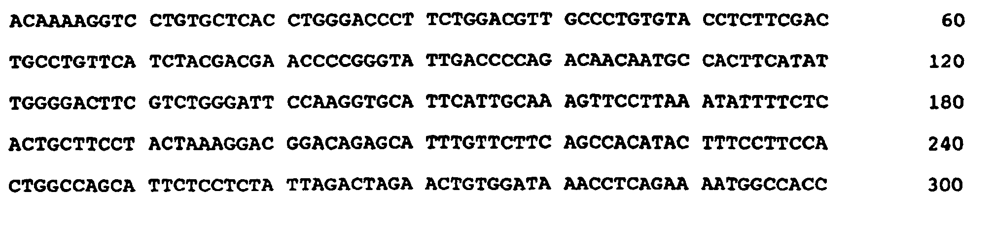
  • (xi) SEQUENCE DESCRIPTION: SEQ ID NO: 16:
    Figure 00620001
  • (2) INFORMATION FOR SEQ ID NO: 17:
  • (i) SEQUENCE CHARACTERISTICS:
  • (A) LENGTH: 22 base pairs
  • (B) TYPE: nucleic acid
  • (C) STRANDEDNESS: single
  • (D) TOPOLOGY: linear
  • (ii) MOLECULE TYPE: other nucleic acid
  • (A) DESCRIPTION: /desc = "PRIMER"
  • (xi) SEQUENCE DESCRIPTION: SEQ ID NO: 17:
    Figure 00620002
  • (2) INFORMATION FOR SEQ ID NO: 18:
  • (i) SEQUENCE CHARACTERISTICS:
  • (A) LENGTH: 21 base pairs
  • (B) TYPE: nucleic acid
  • (C) STRANDEDNESS: single
  • (D) TOPOLOGY: linear
  • (ii) MOLECULE TYPE: other nucleic acid
  • (A) DESCRIPTION: /desc = "PRIMER"
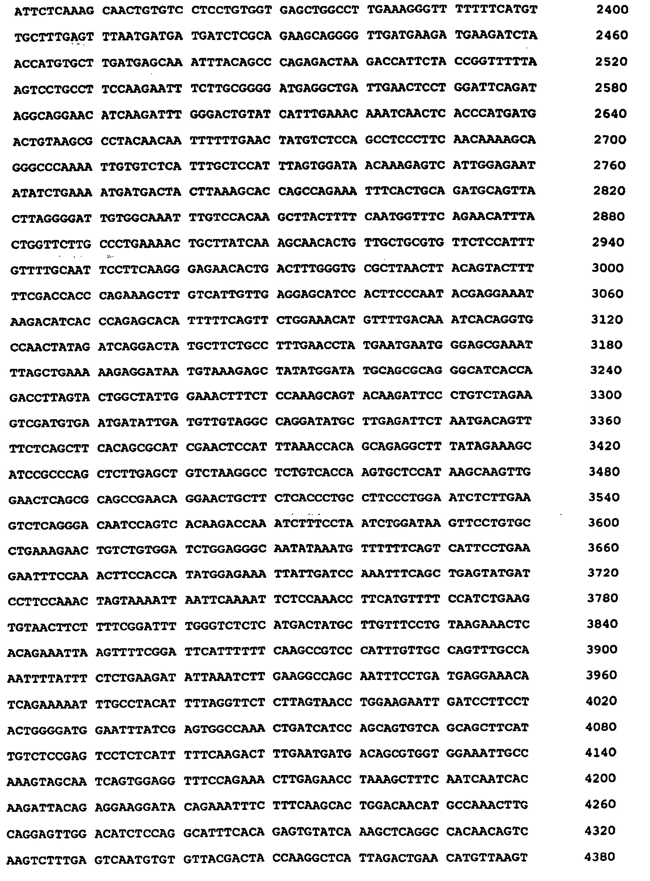
  • (xi) SEQUENCE DESCRIPTION: SEQ ID NO: 18:
    Figure 00620003
  • (2) INFORMATION FOR SEQ ID NO: 19:
  • (i) SEQUENCE CHARACTERISTICS:
  • (A) LENGTH: 22 base pairs
  • (B) TYPE: nucleic acid
  • (C) STRANDEDNESS: single
  • (D) TOPOLOGY: linear
  • (ii) MOLECULE TYPE: other nucleic acid
  • (A) DESCRIPTION: /desc = "PRIMER"
  • (xi) SEQUENCE DESCRIPTION: SEQ ID NO: 19:
    Figure 00630001
  • (2) INFORMATION FOR SEQ ID NO: 20:
  • (i) SEQUENCE CHARACTERISTICS:
  • (A) LENGTH: 21 base pairs
  • (B) TYPE: nucleic acid
  • (C) STRANDEDNESS: single
  • (D) TOPOLOGY: linear
  • (ii) MOLECULE TYPE: other nucleic acid
  • (A) DESCRIPTION: /desc = "PRIMER"
  • (xi) SEQUENCE DESCRIPTION: SEQ ID NO: 20:
    Figure 00630002
  • (2) INFORMATION FOR SEQ ID NO: 21:
  • (i) SEQUENCE CHARACTERISTICS:
  • (A) LENGTH: 6124 base pairs
  • (B) TYPE: nucleic acid
  • (C) STRANDEDNESS: single
  • (D) TOPOLOGY: linear
  • (ii) MOLECULE TYPE: cDNA
  • (xi) SEQUENCE DESCRIPTION: SEQ ID NO: 21:
    Figure 00630003
    Figure 00640001
    Figure 00650001
    Figure 00660001
  • (2) INFORMATION FOR SEQ ID NO: 22:
  • (i) SEQUENCE CHARACTERISTICS:
  • (A) LENGTH: 1403 amino acids
  • (B) TYPE: amino acid
  • (C) STRANDEDNESS: single
  • (D) TOPOLOGY: linear
  • (ii) MOLECULE TYPE: protein
  • (xi) SEQUENCE DESCRIPTION: SEQ ID NO: 22:
    Figure 00670001
    Figure 00680001
    Figure 00690001
    Figure 00700001
    Figure 00710001
    Figure 00720001
  • (2) INFORMATION FOR SEQ ID NO: 23:
  • (i) SEQUENCE CHARACTERISTICS:
  • (A) LENGTH: 6228 base pairs
  • (B) TYPE: nucleic acid
  • (C) STRANDEDNESS: single
  • (D) TOPOLOGY: linear
  • (ii) MOLECULE TYPE: cDNA
  • (xi) SEQUENCE DESCRIPTION: SEQ ID NO: 23:
    Figure 00720002
    Figure 00730001
    Figure 00740001
    Figure 00750001
  • (2) INFORMATION FOR SEQ ID NO: 24:
  • (i) SEQUENCE CHARACTERISTICS:
  • (A) LENGTH: 1403 amino acids
  • (B) TYPE: amino acid
  • (C) STRANDEDNESS: single
  • (D) TOPOLOGY: linear
  • (ii) MOLECULE TYPE: protein
  • (xi) SEQUENCE DESCRIPTION: SEQ ID NO: 24:
    Figure 00750002
    Figure 00760001
    Figure 00770001
    Figure 00780001
    Figure 00790001
    Figure 00800001
    Figure 00810001
  • (2) INFORMATION FOR SEQ ID NO: 25:
  • (i) SEQUENCE CHARACTERISTICS:
  • (A) LENGTH: 24 base pairs
  • (B) TYPE: nucleic acid
  • (C) STRANDEDNESS: single
  • (D) TOPOLOGY: linear
  • (ii) MOLECULE TYPE: other nucleic acid
  • (A) DESCRIPTION: /desc = "PRIMER"
  • (xi) SEQUENCE DESCRIPTION: SEQ ID NO: 25:
    Figure 00810002
  • (2) INFORMATION FOR SEQ ID NO: 26:
  • (i) SEQUENCE CHARACTERISTICS:
  • (A) LENGTH: 22 base pairs
  • (B) TYPE: nucleic acid
  • (C) STRANDEDNESS: single
  • (D) TOPOLOGY: linear
  • (ii) MOLECULE TYPE: other nucleic acid
  • (A) DESCRIPTION: /desc = "PRIMER"
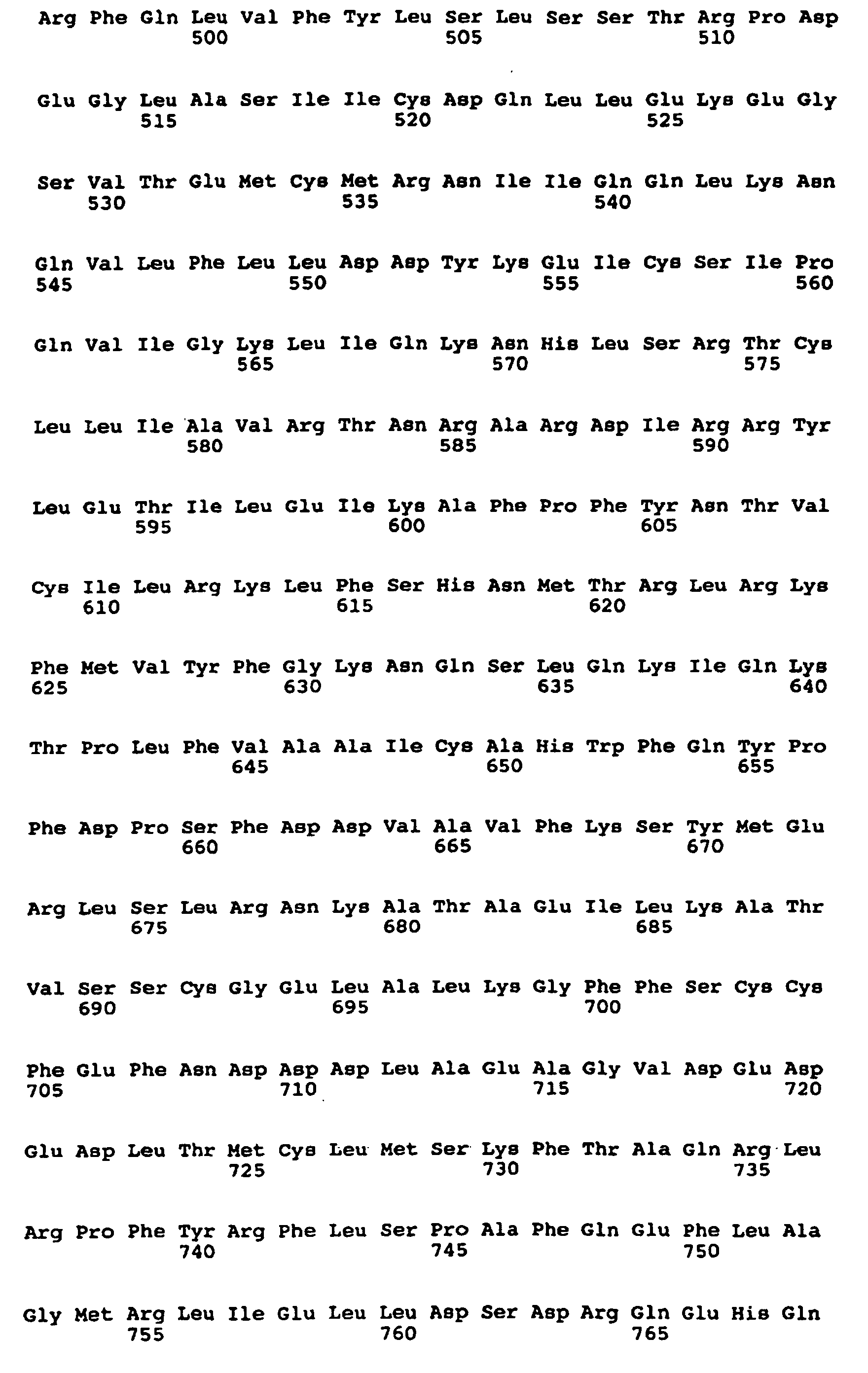
  • (xi) SEQUENCE DESCRIPTION: SEQ ID NO: 26:
    Figure 00810003
  • (2) INFORMATION FOR SEQ ID NO: 27:
  • (i) SEQUENCE CHARACTERISTICS:
  • (A) LENGTH: 24 base pairs
  • (B) TYPE: nucleic acid
  • (C) STRANDEDNESS: single
  • (D) TOPOLOGY: linear
  • (ii) MOLECULE TYPE: other nucleic acid
  • (A) DESCRIPTION: /desc = "PRIMER" (xi) SEQUENCE DESCRIPTION: SEQ ID NO: 27:
    Figure 00810004
  • Claims (17)

    1. A substantially pure nucleic acid having at least 90% nucleic acid sequence identity to the full length Neuronal Apoptosis Inhibitor Protein (NAIP) nucleic acid sequence of SEQ ID NO: 21 and including nucleotides 3734 to 3886 (exon 14a) and 4139 to 4503 (exon 17) of SEQ ID NO: 21 or having at least 90% nucleic acid sequence identity to the full length NAIP nucleic acid sequence of SEQ ID NO: 23 and including nucleotides 3838 to 3990 of SEQ ID NO:23 and nucleotides 4243 to 4605 of SEQ ID 23 said nucleic acid encoding a NAIP polypeptide that inhibits apoptosis.
    2. A substantially pure nucleic acid according to claim 1 and having the sequence of SEQ ID NO:21 or degenerate variants thereof and encode a polypeptide having the amino acid sequence of SEQ ID NO:22, wherein said polypeptide is capable of inhibiting apoptosis.
    3. The nucleic acid of any of claims 1 or 2, wherein said nucleic acid is operably linked to a regulatory-sequence for expression of said NAIP polypeptide and wherein said regulatory sequence comprises a promoter.
    4. The nucleic acid of claim 3, wherein said promoter is a constitutive promoter, is inducible by one or more external agents, or is cell-type specific.
    5. A vector comprising the nucleic acid of any of claims 1 to 4 and capable of directing expression of a NAIP polypeptide that is capable of inhibiting apoptosis.
    6. A host cell that contains the vector of claim 5.
    7. A substantially pure mammalian NAIP polypeptide encoded by the nucleic acid sequence of any of claims 1 to claim 4.
    8. A therapeutic composition comprising an effective amount of a NAIP polypeptide according to claim 7 in a physiologically acceptable carrier.
    9. A non-human animal transgenic for a NAIP nucleic acid sequence of any of claims 1 to claim 4.
    10. An in vitro method of obtaining a NAIP polypeptide, said method comprising:
      (a) providing a cell with a nucleic acid according to any of claims 3 to claim 4, said nucleic acid being positioned for expression in said cell;
      (b) culturing said cell under conditions for expressing said DNA; and
      (c) isolating said NAIP polypeptide.
    11. An in vitro method of identifying a NAIP nucleic acid in a mammalian cell, said method comprising:
      (a) providing a preparation of mammalian cellular DNA of said cell;
      (b) providing a detectably-labelled DNA sequence that binds specifically to nucleotides 3734 to 3886 (exon 14a) and 4139 to 4503 (exon 17) of SEQ ID NO: 21 or nucleotides 3838 to 3990 (exon 14a) and nucleotides 4243 to 4605 (exon 17) of SEQ ID NO: 23;
      (c) contacting said preparation of cellular DNA with said detectably-labelled DNA sequence under hybridization conditions that provide detection of nucleic acids having at least 50% nucleic acid sequence identity; and
      (d) identifying a NAIP nucleic acid by its association with said detectably-labelled DNA sequence.
    12. An in vitro method of isolating a NAIP nucleic acid as defined in claim 1 or claim 2, or portion thereof, said method comprising amplifying by PCR said NAIP nucleic acid, or portion thereof, using oligonucleotide primers wherein said primers;
      (a)are each greater than 13 nucleotides in length;
      (b)each have regions of complementarity to opposite DNA strands comprising nucleotides 3838 to 3990 (exon 14a) or nucleotides 4243 to 4605 (exon 17) of SEQ ID NO: 23; and
      (c)optionally contain sequences capable of producing restriction enzyme cut sites in the amplified product;
         and isolating said NAIP nucleic acid or portion thereof.
    13. An in vitro method of identifying a compound that modulates apoptosis, said method comprising:
      contacting in vitro a cell expressing a NAIP polypeptide with a candidate compound and monitoring expression of a NAIP nucleic acid as defined in claim 1 or claim 2 by detecting a nucleic acid sequence comprising nucleotides 3838 to 3990 of SEQ ID NO:23 or nucleotides 4243 to 4605 of SEQ ID NO:23, an alteration in the level of said expression of said nucleic acid indicating the presence of a compound which modulates apoptosis.
    14. A neuronal apoptosis inhibitor protein (NAIP) nucleic acid according to claim 1 or claim 2 or a polypeptide encoded by said nucleic acid sequence, for use as a medicament.
    15. Use of an NAIP nucleic acid according to claim 1 or claim 2 or a polypeptide encoded by said nucleic acid sequence, for the manufacture of a medicament for the treatment of spinal muscular atrophy, ALS, AIDS, a neurodegenerative disease, a myelodysplastic syndrome or an ischemic injury .
    16. A NAIP antisense molecule comprising nucleic acid that is complimentary to nucleotides 3734 to 3886 (exon 14a) or to nucleotides 4139 to 4503 (exon 17) of SEQ ID NO: 21 or to nucleotides 3838 to 3990 of SEQ ID NO:23 or nucleotides 4243 to 4605 of SEQ ID 23.
    17. A NAIP antisense molecule according to claim 16 for use as a medicament.
    EP97902522A 1996-01-19 1997-01-17 Use of neuronal apoptosis inhibitor protein (naip) Expired - Lifetime EP0815231B1 (en)

    Priority Applications (1)

    Application Number Priority Date Filing Date Title
    EP05106334A EP1609864A3 (en) 1996-01-19 1997-01-17 Use of neuronal apoptosis inhibitor protein (NAIP)

    Applications Claiming Priority (3)

    Application Number Priority Date Filing Date Title
    GBGB9601108.5A GB9601108D0 (en) 1996-01-19 1996-01-19 Neuronal apoptosis inhibitor protein (NAIP)
    GB9601108 1996-01-19
    PCT/IB1997/000142 WO1997026331A2 (en) 1996-01-19 1997-01-17 Use of neuronal apoptosis inhibitor protein (naip)

    Related Child Applications (1)

    Application Number Title Priority Date Filing Date
    EP05106334A Division EP1609864A3 (en) 1996-01-19 1997-01-17 Use of neuronal apoptosis inhibitor protein (NAIP)

    Publications (2)

    Publication Number Publication Date
    EP0815231A2 EP0815231A2 (en) 1998-01-07
    EP0815231B1 true EP0815231B1 (en) 2005-08-03

    Family

    ID=10787268

    Family Applications (2)

    Application Number Title Priority Date Filing Date
    EP05106334A Withdrawn EP1609864A3 (en) 1996-01-19 1997-01-17 Use of neuronal apoptosis inhibitor protein (NAIP)
    EP97902522A Expired - Lifetime EP0815231B1 (en) 1996-01-19 1997-01-17 Use of neuronal apoptosis inhibitor protein (naip)

    Family Applications Before (1)

    Application Number Title Priority Date Filing Date
    EP05106334A Withdrawn EP1609864A3 (en) 1996-01-19 1997-01-17 Use of neuronal apoptosis inhibitor protein (NAIP)

    Country Status (11)

    Country Link
    US (2) US6994957B2 (en)
    EP (2) EP1609864A3 (en)
    JP (1) JP4005135B2 (en)
    AT (1) ATE301192T1 (en)
    AU (1) AU1614997A (en)
    CA (1) CA2215793C (en)
    DE (1) DE69733861T2 (en)
    DK (1) DK0815231T3 (en)
    ES (1) ES2245790T3 (en)
    GB (1) GB9601108D0 (en)
    WO (1) WO1997026331A2 (en)

    Families Citing this family (13)

    * Cited by examiner, † Cited by third party
    Publication number Priority date Publication date Assignee Title
    US6156535A (en) * 1995-08-04 2000-12-05 University Of Ottawa Mammalian IAP gene family, primers, probes, and detection methods
    GB9601108D0 (en) * 1996-01-19 1996-03-20 Univ Ottawa Neuronal apoptosis inhibitor protein (NAIP)
    CA2222453C (en) 1996-04-26 2009-06-30 Universite D'ottawa/ University Of Ottawa Therapeutic and drug screening methods for the treatment and prevention of neuronal disease
    US6133437A (en) 1997-02-13 2000-10-17 Apoptogen, Inc. Modulation of IAPs for the treatment of proliferative diseases
    JPH11116599A (en) * 1997-10-14 1999-04-27 Japan Science & Technology Corp Apoptosis inhibitory protein, gene encoding this protein and its cDNA
    US6171821B1 (en) * 1998-07-24 2001-01-09 Apoptogen, Inc. XIAP IRES and uses thereof
    JP2000125861A (en) 1998-10-26 2000-05-09 Japan Science & Technology Corp Monoclonal antibody against human apoptosis inhibitor protein NAIP and method for assaying NAIP
    US6673917B1 (en) 2000-09-28 2004-01-06 University Of Ottawa Antisense IAP nucleic acids and uses thereof
    WO2003080638A2 (en) 2002-03-27 2003-10-02 Aegera Therapeutics, Inc. Antisense iap nucleobase oligomers and uses thereof
    US8012944B2 (en) 2003-10-30 2011-09-06 Pharmascience Inc. Method for treating cancer using IAP antisense oligomer and chemotherapeutic agent
    JPWO2006051757A1 (en) * 2004-11-10 2008-05-29 株式会社ニュージェン・ファーマ Therapeutic or preventive agent for ischemic heart disease or ischemic cardiomyopathy
    PL1889065T3 (en) 2005-05-18 2013-12-31 Novartis Ag Methods for diagnosis and treatment of diseases having an autoimmune and/or inflammatory component
    KR101779370B1 (en) 2016-06-29 2017-09-19 한국수력원자력 주식회사 Apoptosis regulatory gene detected irradiated-thymic lymphoma and detecting method of thereof

    Family Cites Families (7)

    * Cited by examiner, † Cited by third party
    Publication number Priority date Publication date Assignee Title
    WO1994006814A1 (en) 1992-09-14 1994-03-31 The General Hospital Corporation Ikaros: a t cell pathway regulatory gene
    EP0770129B1 (en) 1994-01-18 2005-11-23 The Scripps Research Institute Zinc finger protein derivatives and methods therefor
    JP3944654B2 (en) 1994-10-18 2007-07-11 ザ ユニバーシティー オブ オタワ Neuronal apoptosis inhibitor protein and its gene sequence, and mutation of the gene causing spinal muscular atrophy
    US6156535A (en) * 1995-08-04 2000-12-05 University Of Ottawa Mammalian IAP gene family, primers, probes, and detection methods
    US5919912A (en) 1995-08-04 1999-07-06 University Of Ottawa Mammalian IAP antibodies and diagnostic kits
    GB9601108D0 (en) 1996-01-19 1996-03-20 Univ Ottawa Neuronal apoptosis inhibitor protein (NAIP)
    US6133437A (en) 1997-02-13 2000-10-17 Apoptogen, Inc. Modulation of IAPs for the treatment of proliferative diseases

    Also Published As

    Publication number Publication date
    US7235372B2 (en) 2007-06-26
    EP1609864A3 (en) 2007-12-12
    ATE301192T1 (en) 2005-08-15
    CA2215793C (en) 2005-07-05
    ES2245790T3 (en) 2006-01-16
    US6994957B2 (en) 2006-02-07
    US20020137028A1 (en) 2002-09-26
    GB9601108D0 (en) 1996-03-20
    US20060172348A1 (en) 2006-08-03
    JPH11503620A (en) 1999-03-30
    DK0815231T3 (en) 2005-12-12
    JP4005135B2 (en) 2007-11-07
    AU1614997A (en) 1997-08-11
    DE69733861T2 (en) 2006-05-24
    CA2215793A1 (en) 1997-07-24
    WO1997026331A3 (en) 1997-10-02
    EP1609864A2 (en) 2005-12-28
    WO1997026331A2 (en) 1997-07-24
    DE69733861D1 (en) 2005-09-08
    EP0815231A2 (en) 1998-01-07

    Similar Documents

    Publication Publication Date Title
    EP0837939B1 (en) Mammalian apoptosis inhibitor protein gene family, primers, probes and detection methods
    CA2273821C (en) Detection and modulation of the iaps and naip for the diagnosis and treatment of proliferative disease
    US6946544B2 (en) XAF genes and polypeptides: methods and reagents for modulating apoptosis
    US6994957B2 (en) Use of neuronal apoptosis inhibitor protein (NAIP)
    US6331412B1 (en) Methods and compounds for modulating male fertility
    CA2403947C (en) Bir domains of mammalian iap gene family
    CA2512366A1 (en) Mammalian iap gene family, primers, probes and detection methods
    HK1103241A (en) Mammalian apoptosis inhibitor protein gene family, primers, probes and detection methods
    JPH1132780A (en) Xaf gene and polypeptide: regulation of apoptosis and reagent therefor

    Legal Events

    Date Code Title Description
    PUAI Public reference made under article 153(3) epc to a published international application that has entered the european phase

    Free format text: ORIGINAL CODE: 0009012

    17P Request for examination filed

    Effective date: 19971008

    AK Designated contracting states

    Kind code of ref document: A2

    Designated state(s): AT BE CH DE DK ES FI FR GB GR IE IT LI LU MC NL PT SE

    RIN1 Information on inventor provided before grant (corrected)

    Inventor name: TAMAI, KATSU

    Inventor name: ROBERTSON, GEORGE

    Inventor name: ROY, NATALIE

    Inventor name: MACKENZIE, ALEXANDER, E.

    Inventor name: KORNELUK, ROBERT, G.

    17Q First examination report despatched

    Effective date: 20020711

    GRAP Despatch of communication of intention to grant a patent

    Free format text: ORIGINAL CODE: EPIDOSNIGR1

    DAX Request for extension of the european patent (deleted)
    GRAS Grant fee paid

    Free format text: ORIGINAL CODE: EPIDOSNIGR3

    GRAA (expected) grant

    Free format text: ORIGINAL CODE: 0009210

    AK Designated contracting states

    Kind code of ref document: B1

    Designated state(s): AT BE CH DE DK ES FI FR GB GR IE IT LI LU MC NL PT SE

    PG25 Lapsed in a contracting state [announced via postgrant information from national office to epo]

    Ref country code: NL

    Free format text: LAPSE BECAUSE OF FAILURE TO SUBMIT A TRANSLATION OF THE DESCRIPTION OR TO PAY THE FEE WITHIN THE PRESCRIBED TIME-LIMIT

    Effective date: 20050803

    Ref country code: LI

    Free format text: LAPSE BECAUSE OF FAILURE TO SUBMIT A TRANSLATION OF THE DESCRIPTION OR TO PAY THE FEE WITHIN THE PRESCRIBED TIME-LIMIT

    Effective date: 20050803

    Ref country code: FI

    Free format text: LAPSE BECAUSE OF FAILURE TO SUBMIT A TRANSLATION OF THE DESCRIPTION OR TO PAY THE FEE WITHIN THE PRESCRIBED TIME-LIMIT

    Effective date: 20050803

    Ref country code: CH

    Free format text: LAPSE BECAUSE OF FAILURE TO SUBMIT A TRANSLATION OF THE DESCRIPTION OR TO PAY THE FEE WITHIN THE PRESCRIBED TIME-LIMIT

    Effective date: 20050803

    Ref country code: BE

    Free format text: LAPSE BECAUSE OF FAILURE TO SUBMIT A TRANSLATION OF THE DESCRIPTION OR TO PAY THE FEE WITHIN THE PRESCRIBED TIME-LIMIT

    Effective date: 20050803

    Ref country code: AT

    Free format text: LAPSE BECAUSE OF FAILURE TO SUBMIT A TRANSLATION OF THE DESCRIPTION OR TO PAY THE FEE WITHIN THE PRESCRIBED TIME-LIMIT

    Effective date: 20050803

    REG Reference to a national code

    Ref country code: GB

    Ref legal event code: FG4D

    REG Reference to a national code

    Ref country code: CH

    Ref legal event code: EP

    REG Reference to a national code

    Ref country code: IE

    Ref legal event code: FG4D

    RIN2 Information on inventor provided after grant (corrected)

    Inventor name: ROBERTSON, GEORGE

    Inventor name: ROY, NATALIE

    Inventor name: MACKENZIE, ALEXANDER, E.

    Inventor name: KORNELUK, ROBERT, G.

    REF Corresponds to:

    Ref document number: 69733861

    Country of ref document: DE

    Date of ref document: 20050908

    Kind code of ref document: P

    REG Reference to a national code

    Ref country code: SE

    Ref legal event code: TRGR

    PG25 Lapsed in a contracting state [announced via postgrant information from national office to epo]

    Ref country code: GR

    Free format text: LAPSE BECAUSE OF FAILURE TO SUBMIT A TRANSLATION OF THE DESCRIPTION OR TO PAY THE FEE WITHIN THE PRESCRIBED TIME-LIMIT

    Effective date: 20051103

    REG Reference to a national code

    Ref country code: DK

    Ref legal event code: T3

    PG25 Lapsed in a contracting state [announced via postgrant information from national office to epo]

    Ref country code: PT

    Free format text: LAPSE BECAUSE OF FAILURE TO SUBMIT A TRANSLATION OF THE DESCRIPTION OR TO PAY THE FEE WITHIN THE PRESCRIBED TIME-LIMIT

    Effective date: 20060103

    REG Reference to a national code

    Ref country code: ES

    Ref legal event code: FG2A

    Ref document number: 2245790

    Country of ref document: ES

    Kind code of ref document: T3

    PG25 Lapsed in a contracting state [announced via postgrant information from national office to epo]

    Ref country code: IE

    Free format text: LAPSE BECAUSE OF NON-PAYMENT OF DUE FEES

    Effective date: 20060117

    PG25 Lapsed in a contracting state [announced via postgrant information from national office to epo]

    Ref country code: MC

    Free format text: LAPSE BECAUSE OF NON-PAYMENT OF DUE FEES

    Effective date: 20060131

    Ref country code: LU

    Free format text: LAPSE BECAUSE OF NON-PAYMENT OF DUE FEES

    Effective date: 20060131

    NLV1 Nl: lapsed or annulled due to failure to fulfill the requirements of art. 29p and 29m of the patents act
    REG Reference to a national code

    Ref country code: CH

    Ref legal event code: PL

    ET Fr: translation filed
    PLBE No opposition filed within time limit

    Free format text: ORIGINAL CODE: 0009261

    STAA Information on the status of an ep patent application or granted ep patent

    Free format text: STATUS: NO OPPOSITION FILED WITHIN TIME LIMIT

    26N No opposition filed

    Effective date: 20060504

    REG Reference to a national code

    Ref country code: IE

    Ref legal event code: MM4A

    PGFP Annual fee paid to national office [announced via postgrant information from national office to epo]

    Ref country code: ES

    Payment date: 20080128

    Year of fee payment: 12

    Ref country code: DK

    Payment date: 20080130

    Year of fee payment: 12

    PGFP Annual fee paid to national office [announced via postgrant information from national office to epo]

    Ref country code: SE

    Payment date: 20080129

    Year of fee payment: 12

    Ref country code: IT

    Payment date: 20080129

    Year of fee payment: 12

    PGFP Annual fee paid to national office [announced via postgrant information from national office to epo]

    Ref country code: FR

    Payment date: 20080117

    Year of fee payment: 12

    Ref country code: DE

    Payment date: 20080229

    Year of fee payment: 12

    EUG Se: european patent has lapsed
    REG Reference to a national code

    Ref country code: DK

    Ref legal event code: EBP

    PG25 Lapsed in a contracting state [announced via postgrant information from national office to epo]

    Ref country code: DE

    Free format text: LAPSE BECAUSE OF NON-PAYMENT OF DUE FEES

    Effective date: 20090801

    REG Reference to a national code

    Ref country code: FR

    Ref legal event code: ST

    Effective date: 20091030

    REG Reference to a national code

    Ref country code: ES

    Ref legal event code: FD2A

    Effective date: 20090119

    PG25 Lapsed in a contracting state [announced via postgrant information from national office to epo]

    Ref country code: FR

    Free format text: LAPSE BECAUSE OF NON-PAYMENT OF DUE FEES

    Effective date: 20090202

    Ref country code: ES

    Free format text: LAPSE BECAUSE OF NON-PAYMENT OF DUE FEES

    Effective date: 20090119

    PGFP Annual fee paid to national office [announced via postgrant information from national office to epo]

    Ref country code: GB

    Payment date: 20100125

    Year of fee payment: 14

    PG25 Lapsed in a contracting state [announced via postgrant information from national office to epo]

    Ref country code: DK

    Free format text: LAPSE BECAUSE OF NON-PAYMENT OF DUE FEES

    Effective date: 20090731

    PG25 Lapsed in a contracting state [announced via postgrant information from national office to epo]

    Ref country code: IT

    Free format text: LAPSE BECAUSE OF NON-PAYMENT OF DUE FEES

    Effective date: 20090117

    PG25 Lapsed in a contracting state [announced via postgrant information from national office to epo]

    Ref country code: SE

    Free format text: LAPSE BECAUSE OF NON-PAYMENT OF DUE FEES

    Effective date: 20090118

    GBPC Gb: european patent ceased through non-payment of renewal fee

    Effective date: 20110117

    PG25 Lapsed in a contracting state [announced via postgrant information from national office to epo]

    Ref country code: GB

    Free format text: LAPSE BECAUSE OF NON-PAYMENT OF DUE FEES

    Effective date: 20110117