EP0814827B1 - Vascular endothelial growth factor-b - Google Patents
Vascular endothelial growth factor-b Download PDFInfo
- Publication number
- EP0814827B1 EP0814827B1 EP96909573A EP96909573A EP0814827B1 EP 0814827 B1 EP0814827 B1 EP 0814827B1 EP 96909573 A EP96909573 A EP 96909573A EP 96909573 A EP96909573 A EP 96909573A EP 0814827 B1 EP0814827 B1 EP 0814827B1
- Authority
- EP
- European Patent Office
- Prior art keywords
- seq
- vegf
- protein
- amino acid
- sequence
- Prior art date
- Legal status (The legal status is an assumption and is not a legal conclusion. Google has not performed a legal analysis and makes no representation as to the accuracy of the status listed.)
- Expired - Lifetime
Links
Images








Classifications
-
- C—CHEMISTRY; METALLURGY
- C07—ORGANIC CHEMISTRY
- C07K—PEPTIDES
- C07K14/00—Peptides having more than 20 amino acids; Gastrins; Somatostatins; Melanotropins; Derivatives thereof
- C07K14/435—Peptides having more than 20 amino acids; Gastrins; Somatostatins; Melanotropins; Derivatives thereof from animals; from humans
- C07K14/52—Cytokines; Lymphokines; Interferons
-
- A—HUMAN NECESSITIES
- A61—MEDICAL OR VETERINARY SCIENCE; HYGIENE
- A61P—SPECIFIC THERAPEUTIC ACTIVITY OF CHEMICAL COMPOUNDS OR MEDICINAL PREPARATIONS
- A61P35/00—Antineoplastic agents
-
- A—HUMAN NECESSITIES
- A61—MEDICAL OR VETERINARY SCIENCE; HYGIENE
- A61P—SPECIFIC THERAPEUTIC ACTIVITY OF CHEMICAL COMPOUNDS OR MEDICINAL PREPARATIONS
- A61P43/00—Drugs for specific purposes, not provided for in groups A61P1/00-A61P41/00
-
- A—HUMAN NECESSITIES
- A61—MEDICAL OR VETERINARY SCIENCE; HYGIENE
- A61P—SPECIFIC THERAPEUTIC ACTIVITY OF CHEMICAL COMPOUNDS OR MEDICINAL PREPARATIONS
- A61P9/00—Drugs for disorders of the cardiovascular system
-
- A—HUMAN NECESSITIES
- A61—MEDICAL OR VETERINARY SCIENCE; HYGIENE
- A61K—PREPARATIONS FOR MEDICAL, DENTAL OR TOILETRY PURPOSES
- A61K38/00—Medicinal preparations containing peptides
Definitions
- Angiogenesis or the proliferation of new capillaries from pre-existing blood vessels, is a fundamental process necessary for normal growth and development of tissues. It is a prerequisite for the development and differentiation of the vascular tree, as well as for a wide variety of fundamental physiological processes including embryogenesis, somatic growth, tissue and organ repair and regeneration, cyclical growth of the corpus luteum and endometrium, and development and differentiation of the nervous system.
- angiogenesis occurs in the follicle during its development, in the corpus luteum following ovulation and in the placenta to establish and maintain pregnancy.
- Angiogenesis additionally occurs as part of the body's repair processes, e.g. in the healing of wounds and fractures.
- Angiogenesis is also a factor in tumor growth, since a tumor must continuously stimulate growth of new capillary blood vessels in order to grow.
- Capillary blood vessels consist of endothelial cells and pericytes. These two cell types carry all of the genetic information to form tubes, branches and entire capillary networks. Specific angiogenic molecules can initiate this process.
- angiogenesis regulators see Klagsbrun et al., "Regulators of Angiogenesis", Ann. Rev. Physiol ., 53:217-39 (1991); and Folkman et al., "Angiogenesis,” J. Biol. Chem ., 267:10931-934 (1992).
- RTKs endothelial receptor tyrosine kinases
- vascular endothelial growth factor vascular endothelial growth factor
- Ferrara et al. "The Vascular Endothelial Growth Factor Family of Polypeptides," J. Cellular Biochem ., 47:211-218 (1991); Connolly, “Vascular Permeability Factor: A Unique Regulator of Blood Vessel Function,” J. Cellular Biochem ., 47:219-223 (1991).
- VEGF is a potent vasoactive protein that has been detected in media conditioned by a number of cell lines including bovine pituitary follicular cells.
- VEGF is a glycosylated cationic 46-48 kD dimer made up of two 24 kD subunits. It is inactivated by sulfhydryl reducing agents, resistant to acidic pH and to heating, and binds to immobilized heparin. VEGF is sometimes referred to as vascular permeability factor (VPF) because it increases fluid leakage from blood vessels following intradermal injection. It also has been called by the name vasculotropin.
- VPF vascular permeability factor
- VEGF 121 and VEGF 165 are soluble proteins.
- VEGF 189 and VEGF 206 appear to be mostly cell-associated. All of the isoforms of VEGF are biologically active. For example, each of the species when applied intradermally is able to induce extravasation of Evans blue.
- VEGF various species of VEGF are encoded by the same gene and arise from alternative splicing of messenger RNA. This conclusion is supported by Southern blot analysis of human genomic DNA, which shows that the restriction pattern is identical using either a probe for VEGF 165 or one which contains the insertion in VEGF 206 . Analysis of genomic clones in the area of putative mRNA splicing also shows an intron/exon structure consistent with alternative splicing.
- the different isoforms of VEGF have different chemical properties which may regulate cellular release, compartmentalization, bioavailability and possibly also modulate the signalling properties of the growth factors.
- VEGF is a member of the platelet-derived growth factor (PDGF) family.
- VEGF and PlGF are ligands for two endothelial RTKs, flt-1 (VEGF receptor 1, VEGFR1) and flk-1/KDR (VEGF receptor 2, VEGFR2).
- the amino acid sequence of VEGF exhibits approximately 20% homology to the sequences of the A and B chains of PDGF, as well as complete conservation of the eight cysteine residues found in both mature PDGF chains.
- VEGF 165 , VEGF 189 and VEGF 206 also contain eight additional cysteine residues within the carboxy-terminal region.
- VEGF vascular endothelial growth factor
- the amino-terminal sequence of VEGF is preceded by 26 amino acids corresponding to a typical signal sequence.
- the mature protein is generated directly following signal sequence cleavage without any intervening prosequence.
- the existence of a potential glycosylation site at Asn 74 is consistent with other evidence that VEGF is a glycoprotein, but the polypeptide has been reported to exist in both glycosylated and deglycosylated species.
- VEGF can have diverse effects that depend on the specific biological context in which it is found.
- VEGF and its high affinity receptors flt-1 and KDR/flk-1 are required for the formation and maintenance of the vascular system as well as for both physiological and pathological angiogenesis.
- VEGF is a potent endothelial cell mitogen and directly contributes to induction of angiogenesis in vivo by promoting endothelial cell growth during normal embryonic development, wound healing, and tissue regeneration and reorganization.
- VEGF is also involved in pathological processes such as growth and metastasis of solid tumors and ischemia-induced retinal disorders.
- a most striking property of VEGF is its specificity.
- VEGF vascular endothelial cells
- the target cell specificity of VEGF is thus restricted to vascular endothelial cells.
- VEGF can trigger the entire sequence of events leading to angiogenesis and stimulates angiogenesis in vivo in the cornea and in a healing bone graft model.
- VEGF endothelial growth factor
- VEGF mRNA is temporally and spatially related to the physiological proliferation of capillary blood vessels in the ovarian corpus luteum or in the developing brain. VEGF expression is triggered by hypoxia so that endothelial cell proliferation and angiogenesis appear to be especially stimulated in ischemic areas. VEGF is also a potent chemoattractant for monocytes. In addition, VEGF induces plasminogen activator and plasminogen activator inhibitor in endothelial cells.
- Tumor cells release angiogenic molecules such as VEGF, and monoclonal antibodies to VEGF have been shown to inhibit the growth of certain types of tumor such as rhabdomyosarcoma. See Kim et al., "Inhibition of Vascular Endothelial Growth Factor-Induced Angiogenesis Suppresses Tumor Growth in vivo ,” Nature , 362:841-844 (1993). This suggests that blocking VEGF action is of potential therapeutic significance in treating tumors in general, and highly-vascularized, aggressive tumors in particular.
- Another object of the invention is to provide isolated DNA sequences which encode a new growth factor which promotes proliferation of endothelial cells.
- an isolated DNA which codes for a protein exhibiting the following characteristic amino acid sequence (SEQ ID NO:16): and having the property of promoting proliferation of endothelial cells or mesodermal cells, the DNA being selected from the group consisting of the DNA of Figures 1 and 2 (SEQ ID NO:1), the DNA of Figure 3 (SEQ ID NO:4), the DNA of Figure 5 (SEQ ID NO:6); the DNA of Figure 7 (SEQ ID NO:8), the DNA of Figure 10 (SEQ ID NO:10), the DNA of Figure 12 (SEQ ID NO:12), the DNA of Figure 14 (SEQ ID NO:14), and DNA's which hybridize under stringent conditions with at least one of the foregoing DNA sequences.
- SEQ ID NO:16 characteristic amino acid sequence
- the objects are also achieved by providing a protein exhibiting the following characteristic amino acid sequence (SEQ ID NO:16) and having the property of promoting proliferation of endothelial cells or mesodermal cells, which protein comprises a sequence of amino acids substantially corresponding to an amino acid sequence selected from the group consisting of the amino acid sequence of Figure 1 (SEQ ID NO:2), the amino acid sequence of Figure 2 (SEQ ID NO:3), the amino acid sequence of Figure 4 (SEQ ID NO:5), the amino acid sequence of Figure 6 (SEQ ID NO:7), the amino acid sequence of Figure 8 (SEQ ID NO:9), the amino acid sequenc of Figure 11 (SEQ ID NO:11), the amino acid sequence of Figure 13 (SEO ID NO:13), and the amino acid sequence of Figure 15 (SEQ ID NO:15).
- SEQ ID NO:16 characteristic amino acid sequence
- the objects are achieved by providing pharmaceutical preparations which comprise such proteins; and by providing antibodies which react with or recognize such proteins.
- vascular endothelial growth factor B vascular endothelial growth factor B
- P1GF placenta growth factor
- All of the VEGF-B forms contain the characteristic amino acid sequence (SEQ ID NO:16) (wherein Xaa represents a variable residue), which is an earmark of the PDGF/VEGF family of growth factors. This characteristic amino acid sequence can be found at amino acids 70 to 82 in Figures 4, 6, 8, 11, 13 and 15.
- Clinical applications of the invention include diagnostic applications, acceleration of angiogenesis in wound healing, and inhibition of angiogenesis. Quantitation of VEGF-B in cancer biopsy specimens may be useful as an indicator of future metastatic risk. Topical application of VEGF-B preparations to chronic wounds may accelerate angiogenesis and wound healing. VEGF-B may be used in a manner analogous to VEGF.
- the objects are achieved by providing diagnostic/prognostic means typically in the form of test kits.
- a diagnostic/prognostic test kit comprising antibodies to the new growth factor of the invention and means for detecting, and more preferably evaluating, binding between the antibodies and the new growth factor of the invention.
- either the antibody or the new growth factor is labelled, and either the antibody or the growth factor is substrate-bound, such that the growth factor-antibody interaction can be established by determining the amount of label attached to the substrate following binding between the antibody and the growth factor.
- the diagnostic/prognostic means may be provided as a conventional ELISA kit.
- the diagnostic/prognostic means may comprise PCR means for establishing the genomic sequence structure of a VEGF-B gene of a test individual and comparing this sequence structure with that disclosed in this application in order to detect any abnormalities, with a view to establishing whether any aberrations in VEGF-B expression are related to a given disease condition.
- a yet further aspect of the invention concerns an antibody which recognizes VEGF-B and which is suitably labelled.
- compositions which comprise VEGF-B protein may optionally further comprise either VEGF or heparin or both.
- a medicament which comprises VEGF-B protein and heparin for treating conditions characterized by lack of, or reduction in, angiogenesis.
- the invention in another aspect, relates to a protein dimer comprising VEGF-B protein, particularly a disulfide-linked dimer.
- the protein dimers of the invention include both homodimers of VEGF-B protein and heterodimers of VEGF-B and VEGF.
- a method for facilitating release of VEGF and/or VEGF-B from a cell comprising exposing a cell which expresses either or both of the aforementioned growth factors to heparin.
- Another aspect of the invention involves providing a vector comprising an anti-sense nucleotide sequence which is complementary to at least a part of the DNA sequences disclosed herein which encode the new growth factor of the invention which promotes proliferation of endothelial cells.
- a vector comprising an anti-sense sequence may be used to inhibit, or at least mitigate, VEGF-B expression.
- the use of a vector of this type to inhibit VEGF-B expression is favored in instances where VEGF-B expression is associated with a disease such as in instances where tumors produce VEGF-B in order to provide for angiogenesis. Transformation of such tumor cells with a vector containing an anti-sense nucleotide sequence would suppress or retard angiogenesis and so would inhibit or retard growth of the tumor.
- the present invention thus is directed to new vascular endothelial growth factors, hereinafter referred to as VEGF-B growth factors, which share the angiogenic and other properties of VEGF, but which are distributed and expressed in tissues differently from VEGF.
- VEGF-B growth factors new vascular endothelial growth factors, hereinafter referred to as VEGF-B growth factors, which share the angiogenic and other properties of VEGF, but which are distributed and expressed in tissues differently from VEGF.
- VEGF-B growth factors are members of the family of platelet derived growth factors and are a growth factors which promote mitosis and proliferation of vascular endothelial cells and/or mesodermal cells. They are produced by expression of DNA sequences which correspond to, or which are hybridizable under stringent conditions with, any one of the DNA sequences depicted in Figures 1 and 2 (SEQ ID NO:1), Figure 3 (SEQ ID NO:4), Figure 5 (SEQ ID NO:6), Figure 7 (SEQ ID NO:8), Figure 10 (SEQ ID NO:10), Figure 12 (SEQ ID NO:12) or Figure 14 (SEQ ID NO:14).
- angiogenic proteins encoded by DNA sequences which hybridize under stringent conditions include, for example, 50% formamide, 5 x SSPE buffer, 5 x Denhardts solution, 0.5% SDS and 100 ⁇ g/ml of salmon sperm DNA at 42°C overnight, followed by washing 2 x 30 minutes in 2 x SSC at 55°C.
- the invention is also directed to an isolated and/or purified DNA which corresponds to, or which hybridizes under stringent conditions with, any one of the foregoing DNA sequences.
- the invention is directed to antibodies of VEGF-B growth factors, and particularly to monoclonal antibodies.
- VEGF-B proteins are believed to interact with protein tyrosine kinase growth factor receptors. Details of such receptors are known in the art [See e.g. Wilks, A.F., "Protein Tyrosine Kinase Growth Factor Receptors and Their Ligands in Development, Differentiation, and Cancer," Adv. Cancer Res., 60:43-73 (1993)].
- Various adult mouse tissues were tested for expression of transcripts corresponding to VEGF-B by Northern blotting.
- the size of the mRNA was 1.3-1.4 kb.
- a mouse multiple tissue Northern blot (MTN, Clontech) was probed with the ⁇ 0.9 kb SalI/NotI fragment derived from the pPC67 yeast expression vectors described above. The probe was labelled with 32 P-dCTP using random priming (specific activity 10 8 -10 9 cpm/ ⁇ g of DNA). The blot was hybridized overnight at 42°C using 50% formamide, 5 x SSPE buffer, 2% SDS, 10 x Denhardts solution, 100 ⁇ g/ml salmon sperm DNA and 1x10 6 cpm of the labelled probe/ml.
- the blot was washed at room temperature for 2 x 30 min in 2 x SSC containing 0.05% SDS and then for 2 x 20 min at 52°C in 0.1 x SSC containing 0.1% SDS. The blot was then exposed at -70°C for three days using intensifying screens. Kodak XAR film was used.
- a human multiple tissue Northern blot (MNT) from Clontech was probed using the murine partial cDNA to determine relative VEGF-B expression levels in various human tissues.
- the size of the transcript was 1.3-1.4 kb. The conditions were identical to those used for the mouse Northern blot described above.
- the relative VEGF-B transcript levels for the human Northern blot are listed in the following Table 2. For comparison purposes, Table 2 also lists relative expression level data from the literature for VEGF in various mammalian systems.
- VEGF-B (Northern blot) VEGF (from literature) human human murine guinea pig heart +++++ ++ +++ brain + + + placenta + lung + ++++ ++ liver (+) ++ (+) + skeletal muscle ++++ +++ + kidney + ++ + ++ pancreas +++ spleen ++ - + thymus + - prostate +++ testis ++ (+) ovary +++ - small intestine ++ colon +++ peripheral blood leucocytes +
- VEGF vascular endothelial growth factor
- VEGF-B is very strongly expressed by the same tissue.
- VEGF is strongly expressed by human lung tissue, but VEGF-B is only weakly expressed by human lung tissue.
- human liver tissue expresses VEGF at a moderate level, but VEGF-B is expressed only very weakly.
- VEGF-B transcripts The expression of VEGF-B transcripts was further analyzed in mouse and human tissues by Northern blotting and compared with the expression of VEGF transcripts.
- Mouse and human multiple tissue Northern (MTN) blots (Clontech) were hybridized with a 32 P-labelled mouse VEGF-B probe ( ⁇ 0.9 kb SalI/NotI insert of the clone pcif 2).
- VEGF expression was analyzed with 32 P-labelled VEGF 165 cDNA as the probe.
- the hybridizations were carried out at 42°C in 50% deionized formamide, 5 x SSC pH 7.0, 1% SDS, 5 x Denhardt's solution and 100 ⁇ g/ml of denatured salmon sperm DNA.
- RNA probes (a 383 bp antisense probe and a 169 bp sense probe) were generated from a linearized plasmid containing a 440 bp Sal I/Sac I fragment derived from the pcif 2 cDNA clone.
- Radiolabelled RNA was synthesized using T7 and SP6 RNA polymerases and [ 35 S]UTP (Amersham Inc.). Alkaline hydrolysis of the probes was omitted. Hematoxylin was used for counterstaining. Control hybridizations with sense strand and RNAse A-treated sections did not give signals above background.
- VEGF-B and VEGF are coexpressed in many human and mouse tissues.
- VEGF-B transcripts were further examined by in situ hybridization in sections from adult mouse heart and skeletal muscle and from the early (E 10) mouse embryo.
- E 10) mouse embryo In the adult heart, VEGF-B transcripts are prominently expressed in the myocardium, while no specific signal is detected in arterial smooth muscle.
- VEGF-B transcripts are expressed by some of the myofibers whereas others seem to lack the transcript.
- E 10 mouse embryo VEGF-B transcripts are detected mainly in the developing heart. The myocardium of the adult mouse heart has a prominent signal. In striated muscle, VEGF-B expression is seen in subpopulations of myofibers. Strong signals were also obtained in the developing heart of the E10 mouse embryo.
- VEGF-B vascular endothelial growth factor-B
- VEGF-B transcrips are expressed primarily in muscular tissues.
- VEGF-B is particularly abundant in heart and skeletal muscle and is co-expressed with VEGF in these and other tissues.
- VEGF-B forms cell surface associated, disulfide-linked homodimers and heterodimers with VEGF when coexpressed.
- a Northern blot analysis of the expression of VEGF-B 186 transcripts in several mouse and human tissues using a VEGF-B 186 isoform specific probe is shown in Figure 21.
- the chromosomal location of the VEGF-B gene was assessed by Southern blotting and polymerase chain reaction analysis of somatic cell hybrids and fluorescense in situ hybridization (FISH) of metaphase chromosomes.
- FISH fluorescense in situ hybridization
- the VEGF-B gene was found on chromosome 11q13, proximal of the cyclin D1 gene. It is interesting that although the cyclin D1 gene is amplified in a number of human carcinomas, the VEGF-B gene was not amplified in several mammary carcinoma cell lines which contained amplified cyclin D1. Nevertheless, mutations in the VEGF-B gene may be related to vascular malformations and/or cardiovascular diseases.
- Example 1 Partial cDNA clone with two reading frames.
- a partial cDNA clone encoding murine VEGF-B was identified as follows.
- a cDNA library (E 14.5) derived from poly A+ mRNA isolated from 14.5 day old mouse embryos [Chevray P. and Nathans D., "Protein interaction cloning in yeast: Identification of mammalian proteins that react with the leucine zipper of Jun," Proc. Natl . Acad. Sci. USA, 89:5789-93 (1992)) was screened for cellular proteins which potentially might interact with cellular retinoic acid-binding protein type 1 (CRABP-I) using a yeast two-hybrid interaction trap screening technique as described by Gyuris J., Golemis E., Chertkov H.
- CRABP-I retinoic acid-binding protein type 1
- This screening technique involves a fusion protein that contains a binding domain and that is known to be transcriptionally inert (the "bait”); reporter genes that have no basal transcription and that are bound by the bait; and an expression library which encodes proteins expressed as chimeras and whose amino termini contain an activation domain and other useful moieties (the "prey”).
- the screened library was a 14.5 day mouse embryo plasmid library in the yeast expression vector pPC67 obtained from Dr. Pierre Chevray of the Johns Hopkins University, School of Medicine, 725 North Wolfe St., Baltimore, MD 21205.
- a positive cDNA clone (pcif-2) was recovered from the screening.
- the positive clone was sequenced using well known, conventional techniques and found to encode a protein highly homologous to VEGF and the other members of the PDGF family of growth factors.
- the ⁇ 0.9 kb SalI/NotI insert in the plasmid pPC67 was cloned into pBluescript and fully sequenced using T7 and T3 vector primers together with internal primers.
- the plasmid pBluescript is commercially available from Stratagene Inc., LaJolla, California.
- the cDNA insert was found to be 886 base pairs long and to encode two polypeptides in different reading frames which were homologous to the N-terminal end and the C-terminal end, respectively, of VEGF.
- This novel growth factor is referred to hereinafter as VEGF-B.
- the clone is partial and lacks several amino acids in the amino terminal region and the entire signal sequence.
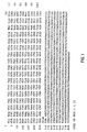
- Figure 1 shows the nucleotide sequence (SEQ ID NO:1) of this partial cDNA clone of VEGF-B and the amino acid sequence (SEQ ID NO:2) encoded in the first reading frame thereof.
- the DNA sequence of Figure 1 was obtained by conventional sequencing of a clone (pcif-2) in the yeast expression vector pPC67.
- the clone comprised 886 base pairs and encoded a part of murine VEGF-B.
- the isolated cDNA sequence will hybridize with the mammalian genomic DNA, e.g. either murine or human, which contains the VEGF-B gene.
- the genomic DNA will contain one or more promoter sequence(s) which give and direct expression of VEGF-B in one or more specific tissues.
- the coding sequence of VEGF-B may be linked to muscle-specific promoter elements which in turn are specific to certain types of muscle fibers.
- VEGF-B comes in several splicing variants.
- the 5'end of the cloned cDNA sequence encodes an 102 amino acid long protein with significant homology to the N-terminal domains of VEGF, PlGF and PDGF A and B. In particular, a number of cysteine residues are perfectly conserved within this group of proteins.
- Figure 1 further depicts the deduced amino acid sequence (SEQ ID NO:2) of this first protein.
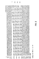
- FIG. 2 again shows the nucleotide sequence (SEQ ID NO:1) of Figure 1, but this time includes the deduced amino acid sequece (SEQ ID NO:3) of the second protein, which is encoded in the second reading frame and is 54 amino acids long. It thus appears that the VEGF-B gene encodes different proteins using alternative splicing of the primary transcript. The last part of the clone, encoding the second peptide might be expressed as a functional protein in other spliced variants of VEGF-B.
- the aforedescribed proteins may exist in combined association with an additional N-terminal sequence of approximately five (5) to ten (10) amino acids, as well as a further leader sequence of approximately twenty-one (21) to twenty-eight (28) amino acids.
- an additional N-terminal sequence of approximately five (5) to ten (10) amino acids as well as a further leader sequence of approximately twenty-one (21) to twenty-eight (28) amino acids.
- such combined amino acid sequences exhibit the property of promoting the proliferation of endothelial cells and the DNA sequences which code for such combined peptide sequences will hybridize under stringent conditions with the DNA sequence of Figures 1 and 2, such amino acid sequences and the DNA which codes for them are expressly contemplated to be within the scope of the present invention.
- Example 2 Cloning of full length cDNA's for mouse VEGF-B.
- an adult mouse heart lambda ZAP-II cDNA library obtained from Stratagene Inc., of La Jolla, California was screened using standard techniques. The library was titrated and plated as recommended and filters were prepared. Following prehybridization at 42 °C in 50 % formamide, 5 x SSPE, 5 x Denharts solution, 1 % SDS and 100 ug of salmon sperm DNA/ml, the filters were hybridized at the same temperature and in the same solution containing the denatured radiolabelled probe using 10 6 cpm/ml of hybridization solution.
- the probe was labelled using a random priming kit (Amersham). After 16 hours the filters were washed in 2 x SSC containing 0.5 % SDS for 2 x 30 mins at 52 °C. The filters were exposed overnight using intensifying screens at -70 °C. Positive clones were rescreened two times until all plaques on a plate were positive. The inserts from several positive clones were subcloned into the plasmid pBluescript SK+ by in vivo excision as recommended by the supplier.
- Nucleotide sequence analyses revealed that two of the cDNA clones were substantially identical, although they differed in length, and one had a mutation.
- One of the clones was full length and contained an open reading frame encoding 188 amino acid residues in which the first 21 amino acids are a clevable signal sequence.
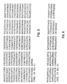
- the other of the two substantially identical clones terminated at the G of the start initiation codon. It could be inferred by sequence analysis of additional clones that the sequence preceeding the G reads ACCAT. Both of the clones were found to have the same coding region nucleotide sequence, which is depicted in Figure 3 (SEQ ID NO:4).
- the figure omits the initial thymine and adenine of the start codon (TAG) which were not present in the isolated clones.
- TAG start codon
- the deduced amino acid sequence of the open reading frame of the coding region of both of these two cDNA clones is shown in Figure 4 (SEQ ID NO:5).
- the resulting protein encoded by this sequence is referred to hereinafter as VEGF-B 167 .
- the subscript number refers to the number of amino acids in the mature protein without the signal sequence.
- the coding part of the third clone had a nucleotide sequence identical to those of the preceding two clones except for a 21 bp insertion.
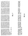
- Figure 5 shows the nucleotide sequence of this third clone (SEQ ID NO:6). To facilitate identification, the 21 extra bases are underlined in the Figure. This insertion gives rise to 7 additional amino acid residues in the mature protein. Thus the resulting protein encoded by this longer cDNA is termed VEGF-B 174 .
- the amino acid sequence of the protein encoded by the cDNA of Figure 5 is depicted in Figure 6 (SEQ ID NO:7). The seven additional amino acids also are underlined in the figure for ease of identification.
- the additional amino acids are inserted into the sequence in a splice site, and sequencing of mouse genomic DNA clones indicates that these additional amino acids are the result of true alternative splicing. Furthermore, based on what is known about the receptor binding site locations of PDGF, the insertion occurs in a position in the protein which is probably part of a receptor binding site. The insertion is thus likely to affect receptor binding and could be of functional importance in influencing antagonist and/or different receptor specificify.
- Figure 9 shows a multiple amino acid sequence alignment for comparison purposes of the 167 amino acid variant of mouse Vascular Endothelial Growth Factor B (mVEGF-B 167 ), mouse Vascular Endothelial Growth Factor (mVEGF 164 ), human Placenta Growth Factor (hPlGF), mouse Platelet Derived Growth Factor A (mPDGF A), and mouse Platelet Derived Growth Factor B (mPDGF B), Amino acid residues identical to mouse VEGF-B 167 are boxed.
- the homologous relationship of the sequences is apparent, and the figure clearly demonstrates the conserved structure of the growth factors belonging to the PDGF/VEGF family of growth factors, and that VEGF-B is a structural homolog of the other growth factors of this group.
- mouse VEGF-B is approximately 43% identical to mouse VEGF 164 , approximately 30% identical to human PlGF, and approximately 20% identical to mouse PDGF A and B.
- the conserved cysteine residues are particularly noteworthy. It can be seen that the first eight cysteine residues in the N-terminal domains (i.e. the PDGF-like domains) of the five growth factors are shared by all members of this family, and it is thus evident that the eight cysteine residues, which are involved in intramolecular and intermolecular disulfide bonding, are invariant among these growth factors. Furthermore, the C-terminal domains of mouse VEGF-B 167 and VEGF 164 also display a significant similarity with eight additional conserved cysteine residues and several stretches of basic amino acids.
- Example 4 Cloning of human VEGF-B cDNA.
- oligonucleotides were designed that would amplify the whole coding region of putative human cDNA corresponding to mouse VEGF-B 167 isoform: These oligonucleotides were used to amplify by polymerase chain reaction (PCR) the whole coding region of human VEGF-B from oligo-dT primed human erythroleukemia cell (HEL) RNA. The amplified product was cloned into the pCR II-vector of TA cloning kit (Invitrogen) and sequenced using standard techniques. The nucleotide sequence of the human VEGF-B cDNA clone is shown in Fig. 10 (SEQ ID NO:10), and the deduced amino acid sequence of human VEGF-B 167 is shown in Fig. 11 (SEQ ID NO:11).
- the full length mouse cDNA clone of Example 2 and the full length human cDNA clone of Example 4 each encode a polypeptide of 188 amino acids containing an N-terminal hydrophobic putative signal sequence.
- the signal peptidase cleavage site is believed to be located between Ala 21 and Pro 22.
- the putative cleavage site of the signal peptidase is indicated in Figure 16 by an arrow. Accordingly, the processed VEGF-B polypeptides of these two clones each contain 167 amino acids.
- Example 2 The clone which exhibited the 340 bp Spe1/BamH1 fragment isolated in Example 2 was analyzed, and the major portion was found to be identical to the first two clones of Example 2 which exhibited the 240 bp Spe1/BamH1 fragment. The difference is due to the presence of an insertion in the C-terminal part of the sequence.
- This 340 bp Spe1/BamH1 DNA fragement encodes a further isoform of mouse VEGF-B containing 207 amino acids.
- the coding portion of the DNA encoding this protein (SEQ ID NO:12) is illustrated in Figure 12, and the translated amino acid sequence (SEQ ID NO:13) is illustrated in Figure 13.
- the mature protein After cleavage of the 21 amino acid leader sequence, the mature protein contains 186 amino acids and is referred to as mVEGF-B 186 .
- This isoform is clearly a result of alternative DNA splicing as described below with reference to Figure 17.
- the H.1 clone isolated as described in Example 4 was found to encode a 207 amino acid isoform of human VEGF-B.
- the coding portion of the DNA (SEQ ID NO:14) encoding this protein is illustrated in Figure 14 and the translated amino acid sequence (SEQ ID NO:15) is illustrated in Figure 15. Again, this isoform, which is designated hVEGF-B 186 , appears to be a product of alternative splicing.
- Both the mVEGF-B 186 of Example 5 and the hVEGF-B 186 of Example 6 include a 101 base pair insertion between nucleotides 414 and 415 of the coding sequence of VEGF-B 167 . Following the insertion, the nucleotide sequences of these cDNA clones were identical to the correponding VEGF-B 167 sequences. The position of the 101 base pair insertion corresponds to the exon 5-exon 6 junction in VEGF. The insertion results in a frameshift which causes the C-terminal domains of the two VEGF-B isoforms to be entirely different.
- the C-terminal domain of VEGF-B 167 is strongly basic (net charge +13) and binds heparin.
- the C-terminal domain of VEGF-B 186 is weakly basic (net charge +5) and has a long stretch of hydrophobic amino acid residues in its C-terminus.
- VEGF-B 186 The hydrophobic tail in VEGF-B 186 is unlikely to behave as a transmembrane domain since this variant of VEGF-B is secreted from cells. Therefore, despite an identical N-terminal domain, these two principal isoforms of VEGF-B have very different biochemical properties.
- the absence of the highly basic heparin-binding domain from VEGF-B 186 allows the protein to be freely secreted from cells.
- the secretion of VEGF-B 186 is remarkably slow; in a pulse chase experiment using transfected cells, VEGF-B 186 homodimers were not found in the medium before 1 hour. In contrast, VEGF homodimers and VEGF-B 186 ⁇ VEGF dimers appear in the medium within 30 minutes.
- Figure 16 shows the aligned amino acid sequences of mouse and human VEGF-B 167 and VEGF-B 186 (SEQ ID NOS:5, 11, 13 & 15) in one-letter code. Identical residues are enclosed in boxes, while amino acid residues which differ between mouse and human VEGF-B 167 and VEGF-B 186 isoforms are outside the boxes.
- Mouse and human VEGF-B display approximately 88 % amino acid sequence identity and are highly basic, especially in their C-terminal regions.
- the C-terminal domains of murine and human VEGF-B 186 are approximately 85% identical at the amino acid level.
- the C-terminal domains of murine and human VEGF-B 167 are approximately 84% identical at the amino acid level.
- Both polypeptides lack the consensus sequence for N-linked glycosylation (N-X-T/S).
- the arrow indicates the putative cleavage site for the signal peptidase between Ala 21 and Pro 22 .
- the mouse and human VEGF-B 167 amino acid sequences are highly homologous with only 20 replacements out the the 167 residues. The replacements are clustered in the N-terminus, in two regions around amino acids 60 and 145. All cysteine residues in both VEGF-B 167 proteins are invariant, but the eight cysteine residues in the C-terminal end of VEGF-B 167 are not conserved in the VEGF-B 186 isoforms.
- mouse and human sequences in the region between residues 66 and 129 are identical apart from one evolutionarily conserved replacement (Q105R). This is of importance since the receptor binding domains are found within this portion of the protein (compared to PDGF structure). From this it can be concluded that it is likely that mouse and human VEGF-B will exhibit cross-reactive binding on the receptor level and thus display identical or similar biological activities.
- Example 7 Exon-intron structures of mouse and human VEGF-B genes.
- the structure of the human VEGF-B gene was determined by restriction mapping and nucleotide sequence analysis of cloned PCR fragments obtained from PCR reactions employing human genomic DNA as the template, except in the case of the first exon and intron, which were identified from a genomic ⁇ -clone.
- the structure of the mouse gene was determined by restriction mapping and nucleotide sequence analysis of cloned PCR fragments amplified using different combinations of primers. As a template in these PCR amplifications an isolated genomic ⁇ clone containing the entire mouse VEGF-B gene was used.
- VEGF-B Several X clones for the mouse VEGF-B gene were isolated from a 129/Sw ⁇ FIX genomic library as recommended by the supplier (Stratagene, Inc.). The ⁇ 0.9 kb SalI/NotI insert of the pcif2 cDNA for VEGF-B (SEQ ID NO:1) was used as the probe. ⁇ DNA from several positive clones were isolated from plate lysates. One of the positive ⁇ -clones (clone 10) was subcloned as BamH1 fragments into pBluescript SK (Stratagene Inc.).
- Isolated DNA from this same clone was also used as the template in PCR reactions (100 ng of ⁇ DNA/reaction) and the coding parts of the mouse VEGF-B gene were amplified using different combinations of primers.
- the nucleotide sequences of these primers were derived from the cDNA clones encoding murine VEGF-B 167 and murine VEGP-B 186 .
- Taq DNA polymerase 2.5 U/reaction
- the generated PCR fragments were directly cloned into the TA-cloning vector pCR II (Invitrogen Inc.).
- the exon-intron structure of the mouse VEGF-B gene was established by nucleotide sequence analysis of the subcloned Bam H1 genomic fragments and of the cloned PCR products.
- a human genomic ⁇ -clone was isolated by screening 1 x 10 6 clones of a human genomic library in EMBL-3 SP6/T7 (Clontech) using high stringency conditions with a 90 bp PCR-fragment spanning 5' sequences of human VEGF-B cDNA as the probe.
- the washing conditions were: one wash at 1 x SSC at room temperature for 30 minutes and two washes at 1 x SSC at 65°C for 30 minutes.
- Primers for the PCR were: The positive ⁇ -clone was subcloned as SacI fragments into pGEM 3Z vector (Promega) and was found to carry the 5'-region of the gene.
- the remaining parts of the human VEGF-B gene were amplified by PCR using genomic DNA as the template. Different combinations of primers derived from the human cDNA sequence were used. Dynazyme DNA polymerase (2.5 U/reaction, Finnzymes) was used. The amplified PCR fragments were directly cloned into the TA-cloning vector pCR II (Invitrogen Inc.). The exon-intron boundaries and the length of the short introns of the mouse and human VEGF-B genes were determined by nucleotide sequence analysis using vector specific primers or suitable primers derived from the cDNA sequences. The length of the larger introns were calculated based on the length of the amplified PCR fragments when analyzed by agarose gel electrophoresis.
- FIG. 17 is a schematic representation of the structures of the mouse and human genes for VEGF-B.
- the exon sizes in base pairs are noted inside the boxes, and the sizes of the introns are noted between the boxes.
- the introns are not shown to scale.
- the structures of the untranslated flanking regions of mouse and human VEGF-B genes were not established and are represented by gray boxes.
- the exon-intron junctions in both genes are listed in the following Table 3:
- exon 6 contains an alternative splice acceptor site which enables the gene to produce two different transcripts for VEGF-B isoforms.
- VEGF-B 167 uses exons 1-5, the last part of exon 6, and exon 7 (TGA).
- VEGF-B 186 uses exons 1 through 5, the first part of exon 6, and terminates in the last part of exon 6 (TAG).
- Exon 7 is not translated in VEGF-B 186 since the insertion of the first part of exon 6 introduces a frame shift and gives rise to a stop codon in the last part of exon 6.
- the position of the stop codon (TAG) for VEGF-B 186 is marked in exon 6B, and the stop codon (TGA) for VEGF-B 167 is marked in exon 7.
- the introns in both genes vary from 161 bp to approximately 2.6 kb.
- the length of each exon and the locations of the splice junctions in the two genes were identical, and all splice donor and acceptor sites follow the canonical GT/AG rules, Padgett et al., Annual Rev . of Biochemistry, 55:1119-50 (1986).
- the only notable difference between the mouse and the human genes are the length of introns 1, 4 and 6 which are longer in the mouse gene. All exon-intron boundaries were found to be conserved between VEGF-B and VEGF, but the introns in the VEGF-B genes were generally smaller than in the VEGF gene.
- the 300 bp-intron after the exon 5 in VEGF-B differs from the corresponding one in VEGF, which is 3 kb in length and contains an alternatively spliced exon found in the transcripts for VEGF 189 and VEGF 206 , encoding many basic amino acid residues.
- this intron in VEGF-B was analyzed more carefully, no exon corresponding to the 6th exon of VEGF could be found. Instead, the 3' end of this intron and the following exon were found to be identical with the corresponding sequences of the cDNA clones encoding VEGF-B 186 .
- the mRNA for VEGF-B 186 is formed by use of an alternative splice acceptor site during mRNA splicing, resulting in an insertion of a 101 bp intron sequence into these mRNAs.
- Figure 18 shows a comparative hydrophilicity analysis of murine VEGF-B 167 and VEGF-B 186 .
- the profiles were generated according to Kyle and Dolittle using a window of nine (9) residues.
- the pattern of hydrophilicity/hydrophobicity is essentially identical from amino acid 1 through amino acid 115. After amino acid 115, the hydrophilicity/hydrophobicity patterns diverge because of the frame shift introduced by the first part of exon 6.
- VEGF-B 167 and VEGF-B 186 can be expected to exhibit both similar and dissimilar activities.
- Figure 19 is a dendrogram showing the phylogenetic relationship of the amino acid sequences of five members of the VEGF/PDGF family of growth factors. The number of replacements or substitutions decreases from the left to the right of the chart. It can be seen that VEGF-B lies between VEGF and the platelet derived growth factor (PDGF) group.
- PDGF platelet derived growth factor
- Antisera to mouse VEGF-B were raised by immunizing rabbits with a 18-mer oligopeptide comprising the N-terminal region of processed VEGF-B, coupled to keyhole limpet hemocyanin. Cysteine residues were introduced as the N-terminal and C-terminal amino acid residues to allow coupling of the peptide to the carrier protein using SPDP (Pharmacia).
- SPDP Pharmacia
- the sequence of the oligopeptide was Each rabbit received a subcutaneous injection with 300 ⁇ g of the peptide conjugate emulsified in Complete Freunds Adjuvant. Subcutaneous booster injections were given every second week with the same amount of antigen emulsified in Incomplete Freunds Adjuvant. Sera were obtained after the second booster injections.
- Antipeptide antiserum to human VEGF-B was generated by immunizing rabbits with a branched 23-mer oligopeptide comprising the following N-terminal region amino acid residue sequence (SEQ ID NO:22):
- the branched 23-mer oligo peptide was synthesized according to Tam, "Synthetic peptide vaccine design: synthesis and properties of a high-density multiple antigenic peptide system", Proc. Natl. Acad. Sci . USA , Vol. 85, pages 5409-413 (1988).
- rabbits were subcutaneously injected with 500 ⁇ g of the branched peptide emulsified in Complete Freunds Adjuvant.
- 200 ⁇ g of the antigen emulsified in Incomplete Freunds Adjuvant was injected.
- Antisera were collected after the second and third boosters by conventional techniques.
- Example 9 Biochemical properties of VEGF-B 167 , homodimerization, and heterodimerization with VEGF.
- VEGF-B 167 The biochemical properties of human VEGF-B 167 were examined in transfected human embryonic kidney 293EBNA cells (Invitrogen, Inc.). cDNA inserts encoding human VEGF-B 167 and human VEGF 165 [see Keck et al., Science , Vol. 246, pages 1309-312 (1989)] were individually cloned into the pREP7 expression vector (Invitrogen, Inc.). Human embryo kidney 293EBNA cells (expressing Epstein-Barr virus nuclear antigen-1) were transfected by transient transfection with the respective expression plasmids using calcium phosphate precipitation, and the cells were incubated for 48 hours.
- cells also were transfected with an expression vector containing the VEGF-B 167 cDNA in reverse orientation. Monolayers of cells were incubated in methionine-free and cysteine-free medium for 30 minutes followed by labeling with 100 ⁇ Ci/ml [ 35 S]methionine and [ 35 S]cysteine (Promix, Amersham Inc.) in the same medium for 2 hours. The labeling medium was replaced with normal medium without serum, and labelled proteins were chased for 6 hours. Heparin was included during the chase when indicated (100 ⁇ g/ml).
- VEGF-B 167 was expressed in the cells transfected with the plasmids containing the VEGF-B 167 DNA. Aliquots of the culture supernatants from cells treated or untreated with heparin and detergent solubilized cell lysates were subjected to immunoprecipitation with the specific antipeptide antiserum to VEGF-B obtained as described in Example 8 and analyzed by SDS-PAGE under reducing conditions unless otherwise indicated. The data show that VEGF-B 167 homodimers and VEGF-B 167 - VEGF 165 heterodimers are released from cells by heparin.
- VEGF-B 167 was released from cells and found in the supernatant. If cells were not treated with heparin, VEGF-B 167 remained cell-associated and was not released into the culture medium. Under the same conditions, VEGF 165 homodimers are secreted from the cells and found in the culture supernatants without heparin treatment.
- VEGF-B 167 Under reducing conditions, human VEGF-B 167 migrated with a Mr of 21 kDa. Analysis of culture supernatants under non-reducing conditions showed that VEGF-B 167 migrated as an Mr 42 kDa species indicating a dimeric structure. These results suggest that VEGF-B 167 forms disulfide-linked dimers associated with the cell surface, probably through ionic interactions with extracellular heparan sulfate proteoglycans. The association is likely to be mediated by the C-terminal basic domain, as observed for the longer splice variants of VEGF.
- VEGF 165 could also form heterodimers with VEGF-B 167 .
- 293EBNA cells were co-transfected with expression vectors encoding both human VEGF 165 and human VEGF-B 167 , and VEGF-B 167 was expressed in combination with VEGF 165 .
- Metabolically labelled proteins were chased in the presence of heparin, and immunoprecipitations were carried out with antisera to either VEGF-B 167 or VEGF 165 .
- the antiserum to human VEGF was from R&D Systems.
- VEGF-B 167 ⁇ VEGF 165 heterodimers migrated as Mr 42-46 kDa species.
- the results show that VEGF-B can form disulfide linked heterodimers with VEGF, which, in the absence of heparin, remain cell-associated. Since homodimers of VEGF 165 are efficiently secreted into the media, VEGF-B appears to determine the secretion of the heterodimer.
- VEGF-B is synthesized normally in the endoplasmic reticulum of the source cell for subsequent export.
- Recombinant VEGF-B may be produced by inserting a DNA sequence encoding the VEGF-B protein together with suitable operatively linked promoter and control sequences into a suitable vector, such as the well known plasmid pBR322 or a derivative thereof, transforming or transfecting a suitable host cell, such as E . coli or a Cos cell, with the resulting vector or other systems well known in the art, screening the resulting transformants or transfectants for VEGF-B expression, and then culturing cell lines or bacterial cell strains which are positive for the expression of VEGF-B.
- a suitable vector such as the well known plasmid pBR322 or a derivative thereof
- suitable host cell such as E . coli or a Cos cell
- Either a eukaryotic vector or a prokaryotic vector may be used, depending on the type of cell which is to be transfected or transformed therewith.
- a particularly preferred system for production of recombinant VEGF-B is the baculovirus - insect cell system, which has proved capable of producing excellent yields of recombinant protein.
- Example 10 VEGF-B expression using the baculovirus system.
- the complete human VEGF-B 167 gene was inserted into a commercially available plasmid pCRII (Invitrogen Corp.).
- the HindIII-XbaI fragment from the resulting plasmid pCRII-VEGF-B 167 which encodes the whole open reading frame of VEGF-B 167 then was cloned into pFASTBAC1, and both the 3' - and 5'-junctions were sequenced.
- Bacmid-DNA was prepared according to the manufacturers instructions for the "Bac-To-BacTM Baculovirus Expression System" (Life Technologies Inc.) and lipofected to Sf900II-adapted Sf9 cells (obtained from Dr. Christian Oker-Blom). Sf9 cells are from the American Type Culture Collection Cell Repository Line Bank, Rockville MD (ATCC CRL-1711). The transfected cells were then cultured on standard TMN-PH medium in 25 cm 2 culture dishes.
- VEGF-B vascular endothelial growth factor-B
- Example 9 Western blotting. Lysates from three out of four independently transfected cell cultures were found positive for VEGF-B, although the intensity of the signal in the Western blot varied.
- the expressed VEGF-B polypeptide in each case was found to correspond in size to the protein expressed in mammalian cells in Example 9.
- the viral stock from the cells that gave the strongest signal in Western blotting was amplified two rounds by infecting cells and collecting new virus from the medium. The resulting supernatant was analyzed. Uninfected cells were also analyzed as a negative control. Time course analysis showed that cells harvested between 48 and 72 hours after infection contained the greatest amount of VEGF-B. After 96 hours post infection, as a result of virus-induced cell lysis, VEGF-B could also be detected in the culture supernatant by immunoprecipitation and western blotting. Recombinant VEGF-B could be precipitated from the lysate between 20% and 40% (NH 4 ) 2 SO 4 .
- a polymerase chain reaction (PCR) fragment from nucleotide position 68 to 141 was used to introduce a BamHI restriction site immediately after the signal cleavage site in the plasmid pCRII-VEGF-B 167 from Example 10.1.
- the BamHI fragment from this modified pCRII-VEGF-B 167 construct was cloned into BamHI opened pVTBac [Tessier et al., "Enhanced secretion from insect cells of a foreign protein fused to the honeybee mellitin signal peptide", Gene, Vol. 98, page 177 (1991)]. Both 3'- and 5'-junctions were sequenced.
- Sf9 cells were cotransfected with the aforedescribed pVTBac vector which contained the human VGEF-B 167 gene, and with linearized baculovirus DNA (InsectinTM, Invitrogen Corp.). The transfected cells then were cultured in TMN-FH medium.
- a cDNA insert encoding murine VEGF-B 186 (EcoR1 cut cDNA fragment from a murine VEGF-B 186 cDNA (SEQ ID NO:12) clone) was cloned into pFASTBAC 1.
- An EcoR1 cut cDNA fragment from murine VEGF-B 167 (SEQ ID NO:4) was also cloned into pFASTBAC 1.
- the resulting plasmids were transformed into bacteria as described in 10.1 above, and recombined plasmids were isolated and lipofected into Sf9 and Sf21 cells.
- Supernatants containing recombinant baculovirus were amplified by several rounds of reinfection of Sf21 cells.
- the final titers of the baculovirus stocks were determined by plaque titration and found to vary between 4 x 10 8 and 2 x 10 9 baculovirus particles per milliliter of stock supernatant.
- Example 11 Large Scale Production of Recombinant VEGF-B.
- Sf21 cells [see Vaughn et al., In Vitro, 13:213-17 (1977)] were infected with the baculovirus stocks of Example 10 at a multiplicity of infection of 10 virus particles per cell.
- the infected Sf21 cells were grown in roller flasks and seeded at a density of 2 x 10 6 cells per ml of serum free medium (Sf900II, Gibco-BRL) for 96 hours. Culture media and cells were then harvested. Aliquots of the cell lysates and of the media were analyzed by SDS-PAGE.
- VEGF-B 167 polypeptides were of the expected sizes of 21.5 kDa. Both proteins were retained intracellularly in the infected cells and not released into the medium.
- mouse VEGF-B 186 was readily secreted into the medium in a dimeric form. The VEGF-B 186 homodimers migrated as a 52-54 kDa species which suggested that insect cell produced protein did not undergo the same covalent modification as found for VEGF-B 186 secreted from transfected Cos-1 cells.
- Example 12 Transfection and analysis of Cos-1 cells expressing VEGF-B 186 .
- cDNA inserts encoding mouse VEGF-B 186 and human VEGF 165 were cloned into the pSG5 expression vector [Green et al., Nucleic Acid Res ., 16:369 (1988)].
- Cos-1 cells were maintained in minimal essential medium (MEM) containing 10% fetal calf serum, 2mM glutamine and appropriate antibiotics. For transfections, the cells were replated into 90 mm Petri dishes. The cells were transfected with the expression vectors, separately or in combination, using calcium phosphate precipitation and incubated for 36-48 hours.
- MEM minimal essential medium
- Monolayers of cells were incubated in medium free of methionine and cysteine for 30 min and then incubated in the same medium containing 100 ⁇ Ci/ml of [ 35 S]-methionine and [ 35 S]-cysteine for 2 hours (Promix Amersham Inc.).
- the cells were labeled for 30 minutes, washed twice with normal medium and then incubated for up to 6 hours in MEM without fetal calf serum. Media were collected after the chase period and the cells were solubilized in 10 mM Tris buffer pH 7.5 containing 50 mM NaCl, 0.5% deoxycholate, 0.5% nonidet P-40 and 0.1% SDS. Aliquots of the media and the cell lysates were subjected to immunoprecipitation using the specific antiserum to mouse VEGF-B from Example 8a and a specific antiserum to human VEGF commercially available from R&D Systems. The precipitates were analyzed by SDS-PAGE.
- Example 13 Biochemical properties of VEGF-B 186 expressed in transfected Cos-1 cells.
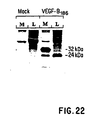
- VEGF-B 186 The biochemical properties of mouse VEGF-B 186 were examined in Cos-1 cells transiently transfected as described in Example 12 with an appropriate expression vector. The cells were metabolically labelled, and proteins from the labelled cells were immunoprecipitated using an antipeptide antibody to VEGF-B. The precipitated material was subjected to SDS-PAGE analysis under reducing conditions. Both the cell culture medium (M) and a detergent solubilized cell lysate (L) were analyzed. The results are shown in Figure 22. It can be seen that cell associated VEGF-B 186 migrated as an approximately M r 24,000 polypeptide under reducing conditions.
- VEGF-B 186 present in the medium of transfected cells migrated as a M r 32,000 species, suggesting that the protein was covalently modified during its intracellular transport and secretion.
- the corresponding molecules were not detected in cell lysates or media from mock transfected Cos-1 cells used as a control.
- Example 14 Biosynthesis of VEGF-B 186 homodimers.
- VEGF-B 186 The biosynthesis of VEGF-B 186 homodimers was examined by pulse-chase experiments. Transfected Cos-1 cells were metabolically labelled for 30 minutes and then chased for up to 4 hours. Immunoprecipitation and SDS-PAGE analysis of detergent solubilized cell lysates and media showed that the cell-associated approximately M r 24,000 species was readily detected in the lysates throughout the chase period. The decrease in the intensity of this molecular species was associated with an increase in the M r 32,000 protein present in the media. The M r 32,000 species appeared in the medium after 1 hour of chase. Highest levels of secreted VEGF-B 186 were obtained after the 4 hour chase period. No intermediates were detected in the cell lysates, but the secreted M r 32,000 protein appeared slightly heterogenous. The nature of the modification is presently unknown, but N-linked glycosylation can be excluded in the absence of consensus sites for this modification.
- Example 15 Formation of heterodimers by VEGF-B 186 .
- VEGF-B and VEGF are coexpressed in many tissues and VEGF-B 167 ⁇ VEGF 165 heterodimers are readily formed when coexpressed in transfected cells.
- VEGF-B 186 also could form heterodimers with VEGF 165 .
- Cos-1 cells were transfected as described above with the appropriate expression vectors, either alone or in combination. Metabolically labelled proteins present in the media from the transfected cells were subjected to immunoprecipitations using antisera to VEGF-B and VEGF.
- Figure 23A shows the results of SDS-PAGE analysis under reducing conditions of the immunoprecipitates from the cell culture media of transiently transfected Cos-1 cells separately expressing VEGF-B 186 and VEGF, respectively. It can be seen that the antisera were specific for VEGF-B and VEGF, respectively, with no detectable cross-reactivity.
- Cos-1 cells were cotransfected with expression vectors for VEGF-B 186 and VEGF 165 .
- Cell culture media (M) and detergent solubilized lysates (L) from the resulting cells which coexpressed VEGF-B 186 and VEGF 165 were subjected to immunoprecipitation and SDS-PAGE analysis under reducing conditions. The results are shown in Figure 23B. The test showed that murine VEGF-B 186 and human VEGF 165 form intracellular and secreted heterodimers.
- VEGF-B 186 ⁇ VEGF heterodimers remained cell-associated throughout the chase. These results suggested that heterodimer formation with VEGF promoted the secretion of VEGF-B 186 compared to the secretion of VEGF-B 186 homodimers. Furthermore, the presence of heterodimers already following the 30 minute labelling period suggested that the slow release of VEGF-B 186 homodimers was not due an impaired ability of VEGF-B 186 to dimerize.
- Example 16 Purification of Secreted VEGF-B 186 Homodimers.
- VEGF-B 186 homodimers were isolated from serum free culture media of baculovirus infected Sf21 cells as follows:
- the major contaminating protein in the culture media was the baculovirus protein gp64/67, an acidic protein secreted by baculovirus infected cells.
- the culture media was concentrated twenty-fold by ultrafiltration and then passed over a Sephadex G-25 column equilibrated in 20 mM phosphate buffer pH 6.5 containing 20 mM NaCL. Eluted proteins were then passed over a CM-Sepharose (Pharmacia) ion-exchange column equilibrated in the same buffer. The column was washed with the phosphate buffer to remove unbound proteins, and bound proteins were eluted by stepwise increasing the NaCl concentration of the elution buffer.
- VEGF-B 186 homodimers were 5-15% pure by this procedure.
- the VEGF-B 186 homodimers are purified to homogeneity on a MonoS column coupled to a FLPC system (Pharmacia). Bound protein is eluted with a linear gradient of NaCl in 20 mM phosphate buffer pH 6.5.
- RNA extracted from mouse brain, heart, liver and kidney and from human embryonic heart and skeletal muscle was analyzed by PCR using four pairs of specific primers covering exons 4 to 7 and exons 3 to 7 in the mouse and human VEGF-B genes, respectively.
- RNA from mouse and human tissues were isolated using standard procedures as disclosed by Chirgwin et al., Biochemistry, 18:5294-99 (1979). Two to five ⁇ g of total RNA per reaction were used for first strand cDNA synthesis using avian myelostosis virus reverse transcriptase (20 U/reaction). The reactions were primed with oligo-(dT) 18 . Aliquots of these reactions were used as templates in PCR reactions using Taq DNA polymerase (2.5 U/reaction). To amplify mouse cDNA, two pairs of primers were used.
- the oligonucleotide probes were 5'-CTCTGTTCCGGGCTGGGACTCTA (exon 6A) (SEQ ID NO:26) and 5'-TCAGGGCGTTGACGGCGCTGGGTGCAA (exon 6B) (SEQ ID NO:27)
- the oligonucleotide probes were labeled with [ 32 P]dCTP using terminal transferase to high specific activity.
- Hybridizations using 1 x 10 6 cpm of labeled probe/ml of solution, were carried out at 37° C in 6 x SSC containing 5 X Denhardt's solution, 0.5% SDS and 100 ⁇ g/ml of salmon sperm DNA. The filters were washed at the same temperature in 6 x SSC containing 0.5% SDS for 2 x 15 min and exposed to film.
- the two pairs of primers used for amplification of human cDNA were combined using two different 5'-primers, located in exon 3 and located in exon 4, with a common 3'-primer, located in exon 7. Aliquots of the amplified products were analyzed by agarose gel electrophoresis. The aliquots were directly cloned in the TA-cloning vector pCR II (Invitrogen, Inc.), and generated plasmids were analyzed by nucleotide sequencing. Amplification of GAPDH served as a control.
- the PCR-amplified DNA was transferred to a filter and probed with specific oligonucleotide probes for exons 6A and 6B, respectively.
- the autoradiograms showed that an exon 6-specific probe hybridized with the 316 bp band while the exon 6B specific probe hybridized with both the 215 bp and the 316 bp amplified bands.
- VEGF-B 167 and VEGF-B 186 are the two major isoforms of VEGF-B in tissues.
- the pattern of the PCR products and the location of the primers indicate that if any still longer splice isoforms exist for VEGF-B, such transcripts use a splice acceptor site located a little more 5' than in the case of VEGF-B 186 .
- PCR products corresponding to VEGF 121 which lacks heparin binding domains, i.e. sequences corresponding to exon 6 in VEGF-B, were not detected.
- splicing of exon 5 to exon 7 would give rise to a transcript encoding an isoform of VEGF-B corresponding to VEGF 121 , and this putative isoform of VEGF-B might be expressed in tissues other than those analyzed in this example.
- Example 18 Stimulation of Cell Proliferation.
- VEGF-B 167 The ability of VEGF-B 167 to stimulate endothelial cell proliferation was established through analysis of [ 3 H]thymidine incorporation in human umbilical vein endothelial cells (HUVEC) and in bovine capillary endothelial (BCE) cells.
- 293EBNA cells were transfected as described above with expression vectors for VEGF-B 167 , VEGF 165 or empty vector (mock) in the presence of 1 ⁇ g/ml heparin.
- Conditioned media from these cells were diluted in respective media, applied to human umbilical vein endothelial cells (HUVEC) and to bovine capillary endothelial (BCE) cells and incorporation of [ 3 H]thymidine was measured.
- HUVEC human umbilical vein endothelial cells
- BCE bovine capillary endothelial
- conditioned media containing human VEGF-B and human VEGF 165 were collected from 293EBNA cells transfected with the appropriate expression vectors or with empty vector (mock) in the presence of heparin (1 ⁇ g/ml) 48 hours posttransfection.
- Second passage HUVEC were plated into 96-well plates (4 x 10 3 cells/well) in M-199 medium supplemented with 10% fetal bovine serum and incubated for 24 hours.
- Conditioned media were diluted with the growth medium, and cells were stimulated for 48 hours.
- Fresh conditioned media containing 10 ⁇ Ci/ml of [ 3 H]thymidine (Amersham Inc.) were added to the cells, and stimulations were continued for another 48 hours.
- BCE cells were washed with PBS and trypsinized, and incorporated radioactivity was determined by liquid scintillation counting.
- BCE cells were seeded into 24-well plates and grown until confluence in minimal essential medium (MEM) supplemented with 10% fetal calf serum. Cells were starved in MEM supplemented with 3% fetal calf serum for 72 hours, after which conditioned media diluted into serum-free medium were added to the cells and the cells were stimulated for 24 hours. [ 3 H]Thymidine was included during the last 4 hours of the stimulation (1 ⁇ Ci/ml). Stimulations with bFGF were carried out as above using 6 ng/ml of recombinant bFGF (Synergen Inc.). Cells were washed with PBS, lysed with NaOH, and incorporated radioactivity was determined by liquid scintillation counting.
- Figure 20 is a bar graph showing fold of induction of [ 3 H]thymidine incorporation by VEGF-B 167 in human umbilical vein endothelial cells (HUVEC) and in bovine capillary endothelial (BCE) cells, as compared to basal activity induced by conditioned medium from the mock transfected cells. For comparison purposes, the induction by VEGF 165 and by bFGF are also shown. The bars show the mean ⁇ standard deviation of parallel samples. Similar results were obtained in several other independent experiments.
- test results clearly show that VEGF-B induced [ 3 H]thymidine incorporation in both HUVEC and BCE cells and stimulated proliferation of endothelial cells in vitro, thereby demonstrating that VEGF-B is an endothelial growth factor.
- Example 19 Identification of human VEGF-B promoter DNA clones and activity.
- a human genomic DNA library in bacteriophage ⁇ EMBL was screened using a 5' PCR fragment containing the sequences from the VEGF-B first and second exons as a probe. Two positive clones were obtained, and one of these was subcloned in the Bluescript SKII plasmid as Sac I fragments. A 1.4 kb fragment was obtained, which contained about 0.4 kb of sequences upstream from an Nco I site present in the cDNA (located less than 100 bp upstream of the ATG translational initiation site).
- an XhoI fragment of about 6 kb from the other ⁇ clone was subcloned into the pGEMEX plasmid. This subclone contained about 1.5 kb of sequences upstream from the NcoI site.
- the SacI/NcoI fragment and an EcoRI (polylinker) - NcoI fragment were subcloned into pGL3 basic vector (Promega) in the respective transcriptional orientation. DNA of these subclones, and from the pGL3 control vector containing the SV40 promoter, was transfected into HeLa cells using calcium phosphate precipitation. Two days after transfection, the luciferase activities were measured from lysates of the transfected cells.
- the 400 bp SacI/NcoI fragment has promoter activity equal to about 30% of the activity of the pGL3 control vector, while the 1.5 kb fragment gave only background activity.
- the structure of the cloned fragments is illustrated in Figure 24.
- the 1.5 kb fragment upstream of the Nco I site was sequenced.
- the resulting sequence (SEQ ID NO:17) is illustrated in Figure 25.
- the sequence obtained revealed a putative silencer element [Weissman and Singer, Molecular and Cellular Biology , 11:4228-234 (1991)] composed of two eight-base pair stretches between nucleotides 166-187 (boxed in the drawing). This silencer may be responsible for the relative lack of activity of the 1.5 kb fragment.
- Example 20 Analysis of VEGF-B mRNA in melanomas, normal skin and muscle by RT-PCR.
- Normal skin and melanoma tissues were obtained from patients attending the Department of Radiotherapy and Oncology, Helsinki University Central Hospital.
- Four metastatic melanoma specimens were obtained freshly after surgical excision, immediately embedded in Tissue-tek (Miles) and frozen in liquid nitrogen.
- Samples of normal skin were obtained from volunteer patients undergoing surgery for mammary carcinoma and excision of a cutaneous naevus. All specimens were inspected by a pathologist to confirm the diagnosis.
- the primer sequences were selected from the VEGF-B and ⁇ -actin genes as follows: [ ⁇ -actin sequences comprise nucleotides 2105-2125 and 2411-2432 from Ng et al., Mol. Cell Biol. 5:2720-732 (1985)].
- An aliquot of 4 ⁇ l from the cDNA reaction product was heated to 94°C for 5 minutes and used as a template for PCR amplification with 20 pmol of primers, 10x PCR buffer, 1 ⁇ l of 20 mM dNTPs and 2.5 U of Taq polymerase. Final volume was adjusted to 100 ⁇ l with DEPC treated water.
- Denaturation was at 95°C for 1 minute, annealing at 62°C for 45 seconds, and polymerization at 72°C for 50 seconds, for a total of 35 cycles for VEGF-B and 25 cycles for ⁇ -actin. After every 5 cycles, 15 ⁇ l aliquots were taken for analysis.
- Electrophoresis of 5 ⁇ l of the PCR reaction mix was performed in a 2% agarose gel containing ethidium bromide.
- the size marker DNA fragments ranged in length from 24 to 726 base pairs ( ⁇ X174 DNA/Hinf I marker from Promega, Madison, WI, USA).
- the tested samples thus included four metastatic melanomas, muscle, normal skin, a negative control (without reverse transcriptase), and the ⁇ X174 DNA/Hinf I size marker.
- the results of the RT-PCR analysis for VEGF-B (PCR product lengths 323 and 234 bp) and for ⁇ -actin show that VEGF-B is highly expressed in all melanomas studied, at levels approximately similar to the expression in muscle tissue. On the other hand, normal skin has very little of the VEGF-B RNA. Similar conclusions can be drawn from Northern blotting and hybridization analysis.
- VEGF-B is a novel growth factor for endothelial cells which plays a role in vascularization, in particular of muscle.
- Collateral artery growth in ischemic heart or limb may be promoted by arterial administration of a VEGF-B bolus using techniques described by Takeshita et al., Am. J. Pathol ., 147:1649-60 (1995).
- the cell-association of VEGF-B may have several implications for regulation of vascularization and endothelial cell growth. In developing embryos and in contractile tissues, cell-associated VEGF-B may provide spatial cues to outgrowing endothelial cells during establishment and maintenance of the vascular tree.
- VEGF-B Reendothelialization of arterial injury may be promoted by direct application of VEGF-B using techniques described by Asahara et al., Circulation , 91(11):2793-802 (1995).
- the ability of VEGF-B to modulate the secretion of VEGF by heterodimer formation suggests an indirect role of VEGF-B in VEGF signalling, thereby regulating receptor binding and/or activation as described by Potgens et al., J. Biol . Chem ., 269(52):32879-85 (1994).
- the formation of multiple heterodimeric complexes of these growth factors could provide a basis for a diverse array of regulatory signals for endothelial cells.
- VEGF-B can be used as a growth factor for populations of endothelial cells in vitro.
- VEGF-B may be used to promote desirable angiogenesis, i.e. the formation of new blood vessels and capillaries; see Takeshita et al., supra.
- it may be useful in promoting the development of the corpus luteum and endometrium as an aid to initiating and/or maintaining pregnancy. It would also be useful in bone repair by virtue of its action on endothelial cells.
- Administration of VEGF-B may also be useful in supporting embryogenesis, as well as somatic growth and vascular development and differentiation.
- Topical application of VEGF-B to wounds may be useful in promoting wound healing, and oral administration of VEGF-B may be useful to accelerate the healing of gastric and/or duodenal ulcers.
- the ability of VEGF-B to modulate the secretion of VEGF by heterodimer formation could provide a therapeutic role for VEGF-B in diseases where VEGF agonists would be useful; see Potgens et al., supra.
- VEGF-B may exert proliferative effects on mesodermal cells either directly or via improvements in the blood supply.
- VEGF-B has been found to be overexpressed in tumors, such as melanomas. Consequently, assays for VEGF-B expression can be used as tools in tumor diagnosis, and suppression of VEGF-B expression, for example with monoclonal antibodies, may be useful to retard tumor growth.
- Tumor assays for VEGF-B may be useful as indicators of metastatic risk.
- use of VEGF-B antibodies analogous to the procedures described by Takahashi et al., Cancer Res ., 55:3964-68 (1995) in order to quantitate neovascularization and proliferation could be used as an indicator of metastatic risk from colon cancer.
- Assays of VEGF-B in body fluids or the tumor itself by histochemistry may be useful as a tumor prognostic factor.
- An ELISA analogous to the procedure described by Kondo et al., Biochemica et Biophysica Acta , 1221(2) :211-14 (1994) may be useful to detect VEGF-B upregulation as a tumor screen.
- An enzyme linked immunoabsorbent assay of VEGF-B expression using techniques described by Boocock et al., J. Natl . Cancer Inst ., 87:506-16 (1995) may be useful as a diagnostic index of ovarian cancer.
- An assay of VEGF-B expression similar to the VEGF assay described by Weindel et al., Neurosurgery, 35:439-48 (1994) may be useful as an indicator of malignancy in brain tumors.
- VEGF-B may also be useful in promoting tumor growth in laboratory animals in order to test anti-tumorigenic drugs.
- VEGF-B may also be useful to increase the microvascularity of hypoxic areas of tumors and make them more sensitive to radiation, radiation sensitizing drugs, etc.
- VEGF-B The angiogenic action of VEGF-B may be useful in treating ischemic conditions.
- Administration of an intraarterial bolus of VEGF-B by the techniques described in Bauters et al., American Journal of Physiology, 267(4 Pt 2):H1263-71 (1994) may be useful to treat lower extremity ischemia and increase perfusion in the extremities.
- an angiogenic response may be produced in tissue injected with fibroblast cells transduced with a virus which expresses VGEF-B in order to treat tissue ischemia (e.g. myocardial ischemia).
- VEGF-B or agonists could be used to stimulate the development of collateral circulation in cases of arterial and/or venous obstruction, e.g. myocardial infarcts, ischaemic limbs, deep venous thrombisis, and/or postpartum vascular problems; see Takeshita et al, supra.
- venous obstruction e.g. myocardial infarcts, ischaemic limbs, deep venous thrombisis, and/or postpartum vascular problems; see Takeshita et al, supra.
- a VEGF-B/VEGF-B receptor system may be used as an assay system to detect small molecules as agonists/antagonists for development as new drugs.
- small molecules which could be detected include, but are not limited to, organic chemicals, peptides, and RNA molecules.
- compositions may be produced by admixing a pharmaceutically effective amount of VEGF-B protein with one or more suitable carriers or adjuvants such as water, mineral oil, polyethylene glycol, starch, talcum, lactose, thickeners, stabilizers, suspending agents, etc.
- suitable carriers or adjuvants such as water, mineral oil, polyethylene glycol, starch, talcum, lactose, thickeners, stabilizers, suspending agents, etc.
- Such compositions may be in the form of solutions, suspensions, tablets, capsules, creams, salves, ointments, or other conventional forms.
- VEGF-B protein also can be used to produce antibodies.
- conventional antibody production techniques may be used to produce VEGF-B antibodies.
- specific monoclonal antibodies may be produced via immunization of fusion proteins obtained by recombinant DNA expression.
- Labelled monoclonal antibodies should be useful in screening for conditions associated with abnormal levels of VEGF-B in the body.
- an assay of VEGF-B in synovial fluids and/or joint tissue by immunofluorometric techniques analogous to the the procedure described by Fava et al., Journal of Experimental Medicine, 180:341-46 (1994) may be useful as a diagnostic indicator of rheumatoid arthritis.
- a radioimmunoassay of VEGF-B in occular fluid using techniques described by Aiello et al., in New England Journal of Medicine, 331(22):1480-87 (1994) may be useful as a diagnostic indicator of diabetic retinopathy, neovascularization of the iris or retinal vein occlusion.
- Immunoassays of VEGF-B levels in blood, urine or other bodily fluids may be useful also as a tumor marker; see Kondo et al., supra.
- These monoclonal antibodies to VEGF-B also may be useful in inhibiting angiogenesis associated with high levels of VEGF-B in the body, e.g.
- Treatment with a monoclonal antibody specific for VEGF-B using techniques analogous to those described by Kim et al., in Nature , 362(6243) :841-44 (1993) may be useful to suppress or inhibit tumor growth in vivo .
- Intravenous and/or subcutaneous injection of monoclonal antibodies to VEGF-B using procedures like those described by Asano et al., in Cancer Research, 55:5296-5301 (1995) may be useful to inhibit neovascularization and primary and metastatic growth of solid tumors.
- chiaserization or humanization of such monoclonal antibodies is to be preferred.
- Treatment may be effected, e.g., by twice weekly intraperitoneal injection of 10 to 500 ⁇ g, preferably 50-100 ⁇ g of monoclonal antibody.
- VEGF-B antagonists such as antibodies also may be useful to inhibit new blood vessels in diabetic retinopathy, psoriasis, arthopathies and/or vascular tumors such as haemangiomas; see Aiello et al., supra.
Landscapes
- Health & Medical Sciences (AREA)
- Chemical & Material Sciences (AREA)
- Life Sciences & Earth Sciences (AREA)
- Organic Chemistry (AREA)
- General Health & Medical Sciences (AREA)
- Medicinal Chemistry (AREA)
- Veterinary Medicine (AREA)
- Chemical Kinetics & Catalysis (AREA)
- Public Health (AREA)
- Animal Behavior & Ethology (AREA)
- Pharmacology & Pharmacy (AREA)
- Nuclear Medicine, Radiotherapy & Molecular Imaging (AREA)
- General Chemical & Material Sciences (AREA)
- Zoology (AREA)
- Genetics & Genomics (AREA)
- Bioinformatics & Cheminformatics (AREA)
- Toxicology (AREA)
- Biochemistry (AREA)
- Biophysics (AREA)
- Proteomics, Peptides & Aminoacids (AREA)
- Molecular Biology (AREA)
- Gastroenterology & Hepatology (AREA)
- Engineering & Computer Science (AREA)
- Heart & Thoracic Surgery (AREA)
- Cardiology (AREA)
- Peptides Or Proteins (AREA)
- Medicines That Contain Protein Lipid Enzymes And Other Medicines (AREA)
- Micro-Organisms Or Cultivation Processes Thereof (AREA)
- Medicines Containing Antibodies Or Antigens For Use As Internal Diagnostic Agents (AREA)
- Preparation Of Compounds By Using Micro-Organisms (AREA)
- Measuring Or Testing Involving Enzymes Or Micro-Organisms (AREA)
- Medicines Containing Material From Animals Or Micro-Organisms (AREA)
Abstract
Description



| Distribution of VEGF-B Transcripts in the Adult Mouse | |
| Tissue | Relative Expression Level |
| Heart | +++++ |
| Brain | +++ |
| Spleen | (+) |
| Lung | ++ |
| Liver | + |
| Skeletal Muscle | ++++ |
| Kidney | +++ |
| Testis | (+) |
| +++++ = very strong expression; ++ = rather weak expression; ++++ = strong expression; + = weak expression; +++ = moderate expression; (+) = very weak expression. |
| Tissues | Relative Expression Levels | |||
| VEGF-B (Northern blot) | VEGF (from literature) | |||
| human | human | murine | guinea pig | |
| heart | +++++ | ++ | +++ | +++ |
| brain | + | + | + | |
| placenta | + | |||
| lung | + | ++++ | ++ | |
| liver | (+) | ++ | (+) | + |
| skeletal muscle | ++++ | +++ | + | |
| kidney | + | ++ | + | ++ |
| pancreas | +++ | |||
| spleen | ++ | - | + | |
| thymus | + | - | ||
| prostate | +++ | |||
| testis | ++ | (+) | ||
| ovary | +++ | - | ||
| small intestine | ++ | |||
| colon | +++ | |||
| peripheral blood leucocytes | + |












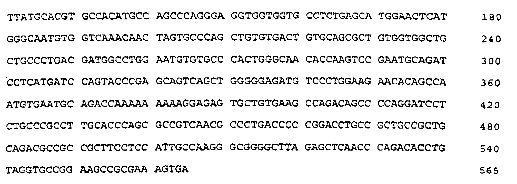
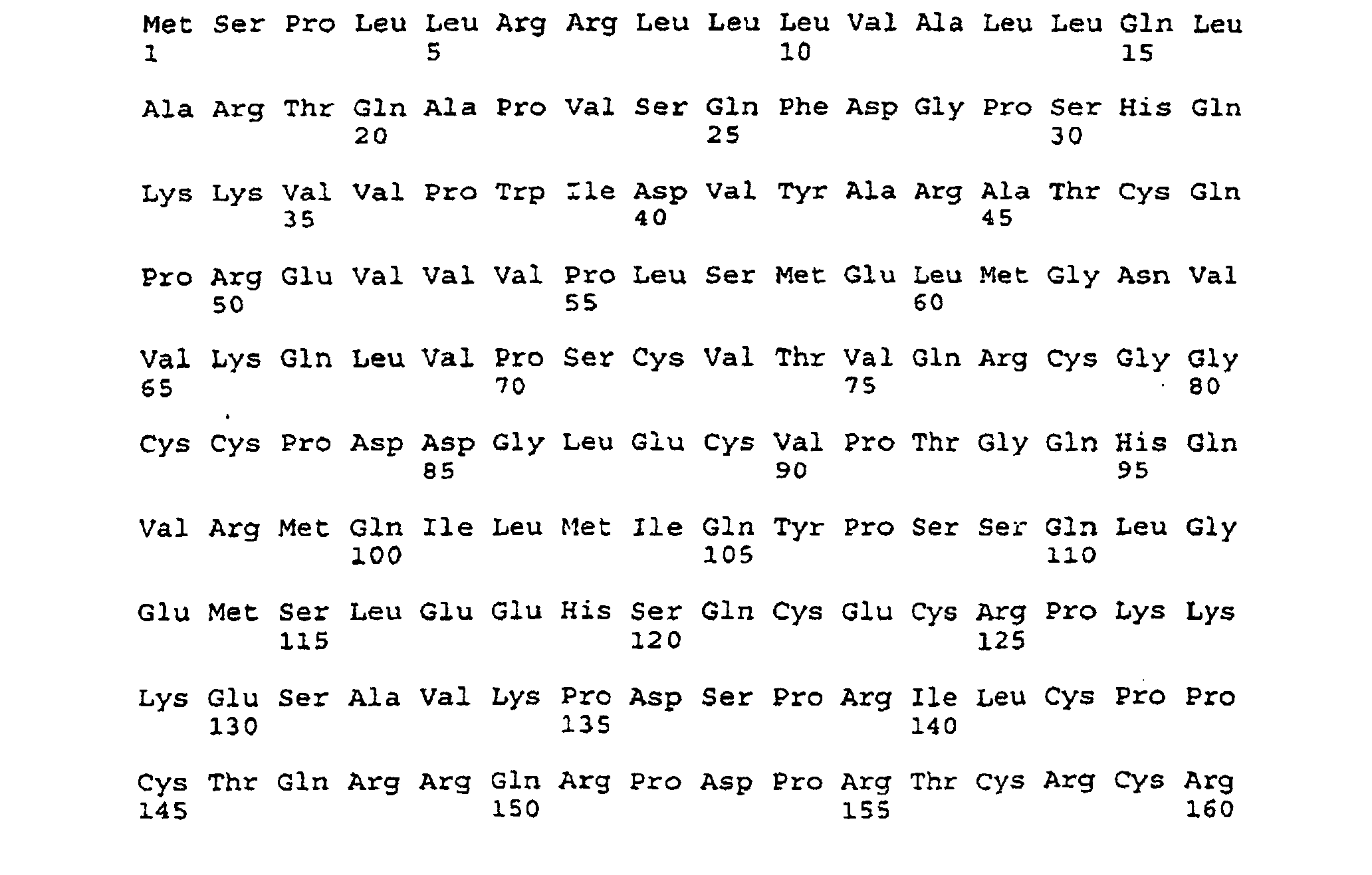

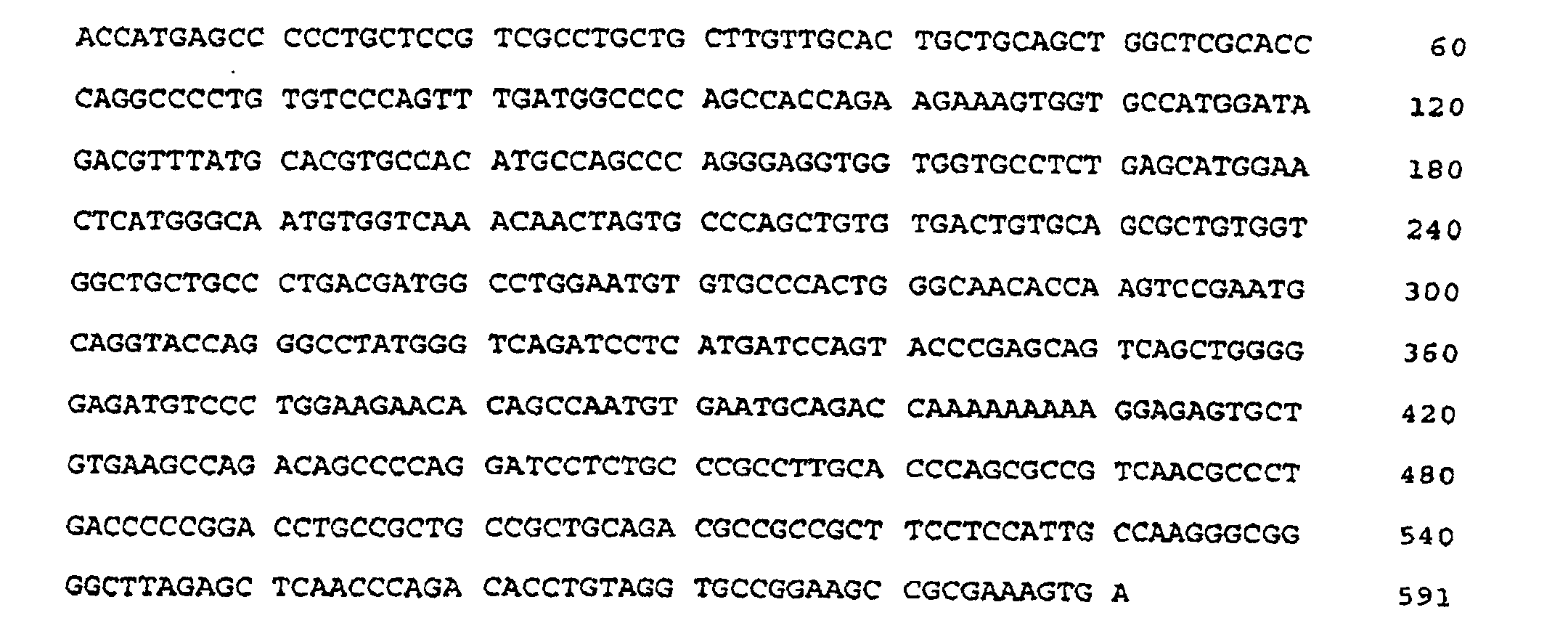

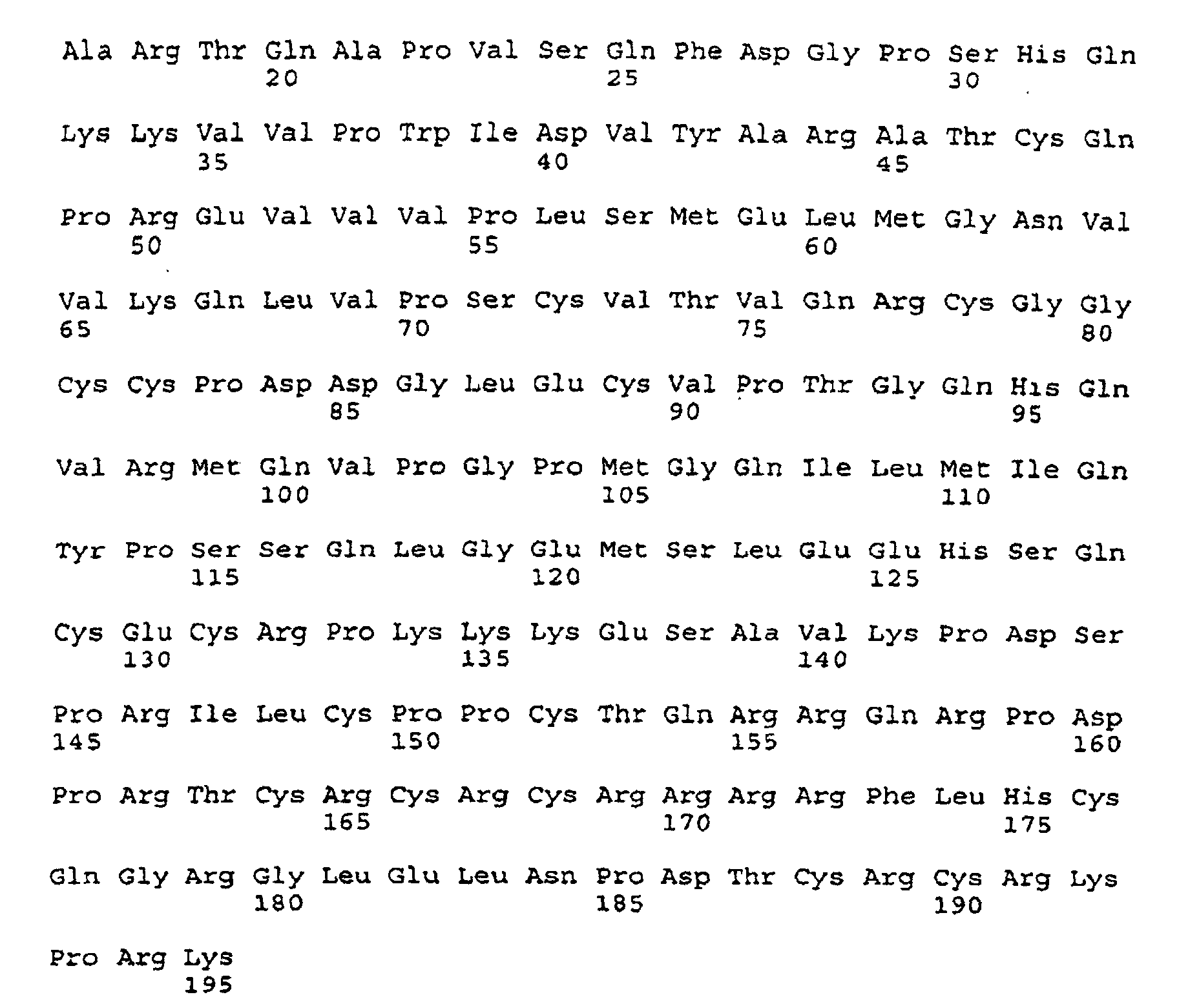

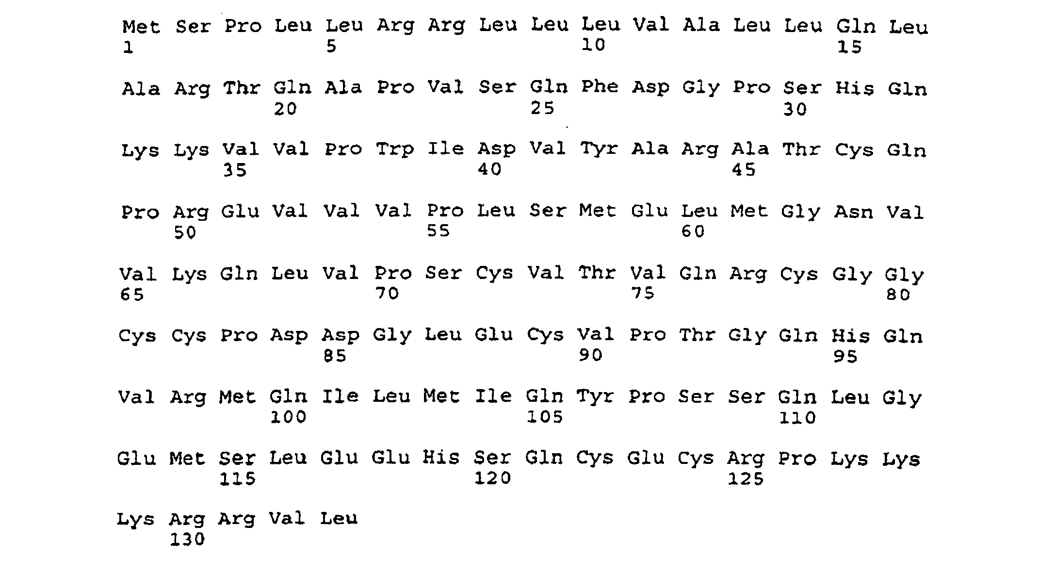


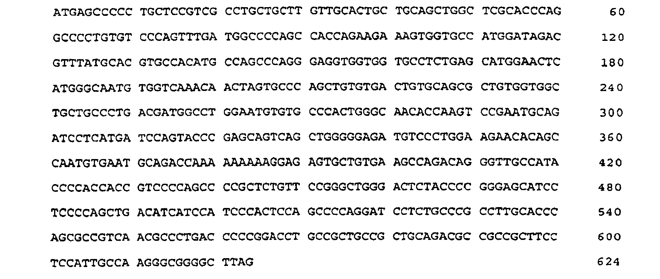


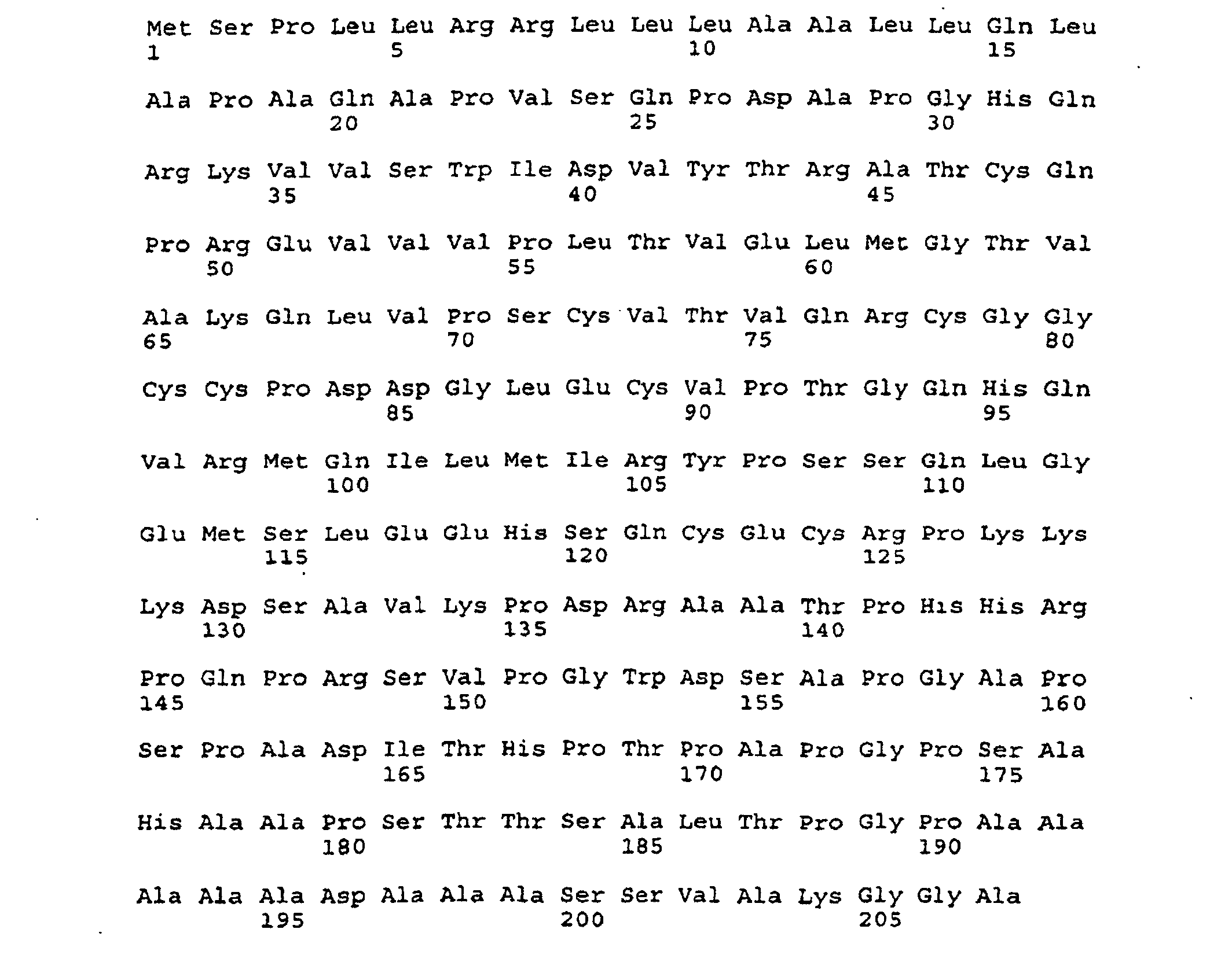

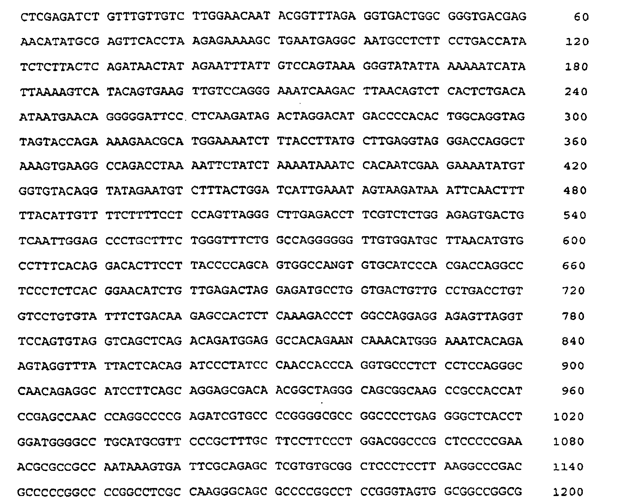
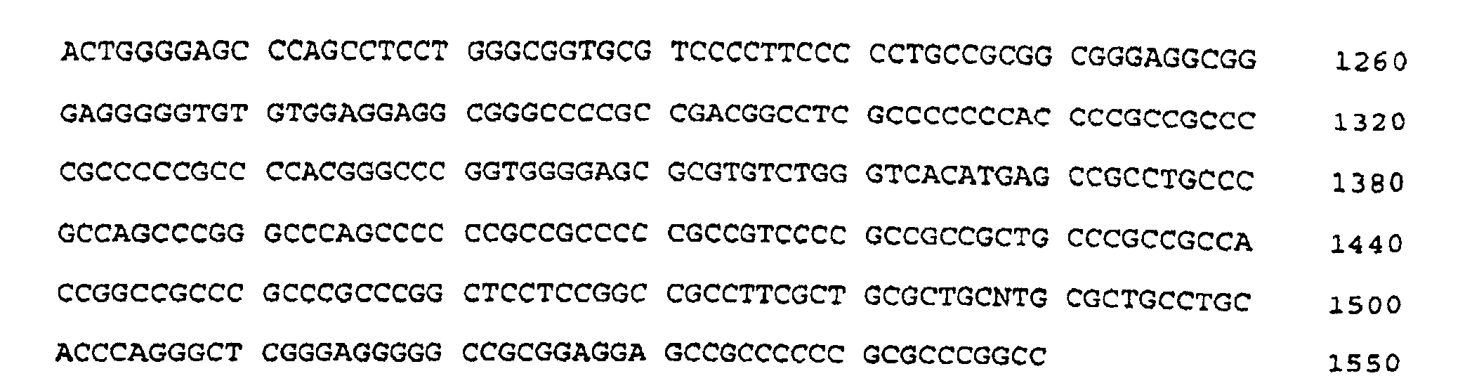














Claims (63)
- An isolated nucleic acid which is selected from:i) SEQ ID NO: 1; SEQ ID NO: 4; SEQ ID NO: 6; SEQ ID NO: 8; SEQ ID NO: 10; SEQ ID NO: 12; SEQ ID NO: 14; andii) sequences which:a) hybridise under stringent conditions to the sequences in (i)b) encode a protein which comprises a characteristic amino acid sequence Pro-Xaa-Cys-Val-Xaa-Xaa-Xaa-Arg-Cys-Xaa-Gly-Cys-Cys (SEQ ID NO:16); andc) encode a protein which has the property of promoting proliferation of endothelial cells or mesodermal cells.
- A nucleic acid according to claim 1, wherein said nucleic acid is a cDNA.
- A nucleic acid according to claim 1 or 2 comprising a cDNA corresponding to the DNA of Figures 1 and 2 (SEQ ID NO: 1).
- A nucleic acid according to any of claims 1-3 wherein said nucleic acid sequence is a mammalian DNA.
- A nucleic acid according to claim 4, wherein said nucleic acid is a murine DNA.
- A nucleic acid according to claim 4, wherein said nucleic acid is a human DNA.
- A nucleic acid according to any of claims 1-6 wherein said nucleic acid codes for a protein which promotes proliferation of vascular endothelial cells.
- A nucleic acid according to claim 1, comprising a cDNA corresponding to the DNS of SEQ ID NO: 4.
- A nucleic acid according to claim 1, comprising a cDNA corresponding to the DNA of SEQ ID NO: 6.
- A nucleic acid according to claim 1, comprising a cDNA corresponding to the DNA of SEQ ID NO: 8.
- A nucleic acid according to claim 1, comprising a cDNA corresponding to the DNA of SEQ ID NO: 10.
- A nucleic acid according to claim 1, comprising a cDNA corresponding to the DNA of SEQ ID NO: 12.
- A nucleic acid according to claim 1, comprising a cDNA corresponding to the DNA of SEQ ID NO: 14.
- A vector comprising a nucleic acid according to any of claims 1-13 operably linked with a promoter sequence.
- A vector according to claim 14, wherein said vector is a eukaryotic vector.
- A vector according to claim 14, wherein said vector is a prokaryotic vector.
- A vector according to claim 14, wherein said vector is a plasmid.
- An isolated protein which exhibits a characteristic sequence Pro-Xaa-Cys-Val-Xaa-Xaa-Xaa-Arg-Cys-Xaa-Gly-Cys-Cys (SEQ ID NO: 16) and has the property of promoting proliferation of endothelial cells or mesodermal cells, said protein comprising a sequence of amino acids corresponding to an amino acid sequence selected from the group consisting of the amino acid sequence of Figure 1 (SEQ ID NO: 2), the amino acid sequence of Figure 2 (SEQ ID NO: 3), the amino acid sequence of Figure 4 (SEQ ID NO: 5), the amino acid sequence of Figure 6 (SEQ ID NO: 7), the amino acid sequence of Figure 8 (SEQ ID NO: 9), the amino acid sequence of Figure 11 (SEQ ID NO: 11), the amino acid sequence of Figure 13 (SEQ ID NO: 13), and the amino acid sequence of Figure 15 (SEQ ID NO: 15).
- An isolated protein according to claim 18, wherein said protein comprises a sequence of amino acids corresponding to the amino acid sequence of SEQ ID NO: 2.
- An isolated protein according to claim 18, wherein said protein comprises an amino acid sequence corresponding to the amino acid sequence of SEQ ID NO: 3.
- An isolated protein according to claim 18, wherein said protein comprises a sequence of amino acids corresponding to the amino acid sequence of SEQ ID NO: 5.
- An isolated protein according to claim 18, wherein said protein comprises a sequence of amino acids corresponding to the amino acid sequence of SEQ ID NO: 7.
- An isolated protein according to claim 18, wherein said protein comprises a sequence of amino acids corresponding to the amino acid sequence of SEQ ID NO: 9.
- An isolated protein according to claim 18, wherein said protein comprises a sequence of amino acids corresponding to the amino acid sequence of SEQ ID NO: 11.
- An isolated protein according to claim 18, wherein said protein comprises a sequence of amino acids corresponding to the amino acid sequence of SEQ ID NO: 13.
- An isolated protein according to claim 18, wherein said protein comprises a sequence of amino acids corresponding to the amino acid sequence of SEQ ID NO: 15.
- An isolated protein according to any of claims 18-26, wherein said protein is a mammalian protein.
- An isolated protein according to claim 27, wherein said protein is a murine protein.
- An isolated protein according to claim 27, wherein said protein is a human protein.
- An isolated protein according to any of claims 18-29, wherein said protein promotes proliferation of vascular endothelial cells.
- An isolated protein produced by expression of a DNA selected from the group consisting of the DNA of Figures 1 and 2 (SEQ ID NO: 1), the DNA of Figure 3 (SEQ ID NO: 4), the DNA of Figure 5 (SEQ ID NO: 6), the DNA of Figure 7 (SEQ ID NO: 8), the DNA of Figure 10 (SEQ ID NO: 10), the DNA of Figure 12 (SEQ ID NO: 12), the DNA of Figure 14 (SEQ ID NO: 14), and DNA which hybridize under stringent conditions with at least one of the foregoing DNA sequences.
- A pharmaceutical composition comprising a protein according to claims 18-30, and at least one conventional pharmaceutical carrier or diluent.
- An antibody which binds with a protein according to claims 18-30 wherein said antibody does not bind VEGF-2.
- An antibody according to claim 33, wherein said antibody is a monoclonal antibody.
- A host cell transformed or transfected with a vector according to claim 14, such that said host cell expresses a protein having the property of promoting proliferation of endothelial or mesodermal cells.
- A transfected host cell according to claim 35, wherein said host cell is a eukaryotic cell.
- A transfected host cell according to claim 35 or 36, wherein said host cell is a COS cell.
- A transformed host cell according to claim 35, wherein said host cell is a prokaryotic cell.
- A transformed host cell according to claim 35, wherein said host cell is a 293EBNA cell.
- A transformed host cell according to claim 35 or 36, wherein said host cell is an insect cell.
- A diagnostic means for quantitatively detecting a protein according to any of Claims 18-29 in a test sample, said means comprising an antibody according to claim 33 or 34, which binds with said protein in order to detect the amount of said protein in the sample.
- A diagnostic means according to claim 41, wherein said antibody is a labelled antibody.
- A diagnostic means for detecting a nucleic acid in a test sample, said means comprising at least one pair of primers adapted to specifically bind a nucleic acid sequence according to any of Claims 1-13, and amplifying said test nucleic acid by a polymerase chain reaction.
- A diagnostic means according to Claim 43 wherein said polymerase chain reaction facilitates a sequence comparison of a nucleic acid in a test sample with a nucleic acid according to any of Claims 1-13.
- A pharmaceutical composition comprising antibodies according to claim 33 or 34, and at least one pharmaceutically acceptable caster or adjuvant.
- A pharmaceutical composition comprising a protein according to any of claims 18-30, and VEGF.
- A pharmaceutical composition according to claim 46, further comprising heparin.
- A pharmaceutical composition according to claim 32, further comprising heparin.
- A phamaceutical composition comprising a protein according to any of Claims 18-30.
- An isolated protein dimer comprising a protein according to any of claims 18-30.
- An isolated protein dimer according to claim 50, wherein said protein dimer is a homodimer of said protein.
- An isolated protein dimer according to claim 50, wherein said protein dimer is a heterodimer of said protein and VEGF.
- An isolated protein dimer according to claim 50, wherein said protein dimer is a disulfide-linked dimer.
- An in vitro method of promoting release of at least one protein selected from the group consisting of proteins according to any of claims 18-30, from a cell which expresses said at least one protein, said method comprising exposing the cell to heparin.
- A vector comprising an anti-sense nucleotide sequence complementary to at least part of a nucleic acid sequence according to any of Claims 1-13.
- An in vitro method of retarding expression of a protein according to any of Claims 18-29 from a cell which expresses said protein, said method comprising transfecting said cell with a vector according to claim 55.
- A method according to claim 56, wherein said cell is a tumor cell.
- An isolated nucleic acid comprising a nucleotide sequence corresponding to the sequence of SEQ ID NO: 17, or a nucleic acid which hybridize under stringent conditions with said nucleotide sequence.
- A host cell transformed or transfected with an expression vector comprising a nucleic acid sequence according to any of claims 1-13, such that said host cell expresses a protein according to any of Claims 18-29.
- A host cell according to claim 59, wherein said protein comprises a sequence of amino acids corresponding to an amino acid sequence selected from the group consisting of the amino acid sequence of Figure 1 (SEQ ID NO: 2), the amino acid sequence of Figure 2 (SEQ ID NO: 3), the amino acid sequence of Figure 4 (SEQ ID NO: 5), the amino acid sequence of Figure 6 (SEQ ID NO:7), the amino acid sequence of Figure 8 (SEQ ID NO: 9), the amino acid sequence of Figure 11 (SEQ ID NO: 11), the amino acid sequence of Figure 13 (SEQ ID NO: 13), and the amino acid sequence of Figure 15 (SEQ ID NO: 15).
- A diagnostic test consisting of a polymerase chain reaction for detecting a nucleic acid in a test sample, said reaction comprising at least one pair of primers complementary to a nucleic acid sequence according to any of claims 1-13.
- An in vitro method for promoting the release of at least one protein selected from the group consisting of proteins according to claims 18-30, and VEGF from a cell which expresses said at least one protein, said method comprising exposing the cell to heparin to induce release of said at least one protein.
- A method of making a vector suitable for expression of a protein according to any of Claims 18-29, said method comprising incorporating a nucleotide sequence according to any of claims 1-13.
Applications Claiming Priority (7)
| Application Number | Priority Date | Filing Date | Title |
|---|---|---|---|
| US569063 | 1990-08-17 | ||
| US39765195A | 1995-03-01 | 1995-03-01 | |
| US397651 | 1995-03-01 | ||
| US08/469,427 US5607918A (en) | 1995-03-01 | 1995-06-06 | Vascular endothelial growth factor-B and DNA coding therefor |
| US08/569,063 US5928939A (en) | 1995-03-01 | 1995-12-06 | Vascular endothelial growth factor-b and dna coding therefor |
| PCT/US1996/002957 WO1996026736A1 (en) | 1995-03-01 | 1996-03-01 | Vascular endothelial growth factor-b |
| US469427 | 1999-12-23 |
Publications (3)
| Publication Number | Publication Date |
|---|---|
| EP0814827A1 EP0814827A1 (en) | 1998-01-07 |
| EP0814827A4 EP0814827A4 (en) | 1999-11-10 |
| EP0814827B1 true EP0814827B1 (en) | 2003-07-23 |
Family
ID=27410265
Family Applications (1)
| Application Number | Title | Priority Date | Filing Date |
|---|---|---|---|
| EP96909573A Expired - Lifetime EP0814827B1 (en) | 1995-03-01 | 1996-03-01 | Vascular endothelial growth factor-b |
Country Status (14)
| Country | Link |
|---|---|
| US (5) | US5928939A (en) |
| EP (1) | EP0814827B1 (en) |
| JP (2) | JP3275024B2 (en) |
| CN (1) | CN1146440C (en) |
| AT (1) | ATE245437T1 (en) |
| AU (1) | AU741856B2 (en) |
| CA (1) | CA2211687C (en) |
| DE (1) | DE69629181T2 (en) |
| DK (1) | DK0814827T3 (en) |
| ES (1) | ES2202432T3 (en) |
| NO (1) | NO973933L (en) |
| NZ (1) | NZ305091A (en) |
| PT (1) | PT814827E (en) |
| WO (1) | WO1996026736A1 (en) |
Cited By (1)
| Publication number | Priority date | Publication date | Assignee | Title |
|---|---|---|---|---|
| US7696320B2 (en) | 2004-08-24 | 2010-04-13 | Domantis Limited | Ligands that have binding specificity for VEGF and/or EGFR and methods of use therefor |
Families Citing this family (118)
| Publication number | Priority date | Publication date | Assignee | Title |
|---|---|---|---|---|
| US7186688B1 (en) | 1994-03-08 | 2007-03-06 | Human Genome Sciences, Inc. | Methods of stimulating angiogenesis in a patient by administering vascular endothelial growth factor 2 |
| US6608182B1 (en) * | 1994-03-08 | 2003-08-19 | Human Genome Sciences, Inc. | Human vascular endothelial growth factor 2 |
| US7109308B1 (en) | 1994-03-08 | 2006-09-19 | Human Genome Sciences, Inc. | Antibodies to human vascular endothelial growth factor 2 |
| US7153827B1 (en) | 1994-03-08 | 2006-12-26 | Human Genome Sciences, Inc. | Vascular endothelial growth factor 2 and methods of use |
| EP0751992B1 (en) * | 1994-03-08 | 2005-11-09 | Human Genome Sciences, Inc. | Vascular endothelial growth factor 2 |
| US5932540A (en) | 1994-03-08 | 1999-08-03 | Human Genome Sciences, Inc. | Vascular endothelial growth factor 2 |
| US6040157A (en) * | 1994-03-08 | 2000-03-21 | Human Genome Sciences, Inc. | Vascular endothelial growth factor 2 |
| US6734285B2 (en) | 1994-03-08 | 2004-05-11 | Human Genome Sciences, Inc. | Vascular endothelial growth factor 2 proteins and compositions |
| US7597886B2 (en) | 1994-11-07 | 2009-10-06 | Human Genome Sciences, Inc. | Tumor necrosis factor-gamma |
| US5928939A (en) * | 1995-03-01 | 1999-07-27 | Ludwig Institute For Cancer Research | Vascular endothelial growth factor-b and dna coding therefor |
| US7160991B1 (en) * | 1995-03-02 | 2007-01-09 | Ludwig Institute For Cancer Research | Vascular endothelial growth factor polypeptides |
| JPH11500139A (en) * | 1995-03-02 | 1999-01-06 | アムラド・オペレイションズ・プロプライエタリー・リミテッド | Novel growth factor and gene sequence encoding it |
| US7429646B1 (en) | 1995-06-05 | 2008-09-30 | Human Genome Sciences, Inc. | Antibodies to human tumor necrosis factor receptor-like 2 |
| EP0873348A4 (en) * | 1995-06-06 | 2000-11-08 | Human Genome Sciences Inc | GROWTH FACTOR 3 OF THE HUMAN VASCULAR ENDOTHELIUM |
| US7423125B2 (en) * | 1995-08-01 | 2008-09-09 | Vegenics Limited | Antibodies to VEGF-C |
| ES2202469T5 (en) | 1995-09-08 | 2011-06-06 | Genentech, Inc. | PROTEIN RELATED TO VEGF. |
| WO1997012972A2 (en) | 1995-09-29 | 1997-04-10 | Universita'degli Studi Di Siena | Regulated genes and uses thereof |
| AU1116297A (en) | 1995-11-08 | 1997-05-29 | Immunex Corporation | Flk-1 binding protein |
| US6994989B1 (en) | 1995-11-08 | 2006-02-07 | Immunex Corp. | FLK-1 binding proteins |
| US7357927B2 (en) | 1996-03-12 | 2008-04-15 | Human Genome Sciences, Inc. | Death domain containing receptors |
| US6713061B1 (en) | 1996-03-12 | 2004-03-30 | Human Genome Sciences, Inc. | Death domain containing receptors |
| WO1997033904A1 (en) | 1996-03-12 | 1997-09-18 | Human Genome Sciences, Inc. | Death domain containing receptors |
| US7964190B2 (en) | 1996-03-22 | 2011-06-21 | Human Genome Sciences, Inc. | Methods and compositions for decreasing T-cell activity |
| US6635743B1 (en) | 1996-03-22 | 2003-10-21 | Human Genome Sciences, Inc. | Apoptosis inducing molecule II and methods of use |
| CA2263890C (en) | 1996-08-23 | 2013-05-14 | Ludwig Institute For Cancer Research | Recombinant vascular endothelial cell growth factor d (vegf-d) |
| WO1998024811A2 (en) * | 1996-12-06 | 1998-06-11 | Zymogenetics, Inc. | Vascular endothelial growth factor |
| ZA9711456B (en) * | 1996-12-20 | 1998-06-30 | Ludwig Inst Cancer Res | VEGF-B/receptor complex and uses thereof. |
| WO1998039035A1 (en) | 1997-03-07 | 1998-09-11 | The Wistar Institute Of Anatomy & Biology | Method and compositions for healing tissue defects and inducing hypervascularity in mammalian tissue |
| JP2001524828A (en) * | 1997-04-25 | 2001-12-04 | コラテラル・セラピューティクス | Truncated VEGF-related protein |
| US6281175B1 (en) | 1997-09-23 | 2001-08-28 | Scimed Life Systems, Inc. | Medical emulsion for lubrication and delivery of drugs |
| EP1053326A2 (en) | 1998-02-06 | 2000-11-22 | Collateral Therapeutics | Variants of the angiogenic factor vascular endothelial cell growth factor: vegf |
| US20030118567A1 (en) * | 1999-03-26 | 2003-06-26 | Stewart Duncan John | Cell-based therapy for the pulmonary system |
| DE19813774A1 (en) * | 1998-03-27 | 1999-09-30 | Max Planck Gesellschaft | Parapox virus-encoded vascular endothelial cell growth factor (PPV-VEGF) |
| JP2002534061A (en) | 1998-11-10 | 2002-10-15 | ルードヴィッヒ・インスティテュート・フォア・キャンサー・リサーチ | Platelet-derived growth factor D, DNA encoding the same, and use thereof |
| US6783953B1 (en) | 1998-12-22 | 2004-08-31 | Janssen Pharmaceutica N.V. | Vascular endothelial growth factor-X |
| AU3693000A (en) * | 1999-01-29 | 2000-08-18 | Licentia Ltd | Platelet-derived growth factor/vascular endothelial growth factor-like growth factor h, and uses thereof |
| US7223724B1 (en) | 1999-02-08 | 2007-05-29 | Human Genome Sciences, Inc. | Use of vascular endothelial growth factor to treat photoreceptor cells |
| EP2357192A1 (en) | 1999-02-26 | 2011-08-17 | Human Genome Sciences, Inc. | Human endokine alpha and methods of use |
| NZ513780A (en) * | 1999-03-03 | 2001-09-28 | Ludwig Inst Cancer Res | Heart abnormalities in vascular endothelial growth factor B (VEGF-B) deficient animals and methods relating to these heart abnormalities |
| AU4662400A (en) * | 1999-04-26 | 2000-11-10 | Helsinki University Licensing Ltd | Role of vascular endothelial growth factor-b (vegf-b) in developing bones and uses thereof |
| CA2375617A1 (en) * | 1999-06-07 | 2000-12-14 | Edwards Lifesciences Corporation | Targeted angiogenesis |
| WO2001008684A1 (en) * | 1999-07-28 | 2001-02-08 | The Board Of Trustees Of The Leland Stanford Junior University | Nicotine in therapeutic angiogenesis and vasculogenesis |
| US8188043B2 (en) * | 1999-07-28 | 2012-05-29 | The Board Of Trustees Of The Leland Stanford Jr. University | Nicotine in therapeutic angiogenesis and vasculogenesis |
| DE60136334D1 (en) * | 2000-02-02 | 2008-12-11 | Us Health | Cd40-ligand als adjuvant für respiratorisches syncytialvirus-impfstoff |
| AUPQ568100A0 (en) * | 2000-02-16 | 2000-03-09 | Amrad Operations Pty. Limited | A method for producing recombinant molecules |
| AU2001239884B2 (en) * | 2000-02-25 | 2006-08-10 | Vegenics Limited | Materials and methods involving hybrid vascular endothelial growth factor DNAs and proteins and screening methods for modulators |
| AUPQ592100A0 (en) * | 2000-02-29 | 2000-03-23 | Council Of The Queensland Institute Of Medical Research, The | A method of treatment and prophylaxis |
| US20030104977A1 (en) * | 2000-03-31 | 2003-06-05 | Ugo Ripamonti | Methods for inducing angiogenesis using morphogenic proteins and stimulatory factors |
| EP1274720A4 (en) | 2000-04-12 | 2004-08-18 | Human Genome Sciences Inc | ALBUMIN FUSED PROTEINS |
| CA2409948A1 (en) * | 2000-05-17 | 2001-11-22 | Xuri Li | Methods for detecting for the presence of tumor cells and for screening for anti-tumor agents |
| CN1461342A (en) * | 2000-07-26 | 2003-12-10 | 路德维格癌症研究所 | Glycosylated VEGF-B and methods for increasing the amount of soluble VEGF-B |
| US7273751B2 (en) * | 2000-08-04 | 2007-09-25 | Human Genome Science, Inc. | Vascular endothelial growth factor-2 |
| AU2001296521A1 (en) * | 2000-10-02 | 2002-04-15 | St. Elizabeth's Medical Center Of Boston, Inc. | Use of lymphangiogenic agents to treat lymphatic disorders |
| JP2004527462A (en) * | 2000-11-01 | 2004-09-09 | ルードヴィッヒ インスティテュート フォー キャンサー リサーチ | INVIVO stimulation of angiogenic activity |
| AU2884102A (en) * | 2000-12-07 | 2002-06-18 | Sangamo Biosciences Inc | Regulation of angiogenesis with zinc finger proteins |
| US7067317B2 (en) * | 2000-12-07 | 2006-06-27 | Sangamo Biosciences, Inc. | Regulation of angiogenesis with zinc finger proteins |
| US7611711B2 (en) | 2001-01-17 | 2009-11-03 | Vegenics Limited | VEGFR-3 inhibitor materials and methods |
| US20070111944A1 (en) * | 2001-02-16 | 2007-05-17 | Scrofani Sergio D B | Purification of vascular endothelial growth factor-B |
| US20070050857A1 (en) * | 2001-02-28 | 2007-03-01 | Hayward Nick K | Method of treatment and prophylaxis |
| AU2002303261A1 (en) * | 2001-04-06 | 2002-10-21 | Georgetown University | Gene brcc2 and diagnostic and therapeutic uses thereof |
| DE60236646D1 (en) | 2001-04-13 | 2010-07-22 | Human Genome Sciences Inc | Anti-VEGF-2 antibodies |
| AU2002252631A1 (en) * | 2001-04-13 | 2002-10-28 | Human Genome Sciences, Inc. | Vascular endothelial growth factor 2 |
| WO2002083850A2 (en) * | 2001-04-13 | 2002-10-24 | Human Genome Sciences, Inc. | Vascular endothelial growth factor 2 |
| US20050232921A1 (en) * | 2001-04-13 | 2005-10-20 | Rosen Craig A | Vascular endothelial growth factor 2 |
| US7402312B2 (en) * | 2001-04-13 | 2008-07-22 | Human Genome Sciences, Inc. | Antibodies to vascular endothelial growth factor 2 (VEGF-2) |
| DE60234100D1 (en) * | 2001-04-24 | 2009-12-03 | Vlaams Interuniv Inst Biotech | USE OF "HYPOXIA INDUCIBLE FACTOR 2ALPHA" FOR THE TREATMENT OF "NEONATAL RESPIRATORY DISTRESS SYNDROME" |
| WO2003000009A2 (en) * | 2001-06-20 | 2003-01-03 | Ludwig Institute For Cancer Research | Stimulation of vascularization with vegf-b |
| AU2002319402B2 (en) * | 2001-06-28 | 2008-09-11 | Domantis Limited | Dual-specific ligand and its use |
| US20050271663A1 (en) * | 2001-06-28 | 2005-12-08 | Domantis Limited | Compositions and methods for treating inflammatory disorders |
| CN1555488A (en) * | 2001-07-12 | 2004-12-15 | 路德维格癌症研究院 | Lymphatic endothelial cell material and methods |
| WO2003018752A2 (en) | 2001-08-23 | 2003-03-06 | The Wistar Institute Of Anatomy And Biology | An organotypic intestinal culture and methods of use thereof |
| US20030113324A1 (en) | 2001-10-01 | 2003-06-19 | Kari Alitalo | Neuropilin/VEGF-C/VEGFR-3 materials and methods |
| US20040214766A1 (en) * | 2001-10-01 | 2004-10-28 | Kari Alitalo | VEGF-C or VEGF-D materials and methods for treatment of neuropathologies |
| PT1463751E (en) | 2001-12-21 | 2013-08-26 | Human Genome Sciences Inc | ALBUMINE FUSION PROTEINS |
| US20030221931A1 (en) * | 2002-02-28 | 2003-12-04 | Steve Marsh | Sliding device |
| US20040005671A1 (en) * | 2002-05-17 | 2004-01-08 | Amrad Operations Pty Ltd. | Immunointeractive molecules |
| AU2003237456A1 (en) * | 2002-06-05 | 2003-12-22 | Sopherion Therapeutics, Inc. | Method to screen ligands using eukaryotic cell display |
| EP1912069A3 (en) | 2002-06-05 | 2008-07-09 | Sopherion Therapeutics, Inc. | Method to screen ligands using eukaryotic cell display |
| US20030232439A1 (en) * | 2002-06-17 | 2003-12-18 | Isis Pharmaceuticals Inc. | Antisense modulation of VEGF-B expression |
| US9321832B2 (en) * | 2002-06-28 | 2016-04-26 | Domantis Limited | Ligand |
| EP1600459A3 (en) * | 2002-06-28 | 2005-12-07 | Domantis Limited | Ligand |
| US7479270B2 (en) | 2002-07-23 | 2009-01-20 | Vegenics Limited | Methods and compositions for activating VEGF-D and VEGF-C |
| CA2547899C (en) * | 2002-12-05 | 2014-04-15 | Case Western Reserve University | Cell-based therapies for ischemia |
| US7470538B2 (en) * | 2002-12-05 | 2008-12-30 | Case Western Reserve University | Cell-based therapies for ischemia |
| JP2006523090A (en) * | 2002-12-27 | 2006-10-12 | ドマンティス リミテッド | Bispecific single domain antibody specific for ligand and for ligand receptor |
| EP1594527A2 (en) * | 2003-02-04 | 2005-11-16 | Ludwig Institute For Cancer Research | Vegf-b and pdgf modulation of stem cells |
| NZ592039A (en) | 2003-08-27 | 2013-03-28 | Ophthotech Corp | Combination therapy for the treatment of ocular neovascular disorders |
| EP1560025A3 (en) * | 2003-10-03 | 2011-09-07 | F. Hoffmann-La Roche AG | Specific markers for diabetes |
| CA2552241C (en) | 2003-12-30 | 2013-10-01 | Durect Corporation | Co-polymeric devices for controlled release of active agents |
| US20050214280A1 (en) * | 2004-03-02 | 2005-09-29 | Ludwig Institute For Cancer Research | Method for inhibiting tumor formation and growth |
| WO2005087177A2 (en) * | 2004-03-05 | 2005-09-22 | Ludwig Institute For Cancer Research | Chimeric anti-vegf-d antibodies and humanized anti-vegf-d antibodies and methods of using same |
| WO2005087812A1 (en) * | 2004-03-05 | 2005-09-22 | Ludwig Institute For Cancer Research | Multivalent antibody materials and methods for vegf/pdgf family of growth factors |
| JP4903689B2 (en) * | 2004-04-08 | 2012-03-28 | サンガモ バイオサイエンシズ インコーポレイテッド | Methods and compositions for treating neuropathy and neurodegenerative symptoms |
| EP2476427B1 (en) * | 2004-08-02 | 2018-01-17 | Zenyth Operations PTY. Ltd. | A method of treating cancer comprising a VEGF-B antagonist |
| US7595149B1 (en) | 2004-11-08 | 2009-09-29 | University Of Kentucky Research Foundation | Methods for cancer detection |
| EP1869075B1 (en) * | 2005-02-28 | 2012-04-11 | Sangamo BioSciences, Inc. | Anti-angiogenic methods and compositions |
| MX2007012766A (en) * | 2005-04-12 | 2008-10-01 | Intradigm Corp | Composition and methods of rnai therapeutics for treatment of cancer and other neovascularization diseases. |
| US7893244B2 (en) * | 2005-04-12 | 2011-02-22 | Intradigm Corporation | Composition and methods of RNAi therapeutics for treatment of cancer and other neovascularization diseases |
| WO2007022287A2 (en) | 2005-08-15 | 2007-02-22 | Vegenics Limited | Modified vegf and pdgf with improved angiogenic properties |
| CN101454344A (en) * | 2005-12-01 | 2009-06-10 | 杜门蒂斯有限公司 | Noncompetitive domain antibody formats that bind interleukin 1 receptor type 1 |
| DK2548578T3 (en) | 2006-05-17 | 2014-10-06 | Ludwig Inst Cancer Res | Targeting of VEGF-B regulation of fatty acid transporters for modulating human diseases |
| WO2008085229A2 (en) * | 2006-11-15 | 2008-07-17 | Arteriocyte Inc. | Cell-based therapies for treating liver disease |
| NZ580530A (en) * | 2007-06-06 | 2012-04-27 | Domantis Ltd | Anti vegf polypeptides, antibody variable domains and antagonists |
| US7819579B2 (en) * | 2007-11-26 | 2010-10-26 | Schmulenson Harold K | Radiation sensing device and holder |
| WO2009075565A1 (en) * | 2007-12-12 | 2009-06-18 | Erasmus University Medical Center Rotterdam | Methods for controlling vasculogenesis |
| JP5690593B2 (en) | 2007-12-26 | 2015-03-25 | ヴァクシネックス, インコーポレイテッド | Anti-C35 antibody combination therapy and method |
| WO2009126698A2 (en) * | 2008-04-09 | 2009-10-15 | Ludwig Institute For Cancer Research | Regulation of fatty acid transporters |
| IL205774A0 (en) * | 2010-05-13 | 2010-12-30 | Muhammad Abdulrazik | Novel compounds for the treatment of glaucoma and ocular hypertension |
| KR101335203B1 (en) * | 2010-03-26 | 2013-11-29 | 숙명여자대학교산학협력단 | Peptides for Promotion of Angiogenesis and the use thereof |
| WO2011154308A1 (en) | 2010-06-08 | 2011-12-15 | Proyecto De Biomedicina Cima, S.L. | New compositions and cell therapy methods for the treatment of cirrhosis |
| RU2477529C2 (en) * | 2011-04-28 | 2013-03-10 | Общество С Ограниченной Ответственностью "Научно-Производственная Фирма "Материа Медика Холдинг" | Method for endothelial dysfunction correction by solution of homoeopathic dilutions of human endothelial nitrogen oxide synthase antibodies |
| KR102948178B1 (en) | 2013-07-12 | 2026-04-10 | 아스텔라스 유에스 엘엘씨 | Methods for treating or preventing ophthalmological conditions |
| CN105979965B (en) * | 2013-12-18 | 2021-07-09 | 杰特有限公司 | Method of treating wounds |
| CN107661492A (en) * | 2017-08-31 | 2018-02-06 | 中山大学中山眼科中心 | VEGF B new application |
| CN107537026A (en) * | 2017-08-31 | 2018-01-05 | 中山大学中山眼科中心 | VEGF B application |
| US10745454B2 (en) * | 2018-01-31 | 2020-08-18 | Seyed Mohsen Asghari | Method of synthesizing antagonist peptides for cell growth |
| CN109337931A (en) * | 2018-11-02 | 2019-02-15 | 东北师范大学 | Application of VEGFB gene and construction of VEGFB tumor-inducing and tumor-suppressing animal models |
| CN110777146A (en) * | 2019-11-28 | 2020-02-11 | 南通大学 | A kind of construction method of PDGFB promoter activity reporter plasmid |
Family Cites Families (12)
| Publication number | Priority date | Publication date | Assignee | Title |
|---|---|---|---|---|
| US4659678A (en) * | 1982-09-29 | 1987-04-21 | Serono Diagnostics Limited | Immunoassay of antigens |
| US4456550A (en) * | 1982-11-22 | 1984-06-26 | President And Fellows Of Harvard College | Vascular permeability factor |
| US5036003A (en) * | 1987-08-21 | 1991-07-30 | Monsanto Company | Antibodies to VPF |
| US5240848A (en) * | 1988-11-21 | 1993-08-31 | Monsanto Company | Dna sequences encoding human vascular permeability factor having 189 amino acids |
| US5332671A (en) * | 1989-05-12 | 1994-07-26 | Genetech, Inc. | Production of vascular endothelial cell growth factor and DNA encoding same |
| US5194596A (en) * | 1989-07-27 | 1993-03-16 | California Biotechnology Inc. | Production of vascular endothelial cell growth factor |
| US5726152A (en) * | 1990-09-21 | 1998-03-10 | Merck & Co., Inc. | Vascular endothelial cell growth factor II |
| EP0751992B1 (en) * | 1994-03-08 | 2005-11-09 | Human Genome Sciences, Inc. | Vascular endothelial growth factor 2 |
| US5607918A (en) * | 1995-03-01 | 1997-03-04 | Ludwig Institute For Cancer Research | Vascular endothelial growth factor-B and DNA coding therefor |
| US5928939A (en) * | 1995-03-01 | 1999-07-27 | Ludwig Institute For Cancer Research | Vascular endothelial growth factor-b and dna coding therefor |
| JPH11500139A (en) * | 1995-03-02 | 1999-01-06 | アムラド・オペレイションズ・プロプライエタリー・リミテッド | Novel growth factor and gene sequence encoding it |
| EP0873348A4 (en) * | 1995-06-06 | 2000-11-08 | Human Genome Sciences Inc | GROWTH FACTOR 3 OF THE HUMAN VASCULAR ENDOTHELIUM |
-
1995
- 1995-12-06 US US08/569,063 patent/US5928939A/en not_active Expired - Lifetime
-
1996
- 1996-03-01 AT AT96909573T patent/ATE245437T1/en active
- 1996-03-01 JP JP52645096A patent/JP3275024B2/en not_active Expired - Fee Related
- 1996-03-01 EP EP96909573A patent/EP0814827B1/en not_active Expired - Lifetime
- 1996-03-01 US US08/609,443 patent/US5840693A/en not_active Expired - Lifetime
- 1996-03-01 ES ES96909573T patent/ES2202432T3/en not_active Expired - Lifetime
- 1996-03-01 CN CNB961922036A patent/CN1146440C/en not_active Expired - Fee Related
- 1996-03-01 CA CA2211687A patent/CA2211687C/en not_active Expired - Lifetime
- 1996-03-01 DK DK96909573T patent/DK0814827T3/en active
- 1996-03-01 NZ NZ305091A patent/NZ305091A/en not_active IP Right Cessation
- 1996-03-01 WO PCT/US1996/002957 patent/WO1996026736A1/en not_active Ceased
- 1996-03-01 PT PT96909573T patent/PT814827E/en unknown
- 1996-03-01 DE DE69629181T patent/DE69629181T2/en not_active Expired - Lifetime
- 1996-03-01 AU AU53021/96A patent/AU741856B2/en not_active Expired
-
1997
- 1997-05-06 US US08/851,896 patent/US6331301B1/en not_active Expired - Lifetime
- 1997-08-27 NO NO973933A patent/NO973933L/en unknown
-
2000
- 2000-02-02 JP JP2000024574A patent/JP3297687B2/en not_active Expired - Lifetime
-
2001
- 2001-09-25 US US09/961,756 patent/US20030170253A1/en not_active Abandoned
-
2004
- 2004-09-24 US US10/948,736 patent/US7341996B2/en not_active Expired - Fee Related
Cited By (1)
| Publication number | Priority date | Publication date | Assignee | Title |
|---|---|---|---|---|
| US7696320B2 (en) | 2004-08-24 | 2010-04-13 | Domantis Limited | Ligands that have binding specificity for VEGF and/or EGFR and methods of use therefor |
Also Published As
| Publication number | Publication date |
|---|---|
| US20050064493A1 (en) | 2005-03-24 |
| DK0814827T3 (en) | 2003-11-10 |
| JP2000228982A (en) | 2000-08-22 |
| US5840693A (en) | 1998-11-24 |
| US7341996B2 (en) | 2008-03-11 |
| AU741856B2 (en) | 2001-12-13 |
| ATE245437T1 (en) | 2003-08-15 |
| US20030170253A1 (en) | 2003-09-11 |
| PT814827E (en) | 2003-11-28 |
| US5928939A (en) | 1999-07-27 |
| DE69629181D1 (en) | 2003-08-28 |
| CA2211687A1 (en) | 1996-09-06 |
| EP0814827A1 (en) | 1998-01-07 |
| WO1996026736A1 (en) | 1996-09-06 |
| CN1176603A (en) | 1998-03-18 |
| NZ305091A (en) | 1998-11-25 |
| NO973933D0 (en) | 1997-08-27 |
| DE69629181T2 (en) | 2004-05-13 |
| HK1008485A1 (en) | 1999-07-30 |
| AU5302196A (en) | 1996-09-18 |
| NO973933L (en) | 1997-09-01 |
| JPH10510718A (en) | 1998-10-20 |
| EP0814827A4 (en) | 1999-11-10 |
| ES2202432T3 (en) | 2004-04-01 |
| CA2211687C (en) | 2011-05-24 |
| US6331301B1 (en) | 2001-12-18 |
| JP3297687B2 (en) | 2002-07-02 |
| CN1146440C (en) | 2004-04-21 |
| JP3275024B2 (en) | 2002-04-15 |
Similar Documents
| Publication | Publication Date | Title |
|---|---|---|
| US5840693A (en) | Vascular endothelial growth factor-B | |
| US6235713B1 (en) | Vascular endothelial growth factor-D (VEGF-D) polypeptides | |
| US5607918A (en) | Vascular endothelial growth factor-B and DNA coding therefor | |
| JP3039802B2 (en) | Receptor for fibroblast growth factor | |
| JP2003521893A (en) | Fibroblast growth factor-like polypeptide | |
| US20020164687A1 (en) | Platelet-derived growth factor C, DNA coding therefor, and uses thereof |
Legal Events
| Date | Code | Title | Description |
|---|---|---|---|
| PUAI | Public reference made under article 153(3) epc to a published international application that has entered the european phase |
Free format text: ORIGINAL CODE: 0009012 |
|
| 17P | Request for examination filed |
Effective date: 19970926 |
|
| AK | Designated contracting states |
Kind code of ref document: A1 Designated state(s): AT BE CH DE DK ES FI FR GB GR IE IT LI LU MC NL PT SE |
|
| A4 | Supplementary search report drawn up and despatched |
Effective date: 19990924 |
|
| AK | Designated contracting states |
Kind code of ref document: A4 Designated state(s): AT BE CH DE DK ES FI FR GB GR IE IT LI LU MC NL PT SE |
|
| 17Q | First examination report despatched |
Effective date: 20000222 |
|
| GRAG | Despatch of communication of intention to grant |
Free format text: ORIGINAL CODE: EPIDOS AGRA |
|
| GRAG | Despatch of communication of intention to grant |
Free format text: ORIGINAL CODE: EPIDOS AGRA |
|
| GRAH | Despatch of communication of intention to grant a patent |
Free format text: ORIGINAL CODE: EPIDOS IGRA |
|
| GRAH | Despatch of communication of intention to grant a patent |
Free format text: ORIGINAL CODE: EPIDOS IGRA |
|
| GRAA | (expected) grant |
Free format text: ORIGINAL CODE: 0009210 |
|
| RAP1 | Party data changed (applicant data changed or rights of an application transferred) |
Owner name: LICENTIA LTD. Owner name: LUDWIG INSTITUTE FOR CANCER RESEARCH |
|
| AK | Designated contracting states |
Designated state(s): AT BE CH DE DK ES FI FR GB GR IE IT LI LU MC NL PT SE |
|
| REG | Reference to a national code |
Ref country code: GB Ref legal event code: FG4D |
|
| REG | Reference to a national code |
Ref country code: CH Ref legal event code: EP |
|
| REG | Reference to a national code |
Ref country code: IE Ref legal event code: FG4D |
|
| REF | Corresponds to: |
Ref document number: 69629181 Country of ref document: DE Date of ref document: 20030828 Kind code of ref document: P |
|
| REG | Reference to a national code |
Ref country code: CH Ref legal event code: NV Representative=s name: BRAUN & PARTNER PATENT-, MARKEN-, RECHTSANWAELTE |
|
| REG | Reference to a national code |
Ref country code: DK Ref legal event code: T3 |
|
| REG | Reference to a national code |
Ref country code: SE Ref legal event code: TRGR |
|
| REG | Reference to a national code |
Ref country code: GR Ref legal event code: EP Ref document number: 20030404355 Country of ref document: GR |
|
| REG | Reference to a national code |
Ref country code: ES Ref legal event code: FG2A Ref document number: 2202432 Country of ref document: ES Kind code of ref document: T3 |
|
| ET | Fr: translation filed | ||
| PLBE | No opposition filed within time limit |
Free format text: ORIGINAL CODE: 0009261 |
|
| STAA | Information on the status of an ep patent application or granted ep patent |
Free format text: STATUS: NO OPPOSITION FILED WITHIN TIME LIMIT |
|
| 26N | No opposition filed |
Effective date: 20040426 |
|
| PGFP | Annual fee paid to national office [announced via postgrant information from national office to epo] |
Ref country code: PT Payment date: 20070116 Year of fee payment: 12 |
|
| PGFP | Annual fee paid to national office [announced via postgrant information from national office to epo] |
Ref country code: MC Payment date: 20070119 Year of fee payment: 12 |
|
| PGFP | Annual fee paid to national office [announced via postgrant information from national office to epo] |
Ref country code: LU Payment date: 20070124 Year of fee payment: 12 Ref country code: ES Payment date: 20070124 Year of fee payment: 12 |
|
| PGFP | Annual fee paid to national office [announced via postgrant information from national office to epo] |
Ref country code: GR Payment date: 20070330 Year of fee payment: 12 |
|
| REG | Reference to a national code |
Ref country code: PT Ref legal event code: MM4A Free format text: LAPSE DUE TO NON-PAYMENT OF FEES Effective date: 20080901 |
|
| PG25 | Lapsed in a contracting state [announced via postgrant information from national office to epo] |
Ref country code: MC Free format text: LAPSE BECAUSE OF NON-PAYMENT OF DUE FEES Effective date: 20080331 Ref country code: PT Free format text: LAPSE BECAUSE OF NON-PAYMENT OF DUE FEES Effective date: 20080901 |
|
| REG | Reference to a national code |
Ref country code: ES Ref legal event code: FD2A Effective date: 20080303 |
|
| PG25 | Lapsed in a contracting state [announced via postgrant information from national office to epo] |
Ref country code: GR Free format text: LAPSE BECAUSE OF NON-PAYMENT OF DUE FEES Effective date: 20081002 |
|
| PG25 | Lapsed in a contracting state [announced via postgrant information from national office to epo] |
Ref country code: ES Free format text: LAPSE BECAUSE OF NON-PAYMENT OF DUE FEES Effective date: 20080303 |
|
| PG25 | Lapsed in a contracting state [announced via postgrant information from national office to epo] |
Ref country code: LU Free format text: LAPSE BECAUSE OF NON-PAYMENT OF DUE FEES Effective date: 20080301 |
|
| PGFP | Annual fee paid to national office [announced via postgrant information from national office to epo] |
Ref country code: IE Payment date: 20120321 Year of fee payment: 17 |
|
| PGFP | Annual fee paid to national office [announced via postgrant information from national office to epo] |
Ref country code: FI Payment date: 20120321 Year of fee payment: 17 Ref country code: IT Payment date: 20120324 Year of fee payment: 17 Ref country code: DK Payment date: 20120326 Year of fee payment: 17 |
|
| PGFP | Annual fee paid to national office [announced via postgrant information from national office to epo] |
Ref country code: NL Payment date: 20120327 Year of fee payment: 17 |
|
| PGFP | Annual fee paid to national office [announced via postgrant information from national office to epo] |
Ref country code: AT Payment date: 20120321 Year of fee payment: 17 |
|
| REG | Reference to a national code |
Ref country code: NL Ref legal event code: V1 Effective date: 20131001 |
|
| REG | Reference to a national code |
Ref country code: DK Effective date: 20130331 Ref legal event code: EBP |
|
| PG25 | Lapsed in a contracting state [announced via postgrant information from national office to epo] |
Ref country code: FI Free format text: LAPSE BECAUSE OF NON-PAYMENT OF DUE FEES Effective date: 20130301 |
|
| REG | Reference to a national code |
Ref country code: AT Ref legal event code: MM01 Ref document number: 245437 Country of ref document: AT Kind code of ref document: T Effective date: 20130301 |
|
| REG | Reference to a national code |
Ref country code: IE Ref legal event code: MM4A |
|
| PG25 | Lapsed in a contracting state [announced via postgrant information from national office to epo] |
Ref country code: IE Free format text: LAPSE BECAUSE OF NON-PAYMENT OF DUE FEES Effective date: 20130301 Ref country code: AT Free format text: LAPSE BECAUSE OF NON-PAYMENT OF DUE FEES Effective date: 20130301 |
|
| PG25 | Lapsed in a contracting state [announced via postgrant information from national office to epo] |
Ref country code: NL Free format text: LAPSE BECAUSE OF NON-PAYMENT OF DUE FEES Effective date: 20131001 Ref country code: IT Free format text: LAPSE BECAUSE OF NON-PAYMENT OF DUE FEES Effective date: 20130301 |
|
| PG25 | Lapsed in a contracting state [announced via postgrant information from national office to epo] |
Ref country code: DK Free format text: LAPSE BECAUSE OF NON-PAYMENT OF DUE FEES Effective date: 20130331 |
|
| REG | Reference to a national code |
Ref country code: FR Ref legal event code: PLFP Year of fee payment: 20 |
|
| PGFP | Annual fee paid to national office [announced via postgrant information from national office to epo] |
Ref country code: DE Payment date: 20150324 Year of fee payment: 20 Ref country code: CH Payment date: 20150325 Year of fee payment: 20 |
|
| PGFP | Annual fee paid to national office [announced via postgrant information from national office to epo] |
Ref country code: FR Payment date: 20150319 Year of fee payment: 20 Ref country code: SE Payment date: 20150324 Year of fee payment: 20 Ref country code: GB Payment date: 20150324 Year of fee payment: 20 |
|
| PGFP | Annual fee paid to national office [announced via postgrant information from national office to epo] |
Ref country code: BE Payment date: 20150318 Year of fee payment: 20 |
|
| REG | Reference to a national code |
Ref country code: DE Ref legal event code: R071 Ref document number: 69629181 Country of ref document: DE |
|
| REG | Reference to a national code |
Ref country code: CH Ref legal event code: PL |
|
| REG | Reference to a national code |
Ref country code: GB Ref legal event code: PE20 Expiry date: 20160229 |
|
| PG25 | Lapsed in a contracting state [announced via postgrant information from national office to epo] |
Ref country code: GB Free format text: LAPSE BECAUSE OF EXPIRATION OF PROTECTION Effective date: 20160229 |
|
| REG | Reference to a national code |
Ref country code: SE Ref legal event code: EUG |