EP0794254A2 - Process for production of secretory kex2 derivatives - Google Patents
Process for production of secretory kex2 derivatives Download PDFInfo
- Publication number
- EP0794254A2 EP0794254A2 EP97301429A EP97301429A EP0794254A2 EP 0794254 A2 EP0794254 A2 EP 0794254A2 EP 97301429 A EP97301429 A EP 97301429A EP 97301429 A EP97301429 A EP 97301429A EP 0794254 A2 EP0794254 A2 EP 0794254A2
- Authority
- EP
- European Patent Office
- Prior art keywords
- kex2
- amino acid
- arg
- seq
- hpth
- Prior art date
- Legal status (The legal status is an assumption and is not a legal conclusion. Google has not performed a legal analysis and makes no representation as to the accuracy of the status listed.)
- Granted
Links
Images


Classifications
-
- C—CHEMISTRY; METALLURGY
- C07—ORGANIC CHEMISTRY
- C07K—PEPTIDES
- C07K14/00—Peptides having more than 20 amino acids; Gastrins; Somatostatins; Melanotropins; Derivatives thereof
- C07K14/195—Peptides having more than 20 amino acids; Gastrins; Somatostatins; Melanotropins; Derivatives thereof from bacteria
- C07K14/24—Peptides having more than 20 amino acids; Gastrins; Somatostatins; Melanotropins; Derivatives thereof from bacteria from Enterobacteriaceae (F), e.g. Citrobacter, Serratia, Proteus, Providencia, Morganella, Yersinia
- C07K14/245—Escherichia (G)
-
- C—CHEMISTRY; METALLURGY
- C12—BIOCHEMISTRY; BEER; SPIRITS; WINE; VINEGAR; MICROBIOLOGY; ENZYMOLOGY; MUTATION OR GENETIC ENGINEERING
- C12N—MICROORGANISMS OR ENZYMES; COMPOSITIONS THEREOF; PROPAGATING, PRESERVING, OR MAINTAINING MICROORGANISMS; MUTATION OR GENETIC ENGINEERING; CULTURE MEDIA
- C12N9/00—Enzymes; Proenzymes; Compositions thereof; Processes for preparing, activating, inhibiting, separating or purifying enzymes
- C12N9/14—Hydrolases (3)
- C12N9/48—Hydrolases (3) acting on peptide bonds (3.4)
- C12N9/50—Proteinases, e.g. Endopeptidases (3.4.21-3.4.25)
- C12N9/58—Proteinases, e.g. Endopeptidases (3.4.21-3.4.25) derived from fungi
- C12N9/60—Proteinases, e.g. Endopeptidases (3.4.21-3.4.25) derived from fungi from yeast
-
- C—CHEMISTRY; METALLURGY
- C07—ORGANIC CHEMISTRY
- C07K—PEPTIDES
- C07K1/00—General methods for the preparation of peptides, i.e. processes for the organic chemical preparation of peptides or proteins of any length
- C07K1/14—Extraction; Separation; Purification
- C07K1/16—Extraction; Separation; Purification by chromatography
- C07K1/18—Ion-exchange chromatography
-
- C—CHEMISTRY; METALLURGY
- C07—ORGANIC CHEMISTRY
- C07K—PEPTIDES
- C07K1/00—General methods for the preparation of peptides, i.e. processes for the organic chemical preparation of peptides or proteins of any length
- C07K1/14—Extraction; Separation; Purification
- C07K1/16—Extraction; Separation; Purification by chromatography
- C07K1/20—Partition-, reverse-phase or hydrophobic interaction chromatography
-
- C—CHEMISTRY; METALLURGY
- C07—ORGANIC CHEMISTRY
- C07K—PEPTIDES
- C07K14/00—Peptides having more than 20 amino acids; Gastrins; Somatostatins; Melanotropins; Derivatives thereof
- C07K14/37—Peptides having more than 20 amino acids; Gastrins; Somatostatins; Melanotropins; Derivatives thereof from fungi
- C07K14/39—Peptides having more than 20 amino acids; Gastrins; Somatostatins; Melanotropins; Derivatives thereof from fungi from yeasts
-
- C—CHEMISTRY; METALLURGY
- C12—BIOCHEMISTRY; BEER; SPIRITS; WINE; VINEGAR; MICROBIOLOGY; ENZYMOLOGY; MUTATION OR GENETIC ENGINEERING
- C12N—MICROORGANISMS OR ENZYMES; COMPOSITIONS THEREOF; PROPAGATING, PRESERVING, OR MAINTAINING MICROORGANISMS; MUTATION OR GENETIC ENGINEERING; CULTURE MEDIA
- C12N15/00—Mutation or genetic engineering; DNA or RNA concerning genetic engineering, vectors, e.g. plasmids, or their isolation, preparation or purification; Use of hosts therefor
- C12N15/09—Recombinant DNA-technology
- C12N15/11—DNA or RNA fragments; Modified forms thereof; Non-coding nucleic acids having a biological activity
- C12N15/52—Genes encoding for enzymes or proenzymes
-
- C—CHEMISTRY; METALLURGY
- C12—BIOCHEMISTRY; BEER; SPIRITS; WINE; VINEGAR; MICROBIOLOGY; ENZYMOLOGY; MUTATION OR GENETIC ENGINEERING
- C12N—MICROORGANISMS OR ENZYMES; COMPOSITIONS THEREOF; PROPAGATING, PRESERVING, OR MAINTAINING MICROORGANISMS; MUTATION OR GENETIC ENGINEERING; CULTURE MEDIA
- C12N15/00—Mutation or genetic engineering; DNA or RNA concerning genetic engineering, vectors, e.g. plasmids, or their isolation, preparation or purification; Use of hosts therefor
- C12N15/09—Recombinant DNA-technology
- C12N15/63—Introduction of foreign genetic material using vectors; Vectors; Use of hosts therefor; Regulation of expression
Definitions
- the present invention relates to Kex2 derivatives with Kex2 protease activity which are secreted in large amount in culture medium, and to a method for their production.
- the invention also relates to a method of using the aforementioned secretory Kex2 derivatives.
- Enzymatic methods employ lysyl endopeptidase which specifically cleaves the peptide bond of the C-terminal of lysine ( Achromobacter protease I) and Staphylococcal protease V8 which specifically cleaves the peptide bond of the C-terminal of the glutamic acid (Japanese Examined Patent Publication No. 6-87788).
- Achromobacter protease I Achromobacter protease I
- Staphylococcal protease V8 which specifically cleaves the peptide bond of the C-terminal of the glutamic acid
- Prohormone converting enzymes are enzymes which produce peptide hormones from their precursors in vivo, and they are expected to have favorable qualities as enzymes for excision of peptide hormones from proteins, even in vitro.
- Kex2 protease is a prohormone converting enzyme derived from Saccharomyces cerevisiae, and it is a calcium-dependent serine protease which specifically cleaves peptide bonds at the C-terminal ends of Lys-Arg, Arg-Arg and Pro-Arg sequences.
- Kex2 protease is a protein composed of 814 amino acid residues with a signal sequence at the N-terminus and a transmembrane region at the C-terminus with a continuous string of hydrophobic amino acids, and it is localized in the trans Golgi in cells.
- a nucleotide sequence coding for Kex2 protease and the corresponding amino acid sequence are shown in the Sequence Listing as SEQ ID NO.1.
- the Kex2 protease derivative is represented by the number of amino acids counting from amino acid 1 of SEQ ID NO.1.
- the Kex2 derivative with the amino acid sequence from amino acids 1 to 614 of SEQ ID NO.1 is represented as Kex2-614.
- Kex2 derivatives whose secretory production methods have been studied include ss-Kex2 and Kex2 ⁇ p.
- ss-Kex2 is a Kex2 derivative which has a 3 amino acid residue peptide added to Kex2-614, and its production in Saccharomyces cerevisiae has been studied (Brenner et al., Proc. Natl. Acad. Sci. USA, 89, 922-926, 1992). It was expressed in a protease-deficient mutant (pep4) as a host (in a 4 mg/L culture medium), and was purified from the culture supernatant at a purification yield of 20%. The reduced molecular weight of the purified ss-Kex2 treatment with Asn-type sugar chain hydrolyzing enzyme EndoH suggests that it includes Asn-type sugar chains. The pH dependency and substrate specificity of the enzyme activity has also been studied using synthetic substrates.
- Kex2 ⁇ p is a Kex2 derivative represented in this specification by Kex2-666, and studies of its production in the insect cell host Sf9 have shown that 90% of its activity is secreted into the culture supernatant, and that the molecular weight of the secreted Kex2 ⁇ p is 70 kDa, which is smaller than the intracellular molecular weight of 120 kDa (Germain et al., Eur. J. Biochem. 204, 121-126, 1992).
- the 70 kDa molecular weight protein is found in the culture supernatant in which Kex2 is expressed, and replacement of the 385th serine residue by alanine residue of Kex2 ⁇ p (the catalytic portion of Kex2 protease activity) results in Kex2 ⁇ p in the culture supernatant with a molecular weight of 120 kDa, equal to the intracellular molecular weight, the 70 kDa protein is believed to be an autolysate of the C-terminal portion-deficient Kex2 ⁇ p (120 kDa) in the culture medium.
- the general aim herein is to provide new and useful techniques for supplying a large amount of enzymes with Kex2 protease activity, with demonstration of uses of such enzymes.
- a first issue is to increase the amount of it of production of the Kex2 derivatives.
- the enzyme having Kex2 protease activity with the greatest yield hitherto reported has been ss-Kex2, and that yield is about 4 mg per 1 L of culture medium.
- this yield is low in terms of production of enzyme for excision of desired peptides from chimeric proteins on an industrial scale.
- secretory Kex2 derivatives such as Kex2 ⁇ p are believed to possibly undergq autolysis in culture medium and the cleavage site cannot be predicted, it is unknown how to design derivatives to increase the yield.
- autolysis refers to decomposition which brings a reduction in Kex2 protease activity, and does not refer to maturation of Kex2 protease which accompanies the autocleavage of Lys-Arg (amino acids 108-109 of SEQ ID NO.1) (Brenner & Fuller, Proc. Natl. Acad. Sci. USA, 89, 922-926, 1992).
- a second issue is establishment of a purification process for high purity Kex2 derivatives without contamination by other proteases.
- the activity of Kex2 derivatives hitherto reported are evaluated using only synthetic substrates and not protein substrates, and thus the presence of contamination by other proteases cannot be determined.
- the enzymes with Kex2 protease activity should desirably be of a high degree of purity.
- a third issue is setting the conditions for cleaving chimeric proteins by enzymes with Kex2 protease activity. It is well-known to those in the art that the tertiary structure of proteins affects enzyme activity, the enzyme stability under the reaction conditions and recognition of the substrate. However, almost no previous reports have dealt with these points. In particular, since chimeric proteins often form insoluble inclusion bodies in chimeric protein expression methods, denaturing agents such as urea are used for their solubilization. However, it is generally unknown what enzyme structure can retain enzyme activity in the presence of urea. Consequently, it is unclear whether or not Kex2 protease and secretory Kex2 derivatives can be used as enzymes for excision of desired peptides from proteins.
- prohormone converting enzymes including Kex2 protease, as enzymes for releasing desired peptides from chimeric proteins, it is important to establish more efficient expression and purification methods, and set the cleavage conditions for using proteins as substrates in vitro.
- Kex2 derivatives having amino acid sequences from position 1 at the N-terminus to an amino acid at a position between 618 and 698 have notably higher secretory production without undergoing autolysis in culture, and that the production may be further increased by using methylotrophic yeast as the host cells. This can be used for mass supply of Kex2 derivatives.
- the inventors purified the secretory Kex2 derivatives from culture supernatant concentrates to single bands in SDS-PAGE by the 2 steps of anion exchange chromatography and hydrophobic chromatography, and have confirmed that, under conditions in which desired peptides are excised from chimeric proteins, the purified Kex2 derivatives contain no other protease activity which decomposes the peptides and lowers the recovery rate.
- Kex2-660 can be used to excise hPTH (1-34) from the chimeric protein ⁇ Gal-117S4HPPH34 on a semi-large scale, i.e. that for the secretory Kex2 derivative, the yield, purity and excision efficiency of the desired peptide from the chimeric protein can be suitable for production on an industrial scale.
- proteins with Kex2 protease activity which are obtained by transforming host cells with an expression vector comprising DNA coding for a "natural" amino acid sequence whose N-terminus is the Met at amino acid 1 and whose C-terminus is one of the amino acids between amino acids 618 and 698 of the amino acid sequence of the Kex2 protease represented by SEQ ID NO.1, or an amino acid sequence which is this natural amino acid sequence modified by a substitution, deletion or addition of one or more amino acids, and then culturing the resulting transformants and recovering the protein from the culture.
- such proteins are collectively referred to as "enzymes with Kex2 protease activity", “Kex2 protease derivatives", “secretory Kex2 derivatives”, etc.
- genes particularly DNA, coding for the aforementioned proteins, vectors, particularly expression vectors, comprising the aforementioned DNA, and transformants, preferably animal cells or yeast, obtained by transforming host cells with the aforementioned vector.
- Another aspect is a method for producing the aforementioned proteins, comprising the steps of culturing a host which has been transformed with the aforementioned expression vector and recovering the aforementioned protein from the culture.
- the protein is preferably recovered from the culture supernatant by anion exchange chromatography and hydrophobic chromatography.
- the present invention still further provides a method for excision of desired peptides from chimeric proteins using the aforementioned proteins.
- Chimeric protein is a protein obtained by adding a protective peptide to a desired peptide, and the desired peptide may be excised by the aforementioned protein so long as the link between the desired peptide and the protective peptide is an amino acid sequence recognized by the aforementioned protein. Also, even if the junction between a desired peptide and a protective peptide is not an amino acid sequence recognized by the aforementioned protein, a recognition site of the aforementioned protein may be inserted between the desired peptide and the protective peptide to allow the desired peptide to be excised using the aforementioned protein.
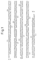
- Fig. 1 shows the sequences of a synthetic oligomers used for construction of a synthetic hProPTH (1-84) gene.
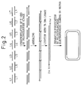
- Fig. 2 shows a process for constructing the synthetic hProPTH (1-84) gene.
- Fig. 3 shows a process for constructing plasmid pG210S(S/X).
- Plac represents the E. coli lactose operon promoter and Ttrp represents the E. coli TrpE attenuator terminator.
- Fig. 4 shows a process for constructing plasmid pGP#19 which expresses the chimeric protein ⁇ Gal-139S(FM)PPH84.
- Fig. 5 shows a process for constructing plasmid pPTH(1-34)pro ⁇ .
- Fig. 6 shows a process for constructing plasmid ptacCATPTH(1-34) which expresses the chimeric protein CATPH34.
- Ptac represents a synthetic promoter of the -35 region of trp promoter and the -10 region of Plac.
- Fig. 7 is a photograph of SDS-PAGE for a sample of the chimeric protein CATPH34 expressed by E. coli, before and after purification.
- Fig. 8 shows a process for constructing plasmid pGP#19PPH34 which expresses the chimeric protein ⁇ Gal-139SPPH34.
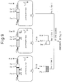
- Fig. 9 shows the former steps in a process for constructing plasmid pG117S4HPPH34 which expresses the chimeric protein ⁇ Gal-117S4HPPH34.
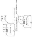
- Fig. 10 shows the latter steps in the process for constructing plasmid pG117S4HPPH34 which expresses the chimeric protein ⁇ Gal-117S4HPPH34.
- Fig. 11 shows the structure of the KEX2 gene and the sequences of the primers synthesized for construction of the secretory Kex2 derivative genes, and their respective annealing sites.
- Fig. 12 shows a process for constructing plasmid pYE-660 which expresses a secretory Kex2 derivative.
- PKEX2 represents a promoter for the KEX2 gene of Saccharomyces cerevisiae.
- Fig. 13 shows a process for constructing plasmid pYE-614 which expresses Kex2-614.
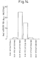
- Fig. 14 is a graph comparing Kex2 activity per OD660 of secretory Kex2 derivatives using a synthetic substrate. The relative activities of the cultures of secretory Kex2 derivative producing strains are given taking the activity of K16-57C[pYE-614] as 1.
- Fig. 15 is a photograph of an electrophoresis which gives a comparison of yields per 200 ⁇ l of culture supernatant of secretory Kex2 derivatives.
- Fig. 16 is a graph showing the activities of Kex2-660 at different urea concentrations, using the synthetic substrate Boc-Leu-Arg-Arg-MCA. The relative activities at each urea concentration are given taking the Kex2-660 activity in the absence of urea as 100%.
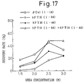
- Fig. 17 is a graph comparing the recovery rates of ⁇ Gal(1-14), hPTH(1-84), hPTH(1-44) and [hPTH(1-84) + hPTH(1-44)] from ⁇ Gal-139S(FM)PPH84, at different urea concentrations (1.5-3.0 M).
- Fig. 18 is a graph comparing the recovery rates of ⁇ Gal(1-14), hPTH(1-84), hPTH(1-44) and [hPTH(1-84) + hPTH(1-44)] from ⁇ Gal-139S(FM)PPH84, at different urea concentrations (3.0-4.0 M).
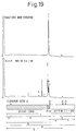
- Fig. 19 shows an elution profile of HPLC for before and after Kex2-660 processing of the chimeric protein ⁇ Gal-139S(FM)PPH84 and a schematic representation of the relationship between identified peptide fragments and ⁇ Gal-139S(FM)PPH84.
- the peak numbers in the profile correspond to the numbers of the fragments.
- Fragments 1, 2, 3 and 4 were identified by determining the amino acid sequences.
- Fragment 7 was estimated based on elution time, and fragment 5 was estimated by correlation between ⁇ Gal(1-14) and hPTH(1-84). Fragment 6 was so designated for fragments which may be eluted.
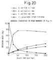
- Fig. 20 is a graph comparing the recovery rates of hPTH(1-84), hPTH(1-44) and hPTH(45-84) from ⁇ Gal-139S(FM)PPH84, at different enzyme concentrations.
- the solid squares, open circles, solid circles and solid triangles represent, respectively, recovery rates for ⁇ Gal-139S(FM)PPH84, hPTH(1-84), hPTH(1-44) and hPTH(45-84).
- the recovery rates were calculated in the following manner.
- hPTH(1-84) and ⁇ Gal-139S(FM)PPH84 the peak area ratio against a known concentration of a corresponding standard substance was used, and for hPTH(1-44) and hPTH(45-84) the peak area ratio against a known concentration of hPTH(1-84) was used, and compensation was made based on the number of amino acid residues of the corresponding peptides.
- Fig. 21 shows an elution profile of HPLC for before and after Kex2-660 processing of the chimeric protein CATPH34.
- Fig. 22 shows a process for constructing plasmid pCU660 which expresses Kex2-660.
- Fig. 23 is a photograph of SDS-PAGE which shows the secretion of Kex2-660 in culture supernatants for different culturing times of TK62/pCU660#10.
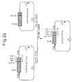
- Fig. 24 shows a process for constructing plasmid pG210ShCT[G].
- Fig. 25 is a graph comparing yield of each secretory Kex2 derivative per OD660 of culture based on Kex2 activities using a synthetic substrate. The yields of each of the secretory Kex2 derivatives are given with the yield of K16-57C[pYE22-614] as 1.
- Fig. 26 is a photograph of SDS-PAGE which gives a comparison of yields per 200 ⁇ l of culture supernatant of secretory Kex2 derivatives.
- Lanes 1 and 12 are developed from molecular weight markers, and lanes 2 through 11 from concentrates of culture supernatants of K16-57C[pYE-22m], K16-57C[pYE22-614], K16-57C[pYE22-630], K16-57C[pYE22-640], K16-57C[pYE22-650], K16-57C[pYE22-660], K16-57C[pYE22-679], K16-57C[pYE22-682], K16-57C[pYE22-688] and K16-57C[pYE22-699].
- the numbers to the left of lane 1 indicate the size (kDa) of the molecular weight markers.
- Fig. 27 shows a process for the construction of Kex2-660 expression plasmid pHIL-660 for a host Pichia pastoris.
- the proteins according to the present invention differ considerably in terms of production and secretion efficiency depending on the length of the protein and particularly the position on the C-terminus. We find that these are proteins of lengths which give high production and secretion efficiency; the Kex2 derivatives have amino acid sequences from Met at amino acid 1 to any of the amino acids at positions 618 to 698 of the amino acid sequence represented by SEQ ID NO.1.
- the C-terminus of a Kex2 protease derivative of the present invention is preferably any one of the amino acids from the position 630 to the position 688 of the amino acid sequence of SEQ ID NO.1, more preferably it is any one of the amino acids from the position 360 to the position 682 of the amino acid sequence of SEQ ID NO.
- amino acid sequence 2 and more preferably it is any one of the amino acids from the position 630 to the position 679.
- the above-mentioned amino acid sequences composed of portions of the amino acid sequence of SEQ ID No.1 are sometimes referred to as natural amino acid sequences for the purpose of the present invention.
- the present invention also encompasses, in addition to Kex2 protease derivatives having the aforementioned natural amino acid sequences, also proteins with Kex2 protease activity having amino acid sequences which are the aforementioned natural amino acid sequences modified by a substitution, deletion or addition of one or more amino acids e.g modifications between said terminus portions, and e.g. usually modifications entailing the substitution, deletion or addition of from 1 to 5 or 10 or 15 or 20 or 30 amino acids, always subject to activity being present.
- the present invention still further provides genes, particularly DNA, coding for the aforementioned various polypeptides.
- the DNA may be prepared according to a conventional method, for example from full-length DNA having the nucleotide sequence represented by SEQ ID No. 1 or another nucleotide sequence coding for the same amino acid sequence, or by cleaving a DNA containing the object DNA, and linking the cleavage product to an oligonucleotide if desired or by introducing a translation termination codon at a suitable location in the DNA.
- DNA coding for one of the aforementioned modified amino acid sequences may be prepared by a conventional method such as site-directed mutagenesis or the polymerase chain reaction (PCR), using the natural full-length DNA having the nucleotide sequence represented by SEQ ID NO.1 or a fragment thereof as a template, and using a primer oligonucleotide containing a desired mutation as a mutagenic primer.
- PCR polymerase chain reaction
- the expression vector according to the invention contains an expression regulating region such as a promoter which is functional in the host used.
- a promoter which is functional in the host used.
- yeast cells when yeast cells are used as the host, glyceraldehyde-3-phosphate dehydrogenase promoter, glycerophosphate kinase promoter, acid phosphatase promoter, alcohol oxidase promoter, formate dehydrogenase promoter, methanol oxidase promoter or the like may be used.
- the host cells used according to the invention may be yeast cells.
- the yeast cells are preferably from Saccharomyces, Pichia, Hansenula or Candida, which include Saccharomyces cerevisiae, Pichia pastoris, Hansenula polymorpha and Candida boidinii.
- Especially preferred yeast is methylotrophic yeast, such as the genera Candida and Pichia, such as Candida beidinii and Pichia pastoris.
- Kex2 derivatives used for the invention are Kex2-614, Kex2-630, Kex2-640, Kex2-650, Kex2-660, Kex2-679, Kex2-682, Kex2-688 and Kex2-699.
- Kex2 derivative genes the Saccharomyces cerevisiae glyceraldehyde-3-phosphate dehydrogenase (GAP) gene promoter was used. Plasmids containing these expression units were introduced into Saccharomyces cerevisiae, which was then cultured overnight at 30°C, and the Kex2 protease activities in the cultures were measured using the synthetic substrate Boc-Leu-Arg-Arg-MCA as the substrate.
- Kex2 protease activity was detected in the culture supernatants of the yeast in which expression units of genes for Kex2-614, Kex2-630, Kex2-640, Kex2-650, Kex2-660, Kex2-679, Kex2-682 or Kex2-688 had been introduced, but no activity was detected in the culture supernatant of the yeast in which the expression unit of the Kex2-699 gene had been introduced.
- the secretion yields per OD660 were found in Example 1 to be significantly higher for Kex2-660 and Kex2-679 than for the hitherto reported Kex2-614.
- the results from analysis of SDS-PAGE of samples prepared to 20-fold concentration by ultrafiltration membrane of 10,000 molecular weight fraction of the culture supernatants showed that not only the Kex2 activity but also the amounts of secretion of Kex2-660 and Kex2-679 were greater than Kex2-614. It was demonstrated that the molecular weights increased with greater numbers of amino acid residues, i.e. no autolysis accumulated in this culturing as occurred with Kex2 ⁇ p in the insect cell host Sf9.
- Example 9 it was shown that the OD660 Kex2 activities of cultures of Kex2-630, Kex2-640, Kex2-650, Kex2-660 and Kex2-679 were at least 10 times higher than the hitherto reported Kex2-614, and the Kex2 activities of Kex2-682 and Kex2-688 were 6 times and 3.4 times greater, respectively, than Kex2-614, while the Kex2 activity of Kex2-699 was undetectable.
- the Kex2-660 production test was conducted changing the expression system from the Saccharomyces cerevisiae system to for example an expression system with the methylotrophic yeast Candida boidinii as the host, which has a high production yield per culture. As a result, it was possible to increase the yield to 340 mg per 1 L of culture supernatant. This is the amount capable of releasing about 200 g of the physiologically active peptide hPTH(1-34) from a chimeric protein, and thus it was demonstrated that the present invention is able to supply an amount of enzyme necessary for excision of useful peptides from chimeric proteins on an industrial scale. In addition, it was found that yeast of the genus Candida is especially preferable as host.
- Kex2-660 which had the largest secretion yield was purified from the culture supernatant.
- ⁇ Gal-139S(FM)PPH84 is a chimeric protein prepared by linking hPTH(1-84) via a Phe-Met sequence and a human parathyroid hormone-derived prosequence (Lys-Ser-Val-Lys-Lys-Arg) to ⁇ Gal-139S, which is a polypeptide from the N-terminus to the 139rd amino acid residue of E.
- coli ⁇ -galactosidase which has been substituted with serine residues at its 76th and 122nd cysteine residues.
- the amino acid sequence of ⁇ Gal-139S is represented as SEQ ID NO.2
- the amino acid sequence of hPTH(1-84) is represented as SEQ ID NO.3.
- the sequence at the N-terminus of the resulting peptide is derived from the peptide fragment expected from the substrate specificity of Kex2 protease, and that the purified Kex2-660 had no contamination by other proteases which might interfere with excision of the desired peptide from the chimeric protein.
- the purified Kex2-660 was used to investigate the cleavage conditions when using a protein as the substrate, in order to deal with the third object.
- Kex2-660 was allowed to act on the synthetic substrate Boc-Leu-Arg-Arg-MCA in the presence of 0 to 4.0 M urea, and it was found that at concentrations of 1.0 M, 2.0 M and 4.0 M the activities were reduced to 70%, 40% and 10%, respectively, compared to absence of urea.
- concentration of the urea solution is generally 2.0 to 4.0 M.
- the sequences predicted to be cleaved by Kex2 protease are at the 4 sites of Arg-Arg (amino acids 13-14 of SEQ ID NO.2, hereunder referred to as cleavage site A), Lys-Arg (prosequence portion, hereunder referred to as cleavage site B) and Pro-Arg (amino acids 43-44 and 51-52 of SEQ ID NO.3, hereunder referred to as cleavage sites C and D, respectively).
- the C-terminal ends of each of the sites are cleaved by the protease.
- Kex2-660 was used at different proportions (25 kU, 50 kU, 100 kU, 150 kU and 200 kU of Kex2-660 to 1 mg of chimeric protein) under conditions at which hPTH(1-84) is excised from chimeric proteins, and the structures of the resulting peptide fragments and -their yields were examined.
- hPTH(1-84) was excised at an efficiency of about 75%.
- about 10% of the ⁇ Gal-139S(FM)PPH84 remained.
- ⁇ Gal-139S(FM)PPH84 decreased with increasing amounts of Kex2-660, and almost completely disappeared at 200 kU/ml.
- the proportion of hPTH(1-44) and hPTH(45-84) also increased simultaneously, while the efficiency of hPTH(1-84) underwent no increase (Fig. 20).
- no decrease in the amount of hPTH(1-84) was seen beyond the increase in hPTH(1-44) even with increasing amounts of Kex2-660, and thus it was confirmed that the Kex2-660 purified in Example 2 described hereunder had no contamination by other proteases with different substrate specificities than Kex2 protease under conditions at which hPTH(1-84) is excised from chimeric proteins.
- Gardella et al. suggested the possibility that contaminating proteases or factor-Xa itself degrades hPTH(1-84), judging from lower hPTH(1-84) recovery rates when the enzyme amount is increased or the reaction time is extended, despite the fact that hPTH(1-84) does not include the factor-Xa recognition site, i.e. the Ile-Glu-Gly-Arg sequence.
- the fact that hPTH(1-84) is obtained at a high recovery rate despite the fact that hPTH(1-84) includes 2 sites of cleavage sequences for Kex2 protease suggests that the purified Kex2 derivatives with increased yields according to the invention are useful as enzymes for excision of desired peptides from chimeric proteins.
- the purified Kex2-660 can excise hPTH(1-34) from the soluble chimeric protein CATPH34 in the absence of urea and from the insoluble chimeric protein ⁇ Gal-117S4HPPH34 in the presence of urea, and thus it functions even when the substrates are chimeric proteins with different protective peptides and cleavage site regions, showing that it has wide industrial application. Also, no protease contamination was detected even in the absence of urea.
- secretory Kex2 derivatives with increased yields which are purified to a single band degrade desired peptides under conditions in which the desired peptides are released from chimeric proteins, irrespective of the presence or absence of urea, and have no contamination of other proteases which lower the recovery rates, that selection of the conditions allows these Kex2 derivatives to recover the desired peptides very efficiently even when the desired peptides include recognition sites for the Kex2 proteases, and that the amounts of expression per 1 L of culture medium are sufficient for release of about 200 g of the desired peptides and thus the secretory Kex2 derivatives obtained according to the invention are supplied in amounts necessary for excision of desired peptides from chimeric proteins on an industrial scale.
- Plasmid pG97S4DhCT[G] is a plasmid which is capable of expressing a chimeric protein wherein hCT[G] (a peptide resulting from addition of a glycine residue to the C-terminus of the 32nd amino acid of human calcitonin) has been linked to a peptide comprising the region from the N-terminus to the 97th amino acid of ⁇ -galactosidase (where the 76th cysteine residue is replaced by a serine residue and the 40th, 41st, 71st and 75th glutamic acid residues are replaced by aspartic acid residues: named ⁇ Gal-97S4D) via a glutamic acid residue, under the E. coli lactose operon promoter.
- hCT[G] a peptide resulting from addition of a glycine residue to the C-terminus of the 32nd amino acid of human calcitonin
- the E. coli strain W3110 containing this plasmid was named Escherichia coli SBM323, and was deposited at the National Institute of Bioscience and Human Technology on August 8, 1991 as FERM BP-3503.
- Plasmid ptacCAT is a plasmid which is capable of expressing the chloramphenicol acetyltransferase gene under the synthetic promoter tac.
- the E. coli strain JM109 containing this plasmid was named Escherichia coli SBM336, and was deposited at the National Institute of Bioscience and Human Technology on March 1, 1996 as FERM BP-5436.
- pG97S4DhCT[G] and ptacCAT were used as materials to construct the soluble hPTH(1-34) chimeric protein-expressing vector ptacCATPTH(1-34) (Reference Example 2 and Figs. 5 and 6).
- Plasmid pG210ShCT[G] is a plasmid in which the gene coding for ⁇ Gal-97S4D from pG97S4DhCT[G] is replaced with the gene coding for ⁇ Gal-210S (a peptide consisting of the N-terminus to the 210th amino acid of ⁇ -galactosidase, wherein the 76th, 122nd and 154th cysteine residues are replaced with serine residues).
- Plasmid PG210ShCT[G] can be obtained by linking of a DNA fragment containing the gene coding for ⁇ Gal-210S obtained by digesting pGH ⁇ 210(Ser)rop with restriction enzymes PvuII and EcoRI and a DNA fragment containing a vector portion obtained by digesting pG97S4DhCT[G] with restriction enzymes PvuII and EcoRI (Fig. 24).
- a method for constructing pGH ⁇ 210(Ser)rop is disclosed in Japanese Examined Patent Publication No. 6-87788.
- pG210ShCT[G] was used as material for cloning of a synthetic human parathyroid hormone precursor (hProPTH(1-84)) gene and construction of plasmid pGP#19 (Reference Example 1 and Fig. 5).
- Plasmid pCRII was acquired from Invitrogen Co. and used for direct cloning of the PCR products.
- Plasmid pYE-22m is an expression vector which utilizes the promoter and terminator for the glyceraldehyde-3-phosphate dehydrogenase (GAP) gene and has a multicloning site (MCS: EcoRI-SalI), with the promoter at the EcoRI end, the TRP1 gene as the selective marker, and a 2 ⁇ m DNA portion (inverted repeats) at the replication origin.
- GAP glyceraldehyde-3-phosphate dehydrogenase
- MCS multicloning site
- the E. coli strain JM109 containing plasmid pYE-22m was named Escherichia coli SBM335, and was deposited at the National Institute of Bioscience and Human Technology on March 1, 1996 as FERM BP-5435 (Fig. 12).
- Plasmid pYE-KEX2 (5.0)b (Mizuno et al., Biochem. Biophys. Res. Commun. 156, 246-254, 1988) was used as a template to construct Kex2 derivative genes by the PCR (Fig. 12).
- Plasmid pYE-KEX2 (RI-PvuII) (Japanese Unexamined Patent Publication No. 1-199578) was used to construct the expression vector pYE-614 for a protein comprising a peptide of 14 amino acids (SEQ ID NO.4) at the C-terminus of Kex2-614 (Fig. 13).
- Plasmid pNOTelI is an expression vector which utilizes the promoter and terminator for the alcohol oxidase gene and which includes a restriction enzyme NotI site, with the URA3 gene as the selective marker (Japanese Unexamined Patent Publication No. 5-344895).
- the competent cell line E. coli JM109 was acquired from Toyobo and used for plasmid preparation and chimeric protein expression.
- E. coli JM101 and M25 (Sugimura et al., Biochem. Biophys. Res. Commun. 153, 753-759, 1988) were used for production of the chimeric proteins CATPH34 and ⁇ Gal-117S4HPPH34, respectively.
- the hosts used for secretory expression of the Kex2 proteases were Saccharomyces cerevisiae K16-57C (MAT ⁇ leu2 trpl ura3 kex2-8: Mizuno et al., Biochem. Biophys. Res. Commun. 156, 246-254, 1988) and Candida boidinii TK62.
- TK62 is a uracil-requiring cell line obtained by URA3 mutation from Candida boidinii S2AOU-1 (Sakai, Y. et al., J. Bacteriol., 173, 7458-7463, 1991).
- This Candida boidinii S2AOU-1 strain was named Candida boidinii SAM1958, and was deposited at the National Institute of Bioscience and Human Technology on February 25, 1992 as FERM BP-3766.
- an LB medium (0.5% (w/v) yeast extract, 1% (w/v) tryptone, 0.5% (w/v) NaCl
- SB medium (1.2% (w/v) yeast extract, 2.4% (w/v) tryptone, 0.5% (v/v) glycerol
- SB2 medium 2% (w/v) yeast extract, 1% (w/v) tryptone, 0.5% (v/v) glycerol, 1.2% (w/v) K 2 HPO 4 , 0.3% (w/v) KH 2 PO 4 ) and NU medium
- yeast extract 1.5% (w/v) glucose, 0.3% (w/v) KH 2 PO 4 , 0.3% (w/v) K 2 HPO 4 , 0.27% (w/v) Na 2 HPO 4 , 0.12% (w/v) (NH 4 ) 2 SO 4 , 0.2 g/L NH 4 Cl, 0.2% (w/v) MgSO 4
- YCDP medium 1% (w/v) yeast extract, 2% (w/v) casamino acid, 2% (w/v) glucose, 100 mM potassium phosphate (pH 6.0) was used.
- BMGY medium 1% (w/v) yeast extract, 2% (w/v) peptone, 1% (v/v) glycerol, 1.34% (v/v) YNB w/o AA: Yeast Nitrogen Base without Amino Acids, 0.4 mg/L biotin, 100 mM potassium phosphate (pH 6.0)), BMMY medium (1% (w/v) yeast extract, 2% (w/v) peptone, 0.5% (v/v) methanol, 1.34% (v/v) YNB w/o AA, 0.4 mg/L biotin, 100 mM potassium phosphate (pH6.0)), YPD medium (1% (w/v) yeast extract, 2% (w/v) peptone, 2% (w/v) glucose) and YPGM medium (1% (w/v) yeast extract, 2% (w/v) peptone, 3% (v/v/
- the DNA primers were synthesized by the phosphoramidite method using an automatic synthesizer (Model 380A, Applied Biosystems). The DNA nucleotide sequences were determined by the dideoxy method.
- Cleavage of the DNA with restriction enzymes was accomplished by reaction for one hour using 3- to 10-fold amounts of the enzyme as indicated by the manufacturer.
- Analysis of the plasmid structures was made using 0.5 to 1 ⁇ g of DNA in a 20 ⁇ l reaction solution, and the DNA was prepared using 3 to 10 ⁇ g of DNA in a 50 to 100 ⁇ l reaction solution.
- the reaction temperature and reaction buffer conditions were as indicated by the manufacturer.
- Agarose gel electrophoresis samples were prepared by adding a 1/5 volume of a pigment solution (15% (w/v) Ficoll aqueous solution containing 0.25% (w/v) bromphenol blue) to the reaction solution.
- the agarose gel electrophoresis buffer used was a TAE buffer (10 mM Tris, 20 mM acetic acid, 2 mM EDTA).
- Mupid-2 (Cosmo Bio, KK.) was used for electrophoresis at 100 volts for one hour, and for preparation of the DNA fragments, a horizontal gel (20 cm x 15 cm x 0.7 cm) was used for electrophoresis at 150 volts for 4 hours or 35 volts for 13 hours. After staining of the gel for 20 minutes with ethidium bromide aqueous solution (0.5 ⁇ g/ml), the DNA bands were detected with ultraviolet irradiation.
- the agarose gel concentrations used were 1.0, 1.5 and 2.0% (w/v) depending on the size of the DNA fragments to be fractionated.
- the DNA in the agarose gel was eluted by placing the gel in a dialysis tube filled with 0.1 x TAE buffer and applying a voltage, or by extraction from the gel using SUPREC-01 (Takara Shuzo, KK.).
- the DNA solutions were treated with phenol and then precipitated with ethanol.
- the ligation reaction was conducted adding 10 units of T4 enzyme ligase in 30 ⁇ l of a reaction solution (67 mM Tris/HCl (pH 7.5), 5 mM MgCl 2 , 5 mM DTT, 1 mM ATP) containing 0.05-1 ⁇ g of DNA fragments and reacting at 16°C for 12 - 18 hours, or using a TAKARA Ligation Kit (Takara Shuzo).
- a reaction solution 67 mM Tris/HCl (pH 7.5), 5 mM MgCl 2 , 5 mM DTT, 1 mM ATP
- E. coli The transformation of E. coli was accomplished by the calcium chloride method (competent cells of JM109 were purchased for use), and the transformants were selected on the basis of drug resistance (ampicillin or tetracycline).
- the transformation of the yeast strain K16-57C was accomplished by the lithium acetate method (METHODS IN YEAST GENETICS; A Laboratory Course Manual, Cold Spring Harbor Laboratory Press), and the transformants were selected on the basis of complementation of tryptophan auxotrophy. Transformation of strain TK62 has been described by Sakai et al. (Sakai et al., J. Bacteriol., 173, 7458-7463, 1991).
- Kex2 activity was according to the method of Mizuno et al. (Mizuno et al., Biochem. Biophys. Res. Commun. 156, 246-254, 1988). That is, 100 ⁇ l of Kex2 diluted with 100 mM Tris/HCl (pH 7.0) was added to 100 ⁇ l of 200 mM Tris/HCl (pH 7.0) solution containing 2 mM CaCl 2 , 0.2% (w/v) Lubrol and 100 ⁇ M Boc-Leu-Arg-Arg-MCA (Peptide Laboratories, KK.), and the mixture was allowed to stand at 37°C for 30 minutes.
- the reaction was terminated by addition of 50 ⁇ l of 25 mM EGTA.
- the amount of Kex2 which released 1 pmol of AMC in one minute under the conditions described above was defined as 1 U.
- SDS-polyacrylamide electrophoresis was carried out according to the method of Laemmli (Laemmli et al., Nature 227, 680-685, 1970). That is, a 1/4 volume of 4xSDS sample buffer (375 mM Tris/HCl (pH 6.8), 30% (v/v) glycerol, 7% (w/v) SDS, 15% (v/v) 2-mercaptoethanol, 0.1% (w/v) bromphenol blue) was added to the sample, and the mixture was heated at 90°C for 5 minutes.
- 4xSDS sample buffer 375 mM Tris/HCl (pH 6.8), 30% (v/v) glycerol, 7% (w/v) SDS, 15% (v/v) 2-mercaptoethanol, 0.1% (w/v) bromphenol blue
- a 10 ⁇ l portion was supplied to an SDS-polyacrylamide gel (55 mm x 85 mm x 1 mm or TEFCO Co.) for electrophoresis at 20 mA for 80 minutes. After electrophoresis, the gel was stained with a staining solution (10% (v/v) acetic acid, 40% (v/v) methanol, 0.25% (w/v) Coomassie brilliant blue R250).
- the hProPTH(1-84) gene was synthesized as the 14 fragments U1 to U7 (SEQ ID NOS.5 to 11) and L1 to L7 (SEQ ID NOS.12 to 18), as shown in Fig. 1.
- the hProPTH(1-84) gene was constructed by linking each of the fragments in the following manner (Fig. 2). First, the DNA fragments U1 (SEQ ID NO.5) and L7 (SEQ ID NO.18) (about 1 ⁇ g each) were reacted at 37°C for 15 minutes in 15 ⁇ l of a phosphorylation reaction solution (50 mM Tris/HCl (pH 7.6), 10 mM MgCl 2 , 5 mM DTT) containing 16 units of T4 polynucleotide kinase and 0.5 nM (over 1 MBq) of [ ⁇ - 32 P]dATP.
- a phosphorylation reaction solution 50 mM Tris/HCl (pH 7.6), 10 mM MgCl 2 , 5 mM DTT
- the aforementioned 7 reaction solutions were pooled into one, and ethanol precipitation was performed to recover the DNA. This was dissolved in an 80 ⁇ l solution of 100 mM Tris/HCl (pH 7.6), 6.5 mM MgCl 2 and 300 mM NaCl. After allowing 40 ⁇ l thereof to stand at 95°C for 5 minutes, the temperature was lowered to 43°C over 30 minutes. After cooling on ice, 40 ⁇ l of ligation B solution (Takara Shuzo, KK.) was added and the mixture was allowed to stand at 26°C for 15 minutes.
- the sample was subjected to 5% polyacrylamide electrophoresis. After electrophoresis, the linked DNA fragments were detected by autoradiography. A DNA fragment corresponding to approximately 280 bp was extracted from the gel and purified according to an established method.
- the approximately 280 bp DNA fragment containing the synthetic hProPTH(1-84) gene includes the restriction enzyme EcoRI site at the 5'-end and the restriction enzyme SalI site at the 3'-end. Cloning of the hProPTH(1-84) gene was accomplished by inserting this EcoRI/SalI DNA fragment at the EcoRI/SalI site of pG210ShCT[G].
- pG210ShCT[G] After cleaving pG210ShCT[G] with restriction enzymes EcoRI and SalI, an approximately 3.5 kb DNA fragment containing the vector portion was prepared. This was linked with the approximately 280 bp DNA fragment of the hProPTH(1-84) gene obtained in 1) above, to obtain plasmid pG210ShProPTH (Fig. 3). pG210ShProPTH was used to transform E. coli JM109, obtaining JM109[pG210ShProPTH].
- linkers KM091 SEQ ID NO.19
- KM092 SEQ ID NO.20
- This linker has the restriction enzyme XhoI and EcoRI sites at either end, and a SacI site between them.
- JM109[pGP#19] was seeded in a 1 L Erlenmeyer flask containing 200 ml of SB medium and cultured at 37°C with shaking for 16 hours.
- the total preculturing solution was transferred into 3 L of NU medium containing 10 ⁇ g/ml tetracycline, and aerobically shake cultured at 37°C using a 5 L fermenter (Model KMJ-5B-4U-FP, product of Mitsuwa Physicochemical Industries, KK.)
- the aeration volume was 3 L/min and the shaking speed was adjusted so that the amount of dissolved oxygen remained over 2.0 ppm.
- the pH was kept at pH 7 using 9% (v/v) ammonia water and 1 M phosphoric acid.
- the carbon source provided was glycerol added at 10 ml per 1 L of culture solution on the 3rd, 9th and 14th hours after the start of culturing, and the nitrogen source was a 5-fold concentration of SB medium added at 10 ml per 1 L of culture solution at 9.5 hours after the start of culturing.
- An antifoaming agent (Disfoam CC-222, Nihon Yushi, KK.) was added at 300 ⁇ l/L at the start of culturing, and was added thereafter as necessary.
- the OD660 after 18 hours of culturing was 55, and about 0.5 mg of the chimeric protein ⁇ Gal-139S(FM)PPH84 was produced per ml of culture solution.
- the chimeric protein was produced as an insoluble inclusion bodies, which was purified in the following manner. A 1.5 L portion of culture solution was subjected to centrifugation at 6000 rpm, 4°C for 10 minutes (20PR-52D, product of Hitachi Laboratory, KK.), and the cells were collected. The cells were suspended in 320 ml of 100 mM Tris/HCl (pH 7.0) and disrupted with a french cell pressure press (twice at 10,000 psi).
- the disrupted cell solution was centrifuged at 4000 rpm, 4°C for 15 minutes (05PR-22, product of Hitachi Laboratory, KK.: 50 ml plastic tube, product of Sumitomo Bakelite, KK.). After suspending the precipitate in 30 ml of 20 mM Tris/HCl (pH 7.0) containing 0.5% (w/w) TritonX-100, the suspension was centrifuged at 3000 rpm, 4°C for 15 minutes, and the precipitate was recovered. This procedure was repeated 4 times to obtain the prepurified chimeric protein.
- the purity of the prepurified chimeric protein was approximately 70% (estimated by SDS-PAGE), and the amount of protein was about 670 mg (assayed by the Bradford method using bovine serum albumin as the standard).
- the prepurified chimeric protein was subjected to high performance liquid chromatography (HPLC: Waters 660E by Millipore, KK.) using a YMC Packed column (2 cm x 25 cm, product of Yamamura Chemical Laboratory) for purification.
- HPLC high performance liquid chromatography
- A 0.1% (v/v) trifluoroacetic acid
- B 0.1% (v/v) TFA/80% (v/v) acetonitrile
- %B 30%-60%/60 minutes
- flow rate 10 ml/min
- R4 linker (R4U: SEQ ID NO.21 and R4L: SEQ ID NO.22) was inserted at the restriction enzyme EcoRI-XhoI site of pG97S4DhCT[G] to construct pG97S4DhCT[G]R4.
- the PTH(1-34) gene prepared by PCR and the pro ⁇ linker described below (pro ⁇ U: SEQ ID NO.23 and pro ⁇ L: SEQ ID NO.24) were inserted at the restriction enzyme XhoI-KpnI site of the obtained plasmid pG97S4DhCT[G]R4, to construct pPTH(1-34)pro ⁇ .
- the PTH(1-34) gene was prepared by PCR with pGP#19 as the template, using primers P1 (SEQ ID NO.25) and P2 (SEQ ID NO.26) (Fig. 5).
- primers CAT1 and CAT3 (SEQ ID NOS.27 and 28) were synthesized in order to insert the restriction enzyme XhoI site at the 3'-end of the CAT (chloramphenicol acetyltransferase) gene.
- the CAT gene having the restriction enzyme XhoI site inserted at the 3'-end thereof was obtained by PCR using CAT1 and CAT3 as the primers and ptacCAT as the template DNA.
- Fig. 7 shows the results of SDS-PAGE for samples before and after purification.
- Lane 1 is the molecular weight marker
- lane 2 is the soluble fraction after cell disruption
- lane 3 is the chimeric protein CATPTH(1-34) after purification.
- the numbers to the left of lane 1 indicate the sizes of the molecular weight markers (kDa).
- the chimeric protein was produced in the soluble fraction, and was easily purified by affinity chromatography using chloramphenicol caproate.
- pGP#19 was used as the template and S01 (SEQ ID NO.29) and S02 (SEQ ID NO.30) as primers for PCR to amplify a DNA fragment in which the 35th codon GTT of hPTH(1-84) was replaced with the translation termination codon TAA, after which the restriction enzyme AatII-SalI DNA fragment was isolated and purified by common methods and exchanged with the corresponding portion of pGP#19 to construct pGP#19PPH34 (Fig. 8).
- Linkers S08 (SEQ ID NO.34) and S09 (SEQ ID NO.35) coding for (His) 4 -Pro-Gly were inserted at the restriction enzyme SmaI site of pG117SPPH34 to construct pG117S4HPPH34 (Fig. 10). The orientation of the linkers was confirmed by determining the DNA nucleotide sequences after preparing the plasmids.
- TE 10 mM Tris/HCl, 1 mM EDTA, pH 8.0
- TE 10 mM Tris/HCl, 1 mM EDTA, pH 8.0
- cell disruption with a high-pressure homogenizer (Manton-Gaullin), centrifugation, and suspension and washing with TE and deionized water, to obtain about 100 g of an inclusion bodies.
- the secretory KEX2 gene was constructed by the PCR.
- the primer sequence is shown in Fig. 11(b).
- KM085 (SEQ ID NO.36) has the restriction enzyme EcoRI site (underlined) at the 5'-end
- primers correspond to the KEX2 gene region shown in Fig. 11(a), with KM085 including a nucleotide sequence coding for the initial methionine of the KEX2 gene, and KM088, KM089, KM090 and KM093 having nucleotide sequences which are antisense to sequences in which the translation termination codon TAA is added directly to the 660th, 679th, 688th and 699th amino acids from the N-terminus, respectively.
- a PCR reaction was conducted using plasmid pYE-KEX2(5.0)b, cut with restriction enzyme EcoRI and in linear form as template, using KM085 and KM088 as primers.
- the reaction purification product was cleaved with restriction enzymes EcoRI and SalI to obtain an EcoRI-SalI DNA fragment.
- This DNA fragment has the DNA nucleotide sequence coding for Kex2-660 (KEX2-660), with the restriction enzyme EcoRI site upstream and the restriction enzyme SalI site downstream.
- KM089, KM090 and KM093 were used instead of the primer KM088, the EcoRI-SalI DNA fragments containing nucleotide sequences coding for Kex2-679, Kex2-688 and Kex2-699 (KEX2-679, KEX2-688, KEX2-699) were recovered and linked with the EcoRI-SalI fragment of plasmid pYE-22m, to obtain plasmids pYE-679, pYE-688 and pYE-699.
- Plasmid pYE-614 was constructed by replacing the BglII-SalI DNA fragment containing a portion of the KEX2 gene of pYE-KEX2 (RI-PvuII) with the BglII-SalI DNA fragment containing a portion of the KEX2-660 gene of pYE-660 (Fig. 13).
- the plasmids (pYE-22m, pYE-614, pYE-660, pYE-679, pYE-688 and pYE-699) were each introduced in strain K16-57C to obtain strains K16-57C[pYE-22m], K16-57C[pYE-614], K16-57C[pYE-660], K16-57C[pYE-679], K16-57C[pYE-688] and K16-57C[pYE-699].
- Kex2 derivative secretion in the culture solutions were determined by assay of Kex2 activity in the culture supernatants and SDS-PAGE of their concentrates.
- the colonies were seeded into 4 ml of YCDP medium and then cultured overnight at 32°C with shaking. After transferring 100 ⁇ l of culture solution to 4 ml of YCDP medium, it was cultured overnight at 32°C with shaking. One ml of the culture solution was centrifuged at 12,000 rpm, 5 minutes, 4°C (MRX-150, Tomy Seiko) to obtain the culture supernatant. After diluting the culture supernatant 2- to 64-fold with 100 mM Tris/HCl (pH 7.0), the Kex2 activity was measured. The results are shown in Fig. 14.
- Kex2 activities per OD660 of K16-57C[pYE-660], K16-57C[pYE-679] and K16-57C[pYE-688] were 25, 15 and 1.2 times greater, respectively, than that of K16-57C[pYE-614]. No Kex2 activity was detected in the culture supernatants of K16-57C[pYE-22m] and K16-57C[pYE-699].
- the results are shown in Fig. 15.
- Lanes 1 and 7 are molecular weight markers, lane 2 is for K16-57C[pYE-22m], lane 3 is for K16-57C[pYE-614], lane 4 is for K16-57C[pYE-660], lane 5 is for K16-57C[pYE-679] and lane 6 is for K16-57C[pYE-688].
- the numbers to the left of lane 1 indicate the sizes of the molecular weight markers (kDa).
- Kex2-660 and Kex2-679 had greater secretion amounts than Kex2-614, similar to their activities. It was also shown that their molecular weights increased correspondingly with the number of amino acid residues, i.e. that no autolysis accumulated in this culturing as occurs with Kex2 ⁇ p in the insect cell host Sf9.
- the secretory yields of Kex2-660 and Kex2-679 were found to be much greater, at least 10 times greater, than the secretory yield of Kex2-614 hitherto reported. Also, since no notable autolysis was observed in the culture supernatants, even higher yields may be expected by investigating other methods of production.
- K16-57C[pYE-660] was cultured overnight at 32°C in 3 L of YCDP medium.
- a 2.3 L portion of the culture supernatant was subjected to concentration and exchange of the buffer solution (20 mM Bis-Tris/HCl (pH 6.0), 50 mM NaCl, 0.2 mM CaCl 2 ), using an ultrafiltration module (UF-LMSII System: UF2CS-3000PS, Toso, KK.) (final volume: 275 ml).
- UF-LMSII System UF2CS-3000PS, Toso, KK.
- a 210 ml portion thereof was adsorbed onto a Q-Sepharose XK16 (Pharmacia, KK.) column equilibrated beforehand with the same buffer solution. After washing with 75 ml of the same buffer solution, elution (120 ml) was performed in the same buffer solution with a linear concentration gradient of 50 to 350 mM NaCl concentration. The flow rate was 3 ml/min. Kex2 activity was recovered in 24 ml of eluted fractions of 150 to 250 mM NaCl concentration.
- a 15 ml portion thereof was adsorbed onto a Phenyl Superose HR 5/5 (Pharmacia) column equilibrated beforehand with a 20 mM Bis-Tris/HCl (pH 6.0), 0.2 mM CaCl 2 solution containing 2 M ammonium sulfate. After washing with 2.5 ml of the same buffer solution, elution (15 ml) was performed in 20 mM Bis-Tris/HCl (pH 6.0), 0.2 mM CaCl 2 with a linear concentration gradient of 2 to 0 M ammonium sulfate. The flow rate was 0.5 ml/min.
- Kex2 activity was recovered in 2.25 ml of eluted fractions of 0.8 to 0.6 M ammonium sulfate concentration.
- the recovery rates of Kex2-660 at each stage are shown in Table 1. The overall recovery rate was determined by integrating the recovery rates at each stage.
- Table 1 Kex2-660 purification Stage Activity (x10 4 U/ml) Recovery rate (%) *1 *2 Culture supernatant 9.3 100 0.22 ⁇ m filtration 9.2 99 99 Ultrafiltration 50 99 97 Q-Sepharose 250 63 61 Phenyl Superose 1,740 93 57 *1: Recovery rate at each stage *2: Recovery rate from culture supernatant sample
- the chimeric protein In chimeric protein expression process, the chimeric protein usually forms insoluble inclusion bodies, and thus urea or the like is used as a denaturing agent to solubilize it.
- urea The effect of urea on the activity of Kex2 protease and secretory Kex2 derivatives has not been reported.
- ⁇ Gal-139S(FM)PPH84 has 4 sites of sequences predicted to be cleaved by Kex2 protease: Arg-Arg (cleavage site A), Lys-Arg (cleavage site B) and Pro-Arg (cleavage sites C and D). Cleavage at cleavage sites A, B and C was confirmed from the identified peptide fragments, but cleavage at cleavage site D could not be confirmed.
- the open squares, open circles, solid circles and open triangles represent, respectively, recovery rates for ⁇ Gal(1-14), hPTH(1-84), hPTH(1-44) and hPTH(1-84) + hPTH(1-44).
- a urea concentration of 3.5 to 4.0 M is preferred at which the cleavage efficiency at cleavage site B is high and the cleavage efficiency at cleavage site C is low.
- Example 3 ⁇ Gal(1-14), hPTH(1-84) and hPTH(1-44) were identified and the effect of urea on the cleavage efficiency of ⁇ Gal-139S(FM)PPH84 by Kex2-660 was investigated based on their peptide recovery rates. However, peptide fragments from hPTH(45-84) could not be identified. Thus, a sample of hPTH(1-84) processed with Kex2-660 was separated and analyzed by HPLC under various elution conditions to identify hPTH(45-84), and it was confirmed that it had eluted out in the fraction which had passed through in elution under the previous conditions. These results demonstrated that cleavage site D undergoes virtually no cleavage.
- Fig. 19 shows an elution profile of HPLC for samples unprocessed with Kex2-660 and processed with 50 kU of Kex2-660. Peaks 1, 2, 3, 4 and 7 correspond, respectively, to hPTH(45-84) (from the amino acid after cleavage site C to the C-terminus of hPTH(1-84)), ⁇ Gal(1-14) (from the N-terminus of ⁇ Gal-139S to cleavage site A), hPTH(1-84), hPTH(1-44) (from the amino acid after cleavage site B to cleavage site C) and ⁇ Gal-139S(FM)PPH84 (full length of the chimeric protein).
- Peaks 6 and 7 were larger when the amount of Kex2-660 was smaller and smaller when the amount was larger, and since peak 5 increased as peaks 6 and 7 decreased, it was concluded that peak 5 was a peptide from the amino acid after cleavage site A to cleavage site B of the chimeric protein, and peak 6 was a peptide from the N-terminus of ⁇ Gal-139 to cleavage site B or from the amino acid after cleavage site A to the C-terminus of hPTH(1-84). (Peak 6 may possibly be from the N-terminus of ⁇ Gal-139S to cleavage site C, but this is unlikely since Arg-Arg is more easily cut than Pro-Arg.)
- peaks 1, 4, 5, 6 and 7 varied in the range of 25 to 200 kU/ml, while the sizes of peaks 2 and 3 were virtually unchanged. Even when 200 kU/ml of Kex2-660 was used, no new peaks appeared. In other words, it was confirmed that no protease activity other than that of Kex2 protease is detected even when using 8 times (200 kU/ml) the necessary amount of Kex2-660 (25 kU/ml) for excision of hPTH(1-84), and that the Kex2-660 purified in Example 2 had no contamination by other interfering proteases under conditions at which hPTH(1-84) is excised from chimeric proteins.
- hPTH(1-84) The recovery rates for the peptide fragments derived from hPTH(1-84) in the range of 25 to 200 kU/ml are summarized in Fig. 20. It is clear that when 50 kU/ml of Kex2-660 was used, hPTH(1-84) was recovered at about 75%. Here, although about 10% of the ⁇ Gal-139S(FM)PPH84 remained, it decreased as the amount of Kex2-660 increased, almost totally disappearing at 200 kU/ml. However, the proportion of hPTH(1-44) also increased simultaneously, and thus the recovery rate of hPTH(1-84) did not increase.
- Kex2-660 is capable of excising desired peptides from chimeric proteins in an efficient manner (an excision efficiency of 75%) even when the desired peptide has a cleavage site for Kex2 protease.
- This excision efficiency is higher than the excision efficiency of 50% for hPTH(1-84) using factor-Xa (Gardella et al., J. Biol. Chem. 265(26), 15854-15859, 1990).
- hPTH(1-84) is obtained at a high yield despite the fact that hPTH(1-84) includes 2 sites of cleavage sequences for Kex2 protease suggests that the purified Kex2 derivatives with increased yields according to the invention are useful as enzymes for excision of desired peptides from chimeric proteins.
- hPTH(1-34) In order to excise hPTH(1-34) from the chimeric protein CATPH34, 239 ⁇ l of deionized water, 1.32 ⁇ l of 1 M CaCl 2 and 30 kU of Kex2-660 were added to 60 ⁇ l taken from the eluate from Reference Example 2, and the mixture was heated at 37°C for one hour. After the reaction the appearing peaks were examined for amino acid analysis, and the amino acid composition was found to match that of hPTH(1-34) (Fig. 21).
- Kex2-660 can be used for excision of hPTH(1-34) from chimeric proteins. Furthermore, Kex2-660 is able to excise desired proteins even when the chimeric proteins have different protective peptides and cleavage site regions, and thus it has wide industrial application.
- Example 1 demonstrated that Kex2-660 undergoes no notable autolysis in culture solution. Thus, greater yields may be expected by using more efficient expression systems. We therefore attempted to produce Kex2-660 in an expression system using the methanol-utilizing yeast Candida boidinii as the host.
- the NKEX2-660 gene was constructed by the PCR in the same manner as Example 1, 1), except that NKEX2 (SEQ ID NO.41) and KM088 were used as the primers.
- NKEX2 contains the nucleotide sequence of the restriction enzyme NotI site (underlined) at the 5'-end, and includes the sequence of bases -107 to -132 upstream from the initial methionine of the KEX2 gene (Fig. 11).
- the NKEX2-660 gene (the KEX2 gene with 132 base pairs of the 5' untranslated region of the KEX2 gene) was cloned in pCRII and then excised using restriction enzyme NotI.
- the NotI DNA fragment containing the NKEX2-660 gene was inserted at the NotI site of plasmid pNOTelI under promoter control to allow expression of the KEX2-660 gene, to thus construct pCU660 (Fig. 22).
- Plasmid pCU660 which had been digested with restriction enzyme BamHI on the URA3 gene and in linear form was introduced into TK62, and the transformed strains TK62/pCU660 were selected. Twenty TK62[pCU660] strains (#1 to #20) were then cultured at 27°C with shaking in BMGY medium. After 2 days, approximately 10 OD ⁇ ml of the preculturing solution was transferred into 1 ml of BMMY medium and further cultured at 27°C with shaking. After 30 hours, the Kex2 activity of the culture supernatant was measured. The 5 strains with the highest activity were cultured in the same manner, and TK62[pCU660]#10 which had reproducible high Kex2 activity was selected and cultured in a fermenter.
- methanol, glycerol and nitrogen source 5% (w/v) yeast extract, 10% (w/v) peptone, 6.7% (v/v) YNB w/o AA; 1/25 volume/addition) were supplemented as appropriate.
- the pH was controlled to remain above pH 5.5 by adding 7.5% (v/v) ammonia water.
- An antifoaming agent (Disfoam CC-222, Nihon Yushi, KK.) was added at 0.5 ml/L, at the start of culturing and was added thereafter as necessary.
- the results of SDS-PAGE for the culture supernatant at each culturing time are shown in Fig. 23.
- the OD600 after 48 hours of culturing was 353, and this culturing produced about 2800 MU of Kex2-660 (corresponding to about 340 mg) per 1 L of culture supernatant.
- Example 1 it was demonstrated that the yields of Kex2 proteases lacking the C-terminal region (Kex2-660 and Kex2-679) were notably higher than those of Kex2-614, Kex2-699 and Kex2-688.
- additional Kex2 proteases lacking the C-terminal region Kex2-630, Kex2-640, Kex2-650 and Kex2-682 were constructed, to further investigate the relationship between the C-terminal region and Kex2 protease yields.
- the secretory Kex2 genes were constructed by the method in Example 1, 1). Specifically, the primer sequences KM100 (SEQ ID NO.42), KM102 (SEQ ID NO.43), KM103 (SEQ ID NO.44) and KM104 (SEQ ID NO.45) have nucleotide sequences which are antisense strands to sequences in which the translation termination codon TAA is added directly after the 630th, 640th, 650th and 682nd amino acids from the N-terminus, respectively.
- Plasmids (pYE-630, pYE-640, pYE-650 and pYE-682) were introduced into strain K16-57C to obtain strains K16-57C[pYE-630], K16-57C[pYE-640], K16-57C[pYE-650] and K16-57C[pYE-682]. These transformants were then cultured, with the secretory Kex2 derivative-producing strains prepared in Example 1, 2), and the Kex2 yields (amount of Kex2 secreted into the culture medium) were determined by measurements of Kex2 activity in the culture supernatants and SDS-PAGE of the culture supernatant concentrates.
- the colonies were inoculated into YCDP medium and then cultured at 30°C with shaking to prepare cells in the logarithmic growth phase. These cells were subcultured in YCDP medium until the OD660 absorbance reached approximately 0.02, and then further cultured for about 16 hours at 30°C.
- the results of Kex2 activity measurement are shown in Fig. 25, and the results of SDS-PAGE are shown in Fig. 26.
- Kex2 activities per OD660 in culture supernatant for K16-57C[pYE-630], K16-57C[pYE-640], K16-57C[pYE-650], K16-57C[pYE-660] and K16-57C[pYE-679] were roughly 12 times that of K16-57C[pYE-614], thus showing no difference in Kex2 yields for the range of KEX2-630 to KEX2-679.
- Kex2 yields for K16-57C[pYE-682] and K16-57C[pYE-688] were, respectively, 6 times and 3.4 times that of K16-57C[pYE-614], i.e. they were lower the longer the C-terminal region (Fig. 25). No Kex2 activity was detected in the culture supernatants of K16-57C[pYE-22m] and K16-57C[pYE-699].
- Example 8 showed that the secretory Kex2 derivative Kex2-660 can be produced in large amounts in an expression system using Candida boidinii as the host.
- The.possibility of producing Kex2-660 with other methylotrophic yeast was investigated using an expression system with Pichia pastoris as the host ( Pichia Expression Kit, Invitrogen Co.).
- a PCR reaction was conducted using plasmid pYE-660, digested with restriction enzyme EcoRI and in linear form, as the template and KM085 (SEQ ID NO.36) and KM088 (SEQ ID NO.37) as primers.
- the reaction purification product was cloned in pCRII (Invitrogen Co.).
- the resulting plasmid was digested with restriction enzyme EcoRI, to obtain a DNA fragment consisting of the KEX2-660 gene with the restriction enzyme EcoRI site at both ends.
- the DNA fragment containing the KEX2-660 gene was inserted at the restriction enzyme EcoRI site of plasmid pHIL-D2 ( Pichia Expression Kit), to obtain plasmid pHIL-660 having the KEX2-660 gene inserted in an orientation allowing its expression under AOX promoter (Fig. 27).
- a fragment containing the KEX2-660 gene obtained by digesting plasmid pHIL-660 with restriction enzyme NotI was introduced into Pichia pastoris GS115 (his - , AOX + , Pichia Expression Kit), and the GS115[pHIL-660] transformants which grew in medium containing no histidine were selected. From these were obtained 5 GS115[pHIL-660](AOX - ) strains which could not grow with methanol alone as the carbon source. These were then cultured in BMMY medium, to obtain GS115[pHIL-660] #23 which had the greatest yield of Kex2 production.
- the Kex2 yield of GS115[pHIL-660] #23 was then examined. First, a colony was seeded into 10 ml of BMGY medium and cultured at 30°C for 2 days with shaking. Cells obtained by centrifugation of 10 ml of the culture medium were suspended in 2 ml of BMMY medium and further cultured at 25°C for 2 days with shaking, after which the Kex2 activity of the culture supernatant was measured. As a result, the production of Kex2-660 was found to be about 1350 KU (corresponding to about 160 ⁇ g) per 1 ml of culture medium.
- Kex2-660 can be produced in large amounts even in an expression system in which the host is Pichia pastoris, methylotrophic yeast other than Candida boidinii.
- Fig. 27 shows the method of constructing the Kex2-660-expressing plasmid pHIL-660 with Pichia pastoris as the host.
Landscapes
- Chemical & Material Sciences (AREA)
- Life Sciences & Earth Sciences (AREA)
- Health & Medical Sciences (AREA)
- Genetics & Genomics (AREA)
- Organic Chemistry (AREA)
- Engineering & Computer Science (AREA)
- Molecular Biology (AREA)
- Biochemistry (AREA)
- Bioinformatics & Cheminformatics (AREA)
- Wood Science & Technology (AREA)
- Zoology (AREA)
- General Health & Medical Sciences (AREA)
- Biomedical Technology (AREA)
- Biotechnology (AREA)
- Medicinal Chemistry (AREA)
- General Engineering & Computer Science (AREA)
- Mycology (AREA)
- Biophysics (AREA)
- Microbiology (AREA)
- Proteomics, Peptides & Aminoacids (AREA)
- Gastroenterology & Hepatology (AREA)
- Physics & Mathematics (AREA)
- Plant Pathology (AREA)
- Analytical Chemistry (AREA)
- Preparation Of Compounds By Using Micro-Organisms (AREA)
- Micro-Organisms Or Cultivation Processes Thereof (AREA)
- Seasonings (AREA)
- Acyclic And Carbocyclic Compounds In Medicinal Compositions (AREA)
- Enzymes And Modification Thereof (AREA)
- Organic Low-Molecular-Weight Compounds And Preparation Thereof (AREA)
Abstract
Description
- The present invention relates to Kex2 derivatives with Kex2 protease activity which are secreted in large amount in culture medium, and to a method for their production. The invention also relates to a method of using the aforementioned secretory Kex2 derivatives.
- Many attempts have been made at methods for producing physiologically active peptides by chimeric protein expression, and chemical or enzymatic cleavage methods have been used for release of the desired proteins. Chemical methods include -cleavage of asparagine residue with nitrous acid and cleavage of methionine residue with CNBr (Itakura et al., Science 198, 1059, 1977). However, these methods necessarily involve modification of the protein of interest, and problems of purification cost.
- Enzymatic methods employ lysyl endopeptidase which specifically cleaves the peptide bond of the C-terminal of lysine (Achromobacter protease I) and Staphylococcal protease V8 which specifically cleaves the peptide bond of the C-terminal of the glutamic acid (Japanese Examined Patent Publication No. 6-87788). However, since these chemical methods and endoproteases recognize a single amino acid residue, it is a precondition that amino acid residue not be present in the desired peptide in order to allow efficient excision of the desired peptide from the chimeric protein, and thus the peptides which can be produced are limited. Efforts have therefore been directed at developing a highly universal cleavage method which recognizes multiple amino acid residues.
- Prohormone converting enzymes are enzymes which produce peptide hormones from their precursors in vivo, and they are expected to have favorable qualities as enzymes for excision of peptide hormones from proteins, even in vitro. Kex2 protease is a prohormone converting enzyme derived from Saccharomyces cerevisiae, and it is a calcium-dependent serine protease which specifically cleaves peptide bonds at the C-terminal ends of Lys-Arg, Arg-Arg and Pro-Arg sequences. Kex2 protease is a protein composed of 814 amino acid residues with a signal sequence at the N-terminus and a transmembrane region at the C-terminus with a continuous string of hydrophobic amino acids, and it is localized in the trans Golgi in cells.
- A nucleotide sequence coding for Kex2 protease and the corresponding amino acid sequence are shown in the Sequence Listing as SEQ ID NO.1. Genetic expression of a Kex2 derivative lacking the C-terminal region in Saccharomyces cerevisiae and subsequent analysis thereof revealed that the Kex2 derivative with the amino acid sequence from
amino acids 1 to 614 of SEQ ID NO.1 retains the Kex2 protease activity, and is secreted in culture medium (Fuller et al., Proc. Natl. Acad. Sci. USA, 86, 1434-1438, 1989, Japanese Unexamined Patent Publication No. 1-199578). In the present specification, the Kex2 protease derivative is represented by the number of amino acids counting fromamino acid 1 of SEQ ID NO.1. For example, the Kex2 derivative with the amino acid sequence fromamino acids 1 to 614 of SEQ ID NO.1 is represented as Kex2-614. - Heretofore known Kex2 derivatives whose secretory production methods have been studied include ss-Kex2 and Kex2Δp.
- ss-Kex2 is a Kex2 derivative which has a 3 amino acid residue peptide added to Kex2-614, and its production in Saccharomyces cerevisiae has been studied (Brenner et al., Proc. Natl. Acad. Sci. USA, 89, 922-926, 1992). It was expressed in a protease-deficient mutant (pep4) as a host (in a 4 mg/L culture medium), and was purified from the culture supernatant at a purification yield of 20%. The reduced molecular weight of the purified ss-Kex2 treatment with Asn-type sugar chain hydrolyzing enzyme EndoH suggests that it includes Asn-type sugar chains. The pH dependency and substrate specificity of the enzyme activity has also been studied using synthetic substrates.
- Kex2Δp is a Kex2 derivative represented in this specification by Kex2-666, and studies of its production in the insect cell host Sf9 have shown that 90% of its activity is secreted into the culture supernatant, and that the molecular weight of the secreted Kex2Δp is 70 kDa, which is smaller than the intracellular molecular weight of 120 kDa (Germain et al., Eur. J. Biochem. 204, 121-126, 1992). In addition, since the 70 kDa molecular weight protein is found in the culture supernatant in which Kex2 is expressed, and replacement of the 385th serine residue by alanine residue of Kex2Δp (the catalytic portion of Kex2 protease activity) results in Kex2Δp in the culture supernatant with a molecular weight of 120 kDa, equal to the intracellular molecular weight, the 70 kDa protein is believed to be an autolysate of the C-terminal portion-deficient Kex2Δp (120 kDa) in the culture medium.
- Attempts have also been made at expression of the derivative Kex2Δ504 in which the cleavage site of the Lys-Arg sequence (amino acids 503-504 of SEQ ID NO.1), expected from the molecular weight of the decomposition product and the substrate specificity of Kex2 protease, is replaced with the Lys-Leu sequence. However in this case as well a 70 kDa protein is found in the culture medium, and since the Lys-Arg sequence is not always cleaved by Kex2Δp during autolysis, and no other sequence exists as the recognition site of Kex2 protease, this suggests the possibility that Kex2Δ504 recognizes a completely different sequence than the one predicted from the synthetic substrate, and cleaves itself.
- Thus, despite research on substrate specificity of Kex2 derivatives using synthetic substrates, the substrate specificity when using proteins is not yet understood. Also, little is known about the secreted amounts of the different Kex2 derivatives, and it is still not known whether stable secretory production of Kex2 derivatives other than Kex2-614 is possible.
- The general aim herein is to provide new and useful techniques for supplying a large amount of enzymes with Kex2 protease activity, with demonstration of uses of such enzymes.
- Specifically, the use of enzymes with Kex2 protease activity for the production of useful peptides by chimeric protein expression on an industrial scale particularly calls for one or more of the following three issues to be addressed.
- A first issue is to increase the amount of it of production of the Kex2 derivatives. The enzyme having Kex2 protease activity with the greatest yield hitherto reported has been ss-Kex2, and that yield is about 4 mg per 1 L of culture medium. However, this yield is low in terms of production of enzyme for excision of desired peptides from chimeric proteins on an industrial scale. Also, since secretory Kex2 derivatives such as Kex2Δp are believed to possibly undergq autolysis in culture medium and the cleavage site cannot be predicted, it is unknown how to design derivatives to increase the yield.
- Consequently, it would be advantageous to find Kex2 derivatives which do not undergo autolysis and to construct a high-expression system for those Kex2 derivatives. In the present specification, autolysis refers to decomposition which brings a reduction in Kex2 protease activity, and does not refer to maturation of Kex2 protease which accompanies the autocleavage of Lys-Arg (amino acids 108-109 of SEQ ID NO.1) (Brenner & Fuller, Proc. Natl. Acad. Sci. USA, 89, 922-926, 1992).
- A second issue is establishment of a purification process for high purity Kex2 derivatives without contamination by other proteases. The activity of Kex2 derivatives hitherto reported are evaluated using only synthetic substrates and not protein substrates, and thus the presence of contamination by other proteases cannot be determined. In particular, it is difficult to achieve precise control of reaction conditions when excising chimeric proteins on an industrial scale, and since contamination by other proteases can notably lower the recovery rates of the object peptides, the enzymes with Kex2 protease activity should desirably be of a high degree of purity.
- A third issue is setting the conditions for cleaving chimeric proteins by enzymes with Kex2 protease activity. It is well-known to those in the art that the tertiary structure of proteins affects enzyme activity, the enzyme stability under the reaction conditions and recognition of the substrate. However, almost no previous reports have dealt with these points. In particular, since chimeric proteins often form insoluble inclusion bodies in chimeric protein expression methods, denaturing agents such as urea are used for their solubilization. However, it is generally unknown what enzyme structure can retain enzyme activity in the presence of urea. Consequently, it is unclear whether or not Kex2 protease and secretory Kex2 derivatives can be used as enzymes for excision of desired peptides from proteins.
- Mass production of other prohormone converting enzymes has also been unsuccessful, and it is also unknown whether these enzymes can be used as enzymes for excision of desired peptides from chimeric proteins in vitro. Consequently, in order to use prohormone converting enzymes, including Kex2 protease, as enzymes for releasing desired peptides from chimeric proteins, it is important to establish more efficient expression and purification methods, and set the cleavage conditions for using proteins as substrates in vitro.
- As a result of research carried out with these issues in mind, the present inventors have found that Kex2 derivatives having amino acid sequences from
position 1 at the N-terminus to an amino acid at a position between 618 and 698 have notably higher secretory production without undergoing autolysis in culture, and that the production may be further increased by using methylotrophic yeast as the host cells. This can be used for mass supply of Kex2 derivatives. In addition, the inventors purified the secretory Kex2 derivatives from culture supernatant concentrates to single bands in SDS-PAGE by the 2 steps of anion exchange chromatography and hydrophobic chromatography, and have confirmed that, under conditions in which desired peptides are excised from chimeric proteins, the purified Kex2 derivatives contain no other protease activity which decomposes the peptides and lowers the recovery rate. - Furthermore, it was found that under conditions in which desired peptides are excised from chimeric proteins, the substrate specificity of secretory Kex2 derivatives is altered by changing urea concentration, and have demonstrated that a desired peptide can be excised from a chimeric protein at an efficiency of 75% even when the desired peptide includes 2 recognition sites for Kex2 protease. It was also demonstrated that Kex2-660 can be used to excise hPTH (1-34) from the chimeric protein βGal-117S4HPPH34 on a semi-large scale, i.e. that for the secretory Kex2 derivative, the yield, purity and excision efficiency of the desired peptide from the chimeric protein can be suitable for production on an industrial scale.
- In one aspect herein we provide proteins with Kex2 protease activity which are obtained by transforming host cells with an expression vector comprising DNA coding for a "natural" amino acid sequence whose N-terminus is the Met at
amino acid 1 and whose C-terminus is one of the amino acids between amino acids 618 and 698 of the amino acid sequence of the Kex2 protease represented by SEQ ID NO.1, or an amino acid sequence which is this natural amino acid sequence modified by a substitution, deletion or addition of one or more amino acids, and then culturing the resulting transformants and recovering the protein from the culture. In the specification, such proteins are collectively referred to as "enzymes with Kex2 protease activity", "Kex2 protease derivatives", "secretory Kex2 derivatives", etc. - Other aspects of the invention provide genes, particularly DNA, coding for the aforementioned proteins, vectors, particularly expression vectors, comprising the aforementioned DNA, and transformants, preferably animal cells or yeast, obtained by transforming host cells with the aforementioned vector.
- Another aspect is a method for producing the aforementioned proteins, comprising the steps of culturing a host which has been transformed with the aforementioned expression vector and recovering the aforementioned protein from the culture. The protein is preferably recovered from the culture supernatant by anion exchange chromatography and hydrophobic chromatography.
- The present invention still further provides a method for excision of desired peptides from chimeric proteins using the aforementioned proteins. Chimeric protein is a protein obtained by adding a protective peptide to a desired peptide, and the desired peptide may be excised by the aforementioned protein so long as the link between the desired peptide and the protective peptide is an amino acid sequence recognized by the aforementioned protein. Also, even if the junction between a desired peptide and a protective peptide is not an amino acid sequence recognized by the aforementioned protein, a recognition site of the aforementioned protein may be inserted between the desired peptide and the protective peptide to allow the desired peptide to be excised using the aforementioned protein.
- Fig. 1 shows the sequences of a synthetic oligomers used for construction of a synthetic hProPTH (1-84) gene.
- Fig. 2 shows a process for constructing the synthetic hProPTH (1-84) gene.
- Fig. 3 shows a process for constructing plasmid pG210S(S/X). Plac represents the E. coli lactose operon promoter and Ttrp represents the E. coli TrpE attenuator terminator.
- Fig. 4 shows a process for constructing
plasmid pGP# 19 which expresses the chimeric protein βGal-139S(FM)PPH84. - Fig. 5 shows a process for constructing plasmid pPTH(1-34)proα.
- Fig. 6 shows a process for constructing plasmid ptacCATPTH(1-34) which expresses the chimeric protein CATPH34. Ptac represents a synthetic promoter of the -35 region of trp promoter and the -10 region of Plac.
- Fig. 7 is a photograph of SDS-PAGE for a sample of the chimeric protein CATPH34 expressed by E. coli, before and after purification.
- Fig. 8 shows a process for constructing plasmid pGP#19PPH34 which expresses the chimeric protein βGal-139SPPH34.
- Fig. 9 shows the former steps in a process for constructing plasmid pG117S4HPPH34 which expresses the chimeric protein βGal-117S4HPPH34.
- Fig. 10 shows the latter steps in the process for constructing plasmid pG117S4HPPH34 which expresses the chimeric protein βGal-117S4HPPH34.
- Fig. 11 shows the structure of the KEX2 gene and the sequences of the primers synthesized for construction of the secretory Kex2 derivative genes, and their respective annealing sites.
- Fig. 12 shows a process for constructing plasmid pYE-660 which expresses a secretory Kex2 derivative. PKEX2 represents a promoter for the KEX2 gene of Saccharomyces cerevisiae.
- Fig. 13 shows a process for constructing plasmid pYE-614 which expresses Kex2-614.
- Fig. 14 is a graph comparing Kex2 activity per OD660 of secretory Kex2 derivatives using a synthetic substrate. The relative activities of the cultures of secretory Kex2 derivative producing strains are given taking the activity of K16-57C[pYE-614] as 1.
- Fig. 15 is a photograph of an electrophoresis which gives a comparison of yields per 200 µl of culture supernatant of secretory Kex2 derivatives.
- Fig. 16 is a graph showing the activities of Kex2-660 at different urea concentrations, using the synthetic substrate Boc-Leu-Arg-Arg-MCA. The relative activities at each urea concentration are given taking the Kex2-660 activity in the absence of urea as 100%.
- Fig. 17 is a graph comparing the recovery rates of βGal(1-14), hPTH(1-84), hPTH(1-44) and [hPTH(1-84) + hPTH(1-44)] from βGal-139S(FM)PPH84, at different urea concentrations (1.5-3.0 M).
- Fig. 18 is a graph comparing the recovery rates of βGal(1-14), hPTH(1-84), hPTH(1-44) and [hPTH(1-84) + hPTH(1-44)] from βGal-139S(FM)PPH84, at different urea concentrations (3.0-4.0 M).
- Fig. 19 shows an elution profile of HPLC for before and after Kex2-660 processing of the chimeric protein βGal-139S(FM)PPH84 and a schematic representation of the relationship between identified peptide fragments and βGal-139S(FM)PPH84. The peak numbers in the profile correspond to the numbers of the fragments.
Fragments Fragment 7 was estimated based on elution time, andfragment 5 was estimated by correlation between βGal(1-14) and hPTH(1-84).Fragment 6 was so designated for fragments which may be eluted. - Fig. 20 is a graph comparing the recovery rates of hPTH(1-84), hPTH(1-44) and hPTH(45-84) from βGal-139S(FM)PPH84, at different enzyme concentrations. The solid squares, open circles, solid circles and solid triangles represent, respectively, recovery rates for βGal-139S(FM)PPH84, hPTH(1-84), hPTH(1-44) and hPTH(45-84). The recovery rates were calculated in the following manner. For hPTH(1-84) and βGal-139S(FM)PPH84, the peak area ratio against a known concentration of a corresponding standard substance was used, and for hPTH(1-44) and hPTH(45-84) the peak area ratio against a known concentration of hPTH(1-84) was used, and compensation was made based on the number of amino acid residues of the corresponding peptides.
- Fig. 21 shows an elution profile of HPLC for before and after Kex2-660 processing of the chimeric protein CATPH34.
- Fig. 22 shows a process for constructing plasmid pCU660 which expresses Kex2-660.
- Fig. 23 is a photograph of SDS-PAGE which shows the secretion of Kex2-660 in culture supernatants for different culturing times of TK62/
pCU660# 10. - Fig. 24 shows a process for constructing plasmid pG210ShCT[G].
- Fig. 25 is a graph comparing yield of each secretory Kex2 derivative per OD660 of culture based on Kex2 activities using a synthetic substrate. The yields of each of the secretory Kex2 derivatives are given with the yield of K16-57C[pYE22-614] as 1.
- Fig. 26 is a photograph of SDS-PAGE which gives a comparison of yields per 200 µl of culture supernatant of secretory Kex2 derivatives.
Lanes lanes 2 through 11 from concentrates of culture supernatants of K16-57C[pYE-22m], K16-57C[pYE22-614], K16-57C[pYE22-630], K16-57C[pYE22-640], K16-57C[pYE22-650], K16-57C[pYE22-660], K16-57C[pYE22-679], K16-57C[pYE22-682], K16-57C[pYE22-688] and K16-57C[pYE22-699]. In this representation of the electropherogram, the numbers to the left oflane 1 indicate the size (kDa) of the molecular weight markers. - Fig. 27 shows a process for the construction of Kex2-660 expression plasmid pHIL-660 for a host Pichia pastoris.
- As will be explained hereunder, the proteins according to the present invention differ considerably in terms of production and secretion efficiency depending on the length of the protein and particularly the position on the C-terminus. We find that these are proteins of lengths which give high production and secretion efficiency; the Kex2 derivatives have amino acid sequences from Met at
amino acid 1 to any of the amino acids at positions 618 to 698 of the amino acid sequence represented by SEQ ID NO.1. The C-terminus of a Kex2 protease derivative of the present invention is preferably any one of the amino acids from theposition 630 to theposition 688 of the amino acid sequence of SEQ ID NO.1, more preferably it is any one of the amino acids from the position 360 to theposition 682 of the amino acid sequence of SEQ ID NO. 2 and more preferably it is any one of the amino acids from theposition 630 to theposition 679. The above-mentioned amino acid sequences composed of portions of the amino acid sequence of SEQ ID No.1 are sometimes referred to as natural amino acid sequences for the purpose of the present invention. - However, it is well-known among those in the art that enzyme activity can be maintained even with substitutions of multiple amino acids by other amino acids, deletion of amino acids or addition of amino acids in regions of amino acid sequences of an enzyme protein other than those regions participating in their activity. Therefore, the present invention also encompasses, in addition to Kex2 protease derivatives having the aforementioned natural amino acid sequences, also proteins with Kex2 protease activity having amino acid sequences which are the aforementioned natural amino acid sequences modified by a substitution, deletion or addition of one or more amino acids e.g modifications between said terminus portions, and e.g. usually modifications entailing the substitution, deletion or addition of from 1 to 5 or 10 or 15 or 20 or 30 amino acids, always subject to activity being present.
- The present invention still further provides genes, particularly DNA, coding for the aforementioned various polypeptides. The DNA may be prepared according to a conventional method, for example from full-length DNA having the nucleotide sequence represented by SEQ ID No. 1 or another nucleotide sequence coding for the same amino acid sequence, or by cleaving a DNA containing the object DNA, and linking the cleavage product to an oligonucleotide if desired or by introducing a translation termination codon at a suitable location in the DNA. Alternatively, DNA coding for one of the aforementioned modified amino acid sequences may be prepared by a conventional method such as site-directed mutagenesis or the polymerase chain reaction (PCR), using the natural full-length DNA having the nucleotide sequence represented by SEQ ID NO.1 or a fragment thereof as a template, and using a primer oligonucleotide containing a desired mutation as a mutagenic primer.
- The expression vector according to the invention contains an expression regulating region such as a promoter which is functional in the host used. For example, when yeast cells are used as the host, glyceraldehyde-3-phosphate dehydrogenase promoter, glycerophosphate kinase promoter, acid phosphatase promoter, alcohol oxidase promoter, formate dehydrogenase promoter, methanol oxidase promoter or the like may be used.
- The host cells used according to the invention may be yeast cells. The yeast cells are preferably from Saccharomyces, Pichia, Hansenula or Candida, which include Saccharomyces cerevisiae, Pichia pastoris, Hansenula polymorpha and Candida boidinii. Especially preferred yeast is methylotrophic yeast, such as the genera Candida and Pichia, such as Candida beidinii and Pichia pastoris.
- In order to render enzymes with Kex2 protease activity usable as enzymes for excision of peptides from chimeric proteins on an industrial scale, the present inventors determined that it was necessary to solve the problems of 1) providing high yields for large supply, 2) high purity, without contamination with other proteases cleaving the desired peptide and 3) establishing conditions for cleavage of substrate protein, and to this end the inventors carried out the following investigation.
- First, we studied the conditions for increasing the yield of enzymes with Kex2 protease activity to deal with the first object. For the purification on an industrial scale of a large amount of enzymes with Kex2 protease activity, not only must the yield be high, but the purification thereof must also be simple, and for this purpose the inventors considered it advantageous to secrete the Kex2 derivatives in culture medium containing few other proteins. The secretory Kex2 derivative ssKex2 has been reported as mentioned above, but its yield is 4 mg/L culture medium which is too low for use on an industrial scale. Thus, genes coding for different secretory Kex2 derivatives were prepared and expressed in Saccharomyces cerevisiae hosts to examine the secretion yields.
- The Kex2 derivatives used for the invention are Kex2-614, Kex2-630, Kex2-640, Kex2-650, Kex2-660, Kex2-679, Kex2-682, Kex2-688 and Kex2-699. For the expression of Kex2 derivative genes, the Saccharomyces cerevisiae glyceraldehyde-3-phosphate dehydrogenase (GAP) gene promoter was used. Plasmids containing these expression units were introduced into Saccharomyces cerevisiae, which was then cultured overnight at 30°C, and the Kex2 protease activities in the cultures were measured using the synthetic substrate Boc-Leu-Arg-Arg-MCA as the substrate.
- As a result, Kex2 protease activity was detected in the culture supernatants of the yeast in which expression units of genes for Kex2-614, Kex2-630, Kex2-640, Kex2-650, Kex2-660, Kex2-679, Kex2-682 or Kex2-688 had been introduced, but no activity was detected in the culture supernatant of the yeast in which the expression unit of the Kex2-699 gene had been introduced. This demonstrated that secretion of Kex2 derivatives in culture supernatants can be achieved using derivative genes including amino acid residues from
position 1 to position m (m = 614 to 688) from the N-terminus. - Furthermore, the secretion yields per OD660 were found in Example 1 to be significantly higher for Kex2-660 and Kex2-679 than for the hitherto reported Kex2-614. Also, the results from analysis of SDS-PAGE of samples prepared to 20-fold concentration by ultrafiltration membrane of 10,000 molecular weight fraction of the culture supernatants showed that not only the Kex2 activity but also the amounts of secretion of Kex2-660 and Kex2-679 were greater than Kex2-614. It was demonstrated that the molecular weights increased with greater numbers of amino acid residues, i.e. no autolysis accumulated in this culturing as occurred with Kex2Δp in the insect cell host Sf9.
- Furthermore, in Example 9 it was shown that the OD660 Kex2 activities of cultures of Kex2-630, Kex2-640, Kex2-650, Kex2-660 and Kex2-679 were at least 10 times higher than the hitherto reported Kex2-614, and the Kex2 activities of Kex2-682 and Kex2-688 were 6 times and 3.4 times greater, respectively, than Kex2-614, while the Kex2 activity of Kex2-699 was undetectable.
- In other words, it was shown that the Kex2 activity of the culture increases when the C-terminal amino acid residues of the expressed Kex2 derivative are up to the 630-679th amino acid residues of Kex2, while the activity decreases as the C-terminal region extends beyond that length.
- Also, the results of SDS-PAGE analysis confirmed that the amount of Kex2 secretion had increased. It was also demonstrated that the molecular weights increase with more amino acid residues of the Kex2 derivatives, and thus no autolysates accumulated in this culturing as occurred with Kex2Δp production in the insect cell host Sf9.
- Since the secretory Kex2 derivatives prepared here were found to accumulate in the culture supernatants without undergoing autolysis, the Kex2-660 production test was conducted changing the expression system from the Saccharomyces cerevisiae system to for example an expression system with the methylotrophic yeast Candida boidinii as the host, which has a high production yield per culture. As a result, it was possible to increase the yield to 340 mg per 1 L of culture supernatant. This is the amount capable of releasing about 200 g of the physiologically active peptide hPTH(1-34) from a chimeric protein, and thus it was demonstrated that the present invention is able to supply an amount of enzyme necessary for excision of useful peptides from chimeric proteins on an industrial scale. In addition, it was found that yeast of the genus Candida is especially preferable as host.
- The second problem to be dealt with was the purity of the secretory Kex2 derivatives. First, Kex2-660 which had the largest secretion yield was purified from the culture supernatant. Kex2-660 secreted extracellularly by Saccharomyces cerevisiae was purified to a single band (57% yield) by concentrating the culture supernatant with ultrafiltration (molecular weight 30,000) in the presence of 0.2 mM calcium, and subjecting it to anion exchange chromatography and hydrophobic chromatography. This recovery rate was the highest yet reported, thus demonstrating that this method allows high purity Kex2 derivatives to be supplied in large amounts.
- Also, in order to determine the substrate specificity of the Kex2 derivatives using protein substrates, as well as the possibility of contamination with other proteases, an excess of purified Kex2-660 was allowed to act on the chimeric protein βGal-139S(FM)PPH84, and the structures of the resulting peptides were determined. βGal-139S(FM)PPH84 is a chimeric protein prepared by linking hPTH(1-84) via a Phe-Met sequence and a human parathyroid hormone-derived prosequence (Lys-Ser-Val-Lys-Lys-Arg) to βGal-139S, which is a polypeptide from the N-terminus to the 139rd amino acid residue of E. coli β-galactosidase which has been substituted with serine residues at its 76th and 122nd cysteine residues. The amino acid sequence of βGal-139S is represented as SEQ ID NO.2, and the amino acid sequence of hPTH(1-84) is represented as SEQ ID NO.3.
- As a result, it was found that the sequence at the N-terminus of the resulting peptide is derived from the peptide fragment expected from the substrate specificity of Kex2 protease, and that the purified Kex2-660 had no contamination by other proteases which might interfere with excision of the desired peptide from the chimeric protein.
- The purified Kex2-660 was used to investigate the cleavage conditions when using a protein as the substrate, in order to deal with the third object.
- First, we studied the effect of urea, which is commonly used for releasing desired peptides from chimeric proteins. Kex2-660 was allowed to act on the synthetic substrate Boc-Leu-Arg-Arg-MCA in the presence of 0 to 4.0 M urea, and it was found that at concentrations of 1.0 M, 2.0 M and 4.0 M the activities were reduced to 70%, 40% and 10%, respectively, compared to absence of urea. When insoluble inclusion bodies are dissolved in a urea solution and protease acts thereon, the concentration of the urea solution is generally 2.0 to 4.0 M. Thus, it was concluded that Kex2-660 can be used for excision of desired peptides from chimeric proteins, if the dissolution conditions of the chimeric proteins are appropriately determined.
- Next, the effect of 1.5 to 3.0 M urea on action of Kex2-660 on the chimeric protein βGal-139S(FM)PPH84 was investigated. The sequences predicted to be cleaved by Kex2 protease are at the 4 sites of Arg-Arg (amino acids 13-14 of SEQ ID NO.2, hereunder referred to as cleavage site A), Lys-Arg (prosequence portion, hereunder referred to as cleavage site B) and Pro-Arg (amino acids 43-44 and 51-52 of SEQ ID NO.3, hereunder referred to as cleavage sites C and D, respectively). The C-terminal ends of each of the sites are cleaved by the protease.
- As a result of investigating the structures and amounts of the peptide fragments produced by the Kex2 protease processing, it was found that higher urea concentrations in the range of 1.5 to 2.5 M gave higher cleavage efficiency by Kex2-660, while there was no difference in cleavage efficiency at urea concentrations of 2.5 M and 3.0 M. In addition, with regard to the effect of urea on substrate specificity, it was found that the cleavage efficiency at cleavage site B improved as the urea concentration increased, but the cleavage efficiency at cleavage site C reached a peak at 2.5 M urea concentration, and thus higher urea concentrations gave better yields of hPTH(1-84) from the chimeric proteins. Also, no cleavage was found at cleavage site D. The same tendency was observed even at urea concentrations of 3.0 to 4.0 M. These discoveries were unpredictable from using synthetic substrates, and were first arrived at by the present invention.
- The inventors then studied the conditions for excision of hPTH(1-84) from βGal-139S(FM)PPH84 using Kex2-660. Kex2-660 was used at different proportions (25 kU, 50 kU, 100 kU, 150 kU and 200 kU of Kex2-660 to 1 mg of chimeric protein) under conditions at which hPTH(1-84) is excised from chimeric proteins, and the structures of the resulting peptide fragments and -their yields were examined. When 50 kU/ml of Kex2-660 was used, hPTH(1-84) was excised at an efficiency of about 75%. Here, about 10% of the βGal-139S(FM)PPH84 remained.
- βGal-139S(FM)PPH84 decreased with increasing amounts of Kex2-660, and almost completely disappeared at 200 kU/ml. However, it was also found that the proportion of hPTH(1-44) and hPTH(45-84) also increased simultaneously, while the efficiency of hPTH(1-84) underwent no increase (Fig. 20). On the other hand, no decrease in the amount of hPTH(1-84) was seen beyond the increase in hPTH(1-44) even with increasing amounts of Kex2-660, and thus it was confirmed that the Kex2-660 purified in Example 2 described hereunder had no contamination by other proteases with different substrate specificities than Kex2 protease under conditions at which hPTH(1-84) is excised from chimeric proteins.
- Furthermore, it was demonstrated that selection of the reaction conditions allows desired peptides to be efficiently excised (with an excision of efficiency of 75%) from chimeric proteins even when the desired peptide includes a cleavage site for the Kex2 protease. This excision of efficiency is higher than the excision of efficiency of 50% for hPTH(1-84) using factor-Xa (Gardella et al., J. Biol. Chem. 265, 26, 15854-15859, 1990).
- Gardella et al. suggested the possibility that contaminating proteases or factor-Xa itself degrades hPTH(1-84), judging from lower hPTH(1-84) recovery rates when the enzyme amount is increased or the reaction time is extended, despite the fact that hPTH(1-84) does not include the factor-Xa recognition site, i.e. the Ile-Glu-Gly-Arg sequence. The fact that hPTH(1-84) is obtained at a high recovery rate despite the fact that hPTH(1-84) includes 2 sites of cleavage sequences for Kex2 protease suggests that the purified Kex2 derivatives with increased yields according to the invention are useful as enzymes for excision of desired peptides from chimeric proteins.
- Furthermore, it was found that the purified Kex2-660 can excise hPTH(1-34) from the soluble chimeric protein CATPH34 in the absence of urea and from the insoluble chimeric protein βGal-117S4HPPH34 in the presence of urea, and thus it functions even when the substrates are chimeric proteins with different protective peptides and cleavage site regions, showing that it has wide industrial application. Also, no protease contamination was detected even in the absence of urea.
- In other words, it was found that secretory Kex2 derivatives with increased yields which are purified to a single band degrade desired peptides under conditions in which the desired peptides are released from chimeric proteins, irrespective of the presence or absence of urea, and have no contamination of other proteases which lower the recovery rates, that selection of the conditions allows these Kex2 derivatives to recover the desired peptides very efficiently even when the desired peptides include recognition sites for the Kex2 proteases, and that the amounts of expression per 1 L of culture medium are sufficient for release of about 200 g of the desired peptides and thus the secretory Kex2 derivatives obtained according to the invention are supplied in amounts necessary for excision of desired peptides from chimeric proteins on an industrial scale.
- The present invention will now be explained in more detail by way of the following Reference Examples and Examples which, however, are not intended to restrict the invention. The plasmids, E. coli and yeast used as materials for the invention and the basic experimental procedures employed for all of the examples will be described first, and then the Reference Examples and Examples will be presented.
- Plasmid pG97S4DhCT[G] is a plasmid which is capable of expressing a chimeric protein wherein hCT[G] (a peptide resulting from addition of a glycine residue to the C-terminus of the 32nd amino acid of human calcitonin) has been linked to a peptide comprising the region from the N-terminus to the 97th amino acid of β-galactosidase (where the 76th cysteine residue is replaced by a serine residue and the 40th, 41st, 71st and 75th glutamic acid residues are replaced by aspartic acid residues: named βGal-97S4D) via a glutamic acid residue, under the E. coli lactose operon promoter.
- By introducing a DNA region coding for the desired peptide in reading frame as an EcoRI-SalI DNA fragment, it is possible to express a chimeric protein with βGal-97S4D. The E. coli strain W3110 containing this plasmid was named Escherichia coli SBM323, and was deposited at the National Institute of Bioscience and Human Technology on August 8, 1991 as FERM BP-3503.
- Plasmid ptacCAT is a plasmid which is capable of expressing the chloramphenicol acetyltransferase gene under the synthetic promoter tac. The E. coli strain JM109 containing this plasmid was named Escherichia coli SBM336, and was deposited at the National Institute of Bioscience and Human Technology on March 1, 1996 as FERM BP-5436. pG97S4DhCT[G] and ptacCAT were used as materials to construct the soluble hPTH(1-34) chimeric protein-expressing vector ptacCATPTH(1-34) (Reference Example 2 and Figs. 5 and 6).
- Plasmid pG210ShCT[G] is a plasmid in which the gene coding for βGal-97S4D from pG97S4DhCT[G] is replaced with the gene coding for βGal-210S (a peptide consisting of the N-terminus to the 210th amino acid of β-galactosidase, wherein the 76th, 122nd and 154th cysteine residues are replaced with serine residues).
- Plasmid PG210ShCT[G] can be obtained by linking of a DNA fragment containing the gene coding for βGal-210S obtained by digesting pGHα210(Ser)rop with restriction enzymes PvuII and EcoRI and a DNA fragment containing a vector portion obtained by digesting pG97S4DhCT[G] with restriction enzymes PvuII and EcoRI (Fig. 24). A method for constructing pGHα210(Ser)rop is disclosed in Japanese Examined Patent Publication No. 6-87788. pG210ShCT[G] was used as material for cloning of a synthetic human parathyroid hormone precursor (hProPTH(1-84)) gene and construction of plasmid pGP#19 (Reference Example 1 and Fig. 5).
- Plasmid pCRII was acquired from Invitrogen Co. and used for direct cloning of the PCR products.
- Plasmid pYE-22m is an expression vector which utilizes the promoter and terminator for the glyceraldehyde-3-phosphate dehydrogenase (GAP) gene and has a multicloning site (MCS: EcoRI-SalI), with the promoter at the EcoRI end, the TRP1 gene as the selective marker, and a 2 µm DNA portion (inverted repeats) at the replication origin. The E. coli strain JM109 containing plasmid pYE-22m was named Escherichia coli SBM335, and was deposited at the National Institute of Bioscience and Human Technology on March 1, 1996 as FERM BP-5435 (Fig. 12).
- Plasmid pYE-KEX2 (5.0)b (Mizuno et al., Biochem. Biophys. Res. Commun. 156, 246-254, 1988) was used as a template to construct Kex2 derivative genes by the PCR (Fig. 12). Plasmid pYE-KEX2 (RI-PvuII) (Japanese Unexamined Patent Publication No. 1-199578) was used to construct the expression vector pYE-614 for a protein comprising a peptide of 14 amino acids (SEQ ID NO.4) at the C-terminus of Kex2-614 (Fig. 13).
- Plasmid pNOTelI is an expression vector which utilizes the promoter and terminator for the alcohol oxidase gene and which includes a restriction enzyme NotI site, with the URA3 gene as the selective marker (Japanese Unexamined Patent Publication No. 5-344895).
- The competent cell line E. coli JM109 was acquired from Toyobo and used for plasmid preparation and chimeric protein expression. E. coli JM101 and M25 (Sugimura et al., Biochem. Biophys. Res. Commun. 153, 753-759, 1988) were used for production of the chimeric proteins CATPH34 and βGal-117S4HPPH34, respectively. The hosts used for secretory expression of the Kex2 proteases were Saccharomyces cerevisiae K16-57C (MAT α leu2 trpl ura3 kex2-8: Mizuno et al., Biochem. Biophys. Res. Commun. 156, 246-254, 1988) and Candida boidinii TK62.
- TK62 is a uracil-requiring cell line obtained by URA3 mutation from Candida boidinii S2AOU-1 (Sakai, Y. et al., J. Bacteriol., 173, 7458-7463, 1991). This Candida boidinii S2AOU-1 strain was named Candida boidinii SAM1958, and was deposited at the National Institute of Bioscience and Human Technology on February 25, 1992 as FERM BP-3766.
- For culturing of E. coli, an LB medium (0.5% (w/v) yeast extract, 1% (w/v) tryptone, 0.5% (w/v) NaCl), SB medium (1.2% (w/v) yeast extract, 2.4% (w/v) tryptone, 0.5% (v/v) glycerol), SB2 medium (2% (w/v) yeast extract, 1% (w/v) tryptone, 0.5% (v/v) glycerol, 1.2% (w/v) K2HPO4, 0.3% (w/v) KH2PO4) and NU medium (0.3% (w/v) yeast extract, 1.5% (w/v) glucose, 0.3% (w/v) KH2PO4, 0.3% (w/v) K2HPO4, 0.27% (w/v) Na2HPO4, 0.12% (w/v) (NH4)2SO4, 0.2 g/L NH4Cl, 0.2% (w/v) MgSO4, 40 mg/L FeSO4·7H2O, 40 mg/L CaCl2·2H2O, 10 mg/L MnSO4·nH2O, 10 mg/L AlCl3·6H2O, 4 mg/L CoCl2·6H2O, 2 mg/L ZnSO4·7H2O, 2 mg/L Na2MoO4·2H2O, 1 mg/L CuCl2·7H2O, 0.5 mg/L H3BO3) were used.
- For culturing of Saccharomyces cerevisiae, YCDP medium (1% (w/v) yeast extract, 2% (w/v) casamino acid, 2% (w/v) glucose, 100 mM potassium phosphate (pH 6.0)) was used.
- For culturing of Candida boidinii and Pichia pastoris, BMGY medium (1% (w/v) yeast extract, 2% (w/v) peptone, 1% (v/v) glycerol, 1.34% (v/v) YNB w/o AA: Yeast Nitrogen Base without Amino Acids, 0.4 mg/L biotin, 100 mM potassium phosphate (pH 6.0)), BMMY medium (1% (w/v) yeast extract, 2% (w/v) peptone, 0.5% (v/v) methanol, 1.34% (v/v) YNB w/o AA, 0.4 mg/L biotin, 100 mM potassium phosphate (pH6.0)), YPD medium (1% (w/v) yeast extract, 2% (w/v) peptone, 2% (w/v) glucose) and YPGM medium (1% (w/v) yeast extract, 2% (w/v) peptone, 3% (v/v) glycerol, 1% (v/v) methanol, 1.34% (v/v) YNB w/o AA, 50 mM potassium phosphate (pH6.0)) were used.
- Unless otherwise specified, the experimental procedures in the Reference Examples and Examples were according to the following methods.
- The DNA primers were synthesized by the phosphoramidite method using an automatic synthesizer (Model 380A, Applied Biosystems). The DNA nucleotide sequences were determined by the dideoxy method.
- Cleavage of the DNA with restriction enzymes was accomplished by reaction for one hour using 3- to 10-fold amounts of the enzyme as indicated by the manufacturer. Analysis of the plasmid structures was made using 0.5 to 1 µg of DNA in a 20 µl reaction solution, and the DNA was prepared using 3 to 10 µg of DNA in a 50 to 100 µl reaction solution. The reaction temperature and reaction buffer conditions were as indicated by the manufacturer.
- Agarose gel electrophoresis samples were prepared by adding a 1/5 volume of a pigment solution (15% (w/v) Ficoll aqueous solution containing 0.25% (w/v) bromphenol blue) to the reaction solution. The agarose gel electrophoresis buffer used was a TAE buffer (10 mM Tris, 20 mM acetic acid, 2 mM EDTA). For structural analysis of the plasmids, Mupid-2 (Cosmo Bio, KK.) was used for electrophoresis at 100 volts for one hour, and for preparation of the DNA fragments, a horizontal gel (20 cm x 15 cm x 0.7 cm) was used for electrophoresis at 150 volts for 4 hours or 35 volts for 13 hours. After staining of the gel for 20 minutes with ethidium bromide aqueous solution (0.5 µg/ml), the DNA bands were detected with ultraviolet irradiation. The agarose gel concentrations used were 1.0, 1.5 and 2.0% (w/v) depending on the size of the DNA fragments to be fractionated.
- The DNA in the agarose gel was eluted by placing the gel in a dialysis tube filled with 0.1 x TAE buffer and applying a voltage, or by extraction from the gel using SUPREC-01 (Takara Shuzo, KK.). The DNA solutions were treated with phenol and then precipitated with ethanol.
- The ligation reaction was conducted adding 10 units of T4 enzyme ligase in 30 µl of a reaction solution (67 mM Tris/HCl (pH 7.5), 5 mM MgCl2, 5 mM DTT, 1 mM ATP) containing 0.05-1 µg of DNA fragments and reacting at 16°C for 12 - 18 hours, or using a TAKARA Ligation Kit (Takara Shuzo).
- The transformation of E. coli was accomplished by the calcium chloride method (competent cells of JM109 were purchased for use), and the transformants were selected on the basis of drug resistance (ampicillin or tetracycline). The transformation of the yeast strain K16-57C was accomplished by the lithium acetate method (METHODS IN YEAST GENETICS; A Laboratory Course Manual, Cold Spring Harbor Laboratory Press), and the transformants were selected on the basis of complementation of tryptophan auxotrophy. Transformation of strain TK62 has been described by Sakai et al. (Sakai et al., J. Bacteriol., 173, 7458-7463, 1991).
- Measurement of Kex2 activity was according to the method of Mizuno et al. (Mizuno et al., Biochem. Biophys. Res. Commun. 156, 246-254, 1988). That is, 100 µl of Kex2 diluted with 100 mM Tris/HCl (pH 7.0) was added to 100 µl of 200 mM Tris/HCl (pH 7.0) solution containing 2 mM CaCl2, 0.2% (w/v) Lubrol and 100 µM Boc-Leu-Arg-Arg-MCA (Peptide Laboratories, KK.), and the mixture was allowed to stand at 37°C for 30 minutes. The reaction was terminated by addition of 50 µl of 25 mM EGTA. The fluorescent intensity of the released fluorescent substance (AMC) was measured using a PANDEX FCA system (Model 10-015-1 of Baxter Travenol (excitation wavelength = 365 nm, base wavelength = 450 nm)). The amount of Kex2 which released 1 pmol of AMC in one minute under the conditions described above was defined as 1 U.
- The SDS-polyacrylamide electrophoresis (SDS-PAGE) was carried out according to the method of Laemmli (Laemmli et al., Nature 227, 680-685, 1970). That is, a 1/4 volume of 4xSDS sample buffer (375 mM Tris/HCl (pH 6.8), 30% (v/v) glycerol, 7% (w/v) SDS, 15% (v/v) 2-mercaptoethanol, 0.1% (w/v) bromphenol blue) was added to the sample, and the mixture was heated at 90°C for 5 minutes. A 10 µl portion was supplied to an SDS-polyacrylamide gel (55 mm x 85 mm x 1 mm or TEFCO Co.) for electrophoresis at 20 mA for 80 minutes. After electrophoresis, the gel was stained with a staining solution (10% (v/v) acetic acid, 40% (v/v) methanol, 0.25% (w/v) Coomassie brilliant blue R250).
- The rest of the basic gene manipulation, except where otherwise stated, was conducted according to the method described in Molecular Cloning (ed. Maniatis et al., Cold Spring Harbor, Cold Spring Harbor Laboratory, New York, 1982).
- The hProPTH(1-84) gene was synthesized as the 14 fragments U1 to U7 (SEQ ID NOS.5 to 11) and L1 to L7 (SEQ ID NOS.12 to 18), as shown in Fig. 1.
- The hProPTH(1-84) gene was constructed by linking each of the fragments in the following manner (Fig. 2). First, the DNA fragments U1 (SEQ ID NO.5) and L7 (SEQ ID NO.18) (about 1 µg each) were reacted at 37°C for 15 minutes in 15 µl of a phosphorylation reaction solution (50 mM Tris/HCl (pH 7.6), 10 mM MgCl2, 5 mM DTT) containing 16 units of T4 polynucleotide kinase and 0.5 nM (over 1 MBq) of [γ-32P]dATP. To this there was added 5 µl of a phosphorylation reaction solution containing 5 mM ATP, and further reaction was conducted at 37°C for 45 minutes. The same procedure was followed for U2 (SEQ ID NO.6) and L6 (SEQ ID NO.17), U3 (SEQ ID NO.7) and L5 (SEQ ID NO.16), U4 (SEQ ID NO.8) and L4 (SEQ ID NO.15), U5 (SEQ ID NO.9) and L3 (SEQ ID NO.14), U6 (SEQ ID NO.10) and L2 (SEQ ID NO.13) and U7 (SEQ ID NO.11) and L1 (SEQ ID NO.12).
- The aforementioned 7 reaction solutions were pooled into one, and ethanol precipitation was performed to recover the DNA. This was dissolved in an 80 µl solution of 100 mM Tris/HCl (pH 7.6), 6.5 mM MgCl2 and 300 mM NaCl. After allowing 40 µl thereof to stand at 95°C for 5 minutes, the temperature was lowered to 43°C over 30 minutes. After cooling on ice, 40 µl of ligation B solution (Takara Shuzo, KK.) was added and the mixture was allowed to stand at 26°C for 15 minutes.
- The sample was subjected to 5% polyacrylamide electrophoresis. After electrophoresis, the linked DNA fragments were detected by autoradiography. A DNA fragment corresponding to approximately 280 bp was extracted from the gel and purified according to an established method.
- The approximately 280 bp DNA fragment containing the synthetic hProPTH(1-84) gene includes the restriction enzyme EcoRI site at the 5'-end and the restriction enzyme SalI site at the 3'-end. Cloning of the hProPTH(1-84) gene was accomplished by inserting this EcoRI/SalI DNA fragment at the EcoRI/SalI site of pG210ShCT[G].
- After cleaving pG210ShCT[G] with restriction enzymes EcoRI and SalI, an approximately 3.5 kb DNA fragment containing the vector portion was prepared. This was linked with the approximately 280 bp DNA fragment of the hProPTH(1-84) gene obtained in 1) above, to obtain plasmid pG210ShProPTH (Fig. 3). pG210ShProPTH was used to transform E. coli JM109, obtaining JM109[pG210ShProPTH].
- Also, the linkers KM091 (SEQ ID NO.19) and KM092 (SEQ ID NO.20) were inserted at the restriction enzyme XhoI/EcoRI site of pG210ShProPTH, to construct plasmid pG210S(S/X) (Fig. 3). This linker has the restriction enzyme XhoI and EcoRI sites at either end, and a SacI site between them.
- After digesting plasmid pG210S(S/X) with restriction enzymes SacI and XhoI, a Kilo-Sequence Deletion Kit (Takara Shuzo, KK.) was used for time-dependent specific deletion of the DNA region coding for βGal-210S. After modification of the ends with Klenow fragment, self-ligation was performed to obtain
plasmid pGP# 19 coding for the chimeric protein βGal-1395(FM)PPH84 which has βGal-139S and hProPTH(1-84) linked via Phe-Met (Fig. 4). E. coli JM109 havingpGP# 19 introduced therein is named JM109[pGP#19]. - JM109[pGP#19] was seeded in a 1 L Erlenmeyer flask containing 200 ml of SB medium and cultured at 37°C with shaking for 16 hours. The total preculturing solution was transferred into 3 L of NU medium containing 10 µg/ml tetracycline, and aerobically shake cultured at 37°C using a 5 L fermenter (Model KMJ-5B-4U-FP, product of Mitsuwa Physicochemical Industries, KK.) The aeration volume was 3 L/min and the shaking speed was adjusted so that the amount of dissolved oxygen remained over 2.0 ppm.
- The pH was kept at
pH 7 using 9% (v/v) ammonia water and 1 M phosphoric acid. The carbon source provided was glycerol added at 10 ml per 1 L of culture solution on the 3rd, 9th and 14th hours after the start of culturing, and the nitrogen source was a 5-fold concentration of SB medium added at 10 ml per 1 L of culture solution at 9.5 hours after the start of culturing. An antifoaming agent (Disfoam CC-222, Nihon Yushi, KK.) was added at 300 µl/L at the start of culturing, and was added thereafter as necessary. - The OD660 after 18 hours of culturing was 55, and about 0.5 mg of the chimeric protein βGal-139S(FM)PPH84 was produced per ml of culture solution. The chimeric protein was produced as an insoluble inclusion bodies, which was purified in the following manner. A 1.5 L portion of culture solution was subjected to centrifugation at 6000 rpm, 4°C for 10 minutes (20PR-52D, product of Hitachi Laboratory, KK.), and the cells were collected. The cells were suspended in 320 ml of 100 mM Tris/HCl (pH 7.0) and disrupted with a french cell pressure press (twice at 10,000 psi).
- The disrupted cell solution was centrifuged at 4000 rpm, 4°C for 15 minutes (05PR-22, product of Hitachi Laboratory, KK.: 50 ml plastic tube, product of Sumitomo Bakelite, KK.). After suspending the precipitate in 30 ml of 20 mM Tris/HCl (pH 7.0) containing 0.5% (w/w) TritonX-100, the suspension was centrifuged at 3000 rpm, 4°C for 15 minutes, and the precipitate was recovered. This procedure was repeated 4 times to obtain the prepurified chimeric protein.
- The purity of the prepurified chimeric protein was approximately 70% (estimated by SDS-PAGE), and the amount of protein was about 670 mg (assayed by the Bradford method using bovine serum albumin as the standard).
- The prepurified chimeric protein was subjected to high performance liquid chromatography (HPLC: Waters 660E by Millipore, KK.) using a YMC Packed column (2 cm x 25 cm, product of Yamamura Chemical Laboratory) for purification. The chimeric protein was eluted with a linear concentration gradient of acetonitrile (A: 0.1% (v/v) trifluoroacetic acid (TFA); B: 0.1% (v/v) TFA/80% (v/v) acetonitrile; %B = 30%-60%/60 minutes, flow rate = 10 ml/min). Each of the fractions was subjected to SDS-PAGE, and the fractions of 95% purity or greater were pooled and lyophilized.
- The lyophilized chimeric protein was again dissolved in 0.1% (v/v) TFA, and then subjected to HPLC for further purification. (The conditions were the same except that the gradient was %B = 40%-60%/60 minutes.) The fractions of 99% purity or greater were collected based on the index of absorbance at 210 nm according to analysis by analyzing HPLC, and were lyophilized into a standard. The amount of protein in the standard was estimated from amino acid analysis.
- An R4 linker (R4U: SEQ ID NO.21 and R4L: SEQ ID NO.22) was inserted at the restriction enzyme EcoRI-XhoI site of pG97S4DhCT[G] to construct pG97S4DhCT[G]R4. The PTH(1-34) gene prepared by PCR and the proα linker described below (proαU: SEQ ID NO.23 and proαL: SEQ ID NO.24) were inserted at the restriction enzyme XhoI-KpnI site of the obtained plasmid pG97S4DhCT[G]R4, to construct pPTH(1-34)proα. The PTH(1-34) gene was prepared by PCR with
pGP# 19 as the template, using primers P1 (SEQ ID NO.25) and P2 (SEQ ID NO.26) (Fig. 5). - Next, primers CAT1 and CAT3 (SEQ ID NOS.27 and 28) were synthesized in order to insert the restriction enzyme XhoI site at the 3'-end of the CAT (chloramphenicol acetyltransferase) gene. The CAT gene having the restriction enzyme XhoI site inserted at the 3'-end thereof was obtained by PCR using CAT1 and CAT3 as the primers and ptacCAT as the template DNA. This was digested with restriction enzymes NcoI and XhoI, after which the ptacCAT-derived SalI-NcoI DNA fragment (3.6 kbp) and the pPTH(1-34)proα-derived XhoI-SalI DNA fragment (0.15 kbp) were linked to construct ptacCATPTH(1-34) (Fig. 6).
- Strain JM101 containing ptacCATPTH(1-34) was cultured in LB medium at 37°C. IPTG (isopropyl betathiogalactoside) was added to a final concentration of 2 mM when the OD660 value of the culture solution reached 0.6, and culturing was continued for 3 hours to produce the chimeric protein CATPH34. After completion of the culturing, centrifugation (8000 rpm, 20 minutes) was performed to collect the cells, and solution A (50 mM Tris/HCl (pH 8.0), 2 mM EDTA, 0.1 mM 2-mercaptoethanol, 0.1 mM PMSF) was added until the OD660 value of the suspension reached 70.
- Next, 3 ml of the cell suspension was subjected to ultrasonic treatment, and after disruption of the cells, the soluble fraction was separated by centrifugation (12,000 rpm, 10 minutes), and applied to a chloramphenicol caproate (Sigma C-8899) column (3 ml) equilibrated with solution A. After washing the column with solution A containing 1 M NaCl, the chimeric protein was eluted with solution A containing 10 mM chloramphenicol and 1 M NaCl. Fig. 7 shows the results of SDS-PAGE for samples before and after purification.
Lane 1 is the molecular weight marker,lane 2 is the soluble fraction after cell disruption, andlane 3 is the chimeric protein CATPTH(1-34) after purification. The numbers to the left oflane 1 indicate the sizes of the molecular weight markers (kDa). - The chimeric protein was produced in the soluble fraction, and was easily purified by affinity chromatography using chloramphenicol caproate.
-
pGP# 19 was used as the template and S01 (SEQ ID NO.29) and S02 (SEQ ID NO.30) as primers for PCR to amplify a DNA fragment in which the 35th codon GTT of hPTH(1-84) was replaced with the translation termination codon TAA, after which the restriction enzyme AatII-SalI DNA fragment was isolated and purified by common methods and exchanged with the corresponding portion ofpGP# 19 to construct pGP#19PPH34 (Fig. 8). - Next, a DNA fragment obtained by amplification by PCR using pG210S(S/X) as the template and S03 (SEQ ID NO.31) and S05 (SEQ ID NO.32) as the primers followed by digestion with restriction enzymes SalI and SmaI, a DNA fragment obtained by amplification by PCR using pGP#19PPH34 as the template and S07 (SEQ ID NO.33) and S02 (SEQ ID NO.30) as the primers followed by digestion with restriction enzymes SalI and SmaI and a restriction enzyme PvuI-SalI DNA fragment containing the replication initiation origin of pGP#19PPH34 were linked with T4 ligase, to construct pG117SPPH34 (Figs. 9 to 10).
- Linkers S08 (SEQ ID NO.34) and S09 (SEQ ID NO.35) coding for (His)4-Pro-Gly were inserted at the restriction enzyme SmaI site of pG117SPPH34 to construct pG117S4HPPH34 (Fig. 10). The orientation of the linkers was confirmed by determining the DNA nucleotide sequences after preparing the plasmids.
- To obtain the chimeric protein βGal-117S4HPPH34 in a large amount, E. coli M25[pG117S4HPPH34] in which the expression vector for the chimeric protein had been introduced was cultured at 37°C in a 20 L SB2 medium. With a cell concentration of OD660 = 1.0, IPTG was added to a final concentration of 1 mM, and culturing was continued until the cell concentration reached OD660 = 12. Disfoam CC-222 (product of Nihon Yushi, KK.) was used as an antifoaming agent. After collecting the cells, they were suspended in TE (10 mM Tris/HCl, 1 mM EDTA, pH 8.0), and this was followed by cell disruption with a high-pressure homogenizer (Manton-Gaullin), centrifugation, and suspension and washing with TE and deionized water, to obtain about 100 g of an inclusion bodies.
- For purification of a large amount of enzymes with Kex2 protease activity, not only must the yields be high, but the purification thereof must also be simple, and for this purpose the inventors considered it advantageous to carry out secretion in a culture solution containing few other proteins. Although ssKex2 has been reported as a secretory Kex2 derivative, its yield is 4 mg/L culture solution which is too low for use on an industrial scale. Thus, different secretory Kex2 derivatives were constructed first, and were expressed in Saccharomyces cerevisiae to investigate the secretion yields, selecting among them the Kex2 derivatives with the greatest secretion yields.
- The secretory KEX2 gene was constructed by the PCR. The primer sequence is shown in Fig. 11(b). KM085 (SEQ ID NO.36) has the restriction enzyme EcoRI site (underlined) at the 5'-end, and KM088 (SEQ ID NO.37), KM089 (SEQ ID NO.38), KM090 (SEQ ID NO.39) and KM093 (SEQ ID NO.40) have the restriction enzyme SalI site (underlined) at their 5'-ends.
- These primers correspond to the KEX2 gene region shown in Fig. 11(a), with KM085 including a nucleotide sequence coding for the initial methionine of the KEX2 gene, and KM088, KM089, KM090 and KM093 having nucleotide sequences which are antisense to sequences in which the translation termination codon TAA is added directly to the 660th, 679th, 688th and 699th amino acids from the N-terminus, respectively.
- A PCR reaction was conducted using plasmid pYE-KEX2(5.0)b, cut with restriction enzyme EcoRI and in linear form as template, using KM085 and KM088 as primers. The reaction purification product was cleaved with restriction enzymes EcoRI and SalI to obtain an EcoRI-SalI DNA fragment. This DNA fragment has the DNA nucleotide sequence coding for Kex2-660 (KEX2-660), with the restriction enzyme EcoRI site upstream and the restriction enzyme SalI site downstream.
- Next, after cleaving plasmid pYE-22m with restriction enzymes EcoRI and SalI, the approximately 8.3 kb DNA fragment containing the vector portion was purified. This was linked with the EcoRI-SalI DNA fragment containing the gene coding for Kex2-660 obtained earlier, to obtain plasmid pYE-660 (Fig. 12).
- In the same manner, KM089, KM090 and KM093 were used instead of the primer KM088, the EcoRI-SalI DNA fragments containing nucleotide sequences coding for Kex2-679, Kex2-688 and Kex2-699 (KEX2-679, KEX2-688, KEX2-699) were recovered and linked with the EcoRI-SalI fragment of plasmid pYE-22m, to obtain plasmids pYE-679, pYE-688 and pYE-699.
- Plasmid pYE-614 was constructed by replacing the BglII-SalI DNA fragment containing a portion of the KEX2 gene of pYE-KEX2 (RI-PvuII) with the BglII-SalI DNA fragment containing a portion of the KEX2-660 gene of pYE-660 (Fig. 13).
- The plasmids (pYE-22m, pYE-614, pYE-660, pYE-679, pYE-688 and pYE-699) were each introduced in strain K16-57C to obtain strains K16-57C[pYE-22m], K16-57C[pYE-614], K16-57C[pYE-660], K16-57C[pYE-679], K16-57C[pYE-688] and K16-57C[pYE-699].
- The amounts of Kex2 derivative secretion in the culture solutions were determined by assay of Kex2 activity in the culture supernatants and SDS-PAGE of their concentrates.
- The colonies were seeded into 4 ml of YCDP medium and then cultured overnight at 32°C with shaking. After transferring 100 µl of culture solution to 4 ml of YCDP medium, it was cultured overnight at 32°C with shaking. One ml of the culture solution was centrifuged at 12,000 rpm, 5 minutes, 4°C (MRX-150, Tomy Seiko) to obtain the culture supernatant. After diluting the culture supernatant 2- to 64-fold with 100 mM Tris/HCl (pH 7.0), the Kex2 activity was measured. The results are shown in Fig. 14. The Kex2 activities per OD660 of K16-57C[pYE-660], K16-57C[pYE-679] and K16-57C[pYE-688] were 25, 15 and 1.2 times greater, respectively, than that of K16-57C[pYE-614]. No Kex2 activity was detected in the culture supernatants of K16-57C[pYE-22m] and K16-57C[pYE-699].
- The samples for SDS-PAGE were prepared by concentrating the culture supernatants to 20-fold using an Ultrafree-C3GC (Millipore, KK.; fractionation molecular weight = 10,000), and approximately 200 µl of culture supernatant was used per lane. The results are shown in Fig. 15.
Lanes lane 2 is for K16-57C[pYE-22m],lane 3 is for K16-57C[pYE-614],lane 4 is for K16-57C[pYE-660],lane 5 is for K16-57C[pYE-679] andlane 6 is for K16-57C[pYE-688]. The numbers to the left oflane 1 indicate the sizes of the molecular weight markers (kDa). - It was shown that Kex2-660 and Kex2-679 had greater secretion amounts than Kex2-614, similar to their activities. It was also shown that their molecular weights increased correspondingly with the number of amino acid residues, i.e. that no autolysis accumulated in this culturing as occurs with Kex2Δp in the insect cell host Sf9.
- The secretory yields of Kex2-660 and Kex2-679 were found to be much greater, at least 10 times greater, than the secretory yield of Kex2-614 hitherto reported. Also, since no notable autolysis was observed in the culture supernatants, even higher yields may be expected by investigating other methods of production.
- Culturing of K16-57C[pYE-660] was carried out on a greater scale, with the object of purifying Kex2-660 from the culture supernatant.
- K16-57C[pYE-660] was cultured overnight at 32°C in 3 L of YCDP medium. A 2.3 L portion of the culture supernatant was subjected to concentration and exchange of the buffer solution (20 mM Bis-Tris/HCl (pH 6.0), 50 mM NaCl, 0.2 mM CaCl2), using an ultrafiltration module (UF-LMSII System: UF2CS-3000PS, Toso, KK.) (final volume: 275 ml).
- A 210 ml portion thereof was adsorbed onto a Q-Sepharose XK16 (Pharmacia, KK.) column equilibrated beforehand with the same buffer solution. After washing with 75 ml of the same buffer solution, elution (120 ml) was performed in the same buffer solution with a linear concentration gradient of 50 to 350 mM NaCl concentration. The flow rate was 3 ml/min. Kex2 activity was recovered in 24 ml of eluted fractions of 150 to 250 mM NaCl concentration. After adding 6.6 g of ammonium sulfate to the eluate, 1 N HCl was used to adjust the pH to 6.0, and the volume was increased to 30 ml with 20 mM Bis-Tris/HCl (pH 6.0), 0.2 mM CaCl2.
- A 15 ml portion thereof was adsorbed onto a
Phenyl Superose HR 5/5 (Pharmacia) column equilibrated beforehand with a 20 mM Bis-Tris/HCl (pH 6.0), 0.2 mM CaCl2 solution containing 2 M ammonium sulfate. After washing with 2.5 ml of the same buffer solution, elution (15 ml) was performed in 20 mM Bis-Tris/HCl (pH 6.0), 0.2 mM CaCl2 with a linear concentration gradient of 2 to 0 M ammonium sulfate. The flow rate was 0.5 ml/min. Kex2 activity was recovered in 2.25 ml of eluted fractions of 0.8 to 0.6 M ammonium sulfate concentration. The recovery rates of Kex2-660 at each stage are shown in Table 1. The overall recovery rate was determined by integrating the recovery rates at each stage.Table 1 Kex2-660 purification Stage Activity (x104 U/ml) Recovery rate (%) *1 *2 Culture supernatant 9.3 100 0.22 µm filtration 9.2 99 99 Ultrafiltration 50 99 97 Q-Sepharose 250 63 61 Phenyl Superose 1,740 93 57 *1: Recovery rate at each stage *2: Recovery rate from culture supernatant sample - In chimeric protein expression process, the chimeric protein usually forms insoluble inclusion bodies, and thus urea or the like is used as a denaturing agent to solubilize it. The effect of urea on the activity of Kex2 protease and secretory Kex2 derivatives has not been reported. Thus, we determined the effect of urea on protease activity of Kex2-660 using Boc-Leu-Arg-Arg-MCA and the chimeric protein βGal-139S(FM)PPH84 as substrates.
- The activities of the Kex2-660 purified in Example 2 (adjusted to a final concentration of 80 to 1200 U/ml) with final urea concentrations of 0, 1, 2 and 4 M was investigated using Boc-Leu-Arg-Arg-MCA. The reaction conditions were the same as the Kex2 activity assay method described earlier, except for urea. The activities at urea concentrations of 1, 2 and 4 M were found to be 70%, 40% and 10%, respectively, with respec-t to 100% activity in the absence of urea (Fig. 16). For activation of protease, etc. after dissolving the insoluble inclusion bodies in urea solution, a 2 to 4 M urea concentration is generally used. Thus, it was concluded that Kex2-660 can be used for excision of desired peptides from chimeric proteins, if the dissolution conditions of the chimeric proteins are appropriate determined.
- The effect of urea on the protease activity of Kex2-660 was investigated using the βGal-139S(FM)PPH84 prepared in Reference Example 1 and the Kex2-660 purified in Example 2. First, reaction was conducted at 37°C for 30 minutes under the reaction conditions described below, using urea concentrations of 1.5 to 3.0 M. After adding a 4-fold volume of 5 N acetic acid, 50 µl of the reaction solution was subjected to high performance liquid chromatography (HPLC: LC6A, product of Shimazu Laboratories) using a YMC-ODS-A302 column (d 4.6 mm x 150 mm, product of Yamamura Chemical Laboratories), and eluted with a linear concentration gradient (A: 0.1% (v/v) trifluoroacetic acid (TFA); B: 0.094% (v/v) TFA/80% (v/v) acetonitrile; %B = 30%-60%/30 minutes, flow rate = 1 ml/min).
2 mg/ml βGal-139S(FM)PPH84 100 mM Tris/HCl (pH 7.0) 1.5 to 3.0 M urea 1 mM CaCl 2 50 kU/ml Kex2-660 - The peaks newly appearing after Kex2-660 processing were divided, and identification and amino acid compositional analysis of the amino acid sequence from the N-terminus identified βGal(1-14), hPTH(1-84) and hPTH(1-44). βGal-139S(FM)PPH84 has 4 sites of sequences predicted to be cleaved by Kex2 protease: Arg-Arg (cleavage site A), Lys-Arg (cleavage site B) and Pro-Arg (cleavage sites C and D). Cleavage at cleavage sites A, B and C was confirmed from the identified peptide fragments, but cleavage at cleavage site D could not be confirmed.
- The recovery rates for each of the peptide fragments produced after Kex2-660 processing were determined in the following manner.
- Recovery rate(%) = FPA x CAA x 100/(CPA x FAA)
- FPA: Peak area for each peptide fragment after Kex2-660 processing
- CAA: Number of amino acids of βGal-139S(FM)PPH84 (231 amino acids)
- CPA: Peak area for βGal-139S(FM)PPH84 before Kex2-660 processing
- FAA: Amino acid number of each peptide fragment
- The results are shown in Fig. 17. The open squares, open circles, solid circles and open triangles represent, respectively, recovery rates for βGal(1-14), hPTH(1-84), hPTH(1-44) and hPTH(1-84) + hPTH(1-44).
- It was shown that in the urea concentration range of 1.5 to 2.5 M, an increase in urea concentration resulted in a higher recovery rate of the peptide excised by Kex2-660, i.e. an increase in the cleavage efficiency at cleavage sites A, B and C.
- On the other hand, it was found that in the urea concentration range of 2.5 to 3.0 M the recovery rate for βGal(1-14) and hPTH(1-44) fell while the recovery rate for [hPTH(1-84) + hPTH(1-44)] was virtually unchanged and that of hPTH(1-84) increased; thus the cleavage efficiency at cleavage site B was unchanged while the cleavage efficiency at cleavage sites A and C fell. In other words, this showed that with cleavage of βGal-139S(FM)PPH84 by Kex2-660, increasing urea concentrations of up to 2.5 M give higher cleavage efficiency, while at urea concentrations of 2.5 to 3.0 M differences in cleavage efficiency occur depending on the sequence.
- The following experiment was then conducted with higher urea concentrations of 3.0 to 4.0 M, to determine the peptide fragment recovery rates, etc.
2 mg/ml βGal-139S(FM)PPH84 100 mM Tris/HCl (pH 7.0) 3.0 to 4.0 M urea 1 mM CaCl 2 20 kU/ml Kex2-660 - The results are shown in Fig. 18. The symbols and calculations for the recovery rates were as explained above.
- As a result, no notable difference was observed in the recovery rates of any of the fragments at urea concentrations of 3.0 to 4.0 M, although increasing urea concentrations provided lower recovery rates for βGal(1-14) and hPTH(1-44), and a tendency for increased hPTH(1-84) was seen. That is, it was found that with increasing urea concentration the cleavage efficiency at cleavage site B was slightly greater, while the cleavage efficiency at cleavage site C was reduced.
- To summarize the results for cleavage of βGal-139S(FM)PPH84 by Kex2-660 at urea concentrations of 1.5 to 4.0 M, it was found that increasing urea concentration within a urea concentration range of 1.5 to 2.5 M gives greater cleavage efficiency at cleavage sites A, B and C, and in the urea concentration range of 2.5 to 3.5 M the cleavage efficiency at cleavage site B increases while the cleavage efficiency at cleavage site C decreases. These discoveries were not predictable from using synthetic substrates, and were first arrived at by the present invention.
- From the standpoint of excision of hPTH(1-84) from chimeric proteins, it became clear that a urea concentration of 3.5 to 4.0 M is preferred at which the cleavage efficiency at cleavage site B is high and the cleavage efficiency at cleavage site C is low.
- In Example 3, βGal(1-14), hPTH(1-84) and hPTH(1-44) were identified and the effect of urea on the cleavage efficiency of βGal-139S(FM)PPH84 by Kex2-660 was investigated based on their peptide recovery rates. However, peptide fragments from hPTH(45-84) could not be identified. Thus, a sample of hPTH(1-84) processed with Kex2-660 was separated and analyzed by HPLC under various elution conditions to identify hPTH(45-84), and it was confirmed that it had eluted out in the fraction which had passed through in elution under the previous conditions. These results demonstrated that cleavage site D undergoes virtually no cleavage.
- Next, different proportions of Kex2-660 (25 kU, 50 kU, 100 kU, 150 kU and 200 kU per 1 mg of chimeric protein) were allowed to act on the chimeric protein, and after 30 minutes the recovery rates of each of the peptide fragments were investigated.- The analysis of the resulting peptide fragments was carried out in the same manner as in Example 3, except that based on the results mentioned above, the gradient conditions were changed to %B = 0%→80%/80 minutes.
1 mg/ml βGal-139S(FM)PPH84 100 mM Tris/HCl (pH 7.0) 4 M urea 1 mM CaCl 2 25 to 200 kU/ml Kex2-660 - Fig. 19 shows an elution profile of HPLC for samples unprocessed with Kex2-660 and processed with 50 kU of Kex2-660.
Peaks -
Peaks peak 5 increased aspeaks peak 5 was a peptide from the amino acid after cleavage site A to cleavage site B of the chimeric protein, andpeak 6 was a peptide from the N-terminus of βGal-139 to cleavage site B or from the amino acid after cleavage site A to the C-terminus of hPTH(1-84). (Peak 6 may possibly be from the N-terminus of βGal-139S to cleavage site C, but this is unlikely since Arg-Arg is more easily cut than Pro-Arg.) - Also, the sizes of
peaks peaks - The recovery rates for the peptide fragments derived from hPTH(1-84) in the range of 25 to 200 kU/ml are summarized in Fig. 20. It is clear that when 50 kU/ml of Kex2-660 was used, hPTH(1-84) was recovered at about 75%. Here, although about 10% of the βGal-139S(FM)PPH84 remained, it decreased as the amount of Kex2-660 increased, almost totally disappearing at 200 kU/ml. However, the proportion of hPTH(1-44) also increased simultaneously, and thus the recovery rate of hPTH(1-84) did not increase. Even when the amount of Kex2-660 was raised, the increase in the hPTH(1-84) decomposition products hPTH(1-44) and hPTH(45-84) was mild, and the recovery rate of hPTH(1-84) was 65-75% in the range of 25 to 200 kU/ml.
- These results demonstrate that Kex2-660 is capable of excising desired peptides from chimeric proteins in an efficient manner (an excision efficiency of 75%) even when the desired peptide has a cleavage site for Kex2 protease. This excision efficiency is higher than the excision efficiency of 50% for hPTH(1-84) using factor-Xa (Gardella et al., J. Biol. Chem. 265(26), 15854-15859, 1990). Gardella et al. have suggested the possibility that contaminating proteases or factor-Xa itself degrades hPTH(1-84), judging from lower hPTH(1-84) recovery rates when the enzyme amount is increased or the reaction time is extended, despite the fact that hPTH(1-84) does not include the factor-Xa recognition site, i.e. the Ile-Glu-Gly-Arg sequence.
- The fact that hPTH(1-84) is obtained at a high yield despite the fact that hPTH(1-84) includes 2 sites of cleavage sequences for Kex2 protease suggests that the purified Kex2 derivatives with increased yields according to the invention are useful as enzymes for excision of desired peptides from chimeric proteins.
- Furthermore, since there was no detection of any other peptide fragments produced by cleavage at sites other than the Kex2 protease site even when the amount of Kex2-660 was increased, the substrate specificity of the Kex2 purified in Example 2 is high, while no other protease activity was detected under conditions at which desired peptides are excised from chimeric proteins.
- In order to excise hPTH(1-34) from the chimeric protein CATPH34, 239 µl of deionized water, 1.32 µl of 1 M CaCl2 and 30 kU of Kex2-660 were added to 60 µl taken from the eluate from Reference Example 2, and the mixture was heated at 37°C for one hour. After the reaction the appearing peaks were examined for amino acid analysis, and the amino acid composition was found to match that of hPTH(1-34) (Fig. 21).
- That is, it was shown that no other protease activity is detected even with an absence of urea in the reaction solution, and thus that Kex2-660 can be used for excision of hPTH(1-34) from chimeric proteins. Furthermore, Kex2-660 is able to excise desired proteins even when the chimeric proteins have different protective peptides and cleavage site regions, and thus it has wide industrial application.
- To 250 ml of the βGal-117S4HPPH34 inclusion bodies suspension (160 g/L) prepared in Reference Example 3 there were added 100 ml of 1 M Tris/HCl (pH 8.2), 50 ml of 5 M NaCl, 500 ml of deionized water and 900 g of urea, and after stirring to dissolution for 30 minutes in a constant temperature bath at 30°C, the solution was diluted with warmed deionized water to 5 L at 30°C.
- To this there was gently added 50 ml of a 250 mM CaCl2 solution while stirring, and then Kex2-660 was added to 20 kU/ml. After 2 hours, 7 g of hPTH(1-34) was excised at an efficiency of over 90%. The amount of Kex2-660 used was less than 1/20,000 of the chimeric protein (weight ratio), and this demonstrated that hPTH(1-34) was efficiently excised from the chimeric protein.
- The results of Example 1 demonstrated that Kex2-660 undergoes no notable autolysis in culture solution. Thus, greater yields may be expected by using more efficient expression systems. We therefore attempted to produce Kex2-660 in an expression system using the methanol-utilizing yeast Candida boidinii as the host.
- The NKEX2-660 gene was constructed by the PCR in the same manner as Example 1, 1), except that NKEX2 (SEQ ID NO.41) and KM088 were used as the primers. NKEX2 contains the nucleotide sequence of the restriction enzyme NotI site (underlined) at the 5'-end, and includes the sequence of bases -107 to -132 upstream from the initial methionine of the KEX2 gene (Fig. 11). The NKEX2-660 gene (the KEX2 gene with 132 base pairs of the 5' untranslated region of the KEX2 gene) was cloned in pCRII and then excised using restriction enzyme NotI. The NotI DNA fragment containing the NKEX2-660 gene was inserted at the NotI site of plasmid pNOTelI under promoter control to allow expression of the KEX2-660 gene, to thus construct pCU660 (Fig. 22).
- Plasmid pCU660 which had been digested with restriction enzyme BamHI on the URA3 gene and in linear form was introduced into TK62, and the transformed strains TK62/pCU660 were selected. Twenty TK62[pCU660] strains (#1 to #20) were then cultured at 27°C with shaking in BMGY medium. After 2 days, approximately 10 OD·ml of the preculturing solution was transferred into 1 ml of BMMY medium and further cultured at 27°C with shaking. After 30 hours, the Kex2 activity of the culture supernatant was measured. The 5 strains with the highest activity were cultured in the same manner, and TK62[pCU660]#10 which had reproducible high Kex2 activity was selected and cultured in a fermenter.
- After transferring 1 ml of glycerol-frozen stock TK62[pCU660]#10 into a 300 ml Erlenmeyer flask containing 25 ml of YPD medium, it was precultured at 27°C for 16 hours. A 10.5 ml portion of the preculture solution (OD600 = 38) was transferred into 2 L of YPGM culturing medium, and a 5 L fermenter (Model KMJ-5B-4U-FP, of Mitsuwa Rika) was used for culturing at 27°C. The aeration volume was 4 L/min and the stirring speed was adjusted so that the amount of dissolved oxygen remained over 2.5 ppm. The methanol, glycerol and nitrogen source (5% (w/v) yeast extract, 10% (w/v) peptone, 6.7% (v/v) YNB w/o AA; 1/25 volume/addition) were supplemented as appropriate.
- The pH was controlled to remain above pH 5.5 by adding 7.5% (v/v) ammonia water. An antifoaming agent (Disfoam CC-222, Nihon Yushi, KK.) was added at 0.5 ml/L, at the start of culturing and was added thereafter as necessary. The results of SDS-PAGE for the culture supernatant at each culturing time are shown in Fig. 23. The OD600 after 48 hours of culturing was 353, and this culturing produced about 2800 MU of Kex2-660 (corresponding to about 340 mg) per 1 L of culture supernatant.
- This yield is capable of excision of 200 g of hPTH(1-34) in Example 7, and thus it was shown that the Kex2 derivative of the invention can- be practically used for excision of desired peptides from chimeric proteins on an industrial scale.
- In Example 1 it was demonstrated that the yields of Kex2 proteases lacking the C-terminal region (Kex2-660 and Kex2-679) were notably higher than those of Kex2-614, Kex2-699 and Kex2-688. In this example, additional Kex2 proteases lacking the C-terminal region (Kex2-630, Kex2-640, Kex2-650 and Kex2-682) were constructed, to further investigate the relationship between the C-terminal region and Kex2 protease yields.
- The secretory Kex2 genes were constructed by the method in Example 1, 1). Specifically, the primer sequences KM100 (SEQ ID NO.42), KM102 (SEQ ID NO.43), KM103 (SEQ ID NO.44) and KM104 (SEQ ID NO.45) have nucleotide sequences which are antisense strands to sequences in which the translation termination codon TAA is added directly after the 630th, 640th, 650th and 682nd amino acids from the N-terminus, respectively.
- Construction of EcoRI-SalI DNA fragments coding for the secretory Kex2 derivative genes and the expression vectors containing them was accomplished by the method in Example 1, 1). The polypeptides from the N-terminus of Kex2 protease to the 630th, 640th, 650th and 682nd amino acids were named Kex2-630, Kex2-640, Kex2-650 and Kex2-682, and the genes coding for them were named KEX2-630, KEX2-640, KEX2-650 and KEX2-682, respectively.
- Plasmids (pYE-630, pYE-640, pYE-650 and pYE-682) were introduced into strain K16-57C to obtain strains K16-57C[pYE-630], K16-57C[pYE-640], K16-57C[pYE-650] and K16-57C[pYE-682]. These transformants were then cultured, with the secretory Kex2 derivative-producing strains prepared in Example 1, 2), and the Kex2 yields (amount of Kex2 secreted into the culture medium) were determined by measurements of Kex2 activity in the culture supernatants and SDS-PAGE of the culture supernatant concentrates.
- The colonies were inoculated into YCDP medium and then cultured at 30°C with shaking to prepare cells in the logarithmic growth phase. These cells were subcultured in YCDP medium until the OD660 absorbance reached approximately 0.02, and then further cultured for about 16 hours at 30°C. The results of Kex2 activity measurement are shown in Fig. 25, and the results of SDS-PAGE are shown in Fig. 26.
- The Kex2 activities per OD660 in culture supernatant for K16-57C[pYE-630], K16-57C[pYE-640], K16-57C[pYE-650], K16-57C[pYE-660] and K16-57C[pYE-679] were roughly 12 times that of K16-57C[pYE-614], thus showing no difference in Kex2 yields for the range of KEX2-630 to KEX2-679.
- In addition, the Kex2 yields for K16-57C[pYE-682] and K16-57C[pYE-688] were, respectively, 6 times and 3.4 times that of K16-57C[pYE-614], i.e. they were lower the longer the C-terminal region (Fig. 25). No Kex2 activity was detected in the culture supernatants of K16-57C[pYE-22m] and K16-57C[pYE-699].
- Also, the results of SDS-PAGE demonstrated that the yields for Kex2-630, Kex2-640, Kex2-650, Kex2-660, Kex2-679, Kex2-682 and Kex2-688 were also greater than for Kex2-614, similar to the Kex2 activities (Fig. 26).
- The results of Example 8 showed that the secretory Kex2 derivative Kex2-660 can be produced in large amounts in an expression system using Candida boidinii as the host. The.possibility of producing Kex2-660 with other methylotrophic yeast was investigated using an expression system with Pichia pastoris as the host (Pichia Expression Kit, Invitrogen Co.).
- A PCR reaction was conducted using plasmid pYE-660, digested with restriction enzyme EcoRI and in linear form, as the template and KM085 (SEQ ID NO.36) and KM088 (SEQ ID NO.37) as primers. The reaction purification product was cloned in pCRII (Invitrogen Co.). The resulting plasmid was digested with restriction enzyme EcoRI, to obtain a DNA fragment consisting of the KEX2-660 gene with the restriction enzyme EcoRI site at both ends.
- The DNA fragment containing the KEX2-660 gene was inserted at the restriction enzyme EcoRI site of plasmid pHIL-D2 (Pichia Expression Kit), to obtain plasmid pHIL-660 having the KEX2-660 gene inserted in an orientation allowing its expression under AOX promoter (Fig. 27).
- A fragment containing the KEX2-660 gene obtained by digesting plasmid pHIL-660 with restriction enzyme NotI was introduced into Pichia pastoris GS115 (his-, AOX+, Pichia Expression Kit), and the GS115[pHIL-660] transformants which grew in medium containing no histidine were selected. From these were obtained 5 GS115[pHIL-660](AOX-) strains which could not grow with methanol alone as the carbon source. These were then cultured in BMMY medium, to obtain GS115[pHIL-660] #23 which had the greatest yield of Kex2 production.
- The Kex2 yield of GS115[pHIL-660] #23 was then examined. First, a colony was seeded into 10 ml of BMGY medium and cultured at 30°C for 2 days with shaking. Cells obtained by centrifugation of 10 ml of the culture medium were suspended in 2 ml of BMMY medium and further cultured at 25°C for 2 days with shaking, after which the Kex2 activity of the culture supernatant was measured. As a result, the production of Kex2-660 was found to be about 1350 KU (corresponding to about 160 µg) per 1 ml of culture medium.
- Thus it was demonstrated that Kex2-660 can be produced in large amounts even in an expression system in which the host is Pichia pastoris, methylotrophic yeast other than Candida boidinii.
- Fig. 27 shows the method of constructing the Kex2-660-expressing plasmid pHIL-660 with Pichia pastoris as the host.


Claims (13)
- A protein with Kex2 protease enzyme activity obtainable by transforming a methylotrophic yeast with an expression vector containing DNA coding for a natural amino acid sequence whose N-terminus is the Met at amino acid 1 and whose C-terminus is one of the amino acids between amino acids 618 (inclusive) and 698 (inclusive) of SEQ ID NO.1, or an amino acid sequence modified by substitution, deletion or addition of one or more amino acids in said natural amino acid sequence, and then culturing the resulting transformant and recovering the protein from said culture.
- A protein according to claim 1, wherein the C-terminal amino acid of said natural amino acid sequence is any amino acid between amino acids 630 (inclusive) and 688 (inclusive) of SEQ ID NO.1.
- A protein according to claim 1, wherein the C-terminal amino acid of said natural amino acid sequence is any amino acid between amino acids 630 (inclusive) and 682 (inclusive) of SEQ ID NO.1.
- A protein according to claim 1, wherein the C-terminal amino acid of said natural amino acid sequence is any amino acid between amino acids 630 (inclusive) and 679 (inclusive) of SEQ ID No.1.
- DNA coding for a protein according to any one of claims 1 to 4.
- An expression vector comprising DNA according to claim 5.
- A transformant obtained by transforming a methylotrophic yeast with an expression vector according to claim 6.
- A transformant according to claim 7, wherein said methylotrophic yeast is yeast belonging to Pichia, Hansenula or Candida.
- A transformant according to claim 8, wherein said yeast is Pichia pastoris, Hansenula polymorpha or Candida boidinii.
- A process for producing a protein according to any one of claims 1 to 4, comprising the steps of culturing a transformant according to any one of claims 7 to 9 and recovering the peptide from said culture.
- A process according to claim 10, wherein said peptide is recovered from the culture supernatant by anion exchange chromatography and hydrophobic chromatography.
- A process for excision of a desired peptide from a chimeric protein, comprising the steps of allowing a protein according to any one of claims 1 to 4 to act on a chimeric protein containing the desired peptide and the sequence Arg-Arg, Lys-Arg or Pro-Arg positioned adjacent to the N-terminus of the desired peptide, and obtaining the desired peptide.
- The use of a protein according to any of claims 1 to 4 for excision of a desired peptide from a chimeric protein containing the desired peptide and the sequence Arg-Arg, Lys-Arg or Pro-Arg positioned adjacent to the N-terminus of the desired peptide, and obtaining the desired peptide.
Applications Claiming Priority (6)
| Application Number | Priority Date | Filing Date | Title |
|---|---|---|---|
| JP73217/96 | 1996-03-04 | ||
| JP7321796 | 1996-03-04 | ||
| JP7321796 | 1996-03-04 | ||
| JP35258096 | 1996-12-16 | ||
| JP35258096 | 1996-12-16 | ||
| JP352580/96 | 1996-12-16 |
Publications (3)
| Publication Number | Publication Date |
|---|---|
| EP0794254A2 true EP0794254A2 (en) | 1997-09-10 |
| EP0794254A3 EP0794254A3 (en) | 2000-01-05 |
| EP0794254B1 EP0794254B1 (en) | 2008-10-01 |
Family
ID=26414373
Family Applications (1)
| Application Number | Title | Priority Date | Filing Date |
|---|---|---|---|
| EP97301429A Expired - Lifetime EP0794254B1 (en) | 1996-03-04 | 1997-03-04 | Process for production of secretory kex2 derivatives |
Country Status (10)
| Country | Link |
|---|---|
| US (1) | US5885821A (en) |
| EP (1) | EP0794254B1 (en) |
| KR (1) | KR100548653B1 (en) |
| CN (1) | CN1216988C (en) |
| AT (1) | ATE409748T1 (en) |
| AU (1) | AU731201B2 (en) |
| CA (1) | CA2198968C (en) |
| DE (1) | DE69739013D1 (en) |
| ES (1) | ES2312171T3 (en) |
| TW (1) | TW557300B (en) |
Cited By (4)
| Publication number | Priority date | Publication date | Assignee | Title |
|---|---|---|---|---|
| EP0794255A2 (en) * | 1996-03-04 | 1997-09-10 | Suntory Limited | Method for cleaving chimeric protein using processing enzyme |
| US7138489B2 (en) | 2002-04-11 | 2006-11-21 | Daiichi Asubio Pharma Co., Ltd. | Method for producing a modified peptide |
| US9388227B2 (en) | 2012-03-29 | 2016-07-12 | Biocon Limited | Process of expressing a fully folded functional two chain insulin glargine |
| US9926570B2 (en) | 2013-03-06 | 2018-03-27 | Glaxosmithkline Llc | Host cells and methods of use |
Families Citing this family (11)
| Publication number | Priority date | Publication date | Assignee | Title |
|---|---|---|---|---|
| US6517974B1 (en) * | 1998-01-30 | 2003-02-11 | Canon Kabushiki Kaisha | Lithium secondary battery and method of manufacturing the lithium secondary battery |
| SK782002A3 (en) | 1999-07-21 | 2003-08-05 | Lexigen Pharm Corp | FC fusion proteins for enhancing the immunogenicity of protein and peptide antigens |
| AU2003255988A1 (en) * | 2002-09-13 | 2004-04-30 | Edupuganti B. Raju | Yeast protein expression secretion system |
| PL2077121T3 (en) | 2003-05-06 | 2011-07-29 | Syntonix Pharmaceuticals Inc | Clotting factor VII-Fc chimeric proteins for treatment of a hemostatic disorder |
| US7348004B2 (en) | 2003-05-06 | 2008-03-25 | Syntonix Pharmaceuticals, Inc. | Immunoglobulin chimeric monomer-dimer hybrids |
| TWI353991B (en) | 2003-05-06 | 2011-12-11 | Syntonix Pharmaceuticals Inc | Immunoglobulin chimeric monomer-dimer hybrids |
| US20050037947A1 (en) * | 2003-05-06 | 2005-02-17 | Bitonti Alan J. | Inhibition of drug binding to serum albumin |
| SG10201707600XA (en) | 2013-03-15 | 2017-11-29 | Biogen Ma Inc | Factor ix polypeptide formulations |
| CN106337042B (en) * | 2015-07-16 | 2019-08-09 | 上海雅心生物技术有限公司 | Double alkali yl enzyme kex2 variant and its high-efficiency expression method |
| CN110982808A (en) * | 2019-12-27 | 2020-04-10 | 万新医药科技(苏州)有限公司 | Kex2 enzyme variants and methods for stable expression |
| CN114717282A (en) * | 2021-12-31 | 2022-07-08 | 北京盛拓达生物技术有限公司 | Composition and application thereof |
Citations (2)
| Publication number | Priority date | Publication date | Assignee | Title |
|---|---|---|---|---|
| EP0327377A2 (en) * | 1988-02-03 | 1989-08-09 | Suntory Limited | KEX2 endoprotease and process for production thereof |
| EP0558024A2 (en) * | 1992-02-28 | 1993-09-01 | Suntory Limited | Novel vector having promoter that is inducible by methanol and/or glycerol |
-
1997
- 1997-03-03 CA CA2198968A patent/CA2198968C/en not_active Expired - Lifetime
- 1997-03-04 KR KR1019970007017A patent/KR100548653B1/en not_active IP Right Cessation
- 1997-03-04 ES ES97301429T patent/ES2312171T3/en not_active Expired - Lifetime
- 1997-03-04 US US08/805,918 patent/US5885821A/en not_active Expired - Lifetime
- 1997-03-04 AU AU15037/97A patent/AU731201B2/en not_active Expired
- 1997-03-04 CN CN971116792A patent/CN1216988C/en not_active Expired - Lifetime
- 1997-03-04 DE DE69739013T patent/DE69739013D1/en not_active Expired - Lifetime
- 1997-03-04 EP EP97301429A patent/EP0794254B1/en not_active Expired - Lifetime
- 1997-03-04 TW TW086102703A patent/TW557300B/en not_active IP Right Cessation
- 1997-03-04 AT AT97301429T patent/ATE409748T1/en active
Patent Citations (2)
| Publication number | Priority date | Publication date | Assignee | Title |
|---|---|---|---|---|
| EP0327377A2 (en) * | 1988-02-03 | 1989-08-09 | Suntory Limited | KEX2 endoprotease and process for production thereof |
| EP0558024A2 (en) * | 1992-02-28 | 1993-09-01 | Suntory Limited | Novel vector having promoter that is inducible by methanol and/or glycerol |
Non-Patent Citations (4)
| Title |
|---|
| BRENNER C ET AL: "STRUCTURAL AND ENZYMATIC CHARACTERIZATION OF A PURIFIED PROHORMONE-PROCESSING ENZYME: SECRETED, SOLUBLE KEX2 PROTEASE" PROCEEDINGS OF THE NATIONAL ACADEMY OF SCIENCES OF USA,US,NATIONAL ACADEMY OF SCIENCE. WASHINGTON, vol. 89, page 922-926 XP002062734 ISSN: 0027-8424 * |
| FULLER ET AL.: "Yeast prohormone processing enzyme (KEX2 gene product) is a Ca2+- dependent serine protease" PROCEEDINGS OF THE NATIONAL ACADEMY OF SCIENCES OF USA, vol. 86, March 1989 (1989-03), pages 1434-1438, XP002122141 * |
| GERMAIN ET AL. : "Expression of the Saccharomyces cerevisiae Kex2p endoprotease in insect cells" EUROPEAN JOURNAL OF BIOCHEMISTRY, vol. 204, 1992, pages 121-126, XP002122140 * |
| SUZUKI ET AL: "High-level production of recombinant human parathyroid hormone 1-34" APPLIED AND ENVIRONMENTAL MICROBIOLOGY,US,WASHINGTON,DC, vol. 64, no. 2, February 1998 (1998-02), page 526-529 XP002084039 ISSN: 0099-2240 * |
Cited By (5)
| Publication number | Priority date | Publication date | Assignee | Title |
|---|---|---|---|---|
| EP0794255A2 (en) * | 1996-03-04 | 1997-09-10 | Suntory Limited | Method for cleaving chimeric protein using processing enzyme |
| EP0794255A3 (en) * | 1996-03-04 | 1999-01-27 | Suntory Limited | Method for cleaving chimeric protein using processing enzyme |
| US7138489B2 (en) | 2002-04-11 | 2006-11-21 | Daiichi Asubio Pharma Co., Ltd. | Method for producing a modified peptide |
| US9388227B2 (en) | 2012-03-29 | 2016-07-12 | Biocon Limited | Process of expressing a fully folded functional two chain insulin glargine |
| US9926570B2 (en) | 2013-03-06 | 2018-03-27 | Glaxosmithkline Llc | Host cells and methods of use |
Also Published As
| Publication number | Publication date |
|---|---|
| CN1168922A (en) | 1997-12-31 |
| TW557300B (en) | 2003-10-11 |
| CA2198968C (en) | 2010-02-09 |
| AU1503797A (en) | 1997-09-11 |
| EP0794254A3 (en) | 2000-01-05 |
| KR970065558A (en) | 1997-10-13 |
| AU731201B2 (en) | 2001-03-29 |
| CN1216988C (en) | 2005-08-31 |
| CA2198968A1 (en) | 1997-09-04 |
| ATE409748T1 (en) | 2008-10-15 |
| KR100548653B1 (en) | 2006-07-14 |
| US5885821A (en) | 1999-03-23 |
| DE69739013D1 (en) | 2008-11-13 |
| EP0794254B1 (en) | 2008-10-01 |
| ES2312171T3 (en) | 2009-02-16 |
Similar Documents
| Publication | Publication Date | Title |
|---|---|---|
| US5885821A (en) | Process for production of secretory Kex2 derivatives | |
| US6500645B1 (en) | N-terminally extended proteins expressed in yeast | |
| CA1335966C (en) | Kex2 endoprotease and process for production thereof | |
| EP0700995B1 (en) | Process for production of protein | |
| Kerry‐Williams et al. | Disruption of the Saccharomyces cerevisiae YAP3 gene reduces the proteolytic degradation of secreted recombinant human albumin | |
| US5234830A (en) | DNA encoding a KEX2 endoprotease without a C-terminal hydrophobic region | |
| AU2733595A (en) | N-terminally extended proteins expressed in yeast | |
| Enderlin et al. | Cloning, nucleotide sequence and functions of XPR6, which codes for a dibasic processing endoprotease from the yeast Yarrowia lipolytica | |
| CA2085308C (en) | Dibasic endoprotease fused to endoplasmic reticulum retention signal and use thereof | |
| EP0797671A1 (en) | A process of producing extracellular proteins in bacteria | |
| JP4180112B2 (en) | Vector for expression of N-terminally extended protein in yeast cells | |
| JP2005538731A (en) | Methods for enhancing production of recombinant polypeptides from host cells | |
| Ledgerwood et al. | Endoproteolytic processing of recombinant proalbumin variants by the yeast Kex2 protease | |
| JP3982866B2 (en) | Method for producing secreted Kex2 derivative | |
| Seeboth et al. | In-vitro processing of yeast α-factor leader fusion proteins using a soluble yscF (Kex2) variant | |
| Pozzuolo et al. | Efficient bacterial expression of fusion proteins and their selective processing by a recombinant Kex-1 protease | |
| KR100507980B1 (en) | Recombinant enterokinase by modifying N-terminus or C-terminus of enterokinase light chain | |
| IE73209B1 (en) | In vitro processing of fusion proteins | |
| Takahashi et al. | Effect of the truncation of the C-terminal region of Kex2 endoprotease on processing of the recombinant Rhizopus oryzae lipase precursor in the co-expression system in yeast | |
| JP4252306B2 (en) | A novel soluble endoprotease for in vitro processing of recombinant proteins | |
| WO1991018988A1 (en) | Barrier protease |
Legal Events
| Date | Code | Title | Description |
|---|---|---|---|
| PUAI | Public reference made under article 153(3) epc to a published international application that has entered the european phase |
Free format text: ORIGINAL CODE: 0009012 |
|
| AK | Designated contracting states |
Kind code of ref document: A2 Designated state(s): AT BE CH DE DK ES FI FR GB GR IE IT LI LU MC NL PT SE |
|
| PUAL | Search report despatched |
Free format text: ORIGINAL CODE: 0009013 |
|
| AK | Designated contracting states |
Kind code of ref document: A3 Designated state(s): AT BE CH DE DK ES FI FR GB GR IE IT LI LU MC NL PT SE |
|
| 17P | Request for examination filed |
Effective date: 20000613 |
|
| 17Q | First examination report despatched |
Effective date: 20020613 |
|
| RAP1 | Party data changed (applicant data changed or rights of an application transferred) |
Owner name: DAIICHI SUNTORY PHARMA CO., LTD. |
|
| RAP1 | Party data changed (applicant data changed or rights of an application transferred) |
Owner name: DAIICHI ASUBIO PHARMA CO., LTD. |
|
| 17Q | First examination report despatched |
Effective date: 20020613 |
|
| RAP1 | Party data changed (applicant data changed or rights of an application transferred) |
Owner name: ASUBIO PHARMA CO., LTD. |
|
| GRAP | Despatch of communication of intention to grant a patent |
Free format text: ORIGINAL CODE: EPIDOSNIGR1 |
|
| GRAS | Grant fee paid |
Free format text: ORIGINAL CODE: EPIDOSNIGR3 |
|
| GRAA | (expected) grant |
Free format text: ORIGINAL CODE: 0009210 |
|
| AK | Designated contracting states |
Kind code of ref document: B1 Designated state(s): AT BE CH DE DK ES FI FR GB GR IE IT LI LU MC NL PT SE |
|
| REG | Reference to a national code |
Ref country code: GB Ref legal event code: FG4D |
|
| REG | Reference to a national code |
Ref country code: CH Ref legal event code: EP |
|
| REG | Reference to a national code |
Ref country code: CH Ref legal event code: NV Representative=s name: E. BLUM & CO. AG PATENT- UND MARKENANWAELTE VSP |
|
| REG | Reference to a national code |
Ref country code: IE Ref legal event code: FG4D |
|
| REF | Corresponds to: |
Ref document number: 69739013 Country of ref document: DE Date of ref document: 20081113 Kind code of ref document: P |
|
| REG | Reference to a national code |
Ref country code: ES Ref legal event code: FG2A Ref document number: 2312171 Country of ref document: ES Kind code of ref document: T3 |
|
| NLV1 | Nl: lapsed or annulled due to failure to fulfill the requirements of art. 29p and 29m of the patents act | ||
| PG25 | Lapsed in a contracting state [announced via postgrant information from national office to epo] |
Ref country code: PT Free format text: LAPSE BECAUSE OF FAILURE TO SUBMIT A TRANSLATION OF THE DESCRIPTION OR TO PAY THE FEE WITHIN THE PRESCRIBED TIME-LIMIT Effective date: 20090302 Ref country code: NL Free format text: LAPSE BECAUSE OF FAILURE TO SUBMIT A TRANSLATION OF THE DESCRIPTION OR TO PAY THE FEE WITHIN THE PRESCRIBED TIME-LIMIT Effective date: 20081001 Ref country code: FI Free format text: LAPSE BECAUSE OF FAILURE TO SUBMIT A TRANSLATION OF THE DESCRIPTION OR TO PAY THE FEE WITHIN THE PRESCRIBED TIME-LIMIT Effective date: 20081001 |
|
| PG25 | Lapsed in a contracting state [announced via postgrant information from national office to epo] |
Ref country code: DK Free format text: LAPSE BECAUSE OF FAILURE TO SUBMIT A TRANSLATION OF THE DESCRIPTION OR TO PAY THE FEE WITHIN THE PRESCRIBED TIME-LIMIT Effective date: 20081001 Ref country code: BE Free format text: LAPSE BECAUSE OF FAILURE TO SUBMIT A TRANSLATION OF THE DESCRIPTION OR TO PAY THE FEE WITHIN THE PRESCRIBED TIME-LIMIT Effective date: 20081001 |
|
| PLBE | No opposition filed within time limit |
Free format text: ORIGINAL CODE: 0009261 |
|
| STAA | Information on the status of an ep patent application or granted ep patent |
Free format text: STATUS: NO OPPOSITION FILED WITHIN TIME LIMIT |
|
| PG25 | Lapsed in a contracting state [announced via postgrant information from national office to epo] |
Ref country code: SE Free format text: LAPSE BECAUSE OF FAILURE TO SUBMIT A TRANSLATION OF THE DESCRIPTION OR TO PAY THE FEE WITHIN THE PRESCRIBED TIME-LIMIT Effective date: 20090101 |
|
| 26N | No opposition filed |
Effective date: 20090702 |
|
| PG25 | Lapsed in a contracting state [announced via postgrant information from national office to epo] |
Ref country code: MC Free format text: LAPSE BECAUSE OF NON-PAYMENT OF DUE FEES Effective date: 20090331 |
|
| REG | Reference to a national code |
Ref country code: IE Ref legal event code: MM4A |
|
| PG25 | Lapsed in a contracting state [announced via postgrant information from national office to epo] |
Ref country code: IE Free format text: LAPSE BECAUSE OF NON-PAYMENT OF DUE FEES Effective date: 20090304 |
|
| REG | Reference to a national code |
Ref country code: CH Ref legal event code: PUE Owner name: DAIICHI SANKYO COMPANY, LIMITED Free format text: ASUBIO PHARMA CO., LTD.#9-11 AKASAKA 2-CHOME#MINATO-KU TOKYO (JP) -TRANSFER TO- DAIICHI SANKYO COMPANY, LIMITED#3-5-1, NIHONBASHI HONCHO CHUO-KU#103-8426 TOKYO (JP) |
|
| REG | Reference to a national code |
Ref country code: GB Ref legal event code: 732E Free format text: REGISTERED BETWEEN 20100923 AND 20100929 |
|
| PG25 | Lapsed in a contracting state [announced via postgrant information from national office to epo] |
Ref country code: GR Free format text: LAPSE BECAUSE OF FAILURE TO SUBMIT A TRANSLATION OF THE DESCRIPTION OR TO PAY THE FEE WITHIN THE PRESCRIBED TIME-LIMIT Effective date: 20090102 |
|
| REG | Reference to a national code |
Ref country code: FR Ref legal event code: TP |
|
| PG25 | Lapsed in a contracting state [announced via postgrant information from national office to epo] |
Ref country code: IT Free format text: LAPSE BECAUSE OF NON-PAYMENT OF DUE FEES Effective date: 20090304 |
|
| PG25 | Lapsed in a contracting state [announced via postgrant information from national office to epo] |
Ref country code: LU Free format text: LAPSE BECAUSE OF NON-PAYMENT OF DUE FEES Effective date: 20090304 |
|
| PGRI | Patent reinstated in contracting state [announced from national office to epo] |
Ref country code: IT Effective date: 20110616 |
|
| REG | Reference to a national code |
Ref country code: FR Ref legal event code: PLFP Year of fee payment: 20 |
|
| PGFP | Annual fee paid to national office [announced via postgrant information from national office to epo] |
Ref country code: DE Payment date: 20160302 Year of fee payment: 20 Ref country code: ES Payment date: 20160211 Year of fee payment: 20 Ref country code: CH Payment date: 20160311 Year of fee payment: 20 |
|
| PGFP | Annual fee paid to national office [announced via postgrant information from national office to epo] |
Ref country code: GB Payment date: 20160302 Year of fee payment: 20 Ref country code: FR Payment date: 20160208 Year of fee payment: 20 Ref country code: AT Payment date: 20160225 Year of fee payment: 20 |
|
| PGFP | Annual fee paid to national office [announced via postgrant information from national office to epo] |
Ref country code: IT Payment date: 20160324 Year of fee payment: 20 |
|
| REG | Reference to a national code |
Ref country code: DE Ref legal event code: R071 Ref document number: 69739013 Country of ref document: DE |
|
| REG | Reference to a national code |
Ref country code: CH Ref legal event code: PL |
|
| REG | Reference to a national code |
Ref country code: GB Ref legal event code: PE20 Expiry date: 20170303 |
|
| REG | Reference to a national code |
Ref country code: AT Ref legal event code: MK07 Ref document number: 409748 Country of ref document: AT Kind code of ref document: T Effective date: 20170304 |
|
| PG25 | Lapsed in a contracting state [announced via postgrant information from national office to epo] |
Ref country code: GB Free format text: LAPSE BECAUSE OF EXPIRATION OF PROTECTION Effective date: 20170303 |
|
| REG | Reference to a national code |
Ref country code: ES Ref legal event code: FD2A Effective date: 20180508 |
|
| PG25 | Lapsed in a contracting state [announced via postgrant information from national office to epo] |
Ref country code: ES Free format text: LAPSE BECAUSE OF EXPIRATION OF PROTECTION Effective date: 20170305 |