EP0787742B1 - Anti-VEGF human monoclonal antibody - Google Patents
Anti-VEGF human monoclonal antibody Download PDFInfo
- Publication number
- EP0787742B1 EP0787742B1 EP96306000A EP96306000A EP0787742B1 EP 0787742 B1 EP0787742 B1 EP 0787742B1 EP 96306000 A EP96306000 A EP 96306000A EP 96306000 A EP96306000 A EP 96306000A EP 0787742 B1 EP0787742 B1 EP 0787742B1
- Authority
- EP
- European Patent Office
- Prior art keywords
- human
- vegf
- human monoclonal
- monoclonal antibody
- antibody
- Prior art date
- Legal status (The legal status is an assumption and is not a legal conclusion. Google has not performed a legal analysis and makes no representation as to the accuracy of the status listed.)
- Expired - Lifetime
Links
Images

Classifications
-
- C—CHEMISTRY; METALLURGY
- C07—ORGANIC CHEMISTRY
- C07K—PEPTIDES
- C07K16/00—Immunoglobulins [IG], e.g. monoclonal or polyclonal antibodies
- C07K16/18—Immunoglobulins [IG], e.g. monoclonal or polyclonal antibodies against material from animals or humans
- C07K16/22—Immunoglobulins [IG], e.g. monoclonal or polyclonal antibodies against material from animals or humans against growth factors ; against growth regulators
-
- A—HUMAN NECESSITIES
- A61—MEDICAL OR VETERINARY SCIENCE; HYGIENE
- A61K—PREPARATIONS FOR MEDICAL, DENTAL OR TOILETRY PURPOSES
- A61K38/00—Medicinal preparations containing peptides
Definitions
- vascular endothelial cell growth factor is a growth factor acting specifically on vascular endothelial cells to promote their vascularization. It is known that vascularization plays an important role in development, wound treatment, inflammation etc. and is further involved in pathological conditions such as diabetic retinopathy, tumor formation, etc. The vascularization consists of 4 steps (1) destruction and degradation of basement membrane by proteases, (2) migration of endothelial cells (3) proliferation of endothelial cells, and (4) differentiation of endothelial cells and formation of tubes and cavities. It is estimated that VEGF is involved in the step of migration of endothelial cells and the subsequent steps.
- VEGF vascular endothelial growth factor
- mouse monoclonal antibodies or rabbit polyclonal antibodies against VEGF have been prepared as substances neutralizing the vascularization activity of VEGF.
- Such antibodies derived from different species of animals other than human, upon administration to human, are recognized as foreign substances. As a result, various antibody reactions occur and generate antibodies against said administered animal antibodies. Given the antibody reactions, the reactivity of the administered antibodies themselves is decreased while their half-life in human blood is significantly reduced. For example, if mouse monoclonal antibodies are administered to human, human anti-mouse antibody reaction (HAMA) will occur.
- HAMA human anti-mouse antibody reaction
- HAMA For reduction of HAMA, it is attempted to prepare chimera antibodies or humanized antibodies by recombination of the antigen binding site of a mouse monoclonal antibody and the constant region of a certain human antibody.
- HAMA cannot completely be prevented from occurring by this method, too, because the components derived from the mouse antibody are recognized as antigens in human bodies.
- human VEGF is one of the biological components in human, it is naturally not possible for those skilled in the art to anticipate the possibility of obtaining antibodies against it. In fact, anti-VEGF human monoclonal antibodies have never been obtained.
- their monoclonal antibodies are those not binding to a biological component with its stereo structure kept normal in vivo .
- WO 94/10202 discloses vascular endothelial cell growth factor (hVEGF) antagonists, including monoclonal antibodies, hVEGF receptors, and hVEGF variants that inhibit the mitogenic, angiogenic, or other biological activity of hVEGF.
- the antagonists are useful for the treatment of diseases and disorders characterized by undesirable or excessive endothelial cell proliferation or neovascularization.
- the monoclonal antibodies and receptors of WO94/10202 are also useful in diagnostic and analytical methods for determining the presence of hVEGF in a test sample.
- FIG. 3 shows the ability of anti-VEGF human monoclonal antibody to inhibit the migration of vascular endothelial cells, where VA01 is anti-VEGF human monoclonal antibody purified from hybridoma strain VA01.
- FIG. 4 shows the ability of anti-VEGF human monoclonal antibody to inhibit the proliferation of vascular endothelial cells, where BL2 is anti-VEGF human monoclonal antibody purified from hybridoma strain BL2.
- Such antibodies may be modified by gene recombination insofar as their ability to bind to VEGF is not deteriorated.
- the anti-VEGF human monoclonal antibodies of the present invention neutralize the vascularization activity of human VEGF.
- This vascularization is a phenomenon consisting of the steps (1) destruction and degradation of basement membrane by proteases (i.e. decomposition of intercellular matrix protein), (2) migration of endothelial cells, (3) proliferation of endothelial cells, and (4) differentiation of endothelial cells and formation of tubes and cavities.
- the inhibition of vascularization by antibody can be checked by examination of the effect of the antibody on the reaction in each step.
- the anti-VEGF monoclonal antibodies of the present invention can inhibit the vascularization by inhibiting the migration of vascular endothelial cells, or the proliferation of vascular endothelial cells, as vascularization activities induced by human VEGF.
- the cell strain producing the anti-VEGF human monoclonal antibodies of the present invention is not particularly limited insofar as it can produce anti-VEGF human monoclonal antibodies.
- the cell strain can be obtained by transformation with Epstein-Barr Virus (EBV) of human lymphocyte producing anti-human VEGF antibodies.
- EBV Epstein-Barr Virus
- the cells can be obtained as hybridoma prepared by cell fusion between the resulting transformant and human myeloma cells.
- the cells can also be obtained as hybridoma by cell fusion between human lymphocyte producing anti-human VEGF antibodies and human myeloma cells.
- an antibody gene is removed from human lymphocyte producing anti-human VEGF antibodies and then integrated into a suitable vector to give producer cells.
- the cells found to have the antibody activity in (b) above were further cultured in 6-wells plate.
- the cells (2 x 10 6 cells), and 1 x 10 6 SHM-D33 cells (obtained from ATCC) previously cultured in IMDM medium containing 20 % FBS, were placed in a centrifuge tube and centrifuged at 1,600 r.p.m. for 6 minutes to give a pellet.
- This pellet was then washed twice with a cell fusion medium (0.25 M mannitol, 0.1 mM calcium chloride, 0.1 mM magnesium chloride, 0.2 mM Tris-HCl buffer (pH 7.2)) to give a pellet again.
- a cell fusion medium (0.25 M mannitol, 0.1 mM calcium chloride, 0.1 mM magnesium chloride, 0.2 mM Tris-HCl buffer (pH 7.2)
- Hybridoma strains VA01 and BL2 were cultured in serum-free medium ASF-101 (a product of Ajinomoto Co., Ltd.) to give 350 ml culture supernatant. This culture supernatant was fractionated with 50 % saturation with ammonium sulfate, and then the precipitates were dissolved again in phosphate buffered saline (PBS). The solution was filtered through 0.2 ⁇ m filter and purified through Superdex 200 column (produced by Pharmacia). The antibody fraction thus obtained was concentrated through an ultrafiltration membrane (30 k fractionation, Fuji Filter), then filtrated through a filter (0.2 ⁇ m) under sterile conditions, divided into aliquots and lyophilized -80°C for storage before use.
- ASF-101 a product of Ajinomoto Co., Ltd.
- Example 2 Confirmation (1) of the binding of anti-VEGF human monoclonal antibody to human VEGF antigen by sandwich ELISA method
- Anti-VEGF rabbit polyclonal antibody (PeproTech) was diluted to 4 ⁇ g/ml in 0.1 M carbonate buffer (pH 9.6) and then put to a 96-wells plate (Greiner) in an amount of 50 ⁇ l per well. The plate was left at 4°C overnight to have the antibody immobilized on it. This plate was washed with a washing solution (10 mM Tris-HCl buffer (pH 8.0) containing 0.02 % sodium azide and 0.05 % Tween 20) and blocked with a blocking solution (PBS containing 0.5 % bovine serum albumin (BSA)).
- a washing solution (10 mM Tris-HCl buffer (pH 8.0) containing 0.02 % sodium azide and 0.05 % Tween 20
- PBS containing 0.5 % bovine serum albumin (BSA)
- the plate was washed with the washing solution, and 50 ⁇ l substrate solution (9.6 % diethanolamine buffer (pH 9.6) containing 0.6 % p-nitrophenyl phosphoric acid and 0.5 mM magnesium chloride) was then put to each well, followed by incubation at room temperature for 30 minutes. The reaction was stopped by adding 50 ⁇ l of 3 N sodium hydroxide to the reaction solution, and then the absorbance of each well at 405 nm was measured in Immuno Reader (produced by Nippon Intermed).
- substrate solution 9.6 % diethanolamine buffer (pH 9.6) containing 0.6 % p-nitrophenyl phosphoric acid and 0.5 mM magnesium chloride
- Example 3 Confirmation (2) of the binding of anti-VEGF human monoclonal antibody to human VEGF antigen by sandwich ELISA method
- Example 2 a plate having the antibody immobilized on it was prepared and blocked. Separately, 0.25 ⁇ g/ml anti-VEGF human monoclonal antibodies purified from hybridoma strains VA01 and BL2 were mixed with 0 to 100 ng/ml human VEGF antigen (PeproTech) in another 96-wells U-bottomed plate, and the mixture was incubated at room temperature for 2 hours. The reactant was transferred to the above antibody-immobilized plate after blocking and then incubated at room temperature for 2 hours.
- PeproTech human VEGF antigen
- the plate was washed with the washing solution, and 100 ⁇ l anti-human IgM antibody (BIO SOURCE) labeled with alkaline phosphatase and diluted 500-fold with the blocking solution was then put to each well, followed by incubatation at room temperature for 2 hours.
- the plate was washed with the washing solution, and 50 ⁇ l of a substrate solution (9.6 % diethanolamine buffer (pH 9.6) containing 0.6 % p-nitrophenyl phosphoric acid and 0.5 mM magnesium chloride) was then put to each well, followed by incubation at room temperature for 30 minutes.
- the reaction was stopped by adding 50 ⁇ l of 3 N sodium hydroxide to the reaction solution, and the absorbance of each well at 405 nm was measured in Immuno Reader (Nippon Intermed).
- the cross reactivity of the anti-VEGF human monoclonal antibodies purified from hybridoma strains VA01 and BL2 was examined by the DIBA method.
- the antigens used were recombinant human VEGF (PeproTech), recombinant human basic fibroblast growth factor (Basic FGF; PeproTech), recombinant human platelet derived growth factor-BB (PDGF-BB; PeproTech), recombinant human tumor necrosis factor (TNF ⁇ ; PeproTech), recombinant human interleukin-8 (IL-8; PeproTech) and recombinant human monocyte chemoattractant protein-1 (MCP-1; PeproTech).
- Example 1 (b) Each antigen was dissolved at a concentration of 50 ⁇ g/ml in water and the antigen was immobilized in the same manner as in Example 1 (b).
- the anti-VEGF human monoclonal antibodies purified from hybridoma strains VA01 and BL2 were diluted to 10, 3, 1, 0.3 and 0.1 ⁇ g/ml with the blocking solution, and their reactivity toward each of the immobilized antigens at predetermined concentrations was examined in the same manner as in Example 1 (b).
- Example 5 Neutralization test of the migration activity of vascular endothelial cells
- Human umbilical venous endothelial cells were suspended in Hanks' solution (Nissui Seiyaku) to a density of 8 x 10 5 cells/ml and then 25 ⁇ l of the suspension was put to a bottom plate of a 48-wells chemotaxis chamber (Neuroprobe).
- a membrane filter with a pore size of 5.0 ⁇ m (Neuroprobe) and a top plate were attached to the bottom plate, and the assembly was placed upside down for 120 minutes in a CO 2 gas incubator at 37°C so that the HUVECs adhered to the membrane filter.
- anti-VEGF human monoclonal antibody purified from hybridoma strain VA01 and human VEGF antigen (PeproTech) were mixed at final concentrations of 34 ⁇ g/ml and 100 ng/ml, respectively, and then they were reacted at 37°C for 30 minutes.
- Physiological saline (Nissui Seiyaku) was used as a control in place of the anti-VEGF human monoclonal antibody.
- Example 6 Neutralization test of the proliferation activity of vascular endothelial cells
- vascular endothelial cells The effect of the anti-VEGF human monoclonal antibody on the proliferation activity of vascular endothelial cells was examined. This proliferation activity, which is induced in a low oxygen atmosphere and considered to trigger tumor vascularization, was determined as follow.
- Microvascular endothelial cells (Kurabo) derived from human skin was suspended in E-GM MV medium (Kurabo) at 2 x 10 4 cells/ml and 500 ⁇ l of the suspension was put to a 24-wells plate. The cells were incubated overnight in a CO 2 gas incubator at 37°C, and then the medium was replaced by 500 ⁇ l fresh RPMI/M199 medium containing 15 % (v/v) fetal bovine serum.
- the anti-VEGF human monoclonal antibody purified from hybridoma strain BL2 was added to the wells in amounts of 0, 1, 5, and 10 ⁇ g/ml respectively, then left in a CO 2 gas incubator at 37°C for 30 minutes, and incubated overnight in a low oxygen atmosphere (2.5 % O 2 ) or in a conventional atmosphere (control) in the CO 2 gas incubator at 37°C.
- [3H]-thymidine was added to the wells at a final concentration of 1 ⁇ Ci/ml and they were reacted for 4 hours and gently washed twice with PBS, and then 200 ⁇ l of 10 % (v/v) cold TCA was added to the cells to fix them.
- the cells were gently washed twice with 5 % (v/v) cold TCA and reacted for 20 minutes by adding 200 ⁇ l of 1 N NaOH. Then, 200 ⁇ l of 1 N HCl was added to the cells and the incorporation of [3H]-thymidine into the cells was determined.
Landscapes
- Chemical & Material Sciences (AREA)
- Organic Chemistry (AREA)
- Health & Medical Sciences (AREA)
- General Health & Medical Sciences (AREA)
- Biochemistry (AREA)
- Biophysics (AREA)
- Life Sciences & Earth Sciences (AREA)
- Genetics & Genomics (AREA)
- Medicinal Chemistry (AREA)
- Molecular Biology (AREA)
- Proteomics, Peptides & Aminoacids (AREA)
- Immunology (AREA)
- Preparation Of Compounds By Using Micro-Organisms (AREA)
- Medicines Containing Antibodies Or Antigens For Use As Internal Diagnostic Agents (AREA)
Description
- The present invention relates to human monoclonal antibodies. More specifically, the present invention relates to human monoclonal antibodies having the ability to neutralize the vascularization activity of human vascular endothelial cell growth factor (referred to hereinafter as "human VEGF") by binding to human VEGF as well as a novel vascularization inhibitor containing said human monoclonal antibodies.
- Vascular endothelial cell growth factor (VEGF) is a growth factor acting specifically on vascular endothelial cells to promote their vascularization. It is known that vascularization plays an important role in development, wound treatment, inflammation etc. and is further involved in pathological conditions such as diabetic retinopathy, tumor formation, etc. The vascularization consists of 4 steps (1) destruction and degradation of basement membrane by proteases, (2) migration of endothelial cells (3) proliferation of endothelial cells, and (4) differentiation of endothelial cells and formation of tubes and cavities. It is estimated that VEGF is involved in the step of migration of endothelial cells and the subsequent steps.
- It is known that the growth of a solid tumor requires tumor vascularization for supplying oxygen and nutrients and the metastasis of tumor cells occurs through blood vessels resulting from the tumor vascularization. VEGF is believed to play a primary role in this vascularization for tumors. Therefore, it is expected that the growth and metastasis of tumor can be inhibited by a certain substance neutralizing the vascularization activity of VEGF.
- In nearly half of diabetics there occurs an eye disease i.e. diabetic retinopathy as one of complications of diabetes. It is believed that the formation of microcapillaries is promoted in diabetic retinopathy by oxygen deficiency and these microcapillaries will sooner or later be ruptured to bleed to form scar tissue, leading to detached retinas. Hence, it is expected that inhibition of vascularization can prevent retinopathy from developing critically. Based on experiments using monkey, Miller et al. reported that VEGF is related very closely to the development of vegetative retinopathy (Miller et al.: Am. J. Pathol. 145, 574-584 (1994)). For this reason, a substance neutralizing the vascularization activity of VEGF is considered useful for preventing or treating diabetic retinopathy.
- With the background as described above, mouse monoclonal antibodies or rabbit polyclonal antibodies against VEGF have been prepared as substances neutralizing the vascularization activity of VEGF.
- For example, Kim et al. immunized a mouse with human VEGF as an antigen, then subjecting spleen cells obtained from the mouse to cell fusion with myeloma cells to prepare a hybridoma, and obtaining anti-VEGF mouse monoclonal antibodies from the hybridoma (Kim et al.: Growth Factors 7, 53-64 (1992)). Kondo et al. immunized a rabbit with human VEGF as an antigen to prepare anti-VEGF rabbit polyclonal antibodies from the serum (Kondo et al.: Biochem. Biophys. Res. Commun. 194, 1234 (1993)). Upon administration into a nude mouse having tumor cells transplanted in it, these antibodies are reported to inhibit the growth of the tumor cells (see, for example, Kim et al., Nature 362, 841 (1993); WO94/10202; and Japanese Patent Application LOP Publication No. 116,163/1994).
- However, these reports contain a mere description of anti-VEGF monoclonal and polyclonal antibodies derived from animals excluding human, and there is no description therein of anti-VEGF monoclonal antibodies derived from human.
- Such antibodies derived from different species of animals other than human, upon administration to human, are recognized as foreign substances. As a result, various antibody reactions occur and generate antibodies against said administered animal antibodies. Given the antibody reactions, the reactivity of the administered antibodies themselves is decreased while their half-life in human blood is significantly reduced. For example, if mouse monoclonal antibodies are administered to human, human anti-mouse antibody reaction (HAMA) will occur.
- For reduction of HAMA, it is attempted to prepare chimera antibodies or humanized antibodies by recombination of the antigen binding site of a mouse monoclonal antibody and the constant region of a certain human antibody. However, HAMA cannot completely be prevented from occurring by this method, too, because the components derived from the mouse antibody are recognized as antigens in human bodies.
- Under these circumstances, it is understood that complete anti-VEGF human monoclonal antibodies will be useful.
- However, it is uncertain whether antibodies binding to human VEGF or antibodies capable of neutralizing the vascularization activity of human VEGF are present in healthy human bodies. Because human VEGF is one of the biological components in human, it is naturally not possible for those skilled in the art to anticipate the possibility of obtaining antibodies against it. In fact, anti-VEGF human monoclonal antibodies have never been obtained.
- Nevertheless, Bendzten et al. have reported that a binding activity of autoantibodies against some cytokines are present in serum (e.g. Bendzten et al.: Immunol Today 11, 167-169 (1990)). However, the report made by Bendzten et al. is concerned with cytokines acting on only the immune system, where no antibody has been isolated. Further, they have suggested that these autoantibodies may participate in the regulation of the life of cytokines, e.g. contribution to cytokine stabilization in blood by binding, delay in excretion into urine, etc. That is, the autoantibodies reported by Bendzten et al. are those against cytokines acting on the immune system, and there is no report on autoantibodies against cytokines (e.g. VEGF) not participating in the immune system.
- In an example of the preparation of human monoclonal antibodies against human biological components, Boyle et al. have obtained human monoclonal antibodies against human tumor necrosis factor (TNF) (Boyle et al.: Cell Immunol 152, 556-568 (1993)). The antibodies thus obtained bind to TNF located on a cell surface or bound to a solid phase, but they do not bind to TNF keeping its stereo structure in a liquid phase, nor can they neutralize TNF activity.
- That is, their monoclonal antibodies are those not binding to a biological component with its stereo structure kept normal in vivo.
- WO 94/10202 discloses vascular endothelial cell growth factor (hVEGF) antagonists, including monoclonal antibodies, hVEGF receptors, and hVEGF variants that inhibit the mitogenic, angiogenic, or other biological activity of hVEGF. The antagonists are useful for the treatment of diseases and disorders characterized by undesirable or excessive endothelial cell proliferation or neovascularization. The monoclonal antibodies and receptors of WO94/10202 are also useful in diagnostic and analytical methods for determining the presence of hVEGF in a test sample.
- From the foregoing, the presence of autoantibodies against cytokine in serum from healthy human bodies was known, but the presence of autoantibodies capable of serving as treatment agents for inhibiting cytokine action by binding specifically to cytokine having a normal stereo structure in vivo was not known.
- As described above, it was absolutely uncertain whether anti-VEGF human monoclonal antibodies can be obtained or not. As a matter of course, there were not known any antibody capable of neutralizing the vascularization activity of human vascular endothelial cells induced by human VEGF.
- The object of the present invention is to provide anti-human VEGF human monoclonal antibodies. More specifically, the object of the present invention is to provide human monoclonal antibodies which can bind to human VEGF to neutralize the vascularization activity of human VEGF. Another object of the present invention is to provide a novel vascularization inhibitor expected to treat and prevent cancer or diabetic retinopathy.
- As a result of their endeavours the present inventors found that human monoclonal antibodies binding to human VEGF can be obtained from human lymphocyte, and they further found that their antibodies can inhibit some vascularization activities induced by human VEGF, i.e. the migration activity of vascular endothelial cells and the proliferation activity of vascular endothelial cells, so that they completed the present invention.
- According to the present invention, there are provided anti-VEGF human monoclonal antibodies binding to human VEGF provided by hybridoma BL2 or hybridoma VAO1 and a novel vascularization inhibitor containing said human monoclonal antibodies. These monoclonal antibodies can be expected to prevent and treat diseases resulting from vascularization by virtue of their ability to inhibit the vascularization of human VEGF believed to be involved closely in tumor vascularization and diabetic retinopathy. There are further disclosed said human monoclonal antibodies are free of antigenicity upon administration to human, in contrast to conventional anti-VEGF mouse monoclonal antibodies and anti-VEGF rabbit polyclonal antibodies reported so far as specific inhibitors of VEGF. Owing to these characteristics, a drug containing the anti-VEGF human monoclonal antibodies provided by the present invention is expected to serve as a vascularization inhibitor useful in human.
- The present invention will now be described by way of example with reference to the accompanying drawings, in which:
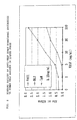
- FIG. 1 shows the binding of anti-VEGF human monoclonal antibody to human VEGF antigen in a liquid phase, where VA01 (B.T. Deposit No FERM BP-5607) is anti-VEGF human monoclonal antibody purified from hybridoma strain VA01, and IgM is human IgM antibody (Rockland). The concentration of each antibody is 20 and 5 µg/ml.
- FIG. 2 shows the binding of anti-VEGF human monoclonal antibody to human VEGF antigen in a liquid phase, where BL2 is anti-VEGF human monoclonal antibody purified from hybridoma strain BL2 (B.T. Deposit No FERm BP-5424); VA01 is anti-VEGF human monoclonal antibody purified from hybridoma strain VA01; and IgM is human IgM antibody (Rockland). The concentration of each antibody is 0.25 µg/ml.
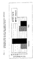
- FIG. 3 shows the ability of anti-VEGF human monoclonal antibody to inhibit the migration of vascular endothelial cells, where VA01 is anti-VEGF human monoclonal antibody purified from hybridoma strain VA01.
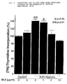
- FIG. 4 shows the ability of anti-VEGF human monoclonal antibody to inhibit the proliferation of vascular endothelial cells, where BL2 is anti-VEGF human monoclonal antibody purified from hybridoma strain BL2.
- The human monoclonal antibodies of the present invention are those derived from human lymphocyte. The globulin type of the human monoclonal antibodies of the present invention is not particularly limited insofar as they have the ability to bind to human VEGF, and examples are IgG, IgM, IgA, IgE, IgD etc. The human monoclonal antibodies of the present invention are not limited insofar as they possess the ability to bind to human VEGF, and they include not only those obtained from a cell line producing such human monoclonal antibodies, but also fragments thereof prepared by enzymatic digestion, gene recombination, etc., and fusion proteins thereof with another protein or factor, etc. Such antibodies may be modified by gene recombination insofar as their ability to bind to VEGF is not deteriorated. In particular, the anti-VEGF human monoclonal antibodies of the present invention neutralize the vascularization activity of human VEGF. This vascularization is a phenomenon consisting of the steps (1) destruction and degradation of basement membrane by proteases (i.e. decomposition of intercellular matrix protein), (2) migration of endothelial cells, (3) proliferation of endothelial cells, and (4) differentiation of endothelial cells and formation of tubes and cavities. Hence, it is considered that the vascularization can be inhibited by inhibiting at least one of these steps. The inhibition of vascularization by antibody can be checked by examination of the effect of the antibody on the reaction in each step. The anti-VEGF monoclonal antibodies of the present invention can inhibit the vascularization by inhibiting the migration of vascular endothelial cells, or the proliferation of vascular endothelial cells, as vascularization activities induced by human VEGF.
- The cell strain producing the anti-VEGF human monoclonal antibodies of the present invention is not particularly limited insofar as it can produce anti-VEGF human monoclonal antibodies. The cell strain can be obtained by transformation with Epstein-Barr Virus (EBV) of human lymphocyte producing anti-human VEGF antibodies. The cells can be obtained as hybridoma prepared by cell fusion between the resulting transformant and human myeloma cells. The cells can also be obtained as hybridoma by cell fusion between human lymphocyte producing anti-human VEGF antibodies and human myeloma cells. Alternatively, an antibody gene is removed from human lymphocyte producing anti-human VEGF antibodies and then integrated into a suitable vector to give producer cells. Examples of such cell strains producing anti-VEGF human monoclonal antibodies include hybridoma strains VA01 (FERM BP-5607) and BL2 (FERM BP-5424). Human lymphocyte producing anti-human VEGF antibodies, from which a cell strain producing the human monoclonal antibodies of the present invention is derived, can be obtained from human peripheral blood, lymph node etc. Any lymphocyte being present in human bodies and capable of producing antibodies having the ability to bind to human VEGF can be used in the present invention. The immunization of mouse, rabbit etc. with VEGF, usually conducted for acquiring monoclonal or polyclonal antibodies, is not required.
- The preparation of the human monoclonal antibodies of the present invention from the cell strain producing anti-VEGF human monoclonal antibodies can be carried out by any of the culture and purification methods known in the art, and these methods are not particularly limited insofar as the growth of cells and the production of anti-VEGF human monoclonal antibodies are not inhibited.
- The vascularization inhibitor of the present invention contains the anti-VEGF human monoclonal antibodies described above and is expected to be useful in preventing or treating diseases such as cancer, diabetic retinopathy etc. in which vascularization is involved.
- The present invention is illustrated by reference to the following examples which are however not intended to limit the scope of the invention.
- 20 ml peripheral blood was collected from a healthy person and a human mononuclear fraction was obtained by separating blood cells with a Ficoll separating solution (Dainippon Pharmaceutical Co., Ltd., Japan). It was washed twice with RPM-1640 medium (Nissui Seiyaku K.K., Japan) to prepare 3 x 10⁷ human mononuclear cells. The human mononuclear cells were centrifuged at 1,600 r.p.m. for 6 minutes to give a pellet. The pellet was suspended again with 30 ml culture supernatant of B95-8 cells at 37°C (i.e. a culture supernatant obtained by culturing B95-8 cells in RPMI-1640 medium containing 20 % fetal calf serum (FCS)). This suspension was transferred to a CO2 gas incubator at 37°C and then infected with EBV for 90 minutes at the same temperature. This suspension was centrifuged at 1,600 r.p.m. for 6 minutes to give a pellet. This pellet was suspended in 60 ml RPMI-1640 medium containing 20 % FCS and then pipetted into six 96-wells plates (Corning). The plates were transferred to a CO2 gas incubator at 37°C and incubated while half of the medium was exchanged with fresh one every 3 to 5 days.
- Colony formation occurred in wells on the plates after 20 days of culture, and the culture supernatant in the wells was examined for antibody activity by dot immmunobinding assay (DIBA) with human VEGF as antigen, as follows. Recombinant human VEGF (PeproTech) as antigen was dissolved at a concentration of 50 µg/ml in water, then spotted in an amount of 0.2 µl per spot on a nitrocellulose membrane (a product of Toyo) equipped with a grid, and dried in air to immobilize the antigen on the membrane. The immobilized antigen was blocked with a blocking solution (Tris-HCl buffered saline (TBS) containing 10 % FCS). 50µl of the culture supernatant was put to each well on 96-wells U-bottomed plate, and the blocked, immobilized antigen was then added to it, and the plate was incubated at room temperature for 2 hours. Each well was washed with TBS, and 50µl peroxidase-labeled anti-human immunoglobulin antibodies (produced by DAKO), previously diluted 500-fold with the blocking solution, was added to each well. The plate was incubated at room temperature for 2 hours. Each well was washed with TBS and stained with Konica Immustein (a product of Konica Corporation) and its coloration was checked with eyes. The same procedure was repeated 29 times for screening of 12,800 samples. As a result, anti-VEGF antibody activity was found in 2 wells.
- The cells found to have the antibody activity in (b) above were further cultured in 6-wells plate. The cells (2 x 10⁶ cells), and 1 x 10⁶ SHM-D33 cells (obtained from ATCC) previously cultured in IMDM medium containing 20 % FBS, were placed in a centrifuge tube and centrifuged at 1,600 r.p.m. for 6 minutes to give a pellet. This pellet was then washed twice with a cell fusion medium (0.25 M mannitol, 0.1 mM calcium chloride, 0.1 mM magnesium chloride, 0.2 mM Tris-HCl buffer (pH 7.2)) to give a pellet again. The pellet was then suspended again in 0.4 ml cell fusion medium and subjected to electric cell fusion (fusion apparatus, Shimadzu SSH-1; fusion chamber, Shimadzu FTC-02). This fusion made use of alternating-current high-frequency electrical field (1 MHz, 40 V, 10 seconds) and direct-current pulse electrical field (2.3 kV/cm, 40 µs, 10 seconds). After fusion, the cells were suspended in IMDM medium containing HAT (Sanko Junyaku) and 1 µM uabain, 20 % FCS, 5 µg/ml human transferrin, 8 µg/ml bovine insulin and 50 mM 2-mercaptoethanol and then put to each well in a 96-wells plate. The plate was transferred to a CO2 gas incubator at 37°C and incubated while half of the medium was exchanged with fresh one every 3 to 5 days. After 14 to 20 days of incubation, colony formation had occurred on wells, and the culture supernatant in the wells examined for antibody activity by the DIBA method described in (b) above. The cells found to have the antibody activity were cloned 3 times by limiting dilution to give hybridoma strains VA01 and BL2 producing anti-VEGF human monoclonal antibodies. The hybridoma strains VA01 and BL2 have been deposited under the Budapest Treaty as FERM BP-5607 and FERM BP-5424 respectively with the National Institute of Bioscience and Human-Technology, Agency of Industrial Science and Technology, Ministry of International Trade and Industry, 1-1-3 Higashi, Tsukuba City, Ibaraki Prefecture, Japan.
- Hybridoma strains VA01 and BL2 were cultured in serum-free medium ASF-101 (a product of Ajinomoto Co., Ltd.) to give 350 ml culture supernatant. This culture supernatant was fractionated with 50 % saturation with ammonium sulfate, and then the precipitates were dissolved again in phosphate buffered saline (PBS). The solution was filtered through 0.2 µm filter and purified through Superdex 200 column (produced by Pharmacia). The antibody fraction thus obtained was concentrated through an ultrafiltration membrane (30 k fractionation, Fuji Filter), then filtrated through a filter (0.2 µm) under sterile conditions, divided into aliquots and lyophilized -80°C for storage before use.
- Anti-VEGF rabbit polyclonal antibody (PeproTech) was diluted to 4 µg/ml in 0.1 M carbonate buffer (pH 9.6) and then put to a 96-wells plate (Greiner) in an amount of 50 µl per well. The plate was left at 4°C overnight to have the antibody immobilized on it. This plate was washed with a washing solution (10 mM Tris-HCl buffer (pH 8.0) containing 0.02 % sodium azide and 0.05 % Tween 20) and blocked with a blocking solution (PBS containing 0.5 % bovine serum albumin (BSA)). Separately, 20 and 5 µg/ml anti-VEGF human monoclonal antibody purified from hybridoma strain VA01 were mixed with 0 to 100 ng/ml human VEGF antigen (PeproTech) in another 96-wells U-bottomed plate, and this plate was incubated at room temperature for 2 hours. The reactant was transferred to the above antibody-immobilized plate after blocking and then incubated at room temperature for 2 hours. The plate was washed with the washing solution, and 100 µl alkaline phosphatase-labeled anti-human IgM antibody (BIO SOURCE) diluted 500-fold with the block solution was then put to each well, followed by incubation at room temperature for 2 hours. The plate was washed with the washing solution, and 50 µl substrate solution (9.6 % diethanolamine buffer (pH 9.6) containing 0.6 % p-nitrophenyl phosphoric acid and 0.5 mM magnesium chloride) was then put to each well, followed by incubation at room temperature for 30 minutes. The reaction was stopped by adding 50 µl of 3 N sodium hydroxide to the reaction solution, and then the absorbance of each well at 405 nm was measured in Immuno Reader (produced by Nippon Intermed).
- The results indicated that the antibody prepared from hybridoma strain VA01 reacts depending on the amount of human VEGF antigen added, as shown in FIG. 1. When the same reaction was carried out using another human IgM antibody (Rockland) of different specificity in place of the VEGF human monoclonal antibody or in the absence of the human monoclonal antibody (control), none of the reaction with human VEGF was observed. This indicated that the anti-VEGF human monoclonal antibody produced by hybridoma strain VA01 can recognize human VEGF in liquid phase to bind to it.
- According to Example 2, a plate having the antibody immobilized on it was prepared and blocked. Separately, 0.25 µg/ml anti-VEGF human monoclonal antibodies purified from hybridoma strains VA01 and BL2 were mixed with 0 to 100 ng/ml human VEGF antigen (PeproTech) in another 96-wells U-bottomed plate, and the mixture was incubated at room temperature for 2 hours. The reactant was transferred to the above antibody-immobilized plate after blocking and then incubated at room temperature for 2 hours. The plate was washed with the washing solution, and 100 µl anti-human IgM antibody (BIO SOURCE) labeled with alkaline phosphatase and diluted 500-fold with the blocking solution was then put to each well, followed by incubatation at room temperature for 2 hours. The plate was washed with the washing solution, and 50 µl of a substrate solution (9.6 % diethanolamine buffer (pH 9.6) containing 0.6 % p-nitrophenyl phosphoric acid and 0.5 mM magnesium chloride) was then put to each well, followed by incubation at room temperature for 30 minutes. The reaction was stopped by adding 50 µl of 3 N sodium hydroxide to the reaction solution, and the absorbance of each well at 405 nm was measured in Immuno Reader (Nippon Intermed).
- The results indicated that the antibodies prepared from hybridoma strains VA01 and BL2 react depending on the amount of human VEGF antigen added, as shown in FIG. 2. When the same reaction was carried out using another human IgM antibody (Rockland) of different specificity in place of the VEGF human monoclonal antibody or in the absence of the human monoclonal antibody (control), none of the reaction with human VEGF was observed. This indicated that the anti-VEGF human monoclonal antibody produced by hybridoma strain VA01 and BL2 can recognize human VEGF in liquid phase to bind to it.
- The cross reactivity of the anti-VEGF human monoclonal antibodies purified from hybridoma strains VA01 and BL2 was examined by the DIBA method. The antigens used were recombinant human VEGF (PeproTech), recombinant human basic fibroblast growth factor (Basic FGF; PeproTech), recombinant human platelet derived growth factor-BB (PDGF-BB; PeproTech), recombinant human tumor necrosis factor (TNFα; PeproTech), recombinant human interleukin-8 (IL-8; PeproTech) and recombinant human monocyte chemoattractant protein-1 (MCP-1; PeproTech). Each antigen was dissolved at a concentration of 50 µg/ml in water and the antigen was immobilized in the same manner as in Example 1 (b). The anti-VEGF human monoclonal antibodies purified from hybridoma strains VA01 and BL2 were diluted to 10, 3, 1, 0.3 and 0.1 µg/ml with the blocking solution, and their reactivity toward each of the immobilized antigens at predetermined concentrations was examined in the same manner as in Example 1 (b). The results indicated that the anti-VEGF human monoclonal antibodies of the present invention react strongly with human VEGF but not with other antigens than human VEGF, as shown in Tables 1 and 2.
Cross reactivity of anti-VEGF human monoclonal antibody VA01 antigen reactivity of each concentration of antibody (µg/ml) 10 3 1 0.3 0.1 0 VEGF +++ +++ +++ +++ +++ - Basic FGF - - - - - - PDGF-BB - - - - - - TNFα - - - - - - IL-8 - - - - - - MCP-1 - - - - - - +++: significantly strong reaction - : no reaction Cross reactivity of anti-VEGF human monoclonal antibody BL2 antigen reactivity at each concentration of antibody (µg/ml) 10 3 1 0.3 0.1 0 VEGF +++ +++ +++ +++ +++ - Basic FGF - - - - - - PDGF-BB - - - - - - TNFα - - - - - - IL-8 - - - - - - MCP-1 - - - - - - +++: significantly strong reaction - : no reaction - The effect of the human monoclonal antibodies on the wandering activity of vascular endothelial cells induced by human VEGF as one step of vascularization was examined. Human umbilical venous endothelial cells (HUVECs) were suspended in Hanks' solution (Nissui Seiyaku) to a density of 8 x 10⁵ cells/ml and then 25 µl of the suspension was put to a bottom plate of a 48-wells chemotaxis chamber (Neuroprobe). A membrane filter with a pore size of 5.0 µm (Neuroprobe) and a top plate were attached to the bottom plate, and the assembly was placed upside down for 120 minutes in a CO2 gas incubator at 37°C so that the HUVECs adhered to the membrane filter. Separately, anti-VEGF human monoclonal antibody purified from hybridoma strain VA01 and human VEGF antigen (PeproTech) were mixed at final concentrations of 34 µg/ml and 100 ng/ml, respectively, and then they were reacted at 37°C for 30 minutes. Physiological saline (Nissui Seiyaku) was used as a control in place of the anti-VEGF human monoclonal antibody. 50 µl of the reactant was added to the top plate and left for 120 minutes in a CO2 gas incubator at 37°C. The membrane filter was removed and stained with Diff-Quick staining solution (The Green Cross Corporation, Japan) and then the migration cells were observed under a microscope at 200 x magnification, and their number was counted. The result indicated that the anti-VEGF human monoclonal antibody purified from hybridoma strain VA01 inhibits the migration of HUVECs by human VEGF antigen, as shown in FIG. 3.
- The effect of the anti-VEGF human monoclonal antibody on the proliferation activity of vascular endothelial cells was examined. This proliferation activity, which is induced in a low oxygen atmosphere and considered to trigger tumor vascularization, was determined as follow. Microvascular endothelial cells (Kurabo) derived from human skin was suspended in E-GM MV medium (Kurabo) at 2 x 10⁴ cells/ml and 500 µl of the suspension was put to a 24-wells plate. The cells were incubated overnight in a CO2 gas incubator at 37°C, and then the medium was replaced by 500 µl fresh RPMI/M199 medium containing 15 % (v/v) fetal bovine serum. The anti-VEGF human monoclonal antibody purified from hybridoma strain BL2 was added to the wells in amounts of 0, 1, 5, and 10 µg/ml respectively, then left in a CO2 gas incubator at 37°C for 30 minutes, and incubated overnight in a low oxygen atmosphere (2.5 % O2) or in a conventional atmosphere (control) in the CO2 gas incubator at 37°C. [3H]-thymidine was added to the wells at a final concentration of 1 µCi/ml and they were reacted for 4 hours and gently washed twice with PBS, and then 200 µl of 10 % (v/v) cold TCA was added to the cells to fix them. After 20 minutes, the cells were gently washed twice with 5 % (v/v) cold TCA and reacted for 20 minutes by adding 200 µl of 1 N NaOH. Then, 200 µl of 1 N HCl was added to the cells and the incorporation of [3H]-thymidine into the cells was determined. The result indicated that the anti-VEGF human monoclonal antibody purified from hybridoma strain BL2 inhibits the proliferation of the vascular endothelial cells by autocrine VEGF, as shown in FIG. 4.
Claims (6)
- A human monoclonal antibody which specifically binds to human vascular endothelial cell growth factor (human VEGF), and inhibits or neutralizes human VEGF promoted vascularization wherein such human monoclonal antibody is produced by hybridoma BL2, which has been deposited under Accession No FERM BF-5424 or hybridoma VAO1, which has been deposited under Accession No FERM BP-5607.
- A composition comprising a human monoclonal antibody according to claim 1.
- Hybridoma VAO1 having Accession No FERM BP-5607.
- Hybridoma BL2 having Accession No FERM BP-5424.
- Use of the human monoclonal antibody of claim 1 for the manufacture of a prophylactic or therapeutic composition for inhibiting or neutralising vascularisation.
- Use of the human monoclonal antibody of claim 1 for the manufacture of a prophylactic or therapeutic composition for inhibiting tumour growth and/or metastasis, or for preventing or treating diabetic retinopathy.
Applications Claiming Priority (6)
| Application Number | Priority Date | Filing Date | Title |
|---|---|---|---|
| JP211454/95 | 1995-08-21 | ||
| JP21145495 | 1995-08-21 | ||
| JP21145495 | 1995-08-21 | ||
| JP66601/96 | 1996-03-22 | ||
| JP6660196 | 1996-03-22 | ||
| JP6660196 | 1996-03-22 |
Publications (2)
| Publication Number | Publication Date |
|---|---|
| EP0787742A1 EP0787742A1 (en) | 1997-08-06 |
| EP0787742B1 true EP0787742B1 (en) | 2001-12-12 |
Family
ID=26407800
Family Applications (1)
| Application Number | Title | Priority Date | Filing Date |
|---|---|---|---|
| EP96306000A Expired - Lifetime EP0787742B1 (en) | 1995-08-21 | 1996-08-16 | Anti-VEGF human monoclonal antibody |
Country Status (3)
| Country | Link |
|---|---|
| US (1) | US5730977A (en) |
| EP (1) | EP0787742B1 (en) |
| DE (1) | DE69617896T2 (en) |
Families Citing this family (49)
| Publication number | Priority date | Publication date | Assignee | Title |
|---|---|---|---|---|
| CA2261433A1 (en) * | 1993-12-09 | 1995-06-10 | Belinda Sanchez Ramirez | Composition comprising autologous epidermal growth factor |
| US7371376B1 (en) | 1996-10-18 | 2008-05-13 | Genentech, Inc. | Anti-ErbB2 antibodies |
| WO2000035956A1 (en) * | 1998-12-16 | 2000-06-22 | Kyowa Hakko Kogyo Co., Ltd. | Antihuman vegf monoclonal antibody |
| US6670321B1 (en) * | 1998-12-30 | 2003-12-30 | The Children's Medical Center Corporation | Prevention and treatment for retinal ischemia and edema |
| KR100940380B1 (en) * | 1999-01-15 | 2010-02-02 | 제넨테크, 인크. | Polypeptide Variants with Altered Effector Function |
| US7183387B1 (en) | 1999-01-15 | 2007-02-27 | Genentech, Inc. | Polypeptide variants with altered effector function |
| US6737056B1 (en) * | 1999-01-15 | 2004-05-18 | Genentech, Inc. | Polypeptide variants with altered effector function |
| US6703020B1 (en) | 1999-04-28 | 2004-03-09 | Board Of Regents, The University Of Texas System | Antibody conjugate methods for selectively inhibiting VEGF |
| IL145941A (en) | 1999-04-28 | 2007-08-19 | Univ Texas | Compositions and methods for cancer treatment by selectively inhibiting vegf |
| US7740841B1 (en) * | 2000-01-28 | 2010-06-22 | Sunnybrook Health Science Center | Therapeutic method for reducing angiogenesis |
| DK1268799T3 (en) * | 2000-03-31 | 2009-07-06 | Imclone Llc | Antagonistic antibodies to VE-cadherin without adverse effects on vascular permeability |
| US20020091082A1 (en) * | 2000-09-13 | 2002-07-11 | Aiello Lloyd P. | Methods of modulating symptoms of hypertension |
| ES2556946T3 (en) | 2000-12-21 | 2016-01-21 | Novartis Ag | Pyrimidinamines as angiogenesis modulators |
| CU22999A1 (en) * | 2001-12-04 | 2004-10-12 | Centro Inmunologia Molecular | METHOD OF TREATMENT OF CHRONIC MALIGNAL AND INFECTIOUS DISEASES |
| KR100988949B1 (en) * | 2001-10-25 | 2010-10-20 | 제넨테크, 인크. | Glycoprotein Composition |
| US20030232400A1 (en) * | 2002-12-20 | 2003-12-18 | Susan Radka | Methods of screening subjects for expression of soluble receptors of vascular endothelial growth factor (VEGF) for use in managing treatment and determining prognostic outcome |
| NZ592039A (en) | 2003-08-27 | 2013-03-28 | Ophthotech Corp | Combination therapy for the treatment of ocular neovascular disorders |
| WO2005087812A1 (en) * | 2004-03-05 | 2005-09-22 | Ludwig Institute For Cancer Research | Multivalent antibody materials and methods for vegf/pdgf family of growth factors |
| WO2006023312A2 (en) * | 2004-08-20 | 2006-03-02 | Carl Deirmengian | Diagnostic assay for source of inflammation |
| EP2946810A1 (en) | 2005-02-03 | 2015-11-25 | TopoTarget UK Limited | Combination therapy using an HDAC inhibitor and Vincristine for treating cancer |
| CN102579417B (en) | 2005-05-13 | 2014-11-26 | 托波塔吉特英国有限公司 | Pharmaceutical formulations of HDAC inhibitors |
| EP2361619A1 (en) | 2005-11-10 | 2011-08-31 | TopoTarget UK Limited | Histone deacetylase (hdac) inhibitors (pxd-101) alone for the treatment of hematological cancer |
| US20070258976A1 (en) * | 2006-05-04 | 2007-11-08 | Ward Keith W | Combination Therapy for Diseases Involving Angiogenesis |
| HUE028379T2 (en) * | 2006-09-29 | 2016-12-28 | Oncomed Pharm Inc | Compositions and methods for diagnosing and treating cancer |
| CU23636A1 (en) | 2006-11-01 | 2011-03-21 | Ct Ingenieria Genetica Biotech | RECOMBINANT ANTIBODIES AGAINST THE GROWTH FACTOR OF THE VASCULAR ENDOTELIUM (VEGF) |
| CA2700173C (en) | 2007-09-25 | 2016-10-11 | Topotarget Uk Limited | Methods of synthesis of certain hydroxamic acid compounds |
| HRP20241467T1 (en) | 2008-06-25 | 2025-01-03 | Novartis Ag | STABLE AND SOLUBLE VEGF-INHIBITING ANTIBODIES |
| KR20110036101A (en) | 2008-06-30 | 2011-04-06 | 안지오블라스트 시스템스 인코퍼레이티드 | Treatment of Ocular Diseases and Excessive Angiogenesis with Combination Therapy |
| PL2752189T3 (en) | 2008-11-22 | 2017-04-28 | F. Hoffmann-La Roche Ag | Use of anti-vegf antibody in combination with chemotherapy for treating breast cancer |
| KR101093717B1 (en) * | 2008-11-26 | 2011-12-19 | 한국생명공학연구원 | VEUF-specific human antibodies |
| MX2011010955A (en) * | 2009-04-20 | 2012-04-02 | Genentech Inc | Adjuvant cancer therapy. |
| MX336476B (en) * | 2009-08-15 | 2016-01-20 | Genentech Inc | ANTI-ANGIOGENESIS THERAPY FOR THE TREATMENT OF BREAST CANCER PREVIOUSLY TREATED. |
| US8883145B2 (en) | 2009-10-16 | 2014-11-11 | Oncomed Pharmaceuticals, Inc. | Methods of treatment with DLL4 antagonists and an anti-hypertensive agent |
| TWI619509B (en) | 2010-02-23 | 2018-04-01 | 建南德克公司 | Combination of carboplatin, paclitaxel and anti-VEGF antibodies in previously untreated stage III or IV ovarian, fallopian tube or primary peritoneal cancer |
| WO2011153243A2 (en) | 2010-06-02 | 2011-12-08 | Genentech, Inc. | Anti-angiogenesis therapy for treating gastric cancer |
| US8551479B2 (en) | 2010-09-10 | 2013-10-08 | Oncomed Pharmaceuticals, Inc. | Methods for treating melanoma |
| CU23895B1 (en) | 2010-12-28 | 2013-05-31 | Biorec Sa | RECOMBINANT ANTIBODIES AGAINST THE GROWTH FACTOR OF THE VASCULAR ENDOTHELIUM (VEGF) OBTAINED BY MUTAGENESIS OF VARIABLE REGIONS |
| ES2692268T5 (en) | 2011-03-29 | 2025-02-26 | Roche Glycart Ag | Antibody fc variants |
| SI3485903T1 (en) | 2011-09-23 | 2023-02-28 | Mereo Biopharma 5, Inc. | Vegf/dll4 binding agents and uses thereof |
| LT3495387T (en) | 2012-07-13 | 2021-11-25 | Roche Glycart Ag | Bispecific anti-vegf/anti-ang-2 antibodies and their use in the treatment of ocular vascular diseases |
| US9599620B2 (en) | 2012-10-31 | 2017-03-21 | Oncomed Pharmaceuticals, Inc. | Methods and monitoring of treatment with a DLL4 antagonist |
| MY199344A (en) | 2013-02-18 | 2023-10-24 | Vegenics Pty Ltd | Ligand binding molecules and uses thereof |
| KR102948178B1 (en) | 2013-07-12 | 2026-04-10 | 아스텔라스 유에스 엘엘씨 | Methods for treating or preventing ophthalmological conditions |
| US20160176962A1 (en) | 2014-10-31 | 2016-06-23 | Oncomed Pharmaceuticals, Inc. | Combination Therapy For Treatment Of Disease |
| ES2968074T3 (en) | 2015-09-23 | 2024-05-07 | Mereo Biopharma 5 Inc | Bi-specific anti-VEGF/DLL4 antibody for use in the treatment of platinum-resistant ovarian cancer |
| CA3012718A1 (en) | 2016-02-08 | 2017-08-17 | Vitrisa Therapeutics, Inc. | Compositions with improved intravitreal half-life and uses thereof |
| EP3694890A4 (en) | 2017-10-12 | 2021-11-03 | Immunowake Inc. | VEGFR ANTIBODY LIGHT CHAIN FUSION PROTEIN |
| WO2019154776A1 (en) | 2018-02-06 | 2019-08-15 | F. Hoffmann-La Roche Ag | Treatment of ophthalmologic diseases |
| SG11202103064YA (en) | 2018-10-29 | 2021-05-28 | Hoffmann La Roche | Antibody formulation |
Family Cites Families (2)
| Publication number | Priority date | Publication date | Assignee | Title |
|---|---|---|---|---|
| CA2002067A1 (en) * | 1988-11-09 | 1990-05-09 | Tamotsu Fukuda | Parent cell line for producing human hybridomas |
| DK1975181T3 (en) * | 1992-10-28 | 2011-06-06 | Genentech Inc | Use of vascular endothelial cell growth factor antagonists |
-
1996
- 1996-08-15 US US08/698,041 patent/US5730977A/en not_active Expired - Fee Related
- 1996-08-16 EP EP96306000A patent/EP0787742B1/en not_active Expired - Lifetime
- 1996-08-16 DE DE69617896T patent/DE69617896T2/en not_active Expired - Fee Related
Also Published As
| Publication number | Publication date |
|---|---|
| DE69617896T2 (en) | 2002-04-25 |
| EP0787742A1 (en) | 1997-08-06 |
| DE69617896D1 (en) | 2002-01-24 |
| US5730977A (en) | 1998-03-24 |
Similar Documents
| Publication | Publication Date | Title |
|---|---|---|
| US5730977A (en) | Anti-VEGF human monoclonal antibody | |
| Snapper et al. | Transforming growth factor-beta 1 is required for secretion of IgG of all subclasses by LPS-activated murine B cells in vitro. | |
| JPH10507926A (en) | AL-1 neurotrophic factor, ligand for EPH-related tyrosine kinase receptor | |
| KR100196540B1 (en) | Axon growth regulator | |
| AU3901600A (en) | Recombinant il-18 antagonists useful in treatment of il-18 mediated disorders | |
| JPH0223156B2 (en) | ||
| JP3866759B2 (en) | Neurite growth regulator | |
| US7282203B2 (en) | Use of NOTCH pathway interfering agents for treatment of plasma cell disorders | |
| US5625039A (en) | Anti-human IgE monoclonal antibodies | |
| DE69431526T2 (en) | LIGAND CAPABLE OF BINDING AT CD26 ADENOSINEEDEAMINASE BONDING POINT | |
| US5582998A (en) | Monoclonal antibodies against human TNF-binding protein I (TNF-BP I) and immunoassays therefor | |
| EP0057107A2 (en) | Method of manufacturing monoclonal antibodies and cells capable of manufacturing such antibodies | |
| US4818689A (en) | Late differentiation antigen associated with helper T lymphocyte function | |
| Trojanowski et al. | Human teratomas express differentiated neural antigens. An immunohistochemical study with anti-neurofilament, anti-glial filament, and anti-myelin basic protein monoclonal antibodies | |
| US20150166639A1 (en) | Nerve elongation promoter and elongation inhibitor | |
| Jiang et al. | Increased intraplatelet and urinary transforming growth factor-β in patients with multiple myeloma | |
| AU668351B2 (en) | Monoclonal antibodies against human TNF-binding protein I (TNF-BP I) | |
| Guo et al. | B cell lines from a subset of patients with common variable immunodeficiency undergo enhanced apoptosis associated with an increased display of CD95 (Apo-1/fas), diminished CD38 expression, and decreased IgG and IgA production | |
| US20240287167A1 (en) | Agent for preventing or treating acute-phase neuromyelitis optica | |
| JPH09316099A (en) | Anti-VEGF human monoclonal antibody | |
| CN117430706B (en) | Adipose-derived stem cell composition for treating diabetes | |
| RU2847366C1 (en) | Preventive or therapeutic agent for the treatment of dementia | |
| US5330894A (en) | Anti-procollagenase monoclonal antibodies and a method for the assay of procollagenase utilizing thereof | |
| US20230090965A1 (en) | Prophylactic or therapeutic agent for dementia | |
| Qing et al. | Antigen association of J6-1 cell membrane associated factor receptor with macrophage colony stimulating factor receptor |
Legal Events
| Date | Code | Title | Description |
|---|---|---|---|
| PUAI | Public reference made under article 153(3) epc to a published international application that has entered the european phase |
Free format text: ORIGINAL CODE: 0009012 |
|
| AK | Designated contracting states |
Kind code of ref document: A1 Designated state(s): DE FR GB IT |
|
| 17P | Request for examination filed |
Effective date: 19970917 |
|
| RAP1 | Party data changed (applicant data changed or rights of an application transferred) |
Owner name: MITSUI CHEMICALS, INC. |
|
| 17Q | First examination report despatched |
Effective date: 20000428 |
|
| GRAG | Despatch of communication of intention to grant |
Free format text: ORIGINAL CODE: EPIDOS AGRA |
|
| GRAG | Despatch of communication of intention to grant |
Free format text: ORIGINAL CODE: EPIDOS AGRA |
|
| GRAH | Despatch of communication of intention to grant a patent |
Free format text: ORIGINAL CODE: EPIDOS IGRA |
|
| GRAG | Despatch of communication of intention to grant |
Free format text: ORIGINAL CODE: EPIDOS AGRA |
|
| GRAH | Despatch of communication of intention to grant a patent |
Free format text: ORIGINAL CODE: EPIDOS IGRA |
|
| GRAH | Despatch of communication of intention to grant a patent |
Free format text: ORIGINAL CODE: EPIDOS IGRA |
|
| GRAA | (expected) grant |
Free format text: ORIGINAL CODE: 0009210 |
|
| AK | Designated contracting states |
Kind code of ref document: B1 Designated state(s): DE FR GB IT |
|
| REG | Reference to a national code |
Ref country code: GB Ref legal event code: IF02 |
|
| REF | Corresponds to: |
Ref document number: 69617896 Country of ref document: DE Date of ref document: 20020124 |
|
| PLBE | No opposition filed within time limit |
Free format text: ORIGINAL CODE: 0009261 |
|
| STAA | Information on the status of an ep patent application or granted ep patent |
Free format text: STATUS: NO OPPOSITION FILED WITHIN TIME LIMIT |
|
| 26N | No opposition filed | ||
| PGFP | Annual fee paid to national office [announced via postgrant information from national office to epo] |
Ref country code: FR Payment date: 20030808 Year of fee payment: 8 |
|
| PGFP | Annual fee paid to national office [announced via postgrant information from national office to epo] |
Ref country code: GB Payment date: 20030813 Year of fee payment: 8 |
|
| PGFP | Annual fee paid to national office [announced via postgrant information from national office to epo] |
Ref country code: DE Payment date: 20030828 Year of fee payment: 8 |
|
| PG25 | Lapsed in a contracting state [announced via postgrant information from national office to epo] |
Ref country code: GB Free format text: LAPSE BECAUSE OF NON-PAYMENT OF DUE FEES Effective date: 20040816 |
|
| PG25 | Lapsed in a contracting state [announced via postgrant information from national office to epo] |
Ref country code: DE Free format text: LAPSE BECAUSE OF NON-PAYMENT OF DUE FEES Effective date: 20050301 |
|
| GBPC | Gb: european patent ceased through non-payment of renewal fee |
Effective date: 20040816 |
|
| PG25 | Lapsed in a contracting state [announced via postgrant information from national office to epo] |
Ref country code: FR Free format text: LAPSE BECAUSE OF NON-PAYMENT OF DUE FEES Effective date: 20050429 |
|
| REG | Reference to a national code |
Ref country code: FR Ref legal event code: ST |
|
| PG25 | Lapsed in a contracting state [announced via postgrant information from national office to epo] |
Ref country code: IT Free format text: LAPSE BECAUSE OF NON-PAYMENT OF DUE FEES;WARNING: LAPSES OF ITALIAN PATENTS WITH EFFECTIVE DATE BEFORE 2007 MAY HAVE OCCURRED AT ANY TIME BEFORE 2007. THE CORRECT EFFECTIVE DATE MAY BE DIFFERENT FROM THE ONE RECORDED. Effective date: 20050816 |