EP0787144B1 - Major outer membrane protein cd of moraxella - Google Patents
Major outer membrane protein cd of moraxella Download PDFInfo
- Publication number
- EP0787144B1 EP0787144B1 EP95944810A EP95944810A EP0787144B1 EP 0787144 B1 EP0787144 B1 EP 0787144B1 EP 95944810 A EP95944810 A EP 95944810A EP 95944810 A EP95944810 A EP 95944810A EP 0787144 B1 EP0787144 B1 EP 0787144B1
- Authority
- EP
- European Patent Office
- Prior art keywords
- protein
- pellet
- catarrhalis
- supernatant
- detergent
- Prior art date
- Legal status (The legal status is an assumption and is not a legal conclusion. Google has not performed a legal analysis and makes no representation as to the accuracy of the status listed.)
- Expired - Lifetime
Links
- 241000588621 Moraxella Species 0.000 title claims description 18
- 101710164702 Major outer membrane protein Proteins 0.000 title description 4
- 108090000623 proteins and genes Proteins 0.000 claims abstract description 176
- 102000004169 proteins and genes Human genes 0.000 claims abstract description 175
- 241000588655 Moraxella catarrhalis Species 0.000 claims abstract description 94
- 239000008188 pellet Substances 0.000 claims abstract description 41
- 239000006228 supernatant Substances 0.000 claims abstract description 24
- 101710116435 Outer membrane protein Proteins 0.000 claims abstract description 22
- 230000001580 bacterial effect Effects 0.000 claims abstract description 16
- 238000005119 centrifugation Methods 0.000 claims abstract description 14
- 239000013592 cell lysate Substances 0.000 claims abstract description 13
- 108010052285 Membrane Proteins Proteins 0.000 claims abstract description 6
- 108010077805 Bacterial Proteins Proteins 0.000 claims abstract description 4
- 238000000034 method Methods 0.000 claims description 38
- 239000003599 detergent Substances 0.000 claims description 27
- 238000000605 extraction Methods 0.000 claims description 20
- 239000000243 solution Substances 0.000 claims description 16
- XSQUKJJJFZCRTK-UHFFFAOYSA-N Urea Chemical compound NC(N)=O XSQUKJJJFZCRTK-UHFFFAOYSA-N 0.000 claims description 11
- 239000003795 chemical substances by application Substances 0.000 claims description 10
- 230000000694 effects Effects 0.000 claims description 10
- 229920004890 Triton X-100 Polymers 0.000 claims description 8
- 239000013504 Triton X-100 Substances 0.000 claims description 8
- 239000004202 carbamide Substances 0.000 claims description 6
- 239000008366 buffered solution Substances 0.000 claims description 4
- 230000002163 immunogen Effects 0.000 abstract description 30
- 239000000203 mixture Substances 0.000 abstract description 29
- 244000052616 bacterial pathogen Species 0.000 abstract description 16
- 201000010099 disease Diseases 0.000 abstract description 13
- 208000037265 diseases, disorders, signs and symptoms Diseases 0.000 abstract description 13
- 239000002158 endotoxin Substances 0.000 abstract description 9
- 229920006008 lipopolysaccharide Polymers 0.000 abstract description 9
- 230000001939 inductive effect Effects 0.000 abstract description 7
- 239000000356 contaminant Substances 0.000 abstract description 4
- 238000001727 in vivo Methods 0.000 abstract description 4
- 150000003904 phospholipids Chemical class 0.000 abstract description 4
- 229960005486 vaccine Drugs 0.000 description 28
- 239000002671 adjuvant Substances 0.000 description 27
- 239000000427 antigen Substances 0.000 description 24
- 108091007433 antigens Proteins 0.000 description 24
- 102000036639 antigens Human genes 0.000 description 24
- 241000894006 Bacteria Species 0.000 description 22
- 210000004027 cell Anatomy 0.000 description 21
- 230000003053 immunization Effects 0.000 description 18
- 239000000523 sample Substances 0.000 description 18
- 241000699666 Mus <mouse, genus> Species 0.000 description 17
- 238000002649 immunization Methods 0.000 description 16
- 206010033078 Otitis media Diseases 0.000 description 14
- 230000028993 immune response Effects 0.000 description 12
- 241000606768 Haemophilus influenzae Species 0.000 description 11
- 241000699670 Mus sp. Species 0.000 description 11
- 239000000463 material Substances 0.000 description 9
- 108091003079 Bovine Serum Albumin Proteins 0.000 description 8
- 239000007983 Tris buffer Substances 0.000 description 8
- 229940098773 bovine serum albumin Drugs 0.000 description 8
- LOKCTEFSRHRXRJ-UHFFFAOYSA-I dipotassium trisodium dihydrogen phosphate hydrogen phosphate dichloride Chemical compound P(=O)(O)(O)[O-].[K+].P(=O)(O)([O-])[O-].[Na+].[Na+].[Cl-].[K+].[Cl-].[Na+] LOKCTEFSRHRXRJ-UHFFFAOYSA-I 0.000 description 8
- 244000052769 pathogen Species 0.000 description 8
- 239000002953 phosphate buffered saline Substances 0.000 description 8
- LENZDBCJOHFCAS-UHFFFAOYSA-N tris Chemical compound OCC(N)(CO)CO LENZDBCJOHFCAS-UHFFFAOYSA-N 0.000 description 8
- 210000000959 ear middle Anatomy 0.000 description 7
- 239000012530 fluid Substances 0.000 description 7
- 230000005847 immunogenicity Effects 0.000 description 7
- 238000004519 manufacturing process Methods 0.000 description 7
- 238000002360 preparation method Methods 0.000 description 7
- -1 OS21* Chemical compound 0.000 description 6
- 238000004458 analytical method Methods 0.000 description 6
- FAPWRFPIFSIZLT-UHFFFAOYSA-M Sodium chloride Chemical compound [Na+].[Cl-] FAPWRFPIFSIZLT-UHFFFAOYSA-M 0.000 description 5
- 229940037003 alum Drugs 0.000 description 5
- 230000000844 anti-bacterial effect Effects 0.000 description 5
- 230000000890 antigenic effect Effects 0.000 description 5
- 230000027455 binding Effects 0.000 description 5
- 238000009739 binding Methods 0.000 description 5
- 238000009472 formulation Methods 0.000 description 5
- 239000012634 fragment Substances 0.000 description 5
- 210000000987 immune system Anatomy 0.000 description 5
- 230000001717 pathogenic effect Effects 0.000 description 5
- 238000002255 vaccination Methods 0.000 description 5
- 108010042708 Acetylmuramyl-Alanyl-Isoglutamine Proteins 0.000 description 4
- 230000015572 biosynthetic process Effects 0.000 description 4
- 239000003153 chemical reaction reagent Substances 0.000 description 4
- 238000000684 flow cytometry Methods 0.000 description 4
- 210000004201 immune sera Anatomy 0.000 description 4
- 229940042743 immune sera Drugs 0.000 description 4
- 238000003119 immunoblot Methods 0.000 description 4
- 238000011534 incubation Methods 0.000 description 4
- 208000015181 infectious disease Diseases 0.000 description 4
- BSOQXXWZTUDTEL-ZUYCGGNHSA-N muramyl dipeptide Chemical compound OC(=O)CC[C@H](C(N)=O)NC(=O)[C@H](C)NC(=O)[C@@H](C)O[C@H]1[C@H](O)[C@@H](CO)O[C@@H](O)[C@@H]1NC(C)=O BSOQXXWZTUDTEL-ZUYCGGNHSA-N 0.000 description 4
- 108090000765 processed proteins & peptides Proteins 0.000 description 4
- 238000000746 purification Methods 0.000 description 4
- 201000009890 sinusitis Diseases 0.000 description 4
- 238000002415 sodium dodecyl sulfate polyacrylamide gel electrophoresis Methods 0.000 description 4
- 239000007790 solid phase Substances 0.000 description 4
- 239000000725 suspension Substances 0.000 description 4
- 238000012360 testing method Methods 0.000 description 4
- 238000011282 treatment Methods 0.000 description 4
- 238000005406 washing Methods 0.000 description 4
- 229920001817 Agar Polymers 0.000 description 3
- 241000700199 Cavia porcellus Species 0.000 description 3
- LFQSCWFLJHTTHZ-UHFFFAOYSA-N Ethanol Chemical compound CCO LFQSCWFLJHTTHZ-UHFFFAOYSA-N 0.000 description 3
- PEDCQBHIVMGVHV-UHFFFAOYSA-N Glycerine Chemical compound OCC(O)CO PEDCQBHIVMGVHV-UHFFFAOYSA-N 0.000 description 3
- 229930186217 Glycolipid Natural products 0.000 description 3
- 108010001336 Horseradish Peroxidase Proteins 0.000 description 3
- DBMJMQXJHONAFJ-UHFFFAOYSA-M Sodium laurylsulphate Chemical compound [Na+].CCCCCCCCCCCCOS([O-])(=O)=O DBMJMQXJHONAFJ-UHFFFAOYSA-M 0.000 description 3
- 230000024932 T cell mediated immunity Effects 0.000 description 3
- 239000004480 active ingredient Substances 0.000 description 3
- 239000008272 agar Substances 0.000 description 3
- 150000001413 amino acids Chemical class 0.000 description 3
- 210000003719 b-lymphocyte Anatomy 0.000 description 3
- 210000004369 blood Anatomy 0.000 description 3
- 239000008280 blood Substances 0.000 description 3
- 239000000872 buffer Substances 0.000 description 3
- 229910052799 carbon Inorganic materials 0.000 description 3
- 230000001413 cellular effect Effects 0.000 description 3
- 238000010790 dilution Methods 0.000 description 3
- 239000012895 dilution Substances 0.000 description 3
- 230000006870 function Effects 0.000 description 3
- 150000004676 glycans Chemical class 0.000 description 3
- 230000012010 growth Effects 0.000 description 3
- 230000005745 host immune response Effects 0.000 description 3
- 230000028996 humoral immune response Effects 0.000 description 3
- 230000003308 immunostimulating effect Effects 0.000 description 3
- 230000021633 leukocyte mediated immunity Effects 0.000 description 3
- 239000012528 membrane Substances 0.000 description 3
- 229920001282 polysaccharide Polymers 0.000 description 3
- 239000005017 polysaccharide Substances 0.000 description 3
- 102000004196 processed proteins & peptides Human genes 0.000 description 3
- 239000000047 product Substances 0.000 description 3
- 239000012460 protein solution Substances 0.000 description 3
- 239000011780 sodium chloride Substances 0.000 description 3
- 229940083575 sodium dodecyl sulfate Drugs 0.000 description 3
- 235000019333 sodium laurylsulphate Nutrition 0.000 description 3
- 239000000829 suppository Substances 0.000 description 3
- VHUVBWVDIFVVBI-SNYZSRNZSA-N (2s)-3-(4-hydroxyphenyl)-2-(octadecylamino)propanoic acid;hydrochloride Chemical compound Cl.CCCCCCCCCCCCCCCCCCN[C@H](C(O)=O)CC1=CC=C(O)C=C1 VHUVBWVDIFVVBI-SNYZSRNZSA-N 0.000 description 2
- HVCOBJNICQPDBP-UHFFFAOYSA-N 3-[3-[3,5-dihydroxy-6-methyl-4-(3,4,5-trihydroxy-6-methyloxan-2-yl)oxyoxan-2-yl]oxydecanoyloxy]decanoic acid;hydrate Chemical class O.OC1C(OC(CC(=O)OC(CCCCCCC)CC(O)=O)CCCCCCC)OC(C)C(O)C1OC1C(O)C(O)C(O)C(C)O1 HVCOBJNICQPDBP-UHFFFAOYSA-N 0.000 description 2
- FTOAOBMCPZCFFF-UHFFFAOYSA-N 5,5-diethylbarbituric acid Chemical compound CCC1(CC)C(=O)NC(=O)NC1=O FTOAOBMCPZCFFF-UHFFFAOYSA-N 0.000 description 2
- XKRFYHLGVUSROY-UHFFFAOYSA-N Argon Chemical compound [Ar] XKRFYHLGVUSROY-UHFFFAOYSA-N 0.000 description 2
- 201000004813 Bronchopneumonia Diseases 0.000 description 2
- 101150028326 CD gene Proteins 0.000 description 2
- 241000283707 Capra Species 0.000 description 2
- 102000014914 Carrier Proteins Human genes 0.000 description 2
- 241000700198 Cavia Species 0.000 description 2
- 108010060123 Conjugate Vaccines Proteins 0.000 description 2
- 238000009007 Diagnostic Kit Methods 0.000 description 2
- KCXVZYZYPLLWCC-UHFFFAOYSA-N EDTA Chemical compound OC(=O)CN(CC(O)=O)CCN(CC(O)=O)CC(O)=O KCXVZYZYPLLWCC-UHFFFAOYSA-N 0.000 description 2
- 238000002965 ELISA Methods 0.000 description 2
- 241000588724 Escherichia coli Species 0.000 description 2
- WSFSSNUMVMOOMR-UHFFFAOYSA-N Formaldehyde Chemical compound O=C WSFSSNUMVMOOMR-UHFFFAOYSA-N 0.000 description 2
- 241000282412 Homo Species 0.000 description 2
- 108060003951 Immunoglobulin Proteins 0.000 description 2
- 108010028921 Lipopeptides Proteins 0.000 description 2
- 108090001030 Lipoproteins Proteins 0.000 description 2
- 102000004895 Lipoproteins Human genes 0.000 description 2
- 241001465754 Metazoa Species 0.000 description 2
- 206010062204 Moraxella infection Diseases 0.000 description 2
- 241000588653 Neisseria Species 0.000 description 2
- 206010035664 Pneumonia Diseases 0.000 description 2
- 206010036790 Productive cough Diseases 0.000 description 2
- UIIMBOGNXHQVGW-UHFFFAOYSA-M Sodium bicarbonate Chemical compound [Na+].OC([O-])=O UIIMBOGNXHQVGW-UHFFFAOYSA-M 0.000 description 2
- 241000193998 Streptococcus pneumoniae Species 0.000 description 2
- 241000700605 Viruses Species 0.000 description 2
- WNROFYMDJYEPJX-UHFFFAOYSA-K aluminium hydroxide Chemical compound [OH-].[OH-].[OH-].[Al+3] WNROFYMDJYEPJX-UHFFFAOYSA-K 0.000 description 2
- ILRRQNADMUWWFW-UHFFFAOYSA-K aluminium phosphate Chemical compound O1[Al]2OP1(=O)O2 ILRRQNADMUWWFW-UHFFFAOYSA-K 0.000 description 2
- 210000000612 antigen-presenting cell Anatomy 0.000 description 2
- 239000007864 aqueous solution Substances 0.000 description 2
- 238000003556 assay Methods 0.000 description 2
- HNYOPLTXPVRDBG-UHFFFAOYSA-N barbituric acid Chemical compound O=C1CC(=O)NC(=O)N1 HNYOPLTXPVRDBG-UHFFFAOYSA-N 0.000 description 2
- 239000002775 capsule Substances 0.000 description 2
- 239000000969 carrier Substances 0.000 description 2
- 238000006243 chemical reaction Methods 0.000 description 2
- 230000000295 complement effect Effects 0.000 description 2
- 229940031670 conjugate vaccine Drugs 0.000 description 2
- 238000000326 densiometry Methods 0.000 description 2
- 238000011161 development Methods 0.000 description 2
- 238000003745 diagnosis Methods 0.000 description 2
- 238000010586 diagram Methods 0.000 description 2
- 239000003814 drug Substances 0.000 description 2
- 229940047650 haemophilus influenzae Drugs 0.000 description 2
- 210000004408 hybridoma Anatomy 0.000 description 2
- 238000003018 immunoassay Methods 0.000 description 2
- 102000018358 immunoglobulin Human genes 0.000 description 2
- 239000002955 immunomodulating agent Substances 0.000 description 2
- 229940121354 immunomodulator Drugs 0.000 description 2
- 239000002502 liposome Substances 0.000 description 2
- 239000002480 mineral oil Substances 0.000 description 2
- 235000010446 mineral oil Nutrition 0.000 description 2
- 230000003571 opsonizing effect Effects 0.000 description 2
- JMANVNJQNLATNU-UHFFFAOYSA-N oxalonitrile Chemical compound N#CC#N JMANVNJQNLATNU-UHFFFAOYSA-N 0.000 description 2
- 239000000546 pharmaceutical excipient Substances 0.000 description 2
- 239000012071 phase Substances 0.000 description 2
- 229920001983 poloxamer Polymers 0.000 description 2
- 229920000642 polymer Polymers 0.000 description 2
- 229920000136 polysorbate Polymers 0.000 description 2
- 230000009257 reactivity Effects 0.000 description 2
- 210000002345 respiratory system Anatomy 0.000 description 2
- 230000004044 response Effects 0.000 description 2
- 229930182490 saponin Natural products 0.000 description 2
- 150000007949 saponins Chemical class 0.000 description 2
- 235000017709 saponins Nutrition 0.000 description 2
- 210000002966 serum Anatomy 0.000 description 2
- 239000011734 sodium Substances 0.000 description 2
- 238000005063 solubilization Methods 0.000 description 2
- 230000007928 solubilization Effects 0.000 description 2
- 208000024794 sputum Diseases 0.000 description 2
- 210000003802 sputum Anatomy 0.000 description 2
- 229940031000 streptococcus pneumoniae Drugs 0.000 description 2
- 238000006467 substitution reaction Methods 0.000 description 2
- 235000000346 sugar Nutrition 0.000 description 2
- 238000013268 sustained release Methods 0.000 description 2
- 239000012730 sustained-release form Substances 0.000 description 2
- 230000008685 targeting Effects 0.000 description 2
- 231100000331 toxic Toxicity 0.000 description 2
- 230000002588 toxic effect Effects 0.000 description 2
- 231100000419 toxicity Toxicity 0.000 description 2
- 230000001988 toxicity Effects 0.000 description 2
- QKNYBSVHEMOAJP-UHFFFAOYSA-N 2-amino-2-(hydroxymethyl)propane-1,3-diol;hydron;chloride Chemical compound Cl.OCC(N)(CO)CO QKNYBSVHEMOAJP-UHFFFAOYSA-N 0.000 description 1
- YRNWIFYIFSBPAU-UHFFFAOYSA-N 4-[4-(dimethylamino)phenyl]-n,n-dimethylaniline Chemical compound C1=CC(N(C)C)=CC=C1C1=CC=C(N(C)C)C=C1 YRNWIFYIFSBPAU-UHFFFAOYSA-N 0.000 description 1
- 206010001076 Acute sinusitis Diseases 0.000 description 1
- 206010053555 Arthritis bacterial Diseases 0.000 description 1
- 238000000035 BCA protein assay Methods 0.000 description 1
- 208000031729 Bacteremia Diseases 0.000 description 1
- 208000035143 Bacterial infection Diseases 0.000 description 1
- 231100000699 Bacterial toxin Toxicity 0.000 description 1
- BTBUEUYNUDRHOZ-UHFFFAOYSA-N Borate Chemical compound [O-]B([O-])[O-] BTBUEUYNUDRHOZ-UHFFFAOYSA-N 0.000 description 1
- 241000283690 Bos taurus Species 0.000 description 1
- 206010006458 Bronchitis chronic Diseases 0.000 description 1
- 108010078791 Carrier Proteins Proteins 0.000 description 1
- 206010057248 Cell death Diseases 0.000 description 1
- 206010009137 Chronic sinusitis Diseases 0.000 description 1
- 208000028698 Cognitive impairment Diseases 0.000 description 1
- 201000007336 Cryptococcosis Diseases 0.000 description 1
- 241000221204 Cryptococcus neoformans Species 0.000 description 1
- 208000035976 Developmental Disabilities Diseases 0.000 description 1
- 108090000204 Dipeptidase 1 Proteins 0.000 description 1
- 238000012286 ELISA Assay Methods 0.000 description 1
- 206010014561 Emphysema Diseases 0.000 description 1
- 102000004190 Enzymes Human genes 0.000 description 1
- 108090000790 Enzymes Proteins 0.000 description 1
- 241000710198 Foot-and-mouth disease virus Species 0.000 description 1
- WQZGKKKJIJFFOK-GASJEMHNSA-N Glucose Natural products OC[C@H]1OC(O)[C@H](O)[C@@H](O)[C@@H]1O WQZGKKKJIJFFOK-GASJEMHNSA-N 0.000 description 1
- 206010018691 Granuloma Diseases 0.000 description 1
- 241000700721 Hepatitis B virus Species 0.000 description 1
- 208000004575 Infectious Arthritis Diseases 0.000 description 1
- 206010022941 Iridocyclitis Diseases 0.000 description 1
- 239000007836 KH2PO4 Substances 0.000 description 1
- 241000588748 Klebsiella Species 0.000 description 1
- 206010024971 Lower respiratory tract infections Diseases 0.000 description 1
- 208000019693 Lung disease Diseases 0.000 description 1
- 102000018697 Membrane Proteins Human genes 0.000 description 1
- 201000009906 Meningitis Diseases 0.000 description 1
- 208000031548 Moraxellaceae Infections Diseases 0.000 description 1
- 102000011931 Nucleoproteins Human genes 0.000 description 1
- 108010061100 Nucleoproteins Proteins 0.000 description 1
- 108020005187 Oligonucleotide Probes Proteins 0.000 description 1
- 241000283973 Oryctolagus cuniculus Species 0.000 description 1
- 229930040373 Paraformaldehyde Natural products 0.000 description 1
- 206010035226 Plasma cell myeloma Diseases 0.000 description 1
- 208000000474 Poliomyelitis Diseases 0.000 description 1
- 239000004793 Polystyrene Substances 0.000 description 1
- 241000288906 Primates Species 0.000 description 1
- 241000589517 Pseudomonas aeruginosa Species 0.000 description 1
- 206010057190 Respiratory tract infections Diseases 0.000 description 1
- 241000293871 Salmonella enterica subsp. enterica serovar Typhi Species 0.000 description 1
- 241000700584 Simplexvirus Species 0.000 description 1
- 238000002105 Southern blotting Methods 0.000 description 1
- 241000191967 Staphylococcus aureus Species 0.000 description 1
- 241000194017 Streptococcus Species 0.000 description 1
- 241000701093 Suid alphaherpesvirus 1 Species 0.000 description 1
- 206010043376 Tetanus Diseases 0.000 description 1
- 206010048038 Wound infection Diseases 0.000 description 1
- UZQJVUCHXGYFLQ-AYDHOLPZSA-N [(2s,3r,4s,5r,6r)-4-[(2s,3r,4s,5r,6r)-4-[(2r,3r,4s,5r,6r)-4-[(2s,3r,4s,5r,6r)-3,5-dihydroxy-6-(hydroxymethyl)-4-[(2s,3r,4s,5s,6r)-3,4,5-trihydroxy-6-(hydroxymethyl)oxan-2-yl]oxyoxan-2-yl]oxy-3,5-dihydroxy-6-(hydroxymethyl)oxan-2-yl]oxy-3,5-dihydroxy-6-(hy Chemical compound O([C@H]1[C@H](O)[C@@H](CO)O[C@H]([C@@H]1O)O[C@H]1[C@H](O)[C@@H](CO)O[C@H]([C@@H]1O)O[C@H]1CC[C@]2(C)[C@H]3CC=C4[C@@]([C@@]3(CC[C@H]2[C@@]1(C=O)C)C)(C)CC(O)[C@]1(CCC(CC14)(C)C)C(=O)O[C@H]1[C@@H]([C@@H](O[C@H]2[C@@H]([C@@H](O[C@H]3[C@@H]([C@@H](O[C@H]4[C@@H]([C@@H](O[C@H]5[C@@H]([C@@H](O)[C@H](O)[C@@H](CO)O5)O)[C@H](O)[C@@H](CO)O4)O)[C@H](O)[C@@H](CO)O3)O)[C@H](O)[C@@H](CO)O2)O)[C@H](O)[C@@H](CO)O1)O)[C@@H]1O[C@H](CO)[C@@H](O)[C@H](O)[C@H]1O UZQJVUCHXGYFLQ-AYDHOLPZSA-N 0.000 description 1
- HIHOWBSBBDRPDW-PTHRTHQKSA-N [(3s,8s,9s,10r,13r,14s,17r)-10,13-dimethyl-17-[(2r)-6-methylheptan-2-yl]-2,3,4,7,8,9,11,12,14,15,16,17-dodecahydro-1h-cyclopenta[a]phenanthren-3-yl] n-[2-(dimethylamino)ethyl]carbamate Chemical compound C1C=C2C[C@@H](OC(=O)NCCN(C)C)CC[C@]2(C)[C@@H]2[C@@H]1[C@@H]1CC[C@H]([C@H](C)CCCC(C)C)[C@@]1(C)CC2 HIHOWBSBBDRPDW-PTHRTHQKSA-N 0.000 description 1
- 230000002159 abnormal effect Effects 0.000 description 1
- 208000038016 acute inflammation Diseases 0.000 description 1
- 230000006022 acute inflammation Effects 0.000 description 1
- 201000008837 acute maxillary sinusitis Diseases 0.000 description 1
- 238000007792 addition Methods 0.000 description 1
- 238000005273 aeration Methods 0.000 description 1
- 229910000147 aluminium phosphate Inorganic materials 0.000 description 1
- 238000003277 amino acid sequence analysis Methods 0.000 description 1
- 201000004612 anterior uveitis Diseases 0.000 description 1
- 239000003242 anti bacterial agent Substances 0.000 description 1
- 230000000259 anti-tumor effect Effects 0.000 description 1
- 229940088710 antibiotic agent Drugs 0.000 description 1
- 230000005875 antibody response Effects 0.000 description 1
- 239000002246 antineoplastic agent Substances 0.000 description 1
- 238000013459 approach Methods 0.000 description 1
- 229910052786 argon Inorganic materials 0.000 description 1
- 206010003246 arthritis Diseases 0.000 description 1
- 230000002238 attenuated effect Effects 0.000 description 1
- 208000022362 bacterial infectious disease Diseases 0.000 description 1
- 239000000688 bacterial toxin Substances 0.000 description 1
- 230000000721 bacterilogical effect Effects 0.000 description 1
- 229960002319 barbital Drugs 0.000 description 1
- 229940125717 barbiturate Drugs 0.000 description 1
- WQZGKKKJIJFFOK-VFUOTHLCSA-N beta-D-glucose Chemical compound OC[C@H]1O[C@@H](O)[C@H](O)[C@@H](O)[C@@H]1O WQZGKKKJIJFFOK-VFUOTHLCSA-N 0.000 description 1
- 102000006635 beta-lactamase Human genes 0.000 description 1
- 239000011230 binding agent Substances 0.000 description 1
- 108091008324 binding proteins Proteins 0.000 description 1
- 239000012867 bioactive agent Substances 0.000 description 1
- 239000012620 biological material Substances 0.000 description 1
- 230000000903 blocking effect Effects 0.000 description 1
- 210000004556 brain Anatomy 0.000 description 1
- 206010006451 bronchitis Diseases 0.000 description 1
- LLSDKQJKOVVTOJ-UHFFFAOYSA-L calcium chloride dihydrate Chemical compound O.O.[Cl-].[Cl-].[Ca+2] LLSDKQJKOVVTOJ-UHFFFAOYSA-L 0.000 description 1
- AXCZMVOFGPJBDE-UHFFFAOYSA-L calcium dihydroxide Chemical compound [OH-].[OH-].[Ca+2] AXCZMVOFGPJBDE-UHFFFAOYSA-L 0.000 description 1
- 239000000920 calcium hydroxide Substances 0.000 description 1
- 229910001861 calcium hydroxide Inorganic materials 0.000 description 1
- 239000001506 calcium phosphate Substances 0.000 description 1
- 229910000389 calcium phosphate Inorganic materials 0.000 description 1
- 235000011010 calcium phosphates Nutrition 0.000 description 1
- 125000004432 carbon atom Chemical group C* 0.000 description 1
- 239000001913 cellulose Substances 0.000 description 1
- 229920002678 cellulose Polymers 0.000 description 1
- 238000012512 characterization method Methods 0.000 description 1
- 230000000973 chemotherapeutic effect Effects 0.000 description 1
- 239000003593 chromogenic compound Substances 0.000 description 1
- 208000007451 chronic bronchitis Diseases 0.000 description 1
- 230000001684 chronic effect Effects 0.000 description 1
- 208000037976 chronic inflammation Diseases 0.000 description 1
- 230000006020 chronic inflammation Effects 0.000 description 1
- 208000024035 chronic otitis media Diseases 0.000 description 1
- 208000027157 chronic rhinosinusitis Diseases 0.000 description 1
- 238000003776 cleavage reaction Methods 0.000 description 1
- 239000012568 clinical material Substances 0.000 description 1
- 238000010367 cloning Methods 0.000 description 1
- 208000010877 cognitive disease Diseases 0.000 description 1
- 230000024203 complement activation Effects 0.000 description 1
- 230000021615 conjugation Effects 0.000 description 1
- 230000009260 cross reactivity Effects 0.000 description 1
- 238000012258 culturing Methods 0.000 description 1
- 230000009089 cytolysis Effects 0.000 description 1
- 210000001151 cytotoxic T lymphocyte Anatomy 0.000 description 1
- 238000012217 deletion Methods 0.000 description 1
- 230000037430 deletion Effects 0.000 description 1
- 229960003964 deoxycholic acid Drugs 0.000 description 1
- KXGVEGMKQFWNSR-LLQZFEROSA-N deoxycholic acid Chemical compound C([C@H]1CC2)[C@H](O)CC[C@]1(C)[C@@H]1[C@@H]2[C@@H]2CC[C@H]([C@@H](CCC(O)=O)C)[C@@]2(C)[C@@H](O)C1 KXGVEGMKQFWNSR-LLQZFEROSA-N 0.000 description 1
- 238000001514 detection method Methods 0.000 description 1
- 239000008121 dextrose Substances 0.000 description 1
- 238000000502 dialysis Methods 0.000 description 1
- 235000014113 dietary fatty acids Nutrition 0.000 description 1
- 238000007865 diluting Methods 0.000 description 1
- 239000003085 diluting agent Substances 0.000 description 1
- 229940079593 drug Drugs 0.000 description 1
- 239000003995 emulsifying agent Substances 0.000 description 1
- 239000000839 emulsion Substances 0.000 description 1
- 230000002255 enzymatic effect Effects 0.000 description 1
- 229940023064 escherichia coli Drugs 0.000 description 1
- IFQUWYZCAGRUJN-UHFFFAOYSA-N ethylenediaminediacetic acid Chemical compound OC(=O)CNCCNCC(O)=O IFQUWYZCAGRUJN-UHFFFAOYSA-N 0.000 description 1
- 239000000194 fatty acid Substances 0.000 description 1
- 229930195729 fatty acid Natural products 0.000 description 1
- 150000004665 fatty acids Chemical class 0.000 description 1
- 108010074605 gamma-Globulins Proteins 0.000 description 1
- 150000002334 glycols Chemical class 0.000 description 1
- 150000002339 glycosphingolipids Chemical class 0.000 description 1
- 239000001963 growth medium Substances 0.000 description 1
- 229940045808 haemophilus influenzae type b Drugs 0.000 description 1
- 208000002672 hepatitis B Diseases 0.000 description 1
- 230000001900 immune effect Effects 0.000 description 1
- 230000001571 immunoadjuvant effect Effects 0.000 description 1
- 229940072221 immunoglobulins Drugs 0.000 description 1
- 239000000568 immunological adjuvant Substances 0.000 description 1
- 230000002584 immunomodulator Effects 0.000 description 1
- 208000022760 infectious otitis media Diseases 0.000 description 1
- 206010022000 influenza Diseases 0.000 description 1
- 238000001802 infusion Methods 0.000 description 1
- 238000002347 injection Methods 0.000 description 1
- 239000007924 injection Substances 0.000 description 1
- 238000003780 insertion Methods 0.000 description 1
- 230000037431 insertion Effects 0.000 description 1
- 230000006799 invasive growth in response to glucose limitation Effects 0.000 description 1
- GZQKNULLWNGMCW-PWQABINMSA-N lipid A (E. coli) Chemical compound O1[C@H](CO)[C@@H](OP(O)(O)=O)[C@H](OC(=O)C[C@@H](CCCCCCCCCCC)OC(=O)CCCCCCCCCCCCC)[C@@H](NC(=O)C[C@@H](CCCCCCCCCCC)OC(=O)CCCCCCCCCCC)[C@@H]1OC[C@@H]1[C@@H](O)[C@H](OC(=O)C[C@H](O)CCCCCCCCCCC)[C@@H](NC(=O)C[C@H](O)CCCCCCCCCCC)[C@@H](OP(O)(O)=O)O1 GZQKNULLWNGMCW-PWQABINMSA-N 0.000 description 1
- 230000029226 lipidation Effects 0.000 description 1
- 150000002632 lipids Chemical group 0.000 description 1
- 239000006193 liquid solution Substances 0.000 description 1
- 230000005923 long-lasting effect Effects 0.000 description 1
- 230000007774 longterm Effects 0.000 description 1
- ZLNQQNXFFQJAID-UHFFFAOYSA-L magnesium carbonate Chemical compound [Mg+2].[O-]C([O-])=O ZLNQQNXFFQJAID-UHFFFAOYSA-L 0.000 description 1
- 239000001095 magnesium carbonate Substances 0.000 description 1
- 229910000021 magnesium carbonate Inorganic materials 0.000 description 1
- DHRRIBDTHFBPNG-UHFFFAOYSA-L magnesium dichloride hexahydrate Chemical compound O.O.O.O.O.O.[Mg+2].[Cl-].[Cl-] DHRRIBDTHFBPNG-UHFFFAOYSA-L 0.000 description 1
- 238000013507 mapping Methods 0.000 description 1
- 239000011159 matrix material Substances 0.000 description 1
- 210000004086 maxillary sinus Anatomy 0.000 description 1
- 201000008836 maxillary sinusitis Diseases 0.000 description 1
- 230000007246 mechanism Effects 0.000 description 1
- 230000001404 mediated effect Effects 0.000 description 1
- 239000002609 medium Substances 0.000 description 1
- 239000011859 microparticle Substances 0.000 description 1
- 230000003278 mimic effect Effects 0.000 description 1
- 238000012986 modification Methods 0.000 description 1
- 230000004048 modification Effects 0.000 description 1
- 229910000402 monopotassium phosphate Inorganic materials 0.000 description 1
- 229940031346 monovalent vaccine Drugs 0.000 description 1
- 201000000050 myeloid neoplasm Diseases 0.000 description 1
- 210000001989 nasopharynx Anatomy 0.000 description 1
- 239000013642 negative control Substances 0.000 description 1
- 230000007935 neutral effect Effects 0.000 description 1
- 230000009871 nonspecific binding Effects 0.000 description 1
- 239000002751 oligonucleotide probe Substances 0.000 description 1
- 239000006179 pH buffering agent Substances 0.000 description 1
- 229920002866 paraformaldehyde Polymers 0.000 description 1
- 230000008447 perception Effects 0.000 description 1
- 239000006187 pill Substances 0.000 description 1
- 229920002627 poly(phosphazenes) Polymers 0.000 description 1
- 229920002223 polystyrene Polymers 0.000 description 1
- GNSKLFRGEWLPPA-UHFFFAOYSA-M potassium dihydrogen phosphate Chemical compound [K+].OP(O)([O-])=O GNSKLFRGEWLPPA-UHFFFAOYSA-M 0.000 description 1
- 230000003389 potentiating effect Effects 0.000 description 1
- 239000000843 powder Substances 0.000 description 1
- 230000037452 priming Effects 0.000 description 1
- 230000001681 protective effect Effects 0.000 description 1
- 238000000159 protein binding assay Methods 0.000 description 1
- 239000002510 pyrogen Substances 0.000 description 1
- 238000011002 quantification Methods 0.000 description 1
- 239000011541 reaction mixture Substances 0.000 description 1
- 238000011160 research Methods 0.000 description 1
- 238000012552 review Methods 0.000 description 1
- CVHZOJJKTDOEJC-UHFFFAOYSA-N saccharin Chemical compound C1=CC=C2C(=O)NS(=O)(=O)C2=C1 CVHZOJJKTDOEJC-UHFFFAOYSA-N 0.000 description 1
- 239000013049 sediment Substances 0.000 description 1
- 238000004062 sedimentation Methods 0.000 description 1
- 238000000926 separation method Methods 0.000 description 1
- 201000001223 septic arthritis Diseases 0.000 description 1
- 238000013207 serial dilution Methods 0.000 description 1
- 229910000030 sodium bicarbonate Inorganic materials 0.000 description 1
- 239000002904 solvent Substances 0.000 description 1
- 238000000527 sonication Methods 0.000 description 1
- 238000001179 sorption measurement Methods 0.000 description 1
- 241000894007 species Species 0.000 description 1
- 239000007858 starting material Substances 0.000 description 1
- 125000004079 stearyl group Chemical group [H]C([*])([H])C([H])([H])C([H])([H])C([H])([H])C([H])([H])C([H])([H])C([H])([H])C([H])([H])C([H])([H])C([H])([H])C([H])([H])C([H])([H])C([H])([H])C([H])([H])C([H])([H])C([H])([H])C([H])([H])C([H])([H])[H] 0.000 description 1
- 230000004936 stimulating effect Effects 0.000 description 1
- 238000003860 storage Methods 0.000 description 1
- 239000000126 substance Substances 0.000 description 1
- 150000008163 sugars Chemical class 0.000 description 1
- 238000001356 surgical procedure Methods 0.000 description 1
- 208000024891 symptom Diseases 0.000 description 1
- 238000003786 synthesis reaction Methods 0.000 description 1
- 230000009897 systematic effect Effects 0.000 description 1
- 239000003826 tablet Substances 0.000 description 1
- 229960000814 tetanus toxoid Drugs 0.000 description 1
- 238000002560 therapeutic procedure Methods 0.000 description 1
- 230000004797 therapeutic response Effects 0.000 description 1
- 238000007483 tonsillectomy Methods 0.000 description 1
- 150000003626 triacylglycerols Chemical class 0.000 description 1
- QORWJWZARLRLPR-UHFFFAOYSA-H tricalcium bis(phosphate) Chemical compound [Ca+2].[Ca+2].[Ca+2].[O-]P([O-])([O-])=O.[O-]P([O-])([O-])=O QORWJWZARLRLPR-UHFFFAOYSA-H 0.000 description 1
- 210000004881 tumor cell Anatomy 0.000 description 1
- 241000712461 unidentified influenza virus Species 0.000 description 1
- 239000003981 vehicle Substances 0.000 description 1
- XLYOFNOQVPJJNP-UHFFFAOYSA-N water Substances O XLYOFNOQVPJJNP-UHFFFAOYSA-N 0.000 description 1
- 238000009736 wetting Methods 0.000 description 1
- 239000000080 wetting agent Substances 0.000 description 1
- UGZADUVQMDAIAO-UHFFFAOYSA-L zinc hydroxide Chemical compound [OH-].[OH-].[Zn+2] UGZADUVQMDAIAO-UHFFFAOYSA-L 0.000 description 1
- 229940007718 zinc hydroxide Drugs 0.000 description 1
- 229910021511 zinc hydroxide Inorganic materials 0.000 description 1
Images





Classifications
-
- C—CHEMISTRY; METALLURGY
- C07—ORGANIC CHEMISTRY
- C07K—PEPTIDES
- C07K14/00—Peptides having more than 20 amino acids; Gastrins; Somatostatins; Melanotropins; Derivatives thereof
- C07K14/195—Peptides having more than 20 amino acids; Gastrins; Somatostatins; Melanotropins; Derivatives thereof from bacteria
- C07K14/21—Peptides having more than 20 amino acids; Gastrins; Somatostatins; Melanotropins; Derivatives thereof from bacteria from Pseudomonadaceae (F)
- C07K14/212—Moraxellaceae, e.g. Acinetobacter, Moraxella, Oligella, Psychrobacter
-
- A—HUMAN NECESSITIES
- A61—MEDICAL OR VETERINARY SCIENCE; HYGIENE
- A61P—SPECIFIC THERAPEUTIC ACTIVITY OF CHEMICAL COMPOUNDS OR MEDICINAL PREPARATIONS
- A61P31/00—Antiinfectives, i.e. antibiotics, antiseptics, chemotherapeutics
- A61P31/04—Antibacterial agents
-
- A—HUMAN NECESSITIES
- A61—MEDICAL OR VETERINARY SCIENCE; HYGIENE
- A61K—PREPARATIONS FOR MEDICAL, DENTAL OR TOILETRY PURPOSES
- A61K39/00—Medicinal preparations containing antigens or antibodies
Definitions
- the present invention relates to the field of immunology and is particularly concerned with major outer membrane proteins from Moraxella, methods of production thereof and uses thereof.
- Otitis media is the most common illness of early childhood with approximately 70% of all children suffering at least one bout of otitis media before the age of seven. Chronic otitis media can lead to hearing, speech and cognitive impairment in children. It is caused by bacterial infection with Streptococcus pneumoniae (approximately 50%), non-typable Haemophilus influenzae (approximately 30%) and Moraxella ( Branhamella ) catarrhalis (approximately 20%). In the United States alone, treatment of otitis media costs between one and two billion dollars per year for antibiotics and surgical procedures, such as tonsillectomies, adenoidectomies and insertion of tympanostomy tubes. Because otitis media occurs at a time in life when language skills are developing at a rapid pace, developmental disabilities specifically related to learning and auditory perception have been documented in youngsters with frequent otitis media.
- M . catarrhalis mainly colonizes the respiratory tract and is predominantly a mucosal pathogen. Studies using cultures of middle ear fluid obtained by tympanocentesis have shown that M. catarrhalis causes approximately 20% of cases of otitis media. (ref. 1 - Throughout this application, various references are referred to in parenthesis to more fully describe the state of the art to which this invention pertains. Full bibliographic information for each citation is found at the end of the specification, immediately preceding the claims.
- M.catarrhalis The incidence of otitis media caused by M.catarrhalis is increasing. As ways of preventing otitis media caused by pneumococcus and non-typeable H.influenzae are developed, the relative importance of M.catarrhalis as a cause of otitis media can be expected to further increase.
- M.catarrhalis is also an important cause of lower respiratory tract infections in adults, particularly in the setting of chronic bronchitis and emphysema (refs. 2, 3, 4, 5, 6, 7 and 8). M.catarrhalis also causes sinusitis in children and adults (refs. 9, 10, 11, 12 and 13) and occasionally causes invasive disease (refs. 14, 15, 16, 17, 18 and 19).
- M.catarrhalis consists of phospholipids, lipopolysaccharide (LPS), and outer membrane proteins (OMPs). Eight of the M.catarrhalis OMPs have been identified as major components. These are designated by letters A through H, beginning with OMP A which has a molecular mass of 98 kDa to OMP H which has a molecular mass of 21 kDa (ref. 20).
- CD Of the major OMPs identified in M.catarrhalis an apparent doublet, named CD, is a heat modifiable protein. It has a molecular mass of 55 kDa at room temperature and a mass of 60 kDa when heated under reducing conditions. This protein is surface exposed and conserved among a variety of M.catarrhalis strains, as demonstrated by a study using CD-specific monoclonal antibodies (ref. 21). The gene encoding CD was recently cloned and expressed in E.coli by Murphy et al (ref. 22). Restriction mapping of 30 isolates of M.catarrhalis with oligonucleotide probes corresponding to sequences in the CD gene produced identical patterns in Southern blot assays, suggesting that the sequence of the CD gene is conserved.
- the heat-modifiable protein CD of M.catarrhalis is a surface exposed, conserved protein that contains at least two epitopes that are present in all studied strains of M.catarrhalis (ref. 21).
- Properties of CD protein indicate that the protein has utility in diagnosis of the vaccination against disease caused by M.catarrhalis or other bacterial pathogens that produce CD protein or produce a protein capable of raising antibodies specifically reactive with CD protein.
- CD protein and methods of purification thereof for use as antigens, immunogenic preparations including vaccines, carriers for other antigens and immunogens and the generation of diagnostic reagents.
- WO-A-95/09025 discloses outer membrane protein CD of M.catarrhalis and a method for isolating the protein different from that described and claimed herein.
- the present invention is directed towards the provision of purified major outer membrane protein CD of M. catarrhalis.
- the present invention provides a method of producing an isolated and purified outer membrane protein CD of a bacterial strain that produces CD protein, comprising the steps of:
- Said bacterial strain may be a Moraxella strain.
- the CD protein may be substantially in its native conformation (so as to have substantially the characteristic immunogenicity of the CD protein in the Moraxella strain) and may be isolated from a M.catarrhalis strain, such as from M.catarrhalis 4223 or RH408. Such isolated and purified CD protein is substantially free from non-CD outer membrane protein, phospholipids and lipopolysaccharide of Moraxella.
- an immunogenic composition comprising an immuno-effective amount of the outer membrane protein.
- the immunogenic composition may be formulated as a vaccine for in vivo administration to a host to confer protection against diseases caused by a bacterial pathogen that produces CD protein or produces a protein capable of inducing antibodies in the host specifically reactive with CD protein.
- the bacterial pathogen is M.catarrhalis.
- the immunogenic compositions may be formulated as a microparticle, capsule, ISCOM or liposome preparation.
- the immunogenic composition may be employed in combination with a targeting molecule for delivery to specific cells of the immune system or to mucosal surfaces.
- Some targeting molecules include strain B12 and fragments of bacterial toxins, as described in WO 92/17167 (Biotech Australia Pty. Ltd.), and monoclonal antibodies, as described in U.S. Patent No. 5194254 (Barber et al).
- the immunogenic compositions may further comprise at least one other immunogenic or immunostimulating material and the immunostimulating material may be at least one adjuvant.
- Suitable adjuvants for use in such compositions include, (but are not limited to) aluminum phosphate, aluminum hydroxide, OS21*, Quil A*, derivatives and components thereof, ISCOM matrix*, calcium phosphate, calcium hydroxide, zinc hydroxide, a glycolipid analog, and octadecyl ester of an amino acid, a muramyl dipeptide, polyphosphazene, ISCOPRP, DC-chol*, DDBA and a lipoprotein or other adjuvants to induce a Th1 response. (*Trade Marks) Particular advantageous combinations of adjuvants are described in WO-A-95/34308).
- the products produced according to the invention may be used in a method of generating an immune response in a host comprising administering thereto an immuno-effective amount of the immunogenic composition as provided herein.
- the immune response may be a humoral or a cell-mediated immune response.
- the immune response may provide protection to the host against diseases caused by a bacterial pathogen that produces CD protein or produces a protein capable of inducing antibodies in the host specifically reactive with CD protein.
- Hosts in which protection against disease may be conferred include primates including humans.
- a vaccine for protection against disease caused by a bacterial pathogen that produces CD protein or produces a protein capable of inducing antibodies in a host specifically reactive with the CD protein may be prepared by a method comprising administering the immunogenic composition provided herein to a test host to determine the relative amounts of the components thereof and a frequency of administration thereof to confer protection against disease caused by a bacterial pathogen that produces CD protein or produces a protein capable of inducing antibodies in a host specifically reactive with the CD protein; and formulating the immunogenic composition in a form suitable for administration to a treated host in accordance with said determined amount and frequency of administration.
- the treated host may be a human.
- Monoclonal antibodies specific for an outer membrane protein CD from a Moraxella strain may be produced by a method comprising:
- the bacterial strain producing CD protein may be a Moraxella catarrhalis.
- the cell lysate may be fractionated by centrifugation thereof and the solubilised extract may be fractionated by centrifugation thereof.
- the step of selectively extracting the first pellet may comprise at least one (including multiple) detergent extractions.
- the extracted pellet may be dispensed and solubilised with a buffered solution comprising a detergent and a solubilising agent at a temperature and for a time to effect solubilisation of the extracted pellet to provide the solubilised extract.
- the buffered solution may have a pH from about 7 to about 8.5 and may contain about 0.1 to about 2 wt% detergent, for example, Triton X-100, and from about 3 to about 8 molar urea as the solubilising agent, such as about 4 molar.
- the solubilisation may be effected at a temperature of about 40° to about 70°C for about 10 to about 120 minutes.
- the CD-containing supernatant usually is subsequently dialysed to remove detergent and solubilising agent to provide a further purified solution of CD protein in non-denatured form.
- non-clumping strain of Moraxella catarrhalis having the identifying characteristics of Moraxella catarrhalis RH408 (ATCC Designation No. 55,637).
- Such non-clumping strain may be particularly advantageous for growth in large scale fermenters where clumping may be a problem.
- This non-clumping strain may be employed in a method of determining the anti- Moraxella anti-bacteriacidal activity of an antiserum, in a further aspect of the invention, which comprises:
- an outer membrane protein CD of a Moraxella strain is used to define a family of outer membrane proteins CD of M. catarrhalis having and includes proteins having variations in their amino acid sequences including those naturally occurring in various strains of Moraxella.
- a first protein is a "functional analog" of a second protein if the first protein is immunologically related to and/or has the same function as the second protein.
- the functional analog may be, for example, a fragment of the protein or a substitution, addition or deletion mutant thereof.
- the present invention provides novel techniques which can be employed for preparing essentially pure CD outer membrane protein.
- Any bacterium that produces CD protein or produces a protein capable of generating antibodies that specifically recognize CD protein, a fragment or analog thereof may be conveniently used to provide the isolated and purified outer membrane protein CD as provided herein.
- the invention includes CD protein produced by recombinant means and analogs thereof which retain the immunogenic properties of the CD protein.
- the bacterium is generally a bacterial pathogen and typically is a strain of Moraxella, such as M . catarrhalis. Such strains are generally available from clinical sources and from bacterial culture collections. Appropriate strains of M . catarrhalis may include M. catarrhalis 4223 and M. catarrhalis RH408.
- FIG. 1 there is illustrated a flow diagram of a method for purifying CD protein from M. catarrhalis according to one embodiment of the invention.
- Whole cells are lysed under conventional buffered conditions to produce a cell lysate.
- the cell lysate is centrifuged to produce a supernatant containing about 95% of the soluble protein which is discarded and a pellet (PPT 1 ).
- This pellet is selectively detergent extracted to remove residual soluble proteins and membrane proteins other than the CD protein from the pellet (PPT 1 ) as well as other contaminants, including phospholipids and lipopolysaccharides.
- Such selective extraction may be effected in any convenient manner.
- One such procedure may involve multiple detergent extractions of the pellet.
- a first detergent extraction may be effected using Triton X-100 to remove substantially the residual soluble proteins followed by centrifugation to form a further pellet (PPT 2 ), with the supernatant again being discarded.
- Triton X-100 extraction may be effected using a solution of concentration about 0.2 to about 1 wt% Triton X-100 under buffer conditions, such as a pH about 7 to about 8.5, typically using Tris at pH 8.0.
- Further detergent extraction of PPT 2 may be effected using sodium dodecylsulfate (SDS) or other convenient detergent, such as sodium deoxycholate, applied in one or more applications to remove substantially the outer membrane proteins other than CD protein and to provide removal of other contaminants, followed by centrifugation to a form a further pellet (PPT 3 ), with the supernatant again being discarded.
- SDS sodium dodecylsulfate
- Such further detergent extraction may be effected using a solution of concentration about 0.1 to about 1 wt% of detergent. Higher concentrations tend to cause some solubilization of CD protein and hence loss of yield and generally are avoided.
- Such detergent solution generally is used under buffered condition, such as about 7 to about 8.5, typically using Tris at pH 8.0.
- the detergent extraction effected on the PPT 1 to product PPT 3 may be carried out over a convenient range of temperature, such as from about 4° to about 30°C. While the above-described selective extraction procedure involves detergent extraction with the different detergents, the selective extraction may be effected with a single detergent of suitable selective extraction capacity.
- the solid phase (PPT 3 ) resulting from the selective detergent extraction of the cell lysate pellet contains CD protein and residual cellular debris and is substantially free from other bacterial proteins and contaminants.
- the CD protein then is solubilized from the solid phase and separated from the cellular debris, such as by centrifugation of the resulting solution.
- Such solubilization may be effected by first dispersing the solid phase using a detergent solution and then using a protein-solubilizing agent, such as urea, to solubilize the suspended CD protein, followed by centrifugation to remove suspended cellular debris.
- the detergent solution used for dispersing the solid phase may be any convenient detergent, such as Triton X-100*, at a concentration of about 0.1 to about 2 wt%, under buffered conditions, such as about pH 7 to about 9.5. (* Trade Mark)
- the amount of solubilising agent used should be at least sufficient to solubilise substantially all the suspended CD protein. For example, when urea is used, a concentration of about 3 to about 8M may be used.
- the solubilisation procedure is facilitated by the use of elevated temperature, such as about 40° to about 70°C, and may be effected for a time at least sufficient to effect substantially complete solubilisation of CD protein, such as about 10 to about 120 minutes.
- the solubilising agent is removed by any convenient procedure, such as by dialysis under conventional conditions, to provide a CD protein solution.
- aqueous solution an isolated and purified non-denatured outer membrane CD protein.
- the aqueous solution of CD protein may be provided in any convenient concentration consistent with the intended use of the material, generally about 100 to about 200 ⁇ g/mL. Further concentration of the solution may be effected by any convenient procedure.
- FIG. 2 there is shown an analysis of the purity of CD protein by SDS-PAGE analysis, purified by the method described herein as typified by that schematically shown in Figure 1.
- Lane 2 shows the M.catarrhalis cell lysate.
- Lane 3 shows the supernatant containing about 95% of the soluble protein obtained by treatment of the cell lysate with 50 mM Tris pH 8.0 and centrifugation.
- Lanes 4 and 5 respectively, show the supernatants obtained after one and two detergent extractions of the pellet obtained by treatment of the cell lysate with 50mM Tris pH 8.0 and centrifugation.
- Lane 6 shows the purified CD protein which has the known molecular weight of the protein (55660 kDa).
- the method for purification of outer membrane CD protein as provided herein produces at least a 70% pure CD protein preparation.
- the illustrated preparation in Lane 6 in Figure 2 is at least 95% pure, as determined by densitometry scanning.
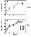
- FIGs 3 and 4 there is illustrated the immunogenicity of isolated and purified outer membrane protein CD of the present invention in mice.
- Figure 3 illustrates the production of CD-specific antibodies in mice following immunization with whole M. catarrhalis cells.
- Figure 4 illustrates the generation of specific antibodies following immunization with the purified CD protein of the invention.
- the purified CD protein is highly immunogenic and of comparable immunogenicity to the CD protein when presented to the immune system as part of the bacterium.
- the CD protein purified as described herein should advantageously be capable of generating antibodies that recognize or neutralize a plurality of Moraxella catarrhalis strains.
- Figure 5 Panel A
- FIG. 5 there is illustrated an immunoblot showing the ability of mouse anti-CD antisera produced by immunizing mice with purified CD protein as provided herein to recognize CD protein from Moraxella catarrhalis isolated from a variety of sources.
- the M. catarrhalis strains tested were as follows: Lane M.
- catarrhalis Source 2 Purified CD Present invention 3 4223 Middle ear fluid 4 5191 Middle ear fluid 5 59 Sputum 6 25240 ATCC 25240 7 135 Middle ear fluid 8 585 Blood 9 3 Sputum 10 8185 Nasopharynx Figure 5 (Panel B) also shows that immune sera obtained by immunizing mice with whole-inactivated M. catarrhalis cells showed high reactivity with purified CD protein (lane 2) or the strains of M. catarrhalis (lanes 3 to 10). This indicates that the CD protein is a highly immunogenic protein in M. catarrhalis.
- FIG. 6 there is shown a comparison of the anti-CD IgG subclass profile produced in mice by immunization either with whole inactivated M. catarrhalis cells or the isolated and purified CD protein provided herein.
- the most predominant responses were of the IgG 1 and IgG 2 isotypes.
- the IgG subclass profile produced by immunization with whole inactivated M. catarrhalis cells or purified CD protein were very similar, indicating the isolated and purified CD protein as described herein to be substantially in its native conformation.
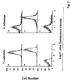
- FIG. 7 there is illustrated a flow cytometry analysis of anti-CD antisera binding to M. catarrhalis.
- Pooled mouse and anti-CD antisera produced by immunization with isolated and purified CD protein as described herein had nearly the same ability to bind to intact M. catarrhalis (Panel A) as that of anti- M . catarrhalis produced by immunization with whole M. catarrhalis cells (Panel B). Binding was specific to M. catarrhalis, since the same anti-CD antisera did not show significant binding to a bacterium, H. influenzae, that does not produce CD protein (Panel D). In contrast, pooled mouse anti- H. influenzae antisera reacted strongly with H. influenzae (Panel E).
- Panels C and F illustrate the substantial lack of binding of pre-immune sera to M. catarrhalis and H . influenzae respectively.
- This flow cytometry analysis indicates that anti-CD antibodies obtained by immunization with isolated and purified CD protein as provided herein recognize CD protein on the surface of intact M. catarrhalis and is further evidence that the CD protein as provided herein is in substantially its native conformation.
- the isolated and purified CD protein as provided herein is useful for generating antibodies that can be used to specifically distinguish M. catarrhalis from other bacterial pathogens that cause otitis media.
- Figure 8 there is illustrated an immunoblot showing the specific reactivity of mouse anti-CD antisera produced by immunizing mice with CD protein as provided herein.
- the bacteria analyzed were as follows: Lane Bacterium Source 1 M. catarrhalis 135 Middle ear fluid 2 M. catarrhalis 5191 Middle ear fluid 3 M. catarrhalis 4223 Middle ear fluid 4 non-typable H. influenzae Otitis media isolate 5 non-typable H. influenzae Otitis media isolate 6 non-typable H. influenzae Otitis media isolate 7.
- S. pneumoniae ATCC 6304 8.
- S. pneumoniae ATCC 6306 9. S. pneumoniae ATCC 6314
- results shown in Table 1 below illustrate the ability of either mouse or guinea pig anti-CD antisera produced by immunization with CD protein as provided herein, to lyse two different strains of M . catarrhalis.
- the results show that both antisera produced by immunization with CD protein isolated from strain 4223 were bactericidal against a homologous non-clumping M. catarrhalis strain RH408 derived from M. catarrhalis strain 4223 and a heterologous non-clumping strain Q8 (a gift from Dr. M.G. Bergeron, Centre Hospitalier de l'liable Laval, St. Foy, Quebec).
- the bacteriacidal titers in guinea pig immune sera were very high (1:1,024).
- the bacteriacidal activities in mouse anti-CD antisera were around 1:128 against both strains, which were comparable to the titre obtained using anti- M . catarrhalis antisera produced by immunization with whole inactivated M. catarrhalis cells.
- the ability of the isolated and purified CD protein as provided herein to generate bacteriacidal antibodies is in vivo evidence of utility of the CD protein as a vaccine to protect against disease caused by M. catarrhalis or other bacteria that produce CD protein or produce a protein capable of generating antibodies that specifically recognize CD protein.
- a vaccine against Moraxella or other bacterial pathogens that produce CD protein or produce a protein capable of inducing antibodies that specifically recognize CD, comprising an immunogenically-effective amount of CD protein as provided herein and a physiologically-acceptable carrier therefor.
- the CD protein provided herein also may be used as a carrier protein for hapten, polysaccharides or peptides to make a conjugate vaccine against antigenic determinants unrelated to CD.
- the CD protein provided herein is useful as a diagnostic reagent, as an antigen or for the generation of anti-CD antibodies, antigen for vaccination against the diseases caused by species of Moraxella and other bacterial pathogens that produce a protein capable of producing antibodies that specifically recognize CD and for detecting infection by Moraxella and other such bacteria.
- the CD protein as provided herein may be used as a carrier molecule to prepare chimeric molecules and conjugate vaccines (including glycoconjugates) against pathogenic bacteria, including encapsulated bacteria.
- glycoconjugates of the present invention may be used to confer protection against disease and infection caused by any bacteria having polysaccharide antigens including lipooligosaccharides (LOS) and PRP.
- Such bacterial pathogens may include, for example, Haemophilus influenzae, Streptococcus pneumoniae, Escherichia coli, Neisseria menigitides, Salmonella typhi, Streptococcus mutants, Cryptococcus neoformans, Klebsiella, Staphylococcus aureus and Pseudomonas aeruginosa.
- antigens which can be conjugated to CD protein and methods to achieve such conjugations are described in published PCT application WO 94/12641.
- the carrier function of CD protein may be used, for example, to induce an immune response against abnormal polysaccharides of tumour cells, or to produce anti-tumour antibodies that can be conjugated to chemotherapeutic or bioactive agents.
- the invention provides an isolated and purified non-denatured outer membrane protein CD isolatable from a Moraxella strain for use as an active pharmaceutical substance, particularly as an active ingredient in a vaccine against disease caused by infection with Moraxella.
- Immunogenic compositions suitable to be used as vaccines, may be prepared from CD protein as disclosed herein.
- the antigenic material is extensively dialyzed to remove undesired small molecular weight molecules and/or lyophilized for more ready formulation into a desired vehicle.
- the immunogenic composition elicits an immune response in a subject which produces antibodies, including anti-CD antibodies and antibodies that are opsonizing or bactericidal. Should the vaccinated subject be challenged by Moraxella or other bacteria that produce proteins capable of producing antibodies that specifically recognize CD protein, the antibodies bind to and inactivate the bacterium. Furthermore, opsonizing or bactericidal anti-CD antibodies may also provide protection by alternative mechanisms.
- Immunogenic compositions including vaccines may be prepared as injectables, as liquid solutions or emulsions.
- the CD protein may be mixed with pharmaceutically acceptable excipients which are compatible with CD protein.
- excipients may include, water, saline, dextrose, glycerol, ethanol, and combinations thereof.
- the immunogenic compositions and vaccines may further contain auxiliary substances, such as wetting or emulsifying agents, pH buffering agents, or adjuvants to enhance the effectiveness thereof.
- Immunogenic compositions and vaccines may be administered parenterally, by injection subcutaneously or intramuscularly.
- the immunogenic compositions formed according to the present invention may be formulated and delivered in a manner to evoke an immune response at mucosal surfaces.
- the immunogenic composition may be administered to mucosal surfaces by, for example, the nasal or oral (intragastric) routes.
- binders and carriers may include, for example, polyalkalene glycols or triglycerides.
- Such suppositories may be formed from mixtures containing the active ingredient(s) in the range of about 0.5 to about 10%, preferably about 1 to 2%.
- Oral formulations may include normally employed incipients such as, for example, pharmaceutical grades of saccharine, cellulose and magnesium carbonate. These compositions can take the form of solutions, suspensions, tablets, pills, capsules, sustained release formulations or powders and contain about 1 to 95% of the CD protein, preferably about 20 to about 75%.
- the immunogenic preparations and vaccines are administered in a manner compatible with the dosage formulation, and in such amount as will be therapeutically effective, protective and immunogenic.
- the quantity to be administered depends on the subject to be treated, including, for example, the capacity of the individual's immune system to synthesize antibodies, and if needed, to produce a cell-mediated immune response. Precise amounts of active ingredient required to be administered depend on the judgment of the practitioner. However, suitable dosage ranges are readily determinable by one skilled in the art and may be of the order of micrograms of the CD protein per vaccination. Suitable regimes for initial administration and booster doses are also variable, but may include an initial administration followed by subsequent administrations. The dosage may also depend on the route of administration and will vary according to the size of the host.
- the concentration of the CD antigen in an immunogenic composition according to the invention is in general about 1 to 95%.
- a vaccine which contains antigenic material of only one pathogen is a monovalent vaccine.
- Vaccines which contain antigenic material of several pathogens are combined vaccines and also belong to the present invention. Such combined vaccines contain, for example, material from various pathogens or from various strains of the same pathogen, or from combinations of various pathogens.
- Immunogenicity can be significantly improved if the antigens are co-administered with adjuvants, commonly used as an about 0.05 to 0.1 percent solution in phosphate-buffered saline.
- adjuvants enhance the immunogenicity of an antigen but are not necessarily immunogenic themselves.
- Adjuvants may act by retaining the antigen locally near the site of administration to produce a depot effect facilitating a slow, sustained release of antigen to cells of the immune system
- Adjuvants can also attract cells of the immune system to an antigen depot and stimulate such cells to elicit immune responses.
- Immunostimulatory agents or adjuvants have been used for many years to improve the host immune responses to, for example, vaccines.
- Intrinsic adjuvants such as lipopolysaccharides, normally are the components of the killed or attenuated bacteria used as vaccines.
- Extrinsic adjuvants are immunomodulators which are typically non-covalently linked to antigens and are formulated to enhance the host immune responses.
- adjuvants have been identified that enhance the immune response to antigens delivered parenterally. Some of these adjuvants are toxic, however, and can cause undesirable side-effects, making them unsuitable for use in humans and many animals.
- alum aluminum hydroxide and aluminum phosphate (collectively commonly referred to as alum) are routinely used as adjuvants in human and veterinary vaccines.
- the efficacy of alum in increasing antibody responses to diptheria and tetanus toxoids is well established and a HBsAg vaccine has been adjuvanted with alum. While the usefulness of alum is well established for some applications, it has limitations. For example, alum is ineffective for influenza vaccination and inconsistently elicits a cell mediated immune response.
- the antibodies elicited by alum-adjuvanted antigens are mainly of the IgG 1 isotype in the mouse, which may not be optimal for protection by some vaccinal agents.
- extrinsic adjuvants can provoke potent immune responses to antigens. These include saponins complexed to membrane protein antigens (immune stimulating complexes), pluronic polymers with mineral oil, killed mycobacteria in mineral oil, Freund's complete adjuvant, bacterial products, such as muramyl dipeptide (MDP) and lipopolysaccharide (LPS), as well as lipid A, and liposomes.
- MDP muramyl dipeptide
- LPS lipopolysaccharide
- FCA complete adjuvant
- cytolysis saponins and Pluronic polymers
- LPS and MDP pyrogenicity
- LPS and MDP pyrogenicity
- FCA is an excellent adjuvant and widely used in research, it is not licensed for use in human or veterinary vaccines because of its toxicity.
- Desirable characteristics of ideal adjuvants include:
- Wiesmuller describes a peptide with a sequence homologous to a foot-and-mouth disease virus protein coupled to an adjuvant tripalmityl-S-glyceryl-cysteinylserylserine, being a synthetic analogue of the N-terminal part of the lipoprotein form Gram negative bacteria.
- Deres et al (ref. 25) describes a peptide with a sequence homologous to a foot-and-mouth disease virus protein coupled to an adjuvant tripalmityl-S-glyceryl-cysteinylserylserine, being a synthetic analogue of the N-terminal part of the lipoprotein form Gram negative bacteria.
- the CD protein produced by the present invention is useful as an immunogen for the generation of anti-CD antibodies, as an antigen in immunoassay including enzyme-linked immunosorbent assays (ELISA), RIAs and other non-enzyme linked antibody binding assays or procedures known in the art for the detection of anti-bacterial, anti- Moraxella , and anti-CD antibodies.
- ELISA enzyme-linked immunosorbent assays
- the CD protein is immobilised on to a selected surface, for example, a surface capable of binding proteins such as the wells of a polystyrene microtiter plate.
- a non-specific protein such as a solution of bovine serum albumin (BSA) that is known to be antigenically neutral with regard to the test sample may be bound to the selected surface.
- BSA bovine serum albumin
- the immobilising surface is then contacted with a sample, such as clinical or biological materials, to be tested in a manner conducive to immune complex (antigen/ antibody) formation.
- a sample such as clinical or biological materials
- This may include diluting the sample with diluents, such as solutions of BSA, bovine gamma globulin (BGG) and/or phosphate buffered saline (PBS)/Tween (Trade Mark).
- BGG bovine gamma globulin
- PBS phosphate buffered saline
- Tween Trade Mark
- the sample is then allowed to incubate for from 2 to 4 hours, at temperatures such as of the order of about 25° to 37°C.
- the sample-contacted surface is washed to remove non-immunocomplexed material.
- the washing procedure may include washing with a solution, such as PBS/Tween or a borate buffer.
- the occurrence, and even amount, of immunocomplex formation may be determined by subjecting the immunocomplex to a second antibody having specificity for the first antibody.
- the second antibody is an antibody having specificity for human immunoglobulins and in general IgG.
- the second antibody may have an associated activity such as an enzymatic activity that will generate, for example, a colour development upon incubating with an appropriate chromogenic substrate. Quantification may then be achieved by measuring the degree of colour generation using, for example, a spectrophotometer.
- a Branhamella catarrhalis strain, RH408, described and referred to herein has been deposited with the American Type Culture Collection (ATCC) located at 12301 Parklawn Drive, Rockville, Maryland, USA pursuant to the Budapest Treaty and prior to the filing of this application.
- Branhamella catarrhalis strain RH408 has been accorded Designation No. 55637 and a filing date of December 13, 1994. Samples of the deposited strain will become available to the public upon grant of a patent based upon this United States patent application.
- the invention described and claimed herein is not to be limited in scope by strains deposited, since the deposited embodiment is intended only as an illustration of the invention. Any equivalent or similar strains that are non-clumping and have the same identifying characteristics as described in this application are within the scope of the invention.
- This Example illustrates the growth of M. catarrhalis.
- M. catarrhalis strain 4223 was inoculated into 20 mL of brain heart infusion (BHI) broth. The culture was incubated overnight with aeration at 37°C. For growth under iron-restricted conditions, one mL of the overnight-culture was inoculated into 20 mL of BHI broth containing 25 ⁇ M EDDA and the culture was grown at 37°C for approximately 3 to 4 h. Cells grown to mid-log phase (A 578 > 0.5) were harvested by centrifugation at 10,000 x g for 20 min. The pellet was used for extraction of CD protein as described in Example 3.
- BHI brain heart infusion
- This Example illustrates the generation of a non-clumping strain (RH408) of M . catarrhalis.
- M. catarrhalis strain 4223 was inoculated into several flasks containing 20 mL of BHI broth, and the cultures were incubated with shaking (170 rpm) overnight at 37°C. Five mL of each overnight culture were transferred to individual 1-mL tubes, and were left sitting undisturbed at room temperature for 3 to 8 hrs, to allow bacteria to sediment. One hundred ⁇ L of the cleared upper phase of each culture medium were used to inoculate 25 mL of BHI broth and cultures were incubated overnight at 37°C, as described above. This passaging was repeated six times, using 25 ⁇ L of cleared medium to inoculate 25 mL of BHI for each overnight culture.
- Non-clumping bacterial cultures were identified by measuring the A 578 at intervals over a 3 hr time period, in order to compare the sedimentation rates of the passaged strains to that of the original M. catarrhalis strain 4223 culture.
- Non-clumping mutants including M. catarrhalis RH408, did not aggregate during the three hr time period.
- strain RH408 had a colony morphology typical for all non-clumping strains.
- This Example illustrates the extraction and purification of CD protein.
- CD protein was isolated from M. catarrhalis by the procedure generally illustrated in Figure 1.
- the residual pellet (PPT 1 ) was extracted first in 40 mL of 50 mM Tris, pH 8.0 containing 0.5% Triton X-100 and 10 mM EDTA and then twice in 40 mL of 50 mM Tris, pH 8.0 containing 0.5% SDS. These two extractions remove the residual soluble proteins as well as the majority of membrane proteins excluding CD protein.
- the pellet obtained from the above extraction (PPT 3 ) was used as the starting material for CD purification.
- the pellet was resuspended in 50 mM Tris, pH 8.0, containing 0.5% Triton X-100, 10 mM EDTA.
- To the resulting suspension was added urea to a concentration of 6M, and then the suspension was heated at 60°C for 30 min to solubilize the CD protein. After centrifugation at 20,000 g for 30 min., the resultant supernatant contained homogenous purified CD protein and was separated from the pellet.
- the purified CD protein solution was dialyzed against 50 mM Tris, pH 8.0 overnight to remove the urea and then further centrifuged at 20,000 x g for 30 min.
- the CD protein remained soluble under these conditions.
- the amount of CD in the final preparation was determined to be about 100 ⁇ g/mL by the BCA protein assay and the purity of CD protein was assessed by SDS-PAGE analysis (see Fig. 2) and densitometry. The purity of the CD protein shown in Figure 2 was about 95%.
- the SDS-PAGE analysis confirmed the CD protein produced had the known molecular weight of 55 to 60 kDa for such protein (ref. 21).
- the identity of the purified CD protein was confirmed by amino acid sequence analysis of cyanogen bromide-cleavage fragments, in comparison to the published sequence (ref. 22).
- the purified CD protein was stored at -20°C.
- This Example illustrates the immunization of mice and guinea pigs with purified CD protein.
- mice Groups of five Balb/c mice were injected three times subcutaneously (s.c.) on days 1, 29 and 43 with either inactivated M. catarrhalis (about 10 5 cells) or purified CD protein produced as described in Example 3 (0.3 ⁇ g to 10 ⁇ g) in the presence of AlPO 4 (1.5 mg per dose). Blood samples were taken on days 14, 28, 42 and 56 for analysing the anti-CD antibody titers by EIAs.
- Groups of two guinea pigs (Charles River, Quebec) were immunized intramuscularly (i.m.) on day 1 with a 10 ⁇ g dose of purified CD protein emulsified in complete Freund's adjuvant (CFA). Animals were boosted on days 14 and 29 with the same dose of protein emulsified in incomplete Freund's adjuvant (IFA). Blood samples were taken on day 42 for analysing bacteriacidal activity of the antiserum.
- CFA complete Freund's adjuvant
- This Example illustrates the EIAs for determination of anti-C/D antibodies in mouse antisera.
- EIAs were performed essentially as described by Panezutti et al. (ref. 23). Microtiter wells were coated with 1 ⁇ g of CD protein for 16 hours at room temperature. The plates were then blocked with 0.1% (w/v) bovine serum albumin in PBS. The mouse sera were serially diluted, added to the wells, then incubated for one hour at room temperature. Affinity-purified F(ab') 2 fragments of goat anti-mouse IgG (Fc specific) antibody conjugated to horseradish peroxidase were used as the second antibody.
- F(ab') 2 fragments of goat anti-mouse IgG (Fc specific) antibody conjugated to horseradish peroxidase were used as the second antibody.
- the reactions were developed using tetramethylbenzidine (TMB/ H 2 O 2 ) and absorbency was measured at 450 nm (using 540 nm as a reference wavelength) in a Flow Multiskan MCC microplate reader.
- the reactive titer of an antiserum was defined as the reciprocal of the dilution consistently showing a two-fold increase in absorbency over that obtained with the pre-immune serum sample.
- This Example illustrates the EIAs to determine the subclasses of anti-CD IgG in mouse sera.
- Microtiter wells were coated with 1 ⁇ g of purified CD.
- the final bleeds of mouse serum samples from the immunogenicity study (as described in Example 4) were pooled and tested in EIAs.
- Rat anti-mouse IgG 1 , IgG 2a , IgG 2b antibodies conjugated to horseradish peroxidase and rabbit anti-mouse IgG 3 conjugated to horseradish peroxidase were used as reagents in EIAs.
- the working dilution of each conjugate was determined using purified antibody subclasses to avoid cross reactivity.
- the reactive titers were determined as described as one in Example 5.
- This Example illustrates the flow cytometry analysis.
- catarrhalis strain RH408 was grown to approximately 2 x 10 8 cfu/ mL as described Example 1 and then a 100 ⁇ l aliquot mixed with 200 ⁇ l of antisera diluted 1/500 in PBS (4mM Na 2 HPO 4 .7H 2 O, 1.5mM KH 2 PO 4 , 140 mM NaCl, 7mM KC1, pH7.3) containing 1% BSA. The samples were then incubated at 4°C for 30 min.
- H. influenzae strain 12 was also grown to 2 x 10 8 cfu/ mL, incubated with the same antisera and used as a negative control. Additional controls included incubation with the conjugates alone in the absence of any primary antiserum.
- the bacteria were washed twice with PBS containing 1% BSA and then mixed with 200 ⁇ l of diluted affinity purified Goat anti-mouse immunoglobulin - DTAF (Jackson ImmunoResearch Labs, Inc., Mississaga, Ontario). Bacteria were incubated with the conjugate for 30 min at 4°C, washed twice in PBS/BSA and resuspended in 1% paraformaldehyde.
- the fluorescence of stained bacteria were assessed using a Coulter Elite flow cytometer. Ten thousand bacteria were evaluated in each analysis. An argon laser was employed and emission signals at 525nm were collected. The level of antibody binding is expressed as the mean channel of fluorescence based on a log scale.
- This Example illustrates the bacteriacidal assay against M. catarrhalis.
- Bacterial cells grown to an A 578 > 0.5 were diluted 1:200,000 in VBS and 25- ⁇ L portions of the bacterial suspension were added to each well.
- a guinea pig complement (Biowhittaker, Walkersville, MD) was diluted 1:10 in VBS and 25 ⁇ L of the solution were added to each well to initiate reactions. The plates were incubated at 37°C for 60 min, and 50 ⁇ L of each reaction mixture was then plated onto Mueller-Hinton agar plate containing 2.2% Mueller-Hinton broth and 1.5% agar.
- the present invention provides an isolated and purified non-denatured outer membrane protein CD which is that of or corresponds to that isolated from a Moraxella strain and methods of isolating the same from bacterial strains. Modifications are possible within the scope of this invention.
Landscapes
- Chemical & Material Sciences (AREA)
- Health & Medical Sciences (AREA)
- Organic Chemistry (AREA)
- General Health & Medical Sciences (AREA)
- Medicinal Chemistry (AREA)
- Life Sciences & Earth Sciences (AREA)
- Proteomics, Peptides & Aminoacids (AREA)
- Molecular Biology (AREA)
- Genetics & Genomics (AREA)
- Biophysics (AREA)
- Biochemistry (AREA)
- Gastroenterology & Hepatology (AREA)
- Veterinary Medicine (AREA)
- Animal Behavior & Ethology (AREA)
- Pharmacology & Pharmacy (AREA)
- Nuclear Medicine, Radiotherapy & Molecular Imaging (AREA)
- General Chemical & Material Sciences (AREA)
- Chemical Kinetics & Catalysis (AREA)
- Oncology (AREA)
- Communicable Diseases (AREA)
- Public Health (AREA)
- Peptides Or Proteins (AREA)
- Medicines Containing Antibodies Or Antigens For Use As Internal Diagnostic Agents (AREA)
- Investigating Or Analysing Biological Materials (AREA)
- Preparation Of Compounds By Using Micro-Organisms (AREA)
- Micro-Organisms Or Cultivation Processes Thereof (AREA)
- Medicinal Preparation (AREA)
- General Preparation And Processing Of Foods (AREA)
- Medicines That Contain Protein Lipid Enzymes And Other Medicines (AREA)
Abstract
Description
| Lane | | Source | |
| 2 | Purified | Present invention | |
| 3 | 4223 | | |
| 4 | 5191 | | |
| 5 | 59 | | |
| 6 | 25240 | ATCC 25240 | |
| 7 | 135 | | |
| 8 | 585 | | |
| 9 | 3 | | |
| 10 | 8185 | Nasopharynx |
| | Bacterium | Source | |
| 1 | M. catarrhalis 135 | | |
| 2 | M. catarrhalis 5191 | | |
| 3 | M. catarrhalis 4223 | | |
| 4 | non-typable H. influenzae | Otitis media isolate | |
| 5 | non-typable H. influenzae | Otitis media isolate | |
| 6 | non-typable H. influenzae | Otitis media isolate | |
| 7. | S. pneumoniae | ATCC 6304 | |
| 8. | S. pneumoniae | ATCC 6306 | |
| 9. | S. pneumoniae | ATCC 6314 |

Claims (11)
- A method of producing an isolated and purified outer membrane protein CD of a bacterial strain that produces CD protein, comprising the steps of:(a) providing a cell mass of the bacterial strain;(b) disrupting the cell mass to provide a cell lysate;(c) fractionating the cell lysate to provide a first supernatant and a first pellet, the first supernatant comprising substantially a large proportion of soluble bacterial proteins;(d) separating said first supernatant from said first pellet;(e) selectively extracting the first pellet to remove substantially all soluble proteins and membrane proteins other than CD protein therefrom to provide a second supernatant and an extracted pellet containing CD protein;(f) separating said second supernatant from said extracted pellet;(g) solubilising the extracted pellet to provide a solubilised extract;(h) fractionating the solubilised extract to provide a CD protein-containing supernatant and a discard pellet; and(i) separating said CD-containing supernatant from said discard pellet.
- A method of Claim 1, wherein said bacterial strain may be a Moraxella strain.
- The method of Claim 1, wherein said bacterial strain is Moraxella catarrhalis.
- The method of Claim 1, wherein the cell lysate is fractionated by centrifugation thereof and the solubilised extract is fractionated by centrifugation thereof.
- The method of Claim 4, wherein the step of selectively extracting the first pellet comprises at least one detergent extraction.
- The method of Claim 5, wherein the step of extracting the first pellet comprising multiple detergent extractions.
- The method of Claim 5, wherein the extracted pellet is dispersed and solubilised with a buffered solution comprising a detergent and a solubilising agent at a temperature and for a time to effect solubilisation of the extracted pellet to provide the solubilised extract.
- The method of Claim 7, wherein the buffered solution has a pH from about 7 to about 8.5 and contains about 0.1 to about 2 wt% detergent and from about 3 to about 8M urea as said solubilising agent.
- The method of Claim 8, wherein the detergent is Triton X-100 (Trade Mark).
- The method of Claim 8, wherein said solubilisation is effected at a temperature of about 40° to about 70°C for about 10 to about 120 minutes.
- The method of Claim 10 including subsequently dialyzing the CD-containing supernatant to remove said detergent and solubilising agent to provide a further purified solution of CD protein in non-denatured form.
Applications Claiming Priority (3)
| Application Number | Priority Date | Filing Date | Title |
|---|---|---|---|
| US32858994A | 1994-10-24 | 1994-10-24 | |
| US328589 | 1994-10-24 | ||
| PCT/CA1995/000604 WO1996012733A1 (en) | 1994-10-24 | 1995-10-23 | Major outer membrane protein cd of moraxella |
Publications (2)
| Publication Number | Publication Date |
|---|---|
| EP0787144A1 EP0787144A1 (en) | 1997-08-06 |
| EP0787144B1 true EP0787144B1 (en) | 2001-12-12 |
Family
ID=23281593
Family Applications (1)
| Application Number | Title | Priority Date | Filing Date |
|---|---|---|---|
| EP95944810A Expired - Lifetime EP0787144B1 (en) | 1994-10-24 | 1995-10-23 | Major outer membrane protein cd of moraxella |
Country Status (14)
| Country | Link |
|---|---|
| US (1) | US6121427A (en) |
| EP (1) | EP0787144B1 (en) |
| JP (1) | JP3534771B2 (en) |
| CN (1) | CN1161376C (en) |
| AT (1) | ATE210678T1 (en) |
| AU (1) | AU706832B2 (en) |
| CA (1) | CA2203540C (en) |
| DE (1) | DE69524626T2 (en) |
| DK (1) | DK0787144T3 (en) |
| ES (1) | ES2169166T3 (en) |
| MX (1) | MX9702952A (en) |
| PT (1) | PT787144E (en) |
| RU (1) | RU2186582C2 (en) |
| WO (1) | WO1996012733A1 (en) |
Families Citing this family (10)
| Publication number | Priority date | Publication date | Assignee | Title |
|---|---|---|---|---|
| US6335018B1 (en) * | 1995-05-01 | 2002-01-01 | Aventis Pasteur Limited | High molecular weight major outer membrane protein of moraxella |
| CA2253942A1 (en) | 1996-05-10 | 1997-11-20 | Biogen, Inc. | Common gamma chain blocking agents |
| US5977337A (en) * | 1997-06-03 | 1999-11-02 | Connaught Laboratories Limited | Lactoferrin receptor genes of Moraxella |
| US7105164B1 (en) | 1998-04-07 | 2006-09-12 | Sanofi Pasteur Limited | HIV-specific cytotoxic T-cell responses |
| GB9808720D0 (en) | 1998-04-23 | 1998-06-24 | Smithkline Beecham Biolog | Novel compounds |
| GB9810084D0 (en) * | 1998-05-11 | 1998-07-08 | Cortecs Uk Ltd | Proteins |
| US6541616B1 (en) | 1998-10-01 | 2003-04-01 | Antex Biologics Inc. | Moraxella catarrhalis protein, gene sequence and uses thereof |
| JP2003500363A (en) * | 1999-05-24 | 2003-01-07 | ゾーマ(ユーエス) エルエルシー | Therapeutic use of BPI protein products in humans with otitis media with effusion |
| US7517968B2 (en) * | 2003-07-07 | 2009-04-14 | Syracuse University | Rapid and inexpensive method for the purification of proteorhodopsin |
| RU2601158C1 (en) * | 2015-05-12 | 2016-10-27 | Федеральное государственное бюджетное научное учреждение "Научно-исследовательский институт вакцин и сывороток им. И.И. Мечникова" (ФГБНУ НИИВС им. И.И. Мечникова) | STRAINS OF Streptococcus pneumoniae (VERSIONS) AND METHOD OF PRODUCING PROTECTIVE PROTEIN FRACTION, POSSESSING INTRASPECIFIC IMMUNOGENIC ACTIVITY |
Family Cites Families (11)
| Publication number | Priority date | Publication date | Assignee | Title |
|---|---|---|---|---|
| FI64813C (en) * | 1980-12-31 | 1984-01-10 | Ilkka Antero Palva | FOERFARANDE FOER PRODUCERING AV ETT UTVALT AEGGVITEAEMNE OCH VID FOERFARANDET ANVAENDA REKOMBINANTPLASMIDVEKTORER |
| US5110908A (en) * | 1986-12-31 | 1992-05-05 | Praxis Biologics, Inc. | Haemophilus influenzae peptides and proteins |
| DE3715840A1 (en) * | 1987-05-12 | 1988-12-01 | Boehringer Mannheim Gmbh | FOR A LYTICALLY EFFECTIVE, CHIMERAL PROTEIN-ENCODING RECOMBINANT DNA AND THEIR USE |
| US5393658A (en) * | 1987-09-28 | 1995-02-28 | New Horizons Diagnostics Corporation | Immunoassay method for the rapid identification of detergent treated antigens |
| ES2056378T3 (en) * | 1989-04-14 | 1994-10-01 | Unilever Nv | EXTRACTION PROCEDURE. |
| US5292869A (en) * | 1989-04-27 | 1994-03-08 | The Board Of Governors Of The University | Method for isolating and purifying transferrin and lactoferrin receptor proteins from bacteria and the preparation of vaccines containing the same |
| US5141743A (en) * | 1989-04-27 | 1992-08-25 | University Technologies International, Inc. | Method for isolating and purifying transferrin and lactoferrin receptor proteins and vaccines containing the same |
| US5552146A (en) * | 1991-08-15 | 1996-09-03 | Board Of Regents, The University Of Texas System | Methods and compositions relating to useful antigens of Moraxella catarrhalis |
| DE69423383T2 (en) * | 1993-05-13 | 2000-08-24 | American Cyanamid Co., Wayne | Production and uses of LOS-reduced outer membrane proteins from gram-negative cocci |
| US5712118A (en) * | 1993-09-29 | 1998-01-27 | Research Foundation Of State University Of New York | Vaccine for branhamella catarrhalis |
| IL111102A0 (en) * | 1993-09-29 | 1994-11-28 | Univ New York State Res Found | Vaccine for branhamella catarrhalis |
-
1995
- 1995-06-07 US US08/474,394 patent/US6121427A/en not_active Expired - Lifetime
- 1995-10-23 MX MX9702952A patent/MX9702952A/en not_active IP Right Cessation
- 1995-10-23 RU RU97108362/13A patent/RU2186582C2/en not_active IP Right Cessation
- 1995-10-23 CN CNB951969633A patent/CN1161376C/en not_active Expired - Fee Related
- 1995-10-23 AU AU36953/95A patent/AU706832B2/en not_active Ceased
- 1995-10-23 DE DE69524626T patent/DE69524626T2/en not_active Expired - Lifetime
- 1995-10-23 DK DK95944810T patent/DK0787144T3/en active
- 1995-10-23 CA CA002203540A patent/CA2203540C/en not_active Expired - Fee Related
- 1995-10-23 WO PCT/CA1995/000604 patent/WO1996012733A1/en not_active Ceased
- 1995-10-23 AT AT95944810T patent/ATE210678T1/en active
- 1995-10-23 ES ES95944810T patent/ES2169166T3/en not_active Expired - Lifetime
- 1995-10-23 PT PT95944810T patent/PT787144E/en unknown
- 1995-10-23 EP EP95944810A patent/EP0787144B1/en not_active Expired - Lifetime
- 1995-10-23 JP JP51356296A patent/JP3534771B2/en not_active Expired - Lifetime
Also Published As
| Publication number | Publication date |
|---|---|
| RU2186582C2 (en) | 2002-08-10 |
| ES2169166T3 (en) | 2002-07-01 |
| DE69524626T2 (en) | 2002-07-04 |
| JP3534771B2 (en) | 2004-06-07 |
| WO1996012733A1 (en) | 1996-05-02 |
| ATE210678T1 (en) | 2001-12-15 |
| US6121427A (en) | 2000-09-19 |
| DK0787144T3 (en) | 2002-01-14 |
| DE69524626D1 (en) | 2002-01-24 |
| PT787144E (en) | 2002-06-28 |
| CN1174559A (en) | 1998-02-25 |
| CA2203540A1 (en) | 1996-05-02 |
| AU3695395A (en) | 1996-05-15 |
| HK1009453A1 (en) | 1999-09-17 |
| CA2203540C (en) | 2005-04-05 |
| EP0787144A1 (en) | 1997-08-06 |
| MX9702952A (en) | 1997-06-28 |
| AU706832B2 (en) | 1999-06-24 |
| JPH10508303A (en) | 1998-08-18 |
| CN1161376C (en) | 2004-08-11 |
Similar Documents
| Publication | Publication Date | Title |
|---|---|---|
| AU711791B2 (en) | Transferrin receptor protein of moraxella | |
| AU722132B2 (en) | High molecular weight major outer membrane protein of moraxella | |
| RU2235128C2 (en) | Genes encoding moraxella transferrin receptor | |
| US6335018B1 (en) | High molecular weight major outer membrane protein of moraxella | |
| US6121427A (en) | Major outer membrane protein CD of branhamella | |
| US6348198B1 (en) | Lactoferrin receptor protein | |
| EP1905831A2 (en) | Lactoferrin receptor genes of moraxella | |
| MXPA00010026A (en) | Transferrin receptor genes of moraxella. | |
| MXPA98002853A (en) | Moraxe transferrine receptor protein | |
| HK1017695B (en) | Transferrin receptor protein of moraxella | |
| HK1009453B (en) | A method for producing a isolated and purified major outer membrane protein cd of moraxella | |
| MXPA99011210A (en) | Lactoferrin receptor genes of moraxella | |
| HK1098509A (en) | Transferrin receptor genes of moraxella |
Legal Events
| Date | Code | Title | Description |
|---|---|---|---|
| PUAI | Public reference made under article 153(3) epc to a published international application that has entered the european phase |
Free format text: ORIGINAL CODE: 0009012 |
|
| 17P | Request for examination filed |
Effective date: 19970506 |
|
| AK | Designated contracting states |
Kind code of ref document: A1 Designated state(s): AT BE CH DE DK ES FR GB GR IE IT LI LU MC NL PT SE |
|
| 17Q | First examination report despatched |
Effective date: 19990406 |
|
| GRAG | Despatch of communication of intention to grant |
Free format text: ORIGINAL CODE: EPIDOS AGRA |
|
| GRAG | Despatch of communication of intention to grant |
Free format text: ORIGINAL CODE: EPIDOS AGRA |
|
| GRAH | Despatch of communication of intention to grant a patent |
Free format text: ORIGINAL CODE: EPIDOS IGRA |
|
| RAP1 | Party data changed (applicant data changed or rights of an application transferred) |
Owner name: AVENTIS PASTEUR LIMITED |
|
| GRAH | Despatch of communication of intention to grant a patent |
Free format text: ORIGINAL CODE: EPIDOS IGRA |
|
| GRAA | (expected) grant |
Free format text: ORIGINAL CODE: 0009210 |
|
| AK | Designated contracting states |
Kind code of ref document: B1 Designated state(s): AT BE CH DE DK ES FR GB GR IE IT LI LU MC NL PT SE |
|
| REF | Corresponds to: |
Ref document number: 210678 Country of ref document: AT Date of ref document: 20011215 Kind code of ref document: T |
|
| REG | Reference to a national code |
Ref country code: CH Ref legal event code: NV Representative=s name: KATZAROV S.A. Ref country code: CH Ref legal event code: EP |
|
| REG | Reference to a national code |
Ref country code: GB Ref legal event code: IF02 |
|
| REG | Reference to a national code |
Ref country code: DK Ref legal event code: T3 |
|
| REG | Reference to a national code |
Ref country code: IE Ref legal event code: FG4D |
|
| REF | Corresponds to: |
Ref document number: 69524626 Country of ref document: DE Date of ref document: 20020124 |
|
| ET | Fr: translation filed | ||
| REG | Reference to a national code |
Ref country code: PT Ref legal event code: SC4A Free format text: AVAILABILITY OF NATIONAL TRANSLATION Effective date: 20020312 |
|
| REG | Reference to a national code |
Ref country code: ES Ref legal event code: FG2A Ref document number: 2169166 Country of ref document: ES Kind code of ref document: T3 |
|
| REG | Reference to a national code |
Ref country code: GR Ref legal event code: EP Ref document number: 20020400992 Country of ref document: GR |
|
| PLBQ | Unpublished change to opponent data |
Free format text: ORIGINAL CODE: EPIDOS OPPO |
|
| PLBI | Opposition filed |
Free format text: ORIGINAL CODE: 0009260 |
|
| PLBF | Reply of patent proprietor to notice(s) of opposition |
Free format text: ORIGINAL CODE: EPIDOS OBSO |
|
| 26 | Opposition filed |
Opponent name: WYETH Effective date: 20020911 |
|
| NLR1 | Nl: opposition has been filed with the epo |
Opponent name: WYETH |
|
| PLBF | Reply of patent proprietor to notice(s) of opposition |
Free format text: ORIGINAL CODE: EPIDOS OBSO |
|
| PLBF | Reply of patent proprietor to notice(s) of opposition |
Free format text: ORIGINAL CODE: EPIDOS OBSO |
|
| RAP2 | Party data changed (patent owner data changed or rights of a patent transferred) |
Owner name: SANOFI PASTEUR LIMITED |
|
| REG | Reference to a national code |
Ref country code: CH Ref legal event code: PUE Owner name: SANOFI PASTEUR LIMITED Free format text: AVENTIS PASTEUR LIMITED#1755 STEELES AVENUE WEST#TORONTO, ONTARIO M2R 3T4 (CA) -TRANSFER TO- SANOFI PASTEUR LIMITED#CAMPUS CONNAUGHT, STEELES AVENUE WEST#TORONTO, ONTARIO M2R 3T4 (CA) |
|
| NLT2 | Nl: modifications (of names), taken from the european patent patent bulletin |
Owner name: SANOFI PASTEUR LIMITED Effective date: 20060614 |
|
| PLCK | Communication despatched that opposition was rejected |
Free format text: ORIGINAL CODE: EPIDOSNREJ1 |
|
| APBP | Date of receipt of notice of appeal recorded |
Free format text: ORIGINAL CODE: EPIDOSNNOA2O |
|
| APAH | Appeal reference modified |
Free format text: ORIGINAL CODE: EPIDOSCREFNO |
|
| APBQ | Date of receipt of statement of grounds of appeal recorded |
Free format text: ORIGINAL CODE: EPIDOSNNOA3O |
|
| APBU | Appeal procedure closed |
Free format text: ORIGINAL CODE: EPIDOSNNOA9O |
|
| PLBN | Opposition rejected |
Free format text: ORIGINAL CODE: 0009273 |
|
| STAA | Information on the status of an ep patent application or granted ep patent |
Free format text: STATUS: OPPOSITION REJECTED |
|
| 27O | Opposition rejected |
Effective date: 20060904 |
|
| NLR2 | Nl: decision of opposition |
Effective date: 20060904 |
|
| PGFP | Annual fee paid to national office [announced via postgrant information from national office to epo] |
Ref country code: MC Payment date: 20120926 Year of fee payment: 18 |
|
| PGFP | Annual fee paid to national office [announced via postgrant information from national office to epo] |
Ref country code: GR Payment date: 20120917 Year of fee payment: 18 Ref country code: LU Payment date: 20121031 Year of fee payment: 18 |
|
| PGFP | Annual fee paid to national office [announced via postgrant information from national office to epo] |
Ref country code: DK Payment date: 20121010 Year of fee payment: 18 |
|
| PGFP | Annual fee paid to national office [announced via postgrant information from national office to epo] |
Ref country code: FR Payment date: 20121018 Year of fee payment: 18 Ref country code: CH Payment date: 20121012 Year of fee payment: 18 Ref country code: BE Payment date: 20121018 Year of fee payment: 18 Ref country code: IE Payment date: 20121010 Year of fee payment: 18 Ref country code: DE Payment date: 20121017 Year of fee payment: 18 |
|
| PGFP | Annual fee paid to national office [announced via postgrant information from national office to epo] |
Ref country code: IT Payment date: 20121016 Year of fee payment: 18 Ref country code: ES Payment date: 20121031 Year of fee payment: 18 Ref country code: SE Payment date: 20121011 Year of fee payment: 18 Ref country code: PT Payment date: 20120424 Year of fee payment: 18 Ref country code: GB Payment date: 20121017 Year of fee payment: 18 |
|
| PGFP | Annual fee paid to national office [announced via postgrant information from national office to epo] |
Ref country code: AT Payment date: 20120927 Year of fee payment: 18 Ref country code: NL Payment date: 20121010 Year of fee payment: 18 |
|
| BERE | Be: lapsed |
Owner name: *AVENTIS PASTEUR LTD Effective date: 20131031 |
|
| REG | Reference to a national code |
Ref country code: PT Ref legal event code: MM4A Free format text: LAPSE DUE TO NON-PAYMENT OF FEES Effective date: 20140423 |
|
| REG | Reference to a national code |
Ref country code: NL Ref legal event code: V1 Effective date: 20140501 |
|
| REG | Reference to a national code |
Ref country code: DK Ref legal event code: EBP Effective date: 20131031 |
|
| PG25 | Lapsed in a contracting state [announced via postgrant information from national office to epo] |
Ref country code: MC Free format text: LAPSE BECAUSE OF NON-PAYMENT OF DUE FEES Effective date: 20131031 |
|
| REG | Reference to a national code |
Ref country code: CH Ref legal event code: PL |
|
| REG | Reference to a national code |
Ref country code: SE Ref legal event code: EUG |
|
| REG | Reference to a national code |
Ref country code: AT Ref legal event code: MM01 Ref document number: 210678 Country of ref document: AT Kind code of ref document: T Effective date: 20131023 |
|
| GBPC | Gb: european patent ceased through non-payment of renewal fee |
Effective date: 20131023 |
|
| REG | Reference to a national code |
Ref country code: GR Ref legal event code: ML Ref document number: 20020400992 Country of ref document: GR Effective date: 20140505 |
|
| REG | Reference to a national code |
Ref country code: IE Ref legal event code: MM4A |
|
| PG25 | Lapsed in a contracting state [announced via postgrant information from national office to epo] |
Ref country code: GB Free format text: LAPSE BECAUSE OF NON-PAYMENT OF DUE FEES Effective date: 20131023 Ref country code: CH Free format text: LAPSE BECAUSE OF NON-PAYMENT OF DUE FEES Effective date: 20131031 Ref country code: LI Free format text: LAPSE BECAUSE OF NON-PAYMENT OF DUE FEES Effective date: 20131031 |
|
| REG | Reference to a national code |
Ref country code: DE Ref legal event code: R119 Ref document number: 69524626 Country of ref document: DE Effective date: 20140501 |
|
| REG | Reference to a national code |
Ref country code: FR Ref legal event code: ST Effective date: 20140630 |
|
| PG25 | Lapsed in a contracting state [announced via postgrant information from national office to epo] |
Ref country code: NL Free format text: LAPSE BECAUSE OF NON-PAYMENT OF DUE FEES Effective date: 20140501 Ref country code: GR Free format text: LAPSE BECAUSE OF NON-PAYMENT OF DUE FEES Effective date: 20140505 Ref country code: AT Free format text: LAPSE BECAUSE OF NON-PAYMENT OF DUE FEES Effective date: 20131023 Ref country code: DE Free format text: LAPSE BECAUSE OF NON-PAYMENT OF DUE FEES Effective date: 20140501 Ref country code: SE Free format text: LAPSE BECAUSE OF NON-PAYMENT OF DUE FEES Effective date: 20131024 Ref country code: PT Free format text: LAPSE BECAUSE OF NON-PAYMENT OF DUE FEES Effective date: 20140423 Ref country code: IT Free format text: LAPSE BECAUSE OF NON-PAYMENT OF DUE FEES Effective date: 20131023 Ref country code: FR Free format text: LAPSE BECAUSE OF NON-PAYMENT OF DUE FEES Effective date: 20131031 |
|
| PG25 | Lapsed in a contracting state [announced via postgrant information from national office to epo] |
Ref country code: BE Free format text: LAPSE BECAUSE OF NON-PAYMENT OF DUE FEES Effective date: 20131031 |
|
| PG25 | Lapsed in a contracting state [announced via postgrant information from national office to epo] |
Ref country code: IE Free format text: LAPSE BECAUSE OF NON-PAYMENT OF DUE FEES Effective date: 20131023 Ref country code: DK Free format text: LAPSE BECAUSE OF NON-PAYMENT OF DUE FEES Effective date: 20131031 |
|
| REG | Reference to a national code |
Ref country code: ES Ref legal event code: FD2A Effective date: 20141107 |
|
| PG25 | Lapsed in a contracting state [announced via postgrant information from national office to epo] |
Ref country code: ES Free format text: LAPSE BECAUSE OF NON-PAYMENT OF DUE FEES Effective date: 20131024 |
|
| PG25 | Lapsed in a contracting state [announced via postgrant information from national office to epo] |
Ref country code: LU Free format text: LAPSE BECAUSE OF NON-PAYMENT OF DUE FEES Effective date: 20131023 |