EP0785277B1 - Probe for diagnosing infectious diseases - Google Patents
Probe for diagnosing infectious diseases Download PDFInfo
- Publication number
- EP0785277B1 EP0785277B1 EP95933618A EP95933618A EP0785277B1 EP 0785277 B1 EP0785277 B1 EP 0785277B1 EP 95933618 A EP95933618 A EP 95933618A EP 95933618 A EP95933618 A EP 95933618A EP 0785277 B1 EP0785277 B1 EP 0785277B1
- Authority
- EP
- European Patent Office
- Prior art keywords
- streptococcus
- seq
- nucleic acid
- probe
- genomic dna
- Prior art date
- Legal status (The legal status is an assumption and is not a legal conclusion. Google has not performed a legal analysis and makes no representation as to the accuracy of the status listed.)
- Expired - Lifetime
Links
- 239000000523 sample Substances 0.000 title claims abstract description 53
- 208000035473 Communicable disease Diseases 0.000 title claims description 20
- 108020004414 DNA Proteins 0.000 claims description 54
- 238000009396 hybridization Methods 0.000 claims description 19
- 108020004707 nucleic acids Proteins 0.000 claims description 18
- 102000039446 nucleic acids Human genes 0.000 claims description 18
- 150000007523 nucleic acids Chemical class 0.000 claims description 18
- 108091028043 Nucleic acid sequence Proteins 0.000 claims description 12
- 238000003745 diagnosis Methods 0.000 claims description 11
- 241000194017 Streptococcus Species 0.000 claims description 7
- 239000002773 nucleotide Substances 0.000 claims description 7
- 125000003729 nucleotide group Chemical group 0.000 claims description 7
- ZHNUHDYFZUAESO-UHFFFAOYSA-N Formamide Chemical compound NC=O ZHNUHDYFZUAESO-UHFFFAOYSA-N 0.000 claims description 6
- 241001134658 Streptococcus mitis Species 0.000 claims description 5
- 241000194023 Streptococcus sanguinis Species 0.000 claims description 5
- 241000194032 Enterococcus faecalis Species 0.000 claims description 4
- 241000194031 Enterococcus faecium Species 0.000 claims description 4
- 241001147749 Gemella morbillorum Species 0.000 claims description 4
- 241000194008 Streptococcus anginosus Species 0.000 claims description 4
- 241001291896 Streptococcus constellatus Species 0.000 claims description 4
- 241000264435 Streptococcus dysgalactiae subsp. equisimilis Species 0.000 claims description 4
- 241000193996 Streptococcus pyogenes Species 0.000 claims description 4
- 241000194024 Streptococcus salivarius Species 0.000 claims description 4
- 230000000295 complement effect Effects 0.000 claims description 3
- 208000015181 infectious disease Diseases 0.000 claims description 3
- 238000005406 washing Methods 0.000 claims 8
- 238000000338 in vitro Methods 0.000 claims 1
- 241000894006 Bacteria Species 0.000 abstract description 67
- 241000193998 Streptococcus pneumoniae Species 0.000 abstract description 36
- 229940031000 streptococcus pneumoniae Drugs 0.000 abstract description 34
- 206010035664 Pneumonia Diseases 0.000 abstract description 12
- 239000012634 fragment Substances 0.000 abstract description 3
- 238000010367 cloning Methods 0.000 abstract description 2
- 108010072454 CTGCAG-specific type II deoxyribonucleases Proteins 0.000 abstract 1
- 230000000793 phophlogistic effect Effects 0.000 abstract 1
- 238000000034 method Methods 0.000 description 24
- 239000000243 solution Substances 0.000 description 13
- 201000001178 Bacterial Pneumonia Diseases 0.000 description 12
- HEMHJVSKTPXQMS-UHFFFAOYSA-M Sodium hydroxide Chemical compound [OH-].[Na+] HEMHJVSKTPXQMS-UHFFFAOYSA-M 0.000 description 9
- 238000001514 detection method Methods 0.000 description 9
- LFQSCWFLJHTTHZ-UHFFFAOYSA-N Ethanol Chemical compound CCO LFQSCWFLJHTTHZ-UHFFFAOYSA-N 0.000 description 8
- 239000002244 precipitate Substances 0.000 description 8
- 210000004369 blood Anatomy 0.000 description 6
- 239000008280 blood Substances 0.000 description 6
- 210000001539 phagocyte Anatomy 0.000 description 6
- 238000002360 preparation method Methods 0.000 description 6
- XLYOFNOQVPJJNP-UHFFFAOYSA-N water Chemical compound O XLYOFNOQVPJJNP-UHFFFAOYSA-N 0.000 description 6
- 239000003242 anti bacterial agent Substances 0.000 description 5
- 238000005119 centrifugation Methods 0.000 description 5
- 238000012163 sequencing technique Methods 0.000 description 5
- 238000002560 therapeutic procedure Methods 0.000 description 5
- HEDRZPFGACZZDS-UHFFFAOYSA-N Chloroform Chemical compound ClC(Cl)Cl HEDRZPFGACZZDS-UHFFFAOYSA-N 0.000 description 4
- 241000191967 Staphylococcus aureus Species 0.000 description 4
- 229940088710 antibiotic agent Drugs 0.000 description 4
- 238000012258 culturing Methods 0.000 description 4
- SUYVUBYJARFZHO-RRKCRQDMSA-N dATP Chemical compound C1=NC=2C(N)=NC=NC=2N1[C@H]1C[C@H](O)[C@@H](COP(O)(=O)OP(O)(=O)OP(O)(O)=O)O1 SUYVUBYJARFZHO-RRKCRQDMSA-N 0.000 description 4
- SUYVUBYJARFZHO-UHFFFAOYSA-N dATP Natural products C1=NC=2C(N)=NC=NC=2N1C1CC(O)C(COP(O)(=O)OP(O)(=O)OP(O)(O)=O)O1 SUYVUBYJARFZHO-UHFFFAOYSA-N 0.000 description 4
- RGWHQCVHVJXOKC-SHYZEUOFSA-J dCTP(4-) Chemical compound O=C1N=C(N)C=CN1[C@@H]1O[C@H](COP([O-])(=O)OP([O-])(=O)OP([O-])([O-])=O)[C@@H](O)C1 RGWHQCVHVJXOKC-SHYZEUOFSA-J 0.000 description 4
- NHVNXKFIZYSCEB-XLPZGREQSA-N dTTP Chemical compound O=C1NC(=O)C(C)=CN1[C@@H]1O[C@H](COP(O)(=O)OP(O)(=O)OP(O)(O)=O)[C@@H](O)C1 NHVNXKFIZYSCEB-XLPZGREQSA-N 0.000 description 4
- 239000013612 plasmid Substances 0.000 description 4
- 108091008146 restriction endonucleases Proteins 0.000 description 4
- 241000894007 species Species 0.000 description 4
- 239000006228 supernatant Substances 0.000 description 4
- 208000024891 symptom Diseases 0.000 description 4
- 239000003298 DNA probe Substances 0.000 description 3
- 241001646716 Escherichia coli K-12 Species 0.000 description 3
- ISWSIDIOOBJBQZ-UHFFFAOYSA-N Phenol Chemical compound OC1=CC=CC=C1 ISWSIDIOOBJBQZ-UHFFFAOYSA-N 0.000 description 3
- 206010036790 Productive cough Diseases 0.000 description 3
- 239000007864 aqueous solution Substances 0.000 description 3
- 230000003115 biocidal effect Effects 0.000 description 3
- 238000007796 conventional method Methods 0.000 description 3
- 239000012153 distilled water Substances 0.000 description 3
- 210000000265 leukocyte Anatomy 0.000 description 3
- 239000002609 medium Substances 0.000 description 3
- 239000000203 mixture Substances 0.000 description 3
- 230000009257 reactivity Effects 0.000 description 3
- 208000024794 sputum Diseases 0.000 description 3
- 210000003802 sputum Anatomy 0.000 description 3
- QKNYBSVHEMOAJP-UHFFFAOYSA-N 2-amino-2-(hydroxymethyl)propane-1,3-diol;hydron;chloride Chemical compound Cl.OCC(N)(CO)CO QKNYBSVHEMOAJP-UHFFFAOYSA-N 0.000 description 2
- 241001225321 Aspergillus fumigatus Species 0.000 description 2
- 208000031729 Bacteremia Diseases 0.000 description 2
- 108010074051 C-Reactive Protein Proteins 0.000 description 2
- 102100032752 C-reactive protein Human genes 0.000 description 2
- 241000222122 Candida albicans Species 0.000 description 2
- 201000007336 Cryptococcosis Diseases 0.000 description 2
- 241000221204 Cryptococcus neoformans Species 0.000 description 2
- IAZDPXIOMUYVGZ-UHFFFAOYSA-N Dimethylsulphoxide Chemical compound CS(C)=O IAZDPXIOMUYVGZ-UHFFFAOYSA-N 0.000 description 2
- KCXVZYZYPLLWCC-UHFFFAOYSA-N EDTA Chemical compound OC(=O)CN(CC(O)=O)CCN(CC(O)=O)CC(O)=O KCXVZYZYPLLWCC-UHFFFAOYSA-N 0.000 description 2
- 241000194033 Enterococcus Species 0.000 description 2
- 238000003794 Gram staining Methods 0.000 description 2
- 206010048612 Hydrothorax Diseases 0.000 description 2
- 206010061218 Inflammation Diseases 0.000 description 2
- 241000588747 Klebsiella pneumoniae Species 0.000 description 2
- 241000837853 Mucor spinosus Species 0.000 description 2
- 241000589517 Pseudomonas aeruginosa Species 0.000 description 2
- VMHLLURERBWHNL-UHFFFAOYSA-M Sodium acetate Chemical compound [Na+].CC([O-])=O VMHLLURERBWHNL-UHFFFAOYSA-M 0.000 description 2
- FAPWRFPIFSIZLT-UHFFFAOYSA-M Sodium chloride Chemical compound [Na+].[Cl-] FAPWRFPIFSIZLT-UHFFFAOYSA-M 0.000 description 2
- DBMJMQXJHONAFJ-UHFFFAOYSA-M Sodium laurylsulphate Chemical compound [Na+].CCCCCCCCCCCCOS([O-])(=O)=O DBMJMQXJHONAFJ-UHFFFAOYSA-M 0.000 description 2
- 241000193985 Streptococcus agalactiae Species 0.000 description 2
- 238000004458 analytical method Methods 0.000 description 2
- 229940091771 aspergillus fumigatus Drugs 0.000 description 2
- 230000001580 bacterial effect Effects 0.000 description 2
- 229940095731 candida albicans Drugs 0.000 description 2
- 210000004027 cell Anatomy 0.000 description 2
- 239000000470 constituent Substances 0.000 description 2
- 238000011109 contamination Methods 0.000 description 2
- 239000013068 control sample Substances 0.000 description 2
- 201000010099 disease Diseases 0.000 description 2
- 208000037265 diseases, disorders, signs and symptoms Diseases 0.000 description 2
- 238000001962 electrophoresis Methods 0.000 description 2
- 230000004054 inflammatory process Effects 0.000 description 2
- 238000002156 mixing Methods 0.000 description 2
- 230000003287 optical effect Effects 0.000 description 2
- SCVFZCLFOSHCOH-UHFFFAOYSA-M potassium acetate Chemical compound [K+].CC([O-])=O SCVFZCLFOSHCOH-UHFFFAOYSA-M 0.000 description 2
- 230000008569 process Effects 0.000 description 2
- 230000035755 proliferation Effects 0.000 description 2
- 230000002685 pulmonary effect Effects 0.000 description 2
- 239000001632 sodium acetate Substances 0.000 description 2
- 235000017281 sodium acetate Nutrition 0.000 description 2
- 230000001018 virulence Effects 0.000 description 2
- OAKPWEUQDVLTCN-NKWVEPMBSA-N 2',3'-Dideoxyadenosine-5-triphosphate Chemical compound C1=NC=2C(N)=NC=NC=2N1[C@H]1CC[C@@H](CO[P@@](O)(=O)O[P@](O)(=O)OP(O)(O)=O)O1 OAKPWEUQDVLTCN-NKWVEPMBSA-N 0.000 description 1
- FWMNVWWHGCHHJJ-SKKKGAJSSA-N 4-amino-1-[(2r)-6-amino-2-[[(2r)-2-[[(2r)-2-[[(2r)-2-amino-3-phenylpropanoyl]amino]-3-phenylpropanoyl]amino]-4-methylpentanoyl]amino]hexanoyl]piperidine-4-carboxylic acid Chemical compound C([C@H](C(=O)N[C@H](CC(C)C)C(=O)N[C@H](CCCCN)C(=O)N1CCC(N)(CC1)C(O)=O)NC(=O)[C@H](N)CC=1C=CC=CC=1)C1=CC=CC=C1 FWMNVWWHGCHHJJ-SKKKGAJSSA-N 0.000 description 1
- 206010048998 Acute phase reaction Diseases 0.000 description 1
- 241000193830 Bacillus <bacterium> Species 0.000 description 1
- 108020000946 Bacterial DNA Proteins 0.000 description 1
- CURLTUGMZLYLDI-UHFFFAOYSA-N Carbon dioxide Chemical compound O=C=O CURLTUGMZLYLDI-UHFFFAOYSA-N 0.000 description 1
- 241001478240 Coccus Species 0.000 description 1
- 206010011224 Cough Diseases 0.000 description 1
- 206010011703 Cyanosis Diseases 0.000 description 1
- 102000053602 DNA Human genes 0.000 description 1
- 108020003215 DNA Probes Proteins 0.000 description 1
- 208000000059 Dyspnea Diseases 0.000 description 1
- 206010013975 Dyspnoeas Diseases 0.000 description 1
- 241000588724 Escherichia coli Species 0.000 description 1
- 241001360526 Escherichia coli ATCC 25922 Species 0.000 description 1
- WQZGKKKJIJFFOK-GASJEMHNSA-N Glucose Natural products OC[C@H]1OC(O)[C@H](O)[C@@H](O)[C@@H]1O WQZGKKKJIJFFOK-GASJEMHNSA-N 0.000 description 1
- 206010019233 Headaches Diseases 0.000 description 1
- 208000004023 Legionellosis Diseases 0.000 description 1
- 201000009906 Meningitis Diseases 0.000 description 1
- RJQXTJLFIWVMTO-TYNCELHUSA-N Methicillin Chemical compound COC1=CC=CC(OC)=C1C(=O)N[C@@H]1C(=O)N2[C@@H](C(O)=O)C(C)(C)S[C@@H]21 RJQXTJLFIWVMTO-TYNCELHUSA-N 0.000 description 1
- 108010014251 Muramidase Proteins 0.000 description 1
- 102000016943 Muramidase Human genes 0.000 description 1
- 108010062010 N-Acetylmuramoyl-L-alanine Amidase Proteins 0.000 description 1
- 239000004677 Nylon Substances 0.000 description 1
- 108091034117 Oligonucleotide Proteins 0.000 description 1
- 206010057249 Phagocytosis Diseases 0.000 description 1
- 208000005384 Pneumocystis Pneumonia Diseases 0.000 description 1
- 206010073755 Pneumocystis jirovecii pneumonia Diseases 0.000 description 1
- 241000191963 Staphylococcus epidermidis Species 0.000 description 1
- 241000751182 Staphylococcus epidermidis ATCC 12228 Species 0.000 description 1
- 241000194026 Streptococcus gordonii Species 0.000 description 1
- 241000194025 Streptococcus oralis Species 0.000 description 1
- JLCPHMBAVCMARE-UHFFFAOYSA-N [3-[[3-[[3-[[3-[[3-[[3-[[3-[[3-[[3-[[3-[[3-[[5-(2-amino-6-oxo-1H-purin-9-yl)-3-[[3-[[3-[[3-[[3-[[3-[[5-(2-amino-6-oxo-1H-purin-9-yl)-3-[[5-(2-amino-6-oxo-1H-purin-9-yl)-3-hydroxyoxolan-2-yl]methoxy-hydroxyphosphoryl]oxyoxolan-2-yl]methoxy-hydroxyphosphoryl]oxy-5-(5-methyl-2,4-dioxopyrimidin-1-yl)oxolan-2-yl]methoxy-hydroxyphosphoryl]oxy-5-(6-aminopurin-9-yl)oxolan-2-yl]methoxy-hydroxyphosphoryl]oxy-5-(6-aminopurin-9-yl)oxolan-2-yl]methoxy-hydroxyphosphoryl]oxy-5-(6-aminopurin-9-yl)oxolan-2-yl]methoxy-hydroxyphosphoryl]oxy-5-(6-aminopurin-9-yl)oxolan-2-yl]methoxy-hydroxyphosphoryl]oxyoxolan-2-yl]methoxy-hydroxyphosphoryl]oxy-5-(5-methyl-2,4-dioxopyrimidin-1-yl)oxolan-2-yl]methoxy-hydroxyphosphoryl]oxy-5-(4-amino-2-oxopyrimidin-1-yl)oxolan-2-yl]methoxy-hydroxyphosphoryl]oxy-5-(5-methyl-2,4-dioxopyrimidin-1-yl)oxolan-2-yl]methoxy-hydroxyphosphoryl]oxy-5-(5-methyl-2,4-dioxopyrimidin-1-yl)oxolan-2-yl]methoxy-hydroxyphosphoryl]oxy-5-(6-aminopurin-9-yl)oxolan-2-yl]methoxy-hydroxyphosphoryl]oxy-5-(6-aminopurin-9-yl)oxolan-2-yl]methoxy-hydroxyphosphoryl]oxy-5-(4-amino-2-oxopyrimidin-1-yl)oxolan-2-yl]methoxy-hydroxyphosphoryl]oxy-5-(4-amino-2-oxopyrimidin-1-yl)oxolan-2-yl]methoxy-hydroxyphosphoryl]oxy-5-(4-amino-2-oxopyrimidin-1-yl)oxolan-2-yl]methoxy-hydroxyphosphoryl]oxy-5-(6-aminopurin-9-yl)oxolan-2-yl]methoxy-hydroxyphosphoryl]oxy-5-(4-amino-2-oxopyrimidin-1-yl)oxolan-2-yl]methyl [5-(6-aminopurin-9-yl)-2-(hydroxymethyl)oxolan-3-yl] hydrogen phosphate Polymers Cc1cn(C2CC(OP(O)(=O)OCC3OC(CC3OP(O)(=O)OCC3OC(CC3O)n3cnc4c3nc(N)[nH]c4=O)n3cnc4c3nc(N)[nH]c4=O)C(COP(O)(=O)OC3CC(OC3COP(O)(=O)OC3CC(OC3COP(O)(=O)OC3CC(OC3COP(O)(=O)OC3CC(OC3COP(O)(=O)OC3CC(OC3COP(O)(=O)OC3CC(OC3COP(O)(=O)OC3CC(OC3COP(O)(=O)OC3CC(OC3COP(O)(=O)OC3CC(OC3COP(O)(=O)OC3CC(OC3COP(O)(=O)OC3CC(OC3COP(O)(=O)OC3CC(OC3COP(O)(=O)OC3CC(OC3COP(O)(=O)OC3CC(OC3COP(O)(=O)OC3CC(OC3COP(O)(=O)OC3CC(OC3COP(O)(=O)OC3CC(OC3CO)n3cnc4c(N)ncnc34)n3ccc(N)nc3=O)n3cnc4c(N)ncnc34)n3ccc(N)nc3=O)n3ccc(N)nc3=O)n3ccc(N)nc3=O)n3cnc4c(N)ncnc34)n3cnc4c(N)ncnc34)n3cc(C)c(=O)[nH]c3=O)n3cc(C)c(=O)[nH]c3=O)n3ccc(N)nc3=O)n3cc(C)c(=O)[nH]c3=O)n3cnc4c3nc(N)[nH]c4=O)n3cnc4c(N)ncnc34)n3cnc4c(N)ncnc34)n3cnc4c(N)ncnc34)n3cnc4c(N)ncnc34)O2)c(=O)[nH]c1=O JLCPHMBAVCMARE-UHFFFAOYSA-N 0.000 description 1
- HDRRAMINWIWTNU-NTSWFWBYSA-N [[(2s,5r)-5-(2-amino-6-oxo-3h-purin-9-yl)oxolan-2-yl]methoxy-hydroxyphosphoryl] phosphono hydrogen phosphate Chemical compound C1=2NC(N)=NC(=O)C=2N=CN1[C@H]1CC[C@@H](COP(O)(=O)OP(O)(=O)OP(O)(O)=O)O1 HDRRAMINWIWTNU-NTSWFWBYSA-N 0.000 description 1
- ARLKCWCREKRROD-POYBYMJQSA-N [[(2s,5r)-5-(4-amino-2-oxopyrimidin-1-yl)oxolan-2-yl]methoxy-hydroxyphosphoryl] phosphono hydrogen phosphate Chemical compound O=C1N=C(N)C=CN1[C@@H]1O[C@H](COP(O)(=O)OP(O)(=O)OP(O)(O)=O)CC1 ARLKCWCREKRROD-POYBYMJQSA-N 0.000 description 1
- 230000004658 acute-phase response Effects 0.000 description 1
- 238000000137 annealing Methods 0.000 description 1
- 208000022531 anorexia Diseases 0.000 description 1
- 206010003246 arthritis Diseases 0.000 description 1
- 210000004556 brain Anatomy 0.000 description 1
- 229940041514 candida albicans extract Drugs 0.000 description 1
- 235000011089 carbon dioxide Nutrition 0.000 description 1
- 210000005056 cell body Anatomy 0.000 description 1
- 230000009260 cross reactivity Effects 0.000 description 1
- 238000012136 culture method Methods 0.000 description 1
- URGJWIFLBWJRMF-JGVFFNPUSA-N ddTTP Chemical compound O=C1NC(=O)C(C)=CN1[C@@H]1O[C@H](COP(O)(=O)OP(O)(=O)OP(O)(O)=O)CC1 URGJWIFLBWJRMF-JGVFFNPUSA-N 0.000 description 1
- 206010061428 decreased appetite Diseases 0.000 description 1
- 230000007123 defense Effects 0.000 description 1
- 238000004925 denaturation Methods 0.000 description 1
- 230000036425 denaturation Effects 0.000 description 1
- 238000013461 design Methods 0.000 description 1
- 230000014670 detection of bacterium Effects 0.000 description 1
- 238000011161 development Methods 0.000 description 1
- 210000000624 ear auricle Anatomy 0.000 description 1
- 230000000694 effects Effects 0.000 description 1
- 108010060371 endo-N-acetylmuramidase Proteins 0.000 description 1
- 238000011156 evaluation Methods 0.000 description 1
- 238000004817 gas chromatography Methods 0.000 description 1
- 239000008103 glucose Substances 0.000 description 1
- 239000001963 growth medium Substances 0.000 description 1
- 231100000869 headache Toxicity 0.000 description 1
- 230000036541 health Effects 0.000 description 1
- 208000021760 high fever Diseases 0.000 description 1
- 230000006872 improvement Effects 0.000 description 1
- 238000001727 in vivo Methods 0.000 description 1
- 210000004969 inflammatory cell Anatomy 0.000 description 1
- 208000027866 inflammatory disease Diseases 0.000 description 1
- 230000002757 inflammatory effect Effects 0.000 description 1
- 238000001802 infusion Methods 0.000 description 1
- 230000009545 invasion Effects 0.000 description 1
- 238000002955 isolation Methods 0.000 description 1
- 231100000518 lethal Toxicity 0.000 description 1
- 230000001665 lethal effect Effects 0.000 description 1
- 239000004325 lysozyme Substances 0.000 description 1
- 229960000274 lysozyme Drugs 0.000 description 1
- 235000010335 lysozyme Nutrition 0.000 description 1
- 210000002540 macrophage Anatomy 0.000 description 1
- 206010025482 malaise Diseases 0.000 description 1
- 239000000463 material Substances 0.000 description 1
- 230000002503 metabolic effect Effects 0.000 description 1
- 229960003085 meticillin Drugs 0.000 description 1
- 239000011259 mixed solution Substances 0.000 description 1
- 238000010369 molecular cloning Methods 0.000 description 1
- 210000001616 monocyte Anatomy 0.000 description 1
- 210000000440 neutrophil Anatomy 0.000 description 1
- 229920001778 nylon Polymers 0.000 description 1
- 244000052769 pathogen Species 0.000 description 1
- 230000007170 pathology Effects 0.000 description 1
- 210000005259 peripheral blood Anatomy 0.000 description 1
- 239000011886 peripheral blood Substances 0.000 description 1
- 230000008782 phagocytosis Effects 0.000 description 1
- 201000000317 pneumocystosis Diseases 0.000 description 1
- 235000011056 potassium acetate Nutrition 0.000 description 1
- 239000000047 product Substances 0.000 description 1
- 230000002062 proliferating effect Effects 0.000 description 1
- 108090000623 proteins and genes Proteins 0.000 description 1
- 230000002285 radioactive effect Effects 0.000 description 1
- 230000009467 reduction Effects 0.000 description 1
- 230000028327 secretion Effects 0.000 description 1
- 238000000926 separation method Methods 0.000 description 1
- 230000035939 shock Effects 0.000 description 1
- 239000011780 sodium chloride Substances 0.000 description 1
- 230000003068 static effect Effects 0.000 description 1
- 239000000126 substance Substances 0.000 description 1
- 239000000725 suspension Substances 0.000 description 1
- 230000009885 systemic effect Effects 0.000 description 1
- 238000012360 testing method Methods 0.000 description 1
- 230000001225 therapeutic effect Effects 0.000 description 1
- 230000000699 topical effect Effects 0.000 description 1
- 230000001131 transforming effect Effects 0.000 description 1
- 230000001960 triggered effect Effects 0.000 description 1
- 239000012137 tryptone Substances 0.000 description 1
- 239000012138 yeast extract Substances 0.000 description 1
Images


Classifications
-
- C—CHEMISTRY; METALLURGY
- C12—BIOCHEMISTRY; BEER; SPIRITS; WINE; VINEGAR; MICROBIOLOGY; ENZYMOLOGY; MUTATION OR GENETIC ENGINEERING
- C12Q—MEASURING OR TESTING PROCESSES INVOLVING ENZYMES, NUCLEIC ACIDS OR MICROORGANISMS; COMPOSITIONS OR TEST PAPERS THEREFOR; PROCESSES OF PREPARING SUCH COMPOSITIONS; CONDITION-RESPONSIVE CONTROL IN MICROBIOLOGICAL OR ENZYMOLOGICAL PROCESSES
- C12Q1/00—Measuring or testing processes involving enzymes, nucleic acids or microorganisms; Compositions therefor; Processes of preparing such compositions
- C12Q1/68—Measuring or testing processes involving enzymes, nucleic acids or microorganisms; Compositions therefor; Processes of preparing such compositions involving nucleic acids
-
- C—CHEMISTRY; METALLURGY
- C12—BIOCHEMISTRY; BEER; SPIRITS; WINE; VINEGAR; MICROBIOLOGY; ENZYMOLOGY; MUTATION OR GENETIC ENGINEERING
- C12Q—MEASURING OR TESTING PROCESSES INVOLVING ENZYMES, NUCLEIC ACIDS OR MICROORGANISMS; COMPOSITIONS OR TEST PAPERS THEREFOR; PROCESSES OF PREPARING SUCH COMPOSITIONS; CONDITION-RESPONSIVE CONTROL IN MICROBIOLOGICAL OR ENZYMOLOGICAL PROCESSES
- C12Q1/00—Measuring or testing processes involving enzymes, nucleic acids or microorganisms; Compositions therefor; Processes of preparing such compositions
- C12Q1/68—Measuring or testing processes involving enzymes, nucleic acids or microorganisms; Compositions therefor; Processes of preparing such compositions involving nucleic acids
- C12Q1/6876—Nucleic acid products used in the analysis of nucleic acids, e.g. primers or probes
- C12Q1/6888—Nucleic acid products used in the analysis of nucleic acids, e.g. primers or probes for detection or identification of organisms
- C12Q1/689—Nucleic acid products used in the analysis of nucleic acids, e.g. primers or probes for detection or identification of organisms for bacteria
-
- C—CHEMISTRY; METALLURGY
- C07—ORGANIC CHEMISTRY
- C07H—SUGARS; DERIVATIVES THEREOF; NUCLEOSIDES; NUCLEOTIDES; NUCLEIC ACIDS
- C07H21/00—Compounds containing two or more mononucleotide units having separate phosphate or polyphosphate groups linked by saccharide radicals of nucleoside groups, e.g. nucleic acids
- C07H21/04—Compounds containing two or more mononucleotide units having separate phosphate or polyphosphate groups linked by saccharide radicals of nucleoside groups, e.g. nucleic acids with deoxyribosyl as saccharide radical
-
- C—CHEMISTRY; METALLURGY
- C12—BIOCHEMISTRY; BEER; SPIRITS; WINE; VINEGAR; MICROBIOLOGY; ENZYMOLOGY; MUTATION OR GENETIC ENGINEERING
- C12N—MICROORGANISMS OR ENZYMES; COMPOSITIONS THEREOF; PROPAGATING, PRESERVING, OR MAINTAINING MICROORGANISMS; MUTATION OR GENETIC ENGINEERING; CULTURE MEDIA
- C12N15/00—Mutation or genetic engineering; DNA or RNA concerning genetic engineering, vectors, e.g. plasmids, or their isolation, preparation or purification; Use of hosts therefor
Definitions
- the present invention relates to a probe which is useful for detecting and identifying the causative bacteria of infectious diseases, especially Streptococcus pneumoniae which is a representative causative bacterium of bacterial pneumonia.
- infection is defined as an invasion and an establishment of an anchorage for growth in a host organism by a pathogenic organism (hereinafter referred to as "bacterium").
- bacterium a pathogenic organism
- the outbreak of a disease caused by proliferation of a bacterium in vivo depends upon the interrelationship between the resistance of the host organism and the virulence of the bacterium.
- the bacterial pneumonia is a serious and exigent infectious diseases in which severe systemic symptoms such as malaise, anorexia and headache have involved, presenting dyspnea, cyanosis, and could be accompanied by bacteremia, cerebrospinal meningitis or arthritis as the complication thereof, finally could sometimes lead to a lethal process.
- phagocytes including neutrophils, monocytes and macrophages primarily function in defense of the body, and that exuded bacteria from the texture of the phagocyte which had predominantly grown have appeared into blood.
- bacterial pneumonia is defined as a case wherein the ability of phagocytosis by cells cannot overcome the virulence of the bacteria and then the bacteria such as Streptococcus pneumoniae settle on the pulmonary lobe and the tissue of bronchia to cause inflammation.
- the following items should be checked: 1) clinical symptoms; 2) culture of a specimen; 3) gram-staining of the bacteria contained in the specimen; and 4) shock state. After those items have been clarified, the course of therapy has been oriented.
- CRP C-Reactive Protein
- a method for identifying the causative bacteria in a patient who is suspected to be suffered from bacterial pneumonia employing the sample collected from sputum, secretion from upper airway, hydrothorax, topical focus, or blood as a specimen to estimate applicability of the sample as a test material by observing inflammatory cells of smear thereof, then, determining cell type by Gram staining e.g., gram negative or positive, and coccus or bacillus, and finally, culturing the bacteria using selection medium to identify the causative bacteria.
- Gram staining e.g., gram negative or positive, and coccus or bacillus
- the pretreatment which requires at least three to four days in total including one to two days for selective isolation of bacteria from a specimen, one day for culture, and one or more days for operation of fixation, and then the culture thereof is continued in practice until the bacteria grow. Therefore, in many cases, the pretreatment requires one week or more. Furthermore, there has been a risk on contamination of a other bacteria which could not be distinguished from the causative bacteria during the culture period.
- the number of bacteria that can be grown is small even under appropriate conditions for culture, because many bacteria in a specimen to be grown have been ingested into phagocyte, dead or on a static state due to antibiotics administered. Therefore, the actual detection rate of bacteria is as low as about 10% when the clinical specimen culture method is employed. In the other words, for the present, the presence of bacteria in 90% of the examined blood from the patient suspected clinically as suffering from pneumonia, which has been cultured for further one or more days, could not be proved after all.
- the present practice depends on a trial and error treatment method, starting when pneumonia is clinically suspected without awaiting the detection results of the identification, wherein an antibiotic having the effectiveness for the widest range of the causative bacteria is administered first while the causative bacteria is still unknown, and if the antibiotic is not effective after one or two days, then another antibiotic will be tested, regardless of the fact that determination of the causative bacteria and selection of the suitable antibiotics are required.
- the constituents of the living body are likewise stained together with bacteria, therefore, a skilled experience to identify bacteria quickly according to their image through a microscope is required, then there may be cases that can be hardly identified.
- pneumonia is a disease wherein a rapid and exact diagnosis has been required as stated above, the conventional diagnosis method could not have satisfied such requirements.
- FR-A-2687168 discloses a DNA probe obtained by treating Streptococcus pneumoniae DNA with the restriction enzymes Hpa I and Pvu II. It also discloses oligonucleotides of 22 and 29 nucleotides in length (SEQ ID NO's 6 and 7, respectively) which hybridise to Streptococcus pneumoniae DNA but not to the DNA of Streptococcus oralis, Streptococcus gordonii, Streptococcus sanguis or Streptococcus mitis.
- the present invention was accomplished in view of the above-described problem in the art, and according to one aspect of this invention, there is provided a probe having a specific reactivity with DNA or RNA obtained from causative bacteria of the infectious diseases, especially, Streptococcus pneumoniae which is the representative causative bacteria of bacterial pneumonia, and nucleotide sequences of a portion of the gene essentially included in Streptococcus pneumoniae being elucidated.
- the probe of the present invention allows significant detection of remaining bacterial DNA, the bacteria being incorporated into phagocytes and destroyed, thereby quick and accurate detection method of the causative bacteria of the infectious diseases would be available without culture and/or growth of the bacteria.
- a primer is designed with reference to information on the nucleotide sequence of the probe, causative bacteria can be identified without hybridization step, through amplifying DNA by means of a PCR technique.
- a non-radioactive probe e.g., a biotinylated probe
- detection in a general laboratory can be performed as well using an optical microscope, and the detection process will be carried out rapidly and simply.
- the extracted DNA was digested completely with restriction enzyme PstI and random cloned into vector pGEM-3Z.
- PstI restriction enzyme
- seven probes unique to the bacteria Streptococcus pneumoniae namely probes comprising DNA fragment showing specificity to DNA from Streptococcus pneumoniae were then selected.
- probe SP-22 The selected probes were then designated as probe SP-22, probe SP-23, probe SP-25, probe SP-26, probe SP-5-15, probe SP-5-34 and probe SP-6-6, and restriction maps of which are illustrated in Figure 1 and Figure 2.
- strains of clinical isolate and deposited strains described below in Table 1 were prepared.
- Human genomic DNA and control sample in Table 1 were obtained and prepared from leukocyte collected from four healthy adult men, and Escherichia coli K-12 JM109 transformant comprising plasmid pGem-32, respectively.
- DNA of each strain was extracted according to the method of the above Example 1 (1), and samples for dot-blot-hybridization were obtained by spotting certain amount (e.g., 5 ⁇ l) of the extracted DNA to a nylon filter and then conducting alkaline denaturation.
- Human genomic DNA sample was prepared from the above-described leukocyte employing Saito-Miura's method ( supra ). Meanwhile, control sample was prepared from the above-described Escherichia coli K-12 JM109 transformant comprising plasmid pGem-32 applying the method for preparing plasmid DNA described in Example 2 (1) below.
- Hybridization on DNA probes from Streptococcus pneumoniae labeled with Digoxigenin-11-dUTP (BRL) was then performed overnight according to Manual of Maniatis (T. Maniatis, et al., "Molecular Cloning (A Laboratory Manual)", Cold Spring Harbour Laboratory (1982)), under the condition of 45% formamide, 5 ⁇ SSC, 42°C.
- any of the probes exhibited reactivity specifically with only DNA obtained from Streptococcus pneumoniae, and did not exhibit cross-reactivity (ability to hybridize) with any DNA obtained from bacteria other than genus Streptococcus , as well as DNA from other species of Streptococcus , thus specificity thereof to the species Streptococcus pneumoniae has been confirmed.
- Nucleotide sequences of DNA probes (7 probes total) of which specificity to the bacterial species were verified in the Example 1, were determined according to the following method.
- Escherichia coli K-12, JM109 transformant comprising the subcloned insert fragment (to be sequenced) in pGem-3Z (Promega), was inoculated in 5ml of Luria-Bactani Medium (bacto-tryptone, 10g/1L; bacto-yeast extract, 5g/1L; NaCl, 10g/1L; adjusted pH to 7.0 with 5N NaOH) and cultured overnight.
- Luria-Bactani Medium bacto-tryptone, 10g/1L
- bacto-yeast extract 5g/1L
- NaCl 10g/1L
- adjusted pH to 7.0 with 5N NaOH adjusted pH to 7.0 with 5N NaOH
- Culture medium was centrifuged (5,000rpm, 5min.) to collect the bacterial cell body.
- a solution of 50mM glucose/50mM Tris-HCl (pH8.0)/10mM EDTA containing 2.5mg/ml of lysozyme (Sigma) was added to the precipitate, and left at room temperature for 5 minutes.
- 0.2M NaOH solution containing 1% of sodium dodecyl sulfate (Sigma) was added and mixed therewith.
- 5M potassium acetate aqueous solution pH 4.8
- the supernatant obtained by centrifugation (15,000rpm, 15min.) of the mixture was treated with phenol/CHCl 3 and added thereto ethanol of two times by volume, then the precipitate was obtained by further centrifugation (12,000rpm, 5min.).
- This precipitate was dissolved in 100 ⁇ l of solution of 10mM Tris-HCl (pH7.5)/0.1mM EDTA, and added thereto 10mg/ml RNaseA (Sigma) solution, then left it at room temperature for 15 minutes.
- concentration of DNA for use as a template was adjusted to 5-10 ug in 32 ⁇ l of solution.
- concentration of DNA for use as a template was adjusted to 5-10 ug in 32 ⁇ l of solution.
- Thirty two ⁇ l of the template DNA solution was transferred to 1.5ml mini-tube (Eppendolf), and added thereto 8 ⁇ l of 2M NaOH aqueous solution, then mixed gently. After instant centrifugation, it was left at room temperature for 10 minutes.
- the precipitate was dissolved in 10 ⁇ l of distilled water, then 2 ⁇ l of fluorescent primer (0.42 A 260 unit/ml, 4-6 pmol), (Fluorescent Primer, M13 Universal Primer; 5'-Fluorescein-d[CGACGTTGTAAAACGACGGCCAGT]-3' (1.6pmol/ ⁇ l; 0.42 A 260 unit/ml); M13 Reverse Primer, 5'-Fluorescein-d[CAGGAAACAGCTATGAC] -3' (2.1pmol/ ⁇ l; 0.42 A 260 unit/ml)) and 2 ⁇ l of buffer for annealing were added thereto, and mixed gently.
- the mixture was heat-treated at 65°C for 5 minutes and rapidly transferred to a circumstance of 37°C and kept the temperature for 10 minutes. After keeping the temperature, it was left at room temperature for 10 minutes or more and centrifuged instantly. Then, a sample was prepared by adding 1 ⁇ l of a buffer for elongation and 3 ⁇ l of dimethyl sulfoxide thereto.
- T7DNA polymerase Two ⁇ l of diluted T7DNA polymerase (Pharmacia; 6-8 units/2 ⁇ l) was added to the DNA sample, and completely mixed through pipetting or mixing it gently. Immediately after the mixing was completed, the mixed solution was dispensed to 4.5 ⁇ l of the above four different solution respectively which had been thermal controlled. Fresh tip was used at each time of dispensation.
- the solution was kept for 5 minutes at 37°C, then 5 ⁇ l of solution for terminating reaction was added to each reaction solution. Fresh tips were also used for dispensation on this dispensation step. Immediately after keeping the solution for 2-3 minutes at 90°C, it was cooled on ice. For electrophoresis, 4-6 ⁇ l of the solution per one lane was applied.
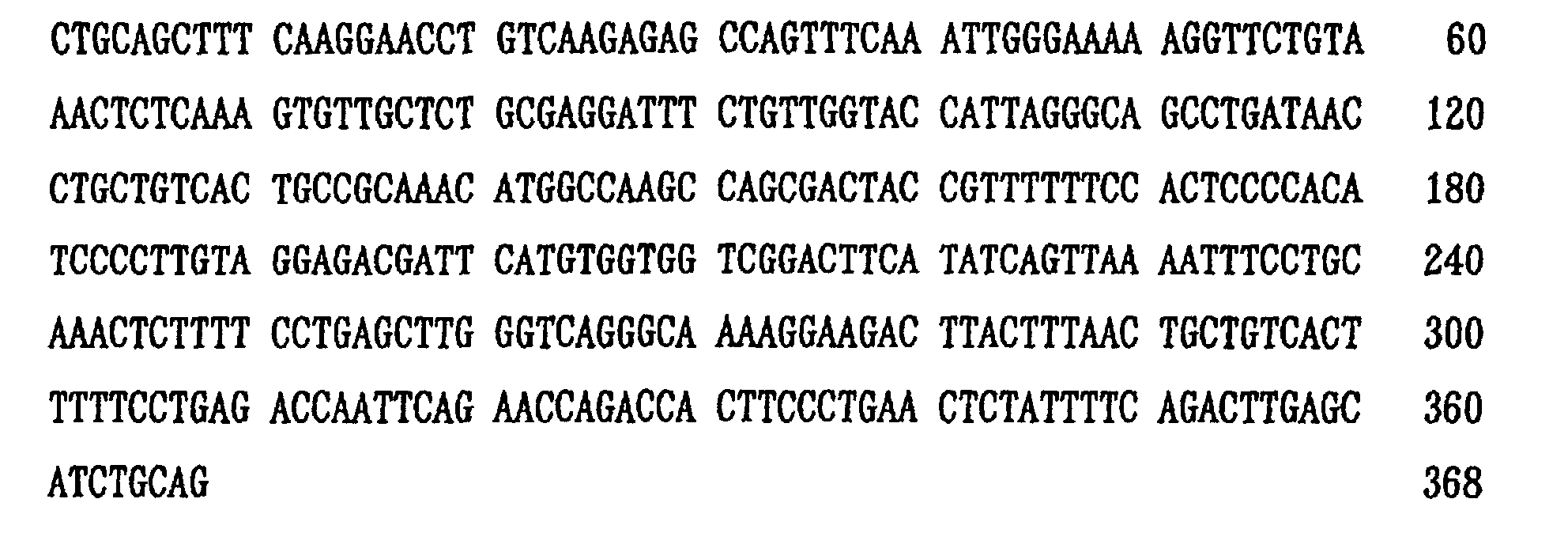
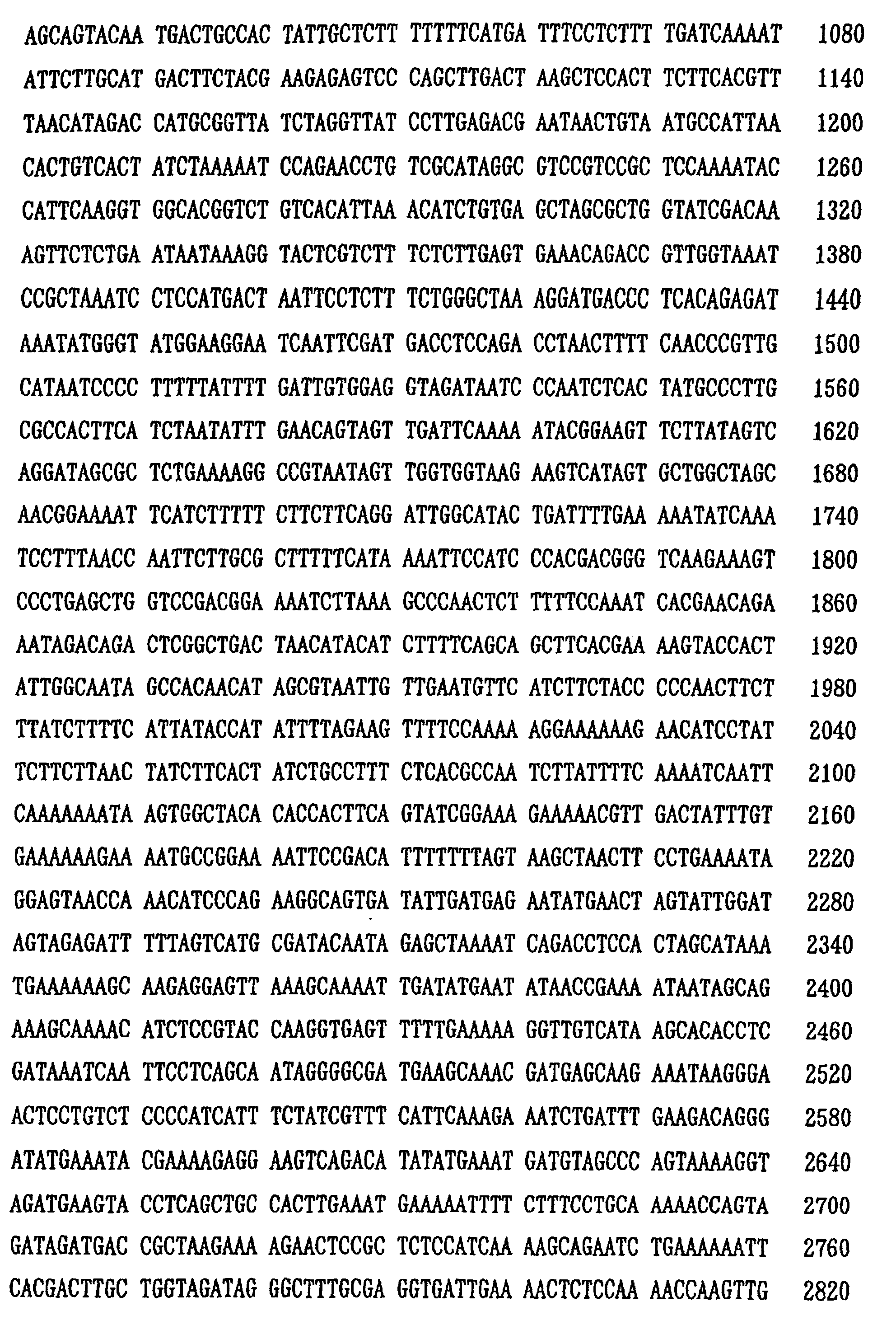
- probe SP-22 SEQ ID NO:1
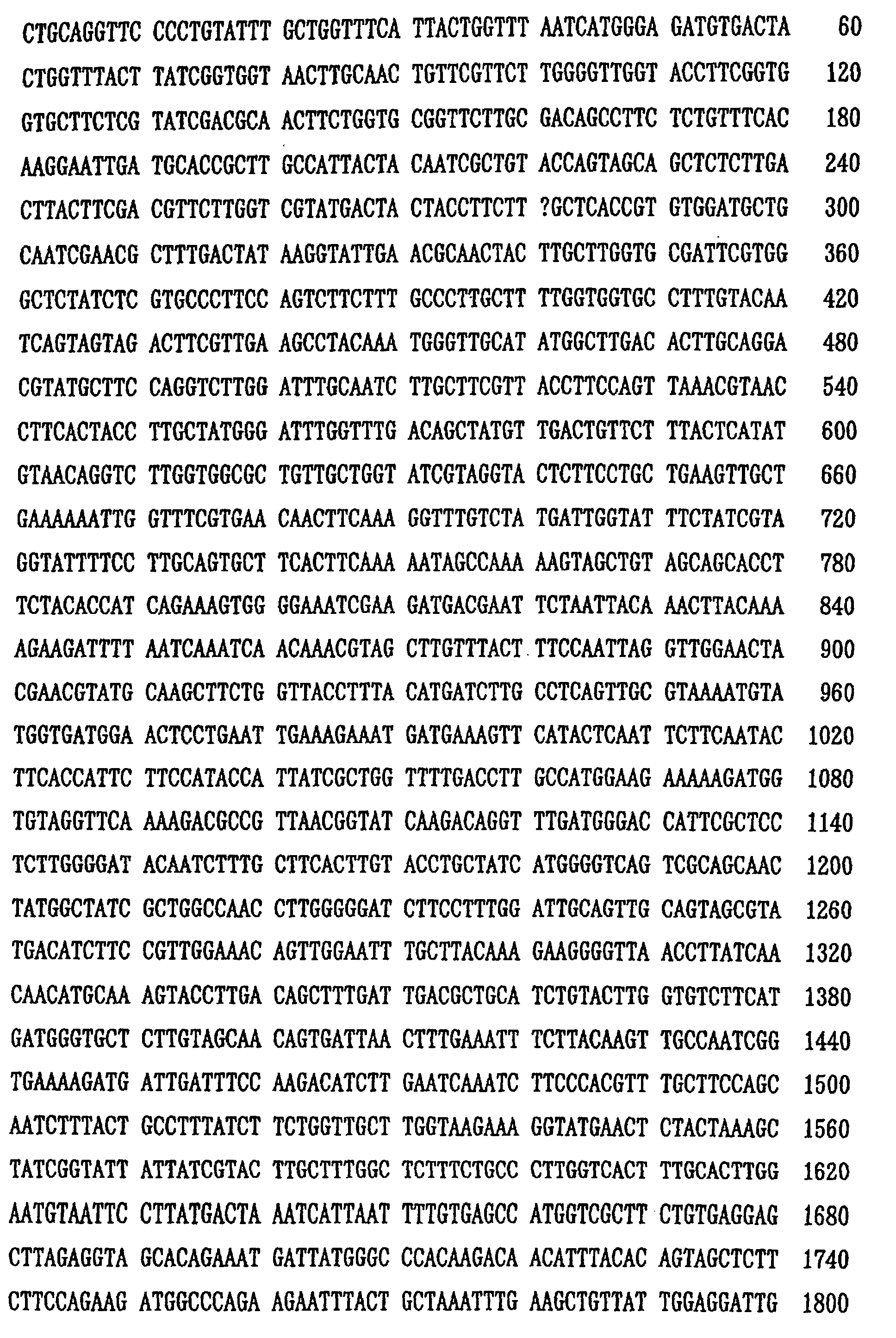
- probe SP-23 SEQ ID NO:2
- probe SP-25 SEQ ID NO:3
- probe SP-26 SEQ ID NO:4
- probe SP-5-15 SEQ ID NO:5
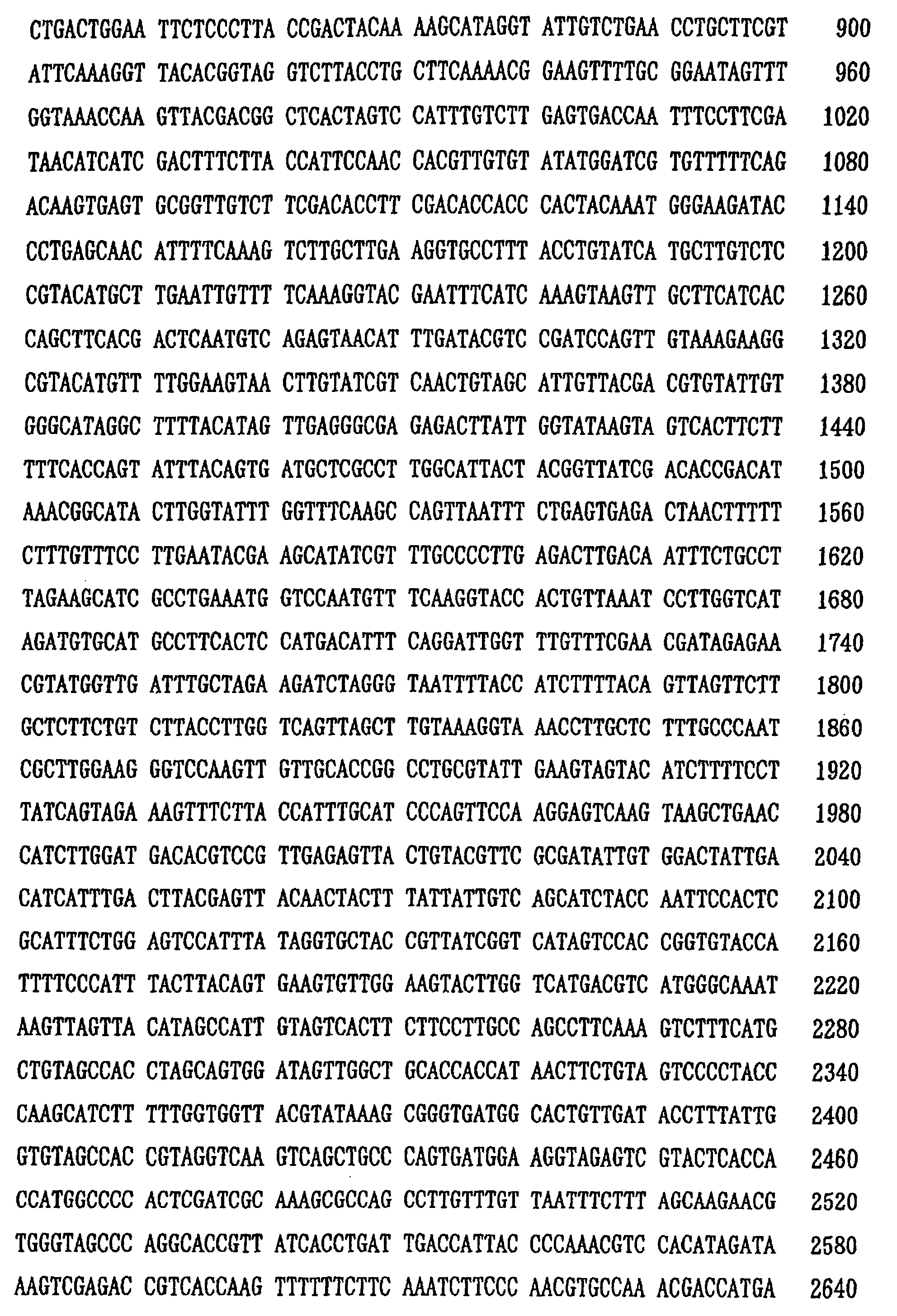
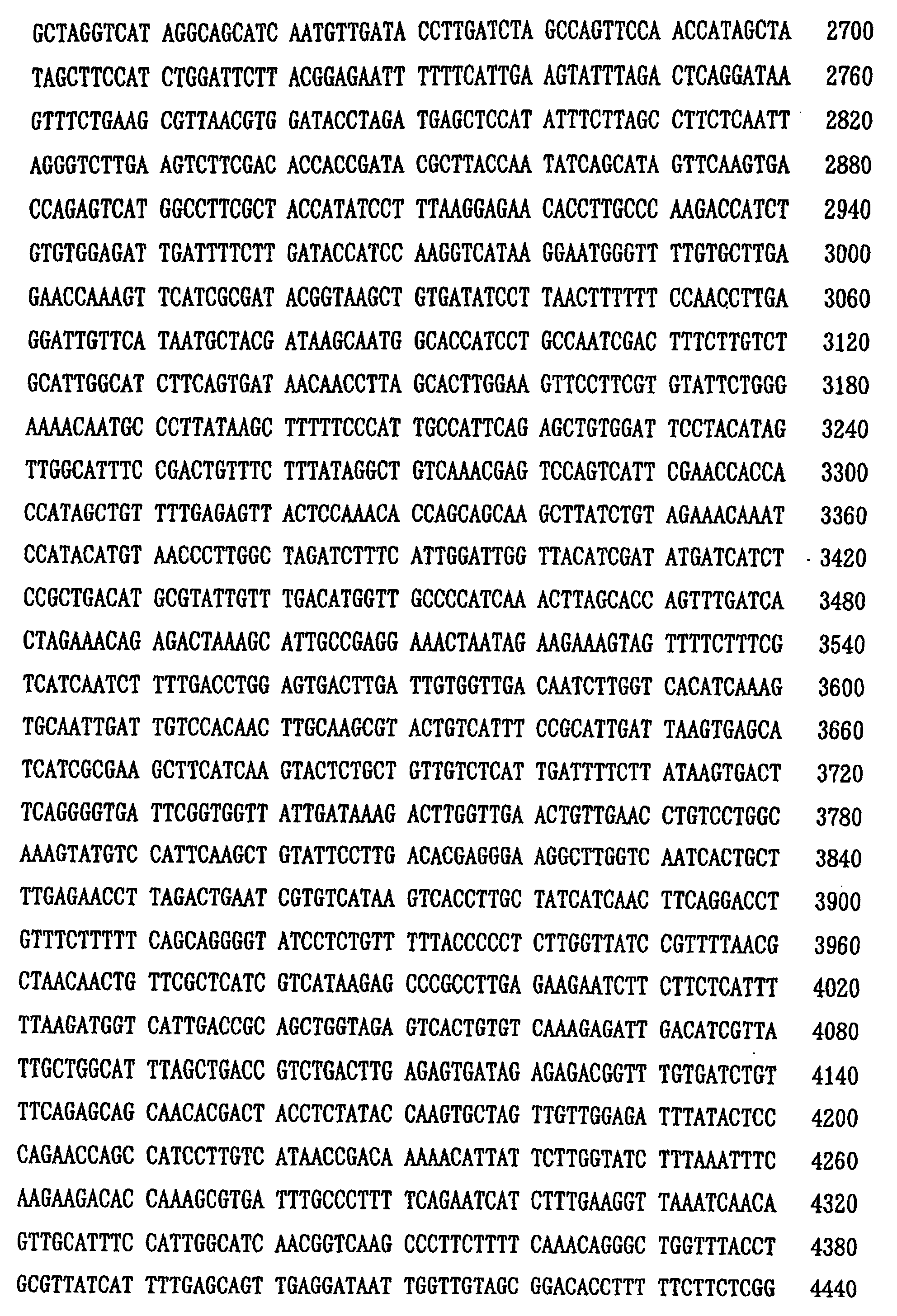
- probe SP-5-34 SEQ ID NO:6
- probe SP-6-6 SEQ ID NO:7
- bacteria for example, causative bacteria ingested into phagocytes can be directly detected without proliferating the bacteria, and can be identified rapidly and accurately. That is to say, according to diagnosis method using a probe of the present invention, identification of the bacteria can be accomplished with single specimen, then, the necessary time for diagnosis can be greatly reduced to about one to two days, while the conventional method (with low detection rate) required 3-4 days, and the detection efficacy is remarkably improved. Therefore, the present invention can provide breakthrough guide for the treatment of bacterial pneumonia, then enable the effective treatment in the early stage of the infectious diseases to the patient, as well as contribute to reduction in the mortality thereby.
- nucleotide sequences of genomic DNA from the clinical specimen with those analyzed in accordance with the present invention, rapid identification of the species of the causative bacteria of pneumonia can be carried out.
- the present invention provides a desired probe for diagnosing the infectious diseases, besides, excellent utilities are expected as a guide for preparing PCR primers and as a standard sequence suitable for the comparison and reference with genomic DNA from the clinical specimen. Additionally, the present invention exerts further outstanding effects, for example, of providing valuable clues for preparation and development of the other probes which specifically react with DNA from causative bacteria of the infectious diseases (causative bacteria of pneumonia).
- nucleotide sequences disclosed in the present application was obtained by random cloning of the genomic DNA from clinical isolates, thus, utilities of the nucleotide sequences disclosed in the present invention should be extended to the complementary sequences thereof.
- DNA obtained from the wild strain might contain a mutated portion, apparently from the disclosure of the Examples above, such mutated DNA portion would never affect the utilities to be derived from the present invention such as the specificity of the probe of the present invention upon use for hybridization for diagnosis of the infectious diseases, and usage of the information on the nucleotide sequences disclosed in the present application to design the primer for PCR technique with the aim of a rapid diagnosis of the infectious diseases.
- SEQUENCE LENGTH 368 base pairs
- SEQUENCE TYPE nucleic acid STRANDEDNESS : double TOPOLOGY : linear MOLECULAR TYPE : Genomic DNA ORIGINAL SOURCE
- ORGANISM Streptococcus pneumoniae STRAIN : clinical isolate SP-22
- SEQUENCE LENGTH 1978 base pairs
- SEQUENCE LENGTH 1124 base pairs
- SEQUENCE TYPE nucleic acid STRANDEDNESS : double TOPOLOGY : linear MOLECULAR TYPE : Genomic DNA ORIGINAL SOURCE
- ORGANISM Streptococcus pneumoniae STRAIN : clinical isolate SP-25
- SEQUENCE LENGTH 5949 pase pairs
- SEQUENCE TYPE nucleic acid STRANDEDNESS : double TOPOLOGY : linear MOLECULAR TYPE : Genomic DNA ORIGINAL SOURCE
- ORGANISM Streptococcus pneumoniae STRAIN : clinical isolate SP-26
- SEQUENCE LENGTH 3568 base pairs
- SEQUENCE LENGTH 4528 base pairs
- SEQUENCE TYPE nucleic acid STRANDEDNESS : double TOPOLOGY : linear MOLECULAR TYPE : Genomic DNA ORIGINAL SOURCE
- ORGANISM Streptococcus pneumoniae STRAIN : clinical isolate SP-5-34
- SEQUENCE LENGTH 5579 base pairs
- SEQUENCE TYPE nucleic acid STRANDEDNESS : double TOPOLOGY : linear MOLECULAR TYPE : Genomic DNA ORIGINAL SOURCE
- ORGANISM Streptococcus pneumoniae STRAIN : clinical isolate SP-6-6
Landscapes
- Chemical & Material Sciences (AREA)
- Life Sciences & Earth Sciences (AREA)
- Organic Chemistry (AREA)
- Health & Medical Sciences (AREA)
- Engineering & Computer Science (AREA)
- Zoology (AREA)
- Wood Science & Technology (AREA)
- Proteomics, Peptides & Aminoacids (AREA)
- Genetics & Genomics (AREA)
- Analytical Chemistry (AREA)
- Biotechnology (AREA)
- Biochemistry (AREA)
- Molecular Biology (AREA)
- Bioinformatics & Cheminformatics (AREA)
- General Engineering & Computer Science (AREA)
- General Health & Medical Sciences (AREA)
- Microbiology (AREA)
- Biophysics (AREA)
- Physics & Mathematics (AREA)
- Immunology (AREA)
- Biomedical Technology (AREA)
- Plant Pathology (AREA)
- Measuring Or Testing Involving Enzymes Or Micro-Organisms (AREA)
- Investigating Or Analysing Biological Materials (AREA)
- Measuring And Recording Apparatus For Diagnosis (AREA)
- Peptides Or Proteins (AREA)
- Saccharide Compounds (AREA)
- Farming Of Fish And Shellfish (AREA)
- Medicines Containing Antibodies Or Antigens For Use As Internal Diagnostic Agents (AREA)
Abstract
Description
| bacterium No. | strain name | |
| 1 | Streptococcus pneumoniae | NYSDH DP-2 |
| 2 | Streptococcus pneumoniae | |
| 3 | Streptococcus pneumoniae | |
| 4 | Streptococcus agalactiae | IFM 58/59 |
| 5 | Streptococcus anginosus | NCTC 8787 |
| 6 | Streptococcus constellatus | ATCC 27823 |
| 7 | Streptococcus equisimilis | NCTC 8543 |
| 8 | Streptococcus faecium | NCTC 7171 |
| 9 | Streptococcus faecalis | ATCC 19433 |
| 10 | Streptococcus mitis | ATCC 9811 |
| 11 | Streptococcus morbillorum | ATCC 27824 |
| 12 | Streptococcus pyogenes | DHI S8 |
| 13 | Streptococcus sanguis | ATCC 10556 |
| 14 | Streptococcus salivarius | ATCC 7073 |
| 15 | Staphylococcus aureus | ATCC 25923 |
| 16 | Staphylococcus epidermidis | ATCC 12228 |
| 17 | Escherichia coli | ATCC 25922 |
| 18 | Klebsiella pneumoniae | clinical isolate |
| 19 | Pseudomonas aeruginosa | ATCC 27583 |
| 20 | Enterococcus agglomerans | clinical isolate |
| 21 | Haemophilis influenzae | |
| 22 | Candida albicans | IFM 40083- |
| 23 | Aspergillus fumigatus | MTU 06001 |
| 24 | Cryptococcus neoformans | MTU 13001 |
| 25 | Mucor spinosus | TIMM 1322 |
| 26 | human genomic DNA | |
| 27 | control | |
| [note] NYSDH ; New York State Department of Health (Albany, New York, U.S.A.) NCTC ; National Collection of Type Cultures (London, England) DHI ; Dairen Hygienic Institute IFM ; Chiba University, Eucaryotic Microorganism Research Center MTU ; Tokyo University, Medical Faculty TIMM ; Teikyo University, Medical Fungus Research Center |
| No. | becterium strain name | (origin) | probe (denotation: SP- ) | ||||||
| 22 | 23 | 25 | 26 | 5-15 | 5-34 | 6-6 | |||
| 1 | Streptococcus pneumoniae | (NYSDH DP-2) | + | + | + | + | + | + | + |
| 2 | Streptococcus pneumoniae | (clinical isolate) | + | + | + | + | + | + | + |
| 3 | Streptococcus pneumoniae | (clinical isolate) | + | + | + | + | + | + | + |
| 4 | Streptococcus agalactiae | (IFM 58/59) | - | - | - | - | - | - | - |
| 5 | Streptococcus anginosus | (NCTC 8787) | - | - | - | - | - | - | - |
| 6 | Streptococcus constellatus | (ATCC 27823) | - | - | - | - | - | - | - |
| 7 | Streptococcus equisimilis | (NCTC 8543) | - | - | - | - | - | - | - |
| 8 | Streptococcus faecium | (NCTC 7171) | - | - | - | - | - | - | - |
| 9 | Streptococcus faecalis | (ATCC 19433) | - | - | - | - | - | - | - |
| 10 | Streptococcus mitis | (ATCC 9811) | - | - | - | - | - | - | - |
| 11 | Streptococcus morbillorum | (ATCC 27824) | - | - | - | - | - | - | - |
| 12 | Streptococcus pyogenes | (DHI S8) | - | - | - | - | - | - | - |
| 13 | Streptococcus sanguis | (ATCC 10556) | - | - | - | - | - | - | - |
| 14 | Streptococcus salivarius | (ATCC 7073) | - | - | - | - | - | - | - |
| 15 | Staphylococcus aureus | (ATCC 25923) | - | - | - | - | - | - | - |
| 16 | Staphylococcus epidermidis | (ATCC 12228) | - | - | - | - | - | - | - |
| 17 | Escherichia coli | (ATCC 25922) | - | - | - | - | - | - | - |
| 18 | Klebsiella pneumoniae | (clinical isolate) | - | - | - | - | - | - | - |
| 19 | Pseudomonas aeruginosa | (ATCC 27583) | - | - | - | - | - | - | - |
| 20 | Enterococcus agglomerans | (clinical isolate) | - | - | - | - | - | - | - |
| 21 | Haemophilis influenzae | (clinical isolate) | - | - | - | - | - | - | - |
| 22 | Candida albicans | (IFM 40083-A type) | - | - | - | - | - | - | - |
| 23 | Aspergillus fumigatus | (MTU 06001) | - | - | - | - | - | - | - |
| 24 | Cryptococcus neoformans | (MTU 13001) | - | - | - | - | - | - | - |
| 25 | Mucor spinosus | (TIMM 1322) | - | - | - | - | - | - | - |
| 26 | human genomic DNA | - | - | - | - | - | - | - | |
| 27 | control | + | + | + | + | + | + | + | |
| [note] "+" denotes that the hybridization signal was detected, "-" denotes that the hybridization signal was not detected, respectively. |
then the precipitate was obtained therefrom by adding ethanol to the supernatant. This precipitate was dried and dissolved in 10 µl of distilled water to give a DNA sample.
SEQUENCE TYPE : nucleic acid
STRANDEDNESS : double
TOPOLOGY : linear
MOLECULAR TYPE : Genomic DNA
ORIGINAL SOURCE
ORGANISM : Streptococcus pneumoniae
STRAIN : clinical isolate SP-22
SEQUENCE TYPE : nucleic acid
STRANDEDNESS : double
TOPOLOGY : linear
MOLECULAR TYPE : Genomic DNA
ORIGINAL SOURCE
ORGANISM: Streptococcus pneumoniae
STRAIN : clinical isolate SP-23

SEQUENCE TYPE : nucleic acid
STRANDEDNESS : double
TOPOLOGY : linear
MOLECULAR TYPE : Genomic DNA
ORIGINAL SOURCE
ORGANISM : Streptococcus pneumoniae
STRAIN : clinical isolate SP-25


SEQUENCE TYPE : nucleic acid
STRANDEDNESS : double
TOPOLOGY : linear
MOLECULAR TYPE : Genomic DNA
ORIGINAL SOURCE
ORGANISM : Streptococcus pneumoniae
STRAIN : clinical isolate SP-26


SEQUENCE TYPE : nucleic acid
STRANDEDNESS : double
TOPOLOGY : linear
MOLECULAR TYPE : Genomic DNA
ORIGINAL SOURCE
ORGANISM : Streptococcus pneumoniae
STRAIN : clinical isolate SP-5-15


SEQUENCE TYPE : nucleic acid
STRANDEDNESS : double
TOPOLOGY : linear
MOLECULAR TYPE : Genomic DNA
ORIGINAL SOURCE
ORGANISM : Streptococcus pneumoniae
STRAIN : clinical isolate SP-5-34


SEQUENCE TYPE : nucleic acid
STRANDEDNESS : double
TOPOLOGY : linear
MOLECULAR TYPE : Genomic DNA
ORIGINAL SOURCE
ORGANISM : Streptococcus pneumoniae
STRAIN : clinical isolate SP-6-6



Claims (6)
- A purified nucleic acid for use as a probe for diagnosing infectious diseases, comprising a nucleotide sequence selected from SEQ ID No: 1, SEQ ID No: 2, SEQ ID No: 3, SEQ ID No: 4, SEQ ID No: 5, SEQ ID No: 6 and SEQ ID No: 7, and nucleotide sequences complementary thereto, wherein said nucleic acid does not hybridise to human genomic DNA under the following hybridization and washing conditions:hybridization overnight at 42°C in a hybridization solution comprising 45% formamide and 5 x SSC; andwashing twice for 20 minutes at 55°C in a washing solution comprising 0.1 x SSC and 0.1% SDS.
- A purified nucleic acid as claimed in claim 1, wherein said nucleic acid hybridizes to genomic DNA of Streptococcus pneunomiae, but fails to hybridise to genomic DNA of Streptococcus agalacitae, Streptococcus anginosus, Streptococcus constellatus, Streptococcus equisimilis, Streptococcus faecium, Streptococcus faecalis, Streptococcus mitis, Streptococcus morbillorum, Streptococcus pyogenes, Streptococcus sanguis, and Streptococcus salivarius, or to human genomic DNA, under the hybridization and washing conditions referred to in claim 1.
- A purified nucleic acid according to claim 1 for use as a probe for diagnosing infectious diseases, wherein the nucleotide sequence of said nucleic acid consists of a nucleotide sequence selected from SEQ ID No: 1, SEQ ID No: 2, SEQ ID No: 3, SEQ ID No: 4, SEQ ID No: 5, SEQ ID No: 6 and SEQ ID No: 7, and nucleotide sequences complementary thereto, wherein said nucleic acid does not hybridise to human genomic DNA under the following hybridization and washing conditions:hybridization overnight at 42°C in a hybridization solution comprising 45% formamide and 5 x SSC; andwashing twice for 20 minutes at 55°C in a washing solution comprising 0.1 x SSC and 0.1% SDS.
- A purified nucleic acid as claimed in claim 3, wherein said nucleic acid hybridizes to genomic DNA of Streptococcus pneunomiae, but fails to hybridize to genomic DNA of Streptococcus agalacitae, Streptococcus anginosus, Streptococcus constellatus, Streptococcus equisimilis, Streptococcus faecium, Streptococcus faecalis, Streptococcus mitis, Streptococcus morbillorum, Streptococcus pyogenes, Streptococcus sanguis, and Streptococcus salivarius, or to human genomic DNA, under the hybridization and washing conditions referred to in claim 3.
- A probe for the diagnosis of infectious diseases comprising a purified nucleic acid according to any one of claims 1 to 4, said nucleic acid further comprising a detectable label.
- Use of the probe as claimed in claim 5 for diagnosing Streptococcus pneunomiae infection in vitro.
Applications Claiming Priority (4)
| Application Number | Priority Date | Filing Date | Title |
|---|---|---|---|
| JP23634894 | 1994-09-30 | ||
| JP23634894A JP3571378B2 (en) | 1994-09-30 | 1994-09-30 | Infectious disease diagnostic probe |
| JP236348/94 | 1994-09-30 | ||
| PCT/JP1995/002036 WO1996010647A1 (en) | 1994-09-30 | 1995-10-02 | Probe for diagnosing infectious diseases |
Publications (3)
| Publication Number | Publication Date |
|---|---|
| EP0785277A1 EP0785277A1 (en) | 1997-07-23 |
| EP0785277A4 EP0785277A4 (en) | 1997-09-10 |
| EP0785277B1 true EP0785277B1 (en) | 2003-02-26 |
Family
ID=16999484
Family Applications (1)
| Application Number | Title | Priority Date | Filing Date |
|---|---|---|---|
| EP95933618A Expired - Lifetime EP0785277B1 (en) | 1994-09-30 | 1995-10-02 | Probe for diagnosing infectious diseases |
Country Status (10)
| Country | Link |
|---|---|
| US (1) | US6660852B1 (en) |
| EP (1) | EP0785277B1 (en) |
| JP (1) | JP3571378B2 (en) |
| KR (1) | KR100238337B1 (en) |
| AT (1) | ATE233326T1 (en) |
| AU (1) | AU689325B2 (en) |
| CA (1) | CA2199938C (en) |
| DE (1) | DE69529758T2 (en) |
| DK (1) | DK0785277T3 (en) |
| WO (1) | WO1996010647A1 (en) |
Families Citing this family (11)
| Publication number | Priority date | Publication date | Assignee | Title |
|---|---|---|---|---|
| WO1997037026A1 (en) * | 1996-04-02 | 1997-10-09 | Smithkline Beecham Corporation | Novel compounds |
| US6348328B1 (en) * | 1997-05-14 | 2002-02-19 | Smithkline Beecham Corporation | Compounds |
| US6271000B1 (en) * | 1996-12-13 | 2001-08-07 | Eli Lilly And Company | Streptococcus pneumoniae gene sequence mraY |
| CA2236441A1 (en) * | 1997-07-01 | 1999-01-01 | Smithkline Beecham Corporation | Gida2 |
| EP0890641A1 (en) * | 1997-07-10 | 1999-01-13 | Smithkline Beecham Corporation | Novel IspA |
| CA2436530C (en) * | 2001-02-02 | 2013-10-01 | Id-Lelystad, Instituut Voor Dierhouderij En Diergezondheid B.V. | Environmentally regulated genes of streptococcus suis |
| IL161407A0 (en) * | 2001-10-15 | 2004-09-27 | Chiron Corp | Treatment of severe pneumonia by administration of tissue factor pathway inhibitor (tfpi) |
| KR20030056628A (en) * | 2001-12-28 | 2003-07-04 | 학교법인 성균관대학 | Identification of streptococcus pneumoniae using alcohol dehydrogenase derived from streptococcus pneumoniae |
| US7828741B2 (en) * | 2002-12-20 | 2010-11-09 | The Charlotte-Mecklenburg Hospital Authority | Utilizing lipopolysaccharide in exhaled breath condensate to diagnose gram negative pneumonia |
| ATE427068T1 (en) * | 2002-12-20 | 2009-04-15 | Charlotte Mecklenburg Hospital | DISPOSABLE HANDHELD DEVICE FOR COLLECTING EXHALED BREATH CONDENSATE |
| WO2005110059A2 (en) * | 2004-03-17 | 2005-11-24 | Chiron Corporation | Treatment of severe community-acquired pneumonia by admistration of tissue factor pathway inhibitor (tfpi) |
Family Cites Families (5)
| Publication number | Priority date | Publication date | Assignee | Title |
|---|---|---|---|---|
| JPH01137976A (en) * | 1987-11-20 | 1989-05-30 | Asahi Chem Ind Co Ltd | Novel dna fragment |
| US5212297A (en) * | 1989-05-16 | 1993-05-18 | University Of Kansas | cDNA clones encoding chicken egg white cystatin |
| ES2125268T3 (en) * | 1991-07-31 | 1999-03-01 | Hoffmann La Roche | METHODS AND REAGENTS FOR DETECTING BACTERIA IN CEPHALORRACHID LIQUID. |
| FR2687168B1 (en) * | 1992-02-10 | 1994-03-25 | Bio Merieux | FRAGMENT OF STREPTOCOCCUS PNEUMONIAE GENOMIC DNA, HYBRIDIZATION PROBE, AMPLIFIER PRIME, REAGENT AND METHOD FOR DETECTION OF STREPTOCOCCUS PNEUMONIAE. |
| KR0159071B1 (en) * | 1992-07-07 | 1998-11-16 | 도다 미끼오 | Probe Diagnosis Probe |
-
1994
- 1994-09-30 JP JP23634894A patent/JP3571378B2/en not_active Expired - Lifetime
-
1995
- 1995-10-02 DE DE69529758T patent/DE69529758T2/en not_active Expired - Lifetime
- 1995-10-02 EP EP95933618A patent/EP0785277B1/en not_active Expired - Lifetime
- 1995-10-02 DK DK95933618T patent/DK0785277T3/en active
- 1995-10-02 US US08/809,254 patent/US6660852B1/en not_active Expired - Fee Related
- 1995-10-02 CA CA002199938A patent/CA2199938C/en not_active Expired - Fee Related
- 1995-10-02 WO PCT/JP1995/002036 patent/WO1996010647A1/en not_active Ceased
- 1995-10-02 KR KR1019960706982A patent/KR100238337B1/en not_active Expired - Fee Related
- 1995-10-02 AT AT95933618T patent/ATE233326T1/en active
- 1995-10-02 AU AU36189/95A patent/AU689325B2/en not_active Ceased
Also Published As
| Publication number | Publication date |
|---|---|
| ATE233326T1 (en) | 2003-03-15 |
| US6660852B1 (en) | 2003-12-09 |
| CA2199938C (en) | 2002-11-19 |
| KR970703431A (en) | 1997-07-03 |
| DE69529758T2 (en) | 2003-11-27 |
| JPH0889295A (en) | 1996-04-09 |
| AU689325B2 (en) | 1998-03-26 |
| EP0785277A4 (en) | 1997-09-10 |
| KR100238337B1 (en) | 2000-03-02 |
| WO1996010647A1 (en) | 1996-04-11 |
| AU3618995A (en) | 1996-04-26 |
| CA2199938A1 (en) | 1996-04-11 |
| EP0785277A1 (en) | 1997-07-23 |
| JP3571378B2 (en) | 2004-09-29 |
| DK0785277T3 (en) | 2003-06-10 |
| DE69529758D1 (en) | 2003-04-03 |
Similar Documents
| Publication | Publication Date | Title |
|---|---|---|
| US5763188A (en) | Probe for diagnosing Escherichia coli, Klebsiella pneumonieae or Enterobacter cloacae | |
| JPWO1994001583A1 (en) | Infectious disease diagnostic probes | |
| EP0670373B1 (en) | Probe for diagnosing candida-infections | |
| CA2199938C (en) | Probe for diagnosing infectious diseases | |
| EP0974658B1 (en) | Probes for the diagnosis of infections caused by streptococcus pyogenes | |
| EP0972833B1 (en) | Probes for the diagnosis of infections caused by klebsiella pneumoniae | |
| US6265568B1 (en) | Probes for the diagnosis of infections caused by bacteroides fragilis | |
| JP2004147660A (en) | Diagnostic probe for infectious disease caused by streptococcus pyogenes microbe |
Legal Events
| Date | Code | Title | Description |
|---|---|---|---|
| PUAI | Public reference made under article 153(3) epc to a published international application that has entered the european phase |
Free format text: ORIGINAL CODE: 0009012 |
|
| 17P | Request for examination filed |
Effective date: 19970428 |
|
| AK | Designated contracting states |
Kind code of ref document: A1 Designated state(s): AT BE CH DE DK FR GB IT LI NL SE |
|
| A4 | Supplementary search report drawn up and despatched | ||
| AK | Designated contracting states |
Kind code of ref document: A4 Designated state(s): AT BE CH DE DK FR GB IT NL SE |
|
| RBV | Designated contracting states (corrected) |
Designated state(s): AT BE CH DE DK FR GB IT LI NL SE |
|
| 17Q | First examination report despatched |
Effective date: 20000426 |
|
| GRAG | Despatch of communication of intention to grant |
Free format text: ORIGINAL CODE: EPIDOS AGRA |
|
| GRAG | Despatch of communication of intention to grant |
Free format text: ORIGINAL CODE: EPIDOS AGRA |
|
| GRAH | Despatch of communication of intention to grant a patent |
Free format text: ORIGINAL CODE: EPIDOS IGRA |
|
| GRAH | Despatch of communication of intention to grant a patent |
Free format text: ORIGINAL CODE: EPIDOS IGRA |
|
| GRAA | (expected) grant |
Free format text: ORIGINAL CODE: 0009210 |
|
| AK | Designated contracting states |
Designated state(s): AT BE CH DE DK FR GB IT LI NL SE |
|
| REG | Reference to a national code |
Ref country code: GB Ref legal event code: FG4D |
|
| REG | Reference to a national code |
Ref country code: CH Ref legal event code: EP |
|
| REF | Corresponds to: |
Ref document number: 69529758 Country of ref document: DE Date of ref document: 20030403 Kind code of ref document: P |
|
| REG | Reference to a national code |
Ref country code: CH Ref legal event code: NV Representative=s name: A. BRAUN, BRAUN, HERITIER, ESCHMANN AG PATENTANWAE |
|
| REG | Reference to a national code |
Ref country code: SE Ref legal event code: TRGR |
|
| REG | Reference to a national code |
Ref country code: DK Ref legal event code: T3 |
|
| ET | Fr: translation filed | ||
| PLBE | No opposition filed within time limit |
Free format text: ORIGINAL CODE: 0009261 |
|
| STAA | Information on the status of an ep patent application or granted ep patent |
Free format text: STATUS: NO OPPOSITION FILED WITHIN TIME LIMIT |
|
| 26N | No opposition filed |
Effective date: 20031127 |
|
| REG | Reference to a national code |
Ref country code: CH Ref legal event code: PFA Owner name: FUSO PHARMACEUTICAL INDUSTRIES LTD. Free format text: FUSO PHARMACEUTICAL INDUSTRIES LTD.#7-10, DOSHOMACHI 1-CHOME, CHUO-KU#OSAKA-SHI, OSAKA 541 (JP) -TRANSFER TO- FUSO PHARMACEUTICAL INDUSTRIES LTD.#7-10, DOSHOMACHI 1-CHOME, CHUO-KU#OSAKA-SHI, OSAKA 541 (JP) |
|
| PGFP | Annual fee paid to national office [announced via postgrant information from national office to epo] |
Ref country code: NL Payment date: 20101029 Year of fee payment: 16 Ref country code: DK Payment date: 20101015 Year of fee payment: 16 Ref country code: AT Payment date: 20101005 Year of fee payment: 16 |
|
| PGFP | Annual fee paid to national office [announced via postgrant information from national office to epo] |
Ref country code: CH Payment date: 20101025 Year of fee payment: 16 |
|
| PGFP | Annual fee paid to national office [announced via postgrant information from national office to epo] |
Ref country code: IT Payment date: 20101006 Year of fee payment: 16 Ref country code: SE Payment date: 20101021 Year of fee payment: 16 Ref country code: BE Payment date: 20101028 Year of fee payment: 16 |
|
| BERE | Be: lapsed |
Owner name: *FUSO PHARMACEUTICAL INDUSTRIES LTD Effective date: 20111031 |
|
| REG | Reference to a national code |
Ref country code: NL Ref legal event code: V1 Effective date: 20120501 |
|
| REG | Reference to a national code |
Ref country code: CH Ref legal event code: PL |
|
| REG | Reference to a national code |
Ref country code: DK Ref legal event code: EBP |
|
| REG | Reference to a national code |
Ref country code: SE Ref legal event code: EUG |
|
| PG25 | Lapsed in a contracting state [announced via postgrant information from national office to epo] |
Ref country code: CH Free format text: LAPSE BECAUSE OF NON-PAYMENT OF DUE FEES Effective date: 20111031 Ref country code: BE Free format text: LAPSE BECAUSE OF NON-PAYMENT OF DUE FEES Effective date: 20111031 Ref country code: NL Free format text: LAPSE BECAUSE OF NON-PAYMENT OF DUE FEES Effective date: 20120501 Ref country code: LI Free format text: LAPSE BECAUSE OF NON-PAYMENT OF DUE FEES Effective date: 20111031 |
|
| PG25 | Lapsed in a contracting state [announced via postgrant information from national office to epo] |
Ref country code: IT Free format text: LAPSE BECAUSE OF NON-PAYMENT OF DUE FEES Effective date: 20111002 |
|
| PG25 | Lapsed in a contracting state [announced via postgrant information from national office to epo] |
Ref country code: DK Free format text: LAPSE BECAUSE OF NON-PAYMENT OF DUE FEES Effective date: 20111031 Ref country code: SE Free format text: LAPSE BECAUSE OF NON-PAYMENT OF DUE FEES Effective date: 20111003 |
|
| PGFP | Annual fee paid to national office [announced via postgrant information from national office to epo] |
Ref country code: GB Payment date: 20120914 Year of fee payment: 18 |
|
| REG | Reference to a national code |
Ref country code: AT Ref legal event code: MM01 Ref document number: 233326 Country of ref document: AT Kind code of ref document: T Effective date: 20111002 |
|
| PG25 | Lapsed in a contracting state [announced via postgrant information from national office to epo] |
Ref country code: AT Free format text: LAPSE BECAUSE OF NON-PAYMENT OF DUE FEES Effective date: 20111002 |
|
| PGFP | Annual fee paid to national office [announced via postgrant information from national office to epo] |
Ref country code: FR Payment date: 20121127 Year of fee payment: 18 Ref country code: DE Payment date: 20121026 Year of fee payment: 18 |
|
| GBPC | Gb: european patent ceased through non-payment of renewal fee |
Effective date: 20131002 |
|
| REG | Reference to a national code |
Ref country code: DE Ref legal event code: R119 Ref document number: 69529758 Country of ref document: DE Effective date: 20140501 |
|
| PG25 | Lapsed in a contracting state [announced via postgrant information from national office to epo] |
Ref country code: GB Free format text: LAPSE BECAUSE OF NON-PAYMENT OF DUE FEES Effective date: 20131002 |
|
| REG | Reference to a national code |
Ref country code: FR Ref legal event code: ST Effective date: 20140630 |
|
| PG25 | Lapsed in a contracting state [announced via postgrant information from national office to epo] |
Ref country code: FR Free format text: LAPSE BECAUSE OF NON-PAYMENT OF DUE FEES Effective date: 20131031 Ref country code: DE Free format text: LAPSE BECAUSE OF NON-PAYMENT OF DUE FEES Effective date: 20140501 |