EP0781848B1 - Fused DNA sequence, fused protein expressed from said fused DNA sequence and method for expressing said fused protein - Google Patents
Fused DNA sequence, fused protein expressed from said fused DNA sequence and method for expressing said fused protein Download PDFInfo
- Publication number
- EP0781848B1 EP0781848B1 EP96120899A EP96120899A EP0781848B1 EP 0781848 B1 EP0781848 B1 EP 0781848B1 EP 96120899 A EP96120899 A EP 96120899A EP 96120899 A EP96120899 A EP 96120899A EP 0781848 B1 EP0781848 B1 EP 0781848B1
- Authority
- EP
- European Patent Office
- Prior art keywords
- dna sequence
- fused
- protein
- bacterium
- fdx
- Prior art date
- Legal status (The legal status is an assumption and is not a legal conclusion. Google has not performed a legal analysis and makes no representation as to the accuracy of the status listed.)
- Expired - Lifetime
Links
Images





Classifications
-
- C—CHEMISTRY; METALLURGY
- C07—ORGANIC CHEMISTRY
- C07K—PEPTIDES
- C07K14/00—Peptides having more than 20 amino acids; Gastrins; Somatostatins; Melanotropins; Derivatives thereof
- C07K14/005—Peptides having more than 20 amino acids; Gastrins; Somatostatins; Melanotropins; Derivatives thereof from viruses
-
- C—CHEMISTRY; METALLURGY
- C07—ORGANIC CHEMISTRY
- C07K—PEPTIDES
- C07K14/00—Peptides having more than 20 amino acids; Gastrins; Somatostatins; Melanotropins; Derivatives thereof
- C07K14/195—Peptides having more than 20 amino acids; Gastrins; Somatostatins; Melanotropins; Derivatives thereof from bacteria
-
- C—CHEMISTRY; METALLURGY
- C07—ORGANIC CHEMISTRY
- C07K—PEPTIDES
- C07K14/00—Peptides having more than 20 amino acids; Gastrins; Somatostatins; Melanotropins; Derivatives thereof
- C07K14/195—Peptides having more than 20 amino acids; Gastrins; Somatostatins; Melanotropins; Derivatives thereof from bacteria
- C07K14/20—Peptides having more than 20 amino acids; Gastrins; Somatostatins; Melanotropins; Derivatives thereof from bacteria from Spirochaetales (O), e.g. Treponema, Leptospira
-
- C—CHEMISTRY; METALLURGY
- C12—BIOCHEMISTRY; BEER; SPIRITS; WINE; VINEGAR; MICROBIOLOGY; ENZYMOLOGY; MUTATION OR GENETIC ENGINEERING
- C12N—MICROORGANISMS OR ENZYMES; COMPOSITIONS THEREOF; PROPAGATING, PRESERVING, OR MAINTAINING MICROORGANISMS; MUTATION OR GENETIC ENGINEERING; CULTURE MEDIA
- C12N15/00—Mutation or genetic engineering; DNA or RNA concerning genetic engineering, vectors, e.g. plasmids, or their isolation, preparation or purification; Use of hosts therefor
- C12N15/09—Recombinant DNA-technology
- C12N15/11—DNA or RNA fragments; Modified forms thereof; Non-coding nucleic acids having a biological activity
- C12N15/62—DNA sequences coding for fusion proteins
-
- C—CHEMISTRY; METALLURGY
- C12—BIOCHEMISTRY; BEER; SPIRITS; WINE; VINEGAR; MICROBIOLOGY; ENZYMOLOGY; MUTATION OR GENETIC ENGINEERING
- C12N—MICROORGANISMS OR ENZYMES; COMPOSITIONS THEREOF; PROPAGATING, PRESERVING, OR MAINTAINING MICROORGANISMS; MUTATION OR GENETIC ENGINEERING; CULTURE MEDIA
- C12N9/00—Enzymes; Proenzymes; Compositions thereof; Processes for preparing, activating, inhibiting, separating or purifying enzymes
- C12N9/10—Transferases (2.)
- C12N9/12—Transferases (2.) transferring phosphorus containing groups, e.g. kinases (2.7)
- C12N9/1229—Phosphotransferases with a phosphate group as acceptor (2.7.4)
-
- C—CHEMISTRY; METALLURGY
- C07—ORGANIC CHEMISTRY
- C07K—PEPTIDES
- C07K2319/00—Fusion polypeptide
-
- C—CHEMISTRY; METALLURGY
- C07—ORGANIC CHEMISTRY
- C07K—PEPTIDES
- C07K2319/00—Fusion polypeptide
- C07K2319/20—Fusion polypeptide containing a tag with affinity for a non-protein ligand
- C07K2319/23—Fusion polypeptide containing a tag with affinity for a non-protein ligand containing a GST-tag
-
- C—CHEMISTRY; METALLURGY
- C07—ORGANIC CHEMISTRY
- C07K—PEPTIDES
- C07K2319/00—Fusion polypeptide
- C07K2319/35—Fusion polypeptide containing a fusion for enhanced stability/folding during expression, e.g. fusions with chaperones or thioredoxin
-
- C—CHEMISTRY; METALLURGY
- C07—ORGANIC CHEMISTRY
- C07K—PEPTIDES
- C07K2319/00—Fusion polypeptide
- C07K2319/40—Fusion polypeptide containing a tag for immunodetection, or an epitope for immunisation
-
- C—CHEMISTRY; METALLURGY
- C12—BIOCHEMISTRY; BEER; SPIRITS; WINE; VINEGAR; MICROBIOLOGY; ENZYMOLOGY; MUTATION OR GENETIC ENGINEERING
- C12N—MICROORGANISMS OR ENZYMES; COMPOSITIONS THEREOF; PROPAGATING, PRESERVING, OR MAINTAINING MICROORGANISMS; MUTATION OR GENETIC ENGINEERING; CULTURE MEDIA
- C12N2740/00—Reverse transcribing RNA viruses
- C12N2740/00011—Details
- C12N2740/10011—Retroviridae
- C12N2740/14011—Deltaretrovirus, e.g. bovine leukeamia virus
- C12N2740/14022—New viral proteins or individual genes, new structural or functional aspects of known viral proteins or genes
Definitions
- This invention relates to expression of a fused protein, more specifically to a fused DNA sequence including a DNA sequence coding a heat-resistant protein, a fused protein expressed by said fused DNA sequence, and a method for expressing said fused protein.
- TRX and GST can be applied to fusion and expression of various proteins which are expressed with difficulty, but even in GST which has been essentially used for the purpose of expressing a soluble fused protein, a fused protein becomes insoluble depending on a protein to be fused so that productivity is lowered, or a fused protein to which TRX is fused may have a problem that a nonspecific reaction is liable to occur. Therefore, it has been desired to provide a fused protein having further excellent operatability and productivity.
- an object of the present invention is to provide a novel fused DNA sequence having excellent operatability and productivity for expressing a desired protein or peptide, a fused protein expressed from said fused DNA sequence, and a method for expressing the fused protein using said fused DNA sequence.
- the present inventors have studied intensively in order to solve the problems in the art and consequently found that when a DNA sequence coding a selected protein or peptide and a DNA sequence coding a heat-resistant protein are fused directly or indirectly and a fused protein is expressed from the resulting fused DNA sequence, the productivity of the desired protein or peptide is raised, and said fused protein has heat resistance to make a purification step simple and easy, to accomplish the present invention.
- the present invention relates to a fused DNA sequence comprising a DNA sequence coding a heat-resistant protein or peptide, fused directly or indirectly to the 5' end of a DNA sequence coding a selected protein or peptide, a fused protein expressed by said fused DNA sequence, and a method for expressing the fused protein using said fused-DNA sequence.
- the fused protein of the present invention has high solubility and can maintain even heat resistance derived from heat-resistant protein genes. Because of such a characteristic of the fused protein, when the fused protein is purified, unnecessary substances can be removed simply and easily by heat treatment so that the fused protein can be obtained with good yield.
- Escherichia coli and Schistosoma japonicum In the case of TRX derived from Escherichia coli and GST derived from Schistosoma japonicum, which have been widely used as a fused protein, Escherichia coli and Schistosoma japonicum can live in bodies of mammals and other creatures so that when a fused protein using TRX or GST is used as an antigen of an immunoreaction, a nonspecific reaction due to Escherichia coli or Schistosoma japonicum might be caused.
- the great characteristic of the fused protein of the present invention resides in that a heat-resistant protein derived from a thermophilic bacterium which cannot live in living bodies of mammals and other creatures is used so that even when the fused protein of the present invention is used as an antigen of an immuno-reaction, a nonspecific reaction derived from the fused protein is caused with difficulty.
- Fig. 1 is a detailed view of an expression vector pW6A.
- Fig. 2 is a detailed view of an expression vector pWF6A.
- Fig. 3 is a graph showing the reactivity of a fused protein and a negative specimen.
- Fig. 4 is a graph showing the reactivity of a HTLV-I-fused protein and a positive specimen.
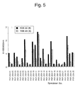
- Fig. 5 is a graph showing the reactivity of a HTLV-II-fused protein and a positive specimen.
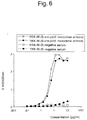
- Fig. 6 is a graph showing the reactivity depending on concentration of a HTLV-I-fused protein.
- Fig. 7 is a graph showing the reactivity depending on concentration of a HTLV-II-fused protein.
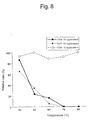
- Fig. 8 is a graph showing the activity of a fused protein in a supernatant subjected to heat treatment.
- Fig. 9 is a graph showing the activity of a fused protein of precipitates subjected to heat treatment.
- Fig. 10 is a view showing the activity of a fused protein after heat treatment and purification.
- Fig. 11 is a detailed view of an expression vector pW6AK.
- Fig. 12 is a view showing the activity of a fused protein after heat treatment and purification.
- the DNA sequence coding a heat-resistant protein of the present invention means a DNA sequence coding a protein which is not thermally denatured even at 55 °C or higher, preferably 75 °C or higher.
- a specific phenomenon of thermal denaturation there may be mentioned inactivation or insolubilization of a protein.
- the DNA sequence coding a protein which is not thermally denatured at 55 °C or higher there may be mentioned, for example, a DNA sequence possessed by a thermophilic bacterium which can live at 55 °C or higher. From the properties of an expressed protein and easiness of post-treatment, it is preferred to use a DNA sequence possessed by the so-called highly thermophilic bacterium which can live at 75 °C or higher.
- thermophilic bacterium there may be mentioned, for example, Thermophilus, Sulfolobus, Pyrococcus, Thermotoga, Pyrobaculum, Pyrodictium, Thermococcus, Thermodiscus, Metanothermus and Metanococcus (FEMS. MICRO. BIOL. REV., Vol.75, pp.117-124 (1990), ANU. REV. MICROBIOL., Vol.47, pp.627-653 (1993)).
- heat-resistant protein there may be mentioned, for example, adenylate kinase derived from a sulfolobus bacterium ( Sulfolobus acidocaldalius Adenylate kinase: Arch. Biochem.
- AK DNA polymerase derived from a Thermophilus bacterium, ferredoxin derived from a Pyrococcus bacterium ( Pyrococcus furiosus Ferredoxin: Biochemistry, Vol.31, pp.11952-11962(1992)) (hereinafter referred to as "FDX" in the specification), glucosidase derived from Pyrococcus furiosus bacterium (Pyrococcus furiosus Glucosidase), rubredoxin derived from Pyrococcus furiosus bacterium (Pyrococcus furiosus Rubredoxin: Biochemistry, Vol.30, pp.10885-10895 (1991)), glutamate dehydrogenase derived from Pyrococcus furiosus bacterium (Pyrococcus furiosus Glutamate dehydrogenase: Gene, Vol.132, pp.189
- DNA coding the heat-resistant protein of the present invention can be purified from these highly thermophilic bacteria, but it can be also synthesized based on a known DNA sequence.
- a known technique such as a ⁇ -cyanoethylphosphoamidite method (Sinha et al., Nucleic Acids Bos., Vol.12, p.4539 (1984)) and a method described in Letsinger, R.L. et al., J. Am. Chem. Soc., vol. 88, p. 5319 (1966) may be suitably used.
- DNA's of FDX derived from Pyrocuccus bacterium and AK derived from Sulfolobus bacterium having amino acid sequences shown in SEQ ID NO: 2 and 4, respectively, are synthesized by the ⁇ -cyanoethylphosphoamidite method. DNA sequences synthesized are shown in SEQ ID NO: 1 and 3, respectively.
- the DNA sequence coding a selected desired protein or peptide of the present invention is not limited to a particular DNA sequence. Any DNA sequence can be used as long as it is a DNA sequence coding a protein or peptide which is desired to be expressed as a fused protein. The present invention is particularly useful when a necessary expression amount of a selected desired protein or peptide can be obtained with difficulty by DNA itself coding said protein or peptide.
- the fused DNA sequence of the present invention can be fused by using a known method such as a ligation method and a linker ligation method.
- a known method such as a ligation method and a linker ligation method.
- the DNA sequence of a selected desired protein or peptide and the DNA sequence of the heat-resistant protein may be fused directly or may be fused indirectly, if necessary.
- a linker sequence is inserted between the DNA sequence coding a desired protein or peptide and the DNA sequence coding the heat-resistant protein.
- linker sequence there can be used a sequence coding a polypeptide for bonding a desired protein or peptide and the heat-resistant protein to each other and a sequence coding a polypeptide which can be cleaved or digested selectively by a known chemical method or enzymatic method.
- linker sequence When the linker sequence is inserted between the DNA sequence coding a desired protein or peptide and the DNA sequence coding the heat-resistant protein, only a selected desired protein or peptide portion can be also purified by, after the fused protein is expressed, cleaving or digesting the linker sequence by using a chemical means such as bromocyan or an enzymatic means such as thrombin or a factor Xa.
- the fused DNA sequence of the present invention is inserted into a vector which is suitable for expression, said vector is introduced into a culture host, and expression of the fused protein is induced. After the host is grown by culture or the like, sonication of the host and purification such as a column operation are carried out to obtain a desired fused protein or peptide.
- Host cells to be used may be any cells such as bacterial cells, eucaryotic cells and mammal cells so long as they are cells which can express a foreign protein or peptide, and there may be mentioned, for example, Escherichia coli, yeast, Bacillus subtilis, Baculo virus and COS cells.
- the fused protein of the present invention may be used as such as a fused protein, or a desired protein or peptide portion thereof obtained by separation and purification may be used.
- a restriction enzyme NdeI site was added to 5'-end, a restriction enzyme EcoRI site was added to 3'-end, and a thrombin-cut site was added to C terminal. This fragment was integrated into the NdeI and EcoRI sites of 4.6 Kb of a pW6A vector prepared from pGEMEX-1 (trade name, produced by Promega Co.) and pGEX-2T (trade name, produced by Pharmacia Biotec Co.) to prepare pWF6A as a vector expressing FDX.
- pGEMEX-1 trade name, produced by Promega Co.
- pGEX-2T trade name, produced by Pharmacia Biotec Co.
- pWF6A contains, at the NdeI and EcoRI sites, genes of a fused protein comprising 96 amino acids including 67 amino acids derived from FDX, 10 amino acids derived from a thrombin-cleaved site and 19 amino acids derived from multi cloning site of pW6A.
- the base sequence of the inserted fragment was confirmed by a DNA sequence kit (trade name: Sequenase kit Ver. 2.0, produced by Amersham United States Biochemical Co.).
- DNA sequence of the FDX inserted into pW6A and amino acids sequence coded by said sequence are shown in SEQ ID NO: 1 and SEQ ID NO: 2, respectively, and DNA sequence of the pW6A is shown in SEQ ID NO: 5.
- ATG of the restriction enzyme site NdeI is shown as 1 and sequences before the stop codon of a multicloning site are shown.
- pWF6A was introduced into host Escherichia coli and then cultured for 2 hours in a medium (hereinafter referred to as "the LB medium” in the specification) containing 1 % of bactotryptone, 0.5 % of yeast extract, 1 % of sodium chloride and 50 ⁇ g/ml of ampicillin and having pH 7.5. Thereafter, 1 mM isopropyl thiogalactopyranoside (hereinafter referred to as "IPTG” in the specification) was added thereto, and the mixture was cultured for 2 hours to induce expression.
- IPTG isopropyl thiogalactopyranoside
- pWF6A prepared in Example 1 was introduced into host Escherichia coli and then cultured under conditions of using the LB medium at 37 °C.
- a concentration of Escherichia coli in a culture broth was made to have such turbidity that absorbance at a wavelength of 600 nm was about 1.0, 1 mM IPTG was added thereto to induce expression.
- centrifugation was carried out to recover Escherichia coli.
- 200 ml of a 50 mM Tris-hydrochloride buffer (hereinafter referred to as "the Tris buffer" in the specification) having pH 8.0 was added to recovered Escherichia coli, followed by sonication treatment under ice cooling.
- the expressed fused protein was recovered in the supernatant as a soluble component.
- this supernatant was subjected heat treatment at 85 °C for 15 minutes, about 80 % of the Escherichia coli protein was thermally denatured and precipitated, and 90 % or more of FDX was recovered in the centrifugation supernatant after the heat treatment.
- This supernatant was purified by ion exchange using a QFF anion exchange column (trade name, manufactured by Pharmacia Biotec Co.) equilibrated with the Tris buffer.
- FDX was recovered at a concentration of about 0.3 M sodium chloride-eluted fraction.
- this FDX fraction was purified by using a RESOURCE RPC column (trade name, manufactured by Pharmacia Biotec Co.) equilibrated with 20 mM sodium hydroxide.
- a RESOURCE RPC column trade name, manufactured by Pharmacia Biotec Co.
- pWT8A prepared as a vector expressing TRX in the same manner as in pWF6A prepared in Example 1 was introduced into host Escherichia coli and then cultured under conditions of using the LB medium at 37 °C. After the same induction of expression as in Example 1 was carried out, Escherichia coli was recovered by centrifugation. An osmotic shock was given to recovered Escherichia coli , and TRX existing at a periplasmic fraction was extracted. Extracted TRX was subjected to first purification by using a RESOURCE RPC column (trade name, manufactured by Pharmacia Biotec Co.) equilibrated with 20 mM sodium hydroxide.
- RESOURCE RPC column trade name, manufactured by Pharmacia Biotec Co.
- TRX When TRX was eluted by acetonitrile, TRX was recovered at a concentration of about 10 % to 20 % acetonitrile-eluted fraction. Recovered TRX was dialyzed to 4 M guanidine hydrochloride and then subjected to second purification by using the reverse phase column under the same conditions. Similarly as in the first purification, purified TRX was recovered at a concentration of about 10 % to 20 % acetonitrile-eluted fraction.
- An anti-Escherichia coli antibody was supposed as a nonspecific reaction substance, and the reactivities of FDX purified in Example 2 and TRX purified in Reference example 1 were examined.
- a SDS-solubilized material of Escherichia coli DH5 ⁇ , a supernatant of Escherichia coli DH5 ⁇ sonicated and a SDS-solubilized material of Escherichia coli to which a pW50 vector (made by Fuji Rebio) was introduced were used as immunogen and immunized to 3 rabbits to prepare the total 9 kinds of the respective anti-Escherichia coli rabbit serums.
- FDX purified in Example 2 and TRX purified in Reference example 1 were subjected to SDS-PAGE according to the Laemmli method and then transferred to nitrocellulose membranes.
- the western blotting method was carried out by using the above 9 kinds of the anti-Escherichia coli rabbit serums diluted 500 times, respectively, as primary antibodies, and using a peroxidase (hereinafter referred to as "POD" in the specification)-labeled anti-rabbit antibody as a secondary antibody.
- POD peroxidase
- 4-chloro-1-naphthol and hydrogen peroxide were used.
- the western blotting method was carried out by 25 samples of human specimen HTLV-I/II mix panel 204 serums (trade name, produced by Boston Biomedica Co.) diluted 50 times, respectively, as primary antibodies, and using POD-labelled anti-human IgG as a secondary antibody. Reactivities at sites where FDX was transferred was not confirmed, but the reactions of 2 samples among 25 samples at sites where TRX was transferred were confirmed. The results are shown in Table 1. Specimen No.
- a specificity test according to the ELISA method was carried out by using the human specimens produced by Boston Biomedica Co. diluted 500 times used in Example 3 as primary antibodies and POD-labelled anti-human IgG as a secondary antibody. For coloring, ABTS and hydrogen peroxide were used. The measurement results were shown by difference between absorbances at a wavelength of 405 nm and a wavelength of 492 nm (difference between absorbances was described as A405/492 nm).
- genomic DNA was extracted by the method of Molecular Cloning by J. Sambrook et al. Next, by using a primer to which EcoRI and BamHI sites were added, the PCR method was carried out in the same manner as in Example 1 to obtain about 400 bp of p19DNA fragments in the respective gag regions. These fragments were integrated into pWF6A to prepare pWFIP19 as a vector expressing p19 of HTLV-I and pWFIIP19 as a vector expressing p19 of HTLV-II.
- DNA sequences of the FDX-fused HTLV-I p19 and FDX-fused HTLV-II p19 each of which is inserted into the vectors are shown in SEQ ID NO: 6 and 8, respectively, and amino acids sequences coded by said DNA sequences are shown in SEQ ID NO: 7 and 9, respectively.
- these vectors were introduced into Escherichia coli, and expression of the respective fused proteins was induced.
- Samples for electrophoresis were prepared under the same conditions as in Example 1. After subjecting to 12.5 % SDS-PAGE according to the Laemmli method, one sheet of gel was subjected to CBB staining, and the other sheet was transferred to nitrocellulose membranes by the method shown in Example 3.
- an anti-native HTLV-I p19 monoclonal antibody (a GIN-7 antibody, Tanaka, Y. et al., Gann., Vol.74, pp.327 to 330 (1983)) or an anti-native HTLV-II p19 monoclonal antibody as a primary antibody, and a POD-labeled anti-mouse IgG as a secondary antibody, these were reacted with the fused proteins by the same method as in Example 3 and coloring was carried out by using 4-chloro-1-naphthol and hydrogen peroxide, expression of the fused proteins reacting with the respective monoclonal antibodies corresponding to the respective fused proteins was recognized.
- Example 6 Expression of FDX-fused HTLV-I p20E(gp21)-fused protein and FDX-fused HTLV-II p20E(gp21)-fused protein
- DNA sequences of the FDX-fused HTLV-I p20E and FDX-fused HTLV-II p20E each of which is inserted into the vectors are shown in SEQ ID NO: 10 and 12, respectively, and amino acids sequences coded by said DNA sequences are shown in SEQ ID NO: 11 and 13, respectively.
- FDX-20(I) FDX-fused HTLV-I p20E-fused protein
- FDX-20(II) FDX-fused HTLV-II p20E-fused protein
- one sheet of gel was subjected to CBB staining, and the other sheet of gel was transferred to nitrocellulose membranes at 120 mA for 3 hours.
- a phosphate buffer containing 1 % of BSA (bovine serum albumin) 1 ⁇ g/ml of an anti-p20E(gp21) monoclonal antibody (F-10, Sugamura, K. et al., J.
- FDX-20(I) and FDX-20(II) were increased by several hundreds times as compared with the case where p20E of HTLV-I and p20E of HTLV-II were expressed directly.
- pWFIE1 and pWFIIE10 prepared in Example 6 were introduced into host Escherichia coli , respectively, and then cultured under conditions of using the LB medium at 37 °C. By preculture, a concentration of Escherichia coli in culture broths was made to have such turbidity that absorbance at a wavelength of 600 nm was about 1.0, 1 mM IPTG was added thereto to induce expression. Three hours after IPTG was added, centrifugation was carried out to recover Escherichia coli .
- solubilized bodies were purified by a RESOURCE RPC column (trade name, manufactured by Pharmacia Biotec Co.) equilibrated with 20 % acetonitrile and 20 mM sodium hydroxide.
- purified FDX-20(I)- and FDX-20(II)-fused proteins were recovered at a concentration of about 30 to 40 % acetonitrile-eluted fractions, respectively.
- Reference example 2 Purification of TRX-fused HTLV-I p20E-fused protein and TRX-fused HTLV-II p20E-fused protein
- p20E(gp21) in an env region of HTLV-I or HTLV-II was introduced into the TRX-expressing vector pWT8A prepared in Reference example 1 to prepare pWTIEl and pWTIIE10, followed by expression.
- TRX-20(I) TRX-fused HTLV-I p20E-fused protein
- TRX-20(II) TRX-fused HTLV-II p20E-fused protein
- the ELISA method was carried out by using these ELISA plates and using the human specimens produced by Boston Biomedica Co. diluted 500 times as primary antibodies and POD-labelled anti-human IgG as a secondary antibody in the same manner as in Example 4.
- FDX-20(I) and FDX-20(II), and TRX-20(I) and TRX-20(II) were reacted with the same specimens. The results are shown in Fig. 4 and Fig. 5.
- the ELISA method was carried out by using these ELISA plates and using the anti-p20E(gp21) monoclonal antibody diluted 500 times as a primary antibody and POD-labelled anti-mouse IgG as a secondary antibody. With respect to a negative serum, the ELISA method was carried out in the same manner as in Example 4. There was no difference in reactivity to the monoclonal antibody, and the FDX-fused proteins in both cases of HTLV-I and HTLV-II had lower reactivities to the negative serum. The results are shown in Fig. 6 and Fig. 7.
- syphilis bacteria Nichols strain from Treponema pallidum
- genomic DNA was extracted from syphilis bacteria-subcultured rabbit testicles.
- a primer was produced based on the known DNA sequences by using a DNA synthesizer (Model 392, trade name, produced by PERKIN ELMER Co.).
- Tp15 a DNA fragment coding a surface antigen of 15 Kda (hereinafter referred to as "Tp15” in the specification) of Treponema pallidum (hereinafter referred to as “Tp” in the specification) was amplified with a thermal cycler (Model PJ1000, trade name, produced by PERKIN ELMER Co.).
- This DNA fragment was integrated into an EcoRI site of a GST-expressing type vector pWG6A in which DNA sequence of GST had been inserted into pW6A to obtain a vector pWGTp15 expressing a protein in which GST and Tp15 were fused (hereinafter referred to as "GST-15" in the specification).
- DNA sequence of the GST-15 inserted into the vector is shown in SEQ ID NO: 14 and amino acids sequence coded by said DNA sequence is shown in SEQ ID NO: 15.
- the vector was introduced into Escherichia coli, and expression of GST-15 was induced.
- a sample for electrophoresis was prepared under the same conditions as in Example 1. After subjecting to 12.5 % SDS-PAGE according to the Laemmli method, one sheet of gel was subjected to CBB staining, and the other sheet was transferred to a nitrocellulose membrane by the method shown in Example 3.
- TRX-15 a DNA fragment of Tp15 amplified in Reference example 3 was integrated into an EcoRI site of the TRX-expressing type vector pWT8A in which DNA sequence of TRX had been inserted into pW6A to obtain a vector pWTTp15 expressing a protein in which TRX and Tp15 were fused (hereinafter referred to as "TRX-15" in the specification).
- DNA sequence of the TRX-15 inserted into the vector is shown in SEQ ID NO: 16 and amino acids sequence coded by said DNA sequence is shown in SEQ ID NO: 17.
- the vector was introduced into Escherichia coli, and expression of TRX-15 was induced.
- a sample for electrophoresis was prepared under the same conditions as in Example 1.
- Example 3 After subjecting to 12.5 % SDS-PAGE according to the Laemmli method, one sheet of gel was subjected to CBB staining, and the other sheet was transferred to a nitrocellulose membrane by the method shown in Example 3.
- an anti-Tp15 monoclonal antibody as a primary antibody
- a POD-labeled anti-mouse IgG as a secondary antibody
- coloring was carried out by using 4-chloro-1-naphthol and hydrogen peroxide, a band was given at about 27 Kda which was the same position as that of the CBB-stained gel.
- a DNA fragment of Tp15 amplified in Reference example 3 was integrated into an EcoRI, BamHI sites of the FDX-expressing type vector pWF6A prepared in Example 1 to obtain a vector pWFTp15 expressing a protein in which FDX and Tp15 were fused (hereinafter referred to as "FDX-15" in the specification).
- DNA sequence of the FDX-15 inserted into the vector is shown in SEQ ID NO: 18 and amino acids sequence coded by said DNA sequence is shown in SEQ ID NO: 19.
- the vector was introduced into Escherichia coli, and expression of FDX-15 was induced.
- a sample for electrophoresis was prepared under the same conditions as in Example 1.
- Example 3 After subjecting to 12.5 % SDS-PAGE according to the Laemmli method, one sheet of gel was subjected to CBB staining, and the other sheet was transferred to a nitrocellulose membrane by the method shown in Example 3.
- an anti-Tp15 monoclpnal antibody as a primary antibody
- a POD-labeled anti-mouse IgG as a secondary antibody
- these were reacted in the same method as in Example 3 and coloring was carried out by using 4-chloro-1-naphthol and hydrogen peroxide, a band was given at about 30 Kda which was the same position as that of the CBB-stained gel.
- the vectors expressing FDX-15, GST-15 and TRX-15 prepared in Example 9, Reference example 3 and Reference example 4 were introduced into host Escherichia coli and then cultured under conditions of using 1 liter of the LB medium at 37 °C, respectively.
- a concentration of Escherichia coli in culture broths was made to have such turbidity that absorbance at a wavelength of 600 nm was about 1.0, 1 mM IPTG was added thereto to induce expression.
- 200 ml of the Tris buffer was added to the cells. After sonication treatment under ice cooling, fused proteins were recovered in the centrifugation supernatants, respectively.
- pWFTp15 prepared in Example 9 was introduced into host Escherichia coli and then cultured under conditions of using 1 liter of the LB medium at 37 °C. By preculture, a concentration of Escherichia coli in culture broths was made to have such turbidity that absorbance at a wavelength of 600 nm was about 1.0, 1 mM IPTG was added thereto to induce expression. The cells were recovered by centrifugation. 200 ml of the Tris buffer was added to the cells, and the cells were sonicated to recover FDX-15 in the centrifugation supernatant.
- GST-15 obtained by introducing pWGTp15 prepared in Reference example 3 into host Escherichia coli , carrying out induction and expression operations in the same manner therein and carrying out purification by a common column operation without carrying out heat treatment and FDX-15 purified by heat treatment were subjected to the western blotting method in the same manner as in Example 10 by using an anti-Tp rabbit antibody. It was shown that even though purification by heat treatment was carried out, FDX-15 retained reactivity. The results are shown in Fig. 10.
- genes of Sulfolobus acidocaldarius AK were synthesized by the assemble PCR method.
- a Taq polymerase produced by Toyobo Co.
- the total base number of 630 bp was amplified under conditions of 30 cycles of 94 °C - 1 minute, 55 °C - 1 minute and 72 °C - 1 minute.
- a restriction enzyme NdeI site was added to 5'-end, a restriction enzyme EcoRI site was added to 3'-end, and a thrombin-cut site was added to C terminal.
- This fragment was integrated into the NdeI and EcoRI sites of 4.6 Kb of a pW6A vector prepared from pGEMEX-1 (trade name, produced by Promega Co.) and pGEX-2T (trade name, produced by Pharmacia Biotec Co.) to prepare pW6AK as a vector expressing AK.
- pW6AK A detailed view of pW6AK is shown in Fig. 11.
- pW6AK contains genes of a fused protein comprising 223 amino acids including 194 amino acids derived from AK, 10 amino acids derived from a thrombin-cleaved site and 19 amino acids derived from multi cloning site of pW6A, at the NdeI and EcoRI sites.
- the base sequence of the inserted fragment was confirmed by a DNA sequence kit (trade name: Sequenase kit Ver. 2.0, produced by Amersham United States Biochemical Co.).
- DNA sequence of the AK inserted into the pW6A is shown in SEQ ID NO: 3 and amino acids sequence coded by said DNA sequence is shown in SEQ ID NO: 4.
- pW6AK was introduced into host Escherichia coli and then cultured for 2 hours in the LB medium.
- pW6AK prepared in Example 12 was introduced into host Escherichia coli and then cultured under conditions of using the LB medium at 37 °C.
- a concentration of Escherichia coli in culture broth was made to have such turbidity that absorbance at a wavelength of 600 nm was about 1.0, 1 mM IPTG was added thereto to induce expression.
- centrifugation was carried out to recover Escherichia coli.
- 200 ml of the Tris buffer was added to recover Escherichia coli, followed by sonication treatment under ice cooling. After centrifugation, the expressed fused protein was recovered in the supernatant as a soluble component.
- This supernatant was purified by a Hydroxy apatite column (manufactured by Bio-rad Lab.) equilibrated with the Tris buffer.
- AK was recovered at a concentration of about 0.2 M sodium phosphate-eluted fraction.
- this AK fraction was purified by gel filtration using a Superdex 200 26/60 column (trade name, manufactured by Pharmacia Biotec Co.) equilibrated with a buffer containing 6 M urea, 0.5 M sodium chloride and 20 mM Tris-hydrochloride having pH 9.4. At a fraction of a molecular weight being about 20,000, purified AK was recovered.
- a DNA fragment of Tp15 amplified in Reference example 3 was integrated into EcoRI, BamHI sites of the AK-expressing type vector pW6AK prepared in Example 12 to obtain a vector pW6AKTp15 expressing a protein in which AK and Tp15 were fused (hereinafter referred to as "AK-15" in the specification).
- DNA sequence of the AK-15 inserted into the vector is shown in SEQ ID NO: 20 and amino acids sequence coded by said DNA sequence is shown in SEQ ID NO: 21.
- the vector was introduced into Escherichia coli, and expression of AK-15 was induced.
- a sample for electrophoresis was prepared under the same conditions as in Example 1.
- Example 3 After subjecting to 12.5 % SDS-PAGE according to the Laemmli method, one sheet of gel was subjected to CBB staining, and the other sheet was transferred to a nitrocellulose membrane by the method shown in Example 3.
- an anti-Tp15 monoclonal antibody as a primary antibody
- a POD-labeled anti-mouse IgG as a secondary antibody
- coloring was carried out by using 4-chloro-1-naphthol and hydrogen peroxide, a band was given at about 40 Kda which was the same position as that of the CBB-stained gel.
- pWAKTp15 prepared in Example 14 was introduced into host Escherichia coli and then cultured under conditions of using 1 liter of the LB medium at 37 °C. By preculture, a concentration of Escherichia coli in culture broth was made to have such turbidity that absorbance at a wavelength of 600 nm was about 1.0, 1 mM IPTG was added thereto to induce expression. The cells were recovered by centrifugation. 200 ml of a 50 mM glycine-sodium hydroxide buffer having pH 10.0 was added to the cells, and the cells were sonicated to recover AK-15 in the centrifugation supernatant.
- the recovered AK-15 fraction was purified by gel filtration using a Superdex 200 26/60 column (trade name, manufactured by Pharmacia Biotec Co.) equilibrated with a buffer containing 6 M urea, 0.5 M sodium chloride and 20 mM Tris-hydrochloride having pH 9.4. At a fraction of a molecular weight being about 40,000, purified AK-15 was recovered.
- a fused DNA sequence having more excellent operatability and productivity than those of a conventional DNA sequence coding a fused protein, a fused protein expressed from said fused DNA sequence, and a method for expressing the fused protein by using said DNA sequence are provided.
Landscapes
- Health & Medical Sciences (AREA)
- Chemical & Material Sciences (AREA)
- Life Sciences & Earth Sciences (AREA)
- Genetics & Genomics (AREA)
- Organic Chemistry (AREA)
- Engineering & Computer Science (AREA)
- Molecular Biology (AREA)
- General Health & Medical Sciences (AREA)
- Biochemistry (AREA)
- Biomedical Technology (AREA)
- Medicinal Chemistry (AREA)
- Bioinformatics & Cheminformatics (AREA)
- Wood Science & Technology (AREA)
- Zoology (AREA)
- Biophysics (AREA)
- General Engineering & Computer Science (AREA)
- Biotechnology (AREA)
- Proteomics, Peptides & Aminoacids (AREA)
- Gastroenterology & Hepatology (AREA)
- Microbiology (AREA)
- Virology (AREA)
- Plant Pathology (AREA)
- Physics & Mathematics (AREA)
- Preparation Of Compounds By Using Micro-Organisms (AREA)
- Peptides Or Proteins (AREA)
- Micro-Organisms Or Cultivation Processes Thereof (AREA)
Abstract
Description
- This invention relates to expression of a fused protein, more specifically to a fused DNA sequence including a DNA sequence coding a heat-resistant protein, a fused protein expressed by said fused DNA sequence, and a method for expressing said fused protein.
- Progress in genetic engineering has enabled analysis of a protein which has been purified from a natural substance, at a genetic level and artificial amplification of a desired protein (Itakura et al., Science, vol. 198, p. 1056 (1977)). By application of a DNA sequence to which thioredoxin (hereinafter referred to as "TRX" in the specification) (International Provisional Patent Publication No. 507209/1993) or glutathione-S-transferase (hereinafter referred to as "GST" in the specification) (International Provisional Patent Publication No. 503441/1989) which has been invented thereafter is fused, even a protein which is inherently expressed with difficulty can be expressed, and a technique of expressing a fused protein has been used widely.
- TRX and GST can be applied to fusion and expression of various proteins which are expressed with difficulty, but even in GST which has been essentially used for the purpose of expressing a soluble fused protein, a fused protein becomes insoluble depending on a protein to be fused so that productivity is lowered, or a fused protein to which TRX is fused may have a problem that a nonspecific reaction is liable to occur. Therefore, it has been desired to provide a fused protein having further excellent operatability and productivity.
- In Prog. Biotechnol. (1995), 10 (Carbohydrate Bioengineering), 77-84, a gene of β-glycosidase which is a heat-resistant protein and a gene of glutathione-S-transferase (GST) which is an expression inducing protein are fused and expressed. With this fused DNA construction, the heat-resistant protein itself is expressed.
- Thus, an object of the present invention is to provide a novel fused DNA sequence having excellent operatability and productivity for expressing a desired protein or peptide, a fused protein expressed from said fused DNA sequence, and a method for expressing the fused protein using said fused DNA sequence.
- The present inventors have studied intensively in order to solve the problems in the art and consequently found that when a DNA sequence coding a selected protein or peptide and a DNA sequence coding a heat-resistant protein are fused directly or indirectly and a fused protein is expressed from the resulting fused DNA sequence, the productivity of the desired protein or peptide is raised, and said fused protein has heat resistance to make a purification step simple and easy, to accomplish the present invention.
- That is, the present invention relates to a fused DNA sequence comprising a DNA sequence coding a heat-resistant protein or peptide, fused directly or indirectly to the 5' end of a DNA sequence coding a selected protein or peptide, a fused protein expressed by said fused DNA sequence, and a method for expressing the fused protein using said fused-DNA sequence.
- The fused protein of the present invention has high solubility and can maintain even heat resistance derived from heat-resistant protein genes. Because of such a characteristic of the fused protein, when the fused protein is purified, unnecessary substances can be removed simply and easily by heat treatment so that the fused protein can be obtained with good yield.
- In the case of TRX derived from Escherichia coli and GST derived from Schistosoma japonicum, which have been widely used as a fused protein, Escherichia coli and Schistosoma japonicum can live in bodies of mammals and other creatures so that when a fused protein using TRX or GST is used as an antigen of an immunoreaction, a nonspecific reaction due to Escherichia coli or Schistosoma japonicum might be caused. To the contrary, the great characteristic of the fused protein of the present invention resides in that a heat-resistant protein derived from a thermophilic bacterium which cannot live in living bodies of mammals and other creatures is used so that even when the fused protein of the present invention is used as an antigen of an immuno-reaction, a nonspecific reaction derived from the fused protein is caused with difficulty.
- Fig. 1 is a detailed view of an expression vector pW6A.
- Fig. 2 is a detailed view of an expression vector pWF6A.
- Fig. 3 is a graph showing the reactivity of a fused protein and a negative specimen.
- Fig. 4 is a graph showing the reactivity of a HTLV-I-fused protein and a positive specimen.
- Fig. 5 is a graph showing the reactivity of a HTLV-II-fused protein and a positive specimen.
- Fig. 6 is a graph showing the reactivity depending on concentration of a HTLV-I-fused protein.
- Fig. 7 is a graph showing the reactivity depending on concentration of a HTLV-II-fused protein.
- Fig. 8 is a graph showing the activity of a fused protein in a supernatant subjected to heat treatment.
- Fig. 9 is a graph showing the activity of a fused protein of precipitates subjected to heat treatment.
- Fig. 10 is a view showing the activity of a fused protein after heat treatment and purification.
- Fig. 11 is a detailed view of an expression vector pW6AK.
- Fig. 12 is a view showing the activity of a fused protein after heat treatment and purification.
- In the following, the present invention is explained in detail.
- The DNA sequence coding a heat-resistant protein of the present invention means a DNA sequence coding a protein which is not thermally denatured even at 55 °C or higher, preferably 75 °C or higher. As a specific phenomenon of thermal denaturation, there may be mentioned inactivation or insolubilization of a protein. As the DNA sequence coding a protein which is not thermally denatured at 55 °C or higher, there may be mentioned, for example, a DNA sequence possessed by a thermophilic bacterium which can live at 55 °C or higher. From the properties of an expressed protein and easiness of post-treatment, it is preferred to use a DNA sequence possessed by the so-called highly thermophilic bacterium which can live at 75 °C or higher. As the highly thermophilic bacterium, there may be mentioned, for example, Thermophilus, Sulfolobus, Pyrococcus, Thermotoga, Pyrobaculum, Pyrodictium, Thermococcus, Thermodiscus, Metanothermus and Metanococcus (FEMS. MICRO. BIOL. REV., Vol.75, pp.117-124 (1990), ANU. REV. MICROBIOL., Vol.47, pp.627-653 (1993)). As the heat-resistant protein, there may be mentioned, for example, adenylate kinase derived from a sulfolobus bacterium (Sulfolobus acidocaldalius Adenylate kinase: Arch. Biochem. Biophys., Vol.207, pp.405-410 (1993)) (hereinafter referred to as "AK" in the specification), DNA polymerase derived from a Thermophilus bacterium, ferredoxin derived from a Pyrococcus bacterium (Pyrococcus furiosus Ferredoxin: Biochemistry, Vol.31, pp.11952-11962(1992)) (hereinafter referred to as "FDX" in the specification), glucosidase derived from Pyrococcus furiosus bacterium (Pyrococcus furiosus Glucosidase), rubredoxin derived from Pyrococcus furiosus bacterium (Pyrococcus furiosus Rubredoxin: Biochemistry, Vol.30, pp.10885-10895 (1991)), glutamate dehydrogenase derived from Pyrococcus furiosus bacterium (Pyrococcus furiosus Glutamate dehydrogenase: Gene, Vol.132, pp.189-197 (1988)), glyceraldehyde phosphate dehydrogenase derived from Metanothermus fervids bacterium (Metanothermus fervids Glyceraldehyde 3-phosphate dehydrogenase: Gene, Vol.64, p.189-197 (1988)), glutamate synthetase derived from Metanococcus volate bacterium (Metanococcus volate Glutamate synthetase: Res. Microbiol., Vol.140, pp.355-371 (1989)), L-lactate dehydrogenase derived from Thermotoga maritina bacterium (Thermotoga maritina L-lactate dehydrogenase: Eur. J. Biochem., Vol.216, pp.709-715 (1993)) and elongation factor derived from Thermococcus celer bacterium (Thermococcus celer Elongation Factor I-alpha: Nucleic acid res. Vol.18, p.3989 (1990)), but the heat-resistant protein coded by the DNA sequence of the present invention is not limited thereby. DNA coding the heat-resistant protein of the present invention can be purified from these highly thermophilic bacteria, but it can be also synthesized based on a known DNA sequence. For synthesis of DNA of the heat-resistant protein, a known technique such as a β-cyanoethylphosphoamidite method (Sinha et al., Nucleic Acids Bos., Vol.12, p.4539 (1984)) and a method described in Letsinger, R.L. et al., J. Am. Chem. Soc., vol. 88, p. 5319 (1966) may be suitably used. In Examples each of which is an embodiment of the present invention, DNA's of FDX derived from Pyrocuccus bacterium and AK derived from Sulfolobus bacterium having amino acid sequences shown in SEQ ID NO: 2 and 4, respectively, are synthesized by the β-cyanoethylphosphoamidite method. DNA sequences synthesized are shown in SEQ ID NO: 1 and 3, respectively.
- The DNA sequence coding a selected desired protein or peptide of the present invention is not limited to a particular DNA sequence. Any DNA sequence can be used as long as it is a DNA sequence coding a protein or peptide which is desired to be expressed as a fused protein. The present invention is particularly useful when a necessary expression amount of a selected desired protein or peptide can be obtained with difficulty by DNA itself coding said protein or peptide.
- The fused DNA sequence of the present invention can be fused by using a known method such as a ligation method and a linker ligation method. When fusion is carried out, the DNA sequence of a selected desired protein or peptide and the DNA sequence of the heat-resistant protein may be fused directly or may be fused indirectly, if necessary. In the case of indirect fusion, a linker sequence is inserted between the DNA sequence coding a desired protein or peptide and the DNA sequence coding the heat-resistant protein. As said linker sequence, there can be used a sequence coding a polypeptide for bonding a desired protein or peptide and the heat-resistant protein to each other and a sequence coding a polypeptide which can be cleaved or digested selectively by a known chemical method or enzymatic method. When the linker sequence is inserted between the DNA sequence coding a desired protein or peptide and the DNA sequence coding the heat-resistant protein, only a selected desired protein or peptide portion can be also purified by, after the fused protein is expressed, cleaving or digesting the linker sequence by using a chemical means such as bromocyan or an enzymatic means such as thrombin or a factor Xa.
- In order to express the fused protein of the present invention, a common technique of genetic engineering can be used. For example, the fused DNA sequence of the present invention is inserted into a vector which is suitable for expression, said vector is introduced into a culture host, and expression of the fused protein is induced. After the host is grown by culture or the like, sonication of the host and purification such as a column operation are carried out to obtain a desired fused protein or peptide. Host cells to be used may be any cells such as bacterial cells, eucaryotic cells and mammal cells so long as they are cells which can express a foreign protein or peptide, and there may be mentioned, for example, Escherichia coli, yeast, Bacillus subtilis, Baculo virus and COS cells.
- The fused protein of the present invention may be used as such as a fused protein, or a desired protein or peptide portion thereof obtained by separation and purification may be used.
- The present invention is described in detail by referring to Reference examples and Examples.
- By using 8 primers of 53 mer prepared based on a known DNA sequence of Pyrococcus furiosus FDX by using a DNA synthesizer (Model 392, trade name, manufactured by PERKIN ELMER Co.), genes of Pyrococcus furiosus FDX were synthesized by the assemble PCR (polymerase chain reaction) method. In the assemble PCR method, a Taq polymerase (produced by Toyobo Co.) was used, and the total base number of 248 bp was amplified under conditions of 30 cycles of 94 °C - 1 minute, 55 °C - 1 minute and 72 °C - 1 minute. A restriction enzyme NdeI site was added to 5'-end, a restriction enzyme EcoRI site was added to 3'-end, and a thrombin-cut site was added to C terminal. This fragment was integrated into the NdeI and EcoRI sites of 4.6 Kb of a pW6A vector prepared from pGEMEX-1 (trade name, produced by Promega Co.) and pGEX-2T (trade name, produced by Pharmacia Biotec Co.) to prepare pWF6A as a vector expressing FDX. A detailed view of pW6A is shown in Fig. 1, and a detailed view of pWF6A is shown in Fig. 2. pWF6A contains, at the NdeI and EcoRI sites, genes of a fused protein comprising 96 amino acids including 67 amino acids derived from FDX, 10 amino acids derived from a thrombin-cleaved site and 19 amino acids derived from multi cloning site of pW6A. The base sequence of the inserted fragment was confirmed by a DNA sequence kit (trade name: Sequenase kit Ver. 2.0, produced by Amersham United States Biochemical Co.). DNA sequence of the FDX inserted into pW6A and amino acids sequence coded by said sequence are shown in SEQ ID NO: 1 and SEQ ID NO: 2, respectively, and DNA sequence of the pW6A is shown in SEQ ID NO: 5. In the sequence table, ATG of the restriction enzyme site NdeI is shown as 1 and sequences before the stop codon of a multicloning site are shown. pWF6A was introduced into host Escherichia coli and then cultured for 2 hours in a medium (hereinafter referred to as "the LB medium" in the specification) containing 1 % of bactotryptone, 0.5 % of yeast extract, 1 % of sodium chloride and 50 µg/ml of ampicillin and having pH 7.5. Thereafter, 1 mM isopropyl thiogalactopyranoside (hereinafter referred to as "IPTG" in the specification) was added thereto, and the mixture was cultured for 2 hours to induce expression. 10 mM Tris-hydrochloride having pH 7.5 and 1 mM ethylenediaminetetraacetic acid (hereinafter abbreviated to as "EDTA" in the specification) (in the following, this buffer is referred to as "a TE buffer" in the specification) were added to the precipitates of Escherichia coli, the precipitates were sonicated, and 15 % sodium dodecylsulfate-polyacrylamide gel electrophoresis (hereinafter referred to as "SDS-PAGE") according to the Laemmli method was carried out. By Coomassie brilliant blue staining (hereinafter referred to as "CBB staining" in the specification), a band was confirmed at about 22 Kda, and FDX of Pyrococcus furiosus forming a dimer was recognized.
- pWF6A prepared in Example 1 was introduced into host Escherichia coli and then cultured under conditions of using the LB medium at 37 °C. By preculture, a concentration of Escherichia coli in a culture broth was made to have such turbidity that absorbance at a wavelength of 600 nm was about 1.0, 1 mM IPTG was added thereto to induce expression. After the mixture was cultured for 3 hours, centrifugation was carried out to recover Escherichia coli. 200 ml of a 50 mM Tris-hydrochloride buffer (hereinafter referred to as "the Tris buffer" in the specification) having pH 8.0 was added to recovered Escherichia coli, followed by sonication treatment under ice cooling. After centrifugation, the expressed fused protein was recovered in the supernatant as a soluble component. When this supernatant was subjected heat treatment at 85 °C for 15 minutes, about 80 % of the Escherichia coli protein was thermally denatured and precipitated, and 90 % or more of FDX was recovered in the centrifugation supernatant after the heat treatment.
- This supernatant was purified by ion exchange using a QFF anion exchange column (trade name, manufactured by Pharmacia Biotec Co.) equilibrated with the Tris buffer. When the supernatant was eluted by a column equilibrated buffer containing sodium chloride, FDX was recovered at a concentration of about 0.3 M sodium chloride-eluted fraction. Then, this FDX fraction was purified by using a RESOURCE RPC column (trade name, manufactured by Pharmacia Biotec Co.) equilibrated with 20 mM sodium hydroxide. When the fraction was eluted by acetonitrile, purified FDX was recovered at a concentration of about 10 % acetonitrile-eluted fraction.
- pWT8A prepared as a vector expressing TRX in the same manner as in pWF6A prepared in Example 1 was introduced into host Escherichia coli and then cultured under conditions of using the LB medium at 37 °C. After the same induction of expression as in Example 1 was carried out, Escherichia coli was recovered by centrifugation. An osmotic shock was given to recovered Escherichia coli, and TRX existing at a periplasmic fraction was extracted. Extracted TRX was subjected to first purification by using a RESOURCE RPC column (trade name, manufactured by Pharmacia Biotec Co.) equilibrated with 20 mM sodium hydroxide. When TRX was eluted by acetonitrile, TRX was recovered at a concentration of about 10 % to 20 % acetonitrile-eluted fraction. Recovered TRX was dialyzed to 4 M guanidine hydrochloride and then subjected to second purification by using the reverse phase column under the same conditions. Similarly as in the first purification, purified TRX was recovered at a concentration of about 10 % to 20 % acetonitrile-eluted fraction.
- An anti-Escherichia coli antibody was supposed as a nonspecific reaction substance, and the reactivities of FDX purified in Example 2 and TRX purified in Reference example 1 were examined.
- A SDS-solubilized material of Escherichia coli DH5α, a supernatant of Escherichia coli DH5α sonicated and a SDS-solubilized material of Escherichia coli to which a pW50 vector (made by Fuji Rebio) was introduced were used as immunogen and immunized to 3 rabbits to prepare the total 9 kinds of the respective anti-Escherichia coli rabbit serums. FDX purified in Example 2 and TRX purified in Reference example 1 were subjected to SDS-PAGE according to the Laemmli method and then transferred to nitrocellulose membranes. After blocking the protein portion adsorbed to the nitrocellulose membranes with 1 % skim milk dissolved in PBS, the western blotting method was carried out by using the above 9 kinds of the anti-Escherichia coli rabbit serums diluted 500 times, respectively, as primary antibodies, and using a peroxidase (hereinafter referred to as "POD" in the specification)-labeled anti-rabbit antibody as a secondary antibody. For coloring, 4-chloro-1-naphthol and hydrogen peroxide were used. At the portion corresponding to the molecular weight of FDX, no substance reacting with the anti-Escherichia coli rabbit antibody was confirmed, but at the portion corresponding to the molecular weight of TRX, among 9 kinds of the anti-Escherichia coli rabbit serums, 6 kinds of the serums in which the supernatant of Escherichia coli DH5α sonicated and the SDS-solubilized material of Escherichia coli into which the pW50 vector was introduced were used as immunogen were reacted, respectively.
- In the same manner as described above, the western blotting method was carried out by 25 samples of human specimen HTLV-I/II mix panel 204 serums (trade name, produced by Boston Biomedica Co.) diluted 50 times, respectively, as primary antibodies, and using POD-labelled anti-human IgG as a secondary antibody. Reactivities at sites where FDX was transferred was not confirmed, but the reactions of 2 samples among 25 samples at sites where TRX was transferred were confirmed. The results are shown in Table 1.
Specimen No. Intensity of reaction (+, -) by western blotting FDX TRX PRP-204-01 - - PRP-204-02 - - PRP-204-03 - - PRP-204-04 - - PRP-204-05 - - PRP-204-06 - - PRP-204-07 - - PRP-204-08 - - PRP-204-09 - - PRP-204-10 - - PRP-204-11 - - PRP-204-12 - + PRP-204-13 - - PRP-204-14 - - PRP-204-15 - - PRP-204-16 - - PRP-204-17 - - PRP-204-18 - - PRP-204-19 - - PRP-204-20 - - PRP-204-21 - - PRP-204-22 - - PRP-204-23 - + PRP-204-24 - - PRP-204-25 - - +: positive, -: negative - On ELISA plates (produced by Becton Deckinson Co.) were sensitized each 50 µl of 25 µg/ml of FDX purified in Example 2 and TRX purified in Reference example 1, respectively.
- After blocking the protein portion adsorbed onto wells of the ELISA plate with 1 % skim milk, a specificity test according to the ELISA method was carried out by using the human specimens produced by Boston Biomedica Co. diluted 500 times used in Example 3 as primary antibodies and POD-labelled anti-human IgG as a secondary antibody. For coloring, ABTS and hydrogen peroxide were used. The measurement results were shown by difference between absorbances at a wavelength of 405 nm and a wavelength of 492 nm (difference between absorbances was described as A405/492 nm). In the reactions with the specimens, whereas there was no specimen exceeding twice of a blank in the case of FDX, the specimens exceeding twice of a blank were confirmed in 6 samples among 25 samples in the case of TRX. FDX derived from Pyrococcus furiosus was different from TRX derived from Escherichia coli in that neither nonspecific reaction nor cross reaction derived from Escherichia coli was recognized. The results are shown in Fig. 3.
- From infected cell lines expressing HTLV-I and HTLV-II, genomic DNA was extracted by the method of Molecular Cloning by J. Sambrook et al. Next, by using a primer to which EcoRI and BamHI sites were added, the PCR method was carried out in the same manner as in Example 1 to obtain about 400 bp of p19DNA fragments in the respective gag regions. These fragments were integrated into pWF6A to prepare pWFIP19 as a vector expressing p19 of HTLV-I and pWFIIP19 as a vector expressing p19 of HTLV-II. DNA sequences of the FDX-fused HTLV-I p19 and FDX-fused HTLV-II p19 each of which is inserted into the vectors are shown in SEQ ID NO: 6 and 8, respectively, and amino acids sequences coded by said DNA sequences are shown in SEQ ID NO: 7 and 9, respectively. In the same manner as in Example 1, these vectors were introduced into Escherichia coli, and expression of the respective fused proteins was induced. Samples for electrophoresis were prepared under the same conditions as in Example 1. After subjecting to 12.5 % SDS-PAGE according to the Laemmli method, one sheet of gel was subjected to CBB staining, and the other sheet was transferred to nitrocellulose membranes by the method shown in Example 3. By using an anti-native HTLV-I p19 monoclonal antibody (a GIN-7 antibody, Tanaka, Y. et al., Gann., Vol.74, pp.327 to 330 (1983)) or an anti-native HTLV-II p19 monoclonal antibody as a primary antibody, and a POD-labeled anti-mouse IgG as a secondary antibody, these were reacted with the fused proteins by the same method as in Example 3 and coloring was carried out by using 4-chloro-1-naphthol and hydrogen peroxide, expression of the fused proteins reacting with the respective monoclonal antibodies corresponding to the respective fused proteins was recognized. These fused proteins gave a band at about 34 Kda which was the same position as that of the CBB-stained gels. The expression amounts of the FDX-fused HTLV-I p19 antigen and the FDX-fused HTLV-II p19 antigen were increased by several hundreds times as compared with the case where the p19 antigen of HTLV-I and the p19 antigen of HTLV-II were expressed directly.
- By the same method as in Example 5, by using DNA of cells infected with HTLV-I and HTLV-II, about 500 bp of p20E(gp21) DNA fragments in the respective env regions were obtained by the PCR method. These DNA fragments were integrated into EcoRI and BamHI sites of pWF6A prepared in Example 1 to prepare pWFIEl as a vector expressing p20E of HTLV-I and pWFIIE10 as a vector expressing p20E of HTLV-II. DNA sequences of the FDX-fused HTLV-I p20E and FDX-fused HTLV-II p20E each of which is inserted into the vectors are shown in SEQ ID NO: 10 and 12, respectively, and amino acids sequences coded by said DNA sequences are shown in SEQ ID NO: 11 and 13, respectively. These vectors were introduced into Escherichia coli, and expression of a FDX-fused HTLV-I p20E-fused protein (hereinafter referred to as "FDX-20(I)" in the specification) and a FDX-fused HTLV-II p20E-fused protein (hereinafter referred to as "FDX-20(II)" in the specification) was induced under the same conditions as in Example 1. In the same manner as in Example 1, Escherichia coli was sonicated. After subjecting to 12.5 % SDS-PAGE according to the Laemmli method, one sheet of gel was subjected to CBB staining, and the other sheet of gel was transferred to nitrocellulose membranes at 120 mA for 3 hours. After blocking the protein portion adsorbed to the nitrocellulose membranes with a phosphate buffer containing 1 % of BSA (bovine serum albumin), 1 µg/ml of an anti-p20E(gp21) monoclonal antibody (F-10, Sugamura, K. et al., J. Immunol., Vol.132, pp.3180 to 3184 (1984)) reacting with p20E(gp21) antigens of native HTLV-I and HTLV-II was reacted with the fused proteins at room temperature for 1 hour, and then reacted with a POD-labeled anti-mouse IgG at room temperature for 1 hour. Subsequently, when coloring was carried out by using 4-chloro-1-naphthol and hydrogen peroxide, expression of fused proteins reacting with the anti-p20E(gp21) monoclonal antibody corresponding to the respective fused proteins was recognized. These fused proteins gave a band at about 32 Kda which was the same position as that of the CBB-stained gels.
- The expression amounts of FDX-20(I) and FDX-20(II) were increased by several hundreds times as compared with the case where p20E of HTLV-I and p20E of HTLV-II were expressed directly.
- pWFIE1 and pWFIIE10 prepared in Example 6 were introduced into host Escherichia coli, respectively, and then cultured under conditions of using the LB medium at 37 °C. By preculture, a concentration of Escherichia coli in culture broths was made to have such turbidity that absorbance at a wavelength of 600 nm was about 1.0, 1 mM IPTG was added thereto to induce expression. Three hours after IPTG was added, centrifugation was carried out to recover Escherichia coli. 200 ml of a 50 mM Tris-hydrochloride buffer containing 1 % Triton X 100 (trade name, produced by Rohm & Haas Co.) and 2 M urea with pH 8.0 was added to recovered Escherichia coli, followed by sonication treatment under ice cooling. Centrifugation was carried out to recover insoluble materials (inclusion bodies). The inclusion bodies were solubilized by using a 4 M guanidine hydrochloride-10 mM dithiothreitol (hereinafter referred to as "DTT" in the specification) solution. The solubilized bodies were purified by a RESOURCE RPC column (trade name, manufactured by Pharmacia Biotec Co.) equilibrated with 20 % acetonitrile and 20 mM sodium hydroxide. When the bodies were eluted by acetonitrile, purified FDX-20(I)- and FDX-20(II)-fused proteins were recovered at a concentration of about 30 to 40 % acetonitrile-eluted fractions, respectively.
- In the same manner as in Example 6, p20E(gp21) in an env region of HTLV-I or HTLV-II was introduced into the TRX-expressing vector pWT8A prepared in Reference example 1 to prepare pWTIEl and pWTIIE10, followed by expression. In the same manner as in Example 7, by the purification method using a RESOURCE RPC column (trade name, manufactured by Pharmacia Biotec Co.), a TRX-fused HTLV-I p20E-fused protein (hereinafter referred to as "TRX-20(I)" in the specification) and a TRX-fused HTLV-II p20E-fused protein (hereinafter referred to as "TRX-20(II)" in the specification) were purified.
- By using FDX-20(I) and FDX-20(II) purified in Example 7 and TRX-20(I) and TRX-20(II) purified in Reference example 2, reactivities with human HTLV specimens in the western blotting method were compared.
- In the same manner as in Example 3, the western blotting method was carried out by using the human specimen HTLV-I/II mix panel PRP-204 series serum produced by Boston Biomedica Co. diluted 50 times as primary antibodies and POD-labelled anti-human IgG as a secondary antibody. FDX-20(I) and FDX-20(II), and TRX-20(I) and TRX-20(II) were reacted with the same specimens, respectively. The results are shown in Table 2.
Specimen No. Intensity of reaction (+, -) by western blotting FDX-20(I) TRX-20(I) FDX-20(II) TRX-20(II) PRP-204-01 + + + + PRP-204-02 - - - - PRP-204-03 + + + + PRP-204-04 - - + + PRP-204-05 + + - - PRP-204-06 - - - + PRP-204-07 + + + + PRP-204-08 - - - - PRP-204-09 + + - - PRP-204-10 + + + + PRP-204-11 + + + + PRP-204-12 ++ ++ ++ ++ PRP-204-13 + + + + PRP-204-14 - - + + PRP-204-15 + + + + PRP-204-16 - - + + PRP-204-17 + + + + PRP-204-18 + + + + PRP-204-19 + + - - PRP-204-20 - - - - PRP-204-21 + + + + PRP-204-22 + + + + PRP-204-23 + + + + PRP-204-24 + + + + PRP-204-25 + + + + +: positive, ++: strongly positive, -: negative - On ELISA plates (produced by Becton Deckinson Co.) were sensitized each 50 µl of FDX-20(I) and FDX-20(II) purified in Example 7 and TRX-20(I) and TRX-20(II) purified in Reference example 2 at a concentration of 3 µg/ml, respectively.
- The ELISA method was carried out by using these ELISA plates and using the human specimens produced by Boston Biomedica Co. diluted 500 times as primary antibodies and POD-labelled anti-human IgG as a secondary antibody in the same manner as in Example 4. FDX-20(I) and FDX-20(II), and TRX-20(I) and TRX-20(II) were reacted with the same specimens. The results are shown in Fig. 4 and Fig. 5.
- In order to examine reactivities to the anti-p20E(gp21) monoclonal antibody and a negative serum, 10 µg/ml to 1/2 dilution series of FDX-20(I) and FDX-20(II) purified in Example 7 and TRX-20(I) and TRX-20(II) purified in Reference example 2 were prepared, respectively, and ELISA plates (produced by Becton Deckinson Co.) were sensitized with each 50 µl thereof.
- The ELISA method was carried out by using these ELISA plates and using the anti-p20E(gp21) monoclonal antibody diluted 500 times as a primary antibody and POD-labelled anti-mouse IgG as a secondary antibody. With respect to a negative serum, the ELISA method was carried out in the same manner as in Example 4. There was no difference in reactivity to the monoclonal antibody, and the FDX-fused proteins in both cases of HTLV-I and HTLV-II had lower reactivities to the negative serum. The results are shown in Fig. 6 and Fig. 7.
- From syphilis bacteria (Nichols strain from Treponema pallidum) purified from syphilis bacteria-subcultured rabbit testicles, genomic DNA was extracted. By using the extracted DNA as a template, a primer was produced based on the known DNA sequences by using a DNA synthesizer (Model 392, trade name, produced by PERKIN ELMER Co.). By using the primer, about 370 bp of a DNA fragment coding a surface antigen of 15 Kda (hereinafter referred to as "Tp15" in the specification) of Treponema pallidum (hereinafter referred to as "Tp" in the specification) was amplified with a thermal cycler (Model PJ1000, trade name, produced by PERKIN ELMER Co.). This DNA fragment was integrated into an EcoRI site of a GST-expressing type vector pWG6A in which DNA sequence of GST had been inserted into pW6A to obtain a vector pWGTp15 expressing a protein in which GST and Tp15 were fused (hereinafter referred to as "GST-15" in the specification). DNA sequence of the GST-15 inserted into the vector is shown in SEQ ID NO: 14 and amino acids sequence coded by said DNA sequence is shown in SEQ ID NO: 15. In the same manner as in Example 1, the vector was introduced into Escherichia coli, and expression of GST-15 was induced. A sample for electrophoresis was prepared under the same conditions as in Example 1. After subjecting to 12.5 % SDS-PAGE according to the Laemmli method, one sheet of gel was subjected to CBB staining, and the other sheet was transferred to a nitrocellulose membrane by the method shown in Example 3. By using an anti-Tp15 monoclonal antibody as a primary antibody and a POD-labeled anti-mouse IgG as a secondary antibody, these were reacted in the same method as in Example 3 and coloring was carried out by using 4-chloro-1-naphthol and hydrogen peroxide, a band was given at about 42 Kda which was the same position as that of the CBB-stained gel.
- A DNA fragment of Tp15 amplified in Reference example 3 was integrated into an EcoRI site of the TRX-expressing type vector pWT8A in which DNA sequence of TRX had been inserted into pW6A to obtain a vector pWTTp15 expressing a protein in which TRX and Tp15 were fused (hereinafter referred to as "TRX-15" in the specification). DNA sequence of the TRX-15 inserted into the vector is shown in SEQ ID NO: 16 and amino acids sequence coded by said DNA sequence is shown in SEQ ID NO: 17. In the same manner as in Example 1, the vector was introduced into Escherichia coli, and expression of TRX-15 was induced. A sample for electrophoresis was prepared under the same conditions as in Example 1. After subjecting to 12.5 % SDS-PAGE according to the Laemmli method, one sheet of gel was subjected to CBB staining, and the other sheet was transferred to a nitrocellulose membrane by the method shown in Example 3. By using an anti-Tp15 monoclonal antibody as a primary antibody and a POD-labeled anti-mouse IgG as a secondary antibody, these were reacted in the same method as in Example 3 and coloring was carried out by using 4-chloro-1-naphthol and hydrogen peroxide, a band was given at about 27 Kda which was the same position as that of the CBB-stained gel.
- A DNA fragment of Tp15 amplified in Reference example 3 was integrated into an EcoRI, BamHI sites of the FDX-expressing type vector pWF6A prepared in Example 1 to obtain a vector pWFTp15 expressing a protein in which FDX and Tp15 were fused (hereinafter referred to as "FDX-15" in the specification). DNA sequence of the FDX-15 inserted into the vector is shown in SEQ ID NO: 18 and amino acids sequence coded by said DNA sequence is shown in SEQ ID NO: 19. In the same manner as in Example 1, the vector was introduced into Escherichia coli, and expression of FDX-15 was induced. A sample for electrophoresis was prepared under the same conditions as in Example 1. After subjecting to 12.5 % SDS-PAGE according to the Laemmli method, one sheet of gel was subjected to CBB staining, and the other sheet was transferred to a nitrocellulose membrane by the method shown in Example 3. By using an anti-Tp15 monoclpnal antibody as a primary antibody and a POD-labeled anti-mouse IgG as a secondary antibody, these were reacted in the same method as in Example 3 and coloring was carried out by using 4-chloro-1-naphthol and hydrogen peroxide, a band was given at about 30 Kda which was the same position as that of the CBB-stained gel.
- The vectors expressing FDX-15, GST-15 and TRX-15 prepared in Example 9, Reference example 3 and Reference example 4 were introduced into host Escherichia coli and then cultured under conditions of using 1 liter of the LB medium at 37 °C, respectively. By preculture, a concentration of Escherichia coli in culture broths was made to have such turbidity that absorbance at a wavelength of 600 nm was about 1.0, 1 mM IPTG was added thereto to induce expression. After the cells were recovered by centrifugation, 200 ml of the Tris buffer was added to the cells. After sonication treatment under ice cooling, fused proteins were recovered in the centrifugation supernatants, respectively. 800 µl of these proteins were taken, respectively, and shaken for 13 minutes in water bath at 40 °C, 50 °C, 60 °C, 70 °C and 80 °C. The respective samples were centrifuged and then separated into supernatants and precipitates, and analysis was carried out by SDS-PAGE and the western blotting method. As a blocking agent of the western blotting method, 1 % skim milk dissolved in PBS was used, and as a primary antibody, an anti-TP rabbit antibody was used. As a secondary antibody, a POD-labelled anti-rabbit antibody was used, and as a coloring agent, 4-chloro-1-naphthol and hydrogen peroxide were used. The result of coloring of western blotting was confirmed by a densitometer. The results are shown in Fig. 8 and Fig. 9. Precipitates of TRX-15 and GST-15 were partially generated at 40 °C by thermal denaturation, about 80 % of TRX-15 and GST-15 were precipitated at 60 °C, and about 100 % of them were precipitated at 70 °C. Almost no precipitate by thermal denaturation of FDX-15 was generated at 40 °C to 80 °C, and even at 80 °C, about 100 % of FDX-15 existed in the supernatant.
- pWFTp15 prepared in Example 9 was introduced into host Escherichia coli and then cultured under conditions of using 1 liter of the LB medium at 37 °C. By preculture, a concentration of Escherichia coli in culture broths was made to have such turbidity that absorbance at a wavelength of 600 nm was about 1.0, 1 mM IPTG was added thereto to induce expression. The cells were recovered by centrifugation. 200 ml of the Tris buffer was added to the cells, and the cells were sonicated to recover FDX-15 in the centrifugation supernatant. Then, by using a hot plate and a water bath, heat treatment at 70 °C for 10 minutes was carried out to recover FDX-15 in the centrifugation supernatant. The supernatant subjected to heat treatment was purified by a QFF anion exchange column (trade name, manufactured by Pharmacia Biotec Co.) equilibrated with the Tris buffer. When the supernatant was eluted by a column equilibrated buffer containing sodium chloride, FDX-15 was recovered at a concentration of about 0.3 M to 0.4 M sodium chloride-eluted fraction. Then, 10 mM DTT was added to the QFF recovered fraction, and the mixture was purified by using a RESOURCE RPC column (trade name, manufactured by Pharmacia Biotec Co.) equilibrated with a 20 mM sodium hydroxide solution. When the mixture was eluted by acetonitrile, FDX-15 was recovered at a concentration of about 20 % to 25 % acetonitrile-eluted fraction. This reverse phase recovered fraction was concentrated by Centriprep (trade name, manufactured by Amicon Inc.), and the concentrate was subjected to gel filtration by a Superdex 200 column (trade name, manufactured by Pharmacia Biotec Co.). When the filtrate was eluted by a buffer containing 6 M urea, 0.5 M sodium chloride and 20 mM Tris-hydrochloride having pH 8.0, purified FDX-15 was recovered at a molecular weight of about 50,000. By heat treatment at 60 °C, about 80 % of the Escherichia coli protein was precipitated by thermal denaturation, but even at 70 °C, almost 100 % of FDX-15 was recovered in the supernatant, and the purification degree was raised by about 5 times only by heat treatment.
- Further, GST-15 obtained by introducing pWGTp15 prepared in Reference example 3 into host Escherichia coli, carrying out induction and expression operations in the same manner therein and carrying out purification by a common column operation without carrying out heat treatment and FDX-15 purified by heat treatment were subjected to the western blotting method in the same manner as in Example 10 by using an anti-Tp rabbit antibody. It was shown that even though purification by heat treatment was carried out, FDX-15 retained reactivity. The results are shown in Fig. 10.
- By using 16 primers of 53 mer prepared based on a known DNA sequence of AK derived from a Sulfolobus bacterium by using a DNA synthesizer (manufactured by Perkin Elmer Co.), genes of Sulfolobus acidocaldarius AK were synthesized by the assemble PCR method. In the assemble PCR method, a Taq polymerase (produced by Toyobo Co.) was used, and the total base number of 630 bp was amplified under conditions of 30 cycles of 94 °C - 1 minute, 55 °C - 1 minute and 72 °C - 1 minute. A restriction enzyme NdeI site was added to 5'-end, a restriction enzyme EcoRI site was added to 3'-end, and a thrombin-cut site was added to C terminal. This fragment was integrated into the NdeI and EcoRI sites of 4.6 Kb of a pW6A vector prepared from pGEMEX-1 (trade name, produced by Promega Co.) and pGEX-2T (trade name, produced by Pharmacia Biotec Co.) to prepare pW6AK as a vector expressing AK. A detailed view of pW6AK is shown in Fig. 11. pW6AK contains genes of a fused protein comprising 223 amino acids including 194 amino acids derived from AK, 10 amino acids derived from a thrombin-cleaved site and 19 amino acids derived from multi cloning site of pW6A, at the NdeI and EcoRI sites. The base sequence of the inserted fragment was confirmed by a DNA sequence kit (trade name: Sequenase kit Ver. 2.0, produced by Amersham United States Biochemical Co.). DNA sequence of the AK inserted into the pW6A is shown in SEQ ID NO: 3 and amino acids sequence coded by said DNA sequence is shown in SEQ ID NO: 4. pW6AK was introduced into host Escherichia coli and then cultured for 2 hours in the LB medium. Thereafter, 1 mM IPTG was added thereto, and the mixture was cultured for 2 hours to induce expression. The TE buffer were added to the precipitates of Escherichia coli, the precipitates were sonicated, and 15 % SDS-PAGE according to the Laemmli method was carried out. By CBB staining, a band was confirmed at about 40 Kda.
- pW6AK prepared in Example 12 was introduced into host Escherichia coli and then cultured under conditions of using the LB medium at 37 °C. By preculture, a concentration of Escherichia coli in culture broth was made to have such turbidity that absorbance at a wavelength of 600 nm was about 1.0, 1 mM IPTG was added thereto to induce expression. After the mixture was cultured for 3 hours, centrifugation was carried out to recover Escherichia coli. 200 ml of the Tris buffer was added to recover Escherichia coli, followed by sonication treatment under ice cooling. After centrifugation, the expressed fused protein was recovered in the supernatant as a soluble component. When this supernatant was subjected to heat treatment at 65 °C for 10 minutes, about 70 % of the Escherichia coli protein was thermally denatured and precipitated, and 80 % or more of AK was recovered in the centrifugation supernatant after the heat treatment.
- This supernatant was purified by a Hydroxy apatite column (manufactured by Bio-rad Lab.) equilibrated with the Tris buffer. When the supernatant was eluted by a sodium phosphate buffer, AK was recovered at a concentration of about 0.2 M sodium phosphate-eluted fraction. Then, this AK fraction was purified by gel filtration using a Superdex 200 26/60 column (trade name, manufactured by Pharmacia Biotec Co.) equilibrated with a buffer containing 6 M urea, 0.5 M sodium chloride and 20 mM Tris-hydrochloride having pH 9.4. At a fraction of a molecular weight being about 20,000, purified AK was recovered.
- A DNA fragment of Tp15 amplified in Reference example 3 was integrated into EcoRI, BamHI sites of the AK-expressing type vector pW6AK prepared in Example 12 to obtain a vector pW6AKTp15 expressing a protein in which AK and Tp15 were fused (hereinafter referred to as "AK-15" in the specification). DNA sequence of the AK-15 inserted into the vector is shown in SEQ ID NO: 20 and amino acids sequence coded by said DNA sequence is shown in SEQ ID NO: 21. In the same manner as in Example 1, the vector was introduced into Escherichia coli, and expression of AK-15 was induced. A sample for electrophoresis was prepared under the same conditions as in Example 1. After subjecting to 12.5 % SDS-PAGE according to the Laemmli method, one sheet of gel was subjected to CBB staining, and the other sheet was transferred to a nitrocellulose membrane by the method shown in Example 3. By using an anti-Tp15 monoclonal antibody as a primary antibody and a POD-labeled anti-mouse IgG as a secondary antibody, these were reacted in the same method as in Example 3 and coloring was carried out by using 4-chloro-1-naphthol and hydrogen peroxide, a band was given at about 40 Kda which was the same position as that of the CBB-stained gel.
- pWAKTp15 prepared in Example 14 was introduced into host Escherichia coli and then cultured under conditions of using 1 liter of the LB medium at 37 °C. By preculture, a concentration of Escherichia coli in culture broth was made to have such turbidity that absorbance at a wavelength of 600 nm was about 1.0, 1 mM IPTG was added thereto to induce expression. The cells were recovered by centrifugation. 200 ml of a 50 mM glycine-sodium hydroxide buffer having pH 10.0 was added to the cells, and the cells were sonicated to recover AK-15 in the centrifugation supernatant. Then, by using a hot plate, heat treatment at 60 °C for 10 minutes was carried out to recover AK-15 in the centrifugation supernatant. The supernatant subjected to heat treatment was dialyzed to a 4 M urea-50 mM sodium acetate buffer having pH 6.0 and then purified by a SFF cation exchange column (trade name, manufactured by Pharmacia Biotec Co.) equilibrated with said buffer. When the supernatant was eluted by a column equilibrated buffer containing sodium chloride, AK-15 was recovered at a concentration of about 0.2 M to 0.4 M sodium chloride-eluted fraction. The recovered AK-15 fraction was purified by gel filtration using a Superdex 200 26/60 column (trade name, manufactured by Pharmacia Biotec Co.) equilibrated with a buffer containing 6 M urea, 0.5 M sodium chloride and 20 mM Tris-hydrochloride having pH 9.4. At a fraction of a molecular weight being about 40,000, purified AK-15 was recovered.
- When the western blotting method was carried out in the same manner as in Example 1 by using an anti-Tp rabbit antibody, it was shown that even though purification by heat treatment was carried out, AK-15 retained reactivity. The results are shown in Fig. 12.
- According to the present invention, a fused DNA sequence having more excellent operatability and productivity than those of a conventional DNA sequence coding a fused protein, a fused protein expressed from said fused DNA sequence, and a method for expressing the fused protein by using said DNA sequence are provided.
-
- (1) GENERAL INFORMATION:
- (i) APPLICANT:
- (A) NAME: Fujirebio Inc.
- (B) STREET: 62-5, Nihonbashi Hama-cho 2-chome
- (C) CITY: Tokyo
- (D) STATE: Chuo-ku
- (E) COUNTRY: Japan
- (F) POSTAL CODE (ZIP): 103-0007
- (ii) TITLE OF INVENTION: Fused DNA sequence, fused protein expressed from said fused DNA sequence and method for expressing said fused protein
- (iii) NUMBER OF SEQUENCES: 21
- (iv) COMPUTER READABLE FORM:
- (A) MEDIUM TYPE: Floppy disk
- (B) COMPUTER: IBM PC compatible
- (C) OPERATING SYSTEM: PC-DOS/MS-DOS
- (D) SOFTWARE: PatentIn Release #1.0, Version #1.30 (EPO)
- (v) CURRENT APPLICATION DATA:
APPLICATION NUMBER: EP 96 120 899.8 - (vi) PRIOR APPLICATION DATA:
- (A) APPLICATION NUMBER: JP 352225/1995
- (B) FILING DATE: 28-DEC-1995
- (2) INFORMATION FOR SEQ ID NO: 1:
- (i) SEQUENCE CHARACTERISTICS:
- (A) LENGTH: 291 base pairs
- (B) TYPE: nucleic acid
- (C) STRANDEDNESS: double
- (D) TOPOLOGY: linear
- (ii) MOLECULE TYPE: other nucleic acid
- (xi) SEQUENCE DESCRIPTION: SEQ ID NO: 1:
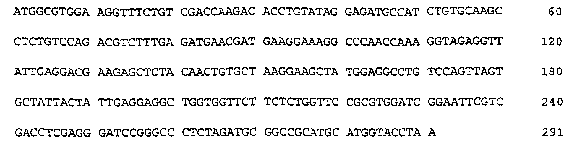
- (2) INFORMATION FOR SEQ ID NO: 2:
- (i) SEQUENCE CHARACTERISTICS:
- (A) LENGTH: 96 amino acids
- (B) TYPE: amino acid
- (C) STRANDEDNESS:
- (D) TOPOLOGY: linear
- (ii) MOLECULE TYPE: protein
- (xi) SEQUENCE DESCRIPTION: SEQ ID NO: 2:

- (2) INFORMATION FOR SEQ ID NO: 3:
- (i) SEQUENCE CHARACTERISTICS:
- (A) LENGTH: 672 base pairs
- (B) TYPE: nucleic acid
- (C) STRANDEDNESS: double
- (D) TOPOLOGY: linear
- (ii) MOLECULE TYPE: other nucleic acid
- (xi) SEQUENCE DESCRIPTION: SEQ ID NO: 3:


- (2) INFORMATION FOR SEQ ID NO: 4:
- (i) SEQUENCE CHARACTERISTICS:
- (A) LENGTH: 223 amino acids
- (B) TYPE: amino acid
- (C) STRANDEDNESS:
- (D) TOPOLOGY: linear
- (ii) MOLECULE TYPE: protein
- (xi) SEQUENCE DESCRIPTION: SEQ ID NO: 4:
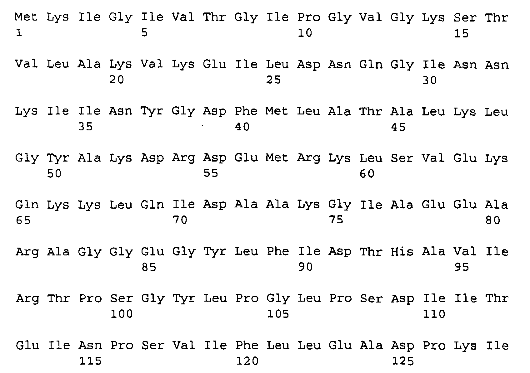
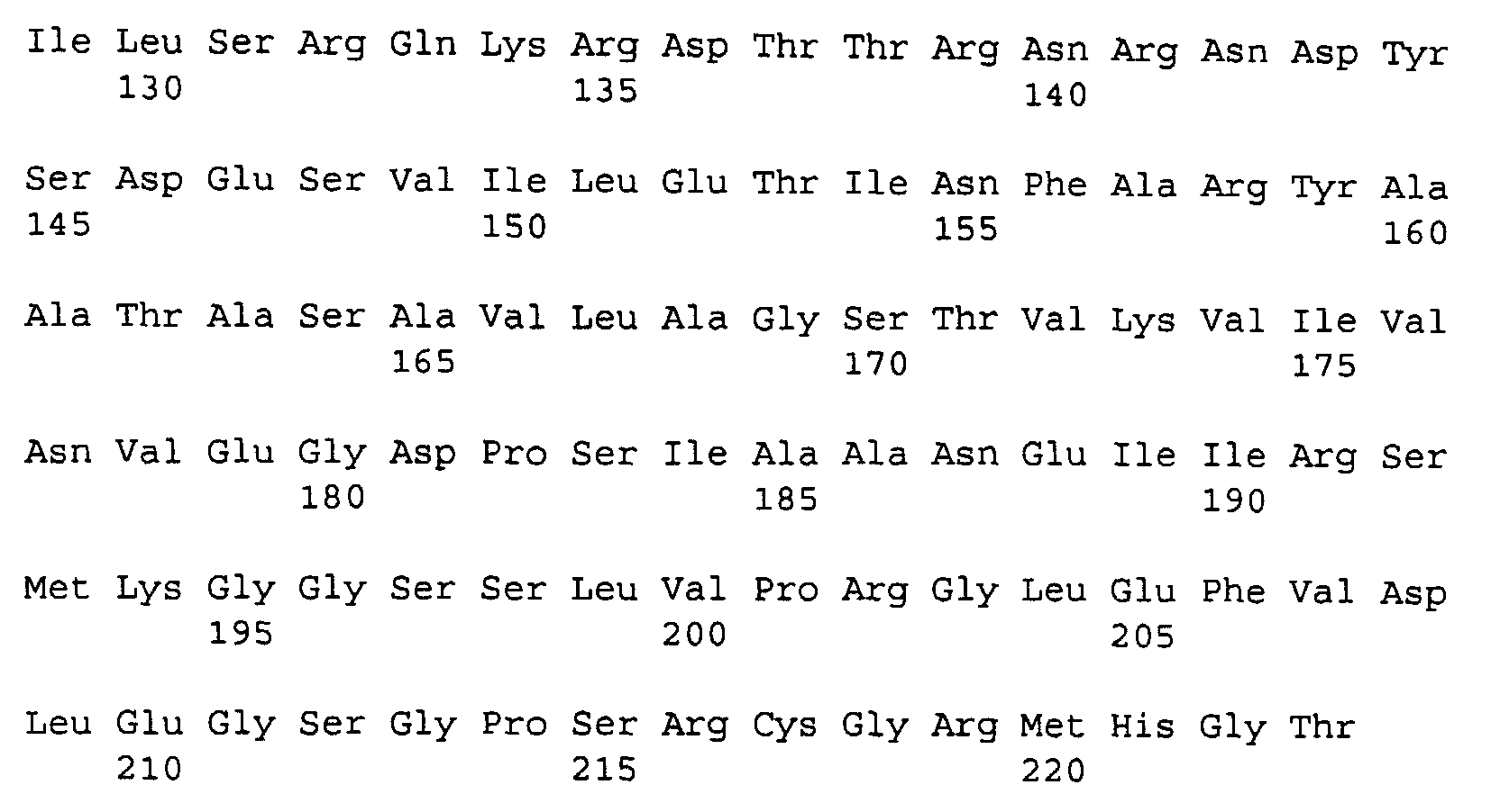
- (2) INFORMATION FOR SEQ ID NO: 5:
- (i) SEQUENCE CHARACTERISTICS:
- (A) LENGTH: 4557 base pairs
- (B) TYPE: nucleic acid
- (C) STRANDEDNESS: double
- (D) TOPOLOGY: circular
- (ii) MOLECULE TYPE: other nucleic acid
- (vi) ORIGINAL SOURCE:
- (A) ORGANISM: E. coli
- (B) STRAIN: BL21(DE3)
- (xi) SEQUENCE DESCRIPTION: SEQ ID NO: 5:
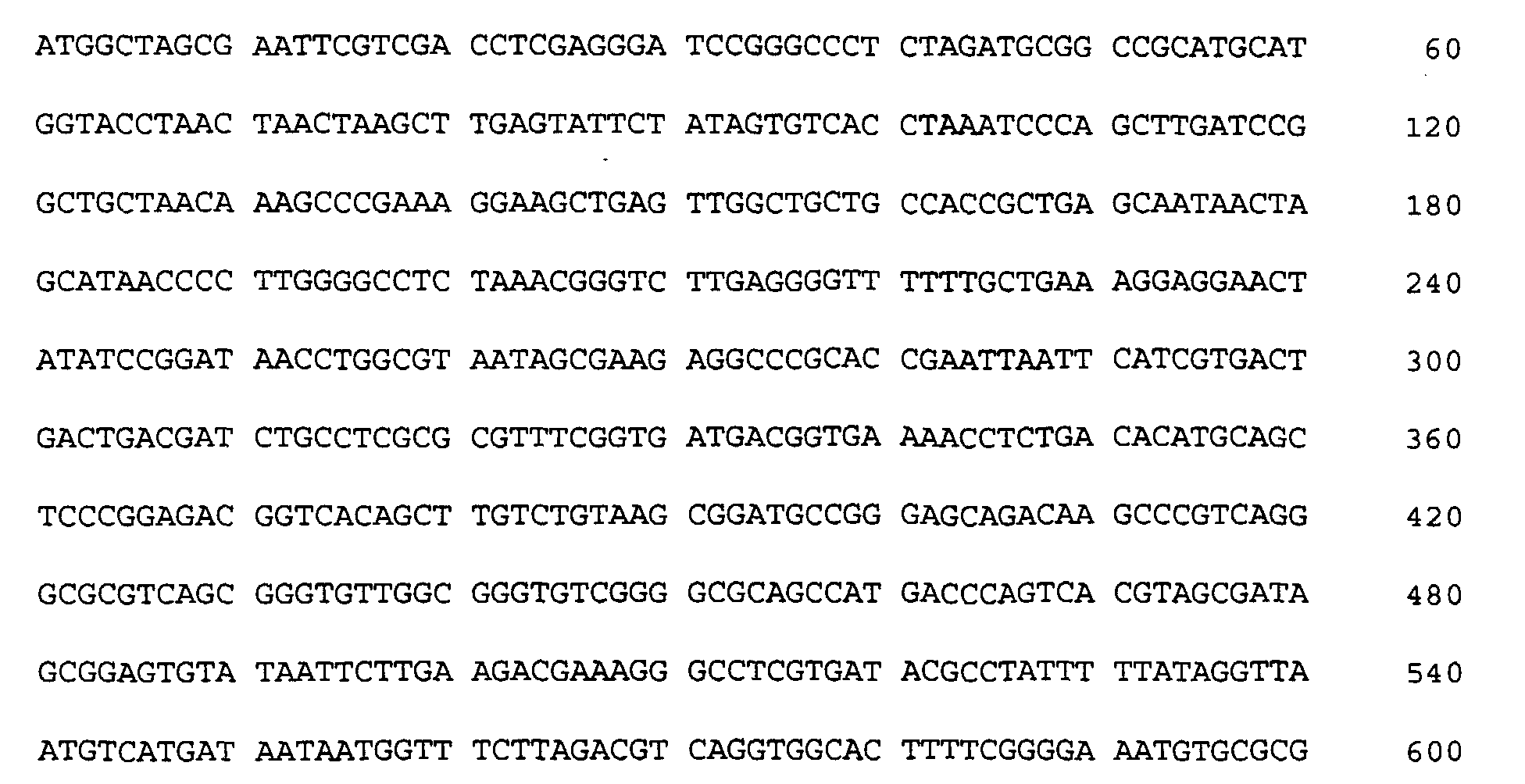

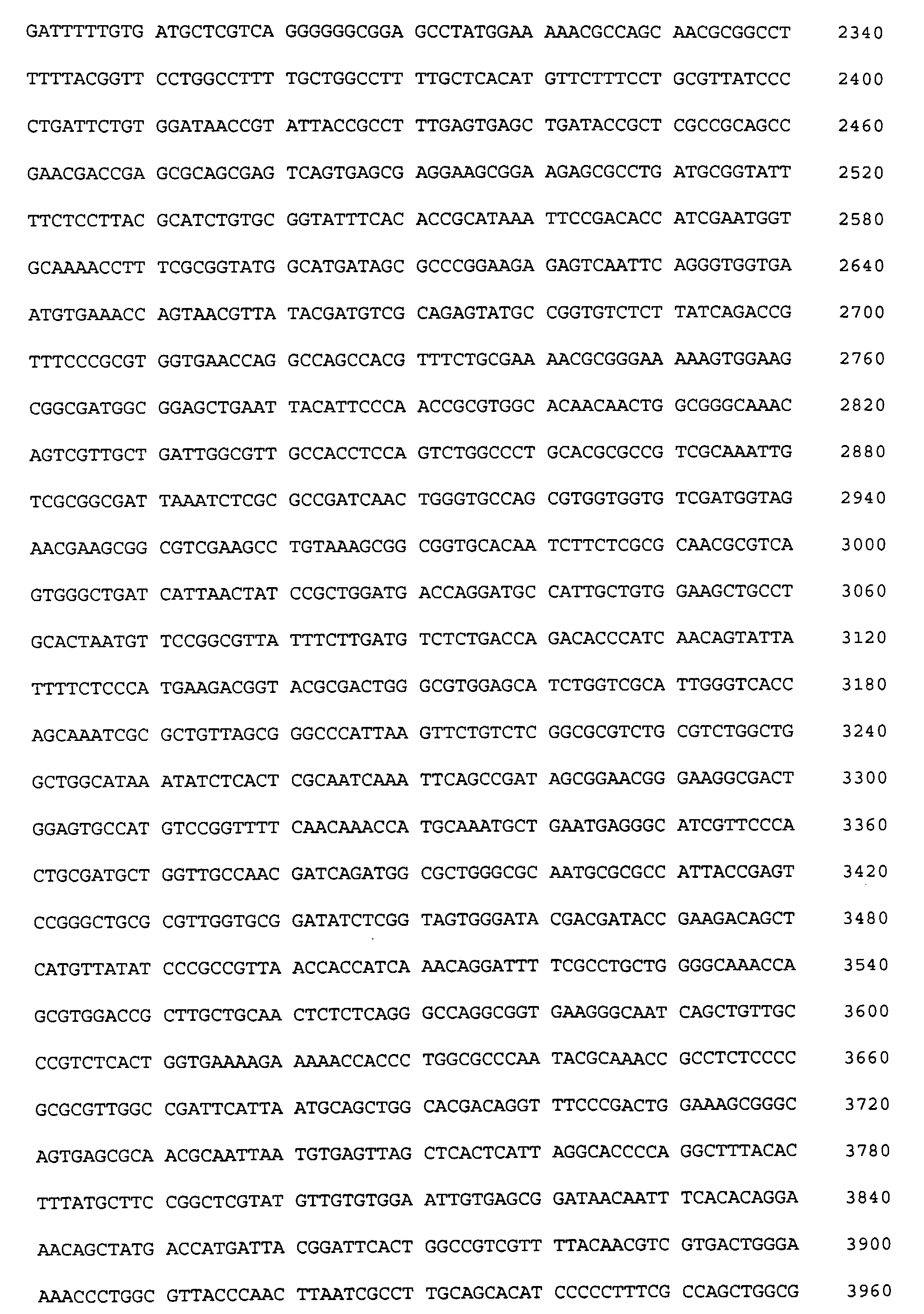

- (2) INFORMATION FOR SEQ ID NO: 6:
- (i) SEQUENCE CHARACTERISTICS:
- (A) LENGTH: 672 base pairs
- (B) TYPE: nucleic acid
- (C) STRANDEDNESS: double
- (D) TOPOLOGY: linear
- (ii) MOLECULE TYPE: other nucleic acid
- (xi) SEQUENCE DESCRIPTION: SEQ ID NO: 6:
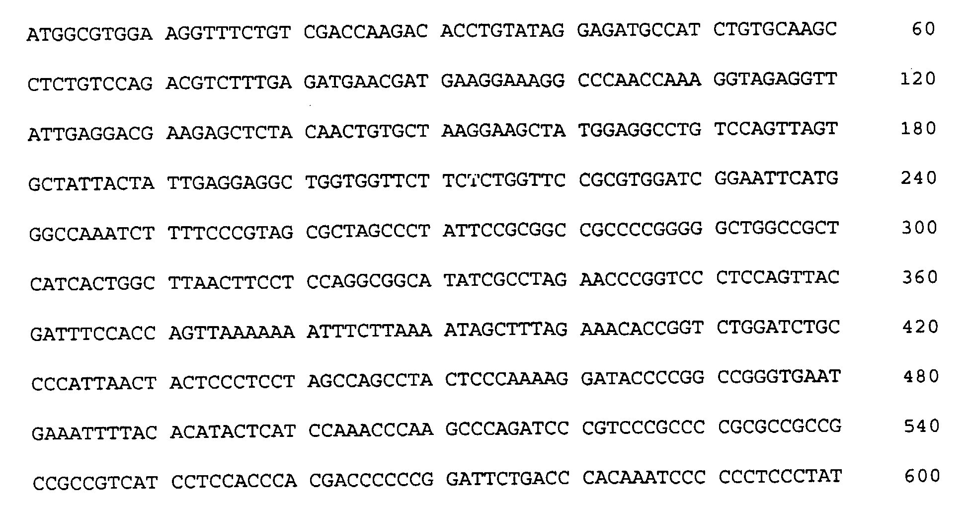

- (2) INFORMATION FOR SEQ ID NO: 7:
- (i) SEQUENCE CHARACTERISTICS:
- (A) LENGTH: 209 amino acids
- (B) TYPE: amino acid
- (C) STRANDEDNESS:
- (D) TOPOLOGY: linear
- (ii) MOLECULE TYPE: protein
- (xi) SEQUENCE DESCRIPTION: SEQ ID NO: 7:
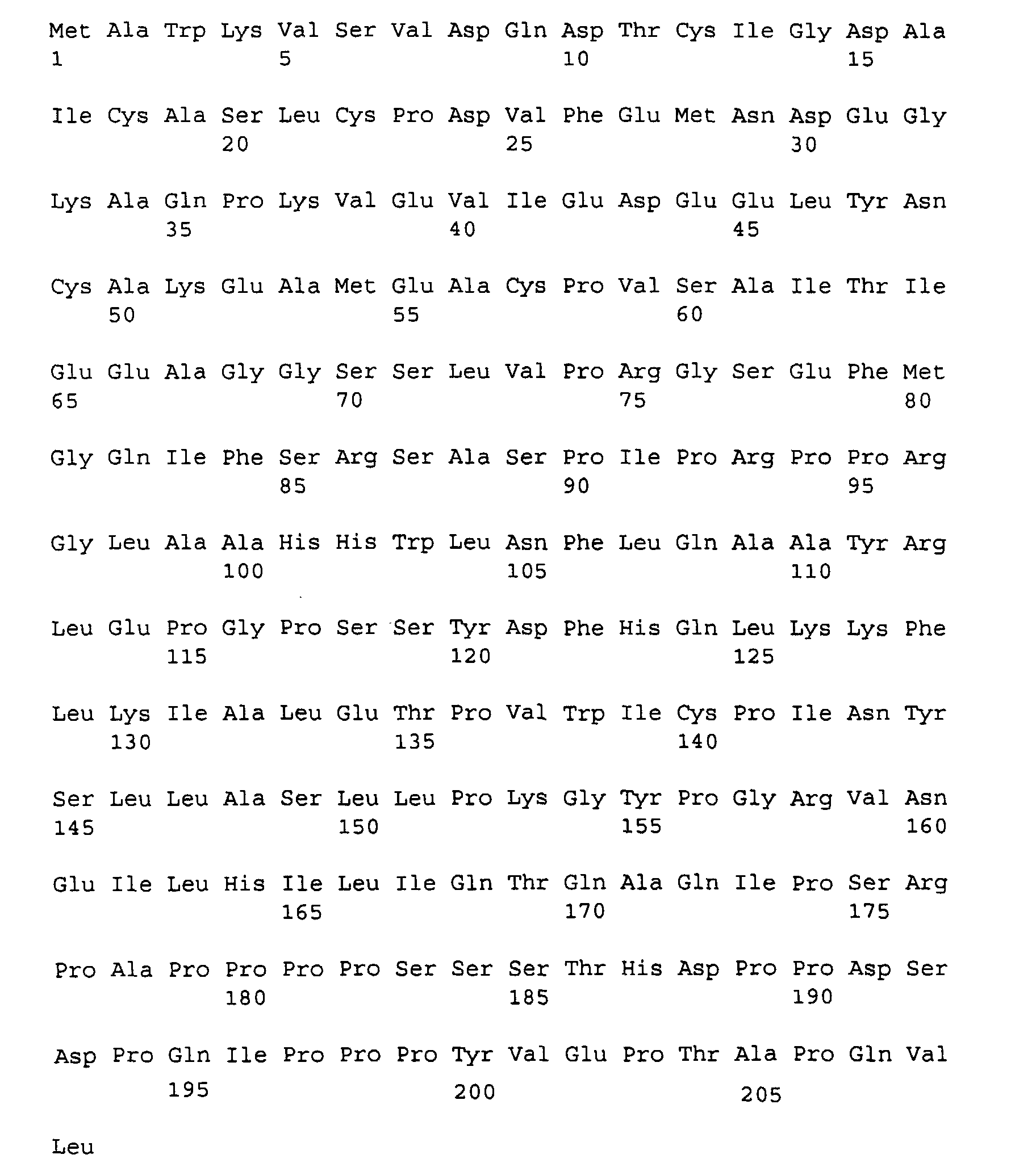
- (2) INFORMATION FOR SEQ ID NO: 8:
- (i) SEQUENCE CHARACTERISTICS:
- (A) LENGTH: 690 base pairs
- (B) TYPE: nucleic acid
- (C) STRANDEDNESS: double
- (D) TOPOLOGY: linear
- (ii) MOLECULE TYPE: other nucleic acid
- (xi) SEQUENCE DESCRIPTION: SEQ ID NO: 8:
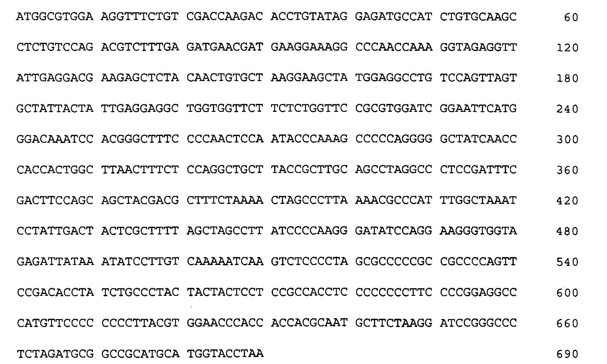
- (2) INFORMATION FOR SEQ ID NO: 9:
- (i) SEQUENCE CHARACTERISTICS:
- (A) LENGTH: 215 amino acids
- (B) TYPE: amino acid
- (C) STRANDEDNESS:
- (D) TOPOLOGY: linear
- (ii) MOLECULE TYPE: protein
- (xi) SEQUENCE DESCRIPTION: SEQ ID NO: 9:

- (2) INFORMATION FOR SEQ ID NO: 10:
- (i) SEQUENCE CHARACTERISTICS:
- (A) LENGTH: 810 base pairs
- (B) TYPE: nucleic acid
- (C) STRANDEDNESS: double
- (D) TOPOLOGY: linear
- (ii) MOLECULE TYPE: other nucleic acid
- (xi) SEQUENCE DESCRIPTION: SEQ ID NO: 10:
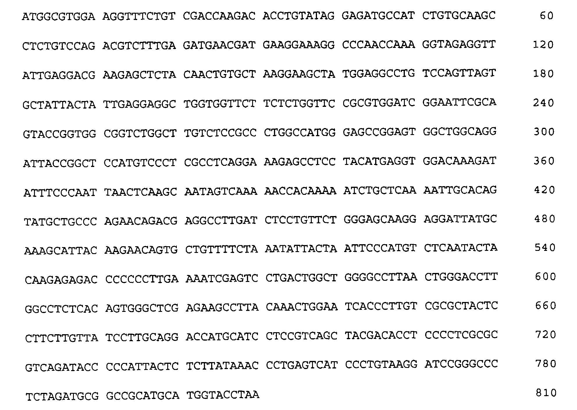
- (2) INFORMATION FOR SEQ ID NO: 11:
- (i) SEQUENCE CHARACTERISTICS:
- (A) LENGTH: 255 amino acids
- (B) TYPE: amino acid
- (C) STRANDEDNESS:
- (D) TOPOLOGY: linear
- (ii) MOLECULE TYPE: protein
- (xi) SEQUENCE DESCRIPTION: SEQ ID NO: 11:
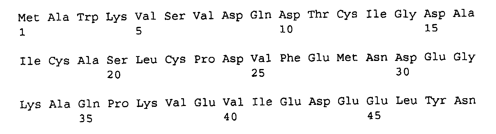

- (2) INFORMATION FOR SEQ ID NO: 12:
- (i) SEQUENCE CHARACTERISTICS:
- (A) LENGTH: 816 base pairs
- (B) TYPE: nucleic acid
- (C) STRANDEDNESS: double
- (D) TOPOLOGY: linear
- (ii) MOLECULE TYPE: other nucleic acid
- (xi) SEQUENCE DESCRIPTION: SEQ ID NO: 12:

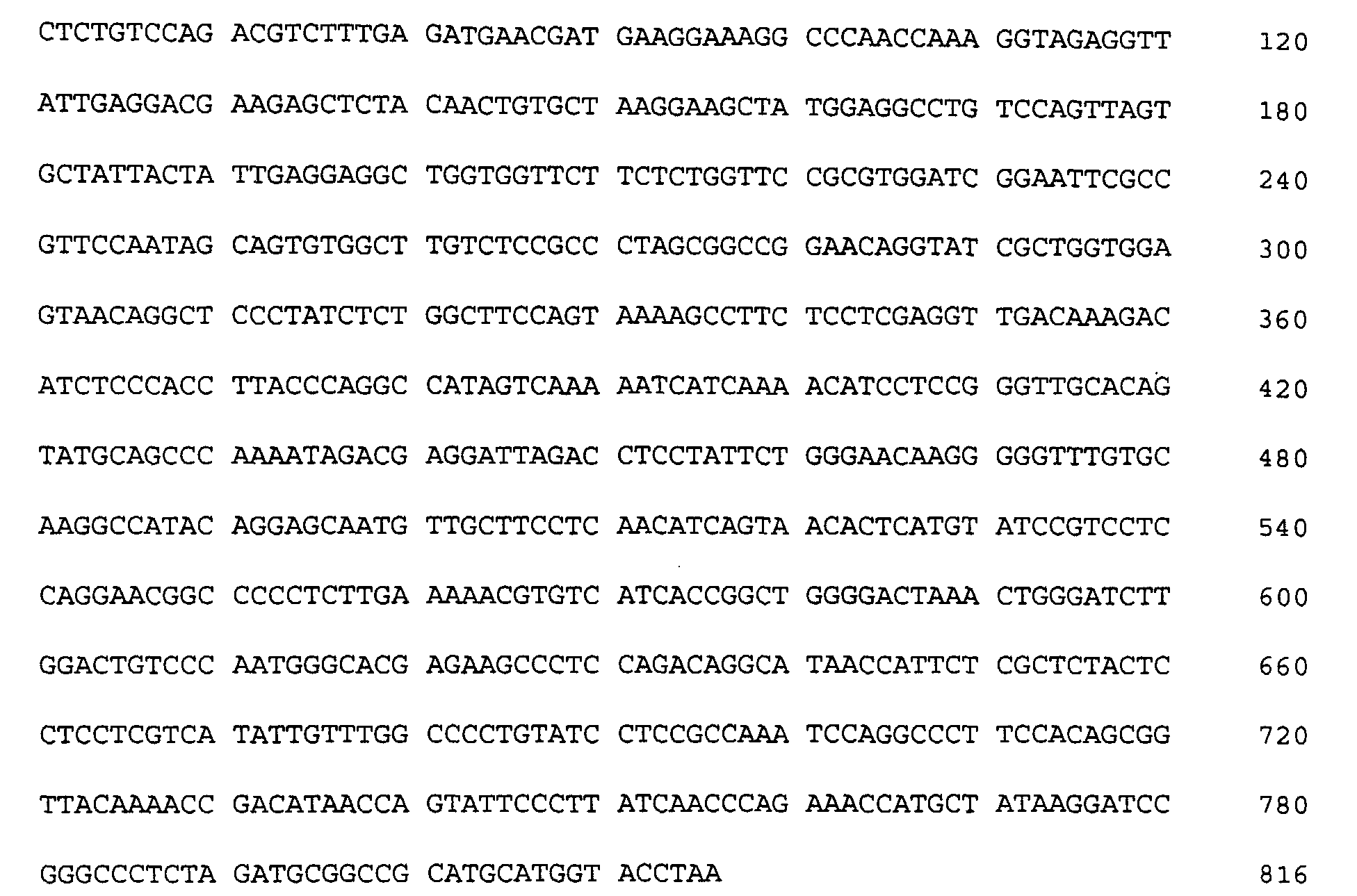
- (2) INFORMATION FOR SEQ ID NO: 13:
- (i) SEQUENCE CHARACTERISTICS:
- (A) LENGTH: 257 amino acids
- (B) TYPE: amino acid
- (C) STRANDEDNESS:
- (D) TOPOLOGY: linear
- (ii) MOLECULE TYPE: protein
- (xi) SEQUENCE DESCRIPTION: SEQ ID NO: 13:


- (2) INFORMATION FOR SEQ ID NO: 14:
- (i) SEQUENCE CHARACTERISTICS:
- (A) LENGTH: 1119 base pairs
- (B) TYPE: nucleic acid
- (C) STRANDEDNESS: double
- (D) TOPOLOGY: linear
- (ii) MOLECULE TYPE: other nucleic acid
- (xi) SEQUENCE DESCRIPTION: SEQ ID NO: 14:


- (2) INFORMATION FOR SEQ ID NO: 15:
- (i) SEQUENCE CHARACTERISTICS:
- (A) LENGTH: 352 amino acids
- (B) TYPE: amino acid
- (C) STRANDEDNESS:
- (D) TOPOLOGY: linear
- (ii) MOLECULE TYPE: protein
- (xi) SEQUENCE DESCRIPTION: SEQ ID NO: 15:
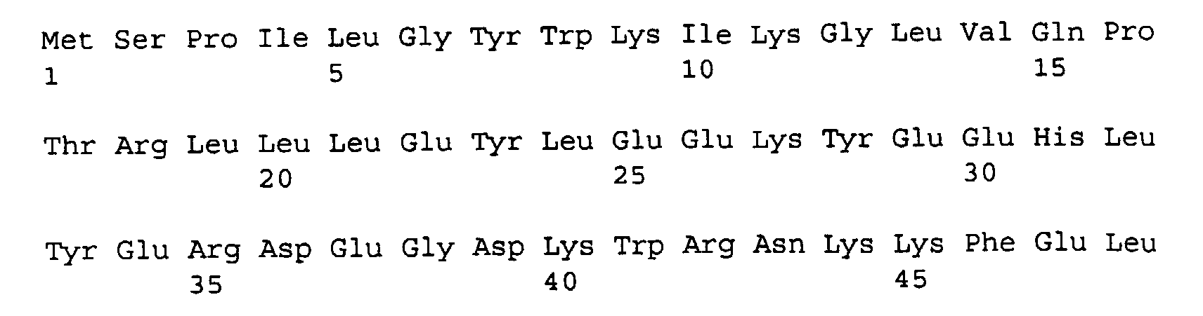
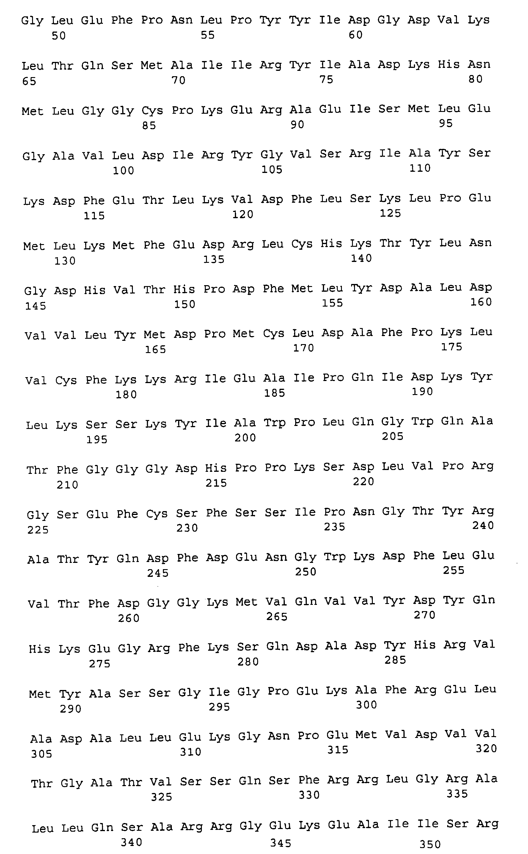
- (2) INFORMATION FOR SEQ ID NO: 16:
- (i) SEQUENCE CHARACTERISTICS:
- (A) LENGTH: 858 base pairs
- (B) TYPE: nucleic acid
- (C) STRANDEDNESS: double
- (D) TOPOLOGY: linear
- (ii) MOLECULE TYPE: DNA (genomic)
- (xi) SEQUENCE DESCRIPTION: SEQ ID NO: 16:

- (2) INFORMATION FOR SEQ ID NO: 17:
- (i) SEQUENCE CHARACTERISTICS:
- (A) LENGTH: 265 amino acids
- (B) TYPE: amino acid
- (C) STRANDEDNESS:
- (D) TOPOLOGY: linear
- (ii) MOLECULE TYPE: protein
- (xi) SEQUENCE DESCRIPTION: SEQ ID NO: 17:
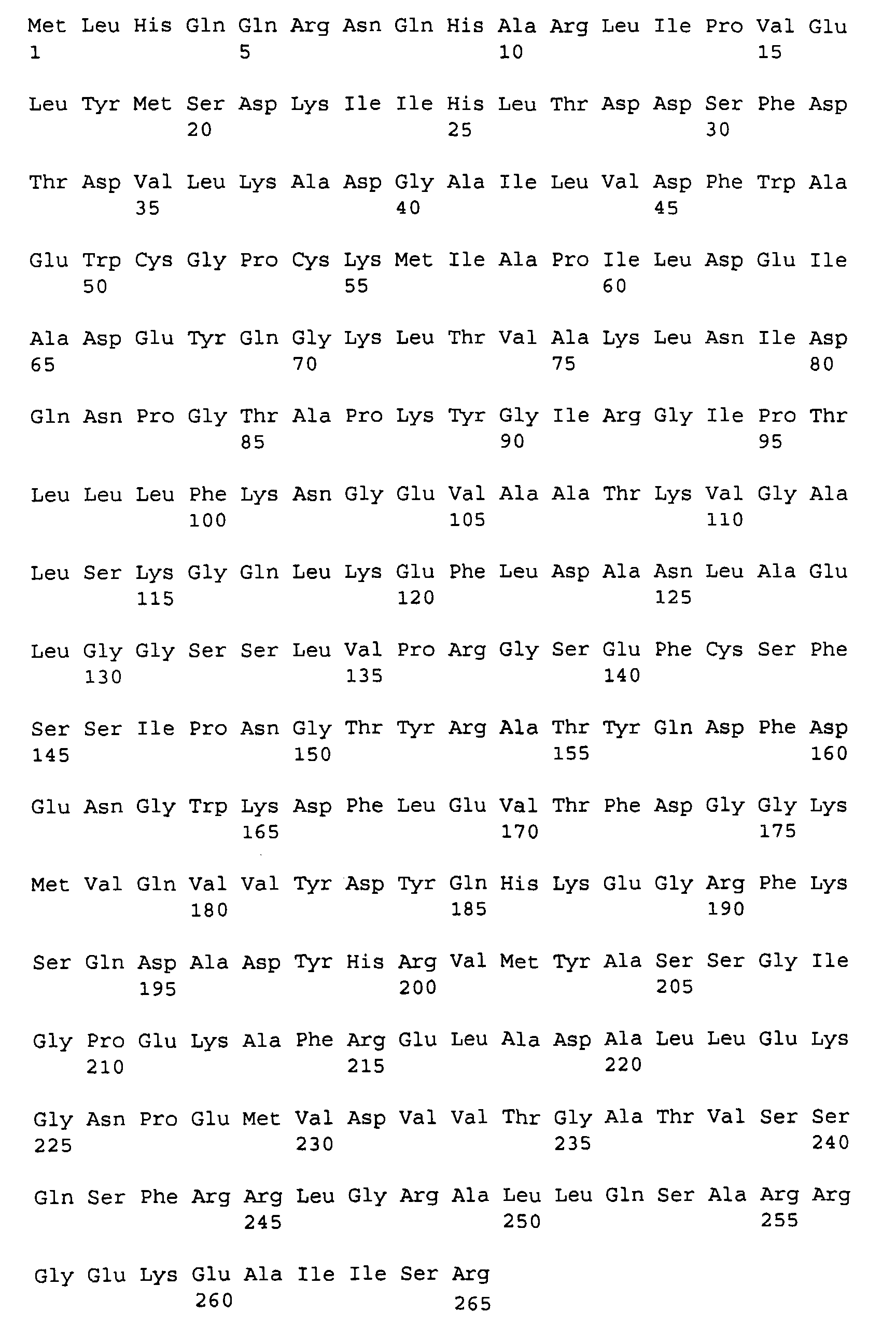
- (2) INFORMATION FOR SEQ ID NO: 18:
- (i) SEQUENCE CHARACTERISTICS:
- (A) LENGTH: 672 base pairs
- (B) TYPE: nucleic acid
- (C) STRANDEDNESS: double
- (D) TOPOLOGY: linear
- (ii) MOLECULE TYPE: other nucleic acid
- (xi) SEQUENCE DESCRIPTION: SEQ ID NO: 18:

- (2) INFORMATION FOR SEQ ID NO: 19:
- (i) SEQUENCE CHARACTERISTICS:
- (A) LENGTH: 203 amino acids
- (B) TYPE: amino acid
- (C) STRANDEDNESS:
- (D) TOPOLOGY: linear
- (ii) MOLECULE TYPE: protein
- (xi) SEQUENCE DESCRIPTION: SEQ ID NO: 19:
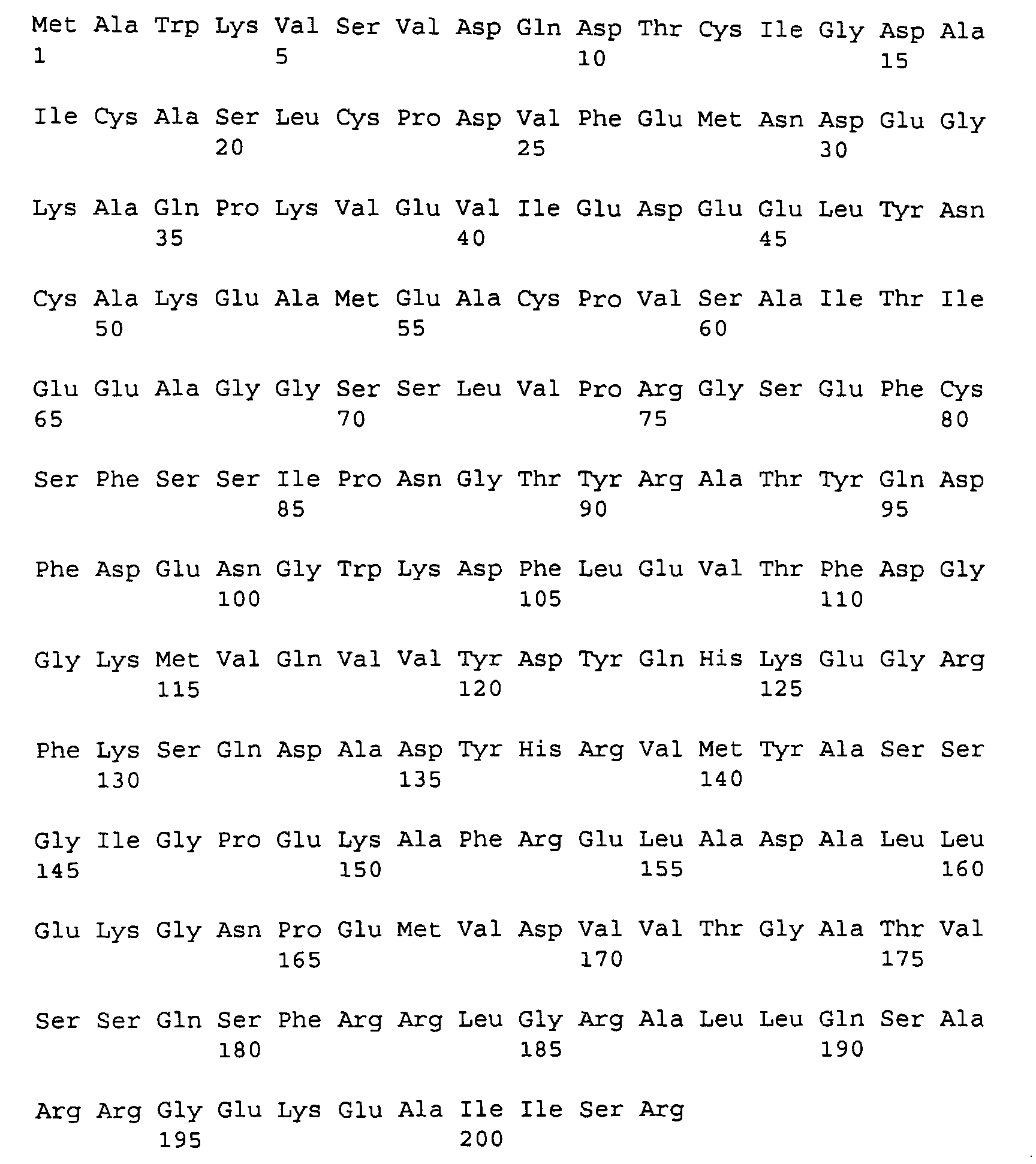
- (2) INFORMATION FOR SEQ ID NO: 20:
- (i) SEQUENCE CHARACTERISTICS:
- (A) LENGTH: 1035 base pairs
- (B) TYPE: nucleic acid
- (C) STRANDEDNESS: double
- (D) TOPOLOGY: linear
- (ii) MOLECULE TYPE: other nucleic acid
- (xi) SEQUENCE DESCRIPTION: SEQ ID NO: 20:

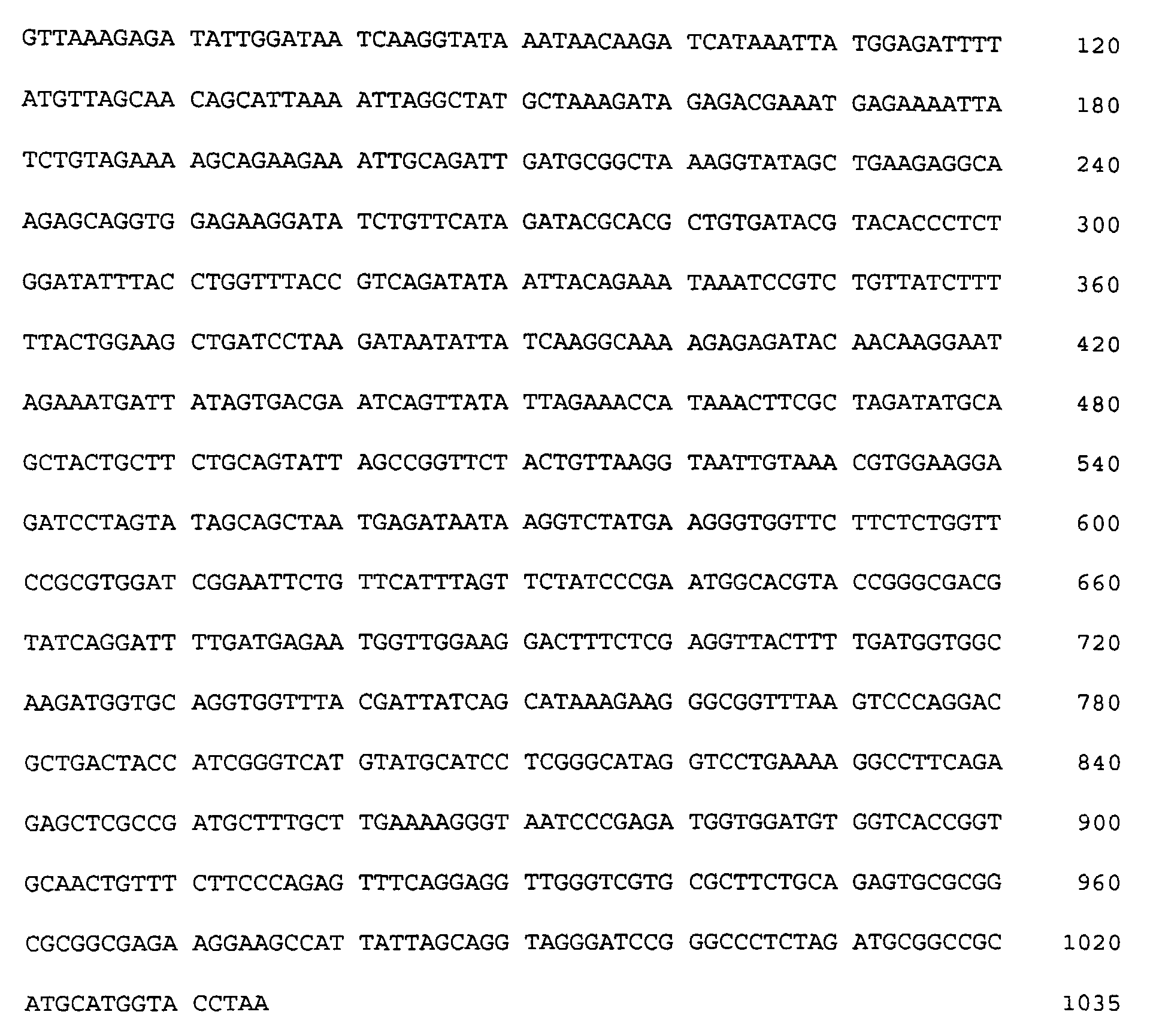
- (2) INFORMATION FOR SEQ ID NO: 21:
- (i) SEQUENCE CHARACTERISTICS:
- (A) LENGTH: 330 amino acids
- (B) TYPE: amino acid
- (C) STRANDEDNESS:
- (D) TOPOLOGY: linear
- (ii) MOLECULE TYPE: protein
- (xi) SEQUENCE DESCRIPTION: SEQ ID NO: 21:
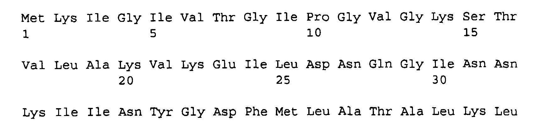

-
Claims (11)
- A fused DNA sequence which comprises a DNA sequence encoding a heat-resistant protein, fused directly or indirectly to the 5'-end of a DNA sequence coding a selected protein or peptide.
- The sequence according to claim 1 wherein the DNA sequence encoding a heat-resistant protein is a DNA sequence derivable from a thermophilic bacterium.
- The sequence according to claim 1 wherein the DNA sequence encoding a heat-resistant protein is a DNA sequence derivable from a highly thermophilic bacterium.
- The sequence according to claim 1 wherein the DNA sequence encoding a heat-resistant protein is a DNA sequence derivable from a Thermophilus bacterium, a Sulfolobus bacterium, a Pyrococcus bacterium, a Thermotoga bacterium, a Pyrobaculum bacterium, a Pyrodictium bacterium, a Thermococcus bacterium, a Thermodiscus bacterium, a Metanothermus bacterium or a Metanococcus bacterium.
- The sequence according to claim 1 wherein the DNA sequence encoding a heat-resistant protein is a DNA sequence derivable from a Pyrococcus bacterium or a Sulfolobus bacterium.
- The sequence according to any one of claims 1 to 5 wherein the DNA sequence encoding a heat-resistant protein is a DNA sequence of heat-resistant ferredoxin or heat-resistant adenylated kinase.
- The sequence according to claim 1 wherein the DNA sequence encoding a heat-resistant protein is a DNA sequence of ferredoxin derivable from a Pyrococcus bacterium or adenylate kinase derived from a Sulfolobus bacterium.
- The sequence according to claim 1 wherein the DNA sequence encoding a heat-resistant protein is a DNA sequence of ferredoxin derivable from a Pyrococcus bacterium.
- The sequence according to claim 1 wherein the DNA sequence encoding a heat-resistant protein is a DNA sequence of adenylate kinase derivable from a Sulfolobus bacterium.
- A fused protein being expressed from the DNA sequence according to any one of claims 1 to 9.
- A method for expressing a fused protein, which comprises using the DNA sequence according to any one of claims 1 to 9.
Applications Claiming Priority (3)
| Application Number | Priority Date | Filing Date | Title |
|---|---|---|---|
| JP352225/95 | 1995-12-28 | ||
| JP35222595 | 1995-12-28 | ||
| JP35222595 | 1995-12-28 |
Publications (3)
| Publication Number | Publication Date |
|---|---|
| EP0781848A2 EP0781848A2 (en) | 1997-07-02 |
| EP0781848A3 EP0781848A3 (en) | 1997-10-01 |
| EP0781848B1 true EP0781848B1 (en) | 2001-02-28 |
Family
ID=18422615
Family Applications (1)
| Application Number | Title | Priority Date | Filing Date |
|---|---|---|---|
| EP96120899A Expired - Lifetime EP0781848B1 (en) | 1995-12-28 | 1996-12-27 | Fused DNA sequence, fused protein expressed from said fused DNA sequence and method for expressing said fused protein |
Country Status (8)
| Country | Link |
|---|---|
| US (2) | US6602689B1 (en) |
| EP (1) | EP0781848B1 (en) |
| KR (1) | KR970048454A (en) |
| AT (1) | ATE199396T1 (en) |
| AU (1) | AU707191B2 (en) |
| CA (1) | CA2194032C (en) |
| DE (1) | DE69611896T2 (en) |
| SG (1) | SG49993A1 (en) |
Families Citing this family (7)
| Publication number | Priority date | Publication date | Assignee | Title |
|---|---|---|---|---|
| EP0985931A3 (en) * | 1998-09-04 | 2000-03-29 | Becton Dickinson and Company | Recombinant antigen immunoassay for the diagnosis of syphilis |
| AU2486900A (en) * | 1998-12-29 | 2000-07-31 | University Of Georgia Research Foundation, Inc., The | Rubredoxin fusion proteins, protein expression system and methods |
| TW200409815A (en) * | 2002-12-12 | 2004-06-16 | Dev Center Biotechnology | Human T-lymphotrophic virus (HTLV) fusion protein, nucleic acid and vector encoding the protein, and kit for testing human T-lymphotrophic virus using the protein |
| WO2005108583A1 (en) * | 2004-05-06 | 2005-11-17 | Prokaria Ehf. | Thermostable polypeptide having polynucleotide kinase activity and/or phosphatase activity |
| GB0803068D0 (en) * | 2008-02-20 | 2008-03-26 | Health Prot Agency | Cross-linked biological indicator |
| US10466245B2 (en) | 2008-02-20 | 2019-11-05 | The Secretary Of State For Health | Covalently linked thermostable kinase for decontamination process validation |
| CN114369170B (en) * | 2021-12-09 | 2022-09-23 | 陕西师范大学 | Glioblastoma therapeutic fusion protein based on targeting pleiotropic growth factor, vaccine and preparation method and use thereof |
Family Cites Families (6)
| Publication number | Priority date | Publication date | Assignee | Title |
|---|---|---|---|---|
| US4797359A (en) * | 1983-05-10 | 1989-01-10 | Board Of Regents, The University Of Texas System | Heat shock regulated production of selected and fused proteins in yeast |
| WO1987005935A1 (en) | 1986-04-04 | 1987-10-08 | Battelle Memorial Institute At Columbus, Ohio Gene | Methods and compositions for expression of competent eukaryotic gene products |
| US5723588A (en) * | 1986-11-04 | 1998-03-03 | Protein Polymer Technologies, Inc. | Protein-enriched thermoplastics |
| NZ224663A (en) | 1987-05-28 | 1990-11-27 | Amrad Corp Ltd | Fusion proteins containing glutathione-s-transferase and expression of foreign polypeptides using glutathione-s-transferase encoding vectors |
| US5270181A (en) | 1991-02-06 | 1993-12-14 | Genetics Institute, Inc. | Peptide and protein fusions to thioredoxin and thioredoxin-like molecules |
| US5700672A (en) * | 1992-07-23 | 1997-12-23 | Stratagene | Purified thermostable pyrococcus furiousus DNA ligase |
-
1996
- 1996-12-24 SG SG1996011906A patent/SG49993A1/en unknown
- 1996-12-24 AU AU76499/96A patent/AU707191B2/en not_active Ceased
- 1996-12-27 CA CA002194032A patent/CA2194032C/en not_active Expired - Fee Related
- 1996-12-27 DE DE69611896T patent/DE69611896T2/en not_active Expired - Fee Related
- 1996-12-27 AT AT96120899T patent/ATE199396T1/en not_active IP Right Cessation
- 1996-12-27 US US08/778,717 patent/US6602689B1/en not_active Expired - Fee Related
- 1996-12-27 EP EP96120899A patent/EP0781848B1/en not_active Expired - Lifetime
- 1996-12-27 KR KR1019960073955A patent/KR970048454A/en not_active Withdrawn
-
2003
- 2003-06-10 US US10/457,372 patent/US7067631B2/en not_active Expired - Fee Related
Also Published As
| Publication number | Publication date |
|---|---|
| CA2194032A1 (en) | 1997-06-29 |
| AU7649996A (en) | 1997-07-03 |
| DE69611896T2 (en) | 2001-07-26 |
| CA2194032C (en) | 2004-09-21 |
| US20040018534A1 (en) | 2004-01-29 |
| ATE199396T1 (en) | 2001-03-15 |
| AU707191B2 (en) | 1999-07-08 |
| US7067631B2 (en) | 2006-06-27 |
| EP0781848A3 (en) | 1997-10-01 |
| SG49993A1 (en) | 1998-06-15 |
| EP0781848A2 (en) | 1997-07-02 |
| KR970048454A (en) | 1997-07-29 |
| US6602689B1 (en) | 2003-08-05 |
| DE69611896D1 (en) | 2001-04-05 |
Similar Documents
| Publication | Publication Date | Title |
|---|---|---|
| US10954495B2 (en) | Nucleic acid modifying enzymes | |
| AU2003284265B2 (en) | Improved Sso7-polymerase conjugate proteins | |
| JP3742659B2 (en) | DNA polymerase-related factors | |
| JPH07506573A (en) | Protein L and its hybrid proteins | |
| JP4038555B2 (en) | Recombinant protein production method and fusion protein | |
| CA2157767A1 (en) | Genetically engineered enzymes and their conjugates for diagnostic assays | |
| JPWO1999000506A1 (en) | DNA polymerase-associated factors | |
| US7067631B2 (en) | Fused DNA sequence, fused protein expressed from said fused DNA sequence and method for expressing said fused protein | |
| JP2887238B2 (en) | Polypeptide | |
| KR100257574B1 (en) | Treponema pallidum fused antigen and assay for anti-treponema pallidum antibodies using the same fused antigen | |
| EP0939800A2 (en) | HELICOBACTER ANTIGEN (gamma-GLUTAMYLTRANSFERASE) AND SEQUENCES ENCODING THE SAME | |
| KR20000064592A (en) | Export System for Recombinant Protein | |
| EP1832652B1 (en) | Improved nucleic acid modifying enzymes | |
| Zalewska et al. | Cloning, expression, and purification of the uropathogenic Escherichia coli invasin DraD | |
| JP3533857B2 (en) | Treponema pallidum fusion antigen and method for measuring anti-treponema pallidum antibody using the fusion antigen | |
| WO1998036076A1 (en) | Modified treponema pallidum-derived antigen protein | |
| KR100274190B1 (en) | Expression of UBHBVCORE Protein, a Specific Antigen of Hepatitis B Virus | |
| JP4252299B2 (en) | Novel disulfide oxidoreductase and protein activation method using the enzyme | |
| CN120289591A (en) | Recombinant antigen, nucleic acid molecule, recombinant expression vector and host cell of Treponema pallidum and application thereof | |
| JP2004261129A (en) | METHOD FOR PRODUCING Glc NUCLEOTIDE |
Legal Events
| Date | Code | Title | Description |
|---|---|---|---|
| PUAI | Public reference made under article 153(3) epc to a published international application that has entered the european phase |
Free format text: ORIGINAL CODE: 0009012 |
|
| AK | Designated contracting states |
Kind code of ref document: A2 Designated state(s): AT BE CH DE DK ES FI FR GB GR IE IT LI LU MC NL PT SE |
|
| PUAL | Search report despatched |
Free format text: ORIGINAL CODE: 0009013 |
|
| AK | Designated contracting states |
Kind code of ref document: A3 Designated state(s): AT BE CH DE DK ES FI FR GB GR IE IT LI LU MC NL PT SE |
|
| 17P | Request for examination filed |
Effective date: 19980318 |
|
| RAP1 | Party data changed (applicant data changed or rights of an application transferred) |
Owner name: FUJIREBIO INC. |
|
| 17Q | First examination report despatched |
Effective date: 19981112 |
|
| RIC1 | Information provided on ipc code assigned before grant |
Free format text: 6C 12N 15/62 A, 6C 07K 14/195 B, 6C 07K 14/20 B, 6C 12N 15/31 B, 6C 12N 15/54 B, 6C 12N 15/48 - |
|
| GRAG | Despatch of communication of intention to grant |
Free format text: ORIGINAL CODE: EPIDOS AGRA |
|
| GRAG | Despatch of communication of intention to grant |
Free format text: ORIGINAL CODE: EPIDOS AGRA |
|
| GRAG | Despatch of communication of intention to grant |
Free format text: ORIGINAL CODE: EPIDOS AGRA |
|
| GRAH | Despatch of communication of intention to grant a patent |
Free format text: ORIGINAL CODE: EPIDOS IGRA |
|
| GRAH | Despatch of communication of intention to grant a patent |
Free format text: ORIGINAL CODE: EPIDOS IGRA |
|
| GRAA | (expected) grant |
Free format text: ORIGINAL CODE: 0009210 |
|
| AK | Designated contracting states |
Kind code of ref document: B1 Designated state(s): AT BE CH DE DK ES FI FR GB GR IE IT LI LU MC NL PT SE |
|
| PG25 | Lapsed in a contracting state [announced via postgrant information from national office to epo] |
Ref country code: SE Free format text: LAPSE BECAUSE OF FAILURE TO SUBMIT A TRANSLATION OF THE DESCRIPTION OR TO PAY THE FEE WITHIN THE PRESCRIBED TIME-LIMIT Effective date: 20010228 Ref country code: NL Free format text: LAPSE BECAUSE OF FAILURE TO SUBMIT A TRANSLATION OF THE DESCRIPTION OR TO PAY THE FEE WITHIN THE PRESCRIBED TIME-LIMIT Effective date: 20010228 Ref country code: FI Free format text: LAPSE BECAUSE OF FAILURE TO SUBMIT A TRANSLATION OF THE DESCRIPTION OR TO PAY THE FEE WITHIN THE PRESCRIBED TIME-LIMIT Effective date: 20010228 Ref country code: ES Free format text: THE PATENT HAS BEEN ANNULLED BY A DECISION OF A NATIONAL AUTHORITY Effective date: 20010228 Ref country code: BE Free format text: LAPSE BECAUSE OF FAILURE TO SUBMIT A TRANSLATION OF THE DESCRIPTION OR TO PAY THE FEE WITHIN THE PRESCRIBED TIME-LIMIT Effective date: 20010228 Ref country code: AT Free format text: LAPSE BECAUSE OF FAILURE TO SUBMIT A TRANSLATION OF THE DESCRIPTION OR TO PAY THE FEE WITHIN THE PRESCRIBED TIME-LIMIT Effective date: 20010228 |
|
| REF | Corresponds to: |
Ref document number: 199396 Country of ref document: AT Date of ref document: 20010315 Kind code of ref document: T |
|
| REG | Reference to a national code |
Ref country code: CH Ref legal event code: EP |
|
| REF | Corresponds to: |
Ref document number: 69611896 Country of ref document: DE Date of ref document: 20010405 |
|
| REG | Reference to a national code |
Ref country code: IE Ref legal event code: FG4D |
|
| ITF | It: translation for a ep patent filed | ||
| REG | Reference to a national code |
Ref country code: CH Ref legal event code: NV Representative=s name: BUECHEL, KAMINSKI & PARTNER PATENTANWAELTE EST |
|
| PG25 | Lapsed in a contracting state [announced via postgrant information from national office to epo] |
Ref country code: PT Free format text: LAPSE BECAUSE OF FAILURE TO SUBMIT A TRANSLATION OF THE DESCRIPTION OR TO PAY THE FEE WITHIN THE PRESCRIBED TIME-LIMIT Effective date: 20010528 |
|
| PG25 | Lapsed in a contracting state [announced via postgrant information from national office to epo] |
Ref country code: DK Free format text: LAPSE BECAUSE OF FAILURE TO SUBMIT A TRANSLATION OF THE DESCRIPTION OR TO PAY THE FEE WITHIN THE PRESCRIBED TIME-LIMIT Effective date: 20010531 |
|
| PG25 | Lapsed in a contracting state [announced via postgrant information from national office to epo] |
Ref country code: GR Free format text: LAPSE BECAUSE OF FAILURE TO SUBMIT A TRANSLATION OF THE DESCRIPTION OR TO PAY THE FEE WITHIN THE PRESCRIBED TIME-LIMIT Effective date: 20010601 |
|
| ET | Fr: translation filed | ||
| NLV1 | Nl: lapsed or annulled due to failure to fulfill the requirements of art. 29p and 29m of the patents act | ||
| PG25 | Lapsed in a contracting state [announced via postgrant information from national office to epo] |
Ref country code: MC Free format text: LAPSE BECAUSE OF NON-PAYMENT OF DUE FEES Effective date: 20011227 Ref country code: LU Free format text: LAPSE BECAUSE OF NON-PAYMENT OF DUE FEES Effective date: 20011227 Ref country code: IE Free format text: LAPSE BECAUSE OF FAILURE TO SUBMIT A TRANSLATION OF THE DESCRIPTION OR TO PAY THE FEE WITHIN THE PRESCRIBED TIME-LIMIT Effective date: 20011227 |
|
| REG | Reference to a national code |
Ref country code: GB Ref legal event code: IF02 |
|
| PLBE | No opposition filed within time limit |
Free format text: ORIGINAL CODE: 0009261 |
|
| STAA | Information on the status of an ep patent application or granted ep patent |
Free format text: STATUS: NO OPPOSITION FILED WITHIN TIME LIMIT |
|
| 26N | No opposition filed | ||
| REG | Reference to a national code |
Ref country code: IE Ref legal event code: MM4A |
|
| REG | Reference to a national code |
Ref country code: GB Ref legal event code: 732E |
|
| REG | Reference to a national code |
Ref country code: CH Ref legal event code: PUE Owner name: FUJIREBIO INC. Free format text: MIRACA HOLDINGS INC.#62-5, NIHONBASHI HAMACHO 2-CHOME CHUO-KU#TOKYO (JP) -TRANSFER TO- FUJIREBIO INC.#62-5, NIHONBASHI HAMACHO 2-CHOME CHUO-KU#TOKYO (JP) Ref country code: CH Ref legal event code: PFA Owner name: MIRACA HOLDINGS INC. Free format text: FUJIREBIO INC.#62-5 NIHONBASHI-HAMACHO 2-CHOME, CHUO-KU#TOKYO 103-0007 (JP) -TRANSFER TO- MIRACA HOLDINGS INC.#62-5, NIHONBASHI HAMACHO 2-CHOME CHUO-KU#TOKYO (JP) Ref country code: CH Ref legal event code: NV Representative=s name: BOVARD AG PATENTANWAELTE |
|
| REG | Reference to a national code |
Ref country code: FR Ref legal event code: TP Ref country code: FR Ref legal event code: CD |
|
| PGFP | Annual fee paid to national office [announced via postgrant information from national office to epo] |
Ref country code: CH Payment date: 20071224 Year of fee payment: 12 |
|
| PGFP | Annual fee paid to national office [announced via postgrant information from national office to epo] |
Ref country code: GB Payment date: 20071227 Year of fee payment: 12 Ref country code: FR Payment date: 20071210 Year of fee payment: 12 |
|
| PGFP | Annual fee paid to national office [announced via postgrant information from national office to epo] |
Ref country code: IT Payment date: 20071228 Year of fee payment: 12 Ref country code: DE Payment date: 20071220 Year of fee payment: 12 |
|
| REG | Reference to a national code |
Ref country code: CH Ref legal event code: PL |
|
| GBPC | Gb: european patent ceased through non-payment of renewal fee |
Effective date: 20081227 |
|
| REG | Reference to a national code |
Ref country code: FR Ref legal event code: ST Effective date: 20090831 |
|
| PG25 | Lapsed in a contracting state [announced via postgrant information from national office to epo] |
Ref country code: LI Free format text: LAPSE BECAUSE OF NON-PAYMENT OF DUE FEES Effective date: 20081231 Ref country code: DE Free format text: LAPSE BECAUSE OF NON-PAYMENT OF DUE FEES Effective date: 20090701 Ref country code: CH Free format text: LAPSE BECAUSE OF NON-PAYMENT OF DUE FEES Effective date: 20081231 |
|
| PG25 | Lapsed in a contracting state [announced via postgrant information from national office to epo] |
Ref country code: GB Free format text: LAPSE BECAUSE OF NON-PAYMENT OF DUE FEES Effective date: 20081227 |
|
| PG25 | Lapsed in a contracting state [announced via postgrant information from national office to epo] |
Ref country code: FR Free format text: LAPSE BECAUSE OF NON-PAYMENT OF DUE FEES Effective date: 20081231 |
|
| PG25 | Lapsed in a contracting state [announced via postgrant information from national office to epo] |
Ref country code: IT Free format text: LAPSE BECAUSE OF NON-PAYMENT OF DUE FEES Effective date: 20081227 |