EP0771195B1 - Method of inhibiting nitric oxide formation - Google Patents
Method of inhibiting nitric oxide formation Download PDFInfo
- Publication number
- EP0771195B1 EP0771195B1 EP94926572A EP94926572A EP0771195B1 EP 0771195 B1 EP0771195 B1 EP 0771195B1 EP 94926572 A EP94926572 A EP 94926572A EP 94926572 A EP94926572 A EP 94926572A EP 0771195 B1 EP0771195 B1 EP 0771195B1
- Authority
- EP
- European Patent Office
- Prior art keywords
- lps
- aminoguanidine
- nitric oxide
- bsa
- acute
- Prior art date
- Legal status (The legal status is an assumption and is not a legal conclusion. Google has not performed a legal analysis and makes no representation as to the accuracy of the status listed.)
- Expired - Lifetime
Links
Images


Classifications
-
- A—HUMAN NECESSITIES
- A61—MEDICAL OR VETERINARY SCIENCE; HYGIENE
- A61K—PREPARATIONS FOR MEDICAL, DENTAL OR TOILETRY PURPOSES
- A61K31/00—Medicinal preparations containing organic active ingredients
- A61K31/185—Acids; Anhydrides, halides or salts thereof, e.g. sulfur acids, imidic, hydrazonic or hydroximic acids
- A61K31/19—Carboxylic acids, e.g. valproic acid
- A61K31/195—Carboxylic acids, e.g. valproic acid having an amino group
-
- A—HUMAN NECESSITIES
- A61—MEDICAL OR VETERINARY SCIENCE; HYGIENE
- A61K—PREPARATIONS FOR MEDICAL, DENTAL OR TOILETRY PURPOSES
- A61K31/00—Medicinal preparations containing organic active ingredients
- A61K31/13—Amines
- A61K31/155—Amidines (), e.g. guanidine (H2N—C(=NH)—NH2), isourea (N=C(OH)—NH2), isothiourea (—N=C(SH)—NH2)
-
- A—HUMAN NECESSITIES
- A61—MEDICAL OR VETERINARY SCIENCE; HYGIENE
- A61P—SPECIFIC THERAPEUTIC ACTIVITY OF CHEMICAL COMPOUNDS OR MEDICINAL PREPARATIONS
- A61P27/00—Drugs for disorders of the senses
- A61P27/02—Ophthalmic agents
-
- A—HUMAN NECESSITIES
- A61—MEDICAL OR VETERINARY SCIENCE; HYGIENE
- A61P—SPECIFIC THERAPEUTIC ACTIVITY OF CHEMICAL COMPOUNDS OR MEDICINAL PREPARATIONS
- A61P29/00—Non-central analgesic, antipyretic or antiinflammatory agents, e.g. antirheumatic agents; Non-steroidal antiinflammatory drugs [NSAID]
Definitions
- This invention relates to the use of aminoguanidine in the manufacture of a medicament for use in a method of inhibiting nitric oxide formation in warm blooded mammals.
- Nitric oxide synthase catalyzes the mixed functional oxidation of L-arginine to L-citrulline and nitric oxide (NO . ) [Stuehr et al., Proc. Natl. Acad. Sci. USA 88 , 7773 (1991)]. NO . appears to function as either a signaling or an effector molecule depending on the isoform of the enzyme.
- the constitutive isoform of nitric oxide synthase produces small amounts of NO . which activate guanylate cyclase resulting in the formation of cGMP which mediates endothelium-dependent relaxation [Moncada et al., Pharmacol.
- NO . is produced in much larger amounts by the cytokine and endotoxin inducible isoform of nitric oxide synthase, and in macrophages functions as an effector molecule which appears to mediate the cytotoxic actions of macrophages on target cells [Hibbs et al., Nitric Oxide from L-Arginine: a Bioregulatory System , S. Moncada and E. Higgs, Eds. Elsevier, NY, pp. 189-223 (1990)]. Since NO .
- vasoactive agents such as histamine and bradykinin
- NO vasoactive agents
- Interleukin-1 has been shown to induce the expression of the cytokine inducible isoform of nitric oxide synthase in pancreatic islets.
- NO The production of NO . has been proposed to be the effector molecule which mediates IL-1's inhibitory effects on islet function [Southern et al., FEBS. Lett. 276 , 42 (1990) and Corbett et al., Biochemical J. 287 , 229 (1992)].
- the glycation link is supported by evidence that aminoguanidine, a nucleophilic hydrazine compound, interferes with the formation of these glycation products and also attenuates the development of several diabetes-induced vascular [Pugliese et al., Diabetes/Metabolism Reviews 7 , 35 (1991); Williamson et al., Diabete & Metab. 16 , 3369 (1990); Soulis-Liparota et al., Diabetes 40 , 1328 (1991)], neural [Kihara et al., Proc. Natl. Acad. Sci. USA 88 , 6107 (1991)] and collagen changes [Brownlee et al., New Engl. J. Med.
- US-A-4,758,583 discloses the use of agents including aminoguanidine, for inhibiting advanced glycosylation of protein which leads to cross-linking and suggests that the incidence of osteoarthritis and glomerulonephritis may be reduced in this way.
- EP-A-0,547,558 discloses the use of aminoguanidine for the manufacture of medicaments for inhibiting nitric oxide formation in conditions mediated by immunological reactions.
- the present invention provides the use of aminoguanidine in the manufacture of a medicament for use in inhibiting nitric oxide production in a mammal to alleviate acute inflammation which is not immunologically-mediated.
- the invention also provides the use of aminoguanidine in the manufacture of a medicament for use in inhibiting nitric oxide production in a mammal to alleviate vascular leakage which is not immunologically-mediated.
- Inflammation can be conveniently divided into acute and chronic conditions.
- Acute inflammation is generally of relatively short duration and lasts for about a few minutes to about one to two days. Its main characteristics are increased blood flow, exudation of fluid and plasma proteins (edema) and emigration of leukocytes, predominantly neutrophils.
- Chronic inflammation is of longer duration and is associated histologically with the presence of lymphocytes and macrophages and with proliferation of blood vessels and connective tissue. Inflammation is manifested by heat, redness, swelling, pain and loss of function. See, e.g., Cotran, Kumar and Robbins, Robbins Pathologic Basis of Disease , 4th ed., W. B. Saunders Company, pp. 40-41 (1989); Chandrasoma and Taylor, Concise Pathology, First Edition, p. 35-44, Appleton & Lange (1991).
- the causes of inflammation are numerous and include such factors as microbial infections (e.g., bacterial and fungal infections), physical agents such as burns, radiation and trauma, chemical agents such as toxins and caustic substances, necrotic tissue and various types of immunologic reactions.
- the present invention is directed to the prevention/treatment of a broad spectrum of diseases which may be linked to the production of nitric oxide by leukocytes (neutrophils and macrophages) and other cells of nonhemopoietic origin as distinguished from diseases mediated by immunologic reactions as claimed in EP-A-0,547,558.
- the treatment of acute inflammatory disease is particularly illustrated herein in greater detail against endotoxin-induced acute uveitis and generalized vascular leakage.
- a standard state-of-the-art animal model is used, namely the endotoxin-induced uveitis in the rat such as described by Cousins et al., Exp. Eye Res.
- the method of the invention is not limited to the treatment of uveitis but includes treatment of other acute and chronic inflammatory diseases as mentioned above.
- diseases include but are not limited to diseases such as, acute and chronic bronchitis, sinusitis, acute and chronic gastroenteritis and colitis; acute and chronic cystitis and urethritis; acute and chronic dermatitis; acute and chronic conjuctivitis; acute and chronic serositis (pericarditis, peritonitis, synovitis, pleuritis and tendonitis); uremic pericarditis; acute and chronic cholecystitis; acute and chronic vaginitis; drug reactions; burns (thermal, chemical, and electrical); and sunburn.
- diseases such as, acute and chronic bronchitis, sinusitis, acute and chronic gastroenteritis and colitis; acute and chronic cystitis and urethritis; acute and chronic dermatitis; acute and chronic conjuctivitis; acute and chronic serositis (pericarditis, peritonitis, synovitis, pleuritis and tendonitis); uremic pericarditis;
- FIG. 1 is a bar graph which shows the effect of increasing dose of lipopolysaccharide (LPS; endotoxin purified from Salmonella minnesota) on regional 125 I-albumin permeation (normalized to controls).
- LPS lipopolysaccharide
- the fold increase (from 0 to in excess of 4-fold increase) over controls is shown on the y axis for various tissues indicated on the x axis at three levels of LPS, namely 50 ⁇ g, 100 ⁇ g and 200 ⁇ g LPS as a divided dose injected into both hind footpads of Lewis rats.
- FIG. 2 is a bar graph which shows the effect of endotoxin dose on regional blood flow changes. Blood flow changes in ml/min/g wet weight are shown as a fold increase (from 0 to in excess of 1.5-fold increase) on the y axis for various tissues indicated on the x axis at two levels of LPS, namely 100 ⁇ g and 200 ⁇ g LPS as a divided dose injected in Lewis rats as in FIG. 1.
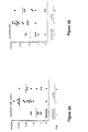
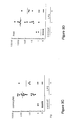
- FIG. 3 in four parts, FIGS. 3A, 3B, 3C and 3D, is a graphical representation which shows aqueous fluid leukocyte cell counts in Lewis rats treated with 100 ⁇ g LPS.
- Cell counts of the aqueous fluid (FIG. 3A) and of three types of infiltrating cells into the aqueous fluid (FIG. 3B, lymphocytes; FIG. 3C, monocytes; and FIG. 3D, PMN) are shown on a logarithmic scale on the y axis for controls (-) and for LPS before (-) and after (+) treatment with 100 mg/Kg aminoguanidine (Ag) on the x axis.
- Each filled in circle (•) represents the cell count from an individual rat.
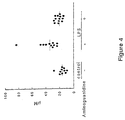
- FIG. 4 is a graphical representation which shows the effect of aminoguanidine on plasma nitrate/nitrite levels in Lewis rats treated with 100 ⁇ g LPS. Plasma nitrate/nitrite levels in ⁇ g are shown on the y axis for control (-) and for LPS before (-) and after (+) treatment with 100 mg/Kg aminoguanidine.
- Endotoxin-induced acute uveitis and generalized vascular leakage inhibition by aminoguanidine
- Rats Male Lewis rats ( ⁇ 200 g) were purchased from Harlan Sprague Dawley (Indianapolis, IN) and housed and cared for in accordance with the guidelines of the Washington University Committee for the Humane Care of Laboratory Animals and in accordance with NIH guidelines on laboratory animal welfare. Rats were housed individually, fed standard rat chow (Ralston Purina, Richmond, IN) and water ad libitum, and were on a 12 hour light/dark cycle. Aminoguanidine (hemisulfate), and lipopolysaccharide (LPS; Salmonella minnesota) were purchased from Sigma (St. Louis, MO). 125 I and 46 Sc microspheres were obtained from NEN Research Products (Boston, MA). 131 I was obtained from ICN Biomedicals, Inc. (Costa Mesa, CA).
- LPS was dissolved in sterile pyrogen-free 0.9% saline at concentrations of 1 ⁇ g/ ⁇ l, and unless stated otherwise, 50 ⁇ l was injected into each hind footpad (100 ⁇ g total dose per rat) using metofane anesthesia as described previously by other investigators [Herbort et al., Graefe's Arch. Clin. Exp. Ophthalmol. 226 , pp. 553-558 (1988); and Hoekzema et al., Invest. Ophthalmol. Vis. Sci. 33 , pp. 532-539 (1992)].
- Control rats received an equal volume of 0.9% saline. 100 mg/kg body weight aminoguanidine was injected subcutaneously in LPS-treated and in control rats at 0, 3, 6, 12, 18 and within 60 minutes of the initiation of the test, which corresponds to 20-24 hours after LPS injection.
- Regional vascular albumin permeation was quantified by use of an isotope dilution technique based on the injection of bovine serum albumin (BSA) labeled with 2 different iodine isotopes, 125 I and 131 I [Pugliese et al., Metabolism 39 pp. 690-697 (1990); Pugliese et al., Diabetes 39 , pp. 323-332 (1990); and Pugliese et al., Diabetes 39 , pp. 312-322 (1990)].
- BSA bovine serum albumin
- Rats were anesthetized with Inactin (Byk Gulden, Konstanz, FRG) ( ⁇ 100 mg/kg body weight injected i.p.), and core body temperature maintained at 37 ⁇ 0.5°C using heat lamps, a 37°C surgical tray, and a rectal temperature probe.
- the left femoral vein, left iliac artery, and right subclavian artery were cannulated with polyethylene tubing (0.58 mm i.d.) filled with heparinized saline (400 U heparin/ml).
- the femoral vein cannula was used for tracer injection and the subclavian artery cannula was connected to a pressure transducer for blood pressure monitoring.
- the left iliac artery was connected to a 1 ml syringe attached to a Harvard Model 940 constant withdrawal pump preset to withdraw at a constant rate of 0.055 ml/min.
- the trachea was intubated and connected to a small rodent respirator for continuous ventilatory support. Microspheres were injected into the left ventricle through a carotid artery cannula.
- the left eye was dissected as previously described [Pugliese et al., Diabetes 39 , pp. 323-332 (1990); and Pugliese et al., Diabetes 39 , pp. 312-322 (1990)] and all tissue samples and arterial plasma samples were weighed, then counted in a gamma spectrometer.
- a quantitative index of 125 I-BSA tissue clearance was calculated as previously described in [Pugliese et al., Metabolism 39 , pp. 690-697 (1990); Pugliese et al., Diabetes 39 , pp. 323-332 (1990); Pugliese et al., and Diabetes 39 , pp.
- 125 I-BSA tissue activity was corrected for tracer contained within the tissue vasculature by multiplying 125 I-BSA activity in the tissue by the ratio of 125 I-BSA/ 131 I-BSA activities in the arterial plasma sample obtained at the end of the test.
- the vascular-corrected 125 I-BSA tissue activity was divided by the time-averaged 125 I-BSA plasma activity (obtained from a well mixed sample of plasma taken from the withdrawal syringe) and by the tracer circulation time (10 min) and then normalized per g tissue wet weight.
- total activity of 46 Sc in each ocular tissue was divided by the activity of 46 Sc in the reference blood sample obtained from the withdrawal pump syringe, multiplied by the pump withdrawal rate, and expressed as ml/g tissue/min [Pugliese et al. Diabetes 39, pp. 323-332 (1990) and Pugliese et al., Diabetes 39, pp. 312-322 (1990)].
- Other non-ocular tissues aorta, kidney, skin, skeletal muscle, sciatic nerve
- LPS induced a generalized vascular dysfunction.
- Aqueous fluid was collected from the left eye only (the right carotid artery was ligated for the blood flow studies and this eye was not used for function studies) using a 100 ⁇ l heparinized capillary tube and placed in a microfuge tube. Well mixed aliquots (2 ⁇ l) were spread on a siliconized glass slide, air dried, stained with Wright's stain, and the total number of cells and a differential cell count were performed using a Leitz orthoplan light microscope.
- Anticoagulated (heparin) plasma samples were centrifuged at 7500 rpm for 1 hour at 4°C using a 10,000 molecular weight cut off Centricon filter (Amicon, Beverly, MA). Plasma nitrate was enzymatically reduced to nitrite using Aspergillus niger nitrate reductase (Sigma, St. Louis, MO).
- the sample was incubated with 40 ⁇ M NADPH and 14 mU of enzyme in a final volume of 50 ⁇ l of 20 mM Tris, pH 7.6; the reaction was terminated after 5 minutes at 20°C by dilution with 50 ⁇ l of water followed by addition of 10 ⁇ l of freshly prepared DAN reagent (0.05 mg/ml in 0.6 M HCl) for determination of nitrite.
- the DAN assay is a modification of the conventional method of Damiani and Burini for the fluorometric determination of nitrite [ Talanta 33, 649-652 (1986)].
- 2,3-Diaminonaphthalene is reacted with nitrite under acidic conditions to form 1-(H)-naphthotriazole, a fluorescent product. After a 10 minute incubation at 20°C, the reaction was terminated with 5 ⁇ l of 2.8 N NaOH, which maximizes the intensity of the fluorescent signal. Formation of the 2,3-diaminonaphthotriazole was measured using a Pandex (IDEXX Laboratories, Inc., Westbrook, ME) fluorescent plate reader with excitation at 365 nm and emission read at 450 nm with a gain setting at 100%. Plasma nitrite levels were calculated by first subtracting the value of the enzyme blank (nitrate reductase plus NADPH) from the experimental reading, then calculating the value using a standard curve for nitrite to which NADPH was added.
- IDEXX Laboratories, Inc., Westbrook, ME fluorescent plate reader with excitation at 365 nm and emission read at 450 n
- Table 1 shows changes in 125 I-albumin permeation resulting from the injection of 100 ⁇ g LPS (50 ⁇ g/footpad) into footpads of Lewis rats.
- the Van der Waerden test indicated highly significant group differences for 125 I-albumin leakage in the retina, anterior uvea, choroid/sclera, and aqueous fluid (p ⁇ 0.0001), while changes were absent in brain.
- aminoguanidine attenuated the LPS-induced 125 I-albumin leakage by ⁇ 75% in the retina, sciatic nerve, and aorta, by ⁇ 50% in the aqueous fluid and anterior uvea, and by ⁇ 30% in the posterior uvea, without affecting 125 I-albumin leakage in controls.
- Figure 2 shows changes in regional blood flows expressed as a fold increase over control values for LPS administered as a divided dose.
- Regional blood flows increased with increasing dose of LPS in the anterior and posterior uveal vasculatures and in the heart, plateaued in the retinal and sciatic nerve at 100 ⁇ g LPS, and was unaffected by LPS in the kidney when measured 20 hours after LPS injection.
- the Van der Waerden test indicated significant group differences for blood flow in the anterior uvea and choroid/sclera but not in the retina, sciatic nerve, brain, heart, and kidney (Table 2).
- LPS increased blood flow -40% in the anterior uvea and ⁇ 25% in the choroid-sclera and these increases were prevented by aminoguanidine.
- lymphocytes Figure 3B
- monocytes Figure 3C
- PMNs Figure 3D
- Aminoguanidine reduced the total number of cells in the aqueous fluid of LPS-treated rats, including ⁇ 90% reduction in the number of PMNs.
- Plasma nitrate/nitrite levels were 17.9 ⁇ 8.6 ⁇ M for controls and were increased ⁇ 65% in LPS-treated rats ( Figure 4). Aminoguanidine treatment prevented the LPS-induced increases in plasma nitrate/nitrite levels.
- aminoguanidine markedly attenuated manifestations of endotoxin-induced acute uveitis (vascular leakage, increased blood flow, and exudation of leukocytes into the aqueous fluid) as well as elevated plasma nitrate/nitrite levels.
- Aminoguanidine also significantly attenuated or completely prevented systemic effects of endotoxin on vascular leakage, i.e., edema (an important manifestation/consequence of inflammation) in nerve, aorta, and small intestine.
- LPS was injected at noon and the animals were sacrificed the following morning.
- a values are mean ⁇ SD
- LPS was injected at noon and the animals were sacrificed the following morning.
- a values are mean ⁇ SD
- Effects of LPS (100 ⁇ g) and aminoguanidine (100 mg/kg) on gravimetric and hemodynamic parameters control LPS LPS + aminoguanidine number of rats 5 6 7 body weight (g) 237 ⁇ 15 a 225 ⁇ 9 229 ⁇ 12 mean arterial blood pressure (mm Hg) 120 ⁇ 9 121 ⁇ 9 124 ⁇ 10 cardiac output (ml/min) 81 ⁇ 9 75 ⁇ 4 77 ⁇ 3 cardiac index (ml/min/100 g b.w.) 343 ⁇ 19 334 ⁇ 13 336 ⁇ 23 total peripheral resistance (mm Hg/ml/min) 1.42 ⁇ 0.10 1.55 ⁇ 0.14 1.54 ⁇ 0.11 G
- Endotoxin-induced acute uveitis and generalized vascular leakage inhibition by methylguanidine
- Rats Male Lewis rats (-200 g) were purchased from Harlan Sprague Dawley (Indianapolis, IN) and housed and cared for in accordance with the guidelines of the University Committee for the Humane Care of Laboratory Animals in accordance with NIH guidelines on laboratory animal welfare. Rats were housed individually, fed standard rat chow (Ralston Purina, Richmond, IN) and water ad libitum, and were on a 12 hour light/dark cycle. Methylguanidine (hydrochloride) and lipopolysaccharide (LPS; Salmonella minnesota) were purchased from Sigma (St. Louis, MO). 125 I was obtained from NEN Research Products (Boston, MA). 131 I was obtained from ICN Biomedicals, Inc. (Costa Mesa, CA).
- LPS was dissolved in sterile pyrogen-free 0.9% saline at concentrations of 1 ⁇ g/ ⁇ l, and 50 ⁇ l was injected into each hind footpad (100 ⁇ g total dose per rat) using metofane anesthesia as described previously by other investigators [Herbort et al., Graefe's Arch. Clin. Exp. Ophthalmol. 226, pp. 553-558 (1988); and Hoekzema et al., Invest. Ophthalmol. Vis. Sci. 33 , pp. 532-539 (1992)].
- Control rats received an equal volume of 0.9% saline.
- 50 mg/kg body weight methylguanidine was injected subcutaneously in LPS-treated and in control rats at 0, 3, 6, 12, 18 and 20-24 hours after LPS injection. Animals were killed for the vascular function studies 20 to 24 hours after LPS injection.
- 125 I-BSA was used to quantify vascular albumin filtration after 10 min of tracer circulation, while 131 I-BSA served as a plasma volume marker for correction of 125 I-BSA tissue activity for tracer contained within vessels.
- Purified monomer BSA (1 mg) was iodinated with 1 mCi of 131 I or 125 I by the iodogen method as previously described in Pugliese et al., Diabetes 39 pp. 323-332 (1990).
- Rats were anesthetized with Inactin (Byk Gulden Konstanz, FRG) (-100 mg/kg body weight injected i.p.), and core body temperature maintained at 37 ⁇ 0.5°C using heat lamps, a 37°C surgical tray, and a rectal temperature probe.
- the left femoral vein, left iliac artery, and right subclavian artery were cannulated with polyethylene tubing (0.58 mm i.d.) filled with heparinized saline (400 U heparin/ml).
- the femoral vein cannula were used for tracer injection and the subclavian artery cannula was connected to a pressure transducer for blood pressure monitoring.
- the left iliac artery was connected to a 1 ml syringe attached to a Harvard Model 940 constant withdrawal pump preset to withdraw at a constant rate of 0.055 ml/min.
- the trachea was intubated and connected to a small rodent respirator for continuous ventilatory support.
- the left eye was dissected as previously described [Pugliese et al., Diabetes 39 , pp. 323-332 (1990) and Pugliese et al., Diabetes 39 , pp. 312-322 (1990)] and all tissue samples and arterial plasma samples were weighed, then counted in a gamma spectrometer.
- a quantitative index of 125 I-BSA tissue clearance was calculated as previously described [Pugliese et al., Metabolism 39 , pp. 690-697 (1990); Pugliese et al., Diabetes 39 , pp. 323-332 (1990); and Pugliese et al., Diabetes 39 , pp.
- 125 I-BSA tissue activity was corrected for tracer contained within the tissue vasculature by multiplying 125 I-BSA activity in the tissue by the ratio of 125 I-BSA/ 131 I-BSA activities in the arterial plasma sample obtained at the end of the test.
- the vascular-corrected 125 I-BSA tissue activity was divided by the time-averaged 125 I-BSA plasma activity (obtained from a well mixed sample of plasma taken from the withdrawal syringe) and by the tracer circulation time (10 min) and then normalized per g tissue wet weight.
- Other non-ocular tissues (aorta, kidney, skin, skeletal muscle, sciatic nerve) also were sampled to assess if LPS induced a generalized vascular dysfunction.
- Table 4 shows changes in 125 I-albumin permeation resulting from the injection of 100 ⁇ g LPS (50 ⁇ g/footpad) into footpads of Lewis rats. Highly significant group differences for 125 I-albumin leakage were evident in the aqueous fluid, anterior uvea, choroid/sclera, retina, sciatic nerve, aorta, and kidney (p ⁇ 0.0001), while smaller increases were observed in the skin and skeletal muscle. No LPS-induced changes were present in brain, heart, or small intestine.
- methylguanidine attenuated the LPS-induced 125 I-albumin leakage in aqueous fluid, anterior uvea, and kidney and prevented the LPS-induced increases in albumin leakage in the choroid/sclera, retina, sciatic nerve, aorta, and skin.
- methylguanidine (at approximately one half of the dose of aminoguanidine used in Example 1) was as effective as (or more effective than) aminoguanidine in preventing endotoxin-induced vascular leakage (an important manifestation/consequence of inflammation) in uveal tissue as well as in the sciatic nerve, aorta, skeletal muscle, skin, and kidney.
- LPS was injected at noon and the animals were sacrificed the following morning. Significantly different from untreated controls: a p ⁇ 0.0001; b p ⁇ 0.005; c p ⁇ 0.05 Significantly different from LPS: d p ⁇ 0.001; e p ⁇ 0.005; f p ⁇ 0.01
- the inhibitors of nitric oxide formation described herein can be used for administration to warm blooded mammals by conventional means, preferably in formulations with pharmaceutically acceptable diluents and carriers.
- the amount of the active inhibitor to be administered must be an effective amount, that is, an amount which is medically beneficial but does not present toxic effects which overweigh the advantages which accompany its use. It would be expected that the adult human daily dosage would normally range upward from about one milligram per kilo of body weight of the drug.
- a suitable route of administration is orally in the form of capsules, tablets, syrups, elixirs and the like, although parenteral administration also can be used, e.g. intraveneously, intraperitoneally or subcutaneously.
- Intraveneous administration of the drug in aqueous solution such as physiologic saline is illustrative.
- Appropriate formulations of the drug in pharmaceutically acceptable diluents and carriers in therapeutic dosage form can be prepared by reference to general texts in the field such as, for example, Remington's Pharmaceutical Sciences , Ed. Arthur Osol. 16th ed., 1980, Mack Publishing Co., Easton, PA.
Landscapes
- Health & Medical Sciences (AREA)
- Animal Behavior & Ethology (AREA)
- Medicinal Chemistry (AREA)
- Pharmacology & Pharmacy (AREA)
- Life Sciences & Earth Sciences (AREA)
- General Health & Medical Sciences (AREA)
- Public Health (AREA)
- Veterinary Medicine (AREA)
- Chemical & Material Sciences (AREA)
- Epidemiology (AREA)
- General Chemical & Material Sciences (AREA)
- Organic Chemistry (AREA)
- Nuclear Medicine, Radiotherapy & Molecular Imaging (AREA)
- Chemical Kinetics & Catalysis (AREA)
- Bioinformatics & Cheminformatics (AREA)
- Engineering & Computer Science (AREA)
- Ophthalmology & Optometry (AREA)
- Pain & Pain Management (AREA)
- Rheumatology (AREA)
- Acyclic And Carbocyclic Compounds In Medicinal Compositions (AREA)
- Nitrogen And Oxygen Or Sulfur-Condensed Heterocyclic Ring Systems (AREA)
- Organic Low-Molecular-Weight Compounds And Preparation Thereof (AREA)
- Pharmaceuticals Containing Other Organic And Inorganic Compounds (AREA)
- Medicines That Contain Protein Lipid Enzymes And Other Medicines (AREA)
Abstract
Description
| Effects of LPS (100 µg) and aminoguanidine (100 mg/kg) on regional 125I-albumin permeation (µg plasma/min/g wet weight) | |||
| control | LPS | LPS + aminoguanidine | |
| number of rats eye | 14 | 19 | 11 |
| aqueous fluid | 65 ± 23a | 294 ± 81b | 204 ± 53b,f |
| anterior uvea | 270 ± 53 | 611 ± 110b | 423 ± 67b,e |
| posterior uvea | 258 ± 75 | 612 ± 110b | 497 ± 96b,f |
| retina | 66 ± 14 | 154 ± 32b | 86 ± 23e |
| sciatic nerve | 62 ± 14 | 167 ± 35b | 91 ± 20c,e |
| aorta | 75 ± 26 | 245 ± 64b | 114 ± 43d,e |
| skeletal muscle | 44 ± 17 | 62 ± 11 | 51 ± 10 |
| skin | 110 ± 27 | 209 ± 73b | 186 ± 49c |
| brain | 23 ± 11 | 24 ± 11 | 25 ± 15 |
| heart | 621 ± 57 | 585 ± 116 | 624 ± 86 |
| small intestine | 377 ± 112 | 567 ± 247d | 375 ± 1009 |
| kidney | 612 ± 212 | 1,173 ± 407b | 1,578 ± 251b,f |
a values are mean ± SD
Significantly different from controls by least square means analysis:
bp<0.0001; cp<0.001; dp<0.05
Significantly different from LPS-treated rats least square means analysis:
ep<0.0001; fp<0.005; gp<0.05
| Effects of LPS (100 µg) and aminoguanidine (100 mg/kg) on regional | |||
| blood flows (ml/min/g wet weight) | |||
| control | LPS | LPS + aminoguanidine | |
| number of rats eye | 11 | 13 | 9 |
| anterior uvea | 1.8 ± 0.4 | 2.5 ± 0.6b | 1.9 ± 0.3d |
| posterior uvea | 3.4 ± 0.6 | 4.3 ± 0.9c | 3.7 ± 0.4 |
| retina | 0.42 ± 0.03 | 0.45 ± 0.07 | 0.42 ± 0.04 |
| sciatic nerve | 0.07 ± 0.01 | 0.08 ± 0.03 | 0.08 ± 0.03 |
| brain | 0.6 ± 0.1 | 0.6 ± 0.1 | 0.6 ± 0.1 |
| heart | 4.9 ± 1.4 | 6.1 ± 1.2 | 5.4 + 1.7 |
| kidney | 4.8 ± 0.9 | 4.7 ± 0.6 | 4.9 ± 0.6 |
a values are mean ± SD
Significantly different from controls by least square means analysis:
bp<0.0001; cp<0.005
Significant different from LPS-treated rats least square means analysis:
dp<0.005
| Effects of LPS (100 µg) and aminoguanidine (100 mg/kg) on gravimetric and hemodynamic parameters | |||
| control | LPS | LPS + aminoguanidine | |
| number of rats | 5 | 6 | 7 |
| body weight (g) | 237 ± 15a | 225 ± 9 | 229 ± 12 |
| mean arterial blood pressure (mm Hg) | 120 ± 9 | 121 ± 9 | 124 ± 10 |
| cardiac output (ml/min) | 81 ± 9 | 75 ± 4 | 77 ± 3 |
| cardiac index (ml/min/100 g b.w.) | 343 ± 19 | 334 ± 13 | 336 ± 23 |
| total peripheral resistance (mm Hg/ml/min) | 1.42 ± 0.10 | 1.55 ± 0.14 | 1.54 ± 0.11 |
| GFR (ml/min/whole kidney) | 0.96 ± 0.07 | 0.97 ± 0.06 | 0.95 ± 0.14 |
| GFR (ml/min/g kidney) | 0.87 ± 0.07 | 0.90 ± 0.11 | 0.87 ± 0.15 |
| filtration fraction (GFR/renal blood flow) | 0.34 ± 0.08 | 0.34 ± 0.05 | 0.31 ± 0.06 |
| renal vascular resistance (mm Hg/ml/min) | 0.97 ± 0.07 | 0.97 ± 0.06 | 0.95 ± 0.14 |
a values are mean ± SD
| Effects of LPS and methylguanidine (mg) on regional 125I-albumin permeationa | |||
| control | LPS | LPS + Mg | |
| number of rats | 5 | 8 | 4 |
| aqueous | 154 ± 100 | 495 ± 283a | 335 ± 30c |
| anterior uvea | 266 ± 52 | 618 ± 106a | 383 ± 32c,e |
| choroid/sclera | 298 ± 67 | 582 ± 77a | 357 ± 50d |
| retina | 68 ± 15 | 168 ± 28a | 67 ± 7d |
| sciatic nerve | 71 ± 14 | 187 ± 32a | 69 ± 3d |
| aorta | 75 ± 24 | 211 ± 43a | 102 ± 8d |
| skeletal muscle | 54 ± 19 | 103 ± 46c | 63 ± 11 |
| skin | 99 ± 29 | 224 ± 65b | 106 ± 16f |
| | 20 ± 4 | 25 ± 6 | 19 ± 5 |
| heart | 623 ± 43 | 699 ± 58 | 629 ± 82 |
| small intestine | 414 ± 40 | 559 ± 147 | 423 ± 188 |
| kidney | 635 ± 239 | 1561 ± 221a | 1042 ± 253c,e |
Male, Lewis rats were treated with 100 µg LPS (50 µg in each hind footpad) ± 50 mg/kg
body weight methylguanidine hydrochloride (at the time of injection of LPS and 3, 6, 12, 18 and 20-24 hours thereafter). LPS was injected at noon and the animals were sacrificed the following morning.
Significantly different from untreated controls:
ap<0.0001; bp<0.005; cp<0.05
Significantly different from LPS:
dp<0.001; ep<0.005; fp<0.01
Claims (5)
- Use of aminoguanidine in the manufacture of a medicament for use in inhibiting nitric oxide production in a mammal to alleviate acute inflammation which is not immunologically-mediated.
- Use of aminoguanidine according to claim 1, wherein the acute inflammation is acute uveitis.
- Use of aminoguanidine according to claim 2, wherein the acute inflammation is caused by endotoxin.
- Use of aminoguanidine according to claim 1 or 2, wherein the acute inflammation is caused by vascular leakage.
- Use of aminoguanidine in the manufacture of a medicament for use in inhibiting nitric oxide production in a mammal to alleviate vascular leakage which is not immunologically-mediated.
Applications Claiming Priority (3)
| Application Number | Priority Date | Filing Date | Title |
|---|---|---|---|
| US110915 | 1993-08-24 | ||
| US08/110,915 US5358969A (en) | 1991-12-16 | 1993-08-24 | Method of inhibiting nitric oxide formation |
| PCT/US1994/009541 WO1995005811A1 (en) | 1993-08-24 | 1994-08-23 | Method of inhibiting nitric oxide formation |
Publications (3)
| Publication Number | Publication Date |
|---|---|
| EP0771195A1 EP0771195A1 (en) | 1997-05-07 |
| EP0771195A4 EP0771195A4 (en) | 1997-12-29 |
| EP0771195B1 true EP0771195B1 (en) | 2001-07-11 |
Family
ID=22335627
Family Applications (1)
| Application Number | Title | Priority Date | Filing Date |
|---|---|---|---|
| EP94926572A Expired - Lifetime EP0771195B1 (en) | 1993-08-24 | 1994-08-23 | Method of inhibiting nitric oxide formation |
Country Status (10)
| Country | Link |
|---|---|
| US (1) | US5358969A (en) |
| EP (1) | EP0771195B1 (en) |
| JP (1) | JPH09502703A (en) |
| AT (1) | ATE202929T1 (en) |
| AU (1) | AU688370B2 (en) |
| CA (1) | CA2169481C (en) |
| DE (1) | DE69427711T2 (en) |
| ES (1) | ES2161782T3 (en) |
| NZ (1) | NZ273167A (en) |
| WO (1) | WO1995005811A1 (en) |
Families Citing this family (15)
| Publication number | Priority date | Publication date | Assignee | Title |
|---|---|---|---|---|
| US5733524A (en) * | 1984-03-19 | 1998-03-31 | The Picower Institute For Medical Research | Methods and materials for the diagnosis and treatment of conditions such as stroke |
| US5700447A (en) * | 1992-05-21 | 1997-12-23 | The Picowder Institute For Medical Research | Methods and materials for the diagnosis and treatment of conditions such as stroke |
| US5801200A (en) * | 1984-03-19 | 1998-09-01 | The Picower Institute For Medical Research | Methods and materials for the diagnosis and treatment of conditions such as stroke |
| US5733933A (en) * | 1984-03-19 | 1998-03-31 | The Picower Institute For Medical Research | Methods and materials for the diagnosis and treatment of conditions such as stroke |
| US5710181A (en) * | 1991-12-16 | 1998-01-20 | Washington University | Inhibition of nitric oxide formation in inflammatory bowel disease |
| US5837738A (en) * | 1991-12-16 | 1998-11-17 | Washington University | Method of inhibiting nitric oxide formation |
| US5859058A (en) * | 1994-07-14 | 1999-01-12 | Glaxo Wellcome Inc. | Nitric oxide synthase inhibitors for inhibiting the production of airway mucas |
| US5583101A (en) * | 1994-07-15 | 1996-12-10 | Harvard College | Use of nitrogen oxide species and adducts to inhibit skeletal muscle contraction |
| FR2730930B1 (en) * | 1995-02-27 | 1997-04-04 | Oreal | USE OF NO-SYNTHASE INHIBITORS TO REDUCE THE IRRITANT SKIN EFFECT OF PRODUCTS USED IN THE COSMETIC OR PHARMACEUTICAL FIELD |
| US5780513A (en) * | 1996-08-22 | 1998-07-14 | Washington University | Method of inhibiting the release of bioactive IL-1 |
| US5789395A (en) * | 1996-08-30 | 1998-08-04 | The Research Foundation Of State University Of New York | Method of using tetracycline compounds for inhibition of endogenous nitric oxide production |
| WO1998033379A1 (en) * | 1997-02-04 | 1998-08-06 | The General Hospital Corporation | A novel method for treating epidermal or dermal conditions |
| BR9913526A (en) | 1998-09-08 | 2001-06-05 | Monsanto Co | Methods of treating osteoarthritis with inducible nitric oxide synthase inhibitors |
| WO2000032179A2 (en) | 1998-12-01 | 2000-06-08 | University Of Kentucky Research Foundation | Use of nicotonic acid derivatives for the treatment of dna damage in skin cells |
| WO2001091773A2 (en) | 2000-05-31 | 2001-12-06 | Encelle, Inc. | Method of stimulating hair growth |
Family Cites Families (10)
| Publication number | Priority date | Publication date | Assignee | Title |
|---|---|---|---|---|
| BE757219A (en) * | 1969-10-13 | 1971-03-16 | Interstop Ag | DISCHARGE DRAWER CLOSURE FOR METAL CASTING TANKS |
| US4544670A (en) * | 1982-08-24 | 1985-10-01 | William H. Rorer, Inc. | Method of treating coccidiosis with acyl guanidines |
| US4983604A (en) * | 1987-11-13 | 1991-01-08 | The Rockefeller University | Inhibitors of nonenzymatic cross-linking |
| US4665192A (en) * | 1984-03-19 | 1987-05-12 | The Rockefeller University | 2-(2-furoyl)-4(5)-2(furanyl)-1H-imidazole |
| US4758583A (en) * | 1984-03-19 | 1988-07-19 | The Rockefeller University | Method and agents for inhibiting protein aging |
| US5218001A (en) * | 1984-03-19 | 1993-06-08 | The Rockefeller University | Inhibitors of the advanced glycosylation of proteins and methods of use therefor |
| US4870210A (en) * | 1987-12-18 | 1989-09-26 | American Home Products Corporation | Aminoguanidine derivative as anti-inflammatory agents |
| DE4034722A1 (en) * | 1990-07-27 | 1992-04-30 | Mannesmann Ag | Introducing molten metal through immersion tube |
| US5246970A (en) * | 1991-12-16 | 1993-09-21 | Washington University | Method of inhibiting nitric oxide formation |
| CA2085399A1 (en) * | 1991-12-16 | 1993-06-17 | Joseph Robert Williamson | Method for inhibiting nitric oxide formation |
-
1993
- 1993-08-24 US US08/110,915 patent/US5358969A/en not_active Expired - Lifetime
-
1994
- 1994-08-23 EP EP94926572A patent/EP0771195B1/en not_active Expired - Lifetime
- 1994-08-23 AU AU76369/94A patent/AU688370B2/en not_active Ceased
- 1994-08-23 AT AT94926572T patent/ATE202929T1/en not_active IP Right Cessation
- 1994-08-23 ES ES94926572T patent/ES2161782T3/en not_active Expired - Lifetime
- 1994-08-23 NZ NZ273167A patent/NZ273167A/en not_active IP Right Cessation
- 1994-08-23 JP JP7507734A patent/JPH09502703A/en active Pending
- 1994-08-23 CA CA002169481A patent/CA2169481C/en not_active Expired - Fee Related
- 1994-08-23 DE DE69427711T patent/DE69427711T2/en not_active Expired - Fee Related
- 1994-08-23 WO PCT/US1994/009541 patent/WO1995005811A1/en not_active Ceased
Also Published As
| Publication number | Publication date |
|---|---|
| AU688370B2 (en) | 1998-03-12 |
| EP0771195A4 (en) | 1997-12-29 |
| JPH09502703A (en) | 1997-03-18 |
| US5358969A (en) | 1994-10-25 |
| DE69427711T2 (en) | 2002-05-08 |
| WO1995005811A1 (en) | 1995-03-02 |
| AU7636994A (en) | 1995-03-21 |
| CA2169481C (en) | 1999-11-16 |
| ES2161782T3 (en) | 2001-12-16 |
| NZ273167A (en) | 1999-08-30 |
| EP0771195A1 (en) | 1997-05-07 |
| ATE202929T1 (en) | 2001-07-15 |
| DE69427711D1 (en) | 2001-08-16 |
| CA2169481A1 (en) | 1995-03-02 |
Similar Documents
| Publication | Publication Date | Title |
|---|---|---|
| Tilton et al. | Endotoxin-induced uveitis in the rat is attenuated by inhibition of nitric oxide production. | |
| US5246971A (en) | Method of inhibiting nitric oxide formation | |
| CA2169481C (en) | Method of inhibiting nitric oxide formation | |
| US5246970A (en) | Method of inhibiting nitric oxide formation | |
| Hammes et al. | Aminoguanidine treatment inhibits the development of experimental diabetic retinopathy. | |
| Costantino et al. | Pharmacological approaches to the treatment of diabetic complications | |
| EP1021175B1 (en) | Compounds and methods for therapeutic intervention in preventing diabetic complications | |
| JP4033936B2 (en) | Nitric oxide production inhibitor | |
| WO1996012483A1 (en) | Method of inhibiting nitric oxide formation | |
| US20200093833A1 (en) | Methods and compositions for ibogaine treatment of impulse control disorder, anxiety-related disorders, violence and/or anger, or regulating food intake | |
| US6040326A (en) | 3-deoxyglucosone production inhibitor | |
| US5710181A (en) | Inhibition of nitric oxide formation in inflammatory bowel disease | |
| EP0547558B1 (en) | Use of aminoguanidine for the manufacture of a medicament for inhibiting nitric oxide formation | |
| US5837738A (en) | Method of inhibiting nitric oxide formation | |
| EP0543018B1 (en) | Antidiabetic agent | |
| WO2000066101A2 (en) | Method of inhibiting glycation product formation | |
| WO1991002810A1 (en) | Nmda oxidizing agents for protecting neurons from injury | |
| JPH07506568A (en) | Use of existing drugs to treat diabetes | |
| EP0199888A2 (en) | Control of heme and iron concentrations in body tissues | |
| JP2002528419A (en) | Compounds for diabetic complications and their therapeutic use | |
| JP2000281650A (en) | 2-mercapto-pyridine-N-oxide derivative / oxovanadium (IV) complex | |
| US20250302823A1 (en) | Compositions and methods for treating hepatic porphyrias with glycine transport inhibitors | |
| EP3441070A1 (en) | Drug for preventing or treating lactic acidosis | |
| TW201909902A (en) | Use and pharmaceutical composition for metabolic disease prevention and/or treatment | |
| HK40002147A (en) | Drug for preventing or treating lactic acidosis |
Legal Events
| Date | Code | Title | Description |
|---|---|---|---|
| PUAI | Public reference made under article 153(3) epc to a published international application that has entered the european phase |
Free format text: ORIGINAL CODE: 0009012 |
|
| 17P | Request for examination filed |
Effective date: 19960229 |
|
| AK | Designated contracting states |
Kind code of ref document: A1 Designated state(s): AT BE CH DE DK ES FR GB GR IE IT LI LU MC NL PT SE |
|
| A4 | Supplementary search report drawn up and despatched |
Effective date: 19971105 |
|
| AK | Designated contracting states |
Kind code of ref document: A4 Designated state(s): AT BE CH DE DK ES FR GB GR IE IT LI LU MC NL PT SE |
|
| 17Q | First examination report despatched |
Effective date: 19990614 |
|
| GRAG | Despatch of communication of intention to grant |
Free format text: ORIGINAL CODE: EPIDOS AGRA |
|
| GRAG | Despatch of communication of intention to grant |
Free format text: ORIGINAL CODE: EPIDOS AGRA |
|
| GRAH | Despatch of communication of intention to grant a patent |
Free format text: ORIGINAL CODE: EPIDOS IGRA |
|
| GRAH | Despatch of communication of intention to grant a patent |
Free format text: ORIGINAL CODE: EPIDOS IGRA |
|
| GRAA | (expected) grant |
Free format text: ORIGINAL CODE: 0009210 |
|
| AK | Designated contracting states |
Kind code of ref document: B1 Designated state(s): AT BE CH DE DK ES FR GB GR IE IT LI LU MC NL PT SE |
|
| PG25 | Lapsed in a contracting state [announced via postgrant information from national office to epo] |
Ref country code: NL Free format text: LAPSE BECAUSE OF FAILURE TO SUBMIT A TRANSLATION OF THE DESCRIPTION OR TO PAY THE FEE WITHIN THE PRESCRIBED TIME-LIMIT Effective date: 20010711 Ref country code: LI Free format text: LAPSE BECAUSE OF FAILURE TO SUBMIT A TRANSLATION OF THE DESCRIPTION OR TO PAY THE FEE WITHIN THE PRESCRIBED TIME-LIMIT Effective date: 20010711 Ref country code: CH Free format text: LAPSE BECAUSE OF FAILURE TO SUBMIT A TRANSLATION OF THE DESCRIPTION OR TO PAY THE FEE WITHIN THE PRESCRIBED TIME-LIMIT Effective date: 20010711 Ref country code: BE Free format text: LAPSE BECAUSE OF FAILURE TO SUBMIT A TRANSLATION OF THE DESCRIPTION OR TO PAY THE FEE WITHIN THE PRESCRIBED TIME-LIMIT Effective date: 20010711 Ref country code: AT Free format text: LAPSE BECAUSE OF FAILURE TO SUBMIT A TRANSLATION OF THE DESCRIPTION OR TO PAY THE FEE WITHIN THE PRESCRIBED TIME-LIMIT Effective date: 20010711 |
|
| REF | Corresponds to: |
Ref document number: 202929 Country of ref document: AT Date of ref document: 20010715 Kind code of ref document: T |
|
| REG | Reference to a national code |
Ref country code: CH Ref legal event code: EP |
|
| REF | Corresponds to: |
Ref document number: 69427711 Country of ref document: DE Date of ref document: 20010816 |
|
| PGFP | Annual fee paid to national office [announced via postgrant information from national office to epo] |
Ref country code: DE Payment date: 20010820 Year of fee payment: 8 |
|
| PGFP | Annual fee paid to national office [announced via postgrant information from national office to epo] |
Ref country code: GB Payment date: 20010822 Year of fee payment: 8 |
|
| REG | Reference to a national code |
Ref country code: IE Ref legal event code: FG4D |
|
| PG25 | Lapsed in a contracting state [announced via postgrant information from national office to epo] |
Ref country code: LU Free format text: LAPSE BECAUSE OF NON-PAYMENT OF DUE FEES Effective date: 20010823 Ref country code: IE Free format text: LAPSE BECAUSE OF NON-PAYMENT OF DUE FEES Effective date: 20010823 |
|
| PGFP | Annual fee paid to national office [announced via postgrant information from national office to epo] |
Ref country code: FR Payment date: 20010825 Year of fee payment: 8 |
|
| ITF | It: translation for a ep patent filed | ||
| PGFP | Annual fee paid to national office [announced via postgrant information from national office to epo] |
Ref country code: ES Payment date: 20010920 Year of fee payment: 8 |
|
| PG25 | Lapsed in a contracting state [announced via postgrant information from national office to epo] |
Ref country code: SE Free format text: LAPSE BECAUSE OF FAILURE TO SUBMIT A TRANSLATION OF THE DESCRIPTION OR TO PAY THE FEE WITHIN THE PRESCRIBED TIME-LIMIT Effective date: 20011011 Ref country code: PT Free format text: LAPSE BECAUSE OF FAILURE TO SUBMIT A TRANSLATION OF THE DESCRIPTION OR TO PAY THE FEE WITHIN THE PRESCRIBED TIME-LIMIT Effective date: 20011011 Ref country code: DK Free format text: LAPSE BECAUSE OF FAILURE TO SUBMIT A TRANSLATION OF THE DESCRIPTION OR TO PAY THE FEE WITHIN THE PRESCRIBED TIME-LIMIT Effective date: 20011011 |
|
| PG25 | Lapsed in a contracting state [announced via postgrant information from national office to epo] |
Ref country code: GR Free format text: LAPSE BECAUSE OF FAILURE TO SUBMIT A TRANSLATION OF THE DESCRIPTION OR TO PAY THE FEE WITHIN THE PRESCRIBED TIME-LIMIT Effective date: 20011012 |
|
| ET | Fr: translation filed | ||
| NLV1 | Nl: lapsed or annulled due to failure to fulfill the requirements of art. 29p and 29m of the patents act | ||
| REG | Reference to a national code |
Ref country code: ES Ref legal event code: FG2A Ref document number: 2161782 Country of ref document: ES Kind code of ref document: T3 |
|
| REG | Reference to a national code |
Ref country code: GB Ref legal event code: IF02 |
|
| REG | Reference to a national code |
Ref country code: CH Ref legal event code: PL |
|
| PG25 | Lapsed in a contracting state [announced via postgrant information from national office to epo] |
Ref country code: MC Free format text: LAPSE BECAUSE OF NON-PAYMENT OF DUE FEES Effective date: 20020301 |
|
| PLBE | No opposition filed within time limit |
Free format text: ORIGINAL CODE: 0009261 |
|
| STAA | Information on the status of an ep patent application or granted ep patent |
Free format text: STATUS: NO OPPOSITION FILED WITHIN TIME LIMIT |
|
| REG | Reference to a national code |
Ref country code: IE Ref legal event code: MM4A |
|
| 26N | No opposition filed | ||
| PG25 | Lapsed in a contracting state [announced via postgrant information from national office to epo] |
Ref country code: GB Free format text: LAPSE BECAUSE OF NON-PAYMENT OF DUE FEES Effective date: 20020823 |
|
| PG25 | Lapsed in a contracting state [announced via postgrant information from national office to epo] |
Ref country code: ES Free format text: LAPSE BECAUSE OF NON-PAYMENT OF DUE FEES Effective date: 20020824 |
|
| PG25 | Lapsed in a contracting state [announced via postgrant information from national office to epo] |
Ref country code: DE Free format text: LAPSE BECAUSE OF NON-PAYMENT OF DUE FEES Effective date: 20030301 |
|
| GBPC | Gb: european patent ceased through non-payment of renewal fee |
Effective date: 20020823 |
|
| PG25 | Lapsed in a contracting state [announced via postgrant information from national office to epo] |
Ref country code: FR Free format text: LAPSE BECAUSE OF NON-PAYMENT OF DUE FEES Effective date: 20030430 |
|
| REG | Reference to a national code |
Ref country code: FR Ref legal event code: ST |
|
| REG | Reference to a national code |
Ref country code: ES Ref legal event code: FD2A Effective date: 20030912 |
|
| PG25 | Lapsed in a contracting state [announced via postgrant information from national office to epo] |
Ref country code: IT Free format text: LAPSE BECAUSE OF NON-PAYMENT OF DUE FEES Effective date: 20050823 |