EP0749482B1 - Recombinant humanized anti-lewis y antibodies - Google Patents
Recombinant humanized anti-lewis y antibodies Download PDFInfo
- Publication number
- EP0749482B1 EP0749482B1 EP95912894A EP95912894A EP0749482B1 EP 0749482 B1 EP0749482 B1 EP 0749482B1 EP 95912894 A EP95912894 A EP 95912894A EP 95912894 A EP95912894 A EP 95912894A EP 0749482 B1 EP0749482 B1 EP 0749482B1
- Authority
- EP
- European Patent Office
- Prior art keywords
- antibody
- lewis
- antibodies
- humanized
- antigen
- Prior art date
- Legal status (The legal status is an assumption and is not a legal conclusion. Google has not performed a legal analysis and makes no representation as to the accuracy of the status listed.)
- Expired - Lifetime
Links
Images


Classifications
-
- C—CHEMISTRY; METALLURGY
- C07—ORGANIC CHEMISTRY
- C07K—PEPTIDES
- C07K16/00—Immunoglobulins [IG], e.g. monoclonal or polyclonal antibodies
- C07K16/18—Immunoglobulins [IG], e.g. monoclonal or polyclonal antibodies against material from animals or humans
- C07K16/34—Immunoglobulins [IG], e.g. monoclonal or polyclonal antibodies against material from animals or humans against blood group antigens
-
- C—CHEMISTRY; METALLURGY
- C07—ORGANIC CHEMISTRY
- C07K—PEPTIDES
- C07K16/00—Immunoglobulins [IG], e.g. monoclonal or polyclonal antibodies
- C07K16/18—Immunoglobulins [IG], e.g. monoclonal or polyclonal antibodies against material from animals or humans
- C07K16/28—Immunoglobulins [IG], e.g. monoclonal or polyclonal antibodies against material from animals or humans against receptors, cell surface antigens or cell surface determinants
- C07K16/30—Immunoglobulins [IG], e.g. monoclonal or polyclonal antibodies against material from animals or humans against receptors, cell surface antigens or cell surface determinants from tumour cells
-
- C—CHEMISTRY; METALLURGY
- C07—ORGANIC CHEMISTRY
- C07K—PEPTIDES
- C07K2317/00—Immunoglobulins specific features
- C07K2317/20—Immunoglobulins specific features characterized by taxonomic origin
- C07K2317/24—Immunoglobulins specific features characterized by taxonomic origin containing regions, domains or residues from different species, e.g. chimeric, humanized or veneered
-
- C—CHEMISTRY; METALLURGY
- C07—ORGANIC CHEMISTRY
- C07K—PEPTIDES
- C07K2319/00—Fusion polypeptide
Definitions
- the present invention is related to the field of molecular biology, and more particularly to humanized antibodies.
- the present invention relates to the generation, by recombinant DNA methods, of novel immunoglobulins specific for the Lewis Y antigen, using, as a starting point, a murine monoclonal antibody against the same antigen.
- Transformation of a normal cell to a malignant cell is often accompanied by a change in the expression of cell surface antigens.
- These different phenotypes can be detected using monoclonal antibodies specific for such antigens.
- different cancer cells can be detected and characterised (Lloyd, K.O. (1983) "Human Tumour Antigens: Detection and Characterisation with Monoclonal Antibodies” in R.B. Herberman, ed., Basic and Clinical Tumour Immunology, pp 159-214, Martinus Nijhoff, Boston).
- Lewis Y is a carbohydrate antigen with the structure
- the Lewis Y antigen is expressed in normal tissues but the level of expression is higher in certain tumour types so that the antigen can be used as a marker for cells of some breast, colon, gastric, esophageal, pancreatic, duodenal, lung, bladder and renal carcinomas and gastric and islet cell neuroendocrine tumours. Its presence on some tumour cells is not accompanied by an increase in its serum levels, thus administered Lewis Y specific antibody is not significantly bound by soluble antigen.
- a murine monoclonal antibody reactive against the Lewis Y antigen has potential in the imaging and treatment of certain tumours in man, this potential may be difficult to realise due to the xenogenic nature of the antibody.
- the problems associated with this are two-fold.
- First, administered xenogenic antibodies are likely to be immunogenic (Bruggemann et al (1989) J. Exp. Med. 170. 2153-2157). In this case, this would cause a human anti-mouse antibody (HAMA) response (Schroff, R. et al (1985) Cancer Res. 45, 879-885), resulting in rapid clearance of the antibody from the circulation.
- HAMA human anti-mouse antibody
- EP120694 Celltech
- EP125023 Genetech
- Such chimaeric antibodies have the advantage since they retain the specificity of the murine antibody but can also stimulate human Fc dependant complement fixation and cell-mediated cytotoxicity.
- the murine variable regions can still elicit a HAMA response (Bruggemann, M. et al (1989) J.Exp.Med. 170 , 2153-2157) thereby limiting the value of chimaeric antibodies as diagnostic and therapeutic agents.
- British Patent Application Number GB2188638A discloses the process of humanization in which only the antigen binding-loops are transferred to a human antibody template (for example Riechmann et al ., Nature , 332 , 323-327; Tempest et al ., (1991) Bio/Technology 9 , 266-271).
- a human antibody template for example Riechmann et al ., Nature , 332 , 323-327; Tempest et al ., (1991) Bio/Technology 9 , 266-271.
- CDRs complementary-determining regions
- Each binding site is formed, in the most part, from three heavy chain and three light chain CDRs, although framework residues can interact with antigen, either directly or indirectly, by altering the CDR conformation.
- 'Reshaped' or 'humanized' antibodies made by the process of CDR-grafting retain the human constant regions necessary for the stimulation of human Fc-dependant effector functions and have less murine content than chimaeric antibodies. Consequently, humanized antibodies are less likely than chimaeric antibodies to evoke a KAMA response when administered to humans, their half-life in circulation should approach that of natural human antibodies, thereby enhancing their diagnostic and therapeutic value.
- the present invention provides the novel humanized monoclonal antibody 35193 HuVHT/HuVKF specific for the Lewis Y antigen. This has been achieved by the generation of a murine monoclonal antibody reactive to the Lewis Y antigen, followed by utilisation of its variable domain structures in the design of the recombinant antibodies. Prior to the work of the inventors, it was not known that 3S193 or any other non-human antibody specific for the the Lewis Y antigen could be humanized so as to retain useful binding specificity.
- Figures 1 and 2 show the nucleotide and amino acid sequences of the heavy (figure 1) and kappa (light) chain (figure 2) variable regions of murine 3S193.
- the CDRs are boxed and residues dictated by PCR primer are underlined.
- Figures 3 (heavy chain) and 4 (light chain) give the nucleotide sequences of the DNA fragments encoding the basic humanized variable regions and their polypeptide products.
- the CDRs are boxed.
- One aspect of the invention is to provide humanized antibodies specific for the Lewis Y antigen.
- Another aspect of the invention is to provide polynucleotides encoding humanized antibodies specific for the Lewis Y antigen.
- Various expression vectors comprising polynucleotides joined to promoter sequences are also provided.
- another aspect of the invention is host cells transformed with expression vectors for the expression of humanized Lewis Y specific antibodies.
- Another aspect of the invention is to provide humanized Lewis Y specific antibodies that are labeled with a detectable label or a therapeutic label.
- Another aspect of the invention is the use of the particular humarised Lewis Y specific antibody in the preparation of a composition for treating and/or diagnosing cancer by administering a composition comprising a humanized Lewis Y specific antibody.
- One method of detecting cancer cells involves the steps of administering a labeled antibody (detectable label) to a patient and subsequently detecting where in the body the labeled antibody has bound.
- humanized antibody refers to a molecule that has its CDRs (complementarily determining regions) derived from a non-human species immunoglobulin and the remainder of the antibody molecule derived mainly from a human immunoglobulin.
- antibody as used herein, unless indicated otherwise, is used broadly to refer to both antibody molecules and a variety of antibody derived molecules.
- Such antibody derived molecules comprise at least one variable region (either a heavy chain of light chain variable region) and include molecules such as Fab fragments, Fab' fragments, F(ab') 2 fragments, Fd fragments, Sc antibodies (single chain antibodies), diabodies, individual antibody light chains, individual antibody heavy chains, chimeric fusions between antibody chains and other molecules, and the like.
- conventional molecular biology methods refers to techniques for manipulating polynucleotides that are well known to the person of ordinary skill in the art of molecular biology. Examples of such well known techniques can be found in Molecular Cloning: A Laboratory Manual 2nd Edition , Sambrook et al , Cold Spring Harbor, NY (1989). Examples of conventional molecular biology techniques include, but are not limited to, in vitro ligation, restriction endonuclease digestion, PCR, cellular transformation, hybridization, electrophoresis, DNA sequencing, and the like.
- variable region as used herein in reference to immunoglobulin molecules has the ordinary meaning given to the term by the person of ordinary skill in the act of immunology. Both antibody heavy chains and antibody light chains may be divided into a "variable region” and a "constant region". The point of division between a variable region and a heavy region may readily be determined by the person of ordinary skill in the art by reference to standard texts describing antibody structure, e.g., Kabat et al "Sequences of Proteins of Immunological Interest: 5th Edition" U.S. Department of Health and Human Services, U.S. Government Printing Office (1991).
- the present invention provides humanized antibody molecules specific for Lewis Y antigen in which at least parts of the CDRs of the heavy and/or light chain variable regions of a human antibody (the receptor antibody) have been substituted by analogous parts of CDRs of a murine monoclonal antibody and the humanized antibody can specifically bind to the same as the Lewis Y antibody.
- the CDR regions of the humanized Lewis Y specific antibody are derived from the murine antibody 3S193.
- the humanized antibodies described herein contain some alterations of the acceptor antibody, i.e., human, heavy and/or light chain variable domain framework regions that are necessary for retaining binding specificity of the donor monoclonal antibody.
- the framework region of some embodiments of the humanized antibodies described herein does not necessarily consist of the precise amino acid sequence of the framework region of a natural occurring human antibody variable region, but contains various substitutions that improve the binding properties of a humanized antibody region that is specific for the same target as the murine antibody 3S193.
- a minimal number of substitutions are made to the framework region in order to avoid large-scale introductions of non-human framework residues and to ensure minimal immunogenicity of the humanized antibody.
- the prefered donor monoclonal antibody of the present invention is the murine antibody 3S193, which is specific for the human Lewis Y cancer antigen.
- the humanized antibodies of the subject invention may be produced by a variety of methods useful for the production of polypeptides, e.g.
- the humanized antibodies are produced by recombinant DNA technology.
- the present invention also relates to the DNA and protein sequence coding for such recombinant antibodies.
- the invention also provides vectors for the production of the recombinant antibodies in mammalian cell hosts.
- the subject invention provides for polynucleotides encoding humanized Lewis Y specific antibodies.
- the subject polynucleotides may have a wide variety of sequences because of the degeneracy of the genetic code.
- a person of ordinary skill in the art may readily change a given polynucleotide sequence encoding a humanized Lewis Y specific antibody into a different polynucleotide encoding the same humanized Lewis Y specific antibody embodiment.
- the polynucleotide sequence encoding the antibody may be varied to take into account factors affecting expression such as coding frequency, RNA secondary structure, and the like.
- the recombinant humanized Lewis Y specific antibodies of the invention may be produced by the following process or other recombinant protein expression methods:
- Host cells may be cotransfected with two expression vectors of the invention, the first vector containing an operon encoding a heavy chain derived polypeptide and the second containing an operon encoding a light chain derived polypeptide.
- the two vectors may contain different selectable markers but, with the exception of the heavy and light chain coding sequences, are preferably identical. This procedure provides for equal expression of heavy and light chain polypeptides.
- a single vector may be used which encodes both heavy and light chain polypeptides.
- the coding sequences for the heavy and light chains may comprise cDNA or genomic DNA or both.
- the humanized Lewis Y specific antibodies of the invention may be produced using recombinant immunoglobulin expression technology.
- the host cell used to express the recombinant antibody of the invention may be either a bacterial cell such as Escherichia coli , or preferably a eukaryotic cell.
- a mammalian cell such as a chinese hamster ovary cell or myeloma, may be used.
- the choice of expression vector is dependent upon the choice of host cell, and may be selected so as to have the desired expression and regulatory characteristics in the selected host cell.
- the general methods for construction of the vector of the invention, transfection of cells to produce the host cell of the invention, culture of cells to produce the antibody of the invention are all conventional molecular biology methods.
- the recombinant antibodies of the invention may be purified by standard procedures of the art, including cross-flow filtration, ammonium sulphate precipitation, affinity column chromatography, gel electrophoresis and the like.
- the humanized antibodies of the present invention may comprise a complete antibody molecule having full length heavy and light chains, or any fragment thereof, such as the Fab or (Fab') 2 fragments, a heavy chain and light chain dimer, or any minimal fragment thereof such as a Fv, an SCA (single chain antibody), and the like, specific for the Lewis Y antigen molecule.
- the humanized Lewis Y specific antibodies of the present invention may be used in conjunction with, or attached to other antibodies (or parts thereof) such as human or humanized monoclonal antibodies. These other antibodies may be reactive with other markers (epitopes) characteristic for the disease against which the antibodies of the invention are directed or may have different specificities chosen, for example, to recruit molecules or cells of the human immune system to the diseased cells.
- the antibodies of the invention (or parts thereof) may be administered with such antibodies (or parts thereof) as separately administered compositions or as a single composition with the two agents linked by conventional chemical or by molecular biological methods.
- the diagnostic and therapeutic value of the antibodies of the invention may be augmented by labelling the humanized antibodies with labels that produce a detectable signal (either in vitro or in vivo) or with a label having a therapeutic property.
- Some labels e.g. radionucleotides may produce a detectable signal and have a therapeutic property. Examples of radionuclide labels include 125 I, 131 I, 14 C.
- Examples of other detectable labels include a fluorescent chromophore such as fluorescein, phycobiliprotein or tetraethyl rhodamine for fluorescence microscopy, an enzyme which produces a fluorescent or colored product for detection by fluorescence, absorbance, visible color or agglutination, which produces an electron dense product for demonstration by electron microscopy; or an electron dense molecule such as ferritin, peroxidase or gold beads for direct or indirect electron microscopic visualization.
- Labels having therapeutic properties include drugs for the treatment of cancer, such as methotrexate and the like.
- the subject invention also provides the use of the particular humanised Lewis Y antibody in the preparation of a composition for treating and/or detecting cancer cells, by the administration of humanized Lewis Y specific antibodies, either labelled or unlabelled, to a patient.
- One method of detecting cancer cells in a human involves the step of administering a labeled humanized Lewis Y specific antibody (labelled with a detectable label) to a human and subsequently detecting bound labeled antibody by the presence of the label.
- the recombinant antibodies of this invention may also be used for the selection and/or isolation of human monoclonal antibodies, and the design and synthesis of peptide or non-peptide compounds (mimetics) which would be useful for the same diagnostic and therapeutic applications as the antibodies (e.g. Saragovi et al., (1991) Science 253 :792-795).
- the antibodies are typically administered in a composition comprising a pharmaceutical carrier.
- a pharmaceutical carrier can be any compatible, non-toxic substance suitable for delivery of the monoclonal antibodies to the patient, Sterile water, alcohol, fats, waxes, and inert solids may be included in the carrier.
- Pharmaceutically accepted adjuvants may also be incorporated into the pharmaceutical composition.
- compositions of the invention may be administered to a patient in a variety of ways.
- the pharmaceutical compositions may be administered parenterally, i.e., subcutaneously, intramuscularly or intravenously.
- this invention provides compositions for parenteral administration which comprise a solution of the human monoclonal antibody or a cocktail thereof dissolved in an acceptable carrier, preferably an aqueous carrier.
- an acceptable carrier preferably an aqueous carrier.
- aqueous carriers can be used, e.g., water, buffered water, 0.4% saline, 0.3% glycine and the like. These solutions are sterile and generally free of particulate matter.
- These compositions may be sterilized by conventional, well known sterilization techniques.
- compositions may contain pharmaceutically acceptable auxiliary substances as required to approximate physiological conditions such as pH adjusting and buffering agents, toxicity adjusting agents and the like, for example sodium acetate, sodium chloride, potassium chloride, calcium chloride, sodium lactate, etc.
- concentration of antibody in these formulations can vary widely, e.g., from less than about 0.5%, usually at or at least about 1% to as much as 15 or 20% by weight and will be selected primarily based on fluid volumes, viscosities, etc., in accordance with the particular mode of administration selected.
- the subject invention provide numerous humanized antibodies specific for the Lewis Y antigen based on the discovery that the CDR regions of the murine monoclonal antibody could be spliced into a human acceptor framework so as to produce a humanized recombinant antibody specific for the Lewis Y antigen.
- Preferred humanized Lewis Y specific antibodies contain additional change in the framework region (or in other regions) to increasing binding for Lewis Y antigen.
- Particularly preferred embodiments of the invention are the exmplified humanized antibody molecules that have superior antigen binding properties.
- the murine monoclonal antibody, 3S193, was produced using standard techniques following immunisation of BALB/c mice with Lewis Y-bearing tumour cells.
- variable regions sequences were determined from heavy and light chain cDNAs which were synthesised from cytoplasmic RNA, essentially as described by Tempest et al ., loc cit .
- cytoplasmic RNA was isolated from 1.5 x 10 7 3S193-producing hybridoma cells by the method of Favalora et al ., (1980) Meth Enzymol, 65 , 718-749. Supernatant obtained from the culture of these cells was assayed for the presence of antibody by solid-phase ELISA using an Inno-Lia mouse monoclonal antibody isotyping kit (Innogenetics, Antwerp, Belgium). The antibody was confirmed to be IgG 3 /k by this method.
- Reverse transcription of 3S193 RNA was initatiated from pirmers based on the 5' end of either the murine IgG 3 (CG3FOR 5'TTAAGCTTAGACAGATGGGGCTGTTGTTGT 3') (SEQ ID NO: 1) or the murine kappa (CK2FOR 5' GGAAGCTTGAAGATGGATACAGTTGGTGCAGC 3') (SEQ ID NO: 2) constant regions genes.
- the reactions consisted of 5 ⁇ g RNA, 0.5 ⁇ M CG3FOR or CK2FOR, 250 ⁇ M each dATP, dCTP, dGTP and dTTP, 50mM Tris-HCl pH 8.3, 75mM KCl, 10mM DTT, 3mM MgCl 2 , 30 units RNase inhibitor (Pharmacia Milton Keynes, United Kingdom) in 50 ⁇ l total volume. Samples were heated at 70°C for 10 min and then cooled at 37°C over 30 min. 100 units M-MLV reverse transcriptase (Life Technologies, Paisley, United Kingdom) was added and the reaction allowed to proceed at 37°C for 1 hour.
- variable region cDNAs were amplifed by the PCR (Saiki et al ., (1988) Science, 239 , 498-491,) using variable regions primers, including those described by Orlandi et al ., (1989) Proc. Natl. Acad. Sci. USA. 86 , 3833-3837, and signal sequence primers, derived from those of Jones and Bendig, (1991) Bio/Technology, 9 , 88-89, as well as the constant region primers which were involved in the first strand cDNA synthesis.
- Several primers combinations gave amplification products of the expected size.
- the additional primers used in the successful PCR amplifications were based on conserved regions at the 5' end of either murine VH (VH1BACK 5'AGGTSMARCTGCAGSAGTCWGG 3') (SEQ ID NO: 3) or murine VK (VK1BACK 5'GACATTCAGCTGACCCAGTCTCCA 3') (SEQ ID NO: 4) genes or were designed from a subset of signal sequence genes of murine heavy chains (SH2BACK 5' TGGAATTCATGRACTTCDGGYTCAACTKRRTTT 3') (SEQ ID NO: 5).
- the reactions thus contained 5 ⁇ g RNA/cDNA hybrid, 0.5 ⁇ M CG2FOR and 0.5 ⁇ m VH1BACK or SH2BACK.
- 5 ⁇ l RNA/cDNA hybrid was amplified with 0.5 ⁇ M each CK2FOR and VK1BACK.
- each reaction contained 250 ⁇ M each dATP, dCTP, dGTP and dTTP, 10mM Tris-HCl pH 8.3, 50mM KCl, 1.5mM MgCl 2 , 0.01% (w/v) gelatin, 0.01% (v/v) Tween 20, 0.01% v/v) NP-40 and 3 units AmpliTaq (Perkin Elmer Catus, Beaconsfield, UK). Amplification was over 25 cycles of 94°C, 30s; 50 or 55°C, 30s; 72°C, 45s plus 72°C, 5 min to end.
- VH product sizes were approximately 400bp (CG3FOR, VH1BACK) and 440 bp (VH1FOR, SH2BACK) as visualized by agarose gel electrophoresis.
- the VK products were of about 370bp.
- the DNAs were purified on Prep-A-Gene matrix (Bio-Rad, Hemel Hempstead, UK) after excision from agarose gel.
- the CG3FOR, VH1BACK and CG2FOR, SH2BACK products were cloned into M13mp18 and M13mp19 (Pharmacia Milton Keynes, United Kingdom) using the restriction sites incorporated into the PCR primers and, for the CG3FOR, SH2BACK product, a Pst I site internal to the VH fragment.
- Clones were sequenced by the dideoxy method (Sanger et al ., (1977) Proc. Natl. Acad. Sci. USA 74 5463-5467) using T7 DNA polymearse (Pharmacia, Milton Keynes, United Kingdom)
- Clones were determined to contain VH inserts by comparison with known VH sequences. VH inserts corresponded to only one VH and represented both fragment orientations.
- the complete VH nucleotide sequence and the amino acid sequence derived by translation of the DNA sequence information are shown in figure 1.
- the extents of the CDRs, as indicated by the boxes, were determined as defined by Kabat et al ., (1991) Sequences of proteins of Immunological Interest (5th edition),U.S. Department of Health and Human Services.
- the amino acid residue numbering system of Kabat et al. , ibid is used to indicate specific amino acid residues in both the murine and human varaible regions in this application.
- VK1BACK product was similary cloned and, from the clones sequenced, two inserts were found which corresponded to an identical VK. The remaining inserts were not VK-like. Two additional amplification primers were created in order to obtain further VK clones.
- the framework 4 sequence of the inserts already obtained was used to design the oligonucleotide VK4FOR (5' TTAAGCTTTTATTTCCAACTTTGTCCC3 ') (SEQ ID NO:6).
- VK10BACK 5'TTGAATTCCAGTGATGTTTTGATGACCCA3'
- VK4FOR and VK10BACK were used in the amplification of CK2FOR-primed first strand cDNA under the conditions described above. This lead to the isolation of several inserts representing the putative 3S193 VK in both fragment orientations.
- the 3S193 VK nucleotide sequence and its amino acid translation are shown in figure 2.
- the CDRs are boxed.
- the sequence at the 3' end is the genuine 3S193 sequence as obtained from CK1FOR, VK1BACK product whereas residues at the 5' end are dictated by the VK10BACK primer and are shown underlined.
- a chimaeric antibody consisting of murine variable and human constant regions, is not necessary to the humanization process but can be useful as its ability to bind antigen can suggest that the correct VH and VK have been cloned from the cDNA of the hybridoma and the antibody can be used as a control in assays to assess the efficacy of the humanized antibodies.
- the VH insert from an M13 clone was amplified using VH1FOR and VH1BACK to introduce the required restriction sites.
- the 50 ⁇ l reaction mixture contained approximately 100ng M13 ssDNA, 0.5 ⁇ M each primer, 250 ⁇ M each dATP, dCTP, dGTP and dTTP, 10mM Tris HCl pH8.3, 50mM KCl 1.5mM MgCl 2 , 0.01% (v/v) Tween-20, 0.01% (w/v) gelatin, 0.01% (v/v) NP-40 and 2 units Thermalase DNA polymerase (IBI Limited, Cambridge, UK).
- the sample was subjected to 15 thermal cycles of 94°C, 30s; 50°C, 30s; 72°C, 1 min followed by an additional 5 min at 72°C.
- the product was digested with PstI and BstEII and cloned into M13VHPCR1 (Orlandi et al ., loc cit).
- the reaction mixtures contained about 100ng M13 ssDNA, 0.5 ⁇ M each primer, 250 ⁇ m each dATP, dCTP dGTP and dTTP, 20mM Tris HCl pH8.8, 10mM KCl 10mM (NH 4 ) 2 SO 4 , 2mM MgSO 4 , 0.01% (v/v) Triton X-100 and 1 unit Vent DNA polymerase in 50 ⁇ l total volume. Amplification took place over 15 cycles of 94°C. 30s; 50°C, 30s; 75°C, 1 min plus 75°C, 5 min to end.
- VK fragment was cloned into pSVhyg (Orlandi et al ., loc cit .), already containing the human kappa constant region gene (Hieter et al. (1992) Cell., 22 197-207).
- the vectors were cotransfected into the rat myeloma YB2/0 (Kilmartin et al . (1982) J. Cell Biol, 93 , 576-582, and into the mouse myeloma Sp2/0-Ag 14 (Schulman et al ., (1978) Nature 276, 269-270), both available from the American Type Culture Collection, Rockville, MD, USA as previously described (Tempest et al ., loc cit ).
- Mycophenolic acid resistant clones screened by ELISA for secretion of human IgG/kappa antibody.
- ELISA positive clones were expanded and antibody purified from culture medium by protein A affinity chromatography.
- Humanized variable domains are created by transferring the antigen-binding loops of the parent antibody to human variable regions frameworks.
- the frameworks used in the case of 3S193 were those of KOL VH (Marquart et al ., (1980) J. Mol. Biol . 141 , 513-524), and REI VK (Epp et al. (1974) Eur. J. Biochem 45, 513-524).
- the basic 3S193 humanized kappa chain HuVK (figure 4) contained only the CDRs of its murine counterpart.
- the 3S193 humanized variable region genes were generated from M13 phage DNAs containing a heavy or kappa variable region gene comprising the required human frameworks and irrelevant CDRs by site-directed mutagenesis.
- the M13 phage were grown in E.coli RZ1032 (dut ung - ) to give single-stranded template DNA containing uracil in place of thymine.
- 0.5 ⁇ g template DNA was mixed with 1 pmol universal M13 forward primer and 1 pmol each phosphorylated mutageneic oligonucleotides in 20 ⁇ l 40mM Tris HCl pH7.5, 20mM MgCl 2 , 50mM NaCl.
- the oligonucleotides were annealed to the template by heating at 80°C for 5 min and cooling slowly at room temperature.
- the mutageneic oligonucleotides were:
- the template utilised for the kappa chain variable region mutagenesis actually encoded framework regions which were related but not identical to REI and the mutagenesis reaction eliminated these discrepancies (utilizing oligonucleotides not described) as well as introducing the 3S193 CDRs.
- the only discrepancy to be discussed specifically herein is at position 71 of the template which encoded a phenylalanine residue not present in REI sequence. This residue was retained in the 3S193 HuVKF but was changed to the REI residue in the 3S193 HuVK in figure using the oligonucleotide REI Y71:
- the murine 3S193 CDR3 and the humanized template CDR3 were identical, so alteration of CDR3 was not required. Limited differences between the mouse and humanized template CDRs 1 and 2 did require alteration of the humanized template CDRs 1 and 2 and the mutagenizing oligonucleotides utilised were:
- dATP, dCTP, dGTP and dTTP were added to 250 ⁇ M final concentration, DTT to 7mM, ATP to 1mM with 0.5 units T7 DNA polymearse (United States Biochemical, Cleveland, OH, USA) and 0.5 units T4 DNA ligase (Life Technologies, Paisley, UK) in the same buffer.
- the 30 ⁇ l reaction was incubated at room temperature, 1h and the DNA ethanol precipitated .
- the DNA was dissolved in 50 ⁇ l 60mM Tris HCl pH8.0, 1mM EDTA, 1mM DTT, 0.1mg/ml BSA containing 1 unit uracil DNA glycosylase (Boehringer Mannheim, Lewis, Hampshire, UK) and incubated 37°c, 1h before NaOH was added to 0.2M and incubation continued at room temperature for 5 min.
- the DNA was ethanol precipitated, dissolved in 20 ⁇ l TE and the insert fragment amplified by PCR.
- the reaction mixture contained 2 ⁇ l mutant DNA, 0.5 ⁇ M each M13 forward and reverse primers, 250 ⁇ M each of dATP, dCTP, dGTP and dTTP, 10mM Tris HCI pH8.3, 50mM KCl, 1.5mM MgCl 2 , 0.01% Tween-20, 0.01% gelatin, 0.01% NP-40 and 2 units Thermalase (IBI, Cambridge, UK) in 50 ⁇ l. Amplification was achieved with 15 cycles of 94°C, 30s; 50°C, 30s; 72°C, 1 min; ending with 72°C, 5 min.
- the product DNAs were cloned into M13mp19 as HindIII-BamHI fragments. Representative clones were sequenced. The HindIII - BamHI fragments were excised from RF DNA of accurately-mutated clones and transferred to the pSVgpt and pSVhyg expression vectors as described in Example 2.
- the initial pair of reaction mixtures contained - 100ng single-stranded M13 DNA, 0.5 ⁇ M mutagenic primer, 0.5 ⁇ M universal pUC/M13 primer and these, and the subsequent joining reactions, were carried out using Vent DNA polymearse as described in Example 2.
- the HindIII BamHI fragments were cloned, sequenced and re-cloned for expression as already described.
- the transfection of myeloma cells and their selection and expansion was carried out as Example 2.
- humanized antibodies such HuVH/HuVK and HuVH/HuVKF
- chimaeric and humanized antibody chain expression vectors were co-transfected to give mixed antibodies which would allow the efficacy of the humanized chains to be examined individually.
- the HuVHT, HuVHA, HuVHAS, and HuVHASY DNAs were transfected with the HuVHF vector.
- the HuVHT/HuVKF combination was transfected into NS0 mouse myeloma cells (Galfe and Milstein, (1981) Mehods in Enzymology, Volume 73B , pp3-75, Academic Press: available from the European Collection of Animal Cell Cultures, Porton Down, Salisbury, UK) in additio to YB2/0 cells).
- the recombinant antibodies were tested by several methods.
- Recombinant antibodies were tested in mixed hemadsorption rosetting assays (Rettig et al . (1987) J. Immunol 138 , 4484-4489; Rettig et al ., (1985) Cancer Res. 45 . 815-821) for their ability to bind to MCF-7 target cells (human breast line) and to negative control Effron melanoma cells.
- the cells were grown in 60-well Terasaki plates to form confluent monolayers. Cells were washed twice with PBS/0.5% BSA and 10 ⁇ l of antibody added to the cells (antibodies serially diluted in DMEM without foetal calf serum).
- the cells were plated at approximately 100 cells/well in medium and grown overnight. The wells were emptied and dilution of test antibody added in medium. The plates were incubated 37°C, 5% CO 2 for 45min and then 10 ⁇ l human serum/medium (1:3) added to each well. After 4h the plates were rinsed twice with absolute methanol, fixed with methanol for 10min, rinsed in distilled water, stained with 2% Giemsa stain in PBS for 25 min, and rinsed again in distilled water. Plates were analyzed under the light microscope and the percent cytoxicity of a given antibody dilution was calculated as follows: (1 - No of cells in well treated with antibody & complement) No of cells in well treated with culture medium only x100(%)
- Some of the recombinant antibodies showed cross-reactivity to Lewis Y-related antigens. To see whether this would cause hemolysis, the antibodies were incubated with 51 Cr-labeled erythrocytes in the present of human serum (Nayayarna et al . (1978) Proc. Natl. Acad. Sci. USA 76 3486). None of the recombinant antibodies caused cell lysis detectable above a background for cells incubated with PBS alone. This suggests that the cross-reactivity of therapeutically or diagnostically administrated antibody would not itself cause a problem although it would be preferable to use an antibody specific for the Lewis-Y antigen to ensure that the maximum amount of antibody is available for binding to the target cells.
- Table 4 provides the amino acid sequences of the varaible regions of the the 3S193 heavy and light chains. Additionally, Table 4 provides the amino acid sequences of the variable regions of several humanized antibody chains derived from 3S193.
- Values are the minimum antibody concentrations ( ⁇ g/ml) which show at least 50% lysis using MCF-7 as the target cell line.
Landscapes
- Chemical & Material Sciences (AREA)
- Health & Medical Sciences (AREA)
- Organic Chemistry (AREA)
- Immunology (AREA)
- Life Sciences & Earth Sciences (AREA)
- Proteomics, Peptides & Aminoacids (AREA)
- Biophysics (AREA)
- General Health & Medical Sciences (AREA)
- Genetics & Genomics (AREA)
- Medicinal Chemistry (AREA)
- Molecular Biology (AREA)
- Biochemistry (AREA)
- Hematology (AREA)
- Cell Biology (AREA)
- Peptides Or Proteins (AREA)
- Preparation Of Compounds By Using Micro-Organisms (AREA)
- Medicines Containing Antibodies Or Antigens For Use As Internal Diagnostic Agents (AREA)
- Micro-Organisms Or Cultivation Processes Thereof (AREA)
- Medicines That Contain Protein Lipid Enzymes And Other Medicines (AREA)
Abstract
Description
- The present invention is related to the field of molecular biology, and more particularly to humanized antibodies.
- The present invention relates to the generation, by recombinant DNA methods, of novel immunoglobulins specific for the Lewis Y antigen, using, as a starting point, a murine monoclonal antibody against the same antigen.
- Transformation of a normal cell to a malignant cell is often accompanied by a change in the expression of cell surface antigens. These different phenotypes can be detected using monoclonal antibodies specific for such antigens. In this way, different cancer cells can be detected and characterised (Lloyd, K.O. (1983) "Human Tumour Antigens: Detection and Characterisation with Monoclonal Antibodies" in R.B. Herberman, ed., Basic and Clinical Tumour Immunology, pp 159-214, Martinus Nijhoff, Boston). Lewis Y is a carbohydrate antigen with the structure
- Fucαl→2Galβ1→4 [Fucα1→3] GlcNAcβ1→3R (Abe et al. (1983) J. Biol. Chem. 258 11793-11797). The Lewis Y antigen is expressed in normal tissues but the level of expression is higher in certain tumour types so that the antigen can be used as a marker for cells of some breast, colon, gastric, esophageal, pancreatic, duodenal, lung, bladder and renal carcinomas and
gastric and islet cell neuroendocrine tumours. Its presence on some tumour cells is not accompanied by an increase in its serum levels, thus administered Lewis Y specific antibody is not significantly bound by soluble antigen. - Although a murine monoclonal antibody reactive against the Lewis Y antigen has potential in the imaging and treatment of certain tumours in man, this potential may be difficult to realise due to the xenogenic nature of the antibody. The problems associated with this are two-fold. First, administered xenogenic antibodies are likely to be immunogenic (Bruggemann et al (1989) J. Exp. Med. 170. 2153-2157). In this case, this would cause a human anti-mouse antibody (HAMA) response (Schroff, R. et al (1985) Cancer Res. 45, 879-885), resulting in rapid clearance of the antibody from the circulation. Second, depending upon the isotypes involved, a murine antibody may be less efficacious than a human counterpart in the stimulation of human complement or cell-mediated cytotoxicity.
- To secure the advantage of a human antibody, whilst making use of the antigen-binding properties of an antibody raised in a different species, workers have made use of recombinant DNA techniques. EP120694 (Celltech) and EP125023 (Genentech) disclose the development of 'chimaeric' antibodies which comprise the variable regions of an antibody from another species and the constant regions of a human antibody. Such chimaeric antibodies have the advantage since they retain the specificity of the murine antibody but can also stimulate human Fc dependant complement fixation and cell-mediated cytotoxicity. However, the murine variable regions can still elicit a HAMA response (Bruggemann, M. et al (1989) J.Exp.Med. 170, 2153-2157) thereby limiting the value of chimaeric antibodies as diagnostic and therapeutic agents.
- British Patent Application Number GB2188638A (Winter) discloses the process of humanization in which only the antigen binding-loops are transferred to a human antibody template (for example Riechmann et al., Nature, 332, 323-327; Tempest et al., (1991) Bio/Technology 9, 266-271). These loops, known as complementary-determining regions (CDRs), are mounted on a scaffold - the frameworks regions. Together these make up the variable domains. Each binding site is formed, in the most part, from three heavy chain and three light chain CDRs, although framework residues can interact with antigen, either directly or indirectly, by altering the CDR conformation. 'Reshaped' or 'humanized' antibodies made by the process of CDR-grafting retain the human constant regions necessary for the stimulation of human Fc-dependant effector functions and have less murine content than chimaeric antibodies. Consequently, humanized antibodies are less likely than chimaeric antibodies to evoke a KAMA response when administered to humans, their half-life in circulation should approach that of natural human antibodies, thereby enhancing their diagnostic and therapeutic value.
- In practice, for the generation of efficacious humanized antibodies retaining the specificity of the original murine antibody, it is not usually sufficient simply to substitute CDRs. There is a requirement for the inclusion of a small number of critical murine antibody residues in the human variable region frameworks. The identity of these residues depends on the structure of both the original murine antibody and the acceptor human, antibody.
- The present invention provides the novel humanized monoclonal antibody 35193 HuVHT/HuVKF specific for the Lewis Y antigen. This has been achieved by the generation of a murine monoclonal antibody reactive to the Lewis Y antigen, followed by utilisation of its variable domain structures in the design of the recombinant antibodies. Prior to the work of the inventors, it was not known that 3S193 or any other non-human antibody specific for the the Lewis Y antigen could be humanized so as to retain useful binding specificity.
- Figures 1 and 2 show the nucleotide and amino acid sequences of the heavy (figure 1) and kappa (light) chain (figure 2) variable regions of murine 3S193. The CDRs are boxed and residues dictated by PCR primer are underlined.
- Figures 3 (heavy chain) and 4 (light chain) give the nucleotide sequences of the DNA fragments encoding the basic humanized variable regions and their polypeptide products. The CDRs are boxed.
- One aspect of the invention is to provide humanized antibodies specific for the Lewis Y antigen.
- Another aspect of the invention is to provide polynucleotides encoding humanized antibodies specific for the Lewis Y antigen. Various expression vectors comprising polynucleotides joined to promoter sequences are also provided. Similarly, another aspect of the invention is host cells transformed with expression vectors for the expression of humanized Lewis Y specific antibodies.
- Another aspect of the invention is to provide humanized Lewis Y specific antibodies that are labeled with a detectable label or a therapeutic label.
- Another aspect of the invention is the use of the particular humarised Lewis Y specific antibody in the preparation of a composition for treating and/or diagnosing cancer by administering a composition comprising a humanized Lewis Y specific antibody. One method of detecting cancer cells involves the steps of administering a labeled antibody (detectable label) to a patient and subsequently detecting where in the body the labeled antibody has bound.
- As used herein, the term "humanized" antibody refers to a molecule that has its CDRs (complementarily determining regions) derived from a non-human species immunoglobulin and the remainder of the antibody molecule derived mainly from a human immunoglobulin. The term "antibody" as used herein, unless indicated otherwise, is used broadly to refer to both antibody molecules and a variety of antibody derived molecules. Such antibody derived molecules comprise at least one variable region (either a heavy chain of light chain variable region) and include molecules such as Fab fragments, Fab' fragments, F(ab')2 fragments, Fd fragments, Sc antibodies (single chain antibodies), diabodies, individual antibody light chains, individual antibody heavy chains, chimeric fusions between antibody chains and other molecules, and the like.
- The term "conventional molecular biology methods" refers to techniques for manipulating polynucleotides that are well known to the person of ordinary skill in the art of molecular biology. Examples of such well known techniques can be found in Molecular Cloning: A Laboratory Manual 2nd Edition, Sambrook et al, Cold Spring Harbor, NY (1989). Examples of conventional molecular biology techniques include, but are not limited to, in vitro ligation, restriction endonuclease digestion, PCR, cellular transformation, hybridization, electrophoresis, DNA sequencing, and the like.
- The term "variable region" as used herein in reference to immunoglobulin molecules has the ordinary meaning given to the term by the person of ordinary skill in the act of immunology. Both antibody heavy chains and antibody light chains may be divided into a "variable region" and a "constant region". The point of division between a variable region and a heavy region may readily be determined by the person of ordinary skill in the art by reference to standard texts describing antibody structure, e.g., Kabat et al "Sequences of Proteins of Immunological Interest: 5th Edition" U.S. Department of Health and Human Services, U.S. Government Printing Office (1991).
- The present invention provides humanized antibody molecules specific for Lewis Y antigen in which at least parts of the CDRs of the heavy and/or light chain variable regions of a human antibody (the receptor antibody) have been substituted by analogous parts of CDRs of a murine monoclonal antibody and the humanized antibody can specifically bind to the same as the Lewis Y antibody. In a preferred embodiment of the subject invention, the CDR regions of the humanized Lewis Y specific antibody are derived from the murine antibody 3S193. The humanized antibodies described herein contain some alterations of the acceptor antibody, i.e., human, heavy and/or light chain variable domain framework regions that are necessary for retaining binding specificity of the donor monoclonal antibody. In other words, the framework region of some embodiments of the humanized antibodies described herein does not necessarily consist of the precise amino acid sequence of the framework region of a natural occurring human antibody variable region, but contains various substitutions that improve the binding properties of a humanized antibody region that is specific for the same target as the murine antibody 3S193. A minimal number of substitutions are made to the framework region in order to avoid large-scale introductions of non-human framework residues and to ensure minimal immunogenicity of the humanized antibody. The prefered donor monoclonal antibody of the present invention is the murine antibody 3S193, which is specific for the human Lewis Y cancer antigen. The humanized antibodies of the subject invention may be produced by a variety of methods useful for the production of polypeptides, e.g. in vitro synthesis, recombinant DNA production, and the like. Preferably, the humanized antibodies are produced by recombinant DNA technology. The present invention also relates to the DNA and protein sequence coding for such recombinant antibodies. The invention also provides vectors for the production of the recombinant antibodies in mammalian cell hosts.
- In addition to providing for humanized Lewis Y specific antibodies, the subject invention provides for polynucleotides encoding humanized Lewis Y specific antibodies. The subject polynucleotides may have a wide variety of sequences because of the degeneracy of the genetic code. A person of ordinary skill in the art may readily change a given polynucleotide sequence encoding a humanized Lewis Y specific antibody into a different polynucleotide encoding the same humanized Lewis Y specific antibody embodiment. The polynucleotide sequence encoding the antibody may be varied to take into account factors affecting expression such as coding frequency, RNA secondary structure, and the like.
- The recombinant humanized Lewis Y specific antibodies of the invention may be produced by the following process or other recombinant protein expression methods:
- a. Constructing, by conventional molecular biology methods, an expression vector comprising an operon that encodes an antibody heavy chain in which the CDRs and a minimal portion of the variable region framework that are required to retain donor antibody binding specificity are derived from a non-human immunoglobulin, such as the murine Lewis Y monoclonal antibody, and the remainder of the antibody is derived from a human immunoglobulin, thereby producing a vector for the expression of a humanized antibody heavy chain.
- b. Constructing, by conventional molecular biology methods, an expression vector comprising an operon that encodes an antibody light chain in which the CDRs and a minimal portion of the variable region framework that are required to retain donor antibody binding specificity are derived from a non-human immunoglobulin, such as the murine Lewis Y monoclonal antibody, and the remainder of the antibody is derived from a human immunoglobulin, thereby producing a vector for the expression of humanized antibody light chain.
- c. Transferring the expression vectors to a host cell by conventional molecular biology methods to produce a transfected host cell for the expression of humanized Lewis Y specific antibodies.
- d. Culturing the transfected cell by conventional molecular biology methods so as to produce humanized Lewis Y specific antibodies.
-
- Host cells may be cotransfected with two expression vectors of the invention, the first vector containing an operon encoding a heavy chain derived polypeptide and the second containing an operon encoding a light chain derived polypeptide. The two vectors may contain different selectable markers but, with the exception of the heavy and light chain coding sequences, are preferably identical. This procedure provides for equal expression of heavy and light chain polypeptides. Alternatively, a single vector may be used which encodes both heavy and light chain polypeptides. The coding sequences for the heavy and light chains may comprise cDNA or genomic DNA or both.
- The humanized Lewis Y specific antibodies of the invention may be produced using recombinant immunoglobulin expression technology.
- The recombinant production of immunoglobulin molecules, including humanized antibodies are described in U.S. patent 4,816,397 (Boss et al), U.S. patent 4,816,567 (Cabilly et al) U.K. patent GB 2,188,638 (Winter et al). and U.K. patent GB 2,209,757. Techniques for the recombinant expression of immunoglobulins, including humanized immunoglobulins, can also be found, among other places in Goeddel et al, Gene Expression Technology Methods in Enzymology Vol. 185 Academic Press (1991), and Borreback, Antibody Engineering, W.H. Freeman (1992). Additional information concerning the generation, design and expression of recombinant antibodies can be found in Mayforth, Designing Antibodies, Academic Press, San Diego (1993).
- The host cell used to express the recombinant antibody of the invention may be either a bacterial cell such as Escherichia coli, or preferably a eukaryotic cell. Preferably a mammalian cell such as a chinese hamster ovary cell or myeloma, may be used. The choice of expression vector is dependent upon the choice of host cell, and may be selected so as to have the desired expression and regulatory characteristics in the selected host cell.
- The general methods for construction of the vector of the invention, transfection of cells to produce the host cell of the invention, culture of cells to produce the antibody of the invention are all conventional molecular biology methods. Likewise, once produced, the recombinant antibodies of the invention may be purified by standard procedures of the art, including cross-flow filtration, ammonium sulphate precipitation, affinity column chromatography, gel electrophoresis and the like.
- The humanized antibodies of the present invention may comprise a complete antibody molecule having full length heavy and light chains, or any fragment thereof, such as the Fab or (Fab')2 fragments, a heavy chain and light chain dimer, or any minimal fragment thereof such as a Fv, an SCA (single chain antibody), and the like, specific for the Lewis Y antigen molecule.
- The humanized Lewis Y specific antibodies of the present invention may be used in conjunction with, or attached to other antibodies (or parts thereof) such as human or humanized monoclonal antibodies. These other antibodies may be reactive with other markers (epitopes) characteristic for the disease against which the antibodies of the invention are directed or may have different specificities chosen, for example, to recruit molecules or cells of the human immune system to the diseased cells. The antibodies of the invention (or parts thereof) may be administered with such antibodies (or parts thereof) as separately administered compositions or as a single composition with the two agents linked by conventional chemical or by molecular biological methods. Additionally the diagnostic and therapeutic value of the antibodies of the invention may be augmented by labelling the humanized antibodies with labels that produce a detectable signal (either in vitro or in vivo) or with a label having a therapeutic property. Some labels, e.g. radionucleotides may produce a detectable signal and have a therapeutic property. Examples of radionuclide labels include 125I, 131I, 14C. Examples of other detectable labels include a fluorescent chromophore such as fluorescein, phycobiliprotein or tetraethyl rhodamine for fluorescence microscopy, an enzyme which produces a fluorescent or colored product for detection by fluorescence, absorbance, visible color or agglutination, which produces an electron dense product for demonstration by electron microscopy; or an electron dense molecule such as ferritin, peroxidase or gold beads for direct or indirect electron microscopic visualization. Labels having therapeutic properties include drugs for the treatment of cancer, such as methotrexate and the like.
- The subject invention also provides the use of the particular humanised Lewis Y antibody in the preparation of a composition for treating and/or detecting cancer cells, by the administration of humanized Lewis Y specific antibodies, either labelled or unlabelled, to a patient. One method of detecting cancer cells in a human involves the step of administering a labeled humanized Lewis Y specific antibody (labelled with a detectable label) to a human and subsequently detecting bound labeled antibody by the presence of the label.
- The recombinant antibodies of this invention may also be used for the selection and/or isolation of human monoclonal antibodies, and the design and synthesis of peptide or non-peptide compounds (mimetics) which would be useful for the same diagnostic and therapeutic applications as the antibodies (e.g. Saragovi et al., (1991) Science 253:792-795).
- When the humanized Lewis Y specific antibodies of the invention are used in vitro, the antibodies are typically administered in a composition comprising a pharmaceutical carrier. A pharmaceutical carrier can be any compatible, non-toxic substance suitable for delivery of the monoclonal antibodies to the patient, Sterile water, alcohol, fats, waxes, and inert solids may be included in the carrier. Pharmaceutically accepted adjuvants (buffering agents, dispersing agent) may also be incorporated into the pharmaceutical composition.
- The humanized antibodies compositions of the invention may be administered to a patient in a variety of ways. Preferably, the pharmaceutical compositions may be administered parenterally, i.e., subcutaneously, intramuscularly or intravenously. Thus, this invention provides compositions for parenteral administration which comprise a solution of the human monoclonal antibody or a cocktail thereof dissolved in an acceptable carrier, preferably an aqueous carrier. A variety of aqueous carriers can be used, e.g., water, buffered water, 0.4% saline, 0.3% glycine and the like. These solutions are sterile and generally free of particulate matter. These compositions may be sterilized by conventional, well known sterilization techniques. The compositions may contain pharmaceutically acceptable auxiliary substances as required to approximate physiological conditions such as pH adjusting and buffering agents, toxicity adjusting agents and the like, for example sodium acetate, sodium chloride, potassium chloride, calcium chloride, sodium lactate, etc. The concentration of antibody in these formulations can vary widely, e.g., from less than about 0.5%, usually at or at least about 1% to as much as 15 or 20% by weight and will be selected primarily based on fluid volumes, viscosities, etc., in accordance with the particular mode of administration selected.
- Actual methods for preparing parenterally administrable compositions and adjustments necessary for administration to subjects will be known or apparent to those skilled in the art and are described in more detail in, for example, Remington's Pharmaceutical Science, 15th Ed., Mack Publishing Company, Easton, Pa (1980), which is incorporated herein by reference.
- The subject invention provide numerous humanized antibodies specific for the Lewis Y antigen based on the discovery that the CDR regions of the murine monoclonal antibody could be spliced into a human acceptor framework so as to produce a humanized recombinant antibody specific for the Lewis Y antigen. Preferred humanized Lewis Y specific antibodies contain additional change in the framework region (or in other regions) to increasing binding for Lewis Y antigen. Particularly preferred embodiments of the invention are the exmplified humanized antibody molecules that have superior antigen binding properties.
- The following examples are offered by way of illustration of the invention.
- Unless otherwise indicated, all general recombinant DNA methodology was performed as described in "Molecular Cloning, A Laboratory Manual" (1989) Eds J. Sambrook el al., published by Cold Spring Harbour Laboratory Press, Cold Spring Harbour, New York and enzymes were purchased from Life Technologies (Paisley, United Kingdom).
- In the following examples these abbreviations may be employed:
- dCTP
- deoxycytidine triphosphate
- dATP
- deoxyadenosine triphosphate
- dGTP
- deoxyguanosine triphosphate
- dTTP
- deoxythymidine triphosphate
- DTT
- dithiothreitol
- C
- cytosine
- A
- adenine
- G
- guanine
- T
- thymine
- PBS
- phosphate buffered saline
- PBST
- phosphate buffered saline containing 0.05% (v/v) Tween - 20
- The murine monoclonal antibody, 3S193, was produced using standard techniques following immunisation of BALB/c mice with Lewis Y-bearing tumour cells.
- The variable regions sequences were determined from heavy and light chain cDNAs which were synthesised from cytoplasmic RNA, essentially as described by Tempest et al., loc cit.
- Approximately 300µg cytoplasmic RNA was isolated from 1.5 x 10⁷ 3S193-producing hybridoma cells by the method of Favalora et al., (1980) Meth Enzymol, 65, 718-749. Supernatant obtained from the culture of these cells was assayed for the presence of antibody by solid-phase ELISA using an Inno-Lia mouse monoclonal antibody isotyping kit (Innogenetics, Antwerp, Belgium). The antibody was confirmed to be IgG3/k by this method.
- Reverse transcription of 3S193 RNA was initatiated from pirmers based on the 5' end of either the murine IgG3 (CG3FOR 5'TTAAGCTTAGACAGATGGGGCTGTTGTTGT 3') (SEQ ID NO: 1) or the murine kappa (CK2FOR 5' GGAAGCTTGAAGATGGATACAGTTGGTGCAGC 3') (SEQ ID NO: 2) constant regions genes. The reactions consisted of 5µg RNA, 0.5µM CG3FOR or CK2FOR, 250µM each dATP, dCTP, dGTP and dTTP, 50mM Tris-HCl pH 8.3, 75mM KCl, 10mM DTT, 3mM MgCl2, 30 units RNase inhibitor (Pharmacia Milton Keynes, United Kingdom) in 50µl total volume. Samples were heated at 70°C for 10 min and then cooled at 37°C over 30 min. 100 units M-MLV reverse transcriptase (Life Technologies, Paisley, United Kingdom) was added and the reaction allowed to proceed at 37°C for 1 hour.
- The murine variable region cDNAs were amplifed by the PCR (Saiki et al., (1988) Science, 239, 498-491,) using variable regions primers, including those described by Orlandi et al., (1989) Proc. Natl. Acad. Sci. USA. 86, 3833-3837, and signal sequence primers, derived from those of Jones and Bendig, (1991) Bio/Technology, 9, 88-89, as well as the constant region primers which were involved in the first strand cDNA synthesis. Several primers combinations gave amplification products of the expected size. The additional primers used in the successful PCR amplifications were based on conserved regions at the 5' end of either murine VH (VH1BACK 5'AGGTSMARCTGCAGSAGTCWGG 3') (SEQ ID NO: 3) or murine VK (VK1BACK 5'GACATTCAGCTGACCCAGTCTCCA 3') (SEQ ID NO: 4) genes or were designed from a subset of signal sequence genes of murine heavy chains (SH2BACK 5' TGGAATTCATGRACTTCDGGYTCAACTKRRTTT 3') (SEQ ID NO: 5). For PCR amplification of the VH, the reactions thus contained 5µg RNA/cDNA hybrid, 0.5µM CG2FOR and 0.5µm VH1BACK or SH2BACK. For the VK, 5µl RNA/cDNA hybrid was amplified with 0.5µM each CK2FOR and VK1BACK. In addition, each reaction contained 250µM each dATP, dCTP, dGTP and dTTP, 10mM Tris-HCl pH 8.3, 50mM KCl, 1.5mM MgCl2, 0.01% (w/v) gelatin, 0.01% (v/v) Tween 20, 0.01% v/v) NP-40 and 3 units AmpliTaq (Perkin Elmer Catus, Beaconsfield, UK). Amplification was over 25 cycles of 94°C, 30s; 50 or 55°C, 30s; 72°C, 45s plus 72°C, 5 min to end. The VH product sizes were approximately 400bp (CG3FOR, VH1BACK) and 440 bp (VH1FOR, SH2BACK) as visualized by agarose gel electrophoresis. The VK products were of about 370bp. The DNAs were purified on Prep-A-Gene matrix (Bio-Rad, Hemel Hempstead, UK) after excision from agarose gel.
- For the VH, the CG3FOR, VH1BACK and CG2FOR, SH2BACK products were cloned into M13mp18 and M13mp19 (Pharmacia Milton Keynes, United Kingdom) using the restriction sites incorporated into the PCR primers and, for the CG3FOR, SH2BACK product, a Pst I site internal to the VH fragment. Clones were sequenced by the dideoxy method (Sanger et al., (1977) Proc. Natl. Acad. Sci. USA 74 5463-5467) using T7 DNA polymearse (Pharmacia, Milton Keynes, United Kingdom)
- Clones were determined to contain VH inserts by comparison with known VH sequences. VH inserts corresponded to only one VH and represented both fragment orientations. The complete VH nucleotide sequence and the amino acid sequence derived by translation of the DNA sequence information are shown in figure 1. The extents of the CDRs, as indicated by the boxes, were determined as defined by Kabat et al., (1991) Sequences of proteins of Immunological Interest (5th edition),U.S. Department of Health and Human Services. The amino acid residue numbering system of Kabat et al., ibid, is used to indicate specific amino acid residues in both the murine and human varaible regions in this application.
- For the VK, CK2FOR, VK1BACK product was similary cloned and, from the clones sequenced, two inserts were found which corresponded to an identical VK. The remaining inserts were not VK-like. Two additional amplification primers were created in order to obtain further VK clones. The framework 4 sequence of the inserts already obtained was used to design the oligonucleotide VK4FOR (5' TTAAGCTTTTATTTCCAACTTTGTCCC3 ') (SEQ ID NO:6). The sequence of the 3S193 VK inserts was highly homologous to that of an VK cDNA which had previously been obtained in our laboratory and so the 5'end of this VK cDNA was used in the design of VK10BACK (5'TTGAATTCCAGTGATGTTTTGATGACCCA3') (SEQ ID No 7) as an alternative to VK1BACK. VK4FOR and VK10BACK were used in the amplification of CK2FOR-primed first strand cDNA under the conditions described above. This lead to the isolation of several inserts representing the putative 3S193 VK in both fragment orientations. The 3S193 VK nucleotide sequence and its amino acid translation are shown in figure 2. The CDRs are boxed. The sequence at the 3' end is the genuine 3S193 sequence as obtained from CK1FOR, VK1BACK product whereas residues at the 5' end are dictated by the VK10BACK primer and are shown underlined.
- The production of a chimaeric antibody, consisting of murine variable and human constant regions, is not necessary to the humanization process but can be useful as its ability to bind antigen can suggest that the correct VH and VK have been cloned from the cDNA of the hybridoma and the antibody can be used as a control in assays to assess the efficacy of the humanized antibodies.
- For the first stage in its transfer to the expression vector, the VH insert from an M13 clone (example 1) was amplified using VH1FOR and VH1BACK to introduce the required restriction sites. The 50µl reaction mixture contained approximately 100ng M13 ssDNA, 0.5µM each primer, 250µM each dATP, dCTP, dGTP and dTTP, 10mM Tris HCl pH8.3, 50mM KCl 1.5mM MgCl2, 0.01% (v/v) Tween-20, 0.01% (w/v) gelatin, 0.01% (v/v) NP-40 and 2 units Thermalase DNA polymerase (IBI Limited, Cambridge, UK). The sample was subjected to 15 thermal cycles of 94°C, 30s; 50°C, 30s; 72°C, 1 min followed by an additional 5 min at 72°C. The product was digested with PstI and BstEII and cloned into M13VHPCR1 (Orlandi et al., loc cit).
- Whilst carrying out the equivalent cloning step for the VK, the internal BgIII site was deleted by the method of overlap extension (Ho et al., (1989) Gene 77. 51-59). Complementary oligonucleotides encompassing the required mutation, were each used in an amplification reaction with either VK1FOR or VK1BACK using an M13 clone of the VK as template. The reaction mixtures contained about 100ng M13 ssDNA, 0.5µM each primer, 250µm each dATP, dCTP dGTP and dTTP, 20mM Tris HCl pH8.8, 10mM KCl 10mM (NH4)2SO4, 2mM MgSO4, 0.01% (v/v) Triton X-100 and 1 unit Vent DNA polymerase in 50µl total volume. Amplification took place over 15 cycles of 94°C. 30s; 50°C, 30s; 75°C, 1 min plus 75°C, 5 min to end. An aliquot of each product was added to a third tube containing 0.5µM each VK1FOR and VK1BACK and the DNAs joined by overlap extension and amplified by Vent DNA polymerase under the conditions described above. The final product was digested with PvuII and BgIII and cloned into the PvuII -BclI backbone of M13VKPCR1 (Orlandi et al., loc cit).
- These manipulations served to place the variable regions behind promoter and signal peptide gene sequences in the correct context for expression. In the case of the VH, this resulted in some changes to the terminal residues: E1→Q, K3→Q, V5→Q, L108→T, A113→S. The genuine N-terminal residues of the VK had not been determined and so the nature of any changes introduced were unknown. There were no alterations at the C-terminus of the VK.
- These expression cassettes were sequenced completely to confirm the absence of spurious mutations and were then excised from M13 RF DNA as HindIII -BamHI fragments. The VH fragment was cloned into a derivative of pSVgpt (Orlandi et al., loc cit.) which contains a human IgG1 constant region gene (Takahashi et al., (1992) Cell 29, 671 -679).
- The VK fragment was cloned into pSVhyg (Orlandi et al., loc cit.), already containing the human kappa constant region gene (Hieter et al. (1992) Cell., 22 197-207).
- The vectors were cotransfected into the rat myeloma YB2/0 (Kilmartin et al. (1982) J. Cell Biol, 93, 576-582, and into the mouse myeloma Sp2/0-Ag 14 (Schulman et al., (1978) Nature 276, 269-270), both available from the American Type Culture Collection, Rockville, MD, USA as previously described (Tempest et al., loc cit). Mycophenolic acid resistant clones screened by ELISA for secretion of human IgG/kappa antibody. ELISA positive clones were expanded and antibody purified from culture medium by protein A affinity chromatography.
- Humanized variable domains are created by transferring the antigen-binding loops of the parent antibody to human variable regions frameworks. The frameworks used in the case of 3S193 were those of KOL VH (Marquart et al., (1980) J. Mol. Biol. 141, 513-524), and REI VK (Epp et al. (1974) Eur. J. Biochem 45, 513-524).
- For the basic 3S193 humanized heavy chain HuVH (figure 3) this involved transfer of the murine CDRs, as defined by Kabat et al, loc cit and one residue from the murine frameworks. This residue, Thr28, is part of the H1 loop discussed by Chothia and Lesk, (1987) J. Mol. Biol. 196, 901-917. Alternative versions of the HuVH contained combinations of the murine residues Thr24, Ala74, Ser76 and Tyr79, with the chain being designated HuVH followed by letters corresponding to the one-letter codes of amino acid(s) included, e.g.,3S193HuVHASY.
- The basic 3S193 humanized kappa chain HuVK (figure 4) contained only the CDRs of its murine counterpart. A variant, HuVKF, additionally included Phe71 (Kabat numbering).
- The 3S193 humanized variable region genes were generated from M13 phage DNAs containing a heavy or kappa variable region gene comprising the required human frameworks and irrelevant CDRs by site-directed mutagenesis. The M13 phage were grown in E.coli RZ1032 (dut ung-) to give single-stranded template DNA containing uracil in place of thymine. 0.5µg template DNA was mixed with 1 pmol universal M13 forward primer and 1 pmol each phosphorylated mutageneic oligonucleotides in 20µl 40mM Tris HCl pH7.5, 20mM MgCl2, 50mM NaCl. The oligonucleotides were annealed to the template by heating at 80°C for 5 min and cooling slowly at room temperature.
- For the VH, the mutageneic oligonucleotides were:



- The template utilised for the kappa chain variable region mutagenesis actually encoded framework regions which were related but not identical to REI and the mutagenesis reaction eliminated these discrepancies (utilizing oligonucleotides not described) as well as introducing the 3S193 CDRs. The only discrepancy to be discussed specifically herein is at position 71 of the template which encoded a phenylalanine residue not present in REI sequence. This residue was retained in the 3S193 HuVKF but was changed to the REI residue in the 3S193 HuVK in figure using the oligonucleotide REI Y71:

- For both the HuVK and HuVKF kappa chain variable regions, the murine 3S193 CDR3 and the humanized template CDR3 were identical, so alteration of CDR3 was not required. Limited differences between the mouse and
humanized template CDRs 1 and 2 did require alteration of thehumanized template CDRs 1 and 2 and the mutagenizing oligonucleotides utilised were:

- Once the oligonucleotides had been annealed to the template, dATP, dCTP, dGTP and dTTP were added to 250µM final concentration, DTT to 7mM, ATP to 1mM with 0.5 units T7 DNA polymearse (United States Biochemical, Cleveland, OH, USA) and 0.5 units T4 DNA ligase (Life Technologies, Paisley, UK) in the same buffer. The 30µl reaction was incubated at room temperature, 1h and the DNA ethanol precipitated . In order to nick the parental strand, the DNA was dissolved in 50µl 60mM Tris HCl pH8.0, 1mM EDTA, 1mM DTT, 0.1mg/ml BSA containing 1 unit uracil DNA glycosylase (Boehringer Mannheim, Lewis, Sussex, UK) and incubated 37°c, 1h before NaOH was added to 0.2M and incubation continued at room temperature for 5 min. The DNA was ethanol precipitated, dissolved in 20µl TE and the insert fragment amplified by PCR. The reaction mixture contained 2µl mutant DNA, 0.5µM each M13 forward and reverse primers, 250µM each of dATP, dCTP, dGTP and dTTP, 10mM Tris HCI pH8.3, 50mM KCl, 1.5mM MgCl2, 0.01% Tween-20, 0.01% gelatin, 0.01% NP-40 and 2 units Thermalase (IBI, Cambridge, UK) in 50µl. Amplification was achieved with 15 cycles of 94°C, 30s; 50°C, 30s; 72°C, 1 min; ending with 72°C, 5 min. The product DNAs were cloned into M13mp19 as HindIII-BamHI fragments. Representative clones were sequenced. The HindIII - BamHI fragments were excised from RF DNA of accurately-mutated clones and transferred to the pSVgpt and pSVhyg expression vectors as described in Example 2.
- Additional mutagenesis to the 3S193 HuVH, to introduce the murine residues Thr24 and Ala74+/- Ser 76+/- Tyr79, was carried out by overlap extension (Ho et al., loc cit; see example 2) using the M13-based humanized variable region gene, 3S153HuVH, as template. Two complementary oligonucleotides encompassing the mutations were each used in an amplification reaction with the appropriate universal pUC/M13 primer. The mutagenic oligonucleotides were:
- For Thr24 (coding strand)


- For Ala74 Ser76 Tyr79 (coding strand)

- For Ala 74 +/- Ser76 (complementary strand).

- The initial pair of reaction mixtures contained - 100ng single-stranded M13 DNA, 0.5µM mutagenic primer, 0.5µM universal pUC/M13 primer and these, and the subsequent joining reactions, were carried out using Vent DNA polymearse as described in Example 2. The HindIII BamHI fragments were cloned, sequenced and re-cloned for expression as already described.
- The transfection of myeloma cells and their selection and expansion was carried out as Example 2. As well as transfections to give humanized antibodies (such HuVH/HuVK and HuVH/HuVKF), chimaeric and humanized antibody chain expression vectors were co-transfected to give mixed antibodies which would allow the efficacy of the humanized chains to be examined individually. The HuVHT, HuVHA, HuVHAS, and HuVHASY DNAs were transfected with the HuVHF vector. The HuVHT/HuVKF combination was transfected into NS0 mouse myeloma cells (Galfe and Milstein, (1981) Mehods in Enzymology, Volume 73B, pp3-75, Academic Press: available from the European Collection of Animal Cell Cultures, Porton Down, Salisbury, UK) in additio to YB2/0 cells).
- The recombinant antibodies were tested by several methods.
- Terasaki plates were coated with 10µl/well synthetic antigen (as shown in table 1, purchased from Chembiomed, Edmonton, Canada or BioCarb AB Lund, Sweden) at 1µg/ml in water by drying overnight at room temperature. The plates were blocked for 1 hour at room temperature using PBS/3% BSA and washed three times with PBS. Serial dilutions of test antibodies in PBS/3% BSA were applied to the wells and the plates incubated for 1 hour before washing as above. Suitable anti-murine antibody or anti-human antibody alkaline phosphatase-conjugated antibodies were diluted in PBS/3% BSA and incubated in the wells for 1.5h. After washing, colour was developed using diethanolamine and p-nitrophenyl phosphate at 37°C for about 20 min and the absorbances measured at 405mn.
- The performances of the antibodies in these ELISAs are illustrated by table 1 which gives the minimum of concentrations of the test antibodies required to give a signal above background. It can be seen that all of the recombinant antibodies are able to bind to the Lewis Y antigen and have varying amounts of reactivity towards the related antigens shown here. Some antibodies, for example HuVH/HuVK and HuVHT/HuVKF, retain the specificity of the murine 35193.
- Recombinant antibodies were tested in mixed hemadsorption rosetting assays (Rettig et al. (1987) J. Immunol 138, 4484-4489; Rettig et al., (1985) Cancer Res. 45. 815-821) for their ability to bind to MCF-7 target cells (human breast line) and to negative control Effron melanoma cells. The cells were grown in 60-well Terasaki plates to form confluent monolayers. Cells were washed twice with PBS/0.5% BSA and 10µl of antibody added to the cells (antibodies serially diluted in DMEM without foetal calf serum). Incubation with test antibody was continued at room temperature for 1 hour after which cells were washed three times with PBS/0.5% BSA and incubated with human red blood cells (type O +) conjugated to protein A (Pierce, Illinois, USA) diluted in PBS/0.5% BSA. Incubation with the indicator cells was continued at room temperature for 30 min after which unbound protein A-red blood cells were removed by washing twice with PBS/0.5% BSA. The percentage rosetting for each dilution of antibody was determined and the minimum concentration of antibody giving 50% or greater rosetting calculated. None of the antibodies showed a reaction to the negative -control lines. The 50% rosetting values for the MCF-7 cells are given in table 2.
- In order to measure cytotoxicity against MCF-7 cells, the cells were plated at approximately 100 cells/well in medium and grown overnight. The wells were emptied and dilution of test antibody added in medium. The plates were incubated 37°C, 5% CO2 for 45min and then 10µl human serum/medium (1:3) added to each well. After 4h the plates were rinsed twice with absolute methanol, fixed with methanol for 10min, rinsed in distilled water, stained with 2% Giemsa stain in PBS for 25 min, and rinsed again in distilled water. Plates were analyzed under the light microscope and the percent cytoxicity of a given antibody dilution was calculated as follows:
- The minimum concentration of antibody giving 50% or greater lysis is shown in table 3. No cytotoxicity was observed when Effron cells were used as the target.
- These results support those of the ELISAs against the synthetic Lewis Y antigen by showing that the recombinant antibodies are able to bind to natural LeY determinants on the cell surface. Some of the decrepancy is performance between the most effective humanized antibodies (for example HuVHT/HuVKF and HuVHASY/HuVKF) and murine 3S193, which varied according to the assay employed, might be explained by the ability of murine IgG3 antibodies (such as 3S193) to participate in cooperative binding to antigen (Greenspan and Cooper (1992) Immunology Today 13, 164-168).
- Some of the recombinant antibodies showed cross-reactivity to Lewis Y-related antigens. To see whether this would cause hemolysis, the antibodies were incubated with 51Cr-labeled erythrocytes in the present of human serum (Nayayarna et al. (1978) Proc. Natl. Acad. Sci. USA 76 3486). None of the recombinant antibodies caused cell lysis detectable above a background for cells incubated with PBS alone. This suggests that the cross-reactivity of therapeutically or diagnostically administrated antibody would not itself cause a problem although it would be preferable to use an antibody specific for the Lewis-Y antigen to ensure that the maximum amount of antibody is available for binding to the target cells.
- These results demonstrate the suitability of antibodies such as 3S193 HuVH/HuVK or 3S193 HuVHT/HuVKF for use in the diagnosis and treatment of Lewis Y - bearing cancers.
Table 4 provides the amino acid sequences of the varaible regions of the the 3S193 heavy and light chains. Additionally, Table 4 provides the amino acid sequences of the variable regions of several humanized antibody chains derived from 3S193.
ROSETTING ASSAY Antibody Experiment A Experiment B murine 0.0016 n.t. MuVH/MuVK 0.0031 n.t. MuVH/HuVK 0.0016 0.0031 MuVH/HuVKF 0.0031 n.t. HuVH/HuVK 0.0063 n.t. HuVH/HuVKF 0.0063 n.t. HuVHT/HuVK 0.0125 n.t. HuVHT/HuVKF 0.0063 0.0125 HuVHA/HuVKF n.t. 0.0125 HuVHAS/HuVKF n.t. 0.0125 HuVHASY/HuVKF n.t. 0.063 HuVHASY/HuVKF n.t. 0.063 - Values are the minimum antibody concentrations (µg/ml) which show at least 50% rosetting using MCF-7 as the target cell line.
CYTOTOXICITY ASSAY Antibody Experiment A Experiment B murine 0.10 n.t. MuVH/MuVK 0.39 n.t. MuVH/HuVK 0.39 0.39 MuVH/HuVKF 0.20 n.t. HuVH/HuVK 3.13 n.t. HuVH/HuVKF 1.56 n.t. HuVHT/HuVK 1.56 n.t. HuVHT/HuVKF 0.78 1.56 HuVHA/HuVKP n.t. 6.25 HuVHAS/HuVKF n.t. 3.13 HuVHASY/HuVKF n.t. 1.56 - Values are the minimum antibody concentrations (µg/ml) which show at least 50% lysis using MCF-7 as the target cell line.


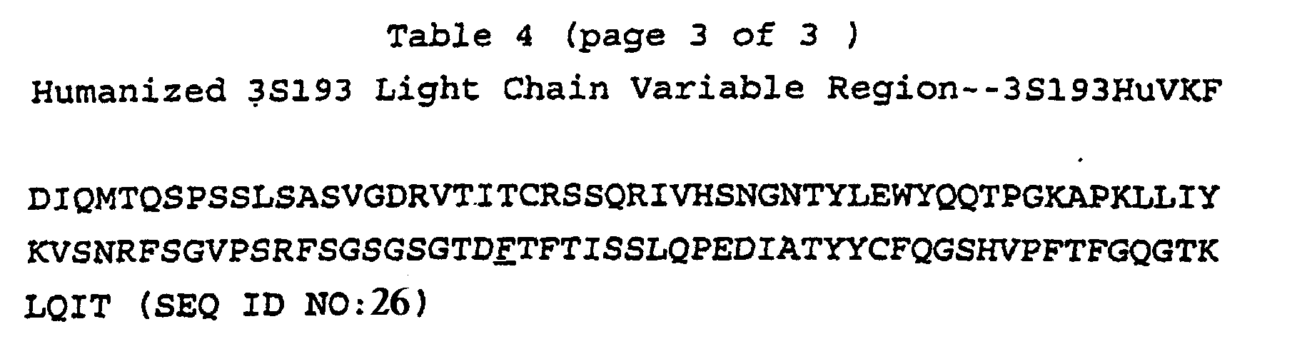
- On March 11, 1994 Applicants have deposited with the American Type Culture Collection, Rockville, Md., USA (ATCC) an NSO cell lines producing humanized antibody 3S193 HuVHT/HuVKF under ATCC accession no. CRL 11573. This deposit was made under the provisions of the Budapest Treaty on the International Recognition of the Deposit of Microorganisms for the purposes of patent procedure and the Regulations thereunder (Budapest Treaty). This assures maintenance of a viable culture for 30 years from date of deposit. The organisms will be made available by ATCC under the terms of the Budapest Treaty, and subject to an agreement between Applicants and ATCC which assures unrestricted availability upon issuance of the pertinent U.S. patent. Availability of the deposited strains is not to be construed as a license to practice the invention in contravention of the rights granted under the authority of any government in accordance with its patent laws.

Claims (8)
- A humanised antibody that specifically binds to the Lewis Y antigen comprising a heavy chain variable region which is Seq. ID No. 21 (3S193HuVHT) and a light chain variable region which is Seq. ID No. 26 (3S193HuVKF).
- An antibody according to claim 1 wherein the framework region of the heavy chain variable region is derived from a KOL framework region.
- An antibody according to claim 1 or 2 wherein the framework region of the light chain variable region is derived from a REI framework region.
- A polynucleotide encoding a polypeptide according to any one of claims 1 to 3.
- An expression vector comprising the polynucleotide sequence of claim 4 in combination with a promoter sequence.
- A host cell transfected with the expression vector of claim 5.
- A labelled immunoglobulin molecule comprising an immunoglobulin molecule according to claims 1 to 3 and a detectable label.
- Use of a humanised antibody according to claim 7 for the manufacture of a composition for the diagnosis of cancer wherein said composition is administered to a patient and bound labelled antibody is subsequently detected by the presence of the label.
Applications Claiming Priority (3)
| Application Number | Priority Date | Filing Date | Title |
|---|---|---|---|
| US207861 | 1994-03-08 | ||
| US08/207,861 US6310185B1 (en) | 1994-03-08 | 1994-03-08 | Recombinant human anti-Lewis Y antibodies |
| PCT/US1995/003096 WO1995024484A1 (en) | 1994-03-08 | 1995-03-08 | Recombinant humanized anti-lewis y antibodies |
Publications (2)
| Publication Number | Publication Date |
|---|---|
| EP0749482A1 EP0749482A1 (en) | 1996-12-27 |
| EP0749482B1 true EP0749482B1 (en) | 2003-06-04 |
Family
ID=22772277
Family Applications (1)
| Application Number | Title | Priority Date | Filing Date |
|---|---|---|---|
| EP95912894A Expired - Lifetime EP0749482B1 (en) | 1994-03-08 | 1995-03-08 | Recombinant humanized anti-lewis y antibodies |
Country Status (10)
| Country | Link |
|---|---|
| US (3) | US6310185B1 (en) |
| EP (1) | EP0749482B1 (en) |
| AT (1) | ATE242326T1 (en) |
| AU (1) | AU696286B2 (en) |
| CA (1) | CA2185114C (en) |
| DE (1) | DE69530975T2 (en) |
| DK (1) | DK0749482T3 (en) |
| ES (1) | ES2199985T3 (en) |
| PT (1) | PT749482E (en) |
| WO (1) | WO1995024484A1 (en) |
Families Citing this family (69)
| Publication number | Priority date | Publication date | Assignee | Title |
|---|---|---|---|---|
| US6310185B1 (en) * | 1994-03-08 | 2001-10-30 | Memorial Sloan Kettering Cancer Center | Recombinant human anti-Lewis Y antibodies |
| RU2162101C1 (en) * | 1999-06-10 | 2001-01-20 | Институт биоорганической химии им. М.М. Шемякина и Ю.А. Овчинникова РАН | Test-system for neoplasm cell study |
| US6565452B2 (en) | 1999-11-01 | 2003-05-20 | Callaway Golf Company | Multiple material golf club head with face insert |
| US6582323B2 (en) | 1999-11-01 | 2003-06-24 | Callaway Golf Company | Multiple material golf club head |
| US6491592B2 (en) | 1999-11-01 | 2002-12-10 | Callaway Golf Company | Multiple material golf club head |
| US7128661B2 (en) * | 1999-11-01 | 2006-10-31 | Callaway Golf Company | Multiple material golf club head |
| US7125344B2 (en) * | 1999-11-01 | 2006-10-24 | Callaway Golf Company | Multiple material golf club head |
| US6739983B2 (en) * | 1999-11-01 | 2004-05-25 | Callaway Golf Company | Golf club head with customizable center of gravity |
| US6663504B2 (en) | 1999-11-01 | 2003-12-16 | Callaway Golf Company | Multiple material golf club head |
| US7118493B2 (en) * | 1999-11-01 | 2006-10-10 | Callaway Golf Company | Multiple material golf club head |
| TW577761B (en) * | 1999-11-01 | 2004-03-01 | Callaway Golf Co | Multiple material golf club head |
| ES2370235T3 (en) * | 2001-02-23 | 2011-12-13 | Immunex Corporation | EFFECTIVE RECOVERY OF CORRECTLY REPLACED PROTEINS. |
| USD479294S1 (en) | 2002-03-14 | 2003-09-02 | Callaway Golf Company | Golf club head |
| USD472592S1 (en) | 2002-03-27 | 2003-04-01 | Callaway Golf Company | Golf club head |
| USD472593S1 (en) | 2002-03-27 | 2003-04-01 | Callaway Golf Company | Golf club head |
| SG187991A1 (en) | 2002-05-02 | 2013-03-28 | Wyeth Corp | Calicheamicin derivative-carrier conjugates |
| EP1534324B1 (en) | 2002-08-20 | 2019-01-16 | Glykos Finland Oy | Tumor specific oligosaccharide epitopes and use thereof |
| US6984181B2 (en) * | 2002-09-25 | 2006-01-10 | Callaway Golf Company | Multiple material golf putter head |
| JP4206915B2 (en) * | 2002-12-27 | 2009-01-14 | 三菱マテリアル株式会社 | Power module substrate |
| US6883624B2 (en) * | 2003-01-31 | 2005-04-26 | Smith International, Inc. | Multi-lobed cutter element for drill bit |
| US6929079B2 (en) * | 2003-02-21 | 2005-08-16 | Smith International, Inc. | Drill bit cutter element having multiple cusps |
| US20060011388A1 (en) * | 2003-01-31 | 2006-01-19 | Mohammed Boudrare | Drill bit and cutter element having multiple extensions |
| USD491243S1 (en) | 2003-06-30 | 2004-06-08 | Callaway Golf Company | Golf club head |
| JP2007528868A (en) * | 2003-08-14 | 2007-10-18 | ワイス | Anti-Lewis Y anti-idiotype antibody and use thereof |
| TW200539855A (en) * | 2004-03-15 | 2005-12-16 | Wyeth Corp | Calicheamicin conjugates |
| TWI384069B (en) * | 2004-08-27 | 2013-02-01 | Pfizer Ireland Pharmaceuticals | Production of polypeptides |
| RU2451082C2 (en) * | 2004-08-27 | 2012-05-20 | Вайет Рисёрч Айрлэнд Лимитед | Production of polypeptides |
| TWI374935B (en) * | 2004-08-27 | 2012-10-21 | Pfizer Ireland Pharmaceuticals | Production of α-abeta |
| US7300773B2 (en) * | 2004-08-27 | 2007-11-27 | Wyeth Research Ireland Limited | Production of TNFR-Ig |
| USD523103S1 (en) | 2004-09-09 | 2006-06-13 | Callaway Golf Company | Golf club head |
| US7549935B2 (en) * | 2005-01-03 | 2009-06-23 | Callaway Golf Company | Golf club head |
| US7166038B2 (en) * | 2005-01-03 | 2007-01-23 | Callaway Golf Company | Golf club head |
| US7559851B2 (en) * | 2005-01-03 | 2009-07-14 | Callaway Golf Company | Golf club with high moment of inertia |
| US7488261B2 (en) * | 2005-01-03 | 2009-02-10 | Callaway Golf Company | Golf club with high moment of inertia |
| US7163468B2 (en) | 2005-01-03 | 2007-01-16 | Callaway Golf Company | Golf club head |
| US7311613B2 (en) * | 2005-01-03 | 2007-12-25 | Callaway Golf Company | Golf club head |
| US7568982B2 (en) * | 2005-01-03 | 2009-08-04 | Callaway Golf Company | Golf club with high moment of inertia |
| US7169060B2 (en) | 2005-01-03 | 2007-01-30 | Callaway Golf Company | Golf club head |
| US7591737B2 (en) * | 2005-01-03 | 2009-09-22 | Callaway Golf Company | Golf club head |
| EP2388271A3 (en) | 2005-02-03 | 2012-08-01 | Antitope Limited | Human Antibodies and Proteins |
| GB2427633B (en) * | 2005-05-17 | 2007-08-15 | Smith International | Drill bit and method of designing a drill bit |
| US7757789B2 (en) * | 2005-06-21 | 2010-07-20 | Smith International, Inc. | Drill bit and insert having bladed interface between substrate and coating |
| FI20055417A0 (en) | 2005-07-20 | 2005-07-20 | Glykos Finland Oy | Cancer specific glycans and their use |
| US8883162B2 (en) | 2005-10-19 | 2014-11-11 | Ibc Pharmaceuticals, Inc. | Multivalent antibody complexes targeting IGF-1R show potent toxicity against solid tumors |
| US20100226884A1 (en) * | 2009-01-20 | 2010-09-09 | Immunomedics, Inc. | Novel Class of Monospecific and Bispecific Humanized Antibodies that Target the Insulin-like Growth Factor Type I Receptor (IGF-1R) |
| US9862770B2 (en) | 2005-10-19 | 2018-01-09 | Ibc Pharmaceuticals, Inc. | Multivalent antibody complexes targeting IGF-1R show potent toxicity against solid tumors |
| EP1957097A1 (en) * | 2005-12-06 | 2008-08-20 | Wyeth a Corporation of the State of Delaware | Interleukin-11 compositions and methods of use |
| NZ596295A (en) | 2006-03-10 | 2013-01-25 | Wyeth Corp | Anti-5T4 antibodies and uses thereof |
| EP2012586A4 (en) * | 2006-04-10 | 2010-08-18 | Abbott Biotech Ltd | Uses and compositions for treatment of ankylosing spondylitis |
| US20100021451A1 (en) | 2006-06-08 | 2010-01-28 | Wong Robert L | Uses and compositions for treatment of ankylosing spondylitis |
| US7743855B2 (en) * | 2006-09-05 | 2010-06-29 | Smith International, Inc. | Drill bit with cutter element having multifaceted, slanted top cutting surface |
| US7686106B2 (en) * | 2007-01-03 | 2010-03-30 | Smith International, Inc. | Rock bit and inserts with wear relief grooves |
| US7798258B2 (en) * | 2007-01-03 | 2010-09-21 | Smith International, Inc. | Drill bit with cutter element having crossing chisel crests |
| US8205692B2 (en) * | 2007-01-03 | 2012-06-26 | Smith International, Inc. | Rock bit and inserts with a chisel crest having a broadened region |
| US7631709B2 (en) | 2007-01-03 | 2009-12-15 | Smith International, Inc. | Drill bit and cutter element having chisel crest with protruding pilot portion |
| US7819754B2 (en) * | 2007-09-13 | 2010-10-26 | Callaway Golf Company | Golf club with removable components |
| US7753809B2 (en) * | 2007-12-19 | 2010-07-13 | Cackett Matthew T | Driver with deep AFT cavity |
| US8414422B2 (en) * | 2009-12-16 | 2013-04-09 | Callaway Golf Company | External weight for golf club head |
| WO2011090492A1 (en) * | 2010-01-19 | 2011-07-28 | Immunomedics, Inc. | Novel class of monospecific and bispecific humanized antibodies that target the insulin-like growth factor type i receptor (igf-1r) |
| WO2012023085A1 (en) | 2010-08-20 | 2012-02-23 | Wyeth Llc | Cell culture of growth factor-free adapted cells |
| US8607899B2 (en) | 2011-02-18 | 2013-12-17 | National Oilwell Varco, L.P. | Rock bit and cutter teeth geometries |
| KR101662412B1 (en) | 2011-07-01 | 2016-10-04 | 암젠 인크 | Mammalian cell culture |
| CA2852021C (en) | 2011-10-21 | 2018-08-28 | Pfizer Inc. | Addition of iron to improve cell culture |
| IL321253A (en) | 2018-05-01 | 2025-08-01 | Amgen Inc | Antibodies with modulated glycan profiles |
| EP3833385A4 (en) | 2018-08-08 | 2021-12-22 | Dragonfly Therapeutics, Inc. | NKG2D, CD16 AND TUMOR-ASSOCIATED ANTIGEN BINDING PROTEINS |
| GB201910899D0 (en) * | 2019-07-31 | 2019-09-11 | Scancell Ltd | Binding members |
| AU2021298824A1 (en) * | 2020-06-30 | 2023-01-19 | Pentixapharm Ag | Humanized antibodies against lewis Y |
| US12551584B1 (en) * | 2020-12-07 | 2026-02-17 | Actinium Pharmaceuticals, Inc. | Lewis Y radioimmunotherapy for the treatment of cancer |
| CN113214402B (en) * | 2021-06-03 | 2022-06-07 | 上海交通大学 | Isolated antigen-binding proteins and their applications |
Family Cites Families (7)
| Publication number | Priority date | Publication date | Assignee | Title |
|---|---|---|---|---|
| US5225539A (en) * | 1986-03-27 | 1993-07-06 | Medical Research Council | Recombinant altered antibodies and methods of making altered antibodies |
| GB8607679D0 (en) * | 1986-03-27 | 1986-04-30 | Winter G P | Recombinant dna product |
| US5242824A (en) * | 1988-12-22 | 1993-09-07 | Oncogen | Monoclonal antibody to human carcinomas |
| IL94872A (en) * | 1989-06-30 | 1995-03-30 | Oncogen | Monoclonal or chimeric antibodies reactive with human carcinomas, recombinant proteins comprising their antigen binding region, pharmaceutical compositions and kits comprising said antibodies and methods for imaging human carcinoma using same |
| GB9019812D0 (en) * | 1990-09-11 | 1990-10-24 | Scotgen Ltd | Novel antibodies for treatment and prevention of infection in animals and man |
| ES2145004T3 (en) * | 1991-08-21 | 2000-07-01 | Novartis Ag | DERIVATIVES OF ANTIBODIES. |
| US6310185B1 (en) * | 1994-03-08 | 2001-10-30 | Memorial Sloan Kettering Cancer Center | Recombinant human anti-Lewis Y antibodies |
-
1994
- 1994-03-08 US US08/207,861 patent/US6310185B1/en not_active Expired - Lifetime
-
1995
- 1995-03-08 ES ES95912894T patent/ES2199985T3/en not_active Expired - Lifetime
- 1995-03-08 CA CA002185114A patent/CA2185114C/en not_active Expired - Fee Related
- 1995-03-08 WO PCT/US1995/003096 patent/WO1995024484A1/en not_active Ceased
- 1995-03-08 EP EP95912894A patent/EP0749482B1/en not_active Expired - Lifetime
- 1995-03-08 PT PT95912894T patent/PT749482E/en unknown
- 1995-03-08 AU AU19907/95A patent/AU696286B2/en not_active Ceased
- 1995-03-08 AT AT95912894T patent/ATE242326T1/en active
- 1995-03-08 DE DE69530975T patent/DE69530975T2/en not_active Expired - Lifetime
- 1995-03-08 DK DK95912894T patent/DK0749482T3/en active
-
1997
- 1997-05-20 US US08/859,648 patent/US6518415B1/en not_active Expired - Fee Related
- 1997-05-20 US US08/859,649 patent/US5874060A/en not_active Expired - Lifetime
Also Published As
| Publication number | Publication date |
|---|---|
| PT749482E (en) | 2003-10-31 |
| ES2199985T3 (en) | 2004-03-01 |
| CA2185114C (en) | 2008-01-22 |
| US6518415B1 (en) | 2003-02-11 |
| AU1990795A (en) | 1995-09-25 |
| DK0749482T3 (en) | 2003-09-29 |
| ATE242326T1 (en) | 2003-06-15 |
| AU696286B2 (en) | 1998-09-03 |
| EP0749482A1 (en) | 1996-12-27 |
| DE69530975D1 (en) | 2003-07-10 |
| DE69530975T2 (en) | 2004-05-19 |
| US5874060A (en) | 1999-02-23 |
| WO1995024484A1 (en) | 1995-09-14 |
| CA2185114A1 (en) | 1995-09-14 |
| US6310185B1 (en) | 2001-10-30 |
Similar Documents
| Publication | Publication Date | Title |
|---|---|---|
| AU696286B2 (en) | Recombinant humanized anti-lewis Y antibodies | |
| AU696946B2 (en) | Recombinant humanized anti-LK26 antibodies | |
| EP0668924B1 (en) | HUMANIZED ANTIBODIES TO Fc RECEPTORS FOR IMMUNOGLOBULIN G ON HUMAN MONONUCLEAR PHAGOCYTES | |
| AU662311B2 (en) | Recombinant antibodies specific for a growth factor receptor | |
| CA2200868C (en) | Cdr-grafted type iii anti-cea humanized mouse monoclonal antibodies | |
| EP0746613B1 (en) | Recombinant humanized anti-fb5 antibodies | |
| US5891996A (en) | Humanized and chimeric monoclonal antibodies that recognize epidermal growth factor receptor (EGF-R); diagnostic and therapeutic use | |
| US5712120A (en) | Method for obtaining modified immunoglobulins with reduced immunogenicity of murine antibody variable domains, compositions containing them | |
| US6204366B1 (en) | Specific binding agents | |
| JP3973682B2 (en) | Interleukin-5 specific recombinant antibody | |
| WO1995024483A9 (en) | Recombinant humanized anti-fb5 antibodies | |
| US5607847A (en) | Recombinant human anti-human immunodeficiency virus antibody | |
| US5795961A (en) | Recombinant human anti-Lewis b antibodies | |
| CA2080260C (en) | Monoclonal antibodies against tumor-associated antigens, processes for the preparation thereof and the use thereof | |
| Tempest et al. | Efficient generation of a reshaped human mAb specific for the α toxin of Clostridium perfringens | |
| AU723073B2 (en) | CDR-grafted type III anti-CEA humanized mouse monoclonal antibodies |
Legal Events
| Date | Code | Title | Description |
|---|---|---|---|
| PUAI | Public reference made under article 153(3) epc to a published international application that has entered the european phase |
Free format text: ORIGINAL CODE: 0009012 |
|
| 17P | Request for examination filed |
Effective date: 19961003 |
|
| AK | Designated contracting states |
Kind code of ref document: A1 Designated state(s): AT BE CH DE DK ES FR GB GR IE IT LI LU MC NL PT SE |
|
| RAP1 | Party data changed (applicant data changed or rights of an application transferred) |
Owner name: LUDWIG INSTITUTE FOR CANCER RESEARCH Owner name: SB2 INC. |
|
| 17Q | First examination report despatched |
Effective date: 20010629 |
|
| GRAG | Despatch of communication of intention to grant |
Free format text: ORIGINAL CODE: EPIDOS AGRA |
|
| RAP1 | Party data changed (applicant data changed or rights of an application transferred) |
Owner name: MEMORIAL SLOAN-KETTERING CANCER CENTER |
|
| GRAG | Despatch of communication of intention to grant |
Free format text: ORIGINAL CODE: EPIDOS AGRA |
|
| GRAH | Despatch of communication of intention to grant a patent |
Free format text: ORIGINAL CODE: EPIDOS IGRA |
|
| RIN1 | Information on inventor provided before grant (corrected) |
Inventor name: ARMOUR, DR. KATHRYN Inventor name: GARIN-CHESA, PILAR Inventor name: KITAMURA, KUNIO Inventor name: WELT, SYDNEY Inventor name: STOCKERT, ELISABETH Inventor name: OLD, LLOYD J. Inventor name: CARR, FRANCIS J. |
|
| GRAH | Despatch of communication of intention to grant a patent |
Free format text: ORIGINAL CODE: EPIDOS IGRA |
|
| GRAA | (expected) grant |
Free format text: ORIGINAL CODE: 0009210 |
|
| AK | Designated contracting states |
Designated state(s): AT BE CH DE DK ES FR GB GR IE IT LI LU MC NL PT SE |
|
| REG | Reference to a national code |
Ref country code: GB Ref legal event code: FG4D |
|
| REG | Reference to a national code |
Ref country code: CH Ref legal event code: EP |
|
| REG | Reference to a national code |
Ref country code: IE Ref legal event code: FG4D |
|
| REF | Corresponds to: |
Ref document number: 69530975 Country of ref document: DE Date of ref document: 20030710 Kind code of ref document: P |
|
| REG | Reference to a national code |
Ref country code: SE Ref legal event code: TRGR |
|
| REG | Reference to a national code |
Ref country code: DK Ref legal event code: T3 |
|
| REG | Reference to a national code |
Ref country code: GR Ref legal event code: EP Ref document number: 20030403495 Country of ref document: GR |
|
| REG | Reference to a national code |
Ref country code: ES Ref legal event code: FG2A Ref document number: 2199985 Country of ref document: ES Kind code of ref document: T3 |
|
| PLBE | No opposition filed within time limit |
Free format text: ORIGINAL CODE: 0009261 |
|
| STAA | Information on the status of an ep patent application or granted ep patent |
Free format text: STATUS: NO OPPOSITION FILED WITHIN TIME LIMIT |
|
| ET | Fr: translation filed | ||
| 26N | No opposition filed |
Effective date: 20040305 |
|
| PGFP | Annual fee paid to national office [announced via postgrant information from national office to epo] |
Ref country code: IT Payment date: 20120327 Year of fee payment: 18 |
|
| PGFP | Annual fee paid to national office [announced via postgrant information from national office to epo] |
Ref country code: IE Payment date: 20130315 Year of fee payment: 19 Ref country code: ES Payment date: 20130320 Year of fee payment: 19 Ref country code: GB Payment date: 20130318 Year of fee payment: 19 Ref country code: SE Payment date: 20130319 Year of fee payment: 19 Ref country code: DK Payment date: 20130320 Year of fee payment: 19 Ref country code: FR Payment date: 20130329 Year of fee payment: 19 Ref country code: LU Payment date: 20130320 Year of fee payment: 19 Ref country code: MC Payment date: 20130319 Year of fee payment: 19 Ref country code: DE Payment date: 20130320 Year of fee payment: 19 Ref country code: CH Payment date: 20130319 Year of fee payment: 19 |
|
| PGFP | Annual fee paid to national office [announced via postgrant information from national office to epo] |
Ref country code: GR Payment date: 20130319 Year of fee payment: 19 Ref country code: NL Payment date: 20130318 Year of fee payment: 19 |
|
| PGFP | Annual fee paid to national office [announced via postgrant information from national office to epo] |
Ref country code: AT Payment date: 20130318 Year of fee payment: 19 Ref country code: PT Payment date: 20130228 Year of fee payment: 19 |
|
| PGFP | Annual fee paid to national office [announced via postgrant information from national office to epo] |
Ref country code: BE Payment date: 20130318 Year of fee payment: 19 |
|
| REG | Reference to a national code |
Ref country code: PT Ref legal event code: MM4A Free format text: LAPSE DUE TO NON-PAYMENT OF FEES Effective date: 20140908 |
|
| REG | Reference to a national code |
Ref country code: DE Ref legal event code: R119 Ref document number: 69530975 Country of ref document: DE |
|
| REG | Reference to a national code |
Ref country code: DK Ref legal event code: EBP Effective date: 20140331 |
|
| REG | Reference to a national code |
Ref country code: NL Ref legal event code: V1 Effective date: 20141001 |
|
| PG25 | Lapsed in a contracting state [announced via postgrant information from national office to epo] |
Ref country code: LU Free format text: LAPSE BECAUSE OF NON-PAYMENT OF DUE FEES Effective date: 20140308 |
|
| REG | Reference to a national code |
Ref country code: CH Ref legal event code: PL |
|
| REG | Reference to a national code |
Ref country code: SE Ref legal event code: EUG |
|
| REG | Reference to a national code |
Ref country code: AT Ref legal event code: MM01 Ref document number: 242326 Country of ref document: AT Kind code of ref document: T Effective date: 20140308 |
|
| GBPC | Gb: european patent ceased through non-payment of renewal fee |
Effective date: 20140308 |
|
| REG | Reference to a national code |
Ref country code: GR Ref legal event code: ML Ref document number: 20030403495 Country of ref document: GR Effective date: 20141002 |
|
| PG25 | Lapsed in a contracting state [announced via postgrant information from national office to epo] |
Ref country code: SE Free format text: LAPSE BECAUSE OF NON-PAYMENT OF DUE FEES Effective date: 20140309 |
|
| REG | Reference to a national code |
Ref country code: FR Ref legal event code: ST Effective date: 20141128 |
|
| PG25 | Lapsed in a contracting state [announced via postgrant information from national office to epo] |
Ref country code: PT Free format text: LAPSE BECAUSE OF NON-PAYMENT OF DUE FEES Effective date: 20140908 |
|
| REG | Reference to a national code |
Ref country code: IE Ref legal event code: MM4A |
|
| REG | Reference to a national code |
Ref country code: DE Ref legal event code: R119 Ref document number: 69530975 Country of ref document: DE Effective date: 20141001 |
|
| PG25 | Lapsed in a contracting state [announced via postgrant information from national office to epo] |
Ref country code: IE Free format text: LAPSE BECAUSE OF NON-PAYMENT OF DUE FEES Effective date: 20140308 Ref country code: LI Free format text: LAPSE BECAUSE OF NON-PAYMENT OF DUE FEES Effective date: 20140331 Ref country code: GB Free format text: LAPSE BECAUSE OF NON-PAYMENT OF DUE FEES Effective date: 20140308 Ref country code: FR Free format text: LAPSE BECAUSE OF NON-PAYMENT OF DUE FEES Effective date: 20140331 Ref country code: DE Free format text: LAPSE BECAUSE OF NON-PAYMENT OF DUE FEES Effective date: 20141001 Ref country code: CH Free format text: LAPSE BECAUSE OF NON-PAYMENT OF DUE FEES Effective date: 20140331 Ref country code: GR Free format text: LAPSE BECAUSE OF NON-PAYMENT OF DUE FEES Effective date: 20141002 |
|
| PG25 | Lapsed in a contracting state [announced via postgrant information from national office to epo] |
Ref country code: NL Free format text: LAPSE BECAUSE OF NON-PAYMENT OF DUE FEES Effective date: 20141001 Ref country code: AT Free format text: LAPSE BECAUSE OF NON-PAYMENT OF DUE FEES Effective date: 20140308 |
|
| REG | Reference to a national code |
Ref country code: PT Ref legal event code: MM4A Free format text: MAXIMUM VALIDITY LIMIT REACHED Effective date: 20150308 |
|
| PG25 | Lapsed in a contracting state [announced via postgrant information from national office to epo] |
Ref country code: IT Free format text: LAPSE BECAUSE OF NON-PAYMENT OF DUE FEES Effective date: 20140308 |
|
| REG | Reference to a national code |
Ref country code: ES Ref legal event code: FD2A Effective date: 20150428 |
|
| PG25 | Lapsed in a contracting state [announced via postgrant information from national office to epo] |
Ref country code: DK Free format text: LAPSE BECAUSE OF NON-PAYMENT OF DUE FEES Effective date: 20140331 Ref country code: PT Free format text: LAPSE BECAUSE OF EXPIRATION OF PROTECTION Effective date: 20150317 |
|
| PG25 | Lapsed in a contracting state [announced via postgrant information from national office to epo] |
Ref country code: ES Free format text: LAPSE BECAUSE OF NON-PAYMENT OF DUE FEES Effective date: 20140309 |
|
| PG25 | Lapsed in a contracting state [announced via postgrant information from national office to epo] |
Ref country code: MC Free format text: LAPSE BECAUSE OF NON-PAYMENT OF DUE FEES Effective date: 20140331 |
|
| PG25 | Lapsed in a contracting state [announced via postgrant information from national office to epo] |
Ref country code: PT Free format text: LAPSE BECAUSE OF EXPIRATION OF PROTECTION Effective date: 20140308 |
|
| PG25 | Lapsed in a contracting state [announced via postgrant information from national office to epo] |
Ref country code: BE Free format text: LAPSE BECAUSE OF NON-PAYMENT OF DUE FEES Effective date: 20140331 |