EP0746611B1 - Method for increasing production of disulfide bonded recombinant proteins by (saccharomyces cerevisiae) - Google Patents
Method for increasing production of disulfide bonded recombinant proteins by (saccharomyces cerevisiae) Download PDFInfo
- Publication number
- EP0746611B1 EP0746611B1 EP93915201A EP93915201A EP0746611B1 EP 0746611 B1 EP0746611 B1 EP 0746611B1 EP 93915201 A EP93915201 A EP 93915201A EP 93915201 A EP93915201 A EP 93915201A EP 0746611 B1 EP0746611 B1 EP 0746611B1
- Authority
- EP
- European Patent Office
- Prior art keywords
- recombinant
- yeast
- protein
- pdi
- gene
- Prior art date
- Legal status (The legal status is an assumption and is not a legal conclusion. Google has not performed a legal analysis and makes no representation as to the accuracy of the status listed.)
- Expired - Lifetime
Links
Images





Classifications
-
- C—CHEMISTRY; METALLURGY
- C12—BIOCHEMISTRY; BEER; SPIRITS; WINE; VINEGAR; MICROBIOLOGY; ENZYMOLOGY; MUTATION OR GENETIC ENGINEERING
- C12N—MICROORGANISMS OR ENZYMES; COMPOSITIONS THEREOF; PROPAGATING, PRESERVING, OR MAINTAINING MICROORGANISMS; MUTATION OR GENETIC ENGINEERING; CULTURE MEDIA
- C12N9/00—Enzymes; Proenzymes; Compositions thereof; Processes for preparing, activating, inhibiting, separating or purifying enzymes
- C12N9/90—Isomerases (5.)
-
- C—CHEMISTRY; METALLURGY
- C07—ORGANIC CHEMISTRY
- C07K—PEPTIDES
- C07K14/00—Peptides having more than 20 amino acids; Gastrins; Somatostatins; Melanotropins; Derivatives thereof
- C07K14/81—Protease inhibitors
- C07K14/8107—Endopeptidase (E.C. 3.4.21-99) inhibitors
- C07K14/811—Serine protease (E.C. 3.4.21) inhibitors
-
- C—CHEMISTRY; METALLURGY
- C07—ORGANIC CHEMISTRY
- C07K—PEPTIDES
- C07K14/00—Peptides having more than 20 amino acids; Gastrins; Somatostatins; Melanotropins; Derivatives thereof
- C07K14/81—Protease inhibitors
- C07K14/815—Protease inhibitors from leeches, e.g. hirudin, eglin
-
- C—CHEMISTRY; METALLURGY
- C12—BIOCHEMISTRY; BEER; SPIRITS; WINE; VINEGAR; MICROBIOLOGY; ENZYMOLOGY; MUTATION OR GENETIC ENGINEERING
- C12N—MICROORGANISMS OR ENZYMES; COMPOSITIONS THEREOF; PROPAGATING, PRESERVING, OR MAINTAINING MICROORGANISMS; MUTATION OR GENETIC ENGINEERING; CULTURE MEDIA
- C12N15/00—Mutation or genetic engineering; DNA or RNA concerning genetic engineering, vectors, e.g. plasmids, or their isolation, preparation or purification; Use of hosts therefor
- C12N15/09—Recombinant DNA-technology
- C12N15/63—Introduction of foreign genetic material using vectors; Vectors; Use of hosts therefor; Regulation of expression
- C12N15/79—Vectors or expression systems specially adapted for eukaryotic hosts
- C12N15/80—Vectors or expression systems specially adapted for eukaryotic hosts for fungi
- C12N15/81—Vectors or expression systems specially adapted for eukaryotic hosts for fungi for yeasts
Definitions
- Protein disulfide isomerase is an enzyme involved in the catalysis of disulfide bond formation in secretory and cell-surface proteins.
- PDI Protein disulfide isomerase
- WGHCK conserved "thioredoxin-like" active site of vertebrate PDI's
- amino acid sequence shows 30-32% identity and 53-56% similarity with mammalian and avian PDI sequences and has a very similar overall organization, namely the presence of two 100 residue segments, each of which is repeated, with the most significant homologies to mammalian and avian PDIs being in the regions (a, a') that contain the conserved "thioredoxin-like" active site.
- the N-terminal region has the characteristics of a cleavable secretory signal sequence and the C-terminal four amino acids (-HDEL)(SEQ.ID.NO.: 2) are consistent with the protein being a component of S . cerevisiae endoplasmic reticulum (E.R.).
- Transformants carrying multiple copies of this gene have 10-fold higher levels of PDI activity and overexpress a protein of the predicted molecular weight.
- the PDI1 gene is unique in the yeast genome and encodes a single 1.8kb transcript that is not found in stationary phase cells, nor is it heat-inducible. Disruption of the PDI1 gene is haplo-lethal indicating that the product of this gene is essential for viability.
- Protein disulfide-isomerase an enzyme which catalyzes thiol:disulfide interchange reactions, is a major resident protein component of the E.R. lumen in secretory cells.
- PDI Protein disulfide-isomerase
- microsomal membranes deficient in PDI show a specific defect in cotranslational protein disulfide formation (Bulleid and Freedman, 1988, Nature, 335 , pp.649-51) implies that the enzyme functions as a catalyst of native disulfide bond formation during the biosynthesis of secretory and cell surface proteins. This role is consistent with what is known of the enzyme's catalytic properties in vitro ; it catalyzes thiol: disulfide interchange reactions leading to net protein disulfide formation, breakage or isomerization, and can catalyze protein folding and the formation of native disulfide bonds in a wide variety of reduced, unfolded protein substrates (Freedman et al ., 1989, Biochem. Soc.
- the DNA and amino acid sequence of the enzyme is known for several species (Scherens, B. et al ., 1991, Yeast, 7 , pp. 185-193; Farguhar, R., et al ., 1991, Gene, 108 , pp. 81-89) and there is increasing information on the mechanism of action of the enzyme purified to homogeneity from mammalian liver (Creighton et al ., 1980, J. Mol. Biol., 142 , pp.43-62; Freedman et al ., 1988, Biochem. Soc.
- PDI is readily isolated from mammalian tissues and the homogeneous enzyme is a homodimer (2 x 57kD) with characteristically acidic pI (4.0-4.5) (Hillson et al ., 1984, Methods Enzymol., 107 , pp.281-292). The enzyme has also been purified from wheat and from the alga Chlamydomonas reinhardii (Kaska et al ., 1990 Biochem. J. 268 , pp.63-68). The activity has been detected in a wide variety of sources, and in a preliminary report, PDI activity was claimed to be detectable in S .
- PDI may be multifunctional in the co- and post-translational modification of secretory proteins (Freedman, 1989, Cell, 57 , pp.1069-72).
- mammalian secretory proteins contain multiple intramolecular and/or intermolecular disulfide bonds. Examples include, but are not limited to, pituitary hormones, interleukins, immunoglobulins, proteases and their inhibitors and other serum proteins. Such proteins are among the prime targets for commercial genetic engineering, but early experience in their expression in bacteria and yeast has highlighted a number of problems in obtaining them as functionally active recombinant products. This has drawn attention to the need for a better understanding of post-translational modifications in general, and of protein folding and disulfide bond formation in particular.
- Disulfide bonded proteins comprising a single folded domain can, in general, be fully reduced and denatured and subsequently renatured in vitro to generate the correctly disulfide-linked state in reasonable yield.
- the process involves rapid formation of a mixed population of many differently disulfide bonded forms which slowly isomerize to give the native disulfide pairing.
- the process is catalysed by thiol/disulfide redox buffers (e.g. GSH and GSSG) and by alkaline pH. Low protein concentrations are required to prevent precipitation and interchain disulfide formation.
- the rate of formation of the native protein, and the optimal obtainable yield both decrease as the number of intramolecular disulfides increases.
- the problem is compounded in proteins containing multiple disulfide bonded domains (e.g. tissue plasminogen activator) in which each domain must fold and form its native disulfide bonds independently.
- the process of disulfide bond formation in vivo occurs co-translationally or as a very early post-translational event. Studies on nascent and newly synthesized secretory proteins in the lumen of the E.R. in mammalian cells show that native disulfide bonds are already formed. The process in vivo appears to be catalyzed by the enzyme protein disulfide-isomerase which is an abundant protein in secretory cells and is located at the luminal face of the endoplasmic reticulum [Freedman, R.B., 1984, Trends in Biochemical Sciences, 9 , 438-441].
- This enzyme in vitro, catalyzes thiol:protein-disulfide interchange reactions in a wide range of protein substrates and has the properties required of a cellular catalyst of native protein disulfide formation [Freedman, R.B. et al ., 1984, Biochem. Soc. Trans., 12 , 939-942]. Further evidence for its role include (i) that its tissue distribution matches that of the synthesis of disulfide bonded secretory proteins [Brockway, B.E.
- the enzyme has been characterized in a number of animal sources [Lambert, N. and Freedman, R.B., 1983, Biochem. J., 213 , pp. 225-234], and in wheat [de Azevedo, G.M.V. et al ., 1983, Biochem. Soc. Trans., 12 , 1043], and a striking conservation of molecular and kinetic properties has been noted [Freedman, R.B. et al ., 1984, Biochem. Soc. Trans. 12 , pp. 939-942; Brockway, B.E. and Freedman, R.B., 1984, Biochem J., 219 , 51-59].
- yeast secretory proteins e.g. killer toxin
- yeast as a versatile host for the expression of commercially-important mammalian proteins is compromised, to some extent, by the limited capacity of the yeast secretory system and by some differences between it and that of higher eukaryotes (e.g. in glycosylation).
- EP 509,841 discloses the production of disulfide-bonded human serum albumin by means of heterologous co-expression of human PDI together with HSA in Saccharomyces cerevisiae.
- This disclosure primarily concerns a "three-gene” process to producing disulfide-bridge proteins involving expression of an ERD2 receptor protein in addition to PDI and HSA.
- this patent application requires in that case that the PDI gene is a fusion gene incorporating DNA encoding the HSA prepro-sequence.
- EP 510,658 discloses a process for enhanced formation of the native conformation of disulfide-bonded proteins in E. coli. This application relates exclusively to the use of prokaryotic host organisms and is enabling only for the use of E. coli.
- EP 293,793 discloses the heterologous expression of human PDI in a variety of host cells including E. coli. simian CO5-7 cells and Saccharomyces cerevisiae and the use of the PDI resulting therefrom for the conversion of suitable proteins to their disulfide-bridged forms.
- the present invention provides an in vitro process for producing disulfide-bonded recombinant proteins comprising:
- said recombinant host cell is other than E. coli
- said gene encoding protein disulfide isomerase does not comprise a DNA fragment coding for a human serum albumin prepro-sequence, and that the process does not involve expression from a recombinant expression cassette of a gene coding for the receptor protein ERD2 from yeast.
- the present invention also provides a strain of the yeast Saccharomyces cerevisiae which overexpresses recombinant protein disulfide isomerase from a recombinant expression cassette containing a gene encoding protein disulfide isomerase and one or more recombinant genes encoding one or more disulfide-bonded proteins other than protein disulfide isomerase from a recombinant expression cassette;
- said gene encoding protein disulfide isomerase does not comprise a DNA fragment coding for a human serum albumin prepro-sequence, and that the yeast does not express a gene coding for the receptor protein ERD2 from yeast from a recombinant expression cassette.
- DNA encoding human and yeast protein disulfide isomerase (PDI) is isolated and cloned into expression casettes or vectors comprising a promoter and transcription terminator.
- the expression casettes or vectors containing PDI-encoding DNA are transferred into host cells which, as a result, overproduce PDI protein.
- These PDI overproducing cells are used as recombinant hosts for the expression of disulfide bonded proteins. Secretion of disulfide bonded proteins is substantially increased in PDI overproducing host cells compared to host cells producing normal levels of PDI.
- peptidyl prolyl cis-trans isomerases include peptidyl prolyl cis-trans isomerases, PDI and other thioredoxin-like proteins, BiP, various molecular chaperones (hsp70, hsp60, etc.), signal peptidase, signal recognition protein, the various proteins involved in translocation of precursors into the E.R., the various structural and functional components of the ER, Golgi, and secretory vesicles plus many proteins not yet characterized (Franzuoff, A., et al ., 1991, supra; Rothman, J.E. and Orci L., 1992, Nature, 355 , pp. 409-415; Gething, M.G.
- the present invention provided very unexpected results in that increased levels of PDI alone caused a significant and substantial increase in the levels of secreted proteins, for example antistasin, a phenomenon which is likely to be related to improved protein folding and/or disulfide bond formation.
- the present invention relates to a method for increasing the production of recombinant proteins by recombinant host cells, by overexpressing DNA encoding protein disulfide isomerase (PDI).
- PDI as used herein refers to an enzyme which specifically catalyzes the formation of intramolecular and intermolecular disulfide bonds.
- the DNA sequence of PDI genes from several species is known in the art. These species include but are not limited to, human, bovine, rat, chicken and yeast. [Mizunaga et al ., 1990, J. Biochem., 108 , pp.846-851; Scherens et al ., 1991, Yeast, 7 , pp.185-193].
- Starting material for the isolation of PDI-encoding DNA may be any cell or tissue type including but not limited to mammalian and other vertebrate cells and tissue, as well as lower eukaryotic cells and tissue.
- the present invention is demonstrated using yeast and human PDI expressed in recombinant yeast host cells. It is readily apparent to one of ordinary skill in the art that the present invention extends to and encompasses other expression hosts including, but not limited to, mammalian cells, plant cells, prokaryotic cells such as bacteria , insect cells and lower eukaryotic cells such as yeast and filamentous fungi. Furthermore, it is readily apparent to one of ordinary skill in the art that the use of PDI encoding DNA derived from sources other than yeast and human cells is encompassed by the present invention. Other sources of PDI-encoding DNA include, but are not limited to, vertebrates other than human such as rat and mouse, non vertebrates such as insects, and lower eukaryotes such as fungi.
- PDI protein disulfide isomerase
- a non-redundant 30-mer oligonucleotide was designed based on yeast codon bias (Sharp et al ., 1986, Nucleic Acids Res., 14 , pp.5125-43) which was end-labelled and used to screen a yeast genomic library constructed in the multicopy YEp plasmid pMA3a (Crouzet and Tuite, 1987, Mol. Gen. Genet., 210 , pp.581-3).
- Two strongly positive clones (designated C7 and C10) were recovered from the screen and preliminary restriction mapping revealed insert sizes of 14kb and 14.5kb, respectively, with the two inserts showing a number of restriction sites in common. The insert of clone C7 was further analysed.
- the C7 clone was digested with a variety of restriction enzymes, the digests transferred to nitrocellulose and probed with the 30-mer "active site" oligonucleotide described above.
- the procedure identified a 5kb Bam HI- Sal 1 fragment and two apparently adjacent Hin dIII fragments of 5.0 and 4.5kb respectively.
- the latter pattern suggested the possible existence of two targets for the "active site” probe as would be predicted for PDI which contains two copies of the active site.
- Preliminary DNA sequence analysis from the two Hin dIII sites revealed an open reading frame with weak homology to vertebrate PDIs, but also demonstrated that there must be a further Hin dIII site since they were not contiguous sequences.
- Detailed restriction mapping coupled with DNA sequencing confirmed this assumption. Using naturally occurring restriction sites and oligonucleotide primers a 2.5kb Hin dIII- Eco RI fragment encompassing the two adjacent Hin dIII sites was sequenced on both strands.
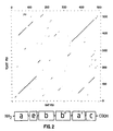
- the DNA sequence predicted a single open reading frame of 1593bp with the potential of encoding a polypeptide of 530 amino acids of predicted molecular weight of 59,082 (Farquhar, R., et al ., supra, see Figure 2).
- the open reading frame had a codon bias typical of yeast mRNAs that encode moderately abundant proteins (Bennetzen and Hall, 1982, J. Biol. Chem., 257 , pp.3029-3031); the calculated codon bias index was 0.60.
- the encoded polypeptide is a component of the S . cerevisiae endoplasmic reticulum; the protein encodes a very hydrophobic N-terminal sequence with the characteristics of a putative secretory signal (Gierasch, 1989, Biochemistry, 28 , pp.923-930) and the four C-terminal amino acids are identical to those in yeast BiP (Normington et al ., 1989, Cell, 57 , pp.1223-36) and have been reported to be the endoplasmic reticulum retention signal for S . cerevisiae (Pelham et al ., 1988, EMBO J., 7 , pp.1757-62).
- S . cerevisiae PDI gene PDI1 The S . cerevisiae PDI1 gene is present in only one copy in the genome. This was confirmed by high stringency hybridisation using the 0.8kb Hin dIII- Stu I fragment described above as a probe against a variety of genomic digests.
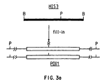
- HIS+ AS3324 transformants (Y1, Y2 and Y3) were studied further and in each case sporulation of the diploids produced only two viable spores per tetrad ( Figure 3) all of which were his - .
- This result indicates that the lethal phenotype was associated with pdil::HIS3 mutation. That the correct gene replacement had arisen in the HIS + transformants Y1 and Y2 was confirmed by Southern hybridisation to blotted yeast genomic DNA digested with PstI using the 800 bp Hin dIII- Stu I fragment as a probe.
- the PDI1 gene contains no internal Pst I sites ( Figure 3) but the HIS3 gene does contain a single PstI site ( Figure 3) this should allow simple identification of the pdi1::HIS3 allele.
- a single 9kb Pst I fragment was detected while in the Y1 and Y2 transformants two bands of 9kb and 2.2kb were detected with the 9kb band presumably consisting of two bands of different origin.
- any of a variety of procedures may be used to molecularly clone yeast PDI-encoding DNA. These methods include, but are not limited to, direct functional expression of the PDI gene following the construction of a PDI containing DNA library in an appropriate expression vector system. Another method is to screen a PDI containing DNA library constructed in a bacteriophage or plasmid shuttle vector with a labelled oligonucleotide probe designed from the amino acid sequence of the PDI proteins. The preferred method consists of screening a human or yeast PDI-containing genomic DNA library constructed in a plasmid shuttle vector with a deduced DNA probe encoding the known amino acid sequence of the enzyme active site.
- libraries as well as libraries constructed from other cells or cell types, may be useful for isolating PDI encoding DNA.
- Other types of libraries include, but are not limited to, cDNA and genomic DNA libraries derived from other human, vertebrate, invertebrate, and lower eukaryotic cells or cell lines, other than yeast cells.
- cDNA libraries may be prepared from cells or cell lines which have PDI activity.
- the selection of cells or cell lines for use in preparing a cDNA library to isolate PDI cDNA may be done by first measuring cell associated PDI activity using the procedures described fully above.
- cDNA libraries can be performed by standard techniques well known in the art. Well known cDNA library construction techniques can be found for example, in Maniatis, T., Fritsch, E.F., Sambrook, J., Molecular Cloning: A Laboratory Manual (Cold Spring Harbor Laboratory, Cold Spring Harbor, New York, 1982).
- PDI-encoding DNA may also be isolated from a suitable genomic DNA library.
- genomic DNA libraries can be performed by standard techniques well known in the art. Well known genomic DNA library construction techiques can be found in Maniatis, T., Fritsch, E.F., Sambrook, J. in Molecular Cloning: A Laboratory Manuel (Cold Spring Harbor Laboratory, Cold Spring Harbor, New York, 1982).
- the cloned PDI obtained through the methods described above may be recombinantly expressed by molecular cloning into an expression vector containing a suitable promoter and other appropriate transcription regulatory elements, and transferred into prokaryotic or eukaryotic host cells to produce recombinant PDI.
- Techniques for such manipulations are fully described in Maniatis, T, et al ., supra , and are well known in the art.
- Expression vectors are defined herein as DNA sequences that are required for the transcription of cloned copies of genes and the translation of their mRNAs in an appropriate host. Such vectors can be used to express eukaryotic genes in a variety of hosts such as bacteria, bluegreen algae, plant cells, fungi, insect cells and animal cells.
- RNA-yeast or bacteria-animal cells Specifically designed vectors allow the shuttling of DNA between hosts, such as between bacteria-yeast or bacteria-animal cells.
- An appropriately constructed expression vector should contain: an origin of replication for autonomous replication in host cells, selectable markers, a limited number of useful restriction enzyme sites, a potential for high copy number, and active promoters.
- a promoter is defined as a DNA sequence that directs RNA polymerase to bind to DNA and initiate RNA synthesis.
- a strong promoter is one which causes mRNAs to be initiated at high frequency.
- Expression vectors may include, but are not limited to, cloning vectors, modified cloning vectors, specifically designed plasmids or viruses.
- mammalian expression vectors may be used to express recombinant PDI in mammalian cells.
- Commercially available mammalian expression vectors which may be suitable for recombinant PDI expression, include but are not limited to, pMClneo (Stratagene), pXT1 (Stratagene), pSG5 (Stratagene), EBO-pSV2-neo (ATCC 37593) pBPV-1(8-2) (ATCC 37110), pdBPV-MMTneo(342-12) (ATCC 37224), pRSVgpt (ATCC 37199), pRSVneo (ATCC 37198), pSV2-dhfr (ATCC 37146), pUCTag (ATCC 37460), and gZD35 (ATCC 37565).
- DNA encoding PDI may also be cloned into an expression vector for expression in a variety of recombinant host cells.
- Recombinant host cells may be prokaryotic, including but not limited to bacteria, or eukaryotic, including but not limited to yeast, mammalian cells including but not limited to cell lines of human, bovine, porcine, monkey and rodent origin, and insect cells including but not limited to Drosophila derived cell lines and Spodoptera frugiperda (SF9) insect cells for use with recombinant Baculovirus expression systems.
- prokaryotic including but not limited to bacteria
- eukaryotic including but not limited to yeast
- mammalian cells including but not limited to cell lines of human, bovine, porcine, monkey and rodent origin
- insect cells including but not limited to Drosophila derived cell lines and Spodoptera frugiperda (SF9) insect cells for use with recombinant Baculovirus expression systems.
- SF9 Spodoptera frugi
- Cell lines derived from mammalian species which may be suitable and which are commercially available, include but are not limited to, CV-1 (ATCC CCL 70), COS-1 (ATCC CRL 1650), COS-7 (ATCC CRL 1651), CHO-K1 (ATCC CCL 61), 3T3 (ATCC CCL 92), NIH/3T3 (ATCC CRL 1658), HeLa (ATCC CCL 2), C127I (ATCC CRL 1616), BS-C-1 (ATCC CCL 26) and MRC-5 (ATCC CCL 171).
- CV-1 ATCC CCL 70
- COS-1 ATCC CRL 1650
- COS-7 ATCC CRL 1651
- CHO-K1 ATCC CCL 61
- 3T3 ATCC CCL 92
- NIH/3T3 ATCC CRL 1658
- HeLa ATCC CCL 2
- C127I ATCC CRL 1616
- BS-C-1 ATCC CCL 26
- MRC-5 ATCC CCL 171.
- a yeast active promoter initiates transcription of the PDI gene in yeast hosts. Therefore, it is readily apparent to those skilled in the art that any yeast-active promoter sequence may be used, including but not limited to, GAL 1, GAL 10, GAL 7, PGK 1, ADH 1, ADH 2, PHO 5, and GAP491 ( TDH 3). It is also readily apparent to those skilled in the art that a suitable assay system, e . g ., immunoblot or RIA or enzyme-linked immunoassay (EIA), may be utilized in order to assay expression of PDI in recombinant hosts.
- a suitable assay system e . g ., immunoblot or RIA or enzyme-linked immunoassay (EIA) may be utilized in order to assay expression of PDI in recombinant hosts.
- S . cerevisiae has 5 genes which encode the enzymes responsible for the utilization of galactose as a carbon source for growth.
- the GAL 1, GAL 2 , GAL 5, GAL 7, and GAL 10 genes respectively encode galactokinase, galactose permease, the major isozyme of phosphoglucomutase, ⁇ -D-galactose-1-phosphate uridyltransferase and uridine diphosphogalactose-4-epimerase. In the absence of galactose, very little expression of these enzymes is detected.
- GAL 1 GAL 2, GAL 5, GAL 7 and GAL 10 genes have been molecularly cloned and sequenced.
- the regulatory and promoter sequences to the 5' sides of the respective coding regions have been placed adjacent to the coding regions of the lacZ gene.
- S . cerevisiae also has 3 genes, each of which encodes an isozyme of ADH.
- ADHII is responsible for the abiliy of S . cerevisiae to utilize ethanol as a carbon source during oxidative growth.
- Expression of the ADH 2 gene, which encodes the ADHII isozyme is catabolite-repressed by glucose, such that there is virtually no transcription of the ADH 2 gene during fermentative growth in the presence of glucose levels of 0.1% (w/v).
- glucose depletion and in the presence of non-repressing carbon sources transcription of the ADH 2 gene is induced 100- to 1000-fold.
- This gene has been molecularly cloned and sequenced, and those regulatory and promoter sequences which are necessary and sufficient for derepression of transcription have been defined.
- Alpha mating factor is a sex pheromone of S . cerevisiae which is required for mating between MAT ⁇ and MAT a cells.
- This tridecapeptide is expressed as a prepropheromone which is directed into the rough endoplasmic reticulum, glycosylated and proteolytically processed to its final mature form which is secreted from cells.
- This biochemical pathway has been exploited as an expression strategy for foreign polypeptides.
- the alpha mating factor gene has been molecularly cloned and its promoter with pre-pro-leader sequence has been utilized to express and secrete a variety of polypeptides.
- the PHO 5 gene promoter has been shown to be inducible by low phosphate concentrations and this also has utility for physiologically regulated expression of foreign proteins in yeast.
- the alpha mating factor promoter is active only in cells which are phenotypically ⁇ .
- SIR S . cerevisiae
- Temperature-sensitive (ts) lesions which interfere with this repression event exist in the gene product of at least one of these loci.
- growth at 35°C abrogates repression, resulting in cells phenotypically a / ⁇ in which the alpha mating factor promoter is inactive.
- Upon temperature shift to 23°C the cells phenotypically revert to ⁇ such that the promoter becomes active.
- the use of strains with a ts SIR lesion has been demonstrated for the controlled expression of several foreign polypeptides.
- Suitable yeast strains include but are not limited to those with genetic and phenotypic characteristics such as protease deficiencies, and altered glycosylation capabilities.
- the genus Saccharomyces is composed of a variety of species. S . cerevisiae is most commonly used as a host for the recombinant DNA-mediated expression of a variety of foreign polypeptides. However, the distinctions among other species of the genus Saccharomyces are not always well-defined. Many of these species are capable of interbreeding with S . cerevisiae and are likely to possess promoters which are analogous or identical to promoters in S . cerevisiae .
- yeast genera such as Candida , Hansenula , Pichia , and Torulopsis have been shown to contain similar metabolic pathways for the utilization of methanol as a sole carbon source for growth.
- the gene for alcohol oxidase an enzyme which participates in this metabolic pathway, has been isolated from Pichia pastoris .
- the P . pastoris alcohol oxidase promoter has been isolated and shown to be susceptible to methanol induction of expression.
- Such an inducible promoter is useful for expression of polypeptides in yeast.
- this promoter has been shown to be active on a plasmid for the inducible expression of heterologous genes in P . pastoris .
- yeast genera to function as hosts for the recombinant DNA-mediated expression of polypeptides in active form. Therefore, it will be readily apparent to those skilled in the art that, for the expression of PDI, the selection of a host strain extends to species from other genera of yeast from the Families Saccharomycetaceae and Cryptococcaceae , including, but not limited to Candida , Hansenula , Kluyveromyces , Pichia , Saccharomyceopsis , and Torulopsis .
- the expression vector may be introduced into host cells via any one of a number of techniques including but not limited to transformation, transfection, protoplast fusion, and electroporation.
- the expression vector-containing cells are clonally propagated and individually analyzed to determine whether they produce PDI protein. Identification of PDI expressing host cell clones may be done by several means, including but not limited to immunological reactivity with anti-PDI antibodies, and the presence of host cell-associated PDI activity.
- Expression of PDI DNA may also be performed using in vitro produced synthetic mRNA.
- Synthetic mRNA can be efficiently translated in various cell-free systems, including but not limited to wheat germ extracts and reticulocyte extracts, as well as efficiently translated in cell based systems, including but not limited to microinjection into frog oocytes.
- PDI may be expressed in a recombinant host from a recombinant expression cassette which is integrated into the host cell genome, in single copy or multiple copies per cell. It is also readily apparent to those skilled in the art that PDI may be expressed in a recombinant host from a recombinant expression cassette which is present on an autonomously replicating plasmid in single copy or multiple copies per cell.
- the recombinant host cell expressing recombinant PDI may be used, in turn, as a host for the expression of other recombinant genes.
- the novel process of the present invention substantially improves the yield of recombinant disulfide-bonded proteins by expressing the DNA encoding the recombinant disulfide-bonded proteins in a host cell which produces recombinant PDI. It is readily apparent to those skilled in the art that a variety of disulfide-bonded proteins may be produced by the process of the present invention.
- the disulfide-bonded proteins include, but are not limited to, proteins which are secreted, or remain cell-associated.
- Recombinant DNA constructs for the expression of recombinant disulfide-bonded proteins may be made by the procedures fully described above for PDI. It is readily apparent to those skilled in the art that the DNA encoding the recombinant disulfide-bonded proteins may be expressed from a recombinant expression cassette which is integrated into the host cell genome, in single copy or multiple copies per cell. It is also readily apparent to those skilled in the art that DNA encoding the recombinant disulfide-bonded protein may be expressed from a recombinant expression cassette which is present on an autonomously replicating plasmid in singly copy or multiple copies per cell.
- DNA encoding PDI and the DNA encoding the recombinant disulfide-bonded proteins may be present on the same plasmid in single copy or multiple copies per cell.
- two or more disulfide bonded proteins may be co-expressed from either integrated or plasmid-borne cassettes, or a combination thereof.
- PDI protein may be recovered to provide purified PDI in active form, capable of catalyzing the formation of disulfide bonds in proteins.
- PDI purification procedures are available and suitable for use. As described above for purification of PDI from natural sources, recombinant PDI may be purified from cell lysates and extracts, or from conditioned culture medium, by various combinations or individual application of salt fractionation, ion exchange chromatography, size exclusion chromatography, hydroxylapatite adsorption chromatography and hydrophobic interaction chromatography.
- recombinant PDI can be separated from other cellular proteins by use of an immuno-affinity column made with monoclonal or polyclonal antibodies specific for PDI.
- Monospecific antibodies to PDI are purified from mammalian antisera containing antibodies reactive against PDI or are prepared as monoclonal antibodies reactive with PDI using the technique of Kohler and Milstein, Nature 256 : 495-497 (1975).
- Monospecific antibody as used herein is defined as a single antibody species or multiple antibody species with homogenous binding characteristics for PDI.
- Homogenous binding as used herein refers to the ability of the antibody species to bind to a specific antigen or epitope, such as those associated with PDI, as described above.
- Enzyme specific antibodies are raised by immunizing animals such as mice, rats, guinea pigs, rabbits, goats, horses and the like, with rabbits being preferred, with an appropriate concentration of PDI either with or without an immune adjuvant.
- Preimmune serum is collected prior to the first immunization.
- Each animal receives between about 0.1 mg and about 1000 mg of PDI associated with an acceptable immune adjuvant.
- acceptable adjuvants include, but are not limited to, Freund's complete, Freund's incomplete, alum-precipitate, water in oil emulsion containing Corynebacterium parvum and tRNA.
- the initial immunization consists of the enzyme in, preferably, Freund's complete adjuvant at multiple sites either subcutaneously (SC), intraperitoneally (IP) or both.
- SC subcutaneously
- IP intraperitoneally
- Each animal is bled at regular intervals, preferably weekly, to determine antibody titer. The animals may or may not receive booster injections following the initial immunization.
- Booster injections are given at about three week intervals until maximal titers are obtained. At about 7 days after each booster immunization or about weekly after a single immunization, the animals are bled, the serum collected, and aliquots are stored at about -20°C.
- Monoclonal antibodies (mAb) reactive with PDI are prepared by immunizing inbred mice, preferably Balb/c, with PDI.
- the mice are immunized by the IP or SC route with about 0.1 mg to about 10 mg, preferably about 1 mg, of PDI in about 0.5 ml buffer or saline incorporated in an equal volume of an acceptable adjuvant, as discussed above. Freund's complete adjuvant is preferred.
- the mice receive an initial immunization on day 0 and are rested for about 3 to about 30 weeks. Immunized mice are given one or more booster immunizations of about 0.1 to about 10 mg of PDI in a buffer solution such as phosphate buffered saline by the intravenous (IV) route.
- IV intravenous
- Lymphocytes from antibody positive mice, preferably splenic lymphocytes, are obtained by removing spleens from immunized mice by standard procedures known in the art.
- Hybridoma cells are produced by mixing the splenic lymphocytes with an appropriate fusion partner, preferably myeloma cells, under conditions which will allow the formation of stable hybridomas. Fusion partners may include, but are not limited to: mouse myelomas P3/NS1/Ag 4-1; MPC-11; S-194 and Sp 2/0, with Sp 2/0 being preferred.
- the antibody producing cells and myeloma cells are fused in polyethylene glycol, about 1000 mol. wt., at concentrations from about 30% to about 50%.
- Fused hybridoma cells are selected by growth in hypoxanthine, thymidine and aminopterin supplemented Dulbecco's Modified Eagles Medium (DMEM) by procedures known in the art.
- DMEM Dulbecco's Modified Eagles Medium
- Supernatant fluids are collected from growth positive wells on about days 14, 18, and 21 and are screened for antibody production by an immunoassay such as solid phase immunoradioassay (SPIRA) using PDI as the antigen.
- SPIRA solid phase immunoradioassay
- the culture fluids are also tested in the Ouchterlony precipitation assay to determine the isotype of the mAb.
- Hybridoma cells from antibody positive wells are cloned by a technique such as the soft agar technique of MacPherson, Soft Agar Techniques, in Tissue Culture Methods and Applications, Kruse and Paterson, Eds., Academic Press, 1973.
- Monoclonal antibodies are produced in vivo by injection of pristane primed Balb/c mice, approximately 0.5 ml per mouse, with about 2 ⁇ 10 6 to about 6 ⁇ 10 6 hybridoma cells about 4 days after priming. Ascites fluid is collected at approximately 8-12 days after cell transfer and the monoclonal antibodies are purified by techniques known in the art.
- mAb In vitro production of mAb is carried out by growing the hydridoma in DMEM containing about 2% fetal calf serum to obtain sufficient quantities of the specific mAb.
- the mAb are purified by techniques known in the art.
- Antibody titers of ascites or hybridoma culture fluids are determined by various serological or immunological assays which include, but are not limited to, precipitation, passive agglutination, enzyme-linked immunosorbent antibody (ELISA) technique and radioimmunoassay (RIA) techniques. Similar assays are used to detect the presence of PDI in body fluids or tissue and cell extracts.
- serological or immunological assays include, but are not limited to, precipitation, passive agglutination, enzyme-linked immunosorbent antibody (ELISA) technique and radioimmunoassay (RIA) techniques. Similar assays are used to detect the presence of PDI in body fluids or tissue and cell extracts.
- PDI antibody affinity columns are made by adding the antibodies to Affigel-10 (Biorad), a gel support which is pre-activated with N-hydroxysuccinimide esters such that the antibodies form covalent linkages with the agarose gel bead support.
- the antibodies are then coupled to the gel via amide bonds with the spacer arm.
- the remaining activated esters are then quenched with 1M ethanolamine HCl (pH 8).
- the column is washed with water followed by 0.23 M glycine HCl (pH 2.6) to remove any non-conjugated antibody or extraneous protein.
- the column is then equilibrated in phosphate buffered saline (pH 7.3) and the cell culture supernatants or cell extracts containing PDI are slowly passed through the column.
- the column is then washed with phosphate buffered saline until the optical density (A 280 ) falls to background, then the protein is eluted with 0.23 M glycine-HCl (pH 2.6).
- the purified PDI protein is then dialyzed against phosphate buffered saline.
- Saccharomyces cerevisiae strains MD40/4C (MAT ⁇ , leu 2-3-112, ura 2, his 3-11,-15, trp 1) and AS3324 ( MAT ⁇ / MAT "a" his 3/ his 3, leu 2/ leu 2, ura 3/ ura 3, trp 1/ trp 1) were grown at 30°C in either YEPD (1% bactopeptone, 1% yeast extract, 2% glucose) or a pH5.8 buffered minimal medium (0.67% Yeast Nitrogen Base without amino acids, 2% glucose, 1% succinic acid, 0.6% MaOH, 50 ⁇ g/ml meso-inositol) supplemented with the necessary base and amino acid requirements.
- YEPD 1% bactopeptone, 1% yeast extract, 2% glucose
- a pH5.8 buffered minimal medium 0.67% Yeast Nitrogen Base without amino acids, 2% glucose, 1% succinic acid, 0.6% MaOH, 50 ⁇ g/ml meso-
- the S . cerevisiae strains JRY188 ( MAT ⁇ , sir 3-8, leu 2-112, trp 1, ura 3-52, his 4; Brake, A.J. et al ., 1984, Proc. Nat'l. Acad. Sci. USA, 81 , pp.4642-4646) and BJ1995 ( MAT ⁇ , leu 2, trp 1, ura 3-52, prb 1-1122, pep 4-3, gal 2; Jones, E.W., 1991, Methods Enzymol., 194 , pp.428-453) were used for evaluation of PDI overexpression and were grown as described in the appropriate examples.
- the Escherichia coli strain DH5 ⁇ ( sup E44 ⁇ lac U169 ( ⁇ 80 1ac Z ⁇ M15) hsd R17 rec Al end Al gyr A96 thi -1 rel Al) was used for plasmid screening manipulations.
- a yeast genomic library containing partial Sau 3A fragments of DNA from the S . cerevisiae strain SKQ2n [ ⁇ /a adel/+ ade2/+ his1/+; Gasion et al ., Supra] cloned into the Bam HI site of the high copy number LEU 2-d, 2 micron-based vector pMA3a (Crouzet and Tuite, 1987, supra) was used to screen for the PDI1 gene.
- a 30-mer oligonucleotide (5'CTTACAGTGACCACACCATGGAGCGTAGAA 3') (SEQ.ID.NO.: 5) was synthesized against the highly conserved 'thioredoxin-like' active site (FYAPWCGHCK) (SEQ.ID.NO.: 4), but using a yeast codon bias (Sharp et al ., 1986, supra).
- oligonucleotide was end-labelled with [ ⁇ - 32 P]dATP [Amersham, 3000Ci/mmol.] and T4 polynucleotide kinase, using DE-52 chromatography to separate the labelled oligonucleotide from unincorporated nucleotides.
- Approximately 20,000 DH5 ⁇ recombinant colonies were screened on nitrocellulose filters by colony hybridisation as follows: each nitrocellulose filter was prehybridised for 16 hours at 37°C in 35% formamide, 6 ⁇ SSC, 1 ⁇ Denhart's solution, 250 ⁇ g/ml denatured salmon sperm DNA, 0.1% SDS.
- the labelled oligonucleotide (specific activity 4.8 ⁇ 10 9 dpm/ ⁇ g) was denatured for 3 minutes at 90°C and then diluted to 2ng/ml in prehydridisation buffer and added to the filters. After incubation at 37°C for a further 16 hours the filters were removed and rinsed for 2 minutes in 4 ⁇ SSC, 0.1% SDS . The filters were then autoradiographed overnight.
- clone C7 was digested with a range of restriction enzymes, and the fragments separated on a 1% agarose gel and transferred to Genescreen Plus membrane (DuPont) using a vacuum blotting apparatus (Hybaid Ltd.) The filter was then prehybridised essentially as described by Maniatis et al . 1982, supra, followed by the addition of the 30-mer oligonucleotide probe, end-labelled and denatured as described above.
- Hybridisation was carried out in 6 ⁇ SSC for 24 hours at 43°C, followed by two washes in 200ml of 2 ⁇ SSC for five minutes at room temperature, two washes in 200ml of 2 ⁇ SSC 0.1% SDS for 1 hour at 65°C and one final wash in 500 ml of 0.1 ⁇ SSC at room temperature.
- the filter was then subject to autoradiography at -70°C for 48 hours.
- a range of sequencing primers (15-18 mers) were synthesized which annealed either to the polylinker regions of the cloning vectors or to previously deduced internal C7 DNA sequences.
- Plasmid DNA was denatured prior to primer annealing in 0.2M NaOH, 2mM EDTA for 30 minutes at 37°C, neutralised by the addition of 0.1 vol. 3M sodium acetate pH5.0 and precipitated with 3 vol. 95% ethanol at -70°C for 15 minutes.
- T7 DNA polymerase (Sequenase, US Biochemicals) was used according to the manufacuturers' instructions for in vitro chain elongation, using [ ⁇ - 32 P]dATP (3000Ci/mmol;ICN) for labelling. Reactions were analysed as previously described (Bossier et al ., 1989, Gene, 78 , pp.323-30).
- Northern blot analys was carried out as follows: 20 ⁇ g total RNA was denatured in 20% formaldehyde 50% deionised formamide by heating at 55°C for 15 minutes and then separated in a 1% agarose gel containing 8% formaldehyde. The RNA was transferred to a nitrocellulose filter (S&S, BA85) by vacuum blotting and the filter boiled in 10mM Tris-HCl for 5 minutes. Hybridisation was carried out overnight at 42°C in 10 ⁇ Denhardts solution, 2 ⁇ SSC, 50mM phosphate buffer pH6.5, 40% formamide, 0.1% SDS, 400 ⁇ g/ml heat denatured salmon sperm DNA and 1-5ng/ml of the probe.
- S&S nitrocellulose filter
- Probes used were : a 0.8kb Hin dIII- Stu I fragment from the PDI1 gene (Farquhar, R. et al., supra, see Figure 2) and plasmid Scp7 containing a portion of the 18S and 25S ribosomal RNA genes of S . cerevisiae cloned into pBR322 (obtained from Dr. B.S. Cox, University of Oxford). The probes were labelled by random primer labelling (BCL) according to the manufacturers' instructions.
- BCL random primer labelling
- a 1.8kb Bam HI fragment carrying the HIS3 gene was released from the plasmid pMA700 (Montiel et al ., 1984, supra) and purified on 1% low melting point agarose (Sigma). The Bam HI sticky ends of the fragment were filled in using dNTPs and the Klenow fragment of DNA polymerase I as described by Maniatis et al ., 1982, supra. A 1.2kb Dra I- Bgl II fragment of the PDI1 gene was then subcloned into the SmaI - Bam HI sites within the polylinker of the plasmid pUC19.
- Scrambled ribonuclease is a fully oxidized mixture containing randomly formed disulfide bonds. It is prepared from commercially available (Sigma) bovine pancreatic ribonuclease A by the following method.
- Isolate reduced protein by acidification of the reaction mixture to pH 4 with glacial acetic acid, followed by immediate elution from a column of Sephadex G-25 with degassed 0.1M acetic acid. Monitor eluted fractions at 280 nm, pool protein-containing fractions, and estimate protein concentration either spectrophotometrically or chemically, using native ribonuclease A as the standard.
- ribonuclease Dilute the sample of reduced ribonuclease to about 0.5 mg/ml with 0.1 M acetic acid. Add solid urea to give a final concentration of 10M, and sarcosine hydrochloride to 0.1M (sarcosine is included to react with cyanate ions that are present in concentrated solutions of urea and can inactivate ribonuclease by carbamylation). Adjust pH to 8.5 with 1M TRIS, and incubate at ambient temperature for 2 to 3 days in the dark, during which time the protein is randomly reoxidized by atmospheric 0 2 . After this incubation, determination of free thiol groups using 5,5'-dithiobis(2-nitrobenzoic acid) shows reoxidation to be complete (less than 0.1 free thiol per ribonuclease molecule).
- the yield of scrambled ribonuclease through this procedure is typically 90-100%.
- the product is stable at 4°C in solution for up to 6 months or, alternatively, may be dialyzed into 50 mM NH 4 ⁇ HCO 3 , pH 7.8, and then lyophilized, yielding a white fluffy solid that may be stored indefinitely at -20°C.
- the substrate, scrambled ribonuclease is essentially inactive in the hydrolytic cleavage of high-molecular-weight RNA, having about 2% of the activity of native ribonuclease.
- the action of PDI in catalyzing interchange of inter- and intramolecular disulfides in scrambled ribonuclease results in regain of the native disulfide pairing, native conformation and concomitant return of ribunuclease activity against RNA.
- the activity of PDI is assayed by a time-course incubation during which aliquots are removed and ribonuclease activity toward RNA is measured.
- the sample of protein disulfide-isomerase is added to 50 mM sodium phosphate buffer, pH 7.5, to a final volume of 900 ⁇ l and preincubated with 10 -5 M dithiothreitol (10 ⁇ l of 1 mM stock solution, made fresh daily) for 2 to 3 minutes at 30°C.
- TRIS-HCl buffer is also acceptable but gives about 25% lower activities.
- the assay is then started by the addition of a 100 ⁇ l aliquot of scrambled ribonuclease (0.5 mg/ml stock solution in 10 mM acetic acid, made fresh daily), and the incubation mixture is maintained at 30°C.
- the volumes above can be reduced 10-fold, to give a final assay volume of 100 ⁇ l.
- Aliquots of 10 ⁇ l are removed at 0.5 minutes and then at 2 to 3 minute intervals for up to 18 minutes, to assay for the reactivation of scrambled ribonuclease.
- Each aliquot is immediately added to an assay mixture of 3 ml of TKM buffer (50 mM TRIS-HCl buffer, pH 7.5, 25 mM KCl, 5 mM NgCl 2 ) containing 0.25 mg of highly polymerized yeast RNA (50 ⁇ l of 5 mg/ml stock solution), in a quartz cuvette previously equilibrated at 30°C.
- Ribonuclease activity is monitored at 30°C using the dual-wavelength mode of a Perkin-Elmer 356 spectrophotometer (bandwidth 2.5 nm), and measuring change in A 260 relative to A 280 ( ⁇ A).
- the rate of RNA hydrolysis ( ⁇ A min -1 ) is constant over 1.5 to 2 minutes; a plot of this rate versus time of withdrawal of the aliquot from the incubation is linear for up to 15 minutes.
- the gradient of this linear portion of the time course ( ⁇ A min -1 min -1 ) is calculated by linear regression analysis of triplicate assays (with correlation coefficient routinely ⁇ 0.99) and taken as a measure of protein disulfide-isomerase activity.
- Control incubations are performed omitting enzyme sample to measure the rate of nonenzymatic reactivation of scrambled ribonuclease by dithiothretoil alone. These rates are usually less then 0.2 ⁇ 10 -3 ⁇ A min -1 min -1 and are subtracted in the calculation of the protein disulfide-isomerase activities of enzyme samples.
- One unit of protein disulfide-isomerase activity is defined as the amount catalyzing reactivation of scrambled ribonuclease at a rate of one ribonuclease unit per minute; one ribonuclease unit is defined as the amount producing a change in A 260 relative to A 280 of 1 adsorbance unit per minute.
- a vector was constructed for integration at LYS 2 according to the following procedure.
- the plasmid pUC19 was digested with Hin dIII and the linear vector fragment was gel-purified. This fragment was then digested with Eco RI and the resulting 2.7 kbp Eco RI- Hin dIII vector fragment was gel-purified. The purified fragment was then ligated with the following synthetic oligonucleotide: which contains an Eco RI cohesive end, Not I site, Hin dIII site, Not I site, and Hin dIII cohesive end, in that order.
- the resulting plasmid, pUC-Not contains a unique Hin dIII site which is immediately flanked on both sides by Not I sites.
- a plasmid for targetting of expression cassettes to integrate at the URA 3 locus was constructed as described below.
- the source of the yeast URA 3 gene was the 1.1 kbp Hin dIII fragment from YRp10 [Parent, S.A. et al ., 1985, Yeast, 1 , pp.83-138].
- the plasmid pUC-Not was digested with Hin dIII, dephosphorylated with calf intestine alkaline phosphatase, and ligated with the 1.1 kbp Hin dIII URA 3 fragment, yielding the plasmid pUC-Not-URA3.
- a plasmid for targetting integration of expression cassettes to the LYS 2 locus was constructed as follows.
- the plasmid YIp600 (Barnes, D.A. and Thorner, J., 1986, Mol. Cell. Biol., 6 , pp.2828-2838] bearing the yeast LYS 2 gene was digested with Eco RI plus Hin dIII and the 4.5 kbp Eco RI- Hin dIII fragment bearing the LYS 2 gene was cloned into pUC19 which had been previously digested with Eco RI plus Hin dIII, yielding pUKC171.
- Plasmid pUC-Not was digested with Hin dIII, dephosphorylated with calf intestine alkaline phosphatase, made flush-ended and then ligated with the 3.7 kbp LYS 2 fragment.
- the resulting plasmid with the expected structure was designated pUC-Not-LYS2 (also called pNL).
- a second vector for integration at LYS 2 was also constructed.
- the plasmid YIp600 was digested with Nco I and the 3.0 kbp Nco I fragment bearing a major portion of the LYS 2 protein coding sequence was gel-purified and made flush-ended.
- the plasmid pUC13 was digested with Bam HI, made flush-ended, and ligated with the 3.0 kbp LYS 2 fragment, yielding the integrating vector pUC13- LYS 2.
- the source of the human PDI coding sequence was the overlapping partial cDNA clones p210 and p1 described by Pihlajaniemi et al . (1987, supra).
- the 0.45 kbp Eco RI- Pst I fragment from p210 which carries the 5'-terminus of the human PDI cDNA was subcloned into pVC18, yielding plasmid pUKC150.
- Plasmid pUKC150 was then digested with Eco RI plus Ava I ( Ava I cuts at the position corresponding to the third amino acid in the coding sequence for mature human PDI).
- the resulting 3.1 kbp vector backbone fragment was gel-purified and ligated with an oligonucleotide adapter having the structure: This adapter reconstitutes the 5-end of the mature PDI coding sequence and contains a Hin dII site in such a position as to allow the precise fusion of the mature human PDI sequence to a desired secretory leader sequence.
- Plasmid pUKC160 was then digested with Hin dII (which cuts within the aforementioned oligo adapter) followed by digestion with Hin dIII.
- the resulting 1.9 kbp Hin dII- Hin dIII fragment bearing the mature human PDI coding sequence was gel-purified and subcloned into plasmid pGS4 which had been previously digested with Stu I plus Hin dIII (pGS4 carries the yeast GAL 1 promoter fused to the alpha mating factor (MF ⁇ 1) pre-pro secretory leader sequence; Shaw, K. J. et al ., 1988, DNA, 7 , 117-126).
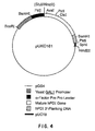
- the junction formed between the blunt ended Stu I and Hin dII termini reconstructs a precise in-frame fusion between the MF ⁇ 1 pre-pro leader sequence and the mature portion of human PDI (resulting plasmid was designated pHKC161; Figure 4).
- the LYS 2 integrating vector pNL (pUC-NotI-LYS2) was digested with Stu I plus Xho I and made flush-ended by treatment with T4 DNA polymerase.
- Plasmid puKC161 was digested with Eco RI plus Hin dIII and the resulting 2.8 kbp Eco RI- Hin dIII fragment bearing the GAL 10 promoter - alpha factor pre-pro leader - human PDI expression cassette was gel-purified and made flush-ended by treatment with T4 DNA polymerase.
- the above flush-ended pNL vector fragment and this expression cassette fragment were ligated together and the ligation mixture was used to transform E . coli strain ATCC 35691.
- pNL-MF ⁇ 1-hPDI Transformants were screened for those containing a plasmid with the expected structure and the resulting plasmid, pNL-MF ⁇ 1-hPDI was prepared in large amounts. When digested with Not I, pNL-MF ⁇ 1-hPDI yields a 6.2 kbp expression cassette flanked on either end by LYS 2 DNA sequences. The digested DNA was used to transform S . cerevisiae strains BJ1995 and JRY188 using the spheroplast method (Hinnen A. et al ., 1978, Proc. Nat'l. Acad. Sci. USA, 75 , pp.1929-1933).
- the Not I ends directed the expression cassette to the chromosomal LYS 2 locus where the cassette integrated via homologous recombination.
- Transformants were screened for those which grow on solid media containing alpha-amino adipic acid (Chattoo, B.B. et al ., 1979, Genetics, 93 , pp. 51; Barnes and Thorner, 1986, supra), indicating the strains are lys - .
- Southern blot analysis of the clonal isolates using a LYS 2 probe confirmed that the expression cassette had integrated at the LYS 2 locus.
- yeast strains which overproduce human PDI using the yeast PDI or human PDI signal sequences
- the PDI cDNA clone pI (Pihlajaniemi et al., 1987, supra) was digested with Pst I and the 1.5 kbp Pst I- Pst I fragment carrying the 3'-region of the human PDI cDNA was gel-purified. This fragment was then inserted into the Pst I site of pUKC150 (described in Example 9 above) yielding the plasmid pUKC151, which contains the intact, full-length human PDI cDNA.
- pUKC151 was digested with Hin dIII and ligated with an appropriate oligonucleotide adapter (containing an Eco RI recognition sequence) to convert the Hin dIII site located at the 3'- end of the PDI cDNA to an Eco RI site.
- the resulting plasmid, pUKC153 contains the intact human PDI coding sequence on a 2.1 kbp Eco RI fragment.
- Plasmid pUKC153 was digested with Eco RI plus Pst I. The resulting 0.47 kbp Eco RI- Pst I and 1.7 kbp Pst I- Eco RI fragments carrying the 5'- and 3'- portions of the human PDI sequence, respectively, were gel-purified.
- pUC19 was digested with Eco RI plus Pst I and the 2.7 kbp vector fragment was gel-purified and then ligated with the aforementioned 0.47 kbp Eco RI- Pst I fragment.
- the ligation mixture was used to transform E . coli ATCC 35691.
- Plasmid DNA was prepared from transformants containing a plasmid with the expected structure. This DNA was digested with Ava I plus Pst I and a 0.38 kbp fragment carrying the 5'-portion of the human PDI sequence was gel-purified.
- pUC19 was digested with Eco RI plus Bam HI and the 2.7 kbp vector fragment was gel-purified.
- the following oligonucleotides were synthesized:
- Oligonucleotides #15165-220 and 15165-249 were kinased and then annealed with oligonucleotides #15165-221 and 15165-250, respectively.
- the following ligation was set up: the pUC19 2.7 kbp Bam HI- Eco RI fragment was ligated with the 1.7 kbp Pst I- Eco RI hPDI 3'-fragment, the 0.38 kbp Pst I- Ava I 5'-hPDI fragment, and the annealed linkers 15165-220 plus 15165-221.
- the following ligation mixture was set up: the pUC19 2.7 kbp Bam HI- Eco RI fragment was ligated with the 1.7 kbp Pst I- Eco RI hPDI 3'-fragment, the 0.38 kbp Pst I- Ava I 5'-hPDI fragment, and the annealed linkers 15165-249 plus 15165-250.
- the annealed linkers contain Bam HI and Ava I cohesive ends and encode the indicated signal peptide sequence plus yeast 5'-nontranslated leader sequence).
- the ligation mixtures were transformed into E . coli ATCC 35691 and transformants were screened for those containing plasmids with the expected structure.
- the DNA sequence across the region including the oligonucleotide linkers and the flanking DNA was confirmed by dideoxy sequencing methods.
- the human PDI coding sequences with either the yeast PDI signal peptide or human PDI signal peptide encoding sequences were designated ySP-hPDI and hSP hPDI, respectively.
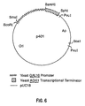
- the plasmid p401 (which contains the GAL 10 promoter and ADH 1 transcription terminator separated by a unique Bam HI site; Figure 6) was digested with Bam HI, made flush-ended, and ligated with the aforementioned cassettes to yield the plasmids pGAL-ySP-hPDI and pGAL-hSP-hPDI, respectively.
- these latter two plasmids were digested with Xba I plus Sac I to generate linear fragments with LYS 2 flanking ends and the linear fragments were used to transform yeast strains BJ1995 and JRY188.
- Transformants which had integrated the desired expression cassettes at LYS 2 were identified by Southern blots of genomic DNA which had been digested with Bgl II and was then hybridized with a LYS 2 probe.
- the resulting strains were BJ1995/hSP-hPDI, BJ1995/ySP-hPDI, JRY188/hSP-hPDI (strain #1148), and JRY188/ySP-hPDI (strain #1157).
- yeast strains which overproduce a C-terminal HDEL mutant of human PDI using the human PDI or yeast PDI signal peptide
- Yeast proteins which are resident in the endoplasmic reticulum normally contain a C-terminal HDEL amino acid sequence which is the signal for retention in the ER (Pelham, et al ., 1988 supra ).
- human PDI has a C-terminal KDEL sequence (Pihlajaniemi, et al ., 1987, supra ) which has been previously shown to function poorly for ER retention in yeast (Lewis, M.J., et al ., 1990, Cell, 61 , pp.1359-1363). Therefore, it was desired to construct a modified human PDI in which the C-terminal KDEL was changed to HDEL. This was accomplished as follows.
- the two plasmids pUC-ySP-hPDI and pUC-hSP-hPDI were digested with Eco RI and Xho I and the resulting 4.0 kbp Eco RI- Xho I fragment containing vector sequences plus the 5'-portion of the hPDI sequence and a 0.5 kbp Xho I- Xho I fragment containing the middle portion of the hPDI coding sequence were gel-purified.
- the following oligonucleotide adapter was then synthesized:
- this adapter was digested with Xho I (now yielding Eco RI and Xho I cohesive ends> and ligated in two separate reactions with the 4.0 kbp Eco RI- Xho I vector fragments containing either the 5'-ySP-hPDI or 5'-hSP-hPDI sequences, respectively.
- the resulting two plasmids were then digested with Xho I and ligated with the aforementioned 0.5 kbp Xho I- Xho I fragment containing the middle portion of the hPDI coding sequence, yielding the plasmids pUC-ySP-hPDI(HDEL) and pUC-hSP-hPDI(HDEL), respectively, into which the Xho I fragment had inserted in the correct orientation to reconstruct the human PDI coding sequence.
- pLYS2-ySP-hPDI(HDEL) and pLYS2-hSP-hPDI(HDEL) were linearized by digestion with Hpa I plus Eco RV and then used in separate reactions for transformation of strains BJ1995 and JRY188. Lys - transformants were selected on solid media containing alpha-amino adipic acid. Isolates containing the desired expression cassette integrated at the LYS 2 locus were identified by Southern blot analysis of genomic DNA.
- strain #1268 The resulting strains were designated BJ1995/ySP-hPDI(HDEL), BJ1995/hSP-hPDI(HDEL), JRY188/ySP-hPDI(HDEL) (strain #1268), and JRY188/hSP-hPDI(HDEL)(strain #1267).
- yeast strains which overproduce C-terminal HDEL mutant of human PDI using the yeast alpha factor secretory leader
- Plasmid pUKC161 ( Figure 4) was digested with Bam HI plus Cla I and the 0.7 kbp Bam HI- Cla I fragment bearing the alpha factor pre-pro leader sequence and 5'-segment of hPDI was gel-purified.
- the plasmid pUC-ySP-hPDI(HDEL) (described in Example 11) was digested with Cla I and Eco RI and the 1.0 kbp Cla I- Eco RI fragment bearing the 3'-segment of hPDI with the C-terminal HDEL modification was gel-purified.
- pUC19 was digested with Bam HI plus Eco RI and the resulting vector fragment was ligated with both the 0.7 kbp Bam HI- Cla I fragment and the 1.0 kbp C la I- Eco RI fragment to yield the plasmid pUC-MF ⁇ 1-hPDI(HDEL).
- This plasmid was digested with Bam HI and the 1.7 kbp Bam HI- Bam HI fragment carrying the PDI cassette was gel-purified and inserted into the Bam HI site of the plasmid p401 ( Figure 6), yielding the plasmid p GAL -MF ⁇ 1-hPDI(HDEL).
- This plasmid was then digested with the enzymes Sma I, Sph I, and Pvu I and the resulting 2.6 kbp Sma I- Sph I fragment bearing the expression cassette was gel-purified and made flush-ended.
- the pUC13-LYS2 vector was digested with Xho I, made flush-ended and then ligated with the above 2.6 kbp flush-ended fragment.
- the resulting plasmid, pLYS2-MF ⁇ 1-hPDI(HDEL) was digested with Hpa I plus Eco RV and then used for transformation of strains JRY188 and BJ1995.
- the resulting transformants were evaluated by Southern blots of genomic DNA (as described in Example 9) to confirm that the desired expression cassette had integrated at the LYS 2 locus.
- the JRY188 transformant was designated strain #1279.
- yeast strains which overexpress the yeast PDI protein from an integrated expression cassette at LYS2 locus
- the plasmid C7 (described in Example 4) bearing the complete yeast PDI 1 gene was digested with Eco RV and the 1.3 kbp Eco RV- Eco RV fragment containing the C-terminal portion of the yeast PDI open reading frame (ORF) (from amino acid 223 to end of ORF) plus the 3'-nontranslated sequence was gel-purified and inserted into the Eco RV site of the plasmid pAT153 [Twigg, A.G. and Sherratt, D., 1980, Nature, 283 , pp.216-218], yielding pUKC169.
- ORF open reading frame
- Plasmid C7 was then digested with Ban I plus Eco RV and the 0.67 kbp Ban I- Eco RV fragment encoding amino acids 6-222 of the yeast PDI ORF was gel-purified and ligated with the following synthetic oligonucleotide adapter: which contains Bam HI and Ban I cohesive ends, respectively, and encodes amino acids 1-5 of the yeast PDI ORF plus 12 basepairs of yeast 5'-nontranslated leader sequence.
- Plasmid pUKC169 was digested with Eco RV and the resulting 1.3 kbp Eco RV- Eco RV fragment bearing the aforementioned C-terminal portion of yeast PDI was gel-purified and then inserted into the unique Eco RV site of pUKC170 thereby regenerating the intact yeast PDI (yPDI) gene. This resulting plasmid was designated pUKC175.
- pUKC175 was digested with Eco NI and the resulting 2.1 kbp fragment bearing the yPDI gene was made flush-ended and gel-purified.
- pUC19 was digested with Sac I plus Sma I, made flush-ended, and ligated with the above flush-ended Eco NI yPDI fragment.
- the ligation mixture was used to transform E . coli DH5 cells and the resulting transformants were screened for those containing plasmids with the yPDI insert in the appropriate orientation such that a Bam HI site in the pUC19 polylinker was located adjacent to the 3'-end of the yPDI coding sequence.
- this construct (designated pUC19-yPDI) now contains the yPDI ORF on a 1.9 kbp Bam HI fragment.
- pUC19-yPDI was digested with Bam HI and the 1.9 Bam HI kbp fragment bearing the yPDI gene was gel-purified and then subcloned into the Bam HI site of the vector pUC18-GAL10p(B)ADH1t (stock #401) ( Figure 6).
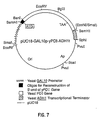
- the resulting plasmid, pUC18-GAL10p-yPDI-ADH1t ( Figure 7), is stock #1015.
- Plasmid pUC18-GAL10p-yPDI-ADH1t was digested with Sma I, Sph I, plus Sac I and the 2.7 kbp Sma I- Sph I fragment carrying the expression cassette was gel-purified, made flush-ended, and then cloned into the unique Stu I site of pUKC171 (pUKC171 contains the 4.5 kbp Eco RI- Hin dIII LYS 2 fragment of YIp600 (Barnes and Thorner, 1986, supra) subcloned into pUC19 which had been previously digested with Eco RI plus Hin dIII).
- the resulting pUKC171-GAL10p-yPDI vector was then digested with Eco RI plus Pvu II to excise the LYS 2- GAL 10p-y PDI - ADH 1t- LYS 2 cassette which was then used to transform S . cerevisiae strains JRY188 and BJ1995.
- the resulting lys - transformants were evaluated by Southern blots of genomic DNA preparations as described in Example 9. Isolates of each strain were found which had the GAL 10p-yPDI cassette integrated at the LYS 2 locus.
- the resulting strains were designated BJ1995/yPDI and JRY188/yPDI (strain #1152).
- the plasmid pUC-Not-URA3 (Example 8) was digested with Apa I plus Nco I (to delete a portion of the URA 3 gene) and made flush-ended.
- Plasmid pUC18-GAL10p-yPDI-ADH1t was digested with Eco RI, Sca I, and Sph I and the 2.8 kbp Eco RI- Sph I fragment bearing the GAL10p-yPDI-ADH1t expression cassette was gel-purified, made flush-ended, and ligated with the above vector fragment, yielding the plasmid pNU-GAL10p-yPDI.
- the URA3-GAL10p-yPDI-ADH1t-URA3 integrating cassette was excised from pNU-GAL10p-yPDI by digestion with Not I. The resulting linear fragment was used to transform yeast strain KHY107. Ura - transformants were selected on solid media containing 5-fluoro-orotic acid (Boeke et al ., 1984, Mol. Gen. Genet., 197 , pp 345). Genomic DNA from the resulting ura - transformants was digested with Bg lII and evaluated by Southern blots using a radiolabelled Eco RI- Pvu II fragment from the GAL10p-yPDI-ADHlt cassette as probe.
- Isolates were identified which had integrated the desired GAL10p-yPDI-ADHlt expression cassette at URA 3.
- Isolate K-Y1 had multiple copies integrated at URA 3 (strain #1136).
- Isolate K-Y3 had one copy integrated at URA 3 (strain #1137).
- yeast strains were grown in 3 ⁇ YEPD liquid medium for 24 hours, at 23°C. After that period, cultures were supplemented with galactose to a final 4.8% concentration. The cultures were then reincubated at 23°C, for an additional period of 24 hours.
- yeast strains were cultivated in 3 ⁇ YEPD for 24 hours at 30°C. The cells were harvested and washed with cold sterile water, and resuspended in the same volume of 3 ⁇ YEPD-Galactose medium. The yeast strains were incubated for a further period between 16 and 25 hours, after which, they were harvested and protein extracted by extraction method 2 (below).
- Proteins were extracted from exponentially growing or stationary phase cells using glass bead disruption, essentially as described by Mellor et al ., (1983, Gene, 24 , pp.1-14).
- Method 1 Protein was extracted by glass bead disruption of cell walls, in the presence of PMSF (0.5mM) in a 25mM phosphate buffer pH 7.0, followed by a freeze-thaw cycle, and soluble protein was recovered by centrifugation for 10 minutes at 13,000 rpm. Secretion was initially assessed by analysis of spent culture liquid, either before or after concentration with PEG (solid), ammonium sulphate (0-80%), or an ultrafiltration membrane ( ⁇ 100 kDa). Protein concentration was determined by the method of Bradford (1976, Anal. Biochem., 72 , pp.248-254).
- Method 2 Intracellular samples were prepared as in method 1, but the culture medium was supplemented with NaOH, and ⁇ -mercaptoethanol (respectively, final concentrations of 0.2M and 1%), left on ice for about 10 minutes, after which TCA was added to a final concentration of 6%. After 30 minutes standing on ice, protein was recovered by centrifugation, washed with cold acetone and resuspended in SDS-PAGE loading buffer.
- Electrophoresis was conducted under the following conditions: 10% SDS-polyacrylamide gel, and 10 ⁇ g protein loaded per lane (protein extraction method 1). Sigma prestained molecular weight standards were run in all the gels. Gels were run in a BioRad mini-Protein II gel system. Extracellular extracts were loaded at 15-20 ⁇ l per lane, without estimating protein concentration. Voltage was kept below 200 volts during electrophoresis.
- Proteins were transferred to nitrocellulose, using a Biometra semi-dry Western blot system. Nitrocellulose membranes were blocked with 5% (w/v) powdered milk for one hour, washed, incubated with anti human-PDI polyclonal antibody for between 3 hours to overnight, at dilutions ranging from 1:500 to 1:750. Membranes were washed and peroxidase-conjugated anti-rabbit IgG was added at a final dilution of 1:1000, and incubation continued for one hour. After washing the blots were developed using an Amersham ECL kit as described by the manufacturer.
- strain 1072A produced secreted hPDI at levels detectable by Western blot.
- the level of detection was 0.05 ⁇ g of purified bovine PDI by the ECL protocol employed. This secreted PDI was shown to be a dimer since it was retained by a 100 kDa cut-off ultrafiltration membrane.
- strain 1072A and its corresponding HDEL variant (1279) were compared, it was found that the human PDI was secreted by both.
- the final culture/induction conditions were optimised in terms of temperature (°C) of growth and induction period. The two strains showed a higher level of PDI synthesis when cultivated at 23°C and then induced for 16 hours at 30°C, or when cultivated and induced at 30°C, for 16 hours.
- Antistasin is a potent protein inhibitor of the blood coagulation Factor Xa.
- Antistasin was isolated from the salivary glands of the Mexican leech Haementeria officinalis (Nutt, E. et al ., 1988, J. Biol. Chem., 263 , pp.10162-10167). The cDNA encoding ATS was subsequently isolated and characterized by Han, J. H. et al . (1989, Gene, 75 , pp.47-57).
- ATS is an ideal reporter protein for the evaluation of the effects of increased levels of PDI activity on the folding and formation of proper disulfide bonds in a heterologous protein secreted by recombinant yeast since ATS has 10 disulfide bonds which must be correctly paired in order for the protein to have biological activity.
- ATS was expressed in yeast using the expression vector pKH4 ⁇ 2 (Jacobson, M.A. et al ., 1989, Gene, 85 , pp.511-516), which contains the galactose-inducible GAL 10 promoter and the yeast MF ⁇ 1 pre-pro secretory leader sequence to direct secretion of heterologous proteins.
- the coding sequence for ATS was isolated by polymerase chain reaction (PCR) methods using subcloned ATS cDNA from clone ⁇ 5C-4 (Han, J. H. et al .; supra) as substrate and the oligonucleotide primers:
- Transformants were selected on synthetic solid media lacking leucine (Schultz, L. et al ., 1987; Gene, 61 , pp.123-133) and streaked for clonal isolates which were used in subsequent analyses. Strains were preserved by storage at -70°C in synthetic media containing 17% glycerol.
- the K991-transformed parental JRY188 strain plus the various transformed derivatives which overproduce either yeast or human PDI were evaluated for secretion of antistasin by the following procedure.
- the indicated strains were streaked from the -70°C frozen glycerol stocks onto leucine-minus synthetic agar plates and grown for 3 days at 30°C.
- Culture tubes (18 x 150 mm) containing 5-mL of 3xYEHD [60g Difco yeast extract, 30g HySoy peptone, 48g glucose per liter] media were inoculated with a small loopful of cells and incubated for about 18 hours at 23°C on a tissue culture roller drum.
- A1, A2 and A3 refer to different clonal isolates of the indicated strain evaluated in parallel.
- Overexpression of yeast PDI results in 4-fold higher secretion of ATS activity on a per cell basis, and about 9-fold higher secretion on volumetric basis for isolate K-Y3-A1.
- yeast host strains which overproduce either yeast PDI or human PDI from a multicopy plasmid
- the multicopy yeast shuttle vector YEp24 (Botstein, D. et al ., 1979, Gene, 8 , pp.17-24) contains the yeast 2-micron DNA origin of replication and the yeast URA 3 gene for selection on uracil-minus synthetic media. YEp24 was digested with Bam HI and the resulting 7.8 kbp Bam HI vector fragment was gel-purified (fragment a).
- the plasmid pUC18-GAL10p-yPDI-ADH1t (#1015) was digested with Eco RI, Sph I, and Sca I; the resulting 2.8 kbp Eco RI- Sph I fragment bearing the GAL 10p-yPDI- ADH 1t expression cassette was gel purified (fragment b).
- Plasmid pUKC161 was digested with Eco RI plus Hin dIII and the 2.8 kbp Eco RI- Hin dIII fragment bearing the GAL1p--MF ⁇ 1 pre-pro--human PDI expression cassette was gel purified (fragment c).
- Transformants containing both plasmids were selected on synthetic media lacking both leucine and uracil and isolated single colonies were restreaked on the same media for selection of clonal isolates.
- Five such clonal isolates for each of the two original cotransformations were inoculated into 5 ml of 3xYEHD medium in culture tubes and incubated for 24 hr at 23°C in a tissue culture roller drum. At the end of that time, galactose was added to a final concentration of 4.8% and the cultures were incubated for an additional 5 days at 23°C. Cell were removed by centrifugation and the clarified media supernatants were assayed for the levels of ATS activity by the Factor Xa inhibition assay.
- the contransformants which contained the YEp24-GAL10p-yPDI plasmid plus the ATS expression vector showed 3- to 26-fold higher levels of secreted ATS activity, depending on the isolate, compared to the parental JRY188 strain containing only the ATS expression vector.
- the contransformants containing the YEp24-GAL1p-MF ⁇ -hPDI plasmid plus the ATS expression vector showed 2- to 3-fold higher levels of secreted ATS activity compared to the parental JRY188 strain containing only the ATS expression vector.
- yeast host strains which overproduce either yeast or human PDI from the same expression vector used for expression of a desired heterologous protein.
- the S . cerevisiae GAL 1 and GAL 10 genes are transcribed divergently from a region between the two structural genes which contains the divergent GAL 1 and GAL 10 promoters and a common GAL 4 binding domain located between the TATA boxes for the two promoters.
- the plasmid pBM272 (Johnston, M. and Davis, R., 1984, Mol. Cell. Biol., 4 , pp.1440) contains this divergent yeast GAL 1- GAL 10 promoter on a 0.85 kbp Eco RI- Hin dIII fragment (with also an internal Bam HI site adjacent to the Hin dIII site).
- This promoter fragment was used to construct a divergent promoter cassette vector, pUC-GAL1/10, which has the following properties: yeast GAL 10 promoter separated from the yeast ADH 1 transcription terminator (0.35 kbp Hin dIII- Sph I fragment) by unique Eco RI and Sma I sites, in that order. Yeast GAL 1 promoter separated from a second copy of the ADH 1 transcription terminator by unique Bam HI and Hin dIII sites. The 3'-end of both ADH 1 terminator elements are flanked by Sph I sites to allow one to isolate the entire divergent promoter expression cassette as an Sph I fragment.
- the vector backbone in this plasmid is pUC18 with the above expression cassette in place of the polylinker.
- the plasmid pUC-GAL1/10 was digested with Bam HI and gel-purified to generate fragment "a".
- the plasmid pUKC161 was digested with Bam HI and the 1.9 kbp Bam HI fragment bearing the alpha factor pre-pro leader fused in-frame to the mature human PDI coding sequence was gel-purified and ligated to vector fragment a, yielding the plasmid pUC-GAL1/10-hPDI, in which the expression of the alpha factor pre-pro--hPDI fusion is under control of the GAL 1 promoter.
- the plasmid pUC18-GAL10p-yPDI-ADH1t (Example 13) was digested with Bam HI and the resulting 1.7 Kbp Bam HI fragment bearing the yeast PDI coding sequence was gel-purified and then ligated with vector fragment a, yielding the plasmid pUC-GAL1/10-yPDI in which the GAL 1 promoter directs the expression of yeast PDI.
- These two resulting plasmids were then digested with Eco RI and made flush-ended, yielding vector fragments b and c bearing the hPDI and yPDI cassettes, respectively.
- the ATS expression vector (K991) was digested with Sal I plus Bgl II and the Sal I- Bgl II fragment bearing the alpha factor pre-pro leader fused in-frame to the coding sequence for mature ATS was gel-purified, made flush-ended, and ligated in separate reactions to the two flush-ended vector fragments b and c .
- the resulting plasmids with the correct structure as determined by restriction mapping were designated pUC-GAL1/10-hPDT/ATS ( Figure 11) and pUC-GAL1/10-yPDI/ATS ( Figure 12), respectively.
- strain JRY188 cotransformed with the antistasin expression vector pKH4 ⁇ 2/ATS and either YEp24-GAL1p-MF ⁇ -hPDI or YEp24-GAL10p-yPDI were evalutated for antistasin secretion after growth at either 23°C or 30°C.
- the parent strain JRY188 transformed only with the antistasin expression vector was grown in parallel. After overnight growth in 3xYEHD medium at either 23°C or 30°C, cell cultures were induced by the addition of galactose to a final concentration of 4.8% and propagated for an additional five days at either 23°C or 30°C, as appropriate.
- tick anticoagulant peptide TRIP
- Tick anticoagulant peptide is a potent, highly selective inhibitor of the blood coagulation factor Xa (Waxman, L. et al ., 1990, Science, 248 , pp. 593-596).
- TAP is a novel serine protease inhibitor isolated from the tick Ornithidoros moubata .
- TAP is composed of 60 amino acids including 6 cysteine residues (Waxman et al ., 1990, supra).
- TAP has been expressed in yeast using the expression vector pKH4-TAP which contains the galactose-inducible GAL10 promoter and the yeast MF ⁇ 1 pre-pro secretory leader sequence fused in frame to the synthetic gene encoding TAP (Neeper, M. et al ., 1990, J. Biol. Chem., 265 , pp.17746-17752).
- This vector contains a slightly modified MF ⁇ 1 pre-pro leader sequence due to the presence of a Bam HI cloning site located at the position of amino acid 79 of the pre-pro leader (Neeper et al ., 1990, supra).
- a second TAP expression vector, pKH4-3B/TAP was constructed which contains the authentic MF ⁇ 1 pre-pro leader sequence fused in-frame to the synthetic gene encoding TAP.
- the plasmid pKH4-TAP containing the synthetic TAP gene (Neeper et al ., 1990, supra) was used as the DNA template in a polymerase chain reaction (PCR) using the following two oligonucleotide primers in order to modify the 5'-and 3'- termini of the synthetic TAP gene, respectively:
- the PCR reaction was carried out by methods well known to those of ordinary skill in the art (Innis, M.A. et al ., editors, 1990, PCR Protocols: A Guide to Methods and Applications, Academic Press, Inc., San Diego CA).
- the resulting PCR product was phosphorylated with T4 polynucleotide kinase, digested with Bam HI, and then gel-purified to yield a 0.2 kbp blunt- Bam HI fragment containing the blunt end at the exact 5'-end of the TAP coding sequence and a cohesive Bam HI end on the 3'-side of the translation termination codon.
- the vector pKH4-3B (Hofmann, K. and Schultz, L.D., 1991, Gene, 101 , pp.105-111) contains a unique Sph I site at the 3'-end of the MF ⁇ 1 pre-pro leader coding sequence.
- pKH4-3B was digested with Sph I, made flush-ended by treatment with T4 DNA polymerase, and then digested with Bgl II.
- the resulting blunt- Bgl II vector fragment was gel-purified and ligated with the aforementioned 0.2 kbp blunt- Bam HI TAP fragment to yield the vector pKH4-3B/TAP.
- yeast strains BJ1995, JRY188, and U9 were cotransformed with the vectors YEp24-GAL10p-yPDI plus either pKH4-TAP or pKH4-3B/TAP.
- Cotransformants containing both plasmids were selected on synthetic medium lacking both leucine and uracil and isolated single colonies were restreaked on the same medium for selection of clonal isolates.
- Three such clonal isolates for each of the different vector/host cotransformations were inoculated into 5-mL of modified 5xLeu - media lacking uracil (5xLeu - Ura - ) and containing 4% glucose in culture tubes.
- the cultures were incubated for 24 hours at 30°C in a tissue culture roller drum. At the end of that time, the cells were recovered by centrifugation and resuspended in 5 mL of 5xLeu - Ura - medium containing 4% galactose. The resulting cultures were incubated at 30°C for an additional 48 hours. Cells are then removed by centrifugation and clarified media samples evaluated for the levels of secreted TAP by SCX-HPLC or Factor Xa inhibition assay (Waxman et al ., 1990, supra).
- the recombinant yeast cells are grown for 24 hours at 23°C, induced by addition of galactose to 4% final concentration, and then incubated for an additional five days at 23°C. Clarified media samples are then evalutated for the levels of secreted TAP as described above.
Landscapes
- Health & Medical Sciences (AREA)
- Chemical & Material Sciences (AREA)
- Life Sciences & Earth Sciences (AREA)
- Organic Chemistry (AREA)
- Genetics & Genomics (AREA)
- Engineering & Computer Science (AREA)
- Zoology (AREA)
- Molecular Biology (AREA)
- Bioinformatics & Cheminformatics (AREA)
- General Health & Medical Sciences (AREA)
- Biochemistry (AREA)
- Wood Science & Technology (AREA)
- General Engineering & Computer Science (AREA)
- Biophysics (AREA)
- Biotechnology (AREA)
- Biomedical Technology (AREA)
- Medicinal Chemistry (AREA)
- Proteomics, Peptides & Aminoacids (AREA)
- Gastroenterology & Hepatology (AREA)
- Microbiology (AREA)
- Mycology (AREA)
- Tropical Medicine & Parasitology (AREA)
- Physics & Mathematics (AREA)
- Plant Pathology (AREA)
- Preparation Of Compounds By Using Micro-Organisms (AREA)
- Micro-Organisms Or Cultivation Processes Thereof (AREA)
- Enzymes And Modification Thereof (AREA)
- Peptides Or Proteins (AREA)
Abstract
Description
- Protein disulfide isomerase (PDI) is an enzyme involved in the catalysis of disulfide bond formation in secretory and cell-surface proteins. Using an oligonucleotide designed to detect the conserved "thioredoxin-like" active site of vertebrate PDI's (WCGHCK) (SEQ.ID.NO.: 1), we have isolated a gene encoding PDI from the lower eukaryote Saccharomyces cerevisiae. The nucleotide sequence and deduced open reading frame of the cloned gene predicts a 530 amino acid protein of molecular weight 59,082 and pI of 4.1, physical properties characteristic of mammalian PDIs. Furthermore, the amino acid sequence shows 30-32% identity and 53-56% similarity with mammalian and avian PDI sequences and has a very similar overall organization, namely the presence of two 100 residue segments, each of which is repeated, with the most significant homologies to mammalian and avian PDIs being in the regions (a, a') that contain the conserved "thioredoxin-like" active site. The N-terminal region has the characteristics of a cleavable secretory signal sequence and the C-terminal four amino acids (-HDEL)(SEQ.ID.NO.: 2) are consistent with the protein being a component of S. cerevisiae endoplasmic reticulum (E.R.). Transformants carrying multiple copies of this gene (designated PDI1) have 10-fold higher levels of PDI activity and overexpress a protein of the predicted molecular weight. The PDI1 gene is unique in the yeast genome and encodes a single 1.8kb transcript that is not found in stationary phase cells, nor is it heat-inducible. Disruption of the PDI1 gene is haplo-lethal indicating that the product of this gene is essential for viability.
- Protein disulfide-isomerase (PDI), an enzyme which catalyzes thiol:disulfide interchange reactions, is a major resident protein component of the E.R. lumen in secretory cells. A body of evidence on the enzyme'e cellular distribution, its subcellar location and its developmental properties suggests that it plays a role in secretory protein biosynthesis (Freedman, 1984, Trends Biochem. Sci. 9, pp.438-41) and this is supported by direct cross-linking studies in situ (Roth and Pierce, 1987, Biochemistry, 26, pp.4179-82). The finding that microsomal membranes deficient in PDI show a specific defect in cotranslational protein disulfide formation (Bulleid and Freedman, 1988, Nature, 335, pp.649-51) implies that the enzyme functions as a catalyst of native disulfide bond formation during the biosynthesis of secretory and cell surface proteins. This role is consistent with what is known of the enzyme's catalytic properties in vitro; it catalyzes thiol: disulfide interchange reactions leading to net protein disulfide formation, breakage or isomerization, and can catalyze protein folding and the formation of native disulfide bonds in a wide variety of reduced, unfolded protein substrates (Freedman et al., 1989, Biochem. Soc. Symp., 55, pp.167-192). The DNA and amino acid sequence of the enzyme is known for several species (Scherens, B. et al., 1991, Yeast, 7, pp. 185-193; Farguhar, R., et al., 1991, Gene, 108, pp. 81-89) and there is increasing information on the mechanism of action of the enzyme purified to homogeneity from mammalian liver (Creighton et al., 1980, J. Mol. Biol., 142, pp.43-62; Freedman et al., 1988, Biochem. Soc. Trans., 16, pp.96-9; Gilbert, 1989, Biochemistry 28, pp.7298-7305; Lundstrom and Holmgren, 1990, J. Biol. Chem., 265, pp.9114-9120; Hawkins and Freedman, 1990, Biochem. J., 275, pp.335-339). Of the many protein factors currently implicated as mediators of protein folding, assembly and translocation in the cell (Rothman, 1989, Cell 59, pp.591-601), PDI is unusual in having a well-defined catalytic activity.
- PDI is readily isolated from mammalian tissues and the homogeneous enzyme is a homodimer (2 x 57kD) with characteristically acidic pI (4.0-4.5) (Hillson et al., 1984, Methods Enzymol., 107, pp.281-292). The enzyme has also been purified from wheat and from the alga Chlamydomonas reinhardii (Kaska et al., 1990 Biochem. J. 268, pp.63-68). The activity has been detected in a wide variety of sources, and in a preliminary report, PDI activity was claimed to be detectable in S. cerevisiae (Williams et al., 1968, FEBS Letts., 2, pp.133-135). Recently, the complete amino acid sequences of a number of PDIs have been reported, largely derived from cloned cDNA sequences; these include the PDIs from rat (Edman et al., 1985, Nature, 317, pp.267-270) bovine (Yamauchi et al., 1987, Biochem. Biophys. Res. Comm., 146, pp.1485-1492) human (Pihlajaniemi et al., 1987, EMBO J., 6, pp.643-9), yeast (Scherens, B., et al., supra; Farquhar, R. et al., supra) and chick (Parkkonen et al., 1988, Biochem. J., 256, pp.1005-1011). The proteins from these vertebrate species show a high degree of sequence conservation throughout and all show several overall features first noted in the rat PDI sequence (Edman et al. 1985 supra). The most significant is the presence within the PDI sequence of two regions of approximately 100 residues strongly homologous to each other and closely related in sequence to thioredoxin, a small redox active-protein containing an active site disulfide/dithiol couple formed between vicinal Cys residues. In thioredoxin the active site sequence is WCGPCK (SEQ.ID.NO.: 3), whereas the corresponding region, found twice in PDI, has the sequence WCGHCK (SEQ.ID.NO.:1). (Other repeats, motif and homologies identified within the PDI sequences are discussed below).
- Sequences corresponding to, or closely related to PDI have been identified in work aimed at analysing functions other than disulfide bond formation. For example, there is clear-cut evidence that PDI acts as the β subunits of the tetrameric α2β2 enzyme prolyl-4-hydroxylase, which catalyzes a major post-translational modification of nascent or newly-synthesized procollagen polypeptides within the E.R. (Pihlajaniemi et al., 1987, supra; Koivu et al., 1987, J. Biol. Chem., 262, pp.6447-49)). There is also evidence suggesting that PDI participates in the system for cotranslational N-glycosylation (Geetha-Habib et al., 1988, Cell, 54, pp.63-68) and recently the proposal has been made that the enzyme participates in the complex which transfers triglyceride to nascent secretory lipoproteins (Wetterau et al., 1990, J. Biol. Chem., 265, pp.9800-7). Thus, PDI may be multifunctional in the co- and post-translational modification of secretory proteins (Freedman, 1989, Cell, 57, pp.1069-72).
- The vast majority of mammalian secretory proteins contain multiple intramolecular and/or intermolecular disulfide bonds. Examples include, but are not limited to, pituitary hormones, interleukins, immunoglobulins, proteases and their inhibitors and other serum proteins. Such proteins are among the prime targets for commercial genetic engineering, but early experience in their expression in bacteria and yeast has highlighted a number of problems in obtaining them as functionally active recombinant products. This has drawn attention to the need for a better understanding of post-translational modifications in general, and of protein folding and disulfide bond formation in particular.
- Disulfide bonded proteins comprising a single folded domain can, in general, be fully reduced and denatured and subsequently renatured in vitro to generate the correctly disulfide-linked state in reasonable yield. The process involves rapid formation of a mixed population of many differently disulfide bonded forms which slowly isomerize to give the native disulfide pairing. The process is catalysed by thiol/disulfide redox buffers (e.g. GSH and GSSG) and by alkaline pH. Low protein concentrations are required to prevent precipitation and interchain disulfide formation. In general the rate of formation of the native protein, and the optimal obtainable yield, both decrease as the number of intramolecular disulfides increases. The problem is compounded in proteins containing multiple disulfide bonded domains (e.g. tissue plasminogen activator) in which each domain must fold and form its native disulfide bonds independently.
- The process of disulfide bond formation in vivo occurs co-translationally or as a very early post-translational event. Studies on nascent and newly synthesized secretory proteins in the lumen of the E.R. in mammalian cells show that native disulfide bonds are already formed. The process in vivo appears to be catalyzed by the enzyme protein disulfide-isomerase which is an abundant protein in secretory cells and is located at the luminal face of the endoplasmic reticulum [Freedman, R.B., 1984, Trends in Biochemical Sciences, 9, 438-441]. This enzyme in vitro, catalyzes thiol:protein-disulfide interchange reactions in a wide range of protein substrates and has the properties required of a cellular catalyst of native protein disulfide formation [Freedman, R.B. et al., 1984, Biochem. Soc. Trans., 12, 939-942]. Further evidence for its role include (i) that its tissue distribution matches that of the synthesis of disulfide bonded secretory proteins [Brockway, B.E. et al., 1980, Biochem, J., 191, 873-876], and (ii) that in a number of systems the amount of enzyme present varies in parallel with a physiological change in the rate of synthesis of disulfide bonded secretory protein [Brockway, B.E. et al., 1980, Biochem J., 191, 873-876; Freedman R.B. et al., 1983, in "Functions of Glutathione:
Biochemical, Physiological, Toxicological & Clinical Aspects", eds. A. Larsson, S. Orrenius, A. Holmgren & B. Mannervik, Raven Press, New York, pp.271-282; Paver, J.L. et al., 1989, FEBS Letters, 242, pp. 357-362]. - The enzyme has been characterized in a number of animal sources [Lambert, N. and Freedman, R.B., 1983, Biochem. J., 213, pp. 225-234], and in wheat [de Azevedo, G.M.V. et al., 1983, Biochem. Soc. Trans., 12, 1043], and a striking conservation of molecular and kinetic properties has been noted [Freedman, R.B. et al., 1984, Biochem. Soc. Trans. 12, pp. 939-942; Brockway, B.E. and Freedman, R.B., 1984, Biochem J., 219, 51-59]. However the enzyme has not been throughly studied in lower eukaryotes or in bacteria. The strong homologies between yeast and higher eukaryotes in the mechanisms and molecular components involved in secretion strongly suggest that the enzyme or an analogue is present in yeast, since at least some yeast secretory proteins (e.g. killer toxin) contain disulfide bonds.
- The application of yeast as a versatile host for the expression of commercially-important mammalian proteins is compromised, to some extent, by the limited capacity of the yeast secretory system and by some differences between it and that of higher eukaryotes (e.g. in glycosylation).
- EP 509,841 (published 21.10.1992) discloses the production of disulfide-bonded human serum albumin by means of heterologous co-expression of human PDI together with HSA in Saccharomyces cerevisiae. This disclosure primarily concerns a "three-gene" process to producing disulfide-bridge proteins involving expression of an ERD2 receptor protein in addition to PDI and HSA. Although encompassing a "two-gene" expression system, this patent application requires in that case that the PDI gene is a fusion gene incorporating DNA encoding the HSA prepro-sequence.
- EP 510,658 discloses a process for enhanced formation of the native conformation of disulfide-bonded proteins in E. coli. This application relates exclusively to the use of prokaryotic host organisms and is enabling only for the use of E. coli.
- EP 293,793 discloses the heterologous expression of human PDI in a variety of host cells including E. coli. simian CO5-7 cells and Saccharomyces cerevisiae and the use of the PDI resulting therefrom for the conversion of suitable proteins to their disulfide-bridged forms.
- The present invention provides an in vitro process for producing disulfide-bonded recombinant proteins comprising:
- (a) over-expressing recombinant protein disulfide isomerase from a recombinant expression cassette containing a gene encoding protein disulfide isomerase in a recombinant host cell; and
- (b) expressing one or more recombinant genes encoding one or more disulfide bonded proteins other than protein disulfide isomerase in the recombinant host cell,
-
- with the proviso that said recombinant host cell is other than E. coli, and that said gene encoding protein disulfide isomerase does not comprise a DNA fragment coding for a human serum albumin prepro-sequence, and that the process does not involve expression from a recombinant expression cassette of a gene coding for the receptor protein ERD2 from yeast.
- The present invention also provides a strain of the yeast Saccharomyces cerevisiae which overexpresses recombinant protein disulfide isomerase from a recombinant expression cassette containing a gene encoding protein disulfide isomerase and one or more recombinant genes encoding one or more disulfide-bonded proteins other than protein disulfide isomerase from a recombinant expression cassette;
- with the proviso that said gene encoding protein disulfide isomerase does not comprise a DNA fragment coding for a human serum albumin prepro-sequence, and that the yeast does not express a gene coding for the receptor protein ERD2 from yeast from a recombinant expression cassette.
- The form "host" throughout the description is used excluding E. coli.
- The forms protein disulfide isomerase and "PDI" throughout the description are used excluding fusions comprising the human serum albumin prepro-sequences.
- DNA encoding human and yeast protein disulfide isomerase (PDI) is isolated and cloned into expression casettes or vectors comprising a promoter and transcription terminator. The expression casettes or vectors containing PDI-encoding DNA are transferred into host cells which, as a result, overproduce PDI protein. These PDI overproducing cells are used as recombinant hosts for the expression of disulfide bonded proteins. Secretion of disulfide bonded proteins is substantially increased in PDI overproducing host cells compared to host cells producing normal levels of PDI.
-
- Figure 1
- shows the SDS-PAGE analysis of a cell-free lysate of an S. Cerevisiae transformant carrying a yeast PDI-encoding gene on a multicopy plasmid.
- Figure 2
- shows the dot plot alignment between yeast PDI and rat PDI using the 'COMPARE' and 'DOTPLOT' software (UWGCG), the domain structure of mammalian PDI shown on the same scale and below the alignment.
- Figure 3
- depicts the strategy and shows results for disruption of the yeast PDI1 gene; Panel (b) shows the results for tetrad analysis of the His+ AS3324 strain heterozygous for the pdil:.HIS3 disruption.
- Figure 4
- shows the structure of plasmid pUKC161.
- Figure 5
- shows the structure of plasmid pUC-ySP-hPDI.
- Figure 6
- shows the structure of plasmid p401, which is also known as pUC18-GAL10p(B)ADH1t.
- Figure 7
- shows the structure of the plasmid pUC18-GAL10p-yPDI-ADH1t.
- Figure 8
- illustrates the structure of plasmid pKH4α2/ATS, which is also known as K991.
- Figure 9
- illustrates the structure of YEp24-GAL10p-yPDI.
- Figure 10
- illustrates the structure of YEP24-GAL1p-MFα-hPDI.
- Figure 11
- illustrates the structure of pUC-GAL1/10-hPDI/ATS.
- Figure 12
- illustrates the structure of pUC-GAL1/10-yPDI/ATS.
- The process of protein folding and secretion in yeast is very complex, involving more than 30 gene products, based on genetic studies (Franzusoff, A. et al., 1991, Methods Enzymology, 194, pp. 662-674). These include peptidyl prolyl cis-trans isomerases, PDI and other thioredoxin-like proteins, BiP, various molecular chaperones (hsp70, hsp60, etc.), signal peptidase, signal recognition protein, the various proteins involved in translocation of precursors into the E.R., the various structural and functional components of the ER, Golgi, and secretory vesicles plus many proteins not yet characterized (Franzuoff, A., et al., 1991, supra; Rothman, J.E. and Orci L., 1992, Nature, 355, pp. 409-415; Gething, M.G. and Sambrook, G., 1992, Nature, 355, pp. 33-45). In view of this complexity, it would seem very unlikely to one of ordinary skill in the art that increasing the levels of only one component (i.e. PDI) would be likely to substantially improve secretion of a particular heterologous protein. Therefore, the present invention provided very unexpected results in that increased levels of PDI alone caused a significant and substantial increase in the levels of secreted proteins, for example antistasin, a phenomenon which is likely to be related to improved protein folding and/or disulfide bond formation.
- The present invention relates to a method for increasing the production of recombinant proteins by recombinant host cells, by overexpressing DNA encoding protein disulfide isomerase (PDI). PDI as used herein refers to an enzyme which specifically catalyzes the formation of intramolecular and intermolecular disulfide bonds.
- The DNA sequence of PDI genes from several species is known in the art. These species include but are not limited to, human, bovine, rat, chicken and yeast. [Mizunaga et al., 1990, J. Biochem., 108, pp.846-851; Scherens et al., 1991, Yeast, 7, pp.185-193].
- Starting material for the isolation of PDI-encoding DNA may be any cell or tissue type including but not limited to mammalian and other vertebrate cells and tissue, as well as lower eukaryotic cells and tissue. The present invention is demonstrated using yeast and human PDI expressed in recombinant yeast host cells. It is readily apparent to one of ordinary skill in the art that the present invention extends to and encompasses other expression hosts including, but not limited to, mammalian cells, plant cells, prokaryotic cells such as bacteria , insect cells and lower eukaryotic cells such as yeast and filamentous fungi. Furthermore, it is readily apparent to one of ordinary skill in the art that the use of PDI encoding DNA derived from sources other than yeast and human cells is encompassed by the present invention. Other sources of PDI-encoding DNA include, but are not limited to, vertebrates other than human such as rat and mouse, non vertebrates such as insects, and lower eukaryotes such as fungi.
- Microsomal membrane fractions prepared from S. cerevisiae by the method of Rothblatt and Meyer (1986, Cell, 44, pp.619-28) had low levels of protein disulfide isomerase (PDI) activity which were enhanced between 8-20 fold by sonication. This suggested the presence within the lumen of the yeast endoplasmic reticulum of an enzyme comparable to PDI found in the same cellular compartment in vertebrates (Mills et al., 1983, Biochem. J., 213, pp.245-8); Lambert and Freedman, 1985, Biochem., J. 228, pp.635-45) and wheat (Roden et al., 1982, FEBS Lett., 138, pp.121-4). The gene coding for PDI was cloned assuming homology to the higher eukaryotic enzymes. The region most likely to show strong conservation would be the a and a' domains which are highly conserved in vertebrate PDIs and show very strong homology to thioredoxin particularly in the region of the two functional dithiol active sites; the consensus sequence for the active site is FYAPWCGHCK (SEQ.ID.NO.: 4)(Parkonnen et al., 1988 supra). A non-redundant 30-mer oligonucleotide was designed based on yeast codon bias (Sharp et al., 1986, Nucleic Acids Res., 14, pp.5125-43) which was end-labelled and used to screen a yeast genomic library constructed in the multicopy YEp plasmid pMA3a (Crouzet and Tuite, 1987, Mol. Gen. Genet., 210, pp.581-3). Two strongly positive clones (designated C7 and C10) were recovered from the screen and preliminary restriction mapping revealed insert sizes of 14kb and 14.5kb, respectively, with the two inserts showing a number of restriction sites in common. The insert of clone C7 was further analysed.
- To confirm that clone C7 did indeed encode PDI, the yeast S. cerevisiae strain MD40/4c [α trp1 ura2 his3 leu2; Tuite et al., 1986, E.M.B.O.J., 1, pp. 603-608] was transformed with clone C7 and with the parent plasmid pMA3a. SDS-PAGE analysis revealed that the C7 transformant overexpressed a major 58kDa polypeptide and possibly a second polypeptide of approximately 77kDa (Figure 1). Furthermore, when the cell-free lysates of the two strains were assayed for PDI activity, the C7 transformant showed 10-fold higher levels of PDI activity (38.6 × 10⁻⁵U/µg protein). These two lines of evidence supported the notion that the C7 clone encoded PDI and not thioredoxin since S. cerevisiae thioredoxin, which has the active site sequence WCGPCK (SEQ.ID.NO.: 3), has a molecular weight of approximately 12kDa (Porque et al., 1970, J. Biol. Chem., 245, pp.2363-70).
- To localise the putative PDI coding sequence, the C7 clone was digested with a variety of restriction enzymes, the digests transferred to nitrocellulose and probed with the 30-mer "active site" oligonucleotide described above. The procedure identified a 5kb BamHI-Sal1 fragment and two apparently adjacent HindIII fragments of 5.0 and 4.5kb respectively. The latter pattern suggested the possible existence of two targets for the "active site" probe as would be predicted for PDI which contains two copies of the active site. Preliminary DNA sequence analysis from the two HindIII sites revealed an open reading frame with weak homology to vertebrate PDIs, but also demonstrated that there must be a further HindIII site since they were not contiguous sequences. Detailed restriction mapping coupled with DNA sequencing confirmed this assumption. Using naturally occurring restriction sites and oligonucleotide primers a 2.5kb HindIII-EcoRI fragment encompassing the two adjacent HindIII sites was sequenced on both strands.
- The DNA sequence predicted a single open reading frame of 1593bp with the potential of encoding a polypeptide of 530 amino acids of predicted molecular weight of 59,082 (Farquhar, R., et al., supra, see Figure 2). The open reading frame had a codon bias typical of yeast mRNAs that encode moderately abundant proteins (Bennetzen and Hall, 1982, J. Biol. Chem., 257, pp.3029-3031); the calculated codon bias index was 0.60.
- Analysis of the determined nucleotide sequence reveals a number of standard yeast promoter and terminator motifs (Farquhar, R. et al., supra, see Figure 2). These include a TATA box homology as part of a (TA)14 sequence located between -100 and -128 relative to the open reading frame, and a pyrimidine rich region (34 out of 37 nucleotides) between position -201 and -238. At the 3' end of the open reading frame, following the TAA translational terminator there are homologies to both the sequence postulated to be a signal for transcription termination and/or polyadenylation in S. cerevisiae (Zaret and Sherman, 1982, Cell, 28, pp.563-73) and the eukaryotic polyadenylation site (Proudfoot and Brownlee, 1976, Nature, 264, pp.211-4).
- To determine whether this cloned gene was transcribed, an 800bp HindIII-StuI fragment internal to the open reading frame was used to probe a Northern blot of total RNA samples prepared from two different strains of S. cerevisiae (MD40/4c and SKQ2n [α/a ade1/+ ade2/+ his1/+; Gasion et al., 1979, J. Biol Chem., 254, pp. 3965-3969]) grown on two different carbon sources, glucose and acetate, to different stages of the growth cycle. In exponentially growing cells a single 1.8kb transcript was detected on glucose and acetate grown cells, while the transcript was barely detectable in non-growing cells. The size of the transcript was as predicted by the open reading frame allowing for approximately 200 nucleotides of non-translated sequence in 5' and 3' regions of the mRNA.
- The predicted amino acid sequence strongly suggested that this was indeed PDI for the following reasons:
- (i) it had a predicted molecular weight of 59kDa and pI (4.1) characteristic of mammalian PDIs;
- (ii) the amino acid sequence showed 30-32% overall identity and 53-56% overall similarity with previously reported mammalian and avian PDI sequences, as defined by BESTFIT software (UWGCG, University of Wisconsin); and
- (iii) it contained two copies of the "thioredoxin-like" active site at positions 58-65 and 403-410 in the amino acid sequence. Furthermore, these sequences were part of larger internal duplications of approximately 100 amino acids which show strong amino acid identity with the duplicated a/a' regions within mammalian PDI (Figure 2). Alignment of the yeast and mammalian PDI sequences also revealed other regions, outside the a and a' regions, which showed significant homology (Figure 2).
-
- In addition, two other features of the encoded polypeptide suggest it is a component of the S. cerevisiae endoplasmic reticulum; the protein encodes a very hydrophobic N-terminal sequence with the characteristics of a putative secretory signal (Gierasch, 1989, Biochemistry, 28, pp.923-930) and the four C-terminal amino acids are identical to those in yeast BiP (Normington et al., 1989, Cell, 57, pp.1223-36) and have been reported to be the endoplasmic reticulum retention signal for S. cerevisiae (Pelham et al., 1988, EMBO J., 7, pp.1757-62).
- We have designated the cloned S. cerevisiae PDI gene PDI1. The S. cerevisiae PDI1 gene is present in only one copy in the genome. This was confirmed by high stringency hybridisation using the 0.8kb HindIII-StuI fragment described above as a probe against a variety of genomic digests.
- To determine whether the single PDI1 gene was essential for viability we constructed a null allele in which the 1.8-kb BamHI fragment carrying the HIS3 gene [Montiel, G.F. et al., 1984, Nucleic Acids Res., 12, pp.1049-1068] was inserted into the EcoRV site within the PDI1 coding sequence (Figure 3). A his3 diploid yeast S. cerevisiae strain (AS3324; [Spalding, A., 1988, Ph. D. Thesis, University of Kent]) was transformed with a DNA fragment carrying the pdil::HIS3 disruption to replace one of the two chromosomal copies of the PDI1 gene with this non-functional allele. Three HIS+ AS3324 transformants (Y1, Y2 and Y3) were studied further and in each case sporulation of the diploids produced only two viable spores per tetrad (Figure 3) all of which were his-. This result indicates that the lethal phenotype was associated with pdil::HIS3 mutation. That the correct gene replacement had arisen in the HIS+ transformants Y1 and Y2 was confirmed by Southern hybridisation to blotted yeast genomic DNA digested with PstI using the 800 bp HindIII-StuI fragment as a probe. Since the PDI1 gene contains no internal PstI sites (Figure 3) but the HIS3 gene does contain a single PstI site (Figure 3) this should allow simple identification of the pdi1::HIS3 allele. As predicted in the untransformed strain AS3324, a single 9kb PstI fragment was detected while in the Y1 and Y2 transformants two bands of 9kb and 2.2kb were detected with the 9kb band presumably consisting of two bands of different origin. These data confirm that the PDI1 gene on one of the two chromosomes had been replaced with the HIS3 allele and that such an event was haplo-lethal.
- Any of a variety of procedures may be used to molecularly clone yeast PDI-encoding DNA. These methods include, but are not limited to, direct functional expression of the PDI gene following the construction of a PDI containing DNA library in an appropriate expression vector system. Another method is to screen a PDI containing DNA library constructed in a bacteriophage or plasmid shuttle vector with a labelled oligonucleotide probe designed from the amino acid sequence of the PDI proteins. The preferred method consists of screening a human or yeast PDI-containing genomic DNA library constructed in a plasmid shuttle vector with a deduced DNA probe encoding the known amino acid sequence of the enzyme active site.
- It is readily apparent to those skilled in the art that other types of libraries, as well as libraries constructed from other cells or cell types, may be useful for isolating PDI encoding DNA. Other types of libraries include, but are not limited to, cDNA and genomic DNA libraries derived from other human, vertebrate, invertebrate, and lower eukaryotic cells or cell lines, other than yeast cells.
- It is readily apparent to those skilled in the art that suitable cDNA libraries may be prepared from cells or cell lines which have PDI activity. The selection of cells or cell lines for use in preparing a cDNA library to isolate PDI cDNA may be done by first measuring cell associated PDI activity using the procedures described fully above.
- Preparation of cDNA libraries can be performed by standard techniques well known in the art. Well known cDNA library construction techniques can be found for example, in Maniatis, T., Fritsch, E.F., Sambrook, J., Molecular Cloning: A Laboratory Manual (Cold Spring Harbor Laboratory, Cold Spring Harbor, New York, 1982).
- It is also readily apparent to those skilled in the art that PDI-encoding DNA may also be isolated from a suitable genomic DNA library.
- Construction of genomic DNA libraries can be performed by standard techniques well known in the art. Well known genomic DNA library construction techiques can be found in Maniatis, T., Fritsch, E.F., Sambrook, J. in Molecular Cloning: A Laboratory Manuel (Cold Spring Harbor Laboratory, Cold Spring Harbor, New York, 1982).
- The cloned PDI obtained through the methods described above may be recombinantly expressed by molecular cloning into an expression vector containing a suitable promoter and other appropriate transcription regulatory elements, and transferred into prokaryotic or eukaryotic host cells to produce recombinant PDI. Techniques for such manipulations are fully described in Maniatis, T, et al., supra, and are well known in the art.
- Expression vectors are defined herein as DNA sequences that are required for the transcription of cloned copies of genes and the translation of their mRNAs in an appropriate host. Such vectors can be used to express eukaryotic genes in a variety of hosts such as bacteria, bluegreen algae, plant cells, fungi, insect cells and animal cells.
- Specifically designed vectors allow the shuttling of DNA between hosts, such as between bacteria-yeast or bacteria-animal cells. An appropriately constructed expression vector should contain: an origin of replication for autonomous replication in host cells, selectable markers, a limited number of useful restriction enzyme sites, a potential for high copy number, and active promoters. A promoter is defined as a DNA sequence that directs RNA polymerase to bind to DNA and initiate RNA synthesis. A strong promoter is one which causes mRNAs to be initiated at high frequency. Expression vectors may include, but are not limited to, cloning vectors, modified cloning vectors, specifically designed plasmids or viruses.
- A variety of mammalian expression vectors may be used to express recombinant PDI in mammalian cells. Commercially available mammalian expression vectors which may be suitable for recombinant PDI expression, include but are not limited to, pMClneo (Stratagene), pXT1 (Stratagene), pSG5 (Stratagene), EBO-pSV2-neo (ATCC 37593) pBPV-1(8-2) (ATCC 37110), pdBPV-MMTneo(342-12) (ATCC 37224), pRSVgpt (ATCC 37199), pRSVneo (ATCC 37198), pSV2-dhfr (ATCC 37146), pUCTag (ATCC 37460), and gZD35 (ATCC 37565).
- DNA encoding PDI may also be cloned into an expression vector for expression in a variety of recombinant host cells. Recombinant host cells may be prokaryotic, including but not limited to bacteria, or eukaryotic, including but not limited to yeast, mammalian cells including but not limited to cell lines of human, bovine, porcine, monkey and rodent origin, and insect cells including but not limited to Drosophila derived cell lines and Spodoptera frugiperda (SF9) insect cells for use with recombinant Baculovirus expression systems. Cell lines derived from mammalian species which may be suitable and which are commercially available, include but are not limited to, CV-1 (ATCC CCL 70), COS-1 (ATCC CRL 1650), COS-7 (ATCC CRL 1651), CHO-K1 (ATCC CCL 61), 3T3 (ATCC CCL 92), NIH/3T3 (ATCC CRL 1658), HeLa (ATCC CCL 2), C127I (ATCC CRL 1616), BS-C-1 (ATCC CCL 26) and MRC-5 (ATCC CCL 171).
- A yeast active promoter initiates transcription of the PDI gene in yeast hosts. Therefore, it is readily apparent to those skilled in the art that any yeast-active promoter sequence may be used, including but not limited to, GAL1, GAL10, GAL 7, PGK1, ADH1, ADH2, PHO5, and GAP491 (TDH3). It is also readily apparent to those skilled in the art that a suitable assay system, e.g., immunoblot or RIA or enzyme-linked immunoassay (EIA), may be utilized in order to assay expression of PDI in recombinant hosts.
- S. cerevisiae has 5 genes which encode the enzymes responsible for the utilization of galactose as a carbon source for growth. The GAL1, GAL2 , GAL5, GAL7, and GAL10 genes respectively encode galactokinase, galactose permease, the major isozyme of phosphoglucomutase, α-D-galactose-1-phosphate uridyltransferase and uridine diphosphogalactose-4-epimerase. In the absence of galactose, very little expression of these enzymes is detected. If cells are grown on glucose and then galactose is added to the culture, these three enzymes are induced coordinately, by at least 1,000-fold, (except for GAL5, which is induced to about 5 fold) at the level of RNA transcription. The GAL1, GAL2, GAL5, GAL7 and GAL10 genes have been molecularly cloned and sequenced. The regulatory and promoter sequences to the 5' sides of the respective coding regions have been placed adjacent to the coding regions of the lacZ gene. These experiments have defined those promoter and regulatory sequences which are necessary and sufficient for galactose induction.
- S. cerevisiae also has 3 genes, each of which encodes an isozyme of ADH. One of these enzymes, ADHII, is responsible for the abiliy of S. cerevisiae to utilize ethanol as a carbon source during oxidative growth. Expression of the ADH2 gene, which encodes the ADHII isozyme, is catabolite-repressed by glucose, such that there is virtually no transcription of the ADH2 gene during fermentative growth in the presence of glucose levels of 0.1% (w/v). Upon glucose depletion and in the presence of non-repressing carbon sources, transcription of the ADH2 gene is induced 100- to 1000-fold. This gene has been molecularly cloned and sequenced, and those regulatory and promoter sequences which are necessary and sufficient for derepression of transcription have been defined.
- Alpha mating factor is a sex pheromone of S. cerevisiae which is required for mating between MATα and MATa cells. This tridecapeptide is expressed as a prepropheromone which is directed into the rough endoplasmic reticulum, glycosylated and proteolytically processed to its final mature form which is secreted from cells. This biochemical pathway has been exploited as an expression strategy for foreign polypeptides. The alpha mating factor gene has been molecularly cloned and its promoter with pre-pro-leader sequence has been utilized to express and secrete a variety of polypeptides. Likewise, the PHO5 gene promoter has been shown to be inducible by low phosphate concentrations and this also has utility for physiologically regulated expression of foreign proteins in yeast.
- The alpha mating factor promoter is active only in cells which are phenotypically α. There are 4 genetic loci in S. cerevisiae, known as SIR, which synthesize proteins required for the repression of other normally silent copies of a and α information. Temperature-sensitive (ts) lesions which interfere with this repression event exist in the gene product of at least one of these loci. In this mutant, growth at 35°C abrogates repression, resulting in cells phenotypically a/α in which the alpha mating factor promoter is inactive. Upon temperature shift to 23°C, the cells phenotypically revert to α such that the promoter becomes active. The use of strains with a ts SIR lesion has been demonstrated for the controlled expression of several foreign polypeptides.
- It is readily apparent to those skilled in the art that the selection of a suitable yeast strain for expression of PDI encompasses a wide variety of candidates. Suitable yeast strains include but are not limited to those with genetic and phenotypic characteristics such as protease deficiencies, and altered glycosylation capabilities.
- The genus Saccharomyces is composed of a variety of species. S. cerevisiae is most commonly used as a host for the recombinant DNA-mediated expression of a variety of foreign polypeptides. However, the distinctions among other species of the genus Saccharomyces are not always well-defined. Many of these species are capable of interbreeding with S. cerevisiae and are likely to possess promoters which are analogous or identical to promoters in S. cerevisiae. Therefore, it will be readily apparent to those skilled in the art that, for the expression of PDI, the selection of a host strain extends to other species of the genus Saccharomyces, including, but not limited to, carlsbergensis, diastaticus, elongisporus, kluyveri, montanus, norbensis, oviformis, rouxii, and uvarum.
- Several yeast genera such as Candida, Hansenula, Pichia, and Torulopsis have been shown to contain similar metabolic pathways for the utilization of methanol as a sole carbon source for growth. The gene for alcohol oxidase, an enzyme which participates in this metabolic pathway, has been isolated from Pichia pastoris. The P. pastoris alcohol oxidase promoter has been isolated and shown to be susceptible to methanol induction of expression. Such an inducible promoter is useful for expression of polypeptides in yeast. In particular, this promoter has been shown to be active on a plasmid for the inducible expression of heterologous genes in P. pastoris. This observation highlights the availability of other yeast genera to function as hosts for the recombinant DNA-mediated expression of polypeptides in active form. Therefore, it will be readily apparent to those skilled in the art that, for the expression of PDI, the selection of a host strain extends to species from other genera of yeast from the Families Saccharomycetaceae and Cryptococcaceae, including, but not limited to Candida, Hansenula, Kluyveromyces, Pichia, Saccharomyceopsis, and Torulopsis.
- The expression vector may be introduced into host cells via any one of a number of techniques including but not limited to transformation, transfection, protoplast fusion, and electroporation. The expression vector-containing cells are clonally propagated and individually analyzed to determine whether they produce PDI protein. Identification of PDI expressing host cell clones may be done by several means, including but not limited to immunological reactivity with anti-PDI antibodies, and the presence of host cell-associated PDI activity.
- Expression of PDI DNA may also be performed using in vitro produced synthetic mRNA. Synthetic mRNA can be efficiently translated in various cell-free systems, including but not limited to wheat germ extracts and reticulocyte extracts, as well as efficiently translated in cell based systems, including but not limited to microinjection into frog oocytes.
- It is readily apparent to those skilled in the art that PDI may be expressed in a recombinant host from a recombinant expression cassette which is integrated into the host cell genome, in single copy or multiple copies per cell. It is also readily apparent to those skilled in the art that PDI may be expressed in a recombinant host from a recombinant expression cassette which is present on an autonomously replicating plasmid in single copy or multiple copies per cell.
- The recombinant host cell expressing recombinant PDI may be used, in turn, as a host for the expression of other recombinant genes. The novel process of the present invention substantially improves the yield of recombinant disulfide-bonded proteins by expressing the DNA encoding the recombinant disulfide-bonded proteins in a host cell which produces recombinant PDI. It is readily apparent to those skilled in the art that a variety of disulfide-bonded proteins may be produced by the process of the present invention. The disulfide-bonded proteins include, but are not limited to, proteins which are secreted, or remain cell-associated. Recombinant DNA constructs for the expression of recombinant disulfide-bonded proteins may be made by the procedures fully described above for PDI. It is readily apparent to those skilled in the art that the DNA encoding the recombinant disulfide-bonded proteins may be expressed from a recombinant expression cassette which is integrated into the host cell genome, in single copy or multiple copies per cell. It is also readily apparent to those skilled in the art that DNA encoding the recombinant disulfide-bonded protein may be expressed from a recombinant expression cassette which is present on an autonomously replicating plasmid in singly copy or multiple copies per cell. In addition, it is readily apparent to those skilled in the art that the DNA encoding PDI and the DNA encoding the recombinant disulfide-bonded proteins may be present on the same plasmid in single copy or multiple copies per cell. Furthermore, it is readily apparent to those skilled in the art that two or more disulfide bonded proteins may be co-expressed from either integrated or plasmid-borne cassettes, or a combination thereof.
- Following expression of PDI in a recombinant host cell, PDI protein may be recovered to provide purified PDI in active form, capable of catalyzing the formation of disulfide bonds in proteins. Several PDI purification procedures are available and suitable for use. As described above for purification of PDI from natural sources, recombinant PDI may be purified from cell lysates and extracts, or from conditioned culture medium, by various combinations or individual application of salt fractionation, ion exchange chromatography, size exclusion chromatography, hydroxylapatite adsorption chromatography and hydrophobic interaction chromatography.
- In addition, recombinant PDI can be separated from other cellular proteins by use of an immuno-affinity column made with monoclonal or polyclonal antibodies specific for PDI.
- Monospecific antibodies to PDI are purified from mammalian antisera containing antibodies reactive against PDI or are prepared as monoclonal antibodies reactive with PDI using the technique of Kohler and Milstein, Nature 256: 495-497 (1975). Monospecific antibody as used herein is defined as a single antibody species or multiple antibody species with homogenous binding characteristics for PDI. Homogenous binding as used herein refers to the ability of the antibody species to bind to a specific antigen or epitope, such as those associated with PDI, as described above. Enzyme specific antibodies are raised by immunizing animals such as mice, rats, guinea pigs, rabbits, goats, horses and the like, with rabbits being preferred, with an appropriate concentration of PDI either with or without an immune adjuvant.
- Preimmune serum is collected prior to the first immunization. Each animal receives between about 0.1 mg and about 1000 mg of PDI associated with an acceptable immune adjuvant. Such acceptable adjuvants include, but are not limited to, Freund's complete, Freund's incomplete, alum-precipitate, water in oil emulsion containing Corynebacterium parvum and tRNA. The initial immunization consists of the enzyme in, preferably, Freund's complete adjuvant at multiple sites either subcutaneously (SC), intraperitoneally (IP) or both. Each animal is bled at regular intervals, preferably weekly, to determine antibody titer. The animals may or may not receive booster injections following the initial immunization. Those animals receiving booster injections are generally given an equal amount of the enzyme in Freund's incomplete adjuvant by the same route. Booster injections are given at about three week intervals until maximal titers are obtained. At about 7 days after each booster immunization or about weekly after a single immunization, the animals are bled, the serum collected, and aliquots are stored at about -20°C.
- Monoclonal antibodies (mAb) reactive with PDI are prepared by immunizing inbred mice, preferably Balb/c, with PDI. The mice are immunized by the IP or SC route with about 0.1 mg to about 10 mg, preferably about 1 mg, of PDI in about 0.5 ml buffer or saline incorporated in an equal volume of an acceptable adjuvant, as discussed above. Freund's complete adjuvant is preferred. The mice receive an initial immunization on day 0 and are rested for about 3 to about 30 weeks. Immunized mice are given one or more booster immunizations of about 0.1 to about 10 mg of PDI in a buffer solution such as phosphate buffered saline by the intravenous (IV) route. Lymphocytes, from antibody positive mice, preferably splenic lymphocytes, are obtained by removing spleens from immunized mice by standard procedures known in the art. Hybridoma cells are produced by mixing the splenic lymphocytes with an appropriate fusion partner, preferably myeloma cells, under conditions which will allow the formation of stable hybridomas. Fusion partners may include, but are not limited to: mouse myelomas P3/NS1/Ag 4-1; MPC-11; S-194 and
Sp 2/0, withSp 2/0 being preferred. The antibody producing cells and myeloma cells are fused in polyethylene glycol, about 1000 mol. wt., at concentrations from about 30% to about 50%. Fused hybridoma cells are selected by growth in hypoxanthine, thymidine and aminopterin supplemented Dulbecco's Modified Eagles Medium (DMEM) by procedures known in the art. Supernatant fluids are collected from growth positive wells on about days 14, 18, and 21 and are screened for antibody production by an immunoassay such as solid phase immunoradioassay (SPIRA) using PDI as the antigen. The culture fluids are also tested in the Ouchterlony precipitation assay to determine the isotype of the mAb. Hybridoma cells from antibody positive wells are cloned by a technique such as the soft agar technique of MacPherson, Soft Agar Techniques, in Tissue Culture Methods and Applications, Kruse and Paterson, Eds., Academic Press, 1973. - Monoclonal antibodies are produced in vivo by injection of pristane primed Balb/c mice, approximately 0.5 ml per mouse, with about 2 × 10⁶ to about 6 × 10⁶ hybridoma cells about 4 days after priming. Ascites fluid is collected at approximately 8-12 days after cell transfer and the monoclonal antibodies are purified by techniques known in the art.
- In vitro production of mAb is carried out by growing the hydridoma in DMEM containing about 2% fetal calf serum to obtain sufficient quantities of the specific mAb. The mAb are purified by techniques known in the art.
- Antibody titers of ascites or hybridoma culture fluids are determined by various serological or immunological assays which include, but are not limited to, precipitation, passive agglutination, enzyme-linked immunosorbent antibody (ELISA) technique and radioimmunoassay (RIA) techniques. Similar assays are used to detect the presence of PDI in body fluids or tissue and cell extracts.
- It is readily apparent to those skilled in the art that the above described methods for producing monospecific antibodies may be utilized to produce antibodies specific for PDI polypeptide fragments, or full-length PDI polypeptide.
- PDI antibody affinity columns are made by adding the antibodies to Affigel-10 (Biorad), a gel support which is pre-activated with N-hydroxysuccinimide esters such that the antibodies form covalent linkages with the agarose gel bead support. The antibodies are then coupled to the gel via amide bonds with the spacer arm. The remaining activated esters are then quenched with 1M ethanolamine HCl (pH 8). The column is washed with water followed by 0.23 M glycine HCl (pH 2.6) to remove any non-conjugated antibody or extraneous protein. The column is then equilibrated in phosphate buffered saline (pH 7.3) and the cell culture supernatants or cell extracts containing PDI are slowly passed through the column. The column is then washed with phosphate buffered saline until the optical density (A280) falls to background, then the protein is eluted with 0.23 M glycine-HCl (pH 2.6). The purified PDI protein is then dialyzed against phosphate buffered saline.
- The following Examples are provided as illustrative of the present invention, without, however, limiting the same thereto.
- The Saccharomyces cerevisiae strains MD40/4C (MATα, leu2-3-112, ura2, his3-11,-15, trp1) and AS3324 (MATα/MAT"a" his3/his3, leu2/leu2, ura3/ura3, trp1/trp1) were grown at 30°C in either YEPD (1% bactopeptone, 1% yeast extract, 2% glucose) or a pH5.8 buffered minimal medium (0.67% Yeast Nitrogen Base without amino acids, 2% glucose, 1% succinic acid, 0.6% MaOH, 50µg/ml meso-inositol) supplemented with the necessary base and amino acid requirements.
- The S. cerevisiae strains JRY188 (MATα, sir3-8, leu2-112, trp1, ura3-52, his4; Brake, A.J. et al., 1984, Proc. Nat'l. Acad. Sci. USA, 81, pp.4642-4646) and BJ1995 (MATα, leu2, trp1, ura3-52, prb1-1122, pep4-3, gal2; Jones, E.W., 1991, Methods Enzymol., 194, pp.428-453) were used for evaluation of PDI overexpression and were grown as described in the appropriate examples.
- The Escherichia coli strain DH5α (supE44ΔlacU169 (801acZΔM15) hsdR17 recAl endAl gyrA96 thi-1 relAl) was used for plasmid screening manipulations.
- Restriction endonuclease digests and DNA ligations were carried out as recommended by the enzyme manufacturers (BCL, BRL). Standard protocols for E. coli transformation (Cohen et al., 1972, P.N.A.S. USA, 69, pp.2110-9) and S. cerevisiae transformation (Beggs, 1978 Nature, 275, pp.104-9; Ito et al., 1983, J. Bacteriol., 153, pp.163-8) were performed. Genomic DNA was prepared from S. cerevisiae by the method of Holm et al. (1986, Gene, 42, pp.169-73).
- A yeast genomic library, containing partial Sau3A fragments of DNA from the S. cerevisiae strain SKQ2n [α/a adel/+ ade2/+ his1/+; Gasion et al., Supra] cloned into the BamHI site of the high copy number LEU2-d, 2 micron-based vector pMA3a (Crouzet and Tuite, 1987, supra) was used to screen for the PDI1 gene. A 30-mer oligonucleotide (5'CTTACAGTGACCACACCATGGAGCGTAGAA 3') (SEQ.ID.NO.: 5) was synthesized against the highly conserved 'thioredoxin-like' active site (FYAPWCGHCK) (SEQ.ID.NO.: 4), but using a yeast codon bias (Sharp et al., 1986, supra).
- To screen the library, 50ng of the oligonucleotide was end-labelled with [γ-32P]dATP [Amersham, 3000Ci/mmol.] and T4 polynucleotide kinase, using DE-52 chromatography to separate the labelled oligonucleotide from unincorporated nucleotides. Approximately 20,000 DH5α recombinant colonies were screened on nitrocellulose filters by colony hybridisation as follows: each nitrocellulose filter was prehybridised for 16 hours at 37°C in 35% formamide, 6 × SSC, 1 × Denhart's solution, 250µg/ml denatured salmon sperm DNA, 0.1% SDS. The labelled oligonucleotide (specific activity 4.8 × 10⁹ dpm/µg) was denatured for 3 minutes at 90°C and then diluted to 2ng/ml in prehydridisation buffer and added to the filters. After incubation at 37°C for a further 16 hours the filters were removed and rinsed for 2 minutes in 4 × SSC, 0.1% SDS . The filters were then autoradiographed overnight.
- 39 potential positive colonies were identified and taken through two further rounds of screening as described above, after which 10 positive clones (labelled C1 to C10) were obtained. Two of these clones (C7 and C10) were restriction mapped and clone C7 was chosen for subsequent studies.
- To identify a suitable sized fragment for sequencing, clone C7 was digested with a range of restriction enzymes, and the fragments separated on a 1% agarose gel and transferred to Genescreen Plus membrane (DuPont) using a vacuum blotting apparatus (Hybaid Ltd.) The filter was then prehybridised essentially as described by Maniatis et al. 1982, supra, followed by the addition of the 30-mer oligonucleotide probe, end-labelled and denatured as described above. Hybridisation was carried out in 6 × SSC for 24 hours at 43°C, followed by two washes in 200ml of 2 × SSC for five minutes at room temperature, two washes in 200ml of 2 × SSC 0.1% SDS for 1 hour at 65°C and one final wash in 500 ml of 0.1 × SSC at room temperature. The filter was then subject to autoradiography at -70°C for 48 hours.
- A 2.4kb HincII-EcoRI fragment from clone C7 was completely sequenced using the dideoxy chain terminator method (Sanger et al., 1977, Proc. Nat'1. Acad. Sci. U.S.A., 74, 5463-67). Suitable restriction fragments for sequencing were subcloned into pUC19 and plasmid DNA prepared for sequencing using the rapid procedure of Holmes and Quigley (1981, Anal. Biochem., pp.193-7). In addition some fragments were cloned into the single-stranded vectors mpl2 or mpl3 (Messing, 1983, Methods Enzymol., 101, pp.20-78). A range of sequencing primers (15-18 mers) were synthesized which annealed either to the polylinker regions of the cloning vectors or to previously deduced internal C7 DNA sequences. Plasmid DNA was denatured prior to primer annealing in 0.2M NaOH, 2mM EDTA for 30 minutes at 37°C, neutralised by the addition of 0.1 vol. 3M sodium acetate pH5.0 and precipitated with 3 vol. 95% ethanol at -70°C for 15 minutes. T7 DNA polymerase (Sequenase, US Biochemicals) was used according to the manufacuturers' instructions for in vitro chain elongation, using [α-32P]dATP (3000Ci/mmol;ICN) for labelling. Reactions were analysed as previously described (Bossier et al., 1989, Gene, 78, pp.323-30).
- Total RNA was prepared from exponentially growing cells (5 × 10⁶ - 1 × 10⁷ cells/ml) or stationary phase cells (2 × 10⁸ cells/ml) of the strain MD40/4c. RNA was also extracted from exponentially growing cells of MD40/4c subjected to a 30 minute heat shock (30°C to 42°C). Total RNA was extracted essentially as described by Dobson et al., (1983, Nucleic Acids Res., 11, 2287-2302).
- Northern blot analys was carried out as follows: 20µg total RNA was denatured in 20% formaldehyde 50% deionised formamide by heating at 55°C for 15 minutes and then separated in a 1% agarose gel containing 8% formaldehyde. The RNA was transferred to a nitrocellulose filter (S&S, BA85) by vacuum blotting and the filter boiled in 10mM Tris-HCl for 5 minutes. Hybridisation was carried out overnight at 42°C in 10 × Denhardts solution, 2 × SSC, 50mM phosphate buffer pH6.5, 40% formamide, 0.1% SDS, 400µg/ml heat denatured salmon sperm DNA and 1-5ng/ml of the probe. Filters were autoradiographed for 1-5 days at -70°C. Probes used were : a 0.8kb HindIII-StuI fragment from the PDI1 gene (Farquhar, R. et al., supra, see Figure 2) and plasmid Scp7 containing a portion of the 18S and 25S ribosomal RNA genes of S. cerevisiae cloned into pBR322 (obtained from Dr. B.S. Cox, University of Oxford). The probes were labelled by random primer labelling (BCL) according to the manufacturers' instructions.
- A 1.8kb BamHI fragment carrying the HIS3 gene was released from the plasmid pMA700 (Montiel et al., 1984, supra) and purified on 1% low melting point agarose (Sigma). The BamHI sticky ends of the fragment were filled in using dNTPs and the Klenow fragment of DNA polymerase I as described by Maniatis et al., 1982, supra. A 1.2kb DraI-BglII fragment of the PDI1 gene was then subcloned into the SmaI-BamHI sites within the polylinker of the plasmid pUC19. Finally, the filled-in BamHI fragment containing the HIS3 gene was ligated into a unique EcoRV site within the PDI1 coding region (Figure 3). The resulting pdi1::HIS3 allele was liberated on a 3.0kb Sal1-EcoRI fragment, purified on low melting agarose and used to transform the diploid strain AS3324 to His+ prototrophy using the lithium acetate transformation protocol of Ito et al., (1983 supra).
- The assay for PDI activity in total protein extracts was as described by Hillson et al., (1984, Methods Enzymol., 107, pp.281-92).
- Scrambled ribonuclease is a fully oxidized mixture containing randomly formed disulfide bonds. It is prepared from commercially available (Sigma) bovine pancreatic ribonuclease A by the following method.
- Incubate ribonuclease at 30 mg/ml (about 2.2 mM) in 50 mM TRIS-HCl buffer, pH 8.6, 8.9 M urea, 130 mM dithiothreitol (approximately 15-fold molar excess of dithiothreitol over reducible disulfide bonds) at ambient temperature for 18 to 20 hours, or at 35°C for 1 hour.
- Isolate reduced protein by acidification of the reaction mixture to
pH 4 with glacial acetic acid, followed by immediate elution from a column of Sephadex G-25 with degassed 0.1M acetic acid. Monitor eluted fractions at 280 nm, pool protein-containing fractions, and estimate protein concentration either spectrophotometrically or chemically, using native ribonuclease A as the standard. - Dilute the sample of reduced ribonuclease to about 0.5 mg/ml with 0.1 M acetic acid. Add solid urea to give a final concentration of 10M, and sarcosine hydrochloride to 0.1M (sarcosine is included to react with cyanate ions that are present in concentrated solutions of urea and can inactivate ribonuclease by carbamylation). Adjust pH to 8.5 with 1M TRIS, and incubate at ambient temperature for 2 to 3 days in the dark, during which time the protein is randomly reoxidized by atmospheric 02. After this incubation, determination of free thiol groups using 5,5'-dithiobis(2-nitrobenzoic acid) shows reoxidation to be complete (less than 0.1 free thiol per ribonuclease molecule).
- Recover the scrambled product by acidification to
pH 4 with glacial acetic acid and elution from Sephadex G-25 in 0.1 M acetic acid. Pool the fractions containing protein, adjust topH 8 with 1 M TRIS, and store at 4°C. - The yield of scrambled ribonuclease through this procedure is typically 90-100%. The product is stable at 4°C in solution for up to 6 months or, alternatively, may be dialyzed into 50 mM NH4·HCO3, pH 7.8, and then lyophilized, yielding a white fluffy solid that may be stored indefinitely at -20°C.
- The substrate, scrambled ribonuclease, is essentially inactive in the hydrolytic cleavage of high-molecular-weight RNA, having about 2% of the activity of native ribonuclease. The action of PDI in catalyzing interchange of inter- and intramolecular disulfides in scrambled ribonuclease results in regain of the native disulfide pairing, native conformation and concomitant return of ribunuclease activity against RNA. Thus, the activity of PDI is assayed by a time-course incubation during which aliquots are removed and ribonuclease activity toward RNA is measured.
- The sample of protein disulfide-isomerase is added to 50 mM sodium phosphate buffer, pH 7.5, to a final volume of 900 µl and preincubated with 10-5 M dithiothreitol (10 µl of 1 mM stock solution, made fresh daily) for 2 to 3 minutes at 30°C. TRIS-HCl buffer is also acceptable but gives about 25% lower activities. The assay is then started by the addition of a 100 µl aliquot of scrambled ribonuclease (0.5 mg/ml stock solution in 10 mM acetic acid, made fresh daily), and the incubation mixture is maintained at 30°C. For work on a smaller scale, the volumes above can be reduced 10-fold, to give a final assay volume of 100 µl. Aliquots of 10 µl are removed at 0.5 minutes and then at 2 to 3 minute intervals for up to 18 minutes, to assay for the reactivation of scrambled ribonuclease. Each aliquot is immediately added to an assay mixture of 3 ml of TKM buffer (50 mM TRIS-HCl buffer, pH 7.5, 25 mM KCl, 5 mM NgCl2) containing 0.25 mg of highly polymerized yeast RNA (50 µl of 5 mg/ml stock solution), in a quartz cuvette previously equilibrated at 30°C. Ribonuclease activity is monitored at 30°C using the dual-wavelength mode of a Perkin-Elmer 356 spectrophotometer (bandwidth 2.5 nm), and measuring change in A260 relative to A280 (ΔA). The rate of RNA hydrolysis (ΔA min-1) is constant over 1.5 to 2 minutes; a plot of this rate versus time of withdrawal of the aliquot from the incubation is linear for up to 15 minutes. The gradient of this linear portion of the time course (ΔA min-1 min-1) is calculated by linear regression analysis of triplicate assays (with correlation coefficient routinely ≥ 0.99) and taken as a measure of protein disulfide-isomerase activity.
- Control incubations are performed omitting enzyme sample to measure the rate of nonenzymatic reactivation of scrambled ribonuclease by dithiothretoil alone. These rates are usually less then 0.2 × 10⁻³ ΔA min-1 min-1 and are subtracted in the calculation of the protein disulfide-isomerase activities of enzyme samples.
- One unit of protein disulfide-isomerase activity is defined as the amount catalyzing reactivation of scrambled ribonuclease at a rate of one ribonuclease unit per minute; one ribonuclease unit is defined as the amount producing a change in A260 relative to A280 of 1 adsorbance unit per minute.
- A vector was constructed for integration at LYS2 according to the following procedure. The plasmid pUC19 was digested with HindIII and the linear vector fragment was gel-purified. This fragment was then digested with EcoRI and the resulting 2.7 kbp EcoRI-HindIII vector fragment was gel-purified. The purified fragment was then ligated with the following synthetic oligonucleotide:
 which contains an EcoRI cohesive end, NotI site, HindIII site, NotI site, and HindIII cohesive end, in that order. The resulting plasmid, pUC-Not, contains a unique HindIII site which is immediately flanked on both sides by NotI sites.
- A plasmid for targetting of expression cassettes to integrate at the URA3 locus was constructed as described below. The source of the yeast URA3 gene was the 1.1 kbp HindIII fragment from YRp10 [Parent, S.A. et al., 1985, Yeast, 1, pp.83-138]. The plasmid pUC-Not was digested with HindIII, dephosphorylated with calf intestine alkaline phosphatase, and ligated with the 1.1 kbp HindIII URA3 fragment, yielding the plasmid pUC-Not-URA3.
- A plasmid for targetting integration of expression cassettes to the LYS2 locus was constructed as follows. The plasmid YIp600 [Barnes, D.A. and Thorner, J., 1986, Mol. Cell. Biol., 6, pp.2828-2838] bearing the yeast LYS2 gene was digested with EcoRI plus HindIII and the 4.5 kbp EcoRI-HindIII fragment bearing the LYS2 gene was cloned into pUC19 which had been previously digested with EcoRI plus HindIII, yielding pUKC171. This plasmid was then digested with PvuII plus BglII and the 3.7 kbp PvuII-BglII fragment bearing the LYS2 gene was gel-purified and made flush-ended. Plasmid pUC-Not was digested with HindIII, dephosphorylated with calf intestine alkaline phosphatase, made flush-ended and then ligated with the 3.7 kbp LYS2 fragment. The resulting plasmid with the expected structure was designated pUC-Not-LYS2 (also called pNL).
- A second vector for integration at LYS2 was also constructed. The plasmid YIp600 was digested with NcoI and the 3.0 kbp NcoI fragment bearing a major portion of the LYS2 protein coding sequence was gel-purified and made flush-ended. The plasmid pUC13 was digested with BamHI, made flush-ended, and ligated with the 3.0 kbp LYS2 fragment, yielding the integrating vector pUC13-LYS2.
- The source of the human PDI coding sequence was the overlapping partial cDNA clones p210 and p1 described by Pihlajaniemi et al. (1987, supra). The 0.45 kbp EcoRI-PstI fragment from p210 which carries the 5'-terminus of the human PDI cDNA was subcloned into pVC18, yielding plasmid pUKC150. Plasmid pUKC150 was then digested with EcoRI plus AvaI (AvaI cuts at the position corresponding to the third amino acid in the coding sequence for mature human PDI). The resulting 3.1 kbp vector backbone fragment was gel-purified and ligated with an oligonucleotide adapter having the structure:
 This adapter reconstitutes the 5-end of the mature PDI coding sequence and contains a HindII site in such a position as to allow the precise fusion of the mature human PDI sequence to a desired secretory leader sequence.
- The resulting plasmid, puKC159, was then digested with PstI, treated with calf intestine alkaline phosphatase, and ligated with the 1.5 kbp PstI-PstI fragment from plasmid pI (Pihlajaniemi et al., 1987, supra) which carries the remainder of the human PDI coding sequence, yielding plasmid pUKC160. Plasmid pUKC160 was then digested with HindII (which cuts within the aforementioned oligo adapter) followed by digestion with HindIII. The resulting 1.9 kbp HindII-HindIII fragment bearing the mature human PDI coding sequence was gel-purified and subcloned into plasmid pGS4 which had been previously digested with StuI plus HindIII (pGS4 carries the yeast GAL1 promoter fused to the alpha mating factor (MFα1) pre-pro secretory leader sequence; Shaw, K. J. et al., 1988, DNA, 7, 117-126). The junction formed between the blunt ended StuI and HindII termini reconstructs a precise in-frame fusion between the MFα1 pre-pro leader sequence and the mature portion of human PDI (resulting plasmid was designated pHKC161; Figure 4).
- The LYS2 integrating vector pNL (pUC-NotI-LYS2) was digested with StuI plus XhoI and made flush-ended by treatment with T4 DNA polymerase. Plasmid puKC161 was digested with EcoRI
plus HindIII and the resulting 2.8 kbp EcoRI-HindIII fragment bearing the GAL10 promoter - alpha factor pre-pro leader - human PDI expression cassette was gel-purified and made flush-ended by treatment with T4 DNA polymerase. The above flush-ended pNL vector fragment and this expression cassette fragment were ligated together and the ligation mixture was used to transform E. coli strain ATCC 35691. Transformants were screened for those containing a plasmid with the expected structure and the resulting plasmid, pNL-MFα1-hPDI was prepared in large amounts. When digested with NotI, pNL-MFα1-hPDI yields a 6.2 kbp expression cassette flanked on either end by LYS2 DNA sequences. The digested DNA was used to transform S. cerevisiae strains BJ1995 and JRY188 using the spheroplast method (Hinnen A. et al., 1978, Proc. Nat'l. Acad. Sci. USA, 75, pp.1929-1933). Acting as a targeting device, the NotI ends directed the expression cassette to the chromosomal LYS2 locus where the cassette integrated via homologous recombination. Transformants were screened for those which grow on solid media containing alpha-amino adipic acid (Chattoo, B.B. et al., 1979, Genetics, 93, pp. 51; Barnes and Thorner, 1986, supra), indicating the strains are lys-. Southern blot analysis of the clonal isolates using a LYS2 probe confirmed that the expression cassette had integrated at the LYS2 locus. Chromosomal DNA preparations digested with BglII showed the expected shift in size from 5.0 to 7.8 kbp for the band hybridizing with the LYS2 probe. The resulting BJ1995 and JRY188 related strains containing the integrated expression cassette were designated BJ1995/alpha-hPDI and JRY188/alpha-hPDI (strain #1072A) respectively. - The PDI cDNA clone pI (Pihlajaniemi et al., 1987, supra) was digested with PstI and the 1.5 kbp PstI-PstI fragment carrying the 3'-region of the human PDI cDNA was gel-purified. This fragment was then inserted into the PstI site of pUKC150 (described in Example 9 above) yielding the plasmid pUKC151, which contains the intact, full-length human PDI cDNA. pUKC151 was digested with HindIII and ligated with an appropriate oligonucleotide adapter (containing an EcoRI recognition sequence) to convert the HindIII site located at the 3'- end of the PDI cDNA to an EcoRI site. The resulting plasmid, pUKC153, contains the intact human PDI coding sequence on a 2.1 kbp EcoRI fragment. Plasmid pUKC153 was digested with EcoRI plus PstI. The resulting 0.47 kbp EcoRI-PstI and 1.7 kbp PstI-EcoRI fragments carrying the 5'- and 3'- portions of the human PDI sequence, respectively, were gel-purified. pUC19 was digested with EcoRI plus PstI and the 2.7 kbp vector fragment was gel-purified and then ligated with the aforementioned 0.47 kbp EcoRI-PstI fragment. The ligation mixture was used to transform E. coli ATCC 35691. Plasmid DNA was prepared from transformants containing a plasmid with the expected structure. This DNA was digested with AvaI plus PstI and a 0.38 kbp fragment carrying the 5'-portion of the human PDI sequence was gel-purified.
- pUC19 was digested with EcoRI plus BamHI and the 2.7 kbp vector fragment was gel-purified. The following oligonucleotides were synthesized:
- 1.
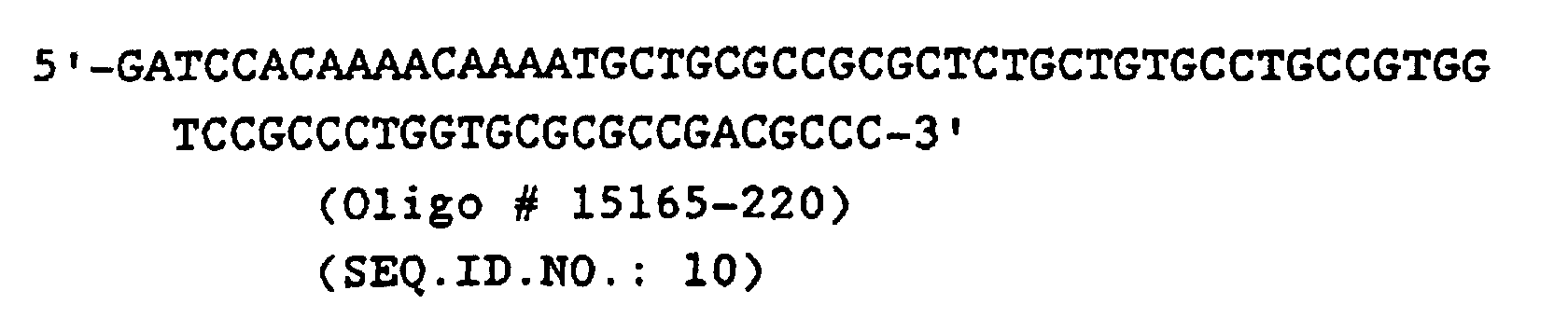
- 2.

- 3.
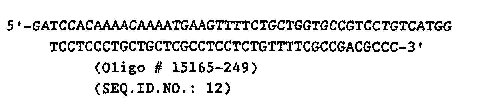
- 4.

-
- Oligonucleotides #15165-220 and 15165-249 were kinased and then annealed with oligonucleotides #15165-221 and 15165-250, respectively. To reconstruct human PDI with the human PDI signal peptide sequence, the following ligation was set up: the pUC19 2.7 kbp BamHI-EcoRI fragment was ligated with the 1.7 kbp PstI-EcoRI hPDI 3'-fragment, the 0.38 kbp PstI-AvaI 5'-hPDI fragment, and the annealed linkers 15165-220 plus 15165-221.
- To reconstruct human PDI with the yeast PDI signal sequence, the following ligation mixture was set up: the pUC19 2.7 kbp BamHI-EcoRI fragment was ligated with the 1.7 kbp PstI-EcoRI hPDI 3'-fragment, the 0.38 kbp PstI-AvaI 5'-hPDI fragment, and the annealed linkers 15165-249 plus 15165-250. (The annealed linkers contain BamHI and AvaI cohesive ends and encode the indicated signal peptide sequence plus yeast 5'-nontranslated leader sequence).
- The ligation mixtures were transformed into E. coli ATCC 35691 and transformants were screened for those containing plasmids with the expected structure. The DNA sequence across the region including the oligonucleotide linkers and the flanking DNA was confirmed by dideoxy sequencing methods. The human PDI coding sequences with either the yeast PDI signal peptide or human PDI signal peptide encoding sequences were designated ySP-hPDI and hSP hPDI, respectively. The two resulting plasmids containing these cassettes (pUC-ySP-hPDI [Figure 5] and pUC-hSP-hPDI, respectively) were digested with SmaI plus BamHI and the resulting 1.5 kbp fragments carrying the hPDI cassettes were gel-purified and made flush-ended.
- The plasmid p401 (which contains the GAL10 promoter and ADH1 transcription terminator separated by a unique BamHI site; Figure 6) was digested with BamHI, made flush-ended, and ligated with the aforementioned cassettes to yield the plasmids
pGAL-ySP-hPDI and pGAL-hSP-hPDI, respectively. These two plasmids were digested with SmaI, SphI, and ScaI and the resulting 3.2 kbp SmaI-SphI fragments bearing the GAL10p-ySP-hPDI and GAL10p-hSP-hPDI expression cassettes were gel-purified, made flush-ended, and then inserted into the XhoI site (made flush-ended) of the LYS2 integrating vector, pUC13-LYS2. The resulting plasmids were designated pLYS2-hSP-hPDI and pLYS2-ySP-hPDI respectively. - For purposes of integrative transformation, these latter two plasmids were digested with XbaI plus SacI to generate linear fragments with LYS2 flanking ends and the linear fragments were used to transform yeast strains BJ1995 and JRY188. Transformants which had integrated the desired expression cassettes at LYS2 were identified by Southern blots of genomic DNA which had been digested with BglII and was then hybridized with a LYS2 probe. The resulting strains were BJ1995/hSP-hPDI, BJ1995/ySP-hPDI, JRY188/hSP-hPDI (strain #1148), and JRY188/ySP-hPDI (strain #1157).
- Yeast proteins which are resident in the endoplasmic reticulum normally contain a C-terminal HDEL amino acid sequence which is the signal for retention in the ER (Pelham, et al., 1988 supra). In contrast, human PDI has a C-terminal KDEL sequence (Pihlajaniemi, et al., 1987, supra) which has been previously shown to function poorly for ER retention in yeast (Lewis, M.J., et al., 1990, Cell, 61, pp.1359-1363). Therefore, it was desired to construct a modified human PDI in which the C-terminal KDEL was changed to HDEL. This was accomplished as follows.
- The two plasmids pUC-ySP-hPDI and pUC-hSP-hPDI (Example 9) were digested with EcoRI and XhoI and the resulting 4.0 kbp EcoRI-XhoI fragment containing vector sequences plus the 5'-portion of the hPDI sequence and a 0.5 kbp XhoI-XhoI fragment containing the middle portion of the hPDI coding sequence were gel-purified. The following oligonucleotide adapter was then synthesized:
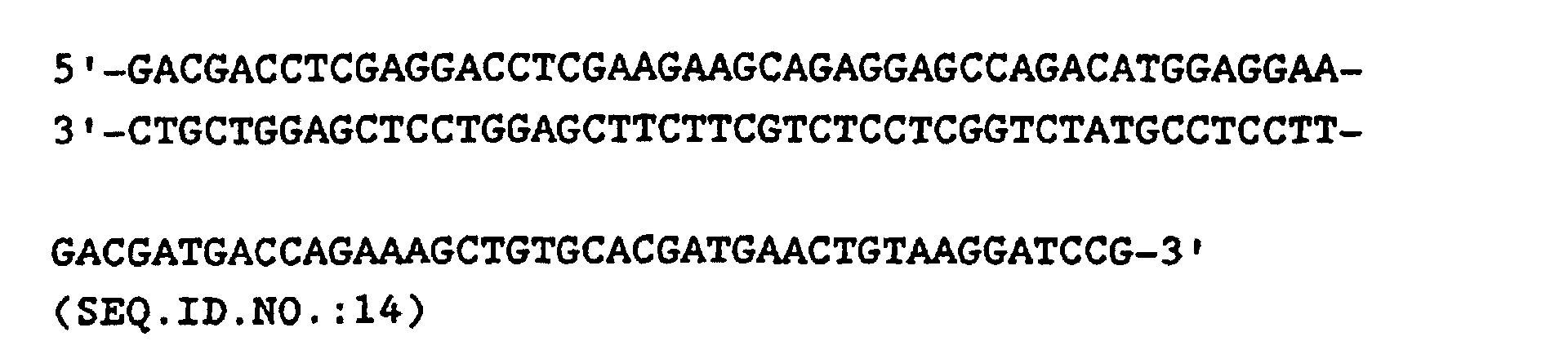

- Following annealing of the two oligos, this adapter was digested with XhoI (now yielding EcoRI and XhoI cohesive ends> and ligated in two separate reactions with the 4.0 kbp EcoRI-XhoI vector fragments containing either the 5'-ySP-hPDI or 5'-hSP-hPDI sequences, respectively. The resulting two plasmids were then digested with XhoI and ligated with the aforementioned 0.5 kbp XhoI-XhoI fragment containing the middle portion of the hPDI coding sequence, yielding the plasmids pUC-ySP-hPDI(HDEL) and pUC-hSP-hPDI(HDEL), respectively, into which the XhoI fragment had inserted in the correct orientation to reconstruct the human PDI coding sequence. These two plasmids were then digested with BamHI and the two different 1.5 kbp BamHI fragments carrying the expression cassettes were gel-purified and then inserted into the BamHI site of p401, yielding pUC-GAL10p-ySP-hPDI(HDEL) and pUC-GAL10p-hSP-hPDI(HDEL), respectively. These two plasmids were then digested with SmaI, SphI and PvuI. The resulting two 2.5 kbp SmaI-SphI fragments were gel-purified, made flush-ended and then ligated with pUC13-LYS2 which previously had been digested with XhoI and made flush-ended. The resulting two plasmids, pLYS2-ySP-hPDI(HDEL) and pLYS2-hSP-hPDI(HDEL) were linearized by digestion with HpaI plus EcoRV and then used in separate reactions for transformation of strains BJ1995 and JRY188. Lys- transformants were selected on solid media containing alpha-amino adipic acid. Isolates containing the desired expression cassette integrated at the LYS2 locus were identified by Southern blot analysis of genomic DNA. The resulting strains were designated BJ1995/ySP-hPDI(HDEL), BJ1995/hSP-hPDI(HDEL), JRY188/ySP-hPDI(HDEL) (strain #1268), and JRY188/hSP-hPDI(HDEL)(strain #1267).
- Plasmid pUKC161 (Figure 4) was digested with BamHI plus ClaI and the 0.7 kbp BamHI-ClaI fragment bearing the alpha factor pre-pro leader sequence and 5'-segment of hPDI was gel-purified. The plasmid pUC-ySP-hPDI(HDEL) (described in Example 11) was digested with ClaI and EcoRI and the 1.0 kbp ClaI-EcoRI fragment bearing the 3'-segment of hPDI with the C-terminal HDEL modification was gel-purified. pUC19 was digested with BamHI plus EcoRI and the resulting vector fragment was ligated with both the 0.7 kbp BamHI-ClaI fragment and the 1.0 kbp C laI-EcoRI fragment to yield the plasmid pUC-MFα1-hPDI(HDEL). This plasmid was digested with BamHI and the 1.7 kbp BamHI-BamHI fragment carrying the PDI cassette was gel-purified and inserted into the BamHI site of the plasmid p401 (Figure 6), yielding the plasmid pGAL-MFα1-hPDI(HDEL). This plasmid was then digested with the enzymes SmaI, SphI, and PvuI and the resulting 2.6 kbp SmaI-SphI fragment bearing the expression cassette was gel-purified and made flush-ended. The pUC13-LYS2 vector was digested with XhoI, made flush-ended and then ligated with the above 2.6 kbp flush-ended fragment. The resulting plasmid, pLYS2-MFα1-hPDI(HDEL) was digested with HpaI plus EcoRV and then used for transformation of strains JRY188 and BJ1995. The resulting transformants were evaluated by Southern blots of genomic DNA (as described in Example 9) to confirm that the desired expression cassette had integrated at the LYS2 locus. The JRY188 transformant was designated strain #1279.
- The plasmid C7 (described in Example 4) bearing the complete yeast PDI1 gene was digested with EcoRV and the 1.3 kbp EcoRV-EcoRV fragment containing the C-terminal portion of the yeast PDI open reading frame (ORF) (from amino acid 223 to end of ORF) plus the 3'-nontranslated sequence was gel-purified and inserted into the EcoRV site of the plasmid pAT153 [Twigg, A.G. and Sherratt, D., 1980, Nature, 283, pp.216-218], yielding pUKC169. Plasmid C7 was then digested with BanI plus EcoRV and the 0.67 kbp BanI-EcoRV fragment encoding amino acids 6-222 of the yeast PDI ORF was gel-purified and ligated with the following synthetic oligonucleotide adapter:
 which contains BamHI and BanI cohesive ends, respectively, and encodes amino acids 1-5 of the yeast PDI ORF plus 12 basepairs of yeast 5'-nontranslated leader sequence. (The ATG initiation codon is underlined.) The resulting 0.7 kbp BamHI-EcoRV fragment was gel-purified and then subcloned into pAT153 which had been previously digested with EcoRV plus BamHI, yielding the plasmid pUKC170.
- Plasmid pUKC169 was digested with EcoRV and the resulting 1.3 kbp EcoRV-EcoRV fragment bearing the aforementioned C-terminal portion of yeast PDI was gel-purified and then inserted into the unique EcoRV site of pUKC170 thereby regenerating the intact yeast PDI (yPDI) gene. This resulting plasmid was designated pUKC175.
- pUKC175 was digested with EcoNI and the resulting 2.1 kbp fragment bearing the yPDI gene was made flush-ended and gel-purified. pUC19 was digested with SacI plus SmaI, made flush-ended, and ligated with the above flush-ended EcoNI yPDI fragment. The ligation mixture was used to transform E. coli DH5 cells and the resulting transformants were screened for those containing plasmids with the yPDI insert in the appropriate orientation such that a BamHI site in the pUC19 polylinker was located adjacent to the 3'-end of the yPDI coding sequence. As a BamHI site already existed at the 5'-end of the yPDI ORF on the EcoNI fragment, this construct (designated pUC19-yPDI) now contains the yPDI ORF on a 1.9 kbp BamHI fragment. pUC19-yPDI was digested with BamHI and the 1.9 BamHI kbp fragment bearing the yPDI gene was gel-purified and then subcloned into the BamHI site of the vector pUC18-GAL10p(B)ADH1t (stock #401) (Figure 6). The resulting plasmid, pUC18-GAL10p-yPDI-ADH1t (Figure 7), is stock #1015. Plasmid pUC18-GAL10p-yPDI-ADH1t was digested with SmaI, SphI, plus SacI and the 2.7 kbp SmaI-SphI fragment carrying the expression cassette was gel-purified, made flush-ended, and then cloned into the unique StuI site of pUKC171 (pUKC171 contains the 4.5 kbp EcoRI-HindIII LYS2 fragment of YIp600 (Barnes and Thorner, 1986, supra) subcloned into pUC19 which had been previously digested with EcoRI plus HindIII). The resulting pUKC171-GAL10p-yPDI vector was then digested with EcoRI plus PvuII to excise the LYS2-GAL10p-yPDI-ADH1t-LYS2 cassette which was then used to transform S. cerevisiae strains JRY188 and BJ1995. The resulting lys- transformants were evaluated by Southern blots of genomic DNA preparations as described in Example 9. Isolates of each strain were found which had the GAL10p-yPDI cassette integrated at the LYS2 locus. The resulting strains were designated BJ1995/yPDI and JRY188/yPDI (strain #1152).
- The plasmid pUC-Not-URA3 (Example 8) was digested with ApaI plus NcoI (to delete a portion of the URA3 gene) and made flush-ended. Plasmid pUC18-GAL10p-yPDI-ADH1t was digested with EcoRI, ScaI, and SphI and the 2.8 kbp EcoRI-SphI fragment bearing the GAL10p-yPDI-ADH1t expression cassette was gel-purified, made flush-ended, and ligated with the above vector fragment, yielding the plasmid pNU-GAL10p-yPDI. The URA3-GAL10p-yPDI-ADH1t-URA3 integrating cassette was excised from pNU-GAL10p-yPDI by digestion with NotI. The resulting linear fragment was used to transform yeast strain KHY107. Ura- transformants were selected on solid media containing 5-fluoro-orotic acid (Boeke et al., 1984, Mol. Gen. Genet., 197, pp 345). Genomic DNA from the resulting ura- transformants was digested with BglII and evaluated by Southern blots using a radiolabelled EcoRI-PvuII fragment from the GAL10p-yPDI-ADHlt cassette as probe. Isolates were identified which had integrated the desired GAL10p-yPDI-ADHlt expression cassette at URA3. Isolate K-Y1 had multiple copies integrated at URA3 (strain #1136). Isolate K-Y3 had one copy integrated at URA3 (strain #1137).
- Yeast strains were grown in 3 × YEPD liquid medium for 24 hours, at 23°C. After that period, cultures were supplemented with galactose to a final 4.8% concentration. The cultures were then reincubated at 23°C, for an additional period of 24 hours. Alternatively, yeast strains were cultivated in 3 × YEPD for 24 hours at 30°C. The cells were harvested and washed with cold sterile water, and resuspended in the same volume of 3 × YEPD-Galactose medium. The yeast strains were incubated for a further period between 16 and 25 hours, after which, they were harvested and protein extracted by extraction method 2 (below).
- Proteins were extracted from exponentially growing or stationary phase cells using glass bead disruption, essentially as described by Mellor et al., (1983, Gene, 24, pp.1-14).
- Method 1: Protein was extracted by glass bead disruption of cell walls, in the presence of PMSF (0.5mM) in a 25mM phosphate buffer pH 7.0, followed by a freeze-thaw cycle, and soluble protein was recovered by centrifugation for 10 minutes at 13,000 rpm. Secretion was initially assessed by analysis of spent culture liquid, either before or after concentration with PEG (solid), ammonium sulphate (0-80%), or an ultrafiltration membrane (<100 kDa). Protein concentration was determined by the method of Bradford (1976, Anal. Biochem., 72, pp.248-254).
- Method 2: Intracellular samples were prepared as in
method 1, but the culture medium was supplemented with NaOH, and β-mercaptoethanol (respectively, final concentrations of 0.2M and 1%), left on ice for about 10 minutes, after which TCA was added to a final concentration of 6%. After 30 minutes standing on ice, protein was recovered by centrifugation, washed with cold acetone and resuspended in SDS-PAGE loading buffer. - 50µg of total soluble protein was analysed by one dimensional SDS-PAGE (12% polyacrylamide) and Coomassie blue staining essentially as described by Schultz et al., (1987, Gene, 54, pp.113-23).
- Electrophoresis was conducted under the following conditions: 10% SDS-polyacrylamide gel, and 10µg protein loaded per lane (protein extraction method 1). Sigma prestained molecular weight standards were run in all the gels. Gels were run in a BioRad mini-Protein II gel system. Extracellular extracts were loaded at 15-20µl per lane, without estimating protein concentration. Voltage was kept below 200 volts during electrophoresis.
- Proteins were transferred to nitrocellulose, using a Biometra semi-dry Western blot system. Nitrocellulose membranes were blocked with 5% (w/v) powdered milk for one hour, washed, incubated with anti human-PDI polyclonal antibody for between 3 hours to overnight, at dilutions ranging from 1:500 to 1:750. Membranes were washed and peroxidase-conjugated anti-rabbit IgG was added at a final dilution of 1:1000, and incubation continued for one hour. After washing the blots were developed using an Amersham ECL kit as described by the manufacturer.
- Initial assays showed that strain 1072A produced secreted hPDI at levels detectable by Western blot. The level of detection was 0.05µg of purified bovine PDI by the ECL protocol employed. This secreted PDI was shown to be a dimer since it was retained by a 100 kDa cut-off ultrafiltration membrane. When strain 1072A and its corresponding HDEL variant (1279) were compared, it was found that the human PDI was secreted by both. In this experiment the final culture/induction conditions were optimised in terms of temperature (°C) of growth and induction period. The two strains showed a higher level of PDI synthesis when cultivated at 23°C and then induced for 16 hours at 30°C, or when cultivated and induced at 30°C, for 16 hours.
- Antistasin is a potent protein inhibitor of the blood coagulation Factor Xa. Antistasin (ATS) was isolated from the salivary glands of the Mexican leech Haementeria officinalis (Nutt, E. et al., 1988, J. Biol. Chem., 263, pp.10162-10167). The cDNA encoding ATS was subsequently isolated and
characterized by Han, J. H. et al. (1989, Gene, 75, pp.47-57). ATS is an ideal reporter protein for the evaluation of the effects of increased levels of PDI activity on the folding and formation of proper disulfide bonds in a heterologous protein secreted by recombinant yeast since ATS has 10 disulfide bonds which must be correctly paired in order for the protein to have biological activity. - ATS was expressed in yeast using the expression vector pKH4α2 (Jacobson, M.A. et al., 1989, Gene, 85, pp.511-516), which contains the galactose-inducible GAL10 promoter and the yeast MFα1 pre-pro secretory leader sequence to direct secretion of heterologous proteins. The coding sequence for ATS was isolated by polymerase chain reaction (PCR) methods using subcloned ATS cDNA from clone λ5C-4 (Han, J. H. et al.; supra) as substrate and the oligonucleotide primers:
- 1.

- 2.
 Both primers contain a BamHI site to facilitate
subcloning of the PCR product. The first primer
inserts a yeast KEX2 yscF endoprotease cleavage site
(Lys-Arg) N-terminal to the first amino acid residue
of mature ATS (the yeast yscF endoprotease cleaves on
the C-terminal side of the Lys-Arg site in this
sequence). The PCR product was digested with BamHI,
gel-purified, and then ligated into BamHI-digested
pKH4α2 to yield pKH4α2/ATS (K991) (Figure 8). This
expression vector was then used to transform the
yeast host strains listed in Table 1 using the
spheroplast method (Hinnen, et al., 1978, supra).
Both primers contain a BamHI site to facilitate
subcloning of the PCR product. The first primer
inserts a yeast KEX2 yscF endoprotease cleavage site
(Lys-Arg) N-terminal to the first amino acid residue
of mature ATS (the yeast yscF endoprotease cleaves on
the C-terminal side of the Lys-Arg site in this
sequence). The PCR product was digested with BamHI,
gel-purified, and then ligated into BamHI-digested
pKH4α2 to yield pKH4α2/ATS (K991) (Figure 8). This
expression vector was then used to transform the
yeast host strains listed in Table 1 using the
spheroplast method (Hinnen, et al., 1978, supra). -
- Transformants were selected on synthetic solid media lacking leucine (Schultz, L. et al., 1987; Gene, 61, pp.123-133) and streaked for clonal isolates which were used in subsequent analyses. Strains were preserved by storage at -70°C in synthetic media containing 17% glycerol.
TransformedStrain Host Strain Original Strain PDICassette 960 239 JRY188 none 1105 1072A JRY188 alpha-hPDI 1176 1157 JRY188 ySP-hPDI 1175 1148 JRY188 hSP-hPDI 1293 1279 JRY188 alpha-hPDI(HDEL) 1294 1267 JRY188 hSP-hPDI(HDEL) 1295 1268 JRY188 ySP-hPDI(HDEL) 1177 1152 JRY188 yPDI 1156 548 KHY107 none 1154 1136 KHY107 yPDI-Al 1155 1137 KHY107 yPDI-A3 - The K991-transformed parental JRY188 strain plus the various transformed derivatives which overproduce either yeast or human PDI were evaluated for secretion of antistasin by the following procedure. The indicated strains were streaked from the -70°C frozen glycerol stocks onto leucine-minus synthetic agar plates and grown for 3 days at 30°C. Culture tubes (18 x 150 mm) containing 5-mL of 3xYEHD [60g Difco yeast extract, 30g HySoy peptone, 48g glucose per liter] media were inoculated with a small loopful of cells and incubated for about 18 hours at 23°C on a tissue culture roller drum. At this stage, cells were induced by the addition of galactose to a final concentration of 4.8% (w/v) and the cultures were incubated for an additional 5 days at 23°C. Cells were then harvested by centrifugation and the clarified media supernatant was retained for assay of antistasin activity, which was measured by inhibition of Factor Xa activity [Nutt, E. et al., 1988, supra]. The experiment was conducted in triplicate and results are summarized in Table 2.
Strain ng ATS per 1.0 OD Relative Level JRY188 25.6 1.0 JRY188/hSP-hPDI 24.4 0.95 JRY188/ySP-hPDI 28.4 1.11 JRY188/alpha-hPDI 77.2 3.0 JRY188/yPDI 65.1 2.54 - The K991-transformed JRY188 and transformed derivative strains which overproduce the HDEL mutant version of human PDI with the three different secretory leaders were grown as described in Example 17 and the clarified media supernatants were evaluated for the levels of secreted ATS by the Factor Xa inhibition assay as described in Example 17. The results are presented in Table 3.
Strain ng ATS per 1.0 OD Relative Level JRY188 18.0 1.0 JRY188/hSP-hPDI(HDEL) 27.5 1.53 JRY188/ySP-hPDI(HDEL) 29.3 1.63 JRY188/alpha-hPDI(HDEL) 31.3 1.74 - The K991-transformed KHY107 and its transformed derivatives which overproduce yeast PDI were grown up and clarified media supernatants were evaluated for the levels of secreted ATS by Factor Xa inhibition assay as described in Example 17. The results are summarized in Table 4.
Strain ATS(mg/L) A600 ATS/A600 KHY107 A1 0.314 23.9 0.013 KHY107 A2 0.244 24.5 0.010 KHY107 A3 0.334 25.5 0.013 K-Y1 A1 1.168 24.8 0.047 K-Y1 A2 1.469 21.8 0.067 K-Y1 A3 1.483 25.3 0.059 K-Y3 A1 3.856 39.0 0.099 K-Y3 A2 2.144 51.2 0.042 K-Y3 A3 1.920 48.0 0.040 - The multicopy yeast shuttle vector YEp24 (Botstein, D. et al., 1979, Gene, 8, pp.17-24) contains the yeast 2-micron DNA origin of replication and the yeast URA3 gene for selection on uracil-minus synthetic media. YEp24 was digested with BamHI and the resulting 7.8 kbp BamHI vector fragment was gel-purified (fragment a). The plasmid pUC18-GAL10p-yPDI-ADH1t (#1015) was digested with EcoRI, SphI, and ScaI; the resulting 2.8 kbp EcoRI-SphI fragment bearing the GAL10p-yPDI-ADH1t expression cassette was gel purified (fragment b). Plasmid pUKC161 was digested with EcoRI plus HindIII and the 2.8 kbp EcoRI-HindIII fragment bearing the GAL1p--MFα1 pre-pro--human PDI expression cassette was gel purified (fragment c). The above three fragments were made flush-ended and then ligated together as follows: (1) vector fragment a and fragment b were ligated together to yield the plasmid YEp24-GAL10p-yPDI (Figure 9); (2) vector fragment a and fragment c were ligated together to yield the plasmid YEp24-GAL1p-MFα-hPDI (Figure 10). Large-scale CsCl preparations of the two resulting plasmid DNAs were made. In two separate transformation reactions, the yeast strain JRY188 was cotransformed with the ATS expression vector K991 (Example 16) and either YEp24-GAL10p-yPDI or YEp24-GAL1p-MFα-hPDI. Transformants containing both plasmids were selected on synthetic media lacking both leucine and uracil and isolated single colonies were restreaked on the same media for selection of clonal isolates. Five such clonal isolates for each of the two original cotransformations were inoculated into 5 ml of 3xYEHD medium in culture tubes and incubated for 24 hr at 23°C in a tissue culture roller drum. At the end of that time, galactose was added to a final concentration of 4.8% and the cultures were incubated for an additional 5 days at 23°C. Cell were removed by centrifugation and the clarified media supernatants were assayed for the levels of ATS activity by the Factor Xa inhibition assay. The contransformants which contained the YEp24-GAL10p-yPDI plasmid plus the ATS expression vector showed 3- to 26-fold higher levels of secreted ATS activity, depending on the isolate, compared to the parental JRY188 strain containing only the ATS expression vector. The contransformants containing the YEp24-GAL1p-MFα-hPDI plasmid plus the ATS expression vector showed 2- to 3-fold higher levels of secreted ATS activity compared to the parental JRY188 strain containing only the ATS expression vector.
- The S. cerevisiae GAL1 and GAL10 genes are transcribed divergently from a region between the two structural genes which contains the divergent GAL1 and GAL10 promoters and a common GAL4 binding domain located between the TATA boxes for the two promoters. The plasmid pBM272 (Johnston, M. and Davis, R., 1984, Mol. Cell. Biol., 4, pp.1440) contains this divergent yeast GAL1-GAL10 promoter on a 0.85 kbp EcoRI-HindIII fragment (with also an internal BamHI site adjacent to the HindIII site). This promoter fragment was used to construct a divergent promoter cassette vector, pUC-GAL1/10, which has the following properties: yeast GAL10 promoter separated from the yeast ADH1 transcription terminator (0.35 kbp HindIII-SphI fragment) by unique EcoRI and SmaI sites, in that order. Yeast GAL1 promoter separated from a second copy of the ADH1 transcription terminator by unique BamHI and HindIII sites. The 3'-end of both ADH1 terminator elements are flanked by SphI sites to allow one to isolate the entire divergent promoter expression cassette as an SphI fragment. The vector backbone in this plasmid is pUC18 with the above expression cassette in place of the polylinker.
- The plasmid pUC-GAL1/10 was digested with BamHI and gel-purified to generate fragment "a". The plasmid pUKC161 was digested with BamHI and the 1.9 kbp BamHI fragment bearing the alpha factor pre-pro leader fused in-frame to the mature human PDI coding sequence was gel-purified and ligated to vector fragment a, yielding the plasmid pUC-GAL1/10-hPDI, in which the expression of the alpha factor pre-pro--hPDI fusion is under control of the GAL1 promoter. The plasmid pUC18-GAL10p-yPDI-ADH1t (Example 13) was digested with BamHI and the resulting 1.7 Kbp BamHI fragment bearing the yeast PDI coding sequence was gel-purified and then ligated with vector fragment a, yielding the plasmid pUC-GAL1/10-yPDI in which the GAL1 promoter directs the expression of yeast PDI. These two resulting plasmids were then digested with EcoRI and made flush-ended, yielding vector fragments b and c bearing the hPDI and yPDI cassettes, respectively.
- The ATS expression vector (K991) was digested with SalI plus BglII and the SalI-BglII fragment bearing the alpha factor pre-pro leader fused in-frame to the coding sequence for mature ATS was gel-purified, made flush-ended, and ligated in separate reactions to the two flush-ended vector fragments b and c. The resulting plasmids with the correct structure as determined by restriction mapping were designated pUC-GAL1/10-hPDT/ATS (Figure 11) and pUC-GAL1/10-yPDI/ATS (Figure 12), respectively. These two plasmids were then digested with SphI to liberate the expression cassettes and the fragments bearing either the hPDI- or
yPDI-related expression cassette were ligated with the yeast shuttle vector pC1/1 (Rosenberg, S. et al. 1984, Nature, 312, pp.77-80) which had been previously digested with SphI. This yielded the resulting two plasmids, pC1/1-GAL1/10-hPDI/ATS and pC1/1-GAL1/10-yPDI/ATS, in which the ATS and PDI-related expression cassettes were present on the same high copy-number vector under control of the GAL10 and GAL1 promoters, respectively. - These two expression vectors were then used to transform yeast strains JRY188, BJ1995, and other suitable yeast host strains. Transformants were selected on leucine-minus media and the resulting transformants were evaluated for expression/secretion of ATS and PDI as described in the preceding examples.
- The results presented in Table 5 (below) clearly show that the isolates which overproduce hPDI secrete several-fold higher levels of antistasin than the control strain which contained only pKH4α2/ATS. Furthermore, the isolates which overproduce yeast PDI secreted 3- to 17-fold higher levels of antistasin than the control strain.
Construct Antistasin (mg/L) pC1/1-GAL1/0-hPDI/ATS Isolate 1 4.7 Isolate 2 5.3 Isolate 3 3.9 Isolate 4 4.6 Isolate 5 5.1 pC1/1-GAL1/10-yPDI/ATS Isolate 1 3.9 Isolate 2 11.7 Isolate 3 5.8 Isolate 4 26.0 Isolate 5 8.2 JRY188 control 1.5 - Selected isolates of strain JRY188 cotransformed with the antistasin expression vector pKH4α2/ATS and either YEp24-GAL1p-MFα-hPDI or YEp24-GAL10p-yPDI were evalutated for antistasin secretion after growth at either 23°C or 30°C. The parent strain JRY188 transformed only with the antistasin expression vector was grown in parallel. After overnight growth in 3xYEHD medium at either 23°C or 30°C, cell cultures were induced by the addition of galactose to a final concentration of 4.8% and propagated for an additional five days at either 23°C or 30°C, as appropriate. Media samples harvested at 3 to 5 days post-induction were evaluated for levels of secreted antistasin by the Factor Xa inhibition assay. The results presented in Table 6 clearly show that antistasin expression was significantly higher at 23°C than at 30°C for all isolates overexpressing PDI, both at 3 and 5 days post-induction.
Isolate Temperature (°C) Antistasin (mg/L) 3 days 5 days hPDI-1 23 0.83 2.11 hPDI-2 23 1.14 2.68 yPDI-1 23 5.93 10.25 yPDI-3 23 3.00 15.92 JRY188 control 23 0.38 0.65 hPDI-1 30 0.49 0.47 hPDI-2 30 0.42 0.47 yPDI-1 30 2.29 4.65 yPDI-3 30 2.71 2.56 JRY188 control 30 0.34 0.30 - Tick anticoagulant peptide (TAP) is a potent, highly selective inhibitor of the blood coagulation factor Xa (Waxman, L. et al., 1990, Science, 248, pp. 593-596). TAP is a novel serine protease inhibitor isolated from the tick Ornithidoros moubata. TAP is composed of 60 amino acids including 6 cysteine residues (Waxman et al., 1990, supra). TAP has been expressed in yeast using the expression vector pKH4-TAP which contains the galactose-inducible GAL10 promoter and the yeast MFα1 pre-pro secretory leader sequence fused in frame to the synthetic gene encoding TAP (Neeper, M. et al., 1990, J. Biol. Chem., 265, pp.17746-17752). This vector contains a slightly modified MFα1 pre-pro leader sequence due to the presence of a BamHI cloning site located at the position of amino acid 79 of the pre-pro leader (Neeper et al., 1990, supra).
- A second TAP expression vector, pKH4-3B/TAP, was constructed which contains the authentic MFα1 pre-pro leader sequence fused in-frame to the synthetic gene encoding TAP. The plasmid pKH4-TAP containing the synthetic TAP gene (Neeper et al., 1990, supra) was used as the DNA template in a polymerase chain reaction (PCR) using the following two oligonucleotide primers in order to modify the 5'-and 3'- termini of the synthetic TAP gene, respectively:


- The PCR reaction was carried out by methods well known to those of ordinary skill in the art (Innis, M.A. et al., editors, 1990, PCR Protocols: A Guide to Methods and Applications, Academic Press, Inc., San Diego CA). The resulting PCR product was phosphorylated with T4 polynucleotide kinase, digested with BamHI, and then gel-purified to yield a 0.2 kbp blunt-BamHI fragment containing the blunt end at the exact 5'-end of the TAP coding sequence and a cohesive BamHI end on the 3'-side of the translation termination codon.
- The vector pKH4-3B (Hofmann, K. and Schultz, L.D., 1991, Gene, 101, pp.105-111) contains a unique SphI site at the 3'-end of the MFα1 pre-pro leader coding sequence. pKH4-3B was digested with SphI, made flush-ended by treatment with T4 DNA polymerase, and then digested with BglII. The resulting blunt-BglII vector fragment was gel-purified and ligated with the aforementioned 0.2 kbp blunt-BamHI TAP fragment to yield the vector pKH4-3B/TAP.
- In separate transformation reactions, the yeast strains BJ1995, JRY188, and U9 were cotransformed with the vectors YEp24-GAL10p-yPDI plus either pKH4-TAP or pKH4-3B/TAP. Cotransformants containing both plasmids were selected on synthetic medium lacking both leucine and uracil and isolated single colonies were restreaked on the same medium for selection of clonal isolates. Three such clonal isolates for each of the different vector/host cotransformations were inoculated into 5-mL of modified 5xLeu- media lacking uracil (5xLeu-Ura-) and containing 4% glucose in culture tubes. The cultures were incubated for 24 hours at 30°C in a tissue culture roller drum. At the end of that time, the cells were recovered by centrifugation and resuspended in 5 mL of 5xLeu-Ura- medium containing 4% galactose. The resulting cultures were incubated at 30°C for an additional 48 hours. Cells are then removed by centrifugation and clarified media samples evaluated for the levels of secreted TAP by SCX-HPLC or Factor Xa inhibition assay (Waxman et al., 1990, supra). As an alternative approach, the recombinant yeast cells are grown for 24 hours at 23°C, induced by addition of galactose to 4% final concentration, and then incubated for an additional five days at 23°C. Clarified media samples are then evalutated for the levels of secreted TAP as described above.
-
- (1) GENERAL INFORMATION:
- (i) APPLICANT: Ellis, Ronald W.
- Schultz, Loren D.
- Marcus, Henry Z.
- Tuite, Michael H.
- Freedman, Robert
- Montgomery, Donna L.
- (ii) TITLE OF INVENTION: Method for Increasing Secretion of Disulfide Bonded Recombinant Proteins by Saccharomyces Cerevisiae
- (iii) NUMBER OF SEQUENCES: 21
- (iv) CORRESPONDENCE ADDRESS:
- (A) ADDRESSEE: Merck & Co., Inc.
- (B) STREET: P.O. Box 2000
- (C) CITY: Rahway
- (D) STATE: New Jersey
- (E) COUNTRY: USA
- (F) ZIP: 07065
- (v) COMPUTER READABLE FORM:
- (A) MEDIUM TYPE: Floppy disk
- (B) COMPUTER: IBM PC compatible
- (C) OPERATING SYSTEM: PC-DOS/MS-DOS
- (D) SOFTWARE: PatentIn Release #1.0, Version #1.25
- (vi) CURRENT APPLICATION DATA:
- (A) APPLICATION NUMBER:
- (B) FILING DATE:
- (C) CLASSIFICATION:
- (viii) ATTORNEY/AGENT INFORMATION:
- (A) NAME: Wallen, John W.
- (B) REGISTRATION NUMBER: 35,403
- (C) REFERENCE/DOCKET NUMBER: 18469
- (ix) TELECOMMUNICATION INFORMATION:
- (A) TELEPHONE: (908) 594-3905
- (B) TELEFAX: (908) 594-4720
- (2) INFORMATION FOR SEQ ID NO:1:
- (i) SEQUENCE CHARACTERISTICS:
- (A) LENGTH: 6 amino acids
- (B) TYPE: amino acid
- (C) STRANDEDNESS: single
- (D) TOPOLOGY: linear
- (ii) MOLECULE TYPE: peptide
- (xi) SEQUENCE DESCRIPTION: SEQ ID NO:1:

- (2) INFORMATION FOR SEQ ID NO:2:
- (i) SEQUENCE CHARACTERISTICS:
- (A) LENGTH: 4 amino acids
- (B) TYPE: amino acid
- (C) STRANDEDNESS: single
- (D) TOPOLOGY: linear
- (ii) MOLECULE TYPE: peptide
- (xi) SEQUENCE DESCRIPTION: SEQ ID NO:2:

- (2) INFORMATION FOR SEQ ID NO:3:
- (i) SEQUENCE CHARACTERISTICS:
- (A) LENGTH: 6 amino acids
- (B) TYPE: amino acid
- (C) STRANDEDNESS: single
- (D) TOPOLOGY: linear
- (ii) MOLECULE TYPE: peptide
- (xi) SEQUENCE DESCRIPTION: SEQ ID NO:3:

- (2) INFORMATION FOR SEQ ID NO:4:
- (i) SEQUENCE CHARACTERISTICS:
- (A) LENGTH: 10 amino acids
- (B) TYPE: amino acid
- (C) STRANDEDNESS: single
- (D) TOPOLOGY: linear
- (ii) MOLECULE TYPE: peptide
- (xi) SEQUENCE DESCRIPTION: SEQ ID NO:4:

- (2) INFORMATION FOR SEQ ID NO:5:
- (i) SEQUENCE CHARACTERISTICS:
- (A) LENGTH: 30 base pairs
- (B) TYPE: nucleic acid
- (C) STRANDEDNESS: single
- (D) TOPOLOGY: linear
- (ii) MOLECULE TYPE: cDNA
- (xi) SEQUENCE DESCRIPTION: SEQ ID NO:5:

- (2) INFORMATION FOR SEQ ID NO:6:
- (i) SEQUENCE CHARACTERISTICS:
- (A) LENGTH: 26 base pairs
- (B) TYPE: nucleic acid
- (C) STRANDEDNESS: single
- (D) TOPOLOGY: linear
- (ii) MOLECULE TYPE: cDNA
- (xi) SEQUENCE DESCRIPTION: SEQ ID NO:6:

- (2) INFORMATION FOR SEQ ID NO:7:
- (i) SEQUENCE CHARACTERISTICS:
- (A) LENGTH: 26 base pairs
- (B) TYPE: nucleic acid
- (C) STRANDEDNESS: single
- (D) TOPOLOGY: linear
- (ii) MOLECULE TYPE: cDNA
- (xi) SEQUENCE DESCRIPTION: SEQ ID NO:7:

- (2) INFORMATION FOR SEQ ID NO:8:
- (i) SEQUENCE CHARACTERISTICS:
- (A) LENGTH: 15 base pairs
- (B) TYPE: nucleic acid
- (C) STRANDEDNESS: single
- (D) TOPOLOGY: linear
- (ii) MOLECULE TYPE: cDNA
- (xi) SEQUENCE DESCRIPTION: SEQ ID NO:8:

- (2) INFORMATION FOR SEQ ID NO:9:
- (i) SEQUENCE CHARACTERISTICS:
- (A) LENGTH: 15 base pairs
- (B) TYPE: nucleic acid
- (C) STRANDEDNESS: single
- (D) TOPOLOGY: linear
- (ii) MOLECULE TYPE: cDNA
- (xi) SEQUENCE DESCRIPTION: SEQ ID NO:9:

- (2) INFORMATION FOR SEQ ID NO:10:
- (i) SEQUENCE CHARACTERISTICS:
- (A) LENGTH: 73 base pairs
- (B) TYPE: nucleic acid
- (C) STRANDEDNESS: single
- (D) TOPOLOGY: linear
- (ii) MOLECULE TYPE: cDNA
- (xi) SEQUENCE DESCRIPTION: SEQ ID NO:10:

- (2) INFORMATION FOR SEQ ID NO:11:
- (i) SEQUENCE CHARACTERISTICS:
- (A) LENGTH: 73 base pairs
- (B) TYPE: nucleic acid
- (C) STRANDEDNESS: single
- (D) TOPOLOGY: linear
- (ii) MOLECULE TYPE: cDNA
- (xi) SEQUENCE DESCRIPTION: SEQ ID NO:11:

- (2) INFORMATION FOR SEQ ID NO:12:
- (i) SEQUENCE CHARACTERISTICS:
- (A) LENGTH: 88 base pairs
- (B) TYPE: nucleic acid
- (C) STRANDEDNESS: single
- (D) TOPOLOGY: linear
- (ii) MOLECULE TYPE: cDNA
- (xi) SEQUENCE DESCRIPTION: SEQ ID NO:12:

- (2) INFORMATION FOR SEQ ID NO:13:
- (i) SEQUENCE CHARACTERISTICS:
- (A) LENGTH: 88 base pairs
- (B) TYPE: nucleic acid
- (C) STRANDEDNESS: single
- (D) TOPOLOGY: linear
- (ii) MOLECULE TYPE: cDNA
- (xi) SEQUENCE DESCRIPTION: SEQ ID NO:13:

- (2) INFORMATION FOR SEQ ID NO:14:
- (i) SEQUENCE CHARACTERISTICS:
- (A) LENGTH: 91 base pairs
- (B) TYPE: nucleic acid
- (C) STRANDEDNESS: single
- (D) TOPOLOGY: linear
- (ii) MOLECULE TYPE: cDNA
- (xi) SEQUENCE DESCRIPTION: SEQ ID NO:14:

- (2) INFORMATION FOR SEQ ID NO:15:
- (i) SEQUENCE CHARACTERISTICS:
- (A) LENGTH: 95 base pairs
- (B) TYPE: nucleic acid
- (C) STRANDEDNESS: single
- (D) TOPOLOGY: linear
- (ii) MOLECULE TYPE: cDNA
- (xi) SEQUENCE DESCRIPTION: SEQ ID NO:15:

- (2) INFORMATION FOR SEQ ID NO:16:
- (i) SEQUENCE CHARACTERISTICS:
- (A) LENGTH: 31 base pairs
- (B) TYPE: nucleic acid
- (C) STRANDEDNESS: single
- (D) TOPOLOGY: linear
- (ii) MOLECULE TYPE: cDNA
- (xi) SEQUENCE DESCRIPTION: SEQ ID NO:16:

- (2) INFORMATION FOR SEQ ID NO:17:
- (i) SEQUENCE CHARACTERISTICS:
- (A) LENGTH: 31 base pairs
- (B) TYPE: nucleic acid
- (C) STRANDEDNESS: single
- (D) TOPOLOGY: linear
- (ii) MOLECULE TYPE: cDNA
- (xi) SEQUENCE DESCRIPTION: SEQ ID NO:17:

- (2) INFORMATION FOR SEQ ID NO:18:
- (i) SEQUENCE CHARACTERISTICS:
- (A) LENGTH: 51 base pairs
- (B) TYPE: nucleic acid
- (C) STRANDEDNESS: single
- (D) TOPOLOGY: linear
- (ii) MOLECULE TYPE: cDNA
- (xi) SEQUENCE DESCRIPTION: SEQ ID NO:18:

- (2) INFORMATION FOR SEQ ID NO:19:
- (i) SEQUENCE CHARACTERISTICS:
- (A) LENGTH: 34 base pairs
- (B) TYPE: nucleic acid
- (C) STRANDEDNESS: single
- (D) TOPOLOGY: linear
- (ii) MOLECULE TYPE: cDNA
- (xi) SEQUENCE DESCRIPTION: SEQ ID NO:19:

- (2) INFORMATION FOR SEQ ID NO:20:
- (i) SEQUENCE CHARACTERISTICS:
- (A) LENGTH: 20 base pairs
- (B) TYPE: nucleic acid
- (C) STRANDEDNESS: single
- (D) TOPOLOGY: linear
- (ii) MOLECULE TYPE: cDNA
- (xi) SEQUENCE DESCRIPTION: SEQ ID NO:20:

- (2) INFORMATION FOR SEQ ID NO:21:
- (i) SEQUENCE CHARACTERISTICS:
- (A) LENGTH: 33 base pairs
- (B) TYPE: nucleic acid
- (C) STRANDEDNESS: single
- (D) TOPOLOGY: linear
- (ii) MOLECULE TYPE: cDNA
- (xi) SEQUENCE DESCRIPTION: SEQ ID NO:21:

-
Claims (24)
- An in vitro process for producing disulfide-bonded recombinant proteins comprising:(a) overexpressing recombinant protein disulfide isomerase from a recombinant expression cassette containing a gene encoding protein disulfide isomerase in a recombinant host cell, and(b) expressing one or more recombinant genes encoding one or more disulfide bonded proteins other than protein disulfide isomerase in the recombinant host cell,
with the proviso that said recombinant host cell is other than E.coli, and that said gene encoding protein disulfide isomerase does not comprise a DNA fragment coding for a human serum albumin prepro-sequence, and that the process does not involve expression from a recombinant expression cassette of a gene coding for the receptor protein ERD2 from yeast. - The process according to Claim 1, wherein the expression cassettes encoding protein disulfide isomerase are integrated into the host cell genome.
- The process according to Claim 1, wherein the expression cassettes encoding protein disulfide isomerase are contained on autonomously replicating plasmids.
- The process according to Claim 3 wherein the recombinant genes encoding one or more disulfide bonded proteins are contained on one or more plasmids.
- The process according to Claim 1 or Claim 3 wherein the recombinant genes encoding one or more disulfide bonded proteins are integrated into the host cell genome.
- The process according to claim 4 wherein the expression cassettes encoding the protein disulfide isomerase and the recombinant gene are contained on the same plasmid.
- The process according to Claim 1 wherein the recombinant host cell is mammalian.
- The process according to Claim 1 wherein the recombinant host cell is yeast.
- The process according to claim 8 wherein the yeast is a strain of a species of the families Saccharomycetaceae or Cryptococcaceae.
- The process according to claim 9 wherein the yeast is a species of the genus Saccharomyces.
- The process according to claim 10 wherein the yeast is Saccharomyces cerevisiae.
- The process according to Claim 1 wherein the one or more recombinant genes of step (b) encode antistasin.
- The process according to Claim 1 wherein the one or more recombinant genes of step (b) encode tick anticoagulant protein.
- The process according to Claim 1 wherein the recombinant protein disulfide isomerase gene is a yeast protein disulfide isomerase gene.
- The process according to Claim 1 wherein the recombinant protein disulfide isomerase gene is a mammalian protein disulfide isomerase gene.
- The process according to Claim 15 wherein the recombinant protein disulfide isomerase gene is a human protein disulfide isomerase gene.
- A process according to any of claims 1-6 and 8-16 wherein the recombinant host is yeast and the disulfide bonded protein is secreted.
- An in vitro process for producing disulfide-bonded recombinant proteins comprising:(a) overexpressing recombinant protein disulfide isomerase from a recombinant expression cassette containing a gene encoding yeast protein disulfide isomerase in a recombinant yeast host; and(b) expressing one or more recombinant genes encoding one or more disulfide bonded proteins other than protein disulfide isomerase in the recombinant host cell.
- The process according to claim 17 or claim 18 wherein the recombinant yeast host is grown at a temperature less than 30 °C for expression of one or more disulfide bonded proteins.
- The process according to claim 19 wherein the recombinant yeast host is grown at a temperature between approximately 20 °C and 26 °C for expression of one or more disulfide bonded proteins.
- The process according to claim 20 wherein the disulfide bonded protein is antistasin.
- The process according to claim 20 wherein the disulfide bonded protein is tick anticoagulant protein.
- A strain of the yeast Saccharomyces cerevisiae which overexpresses recombinant protein disulfide isomerase from a recombinant expression cassette containing a gene encoding protein disulfide isomerase and one or more recombinant genes encoding one or more disulfide-bonded proteins other than protein disulfide isomerase from a recombinant expression cassette;
with the proviso that said gene encoding protein disulfide isomerase does not comprise a DNA fragment coding for a human serum albumin prepro-sequence, and that the yeast does not express a gene coding for the receptor protein ERD2 from yeast from a recombinant expression cassette. - A strain of the yeast Saccharomyces cerevisiae which overexpresses recombinant protein disulfide isomerase from a recombinant expression cassette containing a gene encoding yeast protein disulfide isomerase and one or more recombinant genes encoding one or more disulfide-bonded proteins other than protein disulfide isomerase from a recombinant expression cassette.
Applications Claiming Priority (3)
| Application Number | Priority Date | Filing Date | Title |
|---|---|---|---|
| US07/901,713 US6291205B1 (en) | 1992-06-12 | 1992-06-12 | Method of increasing production of disulfide bonded recombinant proteins by saccharomyces cerevisiae |
| US901713 | 1992-06-12 | ||
| PCT/US1993/005318 WO1993025676A1 (en) | 1992-06-12 | 1993-06-02 | METHOD FOR INCREASING PRODUCTION OF DISULFIDE BONDED RECOMBINANT PROTEINS BY $i(SACCHAROMYCES CEREVISIAE) |
Publications (3)
| Publication Number | Publication Date |
|---|---|
| EP0746611A1 EP0746611A1 (en) | 1996-12-11 |
| EP0746611A4 EP0746611A4 (en) | 1997-03-19 |
| EP0746611B1 true EP0746611B1 (en) | 2003-04-16 |
Family
ID=25414688
Family Applications (1)
| Application Number | Title | Priority Date | Filing Date |
|---|---|---|---|
| EP93915201A Expired - Lifetime EP0746611B1 (en) | 1992-06-12 | 1993-06-02 | Method for increasing production of disulfide bonded recombinant proteins by (saccharomyces cerevisiae) |
Country Status (12)
| Country | Link |
|---|---|
| US (1) | US6291205B1 (en) |
| EP (1) | EP0746611B1 (en) |
| JP (3) | JP3953094B2 (en) |
| KR (1) | KR0171205B1 (en) |
| AT (1) | ATE237684T1 (en) |
| AU (1) | AU679448B2 (en) |
| CA (1) | CA2137142C (en) |
| DE (1) | DE69332893T2 (en) |
| DK (1) | DK0746611T3 (en) |
| ES (1) | ES2194848T3 (en) |
| NZ (1) | NZ253873A (en) |
| WO (1) | WO1993025676A1 (en) |
Families Citing this family (41)
| Publication number | Priority date | Publication date | Assignee | Title |
|---|---|---|---|---|
| ZA931601B (en) * | 1992-03-06 | 1993-10-05 | Squibb & Sons Inc | Microsomal triglyceride transfer protein |
| DK76893D0 (en) * | 1993-06-28 | 1993-06-28 | Novo Nordisk As | |
| EP0635574B1 (en) * | 1993-07-23 | 2003-04-23 | Dsm N.V. | Selection marker gene free recombinant strains, a method for obtaining them and the use of these strains |
| US5789199A (en) * | 1994-11-03 | 1998-08-04 | Genentech, Inc. | Process for bacterial production of polypeptides |
| US5639635A (en) * | 1994-11-03 | 1997-06-17 | Genentech, Inc. | Process for bacterial production of polypeptides |
| AU2660397A (en) * | 1996-04-05 | 1997-10-29 | Board Of Regents, The University Of Texas System | Methods for producing soluble, biologically-active disulfide bond-containing eukaryotic proteins in bacterial cells |
| DK0828004T3 (en) * | 1996-09-04 | 2007-03-19 | Suntory Ltd | Protein disulfide isomerase gene derived from strain of methylotrophic yeast |
| GB9703406D0 (en) | 1997-02-19 | 1997-04-09 | Chiron Spa | Expression of heterologous proteins |
| US6083715A (en) * | 1997-06-09 | 2000-07-04 | Board Of Regents, The University Of Texas System | Methods for producing heterologous disulfide bond-containing polypeptides in bacterial cells |
| US6521421B1 (en) | 1997-07-16 | 2003-02-18 | Genencor International, Inc. | Expression vectors encoding Bacillus subtilis disulfide bond isomerase and methods of secreting proteins in gram-positive microorganisms using the same |
| DE69831571T2 (en) * | 1997-07-16 | 2006-05-04 | Genencor International, Inc., Palo Alto | INCREASING THE PRODUCTION OF PROTEINS IN GRAM-POSITIVE MICROORGANISMS |
| EP1270730A4 (en) * | 2000-03-14 | 2003-05-14 | Itoham Foods Inc | PROCESS FOR THE PREPARATION OF A POLYPEPTIDE COMPRISING A DISULFIDE BINDING |
| WO2003062375A2 (en) * | 2001-12-19 | 2003-07-31 | Genentech, Inc. | Stabilizing polypeptides which have been exposed to urea |
| US20040203132A1 (en) | 2003-02-28 | 2004-10-14 | Cognetix, Inc. | Conus protein disulfide isomerase |
| US7244616B2 (en) | 2003-06-27 | 2007-07-17 | Bayer Pharmaceuticals Corporation | Use of molecular chaperones for the enhanced production of secreted, recombinant proteins in mammalian cells |
| WO2006067511A1 (en) | 2004-12-23 | 2006-06-29 | Novozymes Delta Limited | Gene expression technique |
| GB0329681D0 (en) | 2003-12-23 | 2004-01-28 | Delta Biotechnology Ltd | Gene expression technique |
| GB0329722D0 (en) * | 2003-12-23 | 2004-01-28 | Delta Biotechnology Ltd | Modified plasmid and use thereof |
| US9057061B2 (en) | 2003-12-23 | 2015-06-16 | Novozymes Biopharma Dk A/S | Gene expression technique |
| GB0512707D0 (en) * | 2005-06-22 | 2005-07-27 | Delta Biotechnology Ltd | Gene expression technique |
| US20110034396A1 (en) * | 2005-09-28 | 2011-02-10 | Biovascular, Inc. | Methods and compositions for inhibiting cell migration and treatment of inflammatory conditions |
| CA2623565A1 (en) * | 2005-09-28 | 2007-04-05 | Biovascular, Inc. | Methods and compositions for blocking platelet and cell adhesion, cell migration and inflammation |
| JP2013009685A (en) * | 2005-11-24 | 2013-01-17 | National Institute Of Advanced Industrial Science & Technology | High-efficiency secretory signal peptide and protein expression system using the peptide |
| US8298787B2 (en) * | 2006-06-20 | 2012-10-30 | Mogam Biotechnology Research Institute | Methods for enhancing a secretion efficiency of recombinant foreign protein in yeast expression system |
| FR2919063B1 (en) | 2007-07-19 | 2009-10-02 | Biomerieux Sa | METHOD OF DETERMINING LEUCOCYTE ELASTASE INHIBITOR FOR IN VITRO DIAGNOSIS OF COLORECTAL CANCER. |
| ES2565512T3 (en) * | 2007-07-19 | 2016-04-05 | bioMérieux | Test procedure for liver fatty acid binding protein, ACE and CA19-9 for in vitro diagnosis of colorectal cancer |
| FR2919061B1 (en) | 2007-07-19 | 2009-10-02 | Biomerieux Sa | METHOD OF DOSING PLASTINE-I FOR IN VITRO DIAGNOSIS OF COLORECTAL CANCER. |
| FR2919060B1 (en) | 2007-07-19 | 2012-11-30 | Biomerieux Sa | METHOD OF DETERMINING EZRINE FOR IN VITRO DIAGNOSIS OF COLORECTAL CANCER |
| FR2919065B1 (en) | 2007-07-19 | 2009-10-02 | Biomerieux Sa | METHOD FOR DETERMINING APOLIPOPROTEIN AI FOR IN VITRO DIAGNOSIS OF COLORECTAL CANCER |
| FR2919062B1 (en) * | 2007-07-19 | 2009-10-02 | Biomerieux Sa | METHOD OF DETERMINING AMINOACYLASE 1 FOR IN VITRO DIAGNOSIS OF COLORECTAL CANCER. |
| FR2919064B1 (en) * | 2007-07-19 | 2009-10-02 | Biomerieux Sa | METHOD OF ASSAYING APOLIPOPROTEIN ALL FOR IN VITRO DIAGNOSIS OF COLORECTAL CANCER |
| JP5288379B2 (en) † | 2007-10-31 | 2013-09-11 | 独立行政法人産業技術総合研究所 | Protein high-secretion production method |
| JP2011514157A (en) * | 2008-02-20 | 2011-05-06 | グライコフィ, インコーポレイテッド | Protein production vectors and yeast strains |
| FR2933773B1 (en) | 2008-07-10 | 2013-02-15 | Biomerieux Sa | METHOD FOR DETERMINING THE ISOMERASE DISULFIDE PROTEIN FOR IN VITRO DIAGNOSIS OF COLORECTAL CANCER |
| EP2258854A1 (en) | 2009-05-20 | 2010-12-08 | FH Campus Wien | Eukaryotic host cell comprising an expression enhancer |
| EP2565262A1 (en) * | 2011-08-31 | 2013-03-06 | VTU Holding GmbH | Protein expression |
| CA2903739A1 (en) | 2013-03-06 | 2014-09-12 | Glaxosmithkline Llc | Host cells and methods of use |
| SG10201809152QA (en) | 2014-04-17 | 2018-11-29 | Boehringer Ingelheim Rcv Gmbh | Recombinant host cell for expressing proteins of interest |
| AU2015248815B2 (en) | 2014-04-17 | 2021-07-08 | Boehringer Ingelheim Rcv Gmbh & Co Kg | Recombinant host cell engineered to overexpress helper proteins |
| IL282875B2 (en) | 2018-11-08 | 2026-05-01 | Sutro Biopharma Inc | E coli strains having an oxidative cytoplasm |
| US12351850B2 (en) | 2020-04-30 | 2025-07-08 | Sutro Biopharma, Inc. | Methods of producing full-length antibodies using E. coli |
Family Cites Families (6)
| Publication number | Priority date | Publication date | Assignee | Title |
|---|---|---|---|---|
| WO1988005816A1 (en) * | 1987-01-28 | 1988-08-11 | Takeda Chemical Industries, Ltd. | Polypeptide |
| IL86455A0 (en) | 1987-06-01 | 1988-11-15 | Takeda Chemical Industries Ltd | Polypeptide and production thereof |
| JPH0638771A (en) * | 1990-10-31 | 1994-02-15 | Tonen Corp | Method for expressing human protein disulfide isomerase gene and method for producing polypeptide by co-expression with the gene |
| JPH05317037A (en) * | 1991-10-30 | 1993-12-03 | Tonen Corp | Co-expression system containing yeast ERD2 gene and gene encoding endoplasmic reticulum localized protein as its ligand, and method for producing useful polypeptide using the same |
| EP0509841A3 (en) * | 1991-04-18 | 1993-08-18 | Tonen Corporation | Co-expression system of protein disulfide isomerase gene and useful polypeptide gene and process for producing the polypeptide using its system |
| DE4113750A1 (en) * | 1991-04-26 | 1992-10-29 | Boehringer Mannheim Gmbh | IMPROVEMENT OF RENATURATION IN THE SECRETION OF DISULFID-BRIDGED PROTEINS |
-
1992
- 1992-06-12 US US07/901,713 patent/US6291205B1/en not_active Expired - Lifetime
-
1993
- 1993-06-02 AT AT93915201T patent/ATE237684T1/en not_active IP Right Cessation
- 1993-06-02 DE DE69332893T patent/DE69332893T2/en not_active Expired - Lifetime
- 1993-06-02 WO PCT/US1993/005318 patent/WO1993025676A1/en not_active Ceased
- 1993-06-02 EP EP93915201A patent/EP0746611B1/en not_active Expired - Lifetime
- 1993-06-02 JP JP50158794A patent/JP3953094B2/en not_active Expired - Lifetime
- 1993-06-02 DK DK93915201T patent/DK0746611T3/en active
- 1993-06-02 AU AU45274/93A patent/AU679448B2/en not_active Expired
- 1993-06-02 CA CA002137142A patent/CA2137142C/en not_active Expired - Lifetime
- 1993-06-02 ES ES93915201T patent/ES2194848T3/en not_active Expired - Lifetime
- 1993-06-02 NZ NZ253873A patent/NZ253873A/en not_active IP Right Cessation
-
1994
- 1994-12-09 KR KR1019940704499A patent/KR0171205B1/en not_active Expired - Lifetime
-
2003
- 2003-07-11 JP JP2003273656A patent/JP4148852B2/en not_active Expired - Lifetime
-
2008
- 2008-05-12 JP JP2008124283A patent/JP4562786B2/en not_active Expired - Lifetime
Also Published As
| Publication number | Publication date |
|---|---|
| DE69332893D1 (en) | 2003-05-22 |
| ES2194848T3 (en) | 2003-12-01 |
| JP3953094B2 (en) | 2007-08-01 |
| ATE237684T1 (en) | 2003-05-15 |
| JPH07508881A (en) | 1995-10-05 |
| KR0171205B1 (en) | 1999-02-01 |
| JP2004041210A (en) | 2004-02-12 |
| DK0746611T3 (en) | 2003-05-19 |
| EP0746611A1 (en) | 1996-12-11 |
| JP4562786B2 (en) | 2010-10-13 |
| WO1993025676A1 (en) | 1993-12-23 |
| AU679448B2 (en) | 1997-07-03 |
| US6291205B1 (en) | 2001-09-18 |
| CA2137142C (en) | 2001-09-25 |
| AU4527493A (en) | 1994-01-04 |
| JP2008271976A (en) | 2008-11-13 |
| CA2137142A1 (en) | 1993-12-23 |
| JP4148852B2 (en) | 2008-09-10 |
| DE69332893T2 (en) | 2003-12-24 |
| NZ253873A (en) | 1995-10-26 |
| EP0746611A4 (en) | 1997-03-19 |
Similar Documents
| Publication | Publication Date | Title |
|---|---|---|
| AU679448B2 (en) | Method for increasing production of disulfide bonded recombinant proteins by (saccharomyces cerevisiae) | |
| Van Dyck et al. | A single‐stranded DNA binding protein required for mitochondrial DNA replication in S. cerevisiae is homologous to E. coli SSB. | |
| Gardell et al. | Site-directed mutagenesis shows that tyrosine 248 of carboxypeptidase A does not play a crucial role in catalysis | |
| US5102789A (en) | Production of epideramal growth factor in pichia pastoris yeast cells | |
| KR920007666B1 (en) | Modified fibrinolytic agents | |
| NO840200L (en) | GLUCOAMYLASE CDNA. | |
| JP2989853B2 (en) | Polypeptide, method for producing the same, DNA base sequence, and transformed cell host | |
| US4826763A (en) | Process for preparing glucagon or fragments or derivatives thereof in yeast | |
| HU201115B (en) | Process for producing fibrinolitic materials with yeast | |
| JP2641875B2 (en) | Hybrid protein | |
| JP2916228B2 (en) | Method of preparing microorganisms of genetically engineered recombinant aprotinin variants, homogeneously processed aprotinin variants and their therapeutic use | |
| RU2091490C1 (en) | Method of preparing the heterologic polypeptide in eucaryotic microorganisms | |
| US5258302A (en) | DNA for expression of aprotinin in methylotrophic yeast cells | |
| EP0395309B1 (en) | Human calpastatin-like polypeptide | |
| US5175105A (en) | Process for the production of urokinase using saccharomyes cerevisiae | |
| EP0264118B1 (en) | Novel method for preparing proteins | |
| Kawamura et al. | A multicopy suppressor of nin1-1 of the yeast Saccharomyces cerevisiae is a counterpart of the Drosophila melanogaster diphenol oxidase A2 gene, Dox-A2 | |
| JP3949734B2 (en) | Protease inhibitor production method | |
| Bardwell et al. | Characterization of the RAD10 gene of Saccharomyces cerevisiae and purification of Rad10 protein | |
| CA2149375A1 (en) | Process for preparing recombinant aprotinin and recombinant aprotinin variants having the natural n-terminal sequence | |
| US5861377A (en) | Antistasin type serine protease inhibitors | |
| EP0662514A1 (en) | Hirustasin, an antistasin type serine proteinase inhibitor from Hiruda | |
| JP3549551B2 (en) | S. DNA compound encoding riboflavin synthetase activity of cerevisiae and recombinant DNA expression vector | |
| JPH08256770A (en) | Protease derived from Pichia yeast | |
| Cook et al. | Expression and Purification of Recombinant Tick Anticoagulant Peptide (Y1W/D10R) Double Mutant Secreted bySaccharomyces cerevisiae |
Legal Events
| Date | Code | Title | Description |
|---|---|---|---|
| PUAI | Public reference made under article 153(3) epc to a published international application that has entered the european phase |
Free format text: ORIGINAL CODE: 0009012 |
|
| 17P | Request for examination filed |
Effective date: 19950112 |
|
| AK | Designated contracting states |
Kind code of ref document: A1 Designated state(s): AT BE CH DE DK ES FR GB GR IE IT LI LU NL PT SE |
|
| RHK1 | Main classification (correction) |
Ipc: C12N 15/12 |
|
| A4 | Supplementary search report drawn up and despatched |
Effective date: 19970131 |
|
| AK | Designated contracting states |
Kind code of ref document: A4 Designated state(s): AT BE CH DE DK ES FR GB GR IE IT LI LU NL PT SE |
|
| 17Q | First examination report despatched |
Effective date: 19991108 |
|
| GRAG | Despatch of communication of intention to grant |
Free format text: ORIGINAL CODE: EPIDOS AGRA |
|
| RTI1 | Title (correction) |
Free format text: METHOD FOR INCREASING PRODUCTION OF DISULFIDE BONDED RECOMBINANT PROTEINS BY (SACCHAROMYCES CEREVISIAE) |
|
| RTI1 | Title (correction) |
Free format text: METHOD FOR INCREASING PRODUCTION OF DISULFIDE BONDED RECOMBINANT PROTEINS BY (SACCHAROMYCES CEREVISIAE) |
|
| RAP1 | Party data changed (applicant data changed or rights of an application transferred) |
Owner name: UNIVERSITY OF KENT AT CANTERBURY Owner name: MERCK & CO., INC. |
|
| GRAG | Despatch of communication of intention to grant |
Free format text: ORIGINAL CODE: EPIDOS AGRA |
|
| GRAH | Despatch of communication of intention to grant a patent |
Free format text: ORIGINAL CODE: EPIDOS IGRA |
|
| GRAG | Despatch of communication of intention to grant |
Free format text: ORIGINAL CODE: EPIDOS AGRA |
|
| GRAH | Despatch of communication of intention to grant a patent |
Free format text: ORIGINAL CODE: EPIDOS IGRA |
|
| GRAA | (expected) grant |
Free format text: ORIGINAL CODE: 0009210 |
|
| AK | Designated contracting states |
Designated state(s): AT BE CH DE DK ES FR GB GR IE IT LI LU NL PT SE |
|
| PG25 | Lapsed in a contracting state [announced via postgrant information from national office to epo] |
Ref country code: BE Free format text: LAPSE BECAUSE OF FAILURE TO SUBMIT A TRANSLATION OF THE DESCRIPTION OR TO PAY THE FEE WITHIN THE PRESCRIBED TIME-LIMIT Effective date: 20030416 Ref country code: AT Free format text: LAPSE BECAUSE OF FAILURE TO SUBMIT A TRANSLATION OF THE DESCRIPTION OR TO PAY THE FEE WITHIN THE PRESCRIBED TIME-LIMIT Effective date: 20030416 |
|
| REG | Reference to a national code |
Ref country code: GB Ref legal event code: FG4D |
|
| REG | Reference to a national code |
Ref country code: CH Ref legal event code: EP |
|
| REG | Reference to a national code |
Ref country code: DK Ref legal event code: T3 |
|
| REF | Corresponds to: |
Ref document number: 69332893 Country of ref document: DE Date of ref document: 20030522 Kind code of ref document: P |
|
| REG | Reference to a national code |
Ref country code: IE Ref legal event code: FG4D |
|
| PG25 | Lapsed in a contracting state [announced via postgrant information from national office to epo] |
Ref country code: LU Free format text: LAPSE BECAUSE OF NON-PAYMENT OF DUE FEES Effective date: 20030602 Ref country code: IE Free format text: LAPSE BECAUSE OF NON-PAYMENT OF DUE FEES Effective date: 20030602 |
|
| REG | Reference to a national code |
Ref country code: SE Ref legal event code: TRGR |
|
| PG25 | Lapsed in a contracting state [announced via postgrant information from national office to epo] |
Ref country code: PT Free format text: LAPSE BECAUSE OF FAILURE TO SUBMIT A TRANSLATION OF THE DESCRIPTION OR TO PAY THE FEE WITHIN THE PRESCRIBED TIME-LIMIT Effective date: 20030716 Ref country code: GR Free format text: LAPSE BECAUSE OF FAILURE TO SUBMIT A TRANSLATION OF THE DESCRIPTION OR TO PAY THE FEE WITHIN THE PRESCRIBED TIME-LIMIT Effective date: 20030716 |
|
| REG | Reference to a national code |
Ref country code: CH Ref legal event code: NV Representative=s name: DR. LUSUARDI AG |
|
| ET | Fr: translation filed | ||
| PLBE | No opposition filed within time limit |
Free format text: ORIGINAL CODE: 0009261 |
|
| STAA | Information on the status of an ep patent application or granted ep patent |
Free format text: STATUS: NO OPPOSITION FILED WITHIN TIME LIMIT |
|
| 26N | No opposition filed |
Effective date: 20040119 |
|
| REG | Reference to a national code |
Ref country code: IE Ref legal event code: MM4A |
|
| REG | Reference to a national code |
Ref country code: CH Ref legal event code: PFA Owner name: MERCK & CO., INC. Free format text: MERCK & CO., INC.#126, EAST LINCOLN AVENUE P.O. BOX 2000#RAHWAY NEW JERSEY 07065-0900 (US) $ UNIVERSITY OF KENT AT CANTERBURY#THE REGISTRY#KENT, CANTERBURY CT2 7NZ (GB) -TRANSFER TO- MERCK & CO., INC.#126, EAST LINCOLN AVENUE P.O. BOX 2000#RAHWAY NEW JERSEY 07065-0900 (US) $ UNIVERSITY OF KENT AT CANTERBURY#THE REGISTRY#KENT, CANTERBURY CT2 7NZ (GB) |
|
| REG | Reference to a national code |
Ref country code: CH Ref legal event code: PFA Owner name: UNIVERSITY OF KENT AT CANTERBURY Free format text: MERCK & CO., INC.#126, EAST LINCOLN AVENUE P.O. BOX 2000#RAHWAY NEW JERSEY 07065-0900 (US) $ UNIVERSITY OF KENT AT CANTERBURY#THE REGISTRY#KENT, CANTERBURY CT2 7NZ (GB) -TRANSFER TO- UNIVERSITY OF KENT AT CANTERBURY#THE REGISTRY#KENT, CANTERBURY CT2 7NZ (GB) $ MERCK SHARP & DOHME CORP.#126 EAST LINCOLN AVENUE#RAHWAY, NJ 07065 (US) |
|
| NLT1 | Nl: modifications of names registered in virtue of documents presented to the patent office pursuant to art. 16 a, paragraph 1 |
Owner name: UNIVERSITY OF KENT AT CANTERBURY Owner name: MERCK SHARP & DOHME CORP. |
|
| REG | Reference to a national code |
Ref country code: FR Ref legal event code: CD Ref country code: FR Ref legal event code: CA |
|
| REG | Reference to a national code |
Ref country code: ES Ref legal event code: PC2A Owner name: "MERCK SHARP & Effective date: 20110805 Ref country code: ES Ref legal event code: PC2A Owner name: MERCK SHARP & DOHME CORP. Effective date: 20110805 |
|
| REG | Reference to a national code |
Ref country code: CH Ref legal event code: PFUS Ref country code: CH Ref legal event code: PFA Owner name: UNIVERSITY OF KENT AT CANTERBURY Free format text: UNIVERSITY OF KENT AT CANTERBURY#THE REGISTRY#KENT, CANTERBURY CT2 7NZ (GB) $ SCHERING CORPORATION#2000 GALLOPING HILL ROAD#KENILWORTH, NJ 07033 (US) -TRANSFER TO- UNIVERSITY OF KENT AT CANTERBURY#THE REGISTRY#KENT, CANTERBURY CT2 7NZ (GB) $ MERCK SHARP & DOHME CORP.#126 EAST LINCOLN AVENUE#RAHWAY, NEW JERSEY 07065 (US) |
|
| PGFP | Annual fee paid to national office [announced via postgrant information from national office to epo] |
Ref country code: CH Payment date: 20120427 Year of fee payment: 20 Ref country code: NL Payment date: 20120620 Year of fee payment: 20 Ref country code: DK Payment date: 20120525 Year of fee payment: 20 |
|
| REG | Reference to a national code |
Ref country code: GB Ref legal event code: 732E Free format text: REGISTERED BETWEEN 20120802 AND 20120808 |
|
| PGFP | Annual fee paid to national office [announced via postgrant information from national office to epo] |
Ref country code: SE Payment date: 20120607 Year of fee payment: 20 Ref country code: FR Payment date: 20120614 Year of fee payment: 20 Ref country code: GB Payment date: 20120525 Year of fee payment: 20 |
|
| REG | Reference to a national code |
Ref country code: NL Ref legal event code: SD Effective date: 20120828 |
|
| PGFP | Annual fee paid to national office [announced via postgrant information from national office to epo] |
Ref country code: IT Payment date: 20120622 Year of fee payment: 20 |
|
| REG | Reference to a national code |
Ref country code: NL Ref legal event code: TD Effective date: 20120924 |
|
| REG | Reference to a national code |
Ref country code: ES Ref legal event code: PC2A Owner name: MECK SHARP & DOHME CORP Effective date: 20121024 |
|
| PGFP | Annual fee paid to national office [announced via postgrant information from national office to epo] |
Ref country code: ES Payment date: 20120620 Year of fee payment: 20 Ref country code: DE Payment date: 20120629 Year of fee payment: 20 |
|
| REG | Reference to a national code |
Ref country code: DE Ref legal event code: R082 Ref document number: 69332893 Country of ref document: DE Representative=s name: ABITZ & PARTNER PATENTANWAELTE MBB, DE Effective date: 20121213 Ref country code: DE Ref legal event code: R082 Ref document number: 69332893 Country of ref document: DE Representative=s name: ABITZ & PARTNER, DE Effective date: 20121213 Ref country code: DE Ref legal event code: R081 Ref document number: 69332893 Country of ref document: DE Owner name: MERCK SHARP & DOHME CORP., RAHWAY, US Free format text: FORMER OWNERS: MERCK SHARP & DOHME CORP., 07065-0900 RAHWAY, NEW JERSEY, US; THE UNIVERSITY OF KENT AT CANTERBURY, KENT, CANTERBURY, GB Effective date: 20121213 Ref country code: DE Ref legal event code: R081 Ref document number: 69332893 Country of ref document: DE Owner name: THE UNIVERSITY OF KENT AT CANTERBURY, KENT, GB Free format text: FORMER OWNERS: MERCK SHARP & DOHME CORP., 07065-0900 RAHWAY, NEW JERSEY, US; THE UNIVERSITY OF KENT AT CANTERBURY, KENT, CANTERBURY, GB Effective date: 20121213 Ref country code: DE Ref legal event code: R081 Ref document number: 69332893 Country of ref document: DE Owner name: MERCK SHARP & DOHME CORP., RAHWAY, US Free format text: FORMER OWNER: MERCK SHARP & DOHME CORP., THE UNIVERSITY OF KENT AT CANTE, , GB Effective date: 20121213 Ref country code: DE Ref legal event code: R081 Ref document number: 69332893 Country of ref document: DE Owner name: THE UNIVERSITY OF KENT AT CANTERBURY, KENT, GB Free format text: FORMER OWNER: MERCK SHARP & DOHME CORP., THE UNIVERSITY OF KENT AT CANTE, , GB Effective date: 20121213 Ref country code: DE Ref legal event code: R081 Ref document number: 69332893 Country of ref document: DE Owner name: SCHERING CORPORATION, US Free format text: FORMER OWNER: MERCK SHARP & DOHME CORP., THE UNIVERSITY OF KENT AT CANTE, , GB Effective date: 20121213 Ref country code: DE Ref legal event code: R081 Ref document number: 69332893 Country of ref document: DE Owner name: MERCK SHARP & DOHME CORP., US Free format text: FORMER OWNER: MERCK SHARP & DOHME CORP., THE UNIVERSITY OF KENT AT CANTE, , GB Effective date: 20121213 Ref country code: DE Ref legal event code: R081 Ref document number: 69332893 Country of ref document: DE Owner name: THE UNIVERSITY OF KENT AT CANTERBURY, GB Free format text: FORMER OWNER: MERCK SHARP & DOHME CORP., THE UNIVERSITY OF KENT AT CANTE, , GB Effective date: 20121213 |
|
| REG | Reference to a national code |
Ref country code: FR Ref legal event code: TQ Owner name: UNIVERISTY OF KENT AT CANTERBURY THE REGISTRY , GB Effective date: 20130318 Ref country code: FR Ref legal event code: TQ Owner name: SCHERING CORPORATION, US Effective date: 20130318 Ref country code: FR Ref legal event code: CD Owner name: UNIVERISTY OF KENT AT CANTERBURY THE REGISTRY , GB Effective date: 20130319 Ref country code: FR Ref legal event code: CD Owner name: SCHERING CORPORATION, US Effective date: 20130319 |
|
| REG | Reference to a national code |
Ref country code: DE Ref legal event code: R082 Ref document number: 69332893 Country of ref document: DE Representative=s name: ABITZ & PARTNER, DE |
|
| REG | Reference to a national code |
Ref country code: DE Ref legal event code: R071 Ref document number: 69332893 Country of ref document: DE |
|
| REG | Reference to a national code |
Ref country code: DK Ref legal event code: EUP |
|
| REG | Reference to a national code |
Ref country code: DE Ref legal event code: R071 Ref document number: 69332893 Country of ref document: DE |
|
| REG | Reference to a national code |
Ref country code: NL Ref legal event code: V4 Effective date: 20130602 |
|
| REG | Reference to a national code |
Ref country code: CH Ref legal event code: PL |
|
| REG | Reference to a national code |
Ref country code: GB Ref legal event code: PE20 Expiry date: 20130601 |
|
| REG | Reference to a national code |
Ref country code: DE Ref legal event code: R082 Ref document number: 69332893 Country of ref document: DE Representative=s name: ABITZ & PARTNER PATENTANWAELTE MBB, DE Effective date: 20130515 Ref country code: DE Ref legal event code: R082 Ref document number: 69332893 Country of ref document: DE Representative=s name: ABITZ & PARTNER, DE Effective date: 20130515 Ref country code: DE Ref legal event code: R081 Ref document number: 69332893 Country of ref document: DE Owner name: MERCK SHARP & DOHME CORP., RAHWAY, US Free format text: FORMER OWNERS: SCHERING CORPORATION, RAHWAY, US; THE UNIVERSITY OF KENT AT CANTERBURY, KENT, CANTERBURY, GB Effective date: 20130515 Ref country code: DE Ref legal event code: R081 Ref document number: 69332893 Country of ref document: DE Owner name: THE UNIVERSITY OF KENT AT CANTERBURY, KENT, GB Free format text: FORMER OWNERS: SCHERING CORPORATION, RAHWAY, US; THE UNIVERSITY OF KENT AT CANTERBURY, KENT, CANTERBURY, GB Effective date: 20130515 Ref country code: DE Ref legal event code: R081 Ref document number: 69332893 Country of ref document: DE Owner name: THE UNIVERSITY OF KENT AT CANTERBURY, KENT, GB Free format text: FORMER OWNER: SCHERING CORPORATION, THE UNIVERSITY OF KENT AT CANTE, , GB Effective date: 20130515 Ref country code: DE Ref legal event code: R081 Ref document number: 69332893 Country of ref document: DE Owner name: MERCK SHARP & DOHME CORP., RAHWAY, US Free format text: FORMER OWNER: SCHERING CORPORATION, THE UNIVERSITY OF KENT AT CANTE, , GB Effective date: 20130515 Ref country code: DE Ref legal event code: R081 Ref document number: 69332893 Country of ref document: DE Owner name: THE UNIVERSITY OF KENT AT CANTERBURY, GB Free format text: FORMER OWNER: SCHERING CORPORATION, THE UNIVERSITY OF KENT AT CANTE, , GB Effective date: 20130515 Ref country code: DE Ref legal event code: R081 Ref document number: 69332893 Country of ref document: DE Owner name: MERCK SHARP & DOHME CORP., US Free format text: FORMER OWNER: SCHERING CORPORATION, THE UNIVERSITY OF KENT AT CANTE, , GB Effective date: 20130515 |
|
| PG25 | Lapsed in a contracting state [announced via postgrant information from national office to epo] |
Ref country code: GB Free format text: LAPSE BECAUSE OF EXPIRATION OF PROTECTION Effective date: 20130601 Ref country code: DE Free format text: LAPSE BECAUSE OF EXPIRATION OF PROTECTION Effective date: 20130604 |
|
| REG | Reference to a national code |
Ref country code: ES Ref legal event code: FD2A Effective date: 20140828 |
|
| PG25 | Lapsed in a contracting state [announced via postgrant information from national office to epo] |
Ref country code: ES Free format text: LAPSE BECAUSE OF EXPIRATION OF PROTECTION Effective date: 20130603 |