EP0744614B1 - Use of Isoelectric point markers for isoelectric focusing with fluorescence detection - Google Patents
Use of Isoelectric point markers for isoelectric focusing with fluorescence detection Download PDFInfo
- Publication number
- EP0744614B1 EP0744614B1 EP96105113A EP96105113A EP0744614B1 EP 0744614 B1 EP0744614 B1 EP 0744614B1 EP 96105113 A EP96105113 A EP 96105113A EP 96105113 A EP96105113 A EP 96105113A EP 0744614 B1 EP0744614 B1 EP 0744614B1
- Authority
- EP
- European Patent Office
- Prior art keywords
- arg
- asp
- fluorescence
- acid
- group
- Prior art date
- Legal status (The legal status is an assumption and is not a legal conclusion. Google has not performed a legal analysis and makes no representation as to the accuracy of the status listed.)
- Expired - Lifetime
Links
- 238000001155 isoelectric focusing Methods 0.000 title claims description 46
- 238000001917 fluorescence detection Methods 0.000 title claims description 33
- 102000015636 Oligopeptides Human genes 0.000 claims description 71
- 108010038807 Oligopeptides Proteins 0.000 claims description 71
- 150000001413 amino acids Chemical class 0.000 claims description 63
- 239000007850 fluorescent dye Substances 0.000 claims description 33
- 125000003277 amino group Chemical group 0.000 claims description 16
- 238000000533 capillary isoelectric focusing Methods 0.000 claims description 16
- 239000003550 marker Substances 0.000 claims description 15
- 239000002253 acid Substances 0.000 claims description 11
- XSQUKJJJFZCRTK-UHFFFAOYSA-N Urea Chemical compound NC(N)=O XSQUKJJJFZCRTK-UHFFFAOYSA-N 0.000 claims description 8
- 238000010494 dissociation reaction Methods 0.000 claims description 7
- 230000005593 dissociations Effects 0.000 claims description 7
- PYWVYCXTNDRMGF-UHFFFAOYSA-N rhodamine B Chemical compound [Cl-].C=12C=CC(=[N+](CC)CC)C=C2OC2=CC(N(CC)CC)=CC=C2C=1C1=CC=CC=C1C(O)=O PYWVYCXTNDRMGF-UHFFFAOYSA-N 0.000 claims description 7
- MWPLVEDNUUSJAV-UHFFFAOYSA-N anthracene Chemical compound C1=CC=CC2=CC3=CC=CC=C3C=C21 MWPLVEDNUUSJAV-UHFFFAOYSA-N 0.000 claims description 6
- 150000001875 compounds Chemical class 0.000 claims description 6
- 125000005647 linker group Chemical group 0.000 claims description 5
- -1 squarillium Chemical compound 0.000 claims description 5
- UMGDCJDMYOKAJW-UHFFFAOYSA-N thiourea Chemical compound NC(N)=S UMGDCJDMYOKAJW-UHFFFAOYSA-N 0.000 claims description 5
- 108010064733 Angiotensins Proteins 0.000 claims description 4
- 102000015427 Angiotensins Human genes 0.000 claims description 4
- ANRHNWWPFJCPAZ-UHFFFAOYSA-M thionine Chemical compound [Cl-].C1=CC(N)=CC2=[S+]C3=CC(N)=CC=C3N=C21 ANRHNWWPFJCPAZ-UHFFFAOYSA-M 0.000 claims description 4
- GOLORTLGFDVFDW-UHFFFAOYSA-N 3-(1h-benzimidazol-2-yl)-7-(diethylamino)chromen-2-one Chemical compound C1=CC=C2NC(C3=CC4=CC=C(C=C4OC3=O)N(CC)CC)=NC2=C1 GOLORTLGFDVFDW-UHFFFAOYSA-N 0.000 claims description 3
- 239000004202 carbamide Substances 0.000 claims description 3
- GNBHRKFJIUUOQI-UHFFFAOYSA-N fluorescein Chemical compound O1C(=O)C2=CC=CC=C2C21C1=CC=C(O)C=C1OC1=CC(O)=CC=C21 GNBHRKFJIUUOQI-UHFFFAOYSA-N 0.000 claims description 3
- DLBFLQKQABVKGT-UHFFFAOYSA-L lucifer yellow dye Chemical compound [Li+].[Li+].[O-]S(=O)(=O)C1=CC(C(N(C(=O)NN)C2=O)=O)=C3C2=CC(S([O-])(=O)=O)=CC3=C1N DLBFLQKQABVKGT-UHFFFAOYSA-L 0.000 claims description 3
- 229960000901 mepacrine Drugs 0.000 claims description 3
- INCIMLINXXICKS-UHFFFAOYSA-M pyronin Y Chemical compound [Cl-].C1=CC(=[N+](C)C)C=C2OC3=CC(N(C)C)=CC=C3C=C21 INCIMLINXXICKS-UHFFFAOYSA-M 0.000 claims description 3
- GPKJTRJOBQGKQK-UHFFFAOYSA-N quinacrine Chemical compound C1=C(OC)C=C2C(NC(C)CCCN(CC)CC)=C(C=CC(Cl)=C3)C3=NC2=C1 GPKJTRJOBQGKQK-UHFFFAOYSA-N 0.000 claims description 3
- 229940124530 sulfonamide Drugs 0.000 claims description 3
- 150000001408 amides Chemical class 0.000 claims description 2
- AEOCXXJPGCBFJA-UHFFFAOYSA-N ethionamide Chemical compound CCC1=CC(C(N)=S)=CC=N1 AEOCXXJPGCBFJA-UHFFFAOYSA-N 0.000 claims description 2
- 150000003456 sulfonamides Chemical class 0.000 claims description 2
- 125000001429 N-terminal alpha-amino-acid group Chemical group 0.000 claims 1
- 235000001014 amino acid Nutrition 0.000 description 61
- 229940024606 amino acid Drugs 0.000 description 61
- 108090000765 processed proteins & peptides Proteins 0.000 description 46
- 239000004475 Arginine Substances 0.000 description 33
- 235000018102 proteins Nutrition 0.000 description 27
- 102000004169 proteins and genes Human genes 0.000 description 27
- 108090000623 proteins and genes Proteins 0.000 description 27
- 102000004196 processed proteins & peptides Human genes 0.000 description 21
- 238000012163 sequencing technique Methods 0.000 description 21
- 238000000034 method Methods 0.000 description 17
- 239000000463 material Substances 0.000 description 16
- 239000000523 sample Substances 0.000 description 13
- 238000001514 detection method Methods 0.000 description 12
- 238000006243 chemical reaction Methods 0.000 description 11
- 238000002372 labelling Methods 0.000 description 10
- OKKJLVBELUTLKV-UHFFFAOYSA-N Methanol Chemical compound OC OKKJLVBELUTLKV-UHFFFAOYSA-N 0.000 description 9
- 238000004128 high performance liquid chromatography Methods 0.000 description 9
- 238000004458 analytical method Methods 0.000 description 8
- CZGUSIXMZVURDU-JZXHSEFVSA-N Ile(5)-angiotensin II Chemical compound C([C@@H](C(=O)N[C@@H]([C@@H](C)CC)C(=O)N[C@@H](CC=1NC=NC=1)C(=O)N1[C@@H](CCC1)C(=O)N[C@@H](CC=1C=CC=CC=1)C([O-])=O)NC(=O)[C@@H](NC(=O)[C@H](CCCNC(N)=[NH2+])NC(=O)[C@@H]([NH3+])CC([O-])=O)C(C)C)C1=CC=C(O)C=C1 CZGUSIXMZVURDU-JZXHSEFVSA-N 0.000 description 7
- ORWYRWWVDCYOMK-HBZPZAIKSA-N angiotensin I Chemical compound C([C@@H](C(=O)N[C@@H]([C@@H](C)CC)C(=O)N[C@@H](CC=1NC=NC=1)C(=O)N1[C@@H](CCC1)C(=O)N[C@@H](CC=1C=CC=CC=1)C(=O)N[C@@H](CC=1NC=NC=1)C(=O)N[C@@H](CC(C)C)C(O)=O)NC(=O)[C@@H](NC(=O)[C@H](CCCN=C(N)N)NC(=O)[C@@H](N)CC(O)=O)C(C)C)C1=CC=C(O)C=C1 ORWYRWWVDCYOMK-HBZPZAIKSA-N 0.000 description 7
- 239000000203 mixture Substances 0.000 description 7
- 239000000243 solution Substances 0.000 description 7
- QTBSBXVTEAMEQO-UHFFFAOYSA-N Acetic acid Chemical compound CC(O)=O QTBSBXVTEAMEQO-UHFFFAOYSA-N 0.000 description 6
- WEVYAHXRMPXWCK-UHFFFAOYSA-N Acetonitrile Chemical compound CC#N WEVYAHXRMPXWCK-UHFFFAOYSA-N 0.000 description 6
- DTQVDTLACAAQTR-UHFFFAOYSA-N Trifluoroacetic acid Chemical compound OC(=O)C(F)(F)F DTQVDTLACAAQTR-UHFFFAOYSA-N 0.000 description 6
- 235000009697 arginine Nutrition 0.000 description 6
- 238000013508 migration Methods 0.000 description 6
- 230000005012 migration Effects 0.000 description 6
- 238000000926 separation method Methods 0.000 description 6
- 239000000126 substance Substances 0.000 description 6
- SEFVRKXJJPMVHQ-YUMQZZPRSA-N (2s)-2-[[2-[[(2s)-2-[(2-aminoacetyl)amino]-5-(diaminomethylideneamino)pentanoyl]amino]acetyl]amino]butanedioic acid Chemical compound NC(N)=NCCC[C@H](NC(=O)CN)C(=O)NCC(=O)N[C@@H](CC(O)=O)C(O)=O SEFVRKXJJPMVHQ-YUMQZZPRSA-N 0.000 description 5
- ODKSFYDXXFIFQN-BYPYZUCNSA-P L-argininium(2+) Chemical compound NC(=[NH2+])NCCC[C@H]([NH3+])C(O)=O ODKSFYDXXFIFQN-BYPYZUCNSA-P 0.000 description 5
- KDXKERNSBIXSRK-YFKPBYRVSA-N L-lysine Chemical compound NCCCC[C@H](N)C(O)=O KDXKERNSBIXSRK-YFKPBYRVSA-N 0.000 description 5
- OUYCCCASQSFEME-QMMMGPOBSA-N L-tyrosine Chemical compound OC(=O)[C@@H](N)CC1=CC=C(O)C=C1 OUYCCCASQSFEME-QMMMGPOBSA-N 0.000 description 5
- KDXKERNSBIXSRK-UHFFFAOYSA-N Lysine Natural products NCCCCC(N)C(O)=O KDXKERNSBIXSRK-UHFFFAOYSA-N 0.000 description 5
- JYPVVOOBQVVUQV-CGHBYZBKSA-N angiotensinamide Chemical compound C([C@H](NC(=O)[C@@H](NC(=O)[C@H](CCCN=C(N)N)NC(=O)[C@@H](N)CC(N)=O)C(C)C)C(=O)N[C@@H](C(C)C)C(=O)N[C@@H](CC=1N=CNC=1)C(=O)N1[C@@H](CCC1)C(=O)N[C@@H](CC=1C=CC=CC=1)C(O)=O)C1=CC=C(O)C=C1 JYPVVOOBQVVUQV-CGHBYZBKSA-N 0.000 description 5
- ODKSFYDXXFIFQN-UHFFFAOYSA-N arginine Natural products OC(=O)C(N)CCCNC(N)=N ODKSFYDXXFIFQN-UHFFFAOYSA-N 0.000 description 5
- 239000000975 dye Substances 0.000 description 5
- 238000001962 electrophoresis Methods 0.000 description 5
- 230000005284 excitation Effects 0.000 description 5
- 108010019407 glycyl-arginyl-glycyl-aspartic acid Proteins 0.000 description 5
- 230000008569 process Effects 0.000 description 5
- HEMHJVSKTPXQMS-UHFFFAOYSA-M sodium hydroxide Inorganic materials [OH-].[Na+] HEMHJVSKTPXQMS-UHFFFAOYSA-M 0.000 description 5
- VWFRSNKRTNUMET-UHFFFAOYSA-N 2-[3-(dimethylamino)-6-dimethylazaniumylidenexanthen-9-yl]-5-(2,5-dioxopyrrolidin-1-yl)oxycarbonylbenzoate Chemical compound C=12C=CC(=[N+](C)C)C=C2OC2=CC(N(C)C)=CC=C2C=1C(C(=C1)C([O-])=O)=CC=C1C(=O)ON1C(=O)CCC1=O VWFRSNKRTNUMET-UHFFFAOYSA-N 0.000 description 4
- 239000004472 Lysine Substances 0.000 description 4
- 229950006323 angiotensin ii Drugs 0.000 description 4
- 238000001215 fluorescent labelling Methods 0.000 description 4
- 230000004048 modification Effects 0.000 description 4
- 238000012986 modification Methods 0.000 description 4
- 238000003786 synthesis reaction Methods 0.000 description 4
- OUYCCCASQSFEME-UHFFFAOYSA-N tyrosine Natural products OC(=O)C(N)CC1=CC=C(O)C=C1 OUYCCCASQSFEME-UHFFFAOYSA-N 0.000 description 4
- WHUUTDBJXJRKMK-UHFFFAOYSA-N Glutamic acid Natural products OC(=O)C(N)CCC(O)=O WHUUTDBJXJRKMK-UHFFFAOYSA-N 0.000 description 3
- 108010093488 His-His-His-His-His-His Proteins 0.000 description 3
- CKLJMWTZIZZHCS-REOHCLBHSA-N L-aspartic acid Chemical compound OC(=O)[C@@H](N)CC(O)=O CKLJMWTZIZZHCS-REOHCLBHSA-N 0.000 description 3
- WHUUTDBJXJRKMK-VKHMYHEASA-N L-glutamic acid Chemical compound OC(=O)[C@@H](N)CCC(O)=O WHUUTDBJXJRKMK-VKHMYHEASA-N 0.000 description 3
- HNDVDQJCIGZPNO-YFKPBYRVSA-N L-histidine Chemical compound OC(=O)[C@@H](N)CC1=CN=CN1 HNDVDQJCIGZPNO-YFKPBYRVSA-N 0.000 description 3
- ZMXDDKWLCZADIW-UHFFFAOYSA-N N,N-Dimethylformamide Chemical compound CN(C)C=O ZMXDDKWLCZADIW-UHFFFAOYSA-N 0.000 description 3
- 230000002378 acidificating effect Effects 0.000 description 3
- 239000000872 buffer Substances 0.000 description 3
- 238000002474 experimental method Methods 0.000 description 3
- HNDVDQJCIGZPNO-UHFFFAOYSA-N histidine Natural products OC(=O)C(N)CC1=CN=CN1 HNDVDQJCIGZPNO-UHFFFAOYSA-N 0.000 description 3
- RAXXELZNTBOGNW-UHFFFAOYSA-N imidazole Natural products C1=CNC=N1 RAXXELZNTBOGNW-UHFFFAOYSA-N 0.000 description 3
- 229930014626 natural product Natural products 0.000 description 3
- 239000012071 phase Substances 0.000 description 3
- 229920002401 polyacrylamide Polymers 0.000 description 3
- 230000035945 sensitivity Effects 0.000 description 3
- OBYNJKLOYWCXEP-UHFFFAOYSA-N 2-[3-(dimethylamino)-6-dimethylazaniumylidenexanthen-9-yl]-4-isothiocyanatobenzoate Chemical compound C=12C=CC(=[N+](C)C)C=C2OC2=CC(N(C)C)=CC=C2C=1C1=CC(N=C=S)=CC=C1C([O-])=O OBYNJKLOYWCXEP-UHFFFAOYSA-N 0.000 description 2
- DPRGMUUHUVTFGV-UDSFRJCUSA-N 2-aminoacetic acid (2S)-2-amino-5-(diaminomethylideneamino)pentanoic acid (2S)-2,4-diamino-4-oxobutanoic acid Chemical compound NCC(O)=O.NCC(O)=O.OC(=O)[C@@H](N)CC(N)=O.OC(=O)[C@@H](N)CCCNC(N)=N DPRGMUUHUVTFGV-UDSFRJCUSA-N 0.000 description 2
- WGTTWSUIAFSBBL-DDTUDYMVSA-N 2-aminoacetic acid (2S)-2-amino-5-(diaminomethylideneamino)pentanoic acid (2S)-2-amino-3-(1H-imidazol-5-yl)propanoic acid Chemical compound NCC(O)=O.OC(=O)[C@@H](N)CC1=CNC=N1.OC(=O)[C@@H](N)CCCNC(N)=N.OC(=O)[C@@H](N)CCCNC(N)=N WGTTWSUIAFSBBL-DDTUDYMVSA-N 0.000 description 2
- 102400000344 Angiotensin-1 Human genes 0.000 description 2
- 101800000734 Angiotensin-1 Proteins 0.000 description 2
- 102400000345 Angiotensin-2 Human genes 0.000 description 2
- 101800000733 Angiotensin-2 Proteins 0.000 description 2
- BIOCIVSVEDFKDJ-GUBZILKMSA-N Arg-Arg-Asp Chemical compound [H]N[C@@H](CCCNC(N)=N)C(=O)N[C@@H](CCCNC(N)=N)C(=O)N[C@@H](CC(O)=O)C(O)=O BIOCIVSVEDFKDJ-GUBZILKMSA-N 0.000 description 2
- DCXYFEDJOCDNAF-UHFFFAOYSA-N Asparagine Natural products OC(=O)C(N)CC(N)=O DCXYFEDJOCDNAF-UHFFFAOYSA-N 0.000 description 2
- 102000003846 Carbonic anhydrases Human genes 0.000 description 2
- 108090000209 Carbonic anhydrases Proteins 0.000 description 2
- CKLJMWTZIZZHCS-UHFFFAOYSA-N D-OH-Asp Natural products OC(=O)C(N)CC(O)=O CKLJMWTZIZZHCS-UHFFFAOYSA-N 0.000 description 2
- 102100037362 Fibronectin Human genes 0.000 description 2
- 108010067306 Fibronectins Proteins 0.000 description 2
- DCXYFEDJOCDNAF-REOHCLBHSA-N L-asparagine Chemical compound OC(=O)[C@@H](N)CC(N)=O DCXYFEDJOCDNAF-REOHCLBHSA-N 0.000 description 2
- UFWIBTONFRDIAS-UHFFFAOYSA-N Naphthalene Chemical compound C1=CC=CC2=CC=CC=C21 UFWIBTONFRDIAS-UHFFFAOYSA-N 0.000 description 2
- NBIIXXVUZAFLBC-UHFFFAOYSA-N Phosphoric acid Chemical compound OP(O)(O)=O NBIIXXVUZAFLBC-UHFFFAOYSA-N 0.000 description 2
- SMWDFEZZVXVKRB-UHFFFAOYSA-N Quinoline Chemical compound N1=CC=CC2=CC=CC=C21 SMWDFEZZVXVKRB-UHFFFAOYSA-N 0.000 description 2
- VYPSYNLAJGMNEJ-UHFFFAOYSA-N Silicium dioxide Chemical compound O=[Si]=O VYPSYNLAJGMNEJ-UHFFFAOYSA-N 0.000 description 2
- 235000009582 asparagine Nutrition 0.000 description 2
- 229960001230 asparagine Drugs 0.000 description 2
- 230000015572 biosynthetic process Effects 0.000 description 2
- 210000004899 c-terminal region Anatomy 0.000 description 2
- 238000007385 chemical modification Methods 0.000 description 2
- 239000003792 electrolyte Substances 0.000 description 2
- 125000004185 ester group Chemical group 0.000 description 2
- MHMNJMPURVTYEJ-UHFFFAOYSA-N fluorescein-5-isothiocyanate Chemical compound O1C(=O)C2=CC(N=C=S)=CC=C2C21C1=CC=C(O)C=C1OC1=CC(O)=CC=C21 MHMNJMPURVTYEJ-UHFFFAOYSA-N 0.000 description 2
- 239000012634 fragment Substances 0.000 description 2
- 239000005350 fused silica glass Substances 0.000 description 2
- 235000013922 glutamic acid Nutrition 0.000 description 2
- 239000004220 glutamic acid Substances 0.000 description 2
- CPBQJMYROZQQJC-UHFFFAOYSA-N helium neon Chemical compound [He].[Ne] CPBQJMYROZQQJC-UHFFFAOYSA-N 0.000 description 2
- 239000012046 mixed solvent Substances 0.000 description 2
- 230000037230 mobility Effects 0.000 description 2
- 239000002245 particle Substances 0.000 description 2
- 238000010647 peptide synthesis reaction Methods 0.000 description 2
- YNPNZTXNASCQKK-UHFFFAOYSA-N phenanthrene Chemical compound C1=CC=C2C3=CC=CC=C3C=CC2=C1 YNPNZTXNASCQKK-UHFFFAOYSA-N 0.000 description 2
- 239000000049 pigment Substances 0.000 description 2
- BBEAQIROQSPTKN-UHFFFAOYSA-N pyrene Chemical compound C1=CC=C2C=CC3=CC=CC4=CC=C1C2=C43 BBEAQIROQSPTKN-UHFFFAOYSA-N 0.000 description 2
- 239000004474 valine Substances 0.000 description 2
- SXGZJKUKBWWHRA-UHFFFAOYSA-N 2-(N-morpholiniumyl)ethanesulfonate Chemical compound [O-]S(=O)(=O)CC[NH+]1CCOCC1 SXGZJKUKBWWHRA-UHFFFAOYSA-N 0.000 description 1
- NGNBDVOYPDDBFK-UHFFFAOYSA-N 2-[2,4-di(pentan-2-yl)phenoxy]acetyl chloride Chemical compound CCCC(C)C1=CC=C(OCC(Cl)=O)C(C(C)CCC)=C1 NGNBDVOYPDDBFK-UHFFFAOYSA-N 0.000 description 1
- QKNYBSVHEMOAJP-UHFFFAOYSA-N 2-amino-2-(hydroxymethyl)propane-1,3-diol;hydron;chloride Chemical compound Cl.OCC(N)(CO)CO QKNYBSVHEMOAJP-UHFFFAOYSA-N 0.000 description 1
- YMZMTOFQCVHHFB-UHFFFAOYSA-N 5-carboxytetramethylrhodamine Chemical compound C=12C=CC(N(C)C)=CC2=[O+]C2=CC(N(C)C)=CC=C2C=1C1=CC=C(C(O)=O)C=C1C([O-])=O YMZMTOFQCVHHFB-UHFFFAOYSA-N 0.000 description 1
- KPNUCOPMVSGRCR-DCAQKATOSA-N Asp-His-Arg Chemical compound [H]N[C@@H](CC(O)=O)C(=O)N[C@@H](CC1=CNC=N1)C(=O)N[C@@H](CCCNC(N)=N)C(O)=O KPNUCOPMVSGRCR-DCAQKATOSA-N 0.000 description 1
- 241000283690 Bos taurus Species 0.000 description 1
- 101800004538 Bradykinin Proteins 0.000 description 1
- 102400000967 Bradykinin Human genes 0.000 description 1
- 239000004215 Carbon black (E152) Substances 0.000 description 1
- 241000283073 Equus caballus Species 0.000 description 1
- 108010073178 Glucan 1,4-alpha-Glucosidase Proteins 0.000 description 1
- UPOJUWHGMDJUQZ-IUCAKERBSA-N Gly-Arg-Arg Chemical compound NC(=N)NCCC[C@H](NC(=O)CN)C(=O)N[C@@H](CCCNC(N)=N)C(O)=O UPOJUWHGMDJUQZ-IUCAKERBSA-N 0.000 description 1
- ZRALSGWEFCBTJO-UHFFFAOYSA-N Guanidine Chemical group NC(N)=N ZRALSGWEFCBTJO-UHFFFAOYSA-N 0.000 description 1
- QXZGBUJJYSLZLT-UHFFFAOYSA-N H-Arg-Pro-Pro-Gly-Phe-Ser-Pro-Phe-Arg-OH Natural products NC(N)=NCCCC(N)C(=O)N1CCCC1C(=O)N1C(C(=O)NCC(=O)NC(CC=2C=CC=CC=2)C(=O)NC(CO)C(=O)N2C(CCC2)C(=O)NC(CC=2C=CC=CC=2)C(=O)NC(CCCN=C(N)N)C(O)=O)CCC1 QXZGBUJJYSLZLT-UHFFFAOYSA-N 0.000 description 1
- 102000007513 Hemoglobin A Human genes 0.000 description 1
- 108010085682 Hemoglobin A Proteins 0.000 description 1
- 108010085686 Hemoglobin C Proteins 0.000 description 1
- 102000008192 Lactoglobulins Human genes 0.000 description 1
- 108010060630 Lactoglobulins Proteins 0.000 description 1
- URLZCHNOLZSCCA-VABKMULXSA-N Leu-enkephalin Chemical compound C([C@@H](C(=O)N[C@@H](CC(C)C)C(O)=O)NC(=O)CNC(=O)CNC(=O)[C@@H](N)CC=1C=CC(O)=CC=1)C1=CC=CC=C1 URLZCHNOLZSCCA-VABKMULXSA-N 0.000 description 1
- 102000036675 Myoglobin Human genes 0.000 description 1
- 108010062374 Myoglobin Proteins 0.000 description 1
- HSHXDCVZWHOWCS-UHFFFAOYSA-N N'-hexadecylthiophene-2-carbohydrazide Chemical compound CCCCCCCCCCCCCCCCNNC(=O)c1cccs1 HSHXDCVZWHOWCS-UHFFFAOYSA-N 0.000 description 1
- 102000017922 Neurotensin receptor Human genes 0.000 description 1
- 108060003370 Neurotensin receptor Proteins 0.000 description 1
- 108010053210 Phycocyanin Proteins 0.000 description 1
- 229920001213 Polysorbate 20 Polymers 0.000 description 1
- 102000018690 Trypsinogen Human genes 0.000 description 1
- 108010027252 Trypsinogen Proteins 0.000 description 1
- 238000010521 absorption reaction Methods 0.000 description 1
- 150000008065 acid anhydrides Chemical class 0.000 description 1
- 150000007513 acids Chemical class 0.000 description 1
- 229910000147 aluminium phosphate Inorganic materials 0.000 description 1
- 125000000539 amino acid group Chemical group 0.000 description 1
- 239000000959 ampholyte mixture Substances 0.000 description 1
- 150000001484 arginines Chemical class 0.000 description 1
- 125000006615 aromatic heterocyclic group Chemical group 0.000 description 1
- 230000008033 biological extinction Effects 0.000 description 1
- QXZGBUJJYSLZLT-FDISYFBBSA-N bradykinin Chemical compound NC(=N)NCCC[C@H](N)C(=O)N1CCC[C@H]1C(=O)N1[C@H](C(=O)NCC(=O)N[C@@H](CC=2C=CC=CC=2)C(=O)N[C@@H](CO)C(=O)N2[C@@H](CCC2)C(=O)N[C@@H](CC=2C=CC=CC=2)C(=O)N[C@@H](CCCNC(N)=N)C(O)=O)CCC1 QXZGBUJJYSLZLT-FDISYFBBSA-N 0.000 description 1
- 239000007853 buffer solution Substances 0.000 description 1
- 125000002843 carboxylic acid group Chemical group 0.000 description 1
- 230000008859 change Effects 0.000 description 1
- 239000003153 chemical reaction reagent Substances 0.000 description 1
- 238000004587 chromatography analysis Methods 0.000 description 1
- NKLPQNGYXWVELD-UHFFFAOYSA-M coomassie brilliant blue Chemical compound [Na+].C1=CC(OCC)=CC=C1NC1=CC=C(C(=C2C=CC(C=C2)=[N+](CC)CC=2C=C(C=CC=2)S([O-])(=O)=O)C=2C=CC(=CC=2)N(CC)CC=2C=C(C=CC=2)S([O-])(=O)=O)C=C1 NKLPQNGYXWVELD-UHFFFAOYSA-M 0.000 description 1
- 230000001419 dependent effect Effects 0.000 description 1
- 230000009977 dual effect Effects 0.000 description 1
- 239000012636 effector Substances 0.000 description 1
- 230000005684 electric field Effects 0.000 description 1
- 239000008151 electrolyte solution Substances 0.000 description 1
- 238000001914 filtration Methods 0.000 description 1
- GVEPBJHOBDJJJI-UHFFFAOYSA-N fluoranthrene Natural products C1=CC(C2=CC=CC=C22)=C3C2=CC=CC3=C1 GVEPBJHOBDJJJI-UHFFFAOYSA-N 0.000 description 1
- 238000012921 fluorescence analysis Methods 0.000 description 1
- 229930195733 hydrocarbon Natural products 0.000 description 1
- 150000002430 hydrocarbons Chemical class 0.000 description 1
- 239000001866 hydroxypropyl methyl cellulose Substances 0.000 description 1
- 235000010979 hydroxypropyl methyl cellulose Nutrition 0.000 description 1
- 229920003088 hydroxypropyl methyl cellulose Polymers 0.000 description 1
- 238000005286 illumination Methods 0.000 description 1
- 238000000338 in vitro Methods 0.000 description 1
- 229910001410 inorganic ion Inorganic materials 0.000 description 1
- 230000031700 light absorption Effects 0.000 description 1
- 239000007791 liquid phase Substances 0.000 description 1
- 238000005259 measurement Methods 0.000 description 1
- 229920000609 methyl cellulose Polymers 0.000 description 1
- 239000001923 methylcellulose Substances 0.000 description 1
- 235000010981 methylcellulose Nutrition 0.000 description 1
- 239000003068 molecular probe Substances 0.000 description 1
- 150000007523 nucleic acids Chemical class 0.000 description 1
- 102000039446 nucleic acids Human genes 0.000 description 1
- 108020004707 nucleic acids Proteins 0.000 description 1
- 230000003287 optical effect Effects 0.000 description 1
- 125000002080 perylenyl group Chemical group C1(=CC=C2C=CC=C3C4=CC=CC5=CC=CC(C1=C23)=C45)* 0.000 description 1
- CSHWQDPOILHKBI-UHFFFAOYSA-N peryrene Natural products C1=CC(C2=CC=CC=3C2=C2C=CC=3)=C3C2=CC=CC3=C1 CSHWQDPOILHKBI-UHFFFAOYSA-N 0.000 description 1
- 125000005575 polycyclic aromatic hydrocarbon group Chemical group 0.000 description 1
- 239000000256 polyoxyethylene sorbitan monolaurate Substances 0.000 description 1
- 235000010486 polyoxyethylene sorbitan monolaurate Nutrition 0.000 description 1
- 230000009145 protein modification Effects 0.000 description 1
- 238000000746 purification Methods 0.000 description 1
- 238000011160 research Methods 0.000 description 1
- 239000001022 rhodamine dye Substances 0.000 description 1
- 239000012488 sample solution Substances 0.000 description 1
- 150000003384 small molecules Chemical class 0.000 description 1
- 239000007790 solid phase Substances 0.000 description 1
- 238000004611 spectroscopical analysis Methods 0.000 description 1
- 238000010186 staining Methods 0.000 description 1
- 238000007447 staining method Methods 0.000 description 1
- 125000004079 stearyl group Chemical group [H]C([*])([H])C([H])([H])C([H])([H])C([H])([H])C([H])([H])C([H])([H])C([H])([H])C([H])([H])C([H])([H])C([H])([H])C([H])([H])C([H])([H])C([H])([H])C([H])([H])C([H])([H])C([H])([H])C([H])([H])C([H])([H])[H] 0.000 description 1
- 230000002194 synthesizing effect Effects 0.000 description 1
- 150000003573 thiols Chemical class 0.000 description 1
- XLYOFNOQVPJJNP-UHFFFAOYSA-N water Substances O XLYOFNOQVPJJNP-UHFFFAOYSA-N 0.000 description 1
Images





Classifications
-
- C—CHEMISTRY; METALLURGY
- C07—ORGANIC CHEMISTRY
- C07K—PEPTIDES
- C07K1/00—General methods for the preparation of peptides, i.e. processes for the organic chemical preparation of peptides or proteins of any length
- C07K1/14—Extraction; Separation; Purification
- C07K1/24—Extraction; Separation; Purification by electrochemical means
- C07K1/26—Electrophoresis
- C07K1/28—Isoelectric focusing
-
- G—PHYSICS
- G01—MEASURING; TESTING
- G01N—INVESTIGATING OR ANALYSING MATERIALS BY DETERMINING THEIR CHEMICAL OR PHYSICAL PROPERTIES
- G01N21/00—Investigating or analysing materials by the use of optical means, i.e. using sub-millimetre waves, infrared, visible or ultraviolet light
- G01N21/62—Systems in which the material investigated is excited whereby it emits light or causes a change in wavelength of the incident light
- G01N21/63—Systems in which the material investigated is excited whereby it emits light or causes a change in wavelength of the incident light optically excited
- G01N21/64—Fluorescence; Phosphorescence
- G01N21/6428—Measuring fluorescence of fluorescent products of reactions or of fluorochrome labelled reactive substances, e.g. measuring quenching effects, using measuring "optrodes"
-
- G—PHYSICS
- G01—MEASURING; TESTING
- G01N—INVESTIGATING OR ANALYSING MATERIALS BY DETERMINING THEIR CHEMICAL OR PHYSICAL PROPERTIES
- G01N27/00—Investigating or analysing materials by the use of electric, electrochemical, or magnetic means
- G01N27/26—Investigating or analysing materials by the use of electric, electrochemical, or magnetic means by investigating electrochemical variables; by using electrolysis or electrophoresis
- G01N27/416—Systems
- G01N27/447—Systems using electrophoresis
- G01N27/44704—Details; Accessories
- G01N27/44717—Arrangements for investigating the separated zones, e.g. localising zones
- G01N27/44721—Arrangements for investigating the separated zones, e.g. localising zones by optical means
- G01N27/44726—Arrangements for investigating the separated zones, e.g. localising zones by optical means using specific dyes, markers or binding molecules
-
- G—PHYSICS
- G01—MEASURING; TESTING
- G01N—INVESTIGATING OR ANALYSING MATERIALS BY DETERMINING THEIR CHEMICAL OR PHYSICAL PROPERTIES
- G01N27/00—Investigating or analysing materials by the use of electric, electrochemical, or magnetic means
- G01N27/26—Investigating or analysing materials by the use of electric, electrochemical, or magnetic means by investigating electrochemical variables; by using electrolysis or electrophoresis
- G01N27/416—Systems
- G01N27/447—Systems using electrophoresis
- G01N27/44756—Apparatus specially adapted therefor
- G01N27/44795—Isoelectric focusing
-
- G—PHYSICS
- G01—MEASURING; TESTING
- G01N—INVESTIGATING OR ANALYSING MATERIALS BY DETERMINING THEIR CHEMICAL OR PHYSICAL PROPERTIES
- G01N2410/00—Assays, e.g. immunoassays or enzyme assays, involving peptides of less than 20 animo acids
- G01N2410/02—Angiotensins; Related peptides
-
- Y—GENERAL TAGGING OF NEW TECHNOLOGICAL DEVELOPMENTS; GENERAL TAGGING OF CROSS-SECTIONAL TECHNOLOGIES SPANNING OVER SEVERAL SECTIONS OF THE IPC; TECHNICAL SUBJECTS COVERED BY FORMER USPC CROSS-REFERENCE ART COLLECTIONS [XRACs] AND DIGESTS
- Y10—TECHNICAL SUBJECTS COVERED BY FORMER USPC
- Y10S—TECHNICAL SUBJECTS COVERED BY FORMER USPC CROSS-REFERENCE ART COLLECTIONS [XRACs] AND DIGESTS
- Y10S930/00—Peptide or protein sequence
- Y10S930/01—Peptide or protein sequence
- Y10S930/28—Bound to a nonpeptide drug, nonpeptide label, nonpeptide carrier, or a nonpeptide resin
Definitions
- the present invention relates to the use of oligo peptides comprising a fluorescence dye as isoelectric point markers for isoelectric focusing with fluorescence detection.
- the value of the pH where the effective charge becomes zero is called an isoelectric point of that substance.
- a substance stops as concentrated at a position equal to the isoelectric point on a pH gradient formed in a focusing carrier.
- capillary isoelectric focusing was further developed in which electrophoresis was effected in a fine tube (capillary) made of fused silica and having the inner diameter of 5 to 100 ⁇ m and the length of 30 to 100 cm. Since the capillary isoelectric focusing has a high resolution with a small amount of sample, it has been applied to not only separation and analysis of proteins, but also to separation and analysis of inorganic ions, low-molecular weight compounds, and nucleic acids.
- One way of conventional detection in analytical methods is to focus mainly ultraviolet light or visible light on a sample and to detect amounts of components in the sample by changes of light absorption (ultraviolet or visible light detection).
- Such detection method is, however, difficult to use for the capillary isoelectric focusing, because the inner diameter of the capillary tube is almost equal to optical path length.
- Another detection method with high sensitivity uses fluorescence-labeled samples with a fluorescence labeling material such as dyes. Excitation light is focused on to separated components in the sample, and emitted fluorescence is detected to quantify concentrations of the components (fluorescence detection).
- fluorescence materials(dyes) are usually reacted with the sample to form a chemical bond between an amino group in the sample protein and the fluorescence dye.
- fluorescein isothiocyanate (FITC) and tetramethylrhodamine isothiocyanate (TRITC), etc. are known as fluorescence dyes to be bonded with an amino group (Motonori ONO, Yuichi KANEOKA, Fumio SAKIYAMA, and Hiroshi MAEDA, Chemical Modification of Proteins (II), Gakkai Shuppan Center, 1981).
- the fluorescence detection can be adequately used in the capillary isoelectric focusing to quantify the amounts of separated components in a sample. Therefore, the fluorescence detection combined with a capillary isoelectric focusing system may be a potential analytical system of a mixture of proteins because of dual advantages of high resolution and high detection sensitivity.
- the pH gradient formed in the focusing carrier should be known precisely in isoelectric focusing, because an isoelectric point (pI) is determined based on a stop position of a sample component separated in the focusing carrier. Therefore, pI markers are required as standards to estimate a formed pH gradient.
- the pI's of the proteins are in general dependent on the types of amino acids composing the proteins and N-terminal amino groups, (Chemistry of Proteins (I), Second Series of Lectures on Biochemical Experiments No. 2, the Japanese Biochemical Society, 1987). Then, the isoelectric points, pI's of the proteins per se would be changed by any chemical modification on the N-terminal amino groups of such proteins by the fluorescence label.
- a protein may include several amino acids reactive to a fluorescent labeling material, the number and positions of fluorescence labeling materials bonded thereto are unspecified. Consequently, the modification provides a mixture of proteins labeled at different positions to give different pI's (Flatmark, T. and Vesterberg, O., Acta Chem. Scand., 20, 1497-1503, 1966). Therefore, it is difficult to determine precise pH regions formed in the focusing carrier by such labeled protein mixtures described above.
- the three-dimensional structure of a protein may be altered by the labeling with the fluorescence material (Williamson, A. R., Kreth and H. W., Ann. N. Y. Acad. Sci., 209, 211-224, 1973), which may deteriorate stability of protein per se.
- the isoelectric focusing with fluorescence detection cannot use such labeled proteins with fluorescence materials as pI markers.
- This object is solved by providing an oligopeptide comprising a fluorescence dye as an isoelectric point marker for use in isoelectric focusing with fluorescence detection.
- the present invention concerns the use of markers for isoelectric focusing with fluorescence detection obtained by selecting appropriate oligopeptides and further labeling them with a suitable fluorescence material in order to accurately clarify pH regions formed in the focusing carrier in isoelectric focusing, which are characterized by demonstrating uniform pI's and high storage stability.
- the present invention concerns the use of a marker for isoelectric focusing with fluorescence detection, which is an oligopeptide comprising a fluorescence dye.
- the present invention concerns the use of a marker for isoelectric focusing with fluorescence detection, which is an oligopeptide obtained by bonding the fluorescence dye materials to an N-terminal amino group of the oligopeptide.
- the present invention concerns the use of a marker for isoelectric focusing with fluorescence detection, wherein the bonding is one selected from the group at least comprising amide, thioamide, sulfonamide, urea, thiourea, and urethane bonding groups.
- the present invention concerns the use of a marker for isoelectric focusing with fluorescence detection, wherein the oligopeptide is angiotensin or a related compound thereof.
- the present invention concerns the marker for isoelectric focusing with fluorescence detection, wherein the oligopeptide is at least one oligopeptide selected from the group at least comprising Arg-Arg-Val-Tyr-Ile-His-Lys, Arg-Arg-Lys-His-Tyr, Arg-Arg-His-Tyr, Arg-Lys-His-Tyr, Arg-Arg-Val-Tyr-Ile-Tyr, Arg-Val-Tyr-Ile-His-Pro-Phe-His-Leu, Asn-Arg-Val-Tyr-Val-His-Pro-Phe, Asp-Arg-Val-Tyr-Ile-His-Pro-Phe-His-Leu, Asp-Arg-Val-Tyr-Ile-His-Pro-Phe, Gly-Arg-Gly-Asp, and Arg-Asp-Asp-Asp-Asp-A
- the present invention concerns the use of a marker for isoelectric focusing with fluorescence detection, wherein the fluorescence dye is one selected from the group comprising rhodamine, fluorescein, cyanine, indocyanine, indocarbocyanine, pyronine, lucifer yellow, quinacrine, squarillium, coumarin, fluoroanthranilmaleimide, and anthracene.
- the fluorescence dye is one selected from the group comprising rhodamine, fluorescein, cyanine, indocyanine, indocarbocyanine, pyronine, lucifer yellow, quinacrine, squarillium, coumarin, fluoroanthranilmaleimide, and anthracene.
- the present invention concerns the use of a marker for isoelectric focusing with fluorescence detection, wherein the isoelectric focusing is a capillary isoelectric focusing with fluorescence detection method.
- the present invention concerns the use of a marker for isoelectric focusing with fluorescence detection, wherein the oligopeptide is at least one oligopeptide selected from the group at least comprising Tyr-His-Lys-Arg-Arg, Tyr-His-Lys-Arg, Asp-His-His-Arg, Asp-His-Arg, Arg-Arg-Val-Tyr-Ile-His-Lys, Arg-Arg-Lys-His-Tyr, Arg-Arg-His-Tyr, Arg-Lys-His-Tyr, Arg-Arg-Val-Tyr-Ile-Tyr, Arg-Val-Tyr-Ile-His-Pro-Phe-His-Leu, Asn-Arg-Val-Tyr-Val-His-Pro-Phe, Asp-Arg-Val-Tyr-Ile-His-Pro-Phe-His-Leu, Asp-Arg-Val-T
- the present invention concerns the marker for isoelectric focusing with fluorescence detection, wherein said oligopeptide at least comprises Arg-Val-Tyr-Ile-His-Pro-Phe-His-Leu, Asn-Arg-Val-Tyr-Val-His-Pro-Phe, Asp-Arg-Val-Tyr-Ile-His-Pro-Phe-His-Leu, Asp-Arg-Val-Tyr-Ile-His-Pro-Phe, Gly-Arg-Gly-Asp, Tyr-His-Arg-Arg, Tyr-Tyr-His-Arg-Arg, Tyr-Tyr-His-Lys-Arg, Glu-His-Lys-Arg, Tyr-Tyr-His-His, Tyr-His-His, Asp-Glu-Tyr-His-Lys-Arg, Tyr-His, Asp-Glu-Tyr-His-Lys-Arg, Tyr-His, As
- the present invention employs oligopeptides in particular out of various ampholytes applicable as markers.
- samples to be analyzed are proteins including oligopeptide, or other types of materials which contain peptides as a component. Therefore, the use of oligopeptides comprised of dissociable groups similar to proteins is preferred in order to check pI values of such samples.
- an oligopeptide includes too many amino acids, there would exist a plurality of reaction sites for labeling with fluorescence dye, which would provide a mixture of labeled products depending upon reaction conditions. In this case a marker thus obtained would not demonstrate a single and narrow peak, which is not preferred for pI markers for isoelectric focusing with fluorescence detection.
- the present invention there is no specific limitation on the number of amino acids for an oligopeptide, and the present invention may employ any oligopeptide having a sufficient number of amino groups to achieve an aimed pI accordingly.
- Oligopeptides consist of various types of amino acids as amphoteric compounds that have various types of acid groups and basic groups as acid- base dissociable groups, and the oligopeptides have a N-terminal amino group, and a C-terminal carboxylic acid group.
- amino acids having an acid group include asparaginic acid, glutamic acid, tyrosine, etc.
- amino acids having a basic group include lysine, arginine, histidine, etc.
- an oligopeptide shows a specific charge number at a certain pH. Accordingly, a charge number of an oligopeptide as a whole can be calculated from types and numbers of these acid and basic dissociable groups.
- a charge number (Z) of an oligopeptide as a whole can be estimated by the following equation.
- Z ⁇ i (n i /(1 + K i /[H + ])) - ⁇ j (n j /(1 + [H + ]/K j ))
- the above equation gives a total charge number (Z) of an oligopeptide including n j amino acids, each having a group which is to discharge a proton, thereby resulting in having a negative charge, wherein the group to discharge a proton thereby to have a negative charge has an acid dissociation constant of K j , and n i amino acids, each having a group which is to receive a proton, thereby resulting in having a positive charge, wherein the group to receive a proton thereby to have a positive charge has an acid dissociation constant of K i (Kagaku no Ryoiki, Regions of Chemistry, 36 , 34-50).
- pKa's of above acidic and basic dissociable groups are, for example, ⁇ -carboxylic acid (C-terminal) (3.6), ⁇ -carboxylic acid (Asp) (3.95), ⁇ -carboxylic acid (Glu) (4.45), imidazole (His) (6.45), ⁇ -amino (N-terminal) (7.6), thiol (Cys) (8.5), phenylhydroxy (Tyr) (9.8), ⁇ -amino (Lys) (10.2), and guanidinium group (Arg) (12.5).
- asparaginic acid or glutamic acid is selected to decrease the pI
- arginine or lysine are selected to increase pI
- tyrosine or histidine may be selected in order to determine a pI between them.
- An example of a very small pI is an oligopeptide including at least five asparaginic acids and one arginine as acidic, and basic amino acids, which demonstrates a pI near 3.
- an oligopeptide including at least two arginines, one lysine, one histidine, and one tyrosine shows a pI of not less than 11.
- markers covering a desired range of pI can be obtained by suitably selecting some of various acidic, and basic amino acids.
- a preferred range of pI is between 2 and 13, both inclusive.
- a particularly preferred range of pI is between 3 and 11, both inclusive.
- a preferably applicable peptide synthesis method is, for example, an ordinary organic synthesis (the liquid phase process, the solid phase process, etc., Nobuo IZUMIYA, Tetsuo KATO, Haruhiko AOYAGI, and Michinori WAKI, Fundamentals and Experiments of Peptide Synthesis, Maruzen, 1985), or the use an automated synthesizer.
- the present invention can also suitably employ all or some of the oligopeptides derived from natural products as long as they include necessary amino acid groups and a reactive site for fluorescence labeling thereof.
- oligopeptides obtained from natural products include angiotensin and related compounds thereof (10 to 20 amino acids), bradykinin and related compounds thereof, enkephalin and related compounds thereof, etc. (Dictionary of Biochemistry (Second Edition), Tokyo Kagaku Dojin, 1990).
- an oligopeptide having appropriate amino acids by bonding a chemically necessary amino acid to one selected from the above oligopeptides derived from natural products.
- fluorescence there is no specific limitation on the type of fluorescence applicable in the present invention, and any fluorescence dye including a generally known fluorescent pigment may be employed.
- the fluorescence dye may be rhodamine, fluorescein, cyanine, indocyanine, indocarbocyanine, pyronine, lucifer yellow, quinacrine, squarillium, coumarin, fluoroanthranilmaleimide, etc.
- rhodamine and cyanine pigments are preferably applicable.
- a particularly preferred fluorescence dye derivative in the present invention is a 5-carboxytetramethylrhodamine fluorescence (Handbook of Fluorescent Probes and Research Chemicals, 5th Edition, MOLECULAR PROBES, INC., 1992).
- This rhodamine fluorescence has the maximum absorption (molar extinction coefficient of 63,000) at 546 nm and maximum fluorescence at 579 nm in methanol.
- Particularly preferable fluorescence dyes of the kind are, for example, anthracene, naphthalene, phenanthrene, quinoline, pyrene, perylene, etc. or derivatives thereof.
- hydrocarbon fluorescence dyes may react with the oligopeptides by using an appropriate bonding group as described below.
- the present invention includes no specific limitation on the type of the bonding group for labeling the fluorescence dye to the N-terminal of oligopeptide.
- the group including the fluorescence dye may be bonded with the N-terminal amino group either without or with additional connection group of a suitable binding group.
- the group including the fluorescence is preferably bonded with the N-terminal amino group, for example, by amide bonding, thioamide bonding, sulfonamide bonding, urea bonding, thiourea bonding, urethane bonding, or the like in the following form.
- the group including the fluorescence dye preferably has an active ester group, acid chloride, or acid anhydride.
- a rhodamine fluorescence dye having an active ester group reacts with the N-terminal amino group of oligopeptide to form an amide bonding.
- This reaction can be performed under mild conditions, and gives no change in oligopeptide nor fluorescence dye.
- reaction conditions may be suitably selected in order to enable the reaction to occur only at the N-terminal amino group as desired.
- Well known techniques of organic synthesis can be suitably applied to select appropriate reaction condition.
- one of preferred techniques is to preliminarily protect reactive ⁇ -amino group (for example in lysine) if necessary.
- each marker for capillary isoelectric focusing with fluorescence detection can be checked by ordinary organic analytical methods such as infrared or MR spectroscopies, and also the isoelectric focusing .
- preferred labeling can be realized, for example, by amide-bonding a rhodamine derivative to an amino group of a peptide selected from angiotensin and derivatives thereof.
- the pI of each marker for isoelectric focusing with fluorescence detection can be determined by performing isoelectric focusing to compare a mobility thereof with those of commercially available markers for isoelectric focusing or to directly measure pH at a position of migration of the labeled peptide.
- the isoelectric focusing preferably applicable is isoelectric focusing in polyacrylamide slab gel (see Righetti, P. G., Isoelectric Focusing: Theory, Methodology and Applications, Elsevier, Amsterdam, 1983).
- An ordinary method for example, Coomassie brilliant blue-R 250 stain
- a staining method for example, Coomassie brilliant blue-R 250 stain
- fluorescence from markers can be preferably detected by ordinary fluorescence analysis method, for example a method with a densitometer (for example, Shimazu double wavelength flying spot scanning densitometer CS 9300 PC, manufactured by Shimazu Seisakusho K.K.).
- a densitometer for example, Shimazu double wavelength flying spot scanning densitometer CS 9300 PC, manufactured by Shimazu Seisakusho K.K.
- fluorescence-labeled peptides stop still as concentrated at respective positions on the pH gradient formed in the focusing carrier according to respective pI's by the capillary isoelectric focusing. These positions can be checked by detecting emitted fluorescence with excitation light.
- the present invention includes no specific limitation on the light source for excitation, but use of a laser capable of emitting stable light is in particular preferred.
- the present invention can suitably employ a helium neon laser as excitation light for the rhodamine dye, which is a fluorescence dye.
- the pH gradient formed in the focusing carrier can be checked by a fluorescence detector disposed at one end of the capillary.
- the markers for isoelectric focusing with fluorescence detection according to the present invention are synthesized by bonding a group having a fluorescence dye to oligopeptides, whereby they show uniform pI's and have high stability. Accordingly, the markers can be used as those for high-sensitive analysis where isoelectric points of sample materials are checked by isoelectric focusing after migration, based on the pH gradient formed in the focusing carrier.
- the markers for isoelectric focusing with fluorescence detection according to the present invention can also preferably be applied to the capillary isoelectric focusing method.
- Oligopeptides used in the reaction were purchased from Sigma Co., and were subjected to the following reaction with 5-carboxytetramethylrhodamine succinimidyl ester without further being refined.
- the oligopeptides used in the reaction were angiotensin I (asparaginic acid-arginine-valine-tyrosine-isoleucine-histidine-proline-phenylalanine-histidine-leucine, Asp-Arg-Val-Tyr-Ile-His-Pro-Phe-His-Leu), angiotensin II (asparaginic acid-arginine-valine-tyrosine-isoleucine-histidine-proline-phenylalanine, Asp-Arg-Val-Tyr-Ile-His-Pro-Phe), derivatives of the foregoing two, i.e.
- des-asparaginic acid 1 -angiotensin I arginine-valine-tyrosine-isoleucine-histidine-proline-phenylalanine-histidine-leucine, Arg-Val-Tyr-Ile-His-Pro-Phe-His-Leu
- [asparagine 1 -valine 5 ]-angiotensin II asparagine-arginine-valine-tyrosine-valine-histidine-proline-phenylalanine, Asn-Arg-Val-Tyr-Val-His-Pro-Phe
- fibronectin fragment glycine-arginine-glycine-asparaginic acid, Gly-Arg-Gly-Asp
- oligopeptides were synthesized based on the Boc process by a peptide automatic synthesizing apparatus manufactured by Applied Biosystems Co. (for example, see Lectures on Chemical Experiments 1, Proteins VI, compiled by the Japanese Biochemical Society, 1992), and the oligopeptides obtained were refined by high performance liquid chromatography.
- YMC-Pack ODS-AM a column available from YMC Co., 30 mm in inner diameter ⁇ 250 mm in total length
- the flow rate 20 ml/min the detection wavelength 220 nm
- the mobile phase was acetonitrile gradient-0.1 % trifluoroacetic acid, wherein the gradient was set to the following conditions depending upon the oligopeptides.
- Tyrosine-histidine-arginine-arginine (Tyr-His-Arg-Arg), 0.5 % to 5 %; tyrosine-tyrosine-histidine-arginine-arginine (Tyr-Tyr-His-Arg-Arg), 1 % to 15 %; tyrosine-tyrosine-histidine-lysine-arginine (Tyr-Tyr-His-Lys-Arg), 1 % to 15 %; glutamic acid-histidine-lysine-arginine (Glu-His-Lys-Arg), 0.5 % to 5 %; tyrosine-tyrosine-histidine-histidine (Tyr-Tyr-His-His), 1 % to 15 %; tyrosine-histidine-histidine (Tyr-His-His), 1 % to 10 %; asparaginic acid-glutamic acid-tyros
- oligopeptide was dissolved in 0.1 M 2-(N-Morpholino) ethanesulfonic acid (MES)-NaOH buffer (pH 6) at a concentration of 1mM.
- MES 2-(N-Morpholino) ethanesulfonic acid
- 5-carboxytetramethylrhodamine succinimidyl ester (purchased from MOLECULAR PROBES Co.) was dissolved in dimethylformamide at a concentration of 25 mM.
- the labeled oligopeptides were separated and refined by high performance liquid chromatography under the following conditions.
- TSKgel ODS-80T s purchased from TOSOH CORP, 4.6 mm in inner diameter ⁇ 25 cm in total length
- TSKguardgel ODS-80T s available from TOSOH CORP, 3.2 mm in inner diameter ⁇ 1.5 cm in total length
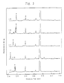
- Fig. 1,2,3 and 4 show chromatograms and aimed peaks for the fourteen peptides labeled with the fluorescent material, thus obtained.
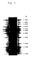
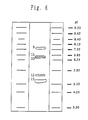
- the 24 fluorescence-labeled oligopeptides thus obtained were subjected to isoelectric focusing using polyacrylamide slab gel (4.2 % T, 4.8 % C, 55 mm ⁇ 120 mm large, 0.5 mm thick) containing 2.5 % carrier ampholyte (40 % solution, pH 3.5-10, purchased from Pharmacia LKB Co.), and mobilities thereof were compared with those of isoelectric focusing markers commercially available (pI calibration kit 3-10, purchased from Pharmacia Biotech Co.).
- the focusing conditions were as follows: started at 50 mV and focusing the voltage was increased at intervals of 15 minutes to 100 V, 200 V, 400 V, and 600 V.
- fluorescence-labeled oligopeptides in the gel were photographed through an orange filter under illumination with ultraviolet light, thereby checking focusing positions thereof.
- the gel was fixed with 20 % trifluoroacetic acid for 20 minutes, then the gel was stained for 20 minutes with 0.05 % of Celva violet 49 (purchased from Celva Inc.) in a mixed solvent of 25 % methanol and 10 % of acetic acid in water.
- Celva violet 49 purchased from Celva Inc.
- Fig. 5,6,and 7 show the focusing positions of the 24 fluorescence-labeled oligopeptides obtained and the commercially available isoelectric focusing markers.
- Table 1 is a list of pI's and calculation values calculated based on Eq. (1) for the respective fluorescence-labeled oligopeptides obtained (numbers 1-24). The table summarized the calculated pI's of other types of oligopeptides having wide ranges of pI values (numbers 25-69).
- a fused silica capillary (purchased from GL Science Co.) of the inner diameter 75 ⁇ m, the outer diameter 375 ⁇ m, and the total length 6 cm, which was coated with linear polyacrylamide on its inner wall was used.
- the anolyte was 10 mM phosphoric acid solution containing 2 % of methyl cellulose (the viscosity of the 2 % solution at 25 °C; 4,000 cP, purchased from Sigma Co.), and the catholyte was 20 mM NaOH solution containing 2 % of hydroxypropylmethyl cellulose (the viscosity of 2 % solution at 25 °C; 4,000 cP, purchased from Sigma Co.). Focusing was carried out for 2 min with high-voltage power supply at the maximum of either 2.4 kV or 10 ⁇ A. Focused fluorescence-labeled peptides were electrophoretically mobilized towards the detection window.
- the helium neon laser (wavelength 543.5 nm, power 1 mW, Model; 05-LGR-151-S, purchased from Melles Griot Co.) was used for excitation. Fluorescence was collected by a 40X objective lens, then the fluorescence intensity was measured by use of a photomultiplier tube(Model; R1387, manufactured by Hamamatsu Photonics K.K.) after filtration with bandpass filter(590nm, 30mm band width, Model DIF-BP-3, manufactured by Nippon Sinkuu, Kogaku) and a cut-off filter(Model SCF-50S-560, manufactured by Sigma Kouki), and the data were collected using an integrator(Model CR4A, manufactured by Shimazu Seisakusho K.K.).
- Fig. 8 shows moving periods and fluorescent intensities in the capillary isoelectric focusing with fluorescence detection, of five peptides out of 24 fluorescence-labeled peptides obtained.
- the present invention was able to provide the fluorescence-labeled oligopeptides by binding a fluorescence dye to oligopeptides. Since the fluorescence dye is bonded to the oligopeptides under mild conditions, the reaction causes no damage on the oligopeptides and the fluorescent material, the number of fluorescent materials and locations thereof bonded to the oligopeptides can be specified, and the obtained oligopeptides containing the fluorescence dye will have highly stable, uniform, and unique pI's. Accordingly, the markers for capillary isoelectric focusing with fluorescence detection obtained in the present invention can be used as markers for estimating isoelectric points of sample materials.
Landscapes
- Chemical & Material Sciences (AREA)
- Health & Medical Sciences (AREA)
- Life Sciences & Earth Sciences (AREA)
- Physics & Mathematics (AREA)
- Chemical Kinetics & Catalysis (AREA)
- Molecular Biology (AREA)
- Immunology (AREA)
- Electrochemistry (AREA)
- Analytical Chemistry (AREA)
- Biochemistry (AREA)
- General Health & Medical Sciences (AREA)
- General Physics & Mathematics (AREA)
- Pathology (AREA)
- Organic Chemistry (AREA)
- Nuclear Medicine, Radiotherapy & Molecular Imaging (AREA)
- General Chemical & Material Sciences (AREA)
- Biophysics (AREA)
- Genetics & Genomics (AREA)
- Medicinal Chemistry (AREA)
- Proteomics, Peptides & Aminoacids (AREA)
- Optics & Photonics (AREA)
- Spectroscopy & Molecular Physics (AREA)
- Investigating Or Analysing Biological Materials (AREA)
- Peptides Or Proteins (AREA)
- Investigating, Analyzing Materials By Fluorescence Or Luminescence (AREA)
Description
- The present invention relates to the use of oligo peptides comprising a fluorescence dye as isoelectric point markers for isoelectric focusing with fluorescence detection.
- Since Picton, H. and Linder, S. E. observed in 1892 that when an electric field is supplied particles with charges on the surfaces which are suspended in an electrolyte solution migrate toward the poles opposite to the charges of the respective particles; this phenomenon, electrophoresis, has been used as means of separation and analysis. After Tiselius has completed his apparatus in 1937, fields of applications have been expanding rapidly heretofore.
- Ordinary electrophoresis utilizes differences of net charge states at a certain specific pH, whereas Vesterberg, O. and Svensson, H. developed carrier ampholytes as amphoteric electrolytes in 1966. The isoelectric focusing has been established utilizing the phenomenon that each amphoteric electrolyte has the effective net charge of zero at a certain pH so as to stop migrating.
- Here, the value of the pH where the effective charge becomes zero is called an isoelectric point of that substance. In the isoelectric focusing method, a substance stops as concentrated at a position equal to the isoelectric point on a pH gradient formed in a focusing carrier.
- Since substances are separated as concentrated in a focusing manner as described , it may provide a separation or analytical method with much higher sensitivity in an electrophoresis.
- Recently a capillary isoelectric focusing was further developed in which electrophoresis was effected in a fine tube (capillary) made of fused silica and having the inner diameter of 5 to 100 µm and the length of 30 to 100 cm. Since the capillary isoelectric focusing has a high resolution with a small amount of sample, it has been applied to not only separation and analysis of proteins, but also to separation and analysis of inorganic ions, low-molecular weight compounds, and nucleic acids.
- There are further attempts to quantify concentrations of separated components in a sample by a detector set at one end of the capillary tube. Labeling of peptides or proteins by the use of fluorescent groups is used for various purposes in the field of biochemistry. In FR-A-2 687 680 for example a process for labeling biologically active peptides or proteins by inter alia fluorescent markers is disclosed. This process can be used for the detection of physiologic effector molecules that will bind to the labeled peptide or protein. Another example for the use of fluorescently labeled peptidic probes is demonstrated in EP-A-0 606 804 where labeled arginine containing peptides are used for in vitro labeling of neurotensin receptors. In US-A-5 245 551 it was shown that a fluorescent dye which is coupled to a protein of interest can be used to determine the concentration of said protein. Further the authors of TRAC, vol. 6, 5, May 1987, pages 116-120 present new techniques that can be employed for quantitative determination of arginine or tyrosine containing peptides by HPLC using fluorescence detection.
- The present invention will become more fully understood from the detailed description given hereinbelow and the accompanying drawings which are given by way of illustration only, and thus are not to be considered as limiting the present invention.
- Further scope of applicability of the present invention will become apparent from the detailed description given hereinafter. However, it should be understood that the detailed description and specific examples, while indicating preferred embodiments of the invention, are given by way of illustration only, since various changes and modifications within the spirit and scope of the invention will become apparent to those skilled in the art from this detailed description.
-
- Fig. 1 is a drawing to show chromatograms and identified specific peaks (*) of five types of fluorescence-labeled peptides(1-5) according to the present invention, as separated by high performance liquid chromatography;
- Fig. 2 is a drawing to show chromatograms and identified specific peaks (*) of nine types of fluorescence-labeled peptides(6-14) according to the present invention, as separated by high performance liquid chromatography;
- Fig. 3 is a drawing to show chromatograms and identified specific peaks (*) of five types of fluorescence-labeled peptides(15-19) according to the present invention, as separated by high performance liquid chromatography;
- Fig. 4 is a drawing to show chromatograms and identified specific peaks (*) of five types of fluorescence-labeled peptides(20-24) according to the present invention, as separated by high performance liquid chromatography;
- Fig. 5 is a drawing to show a migration image after isoelectric focusing of five types of fluorescence-labeled peptides(1-5) according to the present invention and isoelectric focusing markers commercially available;
- Fig. 6 is a drawing to show a migration image after isoelectric focusing of five types of fluorescence-labeled peptides(9-13) according to the present invention and isoelectric focusing markers commercially available;
- Fig. 7 is a drawing to show a migration image after isoelectric focusing of five types of fluorescence-labeled peptides(15-23) according to the present invention and isoelectric focusing markers commercially available; and
- Fig. 8 is a drawing to show a relation between migration period and fluorescence intensity for five types of fluorescence-labeled peptides(1-5) when fluorescence capillary isoelectric focusing is carried out.
-
- In the drawings, reference numerals denote the following peptides.
- 1: des-asparaginic acid1-angiotensin I (arginine-valine-tyrosine-isoleucine-histidine-proline-phenylalanine-histidine-leucine, Arg-Val-Tyr-Ile-His-Pro-Phe-His-Leu);
- 2: [asparagine1-valine5]-angiotensin II (asparagine-arginine-valine-tyrosine-valine-histidine-proline-phenylalanine, Asn-Arg-Val-Tyr-Val-His-Pro-Phe);
- 3: angiotensin I (asparaginic acid-arginine-valine-tyrosine-isoleucine-histidine-proline-phenylalanine-histidine-leucine, Asp-Arg-Val-Tyr-Ile-His-Pro-Phe-His-Leu);
- 4: angiotensin II (asparaginic acid-arginine-valine-tyrosine-isoleucine-histidine-proline-phenylalanine, Asp-Arg-Val-Tyr-Ile-His-Pro-Phe);
- 5: fibronectin fragment (glycine-arginine-glycine-asparaginic acid, Gly-Arg-Gly-Asp);
- 6: tyrosine-histidine-arginine-arginine (Tyr-His-Arg-Arg);
- 7: tyrosine-tyrosine-histidine-arginine-arginine (Tyr-Tyr-His-Arg-Arg);
- 8: tyrosine-tyrosine-histidine-lysine-arginine (Tyr-Tyr-His-Lys-Arg);
- 9: glutamic acid-histidine-lysine-arginine (Glu-His-Lys-Arg);
- 10: tyrosine-tyrosine-histidine-histidine (Tyr-Tyr-His-His);
- 11: tyrosine-histidine-histidine (Tyr-His-His);
- 12: asparaginic acid-glutamic acid-tyrosine-histidine-lysine-arginine (Asp-Glu-Tyr-His-Lys-Arg);
- 13: tyrosine-histidine (Tyr-His);
- 14: asparaginic acid-asparaginic acid-asparaginic acid-asparaginic acid-asparaginic acid-arginine (Asp-Asp-Asp-Asp-Asp-Arg).
- 15: glycine-histidine-arginine-arginine (Glu-His-Arg-Arg).
- 16: asparaginic acid-glutaminic acid-histidine-arginine-arginine-arginine (Asp-Glu-His-Arg-Arg-Arg).
- 17: glutaminic acid-histidine-histidine-histidine-lysine-arginine (Glu-His-His-His-Lys-Arg)
- 18:histidine-histidine-histidine-histidine-histidine ( His-His-His-His-His-His).
- 19: asparagic acid-histidine-histidine-arginine (Asp-His-His-Arg).
- 20: asparagic acid-asparagic acid-glutamic acid-histidine-histidine-histidine-lysine-arginine ( Asp-Asp-Glu-His-His-His-Lys-Arg).
- 21: asparagic acid-asparagic acid-asparagic acid-asparagic acid-glutamic acid-glutamic acid-histidine-arginine-arginine-arginine-arginine-arginine (Asp-Asp-Asp-Asp-Glu-Glu-His-Arg-Arg-Arg-Arg-Arg).
- 22: asparagic acid-asparagic acid-asparagic acid-glutamic acid-histidine-arginine-arginine (Asp-Asp-Asp-Glu-His-His-Arg-Arg).
- 23: asparagic acid-asparagic acid-asparagic acid-glutamic acid-glutamic acid-glutamic acid-histidine-arginine-arginine-arginine (Asp-Asp-Asp-Glu-Glu-Glu-His-Arg-Arg-Arg)
- 24: asparagic acid-glutamic acid-arginine (Asp-Glu-Arg).
-
- One way of conventional detection in analytical methods is to focus mainly ultraviolet light or visible light on a sample and to detect amounts of components in the sample by changes of light absorption (ultraviolet or visible light detection).
- Such detection method is, however, difficult to use for the capillary isoelectric focusing, because the inner diameter of the capillary tube is almost equal to optical path length.
- Another detection method with high sensitivity uses fluorescence-labeled samples with a fluorescence labeling material such as dyes. Excitation light is focused on to separated components in the sample, and emitted fluorescence is detected to quantify concentrations of the components (fluorescence detection).
- For labeling a sample such as a mixture of proteins, fluorescence materials(dyes) are usually reacted with the sample to form a chemical bond between an amino group in the sample protein and the fluorescence dye. For example, fluorescein isothiocyanate (FITC) and tetramethylrhodamine isothiocyanate (TRITC), etc. are known as fluorescence dyes to be bonded with an amino group (Motonori ONO, Yuichi KANEOKA, Fumio SAKIYAMA, and Hiroshi MAEDA, Chemical Modification of Proteins (II), Gakkai Shuppan Center, 1981).
- The fluorescence detection can be adequately used in the capillary isoelectric focusing to quantify the amounts of separated components in a sample. Therefore, the fluorescence detection combined with a capillary isoelectric focusing system may be a potential analytical system of a mixture of proteins because of dual advantages of high resolution and high detection sensitivity.
- The pH gradient formed in the focusing carrier, however, should be known precisely in isoelectric focusing, because an isoelectric point (pI) is determined based on a stop position of a sample component separated in the focusing carrier. Therefore, pI markers are required as standards to estimate a formed pH gradient.
- For electrophoresis or isoelectric focusing with fluorescence detection it is extremely difficult to use conventional protein pI markers as pI standards, which have known pI's (for example, trypsinogen (pI = 9.30), lentilectin (pI = 7.80, 8.00, 8.20), human hemoglobin C (pI = 7.50), human hemoglobin A (pI = 7.10), equine myoglobin (pI = 7.00), human carbonic anhydrase (pI = 6.50), bovine carbonic anhydrase (pI = 6.00), β-lactoglobulin B (pI = 5.10), phycocyanin (pI = 4.65), amyloglucosidase (pI = 3.50), etc.), since these proteins per se can emit only very weak fluorescence. Such proteins need to increase their fluorescence intensity by being labeled with an appropriate fluorescence material.
- However, the pI's of the proteins are in general dependent on the types of amino acids composing the proteins and N-terminal amino groups, (Chemistry of Proteins (I), Second Series of Lectures on Biochemical Experiments No. 2, the Japanese Biochemical Society, 1987). Then, the isoelectric points, pI's of the proteins per se would be changed by any chemical modification on the N-terminal amino groups of such proteins by the fluorescence label.
- Further, since a protein may include several amino acids reactive to a fluorescent labeling material, the number and positions of fluorescence labeling materials bonded thereto are unspecified. Consequently, the modification provides a mixture of proteins labeled at different positions to give different pI's (Flatmark, T. and Vesterberg, O., Acta Chem. Scand., 20, 1497-1503, 1966). Therefore, it is difficult to determine precise pH regions formed in the focusing carrier by such labeled protein mixtures described above.
- Further, the three-dimensional structure of a protein may be altered by the labeling with the fluorescence material (Williamson, A. R., Kreth and H. W., Ann. N. Y. Acad. Sci., 209, 211-224, 1973), which may deteriorate stability of protein per se.
- This would be a problem in respect of storage stability of the proteins as pI markers.
- As discussed above, the isoelectric focusing with fluorescence detection cannot use such labeled proteins with fluorescence materials as pI markers.
- It was therefore the object of the present invention to provide isoelectric point markers, which cover a wide range of isoelectric points, which each have a singular and peculiar isoelectric point and which have an excellent storage stability.
- This object is solved by providing an oligopeptide comprising a fluorescence dye as an isoelectric point marker for use in isoelectric focusing with fluorescence detection.
- The present invention concerns the use of markers for isoelectric focusing with fluorescence detection obtained by selecting appropriate oligopeptides and further labeling them with a suitable fluorescence material in order to accurately clarify pH regions formed in the focusing carrier in isoelectric focusing, which are characterized by demonstrating uniform pI's and high storage stability.
- Namely, the present invention concerns the use of a marker for isoelectric focusing with fluorescence detection, which is an oligopeptide comprising a fluorescence dye.
- Further, the present invention concerns the use of a marker for isoelectric focusing with fluorescence detection, which is an oligopeptide obtained by bonding the fluorescence dye materials to an N-terminal amino group of the oligopeptide.
- Further, the present invention concerns the use of a marker for isoelectric focusing with fluorescence detection, wherein the bonding is one selected from the group at least comprising amide, thioamide, sulfonamide, urea, thiourea, and urethane bonding groups.
- Further, the present invention concerns the use of a marker for isoelectric focusing with fluorescence detection, wherein the oligopeptide comprises at least nj amino acids, each having a group which is to release a proton, thereby resulting in having a negative charge, wherein the group to release the proton thereby to have the negative charge has an acid dissociation constant of Kj, and/or ni amino acids, each having a group which is to receive a proton, thereby resulting in having a positive charge, wherein the group to receive the proton thereby to have the positive charge has an acid dissociation constant of Ki, and wherein, defining that Z = Σi(ni/(1 + Ki/[H+])) - Σj(nj/(1 + [H+]/Kj)) and that pI is -log([H+]) when |Z| < 0.01 is satisfied, 3 < pI < 11.
- Further, the present invention concerns the use of a marker for isoelectric focusing with fluorescence detection, wherein the oligopeptide is angiotensin or a related compound thereof.
- Further the present invention concerns the marker for isoelectric focusing with fluorescence detection, wherein the oligopeptide is at least one oligopeptide selected from the group at least comprising Arg-Arg-Val-Tyr-Ile-His-Lys, Arg-Arg-Lys-His-Tyr, Arg-Arg-His-Tyr, Arg-Lys-His-Tyr, Arg-Arg-Val-Tyr-Ile-Tyr, Arg-Val-Tyr-Ile-His-Pro-Phe-His-Leu, Asn-Arg-Val-Tyr-Val-His-Pro-Phe, Asp-Arg-Val-Tyr-Ile-His-Pro-Phe-His-Leu, Asp-Arg-Val-Tyr-Ile-His-Pro-Phe, Gly-Arg-Gly-Asp, and Arg-Asp-Asp-Asp-Asp-Asp.
- Further, the present invention concerns the use of a marker for isoelectric focusing with fluorescence detection, wherein the fluorescence dye is one selected from the group comprising rhodamine, fluorescein, cyanine, indocyanine, indocarbocyanine, pyronine, lucifer yellow, quinacrine, squarillium, coumarin, fluoroanthranilmaleimide, and anthracene.
- Further, the present invention concerns the use of a marker for isoelectric focusing with fluorescence detection, wherein the isoelectric focusing is a capillary isoelectric focusing with fluorescence detection method.
- Further, the present invention concerns the use of a marker for isoelectric focusing with fluorescence detection, wherein the oligopeptide is at least one oligopeptide selected from the group at least comprising Tyr-His-Lys-Arg-Arg, Tyr-His-Lys-Arg, Asp-His-His-Arg, Asp-His-Arg, Arg-Arg-Val-Tyr-Ile-His-Lys, Arg-Arg-Lys-His-Tyr, Arg-Arg-His-Tyr, Arg-Lys-His-Tyr, Arg-Arg-Val-Tyr-Ile-Tyr, Arg-Val-Tyr-Ile-His-Pro-Phe-His-Leu, Asn-Arg-Val-Tyr-Val-His-Pro-Phe, Asp-Arg-Val-Tyr-Ile-His-Pro-Phe-His-Leu, Asp-Arg-Val-Tyr-Ile-His-Pro-Phe, Gly-Arg-Gly-Asp, Arg-Asp-Asp-Asp-Asp-Asp, Tyr-His-Arg-Arg, Tyr-Tyr-His-Arg-Arg, Tyr-Tyr-His-Lys-Arg, Glu-His-Lys-Arg, Tyr-Tyr-His-His, Tyr-His-His, Asp-Glu-Tyr-His-Lys-Arg, Tyr-His, Asp-Asp-Asp-Asp-Asp, Glu-His-Arg-Arg, Asp-Glu-His-Arg-Arg-Arg, Glu-His-His-His-Lys-Arg, His-His-His-His-His-His, Asp-His-His-Arg, Asp-Asp-Glu-His-His-His-Lys-Arg, Asp-Asp-Asp-Asp-Glu-Glu-His-Arg-Arg-Arg-Arg, Asp-Asp-Asp-Glu-His-His-Arg-Arg, Asp-Asp-Asp-Glu-Glu-Glu-His-Arg-Arg-Arg, and Asp-Glu-Arg.
- Further, the present invention concerns the marker for isoelectric focusing with fluorescence detection, wherein said oligopeptide at least comprises Arg-Val-Tyr-Ile-His-Pro-Phe-His-Leu, Asn-Arg-Val-Tyr-Val-His-Pro-Phe, Asp-Arg-Val-Tyr-Ile-His-Pro-Phe-His-Leu, Asp-Arg-Val-Tyr-Ile-His-Pro-Phe, Gly-Arg-Gly-Asp, Tyr-His-Arg-Arg, Tyr-Tyr-His-Arg-Arg, Tyr-Tyr-His-Lys-Arg, Glu-His-Lys-Arg, Tyr-Tyr-His-His, Tyr-His-His, Asp-Glu-Tyr-His-Lys-Arg, Tyr-His, Asp-Asp-Asp-Asp-Asp-Arg, Glu-His-Arg-Arg, Asp-Glu-His-Arg-Arg-Arg, Glu-His-His-His-Lys-Arg, His-His-His-His-His-His, Asp-His-His-Arg, Asp-Asp-Glu-His-His-His-Lys-Arg, Asp-Asp-Asp-Asp-Glu-Glu-His-Arg-Arg-Arg-Arg-Arg, Asp-Asp-Asp-Glu-His-His-Arg-Arg, Asp-Asp-Asp-Glu-Glu-Glu-His-Arg-Arg-Arg, and Asp-Glu-Arg.
- Embodiments of the present invention will be explained in detail.
- The present invention employs oligopeptides in particular out of various ampholytes applicable as markers. In isoelectric focusing, samples to be analyzed are proteins including oligopeptide, or other types of materials which contain peptides as a component. Therefore, the use of oligopeptides comprised of dissociable groups similar to proteins is preferred in order to check pI values of such samples.
- However, if an oligopeptide includes too many amino acids, there would exist a plurality of reaction sites for labeling with fluorescence dye, which would provide a mixture of labeled products depending upon reaction conditions. In this case a marker thus obtained would not demonstrate a single and narrow peak, which is not preferred for pI markers for isoelectric focusing with fluorescence detection.
- In the present invention there is no specific limitation on the number of amino acids for an oligopeptide, and the present invention may employ any oligopeptide having a sufficient number of amino groups to achieve an aimed pI accordingly.
- Oligopeptides consist of various types of amino acids as amphoteric compounds that have various types of acid groups and basic groups as acid- base dissociable groups, and the oligopeptides have a N-terminal amino group, and a C-terminal carboxylic acid group. For example, amino acids having an acid group include asparaginic acid, glutamic acid, tyrosine, etc., and amino acids having a basic group include lysine, arginine, histidine, etc.
- Because of these acid and basic dissociable groups, an oligopeptide shows a specific charge number at a certain pH. Accordingly, a charge number of an oligopeptide as a whole can be calculated from types and numbers of these acid and basic dissociable groups.
- For example, a charge number (Z) of an oligopeptide as a whole can be estimated by the following equation.
- Further, an isoelectric point pI of the oligopeptide is given as -log([H+]) = pH at Z = 0 in this equation.
- Therefore, it becomes possible to select amino acids necessary for an oligopeptide to demonstrate an aimed pI by utilizing this equation.
- If any group including a fluorescence dye is bonded to the N-terminal amino group of an oligopeptide, basic dissociation of the N-terminal amino group becomes restrained.
- For estimation of a pI and selecting appropriate amino acids, available pKa's of above acidic and basic dissociable groups are, for example, α-carboxylic acid (C-terminal) (3.6), β-carboxylic acid (Asp) (3.95), γ-carboxylic acid (Glu) (4.45), imidazole (His) (6.45), α-amino (N-terminal) (7.6), thiol (Cys) (8.5), phenylhydroxy (Tyr) (9.8), ε-amino (Lys) (10.2), and guanidinium group (Arg) (12.5).
- For example, in the case of an oligopeptide labeled at the N-terminal, asparaginic acid or glutamic acid is selected to decrease the pI, while arginine or lysine are selected to increase pI. Similarly, tyrosine or histidine may be selected in order to determine a pI between them.
- An example of a very small pI is an oligopeptide including at least five asparaginic acids and one arginine as acidic, and basic amino acids, which demonstrates a pI near 3.
- Similarly, an oligopeptide including at least two arginines, one lysine, one histidine, and one tyrosine shows a pI of not less than 11.
- In this manner, markers covering a desired range of pI can be obtained by suitably selecting some of various acidic, and basic amino acids.
- In the present invention, a preferred range of pI is between 2 and 13, both inclusive.
- In the present invention, a particularly preferred range of pI is between 3 and 11, both inclusive.
- Further, the present invention requires no specific limitation for synthesis of an oligopeptide including amino acids appropriately selected. A preferably applicable peptide synthesis method is, for example, an ordinary organic synthesis (the liquid phase process, the solid phase process, etc., Nobuo IZUMIYA, Tetsuo KATO, Haruhiko AOYAGI, and Michinori WAKI, Fundamentals and Experiments of Peptide Synthesis, Maruzen, 1985), or the use an automated synthesizer.
- The present invention can also suitably employ all or some of the oligopeptides derived from natural products as long as they include necessary amino acid groups and a reactive site for fluorescence labeling thereof.
- For example, oligopeptides obtained from natural products, which the present invention can suitably employ, include angiotensin and related compounds thereof (10 to 20 amino acids), bradykinin and related compounds thereof, enkephalin and related compounds thereof, etc. (Dictionary of Biochemistry (Second Edition), Tokyo Kagaku Dojin, 1990).
- Further, it is also possible to synthesize an oligopeptide having appropriate amino acids by bonding a chemically necessary amino acid to one selected from the above oligopeptides derived from natural products.
- There is no specific limitation on the type of fluorescence applicable in the present invention, and any fluorescence dye including a generally known fluorescent pigment may be employed.
- For example, the fluorescence dye may be rhodamine, fluorescein, cyanine, indocyanine, indocarbocyanine, pyronine, lucifer yellow, quinacrine, squarillium, coumarin, fluoroanthranilmaleimide, etc.
- Particularly, rhodamine and cyanine pigments are preferably applicable.
- A particularly preferred fluorescence dye derivative in the present invention is a 5-carboxytetramethylrhodamine fluorescence (Handbook of Fluorescent Probes and Research Chemicals, 5th Edition, MOLECULAR PROBES, INC., 1992).
- This rhodamine fluorescence has the maximum absorption (molar extinction coefficient of 63,000) at 546 nm and maximum fluorescence at 579 nm in methanol.
- Further fluorescence dye suitably applicable in the present invention include aromatic heterocyclic compounds or polycyclic aromatic hydrocarbons (for example, see Taiji NISHIKAWA and Keizo HIRAKI, "Fluorescence and Phosphorescence Analysis," Kyoritsu Shuppan, 1989). Particularly preferable fluorescence dyes of the kind are, for example, anthracene, naphthalene, phenanthrene, quinoline, pyrene, perylene, etc. or derivatives thereof. These types of hydrocarbon fluorescence dyes may react with the oligopeptides by using an appropriate bonding group as described below.
- The present invention includes no specific limitation on the type of the bonding group for labeling the fluorescence dye to the N-terminal of oligopeptide. The group including the fluorescence dye may be bonded with the N-terminal amino group either without or with additional connection group of a suitable binding group.
- In this case, the group including the fluorescence is preferably bonded with the N-terminal amino group, for example, by amide bonding, thioamide bonding, sulfonamide bonding, urea bonding, thiourea bonding, urethane bonding, or the like in the following form.
- There is no specific limitation on a method for introducing the binding group. For example, in the case of amide bonding, the group including the fluorescence dye preferably has an active ester group, acid chloride, or acid anhydride.
- For example, in the present invention, a rhodamine fluorescence dye having an active ester group reacts with the N-terminal amino group of oligopeptide to form an amide bonding.
- This reaction can be performed under mild conditions, and gives no change in oligopeptide nor fluorescence dye.
- Further, if an oligopeptide has a plurality of reactive sites, reaction conditions, reagents, etc. may be suitably selected in order to enable the reaction to occur only at the N-terminal amino group as desired. Well known techniques of organic synthesis can be suitably applied to select appropriate reaction condition.
- For example, one of preferred techniques is to preliminarily protect reactive ε-amino group (for example in lysine) if necessary.
- In the present invention, purity and storage stability of each marker for capillary isoelectric focusing with fluorescence detection can be checked by ordinary organic analytical methods such as infrared or MR spectroscopies, and also the isoelectric focusing .
- Further, if necessary, they can easily be refined using high performance liquid chromatography or other purification techniques. In that case high-efficiency separation and refinement becomes possible by using a reversed-phase column, particularly using octadecyl derivatives. The suitably applicable detection wavelength is 280 nm.
- In the present invention, preferred labeling can be realized, for example, by amide-bonding a rhodamine derivative to an amino group of a peptide selected from angiotensin and derivatives thereof.
- In the present invention, the pI of each marker for isoelectric focusing with fluorescence detection can be determined by performing isoelectric focusing to compare a mobility thereof with those of commercially available markers for isoelectric focusing or to directly measure pH at a position of migration of the labeled peptide.
- The isoelectric focusing preferably applicable is isoelectric focusing in polyacrylamide slab gel (see Righetti, P. G., Isoelectric Focusing: Theory, Methodology and Applications, Elsevier, Amsterdam, 1983).
- An ordinary method (for example, Coomassie brilliant blue-R 250 stain) can suitably be applied for detection by a staining method.
- Further, fluorescence from markers can be preferably detected by ordinary fluorescence analysis method, for example a method with a densitometer (for example, Shimazu double wavelength flying spot scanning densitometer CS 9300 PC, manufactured by Shimazu Seisakusho K.K.).
- There is no specific limitation on a capillary isoelectric focusing apparatus to which the fluorescence-labeled oligopeptides according to the present invention can be applied as markers.
- Generally, fluorescence-labeled peptides stop still as concentrated at respective positions on the pH gradient formed in the focusing carrier according to respective pI's by the capillary isoelectric focusing. These positions can be checked by detecting emitted fluorescence with excitation light.
- The present invention includes no specific limitation on the light source for excitation, but use of a laser capable of emitting stable light is in particular preferred.
- For example, the present invention can suitably employ a helium neon laser as excitation light for the rhodamine dye, which is a fluorescence dye.
- The pH gradient formed in the focusing carrier can be checked by a fluorescence detector disposed at one end of the capillary.
- As explained above, the markers for isoelectric focusing with fluorescence detection according to the present invention are synthesized by bonding a group having a fluorescence dye to oligopeptides, whereby they show uniform pI's and have high stability. Accordingly, the markers can be used as those for high-sensitive analysis where isoelectric points of sample materials are checked by isoelectric focusing after migration, based on the pH gradient formed in the focusing carrier.
- Hence, the markers for isoelectric focusing with fluorescence detection according to the present invention can also preferably be applied to the capillary isoelectric focusing method.
- The present invention will be explained in further detail, based on examples thereof, but it should be noted that the present invention is not intended to be limited to the examples but may involve all applications and modifications within the scope not departing from the essence of the invention.
- Labeling of oligopeptides with 5-carboxytetramethylrhodamine succinimidyl ester:
- Oligopeptides used in the reaction were purchased from Sigma Co., and were subjected to the following reaction with 5-carboxytetramethylrhodamine succinimidyl ester without further being refined. The oligopeptides used in the reaction were angiotensin I (asparaginic acid-arginine-valine-tyrosine-isoleucine-histidine-proline-phenylalanine-histidine-leucine, Asp-Arg-Val-Tyr-Ile-His-Pro-Phe-His-Leu), angiotensin II (asparaginic acid-arginine-valine-tyrosine-isoleucine-histidine-proline-phenylalanine, Asp-Arg-Val-Tyr-Ile-His-Pro-Phe), derivatives of the foregoing two, i.e. des-asparaginic acid1-angiotensin I (arginine-valine-tyrosine-isoleucine-histidine-proline-phenylalanine-histidine-leucine, Arg-Val-Tyr-Ile-His-Pro-Phe-His-Leu) and [asparagine1-valine5]-angiotensin II (asparagine-arginine-valine-tyrosine-valine-histidine-proline-phenylalanine, Asn-Arg-Val-Tyr-Val-His-Pro-Phe), and fibronectin fragment (glycine-arginine-glycine-asparaginic acid, Gly-Arg-Gly-Asp).
- Further, the below oligopeptides were synthesized based on the Boc process by a peptide automatic synthesizing apparatus manufactured by Applied Biosystems Co. (for example, see Lectures on
Chemical Experiments 1, Proteins VI, compiled by the Japanese Biochemical Society, 1992), and the oligopeptides obtained were refined by high performance liquid chromatography. The conditions used for high performance liquid chromatography were as follows: YMC-Pack ODS-AM (a column available from YMC Co., 30 mm in inner diameter × 250 mm in total length); theflow rate 20 ml/min; the detection wavelength 220 nm; the mobile phase was acetonitrile gradient-0.1 % trifluoroacetic acid, wherein the gradient was set to the following conditions depending upon the oligopeptides. Tyrosine-histidine-arginine-arginine (Tyr-His-Arg-Arg), 0.5 % to 5 %; tyrosine-tyrosine-histidine-arginine-arginine (Tyr-Tyr-His-Arg-Arg), 1 % to 15 %; tyrosine-tyrosine-histidine-lysine-arginine (Tyr-Tyr-His-Lys-Arg), 1 % to 15 %; glutamic acid-histidine-lysine-arginine (Glu-His-Lys-Arg), 0.5 % to 5 %; tyrosine-tyrosine-histidine-histidine (Tyr-Tyr-His-His), 1 % to 15 %; tyrosine-histidine-histidine (Tyr-His-His), 1 % to 10 %; asparaginic acid-glutamic acid-tyrosine-histidine-lysine-arginine (Asp-Glu-Tyr-His-Lys-Arg), 1 % to 15 %; tyrosine-histidine (Tyr-His), 1 % to 15 %; asparaginic acid-asparaginic acid-asparaginic acid-asparaginic acid-asparaginic acid-arginine (Asp-Asp-Asp-Asp-Asp-Arg), 1 % to 15 %; glycine-histidine-arginine-arginine (Glu-His-Arg-Arg),1% to 18%; asparaginic acid-glutaminic acid-histidine-arginine-arginine-arginine (Asp-Glu-His-Arg-Arg-Arg), 0% to 15%; glutaminic acid-histidine-histidine-histidine-lysine-arginine (Glu-His-His-His-Lys-Arg), 0% to 15%; histidine-histidine-histidine-histidine-histidine (-His-His-His-His-His-His), 0% to 10%; asparagic acid-histidine-histidine-arginine (Asp-His-His-Arg), 0% to 5%; asparagic acid-asparagic acid-glutamic acid-histidine-histidine-histidine-lysine-arginine (-Asp-Asp-Glu-His-His-His-Lys-Arg), 0% to 15%; asparagic acid-asparagic acid-asparagic acid-asparagic acid-glutamic acid-glutamic acid-histidine-arginine-arginine-arginine-arginine-arginine (Asp-Asp-Asp-Asp-Glu-Glu-His-Arg-Arg-Arg-Arg-Arg), 0% to 15%; asparagic acid-asparagic acid-asparagic acid-glutamic acid-histidine-arginine-arginine (Asp-Asp-Asp-Glu-His-His-Arg-Arg), 0% to 15%; asparagic acid-asparagic acid-asparagic acid-glutamic acid-glutamic acid-glutamic acid-histidine-arginine-arginine-arginine (Asp-Asp-Asp-Glu-Glu-Glu-His-Arg-Arg-Arg), 1% to 15%; and asparagic acid-glutamic acid-arginine (Asp-Glu-Arg), 0% to 4%. - Each oligopeptide was dissolved in 0.1 M 2-(N-Morpholino) ethanesulfonic acid (MES)-NaOH buffer (pH 6) at a concentration of 1mM.
- 5-carboxytetramethylrhodamine succinimidyl ester (purchased from MOLECULAR PROBES Co.) was dissolved in dimethylformamide at a concentration of 25 mM.
- To 20 µl of each oligopeptide solution, 5 µl of the 5-carboxytetramethylrhodamine succinimidyl ester solution was added at 27°C and the mixture was allowed to react overnight.
To stop the reaction, 50 µl of 1 M Tris-HCl buffer solution (pH 8) were added to the mixture. - The labeled oligopeptides were separated and refined by high performance liquid chromatography under the following conditions.
- Column: TSKgel ODS-80Ts (purchased from TOSOH CORP, 4.6 mm in inner diameter × 25 cm in total length) and TSKguardgel ODS-80Ts (available from TOSOH CORP, 3.2 mm in inner diameter × 1.5 cm in total length)
- Mobile phase: 5 % to 55 % acetonitrile gradient-0.1 % trifluoroacetic acid
- Flow rate: 1 ml/min
- Detection wavelength: 280 nm
- Fig. 1,2,3 and 4 show chromatograms and aimed peaks for the fourteen peptides labeled with the fluorescent material, thus obtained.
- The 24 fluorescence-labeled oligopeptides thus obtained were subjected to isoelectric focusing using polyacrylamide slab gel (4.2 % T, 4.8 % C, 55 mm × 120 mm large, 0.5 mm thick) containing 2.5 % carrier ampholyte (40 % solution, pH 3.5-10, purchased from Pharmacia LKB Co.), and mobilities thereof were compared with those of isoelectric focusing markers commercially available (pI calibration kit 3-10, purchased from Pharmacia Biotech Co.).
- The focusing conditions were as follows: started at 50 mV and focusing the voltage was increased at intervals of 15 minutes to 100 V, 200 V, 400 V, and 600 V.
- After focusing, fluorescence-labeled oligopeptides in the gel were photographed through an orange filter under illumination with ultraviolet light, thereby checking focusing positions thereof.
- After that, in order to know focusing positions of the commercially available isoelectric focusing markers, the gel was fixed with 20 % trifluoroacetic acid for 20 minutes, then the gel was stained for 20 minutes with 0.05 % of Celva violet 49 (purchased from Celva Inc.) in a mixed solvent of 25 % methanol and 10 % of acetic acid in water.
- After staining, it was de-stained with a mixed solvent of methanol and acetic acid to obtain a clear background, for checking the focusing positions.
- Fig. 5,6,and 7 show the focusing positions of the 24 fluorescence-labeled oligopeptides obtained and the commercially available isoelectric focusing markers.
- Table 1 is a list of pI's and calculation values calculated based on Eq. (1) for the respective fluorescence-labeled oligopeptides obtained (numbers 1-24). The table summarized the calculated pI's of other types of oligopeptides having wide ranges of pI values (numbers 25-69).
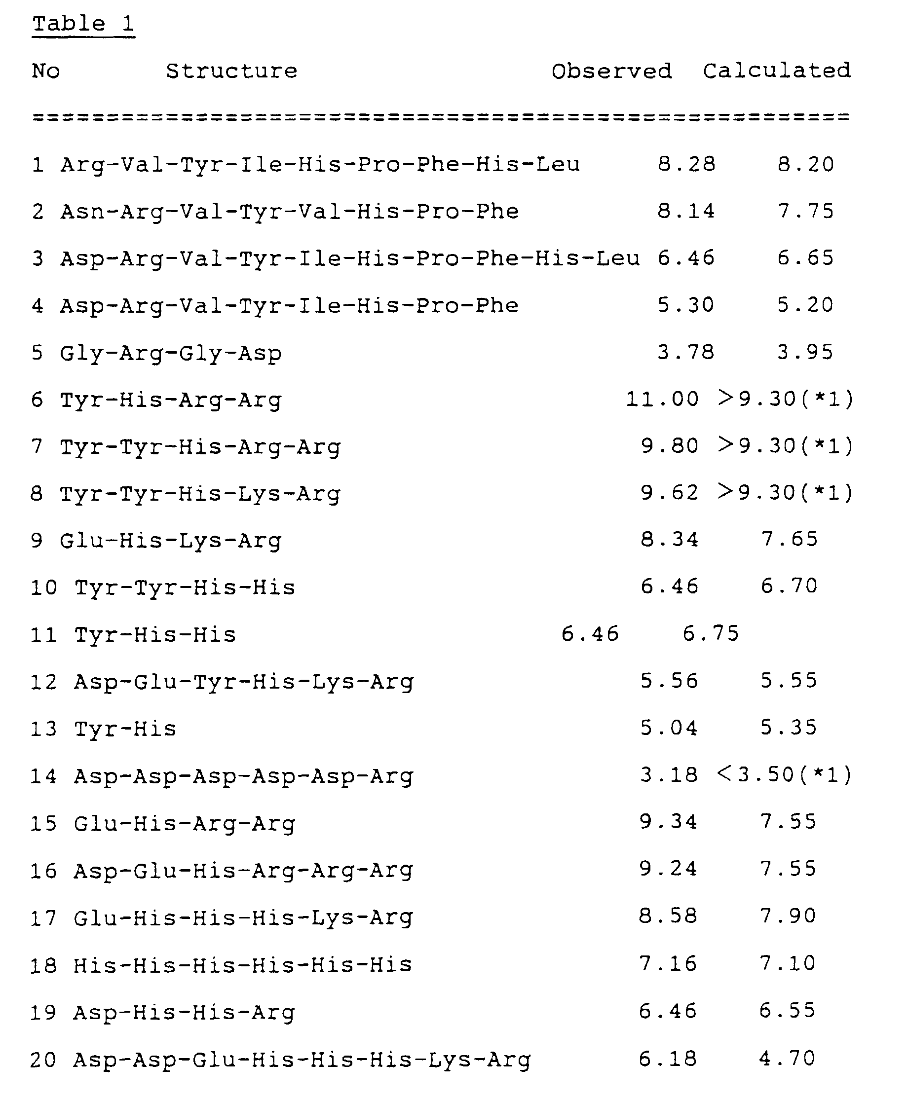
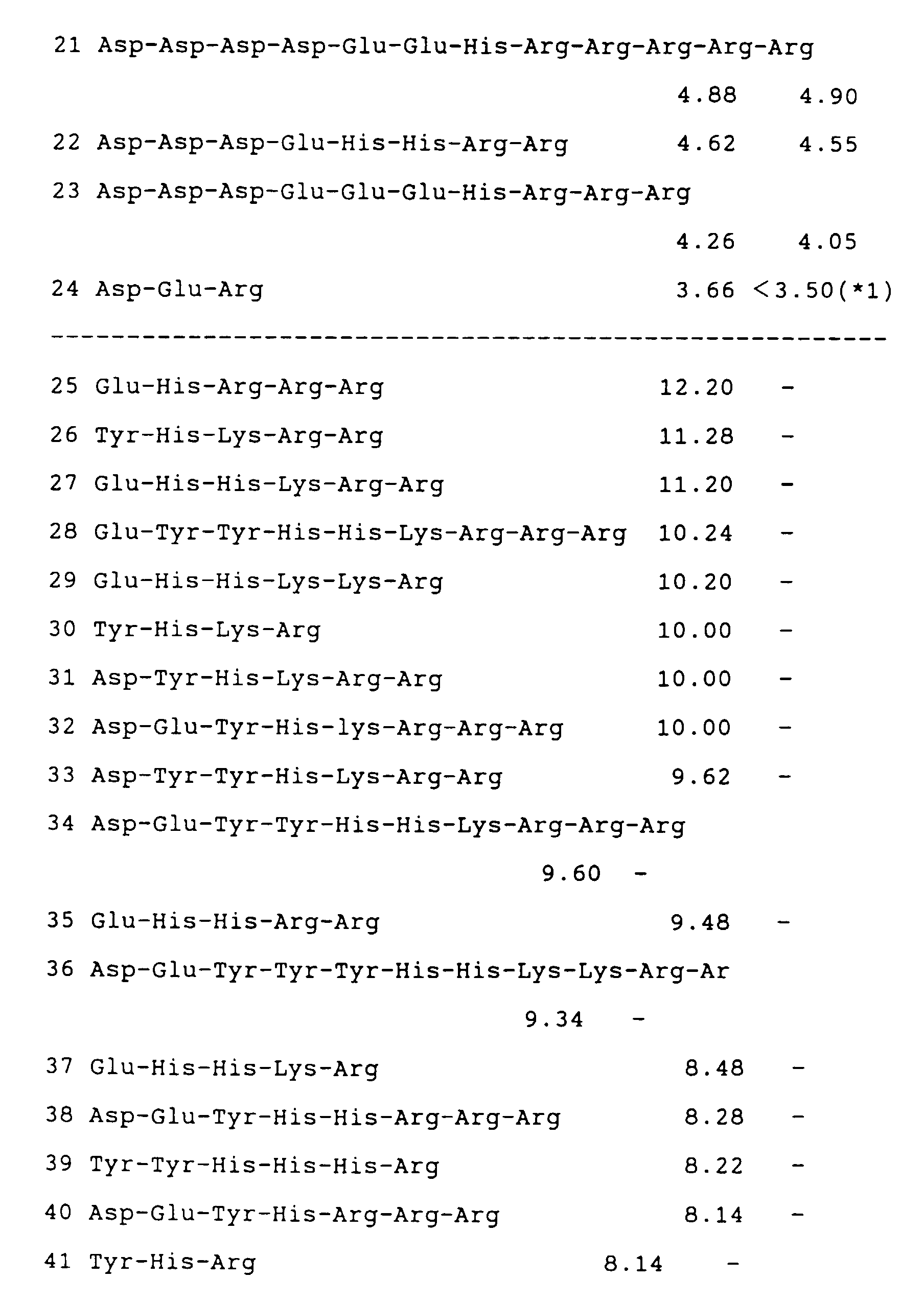


- Then the fourteen fluorescence-labeled oligopeptides obtained were subjected to separation and detection by capillary isoelectric focusing with fluorescence detection (Hjerten, S., Journal of Chromatography, 347, 191-198, 1985).
- A fused silica capillary (purchased from GL Science Co.) of the inner diameter 75 µm, the outer diameter 375 µm, and the
total length 6 cm, which was coated with linear polyacrylamide on its inner wall was used. - The capillary filled with a sample solution, fluorescence-labeled peptides containing 1 % ampholine and 0.1
% Tween 20. - The anolyte was 10 mM phosphoric acid solution containing 2 % of methyl cellulose (the viscosity of the 2 % solution at 25 °C; 4,000 cP, purchased from Sigma Co.), and the catholyte was 20 mM NaOH solution containing 2 % of hydroxypropylmethyl cellulose (the viscosity of 2 % solution at 25 °C; 4,000 cP, purchased from Sigma Co.). Focusing was carried out for 2 min with high-voltage power supply at the maximum of either 2.4 kV or 10 µA.
Focused fluorescence-labeled peptides were electrophoretically mobilized towards the detection window. - The helium neon laser (wavelength 543.5 nm,
power 1 mW, Model; 05-LGR-151-S, purchased from Melles Griot Co.) was used for excitation. Fluorescence was collected by a 40X objective lens, then the fluorescence intensity was measured by use of a photomultiplier tube(Model; R1387, manufactured by Hamamatsu Photonics K.K.) after filtration with bandpass filter(590nm, 30mm band width, Model DIF-BP-3, manufactured by Nippon Sinkuu, Kogaku) and a cut-off filter(Model SCF-50S-560, manufactured by Sigma Kouki), and the data were collected using an integrator(Model CR4A, manufactured by Shimazu Seisakusho K.K.). - Fig. 8 shows moving periods and fluorescent intensities in the capillary isoelectric focusing with fluorescence detection, of five peptides out of 24 fluorescence-labeled peptides obtained.
- As seen from the above results, it was confirmed that the present invention was able to provide the fluorescence-labeled oligopeptides by binding a fluorescence dye to oligopeptides. Since the fluorescence dye is bonded to the oligopeptides under mild conditions, the reaction causes no damage on the oligopeptides and the fluorescent material, the number of fluorescent materials and locations thereof bonded to the oligopeptides can be specified, and the obtained oligopeptides containing the fluorescence dye will have highly stable, uniform, and unique pI's. Accordingly, the markers for capillary isoelectric focusing with fluorescence detection obtained in the present invention can be used as markers for estimating isoelectric points of sample materials.
- From the invention thus described, it will be obvious that the invention may be varied in many ways. Such variations are not to be regarded as a departure from the scope of the invention, and all such modifications as would be obvious to one skilled in the art are intended to be included within the scope of the following claims.
-
- (2) INFORMATION FOR SEQ ID NO:1:
- (i) SEQUENCING CHARACTERISTICS:
- (A)LENGTH: 8 amino acid
- (B)TYPE: amino acid
- (D)TOPOLOGY: linear
- (ii)MOLECULE TYPE:peptide
- (xi) SEQUENCE DESCRIPTION: SEQ ID NO:1:

- (2) INFORMATION FOR SEQ ID NO:2:
- (i) SEQUENCING CHARACTERISTICS:
- (A)LENGTH: 8 amino acid
- (B)TYPE: amino acid
- (D)TOPOLOGY: linear
- (ii)MOLECULE TYPE:peptide
- (xi) SEQUENCE DESCRIPTION: SEQ ID NO:2:

- (2) INFORMATION FOR SEQ ID NO:3:
- (i) SEQUENCING CHARACTERISTICS:
- (A)LENGTH: 10 amino acid
- (B)TYPE: amino acid
- (D)TOPOLOGY: linear
- (ii)MOLECULE TYPE:peptide
- (xi) SEQUENCE DESCRIPTION: SEQ ID NO:3:

- (2) INFORMATION FOR SEQ ID NO:4:
- (i) SEQUENCING CHARACTERISTICS:
- (A)LENGTH: 8 amino acid
- (B)TYPE: amino acid
- (D)TOPOLOGY: linear
- (ii)MOLECULE TYPE:peptide
- (xi) SEQUENCE DESCRIPTION: SEQ ID NO:4:

- (2) INFORMATION FOR SEQ ID NO:5:
- (i) SEQUENCING CHARACTERISTICS:
- (A)LENGTH: 4 amino acid
- (B)TYPE: amino acid
- (D)TOPOLOGY: linear
- (ii)MOLECULE TYPE:peptide
- (xi) SEQUENCE DESCRIPTION: SEQ ID NO:5:

- (2) INFORMATION FOR SEQ ID NO:6:
- (i) SEQUENCING CHARACTERISTICS:
- (A)LENGTH: 4 amino acid
- (B)TYPE: amino acid
- (D)TOPOLOGY: linear
- (ii)MOLECULE TYPE:peptide
- (xi) SEQUENCE DESCRIPTION: SEQ ID NO:6:

- (2) INFORMATION FOR SEQ ID NO:7:
- (i) SEQUENCING CHARACTERISTICS:
- (A)LENGTH: 5 amino acid
- (B)TYPE: amino acid
- (D)TOPOLOGY: linear
- (ii)MOLECULE TYPE:peptide
- (xi) SEQUENCE DESCRIPTION: SEQ ID NO:7:

- (2) INFORMATION FOR SEQ ID NO:8:
- (i) SEQUENCING CHARACTERISTICS:
- (A)LENGTH: 5 amino acid
- (B)TYPE: amino acid
- (D)TOPOLOGY: linear
- (ii)MOLECULE TYPE:peptide
- (xi) SEQUENCE DESCRIPTION: SEQ ID NO:8:
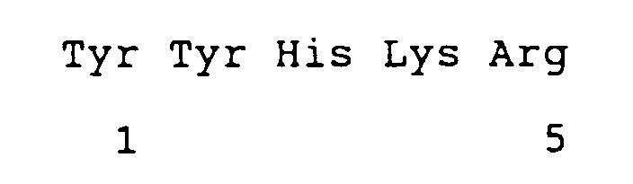
- (2) INFORMATION FOR SEQ ID NO:9:
- (i) SEQUENCING CHARACTERISTICS:
- (A)LENGTH: 4 amino acid
- (B)TYPE: amino acid
- (D)TOPOLOGY: linear
- (ii)MOLECULE TYPE:peptide
- (xi) SEQUENCE DESCRIPTION: SEQ ID NO:9:

- (2) INFORMATION FOR SEQ ID NO:10:
- (i) SEQUENCING CHARACTERISTICS:
- (A)LENGTH: 10 amino acid
- (B)TYPE: amino acid
- (D)TOPOLOGY: linear
- (ii)MOLECULE TYPE:peptide
- (xi) SEQUENCE DESCRIPTION: SEQ ID NO:10:
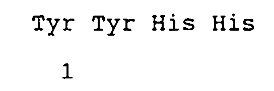
- (2) INFORMATION FOR SEQ ID NO:11:
- (i) SEQUENCING CHARACTERISTICS:
- (A)LENGTH: 6 amino acid
- (B)TYPE: amino acid
- (D)TOPOLOGY: linear
- (ii)MOLECULE TYPE:peptide
- (xi) SEQUENCE DESCRIPTION: SEQ ID NO:11:

- (2) INFORMATION FOR SEQ ID NO:12:
- (i) SEQUENCING CHARACTERISTICS:
- (A)LENGTH: 6 amino acid
- (B)TYPE: amino acid
- (D)TOPOLOGY: linear
- (ii)MOLECULE TYPE:peptide
- (xi) SEQUENCE DESCRIPTION: SEQ ID NO:12:

- (2) INFORMATION FOR SEQ ID NO:13:
- (i) SEQUENCING CHARACTERISTICS:
- (A)LENGTH: 4 amino acid
- (B)TYPE: amino acid
- (D)TOPOLOGY: linear
- (ii)MOLECULE TYPE:peptide
- (xi) SEQUENCE DESCRIPTION: SEQ ID NO:13:

- (2) INFORMATION FOR SEQ ID NO:14:
- (i) SEQUENCING CHARACTERISTICS:
- (A)LENGTH: 6 amino acid
- (B)TYPE: amino acid
- (D)TOPOLOGY: linear
- (ii)MOLECULE TYPE:peptide
- (xi) SEQUENCE DESCRIPTION: SEQ ID NO:14:

- (2) INFORMATION FOR SEQ ID NO:15:
- (i) SEQUENCING CHARACTERISTICS:
- (A)LENGTH: 6 amino acid
- (B)TYPE: amino acid
- (D)TOPOLOGY: linear
- (ii)MOLECULE TYPE:peptide
- (xi) SEQUENCE DESCRIPTION: SEQ ID NO:15:

- (2) INFORMATION FOR SEQ ID NO:16:
- (i) SEQUENCING CHARACTERISTICS:
- (A)LENGTH: 6 amino acid
- (B)TYPE: amino acid
- (D)TOPOLOGY: linear
- (ii)MOLECULE TYPE:peptide
- (xi) SEQUENCE DESCRIPTION: SEQ ID NO:16:

- (2) INFORMATION FOR SEQ ID NO:17:
- (i) SEQUENCING CHARACTERISTICS:
- (A)LENGTH: 4 amino acid
- (B)TYPE: amino acid
- (D)TOPOLOGY: linear
- (ii)MOLECULE TYPE:peptide
- (xi) SEQUENCE DESCRIPTION: SEQ ID NO:17:

- (2) INFORMATION FOR SEQ ID NO:18:
- (i) SEQUENCING CHARACTERISTICS:
- (A)LENGTH: 8 amino acid
- (B)TYPE: amino acid
- (D)TOPOLOGY: linear
- (ii)MOLECULE TYPE:peptide
- (xi) SEQUENCE DESCRIPTION: SEQ ID NO:18:

- (2) INFORMATION FOR SEQ ID NO:19:
- (i) SEQUENCING CHARACTERISTICS:
- (A)LENGTH: 12 amino acid
- (B)TYPE: amino acid
- (D)TOPOLOGY: linear
- (ii)MOLECULE TYPE:peptide
- (xi) SEQUENCE DESCRIPTION: SEQ ID NO:19:

- (2) INFORMATION FOR SEQ ID NO:20:
- (i) SEQUENCING CHARACTERISTICS:
- (A)LENGTH: 8 amino acid
- (B)TYPE: amino acid
- (D)TOPOLOGY: linear
- (ii)MOLECULE TYPE:peptide
- (xi) SEQUENCE DESCRIPTION: SEQ ID NO:20:

- (2) INFORMATION FOR SEQ ID NO:21:
- (i) SEQUENCING CHARACTERISTICS:
- (A)LENGTH: 10 amino acid
- (B)TYPE: amino acid
- (D)TOPOLOGY: linear
- (ii)MOLECULE TYPE:peptide
- (xi) SEQUENCE DESCRIPTION: SEQ ID NO:21:

-
Claims (8)
- Use of an oligopeptide comprising a fluorescence dye as an isoelectric point marker for isoelectric focusing with fluorescence detection.
- Use according to claim 1, wherein the oligopeptide is obtained by bonding said fluorescence dye through a binding group to an amino group of an N-terminal amino acid.
- Use according to claim 2, wherein said binding group is one selected from the group at least comprising amide, thioamide, sulfonamide, urea, thiourea, and urethane bonding groups.
- Use according to any of claims 1 to 3, wherein said oligopeptide comprises at least nj amino acids, each having a group which is to discharge a proton, thereby resulting in having a negative charge, wherein the group to release the proton thereby to have the negative charge has an acid dissociation constant of Kj, and/or ni amino acids, each having a group which is to receive a proton, thereby resulting in having a positive charge, wherein the group to receive the proton thereby to have the positive charge has an acid dissociation constant of Ki, and wherein, defining that
pI is -log([H+]) when |Z| < 0.01 is satisfied, 3 < pI < 11. - Use according to any of claims 1 to 4, wherein said oligopeptide is angiotensin or a related compound thereof.
- Use according to any of claims 1 to 5, wherein said fluorescence dye is one selected from the group comprising rhodamine, fluorescein, cyanine, indocyanine, indocarbocyanine, pyronine, lucifer yellow, quinacrine, squarillium, coumarin, fluoroanthranilmaleimide, and anthracene.
- Use according to any of claims 1 to 6, wherein said oligopeptide is at least one oligopeptide selected from the group at least comprising
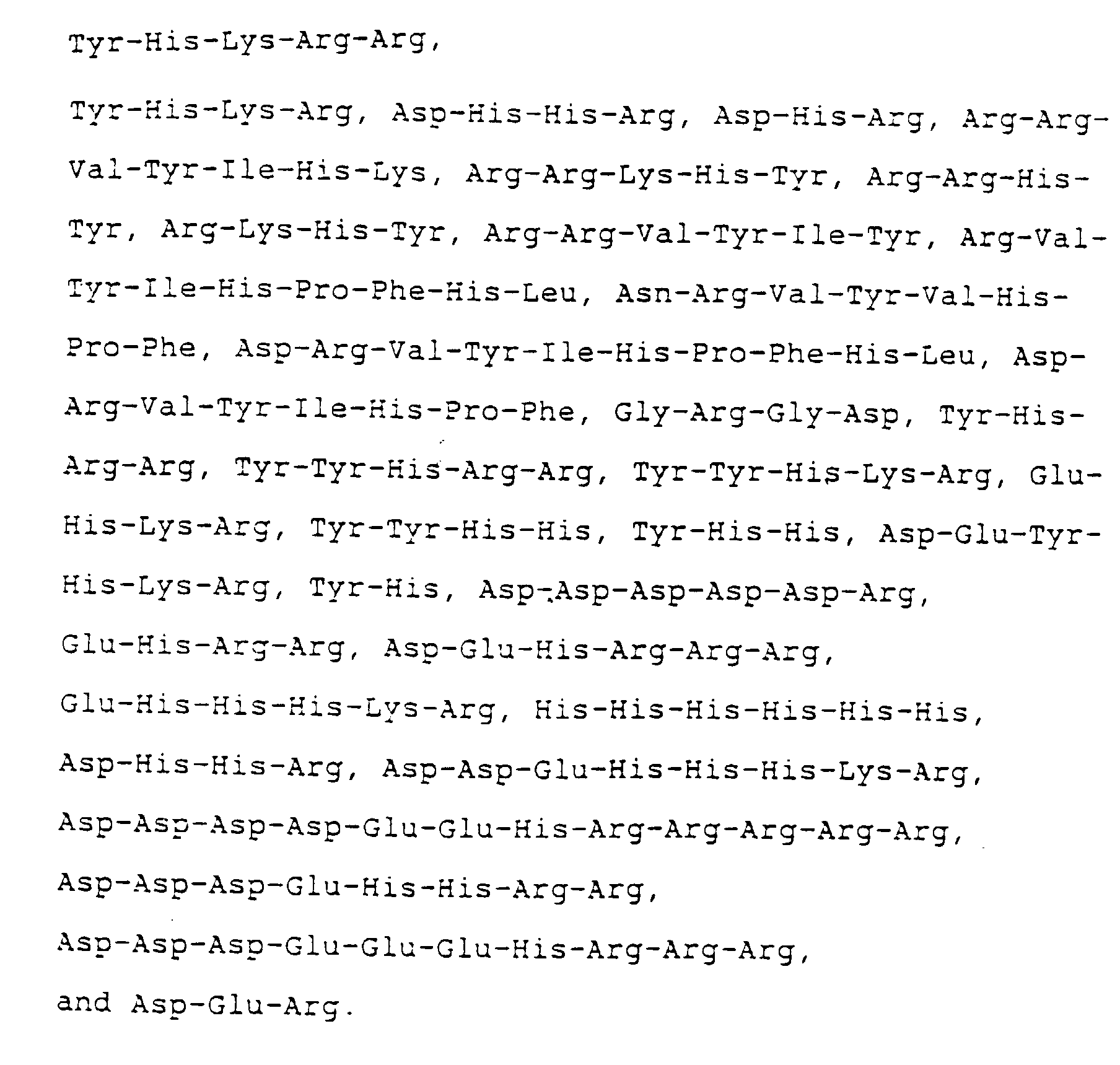
- Use according to any of claims 1 to 7, wherein said isoelectric focusing with fluorescence detection is a capillary isoelectric focusing with fluorescence detection.
Applications Claiming Priority (6)
| Application Number | Priority Date | Filing Date | Title |
|---|---|---|---|
| JP76873/95 | 1995-03-31 | ||
| JP7687395 | 1995-03-31 | ||
| JP7687395 | 1995-03-31 | ||
| JP27119695 | 1995-10-19 | ||
| JP271196/95 | 1995-10-19 | ||
| JP27119695 | 1995-10-19 |
Publications (3)
| Publication Number | Publication Date |
|---|---|
| EP0744614A2 EP0744614A2 (en) | 1996-11-27 |
| EP0744614A3 EP0744614A3 (en) | 1998-06-10 |
| EP0744614B1 true EP0744614B1 (en) | 1999-12-22 |
Family
ID=26417993
Family Applications (1)
| Application Number | Title | Priority Date | Filing Date |
|---|---|---|---|
| EP96105113A Expired - Lifetime EP0744614B1 (en) | 1995-03-31 | 1996-03-29 | Use of Isoelectric point markers for isoelectric focusing with fluorescence detection |
Country Status (3)
| Country | Link |
|---|---|
| US (1) | US5866683A (en) |
| EP (1) | EP0744614B1 (en) |
| DE (1) | DE69605712T2 (en) |
Families Citing this family (25)
| Publication number | Priority date | Publication date | Assignee | Title |
|---|---|---|---|---|
| EP2336313A1 (en) | 1997-01-08 | 2011-06-22 | Life Technologies Corporation | Methods for production of proteins |
| DE19954934A1 (en) * | 1999-11-16 | 2001-05-17 | Otto S Wolfbeis | Process for solubilizing optical markers |
| WO2001009165A2 (en) * | 1999-07-29 | 2001-02-08 | The Johns Hopkins University | ACTIVATION OF PEPTIDE PRODRUGS BY hK2 |
| AU1810101A (en) * | 1999-11-30 | 2001-06-12 | Electric Power Research Institute, Inc. | Capillary electrophoresis probes and method |
| US20040014082A1 (en) * | 2000-08-11 | 2004-01-22 | Invitrogen Corporation | Highly homogeneous molecular markers for electrophoresis |
| AU2001284853A1 (en) * | 2000-08-11 | 2002-02-25 | Invitrogen Corporation | Highly homogeneous molecular markers for electrophoresis |
| EP1485406A4 (en) * | 2002-02-20 | 2006-02-01 | Invitrogen Corp | Highly homogeneous molecular markers for electrophoresis |
| EP1668146B1 (en) * | 2003-09-25 | 2014-11-12 | Life Technologies Corporation | Homogeneous populations of molecules |
| US7407816B2 (en) * | 2004-05-07 | 2008-08-05 | Gentius, Inc | Isoelectric particles and uses thereof |
| JP5073491B2 (en) | 2004-07-19 | 2012-11-14 | プロテインシンプル | Method for the detection of an analyte |
| US20060292649A1 (en) * | 2004-07-19 | 2006-12-28 | Cell Biosciences Inc. | Methods and apparatus for reference lab diagnostics |
| US7846676B2 (en) * | 2004-07-19 | 2010-12-07 | Cell Biosciences, Inc. | Methods and devices for analyte detection |
| US20060292558A1 (en) * | 2004-07-19 | 2006-12-28 | Cell Biosciences Inc. | Methods and apparatus for protein assay diagnostics |
| US7935479B2 (en) * | 2004-07-19 | 2011-05-03 | Cell Biosciences, Inc. | Methods and devices for analyte detection |
| EP1872139A2 (en) * | 2005-04-09 | 2008-01-02 | Cell Biosciences, Inc. | Automated micro-volume assay system |
| US20060286378A1 (en) * | 2005-05-23 | 2006-12-21 | Shivkumar Chiruvolu | Nanostructured composite particles and corresponding processes |
| US8945361B2 (en) * | 2005-09-20 | 2015-02-03 | ProteinSimple | Electrophoresis standards, methods and kits |
| EP2069386A4 (en) * | 2006-07-21 | 2009-10-28 | Life Technologies Corp | Sharply resolving labeled protein molecular weight standards |
| GB0714202D0 (en) * | 2007-07-20 | 2007-08-29 | Ge Healthcare Bio Sciences Ab | Isoelectric point markers |
| US20090023156A1 (en) * | 2007-07-20 | 2009-01-22 | Voss Karl O | Methods and reagents for quantifying analytes |
| CN101556261B (en) * | 2009-05-25 | 2012-01-11 | 北京理工大学 | Method for detecting microorganism isoelectric point by adopting microtubule isoelectric focusing and cutting-blowout culture |
| EP2501714A4 (en) * | 2009-11-17 | 2015-10-07 | Univ Montreal | USEFUL HETEROPPTIDES TO REDUCE NON-SPECIFIC ADSORPTION |
| US11782023B2 (en) | 2018-12-19 | 2023-10-10 | Regeneron Pharmaceuticals, Inc. | Ce-western applications for antibody development |
| BR112021023206A2 (en) | 2019-05-21 | 2022-01-04 | Regeneron Pharma | Methods to identify and quantify host cell protein |
| CN115010789B (en) * | 2022-06-20 | 2023-10-20 | 湖州申科生物技术股份有限公司 | Fluorescent polypeptide for medium electrofocusing quality control in protein dielectrophoresis and preparation method thereof |
Family Cites Families (7)
| Publication number | Priority date | Publication date | Assignee | Title |
|---|---|---|---|---|
| US5439829A (en) * | 1991-01-30 | 1995-08-08 | Eli Lilly And Company | Immobilization of biologically active molecules by changing the Oxidation state of a chelated transition metal ion |
| US5245551A (en) * | 1991-04-10 | 1993-09-14 | University Of Utah Research Foundation | Method of determining extinction coefficient of fluorescent dye and protein concentration of dye-protein conjugate |
| FR2687680A1 (en) * | 1992-02-20 | 1993-08-27 | Centre Nat Rech Scient | Process for labelling proteins and peptides by acylation of their alpha amino functional group by a reagent containing an activated carboxylic functional group |
| CA2086453A1 (en) * | 1992-12-30 | 1994-07-01 | Mcgill University | Marker for neurotensin receptor |
| US5348633A (en) * | 1993-01-22 | 1994-09-20 | Northeastern University | Method for quantitating trace amounts of an analyte in a sample by affinity capillary electrophoresis |
| SE501192C2 (en) | 1993-04-29 | 1994-12-05 | Flygt Ab Itt | Method and Device for Controlling a Valve to Achieve Recirculation in Wastewater Pumping Stations |
| JPH07271196A (en) | 1994-03-28 | 1995-10-20 | Fuji Photo Film Co Ltd | Planographic plate making device |
-
1996
- 1996-03-29 US US08/623,833 patent/US5866683A/en not_active Expired - Fee Related
- 1996-03-29 DE DE69605712T patent/DE69605712T2/en not_active Expired - Fee Related
- 1996-03-29 EP EP96105113A patent/EP0744614B1/en not_active Expired - Lifetime
Also Published As
| Publication number | Publication date |
|---|---|
| DE69605712D1 (en) | 2000-01-27 |
| DE69605712T2 (en) | 2000-04-27 |
| US5866683A (en) | 1999-02-02 |
| EP0744614A2 (en) | 1996-11-27 |
| EP0744614A3 (en) | 1998-06-10 |
Similar Documents
| Publication | Publication Date | Title |
|---|---|---|
| US5866683A (en) | Isoelectric point markers for isoelectric focusing with fluorescence detection | |
| García‐Campaña et al. | LIF detection of peptides and proteins in CE | |
| Issaq et al. | Separation and detection of amino acids and their enantiomers by capillary electrophoresis: a review | |
| Kašička | Recent advances in capillary electrophoresis of peptides | |
| Ueda et al. | Chiral separation of naphthalene-2, 3-dicarboxaldehyde-labeled amino acid enantiomers by cyclodextrin-modified micellar electrokinetic chromatography with laser-induced fluorescence detection | |
| Lacroix et al. | Laser‐induced fluorescence detection schemes for the analysis of proteins and peptides using capillary electrophoresis | |
| Swaile et al. | Laser-based fluorimetric detection schemes for the analysis of proteins by capillary zone electrophoresis | |
| Smith | Developments in amino acid analysis using capillary electrophoresis | |
| US7371745B2 (en) | Bis-transition-metal-chelate probes | |
| US8945361B2 (en) | Electrophoresis standards, methods and kits | |
| Isailovic et al. | Isolation and characterization of R-phycoerythrin subunits and enzymatic digests | |
| Szulc et al. | Improved detection and derivatization in capillary electrophoresis | |
| Horká et al. | Capillary isoelectric focusing with UV-induced fluorescence detection | |
| Shimura et al. | Fluorescence‐labeled peptides as isoelectric point (pI) markers in capillary isoelectric focusing with fluorescence detection | |
| JP3246888B2 (en) | Isoelectric focusing marker for fluorescence detection | |
| JP2828426B2 (en) | Isoelectric focusing marker for fluorescence detection | |
| CA2315051C (en) | Capillary electrophoretic methods to detect new biologically active compounds in complex biological material | |
| De Antonis et al. | Analysis of derivatized peptides by capillary electrophoresis | |
| Vrábel et al. | Sensitive detection and separation of fluorescent derivatives using capillary electrophoresis with laser-induced fluorescence detection with 532 nm Nd: YAG laser | |
| Sze et al. | Laser‐induced fluorescence detection of non‐covalently labeled protein in capillary isoelectric focusing | |
| AU2003228519B2 (en) | Multiplexed capillary electrophoresis systems | |
| Guzman et al. | Capillary electrophoresis for the analytical separation and semi-preparative collection of monoclonal antibodies | |
| Liu et al. | Nanoliter volume kinetic assays | |
| Lee et al. | [19] Capillary electrophoresis detectors: Lasers | |
| Kaneta et al. | Separation and detection of cyanine-labeled amino acids by micellar electrokinetic chromatography combined with fluorescence detection using diode-based solid-state lasers |
Legal Events
| Date | Code | Title | Description |
|---|---|---|---|
| PUAI | Public reference made under article 153(3) epc to a published international application that has entered the european phase |
Free format text: ORIGINAL CODE: 0009012 |
|
| 17P | Request for examination filed |
Effective date: 19960528 |
|
| AK | Designated contracting states |
Kind code of ref document: A2 Designated state(s): CH DE LI SE |
|
| PUAL | Search report despatched |
Free format text: ORIGINAL CODE: 0009013 |
|
| RHK1 | Main classification (correction) |
Ipc: G01N 27/447 |
|
| AK | Designated contracting states |
Kind code of ref document: A3 Designated state(s): CH DE LI SE |
|
| 17Q | First examination report despatched |
Effective date: 19980821 |
|
| GRAG | Despatch of communication of intention to grant |
Free format text: ORIGINAL CODE: EPIDOS AGRA |
|
| GRAG | Despatch of communication of intention to grant |
Free format text: ORIGINAL CODE: EPIDOS AGRA |
|
| GRAH | Despatch of communication of intention to grant a patent |
Free format text: ORIGINAL CODE: EPIDOS IGRA |
|
| GRAH | Despatch of communication of intention to grant a patent |
Free format text: ORIGINAL CODE: EPIDOS IGRA |
|
| GRAA | (expected) grant |
Free format text: ORIGINAL CODE: 0009210 |
|
| AK | Designated contracting states |
Kind code of ref document: B1 Designated state(s): CH DE LI SE |
|
| REG | Reference to a national code |
Ref country code: CH Ref legal event code: NV Representative=s name: ISLER & PEDRAZZINI AG Ref country code: CH Ref legal event code: EP |
|
| REF | Corresponds to: |
Ref document number: 69605712 Country of ref document: DE Date of ref document: 20000127 |
|
| PLBE | No opposition filed within time limit |
Free format text: ORIGINAL CODE: 0009261 |
|
| STAA | Information on the status of an ep patent application or granted ep patent |
Free format text: STATUS: NO OPPOSITION FILED WITHIN TIME LIMIT |
|
| 26N | No opposition filed | ||
| PGFP | Annual fee paid to national office [announced via postgrant information from national office to epo] |
Ref country code: SE Payment date: 20010326 Year of fee payment: 6 |
|
| PGFP | Annual fee paid to national office [announced via postgrant information from national office to epo] |
Ref country code: CH Payment date: 20010403 Year of fee payment: 6 |
|
| PGFP | Annual fee paid to national office [announced via postgrant information from national office to epo] |
Ref country code: DE Payment date: 20010426 Year of fee payment: 6 |
|
| PG25 | Lapsed in a contracting state [announced via postgrant information from national office to epo] |
Ref country code: SE Free format text: LAPSE BECAUSE OF NON-PAYMENT OF DUE FEES Effective date: 20020330 |
|
| PG25 | Lapsed in a contracting state [announced via postgrant information from national office to epo] |
Ref country code: LI Free format text: LAPSE BECAUSE OF NON-PAYMENT OF DUE FEES Effective date: 20020331 Ref country code: CH Free format text: LAPSE BECAUSE OF NON-PAYMENT OF DUE FEES Effective date: 20020331 |
|
| PG25 | Lapsed in a contracting state [announced via postgrant information from national office to epo] |
Ref country code: DE Free format text: LAPSE BECAUSE OF NON-PAYMENT OF DUE FEES Effective date: 20021001 |
|
| EUG | Se: european patent has lapsed |
Ref document number: 96105113.3 |
|
| REG | Reference to a national code |
Ref country code: CH Ref legal event code: PL |