EP0743856B1 - Antigen-based heteropolymers for treating autoimmune diseases - Google Patents
Antigen-based heteropolymers for treating autoimmune diseases Download PDFInfo
- Publication number
- EP0743856B1 EP0743856B1 EP95912577A EP95912577A EP0743856B1 EP 0743856 B1 EP0743856 B1 EP 0743856B1 EP 95912577 A EP95912577 A EP 95912577A EP 95912577 A EP95912577 A EP 95912577A EP 0743856 B1 EP0743856 B1 EP 0743856B1
- Authority
- EP
- European Patent Office
- Prior art keywords
- ahp
- antigen
- red blood
- dsdna
- binding
- Prior art date
- Legal status (The legal status is an assumption and is not a legal conclusion. Google has not performed a legal analysis and makes no representation as to the accuracy of the status listed.)
- Expired - Lifetime
Links
Images




Classifications
-
- C—CHEMISTRY; METALLURGY
- C07—ORGANIC CHEMISTRY
- C07K—PEPTIDES
- C07K16/00—Immunoglobulins [IG], e.g. monoclonal or polyclonal antibodies
- C07K16/18—Immunoglobulins [IG], e.g. monoclonal or polyclonal antibodies against material from animals or humans
- C07K16/28—Immunoglobulins [IG], e.g. monoclonal or polyclonal antibodies against material from animals or humans against receptors, cell surface antigens or cell surface determinants
- C07K16/2896—Immunoglobulins [IG], e.g. monoclonal or polyclonal antibodies against material from animals or humans against receptors, cell surface antigens or cell surface determinants against molecules with a "CD"-designation, not provided for elsewhere
-
- A—HUMAN NECESSITIES
- A61—MEDICAL OR VETERINARY SCIENCE; HYGIENE
- A61K—PREPARATIONS FOR MEDICAL, DENTAL OR TOILETRY PURPOSES
- A61K39/00—Medicinal preparations containing antigens or antibodies
- A61K39/0005—Vertebrate antigens
- A61K39/0008—Antigens related to auto-immune diseases; Preparations to induce self-tolerance
-
- A—HUMAN NECESSITIES
- A61—MEDICAL OR VETERINARY SCIENCE; HYGIENE
- A61K—PREPARATIONS FOR MEDICAL, DENTAL OR TOILETRY PURPOSES
- A61K47/00—Medicinal preparations characterised by the non-active ingredients used, e.g. carriers or inert additives; Targeting or modifying agents chemically bound to the active ingredient
- A61K47/50—Medicinal preparations characterised by the non-active ingredients used, e.g. carriers or inert additives; Targeting or modifying agents chemically bound to the active ingredient the non-active ingredient being chemically bound to the active ingredient, e.g. polymer-drug conjugates
- A61K47/51—Medicinal preparations characterised by the non-active ingredients used, e.g. carriers or inert additives; Targeting or modifying agents chemically bound to the active ingredient the non-active ingredient being chemically bound to the active ingredient, e.g. polymer-drug conjugates the non-active ingredient being a modifying agent
- A61K47/68—Medicinal preparations characterised by the non-active ingredients used, e.g. carriers or inert additives; Targeting or modifying agents chemically bound to the active ingredient the non-active ingredient being chemically bound to the active ingredient, e.g. polymer-drug conjugates the non-active ingredient being a modifying agent the modifying agent being an antibody, an immunoglobulin or a fragment thereof, e.g. an Fc-fragment
- A61K47/6835—Medicinal preparations characterised by the non-active ingredients used, e.g. carriers or inert additives; Targeting or modifying agents chemically bound to the active ingredient the non-active ingredient being chemically bound to the active ingredient, e.g. polymer-drug conjugates the non-active ingredient being a modifying agent the modifying agent being an antibody, an immunoglobulin or a fragment thereof, e.g. an Fc-fragment the modifying agent being an antibody or an immunoglobulin bearing at least one antigen-binding site
- A61K47/6849—Medicinal preparations characterised by the non-active ingredients used, e.g. carriers or inert additives; Targeting or modifying agents chemically bound to the active ingredient the non-active ingredient being chemically bound to the active ingredient, e.g. polymer-drug conjugates the non-active ingredient being a modifying agent the modifying agent being an antibody, an immunoglobulin or a fragment thereof, e.g. an Fc-fragment the modifying agent being an antibody or an immunoglobulin bearing at least one antigen-binding site the antibody targeting a receptor, a cell surface antigen or a cell surface determinant
-
- A—HUMAN NECESSITIES
- A61—MEDICAL OR VETERINARY SCIENCE; HYGIENE
- A61P—SPECIFIC THERAPEUTIC ACTIVITY OF CHEMICAL COMPOUNDS OR MEDICINAL PREPARATIONS
- A61P37/00—Drugs for immunological or allergic disorders
Definitions
- the present invention relates to antigen-based heteropolymers specific for both a specific receptor site on a primate erythrocyte and a target pathogenic autoantibody.
- the present invention further relates to the use of a composition comprising these antigen-based heteropolymers for the preparation of a medicament for treating autoimmune diseases.
- Circulating autoantibodies are responsible for much of the pathogenesis associated with a number of autoimmune diseases including, but not limited to, systemic lupus erythematosus (SLE), autoimmune myocarditis, immune complex mediated kidney disease, rheumatoid arthritis, myasthenia gravis, autoimmune anemias, Sjogren's Syndrome, idiopathic thrombocytopenic purpura, various forms of vasculitis, and at least some of the cellular cytotoxicity accompanying Acquired Immune Deficiency Syndrome (AIDS).
- SLE systemic lupus erythematosus
- autoimmune myocarditis immune complex mediated kidney disease
- rheumatoid arthritis myasthenia gravis
- autoimmune anemias Sjogren's Syndrome
- idiopathic thrombocytopenic purpura various forms of vasculitis
- at least some of the cellular cytotoxicity accompanying Acquired Immune Deficiency Syndrome
- Plasmapheresis which is designed to remove all the circulating antibodies, has been attempted, but only with limited success.
- extracorporeal immunoabsorption procedures have been attempted. These procedures are more specific, and attempt to remove only the pathogenic autoantibodies. This involves allowing the blood or plasma to flow over matrices outside the body which contain the autoantigen which is the natural target of the autoantibodies.
- These methods are slow, invasive, and expensive, and are associated with several technical problems including the need to perform the procedures repeatedly because of their quantitative inefficiency. The complication of complement activation on the matrices exists. Overall, therapeutic successes have been modest, at test.
- Another general, non-specific approach involves aggressive immunosuppressive therapy with corticosteroids, and cytotoxic and nonsteroidal antiinflammatory drugs. Although in many instances clinical improvements have been obtained, there continues to be significant morbidity and mortality in autoimmune diseases despite these medications.
- an objective of the present invention to provide an autoimmune therapy which is highly specific for a target pathogenic autoantibody and which produces fast and quantitative results.
- antigen-based heteropolymers which are specific for both complement receptor site (CR1) on a primate erythrocyte and a target pathogenic autoantibody. More specifically, the above objectives have been met by the present invention which provides a complex which comprises at least one antigen-based heteropolymer comprising a monoclonal antibody specific for binding to a CR1 site on a primate erythrocyte, wherein the monoclonal antibody is crosslinked to an antigen specific for a target pathogenic autoantibody.
- the objectives have been met by the present invention which further provides the use of a composition comprising an effective amount of an antigen-based heteropolymer comprised of a monoclonal antibody specific for the CR1 site on a primate erythrocyte, wherein the monoclonal antibody is crosslinked to an antigen which is specific for a target pathogenic autoantibody for the preparation of a medicament for the treatment of an autoimmune disease in a human or non-human primate.
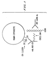
- Figure 1 is a schematic illustration of the antigen-based heteropolymer complex consisting of a monoclonal antibody bound to the CR1 receptor site on a primate erythrocyte.
- the monoclonal antibody is also crosslinked to an autoantigen (dsDNA) which in turn, is bound to the target pathogenic autoantibody (anti-dsDNA antibody).
- dsDNA autoantigen
- anti-dsDNA antibody target pathogenic autoantibody
- Figure 2 is a graph showing the kinetics of the antigen-based heteropolymer-mediated binding of IgG anti-dsDNA antibodies to human red blood cells.
- the counts bound are proportional to the amount of human IgG bound (see below).
- Ma, Mo, and Ha are the last name initials of SLE patients from whom the plasmas or IgG fractions were taken.
- the solid symbols indicate incubation with red blood cells and antigen-based heteropolymer, and the above respective antibodies.
- the open symbols represent incubation with red blood cells and antibodies, without antigen-based heteropolymer.
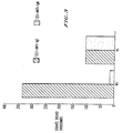
- Figure 3 is a graph showing examination of red blood cells prepared and incubated as described in Figure 2, and probed with 125 I-labeled monoclonal antibodies to both human IgG and human IgM.
- Plasma Ma is representative of samples from patients with severe nephritis and contains almost exclusively IgG anti-dsDNA antibodies at a very high titer.
- Plasma Va contains both IgG and IgM anti-dsDNA antibodies.
- Figure 4 is a graph showing results of control experiments to demonstrate the specificity of binding.
- Human red blood cells were examined with a four-fold dilution of plasma Ma, or an IgG fraction from plasma Ma was examined.
- AHP antigen-based heteropolymer;
- Ma patient with severe nephritis.
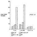
- Figure 5 is a graph showing the results of control experiments which indicate that binding is permissive in normal human serum and verify that the red blood cells are not lysed.
- a two-fold dilution of plasma Ma (diluted in bovine serum albumin) was compared to a similar dilution of plasma Ma in fresh, normal human serum.
- PL plasma;
- NHSC normal human serum complement;
- AHP antigen-based heteropolymer; IRR irrelevant monoclonal antibody.
- Figure 6 is a graph showing the results of dose response experiments illustrating the optimum input of antigen-based heteropolymer to maximize binding of human IgG anti-dsDNA antibodies to 150 ⁇ l of red blood cells (50% hematocrit) in an SLE IgG sample.
- AHP3 was prepared as in Example 1 (see below).
- AHP2 contains 1/2 as much dsDNA as AHP3;
- AHP1 contains 1/4 as much dsDNA as AHP3.
- Figure 7 is a graph showing binding as a function of the concentration of IgG anti-dsDNA antibodies. A 50% dispersion of human red blood cells was examined in a mixture of bovine serum albumin and IgG Mo.
- Figure 8 is a graph showing binding as a function of the concentration of IgG anti-dsDNA antibodies using a very high titer anti-dsDNA plasma (Ma).
- Figure 9 is a graph showing binding as a function of the concentration of IgG anti-dsDNA antibodies using plasma Ma. (7G9 AHP and 1B4 kHP alone as compared to cocktail containing 7G9 and 1B4 together.)
- the present invention is based on the unique structural and biophysical properties of the primate red blood cell complement receptor (CR1).
- the antigen-based heteropolymer complexes of the present invention are specific for both the CR1 site on a primate red blood cell and for a target pathogenic autoantibody.
- the antigen-based heteropolymers of the present invention are prepared from monoclonal antibodies which are specific for the CR1 receptor site on a primate erythrocyte.
- the monoclonal antibodies must also be capable of being crosslinked to an autoantigen which is specific for the target pathogenic autoantibody.
- Examples of such monoclonal antibodies used in the present invention include 1B4, HB8592, and 7G9.
- HB8592 and 1B4 are disclosed in Taylor et al., "Use of heteropolymeric monoclonal antibodies to attach antigens to the C3b receptor of human erythrocytes: A potential therapeutic treatment," Proc. Nat'l. Acad. Sci. , 88:3305-3309 (April 1991); Reist et al., "Antigens pre-bound to the primate erythrocyte complement receptor via crosslinked bispecific monoclonal antibody heteropolymers are rapidly cleared from the circulation, " Eur. J. Immunol. 23:3021-3027 (1993).
- 7G9 is a mAb recently developed in the inventors' laboratory and can be prepared by known techniques.
- Other mAbs to CR1 available and useful include 3D9 and E-11 [(previously used by the present inventors in Proc. Nat'l. Acad. Sci. , Vol. 91 (July 1992)], and 57F and YZ1 (prepared and reported by Nussenzweig and Fearon, respectively). It is likely that any mAb for CR1 will work successfully in the antigen-based heteropolymer system of the present invention.
- the monoclonal antibody is crosslinked to an antigen specific for a target pathogenic autoantibody.
- the crosslinking can be performed by any efficacious crosslinking method.
- purified monoclonal antibodies may first be biotinylated. Typically, each monoclonal antibody may contain 5 biotins.
- the biotinylated purified monoclonal antibody is then crosslinked to a previously biotinylated antigen or autoantigen by the use of streptavidin.
- Other known methods such as use of N-succinimidyl-3-(2-pyridyldithio)propionate (SPDP) can be used for crosslinking the monoclonal antibody to the antigen if the antigen has free amino groups.
- SPDP N-succinimidyl-3-(2-pyridyldithio)propionate
- an antigen-based heteropolymer comprised of factor VIII crosslinked to a mAb to CR1 specifically binds circulating anti-factor VIII autoantibodies to erythrocyte CR1 and facilitates clearance of said autoantibodies.
- Factor VIII is allowed to circulate and facilitate blood clotting.
- autoantibodies which can be cleared by the antigen-based heteropolymer complex of the present invention include, but are not limited to, autoantibodies to the following antigens: the muscle acetylcholine receptor (the antibodies are associated with the disease myasthenia gravis); cardiolipin (associated with the disease lupus); platelet associated proteins (associated with the disease idiopathic thrombocytopenic purpura); the multiple antigens associated with Sjogren's Syndrome; the antigens implicated in the case of tissue transplantation autoimmune reactions; the antigens found on heart muscle (associated with the disease autoimmune myocarditis); the antigens associated with immune complex mediated kidney disease; the dsDNA and ssDNA antigens (associated with lupus nephritis); desmogleins and desmoplakins (associated with pemphigus and pemphigoid); or any other antigen which is well-characterized, can be crosslinked to an anti-CR1 mAb, and is associated with
- the antigen-based heteropolymer complexes When the above antigen-based heteropolymer complexes are injected into the circulation of a human or non-human primate, the antigen-based heteropolymer will readily bind to the red blood cells via the monoclonal antibody at a high percentage and in agreement with the number of CR1 sites on the red blood cells. At approximately the same rate, the antigen-based heteropolymer will then bind to the autoantibody via the antigen, which is crosslinked to the monoclonal antibody.
- the red blood cells which contain the bound antigen-based heteropolymer autoantibody complex can then act therapeutically by facilitating the neutralization and clearance from the circulation of the bound pathogenic autoantibody.
- the antigen-based heteropolymers facilitate binding to the red blood cells and subsequently clear the autoantibody from the circulation of human and non-human primates, without also clearing the red blood cells themselves.
- the present inventors have found for the first time, that by crosslinking anti-CR1 monoclonal antibodies with an autoantigen, thus generating the antigen-based heteropolymers of the present invention, it is possible to bind specifically to red blood cells the vast majority (ca. 80%-95%, Table 1 and Figure 9) of the autoantibodies (specific for an antigen) found in plasmas from patients with a specific autoimmune disease.
- the examples demonstrate that it is possible to bind specifically to red blood cells the vast majority of the autoantibodies to the dsDNA antigen found in plasmas from patients with systemic lupus erythematosus (SLE).
- the present invention utilizes the unique properties of the primate red cell CR1 which enable it to bind and clear complement-opsonized immune complexes from the circulation.
- the immune complexes cleared via this system are taken up by the liver and spleen (Cornacoff, J. Clin. Invest. (1983)).
- the input range of the antigen-based heteropolymer is determined based on the red blood cell concentration and the number of CR1 epitope sites recognized by the anti-CR1 monoclonal antibodies per red blood cell. If the antigen-based heteropolymer complex is added in excess, a fraction of the antigen-based heteropolymer will not bind to red blood cells, but will instead inhibit the uptake of the autoantibodies by the red blood cell. The reason is that when the free antigen-based heteropolymer is in solution, it will simply compete for available autoantibody with antigen-based heteropolymer bound to red blood cells. Thus, the antigen-based heteropolymer-mediated binding of the autoantibodies to human red blood cells follows a bell-shaped curve when binding is examined as a function of the input antigen-based heteropolymer concentration ( Figure 6).
- the saturation problem can be solved by using combinations of antigen-based heteropolymer which contain monoclonal antibodies that bind to different sites on CR1.
- the monoclonal antibodies 7G9 and 1B4 bind to separate and noncompeting sites on red blood cell CR1. Therefore, a "cocktail" containing a mixture of two antigen-based heteropolymers made with respective monoclonal antibodies gives rise to greater binding of autoantibodies to red blood cells.
- Plasma Ma is one of the highest titer SLE IgG anti-dsDNA plasma studied.
- the single antigen-based heteropolymer prepared with either anti-CR1 monoclonal antibodies 7G9 or 1B4 alone cannot absorb all anti-dsDNA antibodies to red blood cells in the undiluted plasma.
- the present invention further encompasses the use of a composition comprising an effective amount of an antigen-based heteropolymer complex of the present invention for the preparation of a medicament for treating an autoimmune disease in a primate.
- the route of administration will likely be by intravenous injection into the blood of a human or non-human primate.
- An effective amount of the antigen-based heteropolymer complex of the present invention is from 1-10 mg, preferably 5 mg administered once. This dosage should clear up to 2 ⁇ g/ml of the autoantibody from the circulation of a primate. In a therapeutic environment, the administration should be repeated until complete clearance of the pathogenic autoantibody.
- the antigen-based heteropolymers of the present invention can be used in combination with certain fluids used for intravenous infusions.
- Red blood cells removed and isolated may also be used as therapeutic agents. Once franked with the antigen-based heteropolymer, these red blood cells can be reintroduced into the patient, where, in the bloodstream free autoantibodies will bind and be immobilized on the red blood cell, and subsequently cleared in accordance with the body's red blood cell clearing mechanism.
- red blood cells are franked with a "cocktail" of at least two antigen-based heteropolymers, which, in addition to binding to the target autoantibody, also bind to several distinct and non-overlapping sites on CR1 of the primate red blood cell.
- a "cocktail" of at least two antigen-based heteropolymers which, in addition to binding to the target autoantibody, also bind to several distinct and non-overlapping sites on CR1 of the primate red blood cell.
- the AHP of the present invention can further be used in the case of clearance of exogenous administered antibodies which become pathogenic.
- Monoclonal antibodies and crosslinked AHP anti-CR1/dSDNA complexes Monoclonal antibodies and crosslinked AHP anti-CR1/dSDNA complexes.
- monoclonal antibodies specific for primate CR1 i.e., 1B4, HB8592, and 7G9, were purified by known methods as set forth in Reist et al., supra .
- the purified monoclonal antibodies were then biotinylated by known methods. Typically, each monoclonal antibody would contain 5 biotins.
- An amount of 24 ⁇ g of biotinylated 7G9 was incubated for 30 minutes at room temperature with 30 ⁇ g of streptavidin (SA) in a volume of 48 ⁇ l borate saline (BS buffer).
- SA streptavidin
- biotinylated dsDNA which was purchased from Dr. W. Emlen of the University of Colorado Medical Center (1 biotin per 30 base pairs in 1 ml of BS buffer) in order to crosslink 7G9 to the biotinylated dsDNA.
- the process was repeated using 1B4 or HB8592.
- the anti-CR1/dsDNA complexes formed were used without any other purification.
- the AHP was added in an "equivalence" of ca. 0.6-1.2 ⁇ g of anti-CR1 mAb per ml blood based on 500-1000 CR1 receptors per red blood cell. After brief incubation, the sample was centrifuged and the supernatant isolated and saved for an independent assay (Farr assay) of the anti-dsDNA antibodies. The red cell pellets were washed 3 times and then an 125 I-labeled monoclonal antibody to human IgG (HB43) was added to the red blood cells to measure uptake of human IgG. (Taylor et al., 88:3305-3309, Proc. Natl. Acad. Sci.
- Figure 2 demonstrates the results of the kinetics of AHP-mediated binding of IgG anti-dsDNA antibodies to human red blood cells. These results show that the complexes of the present invention bind to red blood cells. The results further show that by virtue of the incorporated dsDNA, the AHP complexes facilitate the specific and rapid binding (ca. 80% complete in ca. 5 minutes at 37°C as shown in Figure 2) to red blood cells of high avidity IgG and/or IgM/anti-dsDNA antibodies found in SLE plasmas. The specificity of the binding for anti-dsDNA antibodies and the anti-CR1/dsDNA AHP complex is clear due to the fact that all the binding is abrogated when the AHP is omitted (open symbols in Figure 2).
- Plasma Ma as shown in Figure 3, is representative of samples from patients with severe nephritis, and as previously reported, has almost exclusively IgG anti-dsDNA antibodies at very high titer (Taylor et al., Arthritis and Rheumatism , (1987)].
- the plasma Va was shown in Taylor et al. to have both IgG and IgM anti-dsDNA antibodies.
- the red blood cells were probed with 125 I-labeled monoclonal antibodies to both human IgG (HB43) and human IgM (HB57).
- Plasma Ma or an IgG fraction from plasma Ma, was prepared as set forth above.
- the plasma Ma and the IgG fraction from plasma Ma were diluted four-fold.
- Human red blood cells were used as above in this experiment.
- Five different samples were prepared as follows: a) AHP omitted; b) sheep red blood cells (SRBCS) lacking the CR1 site were substituted for human red blood cells; c) the biotinylated dsDNA was omitted in preparing the AHP; and d) normal plasma or normal IgG was substituted for SLE plasma ( Figure 4).
- Table I shows the results of Farr assays.
- the Farr assay was performed as in Taylor et al., Arthritis and Rheumatism , supra. These assays are specific for anti-dsDNA antibodies.
- the supernatant samples contain the plasma proteins and the vast majority of IgG, not specific for dsDNA, which did not bind to red blood cells.
- These assays indicate that the anti-dsDNA binding activity in the supernatants is reduced considerably for most SLE plasma and IgG fractions (more than 90% in Table 1).
- the IgG anti-dsDNA antibodies are indeed specifically absorbed to the red blood cells in the presence of the AHP complex.
- AHP3 was prepared in accordance with the procedures set forth in Example 1 using 10 ⁇ l of AHP3 per 150 ⁇ l of 50% red blood cells.
- AHP1 and AHP2 were prepared accordingly, except that AHP2 contains 1/2 as much dsDNA as AHP3, and AHP1 contains 1/4 as much dsDNA as AHP3.
- Dose response experiments were performed to find the optimum input of AHP to maximize binding of human IgG anti-dsDNA antibodies to 150 ⁇ l of human red blood cells (50% hematocrit) in an SLE IgG sample.
- AHP3 was chosen as the exemplified AHP because it facilitates the highest level of binding IgG anti-dsDNA to red blood cells.
- Combinations of AHP were prepared which contain monoclonal antibodies that bind to different sites on CR1.
- the monoclonal antibodies 7G9 and 1B4 bind to separate and non-competing sites on the CR1 site of the red blood cell.
- These cocktails produced contain a mixture of the two AHP's made with these respective monoclonal antibodies.
- the results as shown in Figure 9 show that use of a "cocktail" gives rise to even greater binding of IgG anti-dsDNA antibodies to red blood cells.
- plasma Ma is one of the highest titer SLE IgG anti-dsDNA plasma which has been studied.
- the single AHP prepared with either anti-CR1 monoclonal antibody 7G9 or 1B4 alone cannot absorb all the anti-dsDNA antibodies to red blood cells in the undiluted plasma.
- the "cocktail" when used, the continued linearity in the binding (even in the undiluted plasma) suggests that it is possible to absorb to red blood cells, more than 90% of the IgG anti-dsDNA in plasma Ma ( Figure 9). This was confirmed in the Farr assays (see Table 1) of the plasma supernatants which indicated that greater than 90% of the anti-dsDNA antibody binding activity in plasma ma is removed when it is treated with the AHP "cocktail" and human red blood cells.
- the AHP prepared with anti-CR1 monoclonal antibody 1B4 showed slightly better binding than the AHP prepared with monoclonal antibody 7G9. However, saturation at the highest input of Ma was seen in both cases. When the cocktail containing both AHP's was used, on the other hand, binding was enhanced considerably which suggests that all the IgG anti-dsDNA can be bound to red blood cells (Figure 9).
Landscapes
- Health & Medical Sciences (AREA)
- Life Sciences & Earth Sciences (AREA)
- Chemical & Material Sciences (AREA)
- Immunology (AREA)
- General Health & Medical Sciences (AREA)
- Medicinal Chemistry (AREA)
- Organic Chemistry (AREA)
- Veterinary Medicine (AREA)
- Public Health (AREA)
- Animal Behavior & Ethology (AREA)
- Pharmacology & Pharmacy (AREA)
- Epidemiology (AREA)
- Engineering & Computer Science (AREA)
- Bioinformatics & Cheminformatics (AREA)
- Microbiology (AREA)
- Mycology (AREA)
- Cell Biology (AREA)
- Proteomics, Peptides & Aminoacids (AREA)
- Molecular Biology (AREA)
- Genetics & Genomics (AREA)
- Biophysics (AREA)
- Rheumatology (AREA)
- Biochemistry (AREA)
- General Chemical & Material Sciences (AREA)
- Chemical Kinetics & Catalysis (AREA)
- Nuclear Medicine, Radiotherapy & Molecular Imaging (AREA)
- Peptides Or Proteins (AREA)
- Medicines Containing Antibodies Or Antigens For Use As Internal Diagnostic Agents (AREA)
- Preparation Of Compounds By Using Micro-Organisms (AREA)
- Medicines That Contain Protein Lipid Enzymes And Other Medicines (AREA)
- Medicines Containing Material From Animals Or Micro-Organisms (AREA)
- Pharmaceuticals Containing Other Organic And Inorganic Compounds (AREA)
- Nitrogen And Oxygen Or Sulfur-Condensed Heterocyclic Ring Systems (AREA)
Abstract
Description
| Efficiency of Removal of Anti-dsDNA Antibody Binding Activity from SLE Plasmas by adsorption to Red cells in the Presence of a Specific AHP. Analysis of binding of 3H-dsDNA in the Farr Assay. | ||||
| 3H-dsDNA (DPM) in the SN (Unbound Counts) | ||||
| SLE Plasma | Dilution tested in Farr assay | Adsorbed with red cells alone | Adsorbed with red cells +AHP | % Adsorbed |
| Ma/2 | 5 | 58 | 710 | 95 |
| 25 | 270 | 950 | ||
| 100 | NL IgG: (1000) | 970 | ||
| Va/1 | 10 | 680 | 1400 | 90 |
| 25 | 1040 | |||
| 100 | 1420 | |||
| NL IgG: (1800) | ||||
| Co/1 | 4 | 730 | 1250 | 80 |
| 20 | 1280 | 1830 | ||
| 40 | 1550 | 2300 | ||
| NL IgG: (2600) | ||||
| Ha/2 | 5 | 30 | 120 | 85, 95 |
| 20 | 90 | 360 | ||
| 80 | 270 | 630 | ||
| 320 | 390 | 680 | ||
| NL IgG: (1200) | ||||
| Ma/1 | 4 | 370 | 1760 | 90 |
| 8 | 470 | |||
| 16 | 970 | |||
| 32 | 1700 | |||
| NL IgG: (3000) |
Claims (8)
- An antigen-based heteropolymer (AHP) complex comprising a monoclonal antibody specific for binding to complement receptor (CR1) site on a primate erythrocyte, wherein said monoclonal antibody is crosslinked to an antigen specific for a target pathogenic antibody or autoantibody.
- The AHP of claim 1, wherein the monoclonal antibody is produced by the hybridoma deposited with the ATCC and assigned accession number HB8592.
- The AHP of claim 1, wherein the target antibody or autoantibody is selected from the group consisting of antibodies or autoantibodies to the following antigens : factor VIII, muscle acetylcholine receptor, cardiolipin, platelet associated proteins, antigens associated with Sjogren's Syndrome, double stranded deoxyribonucleic acid (dsDNA), and single stranded DNA (ssDNA).
- The AHP of claim 1, wherein said antigen is selected from the group consisting of factor VIII, muscle acetylcholine receptor, cardiolipin, platelet associated proteins, antigens associated with Sjogren's Syndrome, double stranded deoxyribonucleic acid (dsDNA), and single stranded DNA (ssDNA).
- The AHP of any one of claims 1-4 wherein the AHP is franked with a human or non-human primate erythrocyte.
- An AHP cocktail, comprising at least two AHPs according to anyone of claims 1 to 5, wherein each AHP comprises a monoclonal antibody specific for binding to at least two distinct and non-overlapping sites on complement receptor (CR1) on a primate erythrocyte, wherein said monoclonal antibody is crosslinked to an antigen specific for a target pathogenic antibody or autoantibody.
- Pharmaceutical composition characterized in that it comprises an AHP or an AHP cocktail according to any one of claims I to 6.
- Use of a composition comprising the AHP or the AHP cocktail according to any of claims 1 to 6 for the manufacture of a medicament for the treatment of an autoimmune disease.
Applications Claiming Priority (3)
| Application Number | Priority Date | Filing Date | Title |
|---|---|---|---|
| US20257294A | 1994-02-28 | 1994-02-28 | |
| US202572 | 1994-02-28 | ||
| PCT/US1995/002165 WO1995022977A1 (en) | 1994-02-28 | 1995-02-28 | Antigen-based heteropolymers and method for treating autoimmune diseases using the same |
Publications (3)
| Publication Number | Publication Date |
|---|---|
| EP0743856A1 EP0743856A1 (en) | 1996-11-27 |
| EP0743856A4 EP0743856A4 (en) | 2000-04-12 |
| EP0743856B1 true EP0743856B1 (en) | 2003-07-16 |
Family
ID=22750436
Family Applications (1)
| Application Number | Title | Priority Date | Filing Date |
|---|---|---|---|
| EP95912577A Expired - Lifetime EP0743856B1 (en) | 1994-02-28 | 1995-02-28 | Antigen-based heteropolymers for treating autoimmune diseases |
Country Status (11)
| Country | Link |
|---|---|
| US (2) | US5879679A (en) |
| EP (1) | EP0743856B1 (en) |
| JP (1) | JPH09509572A (en) |
| AT (1) | ATE245030T1 (en) |
| AU (1) | AU696964B2 (en) |
| CA (1) | CA2183535A1 (en) |
| DE (1) | DE69531290T2 (en) |
| DK (1) | DK0743856T3 (en) |
| ES (1) | ES2207646T3 (en) |
| PT (1) | PT743856E (en) |
| WO (1) | WO1995022977A1 (en) |
Cited By (2)
| Publication number | Priority date | Publication date | Assignee | Title |
|---|---|---|---|---|
| US10046056B2 (en) | 2014-02-21 | 2018-08-14 | École Polytechnique Fédérale De Lausanne (Epfl) | Glycotargeting therapeutics |
| US10265415B2 (en) | 2010-08-10 | 2019-04-23 | École Polytechnique Fédérale De Lausanne (Epfl) | Compositions for inducing antigen-specific tolerance |
Families Citing this family (30)
| Publication number | Priority date | Publication date | Assignee | Title |
|---|---|---|---|---|
| AU696964B2 (en) * | 1994-02-28 | 1998-09-24 | University Of Virginia Patent Foundation | Antigen-based heteropolymers and method for treating autoimmune diseases using the same |
| CN1399562A (en) * | 1999-11-28 | 2003-02-26 | 拉卓拉药物公司 | Methods of treating lupus based on antibody affinity and screening methods and compositions using such methods |
| JP2004515233A (en) * | 2000-11-01 | 2004-05-27 | イルーシス セラポーティクス,インコーポレーテッド | Method for producing bispecific molecules by protein trans-splicing |
| AU2002306728B2 (en) * | 2001-03-15 | 2007-12-13 | Elusys Therapeutics, Inc. | Polyclonal populations of bispecific molecules and methods of production and uses thereof |
| WO2003007971A1 (en) * | 2001-07-17 | 2003-01-30 | University Of Virginia Patent Foundation | Improved heteropolymer complexes and methods for their use |
| US20030114405A1 (en) * | 2001-08-13 | 2003-06-19 | Linnik Matthew D. | Methods of treating systemic lupus erythematosus in individuals having significantly impaired renal function |
| US7378094B2 (en) * | 2001-08-29 | 2008-05-27 | University Of Virginia Patent Foundation | Therapeutic uses of complement receptor 2 |
| US7105135B2 (en) * | 2001-10-16 | 2006-09-12 | Lockheed Martin Corporation | System and method for large scale detection of hazardous materials in the mail or in other objects |
| CA2485755A1 (en) * | 2002-05-13 | 2003-11-20 | Elusys Therapeutics, Inc. | Purified composition of bispecific molecules and methods of production |
| CA2495047A1 (en) * | 2002-06-13 | 2003-12-24 | Cygene, Inc. | Methods and compositions for in vivo clearance of pathogens |
| AU2003295330A1 (en) * | 2002-09-16 | 2004-05-04 | Elusys Therapeutics, Inc. | Bispecific molecule comprising an anti-cr1 antibody cross-linked to an antigen-binding antibody fragment |
| WO2004060320A2 (en) * | 2002-12-27 | 2004-07-22 | La Jolla Pharmaceutical Company | Methods of improving health-related quality of life in individuals with systemic lupus erythematosus |
| US20050031625A1 (en) * | 2003-03-28 | 2005-02-10 | Elusys Therapeutics, Inc. | Methods and compositions for conversion of antibody activity |
| WO2004089422A2 (en) * | 2003-03-30 | 2004-10-21 | La Jolla Pharmaceutical Co. | Methods of treating and monitoring systemic lupus erythematosus in individuals |
| WO2005005479A1 (en) | 2003-05-09 | 2005-01-20 | University Of Massachusetts | Non-human animals expressing heterologous complement receptor type 1 (cr1) molecules on erythrocytes and uses therefor |
| WO2005116071A2 (en) * | 2004-05-28 | 2005-12-08 | University Of Massachusetts | Snares for pathogenic or infectious agents and uses related thereto |
| US20060263792A1 (en) * | 2004-10-29 | 2006-11-23 | Elusys Therapeutics, Inc. | Use of CR1-binding molecules in clearance and induction of immune responses |
| ZA200804082B (en) | 2005-11-07 | 2010-02-24 | Scripps Research Inst | Compositions and methods for controlling tissue factor signaling specificity |
| US8828077B2 (en) * | 2006-03-15 | 2014-09-09 | Medinol Ltd. | Flat process of preparing drug eluting stents |
| JP6017422B2 (en) | 2010-08-10 | 2016-11-02 | エコール・ポリテクニーク・フェデラル・ドゥ・ローザンヌ(ウペエフエル)Ecole Polytechnique Federale de Lausanne (EPFL) | Erythrocyte binding therapy |
| US9850296B2 (en) | 2010-08-10 | 2017-12-26 | Ecole Polytechnique Federale De Lausanne (Epfl) | Erythrocyte-binding therapeutics |
| FR2968304B1 (en) * | 2010-12-01 | 2014-03-14 | Soc Dexploitation De Produits Pour Les Industries Chimiques Seppic | CATIONIC, OIL FREE AND SURFACTANT - FREE THERAPIES, PROCESS FOR THEIR PREPARATION AND COMPOSITION CONTAINING THE SAME. |
| NZ722352A (en) * | 2012-02-15 | 2022-05-27 | Ecole Polytechnique Fed Lausanne Epfl | Erythrocyte-binding therapeutics |
| US10946079B2 (en) | 2014-02-21 | 2021-03-16 | Ecole Polytechnique Federale De Lausanne | Glycotargeting therapeutics |
| US10953101B2 (en) | 2014-02-21 | 2021-03-23 | École Polytechnique Fédérale De Lausanne (Epfl) | Glycotargeting therapeutics |
| KR20240154672A (en) | 2014-02-21 | 2024-10-25 | 에꼴 뽈리떼끄닉 뻬데랄 드 로잔느 (으뻬에프엘) | Glycotargeting therapeutics |
| EP3638296A1 (en) | 2017-06-16 | 2020-04-22 | The University Of Chicago | Compositions and methods for inducing immune tolerance |
| WO2019075523A1 (en) * | 2017-10-20 | 2019-04-25 | Csl Ltd. | Method |
| SG11202011078VA (en) | 2018-05-09 | 2020-12-30 | Univ Chicago | Compositions and methods concerning immune tolerance |
| EP4574842A1 (en) * | 2023-12-22 | 2025-06-25 | Deutsches Rheuma-Forschungszentrum Berlin | Bispecific conjugate for antigen-specific b cell depletion by t cells |
Family Cites Families (2)
| Publication number | Priority date | Publication date | Assignee | Title |
|---|---|---|---|---|
| WO1992005801A1 (en) * | 1990-10-04 | 1992-04-16 | University Of Virginia Alumni Patents Foundation | Primate erythrocyte bound monoclonal antibody heteropolymers |
| AU696964B2 (en) * | 1994-02-28 | 1998-09-24 | University Of Virginia Patent Foundation | Antigen-based heteropolymers and method for treating autoimmune diseases using the same |
-
1995
- 1995-02-28 AU AU19677/95A patent/AU696964B2/en not_active Ceased
- 1995-02-28 WO PCT/US1995/002165 patent/WO1995022977A1/en not_active Ceased
- 1995-02-28 PT PT95912577T patent/PT743856E/en unknown
- 1995-02-28 ES ES95912577T patent/ES2207646T3/en not_active Expired - Lifetime
- 1995-02-28 AT AT95912577T patent/ATE245030T1/en not_active IP Right Cessation
- 1995-02-28 JP JP7522419A patent/JPH09509572A/en active Pending
- 1995-02-28 DE DE69531290T patent/DE69531290T2/en not_active Expired - Fee Related
- 1995-02-28 CA CA002183535A patent/CA2183535A1/en not_active Abandoned
- 1995-02-28 DK DK95912577T patent/DK0743856T3/en active
- 1995-02-28 EP EP95912577A patent/EP0743856B1/en not_active Expired - Lifetime
- 1995-06-05 US US08/460,861 patent/US5879679A/en not_active Expired - Fee Related
-
2001
- 2001-10-26 US US10/016,763 patent/US20020103343A1/en not_active Abandoned
Cited By (3)
| Publication number | Priority date | Publication date | Assignee | Title |
|---|---|---|---|---|
| US10265415B2 (en) | 2010-08-10 | 2019-04-23 | École Polytechnique Fédérale De Lausanne (Epfl) | Compositions for inducing antigen-specific tolerance |
| US10265416B2 (en) | 2010-08-10 | 2019-04-23 | École Polytechnique Fédérale de Lausanna (EPFL) | Compositions for generation of immune tolerance to specific antigens |
| US10046056B2 (en) | 2014-02-21 | 2018-08-14 | École Polytechnique Fédérale De Lausanne (Epfl) | Glycotargeting therapeutics |
Also Published As
| Publication number | Publication date |
|---|---|
| EP0743856A4 (en) | 2000-04-12 |
| US5879679A (en) | 1999-03-09 |
| WO1995022977A1 (en) | 1995-08-31 |
| AU696964B2 (en) | 1998-09-24 |
| US20020103343A1 (en) | 2002-08-01 |
| EP0743856A1 (en) | 1996-11-27 |
| AU1967795A (en) | 1995-09-11 |
| DE69531290T2 (en) | 2004-05-13 |
| ES2207646T3 (en) | 2004-06-01 |
| DK0743856T3 (en) | 2003-11-10 |
| JPH09509572A (en) | 1997-09-30 |
| DE69531290D1 (en) | 2003-08-21 |
| CA2183535A1 (en) | 1995-08-31 |
| ATE245030T1 (en) | 2003-08-15 |
| PT743856E (en) | 2003-12-31 |
Similar Documents
| Publication | Publication Date | Title |
|---|---|---|
| US5879679A (en) | Antigen-based heteropolymers and method for treating autoimmune diseases using the same | |
| US4443427A (en) | Monoclonal antibody | |
| EP0712307B1 (en) | Methods for b-cell population control | |
| ES2328597T3 (en) | MODULATION OF NKG2D. | |
| US5422258A (en) | Methods for producing high affinity anti-human IgE-monoclonal antibodies which binds to IgE on IgEabearing B cells but not basophils | |
| JP3213314B2 (en) | Monoclonal antibodies against stem cell factor receptor | |
| WO1992004053A1 (en) | Homoconjugated immunoglobulins | |
| AU2003251791B2 (en) | Compositions, methods and kits relating to anti-platelet autoantibodies and inhibitors thereof | |
| JP2820402B2 (en) | Monoclonal antibody against complement component C5a | |
| Ferguson et al. | Antigen‐based heteropolymers: a potential therapy for binding and clearing autoantibodies via erythrocyte cr1 | |
| JPS63276494A (en) | Purification of lfa-3 | |
| Hutt-Fletcher et al. | Studies of the Epstein Barr virus receptor found on Raji cells. II. A comparison of lymphocyte binding sites for Epstein Barr virus and C3d. | |
| US4906469A (en) | Appropriate cytotoxic pharmaceutical combination especially for the treatment of cancers | |
| Madaio et al. | Effect of antibody charge and concentration on deposition of antibody to glomerular basement membrane | |
| UA105493C2 (en) | Method of producing an antibody inducing death of activated t-cells, method of identifying candidate compound for inducing death of activated t-cells and a pharmaceutical composition | |
| Ebling et al. | Idiotypic spreading promotes the production of pathogenic autoantibodies | |
| Goldschmidt et al. | Activated type II collagen reactive T cells are not eliminated by in vivo anti-CD4 treatment. Implications for therapeutic approaches on autoimmune arthritis | |
| US6610501B2 (en) | Porcine protein and uses thereof | |
| JP2009507511A (en) | Compositions and methods for diagnosing and treating inflammation | |
| Kubota | < Mini Review> Lessons from a monoclonal antibody to double-stranded DNA | |
| JP2004532626A (en) | Polyclonal populations of bispecific molecules, and methods for their production and use | |
| KR20010072564A (en) | USE OF ANTI-gp39 ANTIBODIES FOR TREATMENT AND/OR REVERSAL OF LUPUS AND ASSOCIATED KIDNEY DISEASE | |
| DK1737487T3 (en) | Process for the treatment of autoimmune diseases with antibodies | |
| US20040219613A1 (en) | Immune complexes | |
| WO1999041286A1 (en) | DERIVATISED ANTIBODIES WITH EXPOSED CARBOHYDRATE CHAINS CAPABLE OF BINDING TO IMMOBILISED IgG |
Legal Events
| Date | Code | Title | Description |
|---|---|---|---|
| PUAI | Public reference made under article 153(3) epc to a published international application that has entered the european phase |
Free format text: ORIGINAL CODE: 0009012 |
|
| 17P | Request for examination filed |
Effective date: 19960828 |
|
| AK | Designated contracting states |
Kind code of ref document: A1 Designated state(s): AT BE CH DE DK ES FR GB GR IE IT LI LU MC NL PT SE |
|
| A4 | Supplementary search report drawn up and despatched |
Effective date: 20000223 |
|
| AK | Designated contracting states |
Kind code of ref document: A4 Designated state(s): AT BE CH DE DK ES FR GB GR IE IT LI LU MC NL PT SE |
|
| 17Q | First examination report despatched |
Effective date: 20010605 |
|
| GRAH | Despatch of communication of intention to grant a patent |
Free format text: ORIGINAL CODE: EPIDOS IGRA |
|
| RIC1 | Information provided on ipc code assigned before grant |
Ipc: 7A 61P 37/00 B Ipc: 7C 07K 19/00 B Ipc: 7C 07K 16/44 B Ipc: 7C 07K 16/28 B Ipc: 7C 07K 16/18 B Ipc: 7C 07K 16/00 B Ipc: 7C 07H 21/04 B Ipc: 7C 07H 21/02 B Ipc: 7A 61K 39/44 B Ipc: 7A 61K 39/395 B Ipc: 7A 61K 39/00 B Ipc: 7A 61K 35/18 A |
|
| RTI1 | Title (correction) |
Free format text: ANTIGEN-BASED HETEROPOLYMERS FOR TREATING AUTOIMMUNE DISEASES |
|
| GRAH | Despatch of communication of intention to grant a patent |
Free format text: ORIGINAL CODE: EPIDOS IGRA |
|
| GRAA | (expected) grant |
Free format text: ORIGINAL CODE: 0009210 |
|
| AK | Designated contracting states |
Designated state(s): AT BE CH DE DK ES FR GB GR IE IT LI LU MC NL PT SE |
|
| REG | Reference to a national code |
Ref country code: GB Ref legal event code: FG4D |
|
| REG | Reference to a national code |
Ref country code: CH Ref legal event code: EP |
|
| REG | Reference to a national code |
Ref country code: IE Ref legal event code: FG4D |
|
| REF | Corresponds to: |
Ref document number: 69531290 Country of ref document: DE Date of ref document: 20030821 Kind code of ref document: P |
|
| REG | Reference to a national code |
Ref country code: CH Ref legal event code: NV Representative=s name: ING. MARCO ZARDI C/O M. ZARDI & CO. S.A. |
|
| REG | Reference to a national code |
Ref country code: SE Ref legal event code: TRGR |
|
| REG | Reference to a national code |
Ref country code: DK Ref legal event code: T3 |
|
| REG | Reference to a national code |
Ref country code: GR Ref legal event code: EP Ref document number: 20030404226 Country of ref document: GR |
|
| ET | Fr: translation filed | ||
| PLBE | No opposition filed within time limit |
Free format text: ORIGINAL CODE: 0009261 |
|
| STAA | Information on the status of an ep patent application or granted ep patent |
Free format text: STATUS: NO OPPOSITION FILED WITHIN TIME LIMIT |
|
| REG | Reference to a national code |
Ref country code: ES Ref legal event code: FG2A Ref document number: 2207646 Country of ref document: ES Kind code of ref document: T3 |
|
| 26N | No opposition filed |
Effective date: 20040419 |
|
| REG | Reference to a national code |
Ref country code: CH Ref legal event code: PCAR Free format text: ISLER & PEDRAZZINI AG;POSTFACH 1772;8027 ZUERICH (CH) |
|
| PGFP | Annual fee paid to national office [announced via postgrant information from national office to epo] |
Ref country code: ES Payment date: 20080226 Year of fee payment: 14 Ref country code: DK Payment date: 20080228 Year of fee payment: 14 Ref country code: CH Payment date: 20080228 Year of fee payment: 14 |
|
| PGFP | Annual fee paid to national office [announced via postgrant information from national office to epo] |
Ref country code: PT Payment date: 20080131 Year of fee payment: 14 Ref country code: NL Payment date: 20080224 Year of fee payment: 14 Ref country code: MC Payment date: 20080201 Year of fee payment: 14 Ref country code: LU Payment date: 20080307 Year of fee payment: 14 Ref country code: SE Payment date: 20080227 Year of fee payment: 14 Ref country code: IE Payment date: 20080227 Year of fee payment: 14 Ref country code: GB Payment date: 20080227 Year of fee payment: 14 |
|
| PGFP | Annual fee paid to national office [announced via postgrant information from national office to epo] |
Ref country code: AT Payment date: 20080201 Year of fee payment: 14 |
|
| PGFP | Annual fee paid to national office [announced via postgrant information from national office to epo] |
Ref country code: FR Payment date: 20080218 Year of fee payment: 14 Ref country code: DE Payment date: 20080331 Year of fee payment: 14 |
|
| PGFP | Annual fee paid to national office [announced via postgrant information from national office to epo] |
Ref country code: BE Payment date: 20080306 Year of fee payment: 14 |
|
| PGFP | Annual fee paid to national office [announced via postgrant information from national office to epo] |
Ref country code: IT Payment date: 20080408 Year of fee payment: 14 |
|
| PGFP | Annual fee paid to national office [announced via postgrant information from national office to epo] |
Ref country code: GR Payment date: 20080228 Year of fee payment: 14 |
|
| BERE | Be: lapsed |
Owner name: THE *UNIVERSITY OF VIRGINIA PATENT FOUNDATION Effective date: 20090228 |
|
| REG | Reference to a national code |
Ref country code: PT Ref legal event code: MM4A Free format text: LAPSE DUE TO NON-PAYMENT OF FEES Effective date: 20090831 |
|
| PG25 | Lapsed in a contracting state [announced via postgrant information from national office to epo] |
Ref country code: MC Free format text: LAPSE BECAUSE OF NON-PAYMENT OF DUE FEES Effective date: 20090228 |
|
| REG | Reference to a national code |
Ref country code: CH Ref legal event code: PL |
|
| REG | Reference to a national code |
Ref country code: DK Ref legal event code: EBP |
|
| GBPC | Gb: european patent ceased through non-payment of renewal fee |
Effective date: 20090228 |
|
| PG25 | Lapsed in a contracting state [announced via postgrant information from national office to epo] |
Ref country code: PT Free format text: LAPSE BECAUSE OF NON-PAYMENT OF DUE FEES Effective date: 20090831 Ref country code: LI Free format text: LAPSE BECAUSE OF NON-PAYMENT OF DUE FEES Effective date: 20090228 Ref country code: CH Free format text: LAPSE BECAUSE OF NON-PAYMENT OF DUE FEES Effective date: 20090228 Ref country code: AT Free format text: LAPSE BECAUSE OF NON-PAYMENT OF DUE FEES Effective date: 20090228 |
|
| NLV4 | Nl: lapsed or anulled due to non-payment of the annual fee |
Effective date: 20090901 |
|
| EUG | Se: european patent has lapsed | ||
| REG | Reference to a national code |
Ref country code: FR Ref legal event code: ST Effective date: 20091030 |
|
| PG25 | Lapsed in a contracting state [announced via postgrant information from national office to epo] |
Ref country code: NL Free format text: LAPSE BECAUSE OF NON-PAYMENT OF DUE FEES Effective date: 20090901 |
|
| REG | Reference to a national code |
Ref country code: IE Ref legal event code: MM4A |
|
| PG25 | Lapsed in a contracting state [announced via postgrant information from national office to epo] |
Ref country code: IE Free format text: LAPSE BECAUSE OF NON-PAYMENT OF DUE FEES Effective date: 20090302 Ref country code: DE Free format text: LAPSE BECAUSE OF NON-PAYMENT OF DUE FEES Effective date: 20090901 |
|
| PG25 | Lapsed in a contracting state [announced via postgrant information from national office to epo] |
Ref country code: GR Free format text: LAPSE BECAUSE OF NON-PAYMENT OF DUE FEES Effective date: 20090902 Ref country code: BE Free format text: LAPSE BECAUSE OF NON-PAYMENT OF DUE FEES Effective date: 20090228 |
|
| REG | Reference to a national code |
Ref country code: ES Ref legal event code: FD2A Effective date: 20090302 |
|
| PG25 | Lapsed in a contracting state [announced via postgrant information from national office to epo] |
Ref country code: GB Free format text: LAPSE BECAUSE OF NON-PAYMENT OF DUE FEES Effective date: 20090228 Ref country code: FR Free format text: LAPSE BECAUSE OF NON-PAYMENT OF DUE FEES Effective date: 20090302 |
|
| PG25 | Lapsed in a contracting state [announced via postgrant information from national office to epo] |
Ref country code: ES Free format text: LAPSE BECAUSE OF NON-PAYMENT OF DUE FEES Effective date: 20090302 Ref country code: DK Free format text: LAPSE BECAUSE OF NON-PAYMENT OF DUE FEES Effective date: 20090831 |
|
| PG25 | Lapsed in a contracting state [announced via postgrant information from national office to epo] |
Ref country code: IT Free format text: LAPSE BECAUSE OF NON-PAYMENT OF DUE FEES Effective date: 20090228 |
|
| PG25 | Lapsed in a contracting state [announced via postgrant information from national office to epo] |
Ref country code: LU Free format text: LAPSE BECAUSE OF NON-PAYMENT OF DUE FEES Effective date: 20090228 |
|
| PG25 | Lapsed in a contracting state [announced via postgrant information from national office to epo] |
Ref country code: SE Free format text: LAPSE BECAUSE OF NON-PAYMENT OF DUE FEES Effective date: 20090301 |