EP0740703B1 - Diagnosis and treatment of infections due to streptococci and enterococci - Google Patents
Diagnosis and treatment of infections due to streptococci and enterococci Download PDFInfo
- Publication number
- EP0740703B1 EP0740703B1 EP95907070A EP95907070A EP0740703B1 EP 0740703 B1 EP0740703 B1 EP 0740703B1 EP 95907070 A EP95907070 A EP 95907070A EP 95907070 A EP95907070 A EP 95907070A EP 0740703 B1 EP0740703 B1 EP 0740703B1
- Authority
- EP
- European Patent Office
- Prior art keywords
- protein
- streptococci
- seq
- antibody
- endocarditis
- Prior art date
- Legal status (The legal status is an assumption and is not a legal conclusion. Google has not performed a legal analysis and makes no representation as to the accuracy of the status listed.)
- Expired - Lifetime
Links
Images




Classifications
-
- C—CHEMISTRY; METALLURGY
- C07—ORGANIC CHEMISTRY
- C07K—PEPTIDES
- C07K14/00—Peptides having more than 20 amino acids; Gastrins; Somatostatins; Melanotropins; Derivatives thereof
- C07K14/435—Peptides having more than 20 amino acids; Gastrins; Somatostatins; Melanotropins; Derivatives thereof from animals; from humans
- C07K14/78—Connective tissue peptides, e.g. collagen, elastin, laminin, fibronectin, vitronectin or cold insoluble globulin [CIG]
-
- A—HUMAN NECESSITIES
- A61—MEDICAL OR VETERINARY SCIENCE; HYGIENE
- A61P—SPECIFIC THERAPEUTIC ACTIVITY OF CHEMICAL COMPOUNDS OR MEDICINAL PREPARATIONS
- A61P31/00—Antiinfectives, i.e. antibiotics, antiseptics, chemotherapeutics
- A61P31/04—Antibacterial agents
-
- C—CHEMISTRY; METALLURGY
- C07—ORGANIC CHEMISTRY
- C07K—PEPTIDES
- C07K14/00—Peptides having more than 20 amino acids; Gastrins; Somatostatins; Melanotropins; Derivatives thereof
- C07K14/195—Peptides having more than 20 amino acids; Gastrins; Somatostatins; Melanotropins; Derivatives thereof from bacteria
- C07K14/315—Peptides having more than 20 amino acids; Gastrins; Somatostatins; Melanotropins; Derivatives thereof from bacteria from Streptococcus (G), e.g. Enterococci
-
- C—CHEMISTRY; METALLURGY
- C07—ORGANIC CHEMISTRY
- C07K—PEPTIDES
- C07K16/00—Immunoglobulins [IG], e.g. monoclonal or polyclonal antibodies
- C07K16/18—Immunoglobulins [IG], e.g. monoclonal or polyclonal antibodies against material from animals or humans
-
- A—HUMAN NECESSITIES
- A61—MEDICAL OR VETERINARY SCIENCE; HYGIENE
- A61K—PREPARATIONS FOR MEDICAL, DENTAL OR TOILETRY PURPOSES
- A61K38/00—Medicinal preparations containing peptides
-
- C—CHEMISTRY; METALLURGY
- C07—ORGANIC CHEMISTRY
- C07K—PEPTIDES
- C07K2319/00—Fusion polypeptide
Definitions
- This invention concerns the diagnosis, prophylaxis and treatment of infections due to Streptococci and Enterococci, especially endocarditis and septicaemia.
- Endocarditis is commonly caused by Streptococcal and Enterococcal infection. These are bacterial species which grow in the heart valves of an infected patient and cause damage thereto. Endocarditis is currently diagnosed by clinical features, echocardiogram and the presence of heart murmurs. The causative microorganism is usually identified by blood culture (culture-positive endocarditis). However, in approximately 10% of infective endocarditis patients the blood culture is negative. This may lead to a wrong diagnosis and/or delayed treatment.
- VRE vancomycin-resistant enterococci
- One method of diagnosing endocarditis and, more particularly, culture-negative endocarditis is by immunoblotting sera of such patients to reveal raised levels of antibodies against causative microorganisms and also a pattern on immunoblot which appears to be species specific (Clark & Burnie, J.Clin.Pathol. 1991, 44, 152-156; Burnie et al. , J. Clin. Pathol, 1987, 40: 1149-1158; Bumie & Clark, J. Immunol. Methods, 1989, 123: 217-225).
- Immunoblotting is also cumbersome as well as expensive.
- SpaA cell-surface protein antigen of Streptococcus sobrinus
- Microbiol., 37 (supplement): 281) disclose that an antigen specific to S.mutans endocarditis or a related protein may be the immunodominant antigen in S.mutans endocarditis.
- Takahashi, I. et al. (1989, FEBS Letters, 249 (2): 383-388) disclose the cloning of the gene for a 210 kDa protein antigen of Streptococcus sobrinus and compares it with that of a 190 kDa protein antigen of Streptococcus mutans .
- the present invention provides a method of diagnosis and treatment ("treatment" from herein being taken to include prophylaxis) of infections due to Streptocci and Enterococci which is specific to both culture-positive and culture-negative endocarditis, thereby overcoming at least to some extent, the aforesaid problems.
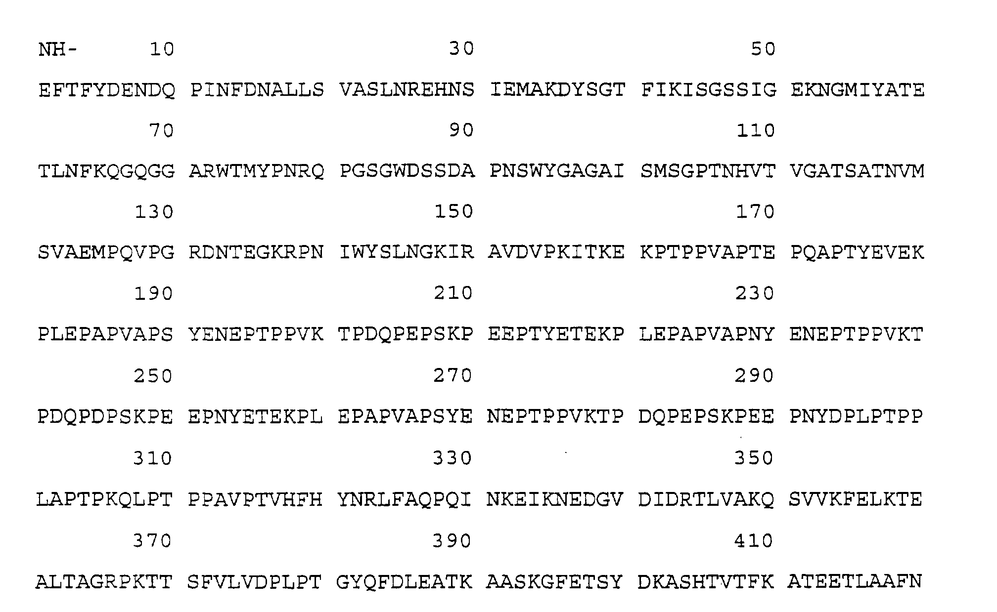
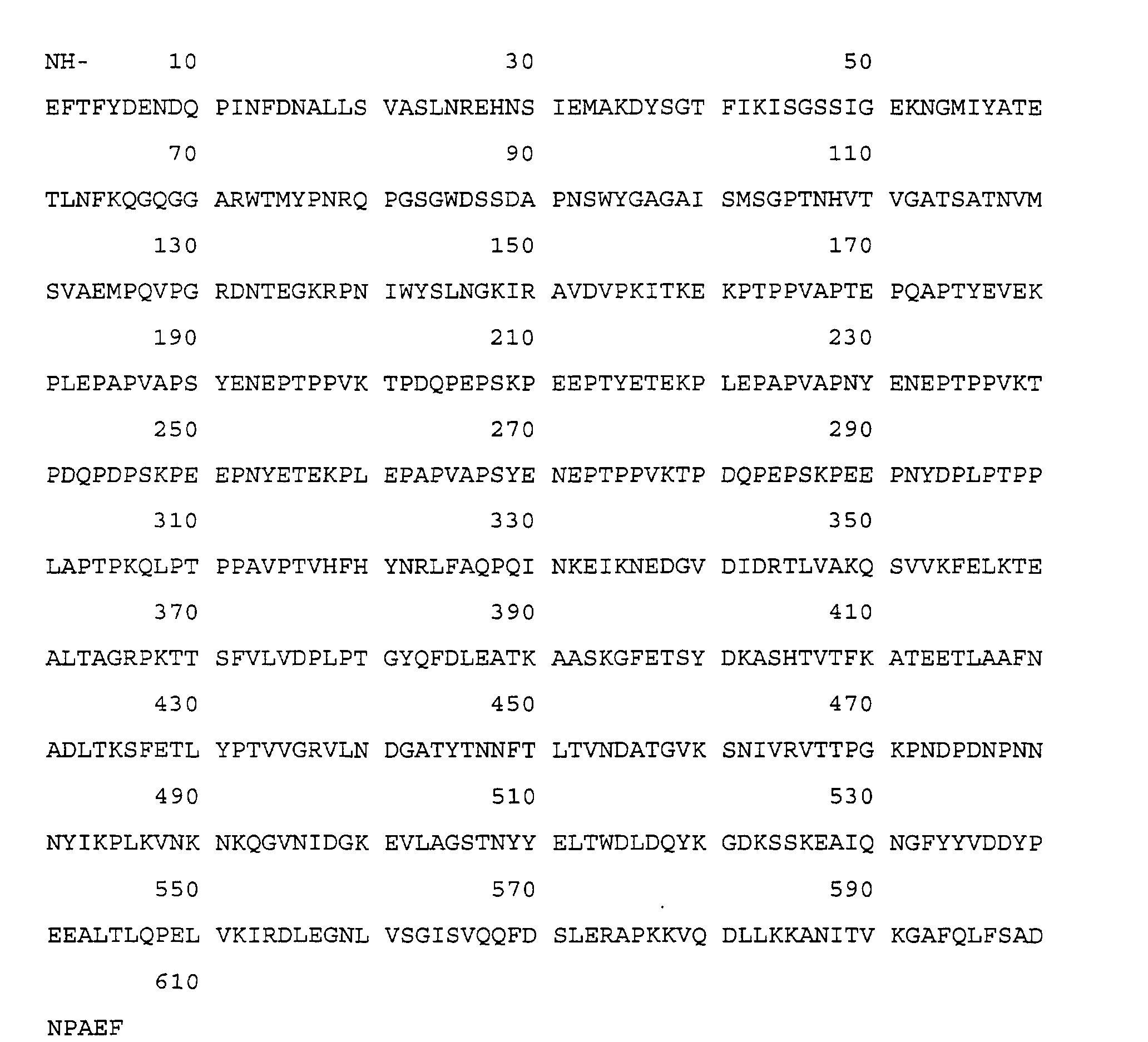
- a purified bacterial protein expressed during infection due to streptococci and isolated from human sera having at least the sequence of formula (1) (SEQ ID NO: 1): or an immunogenic fragment of the sequence of formula 1 (SEQ ID NO:1) of said bacterial protein thereof or an analogue thereof.
- the single letters in formula (1) are each to be understood to represent a separate ammo acid, and each is the conventional single letter symbol used for ammo acids.
- a bacterial protein according to the invention may be further characterised by either one or both of the following features:-
- the bacterial protein may be obtained from one of the group of Streptococcus oralis , Streptococcus sobrinus, Streptococcus ordonii, Streptoccocus sanguis , Streptococcus mutans , Streptococcus mitis, Streptococcus mitior, Streptococcus parasanguis, and Streptococcus bovis.
- the present invention also provides epitopes ("immunogenic fragments"), YEVEKPLEPAPVAPS (SEQ ID NO:3), SYENEPTPPVKTPD (SEQ ID NO: 4), KTPDQPEPSKPEEPT (SEQ ID NO: 5), EPAPVAPSYENEPTP (SEQ ID NO: 6), YEVEKELVDLPVEPS (SEQ ID NO: 7), KTPDQNIPDKPVEPT (SEQ ID NO: 8), TMYPNRQPGSGWDSS (SEQ ID NO: 9) and WYSLNGKIRAVDVPK (SEQ ID NO 10).
- Peptides of this type may be synthesised using conventional liquid or solid phase peptide synthesis techniques.
- Such immunogenic fragments may be obtained from a protein expressed during infection due to Streptococci. They may be obtained from a protein involved in binding to heart valves. Such immunogenic fragments may be fragments of a protein according to the present invention.
- the present invention also provides for the use of such a protein or immunogenic fragment in a method of manufacture of a medicament, for example a vaccine, for the treatment of infection due to Streptococci.
- the invention particularly provides a recombinant bacterial protein expressed during infection due to Streptococci having an amino acid sequence which includes at least the sequence of formula (1) (SEQ ID NO: 1) or an immunogenic fragment of the sequence of formula (1) (SEQ ID NO: 1) of said bacterial protein.
- the present invention also provides a method of diagnosis of culture-positive and culture-negative endocarditis using IgM antibody to a bacterial protein expressed during infection due to Streptococci, said bacterial protein, and therefore IgM antibody raised thereto, falling by 50% within two weeks after commencement of antibiotic treatment, thereby acting as a marker of pathogen kill.
- the present invention also provides the use of a protein or fragment according to the present invention in a method of diagnosis of infection due to Streptococci, the method not being practised on the human or animal body.
- the bacterial protein according to the present invention may be employed, using conventional techniques, for screening to obtain activity inhibiting agents for use in the prophylaxis and treatment due to Streptococci and in particular of culture-positive and culture-negative endocarditis-causing bacterial infection.
- screening methods forms a further aspect of the invention.
- the bacterial protein according to the invention is particularly well suited for the generation of antibodies.
- a bacterial protein expressed during infection due to Streptococci having an amino acid sequence which includes at least the sequence of formula (1) or an immunogenic fragment of the sequence of formula (1) (SEQ ID NO:1) of said bacterial protein for use as an innunogen.
- Standard immunological techniques may be employed with the bacterial protein in order to use it as an immunogen.
- any suitable host may be injected with the protein and the serum collected to yield the desired polyclonal anti-bacterial protein antibody after purification and/or concentration.
- the bacterial protein Prior to injection of the host the bacterial protein may be formulated in a suitable vehicle and thus according to a further aspect of the invention we provide a composition comprising a bacterial protein expressed during infection due to Streptococci and having an amino acid sequence which includes at least the sequence of formula (1) together with a pharmaceutically acceptable carrier, diluent or excipient.
- affinity chromatography employing an immobilised bacterial protein of the present invention as the affinity medium.
- a bacterial protein expressed during infection due to Streptococci having an amino acid sequence which includes at least the sequence of formula (1), or an immunogenic fragment of the sequence of formula (1) (SEQ ID NO:1) of said bacterial protein, covalently bound to an insoluble support.
- bacterial protein or fragment may be used to inhibit said protein, or fragment.
- the use of the bacterial protein expressed during infection according to the invention as immunogens for the production of antibodies generates one type of inhibitor of the action of the protein.
- inhibitors of the bacterial protein are potentially useful in the diagnosis, and in particular the prevention and treatment, of infections due to Streptococci and in particular of both culture-positive and culture-negative endocarditis, and provide a further feature of the invention.
- Inhibitors include any antagonists of the action of the bacterial protein expressed during infection or agents which prevent their production, and in particular those which may be used in treatment of endocarditis-causing bacterial infections.
- Suitable inhibitors include, for example, pharmaceutical reagents, including antibodies, and chemical analogues of the bacterial protein expressed during infection to antagonise the action of the bacterial protein, and anti-sense RNA and DNA to prevent production of the bacterial protein.
- Suitable inhibitors may be determined using appropriate screens, for example, by measuring the ability of a potential inhibitor to antagonise the action of, or prevent the production of a bacterial protein expressed during infection due to Streptococci or an immunogenic fragment according to the invention in a test model for example an animal model such as the mouse model.
- an inhibitor of a protein according to the present invention in a method of manufacture of a medicament for the treatment of infection due to Streptococci, the inhibitor comprising an antisense RNA or DNA, or an antibody or an antigen binding fragment thereof specific to the protein or immunogenic fragment.
- Such inhibitors may be used either alone or where appropriate in combination with other pharmaceutical agents, for example, other antibiotics.
- One particularly useful group of inhibitors according to this aspect of the invention are antibodies capable of recognising and binding to the bacterial proteins.
- the antibody may be a whole antibody or an antigen binding fragment thereof and may in general belong to any immunoglobulin class. Thus, for example, it may be an immunoglobulin M antibody or, in particular, an immunoglobulin G antibody.
- the antibody or fragment may be of animal, for example, mammalian origin and may be for example of murine, rat or human origin. It may be a natural antibody or a fragment thereof, or, if desired, a recombinant antibody fragment, ie., an antibody or antibody fragment which has been produced using recombinant DNA techniques.
- Particular recombinant antibodies or antibody fragments include human recombinant antibodies and in particular include (1) those having an antigen binding site at least part of which is derived from a different antibody, for example those in which the hypervariable or complementarity determining regions of one antibody have been grafted into the variable framework regions of a second, different antibody (as described in EP 239400); (2) recombinant antibodies or fragments wherein non-Fv sequences have been substituted by non-Fv sequences from other, different antibodies (as described in EP 171469, EP 173494 and EP 194276); or (3) recombinant antibodies or fragments possessing substantially the structure of a natural immunoglobulin but wherein the hinge region has a different number of cysteine residues from that found in the natural immunoglobulin but wherein one or more cysteine residues in a surface pocket of the recombinant antibody or fragment is in the place of another amino acid residue present in the natural immunoglobulin (as described in WO 89/01782 and WO
- the antibody or antibody fragment may be of polyclonal, or preferably, monoclonal origin. It may be specific for a single epitope or for a number of epitopes associated with the bacterial protein.
- Antigen binding antibody fragments include, for example, fragments derived by proteolytic cleavage of a whole antibody, such as F(ab')2,Fab' or Fab fragments, or fragments obtained by recombinant DNA techniques, for example Fv fragments (as described in WO 89/02465).
- the antibodies according to the invention may be prepared using well-known immunological techniques employing the bacterial protein expressed during infection as antigen.
- any suitable host may be injected with the bacterial protein and the serum collected to yield the desired polyclonal antibody after appropriate purification and/or concentration (for example by affinity chromatography using the immobilised bacterial protein as the affinity medium).
- splenocytes or lymphocytes may be recovered from the bacterial protein-injected host and immortalised using for example the method of Kohler et al ., Eur. J. Immunol. 6 : 511, 1976, the resulting cells being segregated to obtain a single genetic line producing monoclonal anti-streptococcal or -enterococcal bacterial protein antibodies.
- Antibody fragments may be produced using conventional techniques, for example, by enzymatic digestion with pepsin or papain. Where it is desired to produce recombinant antibodies according to the invention these may be produced using for example the methods described in EP 171469, EP 173494, EP 194276 and EP 239400.
- Antibodies according to the invention may be labelled with a detectable label or may be conjugated with effector molecule for example a drug eg. an antibacterial agent or a toxin or an enzyme, using conventional procedures and the invention extends to such labelled antibodies or antibody conjugates.
- a drug eg. an antibacterial agent or a toxin or an enzyme
- the antibodies according to the invention have a diagnostic and/or preventative and/or therapeutic use.
- the antibodies may be employed to detect whether the bacterial protein is present in a host organism, to confirm whether the host has a particular Streptococcal infection, and especially to test for the presence of such organisms in culture-negative endocarditis, and/or to monitor the progress of therapeutic treatment of such infections.
- Diagnostic methods of this type form a further aspect of the invention and may generally employ standard techniques, for example, immunological methods such as enzyme-linked immunosorbent methods, radioimmuno methods, latex agglutination methods or immunoblotting methods.
- Antibodies according to the invention also have a therapeutic use in the treatment of bacterial infection due to Streptococci, for example, those just described and may be used alone or conjugated to an effector molecule, in the latter case to target the effector molecule, eg an antibacterial agent, to the infecting organism.
- the antibody may be formulated in accordance with conventional procedures, for example, with a pharmaceutically acceptable carrier or excipient, eg., isotonic saline for the administration at an appropriate dosage, depending on the nature of the infection to be treated and the age of the patient.
- mixtures of antibodies may be used for diagnosis and/or prevention and/or treatment, for example mixtures of two or more antibodies recognising different epitopes of a bacterial protein according to the invention, and/or mixtures of antibodies of a different class, eg., mixtures of IgG and IgM antibodies recognising the same or different epitope(s) of a bacterial protein of the invention.
- the protein or any fragment, inhibitor, antibody or antigen binding fragment thereof according to the invention may be used in a method of treatment or diagnosis of the human or animal body.
- the diagnostic test method may be selected from one of the group of enzyme-linked immunosorbent assay, radioimmunoassay, latex agglutination assay and immunoblot assay.
- Such a protein, fragment, inhibitor, antibody or antigen binding fragment may form part of a composition for use in a method of diagnosis or treatment of the human or animal body together with a pharmaceutically acceptable carrier, diluent or excipient.
- the bacterial proteins according to the invention may be prepared by a variety of processes, for example, by protein fractionation from appropriate bacterial cell extracts, using conventional separation techniques such as ion exchange and gel chromatography and electrophoresis, or by the use of recombinant DNA techniques, as more particularly described in the "Experiments" section hereinafter.
- the use of recombinant DNA techniques is particularly suitable for preparing substantially pure bacterial proteins according to the invention.
- Also provided according to the present invention is the use of at least two immunogenic fragments selected from the group of YEVEKPLEPAPVAPS (SEQ ID NO: 3), TMYPNRQPGSGWDSS (SEQ ID NO: 9) and WYSLNGKIRAVDVPK (SEQ ID NO: 10) in the manufacture of a diagnostic test for Streptococci.
- a process for the production of a bacterial protein expressed during infection due to Streptococci having an amino acid sequence which includes at least the sequence of formula (1) (SEQ ID NO:1) or an immunogenic fragment of the sequence of formula 1 (SEQ ID NO : 1) of said bacterial protein comprising the steps of (1) culturing a host organism transformed with a vector including a gene coding for a precursor of said protein and (2) recovering said protein.
- the precursor cleaved in this aspect of the invention is a fusion protein comprising at least a portion of a protein produced in a transformed host organism and at least the amino acid sequence of formula (1).
- fusion proteins form a further aspect of the invention.
- the fusion protein includes a protein produced at a high level by a transformed host organism. Suitable such proteins include at least a portion of a chloramphenicol acetyltransferase (CAT) protein or, preferably at least a portion of the B-galactosidase protein.
- CAT chloramphenicol acetyltransferase
- a DNA sequence according to the invention may be further characterised in that the bacterial protein for which it encodes may be characterised by either one or both of the following features:-
- a DNA sequence may also be further characterised in that the protein for which it encodes is involved in binding to heart valves.
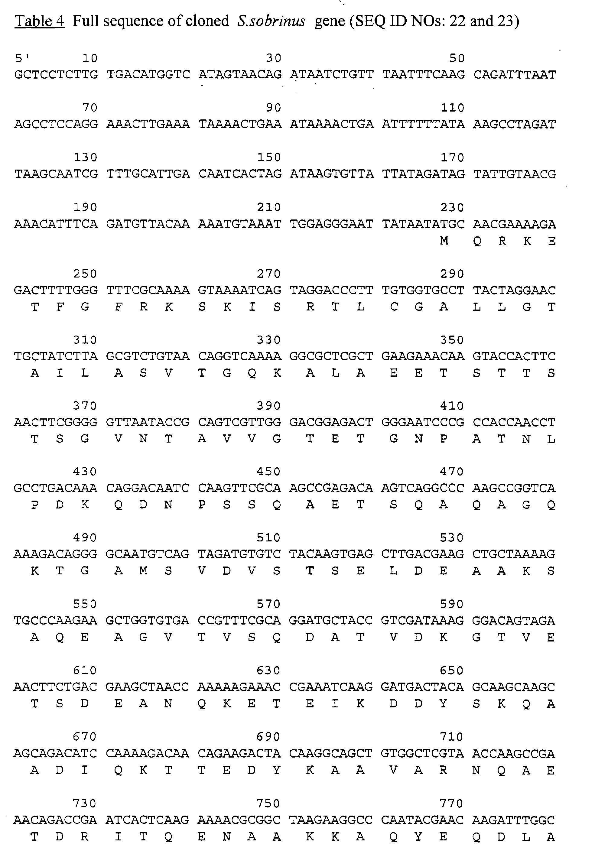
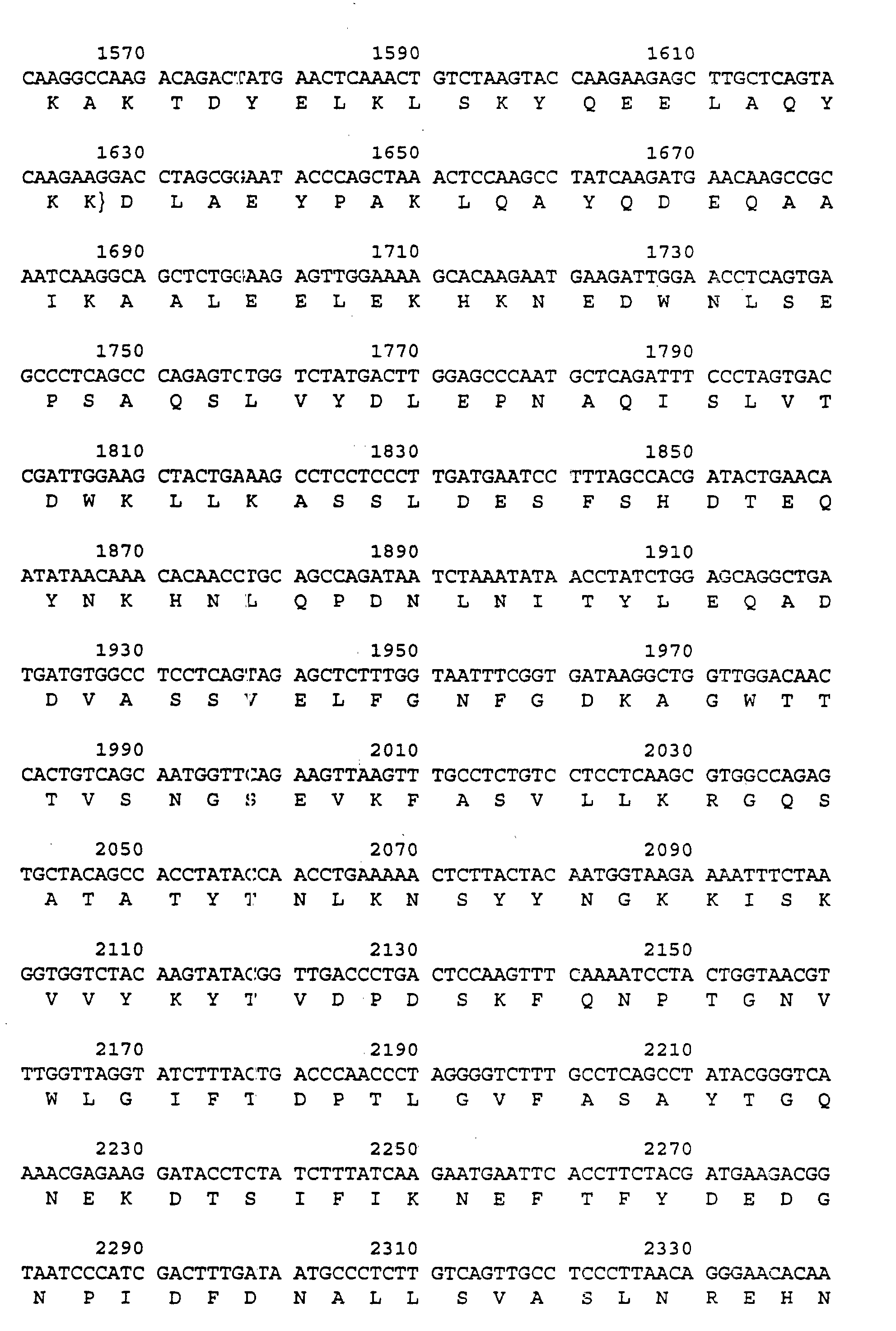
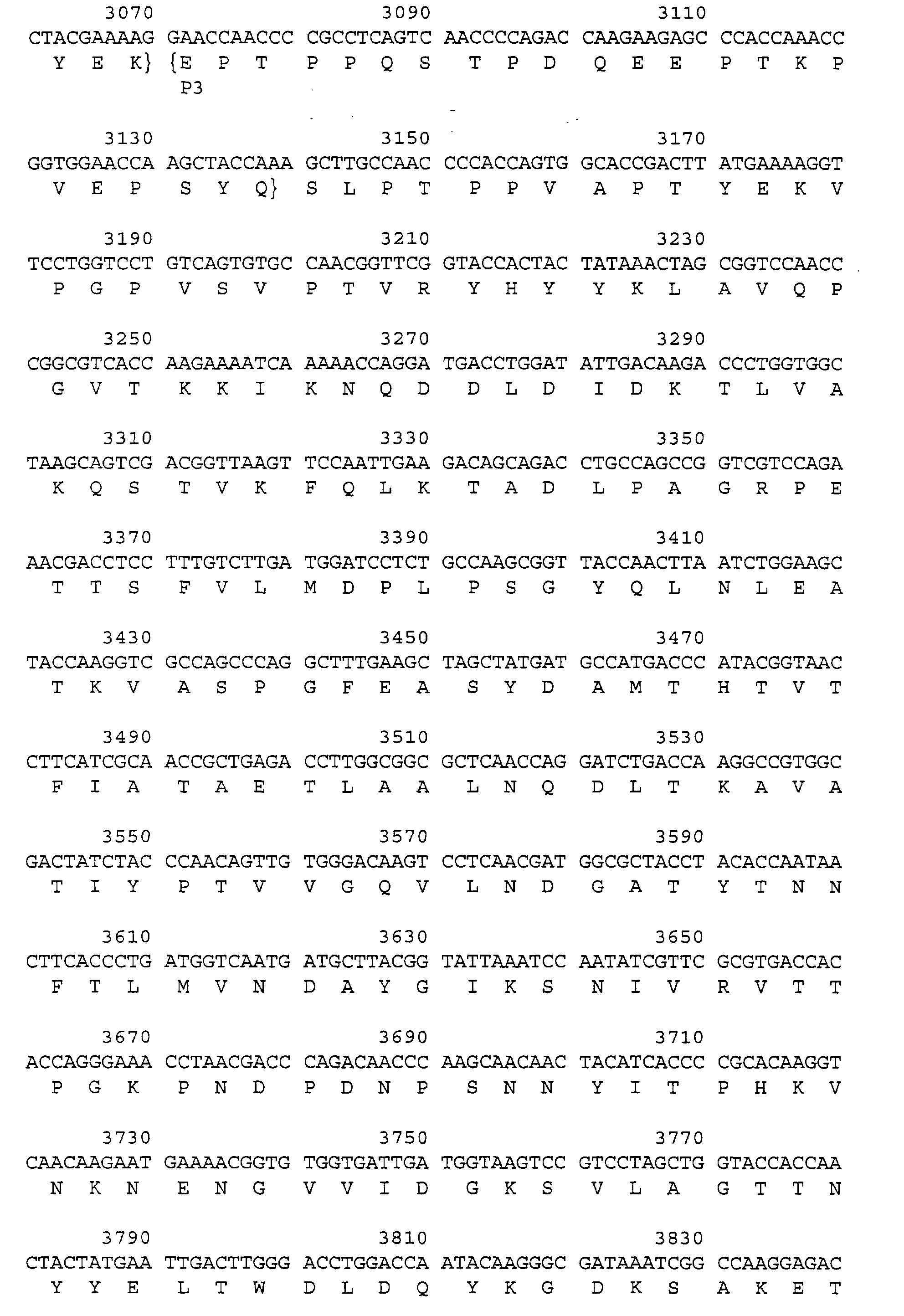
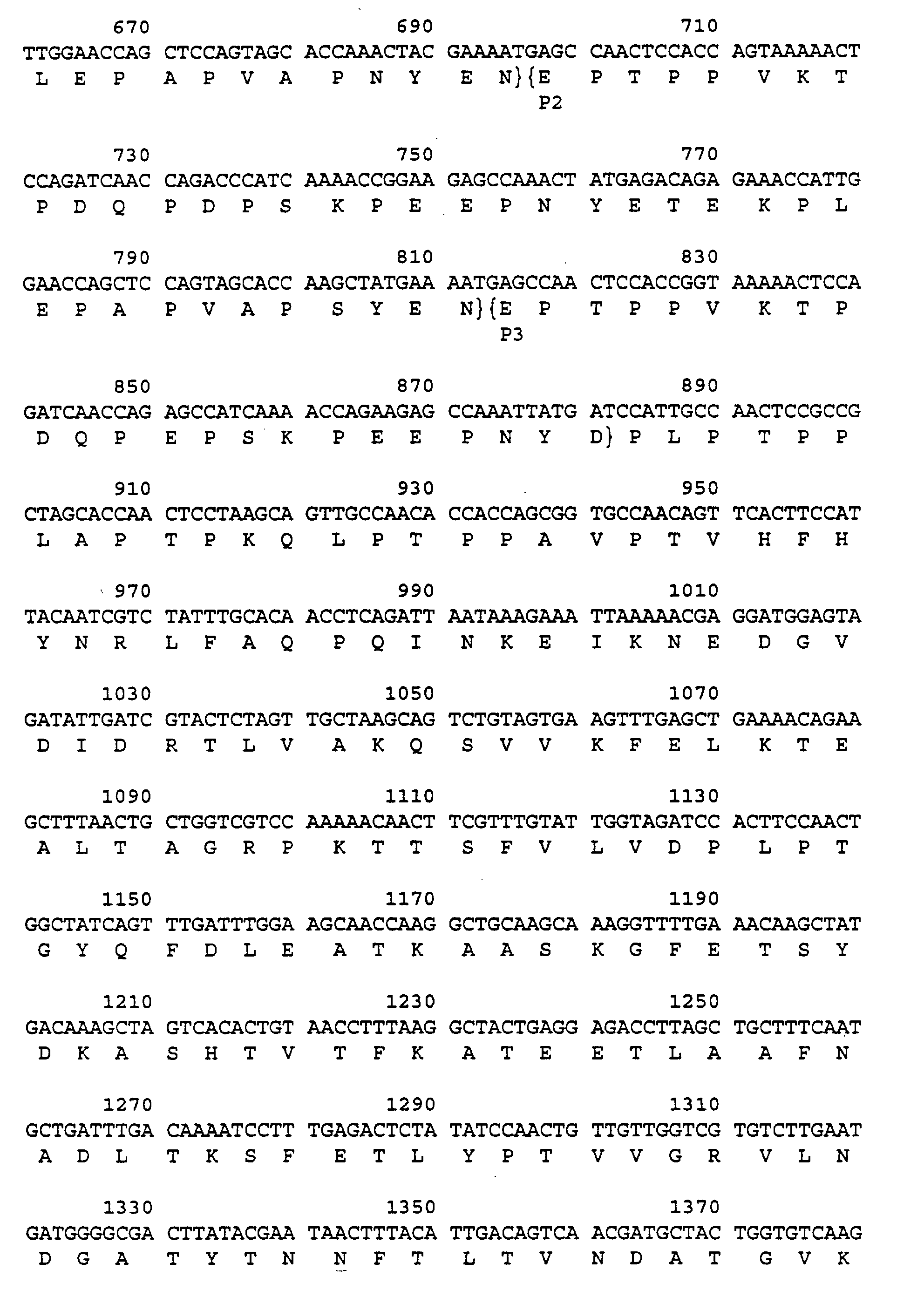
- DNA with this sequence may be obtained from bacterial genomic DNA as described in the "Experiments" section hereinafter.
- the DNA sequence according to this aspect of the invention may be incorporated in an expression vector using conventional techniques.
- an expression vector including a DNA sequence of formula (2) in a further aspect of the invention we provide an expression vector including a DNA sequence of formula (2).
- the vector may be adapted for use in a given host cell by the provision of suitable selectable markers, promoters and other control regions as appropriate.
- Host cells transformed with such vectors form a further aspect of the invention.
- Suitable host organisms include bacteria (eg. E.coli ), and mammalian cells in tissue culture.
- the DNA sequence of formula (2) may also be used to design DNA probes for use in identifying the presence of Steptococcal bacteria in the infected state and the invention extends to such DNA probes.
- Such probes may also be of use for detecting circulating bacterial nucleic acids, for example using a polymerase chain reaction, as a method of diagnosing such bacterial infections.
- the probe may also be synthesised using conventional techniques and may be immobilised on a solid phase, or may be labelled with a detectable label.
- a DNA sequence, vector, probe or inhibitor according to the invention may be used in a method of treatment or diagnosis of the human or animal body.
- the invention also provides fibronectin or an immunogenic fragment thereof or an analogue thereof or an antibody thereto or an antigen binding fragment thereof for use in a method of treatment or diagnosis of the human or animal body for infection due to Streptococci or Enterococci.
- the present invention provides antibodies specific to HSP 90 or immunogenic fragments or analogues thereof for use in a method of diagnosis or treatment of the human or animal body of infection due to streptococci or enterococci due to any one of the group of S.oralis , S.gordonii and S.sanguis.
- antigenic extracts were made from S.sanguis NCTC 7863, S.oralis NCTC 7864 and S.gordinii NCTC 7868.
- Figure 1 shows the immunoblot for S.oralis septicaemias. Paired IgMs and paired IgGs covering pre and post sera from 3 of the cases showing the antibody changes.
- Figure 2 shows the immunoblot of S.gordonii endocarditis.
- Anti-fibronectin antibody against S.gordonii NCTC 7868 in Track 1 crossabsorbed against fibronectin in Track 2.
- Tracks 3-8 show IgM and IgG respectively in 3 cases of S.gordonii endocarditis.
- Figure 3 shows 5 further case of S.gordonii endocarditis versus S.gordonii antigen Tracks 1/2, 3/4, 5/6, 9/10 and 11/12.
- Tracks 7/8 show pre-treatment of antibody levels of the same case as tracks 9/10 showing increased IgM to bands at approximately 85 KDa and IgG to bands at approximately 180, 58, 56 and 52 Kda
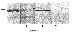
- Figure 4 shows the immunoblot of S.oralis endocarditis, Tracks 3/4 IgM and IgG, antifibronectin antibody crossreacting with 180 KDa band of S.oralis , Track 1 and effect of crossabsorption with fibronectin, Track 2.
- Figure 5 shows the irnmunoblot of S.sanguis endocarditis, showing both IgM (track 3 and 5) and IgG (tracks 4 and 6) respectively of both cases, antifibronectin antibody crossreacting with the 120 KDa band of S.sanguis , Track 1 and the effect of crossabsorption with fibronectin, Track 2.
- the bacteria were harvested by spinning at 5,000 rpm for 10 mins before being washed, and resuspended in 12.5ml of 0.02 M Tris pH 8.2. Then 25 ml of 20 M polyethylene glycol (PEG) 24% in distilled water was added and mixed. Lysozyme was added at 34.6 mg/ml equivalent to 100 ⁇ g/unit obtained by measuring the OD600 of a tenfold dilution of bacterial cultures. After 1 hour at 37°C, the spheroblasts formed were spun down at 5,000rpm for 10 mins and thoroughly resuspended in 50 ml of 10 mM Tris-Cl pH 8.0, 1 mM EDTA (TE).
- PEG polyethylene glycol
- the spheroblasts were lysed with 5.5 ml of 10% sodium dodecyl sulphate (SDS) at 60°C for 15 mins.
- the DNA was purified by standard procedures (Maniatis et al. , Molecular Cloning - A laboratory manual, 2nd edn. Cold Spring Harbour, New York, 1989), RNAase A (sigma) treated (after preboiling the RNAase at 100°C for 15 mins), proteinase K treated, phenol:chloroform:isoamylalcohol (25:24:1) extracted, and ethanol precipitated. Plasmid DNA was removed by ethanol precipitation in the presence of 0.3 M ammonium acetate pH 5.2. Further purification was obtained by dialysis against TE over 36 hrs at 4°C.
- the DNA was mechanically sheared, EcoRI linkers added, fractionated and inserts ligated into lambda ZAPII vector arms.
- the library had an insert size range of 2kb-7kb.
- Serum was taken from a patient with endocarditis and used for antibody screening.
- Escherichia coli XL1 - Blue cells were infected at approximately 3,000 pfu/85mm on L broth agar (Bacto-tryptone 10 g/l, yeast extract 5 g/l, sodium chloride 10 g/l, maltose 2 g/l, bacto-agar 15 g/l). Plaques were transferred to nitrocellulose filters (0.45 ⁇ m pore size, Sartorius AG, Gottingen, Germany), impregnated with 10 mM isopropyl B-D-thiogalactopyranoside (IPTG), at 37°C for 2 hrs, after a 42°C incubation for 3 hrs.
- IPTG isopropyl B-D-thiogalactopyranoside
- the filters were washed 5 times over 100 mins with Tween 20-Tris-buffered saline (150 mM sodium chloride, 10 mM Tris-Cl pH 7.2, Tween 20, 0.05%), and once with salt - Tween 20 solution (150 mM sodium chloride, Tween 20, 0.05%).
- the bound antibody was eluted by adding 5 ml of glycine saline buffer (150 mM sodium chloride, 200 mM glycine-Cl pH 2.8), and shaking at room temperature for 30 mins.
- the buffer was aspirated into a tended, containing Tris 0.04g per 100 ⁇ l, mixed, termed the eluted antibody and stored at 4°C.
- a Western blot of S.sobrinus MUCOB 263 was performed essentially by the hnniunoblot method above with the original serum, depleted serum and eluted antibody being used as the primary antibodies.
- the antigen was the supernatant of the strain following crushing in an X-press.
- the secondary antibody was a thousandfold dilution of anti-human IgM and IgG alkaline phosphatase conjugates in 3% BSA, and the stain was the alkaline phosphatase stain.
- the phagemid was propagated by adding 10 ⁇ l of the phagemid solution to 200 ⁇ l of resuspended E.coli XL1-Blue cells, incubating the culture at 37 °C for 15 mins, and plating 1 ⁇ l, 25 ⁇ l, 75 ⁇ l and 100 ⁇ l aliquots onto L broth agar plates, containing ampicillin 50 ⁇ g/ml.
- the pBluescript SK(-) plasmid DNA was purified from the bacterial colonies with the Magic (RTM) Minipreps DNA purification System (Promega) and alkaline denatured by the method of Maniatis et al. , 1989.
- 8 ⁇ l of solution, containing DNA 1.5 - 200 ⁇ g was added to 2 ⁇ l of 2 M sodium hydroxide, briefly vortexed and spun, and left at room temperature for 10 mins.
- 3 ⁇ l of 3 M sodium acetate (pH 4.8), 7 ⁇ l of distilled water, and 60 ⁇ l of -20°C absolute ethanol were added and the DNA precipitated at - 70 °C for 30 mins.
- the precipitate was pelleted at 13,000 rpm for 1 min, washed with -20°C ethanol 70%, and vacuum-dried. The dried pellets were stored at -20°C.
- DNA sequencing was performed by the two-step chain-termination method, with Sequenase (R) Version 2.0 (Cambridge Bioscience). The annealing step was carried out at 66°C for 2 mins, the labelling at 22°C for 4 mins, and the termination at 38°C for 5 mins. Areas containing secondary structure were resolved by the substitution of dITP for dGTG. Sequencing reactions were run on acrylamide gels 6% for 11, 8, 5 and 2 hrs at 46-51°C.
- PCR Polymerase Chain Reaction
- RTM TA cloning
- Insert DNA was subcloned with the TA Cloning (RTM) System Version 1.3 (InVitrogen Corporation, British Biotechnologies Ltd, Oxon) as specified in the manufacturers protocol. Overnight, 12°C, ligation reactions containing 3.0 ⁇ l of a fourfold dilution, in distilled water, of the 0.7 ⁇ g PCR reaction, 4.7 ⁇ l of doubling dilution of the 0.7 ⁇ g PCR reaction, and a negative control were performed.
- RTM TA Cloning
- the sequence obtained from S.sobrinus had a 99.2% homology to the SpaA antigen of S.sobrinus and a 68.4% homology to the PAc antigen of S.mutans.
- the S.oralis peptide carried a 76.2% homology over 605 amino acids with the S.mutans PAc protein precursor and 73.8% homology over 606 amino acids with both the S.sobrinus SpaA protein precursor and the S.mutans surface antigen I/II precursor. Both sequences contain a three tandem repeat motif of 39 amino acids. Six peptides were derived from these and used to epitope map-this communal sera.
- Epitope mapping of the antigen cloned from S. oralis was carried out according to the protocol outlined by Geysen et al. , J. Immunol. Methods, 102: 259-274 (1987) and references therein. In this epitope mapping, a complete set of overlapping nonapeptides was synthesised for the cloned S. oralis antigen. Peptide 1 covered residues 1-9, peptide 2 covered residues 2-10 and peptide 3 covered residues 3-11 etc. The epitopes were tested against various patient sera. All of the sera were examined at a 1 in 200 dilution for IgG and recording was stopped after 30 minutes. The sera examined were:
- Epitopes were defined as those peptides which:
- This epitope mapping identified a total of 9 epitopes (see table 10) - NFKQGQG, RQPG, SWYGAG, GKIRAV, RLFAQPQ, AGRPK, PTGYQFD, YPTVV and LLKKA (SEQ ID NOs: 13-21 respectively)
- each individual serum was then reexamined for positivity with each well (see table 11).
- peptides 1-4 cover the sequence of S.oralis as short peptides of 15 amino acid length.
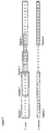
- Peptides 5 and 6 covered the S.sobrinus area which differed. These sequences are listed (Table 6) and illustrated ( Figure 8).
- the six synthetic peptides were produced by Cambridge Biochemicals Ltd., Nantwich and dissolved in a double-distilled water to give a final concentration of 2 mg/ml. Aliquots, 0.5 ml, were stored at -70°C and after one freeze-thaw cycle, stored at -20°C.
- the conjugates were left to react at room temperature for 1 hour.
- the plates were then washed as before and 200 ⁇ l of ABTS stain was added to each well (three tablets of 2.2' amino-bis(3-ethylbenzthiazoline-6-sulfonic acid); diammonium salt; Sigma) and 160 ⁇ l of 30% w/w hydrogen peroxide solution; Sigma, in 60 ml of 125 mM disodium hydrogen orthophosphate buffer brought to pH 4.0 with 1M citric acid).
- This ABTS stain was prepared just prior to use.
- each of the peptides was used in an indirect ELISA.
- YEVEKPLEPAPVAPS Peptide 1; SEQ ID NO: 3
- TMYPNRQPGSGWDSS contains the epitope RQPG - epitope numbers 74-79; Peptide 7; SEQ ID NO: 9
- WYSLNGKIRAVDVPK contains the epitope GKIRAV - epitope numbers 144-147; Peptide 8; SEQ ID NO: 10
- optical densities are given in Table 8 for Peptide 1, and in Table 8a for Peptides 1, 2 and 3.
- a raised IgG was specific to patients with Steptococcal endocarditis and a raised IgM specific to endocarditis due to Streptococcus oralis/gordonii/ sanguis/mitis.
- each of the peptides 1-6 was reacted in an indirect ELISA according to the previous protocol.
- the indirect ELISA produced OD values greater than 2 for all six peptides regardless of whether fibronectin was added to the sandwich. This implied that the polyclonal serum (anti-fibronectin 0.5 mg/ml Sigma [F1509]) reacted with the peptides directly. This serum was then immunoblotted at a dilution of 1 in 40 and again after crossabsorbing, 100 ⁇ l of 1 mg/ml antibody, with 100 ⁇ l of 1 mg/ml of fibronectin (F2006 Sigma) at 37°C for 30 mins.
- V immunoglobulin heavy and light chain variable
- the library was produced essentially as described by Marks et al (J. Mol. Biol., 1991, 222: 581-597) using the pCANTAB 5 vector, which is now commercially available as part of a kit from Pharmacia (Milton Keynes, UK).
- the heavy and light chain V genes obtained from cDNA prepared from the mRNA of peripheral blood lymphocytes of a patient recovering from a viridans streptococcus (blood culture positive) septicaemia, were randomly combined and subcloned into Not I/Sfi I digested pCANTAB 5.
- 16 clones produced 6 types.
- One type (Type A) was present in 7 out of the 16 clones and was the only type to produce a positive reading in the indirect ELISA (0.276-0.318) (control 0.17).
- 16 clones produced 10 types. Two of these, A and B were represented by 3 and 5 clones respectively. None of these clones produced a positive ELISA result. Five of the 8 clones produced a positive ELISA result (range 0.235-0.304 control 0.138) and a unique Bstl fingerprint. One of these (Clone 7) was selected for animal work (PAC 4).
- This experiment was an acute streptococcal infection model using Balb/c mice with an end point of death.
- Conditions were: S.oralis dose: 5.7 x 10 9 Phage dose: 5 x 10 8 pfu/ml Antibody given first, followed by S.oralis 2 hours later. (results of Experiment 1) SURVIVORS Antibody No. of mice 4 hours 24 hours 48 hours Control: no antibody M13K07 phage 15 7 3 1 DEPAGE 13 3 3 2 B3.7 13 6 6 6 B3.14 8 1 1 1 1 PAC 1 9 9 2 2 PAC 2 17 8 8 8 PAC 3 15 6 6 6 6 PAC4 15 2 2 2 DEPAGE - antibody against a Candidal specific carboxy-end HSP90 antigen (sequence.
- B3.7 is a recombinant antibody specific to the HSP90 stress protein, suggesting that antibodies specific to the HSP90 protein may be used to diagnose and treat S. oralis and possibly other streptococcal and enterococcal infections.
- E.faecium dose 3 x 10 10/ ml
- Phage dose 5 x 10 10 pfu/ml
- S spleen
- Non-Neutropenic Controls Neutropenic Controls
- Non-Neutropenic Controls Neutropenic Controls
- Peptides numbered 1-6 correspond to SEQ ID NOs: 3-8 Clinical History of Serum Peptide Number 1 2 3 4 5 6 M G M G M G M G M G M G S.mutans endocarditis 0.341 1.506 0.331 0.381 0.338 1.365 0.445 1.225 0.261 1.491 0.241 0.869 S.oralis endocarditis 0.442 0.902 0.561 0.891 0.491 0.768 0.567 0.936 0.653 0.784 0.373 0.873 S.oralis septicaemia 0.024 0.118 0.036 0.215 0.019 0.123 0.065 0.168 0.043 0.241 0.117 0.218 S.lactis endocarditis 0.306 0.589 0.319 0.528 0.309 0.492 0.377 0.415 0.337 0.421 0.244 0.449 S.aureus endocarditis 0.075 0.177 0.145 0.579 0.068 0.153 0.015 0.127 0.101 0.267
Landscapes
- Chemical & Material Sciences (AREA)
- Health & Medical Sciences (AREA)
- Organic Chemistry (AREA)
- Life Sciences & Earth Sciences (AREA)
- General Health & Medical Sciences (AREA)
- Medicinal Chemistry (AREA)
- Genetics & Genomics (AREA)
- Biophysics (AREA)
- Biochemistry (AREA)
- Molecular Biology (AREA)
- Proteomics, Peptides & Aminoacids (AREA)
- Gastroenterology & Hepatology (AREA)
- Zoology (AREA)
- Toxicology (AREA)
- Immunology (AREA)
- Chemical Kinetics & Catalysis (AREA)
- Oncology (AREA)
- General Chemical & Material Sciences (AREA)
- Nuclear Medicine, Radiotherapy & Molecular Imaging (AREA)
- Pharmacology & Pharmacy (AREA)
- Animal Behavior & Ethology (AREA)
- Public Health (AREA)
- Veterinary Medicine (AREA)
- Communicable Diseases (AREA)
- Peptides Or Proteins (AREA)
- Medicines Containing Antibodies Or Antigens For Use As Internal Diagnostic Agents (AREA)
- Measuring Or Testing Involving Enzymes Or Micro-Organisms (AREA)
- Preparation Of Compounds By Using Micro-Organisms (AREA)
- Saccharide Compounds (AREA)
- Cookers (AREA)
- Glass Compositions (AREA)
- Pharmaceuticals Containing Other Organic And Inorganic Compounds (AREA)
Abstract
Description

The protein or any fragment, inhibitor, antibody or antigen binding fragment thereof according to the invention may be used in a method of treatment or diagnosis of the human or animal body.


1149-1158, antigenic extracts were made from S.sanguis NCTC 7863, S.oralis NCTC 7864 and S.gordinii NCTC 7868.
| | | | ||||
| IgM | IgG | IgM | IgG | IgM | IgG | |
| | 6 | 5 | 12 | 5 | 12 | 10 |
| | 0 | 1 | 28 | 15 | 25 | 21 |
| | 42 | 41 | 14 | 27 | 17 | 21 |
| | 6 | 7 | 0 | 7 | 0 | 2 |
| IgM Peptides | | |||||
| 1 | 7 | 8 | 1 | 7 | 8 | |
| Sensitivity | 50% | 100% | 100% | 41.5% | 41.5% | 83 |
| Specificity | ||||||
| 100% | 33% | 40% | 98% | 64% | 50 | |
| Efficiency | ||||||
| 89% | 48% | 53% | 85% | 59% | 57% |
S.oralis dose: 5.7 x 10⁹
Phage dose: 5 x 10⁸ pfu/ml
Antibody given first, followed by S.oralis 2 hours later.
| (results of Experiment 1) | ||||
| SURVIVORS | ||||
| Antibody | No. of | 4 hours | 24 | 48 hours |
| Control: no antibody M13K07 phage | 15 | 7 | 3 | 1 |
| DEPAGE | 13 | 3 | 3 | 2 |
| B3.7 | 13 | 6 | 6 | 6 |
| B3.14 | 8 | 1 | 1 | 1 |
| | 9 | 9 | 2 | 2 |
| | 17 | 8 | 8 | 8 |
| | 15 | 6 | 6 | 6 |
| PAC4 | 15 | 2 | 2 | 2 |
Fisher exact 2 tailed P value showed statistical significance for B3.7 at 48 hours (P 0.03),
S.oralis dose 2.5 x 10⁹
Phage dose 5 x 10¹⁰ pfu/ml
S.oralis given first, antibody given 24 hours later.
Positive organ count > 104/g/ml
S = Spleen, K = Kidney
S + K = combined spleen and kidney results
5 mice sacrificed on
Repeat antibody injection at
| (results of Experiment 2) | |||||||||
| | | Day 12 | |||||||
| S | K | S+K | S | K | S+K | S | K | S+K | |
| Control:no antibody | 4 | 4 | 8 | 0 | 0 | 0 | 1 | 0 | 1 |
| B3.14 | 1 | 2 | 3 | 0 | 0 | 0 | 0 | 0 | 0 |
| | 0 | 1 | 1 | 1 | 1 | 2 | 1 | 0 | 1 |
| | 1 | 2 | 3 | 0 | 0 | 0 | 0 | 0 | 0 |
E.faecium dose: 3 x 10¹⁰/ ml
Phage dose: 5 x 10¹⁰ pfu/ml
E. faecium given first, antibody given 24 hours later.
Positive > 104 g/ml. S = spleen, K = Kidney
S + K = Spleen + Kidney.
Spontaneous deaths cultured at
5 mice sacrificed on
Remaining
| (results of Experiment 3) | |||||||
| Day 2 (spontaneous deaths) | (n=5) | (n=3 control) (n=5 PAC 2) | |||||
| S | K | S+K | S | K | S+K | ||
| Control no antibody: | 6 | 2 | 5 | 7 | 0 | 0 | 0 |
| | 4 | 2 | 3 | 5 | 0 | 0 | 0 |
Organism: high level (>256 mg/l) vancomycin-resistant E. faecium
E. faecium dose: 5 x 10¹⁰ /ml
Phage dose: 5 x 10¹⁰ pfu/ml
Antibody given first, followed by
| (results of experiment 4) | ||||
| Survivors | ||||
| Antibody | No of mice | 4hours | 24hours | 48hours |
| Control no antibody M13K07 phage | 15 | 10 | 5 | 1 |
| PAC2 | 15 | 12 | 8 | 6 |
| Immunoblot testing of the S.oralis NCTC 7864 antigen against patient sera. | |||||||||||||||
| M.Wt. of S. oralis antigen band | Patient Sera | ||||||||||||||
| Endocarditis | Sequential sera S.oralis. septicaemia n = 12 | Controls | |||||||||||||
| S.oralis Endocarditis | S.gordonii Endocarditis | S.sanguis Endocarditis | Constant Septicaemia | Inc. x 2 or App. | Controls Neutropenic | Controls Non-Neutropenic | |||||||||
| n=2 | n=4 | n=2 | n=20 | n=20 | |||||||||||
| IgM | IgG | IgM | IgG | IgM | IgG | IgM | IgG | IgM | IgG | IgM | | IgM | IgG | ||
| 192 | 1 | 1 | 2 | 10 | 1 | 2 | 1 | 3 | 1 | 5 | 4 | 6 | 2 | 1 | |
| 185 | 1 | 4 | 1 | 1 | 1 | 1 | 3 | 2 | 3 | 1 | 2 | ||||
| 180 | 2 | 2 | 4 | 13 | 1 | 2 | 2 | 9 | 10 | 1 | 3 | 4 | |||
| 155 | 1 | 9 | 1 | 4 | 2 | ||||||||||
| 140 | 1 | 2 | 2 | 6 | 3 | 8 | 1 | 2 | 2 | 8 | 1 | 2 | |||
| 105 | 1 | 1 | 5 | 1 | 1 | 2 | 2 | 1 | 1 | ||||||
| 85 | 2 | 2 | 4 | 11 | 1 | 2 | 8 | 1 | 1 | 3 | 3 | 1 | 1 | ||
| 76 | 1 | 1 | 3 | 1 | 1 | 5 | 2 | 1 | 2 | 2 | |||||
| 65 | 1 | 3 | 1 | ||||||||||||
| 51 | 1 | 6 | 3 | 1 | 11 | 2 | 1 | 11 | 6 | ||||||
| 46 | 2 | 3 | 1 | 1 | 1 | ||||||||||
| 35 | 1 | 1 | 1 | ||||||||||||
| Inc = Increased x 2 App = Appeared |
| Immunoblot testing of the S.gordonii NCTC 7868 antigen against patient sera. | |||||||||||||||
| M.Wt. of S.oralis antigen band | Patient Sera | ||||||||||||||
| Endocarditis | Sequential sera S.oralis. septicaemia n = 12 | Controls | |||||||||||||
| S.oralis Endocarditis | S.gordonii Endocarditis | S.sangius Endocarditis | Constant Septicaemia | Inc. x 2 or App. | Controls Neutropenic | Controls Non-Neutropenic | |||||||||
| n=2 | n=4 | n=2 | n=20 | n=20 | |||||||||||
| IgM | IgG | IgM | IgG | IgM | IgG | IgM | IgG | IgM | IgG | IgM | | IgM | IgG | ||
| 185 | 2 | 2 | 5 | 1 | 7 | ||||||||||
| 180 | 2 | 12 | 14 | 2 | 2 | 6 | 5 | 1 | 2 | ||||||
| 105 | 1 | 1 | 2 | 4 | 1 | 3 | 1 | ||||||||
| 85 | 2 | 1 | 12 | 14 | 1 | 1 | 1 | 10 | 1 | 1 | |||||
| 80 | 5 | 12 | 1 | 1 | 1 | 9 | 1 | 11 | 4 | ||||||
| 76 | 5 | 7 | 1 | 1 | 1 | 10 | 1 | 3 | |||||||
| 72 | 1 | 1 | 5 | 7 | 1 | 1 | 1 | 4 | 1 | 1 | |||||
| 70 | 1 | 4 | 5 | 1 | 2 | 7 | |||||||||
| 65 | 9 | 13 | 1 | 2 | 1 | 4 | 1 | 1 | |||||||
| 58 | 1 | 9 | |||||||||||||
| 56 | 2 | 9 | 1 | ||||||||||||
| 52 | 1 | 8 | 2 | ||||||||||||
| 47 | 2 | 12 | 14 | 1 | 1 | 10 | 2 | 1 | 15 | 2 | 2 | ||||
| 40 | 5 | 8 | 2 | 1 | 8 | 1 | 1 | ||||||||
| 37 | 8 | 8 | 2 | 2 | |||||||||||
| 35 | 3 | 10 | 1 | 1 | 2 | 1 | |||||||||
| 33 | 2 | 9 | |||||||||||||
| 30 | 1 | 11 | 2 | 2 | |||||||||||
| 28 | 2 | 7 | 1 | ||||||||||||
| Inc = Increased App = Appeared |
| Immunoblot testing of the S.sanguis NCTC 7863 antigen against patient sera. | |||||||||||||
| M.Wt. of S.sanguis antigen band | Patient Sera | ||||||||||||
| Endocarditis | Septicaemia | ||||||||||||
| S.oralis Endocarditis | S.gordonii Endocarditis | S.sanguis Endocarditis | S. oralis Septicaemia | Controls Neutropenic | Controls Non-Neutropenic | ||||||||
| n=2 | n=14 | n=2 | n =9 | n=20 | n=20 | ||||||||
| IgM | IgG | IgM | IgG | IgM | IgG | IgM | IgG | IgM | | IgM | IgG | ||
| 120 | 2 | 4 | 14 | 2 | 2 | 1 | 3 | 3 | 4 | ||||
| 85 | 1 | 2 | 2 | 2 | 8 | 3 | 3 | 2 | 4 | ||||
| 83 | 2 | 2 | |||||||||||
| 81 | 2 | 8 | 2 | 2 | |||||||||
| 75 | 1 | 1 | 4 | 2 | 2 | 2 | 5 | 3 | 15 | 8 | |||
| 59 | 1 | 1 | 2 | 1 | 2 | 2 | |||||||
| 57 | 1 | 2 | 2 | 12 | 1 | 2 | 2 | ||||||
| 55 | 1 | 1 | 2 | 6 | 2 | 12 | |||||||
| 50 | 9 | 2 | |||||||||||
| 47 | 2 | 2 | 2 | 9 | 2 | 6 | |||||||
| 42 | 1 | 6 | 2 | 2 | 9 | ||||||||
| 40 | 2 | 2 | 9 | 2 | |||||||||
| 37 | 3 | 2 | 2 | ||||||||||
| 35 | 1 | ||||||||||||
| 32 | |||||||||||||
| 25 | 6 | 2 |





| Peptide Number | |
| 1 | |
| 2 | |
| 3 | |
| 4 | |
| 5 | |
| 6 | KTPDQNIPDKPVEPT |
| Clinical History of | ||||||||||||
| Peptide Number | ||||||||||||
| 1 | 2 | 3 | 4 | 5 | 6 | |||||||
| M | G | M | G | M | G | M | G | M | G | M | G | |
| S.mutans endocarditis | 0.341 | 1.506 | 0.331 | 0.381 | 0.338 | 1.365 | 0.445 | 1.225 | 0.261 | 1.491 | 0.241 | 0.869 |
| S.oralis endocarditis | 0.442 | 0.902 | 0.561 | 0.891 | 0.491 | 0.768 | 0.567 | 0.936 | 0.653 | 0.784 | 0.373 | 0.873 |
| S.oralis septicaemia | 0.024 | 0.118 | 0.036 | 0.215 | 0.019 | 0.123 | 0.065 | 0.168 | 0.043 | 0.241 | 0.117 | 0.218 |
| S.lactis endocarditis | 0.306 | 0.589 | 0.319 | 0.528 | 0.309 | 0.492 | 0.377 | 0.415 | 0.337 | 0.421 | 0.244 | 0.449 |
| S.aureus endocarditis | 0.075 | 0.177 | 0.145 | 0.579 | 0.068 | 0.153 | 0.015 | 0.127 | 0.101 | 0.267 | 0.063 | 0.217 |
| ELISA readings for peptides 1-6 |
| Indirect ELISA results for | ||||||
| Concentration of peptide: 200µl at 10µg/ml | ||||||
| | ||||||
| Patient No. | Sera Date | Mean Blank O.D. | Mean Sample O.D | Standard Deviation | ||
| IgM | IgG | IgM | IgG | |||
| S.mutans Endocarditis | ||||||
| 1 | 16.03.88 | 0.131 | 0.117 | 0.698 | 0.001 | 0.015 |
| 2 | 17.09.87 | 0.193 | 0.012 | 0.742 | 0.003 | 0.0017 |
| S.oralis Endocarditis (n=3) | ||||||
| 3 | 23.05.78 | 0.141 | 0.462 | 0.960 | 0.018 | 0.002 |
| 4 | 12.12.81 | 0.197 | 0.447 | 0.902 | 0.023 | 0.031 |
| 5 | 23.02.84 | 0.121 | 0.043 | 0.038 | 0.001 | 0.008 |
| 30.08.84 | 0.131 | 0.153 | 0.699 | 0.004 | 0.007 | |
| S.gordonii Endocarditis (n=14) | ||||||
| 6 | 1.07.86 | 0.131 | 0.422 | 0.936 | 0.001 | 0.002 |
| 15.08.86 | 0.131 | 0.339 | 0.855 | 0.003 | 0.004 | |
| 22.09.86 | 0.131 | 0.228 | 0.81 | 0.010 | 0.005 | |
| 6.10.96 | 0.131 | 0.173 | 1.06 | 0.003 | 0.002 | |
| 7 | 8.01.87 | 0.131 | 0.487 | 0.685 | 0.010 | 0.012 |
| 8 | 18.12.86 | 0.131 | 0.059 | 0.662 | 0.004 | 0.006 |
| 21.01.87 | 0.131 | 0.270 | 0.715 | 0.005 | 0.009 | |
| 9 | 10.08.86 | 0.131 | 0.43 | 0.41 | 0.007 | 0.004 |
| 10 | 17.07.86 | 0.131 | 0.568 | 0.601 | 0.010 | 0.012 |
| 11 | 14.07.93 | 0.131 | 0.095 | 0.471 | 0.012 | 0.003 |
| 12 | 20.03.93 | 0.158 | 0.164 | 0.404 | 0.001 | 0.03 |
| 22.03.93 | 0.158 | 0.168 | 0.475 | 0.003 | 0.03 | |
| 13 | 06.05.87 | 0.121 | 0.438 | 0.079 | 0.023 | 0.003 |
| 12.08.87 | 0.121 | 0.274 | 0.579 | 0.021 | 0.016 | |
| 14 | 7.06.86 | 0.131 | 0.174 | 0.459 | 0.006 | 0.024 |
| 9.06.86 | 0.151 | 0.144 | 0.469 | 0.004 | 0.013 | |
| 16.06.86 | 0.153 | 0.151 | 0.587 | 0.005 | 0.011 | |
| 19.06.86 | 0.193 | 0.134 | 0.706 | 0.006 | 0.008 | |
| 23.06.86 | 0.151 | 0.149 | 0.562 | 0.007 | 0.011 | |
| 26.06.86 | 0.153 | 0.106 | 0.475 | 0.005 | 0.017 | |
| 15 | 2.12.87 | 0.012 | 0.301 | 1.391 | 0.006 | 0.011 |
| 30.12.87 | 0.131 | 0.217 | 1.083 | 0.005 | 0.007 | |
| S.sanguis Endocarditis (n=2) | ||||||
| 16 | 21.07.86 | 0.121 | 0.46 | 0.543 | 0.006 | 0.011 |
| 7.08.86 | 0.121 | 0.487 | 0.685 | 0.001 | 0.011 | |
| 17 | 14.07.93 | 0.121 | 0.416 | 0.401 | 0.049 | 0.022 |
| S.oralis Septicaemics (n=8) | ||||||
| 18 | 23.11.88 | 0.133 | 0.046 | 0.112 | 0.017 | 0.001 |
| 6.12.88 | 0.131 | 0.024 | 0.118 | 0.001 | 0.004 | |
| 19 | 22.08.89 | 0.131 | 0.023 | 0.194 | 0.001 | 0.003 |
| 6.09.89 | 0.144 | 0.034 | 0.205 | 0.0016 | 0.001 | |
| 17.10.89 | 0.121 | 0.006 | 0.042 | 0.003 | 0.004 | |
| 20 | 11.03.88 | 0.197 | 0.007 | 0.048 | 0.018 | 0.008 |
| 1.04.88 | 0.133 | 0.043 | 0.102 | 0.003 | 0.011 | |
| 10.05.88 | 0.197 | 0.021 | 0.014 | 0.001 | 0.001 | |
| 21 | 27.06.89 | 0.197 | 0.058 | 0.031 | 0.015 | 0.021 |
| 1.80.89 | 0.151 | 0.003 | 0.095 | 0.002 | 0.001 | |
| 22 | 2.08.88 | 0.144 | 0.017 | 0.082 | 0.001 | 0.014 |
| 23 | 4.04.89 | 0.135 | 0.044 | 0.061 | 0.002 | 0.002 |
| 24 | 22.03.93 | 0.193 | 0.029 | 0.022 | 0.001 | 0.001 |
| 22.04.93 | 0.158 | 0.032 | 0.041 | 0.003 | 0.001 | |
| 26.04.93 | 0.139 | 0.034 | 0.044 | 0.011 | 0.021 | |
| 27.04.93 | 0.158 | 0.042 | 0.036 | 0.001 | 0.009 | |
| 25 | 21.07.93 | 0.139 | 0.089 | 0.027 | 0.003 | 0.009 |
| 21.07.93 | 0.139 | 0.113 | 0.031 | 0.009 | 0.005 | |
| 4.08.93 | 0.139 | 0.129 | 0.032 | 0.001 | 0.008 | |
| 17.08.93 | 0.139 | 0.095 | 0.103 | 0.003 | 0.019 | |
| E.faecalis Endocarditis | ||||||
| 26 | 25.02.93 | 0.141 | 0.027 | 0.075 | 0.001 | 0.014 |
| 25.02.93 | 0.141 | 0.024 | 0.063 | 0.001 | 0.003 | |
| 29.02.93 | 0.141 | 0.026 | 0.071 | 0.003 | 0.001 | |
| 27 | 15.06.93 | 0.147 | 0.085 | 0.248 | 0.003 | 0.007 |
| 16.06.93 | 0.147 | 0.071 | 0.275 | 0.002 | 0.013 | |
| 28 | 20.01.92 | 0.141 | 0.041 | 0.069 | 0.004 | 0.001 |
| 29 | 24.06.88 | 0.153 | 0.085 | 0.283 | 0.003 | 0.017 |
| 30 | 10.06.86 | 0.135 | 0.054 | 0.361 | 0.003 | 0.012 |
| 31 | 19.11.87 | 0.133 | 0.033 | 0.652 | 0.009 | 0.006 |
| 32 | 29.01.89 | 0.131 | 0.081 | 0.564 | 0.011 | 0.016 |
| 13.10.89 | 0.133 | 0.024 | 0.281 | 0.003 | 0.022 | |
| 16.11.89 | 0.197 | 0.016 | 0.195 | 0.006 | 0.005 | |
| 8.01.90 | 0.133 | 0.043 | 0.371 | 0.008 | 0.018 | |
| 33 | 10.05.88 | 0.197 | 0.096 | 1.079 | 0.003 | 0.018 |
| 15.05.88 | 0.131 | 0.088 | 1.011 | 0.003 | 0.018 | |
| 21.05.88 | 0.131 | 0.048 | 0.531 | 0.002 | 0.015 | |
| 28.08.88 | 0.131 | 0.156 | 0.674 | 0.003 | 0.013 | |
| 34 | 6.11.86 | 0.141 | 0.086 | 0.082 | 0.013 | 0.001 |
| 35 | 12.01.87 | 0.144 | 0.039 | 0.387 | 0.008 | 0.022 |
| 36 | 5.01.87 | 0.135 | 0.011 | 0.559 | 0.001 | 0.015 |
| E.faecalis Septicaernics | ||||||
| 37 | 30.12.87 | 0.133 | 0.029 | 0.371 | 0.001 | 0.007 |
| 38 | 10.10.85 | 0.193 | 0.018 | 0.175 | 0.004 | 0.025 |
| 39 | 24.08.89 | 0.193 | 0.033 | 0.002 | 0.003 | 0.002 |
| 11.10.85 | 0.151 | 0.052 | 0.266 | 0.001 | 0.008 | |
| 11.10.85 | 0.153 | 0.049 | 0.218 | 0.011 | 0.014 | |
| 40 | 8.01.88 | 0.135 | 0.041 | 0.286 | 0.019 | 0.001 |
| 41 | 27.11.87 | 0.197 | 0.034 | 0.354 | 0.001 | 0.004 |
| 42 | 20.04.85 | 0.144 | 0.02 | 0.132 | 0.003 | 0.006 |
| 43 | 27.06.88 | 0.144 | 0.034 | 0.152 | 0.013 | 0.011 |
| E. faecium Septicaemics | ||||||
| 44 | 19.07.85 | 0.197 | 0.135 | 0.436 | 0.015 | 0.026 |
| 45 | 23.10.85 | 0.144 | 0.029 | 0.269 | 0.011 | 0.008 |
| 28.11.85 | 0.139 | 0.027 | 0.061 | 0.001 | 0.001 | |
| 2.12.85 | 0.139 | 0.055 | 0.057 | 0.003 | 0.003 | |
| S.bovis Endocarditis | ||||||
| 46 | 8.07.86 | 0.133 | 0.155 | 0.429 | 0.023 | 0.013 |
| 47 | 22.03.85 | 0.193 | 0.027 | 0.021 | 0.002 | 0.003 |
| 27.03.85 | 0.151 | 0.012 | 0.153 | 0.003 | 0.019 | |
| 48 | 12.03.87 | 0.139 | 0.255 | 0.124 | 0.001 | 0.007 |
| 6.05.87 | 0.121 | 0.438 | 0.079 | 0.023 | 0.003 | |
| 12.08.87 | 0.121 | 0.274 | 0.141 | 0.021 | 0.016 | |
| 49 | 30.10.85 | 0.131 | 0.073 | 0.242 | 0.011 | 0.022 |
| 50 | 8.05.86 | 0.153 | 0.162 | 0.593 | 0.001 | 0.019 |
| 51 | 12.04.85 | 0.153 | 0.203 | 0.823 | 0.004 | 0.015 |
| 26.04.85 | 0.153 | 0.241 | 1.133 | 0.001 | 0.001 | |
| 30.04.85 | 0.193 | 0.025 | 0.501 | 0.002 | 0.011 | |
| 52 | 30.12.87 | 0.144 | 0.063 | 0.197 | 0.003 | 0.003 |
| 53 | 18.07.93 | 0.139 | 0.191 | 0.069 | 0.019 | 0.008 |
| 18.07.93 | 0.139 | 0.177 | 0.066 | 0.009 | 0.004 | |
| 21.07.93 | 0.139 | 0.108 | 0.035 | 0.011 | 0.006 | |
| 21.07.93 | 0.139 | 0.122 | 0.038 | 0.004 | 0.009 | |
| 24.07.93 | 0.139 | 0.135 | 0.054 | 0.003 | 0.008 | |
| 25.07.93 | 0.139 | 0.141 | 0.073 | 0.013 | 0.015 | |
| 25.07.93 | 0.139 | 0.293 | 0.051 | 0.019 | 0.006 | |
| S.agalactiae Endocarditis | ||||||
| 54 | 20.06.88 | 0.135 | 0.025 | 0.104 | 0.001 | 0.005 |
| 22.06.88 | 0.135 | 0.088 | 0.484 | 0.019 | 0.011 | |
| S.lactis Endocarditis | ||||||
| 55 | 24.03.88 | 0.135 | 0.021 | 0.218 | 0.011 | 0.008 |
| S.pneumoniae Endocarditis | ||||||
| 56 | 4.12.87 | 0.133 | 0.095 | 0.492 | 0.011 | 0.019 |
| 57 | 16.07.86 | 0.131 | 0.103 | 0.281 | 0.008 | 0.006 |
| 58 | 7.10.86 | 0.144 | 0.101 | 0.591 | 0.009 | 0.003 |
| 59 | 21.10.86 | 0.131 | 0.033 | 0.569 | 0.011 | 0.008 |
| Group G Streptococcal endocarditis | ||||||
| 60 | 29.10.86 | 0.131 | 0.131 | 0.651 | 0.009 | 0.019 |
| 61 | 27.03.85 | 0.197 | 0.111 | 0.789 | 0.017 | 0.015 |
| NON NEUTROPEMC CONTROLS Staphylococcus aureus Endocarditis | ||||||
| 62 | 8.02.88 | 0.149 | 0.087 | 0.077 | 0.029 | 0.018 |
| 63 | 28.02.88 | 0.149 | 0.106 | 0.037 | 0.004 | 0.008 |
| Coagulase Negative Staphylococcus Endocarditis | ||||||
| 64 | 30.09.89 | 0.139 | 0.054 | 0.062 | 0.007 | 0.001 |
| 65 | 16.06.89 | 0.153 | 0.051 | 0.215 | 0.013 | 0.007 |
| 1.07.86 | 0.131 | 0.146 | 0.472 | 0.003 | 0.015 | |
| 66 | 24.02.87 | 0.139 | 0.243 | 0.083 | 0.001 | 0.025 |
| 5.03.87 | 0.121 | 0.387 | 0.098 | 0.008 | 0.006 | |
| 67 | 2.01.88 | 0.144 | 0.016 | 0.192 | 0.006 | 0.019 |
| 68 | 10.08.86 | 0.144 | 0.029 | 0.287 | 0.012 | 0.019 |
| 25.10.86 | 0.139 | 0.067 | 0.071 | 0.008 | 0.006 | |
| 69 | 15.02.88 | 0.133 | 0.075 | 0.177 | 0.011 | 0.008 |
| Endocarditis due to Candida albicans | ||||||
| 70 | 24.10.86 | 0.147 | 0.244 | 0.159 | 0.004 | 0.003 |
| Endocarditis due to Candida parapsilosis | ||||||
| 71 | 1.02.88 | 0.153 | 0.077 | 0.192 | 0.006 | 0.008 |
| 11.06.88 | 0.139 | 0.121 | 0.165 | 0.005 | 0.006 | |
| 18.06.88 | 0.158 | 0.258 | 0.216 | 0.007 | 0.004 | |
| 72 | 8.05.86 | 0.139 | 0.087 | 0.081 | 0.008 | 0.007 |
| Endocarditis due to Escherichia coli | ||||||
| 73 | 1.07.88 | 0.135 | 0.038 | 0.358 | 0.003 | 0.018 |
| 2.07.88 | 0.133 | 0.022 | 0.379 | 0.011 | 0.011 | |
| 5.07.88 | 0.197 | 0.073 | 0.344 | 0.008 | 0.002 | |
| SLE | ||||||
| 74 | 13.06.93 | 0.158 | 0.327 | 0.167 | 0.004 | 0.011 |
| 16.06.93 | 0.147 | 0.322 | 0.245 | 0.008 | 0.022 | |
| 75 | 2.05.88 | 0.144 | 0.023 | 0.285 | 0.009 | 0.004 |
| 76 | 18.04.89 | 0.193 | 0.015 | 0.304 | 0.004 | 0.003 |
| Brain abscess due to Streptococcus milleri | ||||||
| 77 | 17.10.86 | 0.153 | 0.182 | 0.312 | 0.006 | 0.022 |
| 30.10.86 | 0.151 | 0.342 | 0.353 | 0.011 | 0.001 | |
| 6.11.86 | 0.193 | 0.011 | 0.185 | 0.003 | 0.013 | |
| NEUTROPENIC CONTROLS | ||||||
| 78 | 7.08.88 | 0.193 | 0.019 | 0.183 | 0.004 | 0.019 |
| 79 | 11.11.86 | 0.151 | 0.101 | 0.251 | 0.013 | 0.009 |
| 80 | 21.10.86 | 0.149 | 0.101 | 0.078 | 0.015 | 0.018 |
| 81 | 25.07.91 | 0.139 | 0.024 | 0.045 | 0.003 | 0.001 |
| 82 | 9.02.89 | 0.012 | 0.151 | 0.147 | 0.003 | 0.003 |
| 83 | 15.06.93 | 0.151 | 0.021 | 0.177 | 0.004 | 0.009 |
| 84 | 23.06.92 | 0.193 | 0.016 | 0.176 | 0.025 | 0.015 |
| 85 | 19.01.88 | 0.149 | 0.108 | 0.033 | 0.015 | 0.001 |
| 86 | 25.10.88 | 0.193 | 0.007 | 0.035 | 0.005 | 0.004 |
| 87 | 21.03.86 | 0.158 | 0.026 | 0.045 | 0.006 | 0.008 |
| 88 | 18.02.86 | 0.139 | 0.037 | 0.081 | 0.001 | 0.008 |
| 89 | 6.08.91 | 0.144 | 0.034 | 0.421 | 0.005 | 0.014 |
| 90 | 24.04.87 | 0.133 | 0.017 | 0.023 | 0.003 | 0.003 |
| 91 | 12.12.89 | 0.144 | 0.013 | 0.047 | 0.001 | 0.004 |
| 92 | 30.10.86 | 0.139 | 0.076 | 0.125 | 0.009 | 0.014 |
| 93 | 2.07.86 | 0.139 | 0.125 | 0.094 | 0.007 | 0.003 |
| 94 | 5.02.87 | 0.149 | 0.119 | 0.032 | 0.001 | 0.003 |
| 95 | 5.09.98 | 0.135 | 0.017 | 0.021 | 0.004 | 0.003 |
| 96 | 5.08.93 | 0.153 | 0.055 | 0.152 | 0.014 | 0.026 |
| 97 | 19.08.93 | 0.158 | 0.022 | 0.058 | 0.001 | 0.004 |
| 1.09.93 | 0.158 | 0.044 | 0.088 | 0.009 | 0.006 | |
| 14.09.93 | 0.158 | 0.026 | 0.144 | 0.003 | 0.022 | |
| 98 | 3.05.87 | 0.141 | 0.131 | 0.252 | 0.004 | 0.014 |
| Indirect ELISA results | ||||||
| Concentration of peptides: 200ml at 10ug/ml | ||||||
| Primary antibody 1/100 | ||||||
| Patient No | Mean Sample Optical Density | |||||
| IgM | IgG | |||||
| 1 | 7 | 8 | 1 | 7 | 8 | |
| S.mutans Endocarditis | ||||||
| 1 | 0.117 | 0.636 | 0.862 | 0.698 | 0.602 | 0.733 |
| S,oralis Endocarditis | ||||||
| 4 | 0.447 | 0.584 | 1.236 | 0.902 | 0.433 | 0.678 |
| S.gordonii Endocarditis | ||||||
| 6 | 0.422 | 0.742 | 1.443 | 0.936 | 0.524 | 1.694 |
| 8 | 0.27 | 0.582 | 0.891 | 0.715 | 0.319 | 1.01 |
| 9 | 0.43 | 1.103 | 0.95 | 0.41 | 0.388 | 0.19 |
| 10 | 0.568 | 0.700 | 0.824 | 0.601 | 0.319 | 0.505 |
| 11 | 0.095 | 0.641 | 1.396 | 0.471 | 0.685 | 0.889 |
| 12 | 0.164 | 0.988 | 1.399 | 0.404 | 0.828 | 0.859 |
| 13 | 0.274 | 1.098 | 1.605 | 0.579 | 0.841 | 0.799 |
| 14 | 0.174 | 0.991 | 1.600 | 0.459 | 0.542 | 0.64 |
| S.sanguis Endocarditis | ||||||
| 16 | 0.46 | 1.335 | 1.366 | 0.543 | 1.802 | 2.267 |
| 17 | 0.417 | 1.126 | 1.07 | 0.401 | 0.335 | 0.632 |
| S.oralis Septicaemias | ||||||
| 18 | 0.046 | 0.136 | 0.243 | 0.112 | 0.324 | 0.536 |
| 19 | 0.023 | 0.324 | 0.314 | 0.194 | 0.294 | 0.586 |
| 20 | 0.007 | 0.228 | 0.295 | 0.048 | 0.272 | 0.607 |
| 21 | 0.058 | 0.206 | 0.280 | 0.031 | 0.289 | 0.476 |
| 22 | 0.017 | 0.316 | 0.879 | 0.982 | 0.384 | 0.824 |
| 24 | 0.029 | 0.911 | 1.327 | 0.022 | 0.475 | 0.754 |
| 25 | 0.089 | 0.348 | 0.720 | 0.027 | 0.238 | 0.369 |
| E. faecalis Endocarditis | ||||||
| 26 | 0.027 | 0.344 | 0.390 | 0.075 | 0.376 | 0.636 |
| 32 | 0.081 | 0.226 | 0.317 | 0.564 | 0.388 | 0.756 |
| 35 | 0.039 | 1.133 | 1.228 | 0.387 | 0.906 | 1.086 |
| 36 | 0.011 | 0.549 | 0.926 | 0.559 | 0.591 | 1.176 |
| E,faecalis Septicaemias | ||||||
| 38 | 0.018 | 0.784 | 1.344 | 0.175 | 0.39 | 0.796 |
| 39 | 0.033 | 0.590 | 0.78 | 0.002 | 0.432 | 0.87 |
| E.faecium Septicaemias | ||||||
| 45 | 0.144 | 0.47 | 1.228 | 0.269 | 0.588 | 1.086 |
| S.oralis Endocarditis | ||||||
| 51 | 0.203 | 1.063 | 1.075 | 0.823 | 0.410 | 0.802 |
| S,aureus Endocarditis | ||||||
| 62 | 0.087 | 0.333 | 0.266 | 0.077 | 0.435 | 0.401 |
| 63 | 0.106 | 0.356 | 0.805 | 0.037 | 0.484 | 0.646 |
| Coagulase negative Staphylococcus endocarditis | ||||||
| 64 | 0.054 | 0.596 | 0.879 | 0.062 | 0.612 | 0.829 |
| 68 | 0.029 | 1.502 | 1.205 | 0.287 | 1.446 | 1.521 |
| SLE | ||||||
| 74 | 0.327 | 0.697 | 1.509 | 0.167 | 0.542 | 1.839 |
| Brain abscess S.milleri | ||||||
| 77 | 0.182 | 1.421 | 2.162 | 0.312 | 0.500 | 0.781 |
| Neutropenic Controls | ||||||
| 78 | 0.193 | 0.570 | 0.551 | 0.183 | 0.804 | 0.428 |
| 79 | 0.151 | 0.579 | 0.491 | 0.251 | 0.726 | 0.428 |
| 80 | 0.149 | 0.744 | 0.628 | 0.078 | 0.525 | 0.512 |
| 81 | 0.139 | 0.422 | 0.414 | 0.045 | 0.530 | 0.412 |
| 82 | 0.012 | 0.406 | 0.382 | 0.147 | 0.931 | 0.809 |
| 83 | 0.151 | 0.328 | 0.394 | 0.177 | 0.233 | 0.396 |
| 84 | 0.193 | 0.601 | 0.533 | 0.176 | 0.950 | 0.683 |
| 85 | 0.149 | 0.581 | 0.34 | 0.033 | 0.372 | 0.60 |
| 86 | 0.193 | 0.456 | 0.325 | 0.035 | 0.616 | 0.313 |
| 87 | 0.158 | 0.610 | 0.597 | 0.045 | 0.542 | 0.377 |
| 88 | 0.139 | 0.559 | 0.475 | 0.081 | 0.682 | 0.497 |
| 89 | 0.144 | 0.628 | 0.130 | 0.421 | 0.477 | 0.166 |
| 90 | 0.133 | 0.142 | 0.182 | 0.023 | 0.280 | 0.730 |
| 91 | 0.144 | 0.394 | 0.305 | 0.047 | 0.636 | 0.317 |
| 92 | 0.139 | 0.403 | 0.323 | 0.125 | 0.616 | 0.588 |
| 93 | 0.139 | 1.021 | 0.23 | 0.094 | 0.858 | 0.57 |
| 94 | 0.149 | 0.42 | 0.47 | 0.032 | 0.53 | 0.432 |
| 95 | 0.135 | 0.463 | 0.436 | 0.021 | 0.711 | 0.674 |
| 96 | 0.153 | 0.568 | 0.539 | 0.152 | 0.760 | 0.587 |
| 97 | 0.158 | 0.743 | 0.650 | 0.058 | 0.914 | 0.535 |
| 98 | 0.141 | 0.366 | 0.384 | 0.252 | 0.528 | 0.366 |
| IgG ≥ 0.6 | IgM ≥ 0.4 | IgM ≥ 0.4 and IgG ≥0.6 | |
| S.mutans endocarditis | 100% | 0% | 100% |
| S.oralis endocarditis | 100% | 66% | 100% |
| S.gordonii endocardits | 60% | 50% | 80% |
| S.sanguis endocarditis | 50% | 100% | 100% |
| S.oralis septicaemias | 0% | 0% | 0% |
| E.faecalis endocarditis | 18% | 0% | 18% |
| E.faecalis septicaemia | 0% | 0% | 0% |
| E.faecium septicaemia | 0% | 0% | 0% |
| S.bovis endocardits | 12.5% | 12.5% | 25% |
| S.agalactiae endocarditis | 0% | 0% | 0% |
| S.lactis endocarditis | 0% | 0% | 0% |
| S.pneumoniae endocarditis | 0% | 0% | 0% |
| Group G streptococcal endocarditis | 100% | 0% | 100% |
| Staphylococcus aureus endocardits | 0% | 0% | 0% |
| Coagulase Negative endocarditis | 0% | 0% | 0% |
| Candida albicans endocarditis | 0% | 0% | 0% |
| Candida parapsilosis endocarditis | 0% | 0% | 0% |
| E. coli endocarditis | 0% | 0% | 0% |
| SLE | 0% | 0% | 0% |
| S.milleri | 0% | 0% | 0% |
| Neutropenic controls | 0% | 0% | 0% |
| Epitope Number | Epitope Sequence | Negative Control | Viridans Endocarditis | S. oralis Septicaemia | S. mutans Endocarditis | E. faecalis Endocarditis | |||||
| n=3 | n=8 | n=5 | n=2 | n=2 | |||||||
| O.D. | S.D | O.D. | S.D. | O.D. | S.D. | O.D. | S.D. | O.D. | S.D. | ||
| 61 | NFKQGQG | 0.400 | 0.027 | 1.039 | 0.353 | 0.429 | 0.200 | 0.833 | 0.125 | 0.588 | 0.054 |
| 62 | 0.388 | 0.034 | 0.972 | 0.347 | 0.419 | 0.230 | 0.808 | 0.122 | 0.508 | 0.027 | |
| 63 | 0.444 | 0.058 | 0.992 | 0.329 | 0.430 | 0.215 | 0.888 | 0.195 | 0.540 | 0.013 | |
| 74 | RQPG | 0.341 | 0.019 | 0.902 | 0.319 | 0.322 | 0.211 | 0.839 | 0.004 | 0.407 | 0.120 |
| 75 | 0.283 | 0.033 | 1.038 | 0.470 | 0.351 | 0.180 | 0.891 | 0.011 | 0.450 | 0.119 | |
| 76 | 0.282 | 0.034 | 1.053 | 0.474 | 0.455 | 0.180 | 0.917 | 0.029 | 0.493 | 0.140 | |
| 77 | 0.350 | 0.045 | 0.981 | 0.358 | 0.309 | 0.070 | 1.041 | 0.043 | 0.472 | 0.130 | |
| 78 | 0.280 | 0.023 | 0.918 | 0.425 | 0.307 | 0.080 | 1.142 | 0.405 | 0.423 | 0.096 | |
| 79 | 0.282 | 0.015 | 0.781 | 0.312 | 0.271 | 0.091 | 1.005 | 0.418 | 0.373 | 0.084 | |
| 90 | SWYGAG | 0.308 | 0.054 | 0.852 | 0.318 | 0.287 | 0.070 | 1.027 | 0.192 | 0.378 | 0.088 |
| 91 | 0.316 | 0.069 | 0.924 | 0.340 | 0.328 | 0.080 | 1.064 | 0.138 | 0.428 | 0.136 | |
| 92 | 0.344 | 0.076 | 0.917 | 0.429 | 0.298 0.060 | 0.914 | 0.033 | 0.377 | 0.115 | ||
| 93 | 0.306 | 0.007 | 0.957 | 0.378 | 0.444 | 0.140 | 1.083 | 0.225 | 0.483 | 0.131 | |
| 144 | GKIRAV | 0.306 | 0.072 | 0.824 | 0.318 | 0.348 | 0.180 | 0.779 | 0.129 | 0.389 | 0.115 |
| 145 | 0.384 | 0.071 | 1.028 | 0.309 | 0.437 | 0.210 | 0.971 | 0.199 | 0.515 | 0.120 | |
| 148 | 0.303 | 0.042 | 0.852 | 0.261 | 0.348 | 0.150 | 0.881 | 0.240 | 0.438 | 0.115 | |
| 147 | 0.395 | 0.035 | 0.933 | 0.285 | 0.397 | 0.180 | 0.944 | 0.258 | 0.483 | 0.131 | |
| 320 | RLFAQPQ | 0.392 | 0.031 | 1.147 | 0.657 | 0.491 | 0.180 | 1.010 | 0.151 | 0.494 0.115 | |
| 321 | 0.453 | 0.049 | 1.209 | 0.417 | 0.548 | 0.230 | 1.284 | 0.300 | 0.649 | 0.171 | |
| 322 | 0.467 | 0.124 | 1.145 | 0.356 | 0.531 | 0.190 | 1.289 | 0.346 | 0.593 | 0.116 | |
| 359 | AGRPK | 0.395 | 0.043 | 0.921 | 0.295 | 0.401 | 0.200 | 0.749 | 0.177 | 0.480 | 0.040 |
| 360 | 0.492 | 0.065 | 1.049 | 0.374 | 0.428 | 0.240 | 0.775 | 0.030 | 0.491 | 0.062 | |
| 361 | 0.407 | 0.032 | 0.907 | 0.318 | 0.382 | 0.190 | 0.657 | 0.094 | 0.0462 | 0.067 | |
| 362 | 0.430 | 0.008 | 1.054 | 0.325 | 0.423 | 0.230 | 0.753 | 0.091 | 0.525 | 0.082 | |
| 363 | 0.421 | 0.042 | 0.946 | 0.279 | 0.391 | 0.021 | 0.665 | 0.088 | 0.477 | 0.083 | |
| 376 | PTGYQFD | 0.277 | 0.035 | 0.791 | 0.479 | 0.428 | 0.080 | 0.762 | 0.190 | 0.339 | 0.095 |
| 377 | 0.415 | 0.039 | 1.045 | 0.427 | 0.359 | 0.140 | 1.548 | 0.728 | 0.487 | 0.155 | |
| 378 | 0.351 | 0.062 | 0.802 | 0.290 | 0.291 | 0.140 | 1.274 | 0.793 | 0.380 | 0.151 | |
| 426 | YPTVV | 0.309 | 0.081 | 0.852 | 0.391 | 0.269 | 0.100 | 1.275 | 0.617 | 0.410 | 0.147 |
| 427 | 0.215 | 0.084 | 0.473 | 0.161 | 0.225 | 0.050 | 0.525 | 0.117 | 0.289 | 0.030 | |
| 428 | 0.325 | 0.013 | 1.170 | 0.457 | 0.450 | 0.180 | 1.143 | 0.125 | 0.561 | 0.194 | |
| 429 | 0.339 | 0.046 | 0.964 | 0.336 | 0.413 | 0.150 | 1.050 | 0.179 | 0.488 | 0.165 | |
| 430 | 0.349 | 0.088 | 0.976 | 0.328 | 0.465 | 0.140 | 1.002 | 0.198 | 0.458 | 0.139 | |
| 577 | LLKKA | 0.279 | 0.042 | 0.863 | 0.302 | 0.353 | 0.190 | 0.681 | 0.142 | 0.466 | 0.081 |
| 578 | 0.349 | 0.019 | 0.907 | 0.260 | 0.410 | 0.210 | 0.808 | 0.270 | 0.557 | 0.103 | |
| 579 | 0.351 | 0.051 | 0.937 | 0.254 | 0.415 | 0.210 | 0.882 | 0.221 | 0.549 | 0.098 | |
| 580 | 0.395 | 0.077 | 1.035 | 0.267 | 0.423 | 0.210 | 0.898 | 0.183 | 0.587 | 0.124 | |
| 581 | 0.331 | 0.029 | 0.847 | 0.261 | 0.581 | 0.150 | 0.843 | 0.153 | 0.482 | 0.141 |
| Peptide Number | Viridans Endocarditis | S.oralis Septicaemia | S. mutans Encocarditis | E. faecalis Endocarditis |
| n=8 | n=5 | n=2 | n=2 | |
| 61 | 7/8 | 1/5 | 2/2 | 0/2 |
| 62 | 5/8 | 1/5 | 2/2 | 0/2 |
| 63 | 5/8 | 1/5 | 2/2 | 0/2 |
| 74 | 6/8 | 0/5 | 2/2 | 0/2 |
| 75 | 7/8 | 0/5 | 2/2 | 0/2 |
| 76 | 7/8 | 0/5 | 2/2 | 0/2 |
| 77 | 6/8 | 0/5 | 2/2 | 0/2 |
| 78 | 6/8 | 0/5 | 2/2 | 0/2 |
| 79 | 4/8 | 0/5 | 2/2 | 0/2 |
| 90 | 5/8 | 0/5 | 2/2 | 0/2 |
| 91 | 5/8 | 0/5 | 2/2 | 0/2 |
| 92 | 5/8 | 0/5 | 2/2 | 0/2 |
| 93 | 5/8 | 0/5 | 2/2 | 0/2 |
| 14 | 6/8 | 0/5 | 1/2 | 0/2 |
| 145 | 7/8 | 1/5 | 2/2 | 0/2 |
| 146 | 6/8 | 0/5 | 2/2 | 0/2 |
| 147 | 6/8 | 0/5 | 2/2 | 0/2 |
| 320 | 7/8 | 1/5 | 2/2 | 0/2 |
| 321 | 8/8 | 1/5 | 2/2 | 1/2 |
| 322 | 7/8 | 1/5 | 2/2 | 0/2 |
| 359 | 6/8 | 1/5 | 1/2 | 0/2 |
| 360 | 7/8 | 1/5 | 2/2 | 0/2 |
| 361 | 5/8 | 1/5 | 1/2 | 0/2 |
| 362 | 7/8 | 1/5 | 1/2 | 0/2 |
| 263 | 6/8 | 1/5 | 1/2 | 0/2 |
| 376 | 4/8 | 0/5 | 1/2 | 0/2 |
| 377 | 6/8 | 0/5 | 2/2 | 0/2 |
| 378 | 4/8 | 0/5 | 2/2 | 0/2 |
| 426 | 4/8 | 0/5 | 2/2 | 0/2 |
| 427 | 1/8 | 0/5 | 0/2 | 0/2 |
| 428 | 7/8 | 1/5 | 2/2 | 0/2 |
| 429 | 7/8 | 0/5 | 2/2 | 0/2 |
| 430 | 5/8 | 0/5 | 2/2 | 0/2 |
| 577 | 5/8 | 0/5 | 112 | 0/2 |
| 578 | 6/8 | 1/5 | 1/2 | 0/2 |
| 579 | 6/8 | 1/5 | 2/2 | 0/2 |
| 580 | 8/8 | 1/5 | 2/2 | 0/2 |
| 581 | 5/8 | 0/5 | 2/2 | 0/2 |
| Positive is > 0.700 |
Claims (28)
- A purified bacterial protein expressed during infection due to streptococci and isolated from human sera having at least the sequence of formula (1) (SEQ ID NO: 1):or an immunogenic fragment of the sequence of formula (1) (SEQ ID NO: 1) of said bacterial protein.
- A bacterial protein according to claim 1 further characterised by either one or both of the following features:-(1) It is an immunodominant conserved antigen; and(2) It reacts with human antibody which protects against septicaemia infection in an animal model (mouse).
- A bacterial protein according to either one of claims 1 or 2 further characterised in that it is involved in binding to heart valves.
- A bacterial protein or immunogenic fragment according to any one of claims 1 to 3 wherein the protein is obtained from any one of the group of Streptococcus oralis, Streptococcus sobrinus, Streptococcus gordonii, Streptoccocus sanguis, Streptococcus mutans, Streptococcus mitis, Streptococcus mitior, Streptococcus parasanguis and Streptococcus bovis.
- An immunogenic fragment comprising either one of the sequences of YEVEKELVDLPVEPS (SEQ ID NO: 7) and KTPDQNIPDKPVEPT (SEQ ID NO: 8).
- An immunogenic fragment comprising any one of the sequences of SYENEPTPPVKTPD (SEQ ID NO: 4), EPAPVAPSYENEPTP (SEQ ID NO: 6) and WYSLNGKIRAVDVPK (SEQ ID NO: 10).
- An immunogenic fragment comprising the sequence YEVEKPLEPAPVAPS (SEQ ID NO: 3).
- An immunogenic fragment comprising the sequence KTPDQPEPSKPEEPT (SEQ ID NO: 5).
- An immunogenic fragment comprising the sequence TMYPNRQPGSGWDSS (SEQ ID NO: 9).
- An immunogenic fragment according to any one of claims 6-9, being a fragment of a protein according to any one of claims 1-4.
- An immunogenic fragment according to any one of claims 5-9, being obtained from a protein expressed during infection due to Streptococci.
- An immunogenic fragment according to any one of claims 5-9 or 11, further characterised in that the protein from which it is obtained is involved in binding to heart valves.
- The use of an inhibitor of a protein according to any one of claims 1-4 in a method of manufacture of a medicament for the treatment of infection due to Streptococci, the inhibitor comprising an antisense RNA or DNA.
- The use of an inhibitor of a protein according to any one of claims 1-4 in a method of manufacture of a diagnostic test for infection due to Streptococci, the inhibitor comprising an antisense RNA or DNA.
- The use of an inhibitor of a protein according to any one of claims 1-4 in an in vitro method of diagnosis of infection due to Streptococci, the inhibitor comprising an antisense RNA or DNA.
- The use of an inhibitor of a protein according to any one of claims 1-4 in a method of manufacture of a medicament for the treatment of infection due to Streptococci, the inhibitor comprising an antibody or an antigen binding fragment thereof specific to the protein or immunogenic fragment of any one of claims 1-12.
- The use of an inhibitor of a protein according to any one of claims 1-4 in a method of manufacture of a diagnostic test for infection due to Streptococci, the inhibitor comprising an antibody or an antigen binding fragment thereof specific to the protein or immunogenic fragment of any one of claims 1-12.
- The use of an inhibitor of a protein according to any one of claims 1-4 in an in vitro method of diagnosis of infection due to Streptococci, the inhibitor comprising an antibody or an antigen binding fragment thereof specific to the protein or immunogenic fragment of any one of claims 1-12.
- The use of an inhibitor according to either one of claims 17 or 18, the method of diagnosis being selected from one of the group of enzyme-linked immunosorbent assay, radioimmunoassay, latex agglutination assay and immunoblot assay.
- The use of an antibody or an antigen binding fragment according to any one of claims 16-19, wherein it is a human recombinant antibody or antigen binding fragment.
- The use of a protein or immunogenic fragment according to any one of claims 1-12 in an in vitro method of diagnosis of infection due to Streptococci.
- The use of at least two immunogenic fragments selected from the group of YEVEKPLEPAPVAPS (SEQ ID NO: 3), TMYPNRQPGSGWDSS (SEQ ID NO: 9) and WYSLNGKIRAVDVPK (SEQ ID NO: 10) in the manufacture of a diagnostic test for Streptococci.
- The use of a protein or immunogenic fragment according to either one of claims 21 or 22 wherein the diagnostic test or diagnostic test method is selected from one of the group of enzyme-linked immunosorbent assay, radioimmunoassay, latex agglutination assay and immunoblot assay.
- The use of a protein or immunogenic fragment according to any one of claims 1-12 in a method of manufacture of a medicament for the treatment of infection due to Streptococci.
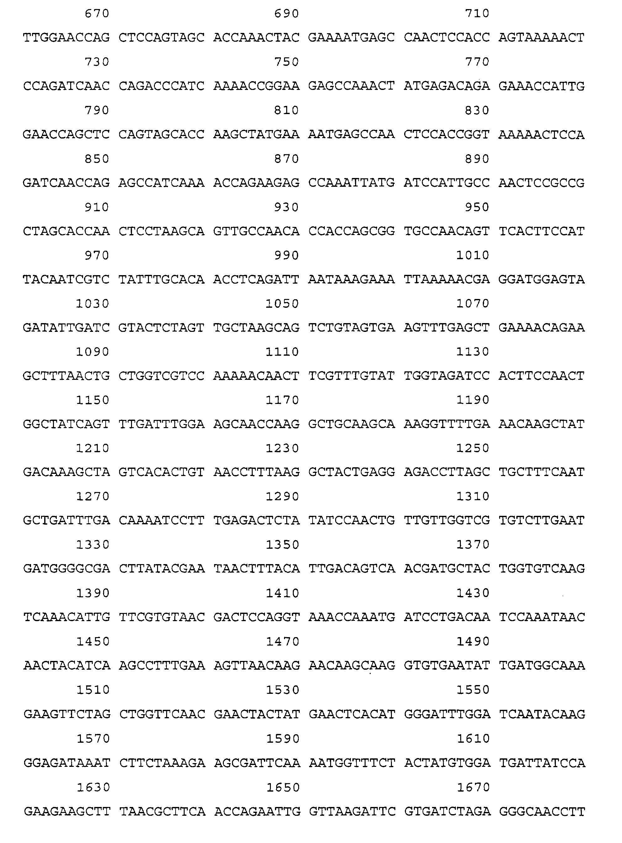
- A DNA sequence coding for a bacterial protein or an immunogenic fragment expressed during infection due to Streptococci having the nucleotide sequence of formula (2) (SEQ ID NO: 2):


- A DNA sequence according to claim 25, the protein for which it encodes being further characterised by either one or both of the following features:-(1) It is an immunodominant conserved antigen; and(2) It reacts with human antibody which protects against septicaemia infection in an animal model (mouse).
- A DNA sequence according to either one of claims 25 or 26 further characterised in that the protein for which it encodes is involved in binding to heart valves.
- An expression vector including the DNA sequence of any one of claims 25-27.
Applications Claiming Priority (3)
| Application Number | Priority Date | Filing Date | Title |
|---|---|---|---|
| GB9401689A GB9401689D0 (en) | 1994-01-28 | 1994-01-28 | Diagnosis and treatment of endocarditis |
| GB9401689 | 1994-01-28 | ||
| PCT/GB1995/000186 WO1995020658A2 (en) | 1994-01-28 | 1995-01-30 | Diagnosis and treatment of infections due to streptococci and enterococci |
Publications (2)
| Publication Number | Publication Date |
|---|---|
| EP0740703A1 EP0740703A1 (en) | 1996-11-06 |
| EP0740703B1 true EP0740703B1 (en) | 2001-08-01 |
Family
ID=10749513
Family Applications (1)
| Application Number | Title | Priority Date | Filing Date |
|---|---|---|---|
| EP95907070A Expired - Lifetime EP0740703B1 (en) | 1994-01-28 | 1995-01-30 | Diagnosis and treatment of infections due to streptococci and enterococci |
Country Status (10)
| Country | Link |
|---|---|
| US (1) | US5861157A (en) |
| EP (1) | EP0740703B1 (en) |
| JP (1) | JP3744937B2 (en) |
| AT (1) | ATE203768T1 (en) |
| AU (1) | AU702144B2 (en) |
| CA (1) | CA2181924C (en) |
| DE (1) | DE69521985T2 (en) |
| GB (1) | GB9401689D0 (en) |
| NZ (1) | NZ278922A (en) |
| WO (1) | WO1995020658A2 (en) |
Families Citing this family (7)
| Publication number | Priority date | Publication date | Assignee | Title |
|---|---|---|---|---|
| GB9614274D0 (en) | 1996-07-06 | 1996-09-04 | Univ Manchester | Treatment and diagnosis of infections of gram positive cocci |
| US6583275B1 (en) | 1997-07-02 | 2003-06-24 | Genome Therapeutics Corporation | Nucleic acid sequences and expression system relating to Enterococcus faecium for diagnostics and therapeutics |
| US6756361B1 (en) * | 1997-10-14 | 2004-06-29 | Nabi | Enterococcus antigens and vaccines |
| FR2791357B1 (en) * | 1999-03-26 | 2003-05-16 | Didier Raoult | WHIPPLE DISEASE DIAGNOSIS |
| FR2826581B1 (en) * | 2001-06-29 | 2005-10-07 | Oreal | USE OF LYS-PRO-VAL TRIPEPTIDE (KPV) IN THE TREATMENT OF MELANOMAS |
| EP2450053B1 (en) * | 2010-11-05 | 2016-06-15 | Universitätsklinikum Freiburg | Novel antigen of enterococcal pathogens and use thereof as vaccine component for therapy and/or prophylaxis |
| US11759460B2 (en) | 2020-08-14 | 2023-09-19 | Devie Medical Gmbh | Method of local antibiotic treatment of infective endocarditis |
Family Cites Families (4)
| Publication number | Priority date | Publication date | Assignee | Title |
|---|---|---|---|---|
| JPS6028999A (en) * | 1983-06-30 | 1985-02-14 | Maruho Kk | Protein having cell proliferation accelerating action, its composition and its preparation |
| SE8801894D0 (en) * | 1988-05-20 | 1988-05-20 | Alfa Laval Agri Int | FIBRONECT BINING PROTEIN |
| JP3095771B2 (en) * | 1988-12-29 | 2000-10-10 | バイオ―テクノロジー・ジエネラル・コーポレイシヨン | Cloning and production of human fibronectin polypeptide analogs and methods of using such polypeptide analogs |
| WO1994017411A1 (en) * | 1993-01-27 | 1994-08-04 | Duke University | Methods and agents for the diagnosis and treatment of rheumatoid arthritis |
-
1994
- 1994-01-28 GB GB9401689A patent/GB9401689D0/en active Pending
-
1995
- 1995-01-30 WO PCT/GB1995/000186 patent/WO1995020658A2/en not_active Ceased
- 1995-01-30 AT AT95907070T patent/ATE203768T1/en active
- 1995-01-30 EP EP95907070A patent/EP0740703B1/en not_active Expired - Lifetime
- 1995-01-30 NZ NZ278922A patent/NZ278922A/en not_active IP Right Cessation
- 1995-01-30 CA CA2181924A patent/CA2181924C/en not_active Expired - Fee Related
- 1995-01-30 AU AU15407/95A patent/AU702144B2/en not_active Ceased
- 1995-01-30 DE DE69521985T patent/DE69521985T2/en not_active Expired - Lifetime
- 1995-01-30 JP JP51995395A patent/JP3744937B2/en not_active Expired - Fee Related
-
1996
- 1996-07-29 US US08/687,956 patent/US5861157A/en not_active Expired - Lifetime
Also Published As
| Publication number | Publication date |
|---|---|
| US5861157A (en) | 1999-01-19 |
| JP3744937B2 (en) | 2006-02-15 |
| GB9401689D0 (en) | 1994-03-23 |
| EP0740703A1 (en) | 1996-11-06 |
| AU1540795A (en) | 1995-08-15 |
| WO1995020658A3 (en) | 1995-10-19 |
| DE69521985D1 (en) | 2001-09-06 |
| CA2181924C (en) | 2010-08-24 |
| WO1995020658A2 (en) | 1995-08-03 |
| ATE203768T1 (en) | 2001-08-15 |
| AU702144B2 (en) | 1999-02-11 |
| CA2181924A1 (en) | 1995-08-03 |
| DE69521985T2 (en) | 2001-11-15 |
| JPH09509569A (en) | 1997-09-30 |
| NZ278922A (en) | 1998-05-27 |
Similar Documents
| Publication | Publication Date | Title |
|---|---|---|
| JP4469026B2 (en) | Streptococcus pneumoniae antigens and vaccines | |
| US8318908B2 (en) | Nucleic acids coding for adhesion factor of group B streptococcus, adhesion factors of group B streptococcus and further uses thereof | |
| AU732520B2 (en) | Choline binding proteins for anti-pneumococcal vaccines | |
| DK175809B1 (en) | Fibronectin-binding protein | |
| US6784164B2 (en) | Choline binding proteins for anti-pneumococcal vaccines | |
| US7128919B2 (en) | Group B streptococcus polypeptides nucleic acids and therapeutic compositions and vaccines thereof | |
| WO1997041151A9 (en) | Choline binding proteins for anti-pneumococcal vaccines | |
| US6277381B1 (en) | Compounds and methods for the diagnosis and treatment of Ehrlichia infection | |
| US20130129737A1 (en) | Group B Streptococcus Polypeptides Nucleic Acids and Therapeutic Compositions and Vaccines Thereof | |
| AU702144B2 (en) | Diagnosis and treatment of infections due to streptococci and enterococci | |
| US20030096950A1 (en) | A polypeptide comprising the amino acid of an n-terminal choline binding protein a truncate, vaccine derived therefrom and uses thereof | |
| US6406883B1 (en) | Lmb gene of Streptococcus agalactiae | |
| EP1037997A1 (en) | Lmb gene of streptococcus agalactiae | |
| US5281694A (en) | Methods and compositions for production of mycoplasmal adhesins | |
| CA2326388A1 (en) | A polypeptide comprising the amino acid of an n-terminal choline binding protein a truncate, vaccine derived therefrom and uses thereof | |
| WO1998038312A1 (en) | Bacterial elastin binding protein, nucleic acid sequence encoding same and diagnostic and therapeutic methods of use thereof | |
| WO2001066571A1 (en) | Therapeutic and prophylactic agents derived from aeromonas hydrophila bacterial surface proteins | |
| JPH11239489A (en) | Dnag | |
| JP2002528112A (en) | protein | |
| JPH10290693A (en) | New fibronectin binding protein compounds |
Legal Events
| Date | Code | Title | Description |
|---|---|---|---|
| PUAI | Public reference made under article 153(3) epc to a published international application that has entered the european phase |
Free format text: ORIGINAL CODE: 0009012 |
|
| 17P | Request for examination filed |
Effective date: 19960819 |
|
| AK | Designated contracting states |
Kind code of ref document: A1 Designated state(s): AT BE CH DE DK ES FR GB GR IE IT LI LU MC NL PT SE |
|
| RAP1 | Party data changed (applicant data changed or rights of an application transferred) |
Owner name: NEUTEC PHARMA PLC |
|
| 17Q | First examination report despatched |
Effective date: 19980911 |
|
| GRAG | Despatch of communication of intention to grant |
Free format text: ORIGINAL CODE: EPIDOS AGRA |
|
| GRAG | Despatch of communication of intention to grant |
Free format text: ORIGINAL CODE: EPIDOS AGRA |
|
| GRAG | Despatch of communication of intention to grant |
Free format text: ORIGINAL CODE: EPIDOS AGRA |
|
| GRAH | Despatch of communication of intention to grant a patent |
Free format text: ORIGINAL CODE: EPIDOS IGRA |
|
| GRAH | Despatch of communication of intention to grant a patent |
Free format text: ORIGINAL CODE: EPIDOS IGRA |
|
| GRAA | (expected) grant |
Free format text: ORIGINAL CODE: 0009210 |
|
| AK | Designated contracting states |
Kind code of ref document: B1 Designated state(s): AT BE CH DE DK ES FR GB GR IE IT LI LU MC NL PT SE |
|
| PG25 | Lapsed in a contracting state [announced via postgrant information from national office to epo] |
Ref country code: NL Free format text: LAPSE BECAUSE OF FAILURE TO SUBMIT A TRANSLATION OF THE DESCRIPTION OR TO PAY THE FEE WITHIN THE PRESCRIBED TIME-LIMIT Effective date: 20010801 Ref country code: IT Free format text: LAPSE BECAUSE OF FAILURE TO SUBMIT A TRANSLATION OF THE DESCRIPTION OR TO PAY THE FEE WITHIN THE PRESCRIBED TIME-LIMIT;WARNING: LAPSES OF ITALIAN PATENTS WITH EFFECTIVE DATE BEFORE 2007 MAY HAVE OCCURRED AT ANY TIME BEFORE 2007. THE CORRECT EFFECTIVE DATE MAY BE DIFFERENT FROM THE ONE RECORDED. Effective date: 20010801 |
|
| REF | Corresponds to: |
Ref document number: 203768 Country of ref document: AT Date of ref document: 20010815 Kind code of ref document: T |
|
| REG | Reference to a national code |
Ref country code: CH Ref legal event code: EP |
|
| REG | Reference to a national code |
Ref country code: CH Ref legal event code: NV Representative=s name: ROTTMANN, ZIMMERMANN + PARTNER AG |
|
| REG | Reference to a national code |
Ref country code: IE Ref legal event code: FG4D |
|
| REF | Corresponds to: |
Ref document number: 69521985 Country of ref document: DE Date of ref document: 20010906 |
|
| PG25 | Lapsed in a contracting state [announced via postgrant information from national office to epo] |
Ref country code: SE Free format text: LAPSE BECAUSE OF FAILURE TO SUBMIT A TRANSLATION OF THE DESCRIPTION OR TO PAY THE FEE WITHIN THE PRESCRIBED TIME-LIMIT Effective date: 20011101 Ref country code: DK Free format text: LAPSE BECAUSE OF FAILURE TO SUBMIT A TRANSLATION OF THE DESCRIPTION OR TO PAY THE FEE WITHIN THE PRESCRIBED TIME-LIMIT Effective date: 20011101 |
|
| PG25 | Lapsed in a contracting state [announced via postgrant information from national office to epo] |
Ref country code: GR Free format text: LAPSE BECAUSE OF FAILURE TO SUBMIT A TRANSLATION OF THE DESCRIPTION OR TO PAY THE FEE WITHIN THE PRESCRIBED TIME-LIMIT Effective date: 20011102 |
|
| PG25 | Lapsed in a contracting state [announced via postgrant information from national office to epo] |
Ref country code: PT Free format text: LAPSE BECAUSE OF FAILURE TO SUBMIT A TRANSLATION OF THE DESCRIPTION OR TO PAY THE FEE WITHIN THE PRESCRIBED TIME-LIMIT Effective date: 20011105 |
|
| EN | Fr: translation not filed | ||
| REG | Reference to a national code |
Ref country code: GB Ref legal event code: IF02 |
|
| NLV1 | Nl: lapsed or annulled due to failure to fulfill the requirements of art. 29p and 29m of the patents act | ||
| PG25 | Lapsed in a contracting state [announced via postgrant information from national office to epo] |
Ref country code: ES Free format text: LAPSE BECAUSE OF FAILURE TO SUBMIT A TRANSLATION OF THE DESCRIPTION OR TO PAY THE FEE WITHIN THE PRESCRIBED TIME-LIMIT Effective date: 20020228 |
|
| EN | Fr: translation not filed |
Free format text: BO 01/52 PAGES: 283, IL Y A LIEU DE SUPPRIMER: LA MENTION DE LA NON REMISE. LA REMISE EST PUBLIEE DANS LE PRESENT BOPI. |
|
| ET | Fr: translation filed | ||
| PLBE | No opposition filed within time limit |
Free format text: ORIGINAL CODE: 0009261 |
|
| STAA | Information on the status of an ep patent application or granted ep patent |
Free format text: STATUS: NO OPPOSITION FILED WITHIN TIME LIMIT |
|
| 26N | No opposition filed | ||
| PG25 | Lapsed in a contracting state [announced via postgrant information from national office to epo] |
Ref country code: FR Free format text: LAPSE BECAUSE OF NON-PAYMENT OF DUE FEES Effective date: 20020930 |
|
| REG | Reference to a national code |
Ref country code: FR Ref legal event code: ST |
|
| REG | Reference to a national code |
Ref country code: FR Ref legal event code: RN |
|
| REG | Reference to a national code |
Ref country code: FR Ref legal event code: D3 |
|
| PGFP | Annual fee paid to national office [announced via postgrant information from national office to epo] |
Ref country code: MC Payment date: 20101230 Year of fee payment: 17 |
|
| PGFP | Annual fee paid to national office [announced via postgrant information from national office to epo] |
Ref country code: IE Payment date: 20110112 Year of fee payment: 17 |
|
| PGFP | Annual fee paid to national office [announced via postgrant information from national office to epo] |
Ref country code: DE Payment date: 20110126 Year of fee payment: 17 Ref country code: CH Payment date: 20110112 Year of fee payment: 17 Ref country code: AT Payment date: 20101222 Year of fee payment: 17 Ref country code: LU Payment date: 20110215 Year of fee payment: 17 Ref country code: FR Payment date: 20110128 Year of fee payment: 17 |
|
| PGFP | Annual fee paid to national office [announced via postgrant information from national office to epo] |
Ref country code: GB Payment date: 20110126 Year of fee payment: 17 Ref country code: BE Payment date: 20110225 Year of fee payment: 17 |
|
| REG | Reference to a national code |
Ref country code: CH Ref legal event code: PFA Owner name: NEUTEC PHARMA PLC Free format text: NEUTEC PHARMA PLC#ST. JAMES'S COURT, BROWN STREET#MANCHESTER M2 2JF (GB) -TRANSFER TO- NEUTEC PHARMA PLC#ST. JAMES'S COURT, BROWN STREET#MANCHESTER M2 2JF (GB) |
|
| BERE | Be: lapsed |
Owner name: *NEUTEC PHARMA P.L.C. Effective date: 20120131 |
|
| PG25 | Lapsed in a contracting state [announced via postgrant information from national office to epo] |
Ref country code: MC Free format text: LAPSE BECAUSE OF NON-PAYMENT OF DUE FEES Effective date: 20120131 |
|
| REG | Reference to a national code |
Ref country code: CH Ref legal event code: PL |
|
| GBPC | Gb: european patent ceased through non-payment of renewal fee |
Effective date: 20120130 |
|
| REG | Reference to a national code |
Ref country code: FR Ref legal event code: ST Effective date: 20120928 |
|
| REG | Reference to a national code |
Ref country code: IE Ref legal event code: MM4A |
|
| PG25 | Lapsed in a contracting state [announced via postgrant information from national office to epo] |
Ref country code: DE Free format text: LAPSE BECAUSE OF NON-PAYMENT OF DUE FEES Effective date: 20120801 Ref country code: GB Free format text: LAPSE BECAUSE OF NON-PAYMENT OF DUE FEES Effective date: 20120130 Ref country code: LI Free format text: LAPSE BECAUSE OF NON-PAYMENT OF DUE FEES Effective date: 20120131 Ref country code: CH Free format text: LAPSE BECAUSE OF NON-PAYMENT OF DUE FEES Effective date: 20120131 |
|
| REG | Reference to a national code |
Ref country code: DE Ref legal event code: R119 Ref document number: 69521985 Country of ref document: DE Effective date: 20120801 |
|
| REG | Reference to a national code |
Ref country code: AT Ref legal event code: MM01 Ref document number: 203768 Country of ref document: AT Kind code of ref document: T Effective date: 20120130 |
|
| PG25 | Lapsed in a contracting state [announced via postgrant information from national office to epo] |
Ref country code: BE Free format text: LAPSE BECAUSE OF NON-PAYMENT OF DUE FEES Effective date: 20120131 Ref country code: FR Free format text: LAPSE BECAUSE OF NON-PAYMENT OF DUE FEES Effective date: 20120131 |
|
| PG25 | Lapsed in a contracting state [announced via postgrant information from national office to epo] |
Ref country code: IE Free format text: LAPSE BECAUSE OF NON-PAYMENT OF DUE FEES Effective date: 20120130 Ref country code: AT Free format text: LAPSE BECAUSE OF NON-PAYMENT OF DUE FEES Effective date: 20120130 |
|
| PG25 | Lapsed in a contracting state [announced via postgrant information from national office to epo] |
Ref country code: LU Free format text: LAPSE BECAUSE OF NON-PAYMENT OF DUE FEES Effective date: 20120130 |