EP0729325B1 - Osteogenic product and use - Google Patents
Osteogenic product and use Download PDFInfo
- Publication number
- EP0729325B1 EP0729325B1 EP95902616A EP95902616A EP0729325B1 EP 0729325 B1 EP0729325 B1 EP 0729325B1 EP 95902616 A EP95902616 A EP 95902616A EP 95902616 A EP95902616 A EP 95902616A EP 0729325 B1 EP0729325 B1 EP 0729325B1
- Authority
- EP
- European Patent Office
- Prior art keywords
- bone
- product
- mole percent
- protein
- aragonite
- Prior art date
- Legal status (The legal status is an assumption and is not a legal conclusion. Google has not performed a legal analysis and makes no representation as to the accuracy of the status listed.)
- Expired - Lifetime
Links
Images


Classifications
-
- A—HUMAN NECESSITIES
- A61—MEDICAL OR VETERINARY SCIENCE; HYGIENE
- A61L—METHODS OR APPARATUS FOR STERILISING MATERIALS OR OBJECTS IN GENERAL; DISINFECTION, STERILISATION OR DEODORISATION OF AIR; CHEMICAL ASPECTS OF BANDAGES, DRESSINGS, ABSORBENT PADS OR SURGICAL ARTICLES; MATERIALS FOR BANDAGES, DRESSINGS, ABSORBENT PADS OR SURGICAL ARTICLES
- A61L27/00—Materials for grafts or prostheses or for coating grafts or prostheses
- A61L27/14—Macromolecular materials
- A61L27/22—Polypeptides or derivatives thereof, e.g. degradation products
- A61L27/227—Other specific proteins or polypeptides not covered by A61L27/222, A61L27/225 or A61L27/24
-
- A—HUMAN NECESSITIES
- A61—MEDICAL OR VETERINARY SCIENCE; HYGIENE
- A61F—FILTERS IMPLANTABLE INTO BLOOD VESSELS; PROSTHESES; DEVICES PROVIDING PATENCY TO, OR PREVENTING COLLAPSING OF, TUBULAR STRUCTURES OF THE BODY, e.g. STENTS; ORTHOPAEDIC, NURSING OR CONTRACEPTIVE DEVICES; FOMENTATION; TREATMENT OR PROTECTION OF EYES OR EARS; BANDAGES, DRESSINGS OR ABSORBENT PADS; FIRST-AID KITS
- A61F2/00—Filters implantable into blood vessels; Prostheses, i.e. artificial substitutes or replacements for parts of the body; Appliances for connecting them with the body; Devices providing patency to, or preventing collapsing of, tubular structures of the body, e.g. stents
- A61F2/02—Prostheses implantable into the body
- A61F2/28—Bones
-
- A—HUMAN NECESSITIES
- A61—MEDICAL OR VETERINARY SCIENCE; HYGIENE
- A61K—PREPARATIONS FOR MEDICAL, DENTAL OR TOILETRY PURPOSES
- A61K38/00—Medicinal preparations containing peptides
- A61K38/16—Peptides having more than 20 amino acids; Gastrins; Somatostatins; Melanotropins; Derivatives thereof
- A61K38/17—Peptides having more than 20 amino acids; Gastrins; Somatostatins; Melanotropins; Derivatives thereof from animals; from humans
- A61K38/18—Growth factors; Growth regulators
- A61K38/1875—Bone morphogenic factor; Osteogenins; Osteogenic factor; Bone-inducing factor
-
- A—HUMAN NECESSITIES
- A61—MEDICAL OR VETERINARY SCIENCE; HYGIENE
- A61L—METHODS OR APPARATUS FOR STERILISING MATERIALS OR OBJECTS IN GENERAL; DISINFECTION, STERILISATION OR DEODORISATION OF AIR; CHEMICAL ASPECTS OF BANDAGES, DRESSINGS, ABSORBENT PADS OR SURGICAL ARTICLES; MATERIALS FOR BANDAGES, DRESSINGS, ABSORBENT PADS OR SURGICAL ARTICLES
- A61L27/00—Materials for grafts or prostheses or for coating grafts or prostheses
- A61L27/02—Inorganic materials
- A61L27/025—Other specific inorganic materials not covered by A61L27/04 - A61L27/12
-
- A—HUMAN NECESSITIES
- A61—MEDICAL OR VETERINARY SCIENCE; HYGIENE
- A61P—SPECIFIC THERAPEUTIC ACTIVITY OF CHEMICAL COMPOUNDS OR MEDICINAL PREPARATIONS
- A61P1/00—Drugs for disorders of the alimentary tract or the digestive system
- A61P1/02—Stomatological preparations, e.g. drugs for caries, aphtae, periodontitis
-
- A—HUMAN NECESSITIES
- A61—MEDICAL OR VETERINARY SCIENCE; HYGIENE
- A61P—SPECIFIC THERAPEUTIC ACTIVITY OF CHEMICAL COMPOUNDS OR MEDICINAL PREPARATIONS
- A61P19/00—Drugs for skeletal disorders
- A61P19/08—Drugs for skeletal disorders for bone diseases, e.g. rachitism, Paget's disease
- A61P19/10—Drugs for skeletal disorders for bone diseases, e.g. rachitism, Paget's disease for osteoporosis
-
- A—HUMAN NECESSITIES
- A61—MEDICAL OR VETERINARY SCIENCE; HYGIENE
- A61P—SPECIFIC THERAPEUTIC ACTIVITY OF CHEMICAL COMPOUNDS OR MEDICINAL PREPARATIONS
- A61P43/00—Drugs for specific purposes, not provided for in groups A61P1/00-A61P41/00
-
- C—CHEMISTRY; METALLURGY
- C07—ORGANIC CHEMISTRY
- C07K—PEPTIDES
- C07K14/00—Peptides having more than 20 amino acids; Gastrins; Somatostatins; Melanotropins; Derivatives thereof
- C07K14/435—Peptides having more than 20 amino acids; Gastrins; Somatostatins; Melanotropins; Derivatives thereof from animals; from humans
- C07K14/475—Growth factors; Growth regulators
- C07K14/51—Bone morphogenetic factor; Osteogenins; Osteogenic factor; Bone-inducing factor
-
- A—HUMAN NECESSITIES
- A61—MEDICAL OR VETERINARY SCIENCE; HYGIENE
- A61F—FILTERS IMPLANTABLE INTO BLOOD VESSELS; PROSTHESES; DEVICES PROVIDING PATENCY TO, OR PREVENTING COLLAPSING OF, TUBULAR STRUCTURES OF THE BODY, e.g. STENTS; ORTHOPAEDIC, NURSING OR CONTRACEPTIVE DEVICES; FOMENTATION; TREATMENT OR PROTECTION OF EYES OR EARS; BANDAGES, DRESSINGS OR ABSORBENT PADS; FIRST-AID KITS
- A61F2/00—Filters implantable into blood vessels; Prostheses, i.e. artificial substitutes or replacements for parts of the body; Appliances for connecting them with the body; Devices providing patency to, or preventing collapsing of, tubular structures of the body, e.g. stents
- A61F2/02—Prostheses implantable into the body
- A61F2/30—Joints
- A61F2/32—Joints for the hip
-
- A—HUMAN NECESSITIES
- A61—MEDICAL OR VETERINARY SCIENCE; HYGIENE
- A61F—FILTERS IMPLANTABLE INTO BLOOD VESSELS; PROSTHESES; DEVICES PROVIDING PATENCY TO, OR PREVENTING COLLAPSING OF, TUBULAR STRUCTURES OF THE BODY, e.g. STENTS; ORTHOPAEDIC, NURSING OR CONTRACEPTIVE DEVICES; FOMENTATION; TREATMENT OR PROTECTION OF EYES OR EARS; BANDAGES, DRESSINGS OR ABSORBENT PADS; FIRST-AID KITS
- A61F2/00—Filters implantable into blood vessels; Prostheses, i.e. artificial substitutes or replacements for parts of the body; Appliances for connecting them with the body; Devices providing patency to, or preventing collapsing of, tubular structures of the body, e.g. stents
- A61F2/02—Prostheses implantable into the body
- A61F2/30—Joints
- A61F2/38—Joints for elbows or knees
-
- A—HUMAN NECESSITIES
- A61—MEDICAL OR VETERINARY SCIENCE; HYGIENE
- A61F—FILTERS IMPLANTABLE INTO BLOOD VESSELS; PROSTHESES; DEVICES PROVIDING PATENCY TO, OR PREVENTING COLLAPSING OF, TUBULAR STRUCTURES OF THE BODY, e.g. STENTS; ORTHOPAEDIC, NURSING OR CONTRACEPTIVE DEVICES; FOMENTATION; TREATMENT OR PROTECTION OF EYES OR EARS; BANDAGES, DRESSINGS OR ABSORBENT PADS; FIRST-AID KITS
- A61F2/00—Filters implantable into blood vessels; Prostheses, i.e. artificial substitutes or replacements for parts of the body; Appliances for connecting them with the body; Devices providing patency to, or preventing collapsing of, tubular structures of the body, e.g. stents
- A61F2/02—Prostheses implantable into the body
- A61F2/30—Joints
- A61F2/44—Joints for the spine, e.g. vertebrae, spinal discs
- A61F2/4455—Joints for the spine, e.g. vertebrae, spinal discs for the fusion of spinal bodies, e.g. intervertebral fusion of adjacent spinal bodies, e.g. fusion cages
-
- A—HUMAN NECESSITIES
- A61—MEDICAL OR VETERINARY SCIENCE; HYGIENE
- A61F—FILTERS IMPLANTABLE INTO BLOOD VESSELS; PROSTHESES; DEVICES PROVIDING PATENCY TO, OR PREVENTING COLLAPSING OF, TUBULAR STRUCTURES OF THE BODY, e.g. STENTS; ORTHOPAEDIC, NURSING OR CONTRACEPTIVE DEVICES; FOMENTATION; TREATMENT OR PROTECTION OF EYES OR EARS; BANDAGES, DRESSINGS OR ABSORBENT PADS; FIRST-AID KITS
- A61F2/00—Filters implantable into blood vessels; Prostheses, i.e. artificial substitutes or replacements for parts of the body; Appliances for connecting them with the body; Devices providing patency to, or preventing collapsing of, tubular structures of the body, e.g. stents
- A61F2/02—Prostheses implantable into the body
- A61F2/30—Joints
- A61F2002/30001—Additional features of subject-matter classified in A61F2/28, A61F2/30 and subgroups thereof
- A61F2002/30003—Material related properties of the prosthesis or of a coating on the prosthesis
- A61F2002/30004—Material related properties of the prosthesis or of a coating on the prosthesis the prosthesis being made from materials having different values of a given property at different locations within the same prosthesis
- A61F2002/30011—Material related properties of the prosthesis or of a coating on the prosthesis the prosthesis being made from materials having different values of a given property at different locations within the same prosthesis differing in porosity
-
- A—HUMAN NECESSITIES
- A61—MEDICAL OR VETERINARY SCIENCE; HYGIENE
- A61F—FILTERS IMPLANTABLE INTO BLOOD VESSELS; PROSTHESES; DEVICES PROVIDING PATENCY TO, OR PREVENTING COLLAPSING OF, TUBULAR STRUCTURES OF THE BODY, e.g. STENTS; ORTHOPAEDIC, NURSING OR CONTRACEPTIVE DEVICES; FOMENTATION; TREATMENT OR PROTECTION OF EYES OR EARS; BANDAGES, DRESSINGS OR ABSORBENT PADS; FIRST-AID KITS
- A61F2/00—Filters implantable into blood vessels; Prostheses, i.e. artificial substitutes or replacements for parts of the body; Appliances for connecting them with the body; Devices providing patency to, or preventing collapsing of, tubular structures of the body, e.g. stents
- A61F2/02—Prostheses implantable into the body
- A61F2/30—Joints
- A61F2002/30001—Additional features of subject-matter classified in A61F2/28, A61F2/30 and subgroups thereof
- A61F2002/30003—Material related properties of the prosthesis or of a coating on the prosthesis
- A61F2002/3006—Properties of materials and coating materials
- A61F2002/30062—(bio)absorbable, biodegradable, bioerodable, (bio)resorbable, resorptive
-
- A—HUMAN NECESSITIES
- A61—MEDICAL OR VETERINARY SCIENCE; HYGIENE
- A61F—FILTERS IMPLANTABLE INTO BLOOD VESSELS; PROSTHESES; DEVICES PROVIDING PATENCY TO, OR PREVENTING COLLAPSING OF, TUBULAR STRUCTURES OF THE BODY, e.g. STENTS; ORTHOPAEDIC, NURSING OR CONTRACEPTIVE DEVICES; FOMENTATION; TREATMENT OR PROTECTION OF EYES OR EARS; BANDAGES, DRESSINGS OR ABSORBENT PADS; FIRST-AID KITS
- A61F2210/00—Particular material properties of prostheses classified in groups A61F2/00 - A61F2/26 or A61F2/82 or A61F9/00 or A61F11/00 or subgroups thereof
- A61F2210/0004—Particular material properties of prostheses classified in groups A61F2/00 - A61F2/26 or A61F2/82 or A61F9/00 or A61F11/00 or subgroups thereof bioabsorbable
-
- A—HUMAN NECESSITIES
- A61—MEDICAL OR VETERINARY SCIENCE; HYGIENE
- A61F—FILTERS IMPLANTABLE INTO BLOOD VESSELS; PROSTHESES; DEVICES PROVIDING PATENCY TO, OR PREVENTING COLLAPSING OF, TUBULAR STRUCTURES OF THE BODY, e.g. STENTS; ORTHOPAEDIC, NURSING OR CONTRACEPTIVE DEVICES; FOMENTATION; TREATMENT OR PROTECTION OF EYES OR EARS; BANDAGES, DRESSINGS OR ABSORBENT PADS; FIRST-AID KITS
- A61F2250/00—Special features of prostheses classified in groups A61F2/00 - A61F2/26 or A61F2/82 or A61F9/00 or A61F11/00 or subgroups thereof
- A61F2250/0014—Special features of prostheses classified in groups A61F2/00 - A61F2/26 or A61F2/82 or A61F9/00 or A61F11/00 or subgroups thereof having different values of a given property or geometrical feature, e.g. mechanical property or material property, at different locations within the same prosthesis
- A61F2250/0023—Special features of prostheses classified in groups A61F2/00 - A61F2/26 or A61F2/82 or A61F9/00 or A61F11/00 or subgroups thereof having different values of a given property or geometrical feature, e.g. mechanical property or material property, at different locations within the same prosthesis differing in porosity
Definitions
- a number of diseases or injuries involving bones are known for which regeneration of pone is a desired treatment.
- Formation of bone in vivo involves an interaction of various inductive proteins and growth factors which act by causing a differentiation of mesenchymal cells into cartilage and then bone-forming cell lines. This mechanism is not completely understood.
- purified protein mixtures or recombinantly produced proteins have been developed which stimulate osteoinductive activity.
- the present invention is directed toward an osteogenic product, comprising aragonite and a bone growth factor.
- the product can also include a material selected from the group consisting of collagen, fibrin, alginate and mixtures thereof.
- the bone growth factor is present at about 10 micrograms bone growth factor/g aragonite and about 1000 micrograms bone growth factor/g aragonite.
- the bone growth factor can be selected from the group consisting of purified bone growth factors, recombinantly produced bone growth factors and mixtures thereof.
- the bone growth factor comprises an amino acid composition of from about 20.7 to about 26.1 mole percent acidic amino acids, about 11.3 to about 15.7 mole percent hydroxy amino acids, about 37.6 to about 42.4 mole percent aliphatic amino acids, about 5.8 to about 7.9 mole percent aromatic amino acids and about 13.3 to about 19.9 mole percent basic amino acids.
- the bone growth factor can also comprise, upon hydrolysis, an amino acid composition of from about 20.7 to about 26.1 mole percent ASP(+ASN) and GLU(+GLN); from about 11.3 to about 15.7 mole percent SER and THR; from about 37.6 to about 42.4 mole percent ALA, GLY, PRO, MET, VAL, ILE, and LEU; from about 5.8 to about 7.9 mole percent TYR and PHE; and from about 13.3 to about 19.9 mole percent HIS, ARG, and LYS, based on the total moles of said amino acids.
- an amino acid composition of from about 20.7 to about 26.1 mole percent ASP(+ASN) and GLU(+GLN); from about 11.3 to about 15.7 mole percent SER and THR; from about 37.6 to about 42.4 mole percent ALA, GLY, PRO, MET, VAL, ILE, and LEU; from about 5.8 to about 7.9 mole percent TYR and PHE; and from about 13.3 to about
- Also described herein is a process for the induction of bone formation, comprising implanting the product of the present invention in a body.
- the process is selected from the group consisting of hip replacement operation, knee replacement operation, spinal fusion, repair of periodontal defects, treatment of osteoporosis, repair of bone defects and repair of bone fractures.
- the present invention is directed toward an osteogenic product which includes calcium carbonate in the form of aragonite and bone growth factor.
- the osteogenic product is particularly useful in processes which include implanting the product in the body for the purpose of inducing formation of bone.
- the aragonite component of the present invention is present in a crystalline form.
- the aragonite has two important functions in products and processes of the present invention. When implanted in a body with bone growth factor and any optional components of the present product, the aragonite functions as an osteoconductive element. Thus, the aragonite acts as a conductive substrate for bone formation.
- Aragonite in the form of natural coral, contains a network of macro- and micropores which allow bone formation through the coral.
- the interconnected pores of the coral allows bone precursor cells and vessels to invade the coral and implant and provide a large surface area for bone apposition. For example, these pores allow macrophages and osteoclasts or osteoclast-like cells to have access to the interior portion of the coral while increasing the volume available for new bone.
- the aragonite is resorbable. Over time, coral in the form of aragonite is readily resorbed by the body. The product of the present invention, therefore, allows for formation of bone throughout a defect area without significant residual unresorbed substrate.
- conventional ceramic materials like hydroxyapatite which have been used as bone growth substrates have the potential of remaining in the bone matrix and being more brittle than bone.
- aragonite is believed to have additional unexpected benefits associated with osteoinduction. As discussed below in more detail in the Example section, the presence of aragonite with bone growth factor in an alginate matrix resulted in bone formation. In the absence of aragonite, however, no bone was formed in the alginate matrix with bone growth factor. Thus, it appears that aragonite has a role in bone formation under some circumstances.
- the argonite is preferably obtained from the skeletons of coral.
- Such corals can include, but are not limited to, Acropora , Goniopora, Lobophylla, Porites and mixtures thereof.
- the aragonite obtained from these corals are in the crystalline form of aragonite, but have different porosities.
- Acropora is about 20% porous and Porites is about 50% porous.
- the use of aragonite having porosities from 0% to more than 50% is within the scope of the present invention.
- Suitable aragonite of the present invention can be produced by recovering naturally occurring coral, cleaning the coral and sterilizing it. The resulting product can then be examined for fractures, such as by X-ray examination. Sterilization can be accomplished by, for example, subjecting the material to gamma radiation.
- a preferred argonite material of the present invention is the material sold under the trademark BIOCORAL by Inoteb of St. Gonnery, France.
- Argonite of the present invention can be used in various forms, including as a block and as particulates.
- the argonite such as coral
- the unitary article is then used, for example, by being placed in a bone defect or as discussed below in more detail, used in a spinal fusion procedure to fuse two vertebrae.
- the argonite can be in particulate form.
- Particulates can be produced by, for example, grinding coral to a desired size. Such particulates are typically between about 80 microns and about 3 millimeters, more preferably between about 125 microns and about 2 mm, and even more preferably between about 150 microns and about 1 mm.
- particulate forms of argonite are typically used with a binder, such as collagen, to form a composite which is used to fill a bone defect for induction of bone formation.
- the pore diameter of argonite is large enough such that cells, such as osteoblasts and macrophages, can infiltrate the pores and to allow for capillary formation in the pores.
- the pore diameter in argonite is typically between about 100 microns and about 500 microns, and more preferably between about 150 microns and about 350 microns.
- the bone growth factor of the present invention is a protein or mixture of proteins which are capable of inducing bone formation when implanted in a body. It should be noted that while most contemplated applications of the present invention are concerned with use in humans, the products and processes of the present invention work in animals as well. Induction of bone formation can be determined by a histological evaluation showing the de novo formation of bone with accompanying osteoblasts, osteoclasts, and osteoid matrix. For example, osteoinductive activity of a bone growth factor can be demonstrated by a test using a substrate onto which material to be tested is deposited. A substrate with deposited material is implanted subcutaneously in a test animal. The implant is subsequently removed and examined microscopically for the presence of bone formation including the presence of osteoblasts, osteoclasts, and osteoid matrix. A suitable procedure is illustrated in Example 5 of U.S. Patent No. 5,290,763.
- Suitable bone growth factor of the present invention can be produced by purification of naturally occurring proteins from bone or by recombinant DNA techniques.
- the term recombinantly produced bone growth factors refers to the production of bone growth factor using recombinant DNA technology.
- nucleic acids encoding proteins having osteogenic activity can be identified by producing antibodies that bind to the proteins.
- the antibodies can be used to isolate, by affinity chromatography, purified populations of a particular osteogenic protein.
- the amino acid sequence can be identified by sequencing the purified protein. It is possible to synthesize DNA oligonucleotides from the known amino acid sequence.
- the oligonucleotides can be used to screen either a genomic DNA and/or cDNA library made from, for example bovine DNA, to identify nucleic acids encoding the osteogenic protein.
- the correct oligonucleotide will hybridize to the appropriate cDNA thereby identifying the cDNA encoding the osteogenic protein encoding gene.
- the antibodies that bind osteogenic proteins can also be used directly to screen a cDNA expression library.
- eukaryotic cDNA sequences encoding osteogenic proteins can be ligated into bacterial expression vectors.
- the expression vectors can be transformed into bacteria, such as E. coli, which express the transformed expression vector and produce the osteogenic protein.
- the transformed bacteria can be screened for expression of the osteogenic protein by lysing the bacteria and contacting the bacteria with radioactively-labelled antibody.
- Recombinant bone growth factor can be produced by transforming genes identified according to the method described above into cells using any process by which nucleic acids are inserted into cells. After transformation, the cell can produce recombinant bone growth factor by expression of the transformed nucleic acids and such bone growth factor can be recovered from the cells.
- Bone Morphogenic Proteins 1-8 or BMP 1-8 which are described in U.S. Patent 5,106,748.
- Purified bone growth factors have been developed by several entities. Collagen Corporation of Palo Alto, California, USA developed a purified protein mixture which is believed to have osteogenic activity and which is described in U.S. Patent Nos.
- a preferred bone growth factor of the present invention and process for making the same is described in detail in related U.S. application serial no. 07/689,459.
- This bone growth factor is particularly preferred because of its high osteogenic activity and because it is a purified bone growth factor.
- the bone growth factor of U.S. Patent No. 5,290,763 exhibits osteoinductive activity at about 3 micrograms when deposited onto a suitable carrier and implanted subcutaneously.
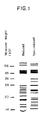
- the bone growth factor is an osteoinductively active mixture of proteins which exhibit the gel separation profile shown in Figure 1. This gel separation profile was obtained using SDS-PAGE.
- the first column is a molecular weight scale which was obtained by performing SDS-PAGE on standards of known molecular weight.
- the second column illustrates the SDS-PAGE profile for a mixture of proteins in accordance with the present invention which have been reduced with 2-mercaptoethanol.
- the third column illustrates the SDS-PAGE profile for a non-reduced mixture of proteins in accordance with the present invention.
- effective protein mixtures can include proteins that differ in molecular weight by plus or minus 5 KD from those shown in Figure 1, and can include fewer or greater numbers of proteins than those shown in Figure 1. Therefore, mixtures of proteins having profiles which comprise substantially all of the protein bands detected in the reduced or nonreduced SDS-PAGE profiles in Figure 1 will be considered to be within the scope of the invention.
- Yet another embodiment of the preferred bone growth factor of the invention includes an osteoinductively active mixture of proteins having, upon hydrolysis, an amino acid composition of from about 20.7 to about 26.1 mole percent acidic amino acids, about 11.3 to about 15.7 mole percent hydroxy amino acids, about 37.6 to about 42.4 mole percent aliphatic amino acids, about 5.8 to about 7.9 mole percent aromatic amino acids and about 13.3 to about 19.9 mole percent basic amino acids.
- the preferred bone growth factor has an amino acid composition of about 20.7 to about 26.1 (preferably about 23.4) mole percent of ASP (+ASN) a GLU(+GLN); about 11.3 to about 15.7 (preferably about 13.5) mol percent SER and THR; about 37.6 to about 42.4 (preferably about 40.0) mole percent ALA, GLY, PRO, VAL, MET, ILE, and LEU; about 5.8 to about 7.9 (preferably about 6.8) mole percent TYR and PHE; and about 13.3 to about 19.9 (preferably about 16.6) mole percent HIS, ARG, and LYS.
- a further embodiment of the preferred bone growth factor is a protein mixture having the approximate amino acid composition shown in Table 1.
- a still further embodiment of the preferred bone growth factor is a protein mixture obtained by any of the purification processes described in U. S. Patent No. 5,290,763.
- a bone growth factor of the present invention is combined with the argonite in various ways when used in the present invention.
- the bone growth factor is typically put into solution and is then applied to the block.
- the bone growth factor containing solution then soaks into the porous structure of the block.
- the solution is then dried, such as by rapid freezing followed by lyophilization, thereby leaving a deposit of the bone growth factor on the block.
- the argonite particulates can be put into a matrix, such as a collagen, fibrin or alginate dispersion, to form a composite which is then dried.
- a bone growth factor containing solution is then applied to the dried composite and allowed to soak in.
- bone growth factor can be mixed into a dispersion which is then mixed with particulate argonite and dried. Specific examples of particulate form products are described in the Example section.
- the amount or dose of bone growth factor used depends on the activity of the bone growth factor and the particular application. In the case of the bone growth factor identified in U.S. application serial no. 07/689,459, the bone growth factor is used in amounts between about 10 micrograms/gram argonite and about 10,000 micrograms/g argonite and more preferably between about 100 micrograms/g argonite and about 350 micrograms/g argonite.
- Products of the present invention can optionally include components in addition to argonite and bone growth factor.
- a product can include a matrix forming material, such as collagen, fibrin or alginate.
- Preferred collagen is Type I bovine tendon atelocollagen.
- a suitable alginate product is identified in Example 3.
- Additional optional components suitable for the present product include other growth factors, such as basic fibroblast growth factor (bFGF) and transforming growth factor beta (TGF-beta) (See Cuevas et al. , Basic Fibroblast Growth Factor (FGF) Promotes Cartilage Repair In Vivo, Biochem Biophys Res Commun 156:611-618, 1988). These growth factors have been implicated as cartilage stimulating and angiogenic agents. bFGF, for example, has been shown to increase the rate of osteoblast replication while simultaneously inhibiting their activity ( Frenkel S, Singh IJ ; The effects of f ibroblast growth factor on osteogenesis in the chick embryo. In: Fundamentals of bone growth: Methodology and applications. Ed. AD Dixon, BG Sarnat, D.
- the other growth factors described above can be incorporated into products in the same manner as bone growth factors. That is, they can be put into solution and applied to a block of argonite or a composite of particulate argonite.
- the present invention includes the claim 10. As noted above, most uses of the present invention are concerned with human applications. The use, however, is suitable for a wide variety of animals, particularly including other mammals.
- the term implanted refers to placing the product of the present invention in any bone defect or other area in which it is desired to have bone grow. By implanting a product, bone formation is induced by the bone growth factor and the argonite functions as an osteoconductive agent. Over time, argonite materials are resorbed allowing for uniform bone formation throughout a defect area.
- the present use can be applied in a variety of applications whenever there is a need to generate bone.
- Such applications include induction of bone formation for hip replacement operations, knee replacement operations, spinal fusion procedures, repair of periodontal defects, treatment of osteoporosis, repair of bone tumor defects and repair of bone fractures.
- the ball and socket joint of a hip is replaced when a person's hip is not functioning properly.
- the ball portion of a joint is replaced by surgical removal of the ball portion from the terminus of the femur.
- the artificial ball portion has a functional ball end with the opposite end being a spike which is inserted into the proximal end of the femur from which the natural ball portion was removed.
- the spike can have a porous surface so that bone growth around the spike can anchor the spike in the femur.
- the product of the present invention in particulate form, is layered or packed between the spike and the cavity in the femur in which spike is to be inserted.
- the socket portion of a joint is replaced by inserting an artificial socket into the natural socket.
- the artificial socket is sized to fit with the artificial ball.
- the artificial socket On the surface of the artificial socket which contacts the natural socket, the artificial socket can have a porous surface.
- the product of the present invention in particulate form, is placed in the natural socket cavity so that upon placement of the artificial socket, the product is between the natural and artificial socket. In this manner, as bone is formed, the artificial socket is anchored in the natural socket.
- aragonite of varying porosities can be used.
- Argonite from the coral Porites is preferred for such applications.
- Knee prostheses have a femoral and a tibial component which are inserted into the distal end of the femur and the surgically prepared end of the tibia, respectively.
- the product of the present invention in particulate form, is layered or packed between the femoral and/or tibial components of the prosthesis and the respective portions of the femur and tibia. In this manner, as bone formation is induced between the prosthesis and the bones, the prosthesis becomes anchored.
- Products of the present invention are also suitable for use in spinal fusion operations in which it is desired to substantially immobilize two vertebrae with respect to each other.
- the product is in particulate composite form.
- the composite can be applied, for example, between adjacent spinous and transverse processes so that upon bone formation throughout the composite material, two adjacent vertebrae are joined by fusion between the respective spinous processes and transverse processes.
- block forms of argonite can be used.
- two vertebrae can be fused by positioning one or more blocks of argonite with bone growth protein between opposing surfaces of the body portion of two adjacent vertebrae in the approximate position of the original disk. The two vertebrae become fused as bone develops between each vertebrae and each side of the block.
- argonite When used in spinal fusion procedures and particularly in the latter described procedure in the lower spine, relatively dense argonite is used. Typically, argonite which is less than about 35% porous, more preferably less than about 25% porous and even more preferably less than about 10% porous is suitable. For example, argonite derived from the coral Acropora is preferred for such procedures.
- the product of the present invention is typically in particulate or composite form and sill conform to the defect shape. As bone growth is induced and the argonite is resorbed, bone fills in the defect.
- particulate or composite forms of the present product are injected in existing bone to offset the effects of osteoporosis in which bone density is lost. For example, if it is determined that bone density is low in a localized area, such an injection can be made in that area.
- a product comprising natural coral and bone growth factor was tested for the ability of the coral to act as a resorbable osteoconductive carrier matrix for the bone growth factor in subcutaneous sites in rats.
- the natural coral implants used comprised the species Porites sp. having an interconnected pore volume of 49 ⁇ 2 percent and a mean pore diameter of 150 ⁇ m.
- Coral particles, measuring 630-710 micrometer ( ⁇ m) (Inoteb, St. Gonnery, France) were used in this experiment.
- Bone growth factor was isolated from the cortical diaphyses of bovine long bones. The marrow and soft tissue was cleaned from the long bones, and the bones were pulverized and demineralized in 1.0 normal (N) hydrochloric acid at a 1:13 weight to volume ratio for 16 hours at 25°C. The bone particles were washed in distilled water and then extracted in a buffered solution comprising of 4 N guanidine hydrochloride buffered with 0.1 N Tris, pH 7.6 at a concentration of 3 milliliters of buffered solution per gram of original powdered bone. The bone was extracted for 48 h at 15°C. The extracted bone particles were then passed through a series of chromatographic purification steps as described in U.S. application serial no. 07/689,459 to extract bone growth factor having bone inductive effect at doses less than 35 microgram ( ⁇ g). The bone growth factor was added to the bovine-derived collagen described above and lyophilized.
- Two sets of discs were used for rat subcutaneous implantation assays in rats.
- One set of discs comprised coral particles combined with a 1 percent dispersion of bovine-derived atelo Type I collagen (American Biomaterials Corp, Plainsboro, NJ).
- the second set of discs comprised 350-375 ⁇ g of the bone growth factor and collagen mixture described above reconstituted in sterile water added to coral particles.
- Coral particles combined with collagen with or without bone growth factor were molded into discs, 8 mm in diameter and 3 mm thick, and lyophilized. The formulation was adequate to prepare about 10 discs.
- Discs containing purified basic fibroblast growth factor (bFGF) for were also prepared for implantation.
- bFGF was added to coral particle discs with or without bone growth factor added.
- 15 nanogram of bFGF (IMEDEX, Chaponost, France) per milligram of coral particle disc was pipetted onto a disc and allowed to soak into the disc.
- the rats were sacrificed using a lethal intraperitoneal injection of Dolethal. Samples were removed, examined grossly, fixed in 40 percent ethanol, dehydrated in increasing concentrations of ethanol, and infiltrated and embedded in polymethylmethacrylate (PMMA). Samples were cut on a low-speed diamond saw, glued to Plexiglas® slides, ground and polished to 40-60 ⁇ m and stained with a combination of Stevenel's blue and Van Gieson picro-fuchsin.
- PMMA polymethylmethacrylate
- CC and CC:bFGF implants were used as negative controls.
- the CC and CC:bFGF samples were infiltrated with fibrous tissue. The remains of the collagen composite were clearly visible at the center of the implant, sometimes undergoing dystrophic mineralization. The coral particulate showed little signs of resorption, especially those particles still surrounded by collagen in the center of the implant. There was no difference in the results of the CC and CC:bFGF implants.
- the CC:BGF and CC:BGF:bFGF samples removed at four weeks were mineralized except in one sample where small amounts of cartilage were still evident.
- mineralizing cartilage and bone formation via osteoblasts were detected in the samples.
- Thin layers of osteoblast-derived bone were seen at the outer boundary of the ossicle.
- Layers of woven bone and mineralized cartilage were detected toward the central areas. Between these two layers there was abundant hematopoietic marrow.
- the coral particles Toward the center of the explant, the coral particles remained fairly large and surrounded by fibrous tissue and the remains of the collagen composite. Further out, the coral could be seen surrounded by bone or mineralizing cartilage and appeared to be in the process of resorption indicated by the particles being smaller in size.
- the CC and CC:bFGF samples implants continued to demonstrate resorption of the coral by macrophages and multinucleated cells when removed after eight weeks of implantation. No bone formation or cartilage, however, was detected.
- the CC:BGF and CC:BGF:bFGF samples continued to demonstrate various levels of mature bone formation after eight weeks of implantation.
- the collagen matrix at the center of the ossicle had been completely replaced by trabecular bone and hematopoietic marrow.
- very little coral was left in these explants and the coral particles that remained were small and surrounded by new bone.
- two separate layers of bone formation with intervening hematopoietic marrow were present, indicating less mature bone formation. Unresorbed coral particles in the center and resorbing smaller particles toward the outer edge were also detected.
- results from the histological staining indicate implants comprising coral particulate are osteoconductive and resorbable.
- the results also indicate that bone growth factor, in the presence or absence of bFGF, induces bone formation.
- bFGF does not contribute to bone formation.
- a semi-quantitative scoring system was used to analyze and compare the amount and maturity of bone formation.
- Table 3 summarizes histological scores and their identifying characteristics used to analyze the samples. Scores of 1-4 indicate evidence of chondrogenesis. Scores of 5-8 indicate absence of cartilage. A higher score indicates a more mature ossicle.
- FIG. 3 summarizes the relative maturity over time of the explants depending on the material implanted. The results indicate that implants containing bone growth factor elicited greater chondroinductive and osteoinductive activity than samples without bone growth factor. The maturity of the ossicle of samples containing bone growth factor increased with increasing implantation time. This was also seen qualitatively by an increasing paucity of cartilage and thickening of the outer bony layer. The results also indicate that samples containing both bone growth factor and bFGF showed a higher average statistical increase in maturity than samples with bone growth factor alone (p ⁇ 0.05). The increase, however, was not detected in the four and eight week explants even though the average increase was higher.
- the semi-quantitative scoring results indicate that the maturity of ossicles was statistically higher in samples containing bone growth factor compared with samples not containing bone growth factor.
- This example demonstrates the effectiveness of an argonite and bone growth factor product to induce bone replacement in defective ulnar bones in rabbits.
- Example 1 The bone formation and coral resorption by CC and CC:BGF composites described in Example 1 were tested in a segmental bony defect model in rabbits. Segments were removed from the ulnar of the forearm of 40 rabbits. Implants having the same shape as the removed segment of ulnar bone, typically about 5 millimeters in diameter and 17 millimeters in length, were fabricated using CC and CC:BGF composites. The implants were surgically placed in the segmental defect in the ulnar as a single piece. Explants were analyzed at 2, 4 and 8 weeks using the histological staining methods described in Example 1.
- the CC explants showed substantially less bone formation and resorption than the CC:BGF explants at all time points.
- the CC:BGF composite provides both osteoinductive and osteoconductive properties.
- the coral is resorbed at a rate similar to that of new forming bone.
- This example compares the effectiveness in bone formation of bone growth protein in an alginate matrix with that of bone growth protein and argonite in an alginate matrix.
- Products were prepared by forming discs from either (1) a 4% aqueous dispersion of alginic acid [Sigma A-7128] sodium salt from the kelp Macrocystis pyrifera (2% solution) or (2) a 4% aqueous dispersion of alginic acid with coral (particulate Porites 300-450 microns with 50% porosity from Inoteb of St. Gonnery, France) at a 1:1 weight ratio.
- a dose of 30-35 micrograms of bone growth factor prepared in accordance with Examples 1 and 2 of U.S. application serial no. 07/689,459 was added to each disc which were then placed in molds and lyophilized. Additionally, control discs were prepared without bone growth factor.
- a small ( ⁇ 6 mm) incision was made in the skin of the ventral thorax region of a female Long-Evans rat, weighing approximately 50 to 100 g.
- a pocket was prepared beneath the skin by blunt dissection.
- One of the previously prepared discs containing bone growth factor was inserted in the pocket and the incision was closed with Tevdek IITM (Ethicon) 5-0 sutures.
- a control disc without bone growth factor was similarly implanted in each animal. The implanted discs were separated from each other by a minimum of 1 cm distance.
- a group of five rats were prepared with "plus coral” discs and a group of five were prepared with "minus coral” discs. After four weeks, the rats were sacrificed by asphyxiation with carbon dioxide and the test materials were removed.
- All control discs and the "minus coral” discs were embedded in poly(glycol methacrylate) and were sectioned into 5 micron sections on a microtome.
- the "plus coral” discs were embedded in poly(methylmethacrylate), cut on a diamond saw and ground and polished to about 50 microns.
- results of this example illustrate that coral has a beneficial effect to enhance formation of bone in a matrix with bone growth factor.
- This example is directed to rat studies using coral block implants with bone growth factor and no additional carrier.
- results from the histological data indicate that coral, in the presence of BGF and in the absence of additional carrier components such as collagen, induces substantial and efficient bone formation.
Landscapes
- Health & Medical Sciences (AREA)
- Life Sciences & Earth Sciences (AREA)
- Chemical & Material Sciences (AREA)
- General Health & Medical Sciences (AREA)
- Animal Behavior & Ethology (AREA)
- Veterinary Medicine (AREA)
- Medicinal Chemistry (AREA)
- Public Health (AREA)
- Orthopedic Medicine & Surgery (AREA)
- Engineering & Computer Science (AREA)
- Organic Chemistry (AREA)
- Transplantation (AREA)
- Oral & Maxillofacial Surgery (AREA)
- Epidemiology (AREA)
- Bioinformatics & Cheminformatics (AREA)
- Pharmacology & Pharmacy (AREA)
- Gastroenterology & Hepatology (AREA)
- Proteomics, Peptides & Aminoacids (AREA)
- Dermatology (AREA)
- Zoology (AREA)
- Nuclear Medicine, Radiotherapy & Molecular Imaging (AREA)
- General Chemical & Material Sciences (AREA)
- Chemical Kinetics & Catalysis (AREA)
- Physical Education & Sports Medicine (AREA)
- Biophysics (AREA)
- Vascular Medicine (AREA)
- Toxicology (AREA)
- Genetics & Genomics (AREA)
- Molecular Biology (AREA)
- Rheumatology (AREA)
- Cardiology (AREA)
- Biomedical Technology (AREA)
- Heart & Thoracic Surgery (AREA)
- Biochemistry (AREA)
- Immunology (AREA)
- Inorganic Chemistry (AREA)
- Materials For Medical Uses (AREA)
- Peptides Or Proteins (AREA)
- Medicines That Contain Protein Lipid Enzymes And Other Medicines (AREA)
- Pharmaceuticals Containing Other Organic And Inorganic Compounds (AREA)
Abstract
Description
- A number of diseases or injuries involving bones are known for which regeneration of pone is a desired treatment. Formation of bone in vivo involves an interaction of various inductive proteins and growth factors which act by causing a differentiation of mesenchymal cells into cartilage and then bone-forming cell lines. This mechanism is not completely understood. However, in efforts to improve orthopedic procedures, purified protein mixtures or recombinantly produced proteins have been developed which stimulate osteoinductive activity.
- While such proteins have been shown to have osteoinductive activity, a difficulty in development of useful products is the identification of suitable delivery vehicles for the proteins. Among the first materials tested for delivery of bone growth proteins was hydroxyapatite. Bone growth protein and hydroxyapatite alone did not demonstrate increased bone formation. Addition of collagen to composites of these materials ameliorated the bone forming response, but the hydroxyapatite remained in the site and was not resorbed.
- There exists a continuing need for the development of improved products and processes for the delivery of bone growth factors in treatment of orthopedic diseases and injuries.
- The present invention is directed toward an osteogenic product, comprising aragonite and a bone growth factor. The product can also include a material selected from the group consisting of collagen, fibrin, alginate and mixtures thereof. In a further embodiment, the bone growth factor is present at about 10 micrograms bone growth factor/g aragonite and about 1000 micrograms bone growth factor/g aragonite.
- The bone growth factor can be selected from the group consisting of purified bone growth factors, recombinantly produced bone growth factors and mixtures thereof. In a preferred embodiment, the bone growth factor comprises an amino acid composition of from about 20.7 to about 26.1 mole percent acidic amino acids, about 11.3 to about 15.7 mole percent hydroxy amino acids, about 37.6 to about 42.4 mole percent aliphatic amino acids, about 5.8 to about 7.9 mole percent aromatic amino acids and about 13.3 to about 19.9 mole percent basic amino acids. The bone growth factor can also comprise, upon hydrolysis, an amino acid composition of from about 20.7 to about 26.1 mole percent ASP(+ASN) and GLU(+GLN); from about 11.3 to about 15.7 mole percent SER and THR; from about 37.6 to about 42.4 mole percent ALA, GLY, PRO, MET, VAL, ILE, and LEU; from about 5.8 to about 7.9 mole percent TYR and PHE; and from about 13.3 to about 19.9 mole percent HIS, ARG, and LYS, based on the total moles of said amino acids.
- Also described herein is a process for the induction of bone formation, comprising implanting the product of the present invention in a body. In preferred embodiments, the process is selected from the group consisting of hip replacement operation, knee replacement operation, spinal fusion, repair of periodontal defects, treatment of osteoporosis, repair of bone defects and repair of bone fractures.
-
- Fig. 1 illustrates an SDS-PAGE of a preferred bone growth factor, both in reduced and nonreduced forms, obtained in accordance with a process of the present invention.
- Fig. 2 is a sketch of a rat showing locations of implantations of osteogenic products of the present invention as described in Example 1.
- Fig. 3 is a bar graph illustrating the results of histological analysis of product explants comparing osteogenic properties of various products as described in Example 1.
-
- The present invention is directed toward an osteogenic product which includes calcium carbonate in the form of aragonite and bone growth factor. The osteogenic product is particularly useful in processes which include implanting the product in the body for the purpose of inducing formation of bone.
- The aragonite component of the present invention is present in a crystalline form. The aragonite has two important functions in products and processes of the present invention. When implanted in a body with bone growth factor and any optional components of the present product, the aragonite functions as an osteoconductive element. Thus, the aragonite acts as a conductive substrate for bone formation.
- Aragonite, in the form of natural coral, contains a network of macro- and micropores which allow bone formation through the coral. The interconnected pores of the coral allows bone precursor cells and vessels to invade the coral and implant and provide a large surface area for bone apposition. For example, these pores allow macrophages and osteoclasts or osteoclast-like cells to have access to the interior portion of the coral while increasing the volume available for new bone. In preferred embodiments, the aragonite is resorbable. Over time, coral in the form of aragonite is readily resorbed by the body. The product of the present invention, therefore, allows for formation of bone throughout a defect area without significant residual unresorbed substrate. In contrast, conventional ceramic materials like hydroxyapatite which have been used as bone growth substrates have the potential of remaining in the bone matrix and being more brittle than bone.
- Moreover, aragonite is believed to have additional unexpected benefits associated with osteoinduction. As discussed below in more detail in the Example section, the presence of aragonite with bone growth factor in an alginate matrix resulted in bone formation. In the absence of aragonite, however, no bone was formed in the alginate matrix with bone growth factor. Thus, it appears that aragonite has a role in bone formation under some circumstances.
- The argonite is preferably obtained from the skeletons of coral. Such corals can include, but are not limited to, Acropora, Goniopora, Lobophylla, Porites and mixtures thereof.
- The aragonite obtained from these corals are in the crystalline form of aragonite, but have different porosities. For example, Acropora is about 20% porous and Porites is about 50% porous. The use of aragonite having porosities from 0% to more than 50% is within the scope of the present invention. Suitable aragonite of the present invention can be produced by recovering naturally occurring coral, cleaning the coral and sterilizing it. The resulting product can then be examined for fractures, such as by X-ray examination. Sterilization can be accomplished by, for example, subjecting the material to gamma radiation. A preferred argonite material of the present invention is the material sold under the trademark BIOCORAL by Inoteb of St. Gonnery, France.
- Argonite of the present invention can be used in various forms, including as a block and as particulates. In block form, the argonite, such as coral, is formed as a unitary article from a larger piece of coral. The unitary article is then used, for example, by being placed in a bone defect or as discussed below in more detail, used in a spinal fusion procedure to fuse two vertebrae.
- Alternatively, the argonite can be in particulate form. Particulates can be produced by, for example, grinding coral to a desired size. Such particulates are typically between about 80 microns and about 3 millimeters, more preferably between about 125 microns and about 2 mm, and even more preferably between about 150 microns and about 1 mm. As discussed below in more detail, particulate forms of argonite are typically used with a binder, such as collagen, to form a composite which is used to fill a bone defect for induction of bone formation.
- The pore diameter of argonite is large enough such that cells, such as osteoblasts and macrophages, can infiltrate the pores and to allow for capillary formation in the pores. Thus, the pore diameter in argonite is typically between about 100 microns and about 500 microns, and more preferably between about 150 microns and about 350 microns.
- The bone growth factor of the present invention is a protein or mixture of proteins which are capable of inducing bone formation when implanted in a body. It should be noted that while most contemplated applications of the present invention are concerned with use in humans, the products and processes of the present invention work in animals as well. Induction of bone formation can be determined by a histological evaluation showing the de novo formation of bone with accompanying osteoblasts, osteoclasts, and osteoid matrix. For example, osteoinductive activity of a bone growth factor can be demonstrated by a test using a substrate onto which material to be tested is deposited. A substrate with deposited material is implanted subcutaneously in a test animal. The implant is subsequently removed and examined microscopically for the presence of bone formation including the presence of osteoblasts, osteoclasts, and osteoid matrix. A suitable procedure is illustrated in Example 5 of U.S. Patent No. 5,290,763.
- No generally accepted scale for evaluating the degree of osteogenic activity exists, however, certain factors are widely recognized as indicating bone formation. Such factors are referenced in the scale of 0-8 which is provided below in Table 3 of Example 1. The 0-4 portion of this scale corresponds to the scoring system described in U.S. patent No. 5,290,763, which is limited to scores of 0-4. The remaining portion of the scale described below, 5-8, references additional levels of maturation of bone formation. The scale described below also includes consideration of resorption of collagen, a factor which is not described in U.S. Patent No. 5,290,763.
- Suitable bone growth factor of the present invention can be produced by purification of naturally occurring proteins from bone or by recombinant DNA techniques. As used herein, the term recombinantly produced bone growth factors refers to the production of bone growth factor using recombinant DNA technology. For example, nucleic acids encoding proteins having osteogenic activity can be identified by producing antibodies that bind to the proteins. The antibodies can be used to isolate, by affinity chromatography, purified populations of a particular osteogenic protein. The amino acid sequence can be identified by sequencing the purified protein. It is possible to synthesize DNA oligonucleotides from the known amino acid sequence. The oligonucleotides can be used to screen either a genomic DNA and/or cDNA library made from, for example bovine DNA, to identify nucleic acids encoding the osteogenic protein. The correct oligonucleotide will hybridize to the appropriate cDNA thereby identifying the cDNA encoding the osteogenic protein encoding gene.
- The antibodies that bind osteogenic proteins can also be used directly to screen a cDNA expression library. For example, eukaryotic cDNA sequences encoding osteogenic proteins can be ligated into bacterial expression vectors. The expression vectors can be transformed into bacteria, such as E. coli, which express the transformed expression vector and produce the osteogenic protein. The transformed bacteria can be screened for expression of the osteogenic protein by lysing the bacteria and contacting the bacteria with radioactively-labelled antibody.
- Recombinant bone growth factor can be produced by transforming genes identified according to the method described above into cells using any process by which nucleic acids are inserted into cells. After transformation, the cell can produce recombinant bone growth factor by expression of the transformed nucleic acids and such bone growth factor can be recovered from the cells.
- A number of naturally occurring proteins from bone or recombinant bone growth factors have been described in the literature and are suitable. Recombinantly produced bone growth factors have been produced by several entities. Creative Biomolecules of Hopkinton, Massachusetts, USA produces a bone growth factor referred to as
Osteogenic Protein 1 or OP1. Genetics Institute of Cambridge, Massachusetts, USA produces a series of bone growth factors referred to as Bone Morphogenic Proteins 1-8 or BMP 1-8 which are described in U.S. Patent 5,106,748. Purified bone growth factors have been developed by several entities. Collagen Corporation of Palo Alto, California, USA developed a purified protein mixture which is believed to have osteogenic activity and which is described in U.S. Patent Nos. 4,774,228; 4,774,322; 4,810,691; and 4,843,063. Marshall Urist of the University of California developed a purified protein mixture which is believed to be osteogenic and which is described in U.S. Patent Nos. 4,455,256; 4,619,989; 4,761,471; 4,789,732; and 4,795,804. International Genetic Engineering, Inc. of Santa Monica, California, USA developed a purified protein mixture which is believed to be osteogenic and which is described in U.S. Patent No. 4,804,744. - A preferred bone growth factor of the present invention and process for making the same is described in detail in related U.S. application serial no. 07/689,459. This bone growth factor is particularly preferred because of its high osteogenic activity and because it is a purified bone growth factor. The bone growth factor of U.S. Patent No. 5,290,763 exhibits osteoinductive activity at about 3 micrograms when deposited onto a suitable carrier and implanted subcutaneously. In one embodiment, the bone growth factor is an osteoinductively active mixture of proteins which exhibit the gel separation profile shown in Figure 1. This gel separation profile was obtained using SDS-PAGE. The first column is a molecular weight scale which was obtained by performing SDS-PAGE on standards of known molecular weight. The second column illustrates the SDS-PAGE profile for a mixture of proteins in accordance with the present invention which have been reduced with 2-mercaptoethanol. The third column illustrates the SDS-PAGE profile for a non-reduced mixture of proteins in accordance with the present invention. Although the mixture of proteins which provide the SDS-PAGE profile illustrated in Figure 1 have been found to have high osteoinductive activity, it is anticipated that mixtures of proteins having SDS-PAGE profiles which differ slightly from that illustrated in Figure 1 will also be effective. For example, effective protein mixtures can include proteins that differ in molecular weight by plus or minus 5 KD from those shown in Figure 1, and can include fewer or greater numbers of proteins than those shown in Figure 1. Therefore, mixtures of proteins having profiles which comprise substantially all of the protein bands detected in the reduced or nonreduced SDS-PAGE profiles in Figure 1 will be considered to be within the scope of the invention.
- Yet another embodiment of the preferred bone growth factor of the invention includes an osteoinductively active mixture of proteins having, upon hydrolysis, an amino acid composition of from about 20.7 to about 26.1 mole percent acidic amino acids, about 11.3 to about 15.7 mole percent hydroxy amino acids, about 37.6 to about 42.4 mole percent aliphatic amino acids, about 5.8 to about 7.9 mole percent aromatic amino acids and about 13.3 to about 19.9 mole percent basic amino acids. More particularly, the preferred bone growth factor has an amino acid composition of about 20.7 to about 26.1 (preferably about 23.4) mole percent of ASP (+ASN) a GLU(+GLN); about 11.3 to about 15.7 (preferably about 13.5) mol percent SER and THR; about 37.6 to about 42.4 (preferably about 40.0) mole percent ALA, GLY, PRO, VAL, MET, ILE, and LEU; about 5.8 to about 7.9 (preferably about 6.8) mole percent TYR and PHE; and about 13.3 to about 19.9 (preferably about 16.6) mole percent HIS, ARG, and LYS. A further embodiment of the preferred bone growth factor is a protein mixture having the approximate amino acid composition shown in Table 1.
Amino Acid Mole Percent Asp 11.14 Glu 12.25 Ser 9.48 Gly 8.50 His 2.28 Arg 7.19 Thr 4.03 Ala 8.05 Pro 7.16 Tyr 3.63 Val 3.79 Met 1.73 Ile 2.75 Leu 8.00 Phe 3.21 Lys 7.11 - A bone growth factor of the present invention is combined with the argonite in various ways when used in the present invention. For example, when the argonite is in block form, the bone growth factor is typically put into solution and is then applied to the block. The bone growth factor containing solution then soaks into the porous structure of the block. The solution is then dried, such as by rapid freezing followed by lyophilization, thereby leaving a deposit of the bone growth factor on the block.
- Alternatively, when the argonite is in particulate form, the argonite particulates can be put into a matrix, such as a collagen, fibrin or alginate dispersion, to form a composite which is then dried. A bone growth factor containing solution is then applied to the dried composite and allowed to soak in. Alternatively, bone growth factor can be mixed into a dispersion which is then mixed with particulate argonite and dried. Specific examples of particulate form products are described in the Example section.
- The amount or dose of bone growth factor used depends on the activity of the bone growth factor and the particular application. In the case of the bone growth factor identified in U.S. application serial no. 07/689,459, the bone growth factor is used in amounts between about 10 micrograms/gram argonite and about 10,000 micrograms/g argonite and more preferably between about 100 micrograms/g argonite and about 350 micrograms/g argonite.
- Products of the present invention can optionally include components in addition to argonite and bone growth factor. For example, as noted above, a product can include a matrix forming material, such as collagen, fibrin or alginate. Preferred collagen is Type I bovine tendon atelocollagen. A suitable alginate product is identified in Example 3.
- Additional optional components suitable for the present product include other growth factors, such as basic fibroblast growth factor (bFGF) and transforming growth factor beta (TGF-beta) (See Cuevas et al., Basic Fibroblast Growth Factor (FGF) Promotes Cartilage Repair In Vivo, Biochem Biophys Res Commun 156:611-618, 1988). These growth factors have been implicated as cartilage stimulating and angiogenic agents. bFGF, for example, has been shown to increase the rate of osteoblast replication while simultaneously inhibiting their activity (Frenkel S, Singh IJ; The effects of f ibroblast growth factor on osteogenesis in the chick embryo. In: Fundamentals of bone growth: Methodology and applications. Ed. AD Dixon, BG Sarnat, D. Hoyte, CRC Press, Boca Raton, FL, USA, pp. 245-259, 1990). This effect is dose dependent, with higher and lower doses causing decreased activity and middle range doses stimulating activity (Aspenberg P, Thorngren KG, Lohmander LS; Dose-dependent stimulation of bone induction by basic fibroblast growth factor in rats. Acta Orthop Scand 62:481-484, 1991).
- The other growth factors described above can be incorporated into products in the same manner as bone growth factors. That is, they can be put into solution and applied to a block of argonite or a composite of particulate argonite.
- The present invention includes the claim 10. As noted above, most uses of the present invention are concerned with human applications. The use, however, is suitable for a wide variety of animals, particularly including other mammals. As used herein, the term implanted refers to placing the product of the present invention in any bone defect or other area in which it is desired to have bone grow. By implanting a product, bone formation is induced by the bone growth factor and the argonite functions as an osteoconductive agent. Over time, argonite materials are resorbed allowing for uniform bone formation throughout a defect area.
- The present use can be applied in a variety of applications whenever there is a need to generate bone. Such applications include induction of bone formation for hip replacement operations, knee replacement operations, spinal fusion procedures, repair of periodontal defects, treatment of osteoporosis, repair of bone tumor defects and repair of bone fractures.
- In the case of hip replacement operations, the ball and socket joint of a hip is replaced when a person's hip is not functioning properly. The ball portion of a joint is replaced by surgical removal of the ball portion from the terminus of the femur. The artificial ball portion has a functional ball end with the opposite end being a spike which is inserted into the proximal end of the femur from which the natural ball portion was removed. The spike can have a porous surface so that bone growth around the spike can anchor the spike in the femur. The product of the present invention, in particulate form, is layered or packed between the spike and the cavity in the femur in which spike is to be inserted. The socket portion of a joint is replaced by inserting an artificial socket into the natural socket. The artificial socket is sized to fit with the artificial ball. On the surface of the artificial socket which contacts the natural socket, the artificial socket can have a porous surface. The product of the present invention, in particulate form, is placed in the natural socket cavity so that upon placement of the artificial socket, the product is between the natural and artificial socket. In this manner, as bone is formed, the artificial socket is anchored in the natural socket.
- In hip replacement procedures and other procedures which use particulate forms of aragonite, aragonite of varying porosities can be used.
- Argonite from the coral Porites is preferred for such applications.
- Products of the present invention are also suitable for use in knee replacement operations. Knee prostheses have a femoral and a tibial component which are inserted into the distal end of the femur and the surgically prepared end of the tibia, respectively. The product of the present invention, in particulate form, is layered or packed between the femoral and/or tibial components of the prosthesis and the respective portions of the femur and tibia. In this manner, as bone formation is induced between the prosthesis and the bones, the prosthesis becomes anchored.
- Products of the present invention are also suitable for use in spinal fusion operations in which it is desired to substantially immobilize two vertebrae with respect to each other. Typically, the product is in particulate composite form. The composite can be applied, for example, between adjacent spinous and transverse processes so that upon bone formation throughout the composite material, two adjacent vertebrae are joined by fusion between the respective spinous processes and transverse processes. Alternatively, block forms of argonite can be used. For example, two vertebrae can be fused by positioning one or more blocks of argonite with bone growth protein between opposing surfaces of the body portion of two adjacent vertebrae in the approximate position of the original disk. The two vertebrae become fused as bone develops between each vertebrae and each side of the block.
- When used in spinal fusion procedures and particularly in the latter described procedure in the lower spine, relatively dense argonite is used. Typically, argonite which is less than about 35% porous, more preferably less than about 25% porous and even more preferably less than about 10% porous is suitable. For example, argonite derived from the coral Acropora is preferred for such procedures.
- In the case of periodontal defects, the product of the present invention is typically in particulate or composite form and sill conform to the defect shape. As bone growth is induced and the argonite is resorbed, bone fills in the defect.
- In the treatment of osteoporosis, particulate or composite forms of the present product are injected in existing bone to offset the effects of osteoporosis in which bone density is lost. For example, if it is determined that bone density is low in a localized area, such an injection can be made in that area.
- A product comprising natural coral and bone growth factor was tested for the ability of the coral to act as a resorbable osteoconductive carrier matrix for the bone growth factor in subcutaneous sites in rats.
- The natural coral implants used comprised the species Porites sp. having an interconnected pore volume of 49 ± 2 percent and a mean pore diameter of 150 µm. Coral particles, measuring 630-710 micrometer (µm) (Inoteb, St. Gonnery, France) were used in this experiment.
- Bone growth factor was isolated from the cortical diaphyses of bovine long bones. The marrow and soft tissue was cleaned from the long bones, and the bones were pulverized and demineralized in 1.0 normal (N) hydrochloric acid at a 1:13 weight to volume ratio for 16 hours at 25°C. The bone particles were washed in distilled water and then extracted in a buffered solution comprising of 4 N guanidine hydrochloride buffered with 0.1 N Tris, pH 7.6 at a concentration of 3 milliliters of buffered solution per gram of original powdered bone. The bone was extracted for 48 h at 15°C. The extracted bone particles were then passed through a series of chromatographic purification steps as described in U.S. application serial no. 07/689,459 to extract bone growth factor having bone inductive effect at doses less than 35 microgram (µg). The bone growth factor was added to the bovine-derived collagen described above and lyophilized.
- Two sets of discs were used for rat subcutaneous implantation assays in rats. One set of discs comprised coral particles combined with a 1 percent dispersion of bovine-derived atelo Type I collagen (American Biomaterials Corp, Plainsboro, NJ). The second set of discs comprised 350-375 µg of the bone growth factor and collagen mixture described above reconstituted in sterile water added to coral particles. Coral particles combined with collagen with or without bone growth factor were molded into discs, 8 mm in diameter and 3 mm thick, and lyophilized. The formulation was adequate to prepare about 10 discs.
- Discs containing purified basic fibroblast growth factor (bFGF) for were also prepared for implantation. bFGF was added to coral particle discs with or without bone growth factor added. 15 nanogram of bFGF (IMEDEX, Chaponost, France) per milligram of coral particle disc was pipetted onto a disc and allowed to soak into the disc.
- Twenty-one Long-Evans rats were used for this study. The rats were anesthetized using 40 milligram per kilogram of Zoletil 50 (tiletamine hydrochloride and zolazepam hydrochloride; Laboratoires Reading, L'Hay-Les-Roses, France). The rats were shaved and prepared for surgery. Using aseptic techniques, four incisions were made in the ventral region and two on either side of the midline. Subcutaneous pockets were created using blunt dissection and the implants placed into the rats in the locations shown in Figure 2. Care was taken to avoid cross-reaction of the implants. Incisions were then closed using staples. Table 2 summarizes the distribution of the implants and the time periods studied.
Distribution of Implants and Sacrifice Times IMPLANT MATERIAL (Number of Implants per Time Period) 2 WKS 4 WKS 8 WKS CC 7 7 7 CC:bFGF 7 7 7 CC:BGF 7 7 7 CC:bFGF:BGF 7 7 7 CC = Coral:Collagen BGF = Bone Growth Factor bFGF = basic Fibroblast Growth Factor - The rats were sacrificed using a lethal intraperitoneal injection of Dolethal. Samples were removed, examined grossly, fixed in 40 percent ethanol, dehydrated in increasing concentrations of ethanol, and infiltrated and embedded in polymethylmethacrylate (PMMA). Samples were cut on a low-speed diamond saw, glued to Plexiglas® slides, ground and polished to 40-60 µm and stained with a combination of Stevenel's blue and Van Gieson picro-fuchsin.
- Twenty-one of twenty-one animals were analyzed. The implants were palpated through the skin and easily visualized at explant. No bone formation was detected using the CC and CC:bFGF implants. Thus, the CC and CC:bFGF implants were used as negative controls. At two weeks, the CC and CC:bFGF samples were infiltrated with fibrous tissue. The remains of the collagen composite were clearly visible at the center of the implant, sometimes undergoing dystrophic mineralization. The coral particulate showed little signs of resorption, especially those particles still surrounded by collagen in the center of the implant. There was no difference in the results of the CC and CC:bFGF implants.
- Significant osteoconductive activity was detected in the samples containing bone growth factor removed after two weeks of implantation. Abundant cartilage formation with chondroblasts and extracellular matrix was detected in both the CC:BGF and CC:BGF:bFGF implants. The cartilage formed an inner ring of the ossicle, with more mature mineralizing cartilage and osteoblastic bone formation forming the outer ring. The newly formed bone was immature and woven. The coral particles toward the center of the implant remained virtually unchanged surrounded by unresorbed collagen matrix. Coral particulate toward the outer edge demonstrated some resorption and was either surrounded by cartilage formation or new bone. The samples often exhibited an infarcted character with what appeared to be fluid retention of the collagen "pushing" the coral toward the outer edge.
- No bone formation was detected in explants of the CC and CC:bFGF implants removed at 4 weeks. Some dystrophic mineralization was present in these samples. Increased resorption of coral particles, in particular towards the outer edge of the explant, was detected when the two week CC and CC:bFGF samples were compared with the four week samples. The explants were generally smaller and flatter than the implanted material.
- The CC:BGF and CC:BGF:bFGF samples removed at four weeks were mineralized except in one sample where small amounts of cartilage were still evident. Generally, mineralizing cartilage and bone formation via osteoblasts were detected in the samples. Thin layers of osteoblast-derived bone were seen at the outer boundary of the ossicle. Layers of woven bone and mineralized cartilage were detected toward the central areas. Between these two layers there was abundant hematopoietic marrow. Toward the center of the explant, the coral particles remained fairly large and surrounded by fibrous tissue and the remains of the collagen composite. Further out, the coral could be seen surrounded by bone or mineralizing cartilage and appeared to be in the process of resorption indicated by the particles being smaller in size.
- The CC and CC:bFGF samples implants continued to demonstrate resorption of the coral by macrophages and multinucleated cells when removed after eight weeks of implantation. No bone formation or cartilage, however, was detected.
- The CC:BGF and CC:BGF:bFGF samples continued to demonstrate various levels of mature bone formation after eight weeks of implantation. In some CC:BGF samples, the collagen matrix at the center of the ossicle had been completely replaced by trabecular bone and hematopoietic marrow. In addition, very little coral was left in these explants and the coral particles that remained were small and surrounded by new bone. In the remaining CC:BGF samples, two separate layers of bone formation with intervening hematopoietic marrow were present, indicating less mature bone formation. Unresorbed coral particles in the center and resorbing smaller particles toward the outer edge were also detected.
- Thus, results from the histological staining indicate implants comprising coral particulate are osteoconductive and resorbable. The results also indicate that bone growth factor, in the presence or absence of bFGF, induces bone formation. bFGF, however, does not contribute to bone formation.
- A semi-quantitative scoring system was used to analyze and compare the amount and maturity of bone formation. Table 3 summarizes histological scores and their identifying characteristics used to analyze the samples. Scores of 1-4 indicate evidence of chondrogenesis. Scores of 5-8 indicate absence of cartilage. A higher score indicates a more mature ossicle.
Scoring System for Histological Sections Score Characteristic Appearance 0 -- No mineralization except dystrophic mineralization -- Coral size unchanged 1 -- Focal areas (<50% of section area) of mineralized tissue of cellular origin -- Minimal coral resorption 2 -- Some mineralization at outer edges -- Little to no hematopoietic marrow -- Minimal coral resorption 3 -- Mineralized tissue shows circular pattern at outer edges -- Some hematopoietic marrow -- Some coral resorption 4 -- Clear, but thin, rim of bone at periphery with active osteoblastic surfaces -- Hematopoietic marrow and coral resorption -- Some chondrocytes still present 5 -- All mineralized tissue osteoblastic, however, randomly oriented and not coherent -- Large quantity of coral remains 6 -- All mineralized tissue osteoblastic; more coherent bone structure at outer edge with gaps -- Less coral; some still large 7 -- All mineralized tissue osteoblastic; continual outer edge of bone -- Majority of small coral particles 8 -- Thick outer bone edge filled with hematopoietic marrow. Bone throughout ossicle -- Any remaining coral surrounded by bone and in process of resorbing - Using the scoring system described in Table 3, histological samples were evaluated as to their cartilage and bone formation capability and the relative maturity of the mineralized tissue. Figure 3 summarizes the relative maturity over time of the explants depending on the material implanted. The results indicate that implants containing bone growth factor elicited greater chondroinductive and osteoinductive activity than samples without bone growth factor. The maturity of the ossicle of samples containing bone growth factor increased with increasing implantation time. This was also seen qualitatively by an increasing paucity of cartilage and thickening of the outer bony layer. The results also indicate that samples containing both bone growth factor and bFGF showed a higher average statistical increase in maturity than samples with bone growth factor alone (p < 0.05). The increase, however, was not detected in the four and eight week explants even though the average increase was higher.
- Thus, consistent with the histological staining results, the semi-quantitative scoring results indicate that the maturity of ossicles was statistically higher in samples containing bone growth factor compared with samples not containing bone growth factor.
- This example demonstrates the effectiveness of an argonite and bone growth factor product to induce bone replacement in defective ulnar bones in rabbits.
- The bone formation and coral resorption by CC and CC:BGF composites described in Example 1 were tested in a segmental bony defect model in rabbits. Segments were removed from the ulnar of the forearm of 40 rabbits. Implants having the same shape as the removed segment of ulnar bone, typically about 5 millimeters in diameter and 17 millimeters in length, were fabricated using CC and CC:BGF composites. The implants were surgically placed in the segmental defect in the ulnar as a single piece. Explants were analyzed at 2, 4 and 8 weeks using the histological staining methods described in Example 1.
- The results indicate that 2 week explants of ulnar bones bridged using CC:BGF composite showed bridging of the defect. Mineralizing cartilage and bone formation via osteoblasts were detected at the regions where the ulnar bone contacted the CC:BGF composite and thin layers of osteoblast-derived bone were seen at the outer boundary of the CC:BGF boundary. The coral particulate of the CC:BGF composite in the 4 week explants was >90% resorbed and replaced by bone. Virtually all of the coral of the CC:BGF composite was resorted in the 8 week explants and replaced with trabecular bone and hematopoietic marrow.
- The CC explants showed substantially less bone formation and resorption than the CC:BGF explants at all time points.
- The results indicate that natural coral provides a moldable bone graft material that can fill non-load bearing bony defects. The CC:BGF composite provides both osteoinductive and osteoconductive properties. In addition, the coral is resorbed at a rate similar to that of new forming bone.
- This example compares the effectiveness in bone formation of bone growth protein in an alginate matrix with that of bone growth protein and argonite in an alginate matrix.
- Products were prepared by forming discs from either (1) a 4% aqueous dispersion of alginic acid [Sigma A-7128] sodium salt from the kelp Macrocystis pyrifera (2% solution) or (2) a 4% aqueous dispersion of alginic acid with coral (particulate Porites 300-450 microns with 50% porosity from Inoteb of St. Gonnery, France) at a 1:1 weight ratio. A dose of 30-35 micrograms of bone growth factor prepared in accordance with Examples 1 and 2 of U.S. application serial no. 07/689,459 was added to each disc which were then placed in molds and lyophilized. Additionally, control discs were prepared without bone growth factor.
- A small (≈6 mm) incision was made in the skin of the ventral thorax region of a female Long-Evans rat, weighing approximately 50 to 100 g. A pocket was prepared beneath the skin by blunt dissection. One of the previously prepared discs containing bone growth factor was inserted in the pocket and the incision was closed with Tevdek II™ (Ethicon) 5-0 sutures. A control disc without bone growth factor was similarly implanted in each animal. The implanted discs were separated from each other by a minimum of 1 cm distance. A group of five rats were prepared with "plus coral" discs and a group of five were prepared with "minus coral" discs. After four weeks, the rats were sacrificed by asphyxiation with carbon dioxide and the test materials were removed.
- All control discs and the "minus coral" discs were embedded in poly(glycol methacrylate) and were sectioned into 5 micron sections on a microtome. The "plus coral" discs were embedded in poly(methylmethacrylate), cut on a diamond saw and ground and polished to about 50 microns.
- Upon X-ray examination, no bone was evident in the controls or in the "minus coral" discs. X-ray examination was not helpful in determining bone formation in the "plus coral" samples.
- Histological analysis of the controls and the "minus coral" samples showed no bone or cartilage formation. Three of the five "plus coral" discs had bone formation as demonstrated by the appearance of cartilage, mineralizing cartilage and bone. The cartilage and bone were located within the alginate network and generally adjacent to coral particles.
- The results of this example illustrate that coral has a beneficial effect to enhance formation of bone in a matrix with bone growth factor.
- This example is directed to rat studies using coral block implants with bone growth factor and no additional carrier.
- 5 x 7 x 2 millimeter rectangular blocks were cut from a larger slab of Porites coral using a diamond saw. The rectangular blocks were cleaned and dried. Five blocks were each uniformly wet with 25 microliters of a 1.43 milligram per milliliter bone growth factor solution in 10 mM HCl. Thus, each block contained a nominal 36 microgram amount of bone growth factor. Five additional blocks were prepared using a 0.14 milligram per milliliter BGF solution. Each block contained a nominal 3.6 micrograms of BGF. All of the blocks were then rapidly frozen on a pre-chilled glass plate at -70°C and then lyophilized overnight.
- 7 millimeter incisions were made in the scalp of anesthetized female Long-Evans rats ranging in weight from 100 to 130 grams. The periosteal membrane was removed by scraping. One BGF containing block was placed on the skull of each of 10 animals. Five animals were implanted with blocks containing 36 micrograms of BGF and 5 animals were implanted with blocks containing 3.6 micrograms of BGF. The incisions were closed with sutures. After 6 weeks, the animals were sacrificed by carbon dioxide asphyxiation. The skulls with the coral blocks attached were removed and fixed in cold methanol. The tissues were dehydrated and embedded in poly(methyl methacrylate). Sections were cut on a low speed diamond saw, ground and polished to about 100 microns.
- Histological examination of stained sections demonstrated substantial proliferation of intramembranous bone growing from the calvarial surface up and into the porous spaces of the coral blocks. There was a greater proliferation of new bone at the interface between the skull and the coral block in the 36 microgram dose group then in the 3.6 microgram dose group. In both groups, the bone that formed was mature and lamellar in nature with evidence of bone marrow elements in the intervening spaces. Numerous active osteoblasts and bone forming surfaces were in evidence. The bone that formed in the coral blocks was in intimate contact with the coral surfaces. The coral blocks were rigidly affixed to the rat skulls.
- Thus, the results from the histological data indicate that coral, in the presence of BGF and in the absence of additional carrier components such as collagen, induces substantial and efficient bone formation.
- While various embodiments of the present invention have been described in detail, it is apparent that modifications and adaptations of those embodiments will occur to those skilled in the art. However, it is to be expressly understood that such modifications and adaptations are within the scope of the present invention, as set forth in the following claims.
Claims (12)
- A composition for use in an osteogenic product, comprising aragonite and a protein or mixture of proteins which is capable of inducing bone formation when implanted in a body.
- A composition for use in an osteogenic product, as claimed in claim 1, further comprising a material selected from collagen, fibrin, alginate and mixtures thereof.
- A composition for use in an osteogenic product, as claimed in claim 1 or claim 2, wherein said protein or mixture of proteins which is capable of inducing bone formation when implanted in a body is selected from purified protein(s), recombinantly produced protein(s) and mixtures thereof.
- A composition for use in an osteogenic product, as claimed in any preceding claim, wherein said protein or mixture of proteins which is capable of inducing bone formation when implanted in a body comprises an amino acid composition of from 20.7 to 26.1 mole percent acidic amino acids, 11.3 to 15.7 mole percent hydroxy amino acids, 37.6 to 42.4 mole percent aliphatic amino acids, 5.8 to 7.9 mole percent aromatic amino acids and 13.3 to 19.9 mole percent basic amino acids, to a total of 100 mole percent.
- A composition for use in an osteogenic product, as claimed in any preceding claim, wherein said protein or mixture of proteins which is capable of inducing bone formation when implanted in a body comprises, upon hydrolysis, an amino acid composition of from 20.7 to 26.1 mole percent ASP(+ASN) and GLU(+GLN); from 11.3 to 15.7 mole percent SER and THR; from 37.6 to 42.4 mole percent ALA, GLY, PRO, MET, VAL, ILE, and LEU; from 5.8 to 7.9 mole percent TYR and PHE; and from 13.3 to 19.9 mole percent HIS, ARG, and LYS, based on the total moles of said amino acids, and to a total of 100 mole per cent.
- A composition for use in an osteogenic product, as claimed in any preceding claim, wherein said product comprises between 10 micrograms protein or mixture of proteins/g aragonite and 1000 micrograms protein or mixture of proteins/g aragonite.
- A composition for use in an osteogenic product, as claimed in any preceding claim, wherein said aragonite is in particulate form.
- A composition for use in an osteogenic product, as claimed in any preceding claim, wherein said aragonite is in the form of a block.
- An osteogenic product comprising aragonite and a protein or mixture of proteins which is capable of inducing bone formation when implanted in a body as defined in any preceding claim.
- Use of aragonite and a protein or mixture of proteins which is capable of inducing bone formation when implanted in a body for the preparation of an osteogenic product for the induction of bone formation.
- Use as claimed in claim 10, wherein said product is for use in a hip replacement operation, knee replacement operation, spinal fusion, repair of peridontal defects, treatment of osteoporosis, repair of bone defects and repair of bone fractures.
- Use as claimed in claim 11, wherein said hip replacement operation involves the implantation of an artificial hip; said aragonite is in particulate form; and said product is positioned between bone and either the ball or socket portion of the artificial hip.
Applications Claiming Priority (3)
| Application Number | Priority Date | Filing Date | Title |
|---|---|---|---|
| US08/152,519 US5563124A (en) | 1991-04-22 | 1993-11-16 | Osteogenic product and process |
| US152519 | 1993-11-16 | ||
| PCT/US1994/013351 WO1995013767A1 (en) | 1993-11-16 | 1994-11-15 | Osteogenic product and process |
Publications (3)
| Publication Number | Publication Date |
|---|---|
| EP0729325A1 EP0729325A1 (en) | 1996-09-04 |
| EP0729325A4 EP0729325A4 (en) | 2000-08-02 |
| EP0729325B1 true EP0729325B1 (en) | 2004-11-03 |
Family
ID=22543272
Family Applications (1)
| Application Number | Title | Priority Date | Filing Date |
|---|---|---|---|
| EP95902616A Expired - Lifetime EP0729325B1 (en) | 1993-11-16 | 1994-11-15 | Osteogenic product and use |
Country Status (7)
| Country | Link |
|---|---|
| US (1) | US5563124A (en) |
| EP (1) | EP0729325B1 (en) |
| JP (1) | JPH09505305A (en) |
| CA (1) | CA2176205C (en) |
| DE (1) | DE69434122T2 (en) |
| ES (1) | ES2230544T3 (en) |
| WO (1) | WO1995013767A1 (en) |
Cited By (2)
| Publication number | Priority date | Publication date | Assignee | Title |
|---|---|---|---|---|
| US7763270B2 (en) | 2002-09-10 | 2010-07-27 | Scil Technology Gmbh | Metal implant coated under reduced oxygen concentration with osteoinductive protein |
| US8546334B2 (en) | 2001-11-19 | 2013-10-01 | Scil Technology Gmbh | Device having osteoinductive and osteoconductive properties |
Families Citing this family (72)
| Publication number | Priority date | Publication date | Assignee | Title |
|---|---|---|---|---|
| US5674292A (en) | 1995-06-07 | 1997-10-07 | Stryker Corporation | Terminally sterilized osteogenic devices and preparation thereof |
| CA2264672C (en) | 1996-10-24 | 2010-11-30 | Spinal Concepts, Inc. | Method and apparatus for spinal fixation |
| US6416515B1 (en) | 1996-10-24 | 2002-07-09 | Spinal Concepts, Inc. | Spinal fixation system |
| DE19652608C1 (en) | 1996-12-18 | 1998-08-27 | Eska Implants Gmbh & Co | Prophylaxis implant against fractures of osteoporotically affected bone segments |
| DE19714167B4 (en) * | 1997-04-07 | 2010-01-28 | Hahn, Rainer, Dr. | Device for supplying treatment medium in hard tissue and use of such |
| US5807554A (en) * | 1997-04-11 | 1998-09-15 | Yng-Wong; Quing Non | Herbal formulations with nacre |
| US6045579A (en) | 1997-05-01 | 2000-04-04 | Spinal Concepts, Inc. | Adjustable height fusion device |
| TW586934B (en) * | 1997-05-19 | 2004-05-11 | Sumitomo Pharma | Immunopotentiating composition |
| IL133739A (en) * | 1997-07-07 | 2005-03-20 | Australian Inst Marine Science | Shaped products or structures for medical or related purposes |
| US5928243A (en) | 1997-07-16 | 1999-07-27 | Spinal Concepts, Inc. | Pedicle probe and depth gage |
| US6454769B2 (en) | 1997-08-04 | 2002-09-24 | Spinal Concepts, Inc. | System and method for stabilizing the human spine with a bone plate |
| US6030389A (en) | 1997-08-04 | 2000-02-29 | Spinal Concepts, Inc. | System and method for stabilizing the human spine with a bone plate |
| EP0896825B1 (en) | 1997-08-14 | 2002-07-17 | Sulzer Innotec Ag | Composition and device for in vivo cartilage repair comprising nanocapsules with osteoinductive and/or chondroinductive factors |
| US6511958B1 (en) | 1997-08-14 | 2003-01-28 | Sulzer Biologics, Inc. | Compositions for regeneration and repair of cartilage lesions |
| US6053921A (en) | 1997-08-26 | 2000-04-25 | Spinal Concepts, Inc. | Surgical cable system and method |
| US5964769A (en) | 1997-08-26 | 1999-10-12 | Spinal Concepts, Inc. | Surgical cable system and method |
| US6077987A (en) * | 1997-09-04 | 2000-06-20 | North Shore-Long Island Jewish Research Institute | Genetic engineering of cells to enhance healing and tissue regeneration |
| US20040081704A1 (en) * | 1998-02-13 | 2004-04-29 | Centerpulse Biologics Inc. | Implantable putty material |
| US20030147860A1 (en) * | 2002-02-07 | 2003-08-07 | Marchosky J. Alexander | Compositions and methods for forming and strengthening bone |
| US6992066B2 (en) * | 1998-10-16 | 2006-01-31 | Zimmer Orthobiologics, Inc. | Povidone-containing carriers for polypeptide growth factors |
| US7087577B2 (en) * | 1998-10-16 | 2006-08-08 | Zimmer Orthobiologies, Inc. | Method of promoting natural bypass |
| WO2000048550A2 (en) * | 1999-02-16 | 2000-08-24 | Sulzer Biologics, Inc. | Device and method for regeneration and repair of cartilage lesions |
| DE19926728B4 (en) | 1999-06-11 | 2011-08-18 | 3M Espe Ag, 82229 | Use of carrier materials and diagnostically useful additives in imaging methods for intraoral diagnostic purposes |
| AU782394B2 (en) | 1999-06-29 | 2005-07-21 | J. Alexander Marchosky | Compositions and methods for forming and strengthening bone |
| US6331179B1 (en) | 2000-01-06 | 2001-12-18 | Spinal Concepts, Inc. | System and method for stabilizing the human spine with a bone plate |
| US6630153B2 (en) * | 2001-02-23 | 2003-10-07 | Smith & Nephew, Inc. | Manufacture of bone graft substitutes |
| AU784377B2 (en) * | 2000-03-14 | 2006-03-23 | Arthur V. Sampaio | Compositions and methods for affecting osteogenesis |
| US6723335B1 (en) | 2000-04-07 | 2004-04-20 | Jeffrey William Moehlenbruck | Methods and compositions for treating intervertebral disc degeneration |
| US7081240B1 (en) * | 2000-06-28 | 2006-07-25 | Zimmer Orthobiologics, Inc. | Protein mixtures for wound healing |
| AU2001276977B2 (en) | 2000-07-19 | 2005-05-19 | Orthologic Corp. | Stimulation of bone growth with thrombin peptide derivatives |
| DE10061195B4 (en) * | 2000-12-08 | 2004-12-02 | 3M Espe Ag | Use of impression materials for the production of treatment devices |
| US6492327B2 (en) | 2000-12-19 | 2002-12-10 | Sulzer Biologics Inc. | Isolation of purified TGF- β1 and TGF -β2 from bone tissue |
| US20020114795A1 (en) * | 2000-12-22 | 2002-08-22 | Thorne Kevin J. | Composition and process for bone growth and repair |
| US6627230B2 (en) | 2000-12-22 | 2003-09-30 | Centerpulse Biologics Inc. | Method of preparing a bone product by removing cancellous bone matrix |
| US6669725B2 (en) | 2000-12-28 | 2003-12-30 | Centerpulse Biologics Inc. | Annuloplasty ring for regeneration of diseased or damaged heart valve annulus |
| US7766947B2 (en) | 2001-10-31 | 2010-08-03 | Ortho Development Corporation | Cervical plate for stabilizing the human spine |
| MXPA04005707A (en) * | 2001-12-10 | 2005-06-20 | Colbar Lifescience Ltd D | Methods, devices, and preparations for intervertebral disc treatment. |
| US7232802B2 (en) | 2001-12-21 | 2007-06-19 | Zimmer Orthobiologics, Inc. | Compositions and methods for promoting myocardial and peripheral angiogenesis |
| CA2702131A1 (en) | 2002-03-11 | 2003-09-25 | Zimmer Spine, Inc. | Instrumentation and procedure for implanting spinal implant devices |
| US7166133B2 (en) | 2002-06-13 | 2007-01-23 | Kensey Nash Corporation | Devices and methods for treating defects in the tissue of a living being |
| US7241874B2 (en) * | 2002-06-26 | 2007-07-10 | Zimmer Ortho Biologics, Inc. | Rapid isolation of osteoinductive protein mixtures from mammalian bone tissue |
| US7622562B2 (en) * | 2002-06-26 | 2009-11-24 | Zimmer Orthobiologics, Inc. | Rapid isolation of osteoinductive protein mixtures from mammalian bone tissue |
| US7163920B2 (en) | 2003-09-30 | 2007-01-16 | Ethicon, Inc. | Peptide with osteogenic activity |
| US8328876B2 (en) | 2003-12-31 | 2012-12-11 | Warsaw Orthopedic, Inc. | Bone matrix compositions and methods |
| US8193312B2 (en) | 2004-01-28 | 2012-06-05 | The Regents Of The University Of California | Bone morphogenic protein binding peptide |
| US8188219B2 (en) | 2004-01-28 | 2012-05-29 | The Regents Of The University Of California | Bone morphogenic protein binding peptide |
| US8415302B2 (en) | 2004-01-28 | 2013-04-09 | The Regents Of The University Of California | Surgical applications for BMP binding protein |
| US20060110422A1 (en) * | 2004-11-19 | 2006-05-25 | Tas Ahmet C | Conversion of calcite powders into macro- and microporous calcium phosphate scaffolds for medical applications |
| AU2005328325B2 (en) | 2004-11-29 | 2012-04-05 | Department Of Veterans Affairs | Activating extraction of demineralized bone matrix |
| CN101365499A (en) | 2005-11-01 | 2009-02-11 | 骨骼技术股份有限公司 | Bone matrix compositions and methods |
| US20080026032A1 (en) * | 2006-07-27 | 2008-01-31 | Zubery Yuval | Composite implants for promoting bone regeneration and augmentation and methods for their preparation and use |
| JP5557448B2 (en) * | 2006-09-21 | 2014-07-23 | 俊樹 大黒 | Hard tissue regeneration promoter |
| US7718616B2 (en) | 2006-12-21 | 2010-05-18 | Zimmer Orthobiologics, Inc. | Bone growth particles and osteoinductive composition thereof |
| US8480715B2 (en) | 2007-05-22 | 2013-07-09 | Zimmer Spine, Inc. | Spinal implant system and method |
| WO2008157492A2 (en) | 2007-06-15 | 2008-12-24 | Osteotech, Inc. | Osteoinductive demineralized cancellous bone |
| US9554920B2 (en) | 2007-06-15 | 2017-01-31 | Warsaw Orthopedic, Inc. | Bone matrix compositions having nanoscale textured surfaces |
| CA2690457C (en) | 2007-06-15 | 2018-02-20 | Osteotech, Inc. | Bone matrix compositions and methods |
| CA2945295C (en) | 2007-06-15 | 2020-01-14 | Warsaw Orthopedic, Inc. | Method of treating tissue |
| US9492278B2 (en) | 2007-07-10 | 2016-11-15 | Warsaw Orthopedic, Inc. | Delivery system |
| US20090149569A1 (en) * | 2007-07-19 | 2009-06-11 | Shastri V Prasad | Surface engineering of tissue graft materials for enhanced porosity and cell adhesion |
| DE102007050440A1 (en) * | 2007-10-18 | 2009-04-23 | Alexakis, Antonis, Dr. med. dent. | Coated granules for the regeneration of bone material |
| CA2702499A1 (en) | 2007-10-19 | 2009-04-23 | Osteotech, Inc. | Demineralized bone matrix compositions and methods |
| EP2070557A1 (en) | 2007-12-12 | 2009-06-17 | Xylos Corporation | Implantable microbial cellulose materials for hard tissue repair and regeneration |
| US9700584B2 (en) | 2007-12-21 | 2017-07-11 | Rti Surgical, Inc. | Osteoinductive putties and methods of making and using such putties |
| EP2257163A4 (en) | 2008-03-10 | 2012-01-18 | Marfly 2 Lp | Bone paste composition |
| US8187304B2 (en) | 2008-11-10 | 2012-05-29 | Malek Michel H | Facet fusion system |
| WO2010093959A2 (en) | 2009-02-12 | 2010-08-19 | Osteotech, Inc. | Delivery systems, tools, and methods of use |
| EP2445512B1 (en) | 2009-06-23 | 2018-08-29 | The Regents of The University of California | Enhancement of bmp retention |
| AU2011329054B2 (en) | 2010-11-15 | 2015-05-28 | Zimmer Orthobiologics, Inc. | Bone void fillers |
| US8551525B2 (en) | 2010-12-23 | 2013-10-08 | Biostructures, Llc | Bone graft materials and methods |
| DE102011119909A1 (en) * | 2011-12-01 | 2013-06-06 | Antonis Alexakis | Regeneration aid for bone defects |
| WO2014084427A1 (en) * | 2012-11-30 | 2014-06-05 | 주식회사 이코바이오 | Composition comprising alginic acid for preventing or treating osteoarthritis |
Family Cites Families (41)
| Publication number | Priority date | Publication date | Assignee | Title |
|---|---|---|---|---|
| US3318774A (en) * | 1961-03-15 | 1967-05-09 | Squibb & Sons Inc | Treatment of osseous and other tissue |
| US3458397A (en) * | 1966-12-08 | 1969-07-29 | Squibb & Sons Inc | Process for producing osteogenic material |
| FR2460657A1 (en) * | 1979-07-12 | 1981-01-30 | Anvar | BIODEGRADABLE IMPLANT FOR USE AS A BONE PROSTHESIS PIECE |
| US4361471A (en) * | 1980-06-23 | 1982-11-30 | Kosarek Louis J | Electrolytic swimming pool chlorination |
| US4294753A (en) * | 1980-08-04 | 1981-10-13 | The Regents Of The University Of California | Bone morphogenetic protein process |
| US4455256A (en) * | 1981-05-05 | 1984-06-19 | The Regents Of The University Of California | Bone morphogenetic protein |
| US4761471A (en) * | 1980-08-04 | 1988-08-02 | The Regents Of The University Of California | Bone morphogenetic protein composition |
| US4619989A (en) * | 1981-05-05 | 1986-10-28 | The Regents Of The University Of Cal. | Bone morphogenetic protein composition |
| US4789732A (en) * | 1980-08-04 | 1988-12-06 | Regents Of The University Of California | Bone morphogenetic protein composition |
| US4394370A (en) * | 1981-09-21 | 1983-07-19 | Jefferies Steven R | Bone graft material for osseous defects and method of making same |
| US4440750A (en) * | 1982-02-12 | 1984-04-03 | Collagen Corporation | Osteogenic composition and method |
| US4774277A (en) * | 1982-03-26 | 1988-09-27 | Exxon Research & Engineering Co. | Blends of polyolefin plastics with elastomeric plasticizers |
| US4434094A (en) * | 1983-04-12 | 1984-02-28 | Collagen Corporation | Partially purified osteogenic factor and process for preparing same from demineralized bone |
| US4795804A (en) * | 1983-08-16 | 1989-01-03 | The Regents Of The University Of California | Bone morphogenetic agents |
| US4804744A (en) * | 1984-01-04 | 1989-02-14 | International Genetic Engineering, Inc. | Osteogenic factors |
| US4608199A (en) * | 1984-03-20 | 1986-08-26 | Arnold Caplan | Bone protein purification process |
| US4596574A (en) * | 1984-05-14 | 1986-06-24 | The Regents Of The University Of California | Biodegradable porous ceramic delivery system for bone morphogenetic protein |
| US4620327A (en) * | 1984-07-05 | 1986-11-04 | Caplan Arnold I | Process of adapting soluble bone protein for use in stimulating osteoinduction |
| US4789663A (en) * | 1984-07-06 | 1988-12-06 | Collagen Corporation | Methods of bone repair using collagen |
| US4627982A (en) * | 1984-07-16 | 1986-12-09 | Collagen Corporation | Partially purified bone-inducing factor |
| US4843063A (en) * | 1984-07-16 | 1989-06-27 | Collagen Corporation | Polypeptide cartilage-inducing factors found in bone |
| EP0169016B2 (en) * | 1984-07-16 | 2004-04-28 | Celtrix Pharmaceuticals, Inc. | Polypeptide cartilage-inducing factors found in bone |
| US4637931A (en) * | 1984-10-09 | 1987-01-20 | The United States Of America As Represented By The Secretary Of The Army | Polyactic-polyglycolic acid copolymer combined with decalcified freeze-dried bone for use as a bone repair material |
| US5001169A (en) * | 1984-10-24 | 1991-03-19 | Collagen Corporation | Inductive collagen-based bone repair preparations |
| US4888366A (en) * | 1984-10-24 | 1989-12-19 | Collagen Corporation | Inductive collagen-based bone repair preparations |
| US4563350A (en) * | 1984-10-24 | 1986-01-07 | Collagen Corporation | Inductive collagen based bone repair preparations |
| US4992226A (en) * | 1985-03-28 | 1991-02-12 | Collagen Corporation | Method of making molds with xenogeneic collagen/mineral preparations for bone repair |
| CA1260391A (en) * | 1985-03-28 | 1989-09-26 | Karl A. Piez | Xenogeneic collagen/mineral preparations in bone repair |
| US4627853A (en) * | 1985-05-29 | 1986-12-09 | American Hospital Supply Corporation | Method of producing prostheses for replacement of articular cartilage and prostheses so produced |
| US4681763A (en) * | 1985-06-11 | 1987-07-21 | University Of Medicine And Dentistry Of New Jersey | Composition for stimulating bone growth |
| JPH0662679B2 (en) * | 1985-06-21 | 1994-08-17 | 新田ゼラチン株式会社 | Tissue-friendly collagen and its manufacturing method |
| US4877864A (en) * | 1987-03-26 | 1989-10-31 | Genetics Institute, Inc. | Osteoinductive factors |
| US5106748A (en) * | 1986-07-01 | 1992-04-21 | Genetics Institute, Inc. | Dna sequences encoding 5 proteins |
| US4743259A (en) * | 1986-10-29 | 1988-05-10 | The University Of Virginia Alumni Patents Foundation | Use of demineralized bone matrix in the repair of segmental defects |
| US4863732A (en) * | 1987-12-16 | 1989-09-05 | Collagen Corporation | Injectable composition for inductive bone repair |
| US4968590A (en) * | 1988-04-08 | 1990-11-06 | Stryker Corporation | Osteogenic proteins and polypeptides |
| US4975526A (en) * | 1989-02-23 | 1990-12-04 | Creative Biomolecules, Inc. | Bone collagen matrix for zenogenic implants |
| US5158934A (en) * | 1989-09-01 | 1992-10-27 | Genentech, Inc. | Method of inducing bone growth using TGF-β |
| US5290763A (en) * | 1991-04-22 | 1994-03-01 | Intermedics Orthopedics/Denver, Inc. | Osteoinductive protein mixtures and purification processes |
| US5270300A (en) * | 1991-09-06 | 1993-12-14 | Robert Francis Shaw | Methods and compositions for the treatment and repair of defects or lesions in cartilage or bone |
| FR2706768B1 (en) * | 1993-05-13 | 1995-12-01 | Inoteb |
-
1993
- 1993-11-16 US US08/152,519 patent/US5563124A/en not_active Expired - Lifetime
-
1994
- 1994-11-15 ES ES95902616T patent/ES2230544T3/en not_active Expired - Lifetime
- 1994-11-15 WO PCT/US1994/013351 patent/WO1995013767A1/en not_active Ceased
- 1994-11-15 EP EP95902616A patent/EP0729325B1/en not_active Expired - Lifetime
- 1994-11-15 JP JP7514629A patent/JPH09505305A/en active Pending
- 1994-11-15 DE DE69434122T patent/DE69434122T2/en not_active Expired - Fee Related
- 1994-11-15 CA CA002176205A patent/CA2176205C/en not_active Expired - Fee Related
Non-Patent Citations (3)
| Title |
|---|
| HIPPOLYTE ET AL: 'Coral and guided tissue regeneration. Histological aspects' J.Parodontol, (1991 sep); 10(3):279-86 * |
| OHGUSHI ET AL: 'Bone formation process in pourous calcium carbonate and hydroxyapatite', J.Biomed.Mater.Res.,(1992 jul); 26(7): 885-95 * |
| ZENG: 'The use of coral as a substitute for maxillofacial bone reconstruction', Zhonghua Ku Qiang Yi Xue Za Zhi, (1991 Nov), 26(6): 345-47 * |
Cited By (3)
| Publication number | Priority date | Publication date | Assignee | Title |
|---|---|---|---|---|
| US8546334B2 (en) | 2001-11-19 | 2013-10-01 | Scil Technology Gmbh | Device having osteoinductive and osteoconductive properties |
| US7763270B2 (en) | 2002-09-10 | 2010-07-27 | Scil Technology Gmbh | Metal implant coated under reduced oxygen concentration with osteoinductive protein |
| US8257728B2 (en) | 2002-09-10 | 2012-09-04 | Scil Technology Gmbh | Metal implant coated under reduced oxygen concentration with osteoinductive protein |
Also Published As
| Publication number | Publication date |
|---|---|
| ES2230544T3 (en) | 2005-05-01 |
| DE69434122D1 (en) | 2004-12-09 |
| EP0729325A1 (en) | 1996-09-04 |
| DE69434122T2 (en) | 2005-10-27 |
| CA2176205C (en) | 2001-09-11 |
| CA2176205A1 (en) | 1995-05-26 |
| US5563124A (en) | 1996-10-08 |
| EP0729325A4 (en) | 2000-08-02 |
| WO1995013767A1 (en) | 1995-05-26 |
| JPH09505305A (en) | 1997-05-27 |
Similar Documents
| Publication | Publication Date | Title |
|---|---|---|
| US5563124A (en) | Osteogenic product and process | |
| JP5623764B2 (en) | Repair of pharynx, trachea and other fibrocartilage tissues | |
| JP3504950B2 (en) | Manufacture of autogenic replacement body parts | |
| EP0714665B1 (en) | Osteogenic devices | |
| EP0883410B1 (en) | An osteogenic device and a method for preparing the device | |
| US6679918B1 (en) | Implantable putty material | |
| Schnettler et al. | Bone ingrowth in bFGF-coated hydroxyapatite ceramic implants | |
| US5840325A (en) | Osteogenic devices | |
| Virolainen et al. | Histomorphometric and molecular biologic comparison of bioactive glass granules and autogenous bone grafts in augmentation of bone defect healing | |
| JP5105216B2 (en) | Implantable putty-like material | |
| US20040176287A1 (en) | Bmp binding proteins for use in bone or cartilage regeneration | |
| US8497236B2 (en) | Implantable putty material | |
| US7026292B1 (en) | Compositions and therapeutic methods using morphogenic proteins and stimulatory factors | |
| JP2009541358A (en) | Compositions and methods for delivering a BMP-2 amplification factor / co-activator to enhance bone formation | |
| AU2002327007A1 (en) | Pore-forming agents for orthopedic cements | |
| CA2288902A1 (en) | Compositions for morphogen-induced osteogenesis | |
| Minamide et al. | Experimental study of carriers of bone morphogenetic protein used for spinal fusion | |
| WO1992007004A1 (en) | Osteogenic protein | |
| EP1844798A1 (en) | Implantable putty material | |
| Sethi et al. | Evaluation of autologous platelet concentrate for intertransverse process lumbar fusion | |
| EP1649865B1 (en) | GDF-5 for use in repairing a nonarticular cartilage defect in an intervertebral disc | |
| AU2002302768A1 (en) | BMP binding proteins for use in bone or cartilage regeneration |
Legal Events
| Date | Code | Title | Description |
|---|---|---|---|
| PUAI | Public reference made under article 153(3) epc to a published international application that has entered the european phase |
Free format text: ORIGINAL CODE: 0009012 |
|
| 17P | Request for examination filed |
Effective date: 19960611 |
|
| AK | Designated contracting states |
Kind code of ref document: A1 Designated state(s): DE ES FR GB IT NL SE |
|
| RIC1 | Information provided on ipc code assigned before grant |
Free format text: 7A 61F 2/28 A, 7A 61F 2/32 B, 7A 61F 2/44 B, 7A 61K 38/18 B, 7A 61K 47/02 B, 7A 61K 47/36 B, 7A 61K 47/42 B, 7A 61K 33/10 B |
|
| A4 | Supplementary search report drawn up and despatched |
Effective date: 20000615 |
|
| AK | Designated contracting states |
Kind code of ref document: A4 Designated state(s): DE ES FR GB IT NL SE |
|
| RAP1 | Party data changed (applicant data changed or rights of an application transferred) |
Owner name: SULZER BIOLOGICS, INC. |
|
| 17Q | First examination report despatched |
Effective date: 20020206 |
|
| RAP1 | Party data changed (applicant data changed or rights of an application transferred) |
Owner name: CENTERPULSE BIOLOGICS, INC. |
|
| GRAP | Despatch of communication of intention to grant a patent |
Free format text: ORIGINAL CODE: EPIDOSNIGR1 |
|
| RTI1 | Title (correction) |
Free format text: OSTEOGENIC PRODUCT AND USE |
|
| GRAS | Grant fee paid |
Free format text: ORIGINAL CODE: EPIDOSNIGR3 |
|
| GRAA | (expected) grant |
Free format text: ORIGINAL CODE: 0009210 |
|
| AK | Designated contracting states |
Kind code of ref document: B1 Designated state(s): DE ES FR GB IT NL SE |
|
| PG25 | Lapsed in a contracting state [announced via postgrant information from national office to epo] |
Ref country code: NL Free format text: LAPSE BECAUSE OF FAILURE TO SUBMIT A TRANSLATION OF THE DESCRIPTION OR TO PAY THE FEE WITHIN THE PRESCRIBED TIME-LIMIT Effective date: 20041103 |
|
| RAP1 | Party data changed (applicant data changed or rights of an application transferred) |
Owner name: ZIMMER ORTHOBIOLOGICS, INC. |
|
| REG | Reference to a national code |
Ref country code: GB Ref legal event code: FG4D |
|
| REF | Corresponds to: |
Ref document number: 69434122 Country of ref document: DE Date of ref document: 20041209 Kind code of ref document: P |
|
| RAP2 | Party data changed (patent owner data changed or rights of a patent transferred) |
Owner name: ZIMMER ORTHOBIOLOGICS, INC. |
|
| PGFP | Annual fee paid to national office [announced via postgrant information from national office to epo] |
Ref country code: SE Payment date: 20041221 Year of fee payment: 11 |
|
| NLT2 | Nl: modifications (of names), taken from the european patent patent bulletin |
Owner name: ZIMMER ORTHOBIOLOGICS, INC. |
|
| PG25 | Lapsed in a contracting state [announced via postgrant information from national office to epo] |
Ref country code: SE Free format text: LAPSE BECAUSE OF FAILURE TO SUBMIT A TRANSLATION OF THE DESCRIPTION OR TO PAY THE FEE WITHIN THE PRESCRIBED TIME-LIMIT Effective date: 20050203 |
|
| PGFP | Annual fee paid to national office [announced via postgrant information from national office to epo] |
Ref country code: NL Payment date: 20050216 Year of fee payment: 12 |
|
| REG | Reference to a national code |
Ref country code: ES Ref legal event code: FG2A Ref document number: 2230544 Country of ref document: ES Kind code of ref document: T3 |
|
| NLV1 | Nl: lapsed or annulled due to failure to fulfill the requirements of art. 29p and 29m of the patents act | ||
| ET | Fr: translation filed | ||
| PLBE | No opposition filed within time limit |
Free format text: ORIGINAL CODE: 0009261 |
|
| STAA | Information on the status of an ep patent application or granted ep patent |
Free format text: STATUS: NO OPPOSITION FILED WITHIN TIME LIMIT |
|
| 26N | No opposition filed |
Effective date: 20050804 |
|
| PGFP | Annual fee paid to national office [announced via postgrant information from national office to epo] |
Ref country code: ES Payment date: 20081126 Year of fee payment: 15 |
|
| PGFP | Annual fee paid to national office [announced via postgrant information from national office to epo] |
Ref country code: IT Payment date: 20081127 Year of fee payment: 15 |
|
| PGFP | Annual fee paid to national office [announced via postgrant information from national office to epo] |
Ref country code: FR Payment date: 20081117 Year of fee payment: 15 |
|
| PGFP | Annual fee paid to national office [announced via postgrant information from national office to epo] |
Ref country code: DE Payment date: 20081223 Year of fee payment: 15 |
|
| PGFP | Annual fee paid to national office [announced via postgrant information from national office to epo] |
Ref country code: GB Payment date: 20081128 Year of fee payment: 15 |
|
| GBPC | Gb: european patent ceased through non-payment of renewal fee |
Effective date: 20091115 |
|
| REG | Reference to a national code |
Ref country code: FR Ref legal event code: ST Effective date: 20100730 |
|
| PG25 | Lapsed in a contracting state [announced via postgrant information from national office to epo] |
Ref country code: FR Free format text: LAPSE BECAUSE OF NON-PAYMENT OF DUE FEES Effective date: 20091130 |
|
| PG25 | Lapsed in a contracting state [announced via postgrant information from national office to epo] |
Ref country code: DE Free format text: LAPSE BECAUSE OF NON-PAYMENT OF DUE FEES Effective date: 20100601 |
|
| PG25 | Lapsed in a contracting state [announced via postgrant information from national office to epo] |
Ref country code: GB Free format text: LAPSE BECAUSE OF NON-PAYMENT OF DUE FEES Effective date: 20091115 |
|
| REG | Reference to a national code |
Ref country code: ES Ref legal event code: FD2A Effective date: 20110310 |
|
| PG25 | Lapsed in a contracting state [announced via postgrant information from national office to epo] |
Ref country code: IT Free format text: LAPSE BECAUSE OF NON-PAYMENT OF DUE FEES Effective date: 20091115 |
|
| PG25 | Lapsed in a contracting state [announced via postgrant information from national office to epo] |
Ref country code: ES Free format text: LAPSE BECAUSE OF NON-PAYMENT OF DUE FEES Effective date: 20110309 |
|
| PG25 | Lapsed in a contracting state [announced via postgrant information from national office to epo] |
Ref country code: ES Free format text: LAPSE BECAUSE OF NON-PAYMENT OF DUE FEES Effective date: 20091116 |