EP0712312B1 - Suppression of inhibitors - Google Patents
Suppression of inhibitors Download PDFInfo
- Publication number
- EP0712312B1 EP0712312B1 EP94920904A EP94920904A EP0712312B1 EP 0712312 B1 EP0712312 B1 EP 0712312B1 EP 94920904 A EP94920904 A EP 94920904A EP 94920904 A EP94920904 A EP 94920904A EP 0712312 B1 EP0712312 B1 EP 0712312B1
- Authority
- EP
- European Patent Office
- Prior art keywords
- pai
- upa
- protease
- compound
- malignant tumour
- Prior art date
- Legal status (The legal status is an assumption and is not a legal conclusion. Google has not performed a legal analysis and makes no representation as to the accuracy of the status listed.)
- Expired - Lifetime
Links
Images




Classifications
-
- G—PHYSICS
- G01—MEASURING; TESTING
- G01N—INVESTIGATING OR ANALYSING MATERIALS BY DETERMINING THEIR CHEMICAL OR PHYSICAL PROPERTIES
- G01N33/00—Investigating or analysing materials by specific methods not covered by groups G01N1/00 - G01N31/00
- G01N33/48—Biological material, e.g. blood, urine; Haemocytometers
- G01N33/50—Chemical analysis of biological material, e.g. blood, urine; Testing involving biospecific ligand binding methods; Immunological testing
- G01N33/53—Immunoassay; Biospecific binding assay; Materials therefor
- G01N33/573—Immunoassay; Biospecific binding assay; Materials therefor for enzymes or isoenzymes
-
- A—HUMAN NECESSITIES
- A61—MEDICAL OR VETERINARY SCIENCE; HYGIENE
- A61K—PREPARATIONS FOR MEDICAL, DENTAL OR TOILETRY PURPOSES
- A61K38/00—Medicinal preparations containing peptides
- A61K38/16—Peptides having more than 20 amino acids; Gastrins; Somatostatins; Melanotropins; Derivatives thereof
- A61K38/43—Enzymes; Proenzymes; Derivatives thereof
- A61K38/46—Hydrolases (3)
- A61K38/48—Hydrolases (3) acting on peptide bonds (3.4)
- A61K38/49—Urokinase; Tissue plasminogen activator
-
- A—HUMAN NECESSITIES
- A61—MEDICAL OR VETERINARY SCIENCE; HYGIENE
- A61K—PREPARATIONS FOR MEDICAL, DENTAL OR TOILETRY PURPOSES
- A61K38/00—Medicinal preparations containing peptides
- A61K38/16—Peptides having more than 20 amino acids; Gastrins; Somatostatins; Melanotropins; Derivatives thereof
- A61K38/55—Protease inhibitors
- A61K38/57—Protease inhibitors from animals; from humans
-
- A—HUMAN NECESSITIES
- A61—MEDICAL OR VETERINARY SCIENCE; HYGIENE
- A61P—SPECIFIC THERAPEUTIC ACTIVITY OF CHEMICAL COMPOUNDS OR MEDICINAL PREPARATIONS
- A61P35/00—Antineoplastic agents
-
- C—CHEMISTRY; METALLURGY
- C07—ORGANIC CHEMISTRY
- C07K—PEPTIDES
- C07K16/00—Immunoglobulins [IG], e.g. monoclonal or polyclonal antibodies
- C07K16/38—Immunoglobulins [IG], e.g. monoclonal or polyclonal antibodies against protease inhibitors of peptide structure
-
- G—PHYSICS
- G01—MEASURING; TESTING
- G01N—INVESTIGATING OR ANALYSING MATERIALS BY DETERMINING THEIR CHEMICAL OR PHYSICAL PROPERTIES
- G01N33/00—Investigating or analysing materials by specific methods not covered by groups G01N1/00 - G01N31/00
- G01N33/48—Biological material, e.g. blood, urine; Haemocytometers
- G01N33/50—Chemical analysis of biological material, e.g. blood, urine; Testing involving biospecific ligand binding methods; Immunological testing
- G01N33/53—Immunoassay; Biospecific binding assay; Materials therefor
- G01N33/575—Immunoassay; Biospecific binding assay; Materials therefor for cancer
-
- A—HUMAN NECESSITIES
- A61—MEDICAL OR VETERINARY SCIENCE; HYGIENE
- A61K—PREPARATIONS FOR MEDICAL, DENTAL OR TOILETRY PURPOSES
- A61K48/00—Medicinal preparations containing genetic material which is inserted into cells of the living body to treat genetic diseases; Gene therapy
-
- G—PHYSICS
- G01—MEASURING; TESTING
- G01N—INVESTIGATING OR ANALYSING MATERIALS BY DETERMINING THEIR CHEMICAL OR PHYSICAL PROPERTIES
- G01N2333/00—Assays involving biological materials from specific organisms or of a specific nature
- G01N2333/435—Assays involving biological materials from specific organisms or of a specific nature from animals; from humans
- G01N2333/46—Assays involving biological materials from specific organisms or of a specific nature from animals; from humans from vertebrates
- G01N2333/47—Assays involving proteins of known structure or function as defined in the subgroups
-
- G—PHYSICS
- G01—MEASURING; TESTING
- G01N—INVESTIGATING OR ANALYSING MATERIALS BY DETERMINING THEIR CHEMICAL OR PHYSICAL PROPERTIES
- G01N2333/00—Assays involving biological materials from specific organisms or of a specific nature
- G01N2333/435—Assays involving biological materials from specific organisms or of a specific nature from animals; from humans
- G01N2333/46—Assays involving biological materials from specific organisms or of a specific nature from animals; from humans from vertebrates
- G01N2333/47—Assays involving proteins of known structure or function as defined in the subgroups
- G01N2333/4701—Details
- G01N2333/4703—Regulators; Modulating activity
- G01N2333/4704—Inhibitors; Supressors
-
- G—PHYSICS
- G01—MEASURING; TESTING
- G01N—INVESTIGATING OR ANALYSING MATERIALS BY DETERMINING THEIR CHEMICAL OR PHYSICAL PROPERTIES
- G01N2333/00—Assays involving biological materials from specific organisms or of a specific nature
- G01N2333/81—Protease inhibitors
Definitions
- proteases In order to invade and spread, cancer cells must degrade extracellular matrix proteins. This degradation is catalyzed by the concerted action of several enzymes including proteases and non-proteolytic matrix-degrading enzymes e.g. metalloproteases such as interstitial collagenases, type IV collagenases and stromelysins, aspartic proteases such as cathepsin D, other degradative enzymes such as heperanase, and serine proteases such as plasmin (1, 73-76).
- metalloproteases such as interstitial collagenases, type IV collagenases and stromelysins, aspartic proteases such as cathepsin D, other degradative enzymes such as heperanase, and serine proteases such as plasmin (1, 73-76).
- Plasmin is formed from its precursor, plasminogen, by two activators, tissue-type plasminogen activator (tPA) which is involved in thrombolysis, and urokinase-type plasminogen activator (uPA) which plays a central role in tissue remodelling, including cancer invasion.
- tissue-type plasminogen activator tPA
- uPA urokinase-type plasminogen activator
- the activation of plasminogen is regulated by two specific plasminogen activator inhibitors (PAI-1 and PAI-2). Both uPA and PAI-1 are present in various types of cancer tissue, and it was recently found that high levels of uPA and PAI-1 in breast cancer tissue each is associated with poor prognosis.
- the high PAI-1 content found in malignant tumours from patients with poor prognosis as described in detail later is involved in promotion of tumour growth, invasion and/or metastasis, most likely by protecting the stromal element of the tumour from degradation due to plasminogen activation formed in the microenvironment during invasion.
- This invention relates to the suppression of inhibitors of proteases and/or of other matrix-degrading enzymes, in particular to the suppression of plasminogen activator inhibitors such as PAI-1 in cancer tissue resulting in inhibition of growth and/or invasion and/or metastasis of tumour e.g. by allowing autodegradation of the tumour tissue, impairment of cancer cell migration and/or impairment of tumour angiogenesis.
- uPA The biochemistry of uPA has been reviewed previously (1). It is a serine protease, which is synthesized as an approximately 50 kD glycosylated single polypeptide chain pro-enzyme, pro-uPA, that is virtually catalytically inactive.
- the human uPA gene is located on chromosome 10 and is transcribed into a 2.5 kb long mRNA.
- Pro-uPA is converted into active uPA, consisting of two polypeptide chains (A and B) held together by a disulphide bond, the A-chain arising from the amino-terminal part of pro-uPA, and the B-chain arising from the carboxy-terminal part.
- the A-chain consists of two structural domains, a growth factor domain with homology to EGF, and a kringle domain (2).
- the B-chain is homologous to the catalytic part of other serine proteases, such as trypsin, chymotrypsin, and plasmin.
- Two-chain uPA can, by the metalloproteinase matrilysin (or PUMP-1) (3), be converted into a 33 kD catalytic active form of uPA consisting of the B-chain and the carboxy-terminal part of the A-chain (low molecular weight uPA), and a 17 kD non-catalytic fragment consisting of the N-terminal part of the A-chain.
- uPA cleaves a single peptide bond in plasminogen, converting it into plasmin, that degrades a broad spectrum of proteins, including fibronectin, fibrin, and laminin (for a review see ref.(1)).
- plasmin activates latent forms of some metalloproteases (4) and affects various growth factor systems, e.g. by activating latent TGF- ⁇ (5,6) and dissociating IGF-I from its binding protein (7) and bFGF from the surface of some cell types (8).
- cytokines and hormones control the expression of uPA in a cell specific way (see reference (9)).
- uPA is produced by many cultured cell types of neoplastic origin. It has been found that explants of tumour tissue released more uPA than the corresponding normal tissue. uPA has been identified in extracts from human lung, colon, endometrial, breast, prostate and renal carcinomas, human melanomas, murine mammary tumours, the murine Lewis lung tumour, and in ascites from human peritoneal carcinomatosis. An immunohistochemical study of invasively growing and metastasing Lewis lung carcinomas in mice consistently showed the presence of uPA, but also a pronounced heterogenecity in the content of uPA in different parts of the individual tumours. A high uPA content was found in areas with invasive growth and degradation of surrounding normal tissue, while other areas were devoid of detectable uPA. The uPA was located in the cytoplasm of the tumour cells and extracelluarly surrounding the tumour cells.
- Degradation of the surrounding normal tissue is a central feature of invasiveness of malignant tumours.
- the constant finding of uPA in malignant tumours and the findings indicating that uPA plays a role in tissue degradation in normal physiological events have led to the assumption that uPA plays a similar role in cancer development.
- the hypothesis of uPA playing a role in tissue destruction involves the assumption that plasmin, together with other proteolytic enzymes, degrades the extracellular matrix. It is noteworthy in this context that most components of the extracellular matrix can be degraded by plasmin. These include laminin, fibronectin, proteoglycans, and possibly some types of collagen, but not all.
- plasmin can activate latent collagenases which in turn can degrade the other types of collagen (4).
- tumour cells secrete matrix-degrading proteinases and that one of the crucial cascades is the plasminogen activation system. Regulation of the proteolysis can take place at many levels including tumour cell-host cell interactions and protease inhibitors produced by the host or by the tumour cells themselves. Expression of matrix-degrading enzymes is not tumour cell specific. The actively invading tumour cells may merely respond to different regulatory signals compared to their non-invasive counterparts (10).
- uPA A specific cell surface receptor for uPA (uPAR) was first detected by a saturable binding of uPA to monocytes and monocyte-like cells (16) and has since been found on many types of cultured cancer cells (17).
- Human uPAR is a single polypeptide-chain, highly glycosylated protein with a molecular weight of 55-60 kD (18). It is translated from a 1.4 kb mRNA (19), encoded by a single gene located on chromosome 19.
- the amino-terminal domain (domain 1) contains the ligand binding region (20), which binds to the EGF-like domain in the uPA molecule (21).
- uPAR is carboxy-terminally anchored to the cell surface by a glycosyl-phosphatidylinositol moiety (22). A possible function of this lipid anchor is to facilitate movement of uPAR on the cell membrane.
- uPAR can be cleaved by uPA and plasmin, releasing domain 1 and leaving domains 2 and 3 on the cell surface (23).
- uPAR binds both active uPA and pro-uPA with a high affinity (K d 10 -9 -10 -11 M), that depends on the cell type.
- Pro-uPA can be activated when it is receptor-bound, and receptor-bound uPA is catalytically active (17,24).
- Binding of pro-uPA to uPAR and concomitant cell surface binding of plasminogen strongly enhances plasmin generation (24,25) and the surface of uPAR expressing cells are preferential sites for plasminogen activation under physiological conditions (26).
- pro-uPA activation is efficiently catalyzed by surface bound plasmin leading to a strong amplification of the overall plasminogen activation reaction (24).
- proteolytic enzymes including plasma kallikrein (27) and cathepsin B (28) can activate pro-uPA.
- plasma kallikrein (27) and cathepsin B (28) can activate pro-uPA.
- the physiological relevance of these latter enzymes in pro-uPA activation remains to be determined and it is still not known how the uPA pathway of plasminogen activation is initiated in vivo .
- uPAR synthesis is regulated by cytokines such as TGF- ⁇ 1, TGF- ⁇ 2, EGF and by the tumour promoter phorbol myristate acetate. This regulation has in some cases been traced back to the transcriptional level, but also changes in the stability of uPAR mRNA play a role ((29,30) and L. Lund, personal communication).
- cytokines such as TGF- ⁇ 1, TGF- ⁇ 2, EGF and by the tumour promoter phorbol myristate acetate.
- PAI-1 and PAI-2 are products of different genes, located on chromosome 7 and 18, respectively. They are both glycoproteins with a molecular weight of approximately 50 kD and both belong to the serine protease inhibitor (serpin) family. PAI-1 and PAI-2 differ in their relative ability to react with uPA and tPA and also in their immunological reactivity. PAI-1 autoinactivates into a latent form but is protected from this inactivation by binding to vitronectin (32).
- uPA and tPA are also inhibited by protease-nexin 1, which in contrast to PAI-1 and PAI-2, also inhibits other trypsin-like proteases such as plasmin and thrombin (33).
- the inhibitors bind to the catalytic B-chain of, active uPA. They do not react with pro-uPA.
- PAI-1 inhibits receptor-bound uPA nearly as efficiently as uPA in solution (24).
- Several cell types internalize and degrade complexes between uPA and PAI-1 or PAI-2 (34).
- uPA and uPAR are expressed at invasive foci in most experimental and human cancers that have so far been investigated. These studies have been performed both at the protein level by immunohistochemistry, and at the mRNA level by in situ hybridization. Because of the strong amplification of the proteolytic activity that is characteristic for the plasminogen activation cascade, uPA and uPAR are of very low abundancy in tissues.
- uPA protein (37) and mRNA (70) are consistently expressed by the cancer cells at invasive foci, and using a recently isolated cDNA for mouse uPAR (38) it has been found that also uPAR mRNA in this experimental carcinoma is expressed by the invading cancer cells (J.Eriksen, personal communication).
- PAI-1 protein is not found in the invasive areas of the Lewis lung carcinoma but is expressed by the cancer cells in non-invasive areas, suggesting that this inhibitor plays a role in protecting the tumour tissue against the proteolytic degradation (39).
- uPA and uPAR are consistently present at invasive foci, but uPA protein (40) and mRNA (41) are not found in the cancer cells but in fibroblast-like stromal cells located adjacent to the invading cancer cells.
- uPAR mRNA (41) and protein are located in cancer cells and in tumour infiltrating macrophages. Therefore, in this type of cancer the malignant cells and the macrophages can presumably bind and utilize uPA released from the fibroblast-like cells.
- PAI-1 mRNA is in colon adenocarcinomas expressed by endothelial cells in the tumour stroma (42), while there is no PAI-1 expression in the surrounding normal tissue, suggesting that PAI-1 also in this type of cancer plays a role in protecting the tumour tissue against degradation.
- Another possible role of PAI-1 is to participate in the process of tumour angiogenesis (77).
- uPA and uPAR mRNA are expressed by the invading cancer cells (43,44).
- uPAR immunoreactivity is located in macrophages infiltrating the invasive foci (45), while uPA mRNA is found in adjacent fibroblast-like cells, and in some cases also in the cancer cells (43,46).
- PAI-1 has been detected by immunohistochemistry in endothelial cells, cancer cells and some non-malignant epithelial cells in breast cancer (47).
- Jänicke et al. (56) thus found in a study of 115 patients with a medium observation time of 12.5 months, that patients with high uPA levels had significantly shorter disease-free survival, the relative risk being 21.1 (95% confidence interval 2.6-174.6). This association was significant in both node-negative and node-positive patients.
- Duffy et al (55) found a significant correlation between survival and uPA-level in 166 breast cancer patients with a medium observation time of 34 months, the relative risk being 11.3 (95% confidence interval 1.22-99.0).
- uPA was assessed in breast cancer cytosol from 671 women. uPA was found significantly associated with relapse-free survival and death both in the group of node negative patients and in the group of node positive patients.
- PAI-1 promotes tumour growth, invasion and/or metastasis.
- a promoting role such as protecting the tumour tissue against degradation by the uPA system, participating in cancer cell migration, participating in tumour angiogenesis, or interfering with activation/inactivation of growth factors. It is likely that some inhibitors of other extracellular proteolytic enzymes and other non-proteolytic matrix-degradating enzymes play a similar role in promoting tumour growth, invasion and/or metastasis.
- the invention relates to the use of a compound, which suppresses the inhibitory activity of PAI-1 in malignant tumour tissue or potential malignant tumour tissue, for preparing a composition for inhibiting malignant tumour growth, invasion and/or metastasis in a patient who has been established to have a high risk of developing a malignant tumour or who has developed a malignant tumour.
- the inhibitory effect of PAI-1 in the malignant tumour tissue or potential malignant tumour tissue is suppressed, inhibited or neutralized, thereby allowing degradation of the malignant tumour tissue or the potential malignant tumour tissue.
- suppression is meant that the inhibitory activity of an inhibitor of a protease or of a non-proteolytic matrix-degrading enzyme is significantly reduced i.e. by a degree of at least 25% but preferably reduced by a higher degree such as about 50%, 60%, 70% or even more such as 75%, 80%, 90% or 95%.
- the degree of inhibition of PAI-1 by various compounds can be established by use of suitable inhibitory tests.
- the term "compound” should be understood as in its broadest context as a substance composed of two or more elements, such that the atoms of the elements are firmly linked together and are present in definite proportions, the term thus including conventional chemical compounds as well as e.g. antibodies. Examples of such tests which may be used separately or successively are described in Examples 4, 5 and 6 using the inhibitor PAI-1 as an example. Evidently it will be within the skill of the man skilled in the art based upon the teaching in the specification to develop and use other similar tests for the purpose of screening compounds being capable of suppressing the inhibitory activity of an inhibitor of a protease or of a non-proteolytic matrix-degrading enzyme.
- the above degree of suppression thus relate to in vitro tests, whereas the suppression is to take place in malignant tumour tissue or potential malignant tumour tissue. It will thus be necessary to test the compounds which have been found inhibitory in in vitro tests and/or in vivo animal test systems also in clinical trials in order to establish whether a certain compound will be suitable for use in the methods according to the invention, the planning and conduct of such clinical trials being within the skill of the man skilled in the art.
- a patient who has been established to have a high risk of developing a malignant tumour or who has developed a malignant tumour is meant a human being (or an animal) who by use of any means of investigation or test known within the art of medicine and surgery has been established to have developed a malignant tumour.
- to have a high risk of developing a malignant tumour is intended to include the situation in which it is not definitively established that the human or animal has developed a malignant tumour but it is contemplated highly likely that he or she has in fact done so or will do so in the near future, i.e. within the coming days, months or years. A such situation will arise e.g.
- tumour markers such as a serum tumour marker or a gene or gene product which indicates that the patient is at high risk of developing a malignant tumour.
- tumour markers are well-known within the art.
- malignant tumour or "malignant tumour tissue” is intended to designate the malignant tumour cells themselves, including micrometastases, as well as the extracellular matrix and the stromal cells of the malignant tumour, e.g. endothelial cells, fibroblasts, macrophages, leucocytes etc.
- potential malignant tumour tissue is intended to designate tissue that has the potential to develop into a malignant tumour; as an example of potential malignant tumour tissue can be mentioned carcinoma in situ .
- matrix-degrading enzyme denotes an enzyme which is capable of degrading components of the extracellular matrix, i.a. proteins.
- Important matrix-degrading enzymes are proteases and an important aspect of the invention is thus the above-mentioned method wherein the protease inhibitory activity of a protease inhibitor is suppressed.
- non-proteolytic enzymes play a role in matrix-degradation, e.g enzymes degrading the extracellular proteo-glycanes.
- such suppression of the inhibitory activity of PAI-1 in malignant tumour tissue or potential malignant tumour tissue allows the PAI-1 directly or indirectly to degrade the malignant tumour tissue or the potential malignant tumour tissue, and/or results in interference with the process of tumour angiogenesis, and/or results in interference with the migrating capacity of malignant tumour cells or of other cells in the tumour stroma and/or results in interference with the effect of growth factors.
- migrating capacity as used herein is meant the capability of tumour cells to invade non-malignant tissue.
- the term relates to both the invasiveness of tumour cells as well as their capability of forming metastases. An interference with the migrating capacity will thus result in a reduction of invasiveness as well as the capability of forming metastatic tissue.
- the patient may be treated solely by the composition prepared by the use of the compound.
- the patient will be subjected also to other forms of treatment, e.g. an operation at which the malignant tumour tissue is removed.
- the composition may be used as a neoadjuvant treatment, as preoperative treatment, as adjuvant treatment or as any kind of combination of these treatments. Also combinations with cytotoxic drugs, endocrine therapy and irradiation represent possible treatment strategies.
- the patient is a patient who has been established to have a high risk of developing a malignant tumour e.g. by having a high-risk-indicating score of a tumour marker such as a serum tumour marker or by having a gene or gene product which indicates that the patient is at high risk of developing a malignant tumour.
- an increased concentration of the inhibitor of the protease or the non-proteolytic matrix-degrading enzyme, said inhibitor being PAI-1 has been established to be a prognostic factor indicating a poor prognosis for the patient having the type of malignant tumour in question.
- the "increased concentration” can thus be above an established "normal value” for e.g. a plasma sample or tissue sample from a particular tissue, or for a certain tumour the concentration can be high when the values of different tumours of the same type are compared (see as an example Example 1).
- a very important embodiment of the present invention thus relates to a use according to the invention wherein the patient is a patient who has an increased concentration of the inhibitor of the protease or the non-proteolytic matrix-degrading enzyme, said inhibitor being PAI-1, in the tumour tissue and/or in plasma and/or serum.
- the increased concentration of the inhibitor of the protease or the non-proteolytic matrix-degrading enzyme in the malignant tumour tissue and/or in plasma is established preoperatively or postoperatively or it might be established on the basis of a plasma sample (with or without operation) or a sample from the malignant tumour, e.g. a biopsy.
- This embodiment is relevant wherein the patient has a malignant tumour type for which it has already been established that an increased concentration of the inhibitor of the protease or the non-proteolytic matrix-degrading enzyme, said inhibitor being PAI-1, in the malignant tumour tissue and/or in plasma is associated with a poor prognosis.
- an increased concentration of the inhibitor of the protease or the non-proteolytic matrix-degrading enzyme is a prognostic factor indicating a poor prognosis for the type of malignant tumour in question.
- the prognostic score of an increased concentration of malignant tumour tissue inhibitor of protease or non-proteolytic matrix-degrading enzyme and/or an increased concentration of plasma inhibitor of the protease or the non-proteolytic matrix-degrading enzyme may be established by using an assay specific for the inhibitor of the protease or the non-proteolytic matrix-degrading enzyme.
- An example of the establishment of such a prognostic score for a certain cancer type is given in Example 1 with reference to lung cancer by use of an PAI-1 ELISA.
- Useful is also a method for establishing the predictive value for having benefit from being treated with a compound which suppresses the inhibitory activity of an inhibitor of a protease or of a non-proteolytic matrix-degrading enzyme in malignant tumour tissue or potential malignant tumour tissue in an individual patient, i.e. to predict the probability of a response of a treatment with the compound in an individual patient.
- malignant tumour types for which this has been established or will be established are mammary carcinomas, urological carcinomas e.g. prostate carcinoma and bladder carcinoma, gynaecological carcinomas e.g. ovarian carcinoma and cervical carcinoma, non-small cell lung tumours, gastrointestinal cancers e.g.
- colon adenocarcinoma and gastric cancers brain tumours, sarcomas, haematological malignancy e.g. lymphoma and skin cancers e.g. melanoma and squamous cell skin cancer.
- any other cancer type such as e.g. head and neck cancer for which it is established that an increased concentration of the inhibitor of the protease or of the non-proteolytic matrix-degrading enzyme is a prognostic factor or has a predictive value are within the scope of the invention.
- the increased concentration of the inhibitor of the protease or the non-proteolytic matrix-degrading enzyme has been established to be a predictive factor indicating efficiency of the treatment on the type of malignant tumour in question with the suppressor of the inhibitor of the protease or of the non-proteolytic matrix-degrading enzyme.
- An example of a predictive assay to identify patients who potentially will benefit from such a treatment is described in Example 2 using bronchogenic adenocarcinoma as an example. Similar experiments can be made for other tumour types.
- the efficacy of the treatment of patients suffering from malignant neoplasia comprising inhibiting a protease inhibitor can be evaluated a priori by determining the level of the protease inhibitor in the neoplastic tissue or in other samples taken from the patients (plasma, serum, urine etc.).
- determining the level of e.g. PAI-1 in tumour tissue from a patient it will be possible to establish whether there is any medical justification for the initiation of PAI-1 inhibition treatment in the patient.
- the malignant tumour tissue as defined above is degraded while substantially no other tissue than malignant tumour tissue is degraded to an extent or degree which give unacceptable side effects.
- PAI-1 is secreted from the malignant tumour cells themselves, including micrometastases, and/or from the stromal components of the malignant tumour, e.g. endothelial cells, fibroblasts, macrophages, leucocytes etc.
- the invention relates to various uses wherein the activity of PAI-1 in the tumour tissue is inhibited by administering to the patient a compound which suppresses the plasminogen activator-inhibitory activity of PAI-1.
- this antibody As can be seen from example 6, one such compound has been identified, namely the antibody PAI-1 clone 2 (WO 87/00549).
- this antibody, active fragments thereof, and/or immunological equivalents thereof is the compound administered to the patient.
- the invention also relates to the use of this antibody in the preparation of a composition for treatment of neoplastic malignancies.
- the antibodies can be used as whole antibodies, fragments thereof (e.g. FV, (FV) 2 ,Fab, Fab', F(ab) 2 ), chimeric, humanized or human antibodies as long as they are binding PAI-1 in a suitable manner. Short-chain antibody fragments containing only the CDR regions or parts thereof conferring the specific binding to PAI-1 are also suitable, especially if the antibody is a labelled one.
- active fragment in this context denotes a binding fragment of the molecule, which is also capable of eliciting the same biological effects on the target molecule as is the antibody itself, although in some instances at higher concentrations than the antibody.
- immunological equivalent is meant a substance exhibiting substantially the same binding specificity as the antibody, and exerting substantially the same effects on the target molecule as the antibody, although in some instances at higher concentrations than the antibody proper.
- antibodies/compounds which resemble as closely as possible antibodies of human origin are, for example, chimeric or humanized (CDR-grafted) antibodies.
- Such antibodies usually are manufactured from a rodent monoclonal antibody (see e.g. for review: Morrison (1992), Annu. Rev. Immunol. 10, 239-265; Winter and Milstein (1991), Nature 349, 293-299).
- tumour specific human antibodies (Borrebaeck et al. (1988), Proc. Natl. Acad. Sci. USA 85, 3995-3999; Borrebaeck (1988), Immunol. Today 9, 355-359) are used for therapeutic purposes.
- it is specifically preferred to prepare human Mabs via phage display libraries as is described, for example, by Griffith et al., EMBO J. 12 (1993) 725-734.
- porcine antibodies disclosed in US 4,132,768 have proven to be non-immunogenic or only very weakly immunogenic in human beings.
- the compounds used for therapy may be administered parenterally, such as intravascularly, intraperitoneally, subcutaneously, intramuscularly, using forms known in the pharmaceutical art.
- the active drug components of the present invention are used in liquid, powdered or lyophilized form and may be combined with a suitable diluent or carrier, such as water, a saline, aqueous dextrose, aqueous buffer, and the like. Preservatives may also be added.
- the compounds used in the present invention are formulated into pharmaceutically acceptable dosage forms by conventional methods known to those skilled in the art.
- the compounds may also be formulated using pharmacologically acceptable acid or base addition salts.
- the compounds or their salts may be used in a suitable hydrated form.
- a nontoxic but therapeutically effective quantity of one or more compounds used in this invention is employed in any treatment.
- the dosage regimen for treating is selected in accordance with a variety of factors including the type, age, weight, sex and medical condition of the patient, type of tumour, the route of administration, and the particular compound employed in the treatment.
- a physician of ordinary skill can readily determine and prescribe the effective amount of the drug required regarding known antibody therapy approaches. In so proceeding, the physician could employ relatively low doses at first, and subsequently, increased dose until a maximum response is obtained.
- the above-described use also comprises one, wherein the compound is a compound which suppresses the plasminogen activator-inhibitory activity of PAI-1 b inhibiting the binding of PAI-1 to vitronectin and a use, wherein the compound is a compound which inhibits the binding of PAI-1 to uPA, but does not inhibit the binding of uPA to uPAR.
- a suitable compound is a compound which is capable of binding to PAI-1 but not capable of converting plasminogen into plasmin, e.g. a non-catalytic variant, derivative or analogue of uPA.
- Another example of a suitable compound is an antibody which is capable of binding to PAI-1 and thereby inhibiting the inactivation of uPA.
- a third example is a compound binding to the protease, e.g. uPA, preventing the effect of the inhibitor, but not inhibiting the protease.
- a compound may be an antibody against the protease.
- the compound which is capable of binding to a protease inhibitor may have a cytotoxic agent coupled to the compound.
- PAI-1 mRNA was found to be located in endothelial cells in the tumour stroma of invasive ductal breast carcinomas (C. Pyke, unpublished results). Subsequent immunohistochemistry confirmed the localization of PAI-1 to the tumour vessels (C. Pyke, unpublished results).
- the association between high PAI-I level and poor prognosis in breast cancer may be related to the above described findings of PAI-1 mRNA being expressed by cells in the tumour stroma and not by cells in the surrounding normal tissue, suggesting a function of PAI-1 in protecting the tumour tissue against the proteolytic degradation which the tumour imposes upon the surrounding normal tissue.
- inhibition of PAI-1 may lead to degradation of the tumour stroma including tumour vessels and thereby inhibition of tumour progression.
- Example 3 In Example 3 is described in situ hybridization for PAI-1 mRNA and immunostaining of PAI-1 protein in human lung cancer. For other cancer types for which it is established that an increased concentration of PAI-1 is a prognostic factor, similar experiments may be performed.
- An important embodiment of the invention relates to a screening method for selecting compounds which are suitable for suppressing the inhibitory activity of an inhibitor of a certain protease or non-proteolytic matrix-degrading enzyme e.g. PAI-1 against uPA, the method comprising one or more of the following steps:
- An important embodiment of the invention relates to a method as described above, wherein the protease inhibitor is PAI-1.
- construction of peptide libraries in both of the phage display vectors pComb3 and pComb8 will allow for isolation of bacteriophages containing peptides of high- and low affinity binding of PAI-1, respectively.
- tumour cells included in the above mentioned assay can either be uPA producing but not PAI-1 producing, the PAI-1 being supplied by mouse cells or by human cells with which the mouse is inoculated; PAI-1 producing tumour cells, the uPA being supplied by mouse cells or by human cells with which the mouse is inoculated; tumour cells producing both the uPA and the PAI-1 or the tumour cells producing neither uPA nor PAI-1, uPA and PAI-1 being supplied by mouse cells or other inoculated human cells.
- the compound which is a suppressor of PAI-1 in malignant tumour tissue or potential malignant tumour tissue is used as a composition, i.e. for use for inhibiting malignant tumour growth, invasion and/or metastasis in a patient who has been established to have a high risk of developing a malignant tumour or who has developed a malignant tumour, such as a compound, which allows the-protease or the non-proteolytic matrix-degrading enzyme directly or indirectly to degrade the malignant tumour tissue or the potential malignant tumour tissue by the suppression of the inhibitory activity of PAI-1 in malignant tumour tissue or potential malignant tumour tissue.
- the invention relates to a compound which
- the invention relates to the use of a compound, which suppresses the inhibitory activity of PAI-1 in malignant tumour tissue or potential malignant tumour tissue, for preparing a composition for inhibiting malignant tumour growth, invasion and/or metastasis in a patient who has been established to have a high risk of developing a malignant tumour or who has developed a malignant tumour.
- the invention relates to the use as described above, wherein the compound
- stage of disease including tumour size and lymph node involvement, within the specific stages, have been reported to be among the strongest prognostic factors (62,63).
- prognostic factors 62,63.
- patients with bronchogenic adenocarcinoma are surgical radically resected (stage I and II)
- stage I and II the 5 year survival is poor, with the 5 year survival being 40% in stage I and 10% in stage II (64).
- a means to characterize and define patients with bronchogenic adenocarcinoma and poor prognosis is needed, to select subgroups of radically resected patients for adjuvant chemotherapy. To our knowledge there is no such prognostic marker used in clinical practice today.
- This example describes an assay for the determination of PAI-1 in tumour extracts of bronchiogenic adenocarcinomas.
- Tumour extracts were made by a procedure including pre-cooling the tissue in liquid nitrogen, mechanical pulverization, extraction with a buffer consisting of 75 mM potassium acetate, 0.3 M NaCl, 0.1 M L-arginine, 10 mM EDTA and 0.25% Triton X-100 pH 4.2, followed by centrifugation at 105.000 g for 1 hour. The supernatant and pellet were stored separately at -80° C.
- tumour tissue The patients from whom the tumour tissue were obtained all had surgery at the Department of Thoracic Surgery, Bispebjerg Hospital. Patients had stage I to IIIb (New International Staging), 81% had been radically resected (macroscopic tumour free and histopathological tumour free resection edge) while the rest was found inoperable at time of thoracotomy.
- the pathological diagnosis using the WHO classification, was in all cases performed by the same experienced pulmonary pathologist. Patients were followed regularly in the out-patient department until one year after resection, and if still found relapse-free no further control was planned. The median observation time was 15.6 months (range 0-73.5 months). Survival data were obtained from the central danish registry of death.
- tumour extracts were obtained from 106 lung cancer patients. PAI-1 analyses were performed on the extracts from all the patients. 70% of the patients (69 of 106), had disease stage I (T1N0 and T2N0). Histopathological tumour size, number of tumour-positive mediastinal lymph nodes, pathological diagnosis, radicality of surgery, age and overall survival were available for all patients.
- PAI-1 was determined using a sandwich ELISA (18) with monoclonal catching and detecting antibodies. As catching antibody was used PAI-1 monoclonal antibody clone 1 and as detecting antibody was used PAI-1 monoclonal antibody clone 7 (PCT/DK86/00080, published as WO 87/00549). This assay detects both latent and active PAI-1, and is in addition recognizing PAI-1 bound to uPA and tPA (unpublished results, J. Gr ⁇ ndahl-Hansen). PAI-1 was measured in interim units by calibration with standard preparations obtained from The National Institute for Biological Standards and Control, Hertfordshire, UK. The intra- and intervariations for both assays were below 11%.
- PAI-1 was measured in the supernatants of the tumour extract of bronchogenic adenocarcinomas from surgical resected patients.
- Table 1 shows the characteristics of the patients. The patients from whom tissue was obtained were selected for surgery intended to be radical, resulting in the majority of patients in the present material having disease stage I and II (stage according to New International Staging Classification). 81% of the patients were radically resected, and no patients were found to have distant metastases. Characterization: 106 patients with bronchiogenic adenocarcinoma variable No.
- Figure 1 shows the distribution of PAI-1 in the tumour extracts. The distribution is skewed toward the lower values. The median value of PAI-1 was 0.86 interim units/mg protein (range 0.00-10.53). The cut-off levels of the lower and upper quartiles of the distribution of the PAI-1 concentrations were identified to classify patients into groups with low, medium and high concentrations of PAI-1. Within PAI-1 these levels were 0.40 InterimU/mg protein and 2.20 InterimU/mg protein.
- tumour PAI-1 level was studied using the X 2 test. As Table 2 illustrates there was no significant association between PAI-1 and other prognostic factors, such as age, disease stage, sex, tumour size or number of involved lymph nodes.
- Variable Correlation with PAI-1 P-value Age 0.63 Sex 0.16 Stage 0.21 Radicality 0.20 Tumour size 0.59 Lymph nodes 0.22
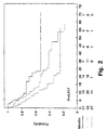
- tumour PAI-1 was a significantly independent prognostic factor for overall survival.
- PAI-1 levels may be a useful prognostic marker to select radically resected patients with bronchogenic adenocarcinoma and poor prognosis for adjuvant treatment.
- the present study was limited to adenocarcinomas within the group of Non Small Cell Lung Cancer (NSCLC) in a relatively small number of patients.
- NSCLC Non Small Cell Lung Cancer
- the prognostic value of PAI-1 should be further evaluated in larger materials and within other subgroups of NSCLC.
- the prognostic value of PAI-1 in other types of cancer might be evaluated in a similar manner (78).
- Example 1 a proportion of lung cancer patients have high levels of PAI-1 in their tumours which predict a poor prognosis. Accordingly, patients with high PAI-1 levels will be candidates for a treatment aiming at inhibiting the binding of PAI-1 to uPA.
- This example describes a predictive assay to identify patients who potentially will benefit from such a treatment.
- Tumour tissue from the patients with bronchogenic adenocarcinoma is stored at -80° C.
- Tumour extracts are made by a procedure including pre-cooling the tissue in liquid nitrogen, mechanical pulverization, extraction with a buffer consisting 75 mM potassium acetate, 0.3 M NaCl, 0.1 M L-arginine, 10 mM EDTA and 0.25% Triton X-100 pH 4.2, followed by centrifugation at 105.000 g for 1 hour. The supernatant and pellet are stored separately at -80° C.
- PAI-1 is determined using a sandwich ELISA (18) with monoclonal catching and detecting antibodies. As catching antibody is used PAI-1 monoclonal antibody clone 1 and as detecting antibody is used PAI-1 monoclonal antibody clone 7 (WO 87/00549). This assay detects both latent and active PAI-1, and is in addition recognizing PAI-1 bound to uPA and tPA (unpublished results, J. Gr ⁇ ndahl-Hansen). PAI-1 is measured in interim units by calibration with standard preparations obtained from The National Institute for Biological Standards and Control, Hertfordshire, UK. The intra- and intervariations for both assays are below 11%. Protein content of the tumour extracts is determined by Bio-Rad Protein Assay (Biorad, Richmond, CA).
- Cryostat sections from 24 samples of pulmonary carcinoma were used for immunohistochemistry. These included 14 cases of squamous cell carcinoma, 5 cases of adenocarcinomas, 3 cases of mixed tumours (adenocarcinoma and squamous cell carcinoma) and 3 cases of large cell undifferentiated carcinoma. Adjacent normal mucosal tissue resected at time of surgery was obtained from 3 of the patients. Monoclonal anti-human PAI-1 antibodies (clones 1 and 2) were used for immunohistochemical stainings. An irrelevant monoclonal antibody (m-anti-TNP) was used as a negative control. The two monoclonal antibodies have previously been shown to be directed against two different epitopes on PAI-1. 5 ⁇ m cryostat sections were subjected to a conventional APAAP (Alkaline Phosphatase-Anti-Alkaline Phosphatase) method as described previously (45).
- APAAP Alkaline Phosphatase-Anti-A
- RNA probes transcribed from a 504 base pair PstI-PstI fragment of human PAI-1 cDNA were applied to deparaffinized, proteinase K-digested sections, hybridized overnight at 48°C, then RNAse-treated and washed in high stringency buffer before being overlaid with photographic emulsion. Slides were developed after 10-14 days of exposure.
- Figure 4 shows immunostaining for PAI-1 on a specimen of lung cancer tissue. Note the intense signal in the stromal compartment.
- Routinely processed paraffin-embedded specimens from 10 of the 24 cases were investigated by in situ hybridization for the presence of mRNA for PAI-1. In all of these specimens a pattern of gene expression for PAI-1 very similar to the patterns of immunostaining was observed.
- the following examples describes an example of a compound screening scheme comprising various successive steps which is established to identify compounds which can be used to inhibit the interaction between PAI-1 and uPA and thereby be used as drugs to inhibit the invasive and metastatic process.
- the initial screening of the compounds is performed e.g. by use of the below described compound screening ELISA and if the compound show inhibitory effect, the further screening of compounds is performed using the next steps in the scheme (Examples 5 and 6).
- a two antibody sandwich ELISA assay is developed for screening compounds for their capability to inhibit the interaction between PAI-1 and uPA in solution.
- the effect of the compound to be screened to inhibit the interaction can be tested at various concentration.
- the compound X is tested and the effect of X on the interaction is examined.
- X is PAI-1 monoclonal antibody which inhibits PAI-1 inhibition of uPA (WO 87/00549).
- the ELISA assay described above can be constructed in a similar manner with uPA antibody as catching antibody and biotinylated PAI-1 antibody or antibodies as detecting antibody(ies).
- Candidate drugs for inhibiting PAI-1 binding to uPA can be found by screening drug libraries consisting of naturally occurring compounds, small molecules and peptides. Also random peptide bacteriophage display or synthetic peptide libraries may be used.
- the capability of a compound to inhibit the binding of PAI-1 to uPA is suitably examined as a step of the scheme aiming to identify and characterize new potential drugs.
- the capability of the compound to inhibit the binding of PAI-1 to uPA and thereby preventing inactivation of uPA on the cell surface can be evaluated.
- the assay is based on that described for determining the inhibition of receptor bound uPA by plasminogen activator inhibitors (61).
- U937 cells (2 x 10 7 /ml) are incubated with uPA (1.4 nM), after washing in 0.05 M glycine-HCl, 0.1 M NaCl, pH3.0 to remove endogenously bound uPA.
- the cells are then washed to remove excess uPA and incubated in 10-mm plastic fluorometer cuvettes at 37°C at a final concentration of 1 x 10 6 /ml, with plasminogen (20 ⁇ g/ml) and the plasmin-specific fluorogenic substrate H-D-Val-Leu-Lys-7-amido-4-methylcoumarin (Bachem) in 0.05 M Tris-HCl, 0.1 M NaCl, pH7.4. PAI-1 is included in these incubations at a concentration of 1 ⁇ g/ml, after pre-incubation with varying concentrations of the test compound or antibody.
- the increase in fluorescence due to hydrolysis of the substrate is measured at 1-minute intervals for 15 minutes, and plasmin generation determined form the rate of change in fluorescence intensity.
- the inhibitory effect of the test compound or antibody against PAI-1 is then determined by comparison of the plasmin generation curves obtained with the various concentrations of the compound with plasmin generation curves made with varying concentrations of PAI-1 (0, 0.25, 0.5, 0.75 and 1 ⁇ g/ml). Thus a percentage inhibitory effect is obtained at any chosen concentration of the compound.
- the last step in the screening of compounds which have been shown in the above described previous steps of the compound screening scheme to inhibit the interaction between PAI-1 and uPA which thereby are potential compounds to be used as drugs to inhibit the progression of cancer, is to evaluate the effect of the compound to inhibit growth as well as the invasive and metastatic process in vivo .
- a mouse model in which the invasive and metastatic process of human cancer cells can be measured has been developed to evaluate this effect (PCT/DK92/00306).
- This example describes an experimental study using neutralizing monoclonal PAI-1 antibodies as a means to inhibit formation of metastasis of a human breast cancer xenograft in nude mice.
- the human breast cancer cell line MDA-MB-231 BAG was routinely propagated in DMEM with 5% Fetal Calf Serum. These cells have been transduced with the lacZ gene as previously described (67). Cells for nude mouse experiments were harvested using a cell scraper instead of using enzymes.
- the human cancer cell type to be used in the assay is labelled by transducing or transfecting the cells with a LacZ gene whereby the cells can be visualized by X-gal staining.
- This transduction has previously been performed by (67) using human MDA-MB-231 and human MDA-MB-435 breast cancer cells.
- the lacZ gene codes for ⁇ -D-galactosidase, the activity of which can be detected by staining with the chromogenic substrate 5-bromo-4-chloro-3-indolyl- ⁇ -D-galactopyranoside (X-gal), which gives a dark blue reaction.
- X-gal 5-bromo-4-chloro-3-indolyl- ⁇ -D-galactopyranoside
- the transduction of the human cancer cells chosen can be performed as described in (67).
- mice 6-8 weeks-old nu/nu META/Bom (Bomholtgaard, Denmark) were used. The mice were kept in laminar flow clean benches and all equipment used was autoclaved.
- PAI-1 monoclonal antibodies clone 2 (WO 87/00549), which neutralizes the inhibitory effect of PAI-1 (R ⁇ mer, personal communication), and clone 3 (WO 87/00549), which does not influence the inhibitory activity of PAI-1 (R ⁇ mer, personal communication), were used.
- the pharmacokinetic .characteristics of the compound or antibody to be screened should be clarified in order to administer and retain the compound or antibody in an effective concentration in the mouse.
- the administration of the compound or antibody to be screened depends on the chemical nature of the compound or antibody and may be performed by interperitoneal injection or subcutaneous injection, by oral administration or local application.
- mice were injected intraperitoneally with a single dose of either PAI-1 monoclonal antibody clone 2 or clone 3 (WO 87/00549) and at different time points after this injection mice were bleed and serum were tested for PAI-1 antibody content. Furthermore, in the experiment with the tumor-bearing animals, serum was obtained from all anti-PAI-1 antibody treated animals by tail vein bleeding every week. The concentration of PAI-1 antibodies were analyzed with an ELISA.
- the transduced cancer cells to be injected into the mouse may be preincubated in a solution containing the compound or antibody in order to ensure a contact between the cells and the compound or antibody. Normally several experiments in which this pretreatment is varied or excluded should be performed in order to clarify and ascertain the overall effect of the compound or the antibody.
- a total number of 2 x 10 6 MDA-MB-231 BAG cells were inoculated subcutaneously into each flank of the animals.
- the tumor cells were always placed inferior to the thoracic wall.
- 250 ⁇ g of antibodies were injected into the mice.
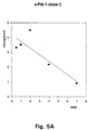
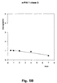
- Fig. 5A and 5B which shows a pharmacokinetic investigation of anti-PAI-1 monoclonal antibody clone 2 and clone 3, respectively, in nude mice injected intraperitoneally with 250 ⁇ g antibody on day 0
- a second injection of 250 ⁇ g antibody was given every week. The mice were sacrificed 6 weeks after cell inoculation.
- tumours The growth of the tumours.should be measured in order to elucidate whether the compound under test affects the growth of the tumour in addition to the possible effect on the invasive and metastatic process.
- the xenografted tumours were measured twice weekly in two dimensions and tumor growth curves were constructed on the basis of a transformed Gompertz function.
- the duration of the assay depends on the nature of the compound or antibody and on the effect of the compound or antibody on the growth and invasive and metastatic process.
- the xenotransplanted tumors were surgically removed from the animals and examined by Northern blotting for the presence of human PAI-1 mRNA and by ELISA for the presence of human PAI-1 protein.
- the xenotransplanted tumors were surgically removed from the animals and examined for mouse PAI-1 mRNA by in situ hybridization with a mouse specific probe.
- the effect of the compound or antibody on the invasive and metastatic process and on the growth of the tumour can be evaluated locally and/or in the lungs, the lymph nodes and in the interperitoneal cavity utilizing the very distinct differentiation between human cells and mouse cells rendered possible by the staining (67-69).
- lungs were excised from each animals and the lungs were processed with X-gal according to the method described by Brunner et al (67).
- blue areas from various lungs were fixed in 4% formalin and processed for routine histology. If a lung presented one or more blue areas it was registered as positive for metastatic lesion.
- an EhISA for ⁇ -D-galactosidase as described by Fujiwara et al (79), could be used as an objective measurement of metastatic spread.
- MDA-MB-231 BAG tumors expressed abundant mRNA and protein for human PAI-1.
- the xenotransplanted tumor was shown to express mouse PAI-1 in the stromal cells including the vessels infiltrating the human tumor cells.
- PAI-1 antibody clone 2 The half-life of PAI-1 antibody clone 2 was estimated to approximately 4 days and for PAI-1 clone 3 approximately 6 days ( Figure 5A and 5B).
- Metastatic spread of human cancer cells was confirmed by the presence of cancer cells in the blue spots, as determined by conventional histologic examinations.
- Fig. 6 which shows tumour area growth curves of human breast carcinoma cell line MDA.MB-231 BAG injected subcutaneously into female nu/nu META/BOM mice on day 0).
Landscapes
- Health & Medical Sciences (AREA)
- Life Sciences & Earth Sciences (AREA)
- Chemical & Material Sciences (AREA)
- Immunology (AREA)
- Engineering & Computer Science (AREA)
- Medicinal Chemistry (AREA)
- General Health & Medical Sciences (AREA)
- Biomedical Technology (AREA)
- Molecular Biology (AREA)
- Hematology (AREA)
- Urology & Nephrology (AREA)
- Proteomics, Peptides & Aminoacids (AREA)
- Biochemistry (AREA)
- Organic Chemistry (AREA)
- Pharmacology & Pharmacy (AREA)
- Veterinary Medicine (AREA)
- Animal Behavior & Ethology (AREA)
- Public Health (AREA)
- Microbiology (AREA)
- Epidemiology (AREA)
- Pathology (AREA)
- Bioinformatics & Cheminformatics (AREA)
- General Physics & Mathematics (AREA)
- Analytical Chemistry (AREA)
- Physics & Mathematics (AREA)
- Biotechnology (AREA)
- Cell Biology (AREA)
- Gastroenterology & Hepatology (AREA)
- Food Science & Technology (AREA)
- Zoology (AREA)
- Biophysics (AREA)
- Genetics & Genomics (AREA)
- Nuclear Medicine, Radiotherapy & Molecular Imaging (AREA)
- General Chemical & Material Sciences (AREA)
- Chemical Kinetics & Catalysis (AREA)
- Medicines That Contain Protein Lipid Enzymes And Other Medicines (AREA)
- Pharmaceuticals Containing Other Organic And Inorganic Compounds (AREA)
- Nitrogen Condensed Heterocyclic Rings (AREA)
- Measuring Or Testing Involving Enzymes Or Micro-Organisms (AREA)
- Medicines Containing Antibodies Or Antigens For Use As Internal Diagnostic Agents (AREA)
Abstract
Description
Examples of malignant tumour types for which this has been established or will be established are mammary carcinomas, urological carcinomas e.g. prostate carcinoma and bladder carcinoma, gynaecological carcinomas e.g. ovarian carcinoma and cervical carcinoma, non-small cell lung tumours, gastrointestinal cancers e.g. colon adenocarcinoma and gastric cancers, brain tumours, sarcomas, haematological malignancy e.g. lymphoma and skin cancers e.g. melanoma and squamous cell skin cancer. Also any other cancer type such as e.g. head and neck cancer for which it is established that an increased concentration of the inhibitor of the protease or of the non-proteolytic matrix-degrading enzyme is a prognostic factor or has a predictive value are within the scope of the invention.
Such a compound may be an antibody against the protease. In still another embodiment of the invention the compound which is capable of binding to a protease inhibitor, may have a cytotoxic agent coupled to the compound.
the method comprising one or more of the following steps:
for preparing a composition for treating a patient who has been established to have a high risk of developing a malignant tumour or who has developed a malignant tumour by inhibiting the plasminogen activator-inhibitory activity of PAI-1 in malignant tumour tissue or potential malignant tumour tissue. One embodiment of the invention relates to a use, wherein the compound additionally
and another embodiment of the invention relates to a use wherein the compound additionally
| Characterization: 106 patients with bronchiogenic adenocarcinoma | |||
| variable | No. of patients N=106 | ||
| Age | < 65 years | 53 | |
| > 65 years | 53 | ||
| | Males | 54 | |
| | 52 | ||
| | T1 | 23 | |
| T2 | 68 | ||
| | 10 | ||
| | 5 | ||
| Positive | N0 | 78 | |
| | 10 | ||
| | 18 | ||
| Stage | I | 69 | |
| II | 8 | ||
| | 23 | ||
| | 6 | ||
| Surgery | Radical | 86 | |
| Not radical | 20 |
| X2 test for association between PAI-1 in tumour extracts and other variables in patients with bronchiogenic adenocarcinomas. | |
| Variable | Correlation with PAI-1 P-value |
| Age | 0.63 |
| Sex | 0.16 |
| Stage | 0.21 |
| Radicality | 0.20 |
| Tumour size | 0.59 |
| Lymph nodes | 0.22 |
| Multivariate Cox regression analysis of 106 patients with bronchiogenic adenocarcinoma | ||
| Variable | Overall survival Multivariate P-value | Relative risk |
| Stage | >0.005 | 2.3 |
| PAI-1 | ||
| low vs. med. | 0.05 | 1.5 (1.1-2.2) |
| low vs. high | 0.005 | 2.3 (1.6-3.3) |
Protein content of the tumour extracts is determined by Bio-Rad Protein Assay (Biorad, Richmond, CA).
Thus a percentage inhibitory effect is obtained at any chosen concentration of the compound.
| TREATMENT | LUNG METASTASES |
| Untreated control | 9/10 |
| PAI-1 | 6/10 |
| PAI-1 | 1/10 |

Claims (23)
- The use of a compound, which
suppresses the inhibitory activity of an inhibitor of a protease or of a non-proteolytic matrix-degrading enzyme in malignant tumour tissue or potential malignant tumour tissue,
said inhibitor being plasminogen activator inhibitor I (PAI-1),
for preparing a composition for inhibiting malignant tumour growth, invasion and/or metastasis in a patient who has been established to have a high risk of developing a malignant tumour or who has developed a malignant tumour. - The use according to claim 1, whereby the protease or the non-proteolytic matrix-degrading enzyme directly or indirectly degrades the malignant tumour tissue or the potential malignant tumour tissue.
- The use according to claim 1 or 2, whereby the suppression of the inhibitory activity of an inhibitor of a protease or of a non-proteolytic matrix-degrading enzyme in malignant tumour tissue or potential malignant tumour tissue interferes with the process of tumour angiogenesis.
- The use according to any of claims 1-3, whereby the suppression of the inhibitory activity of an inhibitor of a protease or of a non-proteolytic matrix-degrading enzyme in malignant tumour tissue or potential malignant tumour tissue interferes with the migrating capacity of malignant tumour cells or of other cells in the tumour stroma.
- The use according to any of claims 1-4, wherein the patient is a patient who has been established to have a carcinoma in situ.
- The use according to any of claims 1-5, wherein the composition is to be used preoperatively.
- The use according to any of claims 1-6, wherein the composition is to be used as a neoadjuvant, and/or as a adjuvant after the malignant tumour tissue is removed by surgical operation.
- The use according to any of claims 1-7, wherein the patient is a patient who has been established to have a high risk of developing a malignant tumour by having a high-risk-indicating score of a tumour marker such as a serum/plasma tumour marker or by having a gene or gene product which indicates that the patient is at high risk of developing a malignant tumour, said malignant tumour being selected from the group consisting of mammary carcinomas, urological carcinomas e.g. prostate carcinoma and bladder carcinoma, gynaecological carcinomas e.g. ovarian carcinoma and cervical carcinoma, non-small cell lung tumours, gastrointestinal cancers e.g. colon adenocarcinoma and gastric cancers, brain tumours, sarcomas, haematological malignancy e.g. lymphoma and skin cancers e.g. melanoma and squamous cell skin cancer.
- The use according to any of claims 1-8, wherein the patient is a patient who has an increased concentration of the inhibitor of the protease or of the non-proteolytic matrix-degrading enzyme in the malignant tumour tissue and/or in plasma and/or serum, said malignant tumour being selected from the group consisting of mammary carcinomas, urological carcinomas e.g. prostate carcinoma and bladder carcinoma, gynaecological carcinomas e.g. ovarian carcinoma and cervical carcinoma, non-small cell lung tumours, gastrointestinal cancers e.g. colon adenocarcinoma and gastric cancers, brain tumours, sarcomas, haematological malignancy e.g. lymphoma and skin cancers e.g. melanoma and squamous cell skin cancer.
- The use according to claim 9, wherein the increased concentration of the inhibitor of the protease or of the non-proteolytic matrix-degrading enzyme has been established to be a prognostic factor indicating a poor prognosis for the patient having the type of malignant tumour in question, said malignant tumour being selected from the group consisting of mammary carcinomas, urological carcinomas e.g. prostate carcinoma and bladder carcinoma, gynaecological carcinomas e.g. ovarian carcinoma and cervical carcinoma, non-small cell lung tumours, gastrointestinal cancers e.g. colon adenocarcinoma and gastric cancers, brain tumours, sarcomas, haematological malignancy e.g. lymphoma and skin cancers e.g. melanoma and squamous cell skin cancer.
- The use according to claim 9, wherein the increased concentration of the inhibitor of the protease or of the non-proteolytic matrix-degrading enzyme has been established to be a predictive factor indicating efficiency of the treatment of the type of malignant tumour in question by suppressing the said inhibitor, said malignant tumour being selected from the group consisting of mammary carcinomas, urological carcinomas e.g. prostate carcinoma and bladder carcinoma, gynaecological carcinomas e.g. ovarian carcinoma and cervical carcinoma, non-small cell lung tumours, gastrointestinal cancers e.g. colon adenocarcinoma and gastric cancers, brain tumours, sarcomas, haematological malignancy e.g. lymphoma and skin cancers e.g. melanoma and squamous cell skin cancer.
- The use according to any of claims 2-11, wherein the malignant tumour tissue degraded, including micrometastases, is tissue comprised of the malignant tumour cells themselves, the extracellular matrix and/or the stromal cells of the malignant tumour, e.g. endothelial cells, fibroblasts, macrophages, leucocytes; and substantially no other tissue than malignant tumour tissue is degraded to an extent or degree which give unacceptable toxic side effects.
- The use according to any of claims 1-12, wherein the compound is a compound which suppresses the plasminogen activator-inhibitory activity of PAI-1 by inhibiting the binding of PAI-1 to vitronectin.
- The use according to any of claims 1-12, wherein the compound is a compound which inhibits the effect of PAI-1 on uPA, but does not inhibit the binding of uPA to uPAR.
- The use according to any of claims 1-12, wherein the compound is a compound which1) inhibits the uPA-binding activity of PAI-1 as determined by adding the compound to a system comprising immobilized PAI-1 and solubilized uPA, uPA bound to PAI-1 being detected by being labelled or by means of a labelled anti-uPA antibody, or adding the compound to a system comprising immobilized uPA and solubilized PAI-1, PAI-1 bound to uPA being detected by being labelled or by means of a labelled anti-PAI-1 antibody, and/or2) inhibits the binding of PAI-1 to uPA as determined by adding the compound to a system comprising radiolabelled PAI-1 or a derivative thereof and cells having uPA bound to the cell surface to uPA receptors and detecting any PAI-1 or derivative binding to uPA by gamma counting of the cells, and/or3) suppresses the inhibitory activity of PAI-1 on uPA as determined by adding the compound to a system comprising PAI-1 and cells having uPA bound on the cell surface to uPA receptors and detecting any effect of PAI-1 either directly by a chromogenic uPA substrate or indirectly by adding plasminogen and measuring plasmin generation using a plasmin-specific substrate such as the fluorogenic substrate H-D-Val-Leu-Lys-7-amido-4-methylcoumarin.
- The use according to claim 15, wherein the compound additionally4) inhibits the activity of PAI-1 in tumour tissue as determined by adding the compound to the malignant cells or other cells in the tumour tissue expressing PAI-1 and subsequently adding uPA, followed by measurement of uPA activity either directly by a chromogenic uPA substrate or indirectly by adding plasminogen and measuring plasmin generation.
- The use according to claim 15 or 16, wherein the compound additionally5) inhibits the growth and/or invasion and/or metastasis of human malignant tumour cells when administered to a nude mouse or a nude rat which is inoculated with human malignant tumour cells which are known to invade and/or metastasize in the presence of uPA and PAI-1 and which are capable of invasion and/or metastasis in the mouse.
- The use according to claim 14, wherein the compound is a compound which all binds to PAI-1 and does not convert plasminogen into plasmin.
- The use according to claim 18, wherein the compound is a derivative or variant of uPA or pro-uPA.
- The use according to claim 18, wherein the compound is an antibody which is capable of binding to PAI-1.
- The use according to any of claims 18-20, wherein the compound which is capable of binding to PAI-1 has a cytotoxic drug coupled thereto.
- A method for selecting a compound for use for preparing a composition for inhibiting malignant tumour growth, invasion and/or metastasis in a patient who has been established to have a high risk of developing a malignant tumour or who has developed a malignant tumour, the compound being capable of suppressing the inhibitory activity of an inhibitor of a protease or of a non-proteolytic matrix-degrading enzyme in malignant tumour tissue or potential malignant tumour tissue, the method comprising one or more of the following steps:1) a screening assay in which the possible suppression of the inhibitory activity of an inhibitor of a protease or of a non-proteolytic matrix-degrading enzyme is determined by adding the compound to a system comprising immobilized inhibitor of the protease or of the non-proteolytic matrix-degrading enzyme and solubilized protease or non-proteolytic matrix-degrading enzyme, the protease or the non-proteolytic matrix-degrading enzyme bound to the inhibitor of the protease or of the non-proteolytic matrix-degrading enzyme being detected by the enzyme being labelled or by means of a labelled antibody directed to the protease or the non-proteolytic matrix-degrading enzyme, or adding the compound to a system comprising immobilized protease or non-proteolytic matrix-degrading enzyme and solubilized inhibitor of the protease or of the non-proteolytic matrix-degrading enzyme, inhibitor of the protease or of the non-proteolytic matrix-degrading enzyme bound to the protease or the non-proteolytic matrix-degrading enzyme being detected by the inhibitor being labelled or by means of a labelled antibody directed to the inhibitor of the protease or of the non-proteolytic matrix-degrading enzyme,2) an assay in which the possible suppression of the inhibitory activity of the inhibitor of the protease or of the non-proteolytic matrix-degrading enzyme in the tumour tissue is determined by adding the compound to a system comprising radiolabelled inhibitor of the protease or of the non-proteolytic matrix-degrading enzyme and tumour cells expressing the protease or the non-proteolytic matrix-degrading enzyme and detecting any inhibitor of the protease or of the non-proteolytic matrix-degrading enzyme binding to the protease or the non-proteolytic matrix-degrading enzyme by gamma counting of the cells,3) a screening assay, in which the potential suppression of the inhibitory activity of an inhibitor of a protease or of a non-proteolytic matrix-degrading enzyme by the compound is determined by using whole cells to which the protease or the non-proteolytic matrix-degrading enzyme is bound on the surface, e.g. by a protease receptor, such as uPA to the uPA receptor,4) a step wherein a compound which has been established to suppress the inhibitory activity of an inhibitor of a protease or of a non-proteolytic matrix-degrading enzyme in the tumour tissue has been administered to a nude mouse or a nude rat which has been inoculated with human malignant tumour cells which are capable of invasion and/or metastasis in the mouse or rat in the presence of the protease or the non-proteolytic matrix-degrading enzyme and the inhibitor of the protease or of the non-proteolytic matrix-degrading enzyme, and selecting, as a suitable compound, a compound inhibiting the growth and/or invasion and/or metastasis of the human malignant tumour cells in the mouse or the rat.
- A method according to claim 22, wherein the inhibitor of the protease is PAI-1.
Applications Claiming Priority (4)
| Application Number | Priority Date | Filing Date | Title |
|---|---|---|---|
| DK93851A DK85193D0 (en) | 1993-07-16 | 1993-07-16 | SUPPRESSION OF INHIBITORS |
| DK851/93 | 1993-07-16 | ||
| DK85193 | 1993-07-16 | ||
| PCT/DK1994/000288 WO1995002413A1 (en) | 1993-07-16 | 1994-07-18 | Suppression of inhibitors |
Publications (2)
| Publication Number | Publication Date |
|---|---|
| EP0712312A1 EP0712312A1 (en) | 1996-05-22 |
| EP0712312B1 true EP0712312B1 (en) | 2004-04-07 |
Family
ID=8098263
Family Applications (1)
| Application Number | Title | Priority Date | Filing Date |
|---|---|---|---|
| EP94920904A Expired - Lifetime EP0712312B1 (en) | 1993-07-16 | 1994-07-18 | Suppression of inhibitors |
Country Status (9)
| Country | Link |
|---|---|
| US (4) | US6224865B1 (en) |
| EP (1) | EP0712312B1 (en) |
| JP (1) | JPH10500097A (en) |
| AT (1) | ATE263569T1 (en) |
| AU (1) | AU7183394A (en) |
| DE (1) | DE69433685T2 (en) |
| DK (1) | DK85193D0 (en) |
| ES (1) | ES2217268T3 (en) |
| WO (1) | WO1995002413A1 (en) |
Cited By (1)
| Publication number | Priority date | Publication date | Assignee | Title |
|---|---|---|---|---|
| US10867706B2 (en) | 2010-07-20 | 2020-12-15 | Applied Invention, Llc | Multi-scale complex systems transdisciplinary analysis of response to therapy |
Families Citing this family (19)
| Publication number | Priority date | Publication date | Assignee | Title |
|---|---|---|---|---|
| DK85193D0 (en) * | 1993-07-16 | 1993-07-16 | Cancerforskningsfondet Af 1989 | SUPPRESSION OF INHIBITORS |
| US6811995B1 (en) * | 1996-04-26 | 2004-11-02 | Children's Medical Center Corporation | Non-invasive enzyme screen for cancer |
| US5994309A (en) | 1997-07-25 | 1999-11-30 | Angstrom Pharmaceuticals, Inc. | Anti-invasive and anti-angiogenic compositions and methods |
| JP2001520200A (en) * | 1997-10-17 | 2001-10-30 | ザ・トラステイーズ・オブ・ザ・ユニバーシテイ・オブ・ペンシルベニア | Compositions and methods for promoting internalization and degradation of urokinase-type plasminogen activator |
| US6750201B1 (en) | 1997-10-17 | 2004-06-15 | The Trustees Of The University Of Pennsylvania | Compositions and methods for promoting internalization and degradation of urokinase-type plasminogen activator |
| US6841153B1 (en) | 1998-10-02 | 2005-01-11 | University Of Florida | Prevention of adhesions |
| WO2000050057A1 (en) * | 1999-02-22 | 2000-08-31 | Bernstein Eric F | Compositions and methods for prevention of photoaging |
| US6986881B1 (en) * | 1999-06-04 | 2006-01-17 | Dana-Farber Cancer Institute, Inc. | Identification of compounds that modify transcriptional responses to hypoxia |
| EP2302385B1 (en) * | 2002-02-13 | 2014-12-03 | Sekisui Diagnostics, LLC | Methods for selecting treatment regimens and predicting outcomes in cancer patients |
| WO2005106475A2 (en) * | 2004-04-16 | 2005-11-10 | Wyeth | Novel method for determination of plasminogen activator inhibitor |
| EP2189469B1 (en) * | 2004-11-18 | 2015-09-16 | The Board Of Trustees Of The University Of Illinois | Multicistronic siRNA constructs to inhibit tumors |
| US20070021365A1 (en) * | 2005-06-21 | 2007-01-25 | The Board Of Trustees Of The Leland Stanford Junior University | Inhibition of Lysyl oxidase for treating tumor growth and diagnostics relating thereto |
| US20070225242A1 (en) * | 2005-06-21 | 2007-09-27 | The Board Of Trustees Of The Leland Stanford Junior University | Method and composition for treating and preventing tumor metastasis in vivo |
| WO2007138705A1 (en) * | 2006-06-01 | 2007-12-06 | Japan As Represented By Director General Of Agency Of National Cancer Center | Tumor suppressor |
| US7981415B2 (en) * | 2007-09-07 | 2011-07-19 | Cisthera, Inc. | Humanized PAI-1 antibodies |
| US7771720B2 (en) * | 2007-09-07 | 2010-08-10 | Cisthera, Inc. | Humanized PAI-1 antibodies |
| US20220296999A1 (en) | 2010-08-13 | 2022-09-22 | Cfph, Llc | Multi-process communication regarding gaming information |
| EA033403B1 (en) * | 2013-08-13 | 2019-10-31 | Sanofi Sa | ANTIBODIES TO PLASMINOGEN 1 (PAI-1) ACTIVATOR INHIBITOR AND WAYS OF THEIR APPLICATION |
| JP2018517892A (en) | 2015-04-10 | 2018-07-05 | アプライド プロテオミクス,インク. | Protein biomarker panel to detect colorectal cancer and advanced adenoma |
Family Cites Families (11)
| Publication number | Priority date | Publication date | Assignee | Title |
|---|---|---|---|---|
| DK319685A (en) * | 1985-07-12 | 1987-01-13 | Fonden Til Fremme Af Eksperime | MONOCLONAL ANTIBODIES, PROCEDURES FOR PRODUCING ANTIBODIES, HYBRIDOMA CELLS PRODUCING ANTIBODIES, AND USING ANTIBODIES |
| US4906457A (en) * | 1988-09-06 | 1990-03-06 | Washington State University Research Foundation, Inc. | Compositions and methods for reducing the risk of sunlight and ultraviolet induced skin cancer |
| WO1993017715A1 (en) * | 1992-03-05 | 1993-09-16 | Board Of Regents, The University Of Texas System | Diagnostic and/or therapeutic agents, targeted to neovascular endothelial cells |
| GB9217331D0 (en) | 1992-08-14 | 1992-09-30 | Xenova Ltd | Pharmaceutical compounds |
| US5744442A (en) * | 1992-08-26 | 1998-04-28 | Bristol Meyers Squibb Company | Regulation of cellular invasiveness |
| DK85193D0 (en) * | 1993-07-16 | 1993-07-16 | Cancerforskningsfondet Af 1989 | SUPPRESSION OF INHIBITORS |
| US5800814A (en) | 1994-04-22 | 1998-09-01 | Oklahoma Medical Research Foundation | Method for inhibition of breast tumor growth |
| GB9410387D0 (en) | 1994-05-24 | 1994-07-13 | Xenova Ltd | Pharmaceutical compounds |
| SI0910407T1 (en) | 1996-05-15 | 2003-10-31 | Altarex Corp. | Method and composition for reconforming multi-epitopic antigens to initiate an immune response |
| US7091323B2 (en) * | 2001-04-24 | 2006-08-15 | Bayer Corporation | Human TIMP-1 antibodies |
| EP1742650A1 (en) * | 2004-03-30 | 2007-01-17 | Den Kgl.Veterin R-Og Landboh Jskole | Improvements in cancer treatment and cancer treatment efficacy prediction by blocking and detecting protease inhibitors |
-
1993
- 1993-07-16 DK DK93851A patent/DK85193D0/en not_active Application Discontinuation
-
1994
- 1994-07-18 WO PCT/DK1994/000288 patent/WO1995002413A1/en not_active Ceased
- 1994-07-18 EP EP94920904A patent/EP0712312B1/en not_active Expired - Lifetime
- 1994-07-18 AT AT94920904T patent/ATE263569T1/en not_active IP Right Cessation
- 1994-07-18 DE DE69433685T patent/DE69433685T2/en not_active Expired - Lifetime
- 1994-07-18 US US08/583,129 patent/US6224865B1/en not_active Expired - Lifetime
- 1994-07-18 JP JP7504300A patent/JPH10500097A/en active Pending
- 1994-07-18 ES ES94920904T patent/ES2217268T3/en not_active Expired - Lifetime
- 1994-07-18 AU AU71833/94A patent/AU7183394A/en not_active Abandoned
-
2001
- 2001-04-18 US US09/836,323 patent/US20010034327A1/en not_active Abandoned
-
2003
- 2003-01-02 US US10/336,513 patent/US20030096755A1/en not_active Abandoned
-
2007
- 2007-12-26 US US11/964,334 patent/US20080166354A1/en not_active Abandoned
Cited By (1)
| Publication number | Priority date | Publication date | Assignee | Title |
|---|---|---|---|---|
| US10867706B2 (en) | 2010-07-20 | 2020-12-15 | Applied Invention, Llc | Multi-scale complex systems transdisciplinary analysis of response to therapy |
Also Published As
| Publication number | Publication date |
|---|---|
| ES2217268T3 (en) | 2004-11-01 |
| JPH10500097A (en) | 1998-01-06 |
| WO1995002413A1 (en) | 1995-01-26 |
| US20010034327A1 (en) | 2001-10-25 |
| AU7183394A (en) | 1995-02-13 |
| DK85193D0 (en) | 1993-07-16 |
| EP0712312A1 (en) | 1996-05-22 |
| US6224865B1 (en) | 2001-05-01 |
| DE69433685T2 (en) | 2005-03-03 |
| DE69433685D1 (en) | 2004-05-13 |
| US20030096755A1 (en) | 2003-05-22 |
| US20080166354A1 (en) | 2008-07-10 |
| ATE263569T1 (en) | 2004-04-15 |
Similar Documents
| Publication | Publication Date | Title |
|---|---|---|
| US20080166354A1 (en) | Suppression of inhibitors | |
| Grøndahl-Hansen et al. | Prognostic significance of the receptor for urokinase plasminogen activator in breast cancer. | |
| Basset et al. | Expression of the stromelysin-3 gene in fibroblastic cells of invasive carcinomas of the breast and other human tissues: a review | |
| Mahmood et al. | Multifaceted role of the urokinase-type plasminogen activator (uPA) and its receptor (uPAR): diagnostic, prognostic, and therapeutic applications | |
| Mekkawy et al. | Involvement of urokinase‐type plasminogen activator system in cancer: An overview | |
| Grøndahl-Hansen et al. | High levels of urokinase-type plasminogen activator and its inhibitor PAI-1 in cytosolic extracts of breast carcinomas are associated with poor prognosis | |
| Höyhtyã et al. | Immunohistochemical localization of matrix metallloproteinase 2 and its specific inhibitor timp‐2 in neoplastic tissues with monclonal antibodies | |
| Yang et al. | Urokinase‐type plasminogen activator and its receptor in colorectal cancer: Independent prognostic factors of metastasis and cancer‐specific survival and potential therapeutic targets | |
| Pedersen et al. | Prognostic impact of urokinase, urokinase receptor, and type 1 plasminogen activator inhibitor in squamous and large cell lung cancer tissue | |
| Lund et al. | Two distinct phases of apoptosis in mammary gland involution: proteinase-independent and-dependent pathways | |
| Pappot et al. | Plasminogen activator inhibitor type 1 in cancer: therapeutic and prognostic implications | |
| de Vries et al. | Plasminogen activators, their inhibitors, and urokinase receptor emerge in late stages of melanocytic tumor progression | |
| Holst-Hansen et al. | Urokinase-type plasminogen activation in three human breast cancer cell lines correlates with their in vitro invasiveness | |
| Thorek et al. | Prostate-specific kallikrein-related peptidases and their relation to prostate cancer biology and detection | |
| Krüger et al. | Reduction of breast carcinoma tumor growth and lung colonization by overexpression of the soluble urokinase-type plasminogen activator receptor (CD87) | |
| Herszényi et al. | Proteases in gastrointestinal neoplastic diseases | |
| Sis et al. | Prognostic significance of matrix metalloproteinase-2, cathepsin D, and tenascin-C expression in colorectal carcinoma | |
| Duffy | Urokinase-type plasminogen activator and malignancy | |
| Duffy | Plasminogen activators and cancer | |
| Pujade‐Lauraine et al. | The plasminogen‐activation system in ovarian tumors | |
| Djonov et al. | Tumor cell specific expression of MMP-2 correlates with tumor vascularisation in breast cancer | |
| EP1355637A2 (en) | Inhibitors of matriptase for the treatment of cancer | |
| Brünner et al. | Urokinase plasminogen activator (uPA) and its type 1 inhibitor (PAI-1): regulators of proteolysis during cancer invasion and prognostic parameters in breast cancer | |
| Gilardoni et al. | Differential expression of low density lipoprotein receptor–related protein 1 (LRP-1) and matrix metalloproteinase-9 (MMP-9) in prostate gland: from normal to malignant lesions | |
| US6677377B2 (en) | Structure based discovery of inhibitors of matriptase for the treatment of cancer and other conditions |
Legal Events
| Date | Code | Title | Description |
|---|---|---|---|
| PUAI | Public reference made under article 153(3) epc to a published international application that has entered the european phase |
Free format text: ORIGINAL CODE: 0009012 |
|
| 17P | Request for examination filed |
Effective date: 19960216 |
|
| AK | Designated contracting states |
Kind code of ref document: A1 Designated state(s): AT BE CH DE DK ES FR GB GR IE IT LI LU MC NL PT SE |
|
| 17Q | First examination report despatched |
Effective date: 19991207 |
|
| GRAP | Despatch of communication of intention to grant a patent |
Free format text: ORIGINAL CODE: EPIDOSNIGR1 |
|
| GRAS | Grant fee paid |
Free format text: ORIGINAL CODE: EPIDOSNIGR3 |
|
| GRAA | (expected) grant |
Free format text: ORIGINAL CODE: 0009210 |
|
| RIN1 | Information on inventor provided before grant (corrected) |
Inventor name: DANO, KELD Inventor name: HANSEN, HEINE, HOI Inventor name: PEDERSEN, HELLE Inventor name: GRONDAHL-HANSEN, JAN Inventor name: PYKE, CHARLES Inventor name: ELLIS, VINCENT18 CAVENDISH AVENUE Inventor name: ROMER, JOHN Inventor name: BRUENNER, NILS |
|
| AK | Designated contracting states |
Kind code of ref document: B1 Designated state(s): AT BE CH DE DK ES FR GB GR IE IT LI LU MC NL PT SE |
|
| REG | Reference to a national code |
Ref country code: GB Ref legal event code: FG4D |
|
| REG | Reference to a national code |
Ref country code: CH Ref legal event code: EP |
|
| REF | Corresponds to: |
Ref document number: 69433685 Country of ref document: DE Date of ref document: 20040513 Kind code of ref document: P |
|
| REG | Reference to a national code |
Ref country code: IE Ref legal event code: FG4D |
|
| REG | Reference to a national code |
Ref country code: CH Ref legal event code: NV Representative=s name: A. BRAUN, BRAUN, HERITIER, ESCHMANN AG PATENTANWAE |
|
| PG25 | Lapsed in a contracting state [announced via postgrant information from national office to epo] |
Ref country code: SE Free format text: LAPSE BECAUSE OF FAILURE TO SUBMIT A TRANSLATION OF THE DESCRIPTION OR TO PAY THE FEE WITHIN THE PRESCRIBED TIME-LIMIT Effective date: 20040707 Ref country code: GR Free format text: LAPSE BECAUSE OF FAILURE TO SUBMIT A TRANSLATION OF THE DESCRIPTION OR TO PAY THE FEE WITHIN THE PRESCRIBED TIME-LIMIT Effective date: 20040707 Ref country code: DK Free format text: LAPSE BECAUSE OF FAILURE TO SUBMIT A TRANSLATION OF THE DESCRIPTION OR TO PAY THE FEE WITHIN THE PRESCRIBED TIME-LIMIT Effective date: 20040707 |
|
| PG25 | Lapsed in a contracting state [announced via postgrant information from national office to epo] |
Ref country code: MC Free format text: LAPSE BECAUSE OF NON-PAYMENT OF DUE FEES Effective date: 20040731 |
|
| REG | Reference to a national code |
Ref country code: ES Ref legal event code: FG2A Ref document number: 2217268 Country of ref document: ES Kind code of ref document: T3 |
|
| ET | Fr: translation filed | ||
| PLBE | No opposition filed within time limit |
Free format text: ORIGINAL CODE: 0009261 |
|
| STAA | Information on the status of an ep patent application or granted ep patent |
Free format text: STATUS: NO OPPOSITION FILED WITHIN TIME LIMIT |
|
| 26N | No opposition filed |
Effective date: 20050110 |
|
| PG25 | Lapsed in a contracting state [announced via postgrant information from national office to epo] |
Ref country code: PT Free format text: LAPSE BECAUSE OF NON-PAYMENT OF DUE FEES Effective date: 20040907 |
|
| REG | Reference to a national code |
Ref country code: CH Ref legal event code: PFA Owner name: CANCERFORSKNINGSFONDEN AF 1989 (FONDEN TIL FREMME Free format text: CANCERFORSKNINGSFONDEN AF 1989 (FONDEN TIL FREMME AF EKSPERIMENTEL CANCERFORSKNING)#C/O ATTORNEY MICHAEL ROSTOCK VESTER VOLDGADE 90#DK-1552 COPENHAGEN V (DK) -TRANSFER TO- CANCERFORSKNINGSFONDEN AF 1989 (FONDEN TIL FREMME AF EKSPERIMENTEL CANCERFORSKNING)#C/O ATTORNEY MICHAEL ROSTOCK VESTER VOLDGADE 90#DK-1552 COPENHAGEN V (DK) |
|
| PGFP | Annual fee paid to national office [announced via postgrant information from national office to epo] |
Ref country code: NL Payment date: 20100714 Year of fee payment: 17 Ref country code: IE Payment date: 20100722 Year of fee payment: 17 Ref country code: ES Payment date: 20100726 Year of fee payment: 17 Ref country code: CH Payment date: 20100726 Year of fee payment: 17 |
|
| PGFP | Annual fee paid to national office [announced via postgrant information from national office to epo] |
Ref country code: LU Payment date: 20100716 Year of fee payment: 17 Ref country code: IT Payment date: 20100726 Year of fee payment: 17 Ref country code: FR Payment date: 20100805 Year of fee payment: 17 Ref country code: DE Payment date: 20100723 Year of fee payment: 17 Ref country code: AT Payment date: 20100714 Year of fee payment: 17 |
|
| PGFP | Annual fee paid to national office [announced via postgrant information from national office to epo] |
Ref country code: GB Payment date: 20100722 Year of fee payment: 17 |
|
| PGFP | Annual fee paid to national office [announced via postgrant information from national office to epo] |
Ref country code: BE Payment date: 20100715 Year of fee payment: 17 |
|
| BERE | Be: lapsed |
Owner name: *CANCERFORSKNINGSFONDEN AF 1989 (FONDEN TIL FREMME Effective date: 20110731 |
|
| REG | Reference to a national code |
Ref country code: NL Ref legal event code: V1 Effective date: 20120201 |
|
| REG | Reference to a national code |
Ref country code: CH Ref legal event code: PL |
|
| GBPC | Gb: european patent ceased through non-payment of renewal fee |
Effective date: 20110718 |
|
| REG | Reference to a national code |
Ref country code: AT Ref legal event code: MM01 Ref document number: 263569 Country of ref document: AT Kind code of ref document: T Effective date: 20110718 |
|
| REG | Reference to a national code |
Ref country code: FR Ref legal event code: ST Effective date: 20120330 |
|
| REG | Reference to a national code |
Ref country code: IE Ref legal event code: MM4A |
|
| PG25 | Lapsed in a contracting state [announced via postgrant information from national office to epo] |
Ref country code: BE Free format text: LAPSE BECAUSE OF NON-PAYMENT OF DUE FEES Effective date: 20110731 Ref country code: CH Free format text: LAPSE BECAUSE OF NON-PAYMENT OF DUE FEES Effective date: 20110731 Ref country code: FR Free format text: LAPSE BECAUSE OF NON-PAYMENT OF DUE FEES Effective date: 20110801 Ref country code: LI Free format text: LAPSE BECAUSE OF NON-PAYMENT OF DUE FEES Effective date: 20110731 Ref country code: DE Free format text: LAPSE BECAUSE OF NON-PAYMENT OF DUE FEES Effective date: 20120201 |
|
| PG25 | Lapsed in a contracting state [announced via postgrant information from national office to epo] |
Ref country code: NL Free format text: LAPSE BECAUSE OF NON-PAYMENT OF DUE FEES Effective date: 20120201 Ref country code: IT Free format text: LAPSE BECAUSE OF NON-PAYMENT OF DUE FEES Effective date: 20110718 |
|
| REG | Reference to a national code |
Ref country code: DE Ref legal event code: R119 Ref document number: 69433685 Country of ref document: DE Effective date: 20120201 |
|
| PG25 | Lapsed in a contracting state [announced via postgrant information from national office to epo] |
Ref country code: GB Free format text: LAPSE BECAUSE OF NON-PAYMENT OF DUE FEES Effective date: 20110718 |
|
| PG25 | Lapsed in a contracting state [announced via postgrant information from national office to epo] |
Ref country code: IE Free format text: LAPSE BECAUSE OF NON-PAYMENT OF DUE FEES Effective date: 20110718 |
|
| REG | Reference to a national code |
Ref country code: ES Ref legal event code: FD2A Effective date: 20121207 |
|
| PG25 | Lapsed in a contracting state [announced via postgrant information from national office to epo] |
Ref country code: AT Free format text: LAPSE BECAUSE OF NON-PAYMENT OF DUE FEES Effective date: 20110718 |
|
| PG25 | Lapsed in a contracting state [announced via postgrant information from national office to epo] |
Ref country code: ES Free format text: LAPSE BECAUSE OF NON-PAYMENT OF DUE FEES Effective date: 20110719 |
|
| PG25 | Lapsed in a contracting state [announced via postgrant information from national office to epo] |
Ref country code: LU Free format text: LAPSE BECAUSE OF NON-PAYMENT OF DUE FEES Effective date: 20110718 |