EP0702692B1 - Regulation of antigen presentation - Google Patents
Regulation of antigen presentation Download PDFInfo
- Publication number
- EP0702692B1 EP0702692B1 EP94917443A EP94917443A EP0702692B1 EP 0702692 B1 EP0702692 B1 EP 0702692B1 EP 94917443 A EP94917443 A EP 94917443A EP 94917443 A EP94917443 A EP 94917443A EP 0702692 B1 EP0702692 B1 EP 0702692B1
- Authority
- EP
- European Patent Office
- Prior art keywords
- peptide
- cathepsin
- mhc class
- cell
- chains
- Prior art date
- Legal status (The legal status is an assumption and is not a legal conclusion. Google has not performed a legal analysis and makes no representation as to the accuracy of the status listed.)
- Expired - Lifetime
Links
- 230000030741 antigen processing and presentation Effects 0.000 title abstract description 19
- 230000033228 biological regulation Effects 0.000 title description 2
- 238000000034 method Methods 0.000 claims abstract description 32
- 108090000765 processed proteins & peptides Proteins 0.000 claims description 147
- 108091054438 MHC class II family Proteins 0.000 claims description 100
- 102000043131 MHC class II family Human genes 0.000 claims description 86
- 210000004027 cell Anatomy 0.000 claims description 49
- 210000001744 T-lymphocyte Anatomy 0.000 claims description 40
- 230000000890 antigenic effect Effects 0.000 claims description 23
- 210000000612 antigen-presenting cell Anatomy 0.000 claims description 18
- 238000011534 incubation Methods 0.000 claims description 15
- 238000000338 in vitro Methods 0.000 claims description 10
- 239000011541 reaction mixture Substances 0.000 claims description 8
- 239000000203 mixture Substances 0.000 claims description 6
- 230000002708 enhancing effect Effects 0.000 claims description 4
- 230000003993 interaction Effects 0.000 claims description 4
- 238000003776 cleavage reaction Methods 0.000 abstract description 70
- 230000007017 scission Effects 0.000 abstract description 70
- 108090000623 proteins and genes Proteins 0.000 abstract description 31
- 101710118538 Protease Proteins 0.000 abstract description 17
- 150000002894 organic compounds Chemical class 0.000 abstract description 16
- 230000035772 mutation Effects 0.000 abstract description 11
- 102000004169 proteins and genes Human genes 0.000 abstract description 11
- 150000001875 compounds Chemical class 0.000 abstract description 8
- 230000002519 immonomodulatory effect Effects 0.000 abstract description 8
- 238000012545 processing Methods 0.000 abstract description 6
- 230000037361 pathway Effects 0.000 abstract description 4
- 238000012216 screening Methods 0.000 abstract description 4
- 230000004075 alteration Effects 0.000 abstract description 2
- 102000004225 Cathepsin B Human genes 0.000 description 75
- 108090000712 Cathepsin B Proteins 0.000 description 75
- 230000029087 digestion Effects 0.000 description 45
- 239000012634 fragment Substances 0.000 description 45
- 102000003908 Cathepsin D Human genes 0.000 description 44
- 108090000258 Cathepsin D Proteins 0.000 description 44
- 102000036639 antigens Human genes 0.000 description 34
- 108091007433 antigens Proteins 0.000 description 34
- 239000000427 antigen Substances 0.000 description 33
- 102000004196 processed proteins & peptides Human genes 0.000 description 30
- 239000013638 trimer Substances 0.000 description 22
- 108020004414 DNA Proteins 0.000 description 19
- 102000018713 Histocompatibility Antigens Class II Human genes 0.000 description 15
- 235000001014 amino acid Nutrition 0.000 description 14
- 238000003556 assay Methods 0.000 description 14
- 108020004707 nucleic acids Proteins 0.000 description 12
- 102000039446 nucleic acids Human genes 0.000 description 12
- 150000007523 nucleic acids Chemical class 0.000 description 12
- 102000035195 Peptidases Human genes 0.000 description 11
- 108091005804 Peptidases Proteins 0.000 description 11
- 102000004190 Enzymes Human genes 0.000 description 10
- 108090000790 Enzymes Proteins 0.000 description 10
- 241000714474 Rous sarcoma virus Species 0.000 description 10
- 150000001413 amino acids Chemical class 0.000 description 10
- 229940088598 enzyme Drugs 0.000 description 10
- 239000012133 immunoprecipitate Substances 0.000 description 10
- 230000005764 inhibitory process Effects 0.000 description 10
- 235000018102 proteins Nutrition 0.000 description 10
- 101100348617 Candida albicans (strain SC5314 / ATCC MYA-2876) NIK1 gene Proteins 0.000 description 9
- PEDCQBHIVMGVHV-UHFFFAOYSA-N Glycerine Chemical compound OCC(O)CO PEDCQBHIVMGVHV-UHFFFAOYSA-N 0.000 description 9
- 239000004365 Protease Substances 0.000 description 9
- 101100007329 Saccharomyces cerevisiae (strain ATCC 204508 / S288c) COS1 gene Proteins 0.000 description 9
- 239000000543 intermediate Substances 0.000 description 9
- 230000003228 microsomal effect Effects 0.000 description 9
- 230000008569 process Effects 0.000 description 9
- 102000005600 Cathepsins Human genes 0.000 description 8
- 108010084457 Cathepsins Proteins 0.000 description 8
- 108090000978 Interleukin-4 Proteins 0.000 description 8
- 108010033276 Peptide Fragments Proteins 0.000 description 8
- 102000007079 Peptide Fragments Human genes 0.000 description 8
- 210000004408 hybridoma Anatomy 0.000 description 8
- 239000012528 membrane Substances 0.000 description 8
- 238000003752 polymerase chain reaction Methods 0.000 description 8
- 241000272201 Columbiformes Species 0.000 description 7
- 102000018832 Cytochromes Human genes 0.000 description 7
- 108010052832 Cytochromes Proteins 0.000 description 7
- 108010093013 HLA-DR1 Antigen Proteins 0.000 description 7
- 108010002350 Interleukin-2 Proteins 0.000 description 7
- 239000012722 SDS sample buffer Substances 0.000 description 7
- 238000001114 immunoprecipitation Methods 0.000 description 7
- 239000000047 product Substances 0.000 description 7
- 238000003786 synthesis reaction Methods 0.000 description 7
- 239000012980 RPMI-1640 medium Substances 0.000 description 6
- 230000003834 intracellular effect Effects 0.000 description 6
- YBYRMVIVWMBXKQ-UHFFFAOYSA-N phenylmethanesulfonyl fluoride Chemical compound FS(=O)(=O)CC1=CC=CC=C1 YBYRMVIVWMBXKQ-UHFFFAOYSA-N 0.000 description 6
- 239000013612 plasmid Substances 0.000 description 6
- 238000002415 sodium dodecyl sulfate polyacrylamide gel electrophoresis Methods 0.000 description 6
- 238000001890 transfection Methods 0.000 description 6
- DGVVWUTYPXICAM-UHFFFAOYSA-N β‐Mercaptoethanol Chemical compound OCCS DGVVWUTYPXICAM-UHFFFAOYSA-N 0.000 description 6
- QKNYBSVHEMOAJP-UHFFFAOYSA-N 2-amino-2-(hydroxymethyl)propane-1,3-diol;hydron;chloride Chemical compound Cl.OCC(N)(CO)CO QKNYBSVHEMOAJP-UHFFFAOYSA-N 0.000 description 5
- FFEARJCKVFRZRR-BYPYZUCNSA-N L-methionine Chemical compound CSCC[C@H](N)C(O)=O FFEARJCKVFRZRR-BYPYZUCNSA-N 0.000 description 5
- 230000015572 biosynthetic process Effects 0.000 description 5
- 238000006243 chemical reaction Methods 0.000 description 5
- 230000006870 function Effects 0.000 description 5
- 239000000499 gel Substances 0.000 description 5
- 230000002163 immunogen Effects 0.000 description 5
- 238000003780 insertion Methods 0.000 description 5
- 230000037431 insertion Effects 0.000 description 5
- 230000001404 mediated effect Effects 0.000 description 5
- 229940046166 oligodeoxynucleotide Drugs 0.000 description 5
- 230000004044 response Effects 0.000 description 5
- 210000002966 serum Anatomy 0.000 description 5
- XUJNEKJLAYXESH-UHFFFAOYSA-N Cysteine Chemical compound SCC(N)C(O)=O XUJNEKJLAYXESH-UHFFFAOYSA-N 0.000 description 4
- 102000008949 Histocompatibility Antigens Class I Human genes 0.000 description 4
- 101000740205 Homo sapiens Sal-like protein 1 Proteins 0.000 description 4
- ROHFNLRQFUQHCH-YFKPBYRVSA-N L-leucine Chemical compound CC(C)C[C@H](N)C(O)=O ROHFNLRQFUQHCH-YFKPBYRVSA-N 0.000 description 4
- GDBQQVLCIARPGH-UHFFFAOYSA-N Leupeptin Natural products CC(C)CC(NC(C)=O)C(=O)NC(CC(C)C)C(=O)NC(C=O)CCCN=C(N)N GDBQQVLCIARPGH-UHFFFAOYSA-N 0.000 description 4
- HDFGOPSGAURCEO-UHFFFAOYSA-N N-ethylmaleimide Chemical compound CCN1C(=O)C=CC1=O HDFGOPSGAURCEO-UHFFFAOYSA-N 0.000 description 4
- 102100037204 Sal-like protein 1 Human genes 0.000 description 4
- 239000007983 Tris buffer Substances 0.000 description 4
- 125000000539 amino acid group Chemical group 0.000 description 4
- 238000000376 autoradiography Methods 0.000 description 4
- 238000007796 conventional method Methods 0.000 description 4
- 210000002472 endoplasmic reticulum Anatomy 0.000 description 4
- 238000010348 incorporation Methods 0.000 description 4
- NOESYZHRGYRDHS-UHFFFAOYSA-N insulin Chemical compound N1C(=O)C(NC(=O)C(CCC(N)=O)NC(=O)C(CCC(O)=O)NC(=O)C(C(C)C)NC(=O)C(NC(=O)CN)C(C)CC)CSSCC(C(NC(CO)C(=O)NC(CC(C)C)C(=O)NC(CC=2C=CC(O)=CC=2)C(=O)NC(CCC(N)=O)C(=O)NC(CC(C)C)C(=O)NC(CCC(O)=O)C(=O)NC(CC(N)=O)C(=O)NC(CC=2C=CC(O)=CC=2)C(=O)NC(CSSCC(NC(=O)C(C(C)C)NC(=O)C(CC(C)C)NC(=O)C(CC=2C=CC(O)=CC=2)NC(=O)C(CC(C)C)NC(=O)C(C)NC(=O)C(CCC(O)=O)NC(=O)C(C(C)C)NC(=O)C(CC(C)C)NC(=O)C(CC=2NC=NC=2)NC(=O)C(CO)NC(=O)CNC2=O)C(=O)NCC(=O)NC(CCC(O)=O)C(=O)NC(CCCNC(N)=N)C(=O)NCC(=O)NC(CC=3C=CC=CC=3)C(=O)NC(CC=3C=CC=CC=3)C(=O)NC(CC=3C=CC(O)=CC=3)C(=O)NC(C(C)O)C(=O)N3C(CCC3)C(=O)NC(CCCCN)C(=O)NC(C)C(O)=O)C(=O)NC(CC(N)=O)C(O)=O)=O)NC(=O)C(C(C)CC)NC(=O)C(CO)NC(=O)C(C(C)O)NC(=O)C1CSSCC2NC(=O)C(CC(C)C)NC(=O)C(NC(=O)C(CCC(N)=O)NC(=O)C(CC(N)=O)NC(=O)C(NC(=O)C(N)CC=1C=CC=CC=1)C(C)C)CC1=CN=CN1 NOESYZHRGYRDHS-UHFFFAOYSA-N 0.000 description 4
- PGLTVOMIXTUURA-UHFFFAOYSA-N iodoacetamide Chemical compound NC(=O)CI PGLTVOMIXTUURA-UHFFFAOYSA-N 0.000 description 4
- GDBQQVLCIARPGH-ULQDDVLXSA-N leupeptin Chemical compound CC(C)C[C@H](NC(C)=O)C(=O)N[C@@H](CC(C)C)C(=O)N[C@H](C=O)CCCN=C(N)N GDBQQVLCIARPGH-ULQDDVLXSA-N 0.000 description 4
- 108010052968 leupeptin Proteins 0.000 description 4
- 239000006166 lysate Substances 0.000 description 4
- 238000006467 substitution reaction Methods 0.000 description 4
- 238000012360 testing method Methods 0.000 description 4
- 230000032258 transport Effects 0.000 description 4
- LENZDBCJOHFCAS-UHFFFAOYSA-N tris Chemical compound OCC(N)(CO)CO LENZDBCJOHFCAS-UHFFFAOYSA-N 0.000 description 4
- 241000712461 unidentified influenza virus Species 0.000 description 4
- KCXVZYZYPLLWCC-UHFFFAOYSA-N EDTA Chemical compound OC(=O)CN(CC(O)=O)CCN(CC(O)=O)CC(O)=O KCXVZYZYPLLWCC-UHFFFAOYSA-N 0.000 description 3
- 108010067802 HLA-DR alpha-Chains Proteins 0.000 description 3
- 206010025323 Lymphomas Diseases 0.000 description 3
- 108091054437 MHC class I family Proteins 0.000 description 3
- 241001529936 Murinae Species 0.000 description 3
- 108091028043 Nucleic acid sequence Proteins 0.000 description 3
- 108020004511 Recombinant DNA Proteins 0.000 description 3
- 101000895926 Streptomyces plicatus Endo-beta-N-acetylglucosaminidase H Proteins 0.000 description 3
- IQFYYKKMVGJFEH-XLPZGREQSA-N Thymidine Chemical compound O=C1NC(=O)C(C)=CN1[C@@H]1O[C@H](CO)[C@@H](O)C1 IQFYYKKMVGJFEH-XLPZGREQSA-N 0.000 description 3
- 235000004279 alanine Nutrition 0.000 description 3
- 125000003275 alpha amino acid group Chemical group 0.000 description 3
- 235000009697 arginine Nutrition 0.000 description 3
- 125000000637 arginyl group Chemical group N[C@@H](CCCNC(N)=N)C(=O)* 0.000 description 3
- 210000004899 c-terminal region Anatomy 0.000 description 3
- 230000002153 concerted effect Effects 0.000 description 3
- 230000001419 dependent effect Effects 0.000 description 3
- 239000000539 dimer Substances 0.000 description 3
- 230000000694 effects Effects 0.000 description 3
- 238000002474 experimental method Methods 0.000 description 3
- 238000010438 heat treatment Methods 0.000 description 3
- 230000002401 inhibitory effect Effects 0.000 description 3
- 230000007246 mechanism Effects 0.000 description 3
- 239000002609 medium Substances 0.000 description 3
- 230000002028 premature Effects 0.000 description 3
- 238000000159 protein binding assay Methods 0.000 description 3
- 230000017854 proteolysis Effects 0.000 description 3
- 238000002741 site-directed mutagenesis Methods 0.000 description 3
- 239000007787 solid Substances 0.000 description 3
- 230000000638 stimulation Effects 0.000 description 3
- 239000013598 vector Substances 0.000 description 3
- 239000004475 Arginine Substances 0.000 description 2
- 208000023275 Autoimmune disease Diseases 0.000 description 2
- 210000002237 B-cell of pancreatic islet Anatomy 0.000 description 2
- 108010075254 C-Peptide Proteins 0.000 description 2
- 238000002965 ELISA Methods 0.000 description 2
- YQYJSBFKSSDGFO-UHFFFAOYSA-N Epihygromycin Natural products OC1C(O)C(C(=O)C)OC1OC(C(=C1)O)=CC=C1C=C(C)C(=O)NC1C(O)C(O)C2OCOC2C1O YQYJSBFKSSDGFO-UHFFFAOYSA-N 0.000 description 2
- 102000004877 Insulin Human genes 0.000 description 2
- 108090001061 Insulin Proteins 0.000 description 2
- QNAYBMKLOCPYGJ-REOHCLBHSA-N L-alanine Chemical compound C[C@H](N)C(O)=O QNAYBMKLOCPYGJ-REOHCLBHSA-N 0.000 description 2
- TWRXJAOTZQYOKJ-UHFFFAOYSA-L Magnesium chloride Chemical compound [Mg+2].[Cl-].[Cl-] TWRXJAOTZQYOKJ-UHFFFAOYSA-L 0.000 description 2
- 108010052285 Membrane Proteins Proteins 0.000 description 2
- GHAZCVNUKKZTLG-UHFFFAOYSA-N N-ethyl-succinimide Natural products CCN1C(=O)CCC1=O GHAZCVNUKKZTLG-UHFFFAOYSA-N 0.000 description 2
- 108091034117 Oligonucleotide Proteins 0.000 description 2
- PXIPVTKHYLBLMZ-UHFFFAOYSA-N Sodium azide Chemical compound [Na+].[N-]=[N+]=[N-] PXIPVTKHYLBLMZ-UHFFFAOYSA-N 0.000 description 2
- FAPWRFPIFSIZLT-UHFFFAOYSA-M Sodium chloride Chemical compound [Na+].[Cl-] FAPWRFPIFSIZLT-UHFFFAOYSA-M 0.000 description 2
- 230000006044 T cell activation Effects 0.000 description 2
- 108010006785 Taq Polymerase Proteins 0.000 description 2
- 229920004890 Triton X-100 Polymers 0.000 description 2
- 230000003321 amplification Effects 0.000 description 2
- 238000004458 analytical method Methods 0.000 description 2
- 238000013459 approach Methods 0.000 description 2
- ODKSFYDXXFIFQN-UHFFFAOYSA-N arginine Natural products OC(=O)C(N)CCCNC(N)=N ODKSFYDXXFIFQN-UHFFFAOYSA-N 0.000 description 2
- 230000006472 autoimmune response Effects 0.000 description 2
- -1 azidobenzoyl Chemical group 0.000 description 2
- 210000003719 b-lymphocyte Anatomy 0.000 description 2
- 230000000903 blocking effect Effects 0.000 description 2
- 244000309466 calf Species 0.000 description 2
- 239000013592 cell lysate Substances 0.000 description 2
- 230000008859 change Effects 0.000 description 2
- 230000006957 competitive inhibition Effects 0.000 description 2
- 239000012228 culture supernatant Substances 0.000 description 2
- 235000018417 cysteine Nutrition 0.000 description 2
- 239000007857 degradation product Substances 0.000 description 2
- 238000000326 densiometry Methods 0.000 description 2
- 238000013461 design Methods 0.000 description 2
- 239000003599 detergent Substances 0.000 description 2
- 238000010790 dilution Methods 0.000 description 2
- 239000012895 dilution Substances 0.000 description 2
- 238000004520 electroporation Methods 0.000 description 2
- 230000002255 enzymatic effect Effects 0.000 description 2
- 108700030379 flavivirus NS3 Proteins 0.000 description 2
- 210000002443 helper t lymphocyte Anatomy 0.000 description 2
- 230000002209 hydrophobic effect Effects 0.000 description 2
- 125000001165 hydrophobic group Chemical group 0.000 description 2
- 230000028993 immune response Effects 0.000 description 2
- 238000001727 in vivo Methods 0.000 description 2
- 239000003112 inhibitor Substances 0.000 description 2
- 229940125396 insulin Drugs 0.000 description 2
- 210000004962 mammalian cell Anatomy 0.000 description 2
- 230000003278 mimic effect Effects 0.000 description 2
- 238000003199 nucleic acid amplification method Methods 0.000 description 2
- 239000002773 nucleotide Substances 0.000 description 2
- 125000003729 nucleotide group Chemical group 0.000 description 2
- 230000001575 pathological effect Effects 0.000 description 2
- 230000002186 photoactivation Effects 0.000 description 2
- 230000002797 proteolythic effect Effects 0.000 description 2
- 229940024999 proteolytic enzymes for treatment of wounds and ulcers Drugs 0.000 description 2
- 230000002829 reductive effect Effects 0.000 description 2
- 210000003935 rough endoplasmic reticulum Anatomy 0.000 description 2
- 239000001509 sodium citrate Substances 0.000 description 2
- NLJMYIDDQXHKNR-UHFFFAOYSA-K sodium citrate Chemical compound O.O.[Na+].[Na+].[Na+].[O-]C(=O)CC(O)(CC([O-])=O)C([O-])=O NLJMYIDDQXHKNR-UHFFFAOYSA-K 0.000 description 2
- 239000006228 supernatant Substances 0.000 description 2
- 230000009897 systematic effect Effects 0.000 description 2
- 206010003445 Ascites Diseases 0.000 description 1
- 102000019260 B-Cell Antigen Receptors Human genes 0.000 description 1
- 108010012919 B-Cell Antigen Receptors Proteins 0.000 description 1
- 108091003079 Bovine Serum Albumin Proteins 0.000 description 1
- 229940123329 Cathepsin B inhibitor Drugs 0.000 description 1
- 108020004705 Codon Proteins 0.000 description 1
- WQZGKKKJIJFFOK-QTVWNMPRSA-N D-mannopyranose Chemical compound OC[C@H]1OC(O)[C@@H](O)[C@@H](O)[C@@H]1O WQZGKKKJIJFFOK-QTVWNMPRSA-N 0.000 description 1
- 238000001712 DNA sequencing Methods 0.000 description 1
- 108010014303 DNA-directed DNA polymerase Proteins 0.000 description 1
- 102000016928 DNA-directed DNA polymerase Human genes 0.000 description 1
- 238000012286 ELISA Assay Methods 0.000 description 1
- 206010064571 Gene mutation Diseases 0.000 description 1
- 102100036242 HLA class II histocompatibility antigen, DQ alpha 2 chain Human genes 0.000 description 1
- 108010053491 HLA-DR beta-Chains Proteins 0.000 description 1
- 108010088652 Histocompatibility Antigens Class I Proteins 0.000 description 1
- 108010027412 Histocompatibility Antigens Class II Proteins 0.000 description 1
- 101000930801 Homo sapiens HLA class II histocompatibility antigen, DQ alpha 2 chain Proteins 0.000 description 1
- 101150083678 IL2 gene Proteins 0.000 description 1
- 108060003951 Immunoglobulin Proteins 0.000 description 1
- 108010063738 Interleukins Proteins 0.000 description 1
- 102000015696 Interleukins Human genes 0.000 description 1
- ROHFNLRQFUQHCH-UHFFFAOYSA-N Leucine Natural products CC(C)CC(N)C(O)=O ROHFNLRQFUQHCH-UHFFFAOYSA-N 0.000 description 1
- 108010074338 Lymphokines Proteins 0.000 description 1
- 102000008072 Lymphokines Human genes 0.000 description 1
- 102000008300 Mutant Proteins Human genes 0.000 description 1
- 108010021466 Mutant Proteins Proteins 0.000 description 1
- 102000004160 Phosphoric Monoester Hydrolases Human genes 0.000 description 1
- 108090000608 Phosphoric Monoester Hydrolases Proteins 0.000 description 1
- 229920002684 Sepharose Polymers 0.000 description 1
- 241000191967 Staphylococcus aureus Species 0.000 description 1
- 101710097834 Thiol protease Proteins 0.000 description 1
- 108091036066 Three prime untranslated region Proteins 0.000 description 1
- AYFVYJQAPQTCCC-UHFFFAOYSA-N Threonine Natural products CC(O)C(N)C(O)=O AYFVYJQAPQTCCC-UHFFFAOYSA-N 0.000 description 1
- 239000004473 Threonine Substances 0.000 description 1
- JLCPHMBAVCMARE-UHFFFAOYSA-N [3-[[3-[[3-[[3-[[3-[[3-[[3-[[3-[[3-[[3-[[3-[[5-(2-amino-6-oxo-1H-purin-9-yl)-3-[[3-[[3-[[3-[[3-[[3-[[5-(2-amino-6-oxo-1H-purin-9-yl)-3-[[5-(2-amino-6-oxo-1H-purin-9-yl)-3-hydroxyoxolan-2-yl]methoxy-hydroxyphosphoryl]oxyoxolan-2-yl]methoxy-hydroxyphosphoryl]oxy-5-(5-methyl-2,4-dioxopyrimidin-1-yl)oxolan-2-yl]methoxy-hydroxyphosphoryl]oxy-5-(6-aminopurin-9-yl)oxolan-2-yl]methoxy-hydroxyphosphoryl]oxy-5-(6-aminopurin-9-yl)oxolan-2-yl]methoxy-hydroxyphosphoryl]oxy-5-(6-aminopurin-9-yl)oxolan-2-yl]methoxy-hydroxyphosphoryl]oxy-5-(6-aminopurin-9-yl)oxolan-2-yl]methoxy-hydroxyphosphoryl]oxyoxolan-2-yl]methoxy-hydroxyphosphoryl]oxy-5-(5-methyl-2,4-dioxopyrimidin-1-yl)oxolan-2-yl]methoxy-hydroxyphosphoryl]oxy-5-(4-amino-2-oxopyrimidin-1-yl)oxolan-2-yl]methoxy-hydroxyphosphoryl]oxy-5-(5-methyl-2,4-dioxopyrimidin-1-yl)oxolan-2-yl]methoxy-hydroxyphosphoryl]oxy-5-(5-methyl-2,4-dioxopyrimidin-1-yl)oxolan-2-yl]methoxy-hydroxyphosphoryl]oxy-5-(6-aminopurin-9-yl)oxolan-2-yl]methoxy-hydroxyphosphoryl]oxy-5-(6-aminopurin-9-yl)oxolan-2-yl]methoxy-hydroxyphosphoryl]oxy-5-(4-amino-2-oxopyrimidin-1-yl)oxolan-2-yl]methoxy-hydroxyphosphoryl]oxy-5-(4-amino-2-oxopyrimidin-1-yl)oxolan-2-yl]methoxy-hydroxyphosphoryl]oxy-5-(4-amino-2-oxopyrimidin-1-yl)oxolan-2-yl]methoxy-hydroxyphosphoryl]oxy-5-(6-aminopurin-9-yl)oxolan-2-yl]methoxy-hydroxyphosphoryl]oxy-5-(4-amino-2-oxopyrimidin-1-yl)oxolan-2-yl]methyl [5-(6-aminopurin-9-yl)-2-(hydroxymethyl)oxolan-3-yl] hydrogen phosphate Polymers Cc1cn(C2CC(OP(O)(=O)OCC3OC(CC3OP(O)(=O)OCC3OC(CC3O)n3cnc4c3nc(N)[nH]c4=O)n3cnc4c3nc(N)[nH]c4=O)C(COP(O)(=O)OC3CC(OC3COP(O)(=O)OC3CC(OC3COP(O)(=O)OC3CC(OC3COP(O)(=O)OC3CC(OC3COP(O)(=O)OC3CC(OC3COP(O)(=O)OC3CC(OC3COP(O)(=O)OC3CC(OC3COP(O)(=O)OC3CC(OC3COP(O)(=O)OC3CC(OC3COP(O)(=O)OC3CC(OC3COP(O)(=O)OC3CC(OC3COP(O)(=O)OC3CC(OC3COP(O)(=O)OC3CC(OC3COP(O)(=O)OC3CC(OC3COP(O)(=O)OC3CC(OC3COP(O)(=O)OC3CC(OC3COP(O)(=O)OC3CC(OC3CO)n3cnc4c(N)ncnc34)n3ccc(N)nc3=O)n3cnc4c(N)ncnc34)n3ccc(N)nc3=O)n3ccc(N)nc3=O)n3ccc(N)nc3=O)n3cnc4c(N)ncnc34)n3cnc4c(N)ncnc34)n3cc(C)c(=O)[nH]c3=O)n3cc(C)c(=O)[nH]c3=O)n3ccc(N)nc3=O)n3cc(C)c(=O)[nH]c3=O)n3cnc4c3nc(N)[nH]c4=O)n3cnc4c(N)ncnc34)n3cnc4c(N)ncnc34)n3cnc4c(N)ncnc34)n3cnc4c(N)ncnc34)O2)c(=O)[nH]c1=O JLCPHMBAVCMARE-UHFFFAOYSA-N 0.000 description 1
- 230000002159 abnormal effect Effects 0.000 description 1
- 239000002253 acid Substances 0.000 description 1
- 230000009471 action Effects 0.000 description 1
- 230000004913 activation Effects 0.000 description 1
- 125000003295 alanine group Chemical group N[C@@H](C)C(=O)* 0.000 description 1
- 150000001295 alanines Chemical class 0.000 description 1
- 238000000137 annealing Methods 0.000 description 1
- 239000012298 atmosphere Substances 0.000 description 1
- 230000000599 auto-anti-genic effect Effects 0.000 description 1
- 125000000852 azido group Chemical group *N=[N+]=[N-] 0.000 description 1
- 239000000872 buffer Substances 0.000 description 1
- 150000001720 carbohydrates Chemical group 0.000 description 1
- 125000003178 carboxy group Chemical group [H]OC(*)=O 0.000 description 1
- 125000002091 cationic group Chemical group 0.000 description 1
- 238000004113 cell culture Methods 0.000 description 1
- 210000000170 cell membrane Anatomy 0.000 description 1
- 239000003795 chemical substances by application Substances 0.000 description 1
- 238000010367 cloning Methods 0.000 description 1
- 239000013599 cloning vector Substances 0.000 description 1
- 238000012790 confirmation Methods 0.000 description 1
- 239000013068 control sample Substances 0.000 description 1
- 210000004748 cultured cell Anatomy 0.000 description 1
- 210000000805 cytoplasm Anatomy 0.000 description 1
- 230000001086 cytosolic effect Effects 0.000 description 1
- 238000004925 denaturation Methods 0.000 description 1
- 230000036425 denaturation Effects 0.000 description 1
- 238000011161 development Methods 0.000 description 1
- 206010012601 diabetes mellitus Diseases 0.000 description 1
- 238000013118 diabetic mouse model Methods 0.000 description 1
- 238000010494 dissociation reaction Methods 0.000 description 1
- 230000005593 dissociations Effects 0.000 description 1
- 239000012645 endogenous antigen Substances 0.000 description 1
- 230000003241 endoproteolytic effect Effects 0.000 description 1
- 239000003623 enhancer Substances 0.000 description 1
- 239000000284 extract Substances 0.000 description 1
- 239000012894 fetal calf serum Substances 0.000 description 1
- 238000013467 fragmentation Methods 0.000 description 1
- 238000006062 fragmentation reaction Methods 0.000 description 1
- 238000002825 functional assay Methods 0.000 description 1
- 238000001502 gel electrophoresis Methods 0.000 description 1
- 238000012637 gene transfection Methods 0.000 description 1
- 150000004676 glycans Polymers 0.000 description 1
- 238000003125 immunofluorescent labeling Methods 0.000 description 1
- 102000018358 immunoglobulin Human genes 0.000 description 1
- 230000004957 immunoregulator effect Effects 0.000 description 1
- 239000003018 immunosuppressive agent Substances 0.000 description 1
- 229940125721 immunosuppressive agent Drugs 0.000 description 1
- 230000002779 inactivation Effects 0.000 description 1
- 230000001939 inductive effect Effects 0.000 description 1
- 208000015181 infectious disease Diseases 0.000 description 1
- 230000002458 infectious effect Effects 0.000 description 1
- 108010093183 influenza hemagglutinin (306-318) Proteins 0.000 description 1
- 230000000977 initiatory effect Effects 0.000 description 1
- 230000000968 intestinal effect Effects 0.000 description 1
- 230000010189 intracellular transport Effects 0.000 description 1
- 108010028930 invariant chain Proteins 0.000 description 1
- 238000002955 isolation Methods 0.000 description 1
- 150000002632 lipids Chemical class 0.000 description 1
- 239000012139 lysis buffer Substances 0.000 description 1
- 210000002540 macrophage Anatomy 0.000 description 1
- 229910001629 magnesium chloride Inorganic materials 0.000 description 1
- 230000014759 maintenance of location Effects 0.000 description 1
- 125000000311 mannosyl group Chemical group C1([C@@H](O)[C@@H](O)[C@H](O)[C@H](O1)CO)* 0.000 description 1
- 239000003550 marker Substances 0.000 description 1
- 238000005259 measurement Methods 0.000 description 1
- 238000002844 melting Methods 0.000 description 1
- 230000008018 melting Effects 0.000 description 1
- 230000002503 metabolic effect Effects 0.000 description 1
- JCDWETOKTFWTHA-UHFFFAOYSA-N methylsulfonylbenzene Chemical compound CS(=O)(=O)C1=CC=CC=C1 JCDWETOKTFWTHA-UHFFFAOYSA-N 0.000 description 1
- 239000000693 micelle Substances 0.000 description 1
- 239000002480 mineral oil Substances 0.000 description 1
- 235000010446 mineral oil Nutrition 0.000 description 1
- 238000002703 mutagenesis Methods 0.000 description 1
- 231100000350 mutagenesis Toxicity 0.000 description 1
- LRMQCJCMKQSEJD-UHFFFAOYSA-N oligo b Polymers O1C(N2C3=NC=NC(N)=C3N=C2)C(OC)C(OC(=O)C=2C=C3C4(OC(=O)C3=CC=2)C2=CC=C(O)C=C2OC2=CC(O)=CC=C24)C1COP(O)(=O)OC1C(C(O2)N3C(N=C(N)C(C)=C3)=O)OCC12COP(O)(=O)OC(C1OC)C(COP(O)(=O)OC2C3(COP(O)(=O)OC4C(C(OC4COP(O)(=O)OC4C(C(OC4COP(O)(=O)OC4C(C(OC4COP(O)(=O)OC4C5(COP(O)(=O)OC6C(C(OC6COP(O)(=O)OC6C7(COP(O)(=O)OC8C(C(OC8COP(O)(=O)OC8C9(CO)COC8C(O9)N8C(N=C(N)C(C)=C8)=O)N8C(NC(=O)C=C8)=O)OC)COC6C(O7)N6C(N=C(N)C(C)=C6)=O)N6C(N=C(N)C=C6)=O)OC)COC4C(O5)N4C(N=C(N)C(C)=C4)=O)N4C5=NC=NC(N)=C5N=C4)OC)N4C5=C(C(NC(N)=N5)=O)N=C4)OC)N4C5=C(C(NC(N)=N5)=O)N=C4)OC)COC2C(O3)N2C(N=C(N)C(C)=C2)=O)OC1N1C=CC(=O)NC1=O LRMQCJCMKQSEJD-UHFFFAOYSA-N 0.000 description 1
- 230000036961 partial effect Effects 0.000 description 1
- 230000004962 physiological condition Effects 0.000 description 1
- 210000004180 plasmocyte Anatomy 0.000 description 1
- 229920002401 polyacrylamide Polymers 0.000 description 1
- 150000004804 polysaccharides Polymers 0.000 description 1
- 238000002360 preparation method Methods 0.000 description 1
- 125000002924 primary amino group Chemical group [H]N([H])* 0.000 description 1
- 230000000750 progressive effect Effects 0.000 description 1
- 230000001681 protective effect Effects 0.000 description 1
- 238000000163 radioactive labelling Methods 0.000 description 1
- 238000009790 rate-determining step (RDS) Methods 0.000 description 1
- 230000001105 regulatory effect Effects 0.000 description 1
- 230000000717 retained effect Effects 0.000 description 1
- 230000035945 sensitivity Effects 0.000 description 1
- 230000009450 sialylation Effects 0.000 description 1
- 239000011780 sodium chloride Substances 0.000 description 1
- ILXAOQAXSHVHTM-UHFFFAOYSA-M sodium;2-amino-2-(hydroxymethyl)propane-1,3-diol;chloride Chemical compound [Na+].[Cl-].OCC(N)(CO)CO ILXAOQAXSHVHTM-UHFFFAOYSA-M 0.000 description 1
- 239000000243 solution Substances 0.000 description 1
- 238000001228 spectrum Methods 0.000 description 1
- 229940126585 therapeutic drug Drugs 0.000 description 1
- 210000001519 tissue Anatomy 0.000 description 1
- 230000009261 transgenic effect Effects 0.000 description 1
- 230000001052 transient effect Effects 0.000 description 1
- 230000007704 transition Effects 0.000 description 1
- 230000003612 virological effect Effects 0.000 description 1
- 238000003260 vortexing Methods 0.000 description 1
Classifications
-
- C—CHEMISTRY; METALLURGY
- C07—ORGANIC CHEMISTRY
- C07K—PEPTIDES
- C07K14/00—Peptides having more than 20 amino acids; Gastrins; Somatostatins; Melanotropins; Derivatives thereof
- C07K14/435—Peptides having more than 20 amino acids; Gastrins; Somatostatins; Melanotropins; Derivatives thereof from animals; from humans
- C07K14/705—Receptors; Cell surface antigens; Cell surface determinants
- C07K14/70503—Immunoglobulin superfamily
- C07K14/70539—MHC-molecules, e.g. HLA-molecules
-
- A—HUMAN NECESSITIES
- A61—MEDICAL OR VETERINARY SCIENCE; HYGIENE
- A61P—SPECIFIC THERAPEUTIC ACTIVITY OF CHEMICAL COMPOUNDS OR MEDICINAL PREPARATIONS
- A61P3/00—Drugs for disorders of the metabolism
- A61P3/08—Drugs for disorders of the metabolism for glucose homeostasis
-
- A—HUMAN NECESSITIES
- A61—MEDICAL OR VETERINARY SCIENCE; HYGIENE
- A61P—SPECIFIC THERAPEUTIC ACTIVITY OF CHEMICAL COMPOUNDS OR MEDICINAL PREPARATIONS
- A61P37/00—Drugs for immunological or allergic disorders
-
- G—PHYSICS
- G01—MEASURING; TESTING
- G01N—INVESTIGATING OR ANALYSING MATERIALS BY DETERMINING THEIR CHEMICAL OR PHYSICAL PROPERTIES
- G01N33/00—Investigating or analysing materials by specific methods not covered by groups G01N1/00 - G01N31/00
- G01N33/48—Biological material, e.g. blood, urine; Haemocytometers
- G01N33/50—Chemical analysis of biological material, e.g. blood, urine; Testing involving biospecific ligand binding methods; Immunological testing
- G01N33/5005—Chemical analysis of biological material, e.g. blood, urine; Testing involving biospecific ligand binding methods; Immunological testing involving human or animal cells
- G01N33/5008—Chemical analysis of biological material, e.g. blood, urine; Testing involving biospecific ligand binding methods; Immunological testing involving human or animal cells for testing or evaluating the effect of chemical or biological compounds, e.g. drugs, cosmetics
- G01N33/5044—Chemical analysis of biological material, e.g. blood, urine; Testing involving biospecific ligand binding methods; Immunological testing involving human or animal cells for testing or evaluating the effect of chemical or biological compounds, e.g. drugs, cosmetics involving specific cell types
- G01N33/5047—Cells of the immune system
- G01N33/505—Cells of the immune system involving T-cells
-
- A—HUMAN NECESSITIES
- A61—MEDICAL OR VETERINARY SCIENCE; HYGIENE
- A61K—PREPARATIONS FOR MEDICAL, DENTAL OR TOILETRY PURPOSES
- A61K38/00—Medicinal preparations containing peptides
-
- Y—GENERAL TAGGING OF NEW TECHNOLOGICAL DEVELOPMENTS; GENERAL TAGGING OF CROSS-SECTIONAL TECHNOLOGIES SPANNING OVER SEVERAL SECTIONS OF THE IPC; TECHNICAL SUBJECTS COVERED BY FORMER USPC CROSS-REFERENCE ART COLLECTIONS [XRACs] AND DIGESTS
- Y10—TECHNICAL SUBJECTS COVERED BY FORMER USPC
- Y10S—TECHNICAL SUBJECTS COVERED BY FORMER USPC CROSS-REFERENCE ART COLLECTIONS [XRACs] AND DIGESTS
- Y10S530/00—Chemistry: natural resins or derivatives; peptides or proteins; lignins or reaction products thereof
- Y10S530/868—Chemistry: natural resins or derivatives; peptides or proteins; lignins or reaction products thereof involving autoimmunity, allergy, immediate hypersensitivity, delayed hypersensitivity, immunosuppression, or immunotolerance
Definitions
- peptides from foreign or self antigens are bound intracellularly to MHC molecules and are then brought to the cell surface where the peptide and the presenting MHC molecules are recognized together as a complex by receptors on T lymphocytes.
- MHC type I molecules present endogenously synthesized antigen
- MHC type II molecules present exogenous antigen.
- Peptide fragments of intracellular infectious organisms are transferred into the endoplasmic reticulum to bind to MHC class I molecules shortly after synthesis.
- Peptide fragments of endocytosed, extracellular antigen are created by proteolysis in a post-Golgi processing compartment where the fragments then become associated with MHC class II molecules.
- MHC class II molecules Most cells are capable of processing and presenting peptide fragments by MHC class I molecules, and specialized classes of antigen presenting cells also present exogenous antigen by the MHC class II pathway.
- macrophages take up and present antigen to T cells which mature into helper or killer cells, and virgin B cells bind antigens to their surface immunoglobulins and then endocytose, digest and present fragments of that antigen via their MHC class II molecules to helper T cells.
- helper T cells release lymphokines and give direct intercellular molecular signals which stimulate the B cells to proliferate and mature into immunoglobulin producing plasma cell "factories”.
- Antigen presentation by MHC class II molecules is crucial to initiation of many protective immune responses and to the origin of autoimmune responses.
- I i protein An important regulator of antigen charging of the MHC class II ⁇ , ⁇ chain complex is the I i protein. This protein joins the MHC class II ⁇ , ⁇ chains at synthesis and is lost in a post-Golgi compartment where it might be digested with the same proteases which generate peptide fragments from foreign antigens. Blocking of the peptide binding site on the MHC class II ⁇ , ⁇ chains until the digestion of I i in the antigen processing compartment, presumably limits autoantigen presentation.
- MHC class II molecules can present endogenously synthesized, viral determinants the circumstances of binding might reflect either (1) transport of such determinants into the post-Golgi antigen binding compartment from the cytoplasm across the membrane of that compartment, or (2) transport of such peptides into the endoplasmic reticulum and 'premature' binding to MHC class II molecules upon 'early' release of I, or flow of such peptides perhaps in protected complexes to the post-Golgi antigen binding compartment.
- the invention relates to an in-vitro method for enhancing presentation of an MHC class II restricted antigenic peptide to a T cell, comprising:
- the invention relates to an in-vitro method for enhancing presentation of an MHC class II restricted antigenic peptide to a T cell, comprising:
- a method for identifying sites which are cleaved during the release of I i from MHC class II ⁇ and ⁇ chains is also disclosed. This can be accomplished, for example, by providing a recombinant DNA construct comprising a DNA sequence encoding I i . The gene is then modified by performing site specific mutagenesis to alter the identity of an amino acid residue which is at or near a putative cleavage recognition site for a predetermined endoprotease in I i , in a manner predicted to alter local secondary structure in the mutant I i . A trimer complex comprising the mutant I i , MHC class II ⁇ chain, and MHC class II ⁇ chain is then formed by the coexpression of corresponding DNA sequences in a cell.
- the trimer complex is then digested with an endoprotease in vitro .
- the sizes of the peptide fragments generated by digestion with the endoprotease is determined by conventional techniques and compared with the fragment sizes determined in e), with fragment sizes generated by digestion of wild type I i with the endoprotease to determine whether cleavage at a pre-existing cleavage site has been altered.
- a cleavage site determined in this manner represents a site which is cleaved during the release of wild type I i from MHC class II ⁇ and ⁇ chains.
- Such methods and mutants identified by such methods are useful for identifying classes of compounds, such as small organic compounds to be further tested for immunomodulatory activity.
- An initial step in such a method is to identify small organic compounds which bind selectively to an intermediate in the I i cleavage pathway. A compound identified in this manner is then contacted with an antigen presenting cell and determining the alteration of antigen presentation.
- the immunoregulatory protein I i which joins the MHC class II ⁇ and ⁇ chains at synthesis, is cleaved and released from the ⁇ , ⁇ chains by proteases (Thomas, L.J. et al., 1988, J. Immunol. , 140:2670-2674) including intracellular proteases cathepsins B and D (Reyes, V.E. et al ., 1991, J . Immunol. , 146:3877-3880), presumably in the same intracellular compartments where protein antigens are digested into peptides which bind to the MHC class II molecules.
- the invention relies upon the identification of cleavage sites in I i on the basis of two-residue motifs (hydrophobic-cationic) for cathepsin B cleavage and two-residues motifs (hydrophobic-hydrophobic) for cathepsin D cleavage. Subsets of those cleavage sites are clustered in regions of the primary amino acid sequence of I i appearing to have local secondary structure and therefore to govern structure and function of the complex.
- the general structure of MHC class II ⁇ and ⁇ chains consists of two proteins which insert at their C-termini through the cell membrane.
- Each of the two external, N-terminal portions of the ⁇ , ⁇ chains coil near the cell surface in a globular domain which supports a ⁇ -pleated sheet on which sits an ⁇ -helix.
- the two chains are approximate mirror images and come together at one edge of the ⁇ -pleated sheet in such a fashion that the sheet supports the two ⁇ -helices in an anti-parallel fashion (Brown, J.H. et al ., 1988, Nature 332:845-850).
- An antigenic peptide binds between the anti-parallel helices (in the antigen binding site) and the entire complex (specifically certain residues on the foreign peptide and on the adjacent ⁇ -helices) is recognized by a receptor on T lymphocytes.
- I i might block peptide charging to the MHC class II ⁇ , ⁇ chains by binding between the anti-parallel ⁇ -helices until cleavage and release. Alternately, I i might interact with the MHC class II ⁇ , ⁇ chains in another fashion which indirectly (allosterically) regulates the capacity of the antigen binding site to receive peptides.
- the invariant chain I i which is bound at synthesis to MHC class II ⁇ , ⁇ chains might direct the intracellular transport of MHC class II ⁇ , ⁇ chain complexes from their site of synthesis in the endoplasmic reticulum to a post-Golgi compartment where the complexes meet endocytosed and digested foreign antigen (Guagliardi, L.E. et al. , 1990, Nature , 343:133-139).
- the presence of the I i chain on the MHC class II ⁇ , ⁇ complexes might prevent the binding of antigenic peptides in the antigen binding site of the MHC class II ⁇ , ⁇ chains (Teyton, L. et al.
- Cathepsins B and D can both digest antigenic proteins into immunogenic fragments and release I i from MHC class II ⁇ , ⁇ chains. Cathepsin B and cathepsin D generate antigenic peptide fragments (Takahashi, H. et al. , 1989, J. Immunol ., 142:2221-2226; Roche, P. et al ., 1990, Nature 345:615-619; Rodriguez, G.M. et al., 1992, J. Imimunol ., 149:2894-2898). Cathepsins are found in the intracellular compartments where peptide charging to MHC class II molecules is thought to occur (Guagliardi, L. E. et al.
- the thiol protease cathepsin B and the aspartyl protease cathepsin D generate p21 and other smaller fragments of I i (Roche, P.A. et al., 1991, Proc. Natl. Acad. Sci. USA , 88:3150-3154; Reyes, V.E. et al. , 1991, J. Immunol ., 146: 3877-3880).
- the cathepsin B inhibitor leupeptin limits the final proteolysis of I i to p21 and p14 fragments (Blum, J.S. et al., 1988, Proc. Natl. Acad. Sci.
- the intracellular cleavage of I i might involve the sequential action of cathepsin D and then cathepsin B. Cathepsin B digestion of purified ⁇ , ⁇ ,I i trimers generated a peptide binding site (Roche, P.A. etal ., 1991, Proc. Natl. Acad. Sci. USA , 88:3150-3154).
- N-terminal nature of a relatively basic, variably sialic acid-derivatized, N-terminal p25 was identified because it contained C 28 , the only cysteine in I i .
- This p25b form, containing [ 35 S]cysteine, could be derived from M 1 -W 168 and M 1 -L 173 .
- Selected I i mutants after proteolytic treatment thus resembling an intermediate in the I i cleavage/release-peptide charging mechanism, can be used for screening for small organic molecules which affect charging and presentation of antigen by the MHC class II ⁇ , ⁇ chains. Furthermore, homologs of fragments of I i can regulate the peptide charging process, or possibly other steps in the immune response.
- a method for identifying sites which are cleaved during the release of I i from MHC class II ⁇ and ⁇ chains are also described.
- This method includes the use of a recombinant DNA construct which includes a DNA sequence encoding I i (see, e.g., Strubin et al., EMBO J. 3 : 869-872 (1984)).
- Site specific mutagenesis is carried out on the recombinant DNA construct, using conventional techniques, to alter the identity of an amino acid residue which is at or near a putative cleavage recognition site for a predetermined endoprotease in a manner predicted to alter cleavage site specificity in the mutant I i .
- the next step in the method for identifying sites which are cleaved during the release of I i from MHC class II ⁇ and ⁇ chains is to form a trimer complex complex comprising mutant I i and MHC class II ⁇ and ⁇ chain.
- This is most conveniently accomplished by the coexpression of the mutant I i gene described above, and MHC class II ⁇ and ⁇ chains.
- This coexpression is accomplished using conventional methods, preferably in cultured mammalian cells such as COS cells.
- DNA encoding MHC class II ⁇ and ⁇ chains can be obtained from the American Type Culture Collection (ATCC) in Rockville, MD.
- the three genes which encode the components of the trimer complex are arranged within DNA expression constructs which contain the regulatory sequences which are known to be required for expression (preferably both in vivo or in vitro ), as well as a selectable marker.
- the use of cultured cells is considered to be in vitro use in the context of this disclosure.
- the constructs are then used to transfect mammalian cells in culture.
- the cells are grown for an appropriate period following transfection and metabolically radiolabeled (for example using [ 35 S]methionine).
- Microsomal membranes are then prepared from the radiolabeled cells by conventional methods, and then solubilized in a nonionic detergent.
- the presence of assembled trimer complex can be confirmed by immunobiochemical methods (e.g., by immunoprecipitation).
- Antibodies reactive with components of the trimer complex which are useful for this confirmation are available from the ATCC and from commercial sources.
- Assembled trimer complex present as a component of the detergent solubilized microsomal membrane preparation is then digested with an endoprotease which is thought to be an intracellular protease present in antigen processing cells.
- Preferred endoproteases include cathepsins B and D.
- Such enzymes are available commercially and are provided with protocols describing appropriate incubation conditions. Digestion periods and enzyme concentrations can be varied to reveal the spectrum of endoproteolytic digestion products ranging from intermediate fragments to the smallest peptide detected. The size of the digestion products is determined, for example by SDS-gel electrophoresis (e.g., in a polyacrylamide electrophoretic medium).
- the sizes of the peptide fragments determined in this manner are then compared with fragment sizes generated by digestion of wild type I i with the endoprotease to determine whether cleavage at a pre-existing cleavage site has been altered.
- a cleavage site which is determined by this comparison represents a site which is cleaved during the release of wild type I i from MHC class II a and ⁇ chains.
- mutant I i molecules have been identified in the manner described above.
- the nomenclature employed herein employs the single letter amino acid code.
- Each mutant identified in this manner is designated by the letter "M" (for mutant) followed by bracketed information specifying the particular mutations present in the mutant.
- M[R 19 ⁇ A; R 20 ⁇ A] denotes a mutant I i in which arginine at position 19 is replaced by alanine and arginine at position 20 is replaced by alainine.
- mutants specifically identified which exhibit an endoprotease digestion pattern which differs from that of the wild type are: M[R 78 ⁇ A;K 80 ⁇ A;K 83 ⁇ A; ⁇ K 86 ⁇ T]; M[K 137 ⁇ A;K 143 ⁇ A]; M[R 151 ⁇ A;K 154 ⁇ A]; and M[L 174 ⁇ V;F 175 ⁇ A].
- Small organic compounds as used herein are organic compounds, preferably less than 3,000 molecular weight and most preferably less than 1,000 molecular weight which are derived by chemical synthesis or obtained as extracts from biological sources. Libraries of such small organic compounds, organized on structural features, are maintained and routinely used for such screening purposes.
- a preferred method for identifying such small organic compounds is to develop an inhibition binding assay using, for example, a T-cell presented peptide or homolog thereof.
- a T cell presented peptide is a peptide which has been demonstrated to contain a unique epitope recognized by a T-cell hybridoma in the context of a MHC class II molecule. These can be generated for example by protease digestion and isolation, or by chemical synthesis.
- a homolog of a T cell-presented peptide is defined as a peptide having amino acid substitutions, fragments of such peptides or a small organic compound which is thought to bear structural homology to a segment of the T cell-presented peptide.
- Such an inhibition assay is carried out by combining the following components in vitro 1) an endoprotease digested first trimer complex comprising a mutant I i having an endoprotease digestion pattern which differs from that of wild type, and MHC class II ⁇ and ⁇ chains, 2) a small organic molecule to be tested for inhibition of T cell-presented peptide binding and 3) a T cell-presented peptide.
- an endoprotease digested first trimer complex comprising a mutant I i having an endoprotease digestion pattern which differs from that of wild type, and MHC class II ⁇ and ⁇ chains
- a small organic molecule to be tested for inhibition of T cell-presented peptide binding if the binding of the T cell-presented peptide to the endoprotease digested trimer complex is blocked in the presence of the small organic compound, it can be concluded that the small organic compound is binding to an intermediate in the staged cleavage process.
- Indicator assays such as an ELISA assay can be used to identify compounds which block the binding of the indicator compound.
- an assay is carried out by linking to a solid support a purified molecule to be tested for binding affinity to the T-cell presented peptide (or homolog thereof).
- the T-cell presented peptide (or homolog thereof) present in a solution is then incubated with the molecule linked to the solid support under conditions appropriate for binding.
- Such binding is determined through the recognition of an indicator attached to the T cell-presented peptide or homolog thereof.
- the selectivity of binding referred to above is defined relative to the binding of the small organic compound to a second trimer complex comprising wild type i i , MHC class II ⁇ and ⁇ chains; and a dimer comprising MHC class-II ⁇ and ⁇ chains.
- Compounds which exhibit a binding affinity for endoprotease digested first trimer complex which is at least about 3-fold higher than the binding affinity for a second trimer complex comprising wild type I i , MHC class II ⁇ and ⁇ chains; and a dimer comprising MHC class II ⁇ and ⁇ chains meet the selectivity requirement of the present method. Binding affinity determinations can be made using a variety of well known techniques (e.g., ELISA).
- the binding affinity determination is carried out separately for each of the following complexes: 1) protease digested first trimer complex, second trimer complex (containing wild type I i ), and MHC class II ⁇ and ⁇ chains. Mutant I i -containing complexes which demonstrate the required selectivity are identified. Such mutants are then used in a similar inhibition binding assay. More specifically, in the case of an ELISA inhibition binding assay, the mutant complex is fixed to a solid support, followed by incubation with a selected small organic molecule. The concentrations of the small organic molecule would be determined empirically. The goal of this screening step is to identify a small organic molecule which binds to the mutant I i -containing complex thereby inhibiting the binding of the T cell-presented peptide (or homolog thereof) to the mutant I i -containing complex.
- Small organic compounds identified in the binding inhibition assay described above can be tested for immunomodulatory activity.
- assays both in vivo and in vitro ) which can be used for this purpose are known in the art.
- One example of such an assay involves the analysis of the modulation of presentation of an immunogenic peptide by an antigen presenting cell to a responder T lymphocyte. More specifically, a small organic compound to be tested for immunomodulatory activity is contacted with a cell culture comprising a known T-cell immunogenic peptide, its restricting antigen presenting cell, and T cells responding specifically to the immunogenic peptide in the context of the antigen presenting cell. In the absence of an inhibitory compound, the combination of components referred to will result in T-cell stimulation.
- T-cell stimulation can be detected, for example, by measurement of interleukin release from a T-cell. If, however, the small organic compound has an immunomodulatory effect, the basal level of T cell stimulation without the inhibitor is detectably altered. Detectably altered, as used herein, neans a change of more than one standard deviation in the measured level of T cell activation in the mixture containing the small organic compound relative to the level of T cell activation in the control sample which does not contain the small organic molecule.
- immunomodulatory peptides are also described herein. Given the hypothesis that I i release from MHC class II molecules occurs as a staged process with the insertion of an immunogenic peptide into the antigen binding site of an MHC class II complex, it is reasonable to propose that peptides of I i bind at selected locations in intermediate complexes and thereby modulate the process of antigen presentation. To test this hypothesis I i peptides and homologs thereof are contacted with an antigen presenting assay of the type described above to determine immunomodulatory activity.
- peptides of I i having immunomodulatory activity have been identified. Both enhancers and inhibitors of antigen presentation have been identified.
- peptides which enhance antigen presentation include (Tyr-I i (78-92), (YRMKLPKPPKPVSKMR) (SEQ ID NO:1) and I i (148-164), (ENLRHLKNTMETLDWKV) (SEQ ID NO:2); and peptides which inhibit antigen presentation include and (LYQELQKLTQTLK). It is noted that Sette et al.
- the disclosure herein has implications relating to the treatment of autoimmune disease.
- the development of therapeutic drugs to block MHC class II ⁇ , ⁇ -mediated antigen presentation to T cells will be greatly enhanced by the use of such transition complex homologs offered by these mutant I i molecules in which progression is blocked to a subsequent step, for example, where a structural change in the class II ⁇ , ⁇ chains locks the antigenic peptides into the antigen binding site.
- MHC class II-mediated presentation of endogenous autoantigenic determinants by certain cells in which MHC class II ⁇ and ⁇ chains are induced with a relative or absolute lack of I i While most models for the MHC class II-mediated presentation of antigen involve internalizing and digesting exogenous antigen which becomes bound to class II ⁇ , ⁇ chains in a post-Golgi/endosomal compartment, antigens endogenously synthesized by the antigen-presenting cell are also presented. Fragments of those antigens which are transported into the endoplasmic reticulum for binding to MHC class I molecules are normally not bound to MHC class II ⁇ , ⁇ chains because the I i molecule prevents insertion into the antigen binding site during transport and processing of the class II molecules.
- MHC class II ⁇ , ⁇ chain may be induced with an absolute or relative lack of I i .
- the endogenous peptides, otherwise destined for binding to class I MHC molecules, could then become bound also to the MHC class II ⁇ , ⁇ chains and subsequently presented to T cells thereby inducing an autoimmune response.
- This presentation of endogenous antigen under such abnormal pathological conditions can be inhibited by replacing the function of I i with proteins generated from mutant I i genes, or with peptide fragments or compounds identified with screens using such structures or designed from such structures.
- this condition can be corrected in such cells by the expression of I i mutants which are not completely cleaved but remain bound to class II MHC ⁇ , ⁇ chains, e.g., M[R 78 ⁇ A;K 80 ⁇ A;K 83 ⁇ A;K 86 ⁇ T], and which do not permit (or inhibit) charging of peptide or presentation of charged peptide at the cell surface to T cells.
- wild type I i or I i mutants which are selected for the characteristic of retention to MHC ⁇ , ⁇ chains are expressed in a cell proposed to present autoantigens in order to block the binding of peptides to the MHC class II ⁇ , ⁇ chains during their transport and processing other than in the post Golgi/endosomal compartment in which the binding of antigen is thought to occur under normal physiological conditions.
- the gene for wild type I i or a selected I i mutant is expressed under the control of the promoter for insulin using established molecular biological techniques to create that construct.
- the insulin promoter-I i gene construct is inserted by transgenic or equivalent methods. Optimal conditions for the refinement of this method to block MHC class II presentation of autoantigens are established in the nonobese diabetic mouse model and are then applied to human subjects for the treatment of diabetes mellitus and other autoimmune disorders.
- This HLA-DR1-restricted, T cell-presented peptide was tested with the DR-1-positive Jesthom cell line.
- the radioiodinated peptide 100 nM was added to the solubilized microsomal membrane proteins from Jesthom cells with cathepsin B which cleaved I i for varying periods of time. After each incubation, the enzyme was inactivated by 1:1 dilution with 10 mM Tris, pH 9.0, containing 2 mM PMSF, 10 mM N -ethylmaleimide acid and 1 mM iodoacetamide. The peptides were then crosslinked to the ⁇ , ⁇ chains after ultraviolet light activation of the azido group.
- the peptide was crosslinked by photoactivation where it was bound, and samples were immunoprecipitated with anti-class II mAb, subjected to SDS-PAGE, and the dried gels were autoradiographed.
- the enzyme was inactivated by 1:1 dilution with 10 mM Tris, pH 9.5, containing 2 mM phenylmethylsulfone, 10 mM N-ethylmaleimide and 1 mM iodoacetamide.
- 125 I-labeled HSAB-MA(18-29) was added for 30 min more and photoactivated for peptide crosslinkage.
- Immunoprecipitates of these samples with anti-MHC class II antibody were processed as above.
- the peptide was bound about 3 times more efficiently to MHC class II ⁇ , ⁇ chains when present during the cathepsin B digestion than when added afterwards, as judged by densitometry of the autoradiographs from the digestions with 0.5 U/ml or 2.5 U/ml of cathepsin B.
- the peptide did not bind to MHC class II ⁇ , ⁇ ,I i complexes which were not treated with cathepsin B.
- the lowest level of cathepsin D seen to cleave I i was 0.1 U/ml. Solubilized microsomal membrane proteins from [ 35 S]methionine-labeled Jesthom cells were treated with the cathepsin D (concentrations 0, 0.01, 0.05, 0.1, 0.5, 2.5, 10.0 U/ml) at pH 5.0 for 30 min at room temperature. At the end of the incubation, the enzyme was inactivated and immunoprecipitates with anti-class II mAb, IVA12 or with anti-I i mAb, VIC-Y1 were formed. The immunoprecipitated samples were analyzed by SDS-PAGE and autoradiography.
- iodinated HSAB-MA (18-29) peptide and unradiolabeled microsomal membranes were incubated with or without 0.1 U/ml cathepsin D for 10 min or 30 min in the presence of varying amounts of cathepsin B (0.02, 0.1, or 0.5 U/ml). After subsequent photoactivation and immunoprecipitation with anti-class II mAb, the samples were subjected to SDS-PAGE and autoradiography.
- I i has cleavage sites specific for each enzyme.
- Cathepsin B cleavage at hydrophobic-cationic sites and cathepsin D cleavage at hydrophobic-hydrophobic sites can be proposed (Table I).
- Cleavage might also be restricted by local I i structures (Table I), including: the transmembranal helix A 32 -L 55 ; a tetraprolyl palindrome (L 81 -M 93 ) with potential cathepsin B cleavage sites LR 78 , MX 80 , PK 83 , PK 86 , VSK 90 , MR 92 ; a second tetraprolyl region P 96 -P 111 ; an amphipathic helix F 146 -V 164 with potential cathepsin B cleavage sites LK 137 , LK 143 , LR 151 , LK 154 , WK 163 , VK 165 ; and a C-terminal tail with potential cathepsin D cleavage site LLF 175 .
- I i gene mutations were generated: [R 78 ⁇ A;K 80 ⁇ A;K 83 ⁇ A;K 86 ⁇ T], [K 137 ⁇ A;K 143 ⁇ A], [R 151 ⁇ A;K 154 ⁇ A], and [L 174 ⁇ V; F 175 ⁇ A].
- the cathepsin B and cathepsin D cleavage patterns of the protein products in COS1 cells transfected with mutant I i and wild type HLA-DR ⁇ and ⁇ genes demonstrated staged cleavage of I i by cathepsin D and cathepsin B.
- cathepsin D cleaves first at LLF 175
- cathepsin B cleavage at LK 137 and LK 143 could release putative helix Phe 146 -Leu 164 followed by peptide insertion into the antigen binding site. That helix would be rapidly destroyed by cathepsin B digestion at the six LR or LK positions in and around the helix.
- Final cleavages in the tight, tetraprolyl palindrome L 81 -M 93 at one or more of six LK or LR sites could lead to generation of small I i fragments which remain associated with ⁇ , ⁇ chains.
- MA(18-29) might bind to MHC class II ⁇ , ⁇ chains at the stage of cathepsin B cleavage at LK 137 and LK 143 or cathepsin B cleavage at the tetraprolyl palindrome LK 131 -M 93 . Further functional studies with these and related I i mutants will resolve the mechanism for peptide insertion into the MHC class II antigen binding site and lead to assays for the selection of allele-non-specific immunosuppressive agents.
- EXAMPLE 7 Expression of Wild Type and Mutant I i Chains With or Without HLA-DR ⁇ , ⁇ Chains in COS1 Cells
- the mutant I i genes constructed with oligonucleotides of Table II were transfected with HLA-DR1 ⁇ and ⁇ genes into COS1 cells after incorporation into a RSV.5(neo) plasmid into which human HLA-DR1 ⁇ , ⁇ genes (RSV-5(neo)-DR ⁇ 120 and RSV.5(gpt)-DR ⁇ 008) had been cloned and driven by Rous sarcoma virus (RSV) long terminal repeat (LTR) sequences (Long et al., 1991).
- the I i gene 45 1-p33-143) (Sekaly et al., 1986) was used for making I i mutations.
- the mutant and WT I i genes were cloned into a RSV.5 (hygromycin) vector. All the genes and the cloning vector were the gift of Dr. Eric Long of NIH.
- the HLA-DR1 ⁇ and ⁇ genes were reconstructed into one plasmid by digestion of RSV.5 (neo) DR1 ⁇ 120 with Nde 1 to linearize the plasmid and of RSV.5 (gpt) DR1 ⁇ 008 with Nde 1 and Bgl II to release the DR1 ⁇ gene.
- Linearized RSV.5 (neo) DR1 ⁇ 120 plasmid was treated with calf intestinal phosphatase and ligated to the purified DR1 ⁇ gene fragment. The product was treated with T 4 DNA polymerase to fill the unmatched ends, and religated.
- PCR polymerase chain reaction
- oligo A corresponding to the RSV.5 vector sequence at Sal 1 site was synthesized to polymerize in a 3' direction during the PCR and a second DNA oligodeoxynucleotide (oligo B) corresponding to the sequence of the 3' untranslated region of the I i gene from 26 nucleotide to 43 nucleotide 3' to the stop codon (Strubin et al., 1984) was synthesized to polymerize in a 5' direction during the PCR. Sal 1 and BamH 1 sites were created in these two oligodeoxynucleotides, respectively, for cloning purposes.
- oligodeoxynucleotides were synthesized according to the sequences where the mutations were to be made (Table 1). PCR reactions were performed in 50 ⁇ l volumes containing: 50 mM KCl, 10 mM Tris-NaCl, pH 8.3, 4 mM MgCl 2 , 100 ⁇ M of each dNTP, 200 ng of each oligonucleotide, and 2.5 units of Taq polymerase. These samples were overlaid with 50 ⁇ l of mineral oil.
- PCR reactions were performed using a DNA Thermal Cycler (Perkin Elmer Cetus) programmed as follows: one cycle of denaturation (94°C, 2 min); annealing (temperature was calculated according to Ho et al ., 2 min); extension (72°C, 2 min). Another 30 cycles of amplification were followed under conditions for the first cycle except 94°C was set for 1 min. Mutagenesis was carried out in two steps: amplification of DNA fragments from the plasmid template and overlap extension joining DNA fragments purified in a low melting gel.
- Second round PCR products were digested with Sal 1 and BamH 1 and ligated to an RSV.5 vector, which contained a hygromycin resistance gene (Long et al., 1991) and was digested with Sal 1 and BamH 1. All molecular biological techniques were performed according to Sambrook et al . (1989) and mutations were confirmed by DNA sequencing.
- COS1 cells were obtained from Dr. P. Newberger of the University of Massachusetts Medical Center (Worcester, MA). The cells were maintained in RPMI-1640 medium supplemented with 2% fetal calf serum and 8% bovine calf serum (Hyclone Laboratories, Inc., Logan, Utah) in 10 cm tissue culture dishes (Falcon, Lincoln Parker) at 37°C in a 5% CO 2 atmosphere. COS1 cells did not express I i as tested by immunoprecipitations with either VIC-Y1 or E1 antibodies. VIC-Y1 recognizes all I i molecules, while E1 recognizes only high mannose forms of I i .
- VIC-Y1 mAb to an N-terminal I i determinant was the gift of Dr. Walter Knapp in An Der Grub, G.m.b.H. (Kaumberg, Austria).
- E1 anti-human I i (183-193) serum was prepared previously (Thomas et al ., 1988).
- IVA12 mAb to human MHC class II molecules was produced as ascites from the hybridoma HB145 from the ATCC.
- lysis buffer (10 mM Tris-HCl, pH 7.4, and 0.5% Triton-X100) was added.
- the dishes were held on ice for 20 min and the cells were scraped from the dish and transferred to a tube. After vortexing for 1 min, lysates were centrifuged in a microcentrifuge at 4°C for 10 min. The supernatants were collected in a second tube and cleared with formalinized Staphylococcus aureus Cowen Strain A (Chemicon, E1 Segundo, CA) and used immunoprecipitations.
- Immunoadsorbants were prepared by incubating 100 ⁇ l of protein A-sepharose (Sigma) with either 1 ⁇ l of IVA12 mAb, 1 ⁇ l of VIC-Y1 mAb, or 25 ⁇ l of E1 antiserum.
- the immunoadsorbants were washed 5 times with buffer (50 mM Tris-HCl, pH 7.4, 150 mM NaCl, 5 mM EDTA, 0. 05% Triton-X100, and 0.02% of NaN 3 ) and, eluted with SDS sample buffer.
- the samples were analyzed by SDS-PAGE and autoradiography (Reyes et al., 1991).
- the COS1 cells Forty-five h after transfections with genes for mutant and wild type I i chains without HLA-DR ⁇ , ⁇ genes, the COS1 cells were labeled 3 h with [ 35 S]methionine, immunoprecipitates by E1 anti-I i (183-193) serum were formed and digested with endoglycosidase H. The samples were examined by SDS-PAGE and autoradiography. The five I i mutant proteins were expressed as strongly as was wild type I i . Because the I i chains were sensitive to endoglycosidase H treatment, they contained high mannose polysaccharide chains.
- VIC-Y1 mAb did not recognize mutant [R 19 R 20 ] while it did the other mutant and wild type I i chains (data not shown), that mutation presumably altered the VIC-Y1 determinant, thus further defining the apparent recognition site of VIC-Y1 at the N-terminus of I i (Quaranta, V. et al., 1984, J. Immunol. , 132:1900-1905).
- Cathepsin B digestions were carried out according to Reyes et al. (1991, J. Immunol., 146:3877-3880).
- the cell lysate from about 5 x 10 5 cells in 150 ⁇ l was brought to pH 5.0 with 0.1 M sodium citrate (pH 2.5).
- the lysates were treated with cathepsin B in the presence of 1 mM DTT and 1 mM EDTA at room temperature for 1 h, or for other times as indicated.
- the samples were brought to pH 7.4 with 10 mM Tris (pH 9.5) containing 2 mM PMSF, 10 mM NEM, and 1 mM iodoacetamide and immunoprecipitates with IVA12 anti-MHC class II ⁇ , ⁇ chains were prepared in order to analyze I i fragments still bound to MHC class II ⁇ , ⁇ chains.
- immunoprecipitates were digested under the same conditions as above in 30 ⁇ l volumes and the reactions were terminated with 3X SDS sample buffer (1X SDS sample buffer contained: 62.5 mM Tris-HCl, pH 6.8, 10% (w/v) glycerol, 5% 2-mercaptoethanol, and 2.3% SDS) and heating at 100°C. The samples were applied directly to the electrophoretic gels.
- Immunoprecipitations with anti-MHC class II mAb IVA-12 of MHC class II ⁇ , ⁇ and I i triple transfectants demonstrated that wild type and all mutant I i chains associated with the class II ⁇ , ⁇ chains and formed approximately equal quantities of trimeric complexes. Digestions of these complexes with cathepsin B, however, revealed different patterns of I i cleavage (Table III). All of the mutants except [R 151 K 154 ] had reduced levels or the absence of certain of the fragments occurring with cathepsin B digestion of wild type I i . The principal wild type forms of I i seen with digestion of wild type I i were p21, p14, p10, and p6.
- mutant [L 174 F 175 ] had a trace amount of p21 and mutant [R 78 K 80 K 83 K 86 ] did not produce p14, p10, and p6.
- mutants [R 19 R 20 ] and [K 137 K 143 ] had weak p21 bands which were about one tenth as strong as the wild type p21 band (comparing the patterns with 5 units cathepsin B with mutant [R 19 R 20 ] and [K 137 K 143 ] versus 0.5 unit cathepsin B with wild type I i ). Appearance of fragments of wild type and mutant I i chains after digestion with cathepsin B.
- the class II MHC ⁇ , ⁇ chain complexes with wild type or mutants I i chains were digested with a series of cathepsin B concentrations including the relatively high dose of 30 U/ml.
- cathepsin B dose I i and all intermediate I i fragments were cleaved except for resistant p6 which was still associated with MHC class II ⁇ , ⁇ chains in immunoprecipitates with IVA-12 mAb to the ⁇ , ⁇ chains.
- CD digestions were carried out according to Reyes et al . (1991).
- the cell lysate from about 5 x 10 5 cells in 150 ⁇ l was brought to pH 5.0 with 0.1 M sodium citrate (pH 2.5).
- the lysates were treated with cathepsin D (Sigma) in the presence of 1 mM EDTA, at room temperature for 1 h, or for other times as indicated.
- the samples were brought to pH 7.4 with 10 mM Tris (pH 9.5) containing 2 mM PMSF, 10 mM NEM, and 1 mM iodoacetamide and immunoprecipitates were prepared to analyze I i fragments still bound to MHC class II ⁇ , ⁇ chains.
- IX SDS sample buffer contained: 62.5 mM Tris-HCl, pH 6.8, 10% (w/v) glycerol, 5% 2-mercaptoethanol, and 2.3% SDS
- the samples were applied directly to the electrophoretic gels.
- mutant [L 174 F 175 ] (Table III).
- L 173 L 174 F 175 thus appears to be a cathepsin D cleavage site.
- mutant [L 174 F 175 ] actually more p14, p10, and p6 were produced, indicating that once the L 173 L 174 F 175 site was altered, other cathepsin D cleavage sites predominated and that these smaller fragments might be derived from p21.
- class II ⁇ , ⁇ trimers were isolated by immunoprecipitation with IVA-12 mAb and then digested them with cathepsin B and analyzed all products by SDS-PAGE.
- Immunoprecipitates were digested under the same conditions as in Example 8 in 30 ⁇ l volumes and the reactions were terminated with 3X SDS sample buffer (1X SDS sample buffer contained: 62.5 mM Tris-HCl, pH 6.8, 10% (w/v) glycerol, 5% 2-mercaptoethanol, and 2.3% SDS) and heating at 100°C. The samples were applied directly to the electrophoretic gels.
- Cathepsin B generated p21 from mutants [R 19 R 20 ] and [K 137 K 143 ] as strongly as it did with wild type I i , indicating that p21 was produced from these two mutants but that it was promptly dissociated from DR1 ⁇ , ⁇ chains.
- mutant [K 137 K 143 ] cathepsin B cleavage might occur at R 151 or K 154 , or still at K 137 or K 143 , to produce p21.
- the native conformation at or near K 137 K 143 is the most preferred site for cathepsin B cleavage to produce p21 which is retained on the ⁇ , ⁇ chains.
- EXAMPLE 11 Inhibition of Antigen Presentation by PH-1.0 Peptide and Enhancement of Antigen Presentation by I i (148-164) and I i (78-92) Peptides
- the synthetic peptide PH-1.0 was designed to mimic I i (146-164) structurally in terms of having a narrow, longitudinal hydrophobic strip-of-helix.
- a series of analogs of PH-1.0 have also been designed to test the effects of systematic substitutions of threonine for leucine within the longitudinal strip-of-helix on helical coiling of peptides against lipid micelles (Lu, S. et al., 1991, J. Biol. Chem. , 266:10054-10057).
- PH-1.0 blocked the presentation of pigeon cytochrome C(81-104) peptide to specific, E k -restricted TPc9.1 murine T cell hybridoma cells (Table IV).
- the competitive inhibition assay was carried out as follows. 5x10 4 paraformaldehyde-fixed CH27 B lymphoma cells (antigen presenting cells) were incubated in 96-well plates with 5x10 4 TPc9.1 T hybridoma cells (irradiated 2200 rads) for 24 hrs in the presence of 6 ⁇ M (81-104) antigenic peptide and different concentrations of the PH-1.0 peptide.
- T cell hybridoma was determined by measuring the IL-2/IL-4 secreted during the 24 hr culture. Culture supernatants were collected and were tested for IL-2/IL-4 content by the ability to support the growth of the IL-2/IL-4-dependent cell line HT-2 as measured by the incorporation of [ 3 H]thymidine. The % inhibition was calculated as: 100 - [(CPM of T+APC+peptide+PH-1.0 / CPM of T+APC+peptide) x 100].
- PH-1.0 Inhibits the MHC Class II-Restricted Presentation of Antigenic Peptide to T cells PH-1.0 Concentration 100 ⁇ M 50 ⁇ M 10 ⁇ M 1 ⁇ M 0 ⁇ M % Inhibition 100 98 61 10 0
- I i (148-164) (SEQ ID NO:2) and I i (78-92) peptides enhanced responses to pigeon cytochrome C(81-104) peptide by the specific, E k -restricted murine T cell hybridoma, TPc9.1 (Table V).
- the assay was carried out as follows.
- 5x10 4 paraformaldehyde-fixed CH27 B lymphoma cells were incubated in 96-well plates with 5x10 4 TPc9.1 T hybridoma cells (irradiated 2200 rads) for 24 hrs in the presence of: no peptides, 50 ⁇ M I i peptide only, 6 ⁇ M pigeon cytochrome C(81-104) peptide only, and 50 ⁇ M I i peptide plus 6 ⁇ M pigeon cytochrome C(81-104) peptide. Each condition was assayed in sextuplet.
- the response of the T cell hybridoma was determined by measuring the IL-2/IL-4 secreted during the 24 hr culture.
- the supernatants were collected and were tested for IL-2/IL-4 content by the ability to support the growth of the IL-2/IL-4-dependent cell line HT-2 as measured by [ 3 H] thymidine incorporation.
- the values presented in Table V are the CPM x 10 -3 incorporated in the HT-2 indicator assay.
- I i Peptides Enhance the MHC Class II-Restricted Presentation of Pigeon Cytochrome C Peptide (81-104) to T cells I i (148-164) (SEQ ID NO:2) I i (78-92) (T+APC) 5.4 5.4 (T+APC) +I i peptide 6.9 5.9 (T+APC) +PGCC(81-104) 50.8 50.8 (T+APC) +I i peptide +PGCC(81-104) 110.2 121.7
- I i Peptides Enhance the MHC Class II-Restricted Presentation of Antigenic Peptide to T cells
Landscapes
- Health & Medical Sciences (AREA)
- Life Sciences & Earth Sciences (AREA)
- Immunology (AREA)
- Chemical & Material Sciences (AREA)
- Engineering & Computer Science (AREA)
- Cell Biology (AREA)
- Biomedical Technology (AREA)
- Organic Chemistry (AREA)
- Molecular Biology (AREA)
- Medicinal Chemistry (AREA)
- General Health & Medical Sciences (AREA)
- Hematology (AREA)
- Biochemistry (AREA)
- Urology & Nephrology (AREA)
- Toxicology (AREA)
- Bioinformatics & Cheminformatics (AREA)
- Biotechnology (AREA)
- Food Science & Technology (AREA)
- Proteomics, Peptides & Aminoacids (AREA)
- Diabetes (AREA)
- Genetics & Genomics (AREA)
- Chemical Kinetics & Catalysis (AREA)
- Veterinary Medicine (AREA)
- Public Health (AREA)
- Tropical Medicine & Parasitology (AREA)
- Animal Behavior & Ethology (AREA)
- Microbiology (AREA)
- General Chemical & Material Sciences (AREA)
- Pharmacology & Pharmacy (AREA)
- Biophysics (AREA)
- Physics & Mathematics (AREA)
- Analytical Chemistry (AREA)
- Nuclear Medicine, Radiotherapy & Molecular Imaging (AREA)
- General Physics & Mathematics (AREA)
- Pathology (AREA)
- Zoology (AREA)
- Gastroenterology & Hepatology (AREA)
- Emergency Medicine (AREA)
- Obesity (AREA)
- Endocrinology (AREA)
Abstract
Description
- Within certain cells, peptides from foreign or self antigens are bound intracellularly to MHC molecules and are then brought to the cell surface where the peptide and the presenting MHC molecules are recognized together as a complex by receptors on T lymphocytes. Generally, MHC type I molecules present endogenously synthesized antigen and MHC type II molecules present exogenous antigen. Peptide fragments of intracellular infectious organisms are transferred into the endoplasmic reticulum to bind to MHC class I molecules shortly after synthesis. Peptide fragments of endocytosed, extracellular antigen are created by proteolysis in a post-Golgi processing compartment where the fragments then become associated with MHC class II molecules. Most cells are capable of processing and presenting peptide fragments by MHC class I molecules, and specialized classes of antigen presenting cells also present exogenous antigen by the MHC class II pathway. For example, macrophages take up and present antigen to T cells which mature into helper or killer cells, and virgin B cells bind antigens to their surface immunoglobulins and then endocytose, digest and present fragments of that antigen via their MHC class II molecules to helper T cells. Those helper T cells release lymphokines and give direct intercellular molecular signals which stimulate the B cells to proliferate and mature into immunoglobulin producing plasma cell "factories". Antigen presentation by MHC class II molecules is crucial to initiation of many protective immune responses and to the origin of autoimmune responses.
- An important regulator of antigen charging of the MHC class II α,β chain complex is the Ii protein. This protein joins the MHC class II α,β chains at synthesis and is lost in a post-Golgi compartment where it might be digested with the same proteases which generate peptide fragments from foreign antigens. Blocking of the peptide binding site on the MHC class II α,β chains until the digestion of Ii in the antigen processing compartment, presumably limits autoantigen presentation. That is, since MHC class II molecules can present endogenously synthesized, viral determinants the circumstances of binding might reflect either (1) transport of such determinants into the post-Golgi antigen binding compartment from the cytoplasm across the membrane of that compartment, or (2) transport of such peptides into the endoplasmic reticulum and 'premature' binding to MHC class II molecules upon 'early' release of I, or flow of such peptides perhaps in protected complexes to the post-Golgi antigen binding compartment.
- The invention relates to an in-vitro method for enhancing presentation of an MHC class II restricted antigenic peptide to a T cell, comprising:
- (a) forming a reaction mixture comprising:
- i) an MHC Class II expressing antigen presenting cell;
- ii) the peptide YRMKLPKPPKPVSKMR (SEQ ID VO:1) ;
- iii) the MHC Class II restricted antigenic peptide which, when added to the incubation mixture, is not in association with an antigen presenting cell; and
- iv) a T cell which is responsive to the MHC Class II restricted antigenic peptide of part iii); and
- (b) incubating the reaction mixture of step a) under conditions appropriate for interaction between the antigen presenting cell and the T cell.
-
- In another aspect the invention relates to an in-vitro method for enhancing presentation of an MHC class II restricted antigenic peptide to a T cell, comprising:
- (a) forming a reaction mixture comprising:
- i) an MHC Class II expressing antigen presenting cell;
- ii) the peptide ENLRHLKNTMETLDWKV (SEQ ID NO:2);
- iii) the MHC Class II restricted antigenic peptide which, when added to the incubation mixture, is not in association with an antigenic presenting cell; and
- iv) a T cell which is responsive to the MHC Class II restricted antigenic peptide of part iii); and
- (b) incubating the reaction mixture of step a) under conditions appropriate for interaction between the antigen presenting cell and the T cell.
-
- A method for identifying sites which are cleaved during the release of Ii from MHC class II α and β chains is also disclosed. This can be accomplished, for example, by providing a recombinant DNA construct comprising a DNA sequence encoding Ii. The gene is then modified by performing site specific mutagenesis to alter the identity of an amino acid residue which is at or near a putative cleavage recognition site for a predetermined endoprotease in Ii, in a manner predicted to alter local secondary structure in the mutant Ii. A trimer complex comprising the mutant Ii, MHC class II α chain, and MHC class II β chain is then formed by the coexpression of corresponding DNA sequences in a cell.
- The trimer complex is then digested with an endoprotease in vitro . The sizes of the peptide fragments generated by digestion with the endoprotease is determined by conventional techniques and compared with the fragment sizes determined in e), with fragment sizes generated by digestion of wild type Ii with the endoprotease to determine whether cleavage at a pre-existing cleavage site has been altered. A cleavage site determined in this manner represents a site which is cleaved during the release of wild type Ii from MHC class II α and β chains.
- Such methods and mutants identified by such methods are useful for identifying classes of compounds, such as small organic compounds to be further tested for immunomodulatory activity. An initial step in such a method is to identify small organic compounds which bind selectively to an intermediate in the Ii cleavage pathway. A compound identified in this manner is then contacted with an antigen presenting cell and determining the alteration of antigen presentation.
- It has been demonstrated that the immunoregulatory protein Ii, which joins the MHC class II α and β chains at synthesis, is cleaved and released from the α,β chains by proteases (Thomas, L.J. et al., 1988, J. Immunol., 140:2670-2674) including intracellular proteases cathepsins B and D (Reyes, V.E. et al., 1991, J. Immunol., 146:3877-3880), presumably in the same intracellular compartments where protein antigens are digested into peptides which bind to the MHC class II molecules. Foreign peptides are found to bind to MHC class II molecules more efficiently when they are present during the cleavage and release of Ii by cathepsin B. Binding of such peptides is not enhanced when present during digestion with cathepsin D alone but trace levels of cathepsin D further increases the level of binding of peptide catalyzed by cathepsin B. These observations led to the hypothesis of a staged cleavage and release of Ii with concurrent insertion of foreign peptide into the MHC class II α,β-Ii fragment intermediate. The validity of this hypothesis is demonstrated herein by the design, creation and testing of mutants at the putative cleavage sites. The invention relies upon the identification of cleavage sites in Ii on the basis of two-residue motifs (hydrophobic-cationic) for cathepsin B cleavage and two-residues motifs (hydrophobic-hydrophobic) for cathepsin D cleavage. Subsets of those cleavage sites are clustered in regions of the primary amino acid sequence of Ii appearing to have local secondary structure and therefore to govern structure and function of the complex. The general structure of MHC class II α and β chains consists of two proteins which insert at their C-termini through the cell membrane. Each of the two external, N-terminal portions of the α,β chains coil near the cell surface in a globular domain which supports a β-pleated sheet on which sits an α-helix. The two chains are approximate mirror images and come together at one edge of the β-pleated sheet in such a fashion that the sheet supports the two α-helices in an anti-parallel fashion (Brown, J.H. et al., 1988, Nature 332:845-850). An antigenic peptide binds between the anti-parallel helices (in the antigen binding site) and the entire complex (specifically certain residues on the foreign peptide and on the adjacent α-helices) is recognized by a receptor on T lymphocytes. Ii might block peptide charging to the MHC class II α,β chains by binding between the anti-parallel α-helices until cleavage and release. Alternately, Ii might interact with the MHC class II α, β chains in another fashion which indirectly (allosterically) regulates the capacity of the antigen binding site to receive peptides.
- The invariant chain Ii which is bound at synthesis to MHC class II α,β chains might direct the intracellular transport of MHC class II α,β chain complexes from their site of synthesis in the endoplasmic reticulum to a post-Golgi compartment where the complexes meet endocytosed and digested foreign antigen (Guagliardi, L.E. et al., 1990, Nature, 343:133-139). The presence of the Ii chain on the MHC class II α,β complexes might prevent the binding of antigenic peptides in the antigen binding site of the MHC class II α,β chains (Teyton, L. et al., 1990, Nature, 348:39-44; Roche, P. et al., 1990, Nature, 345:615-619). In those reports complete removal of Ii from class II α,β chains is proposed to occur before the binding of the foreign peptide. It is disclosed herein that foreign peptide binding occurs as a concerted event with, and is even catalyzed by, the removal of a fragment of proteolyzed Ii.
- Cathepsins B and D can both digest antigenic proteins into immunogenic fragments and release Ii from MHC class II α, β chains. Cathepsin B and cathepsin D generate antigenic peptide fragments (Takahashi, H. et al., 1989, J. Immunol., 142:2221-2226; Roche, P. et al., 1990, Nature 345:615-619; Rodriguez, G.M. et al., 1992, J. Imimunol., 149:2894-2898). Cathepsins are found in the intracellular compartments where peptide charging to MHC class II molecules is thought to occur (Guagliardi, L. E. et al., 1990, Nature, 343:133-139). The thiol protease cathepsin B and the aspartyl protease cathepsin D generate p21 and other smaller fragments of Ii (Roche, P.A. et al., 1991, Proc. Natl. Acad. Sci. USA, 88:3150-3154; Reyes, V.E. et al., 1991, J. Immunol., 146: 3877-3880). The cathepsin B inhibitor leupeptin limits the final proteolysis of Ii to p21 and p14 fragments (Blum, J.S. et al., 1988, Proc. Natl. Acad. Sci. USA, 85:3975-3979; Nguyen, Q.V. et al., 1989, Human Immunol., 24:153-163) and blocks antigen charging in vitro (Puri, J. et al., 1988, J. Immunol., 141:3313-3319). Since cathepsin B but not cathepsin D was inhibited by leupeptin (Bond, J.S., 1989, Commercially available proteases. In: Proteolytic Enzymes: A Practical Approach, edited by R.J. Beynon, et al, pp. 232, IRL Press, New York), the intracellular cleavage of Ii might involve the sequential action of cathepsin D and then cathepsin B. Cathepsin B digestion of purified α,β,Ii trimers generated a peptide binding site (Roche, P.A. etal., 1991, Proc. Natl. Acad. Sci. USA, 88:3150-3154).
- Unique patterns of Ii fragments, still associated with MHC class II α,β chains, or released from them, were seen after proteolytic release with cathepsins B or D (Reyes, V.R. et al., 1991, J. Immunol., 146:3877-3880). Cathepsin B generated p21 and p6 fragments of Ii. Since both contained [35S]cysteine and the only cysteine in Ii is at C28, they were N-terminal and the p21 was most likely to be M1-K138. The p6 fragment could be M1-R61. The transient p24 fragment could be M1-K154. The p21 and p10 Ii fragments, which were increased greatly in leupeptin-treated cells after metabolic radiolabeling, might be accentuated due to partial inhibition by leupeptin of the endogenous cathepsin B which further degrades those fragments (Rudensky, A.Y. et al., 1991, Nature, 353:622-627; Chicz, R.M. et al., 1992, Nature, 358:764-768). Cathepsin D cleaved Ii in two discrete patterns without an apparent mixture of cleavage sites within individual molecules. In addition, the N-terminal nature of a relatively basic, variably sialic acid-derivatized, N-terminal p25 was identified because it contained C28, the only cysteine in Ii. This p25b form, containing [35S]cysteine, could be derived from M1-W168 and M1-L173.
- The discovery that the process of antigen charging during Ii release in the post-Golgi, MHC class II antigen-Ii exchange compartment involves the sequential, or staged cleavage of Ii from a trimer complex comprising Ii and the MHC class II α, β chain is disclosed herein. These results demonstrate that peptide binding to MHC class II α,β chains is a concerted process with the cleavage and release of Ii by endoproteases present in antigen processing cells. Peptides are inserted into the antigen binding site at some stage in the release of Ii fragments. A method to determine stages in the cleavage and release of Ii by the use of Ii mutants at putative cleavage sites is also described. Selected Ii mutants after proteolytic treatment, thus resembling an intermediate in the Ii cleavage/release-peptide charging mechanism, can be used for screening for small organic molecules which affect charging and presentation of antigen by the MHC class II α,β chains. Furthermore, homologs of fragments of Ii can regulate the peptide charging process, or possibly other steps in the immune response.
- A method for identifying sites which are cleaved during the release of Ii from MHC class II α and β chains are also described. This method includes the use of a recombinant DNA construct which includes a DNA sequence encoding Ii (see, e.g., Strubin et al., EMBO J. 3: 869-872 (1984)). Site specific mutagenesis is carried out on the recombinant DNA construct, using conventional techniques, to alter the identity of an amino acid residue which is at or near a putative cleavage recognition site for a predetermined endoprotease in a manner predicted to alter cleavage site specificity in the mutant Ii. The highly repeated patterns of hydrophobic and positively charged adjacent amino acid residues, clustered around two local segments which resemble a tetraprolyl, cationic pseudohelix or kink and an amphipathic amphiphilic α helix, respectively, suggested that those sites are cleaved by cathepsin B. The runs of 3 adjacent hydrophobic residues at Leu97 Leu Met and Leu173 Leu Phe are consistent with the known cleavage site specificity of cathepsin D. Amino acid substitutions predicted to alter cleavage site specificity are generally selected on the basis of an R-group functionality which differs from that of the residue present in the wild type molecule. The most frequently selected amino acid substitution predicted to alter such specified cleavage sites is to replace an amino acid present in the wild type molecule with alanine.
- The next step in the method for identifying sites which are cleaved during the release of Ii from MHC class II α and β chains is to form a trimer complex complex comprising mutant Ii and MHC class II α and β chain. This is most conveniently accomplished by the coexpression of the mutant Ii gene described above, and MHC class II α and β chains. This coexpression is accomplished using conventional methods, preferably in cultured mammalian cells such as COS cells. DNA encoding MHC class II α and β chains can be obtained from the American Type Culture Collection (ATCC) in Rockville, MD. The three genes which encode the components of the trimer complex are arranged within DNA expression constructs which contain the regulatory sequences which are known to be required for expression (preferably both in vivo or in vitro), as well as a selectable marker. The use of cultured cells is considered to be in vitro use in the context of this disclosure. The constructs are then used to transfect mammalian cells in culture. The cells are grown for an appropriate period following transfection and metabolically radiolabeled (for example using [35S]methionine). Microsomal membranes are then prepared from the radiolabeled cells by conventional methods, and then solubilized in a nonionic detergent. At this stage, the presence of assembled trimer complex can be confirmed by immunobiochemical methods (e.g., by immunoprecipitation). Antibodies reactive with components of the trimer complex which are useful for this confirmation are available from the ATCC and from commercial sources.
- Assembled trimer complex present as a component of the detergent solubilized microsomal membrane preparation is then digested with an endoprotease which is thought to be an intracellular protease present in antigen processing cells. Preferred endoproteases include cathepsins B and D. Such enzymes are available commercially and are provided with protocols describing appropriate incubation conditions. Digestion periods and enzyme concentrations can be varied to reveal the spectrum of endoproteolytic digestion products ranging from intermediate fragments to the smallest peptide detected. The size of the digestion products is determined, for example by SDS-gel electrophoresis (e.g., in a polyacrylamide electrophoretic medium). By correlating banding patterns with digestion time it is possible to deduce the sequential patterns of cleavage of the Ii molecule by the respective proteases.
- The sizes of the peptide fragments determined in this manner are then compared with fragment sizes generated by digestion of wild type Ii with the endoprotease to determine whether cleavage at a pre-existing cleavage site has been altered. A cleavage site which is determined by this comparison represents a site which is cleaved during the release of wild type Ii from MHC class II a and β chains.
- As described in the Examples which follow, several mutant Ii molecules have been identified in the manner described above. The nomenclature employed herein employs the single letter amino acid code. Each mutant identified in this manner is designated by the letter "M" (for mutant) followed by bracketed information specifying the particular mutations present in the mutant. For example, M[R19→A; R20→A] denotes a mutant Ii in which arginine at position 19 is replaced by alanine and arginine at position 20 is replaced by alainine. Other mutants specifically identified which exhibit an endoprotease digestion pattern which differs from that of the wild type are: M[R78→A;K80→A;K83→A;→K86→T]; M[K137→A;K143→A]; M[R151→A;K154→A]; and M[L174→V;F175→A].
- Methods for identifying small organic compounds which bind selectively to an intermediate in the Ii cleavage pathway are also described. Such intermediates may, for example, mimic a rate-limiting step in the cleavage/release - peptide charging process. Small organic compounds as used herein are organic compounds, preferably less than 3,000 molecular weight and most preferably less than 1,000 molecular weight which are derived by chemical synthesis or obtained as extracts from biological sources. Libraries of such small organic compounds, organized on structural features, are maintained and routinely used for such screening purposes.
- A preferred method for identifying such small organic compounds is to develop an inhibition binding assay using, for example, a T-cell presented peptide or homolog thereof. A T cell presented peptide is a peptide which has been demonstrated to contain a unique epitope recognized by a T-cell hybridoma in the context of a MHC class II molecule. These can be generated for example by protease digestion and isolation, or by chemical synthesis. As used herein, a homolog of a T cell-presented peptide is defined as a peptide having amino acid substitutions, fragments of such peptides or a small organic compound which is thought to bear structural homology to a segment of the T cell-presented peptide. Such an inhibition assay is carried out by combining the following components in vitro 1) an endoprotease digested first trimer complex comprising a mutant Ii having an endoprotease digestion pattern which differs from that of wild type, and MHC class II α and β chains, 2) a small organic molecule to be tested for inhibition of T cell-presented peptide binding and 3) a T cell-presented peptide. In such an assay, if the binding of the T cell-presented peptide to the endoprotease digested trimer complex is blocked in the presence of the small organic compound, it can be concluded that the small organic compound is binding to an intermediate in the staged cleavage process. Indicator assays such as an ELISA assay can be used to identify compounds which block the binding of the indicator compound. Such an assay is carried out by linking to a solid support a purified molecule to be tested for binding affinity to the T-cell presented peptide (or homolog thereof). The T-cell presented peptide (or homolog thereof) present in a solution is then incubated with the molecule linked to the solid support under conditions appropriate for binding. Such binding is determined through the recognition of an indicator attached to the T cell-presented peptide or homolog thereof.
- The selectivity of binding referred to above is defined relative to the binding of the small organic compound to a second trimer complex comprising wild type ii, MHC class II α and β chains; and a dimer comprising MHC class-II α and β chains. Compounds which exhibit a binding affinity for endoprotease digested first trimer complex which is at least about 3-fold higher than the binding affinity for a second trimer complex comprising wild type Ii, MHC class II α and β chains; and a dimer comprising MHC class II α and β chains meet the selectivity requirement of the present method. Binding affinity determinations can be made using a variety of well known techniques (e.g., ELISA).
- The binding affinity determination is carried out separately for each of the following complexes: 1) protease digested first trimer complex, second trimer complex (containing wild type Ii), and MHC class II α and β chains. Mutant Ii-containing complexes which demonstrate the required selectivity are identified. Such mutants are then used in a similar inhibition binding assay. More specifically, in the case of an ELISA inhibition binding assay, the mutant complex is fixed to a solid support, followed by incubation with a selected small organic molecule. The concentrations of the small organic molecule would be determined empirically. The goal of this screening step is to identify a small organic molecule which binds to the mutant Ii-containing complex thereby inhibiting the binding of the T cell-presented peptide (or homolog thereof) to the mutant Ii-containing complex.
- Small organic compounds identified in the binding inhibition assay described above can be tested for immunomodulatory activity. A variety of assays (both in vivo and in vitro) which can be used for this purpose are known in the art. One example of such an assay involves the analysis of the modulation of presentation of an immunogenic peptide by an antigen presenting cell to a responder T lymphocyte. More specifically, a small organic compound to be tested for immunomodulatory activity is contacted with a cell culture comprising a known T-cell immunogenic peptide, its restricting antigen presenting cell, and T cells responding specifically to the immunogenic peptide in the context of the antigen presenting cell. In the absence of an inhibitory compound, the combination of components referred to will result in T-cell stimulation. T-cell stimulation can be detected, for example, by measurement of interleukin release from a T-cell. If, however, the small organic compound has an immunomodulatory effect, the basal level of T cell stimulation without the inhibitor is detectably altered. Detectably altered, as used herein, neans a change of more than one standard deviation in the measured level of T cell activation in the mixture containing the small organic compound relative to the level of T cell activation in the control sample which does not contain the small organic molecule.
- The identification of immunomodulatory peptides is also described herein. Given the hypothesis that Ii release from MHC class II molecules occurs as a staged process with the insertion of an immunogenic peptide into the antigen binding site of an MHC class II complex, it is reasonable to propose that peptides of Ii bind at selected locations in intermediate complexes and thereby modulate the process of antigen presentation. To test this hypothesis Ii peptides and homologs thereof are contacted with an antigen presenting assay of the type described above to determine immunomodulatory activity.
- Using an assay of this type, peptides of Ii having immunomodulatory activity have been identified. Both enhancers and inhibitors of antigen presentation have been identified. For example, peptides which enhance antigen presentation include (Tyr-Ii(78-92), (YRMKLPKPPKPVSKMR) (SEQ ID NO:1) and Ii (148-164), (ENLRHLKNTMETLDWKV) (SEQ ID NO:2); and peptides which inhibit antigen presentation include and (LYQELQKLTQTLK). It is noted that Sette et al. ((1992) science 258: 1801-1804) have reported a peptide of li having an inhibitory effect on the binding of a T-cell presented peptide. However, the disclosure of an li peptide which enhances antigen presentation has heretofore not been reported in the literature. It is likely that one skilled in the art could obtain a homolog of [Tyr-Ii (78-92), (YRMKLPKPPKPVSKMR)] (SEQ ID NO:1) which inhibits antigen presentation by systematic replacement of amino acid residues which retain binding of the MHC class II α and β chains but do not permit the function associated with enhancement of antigen presentation.
- The disclosure herein has implications relating to the treatment of autoimmune disease. The development of therapeutic drugs to block MHC class II α,β-mediated antigen presentation to T cells will be greatly enhanced by the use of such transition complex homologs offered by these mutant Ii molecules in which progression is blocked to a subsequent step, for example, where a structural change in the class II α,β chains locks the antigenic peptides into the antigen binding site.
- The MHC class II-mediated presentation of endogenous autoantigenic determinants by certain cells in which MHC class II α and β chains are induced with a relative or absolute lack of Ii. While most models for the MHC class II-mediated presentation of antigen involve internalizing and digesting exogenous antigen which becomes bound to class II α,β chains in a post-Golgi/endosomal compartment, antigens endogenously synthesized by the antigen-presenting cell are also presented. Fragments of those antigens which are transported into the endoplasmic reticulum for binding to MHC class I molecules are normally not bound to MHC class II α,β chains because the Ii molecule prevents insertion into the antigen binding site during transport and processing of the class II molecules. In certain cells, for example, pancreatic β cells, under certain pathological conditions MHC class II α,β chain may be induced with an absolute or relative lack of Ii. The endogenous peptides, otherwise destined for binding to class I MHC molecules, could then become bound also to the MHC class II α,β chains and subsequently presented to T cells thereby inducing an autoimmune response.
- This presentation of endogenous antigen under such abnormal pathological conditions can be inhibited by replacing the function of Ii with proteins generated from mutant Ii genes, or with peptide fragments or compounds identified with screens using such structures or designed from such structures. For example, this condition can be corrected in such cells by the expression of Ii mutants which are not completely cleaved but remain bound to class II MHC α, β chains, e.g., M[R78→A;K80→A;K83→A;K86→T], and which do not permit (or inhibit) charging of peptide or presentation of charged peptide at the cell surface to T cells. Not being bound by theory, wild type Ii or Ii mutants which are selected for the characteristic of retention to MHC α,β chains are expressed in a cell proposed to present autoantigens in order to block the binding of peptides to the MHC class II α,β chains during their transport and processing other than in the post Golgi/endosomal compartment in which the binding of antigen is thought to occur under normal physiological conditions. In the case of pancreatic β cells, the gene for wild type Ii or a selected Ii mutant is expressed under the control of the promoter for insulin using established molecular biological techniques to create that construct. The insulin promoter-Ii gene construct is inserted by transgenic or equivalent methods. Optimal conditions for the refinement of this method to block MHC class II presentation of autoantigens are established in the nonobese diabetic mouse model and are then applied to human subjects for the treatment of diabetes mellitus and other autoimmune disorders.
- The efficiency of peptide binding was tested during cathepsin B-mediated release of Ii from solubilized MHC class II α,β,Ii complexes. Digestions with varying concentrations of cathepsin B at pH 5.0 [A: no enzyme (peptide only); B: cathepsin B (0.1 U/ml); C: cathepsin B (0.5 U/ml); D: cathepsin B (2.5 U/ml)] for 5, 15, 30, or 60 min at room temperature or 37°C were carried out in the presence of the N-hydroxysuccinimido azidobenzoyl (HSAB)-labeled, 125I-radiolabeled, influenza virus MA(18-29) peptide which was subsequently crosslinked where it became bound. This HLA-DR1-restricted, T cell-presented peptide was tested with the DR-1-positive Jesthom cell line. The radioiodinated peptide (100 nM) was added to the solubilized microsomal membrane proteins from Jesthom cells with cathepsin B which cleaved Ii for varying periods of time. After each incubation, the enzyme was inactivated by 1:1 dilution with 10 mM Tris, pH 9.0, containing 2 mM PMSF, 10 mM N-ethylmaleimide acid and 1 mM iodoacetamide. The peptides were then crosslinked to the α,β chains after ultraviolet light activation of the azido group. Crosslinked peptide was detected at 5 min of digestion with progressive increases in levels of peptide crosslinked to the MHC class II α and β chains. At comparable time points and temperatures about 10 times as much peptide bound with 0.05 U/ml cathepsin B than with peptide alone, without the enzyme. Peptide binding increased proportionately with the amount of enzyme, the duration of the incubation, and the temperature of incubation.
- Peptide binding was not detected without cathepsin B treatment and much greater amounts of peptide was bound when the peptide was present during Ii proteolysis. The binding of peptide to MHC class II α,β, Ii trimers was compared to binding to MHC class II α,β dimers from which Ii had been released with cathepsin B. Detergent-solubilized microsomal membranes containing MHC class II α,β,Ii trimer were incubated with cathepsin B (0, 0.1, 0.5, 2.5 U/ml) for 30 min, pH 5.0, room temperature, in the presence of 125I-labeled HSAB-MA(18-29) peptide (100 nM). After that incubation, the peptide was crosslinked by photoactivation where it was bound, and samples were immunoprecipitated with anti-class II mAb, subjected to SDS-PAGE, and the dried gels were autoradiographed. Alternately, after 30 min incubation with solubilized microsomal membranes, the enzyme was inactivated by 1:1 dilution with 10 mM Tris, pH 9.5, containing 2 mM phenylmethylsulfone, 10 mM N-ethylmaleimide and 1 mM iodoacetamide. 125I-labeled HSAB-MA(18-29) was added for 30 min more and photoactivated for peptide crosslinkage. Immunoprecipitates of these samples with anti-MHC class II antibody were processed as above. The peptide was bound about 3 times more efficiently to MHC class II α,β chains when present during the cathepsin B digestion than when added afterwards, as judged by densitometry of the autoradiographs from the digestions with 0.5 U/ml or 2.5 U/ml of cathepsin B. The peptide did not bind to MHC class II α,β,Ii complexes which were not treated with cathepsin B.
- Peptide binding was not enhanced during Ii cleavage by cathepsin D. Detergent-solubilized microsomal membranes containing MHC class II α,β,Ii trimer were incubated with varying amounts of cathepsin D for 30 min in the presence of 125I-labeled HSAB-MA(18-29) peptide (100 nM), crosslinked and processed as above. Under a wide range of concentrations of cathepsin D which cleaved Ii but did promptly not lead to complete dissociation of its fragments from MHC class II α,β chains (Reyes, V.E. et al., 1991, J. Immunol., 146:3877-3880), no enhanced binding of the radioiodinated MA(18-29) peptide was seen.
- The finding that peptide binding to MHC class II α,β chains was enhanced by the presence of the peptide during cathepsin B (but not cathepsin D) treatment lead to the conclusion that cleavage and release of these fragments constitutes a concerted process with charging of the MHC class II molecules with antigenic peptides.
- The specificity of [125I]MA(18-29) binding to MHC class II molecules of HLA-DR1+ Jesthom, human B lymphoblastoid cells was tested by competition with: MA(18-29) peptide, HLA-DR1-restricted influenza HA(306-318), HLA-DP-restricted dengue virus NS3(251-265), and HLA-B37-restricted influenza virus NP(336-356). These peptides and 125I-labeled HSAB-MA(18-29) peptide (100 nM) were incubated for 30 min with detergent-solubilized microsomal membranes containing MHC class II α,β,Ii trimer in the presence of 0.5 U/ml cathepsin B, photoactivated for crosslinkage and immunoprecipitates with anti-MHC class II antibody were processed as above. Competition for binding was only seen with the HLA-DR1-restricted peptides, i.e., peptides which were shown by others to be recognized by T lymphocytes after binding human MHC class II HLA-DR1 molecules. That is, the binding of [125I]HSAB-MA(18-29) (10-7 M) to MHC class II molecules was competed with cold HSAB-conjugated MA(18-29) and influenza virus HA(306-318) but not influenza virus NP(336-356) and dengue virus NS3(251-265).
- The lowest level of cathepsin D seen to cleave Ii was 0.1 U/ml. Solubilized microsomal membrane proteins from [35S]methionine-labeled Jesthom cells were treated with the cathepsin D (concentrations 0, 0.01, 0.05, 0.1, 0.5, 2.5, 10.0 U/ml) at pH 5.0 for 30 min at room temperature. At the end of the incubation, the enzyme was inactivated and immunoprecipitates with anti-class II mAb, IVA12 or with anti-Ii mAb, VIC-Y1 were formed. The immunoprecipitated samples were analyzed by SDS-PAGE and autoradiography. In a second experiment, when the lowest level of cathepsin D (0.1 U/ml) seen to cleave Ii was added to the assay for peptide binding in the presence of varying concentrations of cathepsin B (0.02 - 0.5 U/ml), the net level of peptide binding was enhanced about 3 times that seen without cathepsin D, as judged by densitometry of the autoradiographs. For this experiment iodinated HSAB-MA (18-29) peptide and unradiolabeled microsomal membranes were incubated with or without 0.1 U/ml cathepsin D for 10 min or 30 min in the presence of varying amounts of cathepsin B (0.02, 0.1, or 0.5 U/ml). After subsequent photoactivation and immunoprecipitation with anti-class II mAb, the samples were subjected to SDS-PAGE and autoradiography.
- Putative cleavage sites in the sequence of Ii were proposed for the staged cleavage and release of Ii by cathepsin D and cathepsin B in order to test the hypothesis that foreign peptide charging of the MHC class II α,β chain antigen binding site was a concurrent process with the cleavage and release of Ii, Since cathepsin B and cathepsin D cleaved Ii from MHC class II α, β chains with a series of Ii fragments unique to each enzyme (Reyes, V.E. et al., 1991, J. Immunol., 146:3877-3880), Ii has cleavage sites specific for each enzyme. Cathepsin B cleavage at hydrophobic-cationic sites and cathepsin D cleavage at hydrophobic-hydrophobic sites can be proposed (Table I).
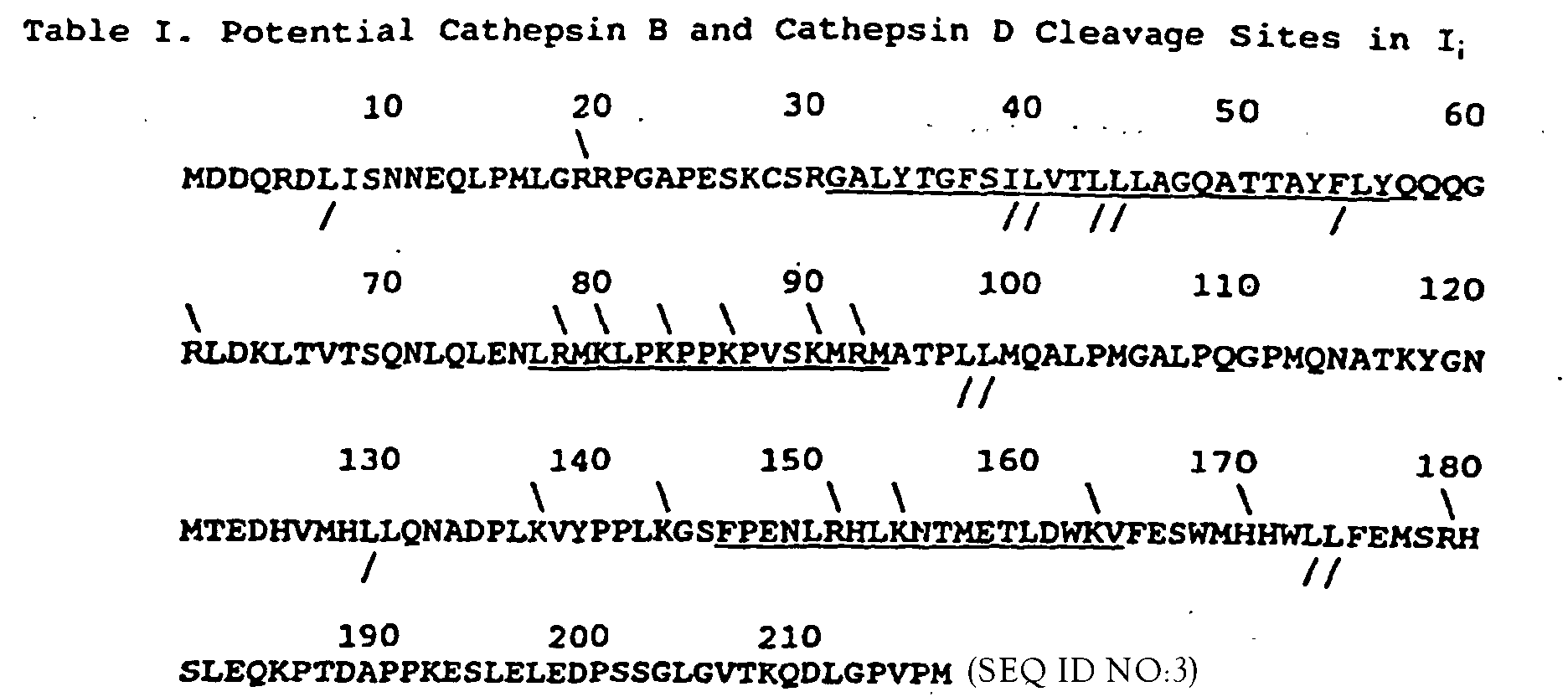
- In Table I, Cathepsin B sites are indicated by above the sequence and cathepsin D sites are indicated by / below the sequence. In each case, cleavage is hypothesized to occur at the carboxyl side of the indicated residue. The transmembranal region Gly31-Gln57, the hexa-cationic, tetraprolyl kink Leu77-Met93, and the α-helix Phe146-Val164 are underlined.
- Cleavage might also be restricted by local Ii structures (Table I), including: the transmembranal helix A32-L55; a tetraprolyl palindrome (L81-M93) with potential cathepsin B cleavage sites LR78, MX80, PK83, PK86, VSK90, MR92; a second tetraprolyl region P96-P111; an amphipathic helix F146-V164 with potential cathepsin B cleavage sites LK137, LK143, LR151, LK154, WK163, VK165; and a C-terminal tail with potential cathepsin D cleavage site LLF175. In order to address the functions of these putative cleavage sites, the following Ii gene mutations were generated: [R78→A;K80→A;K83→A;K86→T], [K137→A;K143→A], [R151→A;K154→A], and [L174→V; F175→A]. The cathepsin B and cathepsin D cleavage patterns of the protein products in COS1 cells transfected with mutant Ii and wild type HLA-DR α and β genes demonstrated staged cleavage of Ii by cathepsin D and cathepsin B. One can hypothesize if cathepsin D cleaves first at LLF175, then cathepsin B cleavage at LK137 and LK143 could release putative helix Phe146-Leu164 followed by peptide insertion into the antigen binding site. That helix would be rapidly destroyed by cathepsin B digestion at the six LR or LK positions in and around the helix. Final cleavages in the tight, tetraprolyl palindrome L81-M93 at one or more of six LK or LR sites could lead to generation of small Ii fragments which remain associated with α,β chains. MA(18-29) might bind to MHC class II α,β chains at the stage of cathepsin B cleavage at LK137 and LK143 or cathepsin B cleavage at the tetraprolyl palindrome LK131-M93. Further functional studies with these and related Ii mutants will resolve the mechanism for peptide insertion into the MHC class II antigen binding site and lead to assays for the selection of allele-non-specific immunosuppressive agents.
- Mutations were constructed in the Ii gene corresponding to one putative cathepsin D site and three clusters of putative cathepsin B sites were constructed. A mutation was also made at the cytoplasmic dibasic site R19R20. Four strategic principles were used in the design of the mutants. (1) Putative cleavage sites should lead to the fragments found experimentally after cathepsin B or cathepsin D digestion. For example, since cathepsin B produces a N-terminal 21 kDa fragment of Ii (Reyes, V.E. et al., 1991, J. Immunol., 146:3877-3880), cleavages at K137 or K143 could produce that fragment. (2) Protease-specific cleavage motifs were identified. Cathepsin B was suspected to attack the unusual frequency of clustered LK and LR residues and cathepsin D was reported to prefer paired hydrophobic residues except when the C-terminal one is Val, Gly or Ala (Bond, J.S., 1989, Commercially available proteases. In: Proteolytic Enzymes: A Practical Approach, edited by R.J. Beynon, et al., pp. 232, IRL Press, New York). (3) The facts that both p21 and naturally occurring Ii digestion products, e.g., L97-G119, bound to MHC class II α,β chains (Chicz, R.M. et al., 1992, Nature, 358:764-768), led to the suggestion that cleavage sites producing p21 and p25 are C-terminal to that region. (4) Hypothesizing local Ii structures with clusters of putative cathepsin B cleavage sites (Table I) led to a model for the staged cleavage and release of Ii by cathepsin B and cathepsin D.
- The mutant Ii genes constructed with oligonucleotides of Table II were transfected with HLA-DR1 α and β genes into COS1 cells after incorporation into a RSV.5(neo) plasmid into which human HLA-DR1 α,β genes (RSV-5(neo)-DRα120 and RSV.5(gpt)-DRβ008) had been cloned and driven by Rous sarcoma virus (RSV) long terminal repeat (LTR) sequences (Long et al., 1991). The Ii gene (45 1-p33-143) (Sekaly et al., 1986) was used for making Ii mutations. The mutant and WT Ii genes were cloned into a RSV.5 (hygromycin) vector. All the genes and the cloning vector were the gift of Dr. Eric Long of NIH. The HLA-DR1 α and β genes were reconstructed into one plasmid by digestion of RSV.5 (neo) DR1α 120 with Nde 1 to linearize the plasmid and of RSV.5 (gpt) DR1β 008 with Nde 1 and Bgl II to release the DR1β gene. Linearized RSV.5 (neo) DR1α 120 plasmid was treated with calf intestinal phosphatase and ligated to the purified DR1β gene fragment. The product was treated with T4 DNA polymerase to fill the unmatched ends, and religated.
- Site-directed mutagenesis was performed by the polymerase chain reaction (PCR) method of Ho et al. (1989) using the oligodeoxynucleotides of Table II. PCRs were carried out using Taq polymerase according to the manufacturer's instructions (Perkin Elmer Cetus, Norwalk CT). One oligodeoxynucleotide (oligo A) corresponding to the RSV.5 vector sequence at Sal 1 site was synthesized to polymerize in a 3' direction during the PCR and a second DNA oligodeoxynucleotide (oligo B) corresponding to the sequence of the 3' untranslated region of the Ii gene from 26 nucleotide to 43 nucleotide 3' to the stop codon (Strubin et al., 1984) was synthesized to polymerize in a 5' direction during the PCR. Sal 1 and BamH 1 sites were created in these two oligodeoxynucleotides, respectively, for cloning purposes. Other oligodeoxynucleotides were synthesized according to the sequences where the mutations were to be made (Table 1). PCR reactions were performed in 50 µl volumes containing: 50 mM KCl, 10 mM Tris-NaCl, pH 8.3, 4 mM MgCl2, 100 µM of each dNTP, 200 ng of each oligonucleotide, and 2.5 units of Taq polymerase. These samples were overlaid with 50 µl of mineral oil. PCR reactions were performed using a DNA Thermal Cycler (Perkin Elmer Cetus) programmed as follows: one cycle of denaturation (94°C, 2 min); annealing (temperature was calculated according to Ho et al., 2 min); extension (72°C, 2 min). Another 30 cycles of amplification were followed under conditions for the first cycle except 94°C was set for 1 min. Mutagenesis was carried out in two steps: amplification of DNA fragments from the plasmid template and overlap extension joining DNA fragments purified in a low melting gel. Second round PCR products were digested with Sal 1 and BamH 1 and ligated to an RSV.5 vector, which contained a hygromycin resistance gene (Long et al., 1991) and was digested with Sal 1 and BamH 1. All molecular biological techniques were performed according to Sambrook et al. (1989) and mutations were confirmed by DNA sequencing.

- COS1 cells were obtained from Dr. P. Newberger of the University of Massachusetts Medical Center (Worcester, MA). The cells were maintained in RPMI-1640 medium supplemented with 2% fetal calf serum and 8% bovine calf serum (Hyclone Laboratories, Inc., Logan, Utah) in 10 cm tissue culture dishes (Falcon, Lincoln Parker) at 37°C in a 5% CO2 atmosphere. COS1 cells did not express Ii as tested by immunoprecipitations with either VIC-Y1 or E1 antibodies. VIC-Y1 recognizes all Ii molecules, while E1 recognizes only high mannose forms of Ii. VIC-Y1 mAb to an N-terminal Ii determinant (Quaranta et al., 1984) was the gift of Dr. Walter Knapp in An Der Grub, G.m.b.H. (Kaumberg, Austria). E1 anti-human Ii(183-193) serum was prepared previously (Thomas et al., 1988). IVA12 mAb to human MHC class II molecules was produced as ascites from the hybridoma HB145 from the ATCC.
- Gene transfection was carried out by electroporation according to Xu and Stavnezer (1992). COS1 cells were grown for 24 h, released with trypsin-EDTA (GIBCO, BRL), washed twice with warmed RPMI-1640 medium without serum, and resuspended in RPMI-1640 medium without serum at 6 x 10⁶ cells/ml. Transfections were carried out at 250 V/1200 µF/ml volume with a PG200 Progenitor II electroporater (Hoefer Scientific Instruments, San Francisco, CA). Each plasmid was used at 24 µg per 6 x 10⁶ cells. After transfection, cells were recultured in complete RPMI-1640 medium at 3 x 10⁶ cells/8 ml/dish. Electroporation efficiently introduced DNA into the COS1 cells. 40-65% of COS1 cells expressed HLA-DR1 molecules at the cell surface 45 h after transfection as determined by immunofluorescent staining.
- 45 h after transfection, cells were labeled for 3 h with [35S]methionine (Sambrook et al., 1989). After pouring off the medium, the dishes were washed twice with 10 ml of warmed, methionine-free, serum-free RPMI-1640 medium and 1.5 ml of warmed, methionine-free RPMI-1640 medium containing 2% FCS and 8% BCS (both dialyzed) was added. Dishes were incubated for 20 min at 37°C in 5% CO2. To each dish was added 0.15 mCi of [35S]methionine (NEN, MA), followed by incubation for 3 h with the rocking of the dishes about every 20 min. After 3 h the dishes were washed twice with cool PBS and then 1 ml of lysis buffer (10 mM Tris-HCl, pH 7.4, and 0.5% Triton-X100) was added. The dishes were held on ice for 20 min and the cells were scraped from the dish and transferred to a tube. After vortexing for 1 min, lysates were centrifuged in a microcentrifuge at 4°C for 10 min. The supernatants were collected in a second tube and cleared with formalinized Staphylococcus aureus Cowen Strain A (Chemicon, E1 Segundo, CA) and used immunoprecipitations. Immunoadsorbants were prepared by incubating 100 µl of protein A-sepharose (Sigma) with either 1 µl of IVA12 mAb, 1 µl of VIC-Y1 mAb, or 25 µl of E1 antiserum. The immunoadsorbants were washed 5 times with buffer (50 mM Tris-HCl, pH 7.4, 150 mM NaCl, 5 mM EDTA, 0. 05% Triton-X100, and 0.02% of NaN3) and, eluted with SDS sample buffer. The samples were analyzed by SDS-PAGE and autoradiography (Reyes et al., 1991).
- Forty-five h after transfections with genes for mutant and wild type Ii chains without HLA-DR α,β genes, the COS1 cells were labeled 3 h with [35S]methionine, immunoprecipitates by E1 anti-Ii(183-193) serum were formed and digested with endoglycosidase H. The samples were examined by SDS-PAGE and autoradiography. The five Ii mutant proteins were expressed as strongly as was wild type Ii. Because the Ii chains were sensitive to endoglycosidase H treatment, they contained high mannose polysaccharide chains. They were presumably sequestered in the rough endoplasmic reticulum, without further processing of carbohydrate side chains, since Marks et al. (1990, J. Cell Biol., 111:839-855) showed that Ii can be sequestered in the rough endoplasmic reticulum without sialylation and with endoglycosidase H sensitivity when the Ii is not bound to class II α, β chains. Since VIC-Y1 mAb did not recognize mutant [R19R20] while it did the other mutant and wild type Ii chains (data not shown), that mutation presumably altered the VIC-Y1 determinant, thus further defining the apparent recognition site of VIC-Y1 at the N-terminus of Ii (Quaranta, V. et al., 1984, J. Immunol., 132:1900-1905).
- Cathepsin B digestions were carried out according to Reyes et al. (1991, J. Immunol., 146:3877-3880). The cell lysate from about 5 x 10⁵ cells in 150 µl was brought to pH 5.0 with 0.1 M sodium citrate (pH 2.5). The lysates were treated with cathepsin B in the presence of 1 mM DTT and 1 mM EDTA at room temperature for 1 h, or for other times as indicated. After enzymatic or control digestions, the samples were brought to pH 7.4 with 10 mM Tris (pH 9.5) containing 2 mM PMSF, 10 mM NEM, and 1 mM iodoacetamide and immunoprecipitates with IVA12 anti-MHC class II α,β chains were prepared in order to analyze Ii fragments still bound to MHC class II α,β chains. In order to analyze Ii fragments both bound to and released from MHC class II α,β chains, immunoprecipitates were digested under the same conditions as above in 30 µl volumes and the reactions were terminated with 3X SDS sample buffer (1X SDS sample buffer contained: 62.5 mM Tris-HCl, pH 6.8, 10% (w/v) glycerol, 5% 2-mercaptoethanol, and 2.3% SDS) and heating at 100°C. The samples were applied directly to the electrophoretic gels. Immunoprecipitations with anti-MHC class II mAb IVA-12 of MHC class II α,β and Ii triple transfectants demonstrated that wild type and all mutant Ii chains associated with the class II α,β chains and formed approximately equal quantities of trimeric complexes. Digestions of these complexes with cathepsin B, however, revealed different patterns of Ii cleavage (Table III). All of the mutants except [R151K154] had reduced levels or the absence of certain of the fragments occurring with cathepsin B digestion of wild type Ii. The principal wild type forms of Ii seen with digestion of wild type Ii were p21, p14, p10, and p6. In contrast to the wild type Ii digestion pattern, mutant [L174F175] had a trace amount of p21 and mutant [R78K80K83K86] did not produce p14, p10, and p6. Also, mutants [R19R20] and [K137K143] had weak p21 bands which were about one tenth as strong as the wild type p21 band (comparing the patterns with 5 units cathepsin B with mutant [R19R20] and [K137K143] versus 0.5 unit cathepsin B with wild type Ii).
Appearance of fragments of wild type and mutant Ii chains after digestion with cathepsin B. Wild Types or Mutant Ii CB FRAGMENTS CD FRAGMENTS p21 p14 p10 p6 p21 p14 p10 p6 WT + + + + + + + + M[R19→A; R20→A] +/- +/- p12 + + +/- - + M[R78→A;K80→A; K83→A ;K86→T] + - - - + - - - M[K137→A;K143→A] +/- + + + + +/- + +/- M(R151→A;K154-A] + +/- + + + + + + M[L174→V;F175→A] 6 + + + - + + + - The letters in parenthesis indicate where the mutations were made. For example, in M[R19→A;R20→A] the arginines in positions 19 and 20 were replaced by alanines. For simplicity throughout this disclosure, the replacing residue is omitted, e.g., [R19→A;R20→A] is abbreviated to [R19R20]. +/- indicates the band from the mutant Ii is weaker than the corresponding one from WT Ii. Fragments of comparable size from digestions with CB or CD are not implied to be cleaved at identical sites.
- The digestion patterns of wild type Ii and mutant [R78K80K83K86] indicated that p14, p10, and p6 were degradation products of p21. In order to clarify the relationship among these smaller Ii fragments one to another, the class II MHC α,β chain complexes with wild type or mutants Ii chains were digested with a series of cathepsin B concentrations including the relatively high dose of 30 U/ml. At that cathepsin B dose, Ii and all intermediate Ii fragments were cleaved except for resistant p6 which was still associated with MHC class II α,β chains in immunoprecipitates with IVA-12 mAb to the α,β chains. This result and the finding that the integrated density of the high dose cathepsin B-generated p6 band was about the sum of the p14, p10 and p6 bands seen at lower dose cathepsin B digestions indicates that p6 is a degradation product of p14 and p10.
- Previously it was reported that cathepsin B and cathepsin D cleaved and released Ii from MHC class II molecules without apparent damage to the α,β chains (Reyes, V.E. et al., 1991, J. Immunol., 146:3877-3880). However, in light of the finding that some Ii mutants protect the class II β chain from loss of 1-2 kilodaltons, it is clear that removal of wild type Ii is associated with a cleavage of the β-chain presumably at K12.
- CD digestions were carried out according to Reyes et al. (1991). The cell lysate from about 5 x 10⁵ cells in 150 µl was brought to pH 5.0 with 0.1 M sodium citrate (pH 2.5). The lysates were treated with cathepsin D (Sigma) in the presence of 1 mM EDTA, at room temperature for 1 h, or for other times as indicated. After enzymatic or control digestions, the samples were brought to pH 7.4 with 10 mM Tris (pH 9.5) containing 2 mM PMSF, 10 mM NEM, and 1 mM iodoacetamide and immunoprecipitates were prepared to analyze Ii fragments still bound to MHC class II α,β chains. In order to analyze Ii fragments both bound to and released from MHC class II α,β chains immunoprecipitates were digested under the same conditions as above in 30 µl volumes and the reactions were terminated with 3X SDS sample buffer (IX SDS sample buffer contained: 62.5 mM Tris-HCl, pH 6.8, 10% (w/v) glycerol, 5% 2-mercaptoethanol, and 2.3% SDS) and heating at 100°C. The samples were applied directly to the electrophoretic gels.
- Upon cathepsin D digestion of transfected trimers, p21 was produced from all of the mutant and wild type Ii, except for mutant [L174F175] (Table III). L173L174F175 thus appears to be a cathepsin D cleavage site. In mutant [L174F175] actually more p14, p10, and p6 were produced, indicating that once the L173L174F175 site was altered, other cathepsin D cleavage sites predominated and that these smaller fragments might be derived from p21. The Ii fragmentation pattern of cathepsin D digestion generally paralleled that of cathepsin B digestion, probably because the cathepsin D sites were in the vicinity of cathepsin B sites. However, some differences were found. (1) Cathepsin D digestion did not lead to premature release of Ii fragments. Mutants [R78K80K83K86] and [K137K143] were somewhat resistant to cathepsin D digestion and had reduced levels of p21 and increased intact Ii relative to the observations with cathepsin B. (2) With these latter two mutants, the p21 band formed clearly upon cathepsin D digestion; but in the case of cathepsin B digestion, the p21 band was more of a smear.
- In order to examine whether the cathepsin B-derived p21 from mutants [R19R20], [K137K143], and [L174F175] was produced, or degraded, or dissociated from HLA-DR1 α,β chains, class II α,β trimers were isolated by immunoprecipitation with IVA-12 mAb and then digested them with cathepsin B and analyzed all products by SDS-PAGE. Immunoprecipitates were digested under the same conditions as in Example 8 in 30 µl volumes and the reactions were terminated with 3X SDS sample buffer (1X SDS sample buffer contained: 62.5 mM Tris-HCl, pH 6.8, 10% (w/v) glycerol, 5% 2-mercaptoethanol, and 2.3% SDS) and heating at 100°C. The samples were applied directly to the electrophoretic gels.
- Cathepsin B generated p21 from mutants [R19R20] and [K137K143] as strongly as it did with wild type Ii, indicating that p21 was produced from these two mutants but that it was promptly dissociated from DR1 α,β chains. In mutant [K137K143], cathepsin B cleavage might occur at R151 or K154, or still at K137 or K143, to produce p21. However, the native conformation at or near K137K143 is the most preferred site for cathepsin B cleavage to produce p21 which is retained on the α, β chains. If cathepsin B does not cleave at K131K143 first, then Ii dissociates from the MHC class II α, β chains. Mutation at R19R20 also led to premature release of p21 after cathepsin B digestion. In the case of mutant [L147F175], p28 and p16 are produced instead of p21, suggesting that mutation at the L174F175 site prevents not only cathepsin D cleavage there but also the cathepsin B cleavage at K137K143. p18 and p16 were released from MHC class II α, β chains after they were produced. The patterns of low molecular weight bands in this experiment were similar to those after high dose cathepsin B digestions showing only p6, with little or no p14 and p10. Probably in these immunoprecipitations, there was less competitive inhibition by other lysate proteins, leading to patterns comparable to the high dose cathepsin B digestions.
- Since the demonstration by DeLisi and Berzofsky that T cell-presented epitopes can be coiled as amphipathic helices (DeLisi, C. et al., 1985, Proc. Natl. Acad. Sci. USA, 82:7048-7052), theories for the selection of such epitopes based on mechanisms of such motifs have been tested (Margalit, H., et al., 1987, J. Immunol., 138:2213-2229; Rothbard, J.B. et al., 1988, EMBO J., 7:93-100; Stille, C.J. et al., 1987, Mol. Immunol., 24:1021-1027; Reyes, V.E. et al., 1988, Mol. Immunol., 25:867-871). Finding such a "perfect" amphipathic helix in Ii(146-164) led to the hypothesis that this helix was a surrogate antigen and was capable of blocking the peptide binding site of MHC class II nolecules (Elliott, W.L. et al., 1987, J. Immunol., 138:2949-2952; Stille, C.J. et al., 1987, Mol. Immunol., 24:1021-1027). The synthetic peptide PH-1.0 was designed to mimic Ii (146-164) structurally in terms of having a narrow, longitudinal hydrophobic strip-of-helix. A series of analogs of PH-1.0 have also been designed to test the effects of systematic substitutions of threonine for leucine within the longitudinal strip-of-helix on helical coiling of peptides against lipid micelles (Lu, S. et al., 1991, J. Biol. Chem., 266:10054-10057).
- The effect of PH-1.0 on antigen presentation was tested in a functional assay. PH-1.0 blocked the presentation of pigeon cytochrome C(81-104) peptide to specific, Ek-restricted TPc9.1 murine T cell hybridoma cells (Table IV). The competitive inhibition assay was carried out as follows. 5x10⁴ paraformaldehyde-fixed CH27 B lymphoma cells (antigen presenting cells) were incubated in 96-well plates with 5x10⁴ TPc9.1 T hybridoma cells (irradiated 2200 rads) for 24 hrs in the presence of 6 µM (81-104) antigenic peptide and different concentrations of the PH-1.0 peptide. Each condition was assayed in triplicate. After incubation, the response of the T cell hybridoma was determined by measuring the IL-2/IL-4 secreted during the 24 hr culture. Culture supernatants were collected and were tested for IL-2/IL-4 content by the ability to support the growth of the IL-2/IL-4-dependent cell line HT-2 as measured by the incorporation of [3H]thymidine. The % inhibition was calculated as: 100 - [(CPM of T+APC+peptide+PH-1.0 / CPM of T+APC+peptide) x 100].
PH-1.0 Inhibits the MHC Class II-Restricted Presentation of Antigenic Peptide to T cells PH-1.0 Concentration 100 µM 50 µM 10 µM 1 µM 0 µM % Inhibition 100 98 61 10 0 - Ii(148-164) (SEQ ID NO:2) and Ii(78-92) peptides enhanced responses to pigeon cytochrome C(81-104) peptide by the specific, Ek-restricted murine T cell hybridoma, TPc9.1 (Table V). The assay was carried out as follows. 5x10⁴ paraformaldehyde-fixed CH27 B lymphoma cells (antigen presenting cells) were incubated in 96-well plates with 5x10⁴ TPc9.1 T hybridoma cells (irradiated 2200 rads) for 24 hrs in the presence of: no peptides, 50 µM Ii peptide only, 6 µM pigeon cytochrome C(81-104) peptide only, and 50 µM Ii peptide plus 6 µM pigeon cytochrome C(81-104) peptide. Each condition was assayed in sextuplet. After incubation, the response of the T cell hybridoma was determined by measuring the IL-2/IL-4 secreted during the 24 hr culture. The supernatants were collected and were tested for IL-2/IL-4 content by the ability to support the growth of the IL-2/IL-4-dependent cell line HT-2 as measured by [3H] thymidine incorporation. The values presented in Table V are the CPM x 10-3 incorporated in the HT-2 indicator assay.
Ii Peptides Enhance the MHC Class II-Restricted Presentation of Pigeon Cytochrome C Peptide (81-104) to T cells Ii (148-164) (SEQ ID NO:2) Ii(78-92) (T+APC) 5.4 5.4 (T+APC) +Ii peptide 6.9 5.9 (T+APC) +PGCC(81-104) 50.8 50.8 (T+APC) +Ii peptide +PGCC(81-104) 110.2 121.7 - The enhancement of responses to pigeon cytochrome C peptide (81-104) by Ii(148-164) and Ii(78-92) was assayed as a function of concentration of the added Ii peptide (Table VI). The values in the Table represent the % enhancement of the pigeon cytochrome C(81-104) peptide-specific, Ek-restricted response by the TPc9.1 murine T cell hybridoma (T) to the peptide presented by paraformaldehyde-fixed CH27 B lymphoma cells (APC). Culture supernatants were collected and were tested for IL-2/IL-4 content by the ability to support the growth of the Il-2/IL-4-dependent cell Line HT-2 as measured by the incorporation of [3H]thymidine. The % enhancement was calculated as: [(CPM of T+APC+peptide+Ii peptide / CPM of T+APC+peptide) x 100]-100.
Ii Peptides Enhance the MHC Class II-Restricted Presentation of Antigenic Peptide to T cells Peptide Concentration 100 µM 50 µM 10 µM 1 µM 0 µM Ii(148-164) (SEQ ID NO:2) 140 91 19 0 0 Ii(78-92) 170 187 101 40 0 -
- (1) GENERAL INFORMATION:
- (i) APPLICANT: Antigen Express, Inc.
- (ii) TITLE OF INVENTION: Regulation of Antigen Presentation
- (iii) NUMBER OF SEQUENCES: 16
- (iv) CORRESPONDENCE ADDRESS:
- (A) ADDRESSEE: Kevin H. Farrell, P.C.
- (B) STREET OR P.O. BOX: P.O. Box 999
- (C) CITY: York Harbor
- (D) STATE: ME
- (E) COUNTRY: USA
- (F) ZIP: 03911
- (v) COMPUTER READABLE FORM:
- (A) MEDIUM TYPE: Floppy disk
- (B) COMPUTER: IBH PC compatible
- (C) OPERATING SYSTEM: PC-DOS/MS-DOS
- (D) SOFTWARE: PatentIn Release #1.0, Version #1.25
- (vii) PRIOR APPLICATION DATA:
- (A) APPLICATION NUMBER: 08/064,400
- (B) FILING DATE: May 19, 1993
- (C) CLASSIFICATION:
- (viii) ATTORNEY/AGENT INFORMATION:
- (A) NAME: Farrell, Kevin M.
- (B) REGISTRATION NUMBER: 35,505
- (C) REFERENCE/DOCKET NUMBER: REH-9301 WO
- (ix) TELECOMMUNICATION INFORMATION:
- (A) TELEPHONE: 207-363-0558
- (B) TELEFAX: 207-363-0528
- (2) INFORMATION FOR SEQ ID NO:1:
- (i) SEQUENCE CHARACTERISTICS:
- (A) LENGTH: 16 amino acids
- (B) TYPE: amino acid
- (C) STRANDEDNESS: single
- (D) TOPOLOGY: linear
- (ii) MOLECULE TYPE: peptide
- (xi) SEQUENCE DESCRIPTION: SEQ ID NO:1:

- (2) INFORMATION FOR SEQ-ID NO:2:
- (i) SEQUENCE CHARACTERISTICS:
- (A) LENGTH: 17 amino acids
- (B) TYPE: amino acid
- (C) STRANDEDNESS: single
- (D) TOPOLOGY: linear
- (ii) MOLECULE TYPE: peptide
- (xi) SEQUENCE DESCRIPTION: SEQ ID NO:2:

- (2) INFORMATION FOR SEQ ID NO:3:
- (i) SEQUENCE CHARACTERISTICS:
- (A) LENGTH: 12 amino acids
- (B) TYPE: amino acid
- (C) STRANDEDNESS: single
- (D) TOPOLOGY: linear
- (ii) MOLECULE TYPE: peptide
- (xi) SEQUENCE DESCRIPTION: SEQ ID. NO:3:

- (2) INFORMATION FOR SEQ ID NO:4:
- (i) SEQUENCE CHARACTERISTICS:
- (A) LENGTH: 216 amino acids
- (B) TYPE: amino acid
- (C) STRANDEDNESS: single
- (D) TOPOLOGY: linear
- (ii) MOLECULE TYPE: protein
- (xi) SEQUENCE DESCRIPTION: SEQ ID NO:4:
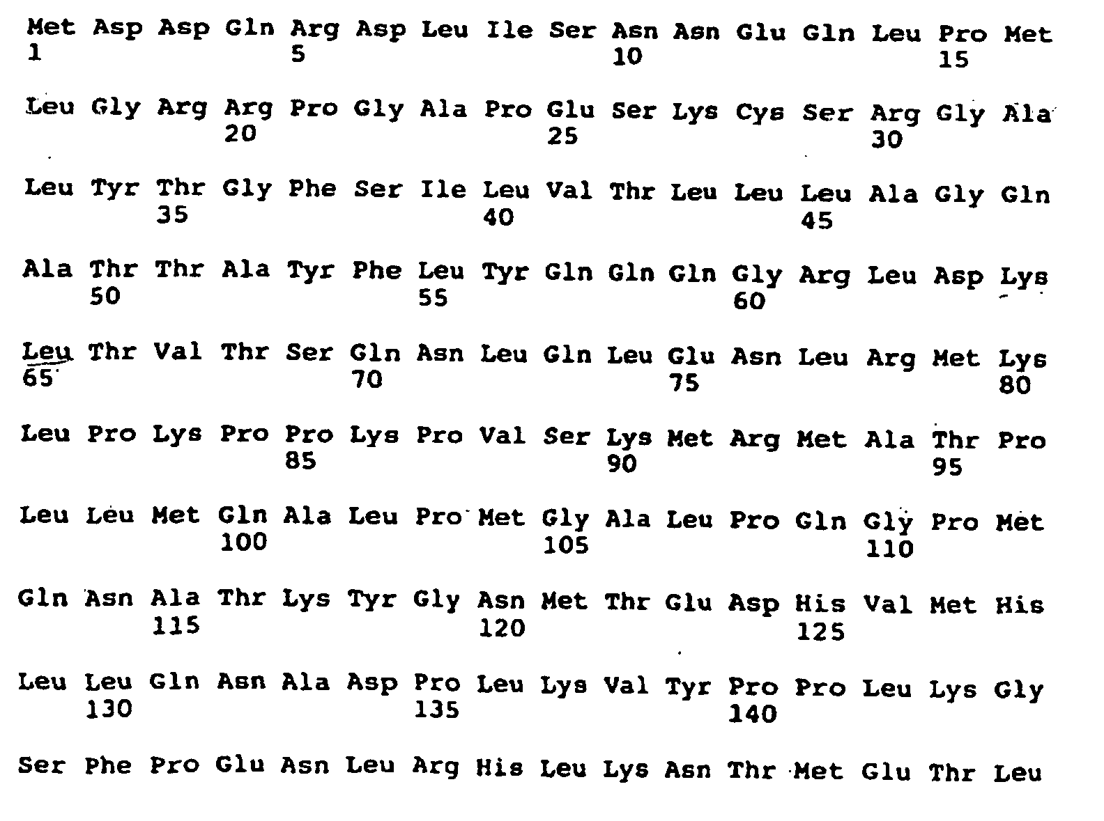
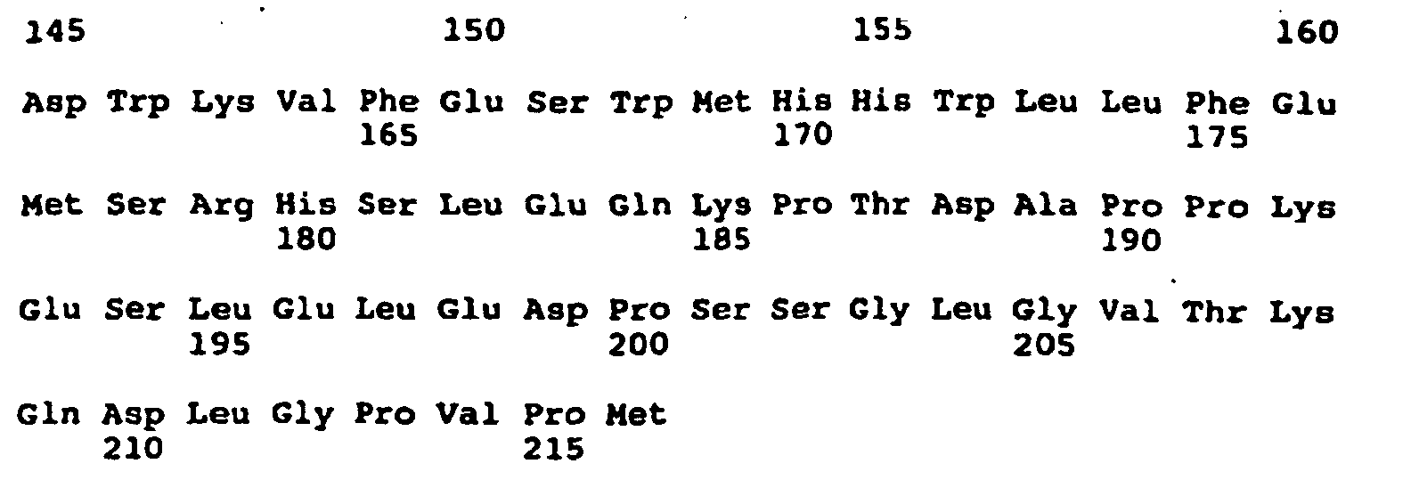
- (2) INFORMATION FOR SEQ ID NO:5:
- (i) SEQUENCE CHARACTERISTICS:
- (A) LENGTH: 21 base pairs
- (B) TYPE: nucleic acid
- (C) STRANDEDNESS: double
- (D) TOPOLOGY: linear
- (ii) MOLECULE TYPE: DNA (genomic)
- (xi) SEQUENCE DESCRIPTION: SEQ ID NO:5:

- (2) INFORMATION FOR SEQ ID NO:6:
- (i) SEQUENCE CHARACTERISTICS:
- (A) LENGTH: 25 base pairs
- (B) TYPE: nucleic acid
- (C) STRANDEDNESS: double
- (D) TOPOLOGY: linear
- (ii) MOLECULE TYPE: DNA (genomic)
- (xi) SEQUENCE DESCRIPTION: SEQ ID NO:6:

- (2) INFORMATION FOR SEQ ID NO:7:
- (i) SEQUENCE CHARACTERISTICS:
- (A) LENGTH: 17 base pairs
- (B) TYPE: nucleic acid
- (C) STRANDEDNESS: double
- (D) TOPOLOGY: linear
- (ii) MOLECULE TYPE: DNA (genomic)
- (xi) SEQUENCE DESCRIPTION: SEQ ID NO:7:

- (2) INFORMATION FOR SEQ ID NO:8:
- (i) SEQUENCE CHARACTERISTICS:
- (A) LENGTH: 17 base pairs
- (B) TYPE: nucleic acid
- (C) STRANDEDNESS: double
- (D) TOPOLOGY: linear
- (ii) MOLECULE TYPE: DNA (genomic)
- (xi) SEQUENCE DESCRIPTION: SEQ ID NO:8:

- (2) INFORMATION FOR SEQ ID NO:9:
- (i) SEQUENCE CHARACTERISTICS:
- (A) LENGTH: 27 base pairs
- (B) TYPE: nucleic acid
- (C) STRANDEDNESS: double
- (D) TOPOLOGY: linear
- (ii) MOLECULE TYPE: DNA (genomic)
- (xi) SEQUENCE DESCRIPTION: SEQ ID NO:9:

- (2) INFORMATION FOR SEQ ID NO:10:
- (i) SEQUENCE CHARACTERISTICS:
- (A) LENGTH: 22 base pairs
- (B) TYPE: nucleic acid
- (C) STRANDEDNESS: double
- (D) TOPOLOGY: linear
- (ii) MOLECULE TYPE: DNA (genomic)
- (xi) SEQUENCE DESCRIPTION: SEQ ID NO:10:

- (2) INFORMATION FOR SEQ ID NO:11:
- (i) SEQUENCE CHARACTERISTICS:
- (A) LENGTH: 20 base pairs
- (B) TYPE: nucleic acid
- (C) STRANDEDNESS: double
- (D) TOPOLOGY: linear
- (ii) MOLECULE TYPE: DNA (genomic)
- (xi) SEQUENCE DESCRIPTION: SEQ ID NO:11:

- (2) INFORMATION FOR SEQ ID NO:12:
- (i) SEQUENCE CHARACTERISTICS:
- (A) LENGTH: 19 base pairs
- (B) TYPE: nucleic acid
- (C) STRANDEDNESS: double
- (D) TOPOLOGY: linear
- (ii) MOLECULE TYPE: DNA (genomic)
- (xi) SEQUENCE DESCRIPTION: SEQ ID NO:12:

- (2) INFORMATION FOR SEQ ID NO:13:
- (i) SEQUENCE CHARACTERISTICS:
- (A) LENGTH: 18 base pairs
- (B) TYPE: nucleic acid
- (C) STRANDEDNESS: double
- (D) TOPOLOGY: linear
- (ii) MOLECULE TYPE: DNA (genomic)
- (xi) SEQUENCE DESCRIPTION: SEQ ID NO:13:

- (2) INFORMATION FOR SEQ ID NO:14:
- (i) SEQUENCE CHARACTERISTICS:
- (A) LENGTH: 18 base pairs
- (B) TYPE: nucleic acid
- (C) STRANDEDNESS: double
- (D) TOPOLOGY: linear
- (ii) MOLECULE TYPE: DNA (genomic)
- (xi) SEQUENCE DESCRIPTION: SEQ ID NO:14:

- (2) INFORMATION FOR SEQ ID NO:15:
- (i) SEQUENCE CHARACTERISTICS:
- (A) LENGTH: 17 base pairs
- (B) TYPE: nucleic acid
- (C) STRANDEDNESS: double
- (D) TOPOLOGY: linear
- (ii) MOLECULE TYPE: DNA (genomic)
- (xi) SEQUENCE DESCRIPTION: SEQ ID NO:15:

- (2) INFORMATION FOR SEQ ID NO:16:
- (i) SEQUENCE CHARACTERISTICS:
- (A) LENGTH: 17 base pairs
- (B) TYPE: nucleic acid
- (C) STRANDEDNESS: double
- (D) TOPOLOGY: linear
- (ii) MOLECULE TYPE: DNA (genomic)
- (xi) SEQUENCE DESCRIPTION: SEQ ID NO:16:

-
Claims (2)
- An in-vitro method for enhancing presentation of an MHC class II restricted antigenic peptide to a T cell, comprising:(a) forming a reaction mixture comprising:i) an MHC Class II expressing antigen presenting cell;ii) the peptide YRMKLPKPPKPVSKMR (SEQ ID NO:1);iii) the MHC Class II restricted antigenic peptide which, when added to the incubation mixture, is not in association with an antigen presenting cell; andiv) a T cell which is responsive to the MHC Class II restricted antigenic peptide of part iii); and(b) incubating the reaction mixture of step a) under conditions appropriate for interaction between the antigen presenting cell and the T cell.
- An in-vitro method for enhancing presentation of an MHC class II restricted antigenic peptide to a T cell, comprising:(a) forming a reaction mixture comprising:i) an MHC Class II expressing antigen presenting cell;ii) the peptide ENLRHLKNTMETLDWKV (SEQ ID NO:2);iii) the MHC Class II restricted antigenic peptide which, when added to the incubation mixture, is not in association with an antigenic presenting cell; andiv) a T cell which is responsive to the MHC Class II restricted antigenic peptide of part iii); and(b) incubating the reaction mixture of step a) under conditions appropriate for interaction between the antigen presenting cell and the T cell.
Applications Claiming Priority (3)
| Application Number | Priority Date | Filing Date | Title |
|---|---|---|---|
| US64400 | 1993-05-19 | ||
| US08/064,400 US5559028A (en) | 1993-05-19 | 1993-05-19 | Methods of enhancing or antigen presentation to T cells inhibiting |
| PCT/US1994/005617 WO1994026773A1 (en) | 1993-05-19 | 1994-05-18 | Regulation of antigen presentation |
Publications (3)
| Publication Number | Publication Date |
|---|---|
| EP0702692A1 EP0702692A1 (en) | 1996-03-27 |
| EP0702692A4 EP0702692A4 (en) | 1998-06-10 |
| EP0702692B1 true EP0702692B1 (en) | 2005-12-21 |
Family
ID=22055708
Family Applications (1)
| Application Number | Title | Priority Date | Filing Date |
|---|---|---|---|
| EP94917443A Expired - Lifetime EP0702692B1 (en) | 1993-05-19 | 1994-05-18 | Regulation of antigen presentation |
Country Status (6)
| Country | Link |
|---|---|
| US (1) | US5559028A (en) |
| EP (1) | EP0702692B1 (en) |
| JP (2) | JP3534761B2 (en) |
| CA (1) | CA2163196C (en) |
| DE (1) | DE69434582T2 (en) |
| WO (1) | WO1994026773A1 (en) |
Families Citing this family (20)
| Publication number | Priority date | Publication date | Assignee | Title |
|---|---|---|---|---|
| JPH10503364A (en) * | 1994-05-10 | 1998-03-31 | ザ ジェネラル ホスピタル コーポレーション | Antisense inhibition of hepatitis C virus |
| US6509165B1 (en) * | 1994-07-08 | 2003-01-21 | Trustees Of Dartmouth College | Detection methods for type I diabetes |
| US6245904B1 (en) * | 1995-09-14 | 2001-06-12 | The University Of Tubingen | Recombinant polypeptide based on the primary sequence of the invariant chain with at least one primary sequence of a specific T-cell epitope or a protein derivative and nucleic acids coding for this recombinant polypeptide |
| DK0912601T4 (en) * | 1996-04-22 | 2009-03-09 | Massachusetts Inst Technology | Suppression of immune response by inhibition of cathepsin S |
| US5726020A (en) * | 1996-06-11 | 1998-03-10 | University Of Massachusetts | Inhibition of II synthesis |
| AU3307497A (en) * | 1996-06-26 | 1998-01-14 | Antigen Express, Inc. | Immunotherapy by modulation of antigen presentation |
| US6326465B1 (en) * | 1997-02-24 | 2001-12-04 | The Johns Hopkins University | Immunomodulatory polypeptides derived from the invariant chain of MHC class II |
| EP1108018A1 (en) * | 1998-08-31 | 2001-06-20 | AstraZeneca AB | Human deltex-like gene zdx |
| US9289487B2 (en) * | 1999-09-14 | 2016-03-22 | Antigen Express, Inc. | II-key/antigenic epitope hybrid peptide vaccines |
| US6432409B1 (en) * | 1999-09-14 | 2002-08-13 | Antigen Express, Inc. | Hybrid peptides modulate the immune response |
| US20030235594A1 (en) * | 1999-09-14 | 2003-12-25 | Antigen Express, Inc. | Ii-Key/antigenic epitope hybrid peptide vaccines |
| AU2002245086A1 (en) * | 2000-10-31 | 2002-07-24 | The General Hospital Corporation | Detection and alteration of mhc class ii presentation phenotypes |
| EP1383919B1 (en) * | 2001-03-29 | 2010-07-07 | Cytokine Pharmasciences, Inc. | Methods and compositions for using mhc class ii invariant chain polypeptide as a receptor for macrophage migration inhibitory factor |
| CA2499123A1 (en) * | 2002-09-17 | 2004-04-15 | Antigen Express, Inc. | Ii-key/antigenic epitope hybrid peptide vaccines |
| US7179645B2 (en) * | 2002-09-24 | 2007-02-20 | Antigen Express, Inc. | Ii-Key/antigenic epitope hybrid peptide vaccines |
| US20080095798A1 (en) * | 2006-10-18 | 2008-04-24 | Robert Humphreys | Ii-key enhanced vaccine potency |
| WO2010027973A1 (en) | 2008-09-02 | 2010-03-11 | Antigen Express, Inc. | Human papillomavirus / li-key hybrids and methods of use |
| AU2009317691A1 (en) * | 2008-11-21 | 2010-05-27 | Kobenhavns Universitet (University Of Copenhagen) | Priming of an immune response |
| CA2750922A1 (en) * | 2009-01-28 | 2010-08-05 | Antigen Express, Inc. | Li-key hybrid peptides that modulate the immune response to influenza |
| AU2012320597C1 (en) | 2011-10-07 | 2015-10-15 | Baxalta GmbH | oxMIF as a diagnostic marker |
-
1993
- 1993-05-19 US US08/064,400 patent/US5559028A/en not_active Expired - Lifetime
-
1994
- 1994-05-18 WO PCT/US1994/005617 patent/WO1994026773A1/en not_active Ceased
- 1994-05-18 EP EP94917443A patent/EP0702692B1/en not_active Expired - Lifetime
- 1994-05-18 CA CA002163196A patent/CA2163196C/en not_active Expired - Lifetime
- 1994-05-18 DE DE69434582T patent/DE69434582T2/en not_active Expired - Lifetime
- 1994-05-18 JP JP52581694A patent/JP3534761B2/en not_active Expired - Fee Related
-
2003
- 2003-12-17 JP JP2003419891A patent/JP4019040B2/en not_active Expired - Fee Related
Non-Patent Citations (1)
| Title |
|---|
| Sette et al: 'Invariant chain peptides in most HLA-DR molecules of an antigen-processing mutant', 1992, Science, vol : 256, pages 1801-1804. * |
Also Published As
| Publication number | Publication date |
|---|---|
| JP2004161777A (en) | 2004-06-10 |
| EP0702692A1 (en) | 1996-03-27 |
| CA2163196A1 (en) | 1994-11-24 |
| CA2163196C (en) | 2008-02-12 |
| DE69434582D1 (en) | 2006-01-26 |
| US5559028A (en) | 1996-09-24 |
| JP4019040B2 (en) | 2007-12-05 |
| EP0702692A4 (en) | 1998-06-10 |
| JPH09509561A (en) | 1997-09-30 |
| WO1994026773A1 (en) | 1994-11-24 |
| JP3534761B2 (en) | 2004-06-07 |
| DE69434582T2 (en) | 2006-10-26 |
Similar Documents
| Publication | Publication Date | Title |
|---|---|---|
| CA2163196C (en) | Regulation of antigen presentation | |
| Parham et al. | Inhibition of alloreactive cytotoxic T lymphocytes by peptides from the α2 domain of HLA–A2 | |
| Ljunggren et al. | Empty MHC class I molecules come out in the cold | |
| Jones et al. | Isolation of complementary DNA clones encoding the human lymphocyte glycoprotein T1/Leu-1 | |
| Shastri et al. | Endogenous generation and presentation of the ovalbumin peptide/Kb complex to T cells | |
| Dick et al. | Proteolytic processing of ovalbumin and beta-galactosidase by the proteasome to a yield antigenic peptides. | |
| Nilsson et al. | Short cytoplasmic sequences serve as retention signals for transmembrane proteins in the endoplasmic reticulum | |
| US5880103A (en) | Immunomodulatory peptides | |
| Cope et al. | T cell responses to a human cartilage autoantigen in the context of rheumatoid arthritis–associated and nonassociated HLA–DR4 alleles | |
| Hosken et al. | An endogenous antigenic peptide bypasses the class I antigen presentation defect in RMA-S. | |
| US5665590A (en) | Method for isolating and directly cloning genes which encode cell-surface and secreted proteins | |
| WO1994004171A9 (en) | Immunomodulatory peptides | |
| Malcherek et al. | MHC class II‐associated invariant chain peptide replacement by T cell epitopes: engineered invariant chain as a vehicle for directed and enhanced MHC class II antigen processing and presentation | |
| Rich et al. | HLA class II-induced translocation of PKC alpha and PKC beta II isoforms is abrogated following truncation of DR beta cytoplasmic domains | |
| Cerundolo et al. | CD8 independence and specificity of cytotoxic T lymphocytes restricted by HLA-Aw68. 1 | |
| Günther et al. | Cytotoxic T lymphocytes of the rat are predominantly restricted by RTL1. A and Not RT1. C-determined major histocompatibility class I antigens | |
| Wheeler et al. | The amino-terminal domain of the v-fms oncogene product includes a functional signal peptide that directs synthesis of a transforming glycoprotein in the absence of feline leukemia virus gag sequences | |
| Zhang et al. | Immunological heterogeneity of autoreactive T lymphocytes against the nicotinic acetylcholine receptor in myasthenic patients | |
| EP1224291A1 (en) | Multiple sclerosis-related superantigen | |
| KR100369286B1 (en) | Prokaryotic Expression of MHC Proteins | |
| Kittlesen et al. | Presentation of newly synthesized glycoproteins to CD4+ T lymphocytes. An analysis using influenza hemagglutinin transport mutants. | |
| Rodgers et al. | Surface expression of β2‐microglobulin‐associated thymus‐leukemia antigen is independent of TAP2 | |
| CA2614785C (en) | Regulation of antigen presentation | |
| Zhu et al. | A recombinant single‐chain human class II MHC molecule (HLA‐DR1) as a covalently linked heterotrimer of α chain, β chain, and antigenic peptide, with immunogenicity in vitro and reduced affinity for bacterial superantigens | |
| Liu et al. | Suppression of the indirect pathway of T cell reactivity by high doses of allopeptide |
Legal Events
| Date | Code | Title | Description |
|---|---|---|---|
| PUAI | Public reference made under article 153(3) epc to a published international application that has entered the european phase |
Free format text: ORIGINAL CODE: 0009012 |
|
| 17P | Request for examination filed |
Effective date: 19951214 |
|
| AK | Designated contracting states |
Kind code of ref document: A1 Designated state(s): DE FR GB IT |
|
| A4 | Supplementary search report drawn up and despatched |
Effective date: 19980428 |
|
| AK | Designated contracting states |
Kind code of ref document: A4 Designated state(s): DE FR GB IT |
|
| RHK1 | Main classification (correction) |
Ipc: C12N 15/12 |
|
| 17Q | First examination report despatched |
Effective date: 20000904 |
|
| GRAP | Despatch of communication of intention to grant a patent |
Free format text: ORIGINAL CODE: EPIDOSNIGR1 |
|
| GRAS | Grant fee paid |
Free format text: ORIGINAL CODE: EPIDOSNIGR3 |
|
| GRAA | (expected) grant |
Free format text: ORIGINAL CODE: 0009210 |
|
| AK | Designated contracting states |
Kind code of ref document: B1 Designated state(s): DE FR GB IT |
|
| PG25 | Lapsed in a contracting state [announced via postgrant information from national office to epo] |
Ref country code: IT Free format text: LAPSE BECAUSE OF FAILURE TO SUBMIT A TRANSLATION OF THE DESCRIPTION OR TO PAY THE FEE WITHIN THE PRESCRIBED TIME-LIMIT;WARNING: LAPSES OF ITALIAN PATENTS WITH EFFECTIVE DATE BEFORE 2007 MAY HAVE OCCURRED AT ANY TIME BEFORE 2007. THE CORRECT EFFECTIVE DATE MAY BE DIFFERENT FROM THE ONE RECORDED. Effective date: 20051221 |
|
| REG | Reference to a national code |
Ref country code: GB Ref legal event code: FG4D |
|
| REF | Corresponds to: |
Ref document number: 69434582 Country of ref document: DE Date of ref document: 20060126 Kind code of ref document: P |
|
| PG25 | Lapsed in a contracting state [announced via postgrant information from national office to epo] |
Ref country code: GB Free format text: LAPSE BECAUSE OF NON-PAYMENT OF DUE FEES Effective date: 20060518 |
|
| ET | Fr: translation filed | ||
| PLBE | No opposition filed within time limit |
Free format text: ORIGINAL CODE: 0009261 |
|
| STAA | Information on the status of an ep patent application or granted ep patent |
Free format text: STATUS: NO OPPOSITION FILED WITHIN TIME LIMIT |
|
| 26N | No opposition filed |
Effective date: 20060922 |
|
| GBPC | Gb: european patent ceased through non-payment of renewal fee |
Effective date: 20060518 |
|
| REG | Reference to a national code |
Ref country code: DE Ref legal event code: R082 Ref document number: 69434582 Country of ref document: DE Representative=s name: MAI DOERR BESIER PATENTANWAELTE, DE |
|
| PGFP | Annual fee paid to national office [announced via postgrant information from national office to epo] |
Ref country code: FR Payment date: 20131108 Year of fee payment: 20 Ref country code: DE Payment date: 20131202 Year of fee payment: 20 |
|
| REG | Reference to a national code |
Ref country code: DE Ref legal event code: R071 Ref document number: 69434582 Country of ref document: DE |
|
| REG | Reference to a national code |
Ref country code: DE Ref legal event code: R071 Ref document number: 69434582 Country of ref document: DE |
|
| PG25 | Lapsed in a contracting state [announced via postgrant information from national office to epo] |
Ref country code: DE Free format text: LAPSE BECAUSE OF EXPIRATION OF PROTECTION Effective date: 20140520 |