EP0698093B1 - VON HIPPEL-LINDAU (VHL) DISEASE GENE AND CORRESPONDING cDNA AND METHODS FOR DETECTING CARRIERS OF THE VHL DISEASE GENE - Google Patents
VON HIPPEL-LINDAU (VHL) DISEASE GENE AND CORRESPONDING cDNA AND METHODS FOR DETECTING CARRIERS OF THE VHL DISEASE GENE Download PDFInfo
- Publication number
- EP0698093B1 EP0698093B1 EP94916774A EP94916774A EP0698093B1 EP 0698093 B1 EP0698093 B1 EP 0698093B1 EP 94916774 A EP94916774 A EP 94916774A EP 94916774 A EP94916774 A EP 94916774A EP 0698093 B1 EP0698093 B1 EP 0698093B1
- Authority
- EP
- European Patent Office
- Prior art keywords
- vhl
- seq
- sequence
- disease gene
- primers
- Prior art date
- Legal status (The legal status is an assumption and is not a legal conclusion. Google has not performed a legal analysis and makes no representation as to the accuracy of the status listed.)
- Expired - Lifetime
Links
Images


Classifications
-
- C—CHEMISTRY; METALLURGY
- C07—ORGANIC CHEMISTRY
- C07K—PEPTIDES
- C07K16/00—Immunoglobulins [IG], e.g. monoclonal or polyclonal antibodies
- C07K16/18—Immunoglobulins [IG], e.g. monoclonal or polyclonal antibodies against material from animals or humans
-
- C—CHEMISTRY; METALLURGY
- C07—ORGANIC CHEMISTRY
- C07K—PEPTIDES
- C07K14/00—Peptides having more than 20 amino acids; Gastrins; Somatostatins; Melanotropins; Derivatives thereof
- C07K14/435—Peptides having more than 20 amino acids; Gastrins; Somatostatins; Melanotropins; Derivatives thereof from animals; from humans
- C07K14/46—Peptides having more than 20 amino acids; Gastrins; Somatostatins; Melanotropins; Derivatives thereof from animals; from humans from vertebrates
- C07K14/47—Peptides having more than 20 amino acids; Gastrins; Somatostatins; Melanotropins; Derivatives thereof from animals; from humans from vertebrates from mammals
- C07K14/4701—Peptides having more than 20 amino acids; Gastrins; Somatostatins; Melanotropins; Derivatives thereof from animals; from humans from vertebrates from mammals not used
- C07K14/4702—Regulators; Modulating activity
- C07K14/4703—Inhibitors; Suppressors
-
- C—CHEMISTRY; METALLURGY
- C12—BIOCHEMISTRY; BEER; SPIRITS; WINE; VINEGAR; MICROBIOLOGY; ENZYMOLOGY; MUTATION OR GENETIC ENGINEERING
- C12Q—MEASURING OR TESTING PROCESSES INVOLVING ENZYMES, NUCLEIC ACIDS OR MICROORGANISMS; COMPOSITIONS OR TEST PAPERS THEREFOR; PROCESSES OF PREPARING SUCH COMPOSITIONS; CONDITION-RESPONSIVE CONTROL IN MICROBIOLOGICAL OR ENZYMOLOGICAL PROCESSES
- C12Q1/00—Measuring or testing processes involving enzymes, nucleic acids or microorganisms; Compositions therefor; Processes of preparing such compositions
- C12Q1/68—Measuring or testing processes involving enzymes, nucleic acids or microorganisms; Compositions therefor; Processes of preparing such compositions involving nucleic acids
- C12Q1/6876—Nucleic acid products used in the analysis of nucleic acids, e.g. primers or probes
- C12Q1/6883—Nucleic acid products used in the analysis of nucleic acids, e.g. primers or probes for diseases caused by alterations of genetic material
- C12Q1/6886—Nucleic acid products used in the analysis of nucleic acids, e.g. primers or probes for diseases caused by alterations of genetic material for cancer
-
- G—PHYSICS
- G01—MEASURING; TESTING
- G01N—INVESTIGATING OR ANALYSING MATERIALS BY DETERMINING THEIR CHEMICAL OR PHYSICAL PROPERTIES
- G01N33/00—Investigating or analysing materials by specific methods not covered by groups G01N1/00 - G01N31/00
- G01N33/48—Biological material, e.g. blood, urine; Haemocytometers
- G01N33/50—Chemical analysis of biological material, e.g. blood, urine; Testing involving biospecific ligand binding methods; Immunological testing
- G01N33/53—Immunoassay; Biospecific binding assay; Materials therefor
- G01N33/575—Immunoassay; Biospecific binding assay; Materials therefor for cancer
- G01N33/57575—Immunoassay; Biospecific binding assay; Materials therefor for cancer involving oncogenic proteins
-
- C—CHEMISTRY; METALLURGY
- C12—BIOCHEMISTRY; BEER; SPIRITS; WINE; VINEGAR; MICROBIOLOGY; ENZYMOLOGY; MUTATION OR GENETIC ENGINEERING
- C12Q—MEASURING OR TESTING PROCESSES INVOLVING ENZYMES, NUCLEIC ACIDS OR MICROORGANISMS; COMPOSITIONS OR TEST PAPERS THEREFOR; PROCESSES OF PREPARING SUCH COMPOSITIONS; CONDITION-RESPONSIVE CONTROL IN MICROBIOLOGICAL OR ENZYMOLOGICAL PROCESSES
- C12Q2600/00—Oligonucleotides characterized by their use
- C12Q2600/112—Disease subtyping, staging or classification
-
- C—CHEMISTRY; METALLURGY
- C12—BIOCHEMISTRY; BEER; SPIRITS; WINE; VINEGAR; MICROBIOLOGY; ENZYMOLOGY; MUTATION OR GENETIC ENGINEERING
- C12Q—MEASURING OR TESTING PROCESSES INVOLVING ENZYMES, NUCLEIC ACIDS OR MICROORGANISMS; COMPOSITIONS OR TEST PAPERS THEREFOR; PROCESSES OF PREPARING SUCH COMPOSITIONS; CONDITION-RESPONSIVE CONTROL IN MICROBIOLOGICAL OR ENZYMOLOGICAL PROCESSES
- C12Q2600/00—Oligonucleotides characterized by their use
- C12Q2600/156—Polymorphic or mutational markers
Definitions
- the invention is in the field of tumor suppressor genes. More specifically, the invention relates to the Von Hippel-Lindau (VHL) disease gene and its corresponding cDNA and to methods for detecting carriers of the VHL disease gene using probes derived from the cDNA.
- VHL Von Hippel-Lindau
- VHL disease is a familial cancer syndrome. This disease in an autosomal dominant disorder and patients who are heterozygous for mutations in the VHL disease gene are predisposed to a variety of cancers, the most frequent being hemangioblastomas of the central nervous system and retina, renal cell carcinoma (RCC) and pheochromocytoma.
- RRC renal cell carcinoma
- pheochromocytoma The multisystem character of the illness, combined with the fact multiple tumors may form in each target organ, produces considerable morbidity and mortality as evidenced by the reduction in life expectancy of affected individuals to 49 years (McKusick, V.A., Mendelian Inheritance in Man (1983) Johns Hopkins University Press, Baltimore and London, p 534-535).
- VHL disease Although the prevalence of VHL disease is only 1 in 36,000, because of its late onset most individuals have children before they realize they have inherited VHL disease. For many years, the only method of presymptomatic or prenatal diagnosis of the disease has been periodic examination of the eye, brain, and abdomen in all asymptomatic members of VHL families. Unfortunately, examination of all target organs is required to ensure detection of disease that may be limited to a single organ. In addition to the obvious inconvenience and the cost of these examinations, they have the additional drawback that they may not yield definitive diagnostic information. Therefore, in order to develop a method which allows the unequivocal diagnosis of VHL disease in individuals at risk, researchers have focused intensive efforts on identifying and isolating the VHL disease gene.
- VHL disease gene is a member of the family of tumor suppressor genes (Tory, K. et al. J. Natl. Canc. Inst. (1989) 81:1097-1101; Maher, E.R. et al. J. Med. Genet. (1990) 27:311-314) and that it behaves in accordance with Knudson's theory of human carcinogenesis (Knudson, A., Proc. Natl. Acad Sci. USA (1971) 68:816-823).
- the identification of DNA markers tightly linked to the VHL disease gene has allowed localization of the VHL disease gene to human chromosome 3p25-p26. (Hosoe, S.
- VHL disease gene located in the VHL region has been cloned (Latif, F. et al. Cancer Res. (1993) 63:861-867).
- this gene was found to detect no mutations in VHL patients and thus, there are currently no available methods which can identify carriers of the VHL disease gene with 100% accuracy.
- the recent identification and isolation of the VHL disease gene (Latif et al., Science, in press, "Identification of the von Hippel-Lindau Disease Tumor Suppressor Gene") and its corresponding cDNA should allow the development of diagnostic methods which provide unequivocal detection of carriers of the VHL disease gene.
- the present invention relates to the von Hippel-Lindau (VHL) disease gene and its corresponding cDNA.
- VHL von Hippel-Lindau
- the invention further relates to methods for detecting carriers of the VHL disease gene.
- the first method comprises analyzing DNA of a subject for mutations of the VHL disease gene associated with VHL disease.
- the second method comprises analyzing RNA of a subject for mutations or alterations in the VHL-specific mRNA associated with VHL disease.
- the third method comprises analyzing protein of a subject for alterations in VHL protein expression associated with VHL disease.
- the invention also encompasses recombinant VHL proteins derived from the VHL cDNA and antibodies directed against said VHL proteins or peptides derived therefrom.
- the invention further relates to a method for treating a carrier of VHL disease gene in which an expression vector containing a nucleic acid sequence representing wild-type VHL gene is administered to the carrier.
- the invention also provides a diagnostic kit for detecting carriers of the VHL disease gene.
- the kit comprises purified and isolated nucleic acid sequences useful as PCR primers in analyzing DNA or RNA for mutations of the VHL disease gene associated with VHL disease.
- Figure 1 shows a genetic and physical map of the chromosome 3p region encompassing the VHL gene. Genetic and physical distances between selected markers are shown in centiMorgans and kilobases respectively. The location of selected cross-overs is indicated by crosses.
- Figure 1 shows the 160 kb cosmid and phage contig covering the VHL region.
- Figures 2A and 2B show Northern blot analysis of the expression of the gene represented by g7 cDNA in various human tissues.
- Figure 2(A) shows low resolution blot containing 2 ⁇ g poly A + mRNA, the tissues are indicated above the lanes.
- Figure 2(B) shows a high resolution blot containing 1 ⁇ g of poly A + mRNA from: lane 1, fetal brain; lane 2, adult brain; lane 3, fetal kidney; lane 4, adult kidney; lane 5, cerebellum; lane 6, adult adrenal; and lane 7, prostate.
- the sizes of the transcripts were determined by the position of the 28S and 18S rRNA bands.
- Figures 3A-3E show detection by Southern blotting analysis of rearrangement mutations in constitutional DNA of VHL affected patients using g7 cDNA as probe.
- Figure 3A DNA from lymphoblastoid cell lines of 7 unrelated VHL patients was digested with EcoRI and analyzed by standard blotting procedures. The normal invariant band is about 20 to 22 kb, the sizes of the aberrant bands probably resulting from intragenic deletions range from 4 to 25 kb. The patients code numbers are indicated above the lanes.
- Figure 3B DNAs from lymphoblastoid cell lines of pedigree members from a new mutation family (coded "S") digested with DraI, HindIII, and PstI.
- Figure 4 shows Southern blot analysis of genomic DNA of VHL patients (only the initials of each patients name are given). The DNAs were digested with EcoRI and probed using different regions of g7 cDNA. Panel A: Total g7 cDNA probe; Panel B: 5' end probe, nucleotides 3-146; Panel C: 3' end probe nucleotides 1277-1600.
- Figures 5A and 5B show the results of polymerase chain reaction-single stranded conformation analysis (PCR-SSCP) of the genomic DNA of VHL patients with the 8 bp insertion mutation (Table 1). Portions of the DNA sequencing gels are shown that display normal ( Figure 5A) and 714insTTGTCCGT mutation sequences ( Figure 5B). The DNA sequence is of the antisense strand; therefore, the inserted bases are 5'ACGGACAA3'. Adjacent to sequencing ladder are shown the positions of the insertion, and the nature of the insertion, as predicted from the sequence.
- PCR-SSCP polymerase chain reaction-single stranded conformation analysis
- Figure 6 shows the results of a "zoo" blot illustrating evolutionary conservation of the putative VHL gene.
- the g7 cDNA shows cross species homology to DNA from mammals, birds, fly, and sea urchin.
- the present invention relates to the VHL disease gene and its corresponding cDNA.
- the region of human chromosome 3 containing the VHL disease gene has been cloned by genomic walking with yeast artificial chromosomes (YACS) and the cloned DNA recovered with cosmids from a chromosome 3 specific library (Latif et al. Science, in press).
- the phage 191 which contains the VHL disease gene was deposited with the American Type Culture Collection (ATCC), 12301 Parklawn Drive, Rockville, MD 20852 on May 13, 1993 (accession number 69311).
- This VHL disease gene represents the wild-type VHL gene where wild-type means the gene not causing VHL disease.
- the present invention is also directed to a cDNA corresponding to the VHL disease gene.
- This cDNA sequence designated g7, is set forth below as SEQ ID NO: 1 and was deposited with the American Type Culture Collection on May 13, 1993 (accession number 69312).
- This cDNA also has GenBank accession No. L15409.
- nucleotides are those standardly used in the art.
- the deduced amino acid sequence of the g7 cDNA is shown as SEQ ID NO:2 below and starts at nucleotide 1 of SEQ ID NO:1 and extends 851 nucleotides.
- analog includes any protein or polypeptide having an amino acid residue sequence substantially identical to a sequence specifically shown herein in which one or more amino acid residues have been conservatively substituted with a functionally similar residue and which displays the functional aspects of the VHL protein as described herein.
- conservative substitutions include, for example, the substitution of one non-polar (i.e. hydrophobic) residue such as isoleucine, valine, leucine or methionine for another; the substitution of one polar (i.e.
- hydrophilic residue for another such as a substitution between arginine and lysine, between glutamine and asparagine, or between glycine and serine; the substitution of one basic residue such as lysine, arginine or histidine for another; or the substitution of one acidic residue, such as aspartic acid or glutamic acid for another.
- conservative substitution may also include the use of a chemically derivatized residue in place of a non-derivatized residue provided that the resulting protein or polypeptide displays the requisite functional activity.
- Chemical derivative refers to a VHL protein or polypeptide having one or more residues chemically derivatized by reaction of a functional side group.
- derivatized molecules include, but are not limited to, those molecules in which free amino groups have been derivatized to form, for example, amine hydrochlorides, p-toluene sulfonyl groups, carbobenzoxy groups, t-butyloxycarbonyl groups, chloroacetyl groups or formyl groups.
- Free carboxyl groups may be derivatized to form salts, methyl and ethyl esters, or other types of esters or hydrazides.
- Free hydroxyl groups may be derivatized to form O-acyl or O-alkyl derivatives.
- the imidazole nitrogen of histidine may be derivatized to form N-im-benzylhistidine.
- chemical derivatives those proteins or peptides which contain one or more naturally-occurring amino acid derivatives of the twenty standard amino acids. For example, 4-hydroxyproline may be substituted for proline; 5-hydroxylysine may be substituted for lysine; 3-methylhistidine may be substituted for histidine; homoserine may be substituted for serine; and ornithine may be substituted for lysine.
- a VHL protein or polypeptide of the present invention also includes any protein or polypeptide having one or more additions and/or deletions of residues relative to the sequence of a protein or polypeptide whose sequence is shown herein, so long as the requisite activity is maintained.
- the present invention also relates to methods for detecting carriers of the VHL disease gene.
- the methods for detection disclosed in the present invention can be used prenatally to screen a fetus or presymptomatically to screen a subject at risk through his/her family history.
- these methods can be used to determine the involvement of the VHL disease gene in other human malignancies such as kidney, lung and bladder cancers.
- the method for detecting carriers of the VHL disease gene comprises analyzing the DNA of a subject for mutations of the VHL disease gene associated with VHL disease.
- subject means a mammal and mutation means inversion, translocation, insertion, deletion or point mutation of the VHL disease gene.
- a biological specimen is obtained from the subject.
- biological specimens that can be obtained for use in the present method include, but are not limited to, tissue biopsies, whole blood, urine, feces or other samples normally tested in the diagnosis of disease.
- Preferred biological specimens are whole blood or urine.
- nucleic acid can be extracted from contaminating cell debris and other protein substances by extraction of the sample with phenol.
- phenol extraction the aqueous sample is mixed with an approximately equal volume of redistilled phenol and centrifuged to separate the two phases.
- the aqueous phase containing the nucleic acid is removed and precipitated with ethanol to yield nucleic acid free of phenol.
- DNA can be purified from the biological sample according to Sidransky, D. et al. (Science (1992) 256:102-105; Science (1991) 252:706) or by the method of Glenn et al. (Glenn, G.M. et al. JAMA (1992) 267:1226-1231).
- the DNA to be analyzed can be either single- or double-stranded.
- Methods for analyzing the DNA for mutations in the VHL disease gene include Southern blotting after digestion with the appropriate restriction enzymes (restriction fragment length polymorphism, RFLP) (Botstein, D. Amer. J. Hum. Genet. (1980) 69:201-205), denaturing gradient electrophoresis technique (Myers, R.M., Nature (1985) 313:495-498), oligonucleotide hybridization (Conner, R. et al., EMBO J. (1984) 3:13321-1326), RNase digestion of a duplex between a probe RNA and the target DNA (Winter, E. et al., Proc. Natl. Acad. Sci. U.S.A.
- RFLP restriction fragment length polymorphism
- DNA is analyzed by Southern analysis.
- the DNA to be analyzed via Southern analysis is digested with one or more restriction enzymes.
- the restriction enzymes to be used in the present invention are those enzymes for whom the presence or absence of their recognition site is linked to VHL disease.
- Preferred restriction enzyme include EcoRI, HindIII, PstI, DraI, BamHI, BglI, BglII, and PvuII.
- resultant DNA fragments are separated by gel electrophoresis and the fragments are detected by hybridization with a labelled nucleic acid probe (Southern, E.M. J. Mol. Biol. (1975) 98:503-517).
- the nucleic acid sequence used as a probe in Southern analysis can be labeled in single-stranded or double-stranded form. Labelling of the nucleic acid sequence can be carried out by techniques known to one skilled in the art. Such labelling techniques can include radiolabels and enzymes (Sambrook, J. et al. (1989) in "Molecular Cloning, A Laboratory Manual", Cold Spring Harbor Press, Plainview, New York). In addition, there are known non-radioactive techniques for signal amplification including methods for attaching chemical moieties to pyrimidine and purine rings (Dale, R.N.K. et al. (1973) Proc. Natl. Acad. Sci., 70:2238-2242; Heck, R.F. 1968) S. Am.
- Chem. Soc., 90:5518-5523 methods which allow detection by chemiluminescence (Barton, S.K. et al. (1992) J. Am. Chem. Soc., 114:8736-8740) and methods utilizing biotinylated nucleic acid probes (Johnson, T. K. et al. (1983) Anal. Biochem., 133:126-131; Erickson, P.F. et al. (1982) J. of Immunology Methods, 51:241-249; Matthaei, F.S. et al. (1986) Anal. Biochem., 157:123-128) and methods which allow detection by fluorescence using commercially available products.
- the size of the probe can range from about 200 nucleotides to about several kilobases. A preferred probe size is about 500 to about 2000 nucleotides.
- Each of the nucleic acid sequences used as a probe in Southern analysis is substantially homologous to the corresponding portion of the cDNA sequence shown in SEQ ID NO:1.
- substantially homologous is meant a level of homology between the nucleic acid sequence used as a probe and the corresponding sequence shown in SEQ ID NO:1.
- the level of homology is in excess of 70%, most preferably in excess of 80%, with a particularly preferred nucleic acid sequence being in excess of 90% homologous with the sequence shown in SEQ ID NO:1.
- the DNA is analyzed for mutations in the VHL disease gene by PCR-SSCP (Orita et al., (1989), Dean et al., (1990)).
- each of the pairs of primers selected for use in PCR are designed to hybridize with sequences in the VHL disease gene which are an appropriate distance apart (at least about 50 nucleotides) in the gene to permit amplification and subsequent detection of mutations in the amplification product.
- Primer pairs which can specifically hybridize to such VHL gene sequences can be derived from the VHL disease gene sequence.
- the primers are derived from the cDNA sequence shown in SEQ ID NO.1.
- Each primer of a pair is a single-stranded oligonucleotide of about 15 to about 50 bases in length which is complementary to a sequence at the 3' end of one of the strands of a double-stranded target sequence.
- Each pair comprises two such primers, one of which is complementary 3' end and the other of which is complementary to the other 3' end of the target sequence.
- the target sequence is generally about 100 to about 300 base pairs long but can be as large as 500-600 base pairs. Optimization of the amplification reaction to obtain sufficiently specific hybridization to the VHL disease gene is well within the skill in the art and is preferably achieved by adjusting the annealing temperature.
- the present invention also provides purified and isolated pairs of primers for use in analysis of DNA for mutations in the VHL gene.
- the nucleic acid sequences of these primers is set forth below as SEQ ID NOs:3-8.
- SEQ ID NO. 3 and SEQ ID NO. 4 represent one pair of primers
- SEQ ID NO. 5 and SEQ ID NO. 6 represent a second pair of primers
- SEQ ID NO. 7 and SEQ ID NO. 8 represent a third pair of primers.
- the primers of this invention can be synthesized using any of the known methods of oligonucleotide synthesis (e.g., the phosphodiester method of Agarwal et al. 1972. Agnew. Chem. Int. Ed. Engl. 11 :451, the phosphotriester method of Hsiung et al. 1979. Nucleic Acids Res. 6 :1371, or the automated diethylphosphoramidite method of Beuacage et al. 1981. Tetrahedron Letters 22 :1859-1862), or they can be isolated fragments of naturally occurring or cloned DNA.
- the known methods of oligonucleotide synthesis e.g., the phosphodiester method of Agarwal et al. 1972. Agnew. Chem. Int. Ed. Engl. 11 :451, the phosphotriester method of Hsiung et al. 1979. Nucleic Acids Res. 6 :1371, or the automated diethylphosphoram
- oligonucleotides can be synthesized by automated instruments sold by a variety of manufacturers or can be commercially custom ordered and prepared.
- the primers can be derivatized to include a detectable label suitable for detecting and/or identifying the primer extension products (e.g., biotin, avidin, or radiolabeled dNTP's), or with a substance which aids in the isolation of the products of amplification (e.g. biotin or avidin).
- SEQ. ID. NO. 3 through SEQ. ID. NO. 8 are synthetic oligonucleotides.
- primer pairs can be selected to hybridize to mutant forms of the VHL disease gene.
- the selected primer pairs will hybridize sufficiently specifically to the mutated gene sequences such that non-specific hybridization to wild-type VHL gene sequences will not prevent identification of the amplification product of the mutant gene sequence.
- Primer pairs which hybridize to mutations in the VHL gene sequence can be used to amplify specific mutant gene sequences present in the DNA of a biological sample.
- the amplification products of PCR can be detected either directly or indirectly.
- direct detection of the amplification products is carried out via labelling of primer pairs.
- Labels suitable for labelling the primers of the present invention are known to one skilled in the art and include radioactive labels, biotin, avidin, enzymes and fluorescent molecules.
- the derived labels can be incorporated into the primers prior to performing the amplification reaction.
- a preferred labelling procedure utilizes radiolabeled ATP and T4 polynucleotide kinase (Sambrook, J. et al. (1989) in "Molecular Cloning, A Laboratory Manual", Cold Spring Harbor Press, Plainview, NY).
- the desired label can be incorporated into the primer extension products during the amplification reaction in the form of one or more labelled dNTPs.
- the labelled amplified PCR products can be analyzed for mutations of the VHL gene associated with VHL disease gene via separating the PCR products by denaturing polyacrylamide gel electrophoresis or via direct sequencing of the PCR-products.
- unlabelled amplification products can be analyzed for mutations in the VHL disease gene via hybridization with nucleic acid probes radioactively labelled or, labelled with biotin, in Southern blots or dot blots.
- Nucleic acid probes useful in the embodiment are those described earlier for Southern analysis.
- the method for detecting carriers of the VHL disease gene comprises analyzing the RNA of a subject for mutations or alterations in VHL-specific mRNA associated with VHL disease.
- RNA derived from blood or a tumor biopsy sample is obtained from said subject where said tumors include, but are not limited to, tumors of the eye brain, liver, kidney, pancreas, and pheochromocytomas.
- RNA to be analyzed can be isolated from blood or tumor biopsy samples as whole cell RNA or as poly(A) + RNA.
- Whole cell RNA can be isolated by methods known to those skilled in the art. Such methods include extraction of RNA by differential precipitation (Birnbiom, H.C. (1988) Nucleic Acids Res., 16:1487-1497), extraction of RNa by organic solvents (Chomczynski, P. et al. (1987) Anal. Biochem., 162:156-159) and extraction of RNA with strong denaturants (Chirgwin, J.M. et al. (1979) Biochemistry, 18:5294-5299).
- Poly(A) + RNA can be selected from whole cell RNA by affinity chromatography on oligo-d(T) columns (Aviv, H. et al. (1972) Proc. Natl. Acad. Sci., 69:1408-1412).
- a preferred method of isolating RNA is extraction of whole cell RNA by acid-phenol (Chomczynski et al. 1987).
- the methods for analyzing the RNA for alterations in the pattern or level of VHL specific mRNA expression linked to VHL disease include Northern blotting (Alwine, J.C. et al. (1977) Proc. Natl. Acad. Sci., 74:5350-5354), dot and slot hybridization (Kafatos, F.C. et al. (1979) Nucleic Acids Res., 7:1541-1522), filter hybridization (Hollander, M.C. et al. (1990) Biotechniques; 9:174-179), RNase protection (Sambrook, J. et al.
- RT-PCR reverse-transcription polymerase chain reaction
- the nucleic acid sequence used as a probe for detecting VHL-specific mRNA expression is substantially homologous to SEQ. ID. NO. 1.
- substantially homologous is meant a level of homology between the nucleic acid sequence and the cDNA sequence of SEQ ID NO.1.
- the level of homology is in excess of 70% more preferably in excess on 80%, with a particularly preferred nucleic acid sequence being in excess of 90% homologous with the cDNA sequence shown in SEQ ID No. 1.
- RT-PCR reverse transcription-polymerase chain reaction
- primers used to amplify the cDNA produced via reverse transcription of RNA are derived from the cDNA sequence shown in SEQ ID No. 1.
- These primers can be labelled as described earlier and the RT-PCR products can be analyzed for mutations of the VHL gene associated with VHL disease via denaturing polyacrylamide gel electrophoresis of the RT-PCR products or via direct sequencing of the RT-PCR products.
- the present invention also encompasses recombinant proteins derived from the cDNA shown in SEQ ID No. 1 and antibodies directed to said proteins (called VHL proteins).
- VHL proteins can be produced by recombinant DNA methodology known to one skilled in the art.
- a nucleic acid sequence capable of encoding a protein comprising all or part of the amino acid sequence shown in SEQ ID NO.2 can be cloned into a vector capable of being transferred into, and replicated in, a host organism.
- a suitable nucleic acid sequence for the purpose of this invention is the sequence shown in SEQ ID NO.1.
- Suitable expression vectors include, but are not limited to, vaccinia virus vectors include, baculovirus vectors, and Ecoli pTRCHIS (Invitrogen Co. San Diego).
- the recombinant expression vector produced by inserting a nucleic acid sequence capable of directing synthesis of VHL protein in a suitable expression vector can be transfected into E coli or into suitable eukaryotic cell systems by methods known to one skilled in the art.
- Cells containing the expressed recombinant VHL protein, cell lysate from cells transfected with a recombinant expression vector or a culture supernatant containing the expressed VHL protein can be used as an immunogen to elicit production of anti-VHL antibodies in a mammal.
- synthetic peptides for use as immunogens from the amino acid sequence shown in SEQ ID NO 2.
- Preferred synthetic peptide sequences for use as an immunogen are shown below:
- the immunogen While it is possible for the immunogen to be a administered to the mammal in pure or substantially pure form, it is preferable to present it as a pharmaceutical composition, formulation or preparation. Suitable mammals for immunization include mice, rabbits and the like.
- the anti-VHL antibody of the present invention is typically produced by immunizing a mammal with an immunologically effective amount of synthetic peptide of this invention.
- the preparation of polyclonal or monoclonal antibodies against such a peptide is well known in the art (Standt et al. (1988) J. Exp. Med. 157:687-704).
- anti-VHL peptide antibody molecules induced by immunization of a mammal with the recombinant VHL protein are then collected from the mammal and those immunospecific for the VHL protein are isolated to the extent desired by well known techniques such as, for example, immunochromatography.
- the method for detecting carriers of the VHL disease gene comprises analyzing the protein of a subject for alterations in VHL protein expression with VHL disease.
- protein is obtained from biological specimens such as tumor biopsy samples and urine and the like.
- the protein can be obtained as a crude lysate or it can be further purified by methods known to one skilled in the art (Sambrook, J. et al. (1989) in "Molecular Cloning, A Laboratory Manual", Cold Spring Harbor press, Plainview, NY).
- Crude protein lysate can be analyzed for VHL protein by immunoassays using anti-VHL antibody.
- Immunoassays of the present invention may be a radioimmunoassay, Western blot assay, immunofluorescent assay, enzyme immunoassay, chemiluminescent assay, immunohistochemical assay and the like. Standard techniques known in the art for ELISA are described in Method in Immunodiagnosis, 2nd Edition, Rose and Bigazzi, eds., John Wiley and Sons, 1980 and Campbell et al., Methods of Immunology, W.A. Benjamin, Inc., 1964, both of which are incorporated herein by reference. Such assays may be a direct, indirect, competitive, or noncompetitive immunoassay as described in the art. (Oellerich, M. 1984. J. Clin. Chem. Clin. BioChem. 22:895-904).
- Detection of the VHL protein anti-VHL antibody complex formed can be accomplished by reaction of the complex with a secondary antibody such as labelled anti-rabbit antibody.
- the label may be an enzyme which is detected by incubating the complex in the presence of a suitable fluorimetric or colorimetric reagent.
- Other detectable labels may also be used, such as radiolabels, or colloidal gold, and the like.
- the labelled VHL protein-anti-VHL antibody complex is then visualized by autoradiography.
- the present invention also relates to a method for treating a carrier of the VHL disease gene in which an expression vector containing a nucleic acid sequence representing the wild type VHL gene is administered to the carrier.
- a nucleic acid sequence representing wild-type VHL gene is that shown in SEQ ID No. 1.
- Such nucleic acid sequence may be inserted into a suitable expression vector by methods known to those skilled in the art (Example 5).
- Expression vectors suitable for producing high efficiency gene transfer in vivo include retroviral, adenoviral and vaccinia viral vectors.
- Expression vectors containing a nucleic acid sequence representing wild-type VHL gene can be administered intravenously, intramuscularly, subcutaneously, intraperitoneally or orally.
- a preferred route of administration is intravenously.
- the invention also provides a diagnostic kit for detecting carriers of the VHL disease gene.
- This diagnostic kit comprises purified and isolated nucleic acid sequences according to SEQ ID. No. 3 through SEQ ID No. 8, said sequences useful as PCR primers in analyzing DNA for mutations of the VHL disease gene linked to VHL disease.
- the subjects analyzed in the following examples were kindred identified by ophthalmologists, urologists, medical geneticists and neurosurgeons in the United States, Europe, and Canada.
- the members of the families resided in Louisiana, Tennessee, Mississippi, Virginia, Pennsylvania, New York, Michigan, Quebec, Nova Scotia, United Kingdom, and the Netherlands.
- Medical records of each family member known to be affected were reviewed.
- Asymptomatic family members and family members in whom there was uncertainty about the diagnosis were examined after informed consent for occult evidence of the illness at the Clinical Center of the National Institutes of Health. The examination consisted of a history and physical examination of the scrotum.
- An asymptomatic member of a VHL family was considered to be affected if one or more of the following disease manifestations were detected: retinal angioma(s), spinal or cerebellar hemangioblastoma(s), pheochromocytoma(s), multiple pancreatic systs, and multiple bilateral renal cysts accompanied by renal cell carcinoma.
- Disease diagnosis was made without knowledge of restriction fragment length polymorphism (RFLP) status.
- RFLP restriction fragment length polymorphism
- Restriction enzymes were from Bethesda Research Laboratory (BRL) (Bethesda, MD), New England Biolabs (Beverly, MA) and Boehringer Mannheim (Indianapolis, IN) and were used as recommended by the manufacturers.
- ⁇ - 32 PdCTP ( ⁇ 3000iu/mmol) was from Amersham (Arlington Heights, IL).
- the various human tissue polyadenylated RNAs used in Northern blotting were purchased from Clonstech (Palo Alto, CA) as was the adult kidney double-stranded complementary DNA sample.
- PCR and RT-PCR bits were from Perkin Elmer/Cetus (Norwalk, CT); deoxynucleotide triphosphates and flourescently labelled dideoxynucleotides were from Applied Biosystems, Inc. (Foster City, CA). Nylon membranes were purchased from MSI, Inc. (Westlore, MA).
- the PCR was performed in a 50 ul reaction volume in a mixture containing 1uM of each primer, 250uM of each deoxynucleotide triphosphate, 5ul of 10X PCR buffer (500MM KCl; 120MM Tris-HCl, pH 8.0; 1.5MM MgCl 2 ; and 0.1% gelatin) and 1.25 units of AmpTaq (Cetus) DNA polymerase, in a first generation automated thermal cycler (Perkin-Elmer/Cetus).
- the PCR conditions consisted of 40 cycles of denaturation for one minute at 94°C, annealing for one minute at specified temperatures (55-65°C) and extension for 4 minutes at 72°C followed by 7 minutes of final extension of 72°C.
- RT-PCR About 5 ug of total cellular RNA was isolated by extraction of lymphoblastoid cell lines or kidney tissues of VHL patients or 2.5 ng of normal adult kidney double-stranded complementary DNA samples were analyzed for expression using RT-PCR kit from Perkin-Elmer/Cetus.
- the primers were derived from the g7 cDNA sequence shown in SEQ ID NO. 1 and the reactions were run using various annealing temperatures.
- the reaction products were analyzed by gel electrophoresis and Southern blotting (Sambrook, J. et al (1989)).
- VHL disease gene resulted from the use of positional cloning strategies (Latif et al., Cancer Res. (1993) 63:861-867; Trofatter et al., Cell (1993) 72:791-800 and The Huntington's Disease Collaborative Research Group; Cell (1993) 72:971-983) previously used in isolating disease genes and is described in Latif et al. (Science, in press, "Identification of the von Hippel-Lindau Disease Tumor Suppressor Gene”). Genetic and physical map of the chromosome 3p region encompassing the VHL gene is shown in Figure 1. The VHL locus was positioned on the map (Fig.
- the chromosome 3 cosmid library was constructed as described in Lerman et al. (Lerman, M.I. et al. Hum. Genet. (1991) 86:567-577). This library was screened by colony hybridization (Sambrook, J. et al. (1989)) using the YAC DNA inserts as probes as described in Baxendale et al. (Baxendale, S. et al. Nucl. Acids Res. (1991) 19:6651). After labeling with 32 P-dCTP, the probes were preassociated with a 1000X excess of sheared human DNA.
- Cosmid contigs were constructed by finding overlapping bands on Southern blots of EcoRI-digested cosmids using whole cosmids as probes. Gaps in the cosmid contigs were closed by chromosome walking using insert-end fragment probes, which were identified by restriction mapping and hybridization to restricted genomic DNA. These insert-end fragment probes were used for each walk step.
- Figure 1 shows the 160 kb cosmid and phage contig covering the VHL region.
- the phage T42 was isolated by screening a total genomic phage library with YAC DNA inserts as described above.
- the phage p191 which contains the VHL disease gene, was isolated by screening a three-hit P1 phage genomic library (Genome System, Inc. St. Louis, MO) with primers chosen from within an exon of the g7 cDNA sequence shown in SEQ ID NO. 1.
- the phage p191 was deposited with the ATCC on May 13, 1993.
- FIG. 1 shows the position of the g7 cDNA isolated by screening the ⁇ gt11 teratocarcinoma cDNA library with a conserved 7 kb fragment at the centromeric end of cosll used as a probe in the screening.
- the orientation of the g7 cDNA was established by sequencing and restriction mapping to the contig. The beginning of the smallest constitutional deletion is indicated by an asterisk and line. Restriction sites: B, Bam HI; E, Eco RI; N, Not I; Nr, Nru I; M, Mlu I.
- the g7 cDNA clone was sub-cloned into the Bluescript KS (+) plasmid (Stratagene, La Jolla, CA). Double-stranded plasmid DNA was used in sequencing reactions performed with Tag Dye Deoxy terminator cycle sequencing kits (Applied Biosystems, Inc.). All sequences were obtained by running the reactions in an ABI 373A automatic sequencing system (Applied Biosystems, Inc.). Initial sequencing was performed with T3 and T7 primers, and "walking" primers were then constructed to continue sequencing. The cDNA clone was sequenced multiple times in one orientation or both orientations.
- RNA Preparation and Northern Blotting Analysis To identify the VHL gene, we evaluated the g7 loci was evaluated by analyzing its expression in target tissues.
- FIG. 2A shows a low resolution blot where each lane contains poly A + mRNA (2 ⁇ g) from: lane 1, fetal brain; lane 2, adult brain; lane 3, fetal kidney; lane 4, adult kidney; lane 5, adult cerebellum; lane 6, adult adrenal; and lane 7, adult prostate while
- Figure 2B shows a high resolution blot of 1 ug of poly A + mRNA from tissues as indicated in Figure 2A.
- the sizes of the transcripts were determined from the position of the 28S and 18S rRNA bands of total RNA run on the same gel. Transcripts were observed in all human tissues tested, including brain and kidney, tissues frequently affected in VHL disease.
- the transcripts were of two distinct sizes, 6 and 6.5 kb, and were expressed in a tissue-specific and developmentally selective manner, i.e. only 6 kb or the 6.5 kb species was expressed in fetal brain and fetal kidney, while both were expressed in adult tissues.
- the two transcripts may represent alternatively spliced forms of g7 mRNA.
- This blot was probed using the g7 cDNA as probe and this probe has been shown to detect a single invariant 20-22 kb EcoRI fragment in normal DNA, as determined by previous tests on more than 100 unrelated DNA samples provided by Centre d'Etude du Polymorphisme Humain (CEPH).
- a high incidence ( ⁇ 12%) of aberrant bands was observed with the bands ranging in size from 4 to 25 kb ( Figure 3A) and thus classified these VHL patients were thus classified as new mutations.
- genomic DNA isolated from VHL patient lymphoblastoid cell lines (Lymphoblastoid cells were immortalized by transformation with Epstein Barr Virus according to standard protocols (Nilison, K. et al., Adv. Cancer Res. (1982) 37:319-380)) was analyzed for alterations by PCR-single-strand-conformational polymorphism (PCR-SSCP) analysis using primers shown in SEQ ID NO. 3 thru SEQ ID NO. 8 and RNA isolated from sporadic renal cell carcinoma (RCC) cell lines (Anglard, P. et al. Cancer Res. (1992) 52:348-356) was analyzed by reverse transcription-polymerase chain reaction (RT-PCR).
- PCR-SSCP PCR-single-strand-conformational polymorphism
- the primers used for RT-PCR of the RCC cell lines are shown as SEQ ID NO. 17 thru SEQ ID NO. 20: whose SEQ ID NO. 17 and NO. 18 are on pair of primers and SEQ ID NO. 19 and SEQ ID NO. 20 are a second pair.
- the results of these analyses are shown in Table 2.
- RCC were chosen because according to Knudson's dictum (Knudson (1971)) sporadic cancers should be associated with mutations in the same loci affected in the hereditary form of the same malignancy. So far aberrant patterns have been identified in five RCC cell lines and proved four of them have been proven to be small (1 to 10 bp) deletions creating frameshift mutations and truncated proteins (TABLE 2).
- the cell lines UMRC5 and RCC "UOK118" have the same 1 bp deletion at nucleotide 737, amino acid 246, creating 28 new amino acids followed by a stop codon. Incidentally, this deletion creates a new Eco RI site, leading to two aberrant bands on Southern blots (not shown).
- Line UMRC6 has a 10 bp deletion (nucleotides 715 to 724) creating a frameshift such that 32 new amino acids are present followed by a new stop codon.
- line A498 has a 5 bp deletion (nucleotides 638 to 642) leading to a premature stop after new 62 amino acids.
- the change is a nonsense (stop codon) mutation resulting from a C to A transversion at nucleotide 761 (TCG ⁇ TAG), creating a truncated protein.
- Zoo blotting using g7 cDNA as a probe was performed on DNA isolated from human (Homo sapiens), chimpanzee (Pan troglodytes), macaque (Macaca fascicularis), cow (Bovis domesticus), rat (Rattus norvigicus), mouse (Mus musculus), chicken (Gallus domesticus), frog (Xenopus laevis), fly (Drosophila melanogaster), sea urchin (Strongilocetrotus purpuratus), and yeast (Saccharomyces ceriviseae), all purchased from BIOS Laboratories (New Haven, CT, USA).
Landscapes
- Health & Medical Sciences (AREA)
- Chemical & Material Sciences (AREA)
- Life Sciences & Earth Sciences (AREA)
- Organic Chemistry (AREA)
- Immunology (AREA)
- Engineering & Computer Science (AREA)
- Proteomics, Peptides & Aminoacids (AREA)
- Molecular Biology (AREA)
- Genetics & Genomics (AREA)
- General Health & Medical Sciences (AREA)
- Biochemistry (AREA)
- Zoology (AREA)
- Medicinal Chemistry (AREA)
- Analytical Chemistry (AREA)
- Pathology (AREA)
- Biophysics (AREA)
- Biotechnology (AREA)
- Physics & Mathematics (AREA)
- Urology & Nephrology (AREA)
- Microbiology (AREA)
- Biomedical Technology (AREA)
- Hematology (AREA)
- Wood Science & Technology (AREA)
- Hospice & Palliative Care (AREA)
- Toxicology (AREA)
- Oncology (AREA)
- Cell Biology (AREA)
- General Physics & Mathematics (AREA)
- Bioinformatics & Cheminformatics (AREA)
- General Engineering & Computer Science (AREA)
- Food Science & Technology (AREA)
- Gastroenterology & Hepatology (AREA)
- Measuring Or Testing Involving Enzymes Or Micro-Organisms (AREA)
- Micro-Organisms Or Cultivation Processes Thereof (AREA)
- Saccharide Compounds (AREA)
- Peptides Or Proteins (AREA)
- Preparation Of Compounds By Using Micro-Organisms (AREA)
Abstract
Description
- The invention is in the field of tumor suppressor genes. More specifically, the invention relates to the Von Hippel-Lindau (VHL) disease gene and its corresponding cDNA and to methods for detecting carriers of the VHL disease gene using probes derived from the cDNA.
- Von Hippel-Lindau (VHL) disease is a familial cancer syndrome. This disease in an autosomal dominant disorder and patients who are heterozygous for mutations in the VHL disease gene are predisposed to a variety of cancers, the most frequent being hemangioblastomas of the central nervous system and retina, renal cell carcinoma (RCC) and pheochromocytoma. The multisystem character of the illness, combined with the fact multiple tumors may form in each target organ, produces considerable morbidity and mortality as evidenced by the reduction in life expectancy of affected individuals to 49 years (McKusick, V.A., Mendelian Inheritance in Man (1983) Johns Hopkins University Press, Baltimore and London, p 534-535). Although the prevalence of VHL disease is only 1 in 36,000, because of its late onset most individuals have children before they realize they have inherited VHL disease. For many years, the only method of presymptomatic or prenatal diagnosis of the disease has been periodic examination of the eye, brain, and abdomen in all asymptomatic members of VHL families. Unfortunately, examination of all target organs is required to ensure detection of disease that may be limited to a single organ. In addition to the obvious inconvenience and the cost of these examinations, they have the additional drawback that they may not yield definitive diagnostic information. Therefore, in order to develop a method which allows the unequivocal diagnosis of VHL disease in individuals at risk, researchers have focused intensive efforts on identifying and isolating the VHL disease gene.
- Results of this research have shown that the VHL disease gene is a member of the family of tumor suppressor genes (Tory, K. et al. J. Natl. Canc. Inst. (1989) 81:1097-1101; Maher, E.R. et al. J. Med. Genet. (1990) 27:311-314) and that it behaves in accordance with Knudson's theory of human carcinogenesis (Knudson, A., Proc. Natl. Acad Sci. USA (1971) 68:816-823). In addition, the identification of DNA markers tightly linked to the VHL disease gene has allowed localization of the VHL disease gene to human chromosome 3p25-p26. (Hosoe, S. et al. Genomics (1990) 8:634-640; Maher, E.R. et al. Genomics (1990) 8:957-960; Glenn, G.M. et al. Hum. Genet. (1990) 87: 207-210, Latif, F. et al. Am J. Hum. Genet. (1992) 51 (suppl.) A63; Tory, K. et al. Genomics (1992) 13:275-286; Richards, F.M. et al. J. Med. Genet. (1993) 30:104-107); Seizinger, B.R. et al. Nature (1988) 332:268-269; Seizinger, B.R. et al. Proc. Natl. Acad. Sci. USA (1991) 88:2864-2868 and Vance J.M. et al. Am J. Hum. Genet. (1993) 51:203-209)). Recently, Glenn et al. (Glenn, G.M. et al. JAMA (1992) 1226-1231) have used DNA markers flanking the VHL disease gene as probes to detect linkage to the VHL disease gene via restriction fragment polymorphism analysis of DNA isolated from individuals who are members of families at risk for VHL disease. Although this DNA polymorphism method results in enhanced accuracy of identification of carriers of VHL disease gene, the method is inherently flawed in that DNA polymorphism analysis does not detect the VHL disease gene itself. More recently, a gene located in the VHL region has been cloned (Latif, F. et al. Cancer Res. (1993) 63:861-867). However, this gene was found to detect no mutations in VHL patients and thus, there are currently no available methods which can identify carriers of the VHL disease gene with 100% accuracy. However, the recent identification and isolation of the VHL disease gene (Latif et al., Science, in press, "Identification of the von Hippel-Lindau Disease Tumor Suppressor Gene") and its corresponding cDNA should allow the development of diagnostic methods which provide unequivocal detection of carriers of the VHL disease gene.
- The present invention relates to the von Hippel-Lindau (VHL) disease gene and its corresponding cDNA.
- The invention further relates to methods for detecting carriers of the VHL disease gene. The first method comprises analyzing DNA of a subject for mutations of the VHL disease gene associated with VHL disease.
- The second method comprises analyzing RNA of a subject for mutations or alterations in the VHL-specific mRNA associated with VHL disease.
- The third method comprises analyzing protein of a subject for alterations in VHL protein expression associated with VHL disease.
- The invention also encompasses recombinant VHL proteins derived from the VHL cDNA and antibodies directed against said VHL proteins or peptides derived therefrom.
- The invention further relates to a method for treating a carrier of VHL disease gene in which an expression vector containing a nucleic acid sequence representing wild-type VHL gene is administered to the carrier.
- The invention also provides a diagnostic kit for detecting carriers of the VHL disease gene. The kit comprises purified and isolated nucleic acid sequences useful as PCR primers in analyzing DNA or RNA for mutations of the VHL disease gene associated with VHL disease.
- Figure 1 (Panel A) shows a genetic and physical map of the chromosome 3p region encompassing the VHL gene. Genetic and physical distances between selected markers are shown in centiMorgans and kilobases respectively. The location of selected cross-overs is indicated by crosses.
- Figure 1 (Panel B) shows the 160 kb cosmid and phage contig covering the VHL region. An enlarged restriction map of cos3, cosll, and phage p191 detailing the position of g7 cDNA isolated by screening a λgt11 teratocarcinoma cDNA library with a conserved 7kb fragment from the centromeric end of cos11. The beginning of the smallest constitutional deletion is indicated by an asterisk and line. Restriction sites: B, Bam Hl; E, Eco Rl; N, Not I; Nr, Nru I; M, Mlu I.
- Figures 2A and 2B show Northern blot analysis of the expression of the gene represented by g7 cDNA in various human tissues. Figure 2(A) shows low resolution blot containing 2 µg poly A+ mRNA, the tissues are indicated above the lanes. Figure 2(B) shows a high resolution blot containing 1 µg of poly A+ mRNA from:
lane 1, fetal brain;lane 2, adult brain;lane 3, fetal kidney;lane 4, adult kidney;lane 5, cerebellum;lane 6, adult adrenal; andlane 7, prostate. The sizes of the transcripts were determined by the position of the 28S and 18S rRNA bands. - Figures 3A-3E show detection by Southern blotting analysis of rearrangement mutations in constitutional DNA of VHL affected patients using g7 cDNA as probe. (Figure 3A) DNA from lymphoblastoid cell lines of 7 unrelated VHL patients was digested with EcoRI and analyzed by standard blotting procedures. The normal invariant band is about 20 to 22 kb, the sizes of the aberrant bands probably resulting from intragenic deletions range from 4 to 25 kb. The patients code numbers are indicated above the lanes. (Figure 3B) DNAs from lymphoblastoid cell lines of pedigree members from a new mutation family (coded "S") digested with DraI, HindIII, and PstI. The pedigree with the position of the affected (dotted circles) and predicted (hatched circle) members is shown (Figure 3C). Males are represented by squares and females by circles. Genetic transmission of the mutant allele (the aberrant band) in a regular VHL family (coded "P"). The DNAs were digested with by EcoRI and analyzed by Southern blotting (Figure 3D); the pedigree is shown (Figure 3E).
- Figure 4 shows Southern blot analysis of genomic DNA of VHL patients (only the initials of each patients name are given). The DNAs were digested with EcoRI and probed using different regions of g7 cDNA. Panel A: Total g7 cDNA probe; Panel B: 5' end probe, nucleotides 3-146; Panel C: 3' end probe nucleotides 1277-1600.
- Figures 5A and 5B show the results of polymerase chain reaction-single stranded conformation analysis (PCR-SSCP) of the genomic DNA of VHL patients with the 8 bp insertion mutation (Table 1). Portions of the DNA sequencing gels are shown that display normal (Figure 5A) and 714insTTGTCCGT mutation sequences (Figure 5B). The DNA sequence is of the antisense strand; therefore, the inserted bases are 5'ACGGACAA3'. Adjacent to sequencing ladder are shown the positions of the insertion, and the nature of the insertion, as predicted from the sequence.
- Figure 6 shows the results of a "zoo" blot illustrating evolutionary conservation of the putative VHL gene. The g7 cDNA shows cross species homology to DNA from mammals, birds, fly, and sea urchin. Lanes: 1, human (Homo sapiens); 2, chimpanzee (Pan troglodytes); 3, macaque (Macaca fascicularis); 4, cow (Bovis domesticus); 5, rat (Rattus norvigicus); 6, mouse (Mus musculus); 7, chicken (Gallus domesticus); 8, frog (Xenopus laevis); 9, fly (Drosophila melanogaster); 10, sea urchin (Strongilocetrotus purpuratus); and 11, yeast (Saccharomyces ceriviseae).
- The present invention relates to the VHL disease gene and its corresponding cDNA. Recently, the region of
human chromosome 3 containing the VHL disease gene has been cloned by genomic walking with yeast artificial chromosomes (YACS) and the cloned DNA recovered with cosmids from achromosome 3 specific library (Latif et al. Science, in press). The phage 191 which contains the VHL disease gene was deposited with the American Type Culture Collection (ATCC), 12301 Parklawn Drive, Rockville, MD 20852 on May 13, 1993 (accession number 69311). This VHL disease gene represents the wild-type VHL gene where wild-type means the gene not causing VHL disease. - The present invention is also directed to a cDNA corresponding to the VHL disease gene. This cDNA sequence, designated g7, is set forth below as SEQ ID NO: 1 and was deposited with the American Type Culture Collection on May 13, 1993 (accession number 69312). This cDNA also has GenBank accession No. L15409.

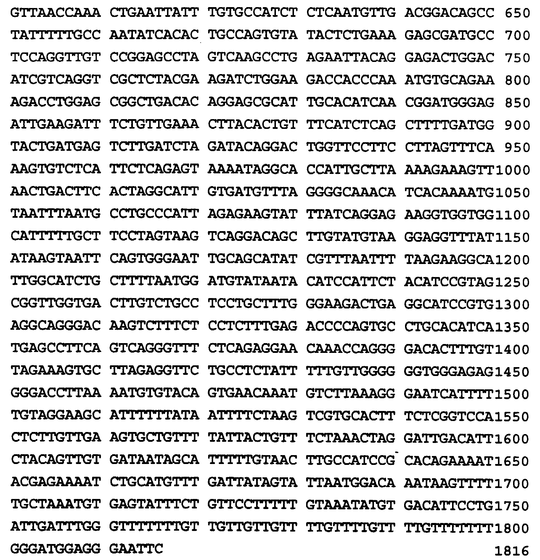
- The abbreviations used for the nucleotides are those standardly used in the art.
- The deduced amino acid sequence of the g7 cDNA is shown as SEQ ID NO:2 below and starts at
nucleotide 1 of SEQ ID NO:1 and extends 851 nucleotides.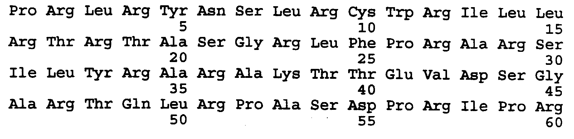

- Variations are contemplated in the cDNA sequence shown in SEQ ID NO:1 which will result in a DNA sequence that is capable of directing production of analogs of the VHL protein shown in SEQ ID NO. 2. It should be noted that the DNA sequence set forth above represents a preferred embodiment of the present invention. Due to the degeneracy of the genetic code, it is to be understood that numerous choices of nucleotides may be made that will lead to a DNA sequence capable of directing production of the instant VHL protein or its analogs. As such, DNA sequences which are functionally equivalent to the sequence set forth above or which are functionally equivalent to sequences that would direct production of analogs of the VHL protein produced pursuant to the amino acid sequence set forth above, are intended to be encompassed within the present invention.
- The term analog includes any protein or polypeptide having an amino acid residue sequence substantially identical to a sequence specifically shown herein in which one or more amino acid residues have been conservatively substituted with a functionally similar residue and which displays the functional aspects of the VHL protein as described herein. Examples of conservative substitutions include, for example, the substitution of one non-polar (i.e. hydrophobic) residue such as isoleucine, valine, leucine or methionine for another; the substitution of one polar (i.e. hydrophilic) residue for another, such as a substitution between arginine and lysine, between glutamine and asparagine, or between glycine and serine; the substitution of one basic residue such as lysine, arginine or histidine for another; or the substitution of one acidic residue, such as aspartic acid or glutamic acid for another.
- The phrase conservative substitution may also include the use of a chemically derivatized residue in place of a non-derivatized residue provided that the resulting protein or polypeptide displays the requisite functional activity.
- Chemical derivative refers to a VHL protein or polypeptide having one or more residues chemically derivatized by reaction of a functional side group. Examples of such derivatized molecules include, but are not limited to, those molecules in which free amino groups have been derivatized to form, for example, amine hydrochlorides, p-toluene sulfonyl groups, carbobenzoxy groups, t-butyloxycarbonyl groups, chloroacetyl groups or formyl groups. Free carboxyl groups may be derivatized to form salts, methyl and ethyl esters, or other types of esters or hydrazides. Free hydroxyl groups may be derivatized to form O-acyl or O-alkyl derivatives. The imidazole nitrogen of histidine may be derivatized to form N-im-benzylhistidine. Also included as chemical derivatives are those proteins or peptides which contain one or more naturally-occurring amino acid derivatives of the twenty standard amino acids. For example, 4-hydroxyproline may be substituted for proline; 5-hydroxylysine may be substituted for lysine; 3-methylhistidine may be substituted for histidine; homoserine may be substituted for serine; and ornithine may be substituted for lysine. A VHL protein or polypeptide of the present invention also includes any protein or polypeptide having one or more additions and/or deletions of residues relative to the sequence of a protein or polypeptide whose sequence is shown herein, so long as the requisite activity is maintained.
- The present invention also relates to methods for detecting carriers of the VHL disease gene.
- It is understood by one skilled in the art that the methods for detection disclosed in the present invention can be used prenatally to screen a fetus or presymptomatically to screen a subject at risk through his/her family history. In addition, these methods can be used to determine the involvement of the VHL disease gene in other human malignancies such as kidney, lung and bladder cancers.
- In one embodiment of the invention, the method for detecting carriers of the VHL disease gene comprises analyzing the DNA of a subject for mutations of the VHL disease gene associated with VHL disease.
- For purposes of the present invention, subject means a mammal and mutation means inversion, translocation, insertion, deletion or point mutation of the VHL disease gene.
- For analysis of the DNA, a biological specimen is obtained from the subject. Examples of biological specimens that can be obtained for use in the present method include, but are not limited to, tissue biopsies, whole blood, urine, feces or other samples normally tested in the diagnosis of disease. Preferred biological specimens are whole blood or urine.
- Although it is not always required, it is preferable to at least partially purify DNA from the biological specimen prior to analysis. For example, after disruption of cells in the specimen, nucleic acid can be extracted from contaminating cell debris and other protein substances by extraction of the sample with phenol. In phenol extraction, the aqueous sample is mixed with an approximately equal volume of redistilled phenol and centrifuged to separate the two phases. The aqueous phase containing the nucleic acid is removed and precipitated with ethanol to yield nucleic acid free of phenol. Alternatively, DNA can be purified from the biological sample according to Sidransky, D. et al. (Science (1992) 256:102-105; Science (1991) 252:706) or by the method of Glenn et al. (Glenn, G.M. et al. JAMA (1992) 267:1226-1231). The DNA to be analyzed can be either single- or double-stranded.
- Methods for analyzing the DNA for mutations in the VHL disease gene include Southern blotting after digestion with the appropriate restriction enzymes (restriction fragment length polymorphism, RFLP) (Botstein, D. Amer. J. Hum. Genet. (1980) 69:201-205), denaturing gradient electrophoresis technique (Myers, R.M., Nature (1985) 313:495-498), oligonucleotide hybridization (Conner, R. et al., EMBO J. (1984) 3:13321-1326), RNase digestion of a duplex between a probe RNA and the target DNA (Winter, E. et al., Proc. Natl. Acad. Sci. U.S.A. (1985) 82:7575-7579), polymerase chain reaction (PCR) (Saiki, P.K. et al., Science (1988) 239:487-491; U.S. Patents 4,683,195 and 4,683,202), ligase chain reaction (LCR) (European Patent Application Nos. 0,320,308 and 0,439,182), and PCR-single stranded conformation analysis (PCR-SSCP) (Orita, M. et al., Genomics (1989) 5:874-879; Dean, M. et al. Cell (1990) 61:863-871). In one preferred embodiment, DNA is analyzed by Southern analysis.
- The DNA to be analyzed via Southern analysis is digested with one or more restriction enzymes. The restriction enzymes to be used in the present invention are those enzymes for whom the presence or absence of their recognition site is linked to VHL disease. Preferred restriction enzyme include EcoRI, HindIII, PstI, DraI, BamHI, BglI, BglII, and PvuII. Following restriction digestion, resultant DNA fragments are separated by gel electrophoresis and the fragments are detected by hybridization with a labelled nucleic acid probe (Southern, E.M. J. Mol. Biol. (1975) 98:503-517).
- The nucleic acid sequence used as a probe in Southern analysis can be labeled in single-stranded or double-stranded form. Labelling of the nucleic acid sequence can be carried out by techniques known to one skilled in the art. Such labelling techniques can include radiolabels and enzymes (Sambrook, J. et al. (1989) in "Molecular Cloning, A Laboratory Manual", Cold Spring Harbor Press, Plainview, New York). In addition, there are known non-radioactive techniques for signal amplification including methods for attaching chemical moieties to pyrimidine and purine rings (Dale, R.N.K. et al. (1973) Proc. Natl. Acad. Sci., 70:2238-2242; Heck, R.F. 1968) S. Am. Chem. Soc., 90:5518-5523), methods which allow detection by chemiluminescence (Barton, S.K. et al. (1992) J. Am. Chem. Soc., 114:8736-8740) and methods utilizing biotinylated nucleic acid probes (Johnson, T. K. et al. (1983) Anal. Biochem., 133:126-131; Erickson, P.F. et al. (1982) J. of Immunology Methods, 51:241-249; Matthaei, F.S. et al. (1986) Anal. Biochem., 157:123-128) and methods which allow detection by fluorescence using commercially available products. The size of the probe can range from about 200 nucleotides to about several kilobases. A preferred probe size is about 500 to about 2000 nucleotides. Each of the nucleic acid sequences used as a probe in Southern analysis is substantially homologous to the corresponding portion of the cDNA sequence shown in SEQ ID NO:1. By "substantially homologous" is meant a level of homology between the nucleic acid sequence used as a probe and the corresponding sequence shown in SEQ ID NO:1. Preferably, the level of homology is in excess of 70%, most preferably in excess of 80%, with a particularly preferred nucleic acid sequence being in excess of 90% homologous with the sequence shown in SEQ ID NO:1. Once the separated DNA fragments are hybridized to the labelled nucleic acid probes, the restriction digest pattern can be visualized by autoradiography and examined for the presence or absence of a restriction fragment length polymorphism (RFLP) associated with VHL disease.
- In a second preferred embodiment, the DNA is analyzed for mutations in the VHL disease gene by PCR-SSCP (Orita et al., (1989), Dean et al., (1990)). In this method, each of the pairs of primers selected for use in PCR are designed to hybridize with sequences in the VHL disease gene which are an appropriate distance apart (at least about 50 nucleotides) in the gene to permit amplification and subsequent detection of mutations in the amplification product. Primer pairs which can specifically hybridize to such VHL gene sequences can be derived from the VHL disease gene sequence. In a preferred embodiment, the primers are derived from the cDNA sequence shown in SEQ ID NO.1. Each primer of a pair is a single-stranded oligonucleotide of about 15 to about 50 bases in length which is complementary to a sequence at the 3' end of one of the strands of a double-stranded target sequence. Each pair comprises two such primers, one of which is complementary 3' end and the other of which is complementary to the other 3' end of the target sequence. The target sequence is generally about 100 to about 300 base pairs long but can be as large as 500-600 base pairs. Optimization of the amplification reaction to obtain sufficiently specific hybridization to the VHL disease gene is well within the skill in the art and is preferably achieved by adjusting the annealing temperature.
- The present invention also provides purified and isolated pairs of primers for use in analysis of DNA for mutations in the VHL gene. The nucleic acid sequences of these primers is set forth below as SEQ ID NOs:3-8.




 where SEQ ID NO. 3 and SEQ ID NO. 4 represent one pair of primers; SEQ ID NO. 5 and SEQ ID NO. 6 represent a second pair of primers and SEQ ID NO. 7 and SEQ ID NO. 8 represent a third pair of primers.
- The primers of this invention can be synthesized using any of the known methods of oligonucleotide synthesis (e.g., the phosphodiester method of Agarwal et al. 1972. Agnew. Chem. Int. Ed. Engl. 11:451, the phosphotriester method of Hsiung et al. 1979. Nucleic Acids Res. 6:1371, or the automated diethylphosphoramidite method of Beuacage et al. 1981. Tetrahedron Letters 22:1859-1862), or they can be isolated fragments of naturally occurring or cloned DNA. In addition, those skilled in the art would be aware that oligonucleotides can be synthesized by automated instruments sold by a variety of manufacturers or can be commercially custom ordered and prepared. In one embodiment, the primers can be derivatized to include a detectable label suitable for detecting and/or identifying the primer extension products (e.g., biotin, avidin, or radiolabeled dNTP's), or with a substance which aids in the isolation of the products of amplification (e.g. biotin or avidin). In a preferred embodiment, SEQ. ID. NO. 3 through SEQ. ID. NO. 8 are synthetic oligonucleotides.
- In an alternative embodiment, primer pairs can be selected to hybridize to mutant forms of the VHL disease gene. The selected primer pairs will hybridize sufficiently specifically to the mutated gene sequences such that non-specific hybridization to wild-type VHL gene sequences will not prevent identification of the amplification product of the mutant gene sequence. Primer pairs which hybridize to mutations in the VHL gene sequence can be used to amplify specific mutant gene sequences present in the DNA of a biological sample.
- The amplification products of PCR can be detected either directly or indirectly. In the PCR-SSCP method, direct detection of the amplification products is carried out via labelling of primer pairs. Labels suitable for labelling the primers of the present invention are known to one skilled in the art and include radioactive labels, biotin, avidin, enzymes and fluorescent molecules. The derived labels can be incorporated into the primers prior to performing the amplification reaction. A preferred labelling procedure utilizes radiolabeled ATP and T4 polynucleotide kinase (Sambrook, J. et al. (1989) in "Molecular Cloning, A Laboratory Manual", Cold Spring Harbor Press, Plainview, NY). Alternatively, the desired label can be incorporated into the primer extension products during the amplification reaction in the form of one or more labelled dNTPs. In the present invention, the labelled amplified PCR products can be analyzed for mutations of the VHL gene associated with VHL disease gene via separating the PCR products by denaturing polyacrylamide gel electrophoresis or via direct sequencing of the PCR-products.
- In yet another embodiment, unlabelled amplification products can be analyzed for mutations in the VHL disease gene via hybridization with nucleic acid probes radioactively labelled or, labelled with biotin, in Southern blots or dot blots. Nucleic acid probes useful in the embodiment are those described earlier for Southern analysis.
- In a second embodiment, the method for detecting carriers of the VHL disease gene comprises analyzing the RNA of a subject for mutations or alterations in VHL-specific mRNA associated with VHL disease.
- For the analysis of RNA by this method, RNA derived from blood or a tumor biopsy sample is obtained from said subject where said tumors include, but are not limited to, tumors of the eye brain, liver, kidney, pancreas, and pheochromocytomas.
- The RNA to be analyzed can be isolated from blood or tumor biopsy samples as whole cell RNA or as poly(A)+ RNA. Whole cell RNA can be isolated by methods known to those skilled in the art. Such methods include extraction of RNA by differential precipitation (Birnbiom, H.C. (1988) Nucleic Acids Res., 16:1487-1497), extraction of RNa by organic solvents (Chomczynski, P. et al. (1987) Anal. Biochem., 162:156-159) and extraction of RNA with strong denaturants (Chirgwin, J.M. et al. (1979) Biochemistry, 18:5294-5299). Poly(A)+ RNA can be selected from whole cell RNA by affinity chromatography on oligo-d(T) columns (Aviv, H. et al. (1972) Proc. Natl. Acad. Sci., 69:1408-1412). A preferred method of isolating RNA is extraction of whole cell RNA by acid-phenol (Chomczynski et al. 1987).
- The methods for analyzing the RNA for alterations in the pattern or level of VHL specific mRNA expression linked to VHL disease include Northern blotting (Alwine, J.C. et al. (1977) Proc. Natl. Acad. Sci., 74:5350-5354), dot and slot hybridization (Kafatos, F.C. et al. (1979) Nucleic Acids Res., 7:1541-1522), filter hybridization (Hollander, M.C. et al. (1990) Biotechniques; 9:174-179), RNase protection (Sambrook, J. et al. (1989) in "Molecular Cloning, A Laboratory Manual", Cold Spring Harbor Press, Plainview, NY) and reverse-transcription polymerase chain reaction (RT-PCR) (Watson, J.D. et al. (1992) in "Recombinant DNA" Second Edition, W.H. Freeman and Company, New York). One preferred method is Northern blotting.
- The nucleic acid sequence used as a probe for detecting VHL-specific mRNA expression is substantially homologous to SEQ. ID. NO. 1. By "substantially homologous" is meant a level of homology between the nucleic acid sequence and the cDNA sequence of SEQ ID NO.1. Preferably, the level of homology is in excess of 70% more preferably in excess on 80%, with a particularly preferred nucleic acid sequence being in excess of 90% homologous with the cDNA sequence shown in SEQ ID No. 1.
- A most preferred method is reverse transcription-polymerase chain reaction (RT-PCR) where the primers used to amplify the cDNA produced via reverse transcription of RNA are derived from the cDNA sequence shown in SEQ ID No. 1. These primers can be labelled as described earlier and the RT-PCR products can be analyzed for mutations of the VHL gene associated with VHL disease via denaturing polyacrylamide gel electrophoresis of the RT-PCR products or via direct sequencing of the RT-PCR products.
- The present invention also encompasses recombinant proteins derived from the cDNA shown in SEQ ID No. 1 and antibodies directed to said proteins (called VHL proteins). Recombinant VHL proteins can be produced by recombinant DNA methodology known to one skilled in the art. For example, a nucleic acid sequence capable of encoding a protein comprising all or part of the amino acid sequence shown in SEQ ID NO.2 can be cloned into a vector capable of being transferred into, and replicated in, a host organism. A suitable nucleic acid sequence for the purpose of this invention is the sequence shown in SEQ ID NO.1. Suitable expression vectors include, but are not limited to, vaccinia virus vectors include, baculovirus vectors, and Ecoli pTRCHIS (Invitrogen Co. San Diego). The recombinant expression vector produced by inserting a nucleic acid sequence capable of directing synthesis of VHL protein in a suitable expression vector can be transfected into E coli or into suitable eukaryotic cell systems by methods known to one skilled in the art.
- Cells containing the expressed recombinant VHL protein, cell lysate from cells transfected with a recombinant expression vector or a culture supernatant containing the expressed VHL protein can be used as an immunogen to elicit production of anti-VHL antibodies in a mammal. Alternatively, one can generate synthetic peptides for use as immunogens from the amino acid sequence shown in
SEQ ID NO 2. Preferred synthetic peptide sequences for use as an immunogen are shown below:

- While it is possible for the immunogen to be a administered to the mammal in pure or substantially pure form, it is preferable to present it as a pharmaceutical composition, formulation or preparation. Suitable mammals for immunization include mice, rabbits and the like. The anti-VHL antibody of the present invention is typically produced by immunizing a mammal with an immunologically effective amount of synthetic peptide of this invention. The preparation of polyclonal or monoclonal antibodies against such a peptide is well known in the art (Standt et al. (1988) J. Exp. Med. 157:687-704). The anti-VHL peptide antibody molecules induced by immunization of a mammal with the recombinant VHL protein are then collected from the mammal and those immunospecific for the VHL protein are isolated to the extent desired by well known techniques such as, for example, immunochromatography.
- In a third embodiment, the method for detecting carriers of the VHL disease gene comprises analyzing the protein of a subject for alterations in VHL protein expression with VHL disease.
- For analysis of protein by this method, protein is obtained from biological specimens such as tumor biopsy samples and urine and the like. The protein can be obtained as a crude lysate or it can be further purified by methods known to one skilled in the art (Sambrook, J. et al. (1989) in "Molecular Cloning, A Laboratory Manual", Cold Spring Harbor press, Plainview, NY).
- Crude protein lysate can be analyzed for VHL protein by immunoassays using anti-VHL antibody.
- Immunoassays of the present invention may be a radioimmunoassay, Western blot assay, immunofluorescent assay, enzyme immunoassay, chemiluminescent assay, immunohistochemical assay and the like. Standard techniques known in the art for ELISA are described in Method in Immunodiagnosis, 2nd Edition, Rose and Bigazzi, eds., John Wiley and Sons, 1980 and Campbell et al., Methods of Immunology, W.A. Benjamin, Inc., 1964, both of which are incorporated herein by reference. Such assays may be a direct, indirect, competitive, or noncompetitive immunoassay as described in the art. (Oellerich, M. 1984. J. Clin. Chem. Clin. BioChem. 22:895-904).
- Detection of the VHL protein anti-VHL antibody complex formed, can be accomplished by reaction of the complex with a secondary antibody such as labelled anti-rabbit antibody. The label may be an enzyme which is detected by incubating the complex in the presence of a suitable fluorimetric or colorimetric reagent. Other detectable labels may also be used, such as radiolabels, or colloidal gold, and the like. The labelled VHL protein-anti-VHL antibody complex is then visualized by autoradiography.
- The present invention also relates to a method for treating a carrier of the VHL disease gene in which an expression vector containing a nucleic acid sequence representing the wild type VHL gene is administered to the carrier. A nucleic acid sequence representing wild-type VHL gene is that shown in SEQ ID No. 1. Such nucleic acid sequence may be inserted into a suitable expression vector by methods known to those skilled in the art (Example 5). Expression vectors suitable for producing high efficiency gene transfer in vivo include retroviral, adenoviral and vaccinia viral vectors.
- Expression vectors containing a nucleic acid sequence representing wild-type VHL gene can be administered intravenously, intramuscularly, subcutaneously, intraperitoneally or orally. A preferred route of administration is intravenously.
- The invention also provides a diagnostic kit for detecting carriers of the VHL disease gene. This diagnostic kit comprises purified and isolated nucleic acid sequences according to SEQ ID. No. 3 through SEQ ID No. 8, said sequences useful as PCR primers in analyzing DNA for mutations of the VHL disease gene linked to VHL disease.
- Any articles or patents referenced herein are incorporated by reference. The following examples illustrate various aspects of the invention but are in no way intended to limit the scope thereof.
- The subjects analyzed in the following examples were kindred identified by ophthalmologists, urologists, medical geneticists and neurosurgeons in the United States, Europe, and Canada. The members of the families resided in Louisiana, Tennessee, Mississippi, Virginia, Pennsylvania, New York, Michigan, Quebec, Nova Scotia, United Kingdom, and the Netherlands. Medical records of each family member known to be affected were reviewed. Asymptomatic family members and family members in whom there was uncertainty about the diagnosis were examined after informed consent for occult evidence of the illness at the Clinical Center of the National Institutes of Health. The examination consisted of a history and physical examination of the scrotum. An asymptomatic member of a VHL family was considered to be affected if one or more of the following disease manifestations were detected: retinal angioma(s), spinal or cerebellar hemangioblastoma(s), pheochromocytoma(s), multiple pancreatic systs, and multiple bilateral renal cysts accompanied by renal cell carcinoma. Disease diagnosis was made without knowledge of restriction fragment length polymorphism (RFLP) status.
- Restriction enzymes were from Bethesda Research Laboratory (BRL) (Bethesda, MD), New England Biolabs (Beverly, MA) and Boehringer Mannheim (Indianapolis, IN) and were used as recommended by the manufacturers. δ-32PdCTP (∼3000iu/mmol) was from Amersham (Arlington Heights, IL). The various human tissue polyadenylated RNAs used in Northern blotting were purchased from Clonstech (Palo Alto, CA) as was the adult kidney double-stranded complementary DNA sample. PCR and RT-PCR bits were from Perkin Elmer/Cetus (Norwalk, CT); deoxynucleotide triphosphates and flourescently labelled dideoxynucleotides were from Applied Biosystems, Inc. (Foster City, CA). Nylon membranes were purchased from MSI, Inc. (Westlore, MA).
- Southern and Northern blottings, filter hybridization and probe labelling were by random priming were performed by standard protocols (Sambrook, J. et al. (1989)). DNA inserts were purified following the GeneClean (Bio 101) (BioRad, Richmond, CA) protocol and used for subcloning or labelling. Oligonucleotides used as primers in PCR or RT-PCR or for sequencing were synthesized on the Applied Biosystems, Inc. Model 392 DNA/RNA synthesizer, according to the manufacturers recommendations. Pulse field goal electrophoresis was carried out using CHEF-DRII or CHEF mapper XA systems as described by the manufacturer (BioRad) under conditions optimal for obtaining the desired resolution.
- The PCR was performed in a 50 ul reaction volume in a mixture containing 1uM of each primer, 250uM of each deoxynucleotide triphosphate, 5ul of 10X PCR buffer (500MM KCl; 120MM Tris-HCl, pH 8.0; 1.5MM MgCl2; and 0.1% gelatin) and 1.25 units of AmpTaq (Cetus) DNA polymerase, in a first generation automated thermal cycler (Perkin-Elmer/Cetus). The PCR conditions consisted of 40 cycles of denaturation for one minute at 94°C, annealing for one minute at specified temperatures (55-65°C) and extension for 4 minutes at 72°C followed by 7 minutes of final extension of 72°C.
- RNA Preparation and Northern Blotting - Total cellular RNA was isolated by extraction of lymphoblastoid cell lines of affected VHL patients or kidney tissues in guanidine thiocyanate followed by centrifugation through a 5.7 M CsCe cushion according to standard protocols (Sambrook, J. et al. (1989)). RNA samples were separated by electrophoresis in 1% agarose gels containing 2.2M formaldehyde, transferred to nylon membranes and hybridized to g7 cDNA probe (Sambrook, J. et al. (1989)).
- RT-PCR - About 5 ug of total cellular RNA was isolated by extraction of lymphoblastoid cell lines or kidney tissues of VHL patients or 2.5 ng of normal adult kidney double-stranded complementary DNA samples were analyzed for expression using RT-PCR kit from Perkin-Elmer/Cetus. The primers were derived from the g7 cDNA sequence shown in SEQ ID NO. 1 and the reactions were run using various annealing temperatures. The reaction products were analyzed by gel electrophoresis and Southern blotting (Sambrook, J. et al (1989)).
- The isolation of the VHL disease gene resulted from the use of positional cloning strategies (Latif et al., Cancer Res. (1993) 63:861-867; Trofatter et al., Cell (1993) 72:791-800 and The Huntington's Disease Collaborative Research Group; Cell (1993) 72:971-983) previously used in isolating disease genes and is described in Latif et al. (Science, in press, "Identification of the von Hippel-Lindau Disease Tumor Suppressor Gene"). Genetic and physical map of the chromosome 3p region encompassing the VHL gene is shown in Figure 1. The VHL locus was positioned on the map (Fig. 1, Panel A) by multipoint linkage analysis and meiotic mapping (Tory et al., 1989); the location of selected cross-overs is indicated by crosses. YAC Library Screening and Analysis of YACs. Copies of the WU and CEPH YAC libraries were obtained from Dr. Craig Chinault (Baylor Institute of Human Genetics, Houston, Texas) and Dr. Daniel Cohen, respectively (centre d' Etude du Polymorphisme Humain, Paris). The WU and CEPH libraries are total human genomic DNA libraries constructed in the PYAC4 vector (Burke, D.T. et al. Science (1987) 236:806-812; Anand, R. et al. Nucleic Acids Res. (1990) 18: 1951-1956). These libraries were screened by sib selection using PCR-based techniques (Greene, E.D. et al., Proc. Natl. Acad Sci. (1990) 87:1213-1217) with primers for the D3S601, D3S587 and D3S18 loci in the VHL region (Figure 1). The sequences of the primers used to positively identify YACs Y52A10, YA101D4, Y132F2 and Y70D2 are shown below as SEQ ID No. 11 thru SEQ ID No. 16:
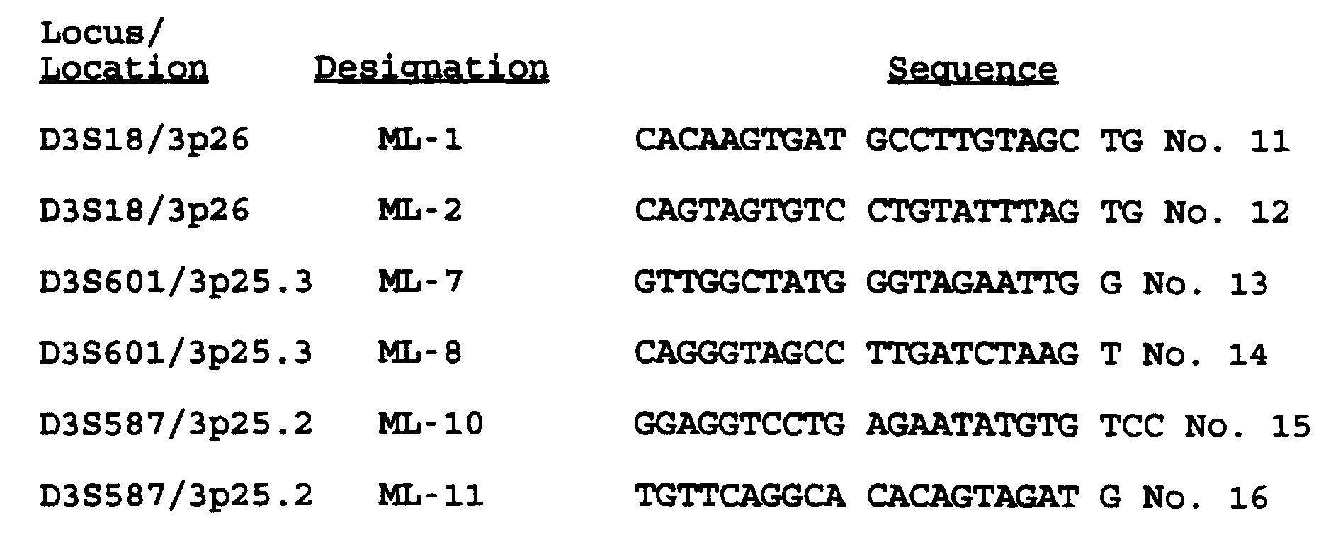
-
Screening Chromosome 3 Cosmid Library and Cosmid Contig Assembly. Thechromosome 3 cosmid library was constructed as described in Lerman et al. (Lerman, M.I. et al. Hum. Genet. (1991) 86:567-577). This library was screened by colony hybridization (Sambrook, J. et al. (1989)) using the YAC DNA inserts as probes as described in Baxendale et al. (Baxendale, S. et al. Nucl. Acids Res. (1991) 19:6651). After labeling with 32P-dCTP, the probes were preassociated with a 1000X excess of sheared human DNA. Cosmid contigs were constructed by finding overlapping bands on Southern blots of EcoRI-digested cosmids using whole cosmids as probes. Gaps in the cosmid contigs were closed by chromosome walking using insert-end fragment probes, which were identified by restriction mapping and hybridization to restricted genomic DNA. These insert-end fragment probes were used for each walk step. Figure 1 shows the 160 kb cosmid and phage contig covering the VHL region. The phage T42 was isolated by screening a total genomic phage library with YAC DNA inserts as described above. The phage p191, which contains the VHL disease gene, was isolated by screening a three-hit P1 phage genomic library (Genome System, Inc. St. Louis, MO) with primers chosen from within an exon of the g7 cDNA sequence shown in SEQ ID NO. 1. The phage p191 was deposited with the ATCC on May 13, 1993. - Screening cDNA Libraries. A λgt11 teratocarcinoma library (gift of Dr. Maxine Singer, National Cancer Institute) was screened by plaque hybridization (Sambrook, J. et al. (1989)) to 10° filter-immobilized cDNA phage clones at a density of 4 x 10⁴ pfu/150-mm filter. Figure 1 (Panel B) shows the position of the g7 cDNA isolated by screening the λgt11 teratocarcinoma cDNA library with a conserved 7 kb fragment at the centromeric end of cosll used as a probe in the screening. The orientation of the g7 cDNA was established by sequencing and restriction mapping to the contig. The beginning of the smallest constitutional deletion is indicated by an asterisk and line. Restriction sites: B, Bam HI; E, Eco RI; N, Not I; Nr, Nru I; M, Mlu I.
- cDNA Sequence and Sequence Analysis. The g7 cDNA clone was sub-cloned into the Bluescript KS (+) plasmid (Stratagene, La Jolla, CA). Double-stranded plasmid DNA was used in sequencing reactions performed with Tag Dye Deoxy terminator cycle sequencing kits (Applied Biosystems, Inc.). All sequences were obtained by running the reactions in an ABI 373A automatic sequencing system (Applied Biosystems, Inc.). Initial sequencing was performed with T3 and T7 primers, and "walking" primers were then constructed to continue sequencing. The cDNA clone was sequenced multiple times in one orientation or both orientations. Database searching, sequence editing, sequence assembly, and sequence analysis were carried out with the University of Wisconsin Genetics Computer Group sequence analysis software package, version 7.0 (Devereaux, J. et al. Nucl. Acids Rev. (1984) 12:387-395). The sequence of the g7 cDNA is shown in SEQ ID No. 1. This cDNA was deposited with the ATCC on May 13, 1993. The cDNA sequence revealed an open reading frame (ORF) of 284 amino acids indicating that the rest represents part of the 3' untranslated region of the mRNA. This ORF showed a high probability score (> 95%) for being a protein coding sequence Fickett, J.W., Nucl. Acids Rev. (1982) 10:5303). Neither the nucleotide nor the predicted amino acid sequences showed any significant homology to genes or proteins in the databases.
- RNA Preparation and Northern Blotting Analysis. To identify the VHL gene, we evaluated the g7 loci was evaluated by analyzing its expression in target tissues.
- The expression pattern of the g7 gene was examined by Northern (RNA) blotting. Figure 2A shows a low resolution blot where each lane contains poly A+ mRNA (2 µg) from:
lane 1, fetal brain;lane 2, adult brain;lane 3, fetal kidney;lane 4, adult kidney;lane 5, adult cerebellum;lane 6, adult adrenal; andlane 7, adult prostate while Figure 2B shows a high resolution blot of 1 ug of poly A+ mRNA from tissues as indicated in Figure 2A. The sizes of the transcripts were determined from the position of the 28S and 18S rRNA bands of total RNA run on the same gel. Transcripts were observed in all human tissues tested, including brain and kidney, tissues frequently affected in VHL disease. The transcripts were of two distinct sizes, 6 and 6.5 kb, and were expressed in a tissue-specific and developmentally selective manner, i.e. only 6 kb or the 6.5 kb species was expressed in fetal brain and fetal kidney, while both were expressed in adult tissues. The two transcripts may represent alternatively spliced forms of g7 mRNA. - RT-PCR Studies of Gene Expression. In order to detect mutations in constitutional DNA of affected patients in pedigrees and in new mutation patients, was conducted an extensive search for mutations (i.e. small intragenic and nonoverlapping deletions or insertions) which were of the loss -of- function type was conducted in constitutional DNA derived from 221 unrelated VHL patients. Southern blot analysis of genomic DNA isolated from the blood (Sambrook, J. et al. (1989)) of seven patients and then digested with EcoRI is shown in Figure 3A. This blot was probed using the g7 cDNA as probe and this probe has been shown to detect a single invariant 20-22 kb EcoRI fragment in normal DNA, as determined by previous tests on more than 100 unrelated DNA samples provided by Centre d'Etude du Polymorphisme Humain (CEPH). A high incidence (≥ 12%) of aberrant bands was observed with the bands ranging in size from 4 to 25 kb (Figure 3A) and thus classified these VHL patients were thus classified as new mutations.
- In order to determine that the single aberrant bands originating from the 20-22 kb invariant fragment were deletions or insertions within this fragment or deletions removing the flanking Eco RI sites, Southern blot analysis was conducted with several other restriction enzyme digests besides Eco RI (Bam HI, Bgl I, Bgl II, Dra I, Eco RV, Hind III, Pst I, and Pvu II). The results of the Southern analysis with a few of these enzymes is shown in Figure 3B. These results demonstrated that the mutations were transmitted with the disease (Figure 3C). Figure 3D shows the results of Southern blotting analysis of DNA isolated form a regular VHL family (coded "P") and digested with EcoRI. The results clearly demonstrate transmission of the mutant allele (the aberrant band) in this VHL family (Figures 3D and 3E).
- To prove the presence of deletions and to map them precisely, subfragments representing region of the g7 cDNA generated by PCR used as probes in Southern blotting analysis of genomic DNA isolated from blood of VHL patients and digested with EcoRI. (Figure 4, where the probes used in each panel are: Figure 4A, total g7 cDNA; Figure 4B, nucleotides 3-146 of g7 cDNA; and Figure 4C, nucleotides 1277-1600 of g7 cDNA). The results unequivocally demonstrated that 18 of the rearrangements were deletions as only part of the cDNA failed to detect the novel band in each patient (Figure 4).
- These deletions could then be classified into three groups as shown in Table 1.
Deletion analysis of VHL patients with aberrant bands at the VHL locus (detected by g7 cDNA). Patient Code Probe : cDNA 5'->3' residue (s) Aberrant Band (kb) Apparent Deletion Size (kb) 3-146 169-391 291-501 585-940 921-1231 1277-1600 3567 ND ND ND ND ND 14 ? 3607 ND ND ND ND ND ND 12 ? 3639 ND ND ND ND ND ND 14 ? 3648 ND ND ND ND ND ND 13 ? 3654 ND ND ND ND ND ND 14 ? JD ND ND ND ND ND ND 17 ? PEM ND ND ND ND ND ND 15 ? MS ND ND ND ND ND ND 15 ? KA ND ND ND ND ND ND 15 ? 3547 D D ND ND ND 23-25 15-18 JM D D D ND ND ND 23-25 15-18 GD D D D ND ND ND 23-25 15-18 3512 ND ND ND ND D D 10 11 3516 ND ND ND ND D D 10 11 3557 ND ND ND ND D D 10 11 3574 ND ND ND ND D D 10 11 VIA ND ND ND ND D D 10 11 IC ND ND ND ND D D 10 11 NE ND ND ND ND D D 10 11 EP ND ND ND ND D D 10 11 MO ND ND ND ND D D 10 11 3569 ND ND ND D D D 12 9 3667 ND ND ND D D D 10 11 3761 ND ND ND D D D 4 17 3819 ND ND ND D D D 12 9 - The finding of three overlapping deletions within the same cDNA provides strong evidence for the identification of the g7 cDNA as the VHL gene.
- To find intragenic deletions or insertions, genomic DNA isolated from VHL patient lymphoblastoid cell lines (Lymphoblastoid cells were immortalized by transformation with Epstein Barr Virus according to standard protocols (Nilison, K. et al., Adv. Cancer Res. (1982) 37:319-380)) was analyzed for alterations by PCR-single-strand-conformational polymorphism (PCR-SSCP) analysis using primers shown in SEQ ID NO. 3 thru SEQ ID NO. 8 and RNA isolated from sporadic renal cell carcinoma (RCC) cell lines (Anglard, P. et al. Cancer Res. (1992) 52:348-356) was analyzed by reverse transcription-polymerase chain reaction (RT-PCR). The primers used for RT-PCR of the RCC cell lines are shown as SEQ ID NO. 17 thru SEQ ID NO. 20:


 whose SEQ ID NO. 17 and NO. 18 are on pair of primers and SEQ ID NO. 19 and SEQ ID NO. 20 are a second pair. The results of these analyses are shown in Table 2.
Germ-line (VHL) and somatic (sporadic RCC) mutations in the VHL candidate gene. Patients Mutation Consequence VHL family "VA" 8 bp (TTGTCCGT) insertion after NT714 frameshift "E" 9 bp in-frame deletion (NT456-464) Three amino acid (153-154) deletion (Arg Val Val) "CS" 3 bp in-frame deletion (NT434-436) One amino acid deletion (146, lie) Sporadic RCC "UOK118" 1 bp deletion (NT737) frameshift "UMRC5" 1 bp deletion (NT737) frameshift "UMRC6" 10 bp deletion (NT715-724) frameshift "A498" 5 bp deletion (NT638-642) frameshift "UOK151" nonsense C → A (NT761) transversion stop codon - RCC were chosen because according to Knudson's dictum (Knudson (1971)) sporadic cancers should be associated with mutations in the same loci affected in the hereditary form of the same malignancy. So far aberrant patterns have been identified in five RCC cell lines and proved four of them have been proven to be small (1 to 10 bp) deletions creating frameshift mutations and truncated proteins (TABLE 2). The cell lines UMRC5 and RCC "UOK118" have the same 1 bp deletion at nucleotide 737, amino acid 246, creating 28 new amino acids followed by a stop codon. Incidentally, this deletion creates a new Eco RI site, leading to two aberrant bands on Southern blots (not shown). Line UMRC6 has a 10 bp deletion (nucleotides 715 to 724) creating a frameshift such that 32 new amino acids are present followed by a new stop codon. Finally, line A498 has a 5 bp deletion (nucleotides 638 to 642) leading to a premature stop after new 62 amino acids. In the fifth RCC cell line, UOK151, the change is a nonsense (stop codon) mutation resulting from a C to A transversion at nucleotide 761 (TCG → TAG), creating a truncated protein. These data suggest that the VHL disease gene plays an important role in sporadic kidney cancer. As such, RT-PCR or PCR-SSCP as described in this application can be used as diagnostic methods to distinguish primary kidney tumors from tumors that spread to the kidney from other tissues or organs and to distinguish different histological types of kidney tumors.
- In the DNA of the VHL lymphoblastioid cell lines derived from VHL patients, SSCP aberrant patterns segregating with the disease were also detected using primers shown in SEQ ID NO. 3 thru SEQ ID NO. 8. One (patient "VA") was found to be an 8 bp (TTGTCCGT) insertion after
nucleotide 714. This insertion created a shift in the reading frame and a truncated protein. The second patient ("CS") had an in-frame 3 bp deletions leading to the removal of amino acid 146 (isoleucine). Finally, patient "E" had an in-frame 9 bp deletion (nucleotides 456 to 464) that resulted in the removal of three amino acids (Arg Val Val) at position 153-155. These combined results strongly support the conclusion that the g7 gene represents the VHL and the sporodic RCC tumor suppressor gene. - In order to determine whether the g7 cDNA is highly conserved across species ranging from mammals to Drosophila and sea urchin, Zoo blotting using g7 cDNA as a probe was performed on DNA isolated from human (Homo sapiens), chimpanzee (Pan troglodytes), macaque (Macaca fascicularis), cow (Bovis domesticus), rat (Rattus norvigicus), mouse (Mus musculus), chicken (Gallus domesticus), frog (Xenopus laevis), fly (Drosophila melanogaster), sea urchin (Strongilocetrotus purpuratus), and yeast (Saccharomyces ceriviseae), all purchased from BIOS Laboratories (New Haven, CT, USA). (Pre)Hybridization was done in Church buffer [G. M. Church and W. Gilbert, Proc. Natl. Acad. Sci. U.S.A., 81, 1991 (1984)] at 65°C for 18 hours. Blots were washed in 0.1 x Church buffer at 60°C for 60 min. The results of the zoo blot are shown in Figure 6. The results demonstrate an extensive evolutionary conservation which is indicative of g7 serving a basic life function and also, of g7 having a tumor suppressor role.
- The contents of all citations, i.e., journal articles, patents and the like, are incorporated herein by reference.
- It is understood that the examples and embodiments described herein are for illustrative purposes and that various modifications and changes in light thereof to persons skilled in the art are included within the spirit and purview of this application and scope of the appended claims.
-
- (i) APPLICANTS: THE GOVERNMENT OF THE UNITED STATES OF AMERICA AS REPRESENTED BY THE SECRETARY, DEPARTMENT OF HEALTH AND HUMAN SERVICES
- (ii) TITLE OF INVENTION: VON HIPPEL-LINDAU (VHL) DISEASE GENE AND CORRESPONDING cDNA AND METHODS FOR DETECTING CARRIERS OF THE VHL DISEASE GENE
- (iii) NUMBER OF SEQUENCES: 20
- (iv) CORRESPONDENCE ADDRESS:
- (A) ADDRESSEE: MORGAN & FINNEGAN
- (B) STREET: 345 PARK AVENUE
- (C) CITY: NEW YORK
- (D) STATE: NEW YORK
- (E) COUNTRY: USA
- (F) ZIP: 10154
- (v) COMPUTER READABLE FORM:
- (A) MEDIUM TYPE: FLOPPY DISK
- (B) COMPUTER: IBM PC COMPATIBLE
- (C) OPERATING SYSTEM: PC-DOS/MS-DOS
- (D) SOFTWARE: WORDPERFECT 5.1
- (vi) CURRENT APPLICATION DATA:
- (A) APPLICATION NUMBER: PCT/US94/
- (B) FILING DATE: 12-MAY-1994
- (vii) PRIOR APPLICATION DATE:
- (A) APPLICATION NUMBER: 08/061,889
- (B) FILING DATE: 14-MAY-1993
- (C) CLASSIFICATION:
- (viii) ATTORNEY/AGENT INFORMATION:
- (A) NAME: RICHARD W. BORK
- (B) REGISTRATION NUMBER: 36,459
- (C) REFERENCE/DOCKET NUMBER: 2026-4078PCT
- (ix) TELECOMMUNICATION INFORMATION:
- (A) TELEPHONE: (212) 758-4800
- (B) TELEFAX: (212) 751-6849
- (C) TELEX: 421792
-
-
- (i) SEQUENCE CHARACTERISTICS:
- (A) LENGTH: 1816 base pairs
- (B) TYPE: nucleic acid
- (C) STRANDEDNESS: single
- (D) TOPOLOGY: linear
- (xi) SEQUENCE DESCRIPTION: SEQ ID NO:1:
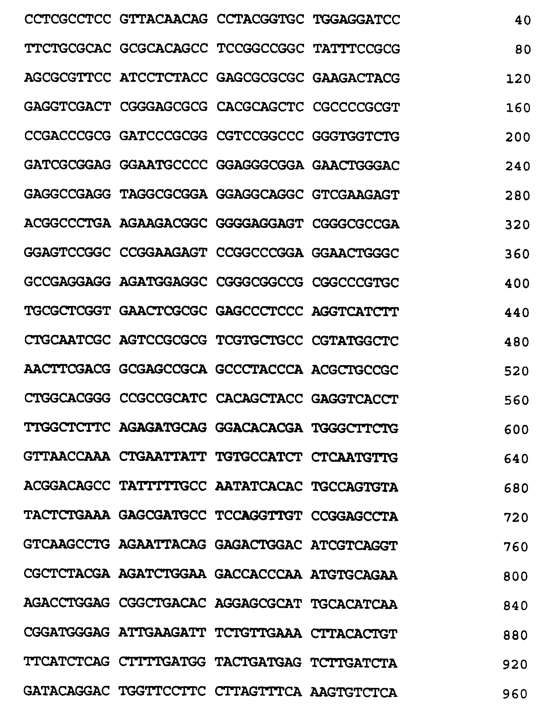

-
-
- (i) SEQUENCE CHARACTERISTICS:
- (A) LENGTH: 284 amino acid residues
- (B) TYPE: amino acid
- (C) STRANDEDNESS: unknown
- (D) TOPOLOGY: unknown
- (xi) SEQUENCE DESCRIPTION: SEQ ID NO:2:

-
-
- (i) SEQUENCE CHARACTERISTICS:
- (A) LENGTH: 37 base pairs
- (B) TYPE: nucleic acid
- (C) STRANDEDNESS: single
- (D) TOPOLOGY: linear
- (xi) SEQUENCE DESCRIPTION: SEQ ID NO:3:

-
-
- (i) SEQUENCE CHARACTERISTICS:
- (A) LENGTH: 33 base pairs
- (B) TYPE: nucleic acid
- (C) STRANDEDNESS: single
- (D) TOPOLOGY: linear
- (xi) SEQUENCE DESCRIPTION: SEQ ID NO:4:

-
-
- (i) SEQUENCE CHARACTERISTICS:
- (A) LENGTH: 33 base pairs
- (B) TYPE: nucleic acid
- (C) STRANDEDNESS: single
- (D) TOPOLOGY: linear
- (xi) SEQUENCE DESCRIPTION: SEQ ID NO:5:

-
-
- (i) SEQUENCE CHARACTERISTICS:
- (A) LENGTH: 33 base pairs
- (B) TYPE: nucleic acid
- (C) STRANDEDNESS: single
- (D) TOPOLOGY: linear
- (xi) SEQUENCE DESCRIPTION: SEQ ID NO:6:

-
-
- (i) SEQUENCE CHARACTERISTICS:
- (A) LENGTH: 31 base pairs
- (B) TYPE: nucleic acid
- (C) STRANDEDNESS: single
- (D) TOPOLOGY: linear
- (xi) SEQUENCE DESCRIPTION: SEQ ID NO:7:

-
-
- (i) SEQUENCE CHARACTERISTICS:
- (A) LENGTH: 33 base pairs
- (B) TYPE: nucleic acid
- (C) STRANDEDNESS: single
- (D) TOPOLOGY: linear
- (xi) SEQUENCE DESCRIPTION: SEQ ID NO:8:

-
-
- (i) SEQUENCE CHARACTERISTICS:
- (A) LENGTH: 14 amino acid residues
- (B) TYPE: amino acid
- (C) STRANDEDNESS: unknown
- (D) TOPOLOGY: unknown
- (xi) SEQUENCE DESCRIPTION: SEQ ID NO:9:

-
-
- (i) SEQUENCE CHARACTERISTICS:
- (A) LENGTH: 13 amino aicd residues
- (B) TYPE: amino acid
- (C) STRANDEDNESS: unknown
- (D) TOPOLOGY: unknown
- (xi) SEQUENCE DESCRIPTION: SEQ ID NO:10:

-
-
- (i) SEQUENCE CHARACTERISTICS:
- (A) LENGTH: 22 base pairs
- (B) TYPE: nucleic acid
- (C) STRANDEDNESS: single
- (D) TOPOLOGY: linear
- (xi) SEQUENCE DESCRIPTION: SEQ ID NO:11:

-
-
- (i) SEQUENCE CHARACTERISTICS:
- (A) LENGTH: 22 base pairs
- (B) TYPE: nucleic acid
- (C) STRANDEDNESS: single
- (D) TOPOLOGY: linear
- (xi) SEQUENCE DESCRIPTION: SEQ ID NO:12:

-
-
- (i) SEQUENCE CHARACTERISTICS:
- (A) LENGTH: 21 base pairs
- (B) TYPE: nucleic acid
- (C) STRANDEDNESS: single
- (D) TOPOLOGY: linear
- (xi) SEQUENCE DESCRIPTION: SEQ ID NO:13:

-
-
- (i) SEQUENCE CHARACTERISTICS:
- (A) LENGTH: 21 base pairs
- (B) TYPE: nucleic acid
- (C) STRANDEDNESS: single
- (D) TOPOLOGY: linear
- (xi) SEQUENCE DESCRIPTION: SEQ ID NO:14:

-
-
- (i) SEQUENCE CHARACTERISTICS:
- (A) LENGTH: 23 base pairs
- (B) TYPE: nucleic acid
- (C) STRANDEDNESS: single
- (D) TOPOLOGY: linear
- (xi) SEQUENCE DESCRIPTION: SEQ ID NO:15:

-
-
- (i) SEQUENCE CHARACTERISTICS:
- (A) LENGTH: 21 base pairs
- (B) TYPE: nucleic acid
- (C) STRANDEDNESS: single
- (D) TOPOLOGY: linear
- (xi) SEQUENCE DESCRIPTION: SEQ ID NO:16:

-
-
- (i) SEQUENCE CHARACTERISTICS:
- (A) LENGTH: 27 base pairs
- (B) TYPE: nucleic acid
- (C) STRANDEDNESS: single
- (D) TOPOLOGY: linear
- (xi) SEQUENCE DESCRIPTION: SEQ ID NO:17:

-
-
- (i) SEQUENCE CHARACTERISTICS:
- (A) LENGTH: 27 base pairs
- (B) TYPE: nucleic acid
- (C) STRANDEDNESS: single
- (D) TOPOLOGY: linear
- (xi) SEQUENCE DESCRIPTION: SEQ ID NO:18:

-
-
- (i) SEQUENCE CHARACTERISTICS:
- (A) LENGTH: 25 base pairs
- (B) TYPE: nucleic acid
- (C) STRANDEDNESS: single
- (D) TOPOLOGY: linear
- (xi) SEQUENCE DESCRIPTION: SEQ ID NO:19:

-
-
- (i) SEQUENCE CHARACTERISTICS:
- (A) LENGTH: 24 base pairs
- (B) TYPE: nucleic acid
- (C) STRANDEDNESS: single
- (D) TOPOLOGY: linear
- (xi) SEQUENCE DESCRIPTION: SEQ ID NO:20:

-
Claims (45)
- A phage containing a VHL disease gene having the identifying characteristics of ATCC deposit number 69311.
- A VHL disease gene sequence encoding a functional VHL protein said gene sequence having(a) the sequence according to SEQ ID NO:1,(b) a sequence which is degenerate as a result of the genetic code to the DNA of SEQ ID NO:1, or(c) a sequence which hybridizes under high stringency conditions to the sequence of (a) or (b).
- A VHL disease gene encoding a protein or analogs thereof, having the sequence contained in SEQ ID NO:2.
- A method for identifying an allele of the VHL disease gene in a biological sample comprising analyzing the sample for an allele of the VHL disease gene and comparing said allele with the gene sequence of claim 2, thereby identifying said allele.
- The method of claim 4, wherein said step of analyzing comprises Southern blot analysis.
- The method of claim 5 wherein said Southern blot analysis utilizes a probe derived from the VHL disease gene sequence of claim 2.
- The method of claim 6, wherein said sequence is a cDNA.
- The method of claim 7, wherein said cDNA has a sequence according to SEQ ID NO: 1.
- The method of claim 8, wherein the probe is selected from the following fragments of SEQ ID NO: 1, 3-146, 169-391, 291-506, 585-940, 921-1231 and 1277-1600.
- The method of claim 4, wherein said step of analyzing is carried out by PCR-SSCP.
- The method of claim 10, wherein the primers used in PCR-SSCP are derived from the VHL disease gene sequence of claim 2.
- The method of claim 11, wherein said sequence has a nucleic acid sequence according to SEQ ID NO: 1.
- The method of claim 12, wherein said primers have nucleic acid sequences according to SEQ ID NO: 3 through SEQ ID NO: 8 and SEQ ID NOS. 17-20.
- The method of claim 4, wherein said step of analyzing comprises PCR.
- The method of claim 14, wherein the primers used in said PCR are derived from the VHL disease gene sequence of claim 2.
- The method of claim 15, wherein said sequence has a nucleic acid sequence according to SEQ ID NO: 1.
- The method of claim 16, wherein the primers have nucleic acid sequences according to SEQ ID NO: 3 through SEQ ID NO:8 and SEQ ID NOS: 17-20.
- The method of claim 4 wherein said step of analyzing comprises Northem blotting.
- The method of claim 18, wherein the Northern blot utilizes a probe derived from the VHL disease gene sequence of claim 2.
- The method of claim 4, wherein said step of analyzing comprises carrying out an RT-PCR reaction on the sample mRNA to produce RT-PCR products.
- The method of claim 20, wherein primers used in said RT-PCR are derived from the VHL disease gene sequence of claim 2.
- The method of claim 21, wherein said sequence has a nucleic acid sequence according to SEQ ID NO: 1.
- The method of claim 22, wherein said primers have a sequence according to SEQ ID NO:3 through SEQ ID NO:8 and SEQ ID NO:17 through SEQ ID NO:20.
- The method of claim 20, wherein said analyzing step further comprises hybridizing the RT-PCR products with a probe derived from the VHL disease gene sequence of claim 2.
- Primers derived from the VHL disease gene sequence of claim 2.
- The primers of claim 25, wherein said sequence is a cDNA.
- The primers of claim 26, wherein said cDNA has a nucleic acid sequence according to SEQ ID NO:1.
- The primers of claim 27, wherein said primers have nucleic acid sequences selected from the group consisting of SEQ ID NO:3 through SEQ ID NO:8, and SEQ ID NO:17 through SEQ ID NO:20.
- A diagnostic kit for use in identifying an allele of the VHL disease gene, said kit comprising the primers of claim 25.
- A method for identifying an allele of the VHL disease gene in a biological sample comprising analyzing the protein of the sample for VHL protein expression by an immunoassay using the anti-VHL antibody of claim 40.
- The method of claim 30, wherein said step of analyzing comprises Western blotting.
- The method of claim 30, wherein said step of analyzing comprises immunohistochemistry.
- An expression vector containing the VHL disease gene sequence of claim 2 or a part thereof which encodes an immunogenic polypeptide.
- A host organism containing the vector of claim 33.
- A VHL protein encoded by the gene sequence of claim 2.
- A process for producing the VHL protein of claim 35 or a part thereof which is immunogenic comprising the steps of culturing in a suitable medium the host organism of claim 34 and isolating said protein.
- The VHL protein or part thereof obtainable by the method of claim 36 for use as an immunogen.
- The VHL protein or part thereof of claim 37 having the amino acid sequence of SEQ ID NO:9 or SEQ ID NO:10.
- A pharmaceutical composition comprising the VHL protein or part thereof of claims 37 or 38.
- An antibody to the VHL protein, or part thereof, of any one of claims 35, 37 or 38.
- The antibody of claim 40, wherein said antibody is selected from the group consisting of polyclonal and monoclonal antibodies.
- A pharmaceutical composition comprising the expression vector of claim 33.
- The method of claims 4 or 30, wherein the allele identified is a mutant allele.
- The method of claim 43, wherein the mutant allele's nucleic acid sequence has a deletion, insertion or point mutation as compared to a VHL disease gene comprising SEQ ID NO:1.
- The method of claim 43, wherein the mutant allele is associated with VHL disease or VHL-related diseases selected from the group consisting of kidney, lung and bladder cancers.
Applications Claiming Priority (3)
| Application Number | Priority Date | Filing Date | Title |
|---|---|---|---|
| US61889 | 1993-05-14 | ||
| US08/061,889 US5654138A (en) | 1993-05-14 | 1993-05-14 | Von hippel-lindau (VHL) disease gene and corresponding cDNA and methods for detecting carriers of the VHL disease gene |
| PCT/US1994/005378 WO1994026894A1 (en) | 1993-05-14 | 1994-05-12 | VON HIPPEL-LINDAU (VHL) DISEASE GENE AND CORRESPONDING cDNA AND METHODS FOR DETECTING CARRIERS OF THE VHL DISEASE GENE |
Publications (2)
| Publication Number | Publication Date |
|---|---|
| EP0698093A1 EP0698093A1 (en) | 1996-02-28 |
| EP0698093B1 true EP0698093B1 (en) | 1999-07-28 |
Family
ID=22038796
Family Applications (1)
| Application Number | Title | Priority Date | Filing Date |
|---|---|---|---|
| EP94916774A Expired - Lifetime EP0698093B1 (en) | 1993-05-14 | 1994-05-12 | VON HIPPEL-LINDAU (VHL) DISEASE GENE AND CORRESPONDING cDNA AND METHODS FOR DETECTING CARRIERS OF THE VHL DISEASE GENE |
Country Status (11)
| Country | Link |
|---|---|
| US (2) | US5654138A (en) |
| EP (1) | EP0698093B1 (en) |
| JP (1) | JP3650397B2 (en) |
| AT (1) | ATE182622T1 (en) |
| AU (1) | AU696234B2 (en) |
| CA (1) | CA2162928A1 (en) |
| DE (1) | DE69419748T2 (en) |
| DK (1) | DK0698093T3 (en) |
| ES (1) | ES2135581T3 (en) |
| GR (1) | GR3030876T3 (en) |
| WO (1) | WO1994026894A1 (en) |
Families Citing this family (21)
| Publication number | Priority date | Publication date | Assignee | Title |
|---|---|---|---|---|
| US6312890B1 (en) * | 1993-05-14 | 2001-11-06 | The United States Of America As Represented By The Department Of Health And Human Services | Partial intron sequence of von hippel-lindau (VHL) disease gene and its use in diagnosis of disease |
| US6013436A (en) * | 1994-07-08 | 2000-01-11 | Visible Genetics, Inc. | Compositions and methods for diagnosis of mutation in the von Hippel-Lindau tumor suppressor gene |
| WO1997012992A2 (en) * | 1995-10-02 | 1997-04-10 | Royal Netherlands Academy Of Arts And Sciences | Diagnosis method for a disease caused by a frameshift mutation in a gene and reagents therefore |
| US5888736A (en) * | 1995-12-22 | 1999-03-30 | Visible Genetics, Inc. | Method, compositions and kit for detection and identification of microorganisms |
| US5789168A (en) * | 1996-05-01 | 1998-08-04 | Visible Genetics Inc. | Method for amplification and sequencing of nucleic acid polymers |
| US5830657A (en) * | 1996-05-01 | 1998-11-03 | Visible Genetics Inc. | Method for single-tube sequencing of nucleic acid polymers |
| US6413718B1 (en) | 1996-05-01 | 2002-07-02 | Visible Genetics Inc. | Method for sequencing of nucleic acid polymers |
| DE69715006T2 (en) * | 1996-05-01 | 2003-01-02 | Visible Genetics, Inc. | METHOD FOR SEQUENCING NUCLEIC ACIDS |
| US6083699A (en) * | 1996-05-01 | 2000-07-04 | Visible Genetics Inc. | Method for bi-directional sequencing of nucleic acid polymers |
| US6214555B1 (en) | 1996-05-01 | 2001-04-10 | Visible Genetics Inc. | Method compositions and kit for detection |
| CA2409309A1 (en) * | 2000-05-19 | 2001-11-29 | Eragen Biosciences, Inc. | Materials and methods for detection of nucleic acids |
| US6977161B2 (en) | 2000-10-14 | 2005-12-20 | Eragen Biosciences, Inc. | Solid support assay systems and methods utilizing non-standard bases |
| US7222059B2 (en) | 2001-11-15 | 2007-05-22 | Siemens Medical Solutions Diagnostics | Electrophoretic trace simulator |
| NZ520322A (en) * | 2002-07-19 | 2005-04-29 | Auckland Uniservices Ltd | Tumor treating composition and methods |
| US20050208023A1 (en) * | 2002-07-19 | 2005-09-22 | Krissansen Geoffrey W | Tumor treating composition and methods |
| WO2004061077A2 (en) * | 2002-12-24 | 2004-07-22 | North Carolina State University | Increasing gamete production with a gene switch |
| US7494467B2 (en) * | 2004-04-16 | 2009-02-24 | Ethicon Endo-Surgery, Inc. | Medical system having multiple ultrasound transducers or an ultrasound transducer and an RF electrode |
| CN101163969B (en) * | 2005-03-17 | 2012-11-14 | 加利福尼亚大学董事会 | Biomarker for sensitivity to mtor inhibitor therapy in kidney cancer |
| WO2012166824A2 (en) * | 2011-05-31 | 2012-12-06 | Oregon Health And Science University | Methods and kits that identify tumors responsive to src inhibitors |
| WO2013028871A1 (en) * | 2011-08-23 | 2013-02-28 | University Of South Florida | Inducible mouse models of lung injury and lung diseases |
| CN110066874A (en) * | 2019-04-26 | 2019-07-30 | 复旦大学附属华山医院 | Hemangioblastoma classification diagnosis kit |
-
1993
- 1993-05-14 US US08/061,889 patent/US5654138A/en not_active Expired - Lifetime
-
1994
- 1994-05-12 DE DE69419748T patent/DE69419748T2/en not_active Expired - Lifetime
- 1994-05-12 ES ES94916774T patent/ES2135581T3/en not_active Expired - Lifetime
- 1994-05-12 JP JP52573994A patent/JP3650397B2/en not_active Expired - Fee Related
- 1994-05-12 EP EP94916774A patent/EP0698093B1/en not_active Expired - Lifetime
- 1994-05-12 AT AT94916774T patent/ATE182622T1/en not_active IP Right Cessation
- 1994-05-12 DK DK94916774T patent/DK0698093T3/en active
- 1994-05-12 AU AU68337/94A patent/AU696234B2/en not_active Ceased
- 1994-05-12 CA CA002162928A patent/CA2162928A1/en not_active Abandoned
- 1994-05-12 WO PCT/US1994/005378 patent/WO1994026894A1/en not_active Ceased
-
1995
- 1995-06-05 US US08/462,611 patent/US5759790A/en not_active Expired - Lifetime
-
1999
- 1999-07-29 GR GR990401912T patent/GR3030876T3/en unknown
Also Published As
| Publication number | Publication date |
|---|---|
| JPH08511158A (en) | 1996-11-26 |
| ES2135581T3 (en) | 1999-11-01 |
| AU696234B2 (en) | 1998-09-03 |
| US5654138A (en) | 1997-08-05 |
| ATE182622T1 (en) | 1999-08-15 |
| DE69419748D1 (en) | 1999-09-02 |
| CA2162928A1 (en) | 1994-11-24 |
| AU6833794A (en) | 1994-12-12 |
| DE69419748T2 (en) | 1999-12-09 |
| JP3650397B2 (en) | 2005-05-18 |
| DK0698093T3 (en) | 1999-11-29 |
| WO1994026894A1 (en) | 1994-11-24 |
| US5759790A (en) | 1998-06-02 |
| GR3030876T3 (en) | 1999-11-30 |
| EP0698093A1 (en) | 1996-02-28 |
Similar Documents
| Publication | Publication Date | Title |
|---|---|---|
| US5654138A (en) | Von hippel-lindau (VHL) disease gene and corresponding cDNA and methods for detecting carriers of the VHL disease gene | |
| CA2257821C (en) | Genes from 20q13 amplicon and their uses | |
| US6429011B1 (en) | Neuronal apoptosis inhibitor protein gene sequence and mutations causative of spinal muscular atrophy | |
| EP0822993B1 (en) | Diagnostic methods and gene therapy using reagents derived from the human metastasis suppressor gene kai1 | |
| AU3359295A (en) | Lung cancer marker | |
| EP0889959B1 (en) | Partial intron sequence of von hippel-lindau (vhl) disease gene and its use in diagnosis of disease | |
| WO1998020143A1 (en) | Reagents and methods useful for detecting diseases of the lung | |
| US20130095475A1 (en) | Genes from the 20q13 amplicon and their uses | |
| EP0870025A1 (en) | Reagents and methods useful for detecting diseases of the breast | |
| EP1222266A2 (en) | Pca3 messenger rna species in benign and malignant prostate tissues | |
| CA2226658A1 (en) | Early onset alzheimer's disease gene and gene products | |
| JP2003507050A (en) | Human endogenous retrovirus for breast cancer | |
| EP0809698B1 (en) | NUCLEOTIDE AND DEDUCED AMINO ACID SEQUENCES OF TUMOR GENE Int6 | |
| AU3708501A (en) | Partial intron sequence of von hippel-lindau (VHL) disease gene and its use in diagnosis of disease | |
| AU760490B2 (en) | Diagnostic methods and gene therapy using reagents derived from the human metastasis suppressor gene KAI1 | |
| AU774736B2 (en) | Genes from 20q13 Amplicon and their uses | |
| JP4437179B6 (en) | Genes from 20q13 amplicons and their use |
Legal Events
| Date | Code | Title | Description |
|---|---|---|---|
| PUAI | Public reference made under article 153(3) epc to a published international application that has entered the european phase |
Free format text: ORIGINAL CODE: 0009012 |
|
| 17P | Request for examination filed |
Effective date: 19951214 |
|
| AK | Designated contracting states |
Kind code of ref document: A1 Designated state(s): AT BE CH DE DK ES FR GB GR IE IT LI LU MC NL PT SE |
|
| 17Q | First examination report despatched |
Effective date: 19970227 |
|
| GRAG | Despatch of communication of intention to grant |
Free format text: ORIGINAL CODE: EPIDOS AGRA |
|
| RHK1 | Main classification (correction) |
Ipc: C12N 15/12 |
|
| GRAG | Despatch of communication of intention to grant |
Free format text: ORIGINAL CODE: EPIDOS AGRA |
|
| GRAG | Despatch of communication of intention to grant |
Free format text: ORIGINAL CODE: EPIDOS AGRA |
|
| GRAH | Despatch of communication of intention to grant a patent |
Free format text: ORIGINAL CODE: EPIDOS IGRA |
|
| GRAH | Despatch of communication of intention to grant a patent |
Free format text: ORIGINAL CODE: EPIDOS IGRA |
|
| GRAA | (expected) grant |
Free format text: ORIGINAL CODE: 0009210 |
|
| RIN1 | Information on inventor provided before grant (corrected) |
Inventor name: LINEHAN, MARSTON Inventor name: ZBAR, BERTON Inventor name: LATIF, FARIDA Inventor name: LERMAN, MICHAEL I. |
|
| AK | Designated contracting states |
Kind code of ref document: B1 Designated state(s): AT BE CH DE DK ES FR GB GR IE IT LI LU MC NL PT SE |
|
| REF | Corresponds to: |
Ref document number: 182622 Country of ref document: AT Date of ref document: 19990815 Kind code of ref document: T |
|
| REG | Reference to a national code |
Ref country code: CH Ref legal event code: EP |
|
| REG | Reference to a national code |
Ref country code: CH Ref legal event code: NV Representative=s name: E. BLUM & CO. PATENTANWAELTE |
|
| REF | Corresponds to: |
Ref document number: 69419748 Country of ref document: DE Date of ref document: 19990902 |
|
| ET | Fr: translation filed | ||
| REG | Reference to a national code |
Ref country code: IE Ref legal event code: FG4D |
|
| REG | Reference to a national code |
Ref country code: ES Ref legal event code: FG2A Ref document number: 2135581 Country of ref document: ES Kind code of ref document: T3 |
|
| REG | Reference to a national code |
Ref country code: DK Ref legal event code: T3 |
|
| REG | Reference to a national code |
Ref country code: PT Ref legal event code: SC4A Free format text: AVAILABILITY OF NATIONAL TRANSLATION Effective date: 19991026 |
|
| PLBE | No opposition filed within time limit |
Free format text: ORIGINAL CODE: 0009261 |
|
| STAA | Information on the status of an ep patent application or granted ep patent |
Free format text: STATUS: NO OPPOSITION FILED WITHIN TIME LIMIT |
|
| 26N | No opposition filed | ||
| REG | Reference to a national code |
Ref country code: GB Ref legal event code: IF02 |
|
| REG | Reference to a national code |
Ref country code: CH Ref legal event code: PFA Owner name: THE GOVERNMENT OF THE UNITED STATES OF AMERICA, A Free format text: THE GOVERNMENT OF THE UNITED STATES OF AMERICA, AS REPRESENTED BY THE SECRETARY OF THE DEPARTMENT OF HEALTH AND HUMAN SERVICES#NATIONAL INSTITUTE OF HEALTH, OFFICE OF TECHNOLOGY TRANSFER, 6011 EXECUTIVE BOULEVARD, SUITE 325#ROCKVILLE, MD 20852-3804 (US) -TRANSFER TO- THE GOVERNMENT OF THE UNITED STATES OF AMERICA, AS REPRESENTED BY THE SECRETARY OF THE DEPARTMENT OF HEALTH AND HUMAN SERVICES#NATIONAL INSTITUTE OF HEALTH, OFFICE OF TECHNOLOGY TRANSFER, 6011 EXECUTIVE BOULEVARD, SUITE 325#ROCKVILLE, MD 20852-3804 (US) |
|
| PGFP | Annual fee paid to national office [announced via postgrant information from national office to epo] |
Ref country code: NL Payment date: 20090524 Year of fee payment: 16 Ref country code: MC Payment date: 20090420 Year of fee payment: 16 Ref country code: IE Payment date: 20090527 Year of fee payment: 16 Ref country code: ES Payment date: 20090526 Year of fee payment: 16 Ref country code: DK Payment date: 20090528 Year of fee payment: 16 |
|
| PGFP | Annual fee paid to national office [announced via postgrant information from national office to epo] |
Ref country code: SE Payment date: 20090528 Year of fee payment: 16 Ref country code: PT Payment date: 20090423 Year of fee payment: 16 Ref country code: LU Payment date: 20090602 Year of fee payment: 16 Ref country code: IT Payment date: 20090528 Year of fee payment: 16 Ref country code: AT Payment date: 20090421 Year of fee payment: 16 |
|
| PGFP | Annual fee paid to national office [announced via postgrant information from national office to epo] |
Ref country code: BE Payment date: 20090624 Year of fee payment: 16 |
|
| PGFP | Annual fee paid to national office [announced via postgrant information from national office to epo] |
Ref country code: CH Payment date: 20090526 Year of fee payment: 16 |
|
| PGFP | Annual fee paid to national office [announced via postgrant information from national office to epo] |
Ref country code: GR Payment date: 20090430 Year of fee payment: 16 |
|
| REG | Reference to a national code |
Ref country code: PT Ref legal event code: MM4A Free format text: LAPSE DUE TO NON-PAYMENT OF FEES Effective date: 20101112 |
|
| BERE | Be: lapsed |
Owner name: THE *GOVERNMENT OF THE UNITED STATES OF AMERICA AS Effective date: 20100531 |
|
| REG | Reference to a national code |
Ref country code: NL Ref legal event code: V1 Effective date: 20101201 |
|
| PG25 | Lapsed in a contracting state [announced via postgrant information from national office to epo] |
Ref country code: MC Free format text: LAPSE BECAUSE OF NON-PAYMENT OF DUE FEES Effective date: 20100531 |
|
| REG | Reference to a national code |
Ref country code: CH Ref legal event code: PL |
|
| REG | Reference to a national code |
Ref country code: DK Ref legal event code: EBP |
|
| PG25 | Lapsed in a contracting state [announced via postgrant information from national office to epo] |
Ref country code: AT Free format text: LAPSE BECAUSE OF NON-PAYMENT OF DUE FEES Effective date: 20100512 |
|
| EUG | Se: european patent has lapsed | ||
| PG25 | Lapsed in a contracting state [announced via postgrant information from national office to epo] |
Ref country code: PT Free format text: LAPSE BECAUSE OF NON-PAYMENT OF DUE FEES Effective date: 20101112 Ref country code: LI Free format text: LAPSE BECAUSE OF NON-PAYMENT OF DUE FEES Effective date: 20100531 Ref country code: CH Free format text: LAPSE BECAUSE OF NON-PAYMENT OF DUE FEES Effective date: 20100531 |
|
| REG | Reference to a national code |
Ref country code: IE Ref legal event code: MM4A |
|
| PG25 | Lapsed in a contracting state [announced via postgrant information from national office to epo] |
Ref country code: IT Free format text: LAPSE BECAUSE OF NON-PAYMENT OF DUE FEES Effective date: 20100512 Ref country code: BE Free format text: LAPSE BECAUSE OF NON-PAYMENT OF DUE FEES Effective date: 20100531 Ref country code: GR Free format text: LAPSE BECAUSE OF NON-PAYMENT OF DUE FEES Effective date: 20101202 Ref country code: SE Free format text: LAPSE BECAUSE OF NON-PAYMENT OF DUE FEES Effective date: 20100513 Ref country code: NL Free format text: LAPSE BECAUSE OF NON-PAYMENT OF DUE FEES Effective date: 20101201 |
|
| PG25 | Lapsed in a contracting state [announced via postgrant information from national office to epo] |
Ref country code: IE Free format text: LAPSE BECAUSE OF NON-PAYMENT OF DUE FEES Effective date: 20100512 Ref country code: DK Free format text: LAPSE BECAUSE OF NON-PAYMENT OF DUE FEES Effective date: 20100531 |
|
| REG | Reference to a national code |
Ref country code: ES Ref legal event code: FD2A Effective date: 20110715 |
|
| PG25 | Lapsed in a contracting state [announced via postgrant information from national office to epo] |
Ref country code: ES Free format text: LAPSE BECAUSE OF NON-PAYMENT OF DUE FEES Effective date: 20110705 |
|
| PG25 | Lapsed in a contracting state [announced via postgrant information from national office to epo] |
Ref country code: ES Free format text: LAPSE BECAUSE OF NON-PAYMENT OF DUE FEES Effective date: 20100513 |
|
| PG25 | Lapsed in a contracting state [announced via postgrant information from national office to epo] |
Ref country code: LU Free format text: LAPSE BECAUSE OF NON-PAYMENT OF DUE FEES Effective date: 20100512 |
|
| PGFP | Annual fee paid to national office [announced via postgrant information from national office to epo] |
Ref country code: DE Payment date: 20130530 Year of fee payment: 20 Ref country code: GB Payment date: 20130528 Year of fee payment: 20 |
|
| PGFP | Annual fee paid to national office [announced via postgrant information from national office to epo] |
Ref country code: FR Payment date: 20130606 Year of fee payment: 20 |
|
| REG | Reference to a national code |
Ref country code: DE Ref legal event code: R071 Ref document number: 69419748 Country of ref document: DE |
|
| REG | Reference to a national code |
Ref country code: DE Ref legal event code: R071 Ref document number: 69419748 Country of ref document: DE |
|
| REG | Reference to a national code |
Ref country code: GB Ref legal event code: PE20 Expiry date: 20140511 |
|
| PG25 | Lapsed in a contracting state [announced via postgrant information from national office to epo] |
Ref country code: GB Free format text: LAPSE BECAUSE OF EXPIRATION OF PROTECTION Effective date: 20140511 |
|
| PG25 | Lapsed in a contracting state [announced via postgrant information from national office to epo] |
Ref country code: DE Free format text: LAPSE BECAUSE OF EXPIRATION OF PROTECTION Effective date: 20140513 |