EP0687911B1 - Methods for assaying endotoxin concentration - Google Patents
Methods for assaying endotoxin concentration Download PDFInfo
- Publication number
- EP0687911B1 EP0687911B1 EP95201582A EP95201582A EP0687911B1 EP 0687911 B1 EP0687911 B1 EP 0687911B1 EP 95201582 A EP95201582 A EP 95201582A EP 95201582 A EP95201582 A EP 95201582A EP 0687911 B1 EP0687911 B1 EP 0687911B1
- Authority
- EP
- European Patent Office
- Prior art keywords
- endotoxin
- binding protein
- protein
- limulus
- proteins
- Prior art date
- Legal status (The legal status is an assumption and is not a legal conclusion. Google has not performed a legal analysis and makes no representation as to the accuracy of the status listed.)
- Expired - Lifetime
Links
- 239000002158 endotoxin Substances 0.000 title claims abstract description 102
- 238000000034 method Methods 0.000 title claims abstract description 20
- 108010072542 endotoxin binding proteins Proteins 0.000 claims abstract description 60
- 239000000463 material Substances 0.000 claims description 15
- 238000010790 dilution Methods 0.000 claims description 7
- 239000012895 dilution Substances 0.000 claims description 7
- 210000001124 body fluid Anatomy 0.000 claims description 2
- 239000010839 body fluid Substances 0.000 claims description 2
- 230000005284 excitation Effects 0.000 claims description 2
- 108090000623 proteins and genes Proteins 0.000 abstract description 56
- 102000004169 proteins and genes Human genes 0.000 abstract description 51
- 238000003556 assay Methods 0.000 abstract description 18
- 241001529572 Chaceon affinis Species 0.000 abstract description 9
- 230000027455 binding Effects 0.000 abstract description 9
- 241000239220 Limulus polyphemus Species 0.000 abstract description 6
- 238000001727 in vivo Methods 0.000 abstract description 6
- 239000008194 pharmaceutical composition Substances 0.000 abstract description 2
- 235000018102 proteins Nutrition 0.000 description 48
- 241000239218 Limulus Species 0.000 description 30
- KFZMGEQAYNKOFK-UHFFFAOYSA-N Isopropanol Chemical compound CC(C)O KFZMGEQAYNKOFK-UHFFFAOYSA-N 0.000 description 14
- FAPWRFPIFSIZLT-UHFFFAOYSA-M Sodium chloride Chemical compound [Na+].[Cl-] FAPWRFPIFSIZLT-UHFFFAOYSA-M 0.000 description 14
- XSQUKJJJFZCRTK-UHFFFAOYSA-N Urea Chemical compound NC(N)=O XSQUKJJJFZCRTK-UHFFFAOYSA-N 0.000 description 14
- 239000012528 membrane Substances 0.000 description 14
- 230000000694 effects Effects 0.000 description 12
- 239000012480 LAL reagent Substances 0.000 description 10
- 239000000243 solution Substances 0.000 description 10
- 238000000108 ultra-filtration Methods 0.000 description 10
- 210000002966 serum Anatomy 0.000 description 9
- 235000002639 sodium chloride Nutrition 0.000 description 9
- 241000894006 Bacteria Species 0.000 description 8
- 241001465754 Metazoa Species 0.000 description 8
- DTQVDTLACAAQTR-UHFFFAOYSA-N Trifluoroacetic acid Chemical compound OC(=O)C(F)(F)F DTQVDTLACAAQTR-UHFFFAOYSA-N 0.000 description 8
- 150000001413 amino acids Chemical group 0.000 description 8
- 239000003398 denaturant Substances 0.000 description 8
- LOKCTEFSRHRXRJ-UHFFFAOYSA-I dipotassium trisodium dihydrogen phosphate hydrogen phosphate dichloride Chemical compound P(=O)(O)(O)[O-].[K+].P(=O)(O)([O-])[O-].[Na+].[Na+].[Cl-].[K+].[Cl-].[Na+] LOKCTEFSRHRXRJ-UHFFFAOYSA-I 0.000 description 8
- 239000002953 phosphate buffered saline Substances 0.000 description 8
- 241000588724 Escherichia coli Species 0.000 description 7
- 241000283973 Oryctolagus cuniculus Species 0.000 description 7
- 239000004202 carbamide Substances 0.000 description 7
- 229920005989 resin Polymers 0.000 description 7
- 239000011347 resin Substances 0.000 description 7
- 239000011780 sodium chloride Substances 0.000 description 7
- HEDRZPFGACZZDS-UHFFFAOYSA-N Chloroform Chemical compound ClC(Cl)Cl HEDRZPFGACZZDS-UHFFFAOYSA-N 0.000 description 6
- 210000004027 cell Anatomy 0.000 description 6
- 238000010828 elution Methods 0.000 description 6
- 102000014914 Carrier Proteins Human genes 0.000 description 5
- -1 HCl and H2SO4) Chemical compound 0.000 description 5
- 108091008324 binding proteins Proteins 0.000 description 5
- 230000036760 body temperature Effects 0.000 description 5
- 239000000284 extract Substances 0.000 description 5
- 230000000415 inactivating effect Effects 0.000 description 5
- 238000002347 injection Methods 0.000 description 5
- 239000007924 injection Substances 0.000 description 5
- GZQKNULLWNGMCW-PWQABINMSA-N lipid A (E. coli) Chemical compound O1[C@H](CO)[C@@H](OP(O)(O)=O)[C@H](OC(=O)C[C@@H](CCCCCCCCCCC)OC(=O)CCCCCCCCCCCCC)[C@@H](NC(=O)C[C@@H](CCCCCCCCCCC)OC(=O)CCCCCCCCCCC)[C@@H]1OC[C@@H]1[C@@H](O)[C@H](OC(=O)C[C@H](O)CCCCCCCCCCC)[C@@H](NC(=O)C[C@H](O)CCCCCCCCCCC)[C@@H](OP(O)(O)=O)O1 GZQKNULLWNGMCW-PWQABINMSA-N 0.000 description 5
- 239000006166 lysate Substances 0.000 description 5
- XLYOFNOQVPJJNP-UHFFFAOYSA-N water Substances O XLYOFNOQVPJJNP-UHFFFAOYSA-N 0.000 description 5
- 101710099705 Anti-lipopolysaccharide factor Proteins 0.000 description 4
- 206010037660 Pyrexia Diseases 0.000 description 4
- 229920002684 Sepharose Polymers 0.000 description 4
- 238000007792 addition Methods 0.000 description 4
- 230000004520 agglutination Effects 0.000 description 4
- 230000007423 decrease Effects 0.000 description 4
- 238000000295 emission spectrum Methods 0.000 description 4
- 238000002474 experimental method Methods 0.000 description 4
- 238000000605 extraction Methods 0.000 description 4
- 229920006008 lipopolysaccharide Polymers 0.000 description 4
- 238000004519 manufacturing process Methods 0.000 description 4
- 239000004005 microsphere Substances 0.000 description 4
- 239000013642 negative control Substances 0.000 description 4
- 238000000746 purification Methods 0.000 description 4
- 239000002510 pyrogen Substances 0.000 description 4
- 230000000171 quenching effect Effects 0.000 description 4
- 230000004044 response Effects 0.000 description 4
- 230000035945 sensitivity Effects 0.000 description 4
- 239000000725 suspension Substances 0.000 description 4
- CSCPPACGZOOCGX-UHFFFAOYSA-N Acetone Chemical compound CC(C)=O CSCPPACGZOOCGX-UHFFFAOYSA-N 0.000 description 3
- WEVYAHXRMPXWCK-UHFFFAOYSA-N Acetonitrile Chemical compound CC#N WEVYAHXRMPXWCK-UHFFFAOYSA-N 0.000 description 3
- 102000004190 Enzymes Human genes 0.000 description 3
- 108090000790 Enzymes Proteins 0.000 description 3
- LFQSCWFLJHTTHZ-UHFFFAOYSA-N Ethanol Chemical compound CCO LFQSCWFLJHTTHZ-UHFFFAOYSA-N 0.000 description 3
- OKKJLVBELUTLKV-UHFFFAOYSA-N Methanol Chemical compound OC OKKJLVBELUTLKV-UHFFFAOYSA-N 0.000 description 3
- VYPSYNLAJGMNEJ-UHFFFAOYSA-N Silicium dioxide Chemical compound O=[Si]=O VYPSYNLAJGMNEJ-UHFFFAOYSA-N 0.000 description 3
- HEMHJVSKTPXQMS-UHFFFAOYSA-M Sodium hydroxide Chemical compound [OH-].[Na+] HEMHJVSKTPXQMS-UHFFFAOYSA-M 0.000 description 3
- 235000001014 amino acid Nutrition 0.000 description 3
- 229940024606 amino acid Drugs 0.000 description 3
- 230000004071 biological effect Effects 0.000 description 3
- 125000002057 carboxymethyl group Chemical group [H]OC(=O)C([H])([H])[*] 0.000 description 3
- 238000005341 cation exchange Methods 0.000 description 3
- 230000001413 cellular effect Effects 0.000 description 3
- 230000008859 change Effects 0.000 description 3
- 229940088598 enzyme Drugs 0.000 description 3
- 239000003446 ligand Substances 0.000 description 3
- 238000002156 mixing Methods 0.000 description 3
- 229920002492 poly(sulfone) Polymers 0.000 description 3
- 238000010791 quenching Methods 0.000 description 3
- 241000894007 species Species 0.000 description 3
- 239000000126 substance Substances 0.000 description 3
- 230000001225 therapeutic effect Effects 0.000 description 3
- 238000004448 titration Methods 0.000 description 3
- HZAXFHJVJLSVMW-UHFFFAOYSA-N 2-Aminoethan-1-ol Chemical compound NCCO HZAXFHJVJLSVMW-UHFFFAOYSA-N 0.000 description 2
- NLXLAEXVIDQMFP-UHFFFAOYSA-N Ammonia chloride Chemical compound [NH4+].[Cl-] NLXLAEXVIDQMFP-UHFFFAOYSA-N 0.000 description 2
- DCXYFEDJOCDNAF-UHFFFAOYSA-N Asparagine Natural products OC(=O)C(N)CC(N)=O DCXYFEDJOCDNAF-UHFFFAOYSA-N 0.000 description 2
- DHMQDGOQFOQNFH-UHFFFAOYSA-N Glycine Chemical compound NCC(O)=O DHMQDGOQFOQNFH-UHFFFAOYSA-N 0.000 description 2
- 241000282412 Homo Species 0.000 description 2
- 238000011050 LAL assay Methods 0.000 description 2
- LRHPLDYGYMQRHN-UHFFFAOYSA-N N-Butanol Chemical compound CCCCO LRHPLDYGYMQRHN-UHFFFAOYSA-N 0.000 description 2
- 102000035195 Peptidases Human genes 0.000 description 2
- 108091005804 Peptidases Proteins 0.000 description 2
- WCUXLLCKKVVCTQ-UHFFFAOYSA-M Potassium chloride Chemical compound [Cl-].[K+] WCUXLLCKKVVCTQ-UHFFFAOYSA-M 0.000 description 2
- 125000000129 anionic group Chemical group 0.000 description 2
- 229960001230 asparagine Drugs 0.000 description 2
- 235000009582 asparagine Nutrition 0.000 description 2
- 230000008901 benefit Effects 0.000 description 2
- 238000004166 bioassay Methods 0.000 description 2
- 230000015572 biosynthetic process Effects 0.000 description 2
- 210000002421 cell wall Anatomy 0.000 description 2
- 229920002678 cellulose Polymers 0.000 description 2
- 239000001913 cellulose Substances 0.000 description 2
- 238000006243 chemical reaction Methods 0.000 description 2
- 238000011097 chromatography purification Methods 0.000 description 2
- 230000035602 clotting Effects 0.000 description 2
- 230000008878 coupling Effects 0.000 description 2
- 238000010168 coupling process Methods 0.000 description 2
- 238000005859 coupling reaction Methods 0.000 description 2
- 238000011082 depyrogenation Methods 0.000 description 2
- 238000011161 development Methods 0.000 description 2
- 239000012149 elution buffer Substances 0.000 description 2
- 238000005516 engineering process Methods 0.000 description 2
- 239000000706 filtrate Substances 0.000 description 2
- 150000004676 glycans Chemical class 0.000 description 2
- 239000012510 hollow fiber Substances 0.000 description 2
- 238000000338 in vitro Methods 0.000 description 2
- 239000003112 inhibitor Substances 0.000 description 2
- 239000002198 insoluble material Substances 0.000 description 2
- 230000002452 interceptive effect Effects 0.000 description 2
- 239000004816 latex Substances 0.000 description 2
- 229920000126 latex Polymers 0.000 description 2
- 238000011068 loading method Methods 0.000 description 2
- 238000005259 measurement Methods 0.000 description 2
- 244000005700 microbiome Species 0.000 description 2
- 239000000203 mixture Substances 0.000 description 2
- 230000007170 pathology Effects 0.000 description 2
- 229920001184 polypeptide Polymers 0.000 description 2
- 108090000765 processed proteins & peptides Proteins 0.000 description 2
- 102000004196 processed proteins & peptides Human genes 0.000 description 2
- 230000000069 prophylactic effect Effects 0.000 description 2
- 239000013014 purified material Substances 0.000 description 2
- 238000011160 research Methods 0.000 description 2
- 239000012465 retentate Substances 0.000 description 2
- 150000003839 salts Chemical class 0.000 description 2
- 230000035939 shock Effects 0.000 description 2
- 239000007787 solid Substances 0.000 description 2
- 230000009870 specific binding Effects 0.000 description 2
- 239000003053 toxin Substances 0.000 description 2
- 231100000765 toxin Toxicity 0.000 description 2
- 229920000936 Agarose Polymers 0.000 description 1
- 101710186708 Agglutinin Proteins 0.000 description 1
- PSLSTUMPZILTAH-BYULHYEWSA-N Asp-Gly-Ile Chemical compound CC[C@H](C)[C@@H](C(O)=O)NC(=O)CNC(=O)[C@@H](N)CC(O)=O PSLSTUMPZILTAH-BYULHYEWSA-N 0.000 description 1
- 208000035143 Bacterial infection Diseases 0.000 description 1
- OKTJSMMVPCPJKN-UHFFFAOYSA-N Carbon Chemical group [C] OKTJSMMVPCPJKN-UHFFFAOYSA-N 0.000 description 1
- 241000239219 Carcinoscorpius rotundicauda Species 0.000 description 1
- 108020004705 Codon Proteins 0.000 description 1
- 239000004471 Glycine Substances 0.000 description 1
- 101710146024 Horcolin Proteins 0.000 description 1
- 102000008100 Human Serum Albumin Human genes 0.000 description 1
- 108091006905 Human Serum Albumin Proteins 0.000 description 1
- 208000001953 Hypotension Diseases 0.000 description 1
- 239000005909 Kieselgur Substances 0.000 description 1
- 241000588747 Klebsiella pneumoniae Species 0.000 description 1
- DCXYFEDJOCDNAF-REOHCLBHSA-N L-asparagine Chemical compound OC(=O)[C@@H](N)CC(N)=O DCXYFEDJOCDNAF-REOHCLBHSA-N 0.000 description 1
- 101710189395 Lectin Proteins 0.000 description 1
- 101000878457 Macrocallista nimbosa FMRFamide Proteins 0.000 description 1
- 101710179758 Mannose-specific lectin Proteins 0.000 description 1
- 101710150763 Mannose-specific lectin 1 Proteins 0.000 description 1
- 101710150745 Mannose-specific lectin 2 Proteins 0.000 description 1
- 241000239205 Merostomata Species 0.000 description 1
- 125000001429 N-terminal alpha-amino-acid group Chemical group 0.000 description 1
- 239000004677 Nylon Substances 0.000 description 1
- 239000002033 PVDF binder Substances 0.000 description 1
- 108010040201 Polymyxins Proteins 0.000 description 1
- 101710093543 Probable non-specific lipid-transfer protein Proteins 0.000 description 1
- 239000004365 Protease Substances 0.000 description 1
- 240000004808 Saccharomyces cerevisiae Species 0.000 description 1
- 241000607662 Salmonella enterica subsp. enterica serovar Abortusequi Species 0.000 description 1
- 241001354013 Salmonella enterica subsp. enterica serovar Enteritidis Species 0.000 description 1
- 241001222774 Salmonella enterica subsp. enterica serovar Minnesota Species 0.000 description 1
- 229920005654 Sephadex Polymers 0.000 description 1
- 239000012507 Sephadex™ Substances 0.000 description 1
- 206010040047 Sepsis Diseases 0.000 description 1
- 206010040070 Septic Shock Diseases 0.000 description 1
- MTCFGRXMJLQNBG-UHFFFAOYSA-N Serine Natural products OCC(N)C(O)=O MTCFGRXMJLQNBG-UHFFFAOYSA-N 0.000 description 1
- 241000607715 Serratia marcescens Species 0.000 description 1
- XUIMIQQOPSSXEZ-UHFFFAOYSA-N Silicon Chemical compound [Si] XUIMIQQOPSSXEZ-UHFFFAOYSA-N 0.000 description 1
- QAOWNCQODCNURD-UHFFFAOYSA-N Sulfuric acid Chemical compound OS(O)(=O)=O QAOWNCQODCNURD-UHFFFAOYSA-N 0.000 description 1
- 241000239221 Tachypleus gigas Species 0.000 description 1
- 241000239224 Tachypleus tridentatus Species 0.000 description 1
- 208000007536 Thrombosis Diseases 0.000 description 1
- 239000007983 Tris buffer Substances 0.000 description 1
- 229920004890 Triton X-100 Polymers 0.000 description 1
- 239000013504 Triton X-100 Substances 0.000 description 1
- 102000004142 Trypsin Human genes 0.000 description 1
- 108090000631 Trypsin Proteins 0.000 description 1
- 239000002253 acid Substances 0.000 description 1
- 150000007513 acids Chemical class 0.000 description 1
- 230000004913 activation Effects 0.000 description 1
- 238000001042 affinity chromatography Methods 0.000 description 1
- 239000000910 agglutinin Substances 0.000 description 1
- 239000003570 air Substances 0.000 description 1
- 150000001298 alcohols Chemical class 0.000 description 1
- 235000019270 ammonium chloride Nutrition 0.000 description 1
- 239000000427 antigen Substances 0.000 description 1
- 102000036639 antigens Human genes 0.000 description 1
- 108091007433 antigens Proteins 0.000 description 1
- 125000000613 asparagine group Chemical group N[C@@H](CC(N)=O)C(=O)* 0.000 description 1
- CKLJMWTZIZZHCS-REOHCLBHSA-N aspartic acid group Chemical group N[C@@H](CC(=O)O)C(=O)O CKLJMWTZIZZHCS-REOHCLBHSA-N 0.000 description 1
- JXLHNMVSKXFWAO-UHFFFAOYSA-N azane;7-fluoro-2,1,3-benzoxadiazole-4-sulfonic acid Chemical compound N.OS(=O)(=O)C1=CC=C(F)C2=NON=C12 JXLHNMVSKXFWAO-UHFFFAOYSA-N 0.000 description 1
- 230000001580 bacterial effect Effects 0.000 description 1
- 239000011324 bead Substances 0.000 description 1
- 238000005842 biochemical reaction Methods 0.000 description 1
- 230000003115 biocidal effect Effects 0.000 description 1
- 230000023555 blood coagulation Effects 0.000 description 1
- 150000001720 carbohydrates Chemical class 0.000 description 1
- 238000005277 cation exchange chromatography Methods 0.000 description 1
- 125000002091 cationic group Chemical group 0.000 description 1
- 210000003855 cell nucleus Anatomy 0.000 description 1
- 239000003153 chemical reaction reagent Substances 0.000 description 1
- 239000003795 chemical substances by application Substances 0.000 description 1
- 238000011210 chromatographic step Methods 0.000 description 1
- 230000003749 cleanliness Effects 0.000 description 1
- 230000000295 complement effect Effects 0.000 description 1
- 230000000536 complexating effect Effects 0.000 description 1
- 239000000470 constituent Substances 0.000 description 1
- 238000011109 contamination Methods 0.000 description 1
- 239000000287 crude extract Substances 0.000 description 1
- 230000001186 cumulative effect Effects 0.000 description 1
- 238000011033 desalting Methods 0.000 description 1
- 239000003599 detergent Substances 0.000 description 1
- 238000000502 dialysis Methods 0.000 description 1
- 208000037265 diseases, disorders, signs and symptoms Diseases 0.000 description 1
- 208000035475 disorder Diseases 0.000 description 1
- 238000010494 dissociation reaction Methods 0.000 description 1
- 230000005593 dissociations Effects 0.000 description 1
- 239000012153 distilled water Substances 0.000 description 1
- 239000003814 drug Substances 0.000 description 1
- 239000003480 eluent Substances 0.000 description 1
- 238000001704 evaporation Methods 0.000 description 1
- 230000008020 evaporation Effects 0.000 description 1
- 238000000695 excitation spectrum Methods 0.000 description 1
- 239000012530 fluid Substances 0.000 description 1
- 229960000789 guanidine hydrochloride Drugs 0.000 description 1
- PJJJBBJSCAKJQF-UHFFFAOYSA-N guanidinium chloride Chemical compound [Cl-].NC(N)=[NH2+] PJJJBBJSCAKJQF-UHFFFAOYSA-N 0.000 description 1
- 210000000087 hemolymph Anatomy 0.000 description 1
- 208000021760 high fever Diseases 0.000 description 1
- 230000002209 hydrophobic effect Effects 0.000 description 1
- 230000036543 hypotension Effects 0.000 description 1
- 230000002163 immunogen Effects 0.000 description 1
- 230000006698 induction Effects 0.000 description 1
- 208000015181 infectious disease Diseases 0.000 description 1
- 230000005764 inhibitory process Effects 0.000 description 1
- 238000001990 intravenous administration Methods 0.000 description 1
- 238000010253 intravenous injection Methods 0.000 description 1
- 210000003734 kidney Anatomy 0.000 description 1
- 150000002632 lipids Chemical class 0.000 description 1
- 239000012160 loading buffer Substances 0.000 description 1
- 230000007246 mechanism Effects 0.000 description 1
- 238000012544 monitoring process Methods 0.000 description 1
- 229920001778 nylon Polymers 0.000 description 1
- 230000003287 optical effect Effects 0.000 description 1
- 239000003960 organic solvent Substances 0.000 description 1
- 239000002245 particle Substances 0.000 description 1
- 239000000825 pharmaceutical preparation Substances 0.000 description 1
- 229940127557 pharmaceutical product Drugs 0.000 description 1
- 229920002239 polyacrylonitrile Polymers 0.000 description 1
- 239000002952 polymeric resin Substances 0.000 description 1
- 229920001282 polysaccharide Polymers 0.000 description 1
- 239000005017 polysaccharide Substances 0.000 description 1
- 150000004804 polysaccharides Polymers 0.000 description 1
- 229920000136 polysorbate Polymers 0.000 description 1
- 229920002981 polyvinylidene fluoride Polymers 0.000 description 1
- 239000001103 potassium chloride Substances 0.000 description 1
- 235000011164 potassium chloride Nutrition 0.000 description 1
- 230000008569 process Effects 0.000 description 1
- 238000012545 processing Methods 0.000 description 1
- 229940024999 proteolytic enzymes for treatment of wounds and ulcers Drugs 0.000 description 1
- 239000012521 purified sample Substances 0.000 description 1
- 230000001698 pyrogenic effect Effects 0.000 description 1
- 239000010453 quartz Substances 0.000 description 1
- 239000002994 raw material Substances 0.000 description 1
- 230000009467 reduction Effects 0.000 description 1
- 239000000523 sample Substances 0.000 description 1
- 230000036303 septic shock Effects 0.000 description 1
- 208000013223 septicemia Diseases 0.000 description 1
- 238000012163 sequencing technique Methods 0.000 description 1
- 229910052710 silicon Inorganic materials 0.000 description 1
- 239000010703 silicon Substances 0.000 description 1
- 238000002415 sodium dodecyl sulfate polyacrylamide gel electrophoresis Methods 0.000 description 1
- 239000002689 soil Substances 0.000 description 1
- 239000002904 solvent Substances 0.000 description 1
- 239000006228 supernatant Substances 0.000 description 1
- 238000010897 surface acoustic wave method Methods 0.000 description 1
- 208000024891 symptom Diseases 0.000 description 1
- 238000012360 testing method Methods 0.000 description 1
- 210000001519 tissue Anatomy 0.000 description 1
- 230000001988 toxicity Effects 0.000 description 1
- 231100000419 toxicity Toxicity 0.000 description 1
- LENZDBCJOHFCAS-UHFFFAOYSA-N tris Chemical compound OCC(N)(CO)CO LENZDBCJOHFCAS-UHFFFAOYSA-N 0.000 description 1
- 239000012588 trypsin Substances 0.000 description 1
- 125000000430 tryptophan group Chemical group [H]N([H])C(C(=O)O*)C([H])([H])C1=C([H])N([H])C2=C([H])C([H])=C([H])C([H])=C12 0.000 description 1
- 239000013598 vector Substances 0.000 description 1
- 229920002554 vinyl polymer Polymers 0.000 description 1
- 239000003643 water by type Substances 0.000 description 1
Images






Classifications
-
- G—PHYSICS
- G01—MEASURING; TESTING
- G01N—INVESTIGATING OR ANALYSING MATERIALS BY DETERMINING THEIR CHEMICAL OR PHYSICAL PROPERTIES
- G01N33/00—Investigating or analysing materials by specific methods not covered by groups G01N1/00 - G01N31/00
- G01N33/48—Biological material, e.g. blood, urine; Haemocytometers
- G01N33/50—Chemical analysis of biological material, e.g. blood, urine; Testing involving biospecific ligand binding methods; Immunological testing
- G01N33/68—Chemical analysis of biological material, e.g. blood, urine; Testing involving biospecific ligand binding methods; Immunological testing involving proteins, peptides or amino acids
-
- A—HUMAN NECESSITIES
- A61—MEDICAL OR VETERINARY SCIENCE; HYGIENE
- A61P—SPECIFIC THERAPEUTIC ACTIVITY OF CHEMICAL COMPOUNDS OR MEDICINAL PREPARATIONS
- A61P31/00—Antiinfectives, i.e. antibiotics, antiseptics, chemotherapeutics
- A61P31/04—Antibacterial agents
-
- A—HUMAN NECESSITIES
- A61—MEDICAL OR VETERINARY SCIENCE; HYGIENE
- A61P—SPECIFIC THERAPEUTIC ACTIVITY OF CHEMICAL COMPOUNDS OR MEDICINAL PREPARATIONS
- A61P37/00—Drugs for immunological or allergic disorders
-
- A—HUMAN NECESSITIES
- A61—MEDICAL OR VETERINARY SCIENCE; HYGIENE
- A61P—SPECIFIC THERAPEUTIC ACTIVITY OF CHEMICAL COMPOUNDS OR MEDICINAL PREPARATIONS
- A61P39/00—General protective or antinoxious agents
- A61P39/02—Antidotes
-
- A—HUMAN NECESSITIES
- A61—MEDICAL OR VETERINARY SCIENCE; HYGIENE
- A61P—SPECIFIC THERAPEUTIC ACTIVITY OF CHEMICAL COMPOUNDS OR MEDICINAL PREPARATIONS
- A61P9/00—Drugs for disorders of the cardiovascular system
- A61P9/02—Non-specific cardiovascular stimulants, e.g. drugs for syncope, antihypotensives
-
- B—PERFORMING OPERATIONS; TRANSPORTING
- B01—PHYSICAL OR CHEMICAL PROCESSES OR APPARATUS IN GENERAL
- B01D—SEPARATION
- B01D15/00—Separating processes involving the treatment of liquids with solid sorbents; Apparatus therefor
- B01D15/08—Selective adsorption, e.g. chromatography
-
- C—CHEMISTRY; METALLURGY
- C07—ORGANIC CHEMISTRY
- C07K—PEPTIDES
- C07K14/00—Peptides having more than 20 amino acids; Gastrins; Somatostatins; Melanotropins; Derivatives thereof
- C07K14/435—Peptides having more than 20 amino acids; Gastrins; Somatostatins; Melanotropins; Derivatives thereof from animals; from humans
- C07K14/43504—Peptides having more than 20 amino acids; Gastrins; Somatostatins; Melanotropins; Derivatives thereof from animals; from humans from invertebrates
- C07K14/43509—Peptides having more than 20 amino acids; Gastrins; Somatostatins; Melanotropins; Derivatives thereof from animals; from humans from invertebrates from crustaceans
-
- G—PHYSICS
- G01—MEASURING; TESTING
- G01N—INVESTIGATING OR ANALYSING MATERIALS BY DETERMINING THEIR CHEMICAL OR PHYSICAL PROPERTIES
- G01N33/00—Investigating or analysing materials by specific methods not covered by groups G01N1/00 - G01N31/00
- G01N33/48—Biological material, e.g. blood, urine; Haemocytometers
- G01N33/50—Chemical analysis of biological material, e.g. blood, urine; Testing involving biospecific ligand binding methods; Immunological testing
- G01N33/53—Immunoassay; Biospecific binding assay; Materials therefor
- G01N33/543—Immunoassay; Biospecific binding assay; Materials therefor with an insoluble carrier for immobilising immunochemicals
- G01N33/54366—Apparatus specially adapted for solid-phase testing
- G01N33/54373—Apparatus specially adapted for solid-phase testing involving physiochemical end-point determination, e.g. wave-guides, FETS, gratings
- G01N33/5438—Electrodes
-
- G—PHYSICS
- G01—MEASURING; TESTING
- G01N—INVESTIGATING OR ANALYSING MATERIALS BY DETERMINING THEIR CHEMICAL OR PHYSICAL PROPERTIES
- G01N33/00—Investigating or analysing materials by specific methods not covered by groups G01N1/00 - G01N31/00
- G01N33/48—Biological material, e.g. blood, urine; Haemocytometers
- G01N33/50—Chemical analysis of biological material, e.g. blood, urine; Testing involving biospecific ligand binding methods; Immunological testing
- G01N33/579—Chemical analysis of biological material, e.g. blood, urine; Testing involving biospecific ligand binding methods; Immunological testing involving limulus lysate
-
- A—HUMAN NECESSITIES
- A61—MEDICAL OR VETERINARY SCIENCE; HYGIENE
- A61K—PREPARATIONS FOR MEDICAL, DENTAL OR TOILETRY PURPOSES
- A61K38/00—Medicinal preparations containing peptides
-
- B—PERFORMING OPERATIONS; TRANSPORTING
- B01—PHYSICAL OR CHEMICAL PROCESSES OR APPARATUS IN GENERAL
- B01D—SEPARATION
- B01D15/00—Separating processes involving the treatment of liquids with solid sorbents; Apparatus therefor
- B01D15/08—Selective adsorption, e.g. chromatography
- B01D15/10—Selective adsorption, e.g. chromatography characterised by constructional or operational features
- B01D15/12—Selective adsorption, e.g. chromatography characterised by constructional or operational features relating to the preparation of the feed
- B01D15/125—Pre-filtration
Definitions
- the present invention is directed to assays for endotoxin, particularly assays using an endotoxin binding protein isolated from a horseshoe crab.
- Endotoxins are high molecular weight complexes, associated with the outer membrane or gram-negative bacteria, that produce pyrogenic reactions upon intravenous administration. Endotoxin is shed from living bacteria and is also released into the environment when bacteria die and decompose. Since gram-negative bacteria are found in great numbers in air, water, and soil, bacterial endotoxin commonly contaminates raw materials and processing equipment used in the manufacturing of, for example, pharmaceuticals.
- Bacterial endotoxin is a complex consisting of lipid, carbohydrate and protein. It is characterized by an overall negative charge, heat stability and high molecular weight. Highly purified endotoxin does not contain protein, and is a lipopolysaccharide (LPS). Depyrogenation can generally be achieved by inactivating or removing endotoxin, depending upon the physicochemical nature of the LPS. LPS consists of three distinct chemical regions, lipid A, which is the innermost region, an intermediate core polysaccharide, and an outermost O-specific polysaccharide side chain which is responsible for an endotoxin's particular immunospecificity.
- LPS lipopolysaccharide
- Bacterial endotoxins are known to have profound biological effects in animals and humans, and to cause severe medical problems when present. Symptoms include induction of high fever, activation of complement, and hypotension. It is critical to avoid endotoxin contamination in any pharmaceutical product or medical device which comes into contact with body fluids. High endotoxin levels in sera due to bacterial diseases, such as septicemia, are not easily treated. Antibiotic treatment of the infection only kills the bacteria, leaving the endotoxin from their cell walls free to cause fever.
- the horseshoe crab Limulus polyphemus is particularly sensitive to endotoxin.
- the cells from their hemolymph (amebocytes) undergo a complex series of biochemical reactions resulting in clot formation, analogous to mammalian blood coagulation. This phenomenon has been exploited in the form of bioassays sensitive to very low endotoxin levels.
- a bioassay of this type is the method of choice for monitoring pharmaceutical manufacturing and is termed Limulus Amebocyte Lysate (LAL). See U.S. Patents 4,276,050, 4,273,557, 4,221,866, 4,201,865, 4,038,147, 3,944,391 and 3,915,805, each of which is incorporated herein by reference.
- the present invention provides
- the endotoxin binding protein may be isolated from any horseshoe crab.
- any of the four known species of horseshoe crabs could be used. These species are:
- the cellular debris produced during lysate production from Limulus amebocytes contains significant amounts of the endotoxin binding protein.
- the cellular debris was found to have even more activity than lysate itself.
- cell debris is meant the insoluble material remaining when Limulus amebocytes are lysed by hypotonic shock. It includes nuclei, cell debris and some lysate proteins. The majority is insoluble material.
- Hypotonic shock may be accomplished by treating the Limulus amebocytes with endotoxin-free distilled water, preferably at 4°C, with shaking (e.g., for 12 h). The mixture is centrifuged, separating the lysate (soluble supernatant) from the cell debris.
- the next step is to extract from the cell debris the Limulus amebocyte binding protein.
- Many solvents have been tested, including water, several alcohols (e.g. methanol, ethanol, isopropanol, and butanol), acetone, chloroform, acetonitrile, acids (e.g. HCl and H 2 SO 4 ), bases (e.g. NaOH and ethanolamine), salts (e.g. NaCl), and detergents (e.g. Tween, triton X-100, and SDS).
- denaturants such as urea and guanidine hydrochloride.
- six molar solutions of denaturants were effective in extracting the protein without affecting biological activity.
- the first step in the purification procedure herein is extraction of cellular debris from Limulus amebocyte lysate with a denaturant to produce an extract.
- concentration of the denaturant can range from 1 M to 10 M. A more preferred range would be 3 M to 8 M. A most preferred concentration is around 6 molar.
- Urea is the preferred denaturant, because it can be made free of endotoxin by ultrafiltration and it is not readily contaminated by endotoxin containing bacteria.
- an ultrafiltration step is performed prior to loading the extract onto a cation exchange column.
- This step is also accomplished using urea or another denaturant as described above for extraction.
- the extract from the cell debris is crudely filtered with a filter aid such as diatomaceous earth (e.g. Celite, Manville Corp.) or one of the cationic or anionic polymeric particles in colloidal suspension (e.g. Biocryl, Supelco division of Rohm and Haas Corp.), then passed through an ultrafiltration membrane.
- a filter aid such as diatomaceous earth (e.g. Celite, Manville Corp.) or one of the cationic or anionic polymeric particles in colloidal suspension (e.g. Biocryl, Supelco division of Rohm and Haas Corp.)
- Ultrafiltration membranes may be used in any known form (e.g., plain film, hollow fiber, tubular and spiral) and may be made of any material.
- Preferred materials are polysulfone, polyvinylidene fluoride, polyacrylonitrile, nylon, or cellulose. Most preferred is a polysulfone, flat or hollow fiber type ultrafiltration membrane.
- the preferred molecular weight cut-off of the ultrafiltration membrane is 20,000 daltons to 50,000 daltons as can be commercially obtained from Millipore, Filtron or other membrane filter manufacturers.
- the most preferred membrane has a 30,000 dalton cut-off.
- the Limulus endotoxin binding protein is now in the filtrate at a very low concentration.
- the filtrate from the first membrane is concentrated using a second ultrafiltration membrane having a molecular weight cut-off of 5,000 to 10,000 daltons, preferably an 8,000 dalton cut-off membrane.
- the second ultrafiltration membrane may be the same or different material as the first ultrafiltration membrane, and is preferably made of polysulfone.
- the principle of operation is the same as the 30,000 cut-off membrane, however, the endotoxin binding protein is now in the retentate with all other proteins greater than e.g. 8,000 daltons.
- the retentate is subjected to cation exchange chromatography using cellulose, cross-linked agarose or hydrophilic vinyl polymer resins derivatized with carboxymethyl (CM), Sulphopropyl (SP), Sulphonate (S), or other anionic group.
- CM carboxymethyl
- SP Sulphopropyl
- S Sulphonate
- CM-Sepharose® The most preferred resin is CM-Sepharose® .
- the pH of loading and elution buffers can range from 4 to 8, with the most preferred range of pH 5 to 5.5.
- urea (or another denaturant as described above for extraction) is an important constituent. All column elution buffers must contain urea (at least about 3 molar) to elute the endotoxin binding protein cleanly from the column.
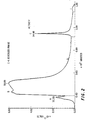
- FIGURE 1 shows the results of the CM Sepharose® chromatographic step.
- the preferred concentration of urea in the eluent used in this step is 1 M to 6 M.
- a preferred concentration of urea in this step is 2 M to 4 M.
- a most preferred concentration is 2.5 M to 3.5 M.
- the salt-eluted peak of activity is applied to a reversed-phase column.
- the preferred method employs a resin having 4, 8, or 18 carbon chains (C-4, C-8 or C-18, respectively).
- the most preferred method employs a C-4 resin commercially available from such resin manufacturers as Vydac, Waters, Tosoh Corp., etc.
- the protein is eluted by a step gradient of, for example, isopropanol and trifluoroacetic acid (TFA).
- the concentration of trifluoroacetic acid is ideally 0.2%, but can range from 0 to 0.4%, preferably 0.15 to 0.25%.
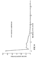
- FIGURE 2 shows the location of the activity during treatment on the reversed-phase column.
- the reversed-phase column step effects desalting and further purifies the material to virtual homogeneity.
- the Limulus protein is remarkably stable to the organic solvents and low pH of this column.
- the protein is stable over a pH range of 1-3 in the presence of TFA.
- the material may now be lyophilized.
- the purified Limulus protein has an isoelectric pH (pI), which is greater than 10, indicating a very basic protein.
- the molecular weight of the protein as determined by SDS-PAGE is 12,500 ⁇ 100.
- the first three N-terminal amino acids of this purified material are Asp-Gly-Ile
- the majority of the purified material had an amino acid sequence which appears to be identical to a protein from Limulus that was isolated by Tanaka et al .
- the material was subsequently purified by Iwanaga et al , as reported in the International Symposium on Pyrogen, held June 23,-26, 1987 in China. However, in the latter case, a different purification procedure was employed.
- variant proteins are those beginning with Asp-Asn- Ser-Gly-, and Ser-Asn-, and also a protein having an N-terminal asparagine with one less amino acid than the natural sequence.
- endotoxin binding protein variants or variant proteins for short
- endotoxin binding protein or variant proteins for short
- variant proteins were not separated from each other. Rather, upon sequencing the most purified samples, some positions were uniformly one amino acid, while other positions showed major variation. This is interpreted as a mixture of proteins showing micro-heterogeneity. It is consistent with the existence of a gene family coding for these proteins whose individual gene sequences are very homologous, but not identical. It remains to be seen if their individual specific activities vary significantly.
- Gene sequences which encode the polypeptides, and vectors containing these gene sequences can be used to transform microorganisms such as E. coli, yeast, etc.
- the transformed microorganisms can be used to produce large quantities of the polypeptide materials, including the variant proteins described above.
- the genes may be altered so as to maximise codon expression in a given host.
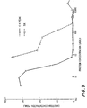
- FIGURE 3 represents a graphical form of such experiments.
- the dotted curve represents the crude extract after ultrafiltration, and the solid line is the most highly purified fraction from the reversed-phase column.
- a known optical property of all proteins is the ability of some aromatic amino acids to fluoresce when excited at certain wavelengths.
- the present inventors looked specifically at tryptophan residues. These are excited at around 283 nm and fluoresce at around 350 nm. See FIGURE 6.
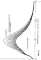
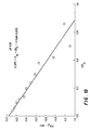
- the inventors examined a fluorescence maxima at 350 nm of the endotoxin binding protein at various concentrations of endotoxin. There was a proportional quenching of that fluorescence, indicative of a tight association. See FIGURE 7. Other proteins as negative controls showed no such proportional change. See FIGURES 8 and 9. From this data, the inventors have been able to calculate the binding affinity constants between the present protein and endotoxin. See FIGURES 9-12.
- the fluorescence quenching experiment may serve as the basis for an assay for endotoxin based on the physical change of a single protein upon exposure to endotoxin.
- the inventors have found that the quenching effect observed upon addition of endotoxin to the endotoxin binding protein does not appear to experience saturation up to a high concentration level of added endotoxin.
- concentration of endotoxin and quenching may be a nonlinear one, the linearity of the relationship extends over a remarkably wide range of concentration of endotoxin.
- the present invention relates to biosensors.
- Biosensors rely on a specific binding ligand immobilised on a solid (e.g. quartz) chip. Electrical potential between two electrodes, or the acoustic wave perturbation between two electrodes is measured.
- the current method for measuring endotoxin is the Limulus amebocyte lysate assay. This involves a cascade of enzymes which are activated by endotoxin and result in the formation of a gel, analogous to a mammalian blood clot. Since multiple enzymes are involved, many substances interfere with the overall reaction, causing enhancement or inhibition. In addition, the key enzymes of the cascade are proteases so other proteolytic enzymes, such as trypsin, can cause gel formation. To eliminate these problems, samples containing interfering substances must be diluted beyond the point where they interfere. In many cases the dilution factor can be hundreds or thousands. The net effect is to decrease the sensitivity in measuring absolute endotoxin concentrations in those samples.
- the electronic component of the biosensor could measure voltage (potentiometric), current (amperometric), light, sound, temperature, or mass (piezoelectric).
- the biosensors of the present invention can be based on technology which is known, such as described in Biotechnology 5 , 437-440 (1987), and Transducers 1987, the abstract entitled “Development or a Surface Acoustic Wave Biosensor", and “Recent Progress in Biosensor Development", Hall, E. A. H., Int. J. Biochem. 1988, 20(4), 357-62, each of which is incorporated herein by reference. Biosensor information is also contained in U.S. Patents 4,721,677, 4,680,268, H 0,000,201, and 4,614,714, each of which is incorporated herein by reference.
- An actual device employing a biosensor would be similar to a flow cell.
- the sample would enter, be exposed to the reactive surface and a measurement made.
- the bound ligand could be washed off to regenerate the surface or left on to measure a cumulative response. This may be adapted to an in-line device to monitor endotoxin contaminating events.
- a portable field unit is feasible. This could be used onsite for checking water purity or process cleanliness during pharmaceutical manufacture, kidney dialysis unit pyrogen checks, or any other application where the conventional LAL is used now. Since endotoxin is a component of gram negative bacterial cell walls, binding of whole bacteria should be measurable. An extension of this technology should make possible remote sensors to monitor water quality in the environment.
- endotoxin binding protein e.g. from Limulus
- the microspheres agglutinate.
- endotoxin binding protein e.g. from Limulus
- Such agglutination is commonly employed in assays based on antibodies to the specific ligand to be measured (Hechemy, K.E.; Michaelson, E.E. Laboratory Management 1984 , 22 (6):27,ff (Part I) and 22 (7):26,ff (Part II); Babson, A.L., Opper, C.A. and Crane L.J.
- the agglutination phenomenon is thought to be due to crossiinking between multiple binding sites on the antibody molecule complexing with multiple sites on the antigen. Due to the high ratio of endotoxin binding protein to endotoxin needed to be effective in our other solution experiments, it is thought the number of endotoxin binding sites on the protein is one or less.
- a multiple binding site reagent is created which is now possible to function as a specific agglutinin.
- the binding protein can be linked to one suspension of beads and endotoxin or lipid A linked to a second suspension.
- agglutination is inversely proportional to the unknown endotoxin concentration.
- E. coli endotoxin was prepared at 100 nanograms per ml. in Phosphate Buffered Saline (PBS). This was mixed with 200 micrograms of Limulus Endotoxin Binding Protein and incubated at 37 degrees Celsius for one hour. Control solutions were prepared of endotoxin (100 nanograms/ml) only and PBS only. Three vials of each of the three solutions were prepared containing 2 ml/vial. 1.5 ml of each solution was injected intravenously into individual 3 kilogram rabbits and their body temperatures measured continuously for six hours with data points recorded every 10 minutes. The final dose of endotoxin or inactivated endotoxin was 50 nanograms/kilogram.
- Limulus Endotoxin Binding Protein is injected intravenously into rabbits at two dose levels, 5 micrograms/kilogram and 50 micrograms/kilogram. Control animals receive injections of PBS. Fifteen minutes later, all animals receive intravenous injections of 50 nanograms/kilogram E. coli endotoxin. Body temperature is monitored continuously over six hours and recorded every 10 minutes. The normal course of increase in temperature peaking at one hour after injection is seen in control animals preinjected with PBS only. Those animals receiving preinjection of endotoxin binding protein at 50 micrograms/kilogram showed an increase of 1.55 (S.D. 0.225) degrees Celsius. Animals receiving 5 micrograms/ kilogram demonstrated temperature increases of 1.85 (S.D. 0.270).
- E. coli endotoxin is injected intravenously into 9 rabbits at a dose of 50 nanograms/kg. After 15 minutes, three were injected intravenously with 5 micrograms Limulus Endotoxin Binding Protein, three were injected intravenously with 50 micrograms Limulus Endotoxin Binding Protein and three received PBS. Volumes of all injections were 0.5 ml/kg. Body temperatures are monitored for 6 hours and data collected every 10 minutes. Animals receiving endotoxin and the PBS only, manifest the normal peak fever response one hour after toxin administration. Those animals receiving therapeutic injections of the Limulus protein exhibit a much reduced fever response proportional to the amount of protein which is administered.
- the protein was tested for its ability to inactivate endotoxin in the presence of whole human serum.
- the assay was conducted in a 96-well tissue culture multiwell plates as described in detail in Novitsky, et al., J. Clin. Micro., 20 :211-216 (1985). To each well were added in order 0.05 ml of serum only or serum with 25 micrograms/ml Limulus Endotoxin Binding Protein, 0.05 ml E. coli endotoxin solution.
- the plates were covered with Parafilm to prevent evaporation, agitated on a mechanical vibrating platform for 15 seconds and incubated at 37 degrees Celsius. Multiwell plates were then uncovered, and 0.1 ml of LAL was added to each well. The plates were subsequently handled and read as described for the LAL endotoxin assay. Serum with the added endotoxin binding protein was able to inactivate increasingly larger amounts of endotoxin (2,000 nanograms/ml) while serum alone was able to bind and/or inactivate only 80 nanograms/ml.
- endotoxin from several species of Gram negative bacteria as well as lipid A were tested. These included endotoxin from Klebsiella pneumoniae, Serratia marcescens, Salmonella enteritidis, Eschericia coli 0113 wild type, E. coli rough mutant (J-5), Salmonella abortus equi, and Lipid A from S. minnesota Re 595.
- the endotoxin binding protein was mixed with the endotoxins or lipid A at a ratio of 5:1 to 1,000:1 in the presence of 10 millimolar Tris buffer, pH 6-8. In all cases the measurable endotoxin after mixing was reduced 85% to 99.5%.
Landscapes
- Health & Medical Sciences (AREA)
- Life Sciences & Earth Sciences (AREA)
- Chemical & Material Sciences (AREA)
- Engineering & Computer Science (AREA)
- Immunology (AREA)
- General Health & Medical Sciences (AREA)
- Medicinal Chemistry (AREA)
- Molecular Biology (AREA)
- Hematology (AREA)
- Organic Chemistry (AREA)
- Biomedical Technology (AREA)
- Urology & Nephrology (AREA)
- Chemical Kinetics & Catalysis (AREA)
- Pharmacology & Pharmacy (AREA)
- Nuclear Medicine, Radiotherapy & Molecular Imaging (AREA)
- Public Health (AREA)
- Veterinary Medicine (AREA)
- Animal Behavior & Ethology (AREA)
- Analytical Chemistry (AREA)
- General Chemical & Material Sciences (AREA)
- Biochemistry (AREA)
- Food Science & Technology (AREA)
- Cell Biology (AREA)
- Biotechnology (AREA)
- Bioinformatics & Cheminformatics (AREA)
- Microbiology (AREA)
- Physics & Mathematics (AREA)
- Pathology (AREA)
- General Physics & Mathematics (AREA)
- Proteomics, Peptides & Aminoacids (AREA)
- Cardiology (AREA)
- Heart & Thoracic Surgery (AREA)
- Toxicology (AREA)
- Gastroenterology & Hepatology (AREA)
- Genetics & Genomics (AREA)
- Insects & Arthropods (AREA)
- Tropical Medicine & Parasitology (AREA)
- Zoology (AREA)
- Communicable Diseases (AREA)
- Biophysics (AREA)
Abstract
Description
- The present invention is directed to assays for endotoxin, particularly assays using an endotoxin binding protein isolated from a horseshoe crab.
- Endotoxins are high molecular weight complexes, associated with the outer membrane or gram-negative bacteria, that produce pyrogenic reactions upon intravenous administration. Endotoxin is shed from living bacteria and is also released into the environment when bacteria die and decompose. Since gram-negative bacteria are found in great numbers in air, water, and soil, bacterial endotoxin commonly contaminates raw materials and processing equipment used in the manufacturing of, for example, pharmaceuticals.
- Bacterial endotoxin is a complex consisting of lipid, carbohydrate and protein. It is characterized by an overall negative charge, heat stability and high molecular weight. Highly purified endotoxin does not contain protein, and is a lipopolysaccharide (LPS). Depyrogenation can generally be achieved by inactivating or removing endotoxin, depending upon the physicochemical nature of the LPS. LPS consists of three distinct chemical regions, lipid A, which is the innermost region, an intermediate core polysaccharide, and an outermost O-specific polysaccharide side chain which is responsible for an endotoxin's particular immunospecificity.
- Bacterial endotoxins are known to have profound biological effects in animals and humans, and to cause severe medical problems when present. Symptoms include induction of high fever, activation of complement, and hypotension. It is critical to avoid endotoxin contamination in any pharmaceutical product or medical device which comes into contact with body fluids. High endotoxin levels in sera due to bacterial diseases, such as septicemia, are not easily treated. Antibiotic treatment of the infection only kills the bacteria, leaving the endotoxin from their cell walls free to cause fever.
- The horseshoe crab Limulus polyphemus is particularly sensitive to endotoxin. The cells from their hemolymph (amebocytes) undergo a complex series of biochemical reactions resulting in clot formation, analogous to mammalian blood coagulation. This phenomenon has been exploited in the form of bioassays sensitive to very low endotoxin levels. Currently, a bioassay of this type is the method of choice for monitoring pharmaceutical manufacturing and is termed Limulus Amebocyte Lysate (LAL). See U.S. Patents 4,276,050, 4,273,557, 4,221,866, 4,201,865, 4,038,147, 3,944,391 and 3,915,805, each of which is incorporated herein by reference.
- It has long been observed that once endotoxin interacts with LAL the toxin is not recoverable from the clot. See Nachum et al, Journal of Invertebrate Pathology, 32:51-58 (1978). This observation lead investigators to postulate two alternative explanations. Either the endotoxin is enzymatically degraded during clot formation or it is bound by some factor causing it to lose toxicity. The present inventors initiated experiments to extract the endotoxin inactivating factor from the LAL.
- Other research groups have experimented with endotoxin binding proteins, also referred to as anti-LPS factor. To the inventor's knowledge, the following publications resulting from work in this area are the most relevant to this invention:
- Tanaka et al, Biochem. Biophys. Res. Comm. 105, 717-723 (1982),
- Iwanaga et al, International Symposium on Pyrogen, 84-84 (June 23-26, 1987),
- Aketagawa et al, J. Biol. Chem. 261, 7354-7365 (1986),
- Hao, U.S. Patent 4,677,194 (June 30, 1987), and
- Nachum et al, J. Inv. Path. 32, 51-58 (1978).
- Tanaka et al, Iwanaga et al, and Aketagawa et al each conducted research on an anti-LPS factor or endotoxin binding protein isolated from a horseshoe crab system. Based on experimental work done in the inventors' laboratory, it appears that a protein involved in the present invention is the same as that isolated by Iwanaga et al and Tanaka et al. However, these publications do not say anything about pharmaceutical utility of the endotoxin binding protein, and it is difficult to predict in vivo activity based on in vitro experimentation. In fact, neither or these references suggests a practical utility for the anti-LPS factor, and in view of the unpredictable nature of in vivo activity, it has previously not been appreciated that the endotoxin binding protein could be used in a pharmaceutical composition. Furthermore, none of the references disclose the use of the endotoxin binding protein for an endotoxin assay, as disclosed in the present invention. It is notable that the present inventors have also discovered certain endotoxin binding protein variants which have amino acid structures that are different from the anti-LPS factor disclosed in the above-described publications.
-
- It is an object of the present invention to provide a method of assaying for endotoxin in a fluid. Thus, the present invention provides
- a method for assaying endotoxin concentration, which comprises contacting serial aqueous dilutions of material suspected of containing endotoxin with a known quantity of endotoxin binding protein, observing the fluorescence emission at at least one wavelength of the endotoxin binding protein before and after contact with aqueous dilutions of said material suspected of containing endotoxin,
- correlating the levels of fluorescence emission with at least one known emission level to thereby determine the quantity of endotoxin present in said material suspected of containing endotoxin.
-
- These and other objects of the present invention which will hereinafter become more readily apparent, have been provided by purifying an endotoxin binding protein from the horseshoe crab Limulus polyphemus, and discovering that it has the capability of binding to endotoxin in vivo. The present inventors have also discovered that there are certain structural variants of the Limulus polyphemus endotoxin binding protein, and it is expected that these materials will also possess endotoxin binding ability such that they may be used in the other aspects of the present invention as well.
- A more complete appreciation of the invention and many of the attendant advantages thereof will be readily obtained as the same becomes better understood by reference to the following detailed description when considered in connection with the accompanying drawings, wherein:
- FIGURE 1 shows cation exchange chromatographic purification of Limulus endotoxin binding protein using CM-Sepharose® resin. Peak 1: flow through, Peak 2: 0.25M NaCl elution, Peak 3: 1 M NaCl elution.
- FIGURE 2 shows reversed phase chromatographic purification of Peak 3 from Fig. 1. Peak A: 25% isopropanol (IPA) elution, Peak B: 35% IPA elution, Peak C: 50% IPA elution, Peak D: column wash with 100% IPA.
- FIGURE 3 is a plot of apparent endotoxin concentration (measured by LAL) versus protein concentration. Such a plot can be used to assess protein specific endotoxin inactivating activity. Specific activity is expressed as the amount of protein needed to achieve 50% reduction of endotoxin in the assay system (5 nanograms of endotoxin per 100 microliters). For convenience, units are expressed as micrograms-1 protein x 105 needed to reduce 5 nanograms/100 microliters of standard endotoxin by 50%.
- FIGURE 4 shows depyrogenation by affinity chromatography with endotoxin binding protein immobilized to a solid support.
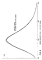
- FIGURE 5 shows fluorescence excitation and emission spectra of Limulus endotoxin binding protein.
- FIGURE 6 shows successive decreases in the emission spectra of Limulus endotoxin binding protein upon successive additions of endotoxin.
- FIGURE 7 shows lack of successive decreases in the emission spectra of human serum albumin upon successive additions of endotoxin (negative control).
- FIGURE 8 shows lack of successive decreases in the emission spectra of peak B, FIGURE 2 upon successive additions of endotoxin (negative control).
- FIGURE 9 shows a fluorescence titration of Limulus endotoxin binding protein at pH 3.86. KD = 2.41 micromolar.
- FIGURE 10 shows fluorescence titration of Limulus endotoxin binding protein at pH 6.91. KD = 0.51 micromolar.
- FIGURE 11 shows fluorescence titration of Limulus endotoxin binding protein at pH 8.80. KD = 2.40 micromolar.
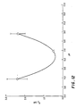
- FIGURE 12 shows the effect of pH on dissociation constant of Limulus endotoxin binding protein.
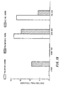
- FIGURE 13 is a comparison of immobilized Limulus endotoxin binding protein compared to two immobilized polymyxin resins in proteinaceous and non-proteinaceous solutions.
- FIGURE 14. Demonstration of activity of Limulus endotoxin binding protein in the presence of total human serum. The dashed line represents serum alone as a control (68 nanograms of endotoxin per ml inactivated). The solid line represents serum plus 25 micrograms/ml added endotoxin binding protein (1600 nanograms endotoxin per ml inactivated).
-
- The endotoxin binding protein may be isolated from any horseshoe crab. For example, any of the four known species of horseshoe crabs could be used. These species are:
- Limulus polyphemus
- Tachypleus gigas
- Tachypleus tridentatus
- Carcinoscorpius rotundicauda Especially preferred among these is Limulus polyphemus, the horseshoe crab which is found along the North American coast. The endotoxin binding protein may be isolated by a procedure which is summarized hereinbelow. The procedure is illustrated for the Limulus horseshoe crab, but these procedures may be applied to any known horseshoe crab, as recited herein.
-
- It was surprisingly found that the cellular debris produced during lysate production from Limulus amebocytes contains significant amounts of the endotoxin binding protein. The cellular debris was found to have even more activity than lysate itself. By "cell debris" is meant the insoluble material remaining when Limulus amebocytes are lysed by hypotonic shock. It includes nuclei, cell debris and some lysate proteins. The majority is insoluble material. Hypotonic shock may be accomplished by treating the Limulus amebocytes with endotoxin-free distilled water, preferably at 4°C, with shaking (e.g., for 12 h). The mixture is centrifuged, separating the lysate (soluble supernatant) from the cell debris.
- The next step is to extract from the cell debris the Limulus amebocyte binding protein. Many solvents have been tested, including water, several alcohols (e.g. methanol, ethanol, isopropanol, and butanol), acetone, chloroform, acetonitrile, acids (e.g. HCl and H2SO4), bases (e.g. NaOH and ethanolamine), salts (e.g. NaCl), and detergents (e.g. Tween, triton X-100, and SDS). The best results were obtained with denaturants such as urea and guanidine hydrochloride. Surprisingly, six molar solutions of denaturants were effective in extracting the protein without affecting biological activity. This concentration of denaturant would be expected to inactivate most proteins. Thus, the first step in the purification procedure herein is extraction of cellular debris from Limulus amebocyte lysate with a denaturant to produce an extract. The concentration of the denaturant can range from 1 M to 10 M. A more preferred range would be 3 M to 8 M. A most preferred concentration is around 6 molar. Urea is the preferred denaturant, because it can be made free of endotoxin by ultrafiltration and it is not readily contaminated by endotoxin containing bacteria.
- Preferably, prior to loading the extract onto a cation exchange column, an ultrafiltration step is performed. This step is also accomplished using urea or another denaturant as described above for extraction. In particular, the extract from the cell debris is crudely filtered with a filter aid such as diatomaceous earth (e.g. Celite, Manville Corp.) or one of the cationic or anionic polymeric particles in colloidal suspension (e.g. Biocryl, Supelco division of Rohm and Haas Corp.), then passed through an ultrafiltration membrane. Ultrafiltration membranes may be used in any known form (e.g., plain film, hollow fiber, tubular and spiral) and may be made of any material. Preferred materials are polysulfone, polyvinylidene fluoride, polyacrylonitrile, nylon, or cellulose. Most preferred is a polysulfone, flat or hollow fiber type ultrafiltration membrane. The preferred molecular weight cut-off of the ultrafiltration membrane is 20,000 daltons to 50,000 daltons as can be commercially obtained from Millipore, Filtron or other membrane filter manufacturers. The most preferred membrane has a 30,000 dalton cut-off. The Limulus endotoxin binding protein is now in the filtrate at a very low concentration. The filtrate from the first membrane is concentrated using a second ultrafiltration membrane having a molecular weight cut-off of 5,000 to 10,000 daltons, preferably an 8,000 dalton cut-off membrane. The second ultrafiltration membrane may be the same or different material as the first ultrafiltration membrane, and is preferably made of polysulfone. The principle of operation is the same as the 30,000 cut-off membrane, however, the endotoxin binding protein is now in the retentate with all other proteins greater than e.g. 8,000 daltons.
- After the above step, the retentate is subjected to cation exchange chromatography using cellulose, cross-linked agarose or hydrophilic vinyl polymer resins derivatized with carboxymethyl (CM), Sulphopropyl (SP), Sulphonate (S), or other anionic group. These can be obtained from several manufacturers including Pharmacia (Sephadex® , Sepharose® ), Tosoh Corp. (Toyopearl® ) or BioRad (Cellex® , Bio-Gel). The most preferred resin is CM-Sepharose® . The pH of loading and elution buffers can range from 4 to 8, with the most preferred range of
pH 5 to 5.5. In this step also, urea (or another denaturant as described above for extraction) is an important constituent. All column elution buffers must contain urea (at least about 3 molar) to elute the endotoxin binding protein cleanly from the column. - The elution from the column is accomplished by a step gradient of a salt such as ammonium chloride, potassium chloride or sodium chloride. Sodium chloride is preferred. FIGURE 1 shows the results of the CM Sepharose® chromatographic step. The preferred concentration of urea in the eluent used in this step is 1 M to 6 M. A preferred concentration of urea in this step is 2 M to 4 M. A most preferred concentration is 2.5 M to 3.5 M. When sodium chloride is used in the eluant, biological activity elutes at a concentration of sodium chloride of from around 0.5 to 1 molar.
- After the cation exchange step, the salt-eluted peak of activity is applied to a reversed-phase column. The preferred method employs a resin having 4, 8, or 18 carbon chains (C-4, C-8 or C-18, respectively). The most preferred method employs a C-4 resin commercially available from such resin manufacturers as Vydac, Waters, Tosoh Corp., etc. The protein is eluted by a step gradient of, for example, isopropanol and trifluoroacetic acid (TFA). The concentration of trifluoroacetic acid is ideally 0.2%, but can range from 0 to 0.4%, preferably 0.15 to 0.25%. FIGURE 2 shows the location of the activity during treatment on the reversed-phase column. The reversed-phase column step effects desalting and further purifies the material to virtual homogeneity. The Limulus protein is remarkably stable to the organic solvents and low pH of this column. The protein is stable over a pH range of 1-3 in the presence of TFA. The material may now be lyophilized. The purified Limulus protein has an isoelectric pH (pI), which is greater than 10, indicating a very basic protein. The molecular weight of the protein as determined by SDS-PAGE is 12,500 ± 100. The first three N-terminal amino acids of this purified material are
Asp-Gly-Ile - It should be noted that Watson and Sullivan, U.S. Patent 4,107,077, teaches that there is an increase in sensitivity of the lysate by organic extraction with chloroform. It appeared logical that a protein which inactivated endotoxin would appear as an inhibitor in the endotoxin assay. Thus, it was hoped that the protein was the inhibitor which is extracted into chloroform and could be recovered from that extract. However, surprisingly, this was not successful. Since the protein is fairly hydrophobic, it may be in the organic extract, but denatured in some way.
- The majority of the purified material had an amino acid sequence which appears to be identical to a protein from Limulus that was isolated by Tanaka et al. The material was subsequently purified by Iwanaga et al, as reported in the International Symposium on Pyrogen, held June 23,-26, 1987 in China. However, in the latter case, a different purification procedure was employed.
- In addition to a protein having an apparently identical amino acid sequence to that reported in Iwanaga et al, some related proteins were also purified, which have different amino acid sequences. These variant proteins have not previously been described. In a first protein, on the N-terminal thereof, a serine rather than an aspartic acid residue is located. Furthermore, an asparagine is located in the second position from the N-terminal rather than a glycine as reported in Iwanaga et al. Therefore, a protein having an initial amino acid sequence of
Ser-Asn-Ile-Trp-Thr-
is also part of the present invention. Other possible protein derivatives are those beginning with
Asp-Asn-
Ser-Gly-, and
Ser-Asn-, and also a protein having an N-terminal asparagine with one less amino acid than the natural sequence. Each of these variant proteins is also part of the present invention, and they may be used in each aspect of the invention described hereinbelow. They will be referred to as endotoxin binding protein variants (or variant proteins for short), as distinguished from the natural sequence endotoxin binding protein, which is the protein having the amino acid sequence reported in Iwanaga et al. - The variant proteins were not separated from each other. Rather, upon sequencing the most purified samples, some positions were uniformly one amino acid, while other positions showed major variation. This is interpreted as a mixture of proteins showing micro-heterogeneity. It is consistent with the existence of a gene family coding for these proteins whose individual gene sequences are very homologous, but not identical. It remains to be seen if their individual specific activities vary significantly.
- Gene sequences which encode the polypeptides, and vectors containing these gene sequences, can be used to transform microorganisms such as E. coli, yeast, etc. The transformed microorganisms can be used to produce large quantities of the polypeptide materials, including the variant proteins described above. The genes may be altered so as to maximise codon expression in a given host. Procedure for Assaying for Endotoxin Binding Activity
- To follow the purification of the endotoxin binding protein during the above purification steps, assays can be performed in solution. One publication describing typical assays for LR50 values is Novitsky et al, J. Clin. Microbiol. pp. 211-216 (1985). A specific procedure follows:
- A 96-well microtiter plate is used. Protein fractions are serially diluted across the top row and mixed with a standard endotoxin solution. There is another series of dilutions done on all samples to get within range of the assay, but basically, the endotoxin recovered in the well after mixing with a suspected endotoxin inactivating protein is measured. This is compared to negative controls and an endotoxin standard curve. It is found that the added endotoxin is inactivated at the high protein concentrations. As the protein is diluted across the plate, a point is reached where it is too dilute to inactivate the added endotoxin. This dilution is a measure of the protein specific activity. FIGURE 3 represents a graphical form of such experiments. In FIGURE 3, the dotted curve represents the crude extract after ultrafiltration, and the solid line is the most highly purified fraction from the reversed-phase column.
- A known optical property of all proteins is the ability of some aromatic amino acids to fluoresce when excited at certain wavelengths. The present inventors looked specifically at tryptophan residues. These are excited at around 283 nm and fluoresce at around 350 nm. See FIGURE 6.
- The inventors examined a fluorescence maxima at 350 nm of the endotoxin binding protein at various concentrations of endotoxin. There was a proportional quenching of that fluorescence, indicative of a tight association. See FIGURE 7. Other proteins as negative controls showed no such proportional change. See FIGURES 8 and 9. From this data, the inventors have been able to calculate the binding affinity constants between the present protein and endotoxin. See FIGURES 9-12.
- The fluorescence quenching experiment may serve as the basis for an assay for endotoxin based on the physical change of a single protein upon exposure to endotoxin. The inventors have found that the quenching effect observed upon addition of endotoxin to the endotoxin binding protein does not appear to experience saturation up to a high concentration level of added endotoxin. In other words, although one would expect the relationship between concentration of endotoxin and quenching to be a nonlinear one, the linearity of the relationship extends over a remarkably wide range of concentration of endotoxin. This unexpected aspect of the present assay enables the assay to be used over a wider range or endotoxin concentrations than could have been expected. Ranges of the ratio of endotoxin binding protein to endotoxin that are expected to be effective are 0.1:1 to 5000:1, preferably 1:1 to 2000:1, most preferably 10:1 to 1000:1.
- In another embodiment, the present invention relates to biosensors. Biosensors rely on a specific binding ligand immobilised on a solid (e.g. quartz) chip. Electrical potential between two electrodes, or the acoustic wave perturbation between two electrodes is measured.
- The current method for measuring endotoxin is the Limulus amebocyte lysate assay. This involves a cascade of enzymes which are activated by endotoxin and result in the formation of a gel, analogous to a mammalian blood clot. Since multiple enzymes are involved, many substances interfere with the overall reaction, causing enhancement or inhibition. In addition, the key enzymes of the cascade are proteases so other proteolytic enzymes, such as trypsin, can cause gel formation. To eliminate these problems, samples containing interfering substances must be diluted beyond the point where they interfere. In many cases the dilution factor can be hundreds or thousands. The net effect is to decrease the sensitivity in measuring absolute endotoxin concentrations in those samples.
- One of the promising potentials that a single endotoxin binding protein has is to eliminate the multiple interfering effects seen in the LAL assay. Even if the sensitivity were one tenth that of LAL, the lack of a need to dilute would compensate, making the sensitivity equivalent. Biosensors take advantage of specific binding affinities of such molecules. Antibodies are an example which have been used. Bound to the surface of a silicon chip, they bind to their immunogen and cause a change in the surface which is measured electrically. Depending on the type of sensor, capacitance, resistance or acoustic wave chances are measurable. Depending on the concentrations and volumes of the samples, the time required for each measurement can be from seconds to minutes.
- The electronic component of the biosensor could measure voltage (potentiometric), current (amperometric), light, sound, temperature, or mass (piezoelectric). The biosensors of the present invention can be based on technology which is known, such as described in
Biotechnology 5, 437-440 (1987), and Transducers 1987, the abstract entitled "Development or a Surface Acoustic Wave Biosensor", and "Recent Progress in Biosensor Development", Hall, E. A. H., Int. J. Biochem. 1988, 20(4), 357-62, each of which is incorporated herein by reference. Biosensor information is also contained in U.S. Patents 4,721,677, 4,680,268, H 0,000,201, and 4,614,714, each of which is incorporated herein by reference. - An actual device employing a biosensor would be similar to a flow cell. The sample would enter, be exposed to the reactive surface and a measurement made. Depending on the chemistry of binding, the bound ligand could be washed off to regenerate the surface or left on to measure a cumulative response. This may be adapted to an in-line device to monitor endotoxin contaminating events.
- Due to the compact nature of the electronics, a portable field unit is feasible. This could be used onsite for checking water purity or process cleanliness during pharmaceutical manufacture, kidney dialysis unit pyrogen checks, or any other application where the conventional LAL is used now. Since endotoxin is a component of gram negative bacterial cell walls, binding of whole bacteria should be measurable. An extension of this technology should make possible remote sensors to monitor water quality in the environment.
- By coupling the binding protein to latex microspheres using a chemistry similar to that used to immobilize the protein on chromatographic resins or membranes, another method to quantitate endotoxin is constituted. By exposing latex microspheres coated with endotoxin binding protein (e.g. from Limulus) to endotoxin, the microspheres agglutinate. Such agglutination is commonly employed in assays based on antibodies to the specific ligand to be measured (Hechemy, K.E.; Michaelson, E.E. Laboratory Management 1984, 22(6):27,ff (Part I) and 22(7):26,ff (Part II); Babson, A.L., Opper, C.A. and Crane L.J. American Journal of Clinical Pathology (1982), 77(4):424-9). In such techniques, the agglutination phenomenon is thought to be due to crossiinking between multiple binding sites on the antibody molecule complexing with multiple sites on the antigen. Due to the high ratio of endotoxin binding protein to endotoxin needed to be effective in our other solution experiments, it is thought the number of endotoxin binding sites on the protein is one or less. By covalently coupling many molecules of binding protein per microsphere, a multiple binding site reagent is created which is now possible to function as a specific agglutinin. Alternatively, the binding protein can be linked to one suspension of beads and endotoxin or lipid A linked to a second suspension. On mixing, these two suspensions will agglutinate. This may also be employed as an assay for endotoxin in solution by the ability of the free endotoxin to inhibit such agglutination. In this case, agglutination is inversely proportional to the unknown endotoxin concentration.
- The invention now being generally described, it will now be illustrated in greater detail by the following examples, which are presented herein for illustration only and are not intended to be limiting of the present invention, unless so indicated.
- E. coli endotoxin was prepared at 100 nanograms per ml. in Phosphate Buffered Saline (PBS). This was mixed with 200 micrograms of Limulus Endotoxin Binding Protein and incubated at 37 degrees Celsius for one hour. Control solutions were prepared of endotoxin (100 nanograms/ml) only and PBS only. Three vials of each of the three solutions were prepared containing 2 ml/vial. 1.5 ml of each solution was injected intravenously into
individual 3 kilogram rabbits and their body temperatures measured continuously for six hours with data points recorded every 10 minutes. The final dose of endotoxin or inactivated endotoxin was 50 nanograms/kilogram. - In rabbits, a 5 nanograms/kilogram dose of endotoxin elicits a measurable fever response, showing a peak at one hour after injection. The present results showed the rabbits receiving only endotoxin at 50 nanograms/kilogram did indeed develop a body temperature elevated 1.64 (S.D. 0.236) degrees Celsius. Those rabbits receiving only PBS or endotoxin inactivated with Limulus Endotoxin Binding Protein maintained a normal body temperature.
- Limulus Endotoxin Binding Protein is injected intravenously into rabbits at two dose levels, 5 micrograms/kilogram and 50 micrograms/kilogram. Control animals receive injections of PBS. Fifteen minutes later, all animals receive intravenous injections of 50 nanograms/kilogram E. coli endotoxin. Body temperature is monitored continuously over six hours and recorded every 10 minutes. The normal course of increase in temperature peaking at one hour after injection is seen in control animals preinjected with PBS only. Those animals receiving preinjection of endotoxin binding protein at 50 micrograms/kilogram showed an increase of 1.55 (S.D. 0.225) degrees Celsius. Animals receiving 5 micrograms/ kilogram demonstrated temperature increases of 1.85 (S.D. 0.270).
- E. coli endotoxin is injected intravenously into 9 rabbits at a dose of 50 nanograms/kg. After 15 minutes, three were injected intravenously with 5 micrograms Limulus Endotoxin Binding Protein, three were injected intravenously with 50 micrograms Limulus Endotoxin Binding Protein and three received PBS. Volumes of all injections were 0.5 ml/kg. Body temperatures are monitored for 6 hours and data collected every 10 minutes. Animals receiving endotoxin and the PBS only, manifest the normal peak fever response one hour after toxin administration. Those animals receiving therapeutic injections of the Limulus protein exhibit a much reduced fever response proportional to the amount of protein which is administered.
- In order to assay the potential effectiveness of the endotoxin binding protein as a therapeutic or prophylactic agent for septic shock or related disorders in humans, the protein was tested for its ability to inactivate endotoxin in the presence of whole human serum. The assay was conducted in a 96-well tissue culture multiwell plates as described in detail in Novitsky, et al., J. Clin. Micro., 20:211-216 (1985). To each well were added in order 0.05 ml of serum only or serum with 25 micrograms/ml Limulus Endotoxin Binding Protein, 0.05 ml E. coli endotoxin solution. The plates were covered with Parafilm to prevent evaporation, agitated on a mechanical vibrating platform for 15 seconds and incubated at 37 degrees Celsius. Multiwell plates were then uncovered, and 0.1 ml of LAL was added to each well. The plates were subsequently handled and read as described for the LAL endotoxin assay. Serum with the added endotoxin binding protein was able to inactivate increasingly larger amounts of endotoxin (2,000 nanograms/ml) while serum alone was able to bind and/or inactivate only 80 nanograms/ml.
- In order to investigate the mechanism of the endotoxin binding phenomenon and determine the breadth of endotoxin types against which the binding protein is effective, endotoxin from several species of Gram negative bacteria as well as lipid A. were tested. These included endotoxin from Klebsiella pneumoniae, Serratia marcescens, Salmonella enteritidis, Eschericia coli 0113 wild type, E. coli rough mutant (J-5), Salmonella abortus equi, and Lipid A from S. minnesota Re 595.
- The endotoxin binding protein was mixed with the endotoxins or lipid A at a ratio of 5:1 to 1,000:1 in the presence of 10 millimolar Tris buffer, pH 6-8. In all cases the measurable endotoxin after mixing was reduced 85% to 99.5%.
- The invention now being fully described, it will be appreciated that the invention may be practiced otherwise than as specifically set forth herein.
Claims (4)
- A method for assaying endotoxin concentration, which comprises contacting serial aqueous dilutions of material suspected of containing endotoxin with a known quantity of endotoxin binding protein,observing the fluorescence emission at at least one wavelength of the endotoxin binding protein before and after contact with aqueous dilutions of said material suspected of containing endotoxin,correlating the levels of fluorescence emission with at least one known emission level to thereby determine the quantity of endotoxin present in said material suspected of containing endotoxin.
- The method of claim 1, wherein said material is a body fluid.
- The method of claim 1 or claim 2, wherein said fluorescence emission of the endotoxin binding protein is measured at from 340 to 360 nanometers.
- The method according to claim 1 or claim 2, wherein said fluorescence emission is produced by excitation at from about 275 to 295 nanometers.
Applications Claiming Priority (3)
| Application Number | Priority Date | Filing Date | Title |
|---|---|---|---|
| US21057588A | 1988-06-23 | 1988-06-23 | |
| US210575 | 1988-06-23 | ||
| EP89907603A EP0379549B1 (en) | 1988-06-23 | 1989-06-21 | Endotoxin binding protein and uses thereof |
Related Parent Applications (2)
| Application Number | Title | Priority Date | Filing Date |
|---|---|---|---|
| EP89907603.8 Division | 1989-06-21 | ||
| EP89907603A Division EP0379549B1 (en) | 1988-06-23 | 1989-06-21 | Endotoxin binding protein and uses thereof |
Publications (3)
| Publication Number | Publication Date |
|---|---|
| EP0687911A2 EP0687911A2 (en) | 1995-12-20 |
| EP0687911A3 EP0687911A3 (en) | 1996-08-21 |
| EP0687911B1 true EP0687911B1 (en) | 2001-10-17 |
Family
ID=22783435
Family Applications (2)
| Application Number | Title | Priority Date | Filing Date |
|---|---|---|---|
| EP95201582A Expired - Lifetime EP0687911B1 (en) | 1988-06-23 | 1989-06-21 | Methods for assaying endotoxin concentration |
| EP89907603A Expired - Lifetime EP0379549B1 (en) | 1988-06-23 | 1989-06-21 | Endotoxin binding protein and uses thereof |
Family Applications After (1)
| Application Number | Title | Priority Date | Filing Date |
|---|---|---|---|
| EP89907603A Expired - Lifetime EP0379549B1 (en) | 1988-06-23 | 1989-06-21 | Endotoxin binding protein and uses thereof |
Country Status (7)
| Country | Link |
|---|---|
| EP (2) | EP0687911B1 (en) |
| JP (1) | JP2774343B2 (en) |
| AT (1) | ATE207205T1 (en) |
| AU (1) | AU3772289A (en) |
| CA (1) | CA1338836C (en) |
| DE (2) | DE68925275T2 (en) |
| WO (1) | WO1989012644A1 (en) |
Families Citing this family (18)
| Publication number | Priority date | Publication date | Assignee | Title |
|---|---|---|---|---|
| DK255887D0 (en) * | 1987-05-20 | 1987-05-20 | Claus Koch | IMMUNOASSAY |
| US5594113A (en) * | 1988-06-23 | 1997-01-14 | Associates Of Cape Cod, Inc. | Endotoxin binding and neutralizing protein and uses thereof |
| JP3117985B2 (en) | 1990-07-24 | 2000-12-18 | マルハ株式会社 | Bacterial shock treatment |
| EP0572541A1 (en) * | 1991-02-22 | 1993-12-08 | Massachusetts Institute Of Technology | Treatment of endotoxemia |
| HUT70259A (en) * | 1991-05-08 | 1995-09-28 | Vertex Pharma | Rfkbp: a novel isomerase and papamycin/fk506 binding protein |
| WO1992020715A1 (en) * | 1991-05-16 | 1992-11-26 | Associates Of Cape Cod, Inc. | Endotoxin binding and neutralizing protein and uses thereof |
| US5439807A (en) * | 1992-05-19 | 1995-08-08 | Xoma Corporation | Methods for the preparation of endotoxin-binding proteins |
| US6274348B1 (en) | 1992-05-19 | 2001-08-14 | Xoma Corporation | Methods for the preparation of positively charged proteins |
| JP3402476B2 (en) * | 1992-08-24 | 2003-05-06 | 生化学工業株式会社 | Lipopolysaccharide binding protein and method for producing the same |
| CA2146667A1 (en) * | 1993-08-18 | 1995-02-23 | Adolf Hoess | Lipopolysaccharide-binding and neutralizing peptides |
| DK0759774T3 (en) * | 1993-09-22 | 2002-10-07 | Xoma Technology Ltd | Method of treating gram-negative bacterial infection by administration of bactericidal / permeability-increasing (BPI) protein product and antibiotic |
| US5624904A (en) * | 1993-11-17 | 1997-04-29 | Massachusetts Institute Of Technology | Method for treating gram positive septicemia |
| US5578568A (en) * | 1994-04-22 | 1996-11-26 | Xoma Corporation | Method of treating conditions associated with intestinal ischemia/reperfusion |
| US5932536A (en) * | 1994-06-14 | 1999-08-03 | The Rockefeller University | Compositions for neutralization of lipopolysaccharides |
| FR2739448B1 (en) * | 1995-09-29 | 1997-10-24 | Thomson Csf | BIOFILMS DETECTION AND MONITORING DEVICE |
| US5780429A (en) * | 1995-12-22 | 1998-07-14 | Marine Biological Laboratory | Anti-LPS factor from horseshoe crabs and methods of use |
| CN1293383C (en) * | 2005-01-13 | 2007-01-03 | 丁友玲 | Preparing method for clinical detection reagent |
| JP7327096B2 (en) * | 2019-11-13 | 2023-08-16 | 株式会社島津製作所 | Molecular separation method |
Family Cites Families (15)
| Publication number | Priority date | Publication date | Assignee | Title |
|---|---|---|---|---|
| US4107077A (en) | 1975-07-14 | 1978-08-15 | Associates Of Cape Cod, Inc. | Limulus lysate of improved sensitivity and preparing the same |
| US4201865A (en) | 1976-01-15 | 1980-05-06 | Gruppo Lepetit S.P.A. | Novel prostaglandin analogues |
| US4038147A (en) | 1976-06-15 | 1977-07-26 | Becton, Dickinson And Company | Method for detecting endotoxins |
| US4221866A (en) | 1978-10-06 | 1980-09-09 | Baxter Travenol Laboratories, Inc. | Method for determining endotoxin concentration |
| US4276050A (en) | 1980-01-10 | 1981-06-30 | Abbott Laboratories | Method for systemic endotoxin detection |
| US4273557A (en) | 1980-03-31 | 1981-06-16 | Mallinckrodt, Inc. | Limulus lysate procedure for determining endotoxins |
| FR2497798A1 (en) * | 1981-01-09 | 1982-07-16 | Pharmindustrie | NOVEL PEPTIDES CARRYING A FLUOROPHORE, PROCESS FOR THEIR PREPARATION AND THEIR APPLICATION TO FLUORIMETRIC DETERMINATION OF ENDOTOXINS |
| US4614714A (en) | 1982-08-21 | 1986-09-30 | Yamasa Shoyu Kabushiki Kaisha | Use of novel L-glutamic acid oxidase |
| US4758655A (en) * | 1983-01-03 | 1988-07-19 | Scripps Clinic And Research Foundation | Synthetic polypeptide corresponding to a portion of the heat-labile enterotoxin of escherichia coli, compositions and methods of therewith |
| US4713347A (en) * | 1985-01-14 | 1987-12-15 | Sensor Diagnostics, Inc. | Measurement of ligand/anti-ligand interactions using bulk conductance |
| US4780529A (en) * | 1985-08-09 | 1988-10-25 | Biotech Research Laboratories, Inc. | Isolation of an endotoxin inactivator from human plasma, and methods of use |
| US4677194A (en) | 1985-08-09 | 1987-06-30 | Biotech Research Laboratories, Inc. | Isolation of an endotoxin inactivator from human plasma |
| USH201H (en) | 1985-08-23 | 1987-01-06 | United States Of America | Biosensors from membrane proteins reconstituted in polymerized lipid bilayers |
| US4680268A (en) | 1985-09-18 | 1987-07-14 | Children's Hospital Medical Center | Implantable gas-containing biosensor and method for measuring an analyte such as glucose |
| US4717658A (en) * | 1985-12-03 | 1988-01-05 | Miles Inc. | Gram negative bacteria screening method with horseshoe crab amebocyte lysate (LAL) |
-
1989
- 1989-06-21 DE DE68925275T patent/DE68925275T2/en not_active Expired - Fee Related
- 1989-06-21 EP EP95201582A patent/EP0687911B1/en not_active Expired - Lifetime
- 1989-06-21 AU AU37722/89A patent/AU3772289A/en not_active Abandoned
- 1989-06-21 JP JP1506939A patent/JP2774343B2/en not_active Expired - Fee Related
- 1989-06-21 WO PCT/US1989/002665 patent/WO1989012644A1/en not_active Ceased
- 1989-06-21 DE DE68929334T patent/DE68929334T2/en not_active Expired - Fee Related
- 1989-06-21 AT AT95201582T patent/ATE207205T1/en not_active IP Right Cessation
- 1989-06-21 EP EP89907603A patent/EP0379549B1/en not_active Expired - Lifetime
- 1989-06-22 CA CA000604870A patent/CA1338836C/en not_active Expired - Fee Related
Also Published As
| Publication number | Publication date |
|---|---|
| JP2774343B2 (en) | 1998-07-09 |
| JPH03501733A (en) | 1991-04-18 |
| DE68929334T2 (en) | 2005-08-18 |
| EP0687911A2 (en) | 1995-12-20 |
| CA1338836C (en) | 1997-01-07 |
| EP0379549B1 (en) | 1995-12-27 |
| DE68925275D1 (en) | 1996-02-08 |
| DE68929334D1 (en) | 2001-11-22 |
| WO1989012644A1 (en) | 1989-12-28 |
| EP0379549A4 (en) | 1992-04-01 |
| EP0687911A3 (en) | 1996-08-21 |
| EP0379549A1 (en) | 1990-08-01 |
| AU3772289A (en) | 1990-01-12 |
| ATE207205T1 (en) | 2001-11-15 |
| DE68925275T2 (en) | 1996-08-22 |
Similar Documents
| Publication | Publication Date | Title |
|---|---|---|
| US6222021B1 (en) | Fragments of endotoxin binding protein and uses thereof | |
| EP0687911A2 (en) | Methods for assaying endotoxin concentration | |
| Liu et al. | Removal of endotoxin from recombinant protein preparations | |
| AU647734B2 (en) | Use of bactericidal/permeability increasing protein or biologically active analogs thereof to treat lipopolysaccharide associated gram negative infections | |
| JP2944721B2 (en) | Endotoxin measuring agent | |
| Kanost et al. | Peptidoglycan fragments elicit antibacterial protein synthesis in larvae of Manduca sexta | |
| EP1516188B1 (en) | Method for identifying and extracting endotoxin | |
| US9229002B2 (en) | Nucleic acids encoding bacteriophage tail proteins | |
| JP3793573B2 (en) | (1 → 3) -β-D-glucan binding protein, antibody recognizing it and use thereof | |
| EP0064274A1 (en) | Method for assaying antigen-antibody reactions and reagent therefor | |
| EP0586573B1 (en) | Endotoxin binding and neutralizing protein and uses thereof | |
| JPH0665294A (en) | Lipopolysaccharide-binding protein and method for producing the same | |
| US20060094071A1 (en) | Method for enriching and tracking pathologic modified prions-proteins(prpsc) | |
| Roth et al. | Lipopolysaccharide-binding proteins of Limulus amebocyte lysate | |
| WO1996041185A1 (en) | Methods and compositions for binding endotoxins | |
| WO1996041185A9 (en) | Methods and compositions for binding endotoxins | |
| JP2002340902A (en) | Microbial or microbial component measurement method and measurement kit used for same | |
| JP2000086701A (en) | Purification of lipopolysaccharide |
Legal Events
| Date | Code | Title | Description |
|---|---|---|---|
| PUAI | Public reference made under article 153(3) epc to a published international application that has entered the european phase |
Free format text: ORIGINAL CODE: 0009012 |
|
| 17P | Request for examination filed |
Effective date: 19950615 |
|
| AC | Divisional application: reference to earlier application |
Ref document number: 379549 Country of ref document: EP |
|
| AK | Designated contracting states |
Kind code of ref document: A2 Designated state(s): AT BE CH DE FR GB IT LI LU NL SE |
|
| PUAL | Search report despatched |
Free format text: ORIGINAL CODE: 0009013 |
|
| AK | Designated contracting states |
Kind code of ref document: A3 Designated state(s): AT BE CH DE FR GB IT LI LU NL SE |
|
| GRAG | Despatch of communication of intention to grant |
Free format text: ORIGINAL CODE: EPIDOS AGRA |
|
| 17Q | First examination report despatched |
Effective date: 20000906 |
|
| GRAG | Despatch of communication of intention to grant |
Free format text: ORIGINAL CODE: EPIDOS AGRA |
|
| GRAH | Despatch of communication of intention to grant a patent |
Free format text: ORIGINAL CODE: EPIDOS IGRA |
|
| GRAH | Despatch of communication of intention to grant a patent |
Free format text: ORIGINAL CODE: EPIDOS IGRA |
|
| GRAA | (expected) grant |
Free format text: ORIGINAL CODE: 0009210 |
|
| AC | Divisional application: reference to earlier application |
Ref document number: 379549 Country of ref document: EP |
|
| AK | Designated contracting states |
Kind code of ref document: B1 Designated state(s): AT BE CH DE FR GB IT LI LU NL SE |
|
| PG25 | Lapsed in a contracting state [announced via postgrant information from national office to epo] |
Ref country code: NL Free format text: LAPSE BECAUSE OF FAILURE TO SUBMIT A TRANSLATION OF THE DESCRIPTION OR TO PAY THE FEE WITHIN THE PRESCRIBED TIME-LIMIT Effective date: 20011017 Ref country code: IT Free format text: LAPSE BECAUSE OF FAILURE TO SUBMIT A TRANSLATION OF THE DESCRIPTION OR TO PAY THE FEE WITHIN THE PRE;WARNING: LAPSES OF ITALIAN PATENTS WITH EFFECTIVE DATE BEFORE 2007 MAY HAVE OCCURRED AT ANY TIME BEFORE 2007. THE CORRECT EFFECTIVE DATE MAY BE DIFFERENT FROM THE ONE RECORDED.SCRIBED TIME-LIMIT Effective date: 20011017 Ref country code: BE Free format text: LAPSE BECAUSE OF FAILURE TO SUBMIT A TRANSLATION OF THE DESCRIPTION OR TO PAY THE FEE WITHIN THE PRESCRIBED TIME-LIMIT Effective date: 20011017 Ref country code: AT Free format text: LAPSE BECAUSE OF FAILURE TO SUBMIT A TRANSLATION OF THE DESCRIPTION OR TO PAY THE FEE WITHIN THE PRESCRIBED TIME-LIMIT Effective date: 20011017 |
|
| REF | Corresponds to: |
Ref document number: 207205 Country of ref document: AT Date of ref document: 20011115 Kind code of ref document: T |
|
| REG | Reference to a national code |
Ref country code: CH Ref legal event code: NV Representative=s name: KIRKER & CIE SA Ref country code: CH Ref legal event code: EP |
|
| REF | Corresponds to: |
Ref document number: 68929334 Country of ref document: DE Date of ref document: 20011122 |
|
| REG | Reference to a national code |
Ref country code: GB Ref legal event code: IF02 |
|
| PG25 | Lapsed in a contracting state [announced via postgrant information from national office to epo] |
Ref country code: SE Free format text: LAPSE BECAUSE OF FAILURE TO SUBMIT A TRANSLATION OF THE DESCRIPTION OR TO PAY THE FEE WITHIN THE PRESCRIBED TIME-LIMIT Effective date: 20020117 |
|
| ET | Fr: translation filed | ||
| NLV1 | Nl: lapsed or annulled due to failure to fulfill the requirements of art. 29p and 29m of the patents act | ||
| PG25 | Lapsed in a contracting state [announced via postgrant information from national office to epo] |
Ref country code: LU Free format text: LAPSE BECAUSE OF NON-PAYMENT OF DUE FEES Effective date: 20020621 |
|
| PLBE | No opposition filed within time limit |
Free format text: ORIGINAL CODE: 0009261 |
|
| STAA | Information on the status of an ep patent application or granted ep patent |
Free format text: STATUS: NO OPPOSITION FILED WITHIN TIME LIMIT |
|
| 26N | No opposition filed | ||
| PGFP | Annual fee paid to national office [announced via postgrant information from national office to epo] |
Ref country code: GB Payment date: 20040616 Year of fee payment: 16 |
|
| PGFP | Annual fee paid to national office [announced via postgrant information from national office to epo] |
Ref country code: FR Payment date: 20040618 Year of fee payment: 16 |
|
| PGFP | Annual fee paid to national office [announced via postgrant information from national office to epo] |
Ref country code: CH Payment date: 20040621 Year of fee payment: 16 |
|
| PGFP | Annual fee paid to national office [announced via postgrant information from national office to epo] |
Ref country code: DE Payment date: 20040802 Year of fee payment: 16 |
|
| PG25 | Lapsed in a contracting state [announced via postgrant information from national office to epo] |
Ref country code: GB Free format text: LAPSE BECAUSE OF NON-PAYMENT OF DUE FEES Effective date: 20050621 |
|
| PG25 | Lapsed in a contracting state [announced via postgrant information from national office to epo] |
Ref country code: LI Free format text: LAPSE BECAUSE OF NON-PAYMENT OF DUE FEES Effective date: 20050630 Ref country code: CH Free format text: LAPSE BECAUSE OF NON-PAYMENT OF DUE FEES Effective date: 20050630 |
|
| PG25 | Lapsed in a contracting state [announced via postgrant information from national office to epo] |
Ref country code: DE Free format text: LAPSE BECAUSE OF NON-PAYMENT OF DUE FEES Effective date: 20060103 |
|
| REG | Reference to a national code |
Ref country code: CH Ref legal event code: PL |
|
| PG25 | Lapsed in a contracting state [announced via postgrant information from national office to epo] |
Ref country code: FR Free format text: LAPSE BECAUSE OF NON-PAYMENT OF DUE FEES Effective date: 20060228 |
|
| GBPC | Gb: european patent ceased through non-payment of renewal fee |
Effective date: 20050621 |
|
| REG | Reference to a national code |
Ref country code: FR Ref legal event code: ST Effective date: 20060228 |