EP0686695B1 - Rat protein kinase C and production thereof - Google Patents
Rat protein kinase C and production thereof Download PDFInfo
- Publication number
- EP0686695B1 EP0686695B1 EP95112109A EP95112109A EP0686695B1 EP 0686695 B1 EP0686695 B1 EP 0686695B1 EP 95112109 A EP95112109 A EP 95112109A EP 95112109 A EP95112109 A EP 95112109A EP 0686695 B1 EP0686695 B1 EP 0686695B1
- Authority
- EP
- European Patent Office
- Prior art keywords
- protein kinase
- rat
- dna
- plasmid
- amino acid
- Prior art date
- Legal status (The legal status is an assumption and is not a legal conclusion. Google has not performed a legal analysis and makes no representation as to the accuracy of the status listed.)
- Expired - Lifetime
Links
- 102000003923 Protein Kinase C Human genes 0.000 title claims abstract description 89
- 108090000244 Rat Proteins Proteins 0.000 title claims abstract description 49
- 238000004519 manufacturing process Methods 0.000 title abstract description 6
- 239000013598 vector Substances 0.000 claims abstract description 12
- 108020004511 Recombinant DNA Proteins 0.000 claims abstract description 7
- 108020004414 DNA Proteins 0.000 claims description 52
- 238000000034 method Methods 0.000 claims description 46
- 108090000765 processed proteins & peptides Proteins 0.000 claims description 21
- 102000004196 processed proteins & peptides Human genes 0.000 claims description 16
- 239000002773 nucleotide Substances 0.000 claims description 14
- 125000003729 nucleotide group Chemical group 0.000 claims description 14
- 229920001184 polypeptide Polymers 0.000 claims description 9
- 239000001963 growth medium Substances 0.000 claims description 3
- 230000008569 process Effects 0.000 claims description 2
- 125000003275 alpha amino acid group Chemical group 0.000 claims 3
- 108091028043 Nucleic acid sequence Proteins 0.000 abstract description 19
- 108091000080 Phosphotransferase Proteins 0.000 abstract description 16
- 102000020233 phosphotransferase Human genes 0.000 abstract description 16
- 239000003153 chemical reaction reagent Substances 0.000 abstract description 7
- 239000003795 chemical substances by application Substances 0.000 abstract description 7
- 230000007246 mechanism Effects 0.000 abstract description 7
- 230000004715 cellular signal transduction Effects 0.000 abstract description 5
- 238000007689 inspection Methods 0.000 abstract description 4
- 238000012216 screening Methods 0.000 abstract description 4
- 206010028980 Neoplasm Diseases 0.000 abstract description 3
- 201000010099 disease Diseases 0.000 abstract description 3
- 208000037265 diseases, disorders, signs and symptoms Diseases 0.000 abstract description 3
- 239000003814 drug Substances 0.000 abstract description 2
- 230000003449 preventive effect Effects 0.000 abstract 1
- 210000004027 cell Anatomy 0.000 description 48
- 239000013612 plasmid Substances 0.000 description 45
- 108090000315 Protein Kinase C Proteins 0.000 description 41
- 108020004635 Complementary DNA Proteins 0.000 description 36
- 239000002299 complementary DNA Substances 0.000 description 36
- 238000010804 cDNA synthesis Methods 0.000 description 35
- 241000700159 Rattus Species 0.000 description 30
- 108090000623 proteins and genes Proteins 0.000 description 29
- 150000001413 amino acids Chemical group 0.000 description 28
- 210000004556 brain Anatomy 0.000 description 22
- 239000000523 sample Substances 0.000 description 19
- 241000588724 Escherichia coli Species 0.000 description 18
- 239000012634 fragment Substances 0.000 description 17
- 239000002609 medium Substances 0.000 description 17
- 244000005700 microbiome Species 0.000 description 17
- FAPWRFPIFSIZLT-UHFFFAOYSA-M Sodium chloride Chemical compound [Na+].[Cl-] FAPWRFPIFSIZLT-UHFFFAOYSA-M 0.000 description 16
- 108091032973 (ribonucleotides)n+m Proteins 0.000 description 14
- 230000000694 effects Effects 0.000 description 14
- 239000000872 buffer Substances 0.000 description 12
- 102000004169 proteins and genes Human genes 0.000 description 12
- 102000004190 Enzymes Human genes 0.000 description 11
- 108090000790 Enzymes Proteins 0.000 description 11
- 210000004102 animal cell Anatomy 0.000 description 11
- 229940088598 enzyme Drugs 0.000 description 11
- 235000018102 proteins Nutrition 0.000 description 10
- 108090000144 Human Proteins Proteins 0.000 description 9
- 102000003839 Human Proteins Human genes 0.000 description 9
- 241000193830 Bacillus <bacterium> Species 0.000 description 8
- 101150111062 C gene Proteins 0.000 description 8
- 241000588722 Escherichia Species 0.000 description 8
- 240000004808 Saccharomyces cerevisiae Species 0.000 description 8
- 235000014680 Saccharomyces cerevisiae Nutrition 0.000 description 8
- DBMJMQXJHONAFJ-UHFFFAOYSA-M Sodium laurylsulphate Chemical compound [Na+].CCCCCCCCCCCCOS([O-])(=O)=O DBMJMQXJHONAFJ-UHFFFAOYSA-M 0.000 description 8
- 108091008146 restriction endonucleases Proteins 0.000 description 8
- 239000011780 sodium chloride Substances 0.000 description 8
- 235000019333 sodium laurylsulphate Nutrition 0.000 description 8
- QKNYBSVHEMOAJP-UHFFFAOYSA-N 2-amino-2-(hydroxymethyl)propane-1,3-diol;hydron;chloride Chemical compound Cl.OCC(N)(CO)CO QKNYBSVHEMOAJP-UHFFFAOYSA-N 0.000 description 7
- 108091034057 RNA (poly(A)) Proteins 0.000 description 7
- 230000001086 cytosolic effect Effects 0.000 description 7
- KCXVZYZYPLLWCC-UHFFFAOYSA-N EDTA Chemical compound OC(=O)CN(CC(O)=O)CCN(CC(O)=O)CC(O)=O KCXVZYZYPLLWCC-UHFFFAOYSA-N 0.000 description 6
- PEDCQBHIVMGVHV-UHFFFAOYSA-N Glycerine Chemical compound OCC(O)CO PEDCQBHIVMGVHV-UHFFFAOYSA-N 0.000 description 6
- HEMHJVSKTPXQMS-UHFFFAOYSA-M Sodium hydroxide Chemical compound [OH-].[Na+] HEMHJVSKTPXQMS-UHFFFAOYSA-M 0.000 description 6
- 238000006243 chemical reaction Methods 0.000 description 6
- 108020004707 nucleic acids Proteins 0.000 description 6
- 102000039446 nucleic acids Human genes 0.000 description 6
- 150000007523 nucleic acids Chemical class 0.000 description 6
- 101710117545 C protein Proteins 0.000 description 5
- 102000012410 DNA Ligases Human genes 0.000 description 5
- 108010061982 DNA Ligases Proteins 0.000 description 5
- 108010002350 Interleukin-2 Proteins 0.000 description 5
- 235000001014 amino acid Nutrition 0.000 description 5
- 229940024606 amino acid Drugs 0.000 description 5
- 238000004440 column chromatography Methods 0.000 description 5
- 150000001982 diacylglycerols Chemical class 0.000 description 5
- 230000014509 gene expression Effects 0.000 description 5
- 238000000746 purification Methods 0.000 description 5
- IJGRMHOSHXDMSA-UHFFFAOYSA-N Atomic nitrogen Chemical compound N#N IJGRMHOSHXDMSA-UHFFFAOYSA-N 0.000 description 4
- 108020004705 Codon Proteins 0.000 description 4
- 108091026898 Leader sequence (mRNA) Proteins 0.000 description 4
- 239000000020 Nitrocellulose Substances 0.000 description 4
- NBIIXXVUZAFLBC-UHFFFAOYSA-N Phosphoric acid Chemical compound OP(O)(O)=O NBIIXXVUZAFLBC-UHFFFAOYSA-N 0.000 description 4
- 238000005273 aeration Methods 0.000 description 4
- 238000013019 agitation Methods 0.000 description 4
- 239000005018 casein Substances 0.000 description 4
- BECPQYXYKAMYBN-UHFFFAOYSA-N casein, tech. Chemical compound NCCCCC(C(O)=O)N=C(O)C(CC(O)=O)N=C(O)C(CCC(O)=N)N=C(O)C(CC(C)C)N=C(O)C(CCC(O)=O)N=C(O)C(CC(O)=O)N=C(O)C(CCC(O)=O)N=C(O)C(C(C)O)N=C(O)C(CCC(O)=N)N=C(O)C(CCC(O)=N)N=C(O)C(CCC(O)=N)N=C(O)C(CCC(O)=O)N=C(O)C(CCC(O)=O)N=C(O)C(COP(O)(O)=O)N=C(O)C(CCC(O)=N)N=C(O)C(N)CC1=CC=CC=C1 BECPQYXYKAMYBN-UHFFFAOYSA-N 0.000 description 4
- 235000021240 caseins Nutrition 0.000 description 4
- 238000009396 hybridization Methods 0.000 description 4
- 239000000203 mixture Substances 0.000 description 4
- 238000010369 molecular cloning Methods 0.000 description 4
- 229920001220 nitrocellulos Polymers 0.000 description 4
- 238000011160 research Methods 0.000 description 4
- 239000000126 substance Substances 0.000 description 4
- WEVYAHXRMPXWCK-UHFFFAOYSA-N Acetonitrile Chemical compound CC#N WEVYAHXRMPXWCK-UHFFFAOYSA-N 0.000 description 3
- ZKHQWZAMYRWXGA-UHFFFAOYSA-N Adenosine triphosphate Natural products C1=NC=2C(N)=NC=NC=2N1C1OC(COP(O)(=O)OP(O)(=O)OP(O)(O)=O)C(O)C1O ZKHQWZAMYRWXGA-UHFFFAOYSA-N 0.000 description 3
- 244000063299 Bacillus subtilis Species 0.000 description 3
- UXVMQQNJUSDDNG-UHFFFAOYSA-L Calcium chloride Chemical compound [Cl-].[Cl-].[Ca+2] UXVMQQNJUSDDNG-UHFFFAOYSA-L 0.000 description 3
- 241001465754 Metazoa Species 0.000 description 3
- 125000000539 amino acid group Chemical group 0.000 description 3
- 239000001110 calcium chloride Substances 0.000 description 3
- 229910001628 calcium chloride Inorganic materials 0.000 description 3
- 230000015556 catabolic process Effects 0.000 description 3
- DEFVIWRASFVYLL-UHFFFAOYSA-N ethylene glycol bis(2-aminoethyl)tetraacetic acid Chemical compound OC(=O)CN(CC(O)=O)CCOCCOCCN(CC(O)=O)CC(O)=O DEFVIWRASFVYLL-UHFFFAOYSA-N 0.000 description 3
- 239000013604 expression vector Substances 0.000 description 3
- 238000004128 high performance liquid chromatography Methods 0.000 description 3
- 229910052588 hydroxylapatite Inorganic materials 0.000 description 3
- 238000002955 isolation Methods 0.000 description 3
- XYJRXVWERLGGKC-UHFFFAOYSA-D pentacalcium;hydroxide;triphosphate Chemical compound [OH-].[Ca+2].[Ca+2].[Ca+2].[Ca+2].[Ca+2].[O-]P([O-])([O-])=O.[O-]P([O-])([O-])=O.[O-]P([O-])([O-])=O XYJRXVWERLGGKC-UHFFFAOYSA-D 0.000 description 3
- 230000009822 protein phosphorylation Effects 0.000 description 3
- 238000010361 transduction Methods 0.000 description 3
- 230000026683 transduction Effects 0.000 description 3
- 230000001131 transforming effect Effects 0.000 description 3
- 238000011144 upstream manufacturing Methods 0.000 description 3
- DGVVWUTYPXICAM-UHFFFAOYSA-N β‐Mercaptoethanol Chemical compound OCCS DGVVWUTYPXICAM-UHFFFAOYSA-N 0.000 description 3
- ZOOGRGPOEVQQDX-UUOKFMHZSA-N 3',5'-cyclic GMP Chemical compound C([C@H]1O2)OP(O)(=O)O[C@H]1[C@@H](O)[C@@H]2N1C(N=C(NC2=O)N)=C2N=C1 ZOOGRGPOEVQQDX-UUOKFMHZSA-N 0.000 description 2
- 241000972773 Aulopiformes Species 0.000 description 2
- 235000014469 Bacillus subtilis Nutrition 0.000 description 2
- 108091003079 Bovine Serum Albumin Proteins 0.000 description 2
- OYPRJOBELJOOCE-UHFFFAOYSA-N Calcium Chemical compound [Ca] OYPRJOBELJOOCE-UHFFFAOYSA-N 0.000 description 2
- OKTJSMMVPCPJKN-UHFFFAOYSA-N Carbon Chemical compound [C] OKTJSMMVPCPJKN-UHFFFAOYSA-N 0.000 description 2
- 241000282693 Cercopithecidae Species 0.000 description 2
- IVOMOUWHDPKRLL-KQYNXXCUSA-N Cyclic adenosine monophosphate Chemical compound C([C@H]1O2)OP(O)(=O)O[C@H]1[C@@H](O)[C@@H]2N1C(N=CN=C2N)=C2N=C1 IVOMOUWHDPKRLL-KQYNXXCUSA-N 0.000 description 2
- 239000006144 Dulbecco’s modified Eagle's medium Substances 0.000 description 2
- 241001452028 Escherichia coli DH1 Species 0.000 description 2
- ZHNUHDYFZUAESO-UHFFFAOYSA-N Formamide Chemical compound NC=O ZHNUHDYFZUAESO-UHFFFAOYSA-N 0.000 description 2
- WQZGKKKJIJFFOK-GASJEMHNSA-N Glucose Natural products OC[C@H]1OC(O)[C@H](O)[C@@H](O)[C@@H]1O WQZGKKKJIJFFOK-GASJEMHNSA-N 0.000 description 2
- WHUUTDBJXJRKMK-UHFFFAOYSA-N Glutamic acid Natural products OC(=O)C(N)CCC(O)=O WHUUTDBJXJRKMK-UHFFFAOYSA-N 0.000 description 2
- DHMQDGOQFOQNFH-UHFFFAOYSA-N Glycine Chemical compound NCC(O)=O DHMQDGOQFOQNFH-UHFFFAOYSA-N 0.000 description 2
- DCXYFEDJOCDNAF-REOHCLBHSA-N L-asparagine Chemical compound OC(=O)[C@@H](N)CC(N)=O DCXYFEDJOCDNAF-REOHCLBHSA-N 0.000 description 2
- AGPKZVBTJJNPAG-WHFBIAKZSA-N L-isoleucine Chemical compound CC[C@H](C)[C@H](N)C(O)=O AGPKZVBTJJNPAG-WHFBIAKZSA-N 0.000 description 2
- COLNVLDHVKWLRT-QMMMGPOBSA-N L-phenylalanine Chemical compound OC(=O)[C@@H](N)CC1=CC=CC=C1 COLNVLDHVKWLRT-QMMMGPOBSA-N 0.000 description 2
- KDXKERNSBIXSRK-UHFFFAOYSA-N Lysine Natural products NCCCCC(N)C(O)=O KDXKERNSBIXSRK-UHFFFAOYSA-N 0.000 description 2
- TWRXJAOTZQYOKJ-UHFFFAOYSA-L Magnesium chloride Chemical compound [Mg+2].[Cl-].[Cl-] TWRXJAOTZQYOKJ-UHFFFAOYSA-L 0.000 description 2
- 108020005187 Oligonucleotide Probes Proteins 0.000 description 2
- 229930006000 Sucrose Natural products 0.000 description 2
- CZMRCDWAGMRECN-UGDNZRGBSA-N Sucrose Chemical compound O[C@H]1[C@H](O)[C@@H](CO)O[C@@]1(CO)O[C@@H]1[C@H](O)[C@@H](O)[C@H](O)[C@@H](CO)O1 CZMRCDWAGMRECN-UGDNZRGBSA-N 0.000 description 2
- IVOMOUWHDPKRLL-UHFFFAOYSA-N UNPD107823 Natural products O1C2COP(O)(=O)OC2C(O)C1N1C(N=CN=C2N)=C2N=C1 IVOMOUWHDPKRLL-UHFFFAOYSA-N 0.000 description 2
- XSQUKJJJFZCRTK-UHFFFAOYSA-N Urea Chemical compound NC(N)=O XSQUKJJJFZCRTK-UHFFFAOYSA-N 0.000 description 2
- 241000700605 Viruses Species 0.000 description 2
- 239000002253 acid Substances 0.000 description 2
- 230000004913 activation Effects 0.000 description 2
- 238000013459 approach Methods 0.000 description 2
- 210000004899 c-terminal region Anatomy 0.000 description 2
- 239000011575 calcium Substances 0.000 description 2
- 229910052791 calcium Inorganic materials 0.000 description 2
- 229910052799 carbon Inorganic materials 0.000 description 2
- 230000003915 cell function Effects 0.000 description 2
- 230000010261 cell growth Effects 0.000 description 2
- 210000000170 cell membrane Anatomy 0.000 description 2
- 239000001913 cellulose Substances 0.000 description 2
- 229920002678 cellulose Polymers 0.000 description 2
- 238000003776 cleavage reaction Methods 0.000 description 2
- 238000010367 cloning Methods 0.000 description 2
- 238000007796 conventional method Methods 0.000 description 2
- ZOOGRGPOEVQQDX-UHFFFAOYSA-N cyclic GMP Natural products O1C2COP(O)(=O)OC2C(O)C1N1C=NC2=C1NC(N)=NC2=O ZOOGRGPOEVQQDX-UHFFFAOYSA-N 0.000 description 2
- 229940095074 cyclic amp Drugs 0.000 description 2
- OPTASPLRGRRNAP-UHFFFAOYSA-N cytosine Chemical compound NC=1C=CNC(=O)N=1 OPTASPLRGRRNAP-UHFFFAOYSA-N 0.000 description 2
- SUYVUBYJARFZHO-RRKCRQDMSA-N dATP Chemical compound C1=NC=2C(N)=NC=NC=2N1[C@H]1C[C@H](O)[C@@H](COP(O)(=O)OP(O)(=O)OP(O)(O)=O)O1 SUYVUBYJARFZHO-RRKCRQDMSA-N 0.000 description 2
- RGWHQCVHVJXOKC-SHYZEUOFSA-N dCTP Chemical compound O=C1N=C(N)C=CN1[C@@H]1O[C@H](CO[P@](O)(=O)O[P@](O)(=O)OP(O)(O)=O)[C@@H](O)C1 RGWHQCVHVJXOKC-SHYZEUOFSA-N 0.000 description 2
- HAAZLUGHYHWQIW-KVQBGUIXSA-N dGTP Chemical compound C1=NC=2C(=O)NC(N)=NC=2N1[C@H]1C[C@H](O)[C@@H](COP(O)(=O)OP(O)(=O)OP(O)(O)=O)O1 HAAZLUGHYHWQIW-KVQBGUIXSA-N 0.000 description 2
- NHVNXKFIZYSCEB-XLPZGREQSA-N dTTP Chemical compound O=C1NC(=O)C(C)=CN1[C@@H]1O[C@H](COP(O)(=O)OP(O)(=O)OP(O)(O)=O)[C@@H](O)C1 NHVNXKFIZYSCEB-XLPZGREQSA-N 0.000 description 2
- 238000006731 degradation reaction Methods 0.000 description 2
- 238000003745 diagnosis Methods 0.000 description 2
- 239000013613 expression plasmid Substances 0.000 description 2
- 239000012091 fetal bovine serum Substances 0.000 description 2
- 239000000499 gel Substances 0.000 description 2
- 239000008103 glucose Substances 0.000 description 2
- 230000012010 growth Effects 0.000 description 2
- 239000003102 growth factor Substances 0.000 description 2
- YQOKLYTXVFAUCW-UHFFFAOYSA-N guanidine;isothiocyanic acid Chemical compound N=C=S.NC(N)=N YQOKLYTXVFAUCW-UHFFFAOYSA-N 0.000 description 2
- UYTPUPDQBNUYGX-UHFFFAOYSA-N guanine Chemical compound O=C1NC(N)=NC2=C1N=CN2 UYTPUPDQBNUYGX-UHFFFAOYSA-N 0.000 description 2
- 229940088597 hormone Drugs 0.000 description 2
- 230000002209 hydrophobic effect Effects 0.000 description 2
- 230000003834 intracellular effect Effects 0.000 description 2
- 239000007788 liquid Substances 0.000 description 2
- 239000000463 material Substances 0.000 description 2
- 108020004999 messenger RNA Proteins 0.000 description 2
- 229910052757 nitrogen Inorganic materials 0.000 description 2
- 239000002751 oligonucleotide probe Substances 0.000 description 2
- 150000004633 phorbol derivatives Chemical class 0.000 description 2
- 239000002644 phorbol ester Substances 0.000 description 2
- 230000026731 phosphorylation Effects 0.000 description 2
- 238000006366 phosphorylation reaction Methods 0.000 description 2
- 238000002360 preparation method Methods 0.000 description 2
- 238000010188 recombinant method Methods 0.000 description 2
- FGDZQCVHDSGLHJ-UHFFFAOYSA-M rubidium chloride Chemical compound [Cl-].[Rb+] FGDZQCVHDSGLHJ-UHFFFAOYSA-M 0.000 description 2
- 235000019515 salmon Nutrition 0.000 description 2
- 230000007017 scission Effects 0.000 description 2
- 238000000926 separation method Methods 0.000 description 2
- 238000004904 shortening Methods 0.000 description 2
- 239000001509 sodium citrate Substances 0.000 description 2
- NLJMYIDDQXHKNR-UHFFFAOYSA-K sodium citrate Chemical compound O.O.[Na+].[Na+].[Na+].[O-]C(=O)CC(O)(CC([O-])=O)C([O-])=O NLJMYIDDQXHKNR-UHFFFAOYSA-K 0.000 description 2
- 238000002415 sodium dodecyl sulfate polyacrylamide gel electrophoresis Methods 0.000 description 2
- 239000005720 sucrose Substances 0.000 description 2
- RWQNBRDOKXIBIV-UHFFFAOYSA-N thymine Chemical compound CC1=CNC(=O)NC1=O RWQNBRDOKXIBIV-UHFFFAOYSA-N 0.000 description 2
- 238000001890 transfection Methods 0.000 description 2
- 230000009466 transformation Effects 0.000 description 2
- 238000013519 translation Methods 0.000 description 2
- LWIHDJKSTIGBAC-UHFFFAOYSA-K tripotassium phosphate Chemical compound [K+].[K+].[K+].[O-]P([O-])([O-])=O LWIHDJKSTIGBAC-UHFFFAOYSA-K 0.000 description 2
- 241001430294 unidentified retrovirus Species 0.000 description 2
- 108700026220 vif Genes Proteins 0.000 description 2
- 238000005406 washing Methods 0.000 description 2
- TZCPCKNHXULUIY-RGULYWFUSA-N 1,2-distearoyl-sn-glycero-3-phosphoserine Chemical compound CCCCCCCCCCCCCCCCCC(=O)OC[C@H](COP(O)(=O)OC[C@H](N)C(O)=O)OC(=O)CCCCCCCCCCCCCCCCC TZCPCKNHXULUIY-RGULYWFUSA-N 0.000 description 1
- PQMRRAQXKWFYQN-UHFFFAOYSA-N 1-phenyl-2-sulfanylideneimidazolidin-4-one Chemical class S=C1NC(=O)CN1C1=CC=CC=C1 PQMRRAQXKWFYQN-UHFFFAOYSA-N 0.000 description 1
- ZKHQWZAMYRWXGA-KQYNXXCUSA-J ATP(4-) Chemical compound C1=NC=2C(N)=NC=NC=2N1[C@@H]1O[C@H](COP([O-])(=O)OP([O-])(=O)OP([O-])([O-])=O)[C@@H](O)[C@H]1O ZKHQWZAMYRWXGA-KQYNXXCUSA-J 0.000 description 1
- 101800000263 Acidic protein Proteins 0.000 description 1
- 229930024421 Adenine Natural products 0.000 description 1
- GFFGJBXGBJISGV-UHFFFAOYSA-N Adenine Chemical compound NC1=NC=NC2=C1N=CN2 GFFGJBXGBJISGV-UHFFFAOYSA-N 0.000 description 1
- 229920001817 Agar Polymers 0.000 description 1
- 102000002260 Alkaline Phosphatase Human genes 0.000 description 1
- 108020004774 Alkaline Phosphatase Proteins 0.000 description 1
- 239000004475 Arginine Substances 0.000 description 1
- DCXYFEDJOCDNAF-UHFFFAOYSA-N Asparagine Natural products OC(=O)C(N)CC(N)=O DCXYFEDJOCDNAF-UHFFFAOYSA-N 0.000 description 1
- 208000005623 Carcinogenesis Diseases 0.000 description 1
- 108091026890 Coding region Proteins 0.000 description 1
- 241000699802 Cricetulus griseus Species 0.000 description 1
- 108020003215 DNA Probes Proteins 0.000 description 1
- 239000003298 DNA probe Substances 0.000 description 1
- 230000004543 DNA replication Effects 0.000 description 1
- 102000016928 DNA-directed DNA polymerase Human genes 0.000 description 1
- 108010014303 DNA-directed DNA polymerase Proteins 0.000 description 1
- 229920001353 Dextrin Polymers 0.000 description 1
- 239000004375 Dextrin Substances 0.000 description 1
- 241000701959 Escherichia virus Lambda Species 0.000 description 1
- 108700039691 Genetic Promoter Regions Proteins 0.000 description 1
- ZWZWYGMENQVNFU-UHFFFAOYSA-N Glycerophosphorylserin Natural products OC(=O)C(N)COP(O)(=O)OCC(O)CO ZWZWYGMENQVNFU-UHFFFAOYSA-N 0.000 description 1
- 239000004471 Glycine Substances 0.000 description 1
- 244000068988 Glycine max Species 0.000 description 1
- 235000010469 Glycine max Nutrition 0.000 description 1
- SQUHHTBVTRBESD-UHFFFAOYSA-N Hexa-Ac-myo-Inositol Natural products CC(=O)OC1C(OC(C)=O)C(OC(C)=O)C(OC(C)=O)C(OC(C)=O)C1OC(C)=O SQUHHTBVTRBESD-UHFFFAOYSA-N 0.000 description 1
- 101000851176 Homo sapiens Pro-epidermal growth factor Proteins 0.000 description 1
- 102000000588 Interleukin-2 Human genes 0.000 description 1
- ONIBWKKTOPOVIA-BYPYZUCNSA-N L-Proline Chemical compound OC(=O)[C@@H]1CCCN1 ONIBWKKTOPOVIA-BYPYZUCNSA-N 0.000 description 1
- QNAYBMKLOCPYGJ-REOHCLBHSA-N L-alanine Chemical compound C[C@H](N)C(O)=O QNAYBMKLOCPYGJ-REOHCLBHSA-N 0.000 description 1
- ROHFNLRQFUQHCH-YFKPBYRVSA-N L-leucine Chemical compound CC(C)C[C@H](N)C(O)=O ROHFNLRQFUQHCH-YFKPBYRVSA-N 0.000 description 1
- FFEARJCKVFRZRR-BYPYZUCNSA-N L-methionine Chemical compound CSCC[C@H](N)C(O)=O FFEARJCKVFRZRR-BYPYZUCNSA-N 0.000 description 1
- OUYCCCASQSFEME-QMMMGPOBSA-N L-tyrosine Chemical compound OC(=O)[C@@H](N)CC1=CC=C(O)C=C1 OUYCCCASQSFEME-QMMMGPOBSA-N 0.000 description 1
- ROHFNLRQFUQHCH-UHFFFAOYSA-N Leucine Natural products CC(C)CC(N)C(O)=O ROHFNLRQFUQHCH-UHFFFAOYSA-N 0.000 description 1
- GDBQQVLCIARPGH-UHFFFAOYSA-N Leupeptin Natural products CC(C)CC(NC(C)=O)C(=O)NC(CC(C)C)C(=O)NC(C=O)CCCN=C(N)N GDBQQVLCIARPGH-UHFFFAOYSA-N 0.000 description 1
- 239000004472 Lysine Substances 0.000 description 1
- 108010053229 Lysyl endopeptidase Proteins 0.000 description 1
- 102000016943 Muramidase Human genes 0.000 description 1
- 108010014251 Muramidase Proteins 0.000 description 1
- 241001529936 Murinae Species 0.000 description 1
- 108010062010 N-Acetylmuramoyl-L-alanine Amidase Proteins 0.000 description 1
- 229910002651 NO3 Inorganic materials 0.000 description 1
- NHNBFGGVMKEFGY-UHFFFAOYSA-N Nitrate Chemical compound [O-][N+]([O-])=O NHNBFGGVMKEFGY-UHFFFAOYSA-N 0.000 description 1
- 108091034117 Oligonucleotide Proteins 0.000 description 1
- 101150012394 PHO5 gene Proteins 0.000 description 1
- 239000001888 Peptone Substances 0.000 description 1
- 108010080698 Peptones Proteins 0.000 description 1
- 108010021757 Polynucleotide 5'-Hydroxyl-Kinase Proteins 0.000 description 1
- 102000008422 Polynucleotide 5'-hydroxyl-kinase Human genes 0.000 description 1
- 229920001213 Polysorbate 20 Polymers 0.000 description 1
- ONIBWKKTOPOVIA-UHFFFAOYSA-N Proline Natural products OC(=O)C1CCCN1 ONIBWKKTOPOVIA-UHFFFAOYSA-N 0.000 description 1
- 102000001253 Protein Kinase Human genes 0.000 description 1
- 239000012980 RPMI-1640 medium Substances 0.000 description 1
- 229910003797 SPO1 Inorganic materials 0.000 description 1
- 229910003798 SPO2 Inorganic materials 0.000 description 1
- 101100150136 Saccharomyces cerevisiae (strain ATCC 204508 / S288c) SPO1 gene Proteins 0.000 description 1
- 101100478210 Schizosaccharomyces pombe (strain 972 / ATCC 24843) spo2 gene Proteins 0.000 description 1
- MTCFGRXMJLQNBG-UHFFFAOYSA-N Serine Natural products OCC(N)C(O)=O MTCFGRXMJLQNBG-UHFFFAOYSA-N 0.000 description 1
- 244000061456 Solanum tuberosum Species 0.000 description 1
- 235000002595 Solanum tuberosum Nutrition 0.000 description 1
- 238000002105 Southern blotting Methods 0.000 description 1
- 229920002472 Starch Polymers 0.000 description 1
- 108091081024 Start codon Proteins 0.000 description 1
- 239000004809 Teflon Substances 0.000 description 1
- 229920006362 Teflon® Polymers 0.000 description 1
- AYFVYJQAPQTCCC-UHFFFAOYSA-N Threonine Natural products CC(O)C(N)C(O)=O AYFVYJQAPQTCCC-UHFFFAOYSA-N 0.000 description 1
- 239000004473 Threonine Substances 0.000 description 1
- QIVBCDIJIAJPQS-UHFFFAOYSA-N Tryptophan Natural products C1=CC=C2C(CC(N)C(O)=O)=CNC2=C1 QIVBCDIJIAJPQS-UHFFFAOYSA-N 0.000 description 1
- 239000006035 Tryptophane Substances 0.000 description 1
- 241000700618 Vaccinia virus Species 0.000 description 1
- KZSNJWFQEVHDMF-UHFFFAOYSA-N Valine Chemical compound CC(C)C(N)C(O)=O KZSNJWFQEVHDMF-UHFFFAOYSA-N 0.000 description 1
- 240000008042 Zea mays Species 0.000 description 1
- 235000005824 Zea mays ssp. parviglumis Nutrition 0.000 description 1
- 235000002017 Zea mays subsp mays Nutrition 0.000 description 1
- JLCPHMBAVCMARE-UHFFFAOYSA-N [3-[[3-[[3-[[3-[[3-[[3-[[3-[[3-[[3-[[3-[[3-[[5-(2-amino-6-oxo-1H-purin-9-yl)-3-[[3-[[3-[[3-[[3-[[3-[[5-(2-amino-6-oxo-1H-purin-9-yl)-3-[[5-(2-amino-6-oxo-1H-purin-9-yl)-3-hydroxyoxolan-2-yl]methoxy-hydroxyphosphoryl]oxyoxolan-2-yl]methoxy-hydroxyphosphoryl]oxy-5-(5-methyl-2,4-dioxopyrimidin-1-yl)oxolan-2-yl]methoxy-hydroxyphosphoryl]oxy-5-(6-aminopurin-9-yl)oxolan-2-yl]methoxy-hydroxyphosphoryl]oxy-5-(6-aminopurin-9-yl)oxolan-2-yl]methoxy-hydroxyphosphoryl]oxy-5-(6-aminopurin-9-yl)oxolan-2-yl]methoxy-hydroxyphosphoryl]oxy-5-(6-aminopurin-9-yl)oxolan-2-yl]methoxy-hydroxyphosphoryl]oxyoxolan-2-yl]methoxy-hydroxyphosphoryl]oxy-5-(5-methyl-2,4-dioxopyrimidin-1-yl)oxolan-2-yl]methoxy-hydroxyphosphoryl]oxy-5-(4-amino-2-oxopyrimidin-1-yl)oxolan-2-yl]methoxy-hydroxyphosphoryl]oxy-5-(5-methyl-2,4-dioxopyrimidin-1-yl)oxolan-2-yl]methoxy-hydroxyphosphoryl]oxy-5-(5-methyl-2,4-dioxopyrimidin-1-yl)oxolan-2-yl]methoxy-hydroxyphosphoryl]oxy-5-(6-aminopurin-9-yl)oxolan-2-yl]methoxy-hydroxyphosphoryl]oxy-5-(6-aminopurin-9-yl)oxolan-2-yl]methoxy-hydroxyphosphoryl]oxy-5-(4-amino-2-oxopyrimidin-1-yl)oxolan-2-yl]methoxy-hydroxyphosphoryl]oxy-5-(4-amino-2-oxopyrimidin-1-yl)oxolan-2-yl]methoxy-hydroxyphosphoryl]oxy-5-(4-amino-2-oxopyrimidin-1-yl)oxolan-2-yl]methoxy-hydroxyphosphoryl]oxy-5-(6-aminopurin-9-yl)oxolan-2-yl]methoxy-hydroxyphosphoryl]oxy-5-(4-amino-2-oxopyrimidin-1-yl)oxolan-2-yl]methyl [5-(6-aminopurin-9-yl)-2-(hydroxymethyl)oxolan-3-yl] hydrogen phosphate Polymers Cc1cn(C2CC(OP(O)(=O)OCC3OC(CC3OP(O)(=O)OCC3OC(CC3O)n3cnc4c3nc(N)[nH]c4=O)n3cnc4c3nc(N)[nH]c4=O)C(COP(O)(=O)OC3CC(OC3COP(O)(=O)OC3CC(OC3COP(O)(=O)OC3CC(OC3COP(O)(=O)OC3CC(OC3COP(O)(=O)OC3CC(OC3COP(O)(=O)OC3CC(OC3COP(O)(=O)OC3CC(OC3COP(O)(=O)OC3CC(OC3COP(O)(=O)OC3CC(OC3COP(O)(=O)OC3CC(OC3COP(O)(=O)OC3CC(OC3COP(O)(=O)OC3CC(OC3COP(O)(=O)OC3CC(OC3COP(O)(=O)OC3CC(OC3COP(O)(=O)OC3CC(OC3COP(O)(=O)OC3CC(OC3COP(O)(=O)OC3CC(OC3CO)n3cnc4c(N)ncnc34)n3ccc(N)nc3=O)n3cnc4c(N)ncnc34)n3ccc(N)nc3=O)n3ccc(N)nc3=O)n3ccc(N)nc3=O)n3cnc4c(N)ncnc34)n3cnc4c(N)ncnc34)n3cc(C)c(=O)[nH]c3=O)n3cc(C)c(=O)[nH]c3=O)n3ccc(N)nc3=O)n3cc(C)c(=O)[nH]c3=O)n3cnc4c3nc(N)[nH]c4=O)n3cnc4c(N)ncnc34)n3cnc4c(N)ncnc34)n3cnc4c(N)ncnc34)n3cnc4c(N)ncnc34)O2)c(=O)[nH]c1=O JLCPHMBAVCMARE-UHFFFAOYSA-N 0.000 description 1
- ZKHQWZAMYRWXGA-KNYAHOBESA-N [[(2r,3s,4r,5r)-5-(6-aminopurin-9-yl)-3,4-dihydroxyoxolan-2-yl]methoxy-hydroxyphosphoryl] dihydroxyphosphoryl hydrogen phosphate Chemical compound C1=NC=2C(N)=NC=NC=2N1[C@@H]1O[C@H](COP(O)(=O)OP(O)(=O)O[32P](O)(O)=O)[C@@H](O)[C@H]1O ZKHQWZAMYRWXGA-KNYAHOBESA-N 0.000 description 1
- 230000001133 acceleration Effects 0.000 description 1
- 230000009471 action Effects 0.000 description 1
- 239000013543 active substance Substances 0.000 description 1
- 230000006978 adaptation Effects 0.000 description 1
- 229960000643 adenine Drugs 0.000 description 1
- 239000008272 agar Substances 0.000 description 1
- 239000011543 agarose gel Substances 0.000 description 1
- 235000004279 alanine Nutrition 0.000 description 1
- 229910000147 aluminium phosphate Inorganic materials 0.000 description 1
- 150000003863 ammonium salts Chemical class 0.000 description 1
- AVKUERGKIZMTKX-NJBDSQKTSA-N ampicillin Chemical compound C1([C@@H](N)C(=O)N[C@H]2[C@H]3SC([C@@H](N3C2=O)C(O)=O)(C)C)=CC=CC=C1 AVKUERGKIZMTKX-NJBDSQKTSA-N 0.000 description 1
- 229960000723 ampicillin Drugs 0.000 description 1
- 238000004458 analytical method Methods 0.000 description 1
- ODKSFYDXXFIFQN-UHFFFAOYSA-N arginine Natural products OC(=O)C(N)CCCNC(N)=N ODKSFYDXXFIFQN-UHFFFAOYSA-N 0.000 description 1
- 235000009582 asparagine Nutrition 0.000 description 1
- 229960001230 asparagine Drugs 0.000 description 1
- 238000000211 autoradiogram Methods 0.000 description 1
- 230000001580 bacterial effect Effects 0.000 description 1
- 230000015572 biosynthetic process Effects 0.000 description 1
- 239000001506 calcium phosphate Substances 0.000 description 1
- 229910000389 calcium phosphate Inorganic materials 0.000 description 1
- 235000011010 calcium phosphates Nutrition 0.000 description 1
- 230000036952 cancer formation Effects 0.000 description 1
- 239000004202 carbamide Substances 0.000 description 1
- 231100000504 carcinogenesis Toxicity 0.000 description 1
- 230000024245 cell differentiation Effects 0.000 description 1
- 230000004663 cell proliferation Effects 0.000 description 1
- 230000001413 cellular effect Effects 0.000 description 1
- 238000005119 centrifugation Methods 0.000 description 1
- 238000004587 chromatography analysis Methods 0.000 description 1
- 238000004891 communication Methods 0.000 description 1
- 238000010276 construction Methods 0.000 description 1
- 235000005822 corn Nutrition 0.000 description 1
- 239000012228 culture supernatant Substances 0.000 description 1
- 238000005520 cutting process Methods 0.000 description 1
- 235000018417 cysteine Nutrition 0.000 description 1
- XUJNEKJLAYXESH-UHFFFAOYSA-N cysteine Natural products SCC(N)C(O)=O XUJNEKJLAYXESH-UHFFFAOYSA-N 0.000 description 1
- 229940104302 cytosine Drugs 0.000 description 1
- SUYVUBYJARFZHO-UHFFFAOYSA-N dATP Natural products C1=NC=2C(N)=NC=NC=2N1C1CC(O)C(COP(O)(=O)OP(O)(=O)OP(O)(O)=O)O1 SUYVUBYJARFZHO-UHFFFAOYSA-N 0.000 description 1
- 238000004925 denaturation Methods 0.000 description 1
- 230000036425 denaturation Effects 0.000 description 1
- 230000001419 dependent effect Effects 0.000 description 1
- 238000011161 development Methods 0.000 description 1
- 230000018109 developmental process Effects 0.000 description 1
- 235000019425 dextrin Nutrition 0.000 description 1
- 238000000502 dialysis Methods 0.000 description 1
- 239000005546 dideoxynucleotide Substances 0.000 description 1
- 230000029087 digestion Effects 0.000 description 1
- 210000001840 diploid cell Anatomy 0.000 description 1
- 229940079593 drug Drugs 0.000 description 1
- 238000001035 drying Methods 0.000 description 1
- 238000001962 electrophoresis Methods 0.000 description 1
- 238000010828 elution Methods 0.000 description 1
- 210000001339 epidermal cell Anatomy 0.000 description 1
- DNJIEGIFACGWOD-UHFFFAOYSA-N ethyl mercaptane Natural products CCS DNJIEGIFACGWOD-UHFFFAOYSA-N 0.000 description 1
- 238000002474 experimental method Methods 0.000 description 1
- 210000002950 fibroblast Anatomy 0.000 description 1
- 230000006870 function Effects 0.000 description 1
- 238000002523 gelfiltration Methods 0.000 description 1
- 235000013922 glutamic acid Nutrition 0.000 description 1
- 239000004220 glutamic acid Substances 0.000 description 1
- ZDXPYRJPNDTMRX-UHFFFAOYSA-N glutamine Natural products OC(=O)C(N)CCC(N)=O ZDXPYRJPNDTMRX-UHFFFAOYSA-N 0.000 description 1
- 239000000122 growth hormone Substances 0.000 description 1
- PJJJBBJSCAKJQF-UHFFFAOYSA-N guanidinium chloride Chemical compound [Cl-].NC(N)=[NH2+] PJJJBBJSCAKJQF-UHFFFAOYSA-N 0.000 description 1
- ZJYYHGLJYGJLLN-UHFFFAOYSA-N guanidinium thiocyanate Chemical compound SC#N.NC(N)=N ZJYYHGLJYGJLLN-UHFFFAOYSA-N 0.000 description 1
- 230000036541 health Effects 0.000 description 1
- HNDVDQJCIGZPNO-UHFFFAOYSA-N histidine Natural products OC(=O)C(N)CC1=CN=CN1 HNDVDQJCIGZPNO-UHFFFAOYSA-N 0.000 description 1
- 239000005556 hormone Substances 0.000 description 1
- 210000005260 human cell Anatomy 0.000 description 1
- 230000006698 induction Effects 0.000 description 1
- 229960000367 inositol Drugs 0.000 description 1
- -1 inositol phospholipid Chemical class 0.000 description 1
- 238000011835 investigation Methods 0.000 description 1
- 238000004255 ion exchange chromatography Methods 0.000 description 1
- AGPKZVBTJJNPAG-UHFFFAOYSA-N isoleucine Natural products CCC(C)C(N)C(O)=O AGPKZVBTJJNPAG-UHFFFAOYSA-N 0.000 description 1
- 229960000310 isoleucine Drugs 0.000 description 1
- GDBQQVLCIARPGH-ULQDDVLXSA-N leupeptin Chemical compound CC(C)C[C@H](NC(C)=O)C(=O)N[C@@H](CC(C)C)C(=O)N[C@H](C=O)CCCN=C(N)N GDBQQVLCIARPGH-ULQDDVLXSA-N 0.000 description 1
- 108010052968 leupeptin Proteins 0.000 description 1
- 238000007169 ligase reaction Methods 0.000 description 1
- 230000037356 lipid metabolism Effects 0.000 description 1
- 229960000274 lysozyme Drugs 0.000 description 1
- 239000004325 lysozyme Substances 0.000 description 1
- 235000010335 lysozyme Nutrition 0.000 description 1
- 229910001629 magnesium chloride Inorganic materials 0.000 description 1
- 235000013372 meat Nutrition 0.000 description 1
- 229930182817 methionine Natural products 0.000 description 1
- 230000011278 mitosis Effects 0.000 description 1
- 229910000403 monosodium phosphate Inorganic materials 0.000 description 1
- 235000019799 monosodium phosphate Nutrition 0.000 description 1
- GVUGOAYIVIDWIO-UFWWTJHBSA-N nepidermin Chemical compound C([C@@H](C(=O)N[C@@H]([C@@H](C)CC)C(=O)NCC(=O)N[C@@H](CCC(O)=O)C(=O)N[C@@H](CCCNC(N)=N)C(=O)N[C@@H](CS)C(=O)N[C@@H](CCC(N)=O)C(=O)N[C@@H](CC=1C=CC(O)=CC=1)C(=O)N[C@@H](CCCNC(N)=N)C(=O)N[C@@H](CC(O)=O)C(=O)N[C@@H](CC(C)C)C(=O)N[C@@H](CCCCN)C(=O)N[C@@H](CC=1C2=CC=CC=C2NC=1)C(=O)N[C@@H](CC=1C2=CC=CC=C2NC=1)C(=O)N[C@@H](CCC(O)=O)C(=O)N[C@@H](CC(C)C)C(=O)N[C@@H](CCCNC(N)=N)C(O)=O)NC(=O)CNC(=O)[C@@H](NC(=O)[C@@H](NC(=O)[C@H](CS)NC(=O)[C@H](CC(N)=O)NC(=O)[C@H](CS)NC(=O)[C@H](C)NC(=O)[C@H](CC=1C=CC(O)=CC=1)NC(=O)[C@H](CCCCN)NC(=O)[C@H](CC(O)=O)NC(=O)[C@H](CC(C)C)NC(=O)[C@H](C)NC(=O)[C@H](CCC(O)=O)NC(=O)[C@@H](NC(=O)[C@H](CC=1C=CC(O)=CC=1)NC(=O)[C@H](CCSC)NC(=O)[C@H](CS)NC(=O)[C@@H](NC(=O)CNC(=O)[C@H](CC(O)=O)NC(=O)[C@H](CC=1NC=NC=1)NC(=O)[C@H](CC(C)C)NC(=O)[C@H](CS)NC(=O)[C@H](CC=1C=CC(O)=CC=1)NC(=O)CNC(=O)[C@H](CC(O)=O)NC(=O)[C@H](CC=1NC=NC=1)NC(=O)[C@H](CO)NC(=O)[C@H](CC(C)C)NC(=O)[C@H]1N(CCC1)C(=O)[C@H](CS)NC(=O)[C@H](CCC(O)=O)NC(=O)[C@H](CO)NC(=O)[C@H](CC(O)=O)NC(=O)[C@H](CO)NC(=O)[C@@H](N)CC(N)=O)C(C)C)[C@@H](C)CC)C(C)C)C(C)C)C1=CC=C(O)C=C1 GVUGOAYIVIDWIO-UFWWTJHBSA-N 0.000 description 1
- 230000003287 optical effect Effects 0.000 description 1
- 101150019841 penP gene Proteins 0.000 description 1
- 235000019319 peptone Nutrition 0.000 description 1
- COLNVLDHVKWLRT-UHFFFAOYSA-N phenylalanine Natural products OC(=O)C(N)CC1=CC=CC=C1 COLNVLDHVKWLRT-UHFFFAOYSA-N 0.000 description 1
- 150000003904 phospholipids Chemical class 0.000 description 1
- 229920003023 plastic Polymers 0.000 description 1
- 239000004033 plastic Substances 0.000 description 1
- 229920002401 polyacrylamide Polymers 0.000 description 1
- 230000008488 polyadenylation Effects 0.000 description 1
- 239000000256 polyoxyethylene sorbitan monolaurate Substances 0.000 description 1
- 235000010486 polyoxyethylene sorbitan monolaurate Nutrition 0.000 description 1
- 229910000160 potassium phosphate Inorganic materials 0.000 description 1
- 239000008057 potassium phosphate buffer Substances 0.000 description 1
- 235000011009 potassium phosphates Nutrition 0.000 description 1
- 239000002244 precipitate Substances 0.000 description 1
- 230000002265 prevention Effects 0.000 description 1
- 239000000047 product Substances 0.000 description 1
- 230000001737 promoting effect Effects 0.000 description 1
- 108060006633 protein kinase Proteins 0.000 description 1
- 101150079601 recA gene Proteins 0.000 description 1
- 238000009877 rendering Methods 0.000 description 1
- 230000010076 replication Effects 0.000 description 1
- 238000010839 reverse transcription Methods 0.000 description 1
- 229920002477 rna polymer Polymers 0.000 description 1
- 229940102127 rubidium chloride Drugs 0.000 description 1
- 238000005185 salting out Methods 0.000 description 1
- CDAISMWEOUEBRE-UHFFFAOYSA-N scyllo-inosotol Natural products OC1C(O)C(O)C(O)C(O)C1O CDAISMWEOUEBRE-UHFFFAOYSA-N 0.000 description 1
- 210000002966 serum Anatomy 0.000 description 1
- 239000002356 single layer Substances 0.000 description 1
- AJPJDKMHJJGVTQ-UHFFFAOYSA-M sodium dihydrogen phosphate Chemical compound [Na+].OP(O)([O-])=O AJPJDKMHJJGVTQ-UHFFFAOYSA-M 0.000 description 1
- 229940083575 sodium dodecyl sulfate Drugs 0.000 description 1
- 239000012064 sodium phosphate buffer Substances 0.000 description 1
- 239000002904 solvent Substances 0.000 description 1
- 239000008107 starch Substances 0.000 description 1
- 235000019698 starch Nutrition 0.000 description 1
- 239000006228 supernatant Substances 0.000 description 1
- 238000003786 synthesis reaction Methods 0.000 description 1
- 238000012360 testing method Methods 0.000 description 1
- 238000010257 thawing Methods 0.000 description 1
- 229940113082 thymine Drugs 0.000 description 1
- 230000014621 translational initiation Effects 0.000 description 1
- QORWJWZARLRLPR-UHFFFAOYSA-H tricalcium bis(phosphate) Chemical compound [Ca+2].[Ca+2].[Ca+2].[O-]P([O-])([O-])=O.[O-]P([O-])([O-])=O QORWJWZARLRLPR-UHFFFAOYSA-H 0.000 description 1
- 229960004799 tryptophan Drugs 0.000 description 1
- 210000004881 tumor cell Anatomy 0.000 description 1
- OUYCCCASQSFEME-UHFFFAOYSA-N tyrosine Natural products OC(=O)C(N)CC1=CC=C(O)C=C1 OUYCCCASQSFEME-UHFFFAOYSA-N 0.000 description 1
- 238000000108 ultra-filtration Methods 0.000 description 1
- 241001515965 unidentified phage Species 0.000 description 1
- 239000012808 vapor phase Substances 0.000 description 1
- 239000011782 vitamin Substances 0.000 description 1
- 229940088594 vitamin Drugs 0.000 description 1
- 235000013343 vitamin Nutrition 0.000 description 1
- 229930003231 vitamin Natural products 0.000 description 1
- 150000003722 vitamin derivatives Chemical class 0.000 description 1
Images







Classifications
-
- C—CHEMISTRY; METALLURGY
- C12—BIOCHEMISTRY; BEER; SPIRITS; WINE; VINEGAR; MICROBIOLOGY; ENZYMOLOGY; MUTATION OR GENETIC ENGINEERING
- C12N—MICROORGANISMS OR ENZYMES; COMPOSITIONS THEREOF; PROPAGATING, PRESERVING, OR MAINTAINING MICROORGANISMS; MUTATION OR GENETIC ENGINEERING; CULTURE MEDIA
- C12N9/00—Enzymes; Proenzymes; Compositions thereof; Processes for preparing, activating, inhibiting, separating or purifying enzymes
- C12N9/10—Transferases (2.)
- C12N9/12—Transferases (2.) transferring phosphorus containing groups, e.g. kinases (2.7)
- C12N9/1205—Phosphotransferases with an alcohol group as acceptor (2.7.1), e.g. protein kinases
-
- C—CHEMISTRY; METALLURGY
- C12—BIOCHEMISTRY; BEER; SPIRITS; WINE; VINEGAR; MICROBIOLOGY; ENZYMOLOGY; MUTATION OR GENETIC ENGINEERING
- C12N—MICROORGANISMS OR ENZYMES; COMPOSITIONS THEREOF; PROPAGATING, PRESERVING, OR MAINTAINING MICROORGANISMS; MUTATION OR GENETIC ENGINEERING; CULTURE MEDIA
- C12N9/00—Enzymes; Proenzymes; Compositions thereof; Processes for preparing, activating, inhibiting, separating or purifying enzymes
- C12N9/10—Transferases (2.)
- C12N9/12—Transferases (2.) transferring phosphorus containing groups, e.g. kinases (2.7)
-
- G—PHYSICS
- G01—MEASURING; TESTING
- G01N—INVESTIGATING OR ANALYSING MATERIALS BY DETERMINING THEIR CHEMICAL OR PHYSICAL PROPERTIES
- G01N33/00—Investigating or analysing materials by specific methods not covered by groups G01N1/00 - G01N31/00
- G01N33/48—Biological material, e.g. blood, urine; Haemocytometers
- G01N33/50—Chemical analysis of biological material, e.g. blood, urine; Testing involving biospecific ligand binding methods; Immunological testing
- G01N33/53—Immunoassay; Biospecific binding assay; Materials therefor
- G01N33/573—Immunoassay; Biospecific binding assay; Materials therefor for enzymes or isoenzymes
Definitions
- the present invention relates to a calcium- and phospholipid-dependent protein phosphorylation enzyme (hereinafter referred to as protein phosphorylation enzyme C or protein kinase C for brevity) and to recombinant DNA technique for the production thereof, especially to rat kinase C.
- protein phosphorylation enzyme C protein phosphorylation enzyme
- protein kinase C protein kinase C for brevity
- cyclic AMP cyclic AMP
- cGMP cyclic GMP
- DG diacylglycerol
- Ca ++ calcium
- protein kinase C is an enzyme which plays an important role in a mechanism of main information transduction of external signals.
- the enzyme isolated from rat brains is an acidic protein having an isoelectric point of pH 5.6 and a molecular weight of approximate 77,000. This enzyme is composed of a single peptide having a hydrophobic region serving as a binding site to cell membranes and a hydrophilic region in which the active center exists.
- the protein kinase C is normally in an inactive form but is activated by calcium, phosphatidyl-serine and DG. ATP is known to serve as the phosphoric acid donor.
- the protein kinase C performs the transduction of extracellular signals into cells through the phosphorylation of proteins as a protein kinase and, therefore, it is one of the indispensable enzymes in studying of extracellular signal reception and transduction mechanism and is a very valuable reagent.
- the enzyme is also activated directly by tumor-promoting phorbol esters. Phorbol esters are known not only to cause promotion of carcinogenesis but also to take part in various biological reactions such as mitosis and differentiation of cells, induction of enzymes and acceleration of lipid metabolism.
- the protein kinase C becomes an important index enzyme in diseased cells or tumor cells resulting from troubles in cellular signal transduction mechanism and that antibodies to the protein kinase C are used as diagnostic and inspection reagents.
- it is essential to provide a large amount of human protein kinase C.
- natural human protein kinase C is present only in a small amount.
- to obtain the human protein kinase C from human tissues involves various problems and is extremely difficult. Therefore, the amino acid sequence of the human protein kinase C and the DNA sequence of the human protein kinase C gene have not been determined yet.
- a protein kinase C is purified from rat brains.
- the amino acid sequence and the gene DNA sequence of rat protein kinase C has not been determined yet. It is also very difficult to obtain rat protein kinase C in a large amount.
- Proteins of animals having resemblance to human also show a high homology in amino acid sequence. A greater part of the differences in amino acids would be introduced by only one point mutuation of codons.
- the present inventors have purified a protein kinase C derived from rat brain which is easily available material, determined the amino acid sequence thereof and elucidated the DNA sequence of the rat protein kinase C gene.
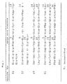
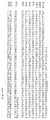
- the DNA sequence of the rat protein kinase C gene is described in detail in Examples 1, 2 and 4 and the DNA sequence and the amino acid sequence deduced from the DNA sequence is shown in Figs. 2 and 8.
- the present inventors have first cloned the rat protein kinase C gene and have cloned the human protein kinase C gene from human cells using a cDNA obtained from the cloned rat protein kinase C gene as a DNA probe.
- the present invention provides:
- the above described DNA (2') for expression of rat protein kinase C may have preferebly the DNA sequence shown in Fig. 4.
- An expression vector containing a DNA having a nucleotide sequence coding for the polypeptide of a rat protein protein kinase C according to the present invention may be produced by a method including the steps of:
- RNA coding for the rat protein kinase C may be obtained from various rat protein kinase C-producing cell lines such as rat brain-derived cells and rat fibroblasts.
- RNA from a rat protein kinase C-producing cell line there may be mentioned the guanidine-thiocyanate method [Chirgwin, J. M., Biochemistry 18 , 5294 (1979)].
- a cDNA is prepared with a reverse transcription enzyme according to a method of, for example, Okayama, H. et al [Molecular and Cellular Biology 2 , 161(1982); ibid. 3 , 280(1980)].
- the resulting cDNA is inserted into a plasmid.
- the plasmid into which the cDNA is to be inserted may be, for example, E . coli -derived pBR322 [Gene 2 , 95(1977)], pBR325 [Gene 4 , 121(1978)], pUCl2 [Gene 19 , 259(1982)] and pUC13 [Gene 19 , 259(1982)] and Bacillus subtilus -derived pUB110 [Biochemical amd Biophysical Research Communication 112 , 678(1983)].
- Other plasmids may be used as long as they can be replicated in a host.
- the cDNA-inserted plasmid may be one obtained with the use of a cDNA library (available from Dr. Okayama, National Institute of Child Health and Human Development, Bethesda, U. S. A.) using E. coli x1776 as a host and obtained by inserting a cDNA prepared from rat normal diploid cell mRNA into pCD vector [Okayama et al, Molecular Cell. Biology 3 , 280(1983)].
- plasmid thus obtained is then transfected to a suitable host such as a microorganism belonging to the genus Escherichia or Bacillus .
- microorganisms belonging to the genus Escherihcia include Escherichia coli K12DH1 [Proc. Natl. Acad. Sci. U.S.A. 60 . 160(1968)], M103 [Nucleic Acids Research 9 , 309(1981)], JA221 [Journal of Molecular Biology 120 , 517(1978)], HB101 [Journal of Molecular Biology 41 , 459(1969)] and C600 [Genetics 39 , 440(1954)].
- microorganisms belonging to the genus Bacillus include Bacillus subtilis MI114 [Gene 24 , 255(1983)] and 207-21 [Journal of Molecular Biology 95 , 87(1984)].
- a method for the transformation may be, for example, the calcium chloride method or calcium chloride/rubidium chloride method of Manitatis, T. et al ["Molecular Cloning", Cold Spring Harbor Laboratory, page 249 (1982)].
- the desired clones are singled out by any known method, for example a colony hybridization method [Gene 10 , 63(1980)] and a DNA base sequence determination method [Proc. Natl. Acad. Sci. U.S.A. 74 , 560(1977); Nucleic Acids Research 9 , 309(1981)].
- a colony hybridization method [Gene 10 , 63(1980)]
- a DNA base sequence determination method [Proc. Natl. Acad. Sci. U.S.A. 74 , 560(1977); Nucleic Acids Research 9 , 309(1981)].
- a microorganism carrying a vector containing a cloned DNA having a nucleotide sequence coding for the rat protein kinase C may be obtained.
- the plasmid is isolated from the microorganism.
- the plasmid having the cloned DNA having a nucleotide sequence coding for the rat protein kinase C is isolated as is, or if desired, is cutout with a restriction enzyme.
- the cloned gene is ligated to a vehicle (vector) suitable for expression thereof at a position downstream of its promoter region, thereby to obtain an expression vector.
- E . coli -derived plasmids such as pBR322, pBR325, pUC12 and pUC13, Bacillus subtilis -derived plasmids such as pUB110, pTP5 and pC194, yeast-derived plasmids such as pSH19 and pSH15, bacterio-phages such as ⁇ -phage, and animal viruses such as retro virus and vaccinia virus.
- the gene may possess at the 5'-terminus ATG serving as a translation initiation codon and at the 3'-terminus TAA, TGA or TAG serving as a translation stop codon.
- a promoter is linked at its upstream. Any promoter may be used in the present invention as long as it is suitable for the host used for the expression of the gene.
- Suitable promoters are trp promoter, lac promoter, recA promoter, ⁇ PL promoter and lpp promoter when the host is a microorganism belonging to the genus Escherichia , SPO1 promoter, SPO2 promoter and penP promoter when the host belongs to Bacillus , and PHO5 promoter, PGK promoter, GAP promoter and ADH promoter when the host is a yeast.
- trp promoter lac promoter, recA promoter, ⁇ PL promoter and lpp promoter when the host is a microorganism belonging to the genus Escherichia
- SPO1 promoter SPO2 promoter and penP promoter when the host belongs to Bacillus
- PHO5 promoter PGK promoter
- GAP promoter GAP promoter
- ADH promoter when the host is a yeast.
- Especially preferred is a combination of a microorganism belonging to the genus Escherichia as the host and
- a promoter derived from SV40 or a promoter of retro virus may be used.
- the former promoter is especially preferred.
- a host there may be used a microorganism belonging to the genus Escherichia or Bacillus , a yeast or an animal cell.
- microorganisms belonging to the genus Escherichia or the genus Bacillus the above-described one may be used.
- suitable yeasts include Saccharomyces cervisiae AH22R - , NA87-11A and DKD-5D.
- suitable animal cells include monkey cells COS-7, Vero, Chinese hamster cell CHO, mouse L cell and human FL cell.
- a microorganism belonging to the genus Escherichia coli may be transformed according to a method disclosed in, for example, Proc. Natl. Acad. Sci. U.S.A. 69 , 2110(1972) or Gene 17 , 107(1982).
- a microorganism belonging to the genus Bacillus may be transformed according to a method disclosed in, for example, Molecular & General Genetics 168 , 111(1979).
- a yeast may be transformed according to a method disclosed in, for example, Proc. Natl. Acad. Sci. U.S.A. 75 , 1929(1978).
- An animal cell may be transformed according to a method disclosed in, for example, Virology 52 , 456(1973).
- a liquid medium As a medium in which a transformant of a host belonging to the genus Escherichia or Bacillus is cultivated, a liquid medium is appropriate.
- the liquid medium may be supplemented with a carbon source, a nitrogen source, an inorganic substance and the like required for the growth of the transformant.
- the carbon source may be, for example, glucose, dextrin, soluble starch or sucrose.
- the nitrogen source may be, for example, an inorganic or organic substance such as an ammonium salt, a nitrate, corn steep liquor, peptone, casein, meat extract, soybean cake or potato extract.
- the inorganic substance may be, for example, calcium chloride, sodium dihydrogen phosphate or magnesium chloride.
- a yeast, vitamin and a growth promoting factor may also be added to the medium.
- the pH of the medium is preferably in the range of about 6 to 8.
- a preferred medium for cultivating a microorganism belonging to the genus Escherichia is M9 medium containing, for example, glucose, casamino acid [Miller, "Journal of Experiments in Molecular Genetics", 431-433, Cold Spring Harbor Laboratory, New York (1972)].
- This medium may supplemented with a suitable agent such as 3 ⁇ -indolylacrylic acid for the purpose of improving the action of the promoter.
- the cultivating is generally performed at 15 to 43°C for 3 to 24 hours with, if necessary, aeration and agitation.
- the cultivating is generally performed at 30 to 40°C for 6 to 24 hours with, if necessary, aeration and agitation.
- Burkholder minimum medium (Bostian, K. L. et al, Proc. Natl. Acad. Sci. U.S.A. 77 , 4505(1980)] may be mentioned as an example of the culture medium.
- the pH of the medium is preferably adjusted to 5 to 8.
- the cultivating is generally performed at 20 to 35°C for 24 to 72 hours with, if necessary, aeration and agitation.
- the medium for cultivating a transformant of a host animal cell may be, for example, MEM medium containing about 5 to 20% of fetal bovine serum [Science 122 , 501(1952)], DMEM medium [Virology 8 , 396(1959)], RPMI1640 medium [The Journal of the American Medical Association 199 , 519(1967)] and 199 medium [Proceeding of the Society for the Biological Medicine 73 , 1(1950).
- the pH of the medium is preferably 6 to 8. The cultivating is generally performed at
- rat protein kinase C protein From the above culture may be separated and purified the rat protein kinase C protein in the following manner.
- the suspended microorganism or cell is then disrupted by an ultrasonic wave, lysozyme (and/or freezing-and-thawing), followed by centrifugation to obtain the rat protein kinase C protein.
- Such known separation and purification methods may be suitably combined for the purification of the rat protein kinase C protein from the above described supernatant.
- Such known separation and purification methods include, for example, methods utilizing the solubility, such as salting out and solvent precipition; methods utilizing mainly the difference in molecular weight, such as dialysis, ultrafiltration, gel filtration and SDS-polyacrylamide gel electrophoresis; methods utilizing the difference in ionization, such as ion exchange chromatography; methods utilizing the diffference in hydrophobic property, such as reverse high performance liquid chromatography; and methods utilizing the difference in isoelectric point, such as isoelectric point electrophoresis.
- rat protein kinase C protein obtained by the gene recombinant method according to the present invention is the protein containing the polypeptide having the amino acid sequence shown in Fig. 1.
- the polypeptide may have Met at its N-terminus.
- Activity of the thus obtained rat protein kinase C activity may be assayed in terms of known protein phosphorylation activity.
- the expression plasmid of the present invention ontaining the gene coding for the rat protein kinase C protein may be introduced for transformation into various kinds of cells, rendering it possible to let the resultant cells produce the rat protein kinase C. Therefore, a large amount of the rat protein kinase C may be obtained.
- the thus produced rat protein kinase C may be used as a reagent or as a diagnosis or inspection material (for example, the amount of protein kinase C used in 0.001 to 10 ⁇ g in one trial) in conjuction with an antibody produced therefrom.
- the protein kinase C according to the present invention may be used as a reagent for investigating cellular signal transduction mechanisms, as a reagent for diagnosis or inspection of deseases (such as tumor) resulting from a trouble in the cellular signal transduction mechanism, or as a screening agent effective in prevention and curing of the diseases.
- the transformant E . coli K12DH1/pTB638 obtained in Example 2 has been deposited at IFO under the accession number IFO 14514 since June 20, 1986 and at FRI under the accession number FERM P-8829 since June 28, 1986, and the deposit has been changed to the deposit according to Budapest Treaty and stored at FRI under the number of FERM BP-1372.
- the transformant E. coli K12DH1/pTB652 obtained in Example 3 has been deposited at IFO under the accession number IFO 14539 since August 29, 1986 and at FRI under the accession number FERM P-8958 since September 5, 1986, and the deposit has been changed to the deposit according to Budapest Treaty under the accession number of FERM BP-1373.
- the transformant E . coli K12DH1/pTB653 obtained in Example 3 has been deposited at IFO under an accession number IFO 14540 since August 29, 1986 and at FRI under the accession number FERM P-8959 since September 5, 1986, and the deposit has been changed to the deposit according to Budapest Treaty under the accession number of FERM BP-1374.
- rat protein kinase C From rat brains was extracted a rat protein kinase C in accordance with the method of Kikkawa [J. Biol. Chem 257 , 13341(1982)]. There were obtained 2.0 mg of a purified protein kinase C sample from brains of 240 rats. The protein kinase C sample (1.9 mg) was dissolved in 1.9 ml of 50 mM Tris-HCl (pH 9.0) solution containing 6 M urea, to which was added lysylendopeptidase to a concentration of 25 ⁇ g/ml, and the mixture was digested at 37°C for 21 hours.
- peptides were applied on a reverse HPLC column (Micro pak Protein C-18-10, 0.4x30 cm) and followed by an acetonitrile gradient elution to obtain fractions of about 50 peptides.
- a reverse HPLC column Micro pak Protein C-18-10, 0.4x30 cm
- acetonitrile gradient elution fractions of about 50 peptides.
- four peptides (peptides Nos. 24, 37, 49 and 51) were subjected to automated Edman degradation using a vapor phase protein sequencer (Model 470A, Applied Biosystems, Inc.) for the determination of their amino acid sequences.
- Phenylthiohydantoin derivatives of amino acids liberated as a result of the Edman degradation were analyzed by HPLC using Micro pak SPC 18-3 column (Varian Associates, Inc.). The amino acid sequence of each of the four peptides was as shown in Fig. 1.
- RNA was extracted from rat brain by the guanidine-isothiocyanate method [Chirgwim et al, Biochemistry 18 , 5294(1978)]. Poly (A) RNA was purified by oligo dT cellulose column chromatography [Aviv and Leder, Proc. Natl. Acad. Sci. USA 69 , 1408(1972)].
- cDNA library was constructed with pcDV1 vector and pL1 linker in accordance with the method of Okayama and Berg [Okayama & Berg, Mol. Cell. Biol. 2 , 161(1982)].
- the vector plasmid containing circularized cDNA was transfected in E . coli DH1.
- E. coli DH1 Starting from 5 ⁇ g of poly(A)RNA, cDNA library composed of about 5x10 5 clones and using E. coli as a host was obtained.
- the above-described rat cDNA library using E . coli DH1 was applied to 10 nitrocellulose filters (Milipore, HATF filter) at about 3x10 4 clones/filter. Then 20 replica filters (each two being paired) were prepared using these filters as master filters. The E . coli on each replica filter was lysed with 0.5 N NaOH solution to expose and denature the plasmid DNA, followed by drying for fixation of the DNA on the filter [Grustein, M. & Hogness, D. S., Proc. Natl. Acad. Sci. USA 72 , 3961(1975)].
- oligonucleotides corresponding to the amino acid sequences were chemically synthesized.
- amino acid residues 7-12 of the peptide No. 24 (Tyr-Ile-Asp-Trp-Glu-Lys)
- amino acid residues 3-8 of the peptide No. 51 (Asp-Trp-Trp-Ala-Phe-Gly)
- amino acid residues 24-29 of the peptide No. 51 Glu-Asp-Glu-Asp-Glu-Leu
- Glu-Asp-Glu-Asp-Glu-Leu were each chemically synthesized for use as a screening probe for rat protein kinase C cDNA.
- the 5'-terminus of each of the oligonucleotide probes was labeled with 32 P using [ ⁇ - 32 P] ATP and T4 polynucleotide kinase.
- the labeled probes Nos. 2 and 3 were separately hybridized with the replica filters on which the plasmid DNA was fixed.
- the hybridization reaction was performed at 42°C for 16 hours in 10 ml of a 5xSSC (0.15 M NaCl, 0.015 M sodium citrate), 5xDenhardt's, 0.1 % SDS and 100 ⁇ g/ml denatured salmon sperum DNA solution containing 10 ⁇ Ci of the labeled probe.
- the filters were washed with a 6xSSC and 0.1 % SDS solution three times at room temperature for 30 minutes and twice at 47°C for 60 minutes in the case of the probe No. 2 and at 43°C for 60 minutes in the case of the probe No.
- a plasmid DNA (pTB638) was then extracted from this strain by the alkaline method [Birnboim, H. C. & Doly, J., Nucleic Acids Res. 1 , 1513(1979)]. After purification, the DNA was cleaved with restriction enzyme BamHI (manufactured by Takara Shuzo) and the product Was electrophoresed on an agarose gel. DNA fragments in the gel were transferred to a nitrocellulose filter (BA85, S & S Inc.) according to the Southern blotting method [Maniatis et al, "Molecular Cloning", Cold spring Harbor Laboratory, pp382 (1982)]. The plasmid DNA fragment on the filter was found to react with all of the above-described oligonucleotide probes.
- nucleotide sequence of a cDNA region of this plasmid DNA was determined by the dideoxynucleotide-chain termination method [Messing, J. et al, Nucleic Acids Res. 9, 309(1981)].
- the DNA sequence and the amino acid sequence deduced from the DNA sequence are shown in Fig. 2.
- the amino acid sequences of the peptides Nos. 24 and 51 determined in Reference Example 1(1) strictly correspond to the nucleotide sequences at Nos. 445-480 and 220-312, respectively.
- the plasmid pTB638 was confirmed to be rat protein kinase C cDNA.
- the cDNA contained a poly(A) structure, the cDNA was found to have codons of C-terminal region of the protein kinase C and untranslated portion of 3'-terminal region of the mRNA.
- the rat protein kinase C contains the amino acid sequence shown in Fig. 2 as a part thereof.
- RNA fragment was extracted from rat brain by the guanidine-isothiocyanate method [Chirgwim et al, Biochemistry 18 , 5294(1978)].
- Poly (A) RNA was Purified by oligo dT cellulose column chromatography [Aviv and Leder, Proc. Natl. Acad. Sci. USA 69 , 1408(1972)].
- cDNA library was constructed with phage vector ⁇ gt10 [Huynh, T. V. et al, "DNA Cloning, A Practical Approach” IRL press, Oxford, p49 (1985)] in accordance with the method of Watson and Jackson [Watson, C. J. & Jackson, J. F., "DNA Cloning, A Practical Approach” IRL press, Oxford, p 49 (1985)].
- cDNA library composed of about 1.5x10 6 clones and using E . coli C600, HflA (Huynh, T. V. et al, supra) as a host was obtained.
- the above-described phage cDNA library using E . coli C600, Hf1A as a host was applied to 10 soft agar plates at about 1x10 5 clones/plate and transferred to nitrocellulose filters (HATF filter, Milipore Inc.) and lysed with 0.5 N NaOH solution. The resulting exposed and denatured phage DNA was dried for fixation on the plates [Maniatis et al, "Molecular Clonging", Cold Spring Harbor Laboratory, p320(1982)].
- E . coli K12DH1/pTB638 (IFO 14514, FERM BP-1372) obtained in Example 2(2) was digested with restriction enzyme PstI to obtain a DNA fragment of 0.7 kb.
- the DNA fragment was labeled with 32 P by the nick translation method (Maniatis et al, supra P109) to give a probe.
- the DNA fixed on the filters was hybridized with the labeled probe by reaction in 10 ml of a 5xSSPE (0.9 M NaCl, 50 mM sodium phosphate buffer (pH 7.4), 5 mM EDTA), 50 % formamide, 5xDenhardt's, 0.1 % SDS and 100 ⁇ g/ml denatured salmon sperum DNA solution containing the labeled probe.
- 5xSSPE 0.9 M NaCl, 50 mM sodium phosphate buffer (pH 7.4), 5 mM EDTA
- 50 % formamide 50 % formamide
- 5xDenhardt's 50 % formamide
- 5xDenhardt's 50 % formamide
- 5xDenhardt's 50 % formamide
- 5xDenhardt's 50 % formamide
- 5xDenhardt's 50 % formamide
- 5xDenhardt's 50 % formamide
- 5xDenhardt's 50
- the washed filters were dried and subjected to radioautography for selecting clones capable of reacting with the probe.
- a phage DNA according to the method of Davis et al [Davis et al, "Advanced Bacterial Genetics", Cold Spring Harbor Laboratory (1980)]. This was cleaved with restriction enzyme BglII and EcoRI to obtain a DNA fragment of about 0.5 kb.
- the DNA fragment was labeled with 32 P in the same manner as described above. Using the labeled DNA fragment as a probe, hybridization was conducted in the same manner as above. Clones capable of reacting with the probe were singled out.
- nucleotide sequences of cDNA regions of several kinds of clones obtained in the above screening were determined by the dideoxynucleotide chain termination method [Messing et al, Nucleic Acids Res. 9 , 309(1981)].
- rat brain protein kinase C by combining cDNA regions of the thus obtained phage clones, the entire coding region of the rat brain protein kinase C can be covered.
- An investigation of the amino acid sequence deduced from the DNA sequence reveals that in rat brain protein kinase C there exist at least two molecular types which have 671 [type I ( ⁇ -1)] and 673 [type II ( ⁇ -2)]amino acids and which differ only in their C-terminal regions.
- Phage clone ⁇ CKR152 DNA shown in Fig. 3 was partially digested with restriction enzyme EcoRI to obtain a cDNA of 1.4 Kb.
- the DNA was mixed with plasmid pTB389 which has been cleaved with restriction enzyme EcoRI and which has been treated with alkaline phosphatase for the removal of its 5' terminal phosphoric acid groups.
- Plasmid pTB389 is obtained by converting each of the PstI cleavage site at the 5'-terminal region and the BamHI cleavage site at the 3'-terminal region of the interleukin-2 gene region of plasmid pTB106 (disclosed in JP-A-61-63282, which corresponds to EP-B- 172,619) to EcoRI, to remove the interleukin-2 gene region. The mixture was reacted with T4 DNA ligase to construct plasmid pTB648.
- the plasmid pTB648 was then partially digested with EcoRI to obtain a DNA of 4.0 kb having 937 bp of 5'-terminal region of the cDNA, followed by the removal of the 5'-terminal phosphoric acid group.
- the resulting DNA was mixed with a cDNA of 2.5 kb obtained by partially digesting the phage clone ⁇ CKR107 shown in Fig. 3 with restriction enzyme EcoRI or with a cDNA of 2.3 kb obtained by digesting the phage clone ⁇ CKR108 with EcoRI.
- the mixtures were each reacted with T4 DNA ligase to construct plasmids pTB651 and pTB650, respectively, each of which is to be used for transforming animal cells and covers the entire rat brain protein kinase C encoding region.
- Interleukin-2 gene expression vector pTB106 for animal cell JP-A-61-63282, which corresponds to EP-B-172,619) was digested with restriction enzyme HgiAI to obtain a DNA fragment of 1.0 kb containing an interleukin-2 gene leader sequence.
- the cohesive end of the DNA fragment was converted to flush end with T4 DNA polymerase reaction, to which was linked EcoRI linker CCGGAATTCCGG with T4 DNAligase, followed by complete digestion with EcoRI and HindIII to separate a SV40DNA-derived sequence (promoter and splice portion) and a first DNA fragment of 0.64 kb consisting of the IL-2 gene leader sequence.
- the above-described plasmid pTB106 was digested with BamHI and HindIII to separate a DNA replication starting region for replication of a plasmid pBR322-derived DNA in E . coli and a second DNA fragment of 2.6 kg having a sequence containing an ampicillin-resistant gene and a SV40DNA-derived polyadenylation region.
- plasmid pTB36l JP-A- 61-88881, which corresponds to EP-B-177,915
- EGF epidermal cell growth factor
- the first to third DNA fragments are ligated with T4 DNA ligase reaction to construct plasmid pTB406.
- the plasmid pTB406 was cleaved at the ClaI and HindIII sites located upstream from the SV40 promoter, into which was inserted a purified ClaI-HindIII DNA fragment of 1.1 kb separated from pTB314 (JP-A-61-63282, which corresponds to EP-B-172,619) and having an Abelson murine luekemia virus (A-MuLV) LTR region, thereby to construct plasmid pTB505.
- JP-A-61-63282 which corresponds to EP-B-172,619
- A-MuLV Abelson murine luekemia virus
- the plasmid pTB505 was further digested with ClaI and BamHI to obtain a DNA fragment of 1.5 kb containing the MuLV-LTR.
- This DNA was mixed with a ClaI-BamHI DNA fragment obtained from the plasmid pTB 650 or pTB651 and the mixture was reacted with T4 DNA ligase to construct plasmid pTB652 and pTB653 having MuLV-LTR at their upstream regions respectively and useful for transforming animal cells (See Fig. 5) (4)
- E . coli K12DH1 was transformed in accordance with the method disclosed in Proc. Natl. Acad.
- Monkey COS-7 cell [Cell 27 ,279-288(1981)] was cultivated on a single layer (5 plastic dishes with Falcon diameter of 100 mm) in DMEM medium supplemented with 5 % fetal bovine serum. After replacing with the same medium, the culture was further continued for 4 hours. Then, a calcium phosphate precipitates containing 30 ⁇ g/dish of the plasmid pTB652 or pTB653 prepared in the conventional manner [Graham et al, Virology 52 , 456(1973)] was added to the cell to obtain cells transfected with pTB652 and pTB653.
- the cells were treated with glycerol and the cultivation of the pTB652-transfected and pTB653-transfected COS-7 cells was further continued for 70-72 hours. Thereafter, the culture supernatant was discarded and the cells were suspended in 1.5 ml of a buffer [0.25 M sucrose, 20 mM Tris-HCl (pH 7.5), 10 mM EGTA, 2 mM EDTA and 20 ⁇ g/ml leupeptin] and homogenized by means of a teflon homogenizer. The homogenate was centrifuged at 55000 rpm for 60 minutes with Beckmann 100.2 rotor to obtain a cytoplasmic fraction.
- a buffer [0.25 M sucrose, 20 mM Tris-HCl (pH 7.5), 10 mM EGTA, 2 mM EDTA and 20 ⁇ g/ml leupeptin] and homogenized by means of a teflon homogenizer. The homogen
- the fraction was assayed for protein kinase C activity according to the conventional method [Kikkawa et al, J. Biol. Chem. 257 , 13341(1982)] to obtain activity value per 1 mg of the protein.
- Table 1 the cytoplasmic fractions of the pTB652-transfected and pTB-652-transfected COS-7 cells showed protein kinase C activity of about three times as strong as that of the non-transfected cell.
- cytoplasmic fractions obtained in the manner described above were each diluted with 6 volumes of 20 mM Tris-HCl buffer (buffer A, pH 7.5) containing 0.5 mM EGTA, 0.5 mM EDTA and 10 mM 2-mercaptoethanol and applied on Pharmacia FPLC-Mono Q column (0.5x5 cm, Pharmacia HR5/5). After washing the column with buffer A, protein kinase C was eluted with 20 ml of a linear gradient NaCl (0-0.6 M) containing buffer A. As shown in Fig. 6, markedly higher enzyme activity was detected in the test samples from pTB652- or pTB653-transfected cell as compared with the non-transfected cell.
- the active fractions eluted from the Mono Q column was electrophoresed on 8.5 % SDS polyacrylamide gel according to the method of Laemmli [Laemmli, U.K., Nature 227 , 680-685 (1970)] and transferred to nitrocellulose filters according to the method of Towbin et al [Towbin, H. et al, Proc. Natl. Acad. Sci. USA 76 , 4350-4354(1979)].
- the filters were each allowed to stand at 4°C overnight in a 10 mM Tris-HCl buffer (pH 7.5) containing 150 mM NaCl, 5 % casein and 20 % normal horse serum and then allowed to stand at room temperature for 1 hour in a 10 mM Tris-HCl buffer (pH 7.5) containing 150 mM NaCl, 1 % casein and anti-protein kinase C antibody [obtained from Dr. P. J. Parker; Coussens, L. S., Science 233, 859-866(1986)].
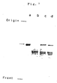
- the filters were washed with a 10 mM Tris-HCl buffer (pH 7.5) containing 0.05 % Tween 20 and 150 mM NaCl and allowed to stand at room temperature for 1.5 hours in a Tris-HCl buffer containing 1 % casein, 150 mM NaCl, 0.1 ⁇ Ci/ml 125 I protein A. After washing the filters in the same manner as above, X-ray films were exposed to the filters to visualize the protein bands reacted with the antibody. As shown in Fig. 7, the sample derived from the transfected cells gave the protein band reacted with the antibody in the same position as that of protein kinase C isolated from rat brain.
- cytoplasmic fractions were obtained from pTB707- and pTB708-transfected cells (each in an amount corresponding to 50 dishes having a 100 mm diameter) and applied to preparatory chromatography using Mono Q column (1x10 cm, Pharmacia HR10/10).
- the active fractions were partialy purified with a hydroxyapatite column.
- the active fractions were then diluted with equivolume of a 20 mM potassium phosphate buffer (buffer B, pH 7.5) containing 0.5 mM EGTA, 0.5 mM EDTA, 10 % glycerol and 10 mM mercaptoethanol and applied on a hydroxylapatite column (KOKEN Type S, 0.78 x 10 cm) of Pharmacia FPLC system.
- the protein kinase C activity was eluted with linear gradient buffer B containing 20-280 mM potassium phosphate.
- the protein kinase C isolated from rat brains gave three peaks (peaks 1, 2 and 3 from the leftside of D in Fig. 8) and the sample derived from the non-transfected cell revealed an activity corresponding to the peak 3.
- the sample derived from the transfected cells showed activities corresponding to the peaks 2 and 3. This suggests that the protein kinase C activity corresponding to the peak 2 is expressed by the cell transfected with the plasmid DNA.
Landscapes
- Health & Medical Sciences (AREA)
- Life Sciences & Earth Sciences (AREA)
- Chemical & Material Sciences (AREA)
- Engineering & Computer Science (AREA)
- Molecular Biology (AREA)
- Wood Science & Technology (AREA)
- Organic Chemistry (AREA)
- Biomedical Technology (AREA)
- Zoology (AREA)
- Genetics & Genomics (AREA)
- Bioinformatics & Cheminformatics (AREA)
- Biotechnology (AREA)
- Microbiology (AREA)
- Immunology (AREA)
- General Health & Medical Sciences (AREA)
- Medicinal Chemistry (AREA)
- Biochemistry (AREA)
- Hematology (AREA)
- Urology & Nephrology (AREA)
- General Engineering & Computer Science (AREA)
- Physics & Mathematics (AREA)
- General Physics & Mathematics (AREA)
- Food Science & Technology (AREA)
- Analytical Chemistry (AREA)
- Pathology (AREA)
- Cell Biology (AREA)
- Enzymes And Modification Thereof (AREA)
- Micro-Organisms Or Cultivation Processes Thereof (AREA)
- Peptides Or Proteins (AREA)
- Preparation Of Compounds By Using Micro-Organisms (AREA)
- Medicines Containing Material From Animals Or Micro-Organisms (AREA)
Abstract
Description
- The present invention relates to a calcium- and phospholipid-dependent protein phosphorylation enzyme (hereinafter referred to as protein phosphorylation enzyme C or protein kinase C for brevity) and to recombinant DNA technique for the production thereof, especially to rat kinase C.
- Numerous kinds of biologically active substances such as cell growth factors, hormones and neutrotransmitters are known to participate in the exertion of various functions and adaptation phenomena of living organisms. These extracellular signals are exerted through intracellular mediators such as cyclic AMP (cAMP), cyclic GMP (cGMP), diacylglycerol (DG) and calcium (Ca++). It has been revealed that DG is produced as a result of inositol phospholipid breakdown in cell membranes by a number of hormones and neutrotransmitters which are responsible for the activation of cellular functions. It is considered that the activation of various cellular functions and cellular proliferation are attained by the phosphorylation of various intracellular proteins with a protein kinase C enzyme which is activated by DG in the presence of Ca++. Thus, protein kinase C is an enzyme which plays an important role in a mechanism of main information transduction of external signals. The enzyme isolated from rat brains is an acidic protein having an isoelectric point of pH 5.6 and a molecular weight of approximate 77,000. This enzyme is composed of a single peptide having a hydrophobic region serving as a binding site to cell membranes and a hydrophilic region in which the active center exists. The protein kinase C is normally in an inactive form but is activated by calcium, phosphatidyl-serine and DG. ATP is known to serve as the phosphoric acid donor.
- Thus, the protein kinase C performs the transduction of extracellular signals into cells through the phosphorylation of proteins as a protein kinase and, therefore, it is one of the indispensable enzymes in studying of extracellular signal reception and transduction mechanism and is a very valuable reagent. The enzyme is also activated directly by tumor-promoting phorbol esters. Phorbol esters are known not only to cause promotion of carcinogenesis but also to take part in various biological reactions such as mitosis and differentiation of cells, induction of enzymes and acceleration of lipid metabolism. In these circumtances, there is a great possibility that the protein kinase C becomes an important index enzyme in diseased cells or tumor cells resulting from troubles in cellular signal transduction mechanism and that antibodies to the protein kinase C are used as diagnostic and inspection reagents. For these purposes, it is essential to provide a large amount of human protein kinase C. However, natural human protein kinase C is present only in a small amount. Further, to obtain the human protein kinase C from human tissues involves various problems and is extremely difficult. Therefore, the amino acid sequence of the human protein kinase C and the DNA sequence of the human protein kinase C gene have not been determined yet.
- On the other hand, with regard to protein kinase C derived from relatively easily available animal stuffs, for example, a protein kinase C is purified from rat brains. However, the amino acid sequence and the gene DNA sequence of rat protein kinase C has not been determined yet. It is also very difficult to obtain rat protein kinase C in a large amount.
- As described above, there are remaining a number of unknown points with respect to the properties, the amino acid sequence and the gene of rat protein kinase C. Therefore, it is highly desired to elucidate the gene coding for the protein kinase C and to provide a production method for the protein as a reagent and an inspecting agent by gene recombinant techniques.
- Proteins of animals having resemblance to human also show a high homology in amino acid sequence. A greater part of the differences in amino acids would be introduced by only one point mutuation of codons. Thus, the present inventors have purified a protein kinase C derived from rat brain which is easily available material, determined the amino acid sequence thereof and elucidated the DNA sequence of the rat protein kinase C gene. The DNA sequence of the rat protein kinase C gene is described in detail in Examples 1, 2 and 4 and the DNA sequence and the amino acid sequence deduced from the DNA sequence is shown in Figs. 2 and 8.
- Although a number of DNA sequences are possible for the DNA sequence of the cloned rat protein kinase C gene by degeneracy of codons, a portion of the DNA sequence is assumed to quite resemble to the DNA sequence of the human protein kinase C. Based on this assumption, the present inventors have first cloned the rat protein kinase C gene and have cloned the human protein kinase C gene from human cells using a cDNA obtained from the cloned rat protein kinase C gene as a DNA probe. By constructing a recombinant DNA having the human protein kinase C gene, and by cultivating a transformant transformed with the recombinant DNA, the human protein kinase C is produced. With the foregoing findings in view, the present inventors have made a further study and have completed the present invention.
- The present invention provides:
- (1) a rat protein kinase C which is a polypeptide comprising the 673 amino acid sequence shown in Fig. 4;
- (2) a recombinant DNA containing a nucleotide sequence coding for a rat protein kinase C, which is a polypeptide comprising the 673 amino acid sequence shown in Fig. 4;
- (3) a transformant transformed with a vector having a DNA comprising a nucleotide sequence coding for a rat protein kinase C, which is a polypeptide comprising the 673 amino acid sequence shown in Fig. 4;
- (4) A process for the production of rat protein kinase C characterized in cultivating a transformat (3) in a culture medium to produce and accumulate the rat kinase in the culture and recovering the same.
-
- Fig. 1 shows the amino acid sequences of four peptide fractions obtained from rat protein kinase C in Example 1(1);
- Fig. 2 shows the nucleotide sequence of rat protein kinase C cDNA and the amino acid sequence deduced from the nucleotide sequence;
- Fig. 3 is a schematic representation showing the manner of overlapping of two types of rat protein kinase C clones obtained in Example 3;
- Fig. 4 shows the entire nucleotide sequence of type I (β-1) rat brain protein kinase C cDNA and the deduced amino acid sequence, wherein the region in the box shows an additional nucleotide sequence added to the type I (β-1) and the nucleotide sequence thus added is the entire nucleotide sequence of type II (β-2);
- Fig. 5 is a scheme for constructing the plasmid for expressing rat brain protein kinase C in animal cells;
- Fig. 6 shows protein kinase C activity pattern in Mono Q column chromatography of cytoplasmic fractions derived from transfected and non-transfected cells, wherein A: pTB652 transfected cell-derived; B: pTB653 transfected cell-derived; C: non-transfected cell-derived ;
- Fig. 7 shows an autoradiogram of the antibody protein isolated by SDS polyacrylamide gel electrophoresis and reacted with an anti-protein kinase C antibody, wherein a: protein kinase C isolated from rat brains; b: non-transfected cell-derived; c:pTB652 transfected cell-derived; d: pTB653 transfected cell-derived; and
- Fig. 8 shows a protein kinase C activity pattern in hydroxylapatite column chromatography of an active fraction on Mono Q column chromatography of cytoplasmic fractions derived from transfected and non-transfected cells, wherein A: pTB707 transfected cell-derived; B: pTB708 transfected cell-derived; C: non-transfected cell-derived; D: rat brain protein kinase C.
- The above described DNA (2') for expression of rat protein kinase C may have preferebly the DNA sequence shown in Fig. 4.
- An expression vector containing a DNA having a nucleotide sequence coding for the polypeptide of a rat protein protein kinase C according to the present invention may be produced by a method including the steps of:
- (a) separating an RNA coding for a rat protein kinase C,
- (b) preparing a single strand complementary DNA (cDNA) and then a double strand DNA from the RNA,
- (c) inserting the complementary DNA into a plasmid,
- (d) transforming a host with the recombinant plasmid,
- (e) after cultivating the thus obtained transformant, isolating a plasmid containing the desired DNA from the transformant by any suitable method such as a colony hybridization method using a rat cDNA as probe,
- (f) cutting out the desired, cloned DNA from the plasmid, and
- (g) ligating the cloned DNA to a vehicle at a position downstream from the promoter.
- The RNA coding for the rat protein kinase C may be obtained from various rat protein kinase C-producing cell lines such as rat brain-derived cells and rat fibroblasts.
- As a method of preparing RNA from a rat protein kinase C-producing cell line, there may be mentioned the guanidine-thiocyanate method [Chirgwin, J. M., Biochemistry 18, 5294 (1979)].
- Using the thus obtained RNA as template, a cDNA is prepared with a reverse transcription enzyme according to a method of, for example, Okayama, H. et al [Molecular and Cellular Biology 2, 161(1982); ibid. 3, 280(1980)]. The resulting cDNA is inserted into a plasmid.
- The plasmid into which the cDNA is to be inserted may be, for example, E. coli-derived pBR322 [Gene 2, 95(1977)], pBR325 [Gene 4, 121(1978)], pUCl2 [Gene 19, 259(1982)] and pUC13 [Gene 19, 259(1982)] and Bacillus subtilus-derived pUB110 [Biochemical amd Biophysical Research Communication 112, 678(1983)]. Other plasmids may be used as long as they can be replicated in a host.
- As a method for inserting the cDNA into a plasmid, there may be mentioned the method of Maniatis, T. et al disclosed in "Molecular Cloning", Cold Spring Harbor Laboratory, page 239 (1982).
- The cDNA-inserted plasmid may be one obtained with the use of a cDNA library (available from Dr. Okayama, National Institute of Child Health and Human Development, Bethesda, U. S. A.) using E. coli x1776 as a host and obtained by inserting a cDNA prepared from rat normal diploid cell mRNA into pCD vector [Okayama et al, Molecular Cell. Biology 3, 280(1983)].
- With the plasmid thus obtained is then transfected to a suitable host such as a microorganism belonging to the genus Escherichia or Bacillus.
- Examples of microorganisms belonging to the genus Escherihcia include Escherichia coli K12DH1 [Proc. Natl. Acad. Sci. U.S.A. 60. 160(1968)], M103 [Nucleic Acids Research 9, 309(1981)], JA221 [Journal of
Molecular Biology 120, 517(1978)], HB101 [Journal of Molecular Biology 41, 459(1969)] and C600 [Genetics 39, 440(1954)]. - Examples of microorganisms belonging to the genus Bacillus include Bacillus subtilis MI114 [
Gene 24, 255(1983)] and 207-21 [Journal of Molecular Biology 95, 87(1984)]. - A method for the transformation may be, for example, the calcium chloride method or calcium chloride/rubidium chloride method of Manitatis, T. et al ["Molecular Cloning", Cold Spring Harbor Laboratory, page 249 (1982)].
- From the thus obtained transformants, the desired clones are singled out by any known method, for example a colony hybridization method [
Gene 10, 63(1980)] and a DNA base sequence determination method [Proc. Natl. Acad. Sci. U.S.A. 74, 560(1977); Nucleic Acids Research 9, 309(1981)]. - In the foregoing manner, a microorganism carrying a vector containing a cloned DNA having a nucleotide sequence coding for the rat protein kinase C may be obtained.
- Then, the plasmid is isolated from the microorganism.
- As a method of the isolation may be mentioned the alkaline method [Birmboim, H. C. et al,
Nucleic Acids Research 1, 1513(1979)]. - The plasmid having the cloned DNA having a nucleotide sequence coding for the rat protein kinase C is isolated as is, or if desired, is cutout with a restriction enzyme.
- The cloned gene is ligated to a vehicle (vector) suitable for expression thereof at a position downstream of its promoter region, thereby to obtain an expression vector.
- As the vector, there may be mentioned E. coli-derived plasmids such as pBR322, pBR325, pUC12 and pUC13, Bacillus subtilis-derived plasmids such as pUB110, pTP5 and pC194, yeast-derived plasmids such as pSH19 and pSH15, bacterio-phages such as λ-phage, and animal viruses such as retro virus and vaccinia virus.
- The gene may possess at the 5'-terminus ATG serving as a translation initiation codon and at the 3'-terminus TAA, TGA or TAG serving as a translation stop codon. To express the gene, a promoter is linked at its upstream. Any promoter may be used in the present invention as long as it is suitable for the host used for the expression of the gene.
- Illustrative of suitable promoters are trp promoter, lac promoter, recA promoter, λPL promoter and lpp promoter when the host is a microorganism belonging to the genus Escherichia, SPO1 promoter, SPO2 promoter and penP promoter when the host belongs to Bacillus, and PHO5 promoter, PGK promoter, GAP promoter and ADH promoter when the host is a yeast. Especially preferred is a combination of a microorganism belonging to the genus Escherichia as the host and trp promoter or λLPL promoter as the promoter.
- When the host is an animal cell, a promoter derived from SV40 or a promoter of retro virus may be used. The former promoter is especially preferred.
- Using the thus constructed vector containing DNA(2), a transformant is prepared.
- As a host, there may be used a microorganism belonging to the genus Escherichia or Bacillus, a yeast or an animal cell.
- As the microorganisms belonging to the genus Escherichia or the genus Bacillus the above-described one may be used. Examples of the suitable yeasts include Saccharomyces cervisiae AH22R-, NA87-11A and DKD-5D. Examples of suitable animal cells include monkey cells COS-7, Vero, Chinese hamster cell CHO, mouse L cell and human FL cell.
- A microorganism belonging to the genus Escherichia coli may be transformed according to a method disclosed in, for example, Proc. Natl. Acad. Sci. U.S.A.69, 2110(1972) or Gene 17, 107(1982). A microorganism belonging to the genus Bacillus may be transformed according to a method disclosed in, for example, Molecular & General Genetics 168, 111(1979). A yeast may be transformed according to a method disclosed in, for example, Proc. Natl. Acad. Sci. U.S.A. 75, 1929(1978). An animal cell may be transformed according to a method disclosed in, for example,
Virology 52, 456(1973). - In the manner described above, a transformant transformed with a DNA (2)-containing vector is obtained.
- As a medium in which a transformant of a host belonging to the genus Escherichia or Bacillus is cultivated, a liquid medium is appropriate. The liquid medium may be supplemented with a carbon source, a nitrogen source, an inorganic substance and the like required for the growth of the transformant. The carbon source may be, for example, glucose, dextrin, soluble starch or sucrose. The nitrogen source may be, for example, an inorganic or organic substance such as an ammonium salt, a nitrate, corn steep liquor, peptone, casein, meat extract, soybean cake or potato extract. The inorganic substance may be, for example, calcium chloride, sodium dihydrogen phosphate or magnesium chloride. A yeast, vitamin and a growth promoting factor may also be added to the medium. The pH of the medium is preferably in the range of about 6 to 8.
- A preferred medium for cultivating a microorganism belonging to the genus Escherichia is M9 medium containing, for example, glucose, casamino acid [Miller, "Journal of Experiments in Molecular Genetics", 431-433, Cold Spring Harbor Laboratory, New York (1972)]. This medium may supplemented with a suitable agent such as 3β-indolylacrylic acid for the purpose of improving the action of the promoter.
- When a microorganism belonging to the genus Escherichia is used as host, the cultivating is generally performed at 15 to 43°C for 3 to 24 hours with, if necessary, aeration and agitation. When a microorganism belonging to the genus Bacillus is used as host, the cultivating is generally performed at 30 to 40°C for 6 to 24 hours with, if necessary, aeration and agitation.
- In case where a transformant of a host being yeast is cultivaled, Burkholder minimum medium [Bostian, K. L. et al, Proc. Natl. Acad. Sci. U.S.A. 77, 4505(1980)] may be mentioned as an example of the culture medium. The pH of the medium is preferably adjusted to 5 to 8. The cultivating is generally performed at 20 to 35°C for 24 to 72 hours with, if necessary, aeration and agitation.
- The medium for cultivating a transformant of a host animal cell may be, for example, MEM medium containing about 5 to 20% of fetal bovine serum [Science 122, 501(1952)], DMEM medium [
Virology 8, 396(1959)], RPMI1640 medium [The Journal of the American Medical Association 199, 519(1967)] and 199 medium [Proceeding of the Society for the Biological Medicine 73, 1(1950). The pH of the medium is preferably 6 to 8. The cultivating is generally performed at - 30 to 40°C for 15 to 60 hours with, if necessary, aeration and agitation.
- From the above culture may be separated and purified the rat protein kinase C protein in the following manner.
- For extracting the rat protein kinase C from the cultured microorganism or cell, there may be suitably adopted a method in which the microorganism or the cell is collected from the culture by any conventional method and is suspended in a buffer containing a protein denaturation agent such as guanidine hydrochloric acid. The suspended microorganism or cell is then disrupted by an ultrasonic wave, lysozyme (and/or freezing-and-thawing), followed by centrifugation to obtain the rat protein kinase C protein.
- Known separation and purification methods may be suitably combined for the purification of the rat protein kinase C protein from the above described supernatant. Such known separation and purification methods include, for example, methods utilizing the solubility, such as salting out and solvent precipition; methods utilizing mainly the difference in molecular weight, such as dialysis, ultrafiltration, gel filtration and SDS-polyacrylamide gel electrophoresis; methods utilizing the difference in ionization, such as ion exchange chromatography; methods utilizing the diffference in hydrophobic property, such as reverse high performance liquid chromatography; and methods utilizing the difference in isoelectric point, such as isoelectric point electrophoresis.
- One example of the rat protein kinase C protein obtained by the gene recombinant method according to the present invention is the protein containing the polypeptide having the amino acid sequence shown in Fig. 1. The polypeptide may have Met at its N-terminus.
- Activity of the thus obtained rat protein kinase C activity may be assayed in terms of known protein phosphorylation activity.
- Various kinds of cells, which originally produce only a small amount of the rat protein kinase C or do not at all produce the rat protein kinase C, gain an ability to produce a large amount of the rat protein kinase C by the transfection with the DNA according to the present invention. And they can produce a large amount of the rat protein kinase C.
- The expression plasmid of the present invention ontaining the gene coding for the rat protein kinase C protein may be introduced for transformation into various kinds of cells, rendering it possible to let the resultant cells produce the rat protein kinase C. Therefore, a large amount of the rat protein kinase C may be obtained.
- The thus produced rat protein kinase C may be used as a reagent or as a diagnosis or inspection material (for example, the amount of protein kinase C used in 0.001 to 10 µg in one trial) in conjuction with an antibody produced therefrom. For example, the protein kinase C according to the present invention may be used as a reagent for investigating cellular signal transduction mechanisms, as a reagent for diagnosis or inspection of deseases (such as tumor) resulting from a trouble in the cellular signal transduction mechanism, or as a screening agent effective in prevention and curing of the diseases.
- In the present specification and accompanying drawings, abbreviations of bases, amino acids and chemicals are based on those according to IUPAC-IUB Commission on Biochemical Nomenclature or those customarily used in the art. Examples of the abbreviations are as follows. Amino acids with optical isomers refer to L-forms except otherwise specifically noted.
- DNA :
- Deoxyribonucleic acid
- cDNA :
- Complementary deoxyribonucleic acid
- A :
- Adenine
- T :
- Thymine
- G :
- Guanine
- C :
- Cytosine
- RNA :
- Ribonucleic acid
- dATP :
- Deoxyadenosine triphosphate
- dTTP :
- Deoxythymidine triphosphate
- dGTP :
- Deoxyguanosine triphosphate
- dCTP :
- Deoxycytidine triphosphate
- ATP :
- Adenosine triphosphate
- EDTA :
- Ethylenediamine tetraacetic acid
- SDS :
- Sodium dodecylsulfate
- Gly :
- Glycine
- Ala :
- Alanine
- Val :
- Valine
- Leu :
- Leucine
- Ile :
- Isoleucine
- Ser :
- Serine
- Thr :
- Threonine
- Cys :
- Cysteine
- Met :
- Methionine
- Glu :
- Glutamic acid
- Asp :
- Asparatic acid
- Lys :
- Lysine
- Arg :
- Arginine
- His :
- Histidine
- Phe :
- Phenylalanine
- Tyr :
- Tyrosine
- Trp :
- Tryptophane
- Pro :
- Proline
- Asn :
- Asparagine
- Gln :
- Glutamine
- The present invention will be described in more detail hereinafter by way of examples.
- The transformant E. coli K12DH1/pTB638 obtained in Example 2 has been deposited at IFO under the accession number IFO 14514 since June 20, 1986 and at FRI under the accession number FERM P-8829 since June 28, 1986, and the deposit has been changed to the deposit according to Budapest Treaty and stored at FRI under the number of FERM BP-1372.
- The transformant E. coli K12DH1/pTB652 obtained in Example 3 has been deposited at IFO under the accession number IFO 14539 since August 29, 1986 and at FRI under the accession number FERM P-8958 since September 5, 1986, and the deposit has been changed to the deposit according to Budapest Treaty under the accession number of FERM BP-1373.
- The transformant E. coli K12DH1/pTB653 obtained in Example 3 has been deposited at IFO under an accession number IFO 14540 since August 29, 1986 and at FRI under the accession number FERM P-8959 since September 5, 1986, and the deposit has been changed to the deposit according to Budapest Treaty under the accession number of FERM BP-1374.
- From rat brains was extracted a rat protein kinase C in accordance with the method of Kikkawa [J. Biol. Chem 257, 13341(1982)]. There were obtained 2.0 mg of a purified protein kinase C sample from brains of 240 rats. The protein kinase C sample (1.9 mg) was dissolved in 1.9 ml of 50 mM Tris-HCl (pH 9.0) solution containing 6 M urea, to which was added lysylendopeptidase to a concentration of 25 µg/ml, and the mixture was digested at 37°C for 21 hours. The thus produced peptides were applied on a reverse HPLC column (Micro pak Protein C-18-10, 0.4x30 cm) and followed by an acetonitrile gradient elution to obtain fractions of about 50 peptides. Of these, four peptides (peptides Nos. 24, 37, 49 and 51) were subjected to automated Edman degradation using a vapor phase protein sequencer (Model 470A, Applied Biosystems, Inc.) for the determination of their amino acid sequences.
- Phenylthiohydantoin derivatives of amino acids liberated as a result of the Edman degradation were analyzed by HPLC using Micro pak SPC 18-3 column (Varian Associates, Inc.). The amino acid sequence of each of the four peptides was as shown in Fig. 1.
- An RNA was extracted from rat brain by the guanidine-isothiocyanate method [Chirgwim et al, Biochemistry 18, 5294(1978)]. Poly (A) RNA was purified by oligo dT cellulose column chromatography [Aviv and Leder, Proc. Natl. Acad. Sci. USA 69, 1408(1972)].
- Using this poly(A)RNA as a template, cDNA library was constructed with pcDV1 vector and pL1 linker in accordance with the method of Okayama and Berg [Okayama & Berg, Mol. Cell. Biol. 2, 161(1982)]. The vector plasmid containing circularized cDNA was transfected in E. coli DH1. Starting from 5 µg of poly(A)RNA, cDNA library composed of about 5x10⁵ clones and using E. coli as a host was obtained.
- The above-described rat cDNA library using E. coli DH1 was applied to 10 nitrocellulose filters (Milipore, HATF filter) at about 3x10⁴ clones/filter. Then 20 replica filters (each two being paired) were prepared using these filters as master filters. The E. coli on each replica filter was lysed with 0.5 N NaOH solution to expose and denature the plasmid DNA, followed by drying for fixation of the DNA on the filter [Grustein, M. & Hogness, D. S., Proc. Natl.
Acad. Sci. USA 72, 3961(1975)]. - On the basis of the amino acid sequence of the peptides which were determined in Example 1(1), oligonucleotides corresponding to the amino acid sequences were chemically synthesized. Thuscorresponding to the amino acid residues 7-12 of the peptide No. 24 (Tyr-Ile-Asp-Trp-Glu-Lys),
 corresponding to the amino acid residues 3-8 of the peptide No. 51 (Asp-Trp-Trp-Ala-Phe-Gly) and
 corresponding to the amino acid residues 24-29 of the peptide No. 51 (Glu-Asp-Glu-Asp-Glu-Leu) were each chemically synthesized for use as a screening probe for rat protein kinase C cDNA.
- The 5'-terminus of each of the oligonucleotide probes was labeled with 32P using [γ-32P] ATP and T4 polynucleotide kinase.
- The labeled probes Nos. 2 and 3 were separately hybridized with the replica filters on which the plasmid DNA was fixed. The hybridization reaction was performed at 42°C for 16 hours in 10 ml of a 5xSSC (0.15 M NaCl, 0.015 M sodium citrate), 5xDenhardt's, 0.1 % SDS and 100 µg/ml denatured salmon sperum DNA solution containing 10 µCi of the labeled probe. Following the reaction, the filters were washed with a 6xSSC and 0.1 % SDS solution three times at room temperature for 30 minutes and twice at 47°C for 60 minutes in the case of the probe No. 2 and at 43°C for 60 minutes in the case of the probe No. 3 [Maniatis, M. et al, "Molecular Cloning", Cold Spring Harbor Laboratory, p309(1982)]. A radioautogram was then taken from each washed filter. Radioautograms of each pair of the replica filters were overlaid to explore a strain capable of reacting with both of the two probes. By this method, a strain E. coli Kl2DH1/pTB638 (IFO 14514, FERM BP-1372) capable of reacting with the both probes was took out from 3x10⁵ colonies.
- A plasmid DNA (pTB638) was then extracted from this strain by the alkaline method [Birnboim, H. C. & Doly, J., Nucleic Acids Res. 1, 1513(1979)]. After purification, the DNA was cleaved with restriction enzyme BamHI (manufactured by Takara Shuzo) and the product Was electrophoresed on an agarose gel. DNA fragments in the gel were transferred to a nitrocellulose filter (BA85, S & S Inc.) according to the Southern blotting method [Maniatis et al, "Molecular Cloning", Cold spring Harbor Laboratory, pp382 (1982)]. The plasmid DNA fragment on the filter was found to react with all of the above-described oligonucleotide probes.
- The nucleotide sequence of a cDNA region of this plasmid DNA was determined by the dideoxynucleotide-chain termination method [Messing, J. et al, Nucleic Acids Res. 9, 309(1981)]. The DNA sequence and the amino acid sequence deduced from the DNA sequence are shown in Fig. 2.
- From the sequences shown, it will be seen that the amino acid sequences of the peptides Nos. 24 and 51 determined in Reference Example 1(1) strictly correspond to the nucleotide sequences at Nos. 445-480 and 220-312, respectively. Thus, the plasmid pTB638 was confirmed to be rat protein kinase C cDNA. As the cDNA contained a poly(A) structure, the cDNA was found to have codons of C-terminal region of the protein kinase C and untranslated portion of 3'-terminal region of the mRNA. The rat protein kinase C contains the amino acid sequence shown in Fig. 2 as a part thereof.
- An RNA fragment was extracted from rat brain by the guanidine-isothiocyanate method [Chirgwim et al, Biochemistry 18, 5294(1978)]. Poly (A) RNA was Purified by oligo dT cellulose column chromatography [Aviv and Leder, Proc. Natl. Acad. Sci. USA 69, 1408(1972)].
- Using this poly(A)RNA as a template, cDNA library was constructed with phage vector λgt10 [Huynh, T. V. et al, "DNA Cloning, A Practical Approach" IRL press, Oxford, p49 (1985)] in accordance with the method of Watson and Jackson [Watson, C. J. & Jackson, J. F., "DNA Cloning, A Practical Approach" IRL press, Oxford, p 49 (1985)]. Starting from 10 µg of poly(A)RNA, cDNA library composed of about 1.5x10⁶ clones and using E. coli C600, HflA (Huynh, T. V. et al, supra) as a host was obtained.
- The above-described phage cDNA library using E. coli C600, Hf1A as a host was applied to 10 soft agar plates at about 1x10⁵ clones/plate and transferred to nitrocellulose filters (HATF filter, Milipore Inc.) and lysed with 0.5 N NaOH solution. The resulting exposed and denatured phage DNA was dried for fixation on the plates [Maniatis et al, "Molecular Clonging", Cold Spring Harbor Laboratory, p320(1982)].
- E. coli K12DH1/pTB638 (IFO 14514, FERM BP-1372) obtained in Example 2(2) was digested with restriction enzyme PstI to obtain a DNA fragment of 0.7 kb. The DNA fragment was labeled with 32P by the nick translation method (Maniatis et al, supra P109) to give a probe.
- The DNA fixed on the filters was hybridized with the labeled probe by reaction in 10 ml of a 5xSSPE (0.9 M NaCl, 50 mM sodium phosphate buffer (pH 7.4), 5 mM EDTA), 50 % formamide, 5xDenhardt's, 0.1 % SDS and 100 µg/ml denatured salmon sperum DNA solution containing the labeled probe. Following the reaction, the filters were washed twice at room temperature for 30 minutes with a 2xSSC(1xSSC=0.15M NaCl, 0.015 M sodium citrate) and 0.1 % SDS and twice at 68°C for 30 minutes with a 1xSSC and 0.1 % SDS solution. The washed filters were dried and subjected to radioautography for selecting clones capable of reacting with the probe. From the clone λCKR107 was extracted a phage DNA according to the method of Davis et al [Davis et al, "Advanced Bacterial Genetics", Cold Spring Harbor Laboratory (1980)]. This was cleaved with restriction enzyme BglII and EcoRI to obtain a DNA fragment of about 0.5 kb. The DNA fragment was labeled with 32P in the same manner as described above. Using the labeled DNA fragment as a probe, hybridization was conducted in the same manner as above. Clones capable of reacting with the probe were singled out. The nucleotide sequences of cDNA regions of several kinds of clones obtained in the above screening were determined by the dideoxynucleotide chain termination method [Messing et al, Nucleic Acids Res. 9, 309(1981)].
- Thus, by combining cDNA regions of the thus obtained phage clones, the entire coding region of the rat brain protein kinase C can be covered. An investigation of the amino acid sequence deduced from the DNA sequence reveals that in rat brain protein kinase C there exist at least two molecular types which have 671 [type I (β-1)] and 673 [type II (β-2)]amino acids and which differ only in their C-terminal regions.
- Overlapping of the clones thus obtained is illustrated in Fig. 3. The entire DNA sequence and the deduced amino acid sequence are shown in Fig. 4.
- Phage clone λCKR152 DNA shown in Fig. 3 was partially digested with restriction enzyme EcoRI to obtain a cDNA of 1.4 Kb. The DNA was mixed with plasmid pTB389 which has been cleaved with restriction enzyme EcoRI and which has been treated with alkaline phosphatase for the removal of its 5' terminal phosphoric acid groups. Plasmid pTB389 is obtained by converting each of the PstI cleavage site at the 5'-terminal region and the BamHI cleavage site at the 3'-terminal region of the interleukin-2 gene region of plasmid pTB106 (disclosed in JP-A-61-63282, which corresponds to EP-B- 172,619) to EcoRI, to remove the interleukin-2 gene region. The mixture was reacted with T4 DNA ligase to construct plasmid pTB648. The plasmid pTB648 was then partially digested with EcoRI to obtain a DNA of 4.0 kb having 937 bp of 5'-terminal region of the cDNA, followed by the removal of the 5'-terminal phosphoric acid group. The resulting DNA was mixed with a cDNA of 2.5 kb obtained by partially digesting the phage clone λCKR107 shown in Fig. 3 with restriction enzyme EcoRI or with a cDNA of 2.3 kb obtained by digesting the phage clone λCKR108 with EcoRI. The mixtures were each reacted with T4 DNA ligase to construct plasmids pTB651 and pTB650, respectively, each of which is to be used for transforming animal cells and covers the entire rat brain protein kinase C encoding region.
- Interleukin-2 gene expression vector pTB106 for animal cell (JP-A-61-63282, which corresponds to EP-B-172,619) was digested with restriction enzyme HgiAI to obtain a DNA fragment of 1.0 kb containing an interleukin-2 gene leader sequence. The cohesive end of the DNA fragment was converted to flush end with T4 DNA polymerase reaction, to which was linked EcoRI linker CCGGAATTCCGG with T4 DNAligase, followed by complete digestion with EcoRI and HindIII to separate a SV40DNA-derived sequence (promoter and splice portion) and a first DNA fragment of 0.64 kb consisting of the IL-2 gene leader sequence. Separately, the above-described plasmid pTB106 was digested with BamHI and HindIII to separate a DNA replication starting region for replication of a plasmid pBR322-derived DNA in E. coli and a second DNA fragment of 2.6 kg having a sequence containing an ampicillin-resistant gene and a SV40DNA-derived polyadenylation region. Further, from plasmid pTB36l (JP-A- 61-88881, which corresponds to EP-B-177,915) cloned human epidermal cell growth factor (EGF) synthesis gene, a third, EcoRI-BamHI DNA fragment of 0.18 kb coding for human EGF was prepared. The first to third DNA fragments are ligated with T4 DNA ligase reaction to construct plasmid pTB406. The plasmid pTB406 was cleaved at the ClaI and HindIII sites located upstream from the SV40 promoter, into which was inserted a purified ClaI-HindIII DNA fragment of 1.1 kb separated from pTB314 (JP-A-61-63282, which corresponds to EP-B-172,619) and having an Abelson murine luekemia virus (A-MuLV) LTR region, thereby to construct plasmid pTB505.
- The plasmid pTB505 was further digested with ClaI and BamHI to obtain a DNA fragment of 1.5 kb containing the MuLV-LTR. This DNA was mixed with a ClaI-BamHI DNA fragment obtained from the
plasmid pTB 650 or pTB651 and the mixture was reacted with T4 DNA ligase to construct plasmid pTB652 and pTB653 having MuLV-LTR at their upstream regions respectively and useful for transforming animal cells (See Fig. 5)
(4) With each of the plasmid pTB652 and plasmid pTB653 obtained in (2) above, E. coli K12DH1 was transformed in accordance with the method disclosed in Proc. Natl. Acad. Sci. USA 69, 2110(1972) to obtain a transformant E. coli K12DH1/pTB652 (IFO 14539, FERM BP-1373) and a transformant E. coli K12DH1/pTB653 (IFO 14540, FERM BP-1374), respectively.. - Monkey COS-7 cell [Cell 27,279-288(1981)] was cultivated on a single layer (5 plastic dishes with Falcon diameter of 100 mm) in DMEM medium supplemented with 5 % fetal bovine serum. After replacing with the same medium, the culture was further continued for 4 hours. Then, a calcium phosphate precipitates containing 30 µg/dish of the plasmid pTB652 or pTB653 prepared in the conventional manner [Graham et al,
Virology 52, 456(1973)] was added to the cell to obtain cells transfected with pTB652 and pTB653. Further four hours later, the cells were treated with glycerol and the cultivation of the pTB652-transfected and pTB653-transfected COS-7 cells was further continued for 70-72 hours. Thereafter, the culture supernatant was discarded and the cells were suspended in 1.5 ml of a buffer [0.25 M sucrose, 20 mM Tris-HCl (pH 7.5), 10 mM EGTA, 2 mM EDTA and 20 µg/ml leupeptin] and homogenized by means of a teflon homogenizer. The homogenate was centrifuged at 55000 rpm for 60 minutes with Beckmann 100.2 rotor to obtain a cytoplasmic fraction. The fraction was assayed for protein kinase C activity according to the conventional method [Kikkawa et al, J. Biol. Chem. 257, 13341(1982)] to obtain activity value per 1 mg of the protein. As shown in Table 1 below, the cytoplasmic fractions of the pTB652-transfected and pTB-652-transfected COS-7 cells showed protein kinase C activity of about three times as strong as that of the non-transfected cell.Table 1 Production of Rat Brain Protein Kinase C with Transfection of Plasmid pTB652 or pTB653 Transfecting Protein Kinase C Activity in Cytoplasmic Plasmid DNA Fraction of Transfected Cell (pmole/min/mg) pTB652 68.82 pTB653 78.30 None 25.46 - The cytoplasmic fractions obtained in the manner described above were each diluted with 6 volumes of 20 mM Tris-HCl buffer (buffer A, pH 7.5) containing 0.5 mM EGTA, 0.5 mM EDTA and 10 mM 2-mercaptoethanol and applied on Pharmacia FPLC-Mono Q column (0.5x5 cm, Pharmacia HR5/5). After washing the column with buffer A, protein kinase C was eluted with 20 ml of a linear gradient NaCl (0-0.6 M) containing buffer A. As shown in Fig. 6, markedly higher enzyme activity was detected in the test samples from pTB652- or pTB653-transfected cell as compared with the non-transfected cell.
- The active fractions eluted from the Mono Q column was electrophoresed on 8.5 % SDS polyacrylamide gel according to the method of Laemmli [Laemmli, U.K., Nature 227, 680-685 (1970)] and transferred to nitrocellulose filters according to the method of Towbin et al [Towbin, H. et al, Proc. Natl. Acad. Sci. USA 76, 4350-4354(1979)]. The filters were each allowed to stand at 4°C overnight in a 10 mM Tris-HCl buffer (pH 7.5) containing 150 mM NaCl, 5 % casein and 20 % normal horse serum and then allowed to stand at room temperature for 1 hour in a 10 mM Tris-HCl buffer (pH 7.5) containing 150 mM NaCl, 1 % casein and anti-protein kinase C antibody [obtained from Dr. P. J. Parker; Coussens, L. S., Science 233, 859-866(1986)]. The filters were washed with a 10 mM Tris-HCl buffer (pH 7.5) containing 0.05
% Tween 20 and 150 mM NaCl and allowed to stand at room temperature for 1.5 hours in a Tris-HCl buffer containing 1 % casein, 150 mM NaCl, 0.1 µCi/ml 125I protein A. After washing the filters in the same manner as above, X-ray films were exposed to the filters to visualize the protein bands reacted with the antibody. As shown in Fig. 7, the sample derived from the transfected cells gave the protein band reacted with the antibody in the same position as that of protein kinase C isolated from rat brain. - Similar results were also obtained in the case of COS-7 cells transfected with pTB707 (having a 5'-untranslated region of 55 base pairs) obtained by shortening the 5' untranslated region of pTB652 and with pTB708 (bearing a 5'-untranslated region of 55 base pairs) obtained by shortening the 5' untranslated region of pTB653.
- Thus, in the same manner as described above, cytoplasmic fractions were obtained from pTB707- and pTB708-transfected cells (each in an amount corresponding to 50 dishes having a 100 mm diameter) and applied to preparatory chromatography using Mono Q column (1x10 cm, Pharmacia HR10/10). The active fractions were partialy purified with a hydroxyapatite column. The active fractions were then diluted with equivolume of a 20 mM potassium phosphate buffer (buffer B, pH 7.5) containing 0.5 mM EGTA, 0.5 mM EDTA, 10 % glycerol and 10 mM mercaptoethanol and applied on a hydroxylapatite column (KOKEN Type S, 0.78 x 10 cm) of Pharmacia FPLC system. The protein kinase C activity was eluted with linear gradient buffer B containing 20-280 mM potassium phosphate. As shown in Fig. 8, the protein kinase C isolated from rat brains gave three peaks (
peaks peak 3. - On the other hand, the sample derived from the transfected cells showed activities corresponding to the
peaks peak 2 is expressed by the cell transfected with the plasmid DNA.
Claims (4)
- A rat protein kinase C which is a polypeptide comprising the 673 amino acid sequence shown in Fig. 4.
- A recombinant DNA containing a nucleotide sequence coding for a rat protein kinase C, which is a polypeptide comprising the 673 amino acid sequence shown in Fig. 4.
- A transformant transformed with a vector having a DNA comprising a nucleotide sequence coding for a rat protein kinase C, which is a polypeptide comprising the 673 amino acid sequence shown in Fig. 4.
- A process for producing a rat protein kinase C which comprises the steps of: cultivating a transformant according to claim 3 in a culture medium, accumulating rat protein kinase C in the culture and recovering the same.
Applications Claiming Priority (9)
| Application Number | Priority Date | Filing Date | Title |
|---|---|---|---|
| JP149385/86 | 1986-06-27 | ||
| JP14938586 | 1986-06-27 | ||
| JP21794486 | 1986-09-18 | ||
| JP217944/86 | 1986-09-18 | ||
| JP281870/86 | 1986-11-28 | ||
| JP28187086 | 1986-11-28 | ||
| JP40160/87 | 1987-02-25 | ||
| JP4016087 | 1987-02-25 | ||
| EP87109223A EP0251244B1 (en) | 1986-06-27 | 1987-06-26 | Polypeptide and production thereof |
Related Parent Applications (2)
| Application Number | Title | Priority Date | Filing Date |
|---|---|---|---|
| EP87109223A Division EP0251244B1 (en) | 1986-06-27 | 1987-06-26 | Polypeptide and production thereof |
| EP87109223.5 Division | 1987-06-26 |
Publications (2)
| Publication Number | Publication Date |
|---|---|
| EP0686695A1 EP0686695A1 (en) | 1995-12-13 |
| EP0686695B1 true EP0686695B1 (en) | 2006-12-06 |
Family
ID=27460860
Family Applications (2)
| Application Number | Title | Priority Date | Filing Date |
|---|---|---|---|
| EP95112109A Expired - Lifetime EP0686695B1 (en) | 1986-06-27 | 1987-06-26 | Rat protein kinase C and production thereof |
| EP87109223A Expired - Lifetime EP0251244B1 (en) | 1986-06-27 | 1987-06-26 | Polypeptide and production thereof |
Family Applications After (1)
| Application Number | Title | Priority Date | Filing Date |
|---|---|---|---|
| EP87109223A Expired - Lifetime EP0251244B1 (en) | 1986-06-27 | 1987-06-26 | Polypeptide and production thereof |
Country Status (7)
| Country | Link |
|---|---|
| US (1) | US5219748A (en) |
| EP (2) | EP0686695B1 (en) |
| JP (2) | JP2593476B2 (en) |
| AT (2) | ATE347592T1 (en) |
| CA (1) | CA1341288C (en) |
| DE (2) | DE3752391T2 (en) |
| ES (1) | ES2093600T3 (en) |
Families Citing this family (12)
| Publication number | Priority date | Publication date | Assignee | Title |
|---|---|---|---|---|
| JPH02500243A (en) * | 1986-08-13 | 1990-02-01 | ジェネテイックス・インスティテュート・インコーポレイテッド | protein kinase C enzyme |
| EP0358325A1 (en) * | 1988-08-10 | 1990-03-14 | Takeda Chemical Industries, Ltd. | Production of protein kinase C by yeast |
| JPH0536077U (en) * | 1991-10-22 | 1993-05-18 | 蔡 憲堂 | Air pump with noise prevention means |
| US5459036A (en) | 1993-03-19 | 1995-10-17 | Max-Planck-Gesellschaft Zur Forderung Der Wissenschaften E.V. | Extracellular signal-regulated kinase, sequences, and methods of production and use |
| DE69728880T2 (en) * | 1996-03-19 | 2005-04-28 | Millennium Pharmaceuticals, Inc., Cambridge | KINASE FOR THE JOB-SPECIFIC PHOSPHORYLATION OF I-kappa B-alpha |
| AU723585B2 (en) * | 1996-03-28 | 2000-08-31 | Trustees Of Boston University | Methods of modulating melanin synthesis |
| US6239117B1 (en) | 1997-02-13 | 2001-05-29 | Albert Einstein College Of Medicine Of Yeshiva University | Gene therapy for regulating bladder smooth muscle tone |
| US6150338A (en) * | 1997-02-13 | 2000-11-21 | Albert Einstein College Of Medicine Of Yeshiva University | Gene therapy for alleviating erectile dysfunction |
| US7030096B1 (en) * | 1997-02-13 | 2006-04-18 | Albert Einstein College Of Medicine Of Yeshiva University | Method of enhancing relaxation of penile smooth muscle by introduction of DNA encoding maxi-K potassium channel protein |
| US6271211B1 (en) | 1997-02-13 | 2001-08-07 | Albert Einstein College Of Medicine Of Yeshiva University | Gene therapy for regulating penile smooth muscle tone |
| CN1311309A (en) * | 2000-03-02 | 2001-09-05 | 上海博德基因开发有限公司 | New polypeptide-human signal conductive protein kinase 17 and polynucleotide for coding such polypeptide |
| RS67720B1 (en) * | 2017-12-14 | 2026-02-27 | Univ Strasbourg | Peptides for treatment and prevention of nonalcoholic fatty liver disease |
Family Cites Families (1)
| Publication number | Priority date | Publication date | Assignee | Title |
|---|---|---|---|---|
| JPH02500243A (en) * | 1986-08-13 | 1990-02-01 | ジェネテイックス・インスティテュート・インコーポレイテッド | protein kinase C enzyme |
-
1987
- 1987-06-26 AT AT95112109T patent/ATE347592T1/en not_active IP Right Cessation
- 1987-06-26 ES ES87109223T patent/ES2093600T3/en not_active Expired - Lifetime
- 1987-06-26 AT AT87109223T patent/ATE142256T1/en not_active IP Right Cessation
- 1987-06-26 EP EP95112109A patent/EP0686695B1/en not_active Expired - Lifetime
- 1987-06-26 DE DE3752391T patent/DE3752391T2/en not_active Expired - Lifetime
- 1987-06-26 DE DE3751893T patent/DE3751893T2/en not_active Expired - Lifetime
- 1987-06-26 EP EP87109223A patent/EP0251244B1/en not_active Expired - Lifetime
- 1987-06-26 CA CA000540667A patent/CA1341288C/en not_active Expired - Fee Related
- 1987-06-26 JP JP62157495A patent/JP2593476B2/en not_active Expired - Lifetime
-
1991
- 1991-02-08 US US07/654,404 patent/US5219748A/en not_active Expired - Lifetime
-
1998
- 1998-05-19 JP JP13710298A patent/JP3146352B2/en not_active Expired - Lifetime
Also Published As
| Publication number | Publication date |
|---|---|
| JP3146352B2 (en) | 2001-03-12 |
| EP0251244A3 (en) | 1989-02-08 |
| DE3751893T2 (en) | 1997-01-16 |
| DE3751893D1 (en) | 1996-10-10 |
| DE3752391T2 (en) | 2007-12-13 |
| JPH11123084A (en) | 1999-05-11 |
| ATE347592T1 (en) | 2006-12-15 |
| EP0251244A2 (en) | 1988-01-07 |
| JPS642572A (en) | 1989-01-06 |
| US5219748A (en) | 1993-06-15 |
| EP0686695A1 (en) | 1995-12-13 |
| ES2093600T3 (en) | 1997-01-01 |
| ATE142256T1 (en) | 1996-09-15 |
| DE3752391D1 (en) | 2007-01-18 |
| EP0251244B1 (en) | 1996-09-04 |
| CA1341288C (en) | 2001-09-04 |
| JP2593476B2 (en) | 1997-03-26 |
Similar Documents
| Publication | Publication Date | Title |
|---|---|---|
| Matsui et al. | Nucleotide and deduced amino acid sequences of a GTP-binding protein family with molecular weights of 25,000 from bovine brain. | |
| Kawata et al. | A novel small molecular weight GTP-binding protein with the same putative effector domain as the ras proteins in bovine brain membranes. Purification, determination of primary structure, and characterization. | |
| Zorzato et al. | Molecular cloning of cDNA encoding human and rabbit forms of the Ca2+ release channel (ryanodine receptor) of skeletal muscle sarcoplasmic reticulum. | |
| Blachly-Dyson et al. | Cloning and functional expression in yeast of two human isoforms of the outer mitochondrial membrane channel, the voltage-dependent anion channel. | |
| US6365725B1 (en) | DNA coding for a human vasoconstrictive peptide and use thereof | |
| DE69839123T2 (en) | PROSTATE TUMOR POLYNUCLEOTIDE AND ANTIGEN COMPOSITIONS | |
| EP0251244B1 (en) | Polypeptide and production thereof | |
| Maucuer et al. | Stathmin gene family: phylogenetic conservation and developmental regulation in Xenopus | |
| McALEESE et al. | Complete amino acid sequence of the neurone‐specific γ isozyme of enolase (NSE) from human brain and comparison with the non‐neuronal α form (NNE) | |
| Gravel et al. | Molecular cloning and characterization of rat brain 2′, 3′‐cyclic nucleotide 3′‐phosphodiesterase isoform 2 | |
| Curmi et al. | Molecular characterization of human stathmin expressed in Escherichia coli: site-directed mutagenesis of two phosphorylatable serines (Ser-25 and Ser-63) | |
| Caricasole et al. | Cloning and characterization of the human phosphoinositide-specific phospholipase C-beta 1 (PLCβ1) | |
| US5324651A (en) | DNA encoding rat and human protein kinase C | |
| JPH012572A (en) | DNA and transformants | |
| US5780245A (en) | Polypeptides having a serotonin receptor activity, nucleic acids coding for these polypeptides and uses | |
| Bayarsaihan et al. | Genomic organization of the genes Gtf2ird1, Gtf2i, and Ncf1 at the mouse chromosome 5 region syntenic to the human chromosome 7q11. 23 Williams syndrome critical region | |
| DE69433902T2 (en) | NOVEL TYROSINE KINASE | |
| WO1995005392A1 (en) | Novel proteins which bind to retinoblastoma protein and their encoding dna sequences | |
| Yamamoto et al. | Multiple small molecular weight gtp-binding proteins in bovine brain cytosol purification and characterization of a 24KDa protein | |
| JP2739895B2 (en) | Polypeptides, DNA and uses thereof | |
| JP2844455B2 (en) | Polypeptides, DNA and uses thereof | |
| JP2771188B2 (en) | Polypeptides, DNA and uses thereof | |
| EP0258489A1 (en) | Human low affinity FC epsilon-receptor, the isolation and purification thereof | |
| US6811987B1 (en) | Human calcium binding protein and a polynucleotide encoding the same | |
| JPH07508655A (en) | Polypeptides with serotonergic receptor (5HT6) activity, nucleic acids encoding these polypeptides, and uses thereof |
Legal Events
| Date | Code | Title | Description |
|---|---|---|---|
| PUAI | Public reference made under article 153(3) epc to a published international application that has entered the european phase |
Free format text: ORIGINAL CODE: 0009012 |
|
| 17P | Request for examination filed |
Effective date: 19950802 |
|
| AC | Divisional application: reference to earlier application |
Ref document number: 251244 Country of ref document: EP |
|
| AK | Designated contracting states |
Kind code of ref document: A1 Designated state(s): AT BE CH DE ES FR GB GR IT LI LU NL SE |
|
| 17Q | First examination report despatched |
Effective date: 20031028 |
|
| RAP1 | Party data changed (applicant data changed or rights of an application transferred) |
Owner name: TAKEDA PHARMACEUTICAL COMPANY LIMITED |
|
| GRAP | Despatch of communication of intention to grant a patent |
Free format text: ORIGINAL CODE: EPIDOSNIGR1 |
|
| RTI1 | Title (correction) |
Free format text: RAT PROTEIN KINASE C AND PRODUCTION THEREOF |
|
| GRAS | Grant fee paid |
Free format text: ORIGINAL CODE: EPIDOSNIGR3 |
|
| GRAA | (expected) grant |
Free format text: ORIGINAL CODE: 0009210 |
|
| AC | Divisional application: reference to earlier application |
Ref document number: 0251244 Country of ref document: EP Kind code of ref document: P |
|
| AK | Designated contracting states |
Kind code of ref document: B1 Designated state(s): AT BE CH DE ES FR GB GR IT LI LU NL SE |
|
| PG25 | Lapsed in a contracting state [announced via postgrant information from national office to epo] |
Ref country code: NL Free format text: LAPSE BECAUSE OF FAILURE TO SUBMIT A TRANSLATION OF THE DESCRIPTION OR TO PAY THE FEE WITHIN THE PRESCRIBED TIME-LIMIT Effective date: 20061206 Ref country code: IT Free format text: LAPSE BECAUSE OF FAILURE TO SUBMIT A TRANSLATION OF THE DESCRIPTION OR TO PAY THE FEE WITHIN THE PRESCRIBED TIME-LIMIT;WARNING: LAPSES OF ITALIAN PATENTS WITH EFFECTIVE DATE BEFORE 2007 MAY HAVE OCCURRED AT ANY TIME BEFORE 2007. THE CORRECT EFFECTIVE DATE MAY BE DIFFERENT FROM THE ONE RECORDED. Effective date: 20061206 Ref country code: BE Free format text: LAPSE BECAUSE OF FAILURE TO SUBMIT A TRANSLATION OF THE DESCRIPTION OR TO PAY THE FEE WITHIN THE PRESCRIBED TIME-LIMIT Effective date: 20061206 Ref country code: AT Free format text: LAPSE BECAUSE OF FAILURE TO SUBMIT A TRANSLATION OF THE DESCRIPTION OR TO PAY THE FEE WITHIN THE PRESCRIBED TIME-LIMIT Effective date: 20061206 |
|
| REG | Reference to a national code |
Ref country code: GB Ref legal event code: FG4D |
|
| REG | Reference to a national code |
Ref country code: CH Ref legal event code: EP |
|
| REF | Corresponds to: |
Ref document number: 3752391 Country of ref document: DE Date of ref document: 20070118 Kind code of ref document: P |
|
| PG25 | Lapsed in a contracting state [announced via postgrant information from national office to epo] |
Ref country code: SE Free format text: LAPSE BECAUSE OF FAILURE TO SUBMIT A TRANSLATION OF THE DESCRIPTION OR TO PAY THE FEE WITHIN THE PRESCRIBED TIME-LIMIT Effective date: 20070306 |
|
| PG25 | Lapsed in a contracting state [announced via postgrant information from national office to epo] |
Ref country code: ES Free format text: LAPSE BECAUSE OF FAILURE TO SUBMIT A TRANSLATION OF THE DESCRIPTION OR TO PAY THE FEE WITHIN THE PRESCRIBED TIME-LIMIT Effective date: 20070317 |
|
| REG | Reference to a national code |
Ref country code: CH Ref legal event code: NV Representative=s name: A. BRAUN, BRAUN, HERITIER, ESCHMANN AG PATENTANWAE |
|
| NLV1 | Nl: lapsed or annulled due to failure to fulfill the requirements of art. 29p and 29m of the patents act | ||
| ET | Fr: translation filed | ||
| REG | Reference to a national code |
Ref country code: CH Ref legal event code: PL |
|
| REG | Reference to a national code |
Ref country code: GB Ref legal event code: PE20 |
|
| PLBE | No opposition filed within time limit |
Free format text: ORIGINAL CODE: 0009261 |
|
| STAA | Information on the status of an ep patent application or granted ep patent |
Free format text: STATUS: NO OPPOSITION FILED WITHIN TIME LIMIT |
|
| 26N | No opposition filed |
Effective date: 20070907 |
|
| PG25 | Lapsed in a contracting state [announced via postgrant information from national office to epo] |
Ref country code: GB Free format text: LAPSE BECAUSE OF EXPIRATION OF PROTECTION Effective date: 20070625 |
|
| PG25 | Lapsed in a contracting state [announced via postgrant information from national office to epo] |
Ref country code: GR Free format text: LAPSE BECAUSE OF FAILURE TO SUBMIT A TRANSLATION OF THE DESCRIPTION OR TO PAY THE FEE WITHIN THE PRESCRIBED TIME-LIMIT Effective date: 20070307 |