EP0675736B1 - Improved synthesis of polymer bio-active conjugates - Google Patents
Improved synthesis of polymer bio-active conjugates Download PDFInfo
- Publication number
- EP0675736B1 EP0675736B1 EP94902692A EP94902692A EP0675736B1 EP 0675736 B1 EP0675736 B1 EP 0675736B1 EP 94902692 A EP94902692 A EP 94902692A EP 94902692 A EP94902692 A EP 94902692A EP 0675736 B1 EP0675736 B1 EP 0675736B1
- Authority
- EP
- European Patent Office
- Prior art keywords
- substance
- mab78
- conjugate
- polymer
- mpeg
- Prior art date
- Legal status (The legal status is an assumption and is not a legal conclusion. Google has not performed a legal analysis and makes no representation as to the accuracy of the status listed.)
- Expired - Lifetime
Links
Images
Classifications
-
- C—CHEMISTRY; METALLURGY
- C07—ORGANIC CHEMISTRY
- C07K—PEPTIDES
- C07K16/00—Immunoglobulins [IG], e.g. monoclonal or polyclonal antibodies
- C07K16/42—Immunoglobulins [IG], e.g. monoclonal or polyclonal antibodies against immunoglobulins
- C07K16/4208—Immunoglobulins [IG], e.g. monoclonal or polyclonal antibodies against immunoglobulins against an idiotypic determinant on Ig
- C07K16/4241—Immunoglobulins [IG], e.g. monoclonal or polyclonal antibodies against immunoglobulins against an idiotypic determinant on Ig against anti-human or anti-animal Ig
-
- A—HUMAN NECESSITIES
- A61—MEDICAL OR VETERINARY SCIENCE; HYGIENE
- A61K—PREPARATIONS FOR MEDICAL, DENTAL OR TOILETRY PURPOSES
- A61K47/00—Medicinal preparations characterised by the non-active ingredients used, e.g. carriers or inert additives; Targeting or modifying agents chemically bound to the active ingredient
- A61K47/50—Medicinal preparations characterised by the non-active ingredients used, e.g. carriers or inert additives; Targeting or modifying agents chemically bound to the active ingredient the non-active ingredient being chemically bound to the active ingredient, e.g. polymer-drug conjugates
- A61K47/51—Medicinal preparations characterised by the non-active ingredients used, e.g. carriers or inert additives; Targeting or modifying agents chemically bound to the active ingredient the non-active ingredient being chemically bound to the active ingredient, e.g. polymer-drug conjugates the non-active ingredient being a modifying agent
- A61K47/56—Medicinal preparations characterised by the non-active ingredients used, e.g. carriers or inert additives; Targeting or modifying agents chemically bound to the active ingredient the non-active ingredient being chemically bound to the active ingredient, e.g. polymer-drug conjugates the non-active ingredient being a modifying agent the modifying agent being an organic macromolecular compound, e.g. an oligomeric, polymeric or dendrimeric molecule
-
- A—HUMAN NECESSITIES
- A61—MEDICAL OR VETERINARY SCIENCE; HYGIENE
- A61K—PREPARATIONS FOR MEDICAL, DENTAL OR TOILETRY PURPOSES
- A61K47/00—Medicinal preparations characterised by the non-active ingredients used, e.g. carriers or inert additives; Targeting or modifying agents chemically bound to the active ingredient
- A61K47/50—Medicinal preparations characterised by the non-active ingredients used, e.g. carriers or inert additives; Targeting or modifying agents chemically bound to the active ingredient the non-active ingredient being chemically bound to the active ingredient, e.g. polymer-drug conjugates
- A61K47/51—Medicinal preparations characterised by the non-active ingredients used, e.g. carriers or inert additives; Targeting or modifying agents chemically bound to the active ingredient the non-active ingredient being chemically bound to the active ingredient, e.g. polymer-drug conjugates the non-active ingredient being a modifying agent
- A61K47/56—Medicinal preparations characterised by the non-active ingredients used, e.g. carriers or inert additives; Targeting or modifying agents chemically bound to the active ingredient the non-active ingredient being chemically bound to the active ingredient, e.g. polymer-drug conjugates the non-active ingredient being a modifying agent the modifying agent being an organic macromolecular compound, e.g. an oligomeric, polymeric or dendrimeric molecule
- A61K47/59—Medicinal preparations characterised by the non-active ingredients used, e.g. carriers or inert additives; Targeting or modifying agents chemically bound to the active ingredient the non-active ingredient being chemically bound to the active ingredient, e.g. polymer-drug conjugates the non-active ingredient being a modifying agent the modifying agent being an organic macromolecular compound, e.g. an oligomeric, polymeric or dendrimeric molecule obtained otherwise than by reactions only involving carbon-to-carbon unsaturated bonds, e.g. polyureas or polyurethanes
- A61K47/60—Medicinal preparations characterised by the non-active ingredients used, e.g. carriers or inert additives; Targeting or modifying agents chemically bound to the active ingredient the non-active ingredient being chemically bound to the active ingredient, e.g. polymer-drug conjugates the non-active ingredient being a modifying agent the modifying agent being an organic macromolecular compound, e.g. an oligomeric, polymeric or dendrimeric molecule obtained otherwise than by reactions only involving carbon-to-carbon unsaturated bonds, e.g. polyureas or polyurethanes the organic macromolecular compound being a polyoxyalkylene oligomer, polymer or dendrimer, e.g. PEG, PPG, PEO or polyglycerol
-
- C—CHEMISTRY; METALLURGY
- C07—ORGANIC CHEMISTRY
- C07K—PEPTIDES
- C07K16/00—Immunoglobulins [IG], e.g. monoclonal or polyclonal antibodies
- C07K16/18—Immunoglobulins [IG], e.g. monoclonal or polyclonal antibodies against material from animals or humans
- C07K16/24—Immunoglobulins [IG], e.g. monoclonal or polyclonal antibodies against material from animals or humans against cytokines, lymphokines or interferons
- C07K16/241—Tumor Necrosis Factors
-
- A—HUMAN NECESSITIES
- A61—MEDICAL OR VETERINARY SCIENCE; HYGIENE
- A61K—PREPARATIONS FOR MEDICAL, DENTAL OR TOILETRY PURPOSES
- A61K38/00—Medicinal preparations containing peptides
-
- Y—GENERAL TAGGING OF NEW TECHNOLOGICAL DEVELOPMENTS; GENERAL TAGGING OF CROSS-SECTIONAL TECHNOLOGIES SPANNING OVER SEVERAL SECTIONS OF THE IPC; TECHNICAL SUBJECTS COVERED BY FORMER USPC CROSS-REFERENCE ART COLLECTIONS [XRACs] AND DIGESTS
- Y02—TECHNOLOGIES OR APPLICATIONS FOR MITIGATION OR ADAPTATION AGAINST CLIMATE CHANGE
- Y02A—TECHNOLOGIES FOR ADAPTATION TO CLIMATE CHANGE
- Y02A50/00—TECHNOLOGIES FOR ADAPTATION TO CLIMATE CHANGE in human health protection, e.g. against extreme weather
- Y02A50/30—Against vector-borne diseases, e.g. mosquito-borne, fly-borne, tick-borne or waterborne diseases whose impact is exacerbated by climate change
Definitions
- the present invention relates to a process for the preparation of a conjugate between a polymer and a bioactive substance.
- EP-A-0 568 500 discloses a conjugate of monoclonal anti-TNF antibody mAb78 with bovine serum albumin (BSA), which is used for immunization purposes.
- BSA bovine serum albumin
- Molecular Immunology: 30, 1123-1131 (1993), issue of August 1992 discloses a conjugate of monoclonal anti-TNF antibody mAb78 with BSA or keyhole limpet hemocyanin (KLH), which is used for immunization in mice.
- EP-A-0 492 448 discloses novel monoclonal anti-TNF ⁇ antibodies, their use in therapy and pharmaceutical compositions containing them. The efficacy of many of these agents, however, is limited for two main reasons.
- heterologous proteins in case of heterologous proteins, another problem adds to the first. This is due to the proteins being recognized as foreign substances (antigens) by the immune system of the species treated, thereby leading to an immune response that abolishes, upon a second administration, the pharmacological activity of the heterologous protein (see, for example, Shawler, D.L., Bartholomew, R.M., Smith, L.M. and Dillman, R.O., Journal of Immunology , 11: 1530-1535, 1985 and Schroff, R.W., Foon, K.A., Beatty, S.M., Oldham, R.K.
- More than 40 proteins have now been modified, mainly using poly(ethylene glycol), but it has been shown that a variety of other polymers can be substituted to provide reduced immunogenicity and protein stabilisation.
- the approach has been used to modify enzymes including arginase, asparaginase, adenosine deaminase galactosidase, lipase, pro-urokinase, streptokinase, superoxide dismutase, trypsin and uricase ( see, for example, Nucci, M.L., Shorr, R. and Abuchowski, A.
- cytokines and growth factors such as interleukin 2 and human granulocyte colonystimulating factor (see , for example, Katre, N.V. Journal of Biological Chemistry 144 , 209-213, 1990: Tanaka, H., Satake Ishikawa, R., Ishikawa, M., Matsuki, S. and Asano, K.
- the molecule may acquire a prolonged half-life in vivo and, in case of heterologous proteins, reduced immunogenicity, but at the expense of a significant or complete loss of the desired biological activity(ies) (see , for example, Kitamura, K., Takahashi,T.,Yamaguchi,T.,Noguchi,A.,Noguchi, A., Takashima, K.-i., Tsurumi, H., Inagake, M., Toyokuni, T. and Hakanori, S.-i., Cancer Research , 51 : 4310-4315, 1991 and Maiti, P.K., Lang, G.M. and Sehon, A.H., International Journal of Cancer , Supplement 3 : 17-22, 1988).
- Loss of biological activity following polymer conjugation has been observed in the case of both antibodies and enzymes, particularly when access of the modified protein to a macromolecular substrate or receptor is essential to produce biological activity.
- inactivation of enzymatic activity is not necessarily a result of polymer conjugation if the domain(s) mediating activity either do not contain functional groups suitable for polymer derivatisation, and/or the binding of polymer molecule(s) does not sterically hinder access of low molecular weight enzyme substrates.
- a second problem associated with synthesis of polymer-protein conjugates has been heterogeneity of the product formed.
- Polymers are by nature heterogeneous, displaying within any sample a range of molecular weights, ie . they are polydisperse, and in addition any preparation also displays a heterogeneity in the number of functional groups available for attachment to the protein to be modified.
- This problem has been previously been exacerbated by the need to control carefully the degree of protein modification to a minimum to ensure retention of substantial biological activity of the protein, whilst concurrently introducing a sufficient number of polymer molecules into the conjugate to facilitate the needed reduction in immunogenicity, and protein stabilisation.
- a process for the preparation of a conjugate between a polymer and a first substance having a biological activity mediated by a domain thereof which process comprises:
- the procedure of the invention preserves the advantages deriving from the conjugation of polymers to therapeutically or diagnostically useful molecules, i.e. prolonged half-life in vivo and reduced immunogenicity in case of heterologous proteins. Further, a desired biological activity is not lost and a homogeneous product can be attained.
- the invention relies upon the use of a second substance that specifically recognizes a domain that mediates the desired biological activity of a first substance which is to be derivatized.
- the second substance can be viewed as a specific binder substance.
- the first substance is allowed to interact with the specific binder before effecting polymer conjugation. This ensures that the domain of the first substance that mediates the desired biological activity of that substance is shielded and consequently unavailable for derivatization by the polymer. Following its elution from the specific binder, the conjugate between the polymer and the first substance can be recovered with fully preserved biological activity.
- the first substance may be any molecule having a desired activity.
- the substance may be a physiologically active substance.
- the first substance is typically an organic macromolecular entity such as a protein or a glycoprotein.
- the second substance binds to a site on the first substance which mediates the activity of the first substance. The second substance therefore protects the active site of the first substance.
- the invention may be applied broadly.
- the first substance may be an antibody or antibody fragment, cytokine, antigen, enzyme, ligand or receptor.
- the second substance may be, respectively, an antigen or antidiotypic antibody, receptor or anticytokine antibody, antibody, enzyme substrate, receptor or ligand. In each case the second substance specifically binds to the first substance to shield the domain of the first substance which is responsible for the activity of the first substance. Examples of pairs of first and second substances are as follows.
- the second substance may be a low molecular weight ligand. This applies generally to all therapeutic proteins having enzymic activity and to proteolytic enzymes.
- the therapeutic protein may be a fibrinolytic enzyme such as pro-UK, UK or tPA and the second substance may be benzamidine or a derivative thereof.
- a heparin-binding protein for example a growth factor such as FGF or HGF, may be the first substance and heparin or a heparin-like molecule or a heparin derivative typically having a low molecular weight and a negative charge may constitute the second substance.
- the first substance any be a protein that interacts with a peptide ligand. This any be applied to any protein having a therapeutic application.
- the first substance any be a DNA binding protein and the second substance any be DNA or an oligonucleotide.
- both biological activities can be preserved through the use of two specific binders each one recognizing one of the two biologically active domains.
- monoclonal antibodies against tumor-associated antigens destroy the relevant target cells through binding on the one hand, to the antigen with their variable regions and, on the other hand, to killer cells (K cells) through their constant regions.
- K cells killer cells
- the K cells become at this point the actual effector of the lytic process that leads to the destruction of the tumorigenic target cell. This process is called antibody-dependent cellular cytotoxicity.
- the first substance's active domain is therefore protected by the second substance.
- the first substance can then be conjugated to a polymer.
- the polymer is an inert, synthetic, polymeric carrier.
- the polymer is usually water-soluble.
- the polymer may be poly(ethylene glycol) (PEG), poly(vinyl pyrrolidone), poly(vinyl alcohol), poly(amino acids), divinylether maleic anhydride, ethylene-maleic anhydride, N-(2-hydroxypropyl)-methacrylamide or dextran.
- PEG poly(ethylene glycol)
- poly(vinyl pyrrolidone) poly(vinyl alcohol)
- poly(amino acids) divinylether maleic anhydride
- ethylene-maleic anhydride ethylene-maleic anhydride
- N-(2-hydroxypropyl)-methacrylamide or dextran dextran.
- the polymer may be derivatised or activated itself prior to use.
- PEGylation may be achieved using, for example, monomethoxypolyethylene glycol-succinimidyl succinate (mPEG).
- Coupling lay be effected using techniques analogous to those already known in the art, for example according to US-A-4179337, US-A-4732863, WO 91/01758, WO 91/15242, US-A-4935465 and US-A-4415665.
- the molar excess may be from 1 to 500 times, or higher, without there being any loss of function activity.
- Conjugation must be carried out under such conditions that the second substance does not disassociate from the first substance during the binding procedure.
- the pH of the reaction medium is about 7, typically from about 6.3 to about 7.8, in particular about 7.2.
- conjugation is effected under conditions such that polymer is conjugated to the first substance at substitution levels for the reduction of immunogenic activity or elongation of the half-life of the biologically active substance.
- the specific binder that affords protection during the polymer conjugation step is first covalently linked to a solid phase such as column packing materials, for instance Sephadex or agarose beads, or a surface, e.g. reaction vessel.
- a solid phase such as column packing materials, for instance Sephadex or agarose beads, or a surface, e.g. reaction vessel.
- the fluid phase containing the derivatized first substance is separated from the solid phase to which the specific binder remains covalently-linked.
- the specific binder may be derivatised itself with a third molecule (e.g.
- the second specific binder may be linked to a solid phase thereby allowing the separation of the polymer-derivatized first molecule from the first specific binder-third molecule complex through passage over a second specific binder-solid phase column which will retain, upon subsequent elution, the first specific binder-third molecule complex, but not the polymer-derivatized first molecule.
- the first substance to which the polymer is conjugated may be released from the second substance in any appropriate fashion. Deprotection may be achieved by providing conditions in which the second substance dissociates from the active domain of the first substance.
- a complex between an antibody to which a polymer is conjugated and an antiidiotypic antibody can be dissociated by adjusting the pH to an acid or alkaline pH.
- a further object of the present invention is to provide a conjugate as obtainable by the process according to the present invention between an inert, synthetic, water soluble polymer and an antibody against human TNF- ⁇ .
- Preferred examples of polymers according to the invention are poly(ethylene glycol) (PEG), poly(vinyl pyrrolidone), poly(vinyl alcohol), poly(amino acids), divinylether maleic anhydride, ethylene-maleic anhydride, N-(2-hydroxypropyl)methacrylamide or dextran.
- the monoclonal antibody against TNF may be either a human or non-human antibody, typically mAb78 as disclosed in EP-A0492488.
- conjugates between a polymer and a monoclonal antibody against TNF according to the invention can be administered to mammals, comprising humans, according to the diseases and administration routes well known to the people skilled in the art.
- the conjugates between a polymer and anti-tumor necrosis factor- ⁇ antibody of the present invention can be employed in mammals, including humans, for prophylactic and/or therapeutic use in any disease state in which TNF- ⁇ and/or TNF- ⁇ are known to exert a pathogenic effect
- disease states are cachexia, septic shock, graft-versus-host disease, AIDS, cerebral malaria, rheumatoid arthritis, chronic and acute inflammatory diseases, myocardial ischaemia and others in which it is already known or will be known in the future that TNF- ⁇ and/or TNF- ⁇ play a detrimental role.
- doses ranging from about 0.5 to about 20 mg/kg of body weight can be used for parenteral administration in adult humans, e.g.
- compositions in treating septic shock for adduct PEG-mAb78.
- Single or multiple administrations of the compositions can be carried out with dose levels and pattern being selected by the treating physician.
- the pharmaceutical formulations should provide an antibody quantity sufficient to effectively treat or prophylactically treat the patient.
- Object of the invention is also a pharmaceutical composition
- a suitable carrier and/or diluent and, as an active principle, a conjugate as obtainable by the process according to the present invention between an inert, synthetic, water soluble polymer and an antibody against TNF- ⁇ , preferably a monoclonal antibody against human TNF- ⁇ .
- the conjugates of this invention can be formulated according to well known methods by including appropriate amounts of the conjugates with a pharmaceutically acceptable carrier and/or diluent. The following are examples of possible formulations:
- the antiidiotypic monoclonal antibody (mAb2) was generated as previously described (M.Monestier, Cancer Research 49 : 123-126, 1989).
- BALB/C mice were immunized two times by i.p. injection of 100 ⁇ g/mouse of purified BSA-mAb78 in Complete Freund's Adjuvant. Animals were boosted i.p. three times, every two weeks, with 100 ⁇ g/mouse of BSA-mAb78 in PBS.
- mice were sacrified three days after the last immunization and their splenocytes fused with non-secreting murine myeloma NSO according to standard techniques (Galfre, C. Methods of Enzymology 73 : 3-46, 1981).
- goat-anti-mouse Ig was diluted at 10 ⁇ g/ml in phosphate buffer pH 7.2 and added (100 ⁇ l/well) to a polyvinylchloride flat bottom-microtitration plate (Falcon 3912).
- biotinylated mAb78 (0.25 ⁇ g/ml in PBS - 0.05% Tween - 1% BSA) was added and incubated for 1 hour at 37°C.
- avidin-peroxidase Vector Laboratories, Burlingame, CA
- PBS-Tween-BSA 50 ⁇ l of peroxidase substrate
- reaction was stopped after 2 minutes with 100 ⁇ l/well of 4M H 2 SO 4 and optical density was read at 492 nm.
- One of the positive hybridomas, selected for the production of antiidiotypic monoclonal antibodies able to interfere with the binding of mAb78 to huTNF ⁇ was cloned twice by limiting dilution.
- the clone 65 producing the antiidiotypic monoclonal antibody (mAb2) was expanded, the mAb2 produced was purified from the supernatant and used to protect the binding side of mAb78 to huTNF ⁇ in the experiments of Example 2 which follows.
- mAb78 (EP-A-0492488; mAb1) were incubated for 30 minutes in 1 ml PBS (phosphate buffered saline) pH 7.2 in the absence or presence of different molar excesses of mPEG (monomethoxy-polyethylene glycol succinimidyl succinate, Polymer Labs., Church Stretton, UK).
- mPEG monoomethoxy-polyethylene glycol succinimidyl succinate, Polymer Labs., Church Stretton, UK.
- mAb78 is a mAb against hu TNF ⁇ .
- a dose of mouse IgG equivalent to that of antiidiotypic mAb2 employed in the protected derivatization 800 ⁇ g, see below was added and the molar excess of mPEG was calculated on the concentration of mAb78 + mouse IgG.
- bovine serum albumin (BSA, Armour, Kankakee, Illinois) was added to a final concentration of 0.5% to
- Sepharose-bound mAb2 (800 ⁇ g) was incubated with 400 ⁇ g mAb78 in 1 ml PBS pH 7.2 for 4 h under constant agitation at room temperature. Then, after centrifugation, the bead-pellet was resuspended for 30 mintues under the same conditions in 1 ml PBS pH 7.2 without or with different molar excesses of mPEG (calculated on the mAb78 + mAb2 concentration).
- Sepharose-bound mAb78/mAb2 complexes were than centrifuged 4 times, then resuspended in glycine-HCl, pH 4 buffer, recentrifuged and resuspended for 5 min in glycine-HCl, pH 2.8 buffer to elute mAb78.
- Upon a final centrifugation supernatants were collected, adjusted to neutral pH and eluted mAb78 was quantified by means of a commercially available kit (Bio-Rad Micro-Assay, Segrate, Italy) following the manufacturer's intructions. Thereafter BSA was added to a final concentration of 0.5% to quench free, unreacted mPEG.
- mice LM cells On day 0 logarithmically growing mouse LM cells (Rubin, B.Y. et al, J. Exp. Med. , 162 : 1099 (1985)) were trypsinized, centrifuged and resuspended at a final concentration of 3 x 10 5 cells/ml of Eagle's minimum essential medium supplemented with 5% FCS and 2mM glutamine (complete MEM). 100 ⁇ l of this suspension were added to the wells of flat bottom microtiter plates (Falcon 3072). Then a dilution of huTNF ⁇ (Esquire Chemie, Zuerich, Switzerland) was set up in complete MEM to have a final concentration, after addition to wells, of 1 ng/ml.
- mAb78 or mAb78-mPEG conjugates were diluted in complete MEM and added to wells to have final concentrations from 200 ng/ml to 0.35 ng/ml.
- 50 ⁇ l of diluted huTNF ⁇ were then incubated with 50 ⁇ l of each mAb78 or mAb78-mPEG conjugate dilution or with 50 ⁇ l complete MEM for 2 hours at 37°C.
- a solution containing actinomycin D (Fluka, Buchs, Switzerland) was set up in complete MEM.
- the final concentration of actinomycin D after addition to the wells was 2 ⁇ g/ml.
- each mAb78-huTNF ⁇ or mAb78-mPEG conjugate-huTNF ⁇ mixture and 50 ⁇ l of actinomycin D were added to the wells of microtiter plates.
- 100 ⁇ l of huTNF ⁇ alone were added.
- actinomycin D-solution and complete MEM were added.
- the plates were incubated for 24 hours at 37°C.
- a fresh solution of MTT (CALBIOCHEM 589511 - La Jolla - CA) 5 mg/ml in PBS was prepared and 40 ⁇ l were transferred into each well. The plates were incubated at 37°C, 5% CO 2 for 4 hours.
- DMSO CARLO ERBA-ITALY
- DMSO CARLO ERBA-ITALY
- OP optical density
- OD values were expressed as percent values of those from the control wells (Actinomycin D alone). These percent values were processed by means of the ELISA-Soft PC program (PERKIN-ELMER-NORWALK, CT USA) and the mAb78 concentration giving 50% cytotoxicity in the presence of 1 ng/ml huTNF ⁇ was thereby determined.
- mAb 78 2 mg were coupled to 1 ml AFFI-gel 10 (Bio-Rad, Segrate, Italy) following the manufacturer's instructions.
- Sepharose-bound mAb 78 (400 ⁇ g) was incubated with 100 ⁇ g huTNF ⁇ in 1 ml PBS pH 7.2 for 4 h under constant rotation at room temperature. Then, after centrifugation and washings, the bead-pellet was resuspended for 3 h under the same conditions in 25 mM sodium borate buffer, pH 8.0, and different molar excesses of mPEG were added (calculated on the mAb 78 + huTNF ⁇ concentration).
- Sepharose bound huTNF ⁇ /mAb78 complex was washed 4 times, then resuspended in universal buffer (6.7 mM citric acid, 6.7 mM orthophosphoric acid, 11.4 mM orthoboric acid), pH 3.0 to elute huTNF ⁇ . After a final centrifugation, supernatants were collected, adjusted to neutral pH and eluted huTNF ⁇ was quantified by means of commercially available kit (Bio-Rad Micro-assay, Segrate, Italy) following the manufacturer's instructions.
- mPEG incorporation was assessed by means of HPLC analysis, on a Superose 6 HR 10/30 column (Pharmacia, Uppsala, Sweden) equilibrated with 0.1 M sodium phosphate buffer, pH 7.0, as molecular weight increase in comparison with the native protein.
- the column was calibrated using commercially available molecular weight standards (Bio-Rad cat.151-1901; Milan, Italy).
- Human recombinant TNF ⁇ receptor extracellular portion of the p55 receptor, linked to a poly-histidine codon, molecular weight 19 Kd; was cloned, produced and purified to homogenity (over 95% as determined by SDS-PAGE) by Farmitalia Carlo Erba, Milan Italy according to the procedure described by Fountoulakis M. et al, 1990, J.B.C. 265, 13268-13275.
- the cDNA for the 55kDa human TNF receptor was isolated by screening a ⁇ gt11 library (Clontech) using as probe two synthetic oligonucleotides designed according to the published nucleotide sequence of the 55kDa receptor (Gray et al.
- the cDNA coding the extracellular domain of 55kDa TNFR was cloned in a plasmid of the pQE series type IV (DIAGEN GmbH - Hilden- Germany) fused in frame to a polyHis tail.
- the insertion of the cDNA was carried out following amplification by Polymerase Chain Reaction (PCR); PCR primers were selected to introduce restriction sites compatible with the sites present in the pQE9 polylinker; in addition a translation stop codon was engineered behind the nucleotides corresponding to the last aminoacid of the extracellular domain.
- the plasmid, called pFC228, was transformed in a K12 derived E.coli strains: M15(pREP4). The new strain was called FICE 284.
- Radioimmunoassay for the determination of the binding of TNF ⁇ -mPEG conjugates to insolubilized recombinant TNF-R1 receptor 5.
- Both the protected and unprotected conjugates displayed a molecular weight substantially higher (128 Kd) than the native TNF- ⁇ (34 Kd).
- the cytotoxic activity of the protected derivatized huTNF ⁇ was determined using mouse LM cells.
- the protected conjugates displayed a better retention of biological activity in comparison with the analogous unprotected conjugates (Table 2).
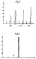
- the protected conjugates were able to displace the binding of 125 I-huTNF ⁇ to insolubilized recombinant human TNF ⁇ receptor at concentration significantly lower than the analogous unprotected conjugates (Fig. 5).
- TNF ⁇ -mPEG cyanuric - chloride
- huTNF ⁇ - mPEG Protected huTNF ⁇ -mPEG 0 0.03 ng/ml 0.03 ng/ml 10x 1.09 ng/ml 0.14 ng/ml 20x 3.90 ng/ml - 30x 5.40 ng/ml 0.25 ng/ml
Landscapes
- Health & Medical Sciences (AREA)
- Chemical & Material Sciences (AREA)
- Medicinal Chemistry (AREA)
- Life Sciences & Earth Sciences (AREA)
- Organic Chemistry (AREA)
- General Health & Medical Sciences (AREA)
- Immunology (AREA)
- Engineering & Computer Science (AREA)
- Animal Behavior & Ethology (AREA)
- Molecular Biology (AREA)
- Proteomics, Peptides & Aminoacids (AREA)
- Biophysics (AREA)
- Biochemistry (AREA)
- Bioinformatics & Cheminformatics (AREA)
- Pharmacology & Pharmacy (AREA)
- Epidemiology (AREA)
- Genetics & Genomics (AREA)
- Public Health (AREA)
- Veterinary Medicine (AREA)
- Medicines That Contain Protein Lipid Enzymes And Other Medicines (AREA)
- Other Resins Obtained By Reactions Not Involving Carbon-To-Carbon Unsaturated Bonds (AREA)
- Medicinal Preparation (AREA)
- Medicines Containing Antibodies Or Antigens For Use As Internal Diagnostic Agents (AREA)
- Immobilizing And Processing Of Enzymes And Microorganisms (AREA)
- Peptides Or Proteins (AREA)
Abstract
Description
Chemical Abstracts no. 20654s (JP-A-2 201 162) discloses a conjugate of a Fab' fragment of an anti-TNFα antibody with an enzyme. Examples of enzyme include horseradish peroxidase, β-galactosidase, alkaline phosphatase and glucose oxidase.
EP-A-0 568 500 discloses a conjugate of monoclonal anti-TNF antibody mAb78 with bovine serum albumin (BSA), which is used for immunization purposes.
Molecular Immunology: 30, 1123-1131 (1993), issue of August 1992, discloses a conjugate of monoclonal anti-TNF antibody mAb78 with BSA or keyhole limpet hemocyanin (KLH), which is used for immunization in mice.
EP-A-0 492 448 discloses novel monoclonal anti-TNFα antibodies, their use in therapy and pharmaceutical compositions containing them.
The efficacy of many of these agents, however, is limited for two main reasons.
- First substance: streptokinase or hirudin, second substance plasminogen or thrombin respectively; and
- first substance: a growth factor such as FGF, EGF, PDGF, HGH or HGF, a growth factor receptor, a lymphokine or cytokine such as an interferon, interleukin, stimulating factor, TNFα or TNFβ or a lymphokine or cytokine receptor; second substance the correpsonding growth factor receptor, the corresonding growth factor, the corresponding lymphokine or cytokine receptor or the corresponding lymphokine or cytokine respectively.
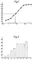
- Figure 1
- shows the cytotoxic activity on LM cells of 1 ng/ml huTNFα in the presence of increasing concentrations of mAb78. The x-axis denotes the concentration of mAb78 in ng/ml. The y-axis denotes the percentage viability of LM cells in the presence of 1 ng/ml huTNFα and different concentrations of mAb78. The Figure indicates the concentration of mAb78 at which 1 ng/ml huTNFα exerts 50% cytotoxic activity (6.7 ng/ml).
- Figure 2
- shows the concentrations in ng/ml of mAb78 or unprotected mAb78-mPEG conjugates at which 1 ng/ml huTNFα exerts 50% cytotoxic activity on LM cells (y-axis). (1) mAb78; (2) unprotected mAb78 derivatized upon addition of a 3-fold molar excess of mPEG (mAb78+mPEG 3x); (3) mAb78 + mPEG 15x (4) mAb78 + mPEG 62.5x; (5) mAb78 + mPEG 125x; (6) mAb78 + mPEG 250x; (7) mAb78 + mPEG 300x; (8) mAb78 + mPEG 500x.
- Figure 3
- shows the concentrations in ng/ml of mAb78, unprotected mAb78-mPEG or protected mAb78-mPEG conjugates at which 1 ng/ml huTNFα exerts 50% cytotoxic activity on LM cells (y-axis). (1) mock-derivatized mAb78; (3) unprotected mAb78 derivatized upon addition of a 15-fold molar excess of mPEG (calculated on the mAb78 concentration); (2) protected mAb78 derivatized upon addition of a 15-fold molar excess of mPEG (calculated on the concentration of mAb78+mAb2); (5) unprotected mAb78 + mPEG 100x; (6) unprotected mAb78 and mouse IgG derivatized upon addition of a 100-fold molar excess of mPEG (calculated on the mAb78 + mouse IgG concentrations); (4) protected mAb78 + mPEG 100x; (7) as (1); (8) as (4); (9) as (5); (10) as (1); (12) unprotected mAb78 + mPEG 500x; (11) protected mAb78 + mPEG 500x.
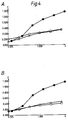
- Figure 4
- shows the detection of mAb78, unprotected mAb78-mPEG or protected mAb78-mPEG conjugates in a mouse IgG-specific ELISA. A : () mock-derivatized mAb78; (▵) unprotected mAb78 derivatized upon addition of a 100-fold molar excess of mPEG (unprotected mAb78 + mPEG 100x); (O) protected mAb78 derivatized upon addition of a 100-fold molar excess of mPEG. B : () mock-derivatized mAb 78; (▵) unprotected mAb78 + mPEG 500x; (O) protected mAb78 + mPEG 500x. x-axis: concentration of mAb78 in ng/ml. y-axis: O.D. units.
- Figure 5
- shows the inhibition of the binding of 125I-huTNFα
to recombinant TNF-R1 receptor by unprotected
huTNFα-mPEG or huTNFα-mPEG protected with
insolubilized anti-huTNFα mAb78. y-axis: amount
of competitor in µg. denotes huTNFα,
 denotes huTNFα-PEG30x and
 denotes protected huTNFα-PEG30x.
The conjugates of this invention can be formulated according to well known methods by including appropriate amounts of the conjugates with a pharmaceutically acceptable carrier and/or diluent.
The following are examples of possible formulations:
| Active Drug | from about 0.05% to about 0.5% w/v |
| Bulking Agent | from about 2.50% to about 5.0% w/v |
| Surface-Active Agent | from about 0.0025% to about 0.025% w/v |
| pH-Adjusting Agent | q.s. to pH 6.5 - 7. |
| Active Drug | from about 0.05% to about 0.5% w/v |
| Surface-Active Agent | from about 0.0025% to about 0.025% w/v |
| pH-Adjusting Agent | q.s. to pH 6.5 - 7 |
| W.F.I | q.s. |
| W.F.I means water for injections. |
Sephadex, Sephacryl, Superose and Sepharose are trademarks.
100 µl of this suspension were added to the wells of flat bottom microtiter plates (Falcon 3072). Then a dilution of huTNFα (Esquire Chemie, Zuerich, Switzerland) was set up in complete MEM to have a final concentration, after addition to wells, of 1 ng/ml.
mAb78 or mAb78-mPEG conjugates were diluted in complete MEM and added to wells to have final concentrations from 200 ng/ml to 0.35 ng/ml.
50 µl of diluted huTNFα were then incubated with 50 µl of each mAb78 or mAb78-mPEG conjugate dilution or with 50 µl complete MEM for 2 hours at 37°C. During this incubation period a solution containing actinomycin D (Fluka, Buchs, Switzerland) was set up in complete MEM.
The final concentration of actinomycin D after addition to the wells was 2 µg/ml. At the end of the incubation period each mAb78-huTNFα or mAb78-mPEG conjugate-huTNFα mixture and 50 µl of actinomycin D were added to the wells of microtiter plates. To some
After the incubation a fresh solution of MTT (CALBIOCHEM 589511 - La Jolla - CA) 5 mg/ml in PBS was prepared and 40 µl were transferred into each well. The plates were incubated at 37°C, 5% CO2 for 4 hours. At the end of the incubation the medium was aspirated. DMSO (CARLO ERBA-ITALY) was then added (200 µl/well) and vigorously pipetted 2-3 times.
The plates were then read in the spectrophotometer at 570 nm wavelength.
The data were expressed in optical density (OP) UNITS. The OD values were expressed as percent values of those from the control wells (Actinomycin D alone).
These percent values were processed by means of the ELISA-Soft PC program (PERKIN-ELMER-NORWALK, CT USA) and the mAb78 concentration giving 50% cytotoxicity in the presence of 1 ng/ml huTNFα was thereby determined.
The cDNA for the 55kDa human TNF receptor, was isolated by screening a λ gt11 library (Clontech) using as probe two synthetic oligonucleotides designed according to the published nucleotide sequence of the 55kDa receptor (Gray et al. 1990 PNAS USA 87, 7380-7837.
The cDNA coding the extracellular domain of 55kDa TNFR was cloned in a plasmid of the pQE series type IV (DIAGEN GmbH - Hilden- Germany) fused in frame to a polyHis tail.
The insertion of the cDNA was carried out following amplification by Polymerase Chain Reaction (PCR); PCR primers were selected to introduce restriction sites compatible with the sites present in the pQE9 polylinker; in addition a translation stop codon was engineered behind the nucleotides corresponding to the last aminoacid of the extracellular domain. The plasmid, called pFC228, was transformed in a K12 derived E.coli strains: M15(pREP4).
The new strain was called FICE 284.
| Concentrations of mAb78, unprotected mAb78-mPEG conjugates or protected mAb78-mPEG conjugates at which 1 ng/ml huTNFα exerts 50% cytotoxic activity on LM cells. | ||
| Concentration (ng/ml) | ||
| 1.Experiment | mAb78 | 6.7 |
| Unprotected mAb78+mPEG 15x | 23.6 | |
| Unprotected mAb78+mPEG 100x | 41.61 | |
| Unprotected mAb78+mouse IgG +mPEG 100x | 37.32 | |
| Protected mAb78+mPEG 15x | 7.0 | |
| Protected mAb78+mPEG 100x | 7.16 | |
| 2.Experiment | mAb78 | 6.93 |
| Unprotected mAb78+mPEG 100x | 39.06 | |
| Protected mAb78+MPEG 100x | 7.06 | |
| 3.Experiment | mAb78 | 7.2 |
| Unprotected mAb78+mPEG 500x | 49.0 | |
| Protected mAb78+mPEG 500x | 7.8 |
| Doses of protected and unprotected TNFα-mPEG giving 50% of cytotoxicity of LM cells. | ||
| mPEG (cyanuric - chloride) | huTNFα - mPEG | Protected huTNFα-mPEG |
| 0 | 0.03 ng/ml | 0.03 ng/ml |
| 10x | 1.09 ng/ml | 0.14 ng/ml |
| 20x | 3.90 ng/ml | - |
| 30x | 5.40 ng/ml | 0.25 ng/ml |
Claims (24)
- A process for the preparation of a conjugate between a polymer and a first substance having a biological activity mediated by a domain thereof, which process comprises:(a) contacting the first substance with a second substance which specifically binds to the said domain of the first substance;(b) conjugating a polymer to the first substance having the second substance bound thereto; and(c) freeing the second substance from the first substance having the polymer conjugated thereto and wherein, when the first substance is a proteolytic enzyme then the polymer is other than polyethylene glycol.
- A process according to claim 1, wherein the first substance is a protein or glycoprotein.
- A process according to claim 2, wherein the first substance is a monoclonal antibody or fragment thereof and the second substance is an antiidiotype antibody or the antigen to which the monoclonal antibody or fragment thereof binds.
- A process according to claim 3, wherein the first substance is a non-human monoclonal antibody specific for human TNFα and the second substance is a corresponding antiidiotype antibody.
- A process according to claim 2, wherein the first substance is TNFα and the second substance is an antibody to which TNFα binds.
- A process according to claim 5, wherein the first substance is human TNFα and the second substance is a corresponding non-human monoclonal antibody.
- A process according to claim 2, wherein the first substance is a protein and the second substance is a monoclonal antibody (mAb) directed against the specific active site of the first substance or the ligand with which the first substance normally interacts.
- A process according to claim 7, wherein the first substance is a fibrinolytic enzyme or a fibrinolytic agent.
- A process according to claim 8, wherein the first substance is streptokinase and the second substance is plasminogen.
- A process according to claim 7, wherein the first substance is a growth factor or a growth factor receptor and the second substance is the corresponding growth factor receptor or a growth factor.
- A process according to claim 10, wherein the first substance is the human growth hormone (HGH).
- A process according to claim 10, wherein the first substance is a growth factor and the second substance is heparin, a heparin-like molecule or a heparin derivative having a low molecular weight and a negative charge.
- A process according to any one of the preceding claims, wherein the polymer, which is conjugated to the first substance having the second substance bound thereto, is a poly(ethylene glycol) or N-(2-hydroxypropyl)methacrylamide polymer.
- A conjugate between an inert, synthetic, water soluble polymer and an antibody against TNFα, as obtainable by the process of claim 1.
- A conjugate according to claim 14, wherein the antibody is mAb78.
- A conjugate between an inert, synthetic, water soluble polymer and a protein, as obtainable by the process of claim 1.
- A conjugate between an inert, synthetic, water soluble polymer and a fibrinolytic enzyme or fibrinolytic agent, as obtainable by the process of claim 1.
- A conjugate according to claim 17, wherein the fibrinolytic agent is streptokinase.
- A conjugate between an inert, synthetic, water soluble polymer and a growth factor or a growth factor receptor, as obtainable by the process of claim 1.
- A conjugate according to claim 19, wherein the growth factor is human growth hormone (HGH).
- A conjugate according to claims 14 to 20, wherein the polymer is chosen from poly(ethylene glycol), poly(vinyl pyrrolidone), poly(vinyl alcohol), poly(amino acids), and polymers of divinylether maleic anhydride, ethylene-maleic anhydride, N-(2-hydroxypropyl)methacrylamide and dextran.
- A pharmaceutical composition comprising a pharmaceutically acceptable carrier and/or diluent and, as an active principle a conjugate as defined in claims 14 to 21.
- A conjugate as defined in claim 14 for use as a prophylactic or therapeutic agent in a disease state in which human TNFα exerts a pathogenic effect.
Applications Claiming Priority (3)
| Application Number | Priority Date | Filing Date | Title |
|---|---|---|---|
| GB9225448 | 1992-12-04 | ||
| GB929225448A GB9225448D0 (en) | 1992-12-04 | 1992-12-04 | Improved synthesis of polymer bioactive conjugates |
| PCT/EP1993/003429 WO1994013322A1 (en) | 1992-12-04 | 1993-12-06 | Improved synthesis of polymer bio-active conjugates |
Publications (2)
| Publication Number | Publication Date |
|---|---|
| EP0675736A1 EP0675736A1 (en) | 1995-10-11 |
| EP0675736B1 true EP0675736B1 (en) | 1998-07-15 |
Family
ID=10726165
Family Applications (1)
| Application Number | Title | Priority Date | Filing Date |
|---|---|---|---|
| EP94902692A Expired - Lifetime EP0675736B1 (en) | 1992-12-04 | 1993-12-06 | Improved synthesis of polymer bio-active conjugates |
Country Status (11)
| Country | Link |
|---|---|
| US (1) | US6172202B1 (en) |
| EP (1) | EP0675736B1 (en) |
| JP (1) | JP3557206B2 (en) |
| AT (1) | ATE168273T1 (en) |
| AU (1) | AU678796B2 (en) |
| CA (1) | CA2150925C (en) |
| DE (1) | DE69319740T2 (en) |
| DK (1) | DK0675736T3 (en) |
| ES (1) | ES2121180T3 (en) |
| GB (1) | GB9225448D0 (en) |
| WO (1) | WO1994013322A1 (en) |
Families Citing this family (34)
| Publication number | Priority date | Publication date | Assignee | Title |
|---|---|---|---|---|
| US5959087A (en) * | 1989-08-07 | 1999-09-28 | Peptide Technology, Ltd. | Tumour necrosis factor binding ligands |
| US20030225254A1 (en) * | 1989-08-07 | 2003-12-04 | Rathjen Deborah Ann | Tumour necrosis factor binding ligands |
| JP3443119B2 (en) | 1989-08-07 | 2003-09-02 | ペプテック リミテッド | Tumor necrosis factor binding ligand |
| US6277969B1 (en) | 1991-03-18 | 2001-08-21 | New York University | Anti-TNF antibodies and peptides of human tumor necrosis factor |
| US6284471B1 (en) | 1991-03-18 | 2001-09-04 | New York University Medical Center | Anti-TNFa antibodies and assays employing anti-TNFa antibodies |
| US20040120952A1 (en) * | 2000-08-07 | 2004-06-24 | Centocor, Inc | Anti-TNF antibodies and peptides of human tumor necrosis factor |
| US7192584B2 (en) * | 1991-03-18 | 2007-03-20 | Centocor, Inc. | Methods of treating psoriasis with anti-TNF antibodies |
| ES2121907T3 (en) * | 1992-08-28 | 1998-12-16 | Bayer Ag | USE OF ANTI-TNF MONOCLONAL ANTIBODIES FOR THE TREATMENT OF BACTERIAL MENINGITIS. |
| US20050260201A1 (en) * | 1993-01-29 | 2005-11-24 | Centocor, Inc. | Methods of treating rheumatoid arthritis using anti-TNF receptor fusion proteins |
| SE9503380D0 (en) * | 1995-09-29 | 1995-09-29 | Pharmacia Ab | Protein derivatives |
| US6548644B1 (en) * | 1997-03-10 | 2003-04-15 | Immunex Corporation | Site protected protein modification |
| DE69921102T2 (en) | 1998-03-05 | 2006-02-02 | Chiron Corp., Emeryville | METHOD FOR IMPROVING THE SERUM HALF-TIME OF BIOLOGICALLY ACTIVE MOLECULES |
| US6451986B1 (en) | 1998-06-22 | 2002-09-17 | Immunex Corporation | Site specific protein modification |
| HK1042114B (en) | 1998-10-23 | 2006-09-29 | Amgen K-A, Inc. | Dimeric thrombopoietin peptide mimetics binding to mp1 receptor and having thrombopoietic activity |
| WO2000051637A1 (en) * | 1999-03-02 | 2000-09-08 | Centocor, Inc. | ANTI-TNFα ANTIBODIES IN THERAPY OF ASTHMA |
| US20110195063A1 (en) * | 2000-08-07 | 2011-08-11 | Centocor, Inc. | Methods of Treating Ankylosing Spondylitis Using Anti-TNF Antibodies and Peptides of Human Tumor Necrosis Factor |
| US20050249735A1 (en) * | 2000-08-07 | 2005-11-10 | Centocor, Inc. | Methods of treating ankylosing spondylitis using anti-TNF antibodies and peptides of human tumor necrosis factor |
| US20060018907A1 (en) * | 2000-08-07 | 2006-01-26 | Centocor, Inc. | Anti-TNF antibodies and peptides of human tumor necrosis factor |
| US7238505B2 (en) | 2000-10-04 | 2007-07-03 | Ahram Biosystems Inc. | Immobilized DNA polymerase |
| US20040091602A1 (en) * | 2000-10-04 | 2004-05-13 | Hwang Hyun Jin | Method for immobilizing biologically active molecules |
| EP1234583A1 (en) * | 2001-02-23 | 2002-08-28 | F. Hoffmann-La Roche Ag | PEG-conjugates of HGF-NK4 |
| US20050271663A1 (en) * | 2001-06-28 | 2005-12-08 | Domantis Limited | Compositions and methods for treating inflammatory disorders |
| US20060073141A1 (en) * | 2001-06-28 | 2006-04-06 | Domantis Limited | Compositions and methods for treating inflammatory disorders |
| TWI327597B (en) * | 2001-08-01 | 2010-07-21 | Centocor Inc | Anti-tnf antibodies, compositions, methods and uses |
| SE0104186D0 (en) * | 2001-12-12 | 2001-12-12 | Darcy Birse | Method and device |
| WO2004019991A2 (en) * | 2002-08-30 | 2004-03-11 | F. Hoffmann-La Roche Ag | Scatter factor/hepatocyte growth factor antagonist nk4 for the treatment of glioma |
| ATE472556T1 (en) * | 2002-12-02 | 2010-07-15 | Amgen Fremont Inc | ANTIBODIES DIRECTED AGAINST THE TUMOR NECROSIS FACTOR AND THEIR USES |
| CA2572765C (en) | 2004-07-08 | 2013-05-21 | Amgen Inc. | Compound having improved bioefficiency when administered in a multidose regimen |
| WO2007117685A2 (en) | 2006-04-07 | 2007-10-18 | Nektar Therapeutics Al, Corporation | Conjugates of an anti-tnf-alpha antibody |
| US9283260B2 (en) | 2006-04-21 | 2016-03-15 | Amgen Inc. | Lyophilized therapeutic peptibody formulations |
| NZ580530A (en) * | 2007-06-06 | 2012-04-27 | Domantis Ltd | Anti vegf polypeptides, antibody variable domains and antagonists |
| US20090038182A1 (en) * | 2007-08-09 | 2009-02-12 | Lans Maris J | Footwear with built-in scale |
| AU2009303368B2 (en) * | 2008-10-15 | 2012-04-05 | Takeda Pharmaceutical Company Limited | Pegylation of recombinant blood coagulation factors in the presence of bound antibodies |
| US10465003B2 (en) | 2016-02-05 | 2019-11-05 | Janssen Biotech, Inc. | Anti-TNF antibodies, compositions, methods and use for the treatment or prevention of type 1 diabetes |
Family Cites Families (13)
| Publication number | Priority date | Publication date | Assignee | Title |
|---|---|---|---|---|
| US4863729A (en) * | 1984-06-20 | 1989-09-05 | Linus Pauling Institute Of Science And Medicine | Method for preparing a macromolecular monoclonal antibody composition |
| US4732863A (en) * | 1984-12-31 | 1988-03-22 | University Of New Mexico | PEG-modified antibody with reduced affinity for cell surface Fc receptors |
| US4699784A (en) * | 1986-02-25 | 1987-10-13 | Center For Molecular Medicine & Immunology | Tumoricidal methotrexate-antibody conjugate |
| US5047227A (en) * | 1988-02-08 | 1991-09-10 | Cytogen Corporation | Novel and improved antibodies for site specific attachment of compounds |
| US4925648A (en) * | 1988-07-29 | 1990-05-15 | Immunomedics, Inc. | Detection and treatment of infectious and inflammatory lesions |
| JPH02201162A (en) * | 1989-01-30 | 1990-08-09 | Teijin Ltd | Enzyme antibody complex, combination of complex/ antibody, kit and diagnostic method using them |
| GB9028123D0 (en) * | 1990-12-28 | 1991-02-13 | Erba Carlo Spa | Monoclonal antibodies against human tumor necrosis factor alpha |
| WO1992012845A2 (en) * | 1991-01-22 | 1992-08-06 | Rover Group Limited | Fabrication of a composite plastics structure, particularly an internal combustion engine inlet manifold |
| SE502083C2 (en) | 1991-05-28 | 1995-08-07 | Misomex Ab | Method and apparatus for automated exposure of photosensitive material by laser light |
| IT1260468B (en) * | 1992-01-29 | 1996-04-09 | METHOD FOR MAINTAINING THE ACTIVITY OF PROTEOLYTIC ENZYMES MODIFIED WITH POLYETHYLENGLYCOL | |
| IT1254315B (en) * | 1992-03-27 | 1995-09-14 | Mini Ricerca Scient Tecnolog | ANTI-IDIOTYPIC MONOCLONAL ANTIBODIES DIRECT AGAINST ANTI-TNF ANTIBODIES. |
| US5314830A (en) * | 1992-10-30 | 1994-05-24 | University Of New Mexico | Immobilized hydrophobically-modified antibodies |
| US5556623A (en) * | 1993-03-30 | 1996-09-17 | Eli Lilly And Company | Antibody-drug conjugates |
-
1992
- 1992-12-04 GB GB929225448A patent/GB9225448D0/en active Pending
-
1993
- 1993-12-06 CA CA002150925A patent/CA2150925C/en not_active Expired - Fee Related
- 1993-12-06 DK DK94902692T patent/DK0675736T3/en active
- 1993-12-06 JP JP51376394A patent/JP3557206B2/en not_active Expired - Fee Related
- 1993-12-06 EP EP94902692A patent/EP0675736B1/en not_active Expired - Lifetime
- 1993-12-06 DE DE69319740T patent/DE69319740T2/en not_active Expired - Lifetime
- 1993-12-06 AT AT94902692T patent/ATE168273T1/en active
- 1993-12-06 AU AU56968/94A patent/AU678796B2/en not_active Ceased
- 1993-12-06 WO PCT/EP1993/003429 patent/WO1994013322A1/en not_active Ceased
- 1993-12-06 ES ES94902692T patent/ES2121180T3/en not_active Expired - Lifetime
-
1997
- 1997-07-07 US US08/889,049 patent/US6172202B1/en not_active Expired - Fee Related
Also Published As
| Publication number | Publication date |
|---|---|
| ATE168273T1 (en) | 1998-08-15 |
| CA2150925A1 (en) | 1994-06-23 |
| GB9225448D0 (en) | 1993-01-27 |
| CA2150925C (en) | 2009-02-03 |
| US6172202B1 (en) | 2001-01-09 |
| DE69319740D1 (en) | 1998-08-20 |
| AU5696894A (en) | 1994-07-04 |
| JPH08504202A (en) | 1996-05-07 |
| ES2121180T3 (en) | 1998-11-16 |
| DE69319740T2 (en) | 1999-03-11 |
| WO1994013322A1 (en) | 1994-06-23 |
| JP3557206B2 (en) | 2004-08-25 |
| DK0675736T3 (en) | 1999-04-19 |
| EP0675736A1 (en) | 1995-10-11 |
| AU678796B2 (en) | 1997-06-12 |
Similar Documents
| Publication | Publication Date | Title |
|---|---|---|
| AU678796B2 (en) | Improved synthesis of polymer bio-active conjugates | |
| EP1037657B1 (en) | Alpha-interferon-polymer-conjugates having enhanced biological activity and methods of preparing the same | |
| EP0862455B1 (en) | Interferon-polymer conjugates and process for preparing the same | |
| KR100303810B1 (en) | Compositions and Methods for Stimulating Megakaryotic Fields and Differentiation | |
| KR101022577B1 (en) | Polymer conjugate with reduced antigenicity, preparation method and use thereof | |
| JP3747070B2 (en) | Improved interferon-polymer conjugate | |
| JP4157277B2 (en) | Substantially pure histidine binding protein-polymer conjugate | |
| CA2978330C (en) | Conjugates of an il-7 moiety and a polymer | |
| JP7235772B2 (en) | Selective Treg stimulator RUR20kD-IL-2 and related compositions | |
| JPH11512298A (en) | Human growth hormone mutant | |
| EP0187658B1 (en) | Method and use for site-specific activation of substances | |
| KR20030060915A (en) | Protein c or activated portein c-like molecules | |
| JP2006521372A (en) | 1: 1 conjugate of biologically active substance and biocompatible polymer, method for producing the same, and pharmaceutical composition containing the same | |
| AU703091B2 (en) | Monoclonal antibodies against soluble TNF-alpha receptors p55 and p75 as well as against TNF-alpha and its analogues | |
| JP4237703B2 (en) | Insulin-like growth factor binding protein-4 and poly (ethylene glycol) conjugate | |
| CN101591649B (en) | Methoxypolyethylene glycol-modified arginine deiminase, preparation thereof and use thereof | |
| EP0363712B1 (en) | Hybrid monoclonal antibodies, their production and use | |
| EA006368B1 (en) | Chemically-modified progenipoietin conjugates | |
| KR100490447B1 (en) | Polypeptides Inducing Production of Interferon-γ, Monoclonal Antibodies and Susceptible Diseases Specific to Dipeptides | |
| SI9600206A (en) | Monoclonic antibodies versus soluble tnf-alpha p55 and p75 receptors and tnf-alpha and their ana |
Legal Events
| Date | Code | Title | Description |
|---|---|---|---|
| PUAI | Public reference made under article 153(3) epc to a published international application that has entered the european phase |
Free format text: ORIGINAL CODE: 0009012 |
|
| 17P | Request for examination filed |
Effective date: 19950606 |
|
| AK | Designated contracting states |
Kind code of ref document: A1 Designated state(s): AT BE CH DE DK ES FR GB GR IE IT LI LU MC NL PT SE |
|
| 17Q | First examination report despatched |
Effective date: 19960208 |
|
| RAP1 | Party data changed (applicant data changed or rights of an application transferred) |
Owner name: PHARMACIA & UPJOHN S.P.A. |
|
| GRAG | Despatch of communication of intention to grant |
Free format text: ORIGINAL CODE: EPIDOS AGRA |
|
| GRAG | Despatch of communication of intention to grant |
Free format text: ORIGINAL CODE: EPIDOS AGRA |
|
| GRAH | Despatch of communication of intention to grant a patent |
Free format text: ORIGINAL CODE: EPIDOS IGRA |
|
| GRAH | Despatch of communication of intention to grant a patent |
Free format text: ORIGINAL CODE: EPIDOS IGRA |
|
| GRAH | Despatch of communication of intention to grant a patent |
Free format text: ORIGINAL CODE: EPIDOS IGRA |
|
| GRAA | (expected) grant |
Free format text: ORIGINAL CODE: 0009210 |
|
| AK | Designated contracting states |
Kind code of ref document: B1 Designated state(s): AT BE CH DE DK ES FR GB GR IE IT LI LU MC NL PT SE |
|
| REF | Corresponds to: |
Ref document number: 168273 Country of ref document: AT Date of ref document: 19980815 Kind code of ref document: T |
|
| REG | Reference to a national code |
Ref country code: CH Ref legal event code: EP |
|
| REF | Corresponds to: |
Ref document number: 69319740 Country of ref document: DE Date of ref document: 19980820 |
|
| REG | Reference to a national code |
Ref country code: IE Ref legal event code: FG4D |
|
| REG | Reference to a national code |
Ref country code: CH Ref legal event code: NV Representative=s name: A. BRAUN, BRAUN, HERITIER, ESCHMANN AG PATENTANWAE |
|
| REG | Reference to a national code |
Ref country code: ES Ref legal event code: FG2A Ref document number: 2121180 Country of ref document: ES Kind code of ref document: T3 |
|
| ET | Fr: translation filed | ||
| REG | Reference to a national code |
Ref country code: PT Ref legal event code: SC4A Free format text: AVAILABILITY OF NATIONAL TRANSLATION Effective date: 19981008 |
|
| REG | Reference to a national code |
Ref country code: DK Ref legal event code: T3 |
|
| PLBE | No opposition filed within time limit |
Free format text: ORIGINAL CODE: 0009261 |
|
| STAA | Information on the status of an ep patent application or granted ep patent |
Free format text: STATUS: NO OPPOSITION FILED WITHIN TIME LIMIT |
|
| 26N | No opposition filed | ||
| REG | Reference to a national code |
Ref country code: GB Ref legal event code: IF02 |
|
| REG | Reference to a national code |
Ref country code: GB Ref legal event code: 732E |
|
| REG | Reference to a national code |
Ref country code: CH Ref legal event code: PUE Owner name: PHARMACIA & UPJOHN S.P.A. TRANSFER- BIOVITRUM AB |
|
| REG | Reference to a national code |
Ref country code: ES Ref legal event code: PC2A |
|
| REG | Reference to a national code |
Ref country code: PT Ref legal event code: PC4A Free format text: BIOVITRUM AB SE Effective date: 20020130 |
|
| NLS | Nl: assignments of ep-patents |
Owner name: BIOVITRUM AB |
|
| NLS | Nl: assignments of ep-patents |
Owner name: BIOVITRUM AB |
|
| REG | Reference to a national code |
Ref country code: CH Ref legal event code: PFA Owner name: BIOVITRUM AB Free format text: BIOVITRUM AB# #112 76 STOCKHOLM (SE) -TRANSFER TO- BIOVITRUM AB# #112 76 STOCKHOLM (SE) |
|
| PGFP | Annual fee paid to national office [announced via postgrant information from national office to epo] |
Ref country code: NL Payment date: 20101124 Year of fee payment: 18 Ref country code: MC Payment date: 20101129 Year of fee payment: 18 Ref country code: IE Payment date: 20101126 Year of fee payment: 18 Ref country code: FR Payment date: 20101203 Year of fee payment: 18 Ref country code: DK Payment date: 20101123 Year of fee payment: 18 Ref country code: AT Payment date: 20101122 Year of fee payment: 18 |
|
| PGFP | Annual fee paid to national office [announced via postgrant information from national office to epo] |
Ref country code: PT Payment date: 20101126 Year of fee payment: 18 Ref country code: LU Payment date: 20101216 Year of fee payment: 18 Ref country code: CH Payment date: 20101026 Year of fee payment: 18 |
|
| PGFP | Annual fee paid to national office [announced via postgrant information from national office to epo] |
Ref country code: GR Payment date: 20101126 Year of fee payment: 18 Ref country code: SE Payment date: 20101122 Year of fee payment: 18 Ref country code: IT Payment date: 20101218 Year of fee payment: 18 Ref country code: GB Payment date: 20101123 Year of fee payment: 18 |
|
| PGFP | Annual fee paid to national office [announced via postgrant information from national office to epo] |
Ref country code: DE Payment date: 20101230 Year of fee payment: 18 |
|
| PGFP | Annual fee paid to national office [announced via postgrant information from national office to epo] |
Ref country code: BE Payment date: 20101213 Year of fee payment: 18 Ref country code: ES Payment date: 20101213 Year of fee payment: 18 |
|
| REG | Reference to a national code |
Ref country code: PT Ref legal event code: MM4A Free format text: LAPSE DUE TO NON-PAYMENT OF FEES Effective date: 20120606 |
|
| BERE | Be: lapsed |
Owner name: *BIOVITRUM A.B. Effective date: 20111231 |
|
| REG | Reference to a national code |
Ref country code: NL Ref legal event code: V1 Effective date: 20120701 |
|
| REG | Reference to a national code |
Ref country code: DK Ref legal event code: EBP |
|
| PG25 | Lapsed in a contracting state [announced via postgrant information from national office to epo] |
Ref country code: MC Free format text: LAPSE BECAUSE OF NON-PAYMENT OF DUE FEES Effective date: 20111231 |
|
| REG | Reference to a national code |
Ref country code: CH Ref legal event code: PL Ref country code: SE Ref legal event code: EUG |
|
| GBPC | Gb: european patent ceased through non-payment of renewal fee |
Effective date: 20111206 |
|
| PG25 | Lapsed in a contracting state [announced via postgrant information from national office to epo] |
Ref country code: PT Free format text: LAPSE BECAUSE OF NON-PAYMENT OF DUE FEES Effective date: 20120606 |
|
| REG | Reference to a national code |
Ref country code: GR Ref legal event code: ML Ref document number: 980402307 Country of ref document: GR Effective date: 20120704 |
|
| REG | Reference to a national code |
Ref country code: FR Ref legal event code: ST Effective date: 20120831 |
|
| REG | Reference to a national code |
Ref country code: IE Ref legal event code: MM4A |
|
| REG | Reference to a national code |
Ref country code: DE Ref legal event code: R119 Ref document number: 69319740 Country of ref document: DE Effective date: 20120703 |
|
| PG25 | Lapsed in a contracting state [announced via postgrant information from national office to epo] |
Ref country code: LI Free format text: LAPSE BECAUSE OF NON-PAYMENT OF DUE FEES Effective date: 20111231 Ref country code: IE Free format text: LAPSE BECAUSE OF NON-PAYMENT OF DUE FEES Effective date: 20111206 Ref country code: BE Free format text: LAPSE BECAUSE OF NON-PAYMENT OF DUE FEES Effective date: 20111231 Ref country code: GB Free format text: LAPSE BECAUSE OF NON-PAYMENT OF DUE FEES Effective date: 20111206 Ref country code: CH Free format text: LAPSE BECAUSE OF NON-PAYMENT OF DUE FEES Effective date: 20111231 Ref country code: DE Free format text: LAPSE BECAUSE OF NON-PAYMENT OF DUE FEES Effective date: 20120703 Ref country code: SE Free format text: LAPSE BECAUSE OF NON-PAYMENT OF DUE FEES Effective date: 20111207 |
|
| PG25 | Lapsed in a contracting state [announced via postgrant information from national office to epo] |
Ref country code: GR Free format text: LAPSE BECAUSE OF NON-PAYMENT OF DUE FEES Effective date: 20120704 Ref country code: IT Free format text: LAPSE BECAUSE OF NON-PAYMENT OF DUE FEES Effective date: 20111206 |
|
| REG | Reference to a national code |
Ref country code: AT Ref legal event code: MM01 Ref document number: 168273 Country of ref document: AT Kind code of ref document: T Effective date: 20111206 |
|
| PG25 | Lapsed in a contracting state [announced via postgrant information from national office to epo] |
Ref country code: DK Free format text: LAPSE BECAUSE OF NON-PAYMENT OF DUE FEES Effective date: 20120102 Ref country code: AT Free format text: LAPSE BECAUSE OF NON-PAYMENT OF DUE FEES Effective date: 20111206 Ref country code: NL Free format text: LAPSE BECAUSE OF NON-PAYMENT OF DUE FEES Effective date: 20120701 |
|
| PG25 | Lapsed in a contracting state [announced via postgrant information from national office to epo] |
Ref country code: FR Free format text: LAPSE BECAUSE OF NON-PAYMENT OF DUE FEES Effective date: 20120102 |
|
| PG25 | Lapsed in a contracting state [announced via postgrant information from national office to epo] |
Ref country code: LU Free format text: LAPSE BECAUSE OF NON-PAYMENT OF DUE FEES Effective date: 20111206 |
|
| REG | Reference to a national code |
Ref country code: ES Ref legal event code: FD2A Effective date: 20130703 |
|
| PG25 | Lapsed in a contracting state [announced via postgrant information from national office to epo] |
Ref country code: ES Free format text: LAPSE BECAUSE OF NON-PAYMENT OF DUE FEES Effective date: 20111207 |