EP0675199B1 - A DNA which codes for the variable region of the anti-human influenza A type virus antibody recognizing the H,N, and H2N2 subtypes of haemagglutinin - Google Patents
A DNA which codes for the variable region of the anti-human influenza A type virus antibody recognizing the H,N, and H2N2 subtypes of haemagglutinin Download PDFInfo
- Publication number
- EP0675199B1 EP0675199B1 EP95301664A EP95301664A EP0675199B1 EP 0675199 B1 EP0675199 B1 EP 0675199B1 EP 95301664 A EP95301664 A EP 95301664A EP 95301664 A EP95301664 A EP 95301664A EP 0675199 B1 EP0675199 B1 EP 0675199B1
- Authority
- EP
- European Patent Office
- Prior art keywords
- seq
- dna
- plasmid
- sequence
- antibody
- Prior art date
- Legal status (The legal status is an assumption and is not a legal conclusion. Google has not performed a legal analysis and makes no representation as to the accuracy of the status listed.)
- Expired - Lifetime
Links
- 241000700605 Viruses Species 0.000 title claims description 68
- 206010022000 influenza Diseases 0.000 title claims description 33
- 241000197304 H2N2 subtype Species 0.000 title claims description 18
- 108020004414 DNA Proteins 0.000 claims description 108
- 230000000694 effects Effects 0.000 claims description 34
- 241000197306 H1N1 subtype Species 0.000 claims description 25
- 241000252870 H3N2 subtype Species 0.000 claims description 22
- 108091028043 Nucleic acid sequence Proteins 0.000 claims description 21
- 238000006386 neutralization reaction Methods 0.000 claims description 16
- 125000003275 alpha amino acid group Chemical group 0.000 claims 2
- 239000013612 plasmid Substances 0.000 description 128
- 108090000623 proteins and genes Proteins 0.000 description 77
- 108090000765 processed proteins & peptides Proteins 0.000 description 55
- 229920001184 polypeptide Polymers 0.000 description 43
- 102000004196 processed proteins & peptides Human genes 0.000 description 43
- 150000001413 amino acids Chemical group 0.000 description 41
- 108020004707 nucleic acids Proteins 0.000 description 40
- 102000039446 nucleic acids Human genes 0.000 description 40
- 150000007523 nucleic acids Chemical class 0.000 description 40
- 239000013615 primer Substances 0.000 description 40
- 210000004027 cell Anatomy 0.000 description 39
- 238000000034 method Methods 0.000 description 39
- 239000012634 fragment Substances 0.000 description 38
- 241000588724 Escherichia coli Species 0.000 description 22
- 108091008146 restriction endonucleases Proteins 0.000 description 20
- 102000004169 proteins and genes Human genes 0.000 description 19
- 108010047041 Complementarity Determining Regions Proteins 0.000 description 17
- 241000699666 Mus <mouse, genus> Species 0.000 description 17
- 210000004408 hybridoma Anatomy 0.000 description 17
- 239000002585 base Substances 0.000 description 16
- 239000002299 complementary DNA Substances 0.000 description 16
- 238000010790 dilution Methods 0.000 description 15
- 239000012895 dilution Substances 0.000 description 15
- 208000037797 influenza A Diseases 0.000 description 14
- 206010003445 Ascites Diseases 0.000 description 13
- 239000000243 solution Substances 0.000 description 13
- 230000015572 biosynthetic process Effects 0.000 description 12
- 238000005516 engineering process Methods 0.000 description 12
- 239000012530 fluid Substances 0.000 description 12
- 238000000246 agarose gel electrophoresis Methods 0.000 description 11
- 239000007853 buffer solution Substances 0.000 description 11
- 108091032973 (ribonucleotides)n+m Proteins 0.000 description 10
- 241000699670 Mus sp. Species 0.000 description 10
- 108091034117 Oligonucleotide Proteins 0.000 description 10
- 239000000427 antigen Substances 0.000 description 10
- 102000036639 antigens Human genes 0.000 description 10
- 108091007433 antigens Proteins 0.000 description 10
- 239000002609 medium Substances 0.000 description 10
- 238000012360 testing method Methods 0.000 description 10
- OKKJLVBELUTLKV-UHFFFAOYSA-N Methanol Chemical compound OC OKKJLVBELUTLKV-UHFFFAOYSA-N 0.000 description 9
- 229940126619 mouse monoclonal antibody Drugs 0.000 description 9
- 239000000047 product Substances 0.000 description 9
- 238000010186 staining Methods 0.000 description 9
- 108700026244 Open Reading Frames Proteins 0.000 description 8
- 239000000499 gel Substances 0.000 description 8
- 238000002360 preparation method Methods 0.000 description 8
- 210000002966 serum Anatomy 0.000 description 8
- 101150044453 Y gene Proteins 0.000 description 7
- 230000003321 amplification Effects 0.000 description 7
- 238000003199 nucleic acid amplification method Methods 0.000 description 7
- 229920000936 Agarose Polymers 0.000 description 6
- 239000006144 Dulbecco’s modified Eagle's medium Substances 0.000 description 6
- LFQSCWFLJHTTHZ-UHFFFAOYSA-N Ethanol Chemical compound CCO LFQSCWFLJHTTHZ-UHFFFAOYSA-N 0.000 description 6
- 229920002684 Sepharose Polymers 0.000 description 6
- 238000006243 chemical reaction Methods 0.000 description 6
- 230000004927 fusion Effects 0.000 description 6
- 239000000203 mixture Substances 0.000 description 6
- 230000035772 mutation Effects 0.000 description 6
- 108091003079 Bovine Serum Albumin Proteins 0.000 description 5
- 125000001429 N-terminal alpha-amino-acid group Chemical group 0.000 description 5
- 101150003160 X gene Proteins 0.000 description 5
- 238000010276 construction Methods 0.000 description 5
- 208000037798 influenza B Diseases 0.000 description 5
- 238000004519 manufacturing process Methods 0.000 description 5
- 239000012528 membrane Substances 0.000 description 5
- 238000000746 purification Methods 0.000 description 5
- 101150069452 z gene Proteins 0.000 description 5
- QKNYBSVHEMOAJP-UHFFFAOYSA-N 2-amino-2-(hydroxymethyl)propane-1,3-diol;hydron;chloride Chemical compound Cl.OCC(N)(CO)CO QKNYBSVHEMOAJP-UHFFFAOYSA-N 0.000 description 4
- 241001465754 Metazoa Species 0.000 description 4
- FAPWRFPIFSIZLT-UHFFFAOYSA-M Sodium chloride Chemical compound [Na+].[Cl-] FAPWRFPIFSIZLT-UHFFFAOYSA-M 0.000 description 4
- DBMJMQXJHONAFJ-UHFFFAOYSA-M Sodium laurylsulphate Chemical compound [Na+].CCCCCCCCCCCCOS([O-])(=O)=O DBMJMQXJHONAFJ-UHFFFAOYSA-M 0.000 description 4
- AVKUERGKIZMTKX-NJBDSQKTSA-N ampicillin Chemical compound C1([C@@H](N)C(=O)N[C@H]2[C@H]3SC([C@@H](N3C2=O)C(O)=O)(C)C)=CC=CC=C1 AVKUERGKIZMTKX-NJBDSQKTSA-N 0.000 description 4
- 229960000723 ampicillin Drugs 0.000 description 4
- 201000010099 disease Diseases 0.000 description 4
- 208000037265 diseases, disorders, signs and symptoms Diseases 0.000 description 4
- 238000010353 genetic engineering Methods 0.000 description 4
- 230000035931 haemagglutination Effects 0.000 description 4
- 238000011534 incubation Methods 0.000 description 4
- 230000000644 propagated effect Effects 0.000 description 4
- 239000000523 sample Substances 0.000 description 4
- 230000003248 secreting effect Effects 0.000 description 4
- 235000019333 sodium laurylsulphate Nutrition 0.000 description 4
- 239000006228 supernatant Substances 0.000 description 4
- 108020004705 Codon Proteins 0.000 description 3
- 239000003155 DNA primer Substances 0.000 description 3
- 102000016928 DNA-directed DNA polymerase Human genes 0.000 description 3
- 108010014303 DNA-directed DNA polymerase Proteins 0.000 description 3
- 108010067060 Immunoglobulin Variable Region Proteins 0.000 description 3
- 102000017727 Immunoglobulin Variable Region Human genes 0.000 description 3
- 241000283973 Oryctolagus cuniculus Species 0.000 description 3
- 239000002033 PVDF binder Substances 0.000 description 3
- 102100036829 Probable peptidyl-tRNA hydrolase Human genes 0.000 description 3
- 108010076504 Protein Sorting Signals Proteins 0.000 description 3
- HEMHJVSKTPXQMS-UHFFFAOYSA-M Sodium hydroxide Chemical compound [OH-].[Na+] HEMHJVSKTPXQMS-UHFFFAOYSA-M 0.000 description 3
- 230000010530 Virus Neutralization Effects 0.000 description 3
- JLCPHMBAVCMARE-UHFFFAOYSA-N [3-[[3-[[3-[[3-[[3-[[3-[[3-[[3-[[3-[[3-[[3-[[5-(2-amino-6-oxo-1H-purin-9-yl)-3-[[3-[[3-[[3-[[3-[[3-[[5-(2-amino-6-oxo-1H-purin-9-yl)-3-[[5-(2-amino-6-oxo-1H-purin-9-yl)-3-hydroxyoxolan-2-yl]methoxy-hydroxyphosphoryl]oxyoxolan-2-yl]methoxy-hydroxyphosphoryl]oxy-5-(5-methyl-2,4-dioxopyrimidin-1-yl)oxolan-2-yl]methoxy-hydroxyphosphoryl]oxy-5-(6-aminopurin-9-yl)oxolan-2-yl]methoxy-hydroxyphosphoryl]oxy-5-(6-aminopurin-9-yl)oxolan-2-yl]methoxy-hydroxyphosphoryl]oxy-5-(6-aminopurin-9-yl)oxolan-2-yl]methoxy-hydroxyphosphoryl]oxy-5-(6-aminopurin-9-yl)oxolan-2-yl]methoxy-hydroxyphosphoryl]oxyoxolan-2-yl]methoxy-hydroxyphosphoryl]oxy-5-(5-methyl-2,4-dioxopyrimidin-1-yl)oxolan-2-yl]methoxy-hydroxyphosphoryl]oxy-5-(4-amino-2-oxopyrimidin-1-yl)oxolan-2-yl]methoxy-hydroxyphosphoryl]oxy-5-(5-methyl-2,4-dioxopyrimidin-1-yl)oxolan-2-yl]methoxy-hydroxyphosphoryl]oxy-5-(5-methyl-2,4-dioxopyrimidin-1-yl)oxolan-2-yl]methoxy-hydroxyphosphoryl]oxy-5-(6-aminopurin-9-yl)oxolan-2-yl]methoxy-hydroxyphosphoryl]oxy-5-(6-aminopurin-9-yl)oxolan-2-yl]methoxy-hydroxyphosphoryl]oxy-5-(4-amino-2-oxopyrimidin-1-yl)oxolan-2-yl]methoxy-hydroxyphosphoryl]oxy-5-(4-amino-2-oxopyrimidin-1-yl)oxolan-2-yl]methoxy-hydroxyphosphoryl]oxy-5-(4-amino-2-oxopyrimidin-1-yl)oxolan-2-yl]methoxy-hydroxyphosphoryl]oxy-5-(6-aminopurin-9-yl)oxolan-2-yl]methoxy-hydroxyphosphoryl]oxy-5-(4-amino-2-oxopyrimidin-1-yl)oxolan-2-yl]methyl [5-(6-aminopurin-9-yl)-2-(hydroxymethyl)oxolan-3-yl] hydrogen phosphate Polymers Cc1cn(C2CC(OP(O)(=O)OCC3OC(CC3OP(O)(=O)OCC3OC(CC3O)n3cnc4c3nc(N)[nH]c4=O)n3cnc4c3nc(N)[nH]c4=O)C(COP(O)(=O)OC3CC(OC3COP(O)(=O)OC3CC(OC3COP(O)(=O)OC3CC(OC3COP(O)(=O)OC3CC(OC3COP(O)(=O)OC3CC(OC3COP(O)(=O)OC3CC(OC3COP(O)(=O)OC3CC(OC3COP(O)(=O)OC3CC(OC3COP(O)(=O)OC3CC(OC3COP(O)(=O)OC3CC(OC3COP(O)(=O)OC3CC(OC3COP(O)(=O)OC3CC(OC3COP(O)(=O)OC3CC(OC3COP(O)(=O)OC3CC(OC3COP(O)(=O)OC3CC(OC3COP(O)(=O)OC3CC(OC3COP(O)(=O)OC3CC(OC3CO)n3cnc4c(N)ncnc34)n3ccc(N)nc3=O)n3cnc4c(N)ncnc34)n3ccc(N)nc3=O)n3ccc(N)nc3=O)n3ccc(N)nc3=O)n3cnc4c(N)ncnc34)n3cnc4c(N)ncnc34)n3cc(C)c(=O)[nH]c3=O)n3cc(C)c(=O)[nH]c3=O)n3ccc(N)nc3=O)n3cc(C)c(=O)[nH]c3=O)n3cnc4c3nc(N)[nH]c4=O)n3cnc4c(N)ncnc34)n3cnc4c(N)ncnc34)n3cnc4c(N)ncnc34)n3cnc4c(N)ncnc34)O2)c(=O)[nH]c1=O JLCPHMBAVCMARE-UHFFFAOYSA-N 0.000 description 3
- 238000002835 absorbance Methods 0.000 description 3
- 239000002253 acid Substances 0.000 description 3
- 239000011543 agarose gel Substances 0.000 description 3
- 239000003513 alkali Substances 0.000 description 3
- 208000029286 amelogenesis imperfecta type 3C Diseases 0.000 description 3
- 230000000903 blocking effect Effects 0.000 description 3
- 238000010367 cloning Methods 0.000 description 3
- 239000012228 culture supernatant Substances 0.000 description 3
- 238000003745 diagnosis Methods 0.000 description 3
- 239000003814 drug Substances 0.000 description 3
- 239000013613 expression plasmid Substances 0.000 description 3
- 239000012091 fetal bovine serum Substances 0.000 description 3
- 238000010438 heat treatment Methods 0.000 description 3
- 229940027941 immunoglobulin g Drugs 0.000 description 3
- 230000005764 inhibitory process Effects 0.000 description 3
- BPHPUYQFMNQIOC-NXRLNHOXSA-N isopropyl beta-D-thiogalactopyranoside Chemical compound CC(C)S[C@@H]1O[C@H](CO)[C@H](O)[C@H](O)[C@H]1O BPHPUYQFMNQIOC-NXRLNHOXSA-N 0.000 description 3
- 230000000813 microbial effect Effects 0.000 description 3
- 238000010369 molecular cloning Methods 0.000 description 3
- 102000013415 peroxidase activity proteins Human genes 0.000 description 3
- 108040007629 peroxidase activity proteins Proteins 0.000 description 3
- 229920002981 polyvinylidene fluoride Polymers 0.000 description 3
- 239000002244 precipitate Substances 0.000 description 3
- 230000003449 preventive effect Effects 0.000 description 3
- 238000011160 research Methods 0.000 description 3
- 239000000126 substance Substances 0.000 description 3
- 230000004083 survival effect Effects 0.000 description 3
- 239000000725 suspension Substances 0.000 description 3
- 238000003786 synthesis reaction Methods 0.000 description 3
- 238000005406 washing Methods 0.000 description 3
- 241000283707 Capra Species 0.000 description 2
- HEDRZPFGACZZDS-UHFFFAOYSA-N Chloroform Chemical compound ClC(Cl)Cl HEDRZPFGACZZDS-UHFFFAOYSA-N 0.000 description 2
- 108091026890 Coding region Proteins 0.000 description 2
- 230000004544 DNA amplification Effects 0.000 description 2
- IAZDPXIOMUYVGZ-UHFFFAOYSA-N Dimethylsulphoxide Chemical compound CS(C)=O IAZDPXIOMUYVGZ-UHFFFAOYSA-N 0.000 description 2
- 238000002965 ELISA Methods 0.000 description 2
- WQZGKKKJIJFFOK-GASJEMHNSA-N Glucose Natural products OC[C@H]1OC(O)[C@H](O)[C@@H](O)[C@@H]1O WQZGKKKJIJFFOK-GASJEMHNSA-N 0.000 description 2
- TWRXJAOTZQYOKJ-UHFFFAOYSA-L Magnesium chloride Chemical compound [Mg+2].[Cl-].[Cl-] TWRXJAOTZQYOKJ-UHFFFAOYSA-L 0.000 description 2
- 238000012408 PCR amplification Methods 0.000 description 2
- 108090000445 Parathyroid hormone Proteins 0.000 description 2
- 206010035226 Plasma cell myeloma Diseases 0.000 description 2
- CDBYLPFSWZWCQE-UHFFFAOYSA-L Sodium Carbonate Chemical compound [Na+].[Na+].[O-]C([O-])=O CDBYLPFSWZWCQE-UHFFFAOYSA-L 0.000 description 2
- QAOWNCQODCNURD-UHFFFAOYSA-N Sulfuric acid Chemical compound OS(O)(=O)=O QAOWNCQODCNURD-UHFFFAOYSA-N 0.000 description 2
- 102000004142 Trypsin Human genes 0.000 description 2
- 108090000631 Trypsin Proteins 0.000 description 2
- 239000002671 adjuvant Substances 0.000 description 2
- 125000000539 amino acid group Chemical group 0.000 description 2
- 238000003556 assay Methods 0.000 description 2
- 238000000376 autoradiography Methods 0.000 description 2
- 210000004369 blood Anatomy 0.000 description 2
- 239000008280 blood Substances 0.000 description 2
- 229940098773 bovine serum albumin Drugs 0.000 description 2
- 238000010805 cDNA synthesis kit Methods 0.000 description 2
- 230000015556 catabolic process Effects 0.000 description 2
- 238000006731 degradation reaction Methods 0.000 description 2
- 238000001514 detection method Methods 0.000 description 2
- 238000011161 development Methods 0.000 description 2
- 238000000605 extraction Methods 0.000 description 2
- 239000000706 filtrate Substances 0.000 description 2
- 239000008103 glucose Substances 0.000 description 2
- 230000005847 immunogenicity Effects 0.000 description 2
- 208000015181 infectious disease Diseases 0.000 description 2
- 230000034217 membrane fusion Effects 0.000 description 2
- 108020004999 messenger RNA Proteins 0.000 description 2
- 238000002156 mixing Methods 0.000 description 2
- 201000000050 myeloid neoplasm Diseases 0.000 description 2
- 229920002401 polyacrylamide Polymers 0.000 description 2
- 230000002265 prevention Effects 0.000 description 2
- XOJVVFBFDXDTEG-UHFFFAOYSA-N pristane Chemical compound CC(C)CCCC(C)CCCC(C)CCCC(C)C XOJVVFBFDXDTEG-UHFFFAOYSA-N 0.000 description 2
- 239000011541 reaction mixture Substances 0.000 description 2
- 102000005962 receptors Human genes 0.000 description 2
- 108020003175 receptors Proteins 0.000 description 2
- 230000009467 reduction Effects 0.000 description 2
- 238000012163 sequencing technique Methods 0.000 description 2
- 239000011780 sodium chloride Substances 0.000 description 2
- 241000894007 species Species 0.000 description 2
- 210000004989 spleen cell Anatomy 0.000 description 2
- 230000009466 transformation Effects 0.000 description 2
- 239000012588 trypsin Substances 0.000 description 2
- 241000712461 unidentified influenza virus Species 0.000 description 2
- 238000011144 upstream manufacturing Methods 0.000 description 2
- DGVVWUTYPXICAM-UHFFFAOYSA-N β‐Mercaptoethanol Chemical compound OCCS DGVVWUTYPXICAM-UHFFFAOYSA-N 0.000 description 2
- JKMHFZQWWAIEOD-UHFFFAOYSA-N 2-[4-(2-hydroxyethyl)piperazin-1-yl]ethanesulfonic acid Chemical compound OCC[NH+]1CCN(CCS([O-])(=O)=O)CC1 JKMHFZQWWAIEOD-UHFFFAOYSA-N 0.000 description 1
- IVLXQGJVBGMLRR-UHFFFAOYSA-N 2-aminoacetic acid;hydron;chloride Chemical compound Cl.NCC(O)=O IVLXQGJVBGMLRR-UHFFFAOYSA-N 0.000 description 1
- GHCZTIFQWKKGSB-UHFFFAOYSA-N 2-hydroxypropane-1,2,3-tricarboxylic acid;phosphoric acid Chemical compound OP(O)(O)=O.OC(=O)CC(O)(C(O)=O)CC(O)=O GHCZTIFQWKKGSB-UHFFFAOYSA-N 0.000 description 1
- KJDSORYAHBAGPP-UHFFFAOYSA-N 4-(3,4-diaminophenyl)benzene-1,2-diamine;hydron;tetrachloride Chemical compound Cl.Cl.Cl.Cl.C1=C(N)C(N)=CC=C1C1=CC=C(N)C(N)=C1 KJDSORYAHBAGPP-UHFFFAOYSA-N 0.000 description 1
- FWMNVWWHGCHHJJ-SKKKGAJSSA-N 4-amino-1-[(2r)-6-amino-2-[[(2r)-2-[[(2r)-2-[[(2r)-2-amino-3-phenylpropanoyl]amino]-3-phenylpropanoyl]amino]-4-methylpentanoyl]amino]hexanoyl]piperidine-4-carboxylic acid Chemical compound C([C@H](C(=O)N[C@H](CC(C)C)C(=O)N[C@H](CCCCN)C(=O)N1CCC(N)(CC1)C(O)=O)NC(=O)[C@H](N)CC=1C=CC=CC=1)C1=CC=CC=C1 FWMNVWWHGCHHJJ-SKKKGAJSSA-N 0.000 description 1
- OPIFSICVWOWJMJ-AEOCFKNESA-N 5-bromo-4-chloro-3-indolyl beta-D-galactoside Chemical compound O[C@@H]1[C@@H](O)[C@@H](O)[C@@H](CO)O[C@H]1OC1=CNC2=CC=C(Br)C(Cl)=C12 OPIFSICVWOWJMJ-AEOCFKNESA-N 0.000 description 1
- 229920001817 Agar Polymers 0.000 description 1
- 206010002199 Anaphylactic shock Diseases 0.000 description 1
- 241000972773 Aulopiformes Species 0.000 description 1
- 241000271566 Aves Species 0.000 description 1
- 101710132601 Capsid protein Proteins 0.000 description 1
- 101710094648 Coat protein Proteins 0.000 description 1
- 230000006820 DNA synthesis Effects 0.000 description 1
- KCXVZYZYPLLWCC-UHFFFAOYSA-N EDTA Chemical compound OC(=O)CN(CC(O)=O)CCN(CC(O)=O)CC(O)=O KCXVZYZYPLLWCC-UHFFFAOYSA-N 0.000 description 1
- 102000004190 Enzymes Human genes 0.000 description 1
- 108090000790 Enzymes Proteins 0.000 description 1
- 229920001917 Ficoll Polymers 0.000 description 1
- 102100021181 Golgi phosphoprotein 3 Human genes 0.000 description 1
- 239000007995 HEPES buffer Substances 0.000 description 1
- 101001135770 Homo sapiens Parathyroid hormone Proteins 0.000 description 1
- 101001135995 Homo sapiens Probable peptidyl-tRNA hydrolase Proteins 0.000 description 1
- 108010054477 Immunoglobulin Fab Fragments Proteins 0.000 description 1
- 102000001706 Immunoglobulin Fab Fragments Human genes 0.000 description 1
- 101710125418 Major capsid protein Proteins 0.000 description 1
- 108010052285 Membrane Proteins Proteins 0.000 description 1
- 102000018697 Membrane Proteins Human genes 0.000 description 1
- 206010028980 Neoplasm Diseases 0.000 description 1
- 102000005348 Neuraminidase Human genes 0.000 description 1
- 108010006232 Neuraminidase Proteins 0.000 description 1
- 101710141454 Nucleoprotein Proteins 0.000 description 1
- 239000004677 Nylon Substances 0.000 description 1
- 108010038807 Oligopeptides Proteins 0.000 description 1
- 102000015636 Oligopeptides Human genes 0.000 description 1
- 241000609499 Palicourea Species 0.000 description 1
- ISWSIDIOOBJBQZ-UHFFFAOYSA-N Phenol Chemical compound OC1=CC=CC=C1 ISWSIDIOOBJBQZ-UHFFFAOYSA-N 0.000 description 1
- 229920001213 Polysorbate 20 Polymers 0.000 description 1
- 101710083689 Probable capsid protein Proteins 0.000 description 1
- 108020004511 Recombinant DNA Proteins 0.000 description 1
- 239000006146 Roswell Park Memorial Institute medium Substances 0.000 description 1
- 229920001615 Tragacanth Polymers 0.000 description 1
- 101150073538 V1 gene Proteins 0.000 description 1
- 108010003533 Viral Envelope Proteins Proteins 0.000 description 1
- 230000005856 abnormality Effects 0.000 description 1
- 239000008272 agar Substances 0.000 description 1
- 125000003277 amino group Chemical group 0.000 description 1
- 208000003455 anaphylaxis Diseases 0.000 description 1
- 230000003466 anti-cipated effect Effects 0.000 description 1
- 230000000840 anti-viral effect Effects 0.000 description 1
- 210000000628 antibody-producing cell Anatomy 0.000 description 1
- 230000000890 antigenic effect Effects 0.000 description 1
- 239000000305 astragalus gummifer gum Substances 0.000 description 1
- RIIWUGSYXOBDMC-UHFFFAOYSA-N benzene-1,2-diamine;hydron;dichloride Chemical compound Cl.Cl.NC1=CC=CC=C1N RIIWUGSYXOBDMC-UHFFFAOYSA-N 0.000 description 1
- 239000000872 buffer Substances 0.000 description 1
- 210000004899 c-terminal region Anatomy 0.000 description 1
- 238000010804 cDNA synthesis Methods 0.000 description 1
- AIYUHDOJVYHVIT-UHFFFAOYSA-M caesium chloride Chemical compound [Cl-].[Cs+] AIYUHDOJVYHVIT-UHFFFAOYSA-M 0.000 description 1
- 201000011510 cancer Diseases 0.000 description 1
- 238000010370 cell cloning Methods 0.000 description 1
- 230000007910 cell fusion Effects 0.000 description 1
- 238000005119 centrifugation Methods 0.000 description 1
- 238000007796 conventional method Methods 0.000 description 1
- SUYVUBYJARFZHO-RRKCRQDMSA-N dATP Chemical compound C1=NC=2C(N)=NC=NC=2N1[C@H]1C[C@H](O)[C@@H](COP(O)(=O)OP(O)(=O)OP(O)(O)=O)O1 SUYVUBYJARFZHO-RRKCRQDMSA-N 0.000 description 1
- SUYVUBYJARFZHO-UHFFFAOYSA-N dATP Natural products C1=NC=2C(N)=NC=NC=2N1C1CC(O)C(COP(O)(=O)OP(O)(=O)OP(O)(O)=O)O1 SUYVUBYJARFZHO-UHFFFAOYSA-N 0.000 description 1
- RGWHQCVHVJXOKC-SHYZEUOFSA-J dCTP(4-) Chemical compound O=C1N=C(N)C=CN1[C@@H]1O[C@H](COP([O-])(=O)OP([O-])(=O)OP([O-])([O-])=O)[C@@H](O)C1 RGWHQCVHVJXOKC-SHYZEUOFSA-J 0.000 description 1
- HAAZLUGHYHWQIW-KVQBGUIXSA-N dGTP Chemical compound C1=NC=2C(=O)NC(N)=NC=2N1[C@H]1C[C@H](O)[C@@H](COP(O)(=O)OP(O)(=O)OP(O)(O)=O)O1 HAAZLUGHYHWQIW-KVQBGUIXSA-N 0.000 description 1
- NHVNXKFIZYSCEB-XLPZGREQSA-N dTTP Chemical compound O=C1NC(=O)C(C)=CN1[C@@H]1O[C@H](COP(O)(=O)OP(O)(=O)OP(O)(O)=O)[C@@H](O)C1 NHVNXKFIZYSCEB-XLPZGREQSA-N 0.000 description 1
- 230000034994 death Effects 0.000 description 1
- 231100000517 death Toxicity 0.000 description 1
- 229940079593 drug Drugs 0.000 description 1
- 238000001962 electrophoresis Methods 0.000 description 1
- 229940088598 enzyme Drugs 0.000 description 1
- 210000003743 erythrocyte Anatomy 0.000 description 1
- 238000013467 fragmentation Methods 0.000 description 1
- 238000006062 fragmentation reaction Methods 0.000 description 1
- 229960001269 glycine hydrochloride Drugs 0.000 description 1
- 239000001963 growth medium Substances 0.000 description 1
- ZJYYHGLJYGJLLN-UHFFFAOYSA-N guanidinium thiocyanate Chemical compound SC#N.NC(N)=N ZJYYHGLJYGJLLN-UHFFFAOYSA-N 0.000 description 1
- 230000036541 health Effects 0.000 description 1
- 108010021083 hen egg lysozyme Proteins 0.000 description 1
- 238000009396 hybridization Methods 0.000 description 1
- 230000001900 immune effect Effects 0.000 description 1
- 230000028993 immune response Effects 0.000 description 1
- 230000003053 immunization Effects 0.000 description 1
- 239000007924 injection Substances 0.000 description 1
- 238000002347 injection Methods 0.000 description 1
- 238000011081 inoculation Methods 0.000 description 1
- 239000007927 intramuscular injection Substances 0.000 description 1
- 238000010255 intramuscular injection Methods 0.000 description 1
- 239000007928 intraperitoneal injection Substances 0.000 description 1
- 238000002372 labelling Methods 0.000 description 1
- 229910001629 magnesium chloride Inorganic materials 0.000 description 1
- 239000000463 material Substances 0.000 description 1
- 210000004779 membrane envelope Anatomy 0.000 description 1
- 238000012986 modification Methods 0.000 description 1
- 230000004048 modification Effects 0.000 description 1
- 238000004264 monolayer culture Methods 0.000 description 1
- 229920001778 nylon Polymers 0.000 description 1
- YBYRMVIVWMBXKQ-UHFFFAOYSA-N phenylmethanesulfonyl fluoride Chemical compound FS(=O)(=O)CC1=CC=CC=C1 YBYRMVIVWMBXKQ-UHFFFAOYSA-N 0.000 description 1
- 239000008055 phosphate buffer solution Substances 0.000 description 1
- 239000002504 physiological saline solution Substances 0.000 description 1
- 229940057838 polyethylene glycol 4000 Drugs 0.000 description 1
- 239000000256 polyoxyethylene sorbitan monolaurate Substances 0.000 description 1
- 235000010486 polyoxyethylene sorbitan monolaurate Nutrition 0.000 description 1
- 229920000036 polyvinylpyrrolidone Polymers 0.000 description 1
- 239000001267 polyvinylpyrrolidone Substances 0.000 description 1
- 235000013855 polyvinylpyrrolidone Nutrition 0.000 description 1
- 230000003389 potentiating effect Effects 0.000 description 1
- 230000001376 precipitating effect Effects 0.000 description 1
- 239000012460 protein solution Substances 0.000 description 1
- 230000003252 repetitive effect Effects 0.000 description 1
- 239000011347 resin Substances 0.000 description 1
- 229920005989 resin Polymers 0.000 description 1
- 235000019515 salmon Nutrition 0.000 description 1
- 238000012216 screening Methods 0.000 description 1
- 230000035945 sensitivity Effects 0.000 description 1
- 229910000029 sodium carbonate Inorganic materials 0.000 description 1
- 239000001509 sodium citrate Substances 0.000 description 1
- NLJMYIDDQXHKNR-UHFFFAOYSA-K sodium citrate Chemical compound O.O.[Na+].[Na+].[Na+].[O-]C(=O)CC(O)(CC([O-])=O)C([O-])=O NLJMYIDDQXHKNR-UHFFFAOYSA-K 0.000 description 1
- 239000007787 solid Substances 0.000 description 1
- 238000005063 solubilization Methods 0.000 description 1
- 230000007928 solubilization Effects 0.000 description 1
- 125000006850 spacer group Chemical group 0.000 description 1
- 210000000952 spleen Anatomy 0.000 description 1
- 238000003756 stirring Methods 0.000 description 1
- 239000000758 substrate Substances 0.000 description 1
- 230000002194 synthesizing effect Effects 0.000 description 1
- 239000008399 tap water Substances 0.000 description 1
- 235000020679 tap water Nutrition 0.000 description 1
- 238000013518 transcription Methods 0.000 description 1
- 230000035897 transcription Effects 0.000 description 1
- 238000010361 transduction Methods 0.000 description 1
- 230000026683 transduction Effects 0.000 description 1
- 238000013519 translation Methods 0.000 description 1
- 238000002525 ultrasonication Methods 0.000 description 1
- 230000007501 viral attachment Effects 0.000 description 1
- 230000009385 viral infection Effects 0.000 description 1
- 230000003612 virological effect Effects 0.000 description 1
- XLYOFNOQVPJJNP-UHFFFAOYSA-N water Substances O XLYOFNOQVPJJNP-UHFFFAOYSA-N 0.000 description 1
- 238000001262 western blot Methods 0.000 description 1
Images
Classifications
-
- C—CHEMISTRY; METALLURGY
- C07—ORGANIC CHEMISTRY
- C07K—PEPTIDES
- C07K16/00—Immunoglobulins [IGs], e.g. monoclonal or polyclonal antibodies
- C07K16/08—Immunoglobulins [IGs], e.g. monoclonal or polyclonal antibodies against material from viruses
- C07K16/10—Immunoglobulins [IGs], e.g. monoclonal or polyclonal antibodies against material from viruses from RNA viruses
- C07K16/1018—Orthomyxoviridae, e.g. influenza virus
-
- A—HUMAN NECESSITIES
- A61—MEDICAL OR VETERINARY SCIENCE; HYGIENE
- A61P—SPECIFIC THERAPEUTIC ACTIVITY OF CHEMICAL COMPOUNDS OR MEDICINAL PREPARATIONS
- A61P31/00—Antiinfectives, i.e. antibiotics, antiseptics, chemotherapeutics
- A61P31/12—Antivirals
-
- A—HUMAN NECESSITIES
- A61—MEDICAL OR VETERINARY SCIENCE; HYGIENE
- A61P—SPECIFIC THERAPEUTIC ACTIVITY OF CHEMICAL COMPOUNDS OR MEDICINAL PREPARATIONS
- A61P31/00—Antiinfectives, i.e. antibiotics, antiseptics, chemotherapeutics
- A61P31/12—Antivirals
- A61P31/14—Antivirals for RNA viruses
- A61P31/16—Antivirals for RNA viruses for influenza or rhinoviruses
Definitions
- This invention relates to a DNA which codes for the variable region of a mouse monoclonal antibody against influenza A type virus. It is useful in the production of artificial antibodies through genetic engineering techniques.
- influenza viruses There are three types (A, B and C) of influenza viruses and the worldwide prevalence of influenza costing a large number of deaths is caused by human influenza A type virus.
- Influenza A type virus is further classified into various subtypes depending on the antigenicities of haemagglutinin (hereinafter referred to simply as HA) and neuraminidase (hereinafter referred to simply as NA) which are viral surface proteins.
- HA haemagglutinin
- NA neuraminidase
- the HA of influenza A type virus comprises two structurally distinct regions, namely, a globular head region and a stem region.
- the globular head region contains a receptor binding site which is responsible for virus attachment to a target cell and participates in the haemagglutination activity of HA.
- the stem region contains a fusion peptide which is necessary for membrane fusion between the viral envelope and an endosomal membrane of the cell and thus relates to fusion activity [Wiley et al., Ann. Rev. Biochem. , 56 , 365 - 394 (1987)].
- anti-HA antibodies which have been obtained hitherto as an antibody capable of recognizing the H1N1 and H2N2 subtypes, recognize the globular head region of HA. However, this region most frequently undergoes antigen mutation. Therefore, these antibodies are not common to the subtypes of human influenza A type virus and, further, lose the recognizing ability with antigenic changes in the HA of the virus.
- Human influenza A type virus periodically changes types of HA and NA and thus causes wide prevalence. It is often observed that vaccinization before winter, i.e, the season of prevalence, produces no effect, since the prevalence is caused by a virus of a different type.
- an antigen site which is common to virus subtypes in HA and NA molecules and hardly undergoes antigen mutation, especially an antibody which recognizes the stereostructure and has a neutralization activity can be acquired, this antibody is usable in the diagnosis, prevention and treatment of infection with influenza A type virus.
- This present inventors obtained the anti-human influenza A type virus antibody which is capable of cross-recognizing the subtypes of influenza A type virus and has a virus neutralization activity and has the characteristics (a) and (b);
- this anti-human influenza A type virus antibody is a monoclonal antibody C179 (hereinafter referred to simply as C179) obtained from Hybridoma C179 (FERM BP-4517). Also the preventive effect of C179 on influenza A type virus has been confirmed ( Japanese Patent Application No. 272538/1992).
- a chimeric antibody wherein the variable region of an antibody derived from a species is bound to the constant region of another antibody derived from a different species (in usual, a chimeric antibody is composed of the variable region of a mouse antibody bound to the constant region of a human antibody) is constructed through recombinant DNA techniques, then an antibody substantially having a low immunogenicity and being suitable for clinical uses compared with the mouse antibody can be obtained, since the major part of this chimeric antibody originates in human being.
- Chimeric antibodies have been reported by Shaw et al., J. Immun. , 138 , 4534 (1987), Morrison, S., et al., Pro. Nat. Acad. Sci.
- a chimeric antibody composed of a variable region originating in mouse and a human constant region, a bispecific chimeric antibody having two antigen specificities different from each other, single-stranded antibody and an oligopeptide corresponding to the complementarity-determining region (CDR) having an antibody activity.
- CDR complementarity-determining region
- the first aspect of the present invention relates to a DNA coding for the variable region of the anti-human influenza A type virus antibody having the following characteristics (a) and (b);
- the second aspect of the present invention relates to a DNA coding for CDR in DNA which codes for the variable region of the anti-human influenza A type virus antibody of the first aspect of the invention.
- variable region means an amino acid sequence which contains at least the CDR in its amino acid sequence.
- hybridomas capable of producing the above-mentioned antibody As such hybridomas, Japanese Patent Application No. 272538/1992 has disclosed mouse hybridomas producing C179 (Hybridoma C179: FERM BP-4517) and the properties of this antibody. A method for the preparation of these hybridomas and the properties of C179 will be described in Referential Example A hereinafter.
- the hybridomas are disrupted to thereby prepare the full RNA.
- a single-stranded cDNA is prepared in accordance with the Gubler-Hoffman method.
- oligonucleotide primers represented by the SEQ ID NO: 11 and 12 in the sequence listing are used respectively as a 5'-terminal primer and a 3'-terminal primer for PCR.
- the oligonucleotide primer represented by the SEQ ID NO: 11 in the sequence listing is hybridized with the heavy chain leader sequence, while the oligonucleotide primer represented by the SEQ ID NO: 12 in the sequence listing is hybridized with the heavy chain constant region.
- the oligonucleotide primer represented by the SEQ ID NO: 13 in the sequence listing is used as a 3'-terminal primer. This primer is hybridized with the light chain constant region of the mouse monoclonal antibody.
- C179 is highly purified and the N-terminal amino acid sequences of its heavy chain and light chain are determined.
- the N-terminal amino acid sequence of the heavy chain of the monoclonal antibody and the N-terminal amino acid sequence of the light chain thereof are represented by the SEQ ID NO: 14 and 15 in the sequence listing respectively.
- the product of the above-mentioned amplification is integrated into an appropriate vector such as pT7 blue T vector (manufactured by Novagen) or pGEM-4ZDNA (manufactured by Promega) to thereby give a plasmid containing a DNA fragment which codes for the target variable region of the monoclonal antibody.
- an appropriate vector such as pT7 blue T vector (manufactured by Novagen) or pGEM-4ZDNA (manufactured by Promega) to thereby give a plasmid containing a DNA fragment which codes for the target variable region of the monoclonal antibody.
- sequence of the cloned DNA is determined and the sequences represented by the SEQ ID NO: 3 and 4 in the sequence listing are compared with the DNA sequences anticipated from those represented by the SEQ ID NO: 14 and 15 in the sequence listing.
- a DNA which is an example of the first aspect of the present invention and amino acid sequences coded for by this DNA are provided. These sequences are represented by the SEQ ID NO: 1 and 2 in the sequence listing. Cloning of the target DNA and the determination of its sequence will be described in greater detail in Examples 1 and 2 hereinafter.
- the present invention further provides a DNA which codes for the CDR of each variable region.
- variable region composed of the heavy chain and the light chain forms an antigen binding domain.
- This region located over the heavy and light chains contains 4 framework regions which have been relatively well conserved. These regions are ligated with 3 CDRs. These CDRs are determined through a comparison with known amino acid sequences in the variable region.
- the amino acid sequences of the CDRs provided by the present invention are described in the SEQ ID NO: 5 to 10 in the sequence listing.
- the present invention provides a variable region and a CDR which are useful as a material for producing chimeric antibodies or humanized antibodies and DNAs coding for the same.
- Fab, Fv, scFv, etc. can be produced through genetic engineering techniques.
- scFv is a single-stranded polypeptide constructed by binding the heavy chain variable region to the light chain variable region via a spacer consisting of several amino acid residues.
- An example of the plasmid to be used in the construction of Fv is a plasmid pM13Fv described in Japanese Patent Application No. 246184/1993.
- An Escherichia coli JM109 strain having this plasmid introduced thereinto was named Escherichia coli JMlO9/pM13Fv. This strain was deposited on June 22,1993 at National Institute of Bioscience and Human-Technology Agency of Industrial Science and Technology in accordance with tha Budapest Treaty under the accession number FERM BP-4968.
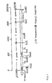
- Fig. 1 is a schematic illustration of the gene sequence of the plasmid pM13Fv.
- the X gene corresponds to the part located between Hin d III and Sal I
- the Y gene corresponds to the part located between Sal I and Sal I
- the Z gene corresponds to the part located between Sal I and Eco RI.
- the gene sequence located between Hin dIII and Eco RI sites, which corresponds to X-Y-Z, is described in the SEQ ID NO: 16 in the sequence listing.
- the X-Y-Z genes has been inserted between the Hin dIII and Eco RI sites of a plasmid pTZ19R (manufactured by Pharmacia) which is a common phagemid vector.
- the X gene existing in the downstream of the promoter originating in the vector comprises an SD sequence (Shine-Dalgarno sequence) followed by the first open reading frame by which a secretory signal peptide and the heavy chain variable region (hereinafter referred to simply as V H ) are coded for.
- V L secretory signal peptide and the light chain variable region
- a restriction enzyme Sal I site (base no. 891 to 896 in the SEQ ID NO: 16) is located at the 3'-terminal part of the V L gene.
- the Y gene containing a gene (SEQ ID NO: 17) which codes for a partial sequence ( ⁇ cpIII polypeptide) of a linear phage coat protein III exists.
- the second open reading frame codes for a fused polypeptide comprising the secretory signal polypeptide, V L and the ⁇ cpIII polypeptide.
- the Y gene is ligated to the Z gene at the restriction enzyme Sal I site which is located in the further downstream of the ⁇ cpIII polypeptide gene coded for by the Y gene.
- the Z gene is a gene containing a gene (SEQ ID NO: 18) which codes for a polypeptide containing two Fc binding domain-like structures of protein A.
- the polypeptide containing two Fc binding domain-like structures of protein A which is coded for by the Z gene, is ligated in such a manner as to follow the second open reading frame of the X gene.
- the restriction enzyme Sal I namely, the second open reading frame coded for by the residual gene codes for a fused polypeptide composed of the secretory signal peptide, V L and the polypeptide containing two Fc binding domain-like structures of protein A.
- an Fv gene originating in a monoclonal antibody against hen egg lysozyme (hereinafter referred to simply as HEL) has been inserted.
- HEL hen egg lysozyme
- the plasmid pFv-PP is cleaved at the Pst I site located in the base no. 115 to 120 in the DNA sequence represented by the SEQ ID NO: 36 and at the Sma I site located in the base no. 467 to 472 therein to thereby eliminate the V H gene having been inserted thereinto.
- V H gene which has been prepared in such a manner as to suit the open reading frame may be inserted thereinto.
- a Pst I- Sma I fragment suiting the open reading frame may be a DNA synthesized by using a DNA synthesizer.
- a DNA fragment obtained by digesting a DNA fragment, which has been prepared by the PCR method with the use of a primer for a V H gene amplification synthesized by a conventional method in such a manner as to contain the Pst I site or the Sma I site, with Pst I or Sma I may be used therefor.
- the V L gene can be replaced by a DNA fragment coding for a V L gene between the Sac I site located in the base no. 583 to 588 in the DNA sequence represented by the SEQ ID NO: 36 in the sequence listing and the Sal I site located in the base no. 891 to 896 therein.
- the plasmid pFv-PP has been converted into a form capable of expressing a fused polypeptide of a polypeptide containing an antibody variable domain with another polypeptide having two Fc binding domain-like structures of protein A.
- This fused polypeptide of a polypeptide containing the antibody variable domain with another polypeptide having an Fc affinity is a highly useful one.
- the first reason therefor resides in that it can be easily purified.
- the fused polypeptide can be recovered from the culture by using a resin onto which a polypeptide or a protein containing Fc has been immobilized.
- the second reason therefor resides in that a stable multivalent antigen complex can be formed by mixing this fused polypeptide with a polypeptide or a protein containing Fc, for example, human IgG. Because of being multivalent, this complex has a higher binding stability than that of a monovalent one and is useful in, for example, ELISA, Western blotting, etc.
- the fused polypeptide is useful from the viewpoint that it can be easily detected depending on the properties of the Fc-containing polypeptide to be mixed therewith.
- Effective examples of the polypeptide containing the antibody variable site to be used for the formation of such a fused polypeptide include an Fv fragment, an scFv fragment and an Fab fragment.
- the DNA represented by the SEQ ID NO: 3 is amplified by the PCR method with the use of a primer represented by the SEQ ID NO: 19 for the Pst I site introduction and another primer represented by the SEQ ID NO: 20 for the Sma I site introduction.
- a DNA coding for V H to be inserted into the plasmid pFvPP (hereinafter referred to simply as the V H insert) is prepared.
- the DNA represented by the SEQ ID NO: 4 is amplified by the PCR method with the use of a primer represented by the SEQ ID NO: 21 for the Sac I site introduction and another primer represented by the SEQ ID NO: 22 for the Sal I site introduction.
- V L insert a DNA coding for V L to be inserted into the plasmid pFv-PP (hereinafter referred to simply as the V L insert) is prepared.
- the plasmid pFv-PP is cleaved with Pst I and Sma I and the V H insert is inserted into the plasmid part.
- the plasmid having the V H insert is cleaved with Sac I and Sal I and the V L insert is inserted into the plasmid part.
- This plasmid is named T19C179FvproA while an Escherichia coli BMH71-18 strain transformed by this plasmid is named Escherichia coli BMH71-18/T19C179FvproA.
- the V H and V L Of the present invention can be expressed as a fused polypeptide of Fv with a polypeptide having two Fc binding domain-like structures of protein A.
- This fused polypeptide can be easily purified by using an Fc-binding carrier, for example, IgG-Sepharose 6FF (manufactured by Pharmacia) and is usable as an artificial antibody for detecting influenza A type virus.
- V H and V L of the present invention can be expressed in the form of scFv.
- the DNA represented by the SEQ ID NO: 3 is amplified by the PCR method with the use of the above-mentioned primer represented by the SEQ ID NO: 19 and another primer represented by the SEQ ID NO: 23 for introducing a linker.
- the oligonucleotides represented by the SEQ ID NO: 24 and 25 are annealed and then cleaved with Pst I and Sac I to thereby give a linker for the preparation of scFv.
- the above-mentioned plasmid T19C179FvproA is cleaved with Pst I and Sac I and the linker is inserted into the plasmid part.
- this plasmid is cleaved with Bam HI and Pst I and the above-mentioned PCR-amplified DNA is inserted thereinto.
- a plasmid having a DNA, wherein the DNA coding for V H is ligated to the DNA coding for V L with a linker, inserted thereinto can be constructed.
- This plasmid is named T19C179ScFVproA while an Escherichia coli BMH71-18 strain transformed by this plasmid is named Escherichia coli BMH71-18/T19C179ScFVproA
- This strain was deposited on February 28,1994 at National Institute of Bioscience and Human-Technology Agency of Industrial Science and Technology (1-3, Higashi 1 chome Tsukuba-shi Ibaraki-ken 305,Japan) in accordance with the Budapest Treaty under the accession number FERM BP-4969.
- the V H and V L of the present invention can be expressed as a fused polypeptide of scFv with a polypeptide having an two Fc binding domain-like structures of protein A.
- This fused polypeptide can be also easily purified by using IgG-Sepharose 6FF and is usable as an artificial antibody for detecting influenza A type virus.
- a stop codon can be prepared in the next position of the DNA coding for V L of each of the plasmids T19C179FvproA and T19C179ScFVproA by the site-specific mutation method.
- the modified plasmid thus formed is usable as a plasmid for the Fv expression or the scFV expression with the use of the DNA coding for V H and V L of the present invention.
- the present invention provides a gene coding for V H of a gene coding for anti-human influenza A type virus antibody, a gene coding for V L of the same and a gene coding for CDR of the same.
- genes are useful in the preparation of Fv, scFv, Fab, artificial antibodies containing the same, chimeric antibodies, humanized antibodies and single CDR polypeptides and the proteins and polypeptides thus obtained are useful in the diagnosis and treatment of human influenza A type virus.
- genes coding for proteins and polypeptides which are similar in properties to the V H , V L and CDR of the present invention, can be obtained, though the obtained genes are somewhat different in sequence from the genes of the present invention.
- the term "strict conditions" as used herein has the following meaning.
- a nylon membrane having a DNA immobilized thereon is hybridized with a probe at 65°C for 20 hours in a solution containing 6 x SSC (1 x SSC corresponding to 8.76 g of sodium chloride and 4.41 g of sodium citrate dissolved in 11 of water), 1% of sodium lauryl sulfate, 100 ⁇ g/ml of salmon sperm DNA and 5 x Danhardt's (containing 0.1% portions of bovine serum albumin, polyvinylpyrrolidone and Ficoll).
- An antibody prepared by using the genes of the present invention recognizes the H1N1 and H2N2 subtypes in common but not the H3N2 subtype. Therefore human influenza A type virus in a specimen can be readily typed at a high sensitivity by using the antibody prepared with the use of the genes of the present invention in a combination with an antibody capable of recognizing the H3N2 subtype [for example, a monoclonal antibody AI3C (developed by Takara Shuzo Co., Ltd.; development name: F49) produced by a hybridoma AI3C described in Japanese Patent Application No. 115216/1993] and a polyclonal antibody (manufactured by Takara Shuzo Co., Ltd.) for detecting human influenza B type virus.
- a monoclonal antibody AI3C developed by Takara Shuzo Co., Ltd.; development name: F49
- a polyclonal antibody manufactured by Takara Shuzo Co., Ltd.
- a hybridoma AI3C was deposited on November 11, 1992 at National Institute of Bioscience and Human-Technology Agency of Industrial Science and Technology (1-3, Higashi 1 chome Tsukuba-shi Ibaraki-ken 305, Japan) in accordance with the Budapest Treaty under the accession number FERM BP-4516. Furthermore, the antibody prepared by using the genes of the present invention recognizes the H1N1 and H2N2 subtypes in common and has a neutralization activity. Thus it is useful in the prevention and treatment of diseases in which viruses of these subtypes participate.
- Virus strains of the H1N1 subtype used included A/Bangkok/10/83, A/Yamagata/120/86, A/Osaka/930/88, A/Suita/1/8 (each being a stock of the Research Institute for Microbial Diseases, Osaka University), A/PR/8/34(ATCC VR-95)and A1/FM/1/47 (ATCC VR-97)were used.
- Virus strains of the H2N2 subtype used included A/Okuda/57, A/Adachi/2/57, A/Kumamoto/1/65, A/Kaizuka/2/65 and A/Izumi/5/65(each being a stock of the Research Institute for Microbial Diseases, Osaka University)were used.
- Virus strains of the H3N2 subtype used included A/Fukuoka/C29/85, A/Sichuan/2/87, A/Ibaraki/1/90, A/Suita/1/90, A/Kitakyushu/159/93(each being a stock of the Research Institute for Microbial Diseases, Osaka University) and A2/Aichi/2/68(ATCC VR-547) were used.
- a strain of influenza B type virus used was B/Nagasaki/1/87. Each strain was inoculated into the allantoic cavity of an embryonated hen egg aged 11 days, incubated at 34°C for 4 days and then harvested.
- C179 inhibited the polykaryon formation by all of the H1N1 and H2N2 subtype strains but did not inhibit the formation by the H3N2 subtype strain and the influenza B type virus strain.
- C179 is an antibody which specifically recognizes the H1N1 and H2N2 subtypes, inhibits membrane fusion of viruses and exhibits a neutralization activity.
- Table 1 summarizes these results.
- Antibody titers of C179 measured by Virus Staining activity Neutralization activity HI activity Fusion inhibition activity H1N1 A/PR/8/34 1,638,400 512 ⁇ 32 + A/Bangkok/10/83 1,638,400 512 ⁇ 32 + A/Yamagata/120/86 409,600 1,024 ⁇ 32 + A/Osaka/930/88 409,600 512 ⁇ 32 + A/Suita/1/89 409,600 1,024 ⁇ 32 + A1/FM/1/47 409,600 512 ⁇ 32 + H2N2 A/Okuda/57 1,638,400 1,024 ⁇ 32 + A/Adachi/2/57 1,638,400 1,024 ⁇ 32 + A/Kumamoto/1/65 409,600 1,024
- each number represents the dilution ratio of the ascites fluid of the Referential example A-2-(3), a staining titer is expressed in the maximum dilution ratio of the ascites fluid whereby cells can be stained in the staining test, while a neutralization activity is expressed in the maximum dilution ratio of the ascites fluid whereby the formation of focus can be suppressed up to a level corresponding to one half of the focus count in the control lot wherein no antibody is added.
- Symbol + means that polykaryon formation is completely inhibited by a 1000-fold dilution of the ascites fluid, while symbol - means that polykaryon formation is not inhibited even by using a 10-fold dilution of the ascites fluid.
- a 32-fold dilution of the ascites fluid shows no HI activity.
- mice In order to examine the preventive effect of C179, an influenza A type virus infection test was carried out by using mice. 1 ml/animal of a C179 solution (1 mg/ml in PBS) was intraperitoneally administered to 10 Balb/c mice. After 1 day, 25 ⁇ l of a 1000-fold dilution of A1/FM/1/47 (4000 HAU) of the H1N1 subtype was intranasally administered. As a control, 12 mice were inoculated with the virus alone.
- Fig. 2 shows, 8 mice in the control group died (two mice after 5 days, five after 6 days and one after 8 days). Other surviving mice in this group were extremely weakened. In contrast, the mice administered with C179 showed no abnormality and all remained healthy even after 14 days.
- Fig. 2 is a graph showing the survival ratios of the C179-administered group and the control group wherein the ordinate indicates the survival ratio while the abscissa indicates the time (days) after the infection with the virus.
- a plasmid pSW1V H D1.3V K D1.3 coding for an Fv fragment originating in a monoclonal antibody D1.3 against HEL was provided by Dr. Gregg Winter (Cambridge, UK), though it somewhat differed in the sequence from the one reported in references.
- pSW1V H D1.3V K D1.3 a gene coding for the Fv fragment had been inserted between Hin dIII and Eco RI of pUC19. The sequence of this Hin dIII- Eco RI insert is described in the SEQ ID NO: 26.
- the plasmid pSW1V H D1.3V K D1.3 was digested with restriction enzymes Hin dIII and Sma I and separated by agarose gel electrophoresis. Thus a DNA fragment of about 470 bp containing the V H gene was extracted and purified. This DNA fragment was inserted between the Hin dIII and Sma I sites of a plasmid pTZ19R (manufactured by Pharmacia) to thereby give a plasmid T19VH.
- the plasmid pSW1V H D1.3V K D1.3 was digested with restriction enzymes Eco RI and Sma I and separated by agarose gel electrophoresis.
- a DNA fragment of about 450 bp containing the V L gene was extracted and purified.
- This DNA fragment was inserted between the Eco RI and Sma I sites of a plasmid pTZ18R (manufactured by Pharmacia) to thereby give a plasmid T18VL.
- PCR was effected with the use of a primer VL3' Sal I having the restriction enzyme Eco RI recognition sequence represented by the SEQ ID NO: 27 and an Sal I recognition sequence and a primer UPMCS corresponding to a sequence originating in pTZ18R represented by the SEQ ID NO: 28.
- the DNA thus amplified was digested with restriction enzymes Hin dIII and Eco RI. After separating by agarose gel electrophoresis, a Hin dIII, Eco RI fragment was extracted and purified.
- This DNA fragment was inserted between the Hin dIII and Eco RI sites of the plasmid pTZ18R to thereby give a plasmid T18VLS.
- This plasmid T18VLS has a restriction enzyme Sal I recognition sequence in the 3'-terminal side of the V1 gene.
- This plasmid T18VLS was digested with restriction enzymes Eco RI and Sma I and separated by agarose gel electrophoresis. Thus a DNA fragment of about 430 bp containing the V L gene was extracted and purified. This DNA fragment was inserted between the Eco RI and Sma I sites of the plasmid T19VH having the V H gene which had been previously constructed. Thus a plasmid T19VHVLS was obtained.
- This plasmid T19VHVLS has the V H and V L genes in the downstream of the pTZ19Rlac promoter.
- DNA amplification was effected by the PCR method with the use of a primer ProA5' having a restriction enzyme Sal I recognition sequence represented by the SEQ ID NO: 29 and another primer ProA3' having a restriction enzyme Eco RI recognition sequence represented by the SEQ ID NO: 30.
- the DNA thus amplified which contained a gene (SEQ ID NO: 18) coding for a polypeptide containing two Fc binding domain-like structures of protein A, was digested with restriction enzymes Sal I and Eco RI and subjected to agarose gel electrophoresis. Thus a DNA fragment of about 390 bp was extracted and purified.
- This DNA fragment was inserted between the Sal I and Eco RI sites of pTZ18R to thereby give a plasmid T18PA.
- this plasmid T18PA was digested with restriction enzymes Sal I and Eco RI and subjected to agarose gel electrophoresis.
- a DNA fragment of about 390 bp was extracted and purified.
- This DNA fragment was inserted between the Sal I and Eco RI sites of the plasmid T19VHVLS which had been previously obtained to thereby construct a plasmid pFv-PP.
- This plasmid pFv-PP codes for a fused polypeptide which contains the V H fragment and the V L fragment having two Fc binding domain-like structures of protein A (58 amino acid residues of SEQ ID NO: 31) at the C-terminus thereof.
- a DNA coding for the C-terminal polypeptide of cpIII was amplified by the PCR method with the use of a primer cpIII5'-1 having a restriction enzyme Sal I recognition sequence represented by the SEQ ID NO: 32 and another primer cpIII3' having a restriction enzyme Sal I recognition sequence and an Nhe I recognition sequence represented by the SEQ ID NO: 33.
- This DNA was digested with a restriction enzyme Sal I and subjected to agarose gel electrophoresis. Thus a DNA fragment of about 650 bp was extracted and purified.
- This DNA fragment was inserted into the Sal I site of the plasmid pFv-PP obtained in the above Referential example B (1-1).
- a plasmid pM13Fv one in which the sequence originating in the primer cpIII5'-1 had been inserted following the V L gene in such a direction as to be ligated therewith was referred to as a plasmid pM13Fv.
- This plasmid pM13Fv has been constructed in such a manner as to express a fused polypeptide wherein the V H fragment is ligated with the V L fragment having ⁇ cpIII coded for by the gene represented by the SEQ ID NO: 17 in the sequence listing at the C-terminus.
- Fig. 1 is a schematic illustration of a DNA fragment inserted between the Hind III and Eco RI sites of the plasmid pM13Fv.
- the DNA sequence thereof is represented by the SEQ ID NO: 16 in the sequence listing.
- This plasmid pM13Fv can be easily converted into the plasmid pFv-PP constructed in the above Referential example B (1-1) by digesting with a restriction enzyme Sal I and subjecting to self ligation.
- An Escherichia coli JM109 strain having this plasmid pM13Fv introduced thereinto was named Escherichia coli JM109/pM13Fv.
- Example 1 Cloning of a DNA which codes for the variable region of a mouse monoclonal antibody against human influenza A type virus
- the full RNA was prepared from C179-producing hybridomas (FERM BP-4517) in accordance with the method of Chirgwin et al., Biochemistry , 18 , 5294 (1979). Namely, about 1 x 10 7 C179-producing hybridomas were added to 20 ml of 4 M guanidine thiocyanate (manufactured by Fluka) and injection and suction were repeated 5 times by using a syringe to thereby solubilize the cells. After the completion of the solubilization, the cell extract was layered over a 5.3 M solution of cesium chloride and centrifuged to thereby precipitate RNA. The RNA precipitate was dissolved in a buffer solution, treated successively with phenol and chloroform and precipitated from ethanol. Thus RNA was recovered to thereby give the full RNA.
- C179-producing hybridomas (FERM BP-4517) in accordance with the method of Chirgwin et al., Biochemistry , 18 , 52
- a single-stranded cDNA was synthesized with the use of about 5 ⁇ g of the full RNA prepared above and 0.5 ⁇ g of an oligo d(T) primer.
- the obtained cDNA was used in the amplification of a gene coding for mouse V H by the PCR method.
- the synthesis of the cDNA was effected at 42°C for 120 minutes and, after the completion of the reaction, 2 ⁇ l of RNaseA (400 ⁇ g/ml) was added thereto and reacted at 37°C for 20 minutes.
- the PCR method was carried out with the use of GeneAmpTM PCR System 9600 (manufactured by Perkin-Elmer).
- the primers used in the PCR method were an oligonucleotide primer represented by the SEQ ID NO: 11 (being hybridizable with the mouse heavy chain leader sequence) and another oligonucleotide primer represented by the SEQ ID NO: 12 (being hybridizable with the mouse heavy chain constant ration).
- a PCR solution containing 10 ⁇ l of a PCR buffer solution comprising 10 mM of Tris-HCl (pH 8.3), 50 mM of KCl, 0.1 mM of dATP, 0.1 mM of dGTP, 0.1 mM of dCTP, 0.1 mM of dTTP and 1.5 mM of MgCl 2 , 1 ⁇ l of 2.5 U DNA polymerase AmpliTaq (manufactured by Perkin-Elmer Cetus), 2.5 ⁇ l portions of 10 pmol oligonucleotide primers represented by the SEQ ID NO: 11 and 12, 1 ⁇ l of the single-stranded cDNA described in Example 1-(2) and 83 ⁇ l of H 2 O was maintained at an initial temperature of 94°C for 1 minute.
- a PCR buffer solution comprising 10 mM of Tris-HCl (pH 8.3), 50 mM of KCl, 0.1 mM of dATP, 0.1
- the DNA product thus amplified by the PCR method as described above was separated by agarose gel electrophoresis with the use of agarose (manufactured by FMC Bio Products). From an agarose piece containing a DNA fragment of about 550 bp, the target DNA was purified by using SuprecTM-O1 (a centrifuging tube provided with a filter for recovering DNA: manufactured by Takara Shuzo Co., Ltd.).
- SuprecTM-O1 a centrifuging tube provided with a filter for recovering DNA: manufactured by Takara Shuzo Co., Ltd.
- coli cell were inoculated onto an L-broth agar medium containing 50 ⁇ g/ml of ampicillin, 0.1 mM of isopropyl- ⁇ -D-thiogalactopyranoside (IPTG: manufactured by Takara Shuzo Co., Ltd.) and 40 ⁇ g/ml of 5-bromo-4-chloro-3-indolyl- ⁇ -D-galactoside. After incubating at 37°C overnight, white colonies of E . coli on the plate were selected to thereby give a transformant.
- IPTG isopropyl- ⁇ -D-thiogalactopyranoside
- This transformant was incubated in 5 ml of the L-broth medium containing 50 ⁇ g/ml of ampicillin at 37°C overnight. From this culture, a plasmid DNA was prepared in accordance with the alkali method (Molecular Cloning, A laboratory Manual, cited above). The plasmid thus obtained was named pC179H.
- the double-stranded cDNA synthesized by using the cDNA synthesis kit in the above step was separated by agarose gel electrophoresis with the use of agarose (manufactured by FMC Bio Products). This gel was subjected to autoradiography and a double-stranded cDNA of about 400 bp was purified from an agarose piece containing a signal DNA fragment of about 400 bp with the use of Suprec-O1.
- plasmid pC179L About 0.2 ⁇ g of the DNA fragment of about 400 bp, which had been obtained in the above step in accordance with the method of Example 1-(3)-(c), was lighted to the blunt end of about 0.1 ⁇ g of pGEM-4ZDNA, which had been prepared by digesting a plasmid pGEM-4ZDNA with Hin cII, and this plasmid was integrated into E . coli JM109. After incubating, a transformant and a plasmid DNA were prepared. The plasmid thus obtained was named plasmid pC179L.
- N-terminal amino acid sequences of the heavy chain and light chain of C179 are described respectively in the SEQ ID NO: 14 and 15.
- the yield of each PTH amino acid obtained by analyzing the N-terminal amino acid sequence of the heavy chain corresponded to about 10% of the yield of the PTH amino acid in the light chain. Thus it was estimated that the amino groups in the heavy chain N-terminal Glu were mostly blocked.
- the cDNA sequences coding regions in the above-mentioned plasmids pC179H and pC179L were determined by using a BcaBESTTM dideoxy sequencing kit (manufactured by Takara Shuzo Co., Ltd.). First, about 3 ⁇ g of each plasmid obtained above was denatured with 0.2 N NaOH and then annealed with a primer for sequencing. Then it was labeled with 32 P-dCTP in accordance with the indication of the kit.
- the labeled DNA was electrophoresed on a 6% polyacrylamide gel. Then the gel was dried and subjected to autoradiography to thereby determine the DNA sequence.
- the cDNA sequences coding regions of these plasmids are described in the SEQ ID NO: 3 and 4 respectively.
- the plasmid pC179H is a plasmid which contains a gene coding for mouse V H .
- the amino acid sequence coded for by the V H gene contained in this plasmid pC179H is described in the SEQ ID NO: 1.
- the amino acid sequence ranging from the N-terminal Asp to Ala 13 in the light chain represented by the SEQ ID NO: 15 completely agreed with the amino acid sequence coded for by the DNA sequence of the base No. 1 to 39 of the gene represented by the SEQ ID NO: 4.
- the plasmid pC179L is a plasmid which contains a gene coding for mouse V L .
- the amino acid sequence coded for by the V L gene contained in this plasmid pC179L is described in the SEQ ID NO: 2.
- each complementarity-determining region is composed of 4 framework regions which are ligated to each other with 3 hypervariable regions, i.e., CDRs.
- the amino acid sequences of the framework regions have been relatively well conserved, while the amino acid sequences of CDRs are liable to undergo mutation [Kabat, E.A., et al., Sequences of Proteins of Immunological Interest, US Department of Health and Human Services (1983)].
- amino acid sequences SEQ ID NO: 1 and 2 of the variable regions of mouse monoclonal antibody against human influenza A type virus were examined with the use of the data base of amino acid sequences of antibodies prepared by Kabat et al. as described above to thereby examine the homology.
- amino acid sequences of the CDRs were identified as those represented by the SEQ ID NO: 5 to 10.
- the SEQ ID NO: 5, 6, 7, 8, 9 and 10 show respectively the amino acid sequences of CDR1 of V H , CDR2 of V H , CDR3 of V H , CDR1 of V L , CDR2 of V L , CDR3 of V L .
- the amino acid sequences of CDRs of V H and V L have been identified and DNAs coding for these amino acids are provided by the present invention.
- the plasmid pFv-PP described in the above Referential example B may be cited.
- This plasmid has the Pst I and Sma I sites, into which the DNA coding for V H is to be integrated, and the Sac I and Sal I sites, into which the DNA coding for V L is to be integrated.
- the Pst I site was introduced into the upstream terminus of the V H gene obtained from the plasmid pC179H and the Sma I site was introduced into the downstream terminus thereof by the PCR method. Further, the Sac I site was introduced into the upstream terminus of the V L gene obtained from the plasmid pC179L and the Sal I site was introduced into the downstream terminus thereof by the PCR method.
- V H forward primer for the Pst I site introduction represented by the SEQ ID NO: 19 a V L backward primer for the Sma I site introduction represented by the SEQ ID NO: 20
- V L forward primer for the Sac I site introduction represented by the SEQ ID NO: 21 and a V L backward primer for the Sal I site introduction represented by the SEQ ID NO: 22 were designed and synthesized.
- PCR was effected in accordance with the method described in Example 1-(3)-(a). Namely, 100 ⁇ l of a PCR solution containing 1 ⁇ l of the plasmid pC179H (10 ⁇ g/, ⁇ l), 2.5 ⁇ l portions of the primers (20 pmol/ ⁇ l) represented by the SEQ ID NO: 19 and 20, 10 ⁇ l of a PCR buffer solution, 1 ⁇ l of 2.5 U DNA polymerase AmpliTaq and 83 ⁇ l of H 2 O was subjected to PCR for 25 cycles with each cycle consisting of 30 seconds at 94°C, 1 minute at 55°C and 3 minutes at 72°C to thereby amplify the V H insert to be integrated into the plasmid pFv-PP.
- PCR was effected in the same manner with the use of the plasmid pC179L and the primers represented by the SEQ ID NO: 21 and 22 to thereby amplify the V L insert to be integrated into the plasmid pFv-PP.
- Each PCR amplification product was cleaved with Pst I, Sma I and Sac I and Sal I and electrophoresed on an agarose gel. Then the gel pieces were excised and purified by Suprec-O1 to thereby prepare the V H and V L inserts.
- the plasmid pFv-PP was cleaved with Pst I and Sma I and electrophoresed on an agarose gel.
- the gel piece corresponding to the plasmid was excised and purified by Suprec-O1.
- the V H insert was excised and purified by Suprec-O1.
- the V H insert was excised and purified by Suprec-O1.
- the V H insert was propagated in an Escherichia coli BMH71-18 strain.
- the plasmid was prepared from the clone by the alkali extraction method and the plasmid thus obtained was cleaved with Sac I and Sal I.
- this plasmid was electrophoresed on an agarose gel and the gel piece corresponding to the plasmid was excised and purified by Suprec-O1. Into the purified plasmid was inserted the V L insert. Then this plasmid was propagated in an Escherichia coli BMH71-18 strain to thereby give clone. Next, a plasmid was prepared from the clone and the DNA sequences of the V H and V L inserts were analyzed, thus confirming that no error had occurred during the amplification by the PCR methods. The plasmid capable of expressing Fv of C179 was named plasmid T19C179FvproA. The E . coli BME71-18 strain transformed by this plasmid was named Escherichia coli BMH71-18/ T19C179FvproA.
- This transformant expresses the fused polypeptide of C179 with two Fc binding domain-like structures of protein A (hereinafter referred to simply as C179Fv-PP).
- C179Fv-PP The DNA sequence coding for C179Fv-PP is described in the SEQ ID NO: 34.
- the part of the base no. 106 to 465 is a sequence coding for V H
- the part of the base no. 601 to 902 is a sequence coding for V L
- the parts of the base no. 928 to 1101 and 1102 to 1275 are sequences coding respectively for the Fc binding domain-like structures of protein A.
- the C179Fv-PP thus expressed can be easily purified with IgG-Sepharose 6FF. Also, it can be converted into a plasmid, which expresses exclusively Fv of C179, by introducing a stop codon after the V L insert of the plasmid T19C179FvproA by the site-specific mutation method.
- a plasmid for scFv expression was constructed by introducing a linker between the V H insert and the V L insert in the following manner.
- a primer for the introduction of a linker represented by the SEQ ID NO: 23 was designed and synthesized.
- the DNA product thus amplified by the PCR method in the above step was recovered by precipitating from ethanol.
- the DNA precipitate was digested with 10 U of restriction enzymes Pst I and Bam HI at 37°C for 4 hours.
- the DNA fragments thus formed were separated by agarose gel electrophoresis.
- the target DNA fragment was purified by using Suprec-O1.
- Oligonucleotides for synthesizing linkers represented by the SEQ ID NO: 24 and 25 were designed and synthesized. Then 200 pmol portions of these oligonucleotides were added and reacted at 65°C for 5 minutes. Then they were slowly annealed at room temperature and were blunted by using a DNA blunting kit (manufactured by Takara Shuzo Co.,Ltd.). Next, the DNA solution was digested with 10 U of Pst I and Sac I at 37°C for 4 hours and the DNA fragments thus formed were separated by agarose gel electrophoresis. From an agarose piece containing a DNA fragment of about 60 bp, the target DNA fragment was purified by using Suprec-O1.
- the plasmid part prepared by digesting the above-mentioned plasmid T19C179FvproA with Pst I and Sac I was inserted the above-mentioned linker for scFv by using a DNA ligation kit. Further, the plasmid having the linker for scFv inserted thereinto was propagated in an Escherichia coli BMH71-18 strain. From the clone thus obtained, a plasmid was prepared by the alkali extraction method. Thus 2 ⁇ g of a plasmid having the linker for scFv inserted thereinto was prepared.
- This plasmid capable of expressing scFv of C179 was named plasmid T19C179ScFVproA.
- the Escherichia coli BMH71-18 strain transformed by this plasmid was named Escherichia coli BMH71-18/T19C179ScFVproA
- This deposit was deposited on February 28, 1994 at National Institute of Bioscience and Human-Technology Agency of Industrial Science and Technology (1-3, Higashi 1 chome Tsukuba-shi Ibaraki-ken 305, JAPAN) in accordance with the Budapest Treaty under the accesion number FERM BP-4969.
- the DNAs coding for the V H and V L genes and each CDR according to the present invention can be prepared from the above-mentioned plasmid T19C179ScFvproA.
- the transformant prepared in the above-mentioned manner was inoculated into a 2 x YT medium (Molecular Cloning, A Laboratory Manual, cited above) containing 50 ⁇ g/ml of ampicillin and 0.1% of glucose and incubated under shaking at 30°C overnight.
- the culture was then inoculated into 100 ml of a 2 x YT medium containing 50 ⁇ g/ml of ampicillin and 0.1% of glucose and incubated under shaking at 30°C.
- a spectrophotometer reached an O.D. 600 Of 1
- IPTG was added thereto in such a manner as to give a final concentration of 1 mM and the incubation was continued under shaking at 30°C for additional 20 hours. After the completion of the incubation, the cells were harvested by centrifugation.
- the cells were suspended in 5 ml of a 10 mM Tris-HCl buffer solution (pH 8.0), disrupted by ultrasonication and centrifuged to thereby give the supernatant.
- the filtrate was slowly passed through a column packed with IgG Sepharose 6FF to thereby allow the column to adsorb the scFv fragment.
- a buffer solution for binding 10 mM Tris-HCl buffer solution (pH 8.0), 140 mM NaCl, 1 mM EDTA, 0.1 mM PMSF (phenylmethylsulfonyl fluoride)]
- the target fraction was eluted with a 50 mM glycine hydrochloride buffer solution (pH 2.5).
- the eluate was quickly neutralized with a 1 M Tris-HCl buffer solution (pH 8.0).
- scFv-PP fused polypeptide of scFv of C179 with two Fc binding domain-like structures of protein A
- the DNA sequence coding for this scFv-PP is described in the SEQ ID NO: 35.
- the sequence of the base no. 106 to 471 is one coding for V H
- the sequence of the base no. 529 to 830 is one coding for V L
- the sequences of the base no. 856 to 1029 and the base no. 1030 to 1203 are sequences coding respectively for the Fc binding domain-like structures of protein A.
- it can be converted into a plasmid, which expresses exclusively scFv of C179, by introducing a stop codon after the V L insert of the plasmid T19C179FVproA by the site-specific mutation method.
- Human influenza A type virus was detected with the scFv obtained by the present invention by the ELISA method in the following manner.
- scFv-PP (10 ⁇ g/ml) having the antibody variable region of C179 expressed therein obtained in Example 4 was added in 100 ⁇ l portions into the wells of 96-well microplate (Falcon 3072; manufactured by Becton Dickinson Labware). Then the plate was maintained at 37°C for 1 hour and 30 minutes to thereby immobilize scFv-PP onto the plate followed by the blocking with the use of a protein solution for blocking (Block Ace; manufactured by Snow Brand).
- the anti-A/Okuda/57 serum was prepared in the following manner. Namely, A/Okuda/57 (5000 HA U) was suspended in Freund's complete adjuvant prior to use and intramuscularly injected into a rabbit twice at an interval of 1 month. 10 days after the second intramuscular injection, the whole blood of the animal was collected from the heart and a serum was prepared to thereby give the anti-A/Okuda/57 serum.
- the scFv obtained by the present invention shows avidities for the H1N1 and H2N2 subtypes but not for the H3N2 subtype. Accordingly, the scFv-PP obtained by the present invention is useful in the detection and typing of human influenza A type virus.
- genes which recognize the H1N1 and H2N2 subtypes of human influenza A type virus in common and code for the variable region and CDRs of a monoclonal antibody having a neutralization activity against this virus have been specified. These genes are useful in the production of various artificial antibodies and antigen-binding polypeptides through genetic engineering techniques. Further, the artificial antibodies and polypeptides thus obtained are useful in the diagnosis and treatment of human influenza.
Description
- This invention relates to a DNA which codes for the variable region of a mouse monoclonal antibody against influenza A type virus. It is useful in the production of artificial antibodies through genetic engineering techniques.
- There are three types (A, B and C) of influenza viruses and the worldwide prevalence of influenza costing a large number of deaths is caused by human influenza A type virus.
- Influenza A type virus is further classified into various subtypes depending on the antigenicities of haemagglutinin (hereinafter referred to simply as HA) and neuraminidase (hereinafter referred to simply as NA) which are viral surface proteins. There have been known so far three subtypes of human influenza A type viruses, namely, the H1N1, H2N2 and H3N2 subtypes.
- The HA of influenza A type virus comprises two structurally distinct regions, namely, a globular head region and a stem region. The globular head region contains a receptor binding site which is responsible for virus attachment to a target cell and participates in the haemagglutination activity of HA. On the other hand, the stem region contains a fusion peptide which is necessary for membrane fusion between the viral envelope and an endosomal membrane of the cell and thus relates to fusion activity [Wiley et al., Ann. Rev. Biochem., 56, 365 - 394 (1987)].
- All of anti-HA antibodies, which have been obtained hitherto as an antibody capable of recognizing the H1N1 and H2N2 subtypes, recognize the globular head region of HA. However, this region most frequently undergoes antigen mutation. Therefore, these antibodies are not common to the subtypes of human influenza A type virus and, further, lose the recognizing ability with antigenic changes in the HA of the virus.
- On the other hand, Green et al. have synthesized a polypeptide based on an amino acid sequence in the stem region of HA of the H3N2 subtype and obtained antibodies against this polypeptide. However, these antibodies have a low neutralization activity (Published Japanese Translation of PCT Patent Applications from Other Countries, No. 501714/1984). Furthermore, the polypeptide per se employed as an antigen does not react with rabbit antiviral serum obtained by immunizing with the H3N2 subtype, which suggests that there is a problem from the viewpoint of antigenicity too [Cell, 28, 477 - 487 (1982)]
- Human influenza A type virus periodically changes types of HA and NA and thus causes wide prevalence. It is often observed that vaccinization before winter, i.e, the season of prevalence, produces no effect, since the prevalence is caused by a virus of a different type. However, if an antigen site which is common to virus subtypes in HA and NA molecules and hardly undergoes antigen mutation, especially an antibody which recognizes the stereostructure and has a neutralization activity can be acquired, this antibody is usable in the diagnosis, prevention and treatment of infection with influenza A type virus.
- This present inventors obtained the anti-human influenza A type virus antibody which is capable of cross-recognizing the subtypes of influenza A type virus and has a virus neutralization activity and has the characteristics (a) and (b);
- (a) recognizing the stem region of haemaggulutinin molecule of the H1N1 and H2N2 subtypes of human influenza A type virus but not recognizing the stem region of haemaggulutinin molecule of the H3N2 subtype thereof; and
- (b) having a neutralization activity for the H1N1 and H2N2 subtypes of human influenza A type virus but no neutralization activity for the H3N2 subtype thereof.
-
- One example of this anti-human influenza A type virus antibody is a monoclonal antibody C179 (hereinafter referred to simply as C179) obtained from Hybridoma C179 (FERM BP-4517). Also the preventive effect of C179 on influenza A type virus has been confirmed (Japanese Patent Application No. 272538/1992).
- However this monoclonal antibody which is capable of cross-recognizing the subtypes of influenza A type virus and has a virus neutralization activity has been derived from mouse hybridomas. Therefore the immunogenicity of this mouse antibody imposes various restrictions on the application thereof to clinical medicine as a drug for human being. That is to say, the use of a mouse antibody frequently induces an immune response and causes an anaphylactic shock. Further, repeated administration of the mouse antibody lowers the potency of the effect of the antibody or extremely quickens its clearance from the blood. Therefore such a mouse antibody substantially ranks low in the therapeutics. Although these difficulties encountering in the use of a mouse antibody can be overcome by using a human monoclonal antibody clinically, it is highly difficult to steadily produce a human monoclonal antibody corresponding to, for example, C179 while sustaining the desired specificity and affinity thereof.
- It is also known that a cell line producing a monoclonal antibody would generally suffer from a decrease in the antibody productivity with repetitive subculturing. In order to solve this problem, it is needed to examine the possibility of mass expression through gene transduction following the cell cloning of antibody-producing cells or the cloning of an antibody gene.
- From these viewpoints, it is considered that when a chimeric antibody wherein the variable region of an antibody derived from a species is bound to the constant region of another antibody derived from a different species (in usual, a chimeric antibody is composed of the variable region of a mouse antibody bound to the constant region of a human antibody) is constructed through recombinant DNA techniques, then an antibody substantially having a low immunogenicity and being suitable for clinical uses compared with the mouse antibody can be obtained, since the major part of this chimeric antibody originates in human being. Chimeric antibodies have been reported by Shaw et al., J. Immun., 138, 4534 (1987), Morrison, S., et al., Pro. Nat. Acad. Sci. USA, 81, 6851 - 6855 (1984), etc. and regarded as a general idea. In recent years, therefore, attempts have been made to develop, for example, a chimeric antibody composed of a variable region originating in mouse and a human constant region, a bispecific chimeric antibody having two antigen specificities different from each other, single-stranded antibody and an oligopeptide corresponding to the complementarity-determining region (CDR) having an antibody activity. In the production of these antibodies through genetic engineering techniques and the development of improved antibodies, it is essentially required to isolate a certain gene and to clarify its structure including the amino acid sequence thereof. It has been well known in the art that the specificity of an antibody is restricted to a particular region, i.e., CDR in the variable region thereof. Thus there have been widely effected amino acid replacements in CDR in order to, for example, improve the affinity of an antibody. Accordingly, it is particularly important to clarify the DNA sequence and amino acid sequence of CDR.
- It is an object of the present invention to provide a DNA coding for the variable region of a mouse monoclonal antibody which is useful in the production of a chimeric antibody being hardly recognized as a foreign substance in a human body or an artificial antibody having the complementarity-determining region transplanted into a human antibody exclusively, and a DNA coding for the complementarity-determining region of the above-mentioned variable region.
- The first aspect of the present invention relates to a DNA coding for the variable region of the anti-human influenza A type virus antibody having the following characteristics (a) and (b);
- (a) recognising the stem region of haemagglutinin molecule of the H1N1 and H2N2 subtypes of human influenza A type virus but not recognising the stem region of haemagglutinin molecule of the H3N2 subtype thereof; and
- (b) having a neutralization activity for the H1N1 andH2N2 subtypes of human influenza A type virus but no neutralization activity for the H3N2 subtype thereof, the DNA having DNA sequence coding for the amino acid sequences represented by the SEQ ID No: 1 in the sequence listing and a DNA sequence coding for the amino acid sequences represented by SEQ ID No: 2 in the sequence listing.
-
- The second aspect of the present invention relates to a DNA coding for CDR in DNA which codes for the variable region of the anti-human influenza A type virus antibody of the first aspect of the invention.
- The term "variable region" as used herein means an amino acid sequence which contains at least the CDR in its amino acid sequence.
- In order to clone the DNA coding for the variable region of the anti-human influenza A type virus antibody and having the characteristics (a) and (b) as described above, it is necessary to prepare hybridomas capable of producing the above-mentioned antibody as a gene source. As such hybridomas, Japanese Patent Application No. 272538/1992 has disclosed mouse hybridomas producing C179 (Hybridoma C179: FERM BP-4517) and the properties of this antibody. A method for the preparation of these hybridomas and the properties of C179 will be described in Referential Example A hereinafter.
- To clone the target DNA which codes for the variable region of the mouse monoclonal antibody, the hybridomas are disrupted to thereby prepare the full RNA. By using the full RNA, a single-stranded cDNA is prepared in accordance with the Gubler-Hoffman method.
- Next, the significant part of the above-mentioned cDNA is specifically amplified by the DNA extension method with the use of oligonucleotide primers. For the amplification of the heavy chain variable region of the mouse monoclonal antibody, oligonucleotide primers represented by the SEQ ID NO: 11 and 12 in the sequence listing are used respectively as a 5'-terminal primer and a 3'-terminal primer for PCR.
- The oligonucleotide primer represented by the SEQ ID NO: 11 in the sequence listing is hybridized with the heavy chain leader sequence, while the oligonucleotide primer represented by the SEQ ID NO: 12 in the sequence listing is hybridized with the heavy chain constant region. For the amplification with the use of the light chain variable region primer of the mouse monoclonal antibody, the oligonucleotide primer represented by the SEQ ID NO: 13 in the sequence listing is used as a 3'-terminal primer. This primer is hybridized with the light chain constant region of the mouse monoclonal antibody.
- On the other hand, C179 is highly purified and the N-terminal amino acid sequences of its heavy chain and light chain are determined. The N-terminal amino acid sequence of the heavy chain of the monoclonal antibody and the N-terminal amino acid sequence of the light chain thereof are represented by the SEQ ID NO: 14 and 15 in the sequence listing respectively.
- The product of the above-mentioned amplification is integrated into an appropriate vector such as pT7 blue T vector (manufactured by Novagen) or pGEM-4ZDNA (manufactured by Promega) to thereby give a plasmid containing a DNA fragment which codes for the target variable region of the monoclonal antibody.
- The sequence of the cloned DNA is determined and the sequences represented by the SEQ ID NO: 3 and 4 in the sequence listing are compared with the DNA sequences anticipated from those represented by the SEQ ID NO: 14 and 15 in the sequence listing. As a result, a DNA which is an example of the first aspect of the present invention and amino acid sequences coded for by this DNA are provided. These sequences are represented by the SEQ ID NO: 1 and 2 in the sequence listing. Cloning of the target DNA and the determination of its sequence will be described in greater detail in Examples 1 and 2 hereinafter.
- The present invention further provides a DNA which codes for the CDR of each variable region.
- The variable region composed of the heavy chain and the light chain forms an antigen binding domain. This region located over the heavy and light chains contains 4 framework regions which have been relatively well conserved. These regions are ligated with 3 CDRs. These CDRs are determined through a comparison with known amino acid sequences in the variable region. The amino acid sequences of the CDRs provided by the present invention are described in the SEQ ID NO: 5 to 10 in the sequence listing.
- The present invention provides a variable region and a CDR which are useful as a material for producing chimeric antibodies or humanized antibodies and DNAs coding for the same.
- By using the DNAs of the present invention coding for the heavy chain variable region and the light chain variable region, Fab, Fv, scFv, etc. can be produced through genetic engineering techniques. scFv is a single-stranded polypeptide constructed by binding the heavy chain variable region to the light chain variable region via a spacer consisting of several amino acid residues.
- An example of the plasmid to be used in the construction of Fv is a plasmid pM13Fv described in Japanese Patent Application No. 246184/1993. An Escherichia coli JM109 strain having this plasmid introduced thereinto was named Escherichia coli JMlO9/pM13Fv. This strain was deposited on June 22,1993 at National Institute of Bioscience and Human-Technology Agency of Industrial Science and Technology in accordance with tha Budapest Treaty under the accession number FERM BP-4968.
- Fig. 1 is a schematic illustration of the gene sequence of the plasmid pM13Fv.
- In Fig. 1, the X gene corresponds to the part located between Hind III and SalI, the Y gene corresponds to the part located between SalI and SalI and the Z gene corresponds to the part located between SalI and EcoRI. The gene sequence located between HindIII and EcoRI sites, which corresponds to X-Y-Z, is described in the SEQ ID NO: 16 in the sequence listing.
- In the plasmid pM13Fv, the X-Y-Z genes has been inserted between the HindIII and EcoRI sites of a plasmid pTZ19R (manufactured by Pharmacia) which is a common phagemid vector. The X gene existing in the downstream of the promoter originating in the vector comprises an SD sequence (Shine-Dalgarno sequence) followed by the first open reading frame by which a secretory signal peptide and the heavy chain variable region (hereinafter referred to simply as VH ) are coded for.
- In the downstream thereof, it has another SD sequence followed by the second open reading frame by which a secretory signal peptide and the light chain variable region (hereinafter referred to simply as VL ) are coded for. A restriction enzyme SalI site (base no. 891 to 896 in the SEQ ID NO: 16) is located at the 3'-terminal part of the VL gene. Following the open reading frame of the VL gene, the Y gene containing a gene (SEQ ID NO: 17) which codes for a partial sequence (ΔcpIII polypeptide) of a linear phage coat protein III exists.
- That is to say, the second open reading frame codes for a fused polypeptide comprising the secretory signal polypeptide, VL and the ΔcpIII polypeptide. The Y gene is ligated to the Z gene at the restriction enzyme SalI site which is located in the further downstream of the ΔcpIII polypeptide gene coded for by the Y gene. The Z gene is a gene containing a gene (SEQ ID NO: 18) which codes for a polypeptide containing two Fc binding domain-like structures of protein A.
- When this pM13Fv having the above-mentioned structure is completely digested with the restriction enzyme SalI, the Y gene is excised therefrom. After eliminating the Y gene, the plasmid is subjected to self ligation. Thus the X gene is ligated to the Z gene at the SalI site to thereby give a plasmid pFV-PP.
- Then the polypeptide containing two Fc binding domain-like structures of protein A, which is coded for by the Z gene, is ligated in such a manner as to follow the second open reading frame of the X gene. After eliminating the Y gene by digesting the plasmid pM13Fv with the restriction enzyme SalI, namely, the second open reading frame coded for by the residual gene codes for a fused polypeptide composed of the secretory signal peptide, VL and the polypeptide containing two Fc binding domain-like structures of protein A.
- Into the part corresponding to the X gene in this plasmid pFv-PP, an Fv gene originating in a monoclonal antibody against hen egg lysozyme (hereinafter referred to simply as HEL) has been inserted. In practice, there exist restriction enzyme sites in order to facilitate the replacement of the Fv gene. That is to say, the plasmid pFv-PP is cleaved at the PstI site located in the base no. 115 to 120 in the DNA sequence represented by the SEQ ID NO: 36 and at the SmaI site located in the base no. 467 to 472 therein to thereby eliminate the VH gene having been inserted thereinto. Then an arbitrary VH gene which has been prepared in such a manner as to suit the open reading frame may be inserted thereinto. Such a PstI-SmaI fragment suiting the open reading frame may be a DNA synthesized by using a DNA synthesizer. Alternatively, a DNA fragment obtained by digesting a DNA fragment, which has been prepared by the PCR method with the use of a primer for a VH gene amplification synthesized by a conventional method in such a manner as to contain the PstI site or the SmaI site, with PstI or SmaI may be used therefor. Similarly, the VL gene can be replaced by a DNA fragment coding for a VL gene between the SacI site located in the base no. 583 to 588 in the DNA sequence represented by the SEQ ID NO: 36 in the sequence listing and the SalI site located in the base no. 891 to 896 therein.
- The plasmid pFv-PP has been converted into a form capable of expressing a fused polypeptide of a polypeptide containing an antibody variable domain with another polypeptide having two Fc binding domain-like structures of protein A.
- This fused polypeptide of a polypeptide containing the antibody variable domain with another polypeptide having an Fc affinity is a highly useful one. The first reason therefor resides in that it can be easily purified. For example, the fused polypeptide can be recovered from the culture by using a resin onto which a polypeptide or a protein containing Fc has been immobilized. The second reason therefor resides in that a stable multivalent antigen complex can be formed by mixing this fused polypeptide with a polypeptide or a protein containing Fc, for example, human IgG. Because of being multivalent, this complex has a higher binding stability than that of a monovalent one and is useful in, for example, ELISA, Western blotting, etc. Also, the fused polypeptide is useful from the viewpoint that it can be easily detected depending on the properties of the Fc-containing polypeptide to be mixed therewith. Effective examples of the polypeptide containing the antibody variable site to be used for the formation of such a fused polypeptide include an Fv fragment, an scFv fragment and an Fab fragment.
- For example, the DNA represented by the SEQ ID NO: 3 is amplified by the PCR method with the use of a primer represented by the SEQ ID NO: 19 for the PstI site introduction and another primer represented by the SEQ ID NO: 20 for the SmaI site introduction. Thus a DNA coding for VH to be inserted into the plasmid pFvPP (hereinafter referred to simply as the VH insert) is prepared. On the other hand, the DNA represented by the SEQ ID NO: 4 is amplified by the PCR method with the use of a primer represented by the SEQ ID NO: 21 for the SacI site introduction and another primer represented by the SEQ ID NO: 22 for the SalI site introduction. Thus a DNA coding for VL to be inserted into the plasmid pFv-PP (hereinafter referred to simply as the VL insert) is prepared. Next, the plasmid pFv-PP is cleaved with PstI and SmaI and the VH insert is inserted into the plasmid part.
- Subsequently, the plasmid having the VH insert is cleaved with SacI and SalI and the VL insert is inserted into the plasmid part. Thus the plasmid having the DNA coding for VH and VL of the present invention inserted thereinto can be constructed. This plasmid is named T19C179FvproA while an Escherichia coli BMH71-18 strain transformed by this plasmid is named Escherichia coli BMH71-18/T19C179FvproA. By incubating this E. coli strain, the VH and VL Of the present invention can be expressed as a fused polypeptide of Fv with a polypeptide having two Fc binding domain-like structures of protein A. This fused polypeptide can be easily purified by using an Fc-binding carrier, for example, IgG-Sepharose 6FF (manufactured by Pharmacia) and is usable as an artificial antibody for detecting influenza A type virus.
- Also, the VH and VL of the present invention can be expressed in the form of scFv.
- For example, the DNA represented by the SEQ ID NO: 3 is amplified by the PCR method with the use of the above-mentioned primer represented by the SEQ ID NO: 19 and another primer represented by the SEQ ID NO: 23 for introducing a linker. Next, the oligonucleotides represented by the SEQ ID NO: 24 and 25 are annealed and then cleaved with PstI and SacI to thereby give a linker for the preparation of scFv. The above-mentioned plasmid T19C179FvproA is cleaved with PstI and SacI and the linker is inserted into the plasmid part. Then this plasmid is cleaved with BamHI and PstI and the above-mentioned PCR-amplified DNA is inserted thereinto. Thus a plasmid having a DNA, wherein the DNA coding for VH is ligated to the DNA coding for VL with a linker, inserted thereinto can be constructed. This plasmid is named T19C179ScFVproA while an Escherichia coli BMH71-18 strain transformed by this plasmid is named Escherichia coli BMH71-18/T19C179ScFVproA This strain was deposited on February 28,1994 at National Institute of Bioscience and Human-Technology Agency of Industrial Science and Technology (1-3, Higashi 1 chome Tsukuba-shi Ibaraki-ken 305,Japan) in accordance with the Budapest Treaty under the accession number FERM BP-4969.
- By incubating this E. coli strain, the VH and VL of the present invention can be expressed as a fused polypeptide of scFv with a polypeptide having an two Fc binding domain-like structures of protein A. This fused polypeptide can be also easily purified by using IgG-Sepharose 6FF and is usable as an artificial antibody for detecting influenza A type virus. Further, a stop codon can be prepared in the next position of the DNA coding for VL of each of the plasmids T19C179FvproA and T19C179ScFVproA by the site-specific mutation method. The modified plasmid thus formed is usable as a plasmid for the Fv expression or the scFV expression with the use of the DNA coding for VH and VL of the present invention.
- As discussed above in detail, the present invention provides a gene coding for VH of a gene coding for anti-human influenza A type virus antibody, a gene coding for VL of the same and a gene coding for CDR of the same. These genes are useful in the preparation of Fv, scFv, Fab, artificial antibodies containing the same, chimeric antibodies, humanized antibodies and single CDR polypeptides and the proteins and polypeptides thus obtained are useful in the diagnosis and treatment of human influenza A type virus.
- By effecting hybridization under strict conditions with the use of the genes of the present invention thus obtained as a probe, genes coding for proteins and polypeptides, which are similar in properties to the VH, VL and CDR of the present invention, can be obtained, though the obtained genes are somewhat different in sequence from the genes of the present invention. The term "strict conditions" as used herein has the following meaning. Namely, a nylon membrane having a DNA immobilized thereon is hybridized with a probe at 65°C for 20 hours in a solution containing 6 x SSC (1 x SSC corresponding to 8.76 g of sodium chloride and 4.41 g of sodium citrate dissolved in 11 of water), 1% of sodium lauryl sulfate, 100 µg/ml of salmon sperm DNA and 5 x Danhardt's (containing 0.1% portions of bovine serum albumin, polyvinylpyrrolidone and Ficoll).
- An antibody prepared by using the genes of the present invention recognizes the H1N1 and H2N2 subtypes in common but not the H3N2 subtype. Therefore human influenza A type virus in a specimen can be readily typed at a high sensitivity by using the antibody prepared with the use of the genes of the present invention in a combination with an antibody capable of recognizing the H3N2 subtype [for example, a monoclonal antibody AI3C (developed by Takara Shuzo Co., Ltd.; development name: F49) produced by a hybridoma AI3C described in Japanese Patent Application No. 115216/1993] and a polyclonal antibody (manufactured by Takara Shuzo Co., Ltd.) for detecting human influenza B type virus. A hybridoma AI3C was deposited on November 11, 1992 at National Institute of Bioscience and Human-Technology Agency of Industrial Science and Technology (1-3, Higashi 1 chome Tsukuba-shi Ibaraki-ken 305, Japan) in accordance with the Budapest Treaty under the accession number FERM BP-4516. Furthermore, the antibody prepared by using the genes of the present invention recognizes the H1N1 and H2N2 subtypes in common and has a neutralization activity. Thus it is useful in the prevention and treatment of diseases in which viruses of these subtypes participate.
- To illustrate the anti-influenza A type virus antibodies and Fv and scFv expression plasmids employed in the present invention, the following Referential examples will be given.
- Virus strains of the H1N1 subtype used included A/Bangkok/10/83, A/Yamagata/120/86, A/Osaka/930/88, A/Suita/1/8 (each being a stock of the Research Institute for Microbial Diseases, Osaka University), A/PR/8/34(ATCC VR-95)and A1/FM/1/47 (ATCC VR-97)were used. Virus strains of the H2N2 subtype used included A/Okuda/57, A/Adachi/2/57, A/Kumamoto/1/65, A/Kaizuka/2/65 and A/Izumi/5/65(each being a stock of the Research Institute for Microbial Diseases, Osaka University)were used. Virus strains of the H3N2 subtype, used included A/Fukuoka/C29/85, A/Sichuan/2/87, A/Ibaraki/1/90, A/Suita/1/90, A/Kitakyushu/159/93(each being a stock of the Research Institute for Microbial Diseases, Osaka University) and A2/Aichi/2/68(ATCC VR-547) were used. A strain of influenza B type virus used was B/Nagasaki/1/87. Each strain was inoculated into the allantoic cavity of an embryonated hen egg aged 11 days, incubated at 34°C for 4 days and then harvested.
-
- (1) Balb/c mice were immunized with two doses of A/Okuda/57 strain (320 HAU) prepared in the above Referential example A-1, which had been suspended in Freund's complete adjuvant before use, via intraperitoneal injection one month apart. One month thereafter, the mice were boosted by intraperitoneally injecting a suspension of the same antigen (320 HAU) in PBS. Three days thereafter, the spleen of each animal was taken out and thus spleen cells were prepared. Mouse myeloma cells were prepared by incubating p3x63Ag8 cells in a DME medium containing 10% of fetal bovine serum for 2 days after passage and then washing with physiological saline before cell fusion. The spleen cells were mixed with the myeloma cells at a ratio by cell count of 1 : 5. After centrifuging and removing the supernatant, the precipitated cell clusters were thoroughly loosened and then added to 1 ml of a mixture [polyethylene glycol 4000 (2 g), MEM (2 ml), and dimethyl sulfoxide] under stirring. After maintaining at 37°C for 5 minutes, MEM was slowly added thereto so as to adjust the total amount to 10 ml. After the mixture was centrifuged, the supernatant was removed and the cell clusters were gently loosened. 30 ml of a normal medium (PRMI-1640 containing 10% of fetal bovine serum) was added thereto and the cells were slowly suspended with the use of a measuring pipet.The suspension was pipetted into a 96-well incubation plate and incubated in an incubator containing 5% of CO2 at 37°C for 24 hours. Then HAT medium was added thereto and the incubation was continued for 10 to 14 days. Subsequently, a part of the culture supernatant was sampled and subjected to hybridoma screening.
- (2) To obtain a monoclonal antibody undergoing a cross
reaction between influenza A type virus subtypes, the above-mentioned
culture supernatant, which had not been diluted, was
used as a primary antibody and a staining test on MDCK cells
infected with the three subtypes (H1N1, H2N2 and H3N2) was
effected. The staining test was carried out in accordance with
the above-mentioned method described in Journal of Clinical
Microbiology. Specifically, the MDCK cells infected with the
human influenza A type virus subtype strains (H1N1:
A/Yamagata/120/86, H2N2: A/Okuda/57, H3N2: A/Fukuoka/C29/85)
were rinsed with PBS (pH 7.4) on 96-well microtiter plates
(Falcon 3072; manufactured by Becton Dickinson Labware) and
fixed with absolute ethanol at room temperature for 10 minutes.
Then these cells were continuously treated with 4 antibodies
[the above-mentioned culture supernatant containing the
monoclonal antibody, rabbit anti-mouse immunoglobulin G serum
(manufactured by Organon Teknika) diluted 1000-fold, goat anti-rabbit
immunoglobulin G
serum (manufactured by Organon Teknika) diluted 500-fold, and peroxidase-rabbit anti-peroxidase complex (manufactured by Organon Teknika) diluted 1000-fold, each for 40 minutes, and the cells thus treated were washed with PBS. Finally, the peroxidase reaction was effected by the method of Graham and Karnovsky [see J.Histochem. Cytochem., 14, 291 - 302 (1966)] with the use of 0.01% H2O2 and 0.3 mg/ml of 3,3'-diaminobenzidine tetrahydrochloride in PBS. The stained cells were observed under an ordinary light microscope to sort antibodies recognizing respectively the H1N1 subtype-infected MDCK cells and the H2N2 subtype-infected MDCK cells. Next, the cells in the wells where the production of these antibodies had been confirmed were taken out and treated by the limiting dilution thrice to thereby clone the target cells. The hybridoma strain thus cloned was named Hybridoma C179, while the monoclonal antibody produced thereby was named monoclonal antibody C179. The Hybridoma C179 was deposited on January 28, 1993 at National Institute of Bioscience and Human-Technology Agency of Industrial Science and Technology (1-3 Higashi 1 chome Tsukuba-shi Ibaraki-ken 305,Japan) in accordance with the Budapest Treaty under the accesion number FERM BP-4517. - (3) 5 x 10⁶/animal of the above-mentioned hybridomas were intraperitoneally administered to Balb/c mice treated with pristane. Ten to 21 days thereafter, the ascites of a mouse having ascites cancer thus induced was sampled and centrifuged at 3000 rpm for 5 minutes to thereby remove solid components and give an ascites fluid. This fluid contained about 5 mg/ml of the monoclonal antibody C179 (hereinafter referred to simply as C179). After purifying with Protein A-Sepharose 4B (manufactured by Pharmacia), C179 was confirmed as an antibody of the IgG2a type.
-
-
- (1) A 100-fold dilution of the ascites fluid as described in the above Referential example A-2-(3) was diluted stepwise and the staining test as described in the above Referential example A-2-(2) was effected to examine the antigen recognizing characteristics of C179. The H1N1 subtype strains used included A/PR/8/34, A/Bangkok/10/83, A/Yamagata/120/86, A/Osaka/930/88, A/Suita/1/89 and A1/FM/1/47. The H2N2 subtype strains used included A/Okuda/57, A/Adachi/2/57, A/Kumamoto/1/65, A/Kaizuka/2/65 and A/Izumi/5/65. The H3N2 subtype strains used included A/Aichi/2/68, A/Fukuoka/C29/85, A/Sichuan/2/87, A/Ibaraki/1/90, A/Suita/1/90 and A/Kitakyushu/159/93. Further, B/Nagasaki/1/87 was used as an influenza B type virus strain. C179 recognized all of the H1N1 subtype and H2N2 subtype strains but did not recognize the H3N2 subtype strains and the influenza B type virus strain.
- (2) The neutralization activity of the antibody was determined by effecting the above-mentioned influenza virus rapid focus reduction neutralization test in accordance with the description of Arch. Virol., 86, 129 - 135 (1985) and Microbiol. Immunol., 29, 327 - 335 (1985). The ascites fluid of the above Referential example A-2-(3) was used as an antibody, to which was added thrice by volume as much a receptor destroying enzyme (RDE: manufactured by Takeda Chemical Industries, Ltd.) solution before the use. After reacting at 37°C for 18 hours, the RDE was inactivated by heating at 56°C for 45 minutes. Finally, a 16-fold dilution of the ascites fluid was prepared and subjected as a test sample to the determination as will be described hereinbelow. Namely, 104/well of MDCK cells were pipetted into 96-well microplates. On the next day, the above-mentioned antibody (16-fold dilution) diluted in 4 steps was mixed with the equal amount of the suspension of each virus strain of 30 focus-forming units/well prepared in the above Referential example A-3-(1), and the mixture was kept at 37°C for 1 hour. Then 25 µl of this mixture was pipetted into the wells of the microtiter plates containing the above-mentioned MDCK cells and kept at 37°C for 30 minutes. Then the solution in each well was removed and the well was rinsed with PBS. Next, MEM containing 0.5% of tragacanth gum (manufactured by Wako Pure Chemical Industries, Ltd.) and 5 µg/ml of trypsin was added thereto. After being kept at 37°C for 20 to 24 hours, the solution added above was removed and each well was rinsed with PBS. Then the cells were fixed by treating with absolute ethanol at room temperature for 10 minutes. Then these cells were dried and stained in accordance with the staining test as described in the above Example 2-(2). After the completion of the staining, the cells were rinsed with tap water and dried. Then the stained focus were counted under a light microscope.C179 inhibited the focus formation of all of the H1N1 subtype and H2N2 subtype strains and had a potent virus neutralization activity. On the other hand, it exerted no effect on the focus formation by the H3N2 subtype strains and the influenza B type virus strain. The plaque reduction neutralization test gave similar results.
- (3) The haemagglutination inhibition (HI) activity of the antibody was examined by the following method. The antibody (32-fold dilution) which had been treated with RDE in the same manner as the one described in the above Referential example A-3-(2) was diluted stepwise and mixed with each virus strains (16 HAU) as described in the above Referential example A-3-(1) to effect a reaction at room temperature for 30 minutes. After adding avian erythrocytes and well mixing, the effect of the antibody on the haemagglutination activity of each virus strain was examined. It was found that the haemagglutination activity of none of the virus strains was affected by C179.
- (4) The fusion inhibition activity of the antibody was determined by the above method as described in Nature, 300, 658 - 659 (1982) with a few slight modifications. Namely, monolayer cultures of CV-1 cells were infected with each of the virus strains as described in the above Referential example A-3-(1). 24 hours after the inoculation, the cells were washed twice with DMEM and then kept at 37°C in DMEM containing 10 µg/ml of trypsin for 15 minutes. Subsequently, the cells were washed twice with DMEM and kept at 37°C in the ascites fluid of the above Referential example 2-(3) diluted with DMEM for 30 minutes. Thereafter, the cells were treated for 2 minutes at 37°C with a fusion medium (RPMI free from Na2CO3, containing 0.2% bovine serum albumin, 10 mM MES and 10 mM HEPES) adjusted to pH 5.0. Then the cells were washed twice with DMEM to remove the fusion medium, and then kept at 37°C for 3 hours in DMEM containing 2% of fetal bovine serum. Next, the cells were fixed with absolute methanol and subjected to Giemsa's staining. Then the formation of polykaryons was examined under a light microscope.
-
- C179 inhibited the polykaryon formation by all of the H1N1 and H2N2 subtype strains but did not inhibit the formation by the H3N2 subtype strain and the influenza B type virus strain.
- As discussed above, C179 is an antibody which specifically recognizes the H1N1 and H2N2 subtypes, inhibits membrane fusion of viruses and exhibits a neutralization activity. Table 1 summarizes these results.
Antibody titers of C179 measured by Virus Staining activity Neutralization activity HI activity Fusion inhibition activity H1N1 A/PR/8/34 1,638,400 512 <32 + A/Bangkok/10/83 1,638,400 512 <32 + A/Yamagata/120/86 409,600 1,024 <32 + A/Osaka/930/88 409,600 512 <32 + A/Suita/1/89 409,600 1,024 <32 + A1/FM/1/47 409,600 512 <32 + H2N2 A/Okuda/57 1,638,400 1,024 <32 + A/Adachi/2/57 1,638,400 1,024 <32 + A/Kumamoto/1/65 409,600 1,024 <32 + A/kaizuka/2/65 409,600 2,048 <32 + A/Izumi/5/65 409,600 1,024 <32 + H3N2 A2/Aichi/2/68 <100 <16 <32 - A/Fukuoka/C29/85 <100 <16 <32 - A/Sichuan/2/87 <100 <16 <32 - A/Ibaraki/1/90 <100 <16 <32 - A/Suita/1/90 <100 <16 <32 - A/Kitakyushu/159/93 <100 <16 <32 - B B/Nagasaki/1/87 <100 <16 <32 - - In the above Table 1, each number represents the dilution ratio of the ascites fluid of the Referential example A-2-(3), a staining titer is expressed in the maximum dilution ratio of the ascites fluid whereby cells can be stained in the staining test, while a neutralization activity is expressed in the maximum dilution ratio of the ascites fluid whereby the formation of focus can be suppressed up to a level corresponding to one half of the focus count in the control lot wherein no antibody is added. Symbol + means that polykaryon formation is completely inhibited by a 1000-fold dilution of the ascites fluid, while symbol - means that polykaryon formation is not inhibited even by using a 10-fold dilution of the ascites fluid. A 32-fold dilution of the ascites fluid shows no HI activity.
- In order to examine the preventive effect of C179, an influenza A type virus infection test was carried out by using mice. 1 ml/animal of a C179 solution (1 mg/ml in PBS) was intraperitoneally administered to 10 Balb/c mice. After 1 day, 25 µl of a 1000-fold dilution of A1/FM/1/47 (4000 HAU) of the H1N1 subtype was intranasally administered. As a control, 12 mice were inoculated with the virus alone.
- As Fig. 2 shows, 8 mice in the control group died (two mice after 5 days, five after 6 days and one after 8 days). Other surviving mice in this group were extremely weakened. In contrast, the mice administered with C179 showed no abnormality and all remained healthy even after 14 days.
- Fig. 2 is a graph showing the survival ratios of the C179-administered group and the control group wherein the ordinate indicates the survival ratio while the abscissa indicates the time (days) after the infection with the virus.
- A plasmid pSW1VHD1.3VKD1.3 coding for an Fv fragment originating in a monoclonal antibody D1.3 against HEL was provided by Dr. Gregg Winter (Cambridge, UK), though it somewhat differed in the sequence from the one reported in references. In this plasmid pSW1VHD1.3VKD1.3, a gene coding for the Fv fragment had been inserted between HindIII and EcoRI of pUC19. The sequence of this HindIII-EcoRI insert is described in the SEQ ID NO: 26.
- The plasmid pSW1VHD1.3VKD1.3 was digested with restriction enzymes HindIII and SmaI and separated by agarose gel electrophoresis. Thus a DNA fragment of about 470 bp containing the VH gene was extracted and purified. This DNA fragment was inserted between the HindIII and SmaI sites of a plasmid pTZ19R (manufactured by Pharmacia) to thereby give a plasmid T19VH. Separately, the plasmid pSW1VHD1.3VKD1.3 was digested with restriction enzymes EcoRI and SmaI and separated by agarose gel electrophoresis. Thus a DNA fragment of about 450 bp containing the VL gene was extracted and purified. This DNA fragment was inserted between the EcoRI and SmaI sites of a plasmid pTZ18R (manufactured by Pharmacia) to thereby give a plasmid T18VL. By using this plasmid T18VL as a template, PCR was effected with the use of a primer VL3' SalI having the restriction enzyme EcoRI recognition sequence represented by the SEQ ID NO: 27 and an SalI recognition sequence and a primer UPMCS corresponding to a sequence originating in pTZ18R represented by the SEQ ID NO: 28. Then the DNA thus amplified was digested with restriction enzymes HindIII and EcoRI. After separating by agarose gel electrophoresis, a HindIII, EcoRI fragment was extracted and purified.
- This DNA fragment was inserted between the HindIII and EcoRI sites of the plasmid pTZ18R to thereby give a plasmid T18VLS. This plasmid T18VLS has a restriction enzyme SalI recognition sequence in the 3'-terminal side of the V1 gene.
- This plasmid T18VLS was digested with restriction enzymes EcoRI and SmaI and separated by agarose gel electrophoresis. Thus a DNA fragment of about 430 bp containing the VL gene was extracted and purified. This DNA fragment was inserted between the EcoRI and SmaI sites of the plasmid T19VH having the VH gene which had been previously constructed. Thus a plasmid T19VHVLS was obtained. This plasmid T19VHVLS has the VH and VL genes in the downstream of the pTZ19Rlac promoter.
- Next, by using a plasmid pEZZ18 (manufactured by Pharmacia) as a template, DNA amplification was effected by the PCR method with the use of a primer ProA5' having a restriction enzyme SalI recognition sequence represented by the SEQ ID NO: 29 and another primer ProA3' having a restriction enzyme EcoRI recognition sequence represented by the SEQ ID NO: 30.
- The DNA thus amplified, which contained a gene (SEQ ID NO: 18) coding for a polypeptide containing two Fc binding domain-like structures of protein A, was digested with restriction enzymes SalI and EcoRI and subjected to agarose gel electrophoresis. Thus a DNA fragment of about 390 bp was extracted and purified. This DNA fragment was inserted between the SalI and EcoRI sites of pTZ18R to thereby give a plasmid T18PA. Subsequently, this plasmid T18PA was digested with restriction enzymes SalI and EcoRI and subjected to agarose gel electrophoresis. Thus a DNA fragment of about 390 bp was extracted and purified.
- This DNA fragment was inserted between the SalI and EcoRI sites of the plasmid T19VHVLS which had been previously obtained to thereby construct a plasmid pFv-PP.
- This plasmid pFv-PP codes for a fused polypeptide which contains the VH fragment and the VL fragment having two Fc binding domain-like structures of protein A (58 amino acid residues of SEQ ID NO: 31) at the C-terminus thereof.
- By using a plasmid M13mp18 (manufactured by Takara Shuzo Co., Ltd.) as a template, a DNA coding for the C-terminal polypeptide of cpIII was amplified by the PCR method with the use of a primer cpIII5'-1 having a restriction enzyme SalI recognition sequence represented by the SEQ ID NO: 32 and another primer cpIII3' having a restriction enzyme SalI recognition sequence and an NheI recognition sequence represented by the SEQ ID NO: 33. This DNA was digested with a restriction enzyme SalI and subjected to agarose gel electrophoresis. Thus a DNA fragment of about 650 bp was extracted and purified. This DNA fragment was inserted into the SalI site of the plasmid pFv-PP obtained in the above Referential example B (1-1). Among the plasmids thus obtained, one in which the sequence originating in the primer cpIII5'-1 had been inserted following the VL gene in such a direction as to be ligated therewith was referred to as a plasmid pM13Fv.
- This plasmid pM13Fv has been constructed in such a manner as to express a fused polypeptide wherein the VH fragment is ligated with the VL fragment having ΔcpIII coded for by the gene represented by the SEQ ID NO: 17 in the sequence listing at the C-terminus.
- Fig. 1 is a schematic illustration of a DNA fragment inserted between the HindIII and EcoRI sites of the plasmid pM13Fv. The DNA sequence thereof is represented by the SEQ ID NO: 16 in the sequence listing. This plasmid pM13Fv can be easily converted into the plasmid pFv-PP constructed in the above Referential example B (1-1) by digesting with a restriction enzyme SalI and subjecting to self ligation. An Escherichia coli JM109 strain having this plasmid pM13Fv introduced thereinto was named Escherichia coli JM109/pM13Fv. This strain was deposited on June 22, 1993 at National Institute of Bioscience and Human-Technology Agency of Industrial Science and Technology (1-3 Higashi 1 chome Tsukuba-shi Ibaraki-ken 305, JAPAN) in accordance with the Budapest Treaty under the accession number FERM BP-4968.
-
- [Fig.1] Fig.1 is a schematic illustration of a DNA fragment inserted between the HindIII and EcoRI sites of the plasmid pM13Fv.
- [Fig.2] Fig.2 is a graph showing the survival ratio of a group infected with influenza A type virus.
-
- To further illustrate the present invention in greater detail, and not by way of limitation, the following Examples will be given.
- A DNA which codes for the variable region of a mouse monoclonal antibody against human influenza A type virus was cloned as below:
- The full RNA was prepared from C179-producing hybridomas (FERM BP-4517) in accordance with the method of Chirgwin et al., Biochemistry, 18, 5294 (1979). Namely, about 1 x 10⁷ C179-producing hybridomas were added to 20 ml of 4 M guanidine thiocyanate (manufactured by Fluka) and injection and suction were repeated 5 times by using a syringe to thereby solubilize the cells. After the completion of the solubilization, the cell extract was layered over a 5.3 M solution of cesium chloride and centrifuged to thereby precipitate RNA. The RNA precipitate was dissolved in a buffer solution, treated successively with phenol and chloroform and precipitated from ethanol. Thus RNA was recovered to thereby give the full RNA.
- By using a DNA synthesis kit (manufactured by Amersham) in accordance with the Gubler-Hoffman method, a single-stranded cDNA was synthesized with the use of about 5µg of the full RNA prepared above and 0.5 µg of an oligo d(T) primer. The obtained cDNA was used in the amplification of a gene coding for mouse VH by the PCR method. The synthesis of the cDNA was effected at 42°C for 120 minutes and, after the completion of the reaction, 2 µl of RNaseA (400 µg/ml) was added thereto and reacted at 37°C for 20 minutes.
- The PCR method was carried out with the use of GeneAmp™ PCR System 9600 (manufactured by Perkin-Elmer).
- First, 100 µl of a PCR solution containing 10 µl of a PCR buffer solution comprising 10 mM of Tris-HCl (pH 8.3), 50 mM of KCl, 0.1 mM of dATP, 0.1 mM of dGTP, 0.1 mM of dCTP, 0.1 mM of dTTP and 1.5 mM of MgCl2, 1 µl of 2.5 U DNA polymerase AmpliTaq (manufactured by Perkin-Elmer Cetus), 2.5 µl portions of 10 pmol oligonucleotide primers represented by the SEQ ID NO: 11 and 12, 1 µl of the single-stranded cDNA described in Example 1-(2) and 83 µl of H2O was maintained at an initial temperature of 94°C for 1 minute.
- Next, it was heated at 94°C for 1 minute, at 50°C for 2 minutes and at 72°C for 3 minutes in this order. After repeating this heating cycle 35 times, the reaction mixture was further maintained at 72°C for 10 minutes.
- The DNA product thus amplified by the PCR method as described above was separated by agarose gel electrophoresis with the use of agarose (manufactured by FMC Bio Products). From an agarose piece containing a DNA fragment of about 550 bp, the target DNA was purified by using Suprec™-O1 (a centrifuging tube provided with a filter for recovering DNA: manufactured by Takara Shuzo Co., Ltd.).
- About 0.2 µg of the DNA fragment of about 550 bp thus prepared was ligated to about 0.1 µg of a plasmid pT7 blue T-vector with the use of a DNA ligation kit (manufactured by Takara Shuzo Co., Ltd.).
- Next, 7 µl of the above-mentioned ligation mixture was added to 200 µl of competent cells of E. coli JM109 and these cells were allowed to stand on ice for 30 minutes, at 42°C for 1 minute and then on ice again for 1 minute. Subsequently 800 µl of an SOC medium [Molecular Cloning, A Laboratory Manual, Sambrook et al., Cold Spring Harbor Laboratory Press (1989)] was added and the resulting mixture was incubated at 37°C for 1 hour. Then these E. coli cell were inoculated onto an L-broth agar medium containing 50 µg/ml of ampicillin, 0.1 mM of isopropyl- β-D-thiogalactopyranoside (IPTG: manufactured by Takara Shuzo Co., Ltd.) and 40 µg/ml of 5-bromo-4-chloro-3-indolyl-β-D-galactoside. After incubating at 37°C overnight, white colonies of E. coli on the plate were selected to thereby give a transformant.
- This transformant was incubated in 5 ml of the L-broth medium containing 50 µg/ml of ampicillin at 37°C overnight. From this culture, a plasmid DNA was prepared in accordance with the alkali method (Molecular Cloning, A laboratory Manual, cited above). The plasmid thus obtained was named pC179H.
-
- The double-stranded cDNA synthesized by using the cDNA synthesis kit in the above step was separated by agarose gel electrophoresis with the use of agarose (manufactured by FMC Bio Products). This gel was subjected to autoradiography and a double-stranded cDNA of about 400 bp was purified from an agarose piece containing a signal DNA fragment of about 400 bp with the use of Suprec-O1.
- About 0.2 µg of the DNA fragment of about 400 bp, which had been obtained in the above step in accordance with the method of Example 1-(3)-(c), was lighted to the blunt end of about 0.1 µg of pGEM-4ZDNA, which had been prepared by digesting a plasmid pGEM-4ZDNA with HincII, and this plasmid was integrated into E. coli JM109. After incubating, a transformant and a plasmid DNA were prepared. The plasmid thus obtained was named plasmid pC179L.
-
- (a) 100 µg of C179 prepared by the method described in the above Referential example A-2-(3) was denatured by heating at 95°C for 5 minutes in a buffer solution containing 2% of SDS in the presence of 0.7 M of 2-mercaptoethanol. Next, the sample denatured with SDS was electrophoresed on a 12% polyacrylamide gel. After the completion of the electrophoresis, the gel was immersed in a tris-ε-amino-n-caproic acid buffer solution (pH 9.2) containing 10% of methanol and 0.1% of SDS. After shaking at room temperature for 15 minutes, it was transcribed onto a PVDF membrane (manufactured by Millipore) with the use of a semidry blotter (manufactured by Sartorius). After the completion of the transcription, the PVDF membrane was stained with a 0.1% solution of CBB and discolored with 60% methanol. Then the stained spots of proteins corresponding respectively to the H chain and the L chain were excised therefrom and dried at room temperature.
- (b) The amino acid sequences were analyzed by the automated Edman method by using a gas phase sequencer PSQ1 (manufactured by Shimadzu Corporation). Each PVDF membrane piece obtained in the above step was inserted into a reaction cartridge of the sequencer and the Edman degradation was effected by using the standard program attached to PSQ1. The PTH-derivative formed by the Edman degradation was analyzed and identified by the on-line method with the use of a PTH-amino acid analyzing system PTH1.
-
- The N-terminal amino acid sequences of the heavy chain and light chain of C179 are described respectively in the SEQ ID NO: 14 and 15.
- The yield of each PTH amino acid obtained by analyzing the N-terminal amino acid sequence of the heavy chain corresponded to about 10% of the yield of the PTH amino acid in the light chain. Thus it was estimated that the amino groups in the heavy chain N-terminal Glu were mostly blocked.
- The cDNA sequences coding regions in the above-mentioned plasmids pC179H and pC179L were determined by using a BcaBEST™ dideoxy sequencing kit (manufactured by Takara Shuzo Co., Ltd.). First, about 3 µg of each plasmid obtained above was denatured with 0.2 N NaOH and then annealed with a primer for sequencing. Then it was labeled with 32P-dCTP in accordance with the indication of the kit.
- Next, the labeled DNA was electrophoresed on a 6% polyacrylamide gel. Then the gel was dried and subjected to autoradiography to thereby determine the DNA sequence.
- The cDNA sequences coding regions of these plasmids are described in the SEQ ID NO: 3 and 4 respectively.
- The amino acid sequence ranging from the N-terminal Glu to Leu 18 in the heavy chain represented by the SEQ ID NO: 14 completely agreed with the amino acid sequence coded for by the DNA sequence of the base No. 1 to 54 of the gene represented by the SEQ ID NO: 3. Thus it has been proved that the plasmid pC179H is a plasmid which contains a gene coding for mouse VH. The amino acid sequence coded for by the VH gene contained in this plasmid pC179H is described in the SEQ ID NO: 1.
- Next, the amino acid sequence ranging from the N-terminal Asp to
Ala 13 in the light chain represented by the SEQ ID NO: 15 completely agreed with the amino acid sequence coded for by the DNA sequence of the base No. 1 to 39 of the gene represented by the SEQ ID NO: 4. Thus it has been proved that the plasmid pC179L is a plasmid which contains a gene coding for mouse VL. The amino acid sequence coded for by the VL gene contained in this plasmid pC179L is described in the SEQ ID NO: 2. - Thus the full amino acid sequences of VH and VL represented respectively by the SEQ ID NO: 1 and 2 have been determined and DNAs coding for these amino acid sequences are provided by the present invention.
- The whole structures of the variable regions of the heavy and light chains are similar to each other. Namely, each complementarity-determining region is composed of 4 framework regions which are ligated to each other with 3 hypervariable regions, i.e., CDRs. The amino acid sequences of the framework regions have been relatively well conserved, while the amino acid sequences of CDRs are liable to undergo mutation [Kabat, E.A., et al., Sequences of Proteins of Immunological Interest, US Department of Health and Human Services (1983)].
- Based on these facts, the amino acid sequences (SEQ ID NO: 1 and 2) of the variable regions of mouse monoclonal antibody against human influenza A type virus were examined with the use of the data base of amino acid sequences of antibodies prepared by Kabat et al. as described above to thereby examine the homology. As a result, the amino acid sequences of the CDRs were identified as those represented by the SEQ ID NO: 5 to 10.
- That is to say, the SEQ ID NO: 5, 6, 7, 8, 9 and 10 show respectively the amino acid sequences of CDR1 of VH, CDR2 of VH, CDR3 of VH, CDR1 of VL, CDR2 of VL, CDR3 of VL. Thus the amino acid sequences of CDRs of VH and VL have been identified and DNAs coding for these amino acids are provided by the present invention.
- As a plasmid for Fv expression, the plasmid pFv-PP described in the above Referential example B may be cited. This plasmid has the PstI and SmaI sites, into which the DNA coding for VH is to be integrated, and the SacI and SalI sites, into which the DNA coding for VL is to be integrated. In order to replace the VH gene and the VL gene, which had been prepared respectively from the plasmids pC179H and pC179L obtained in Example 1, by the VH gene and the VL gene in the plasmid pFv-PP respectively followed by the ligation, the PstI site was introduced into the upstream terminus of the VH gene obtained from the plasmid pC179H and the SmaI site was introduced into the downstream terminus thereof by the PCR method. Further, the SacI site was introduced into the upstream terminus of the VL gene obtained from the plasmid pC179L and the SalI site was introduced into the downstream terminus thereof by the PCR method.
- Namely, a VH forward primer for the PstI site introduction represented by the SEQ ID NO: 19, a VL backward primer for the SmaI site introduction represented by the SEQ ID NO: 20, a VL forward primer for the SacI site introduction represented by the SEQ ID NO: 21 and a VL backward primer for the SalI site introduction represented by the SEQ ID NO: 22 were designed and synthesized.
- Next, PCR was effected in accordance with the method described in Example 1-(3)-(a). Namely, 100 µl of a PCR solution containing 1 µl of the plasmid pC179H (10 µg/,µl), 2.5 µl portions of the primers (20 pmol/µl) represented by the SEQ ID NO: 19 and 20, 10 µl of a PCR buffer solution, 1 µl of 2.5 U DNA polymerase AmpliTaq and 83 µl of H2O was subjected to PCR for 25 cycles with each cycle consisting of 30 seconds at 94°C, 1 minute at 55°C and 3 minutes at 72°C to thereby amplify the VH insert to be integrated into the plasmid pFv-PP.
- Similarly, PCR was effected in the same manner with the use of the plasmid pC179L and the primers represented by the SEQ ID NO: 21 and 22 to thereby amplify the VL insert to be integrated into the plasmid pFv-PP.
- Each PCR amplification product was cleaved with PstI, SmaI and SacI and SalI and electrophoresed on an agarose gel. Then the gel pieces were excised and purified by Suprec-O1 to thereby prepare the VH and VL inserts.
- Next, the plasmid pFv-PP was cleaved with PstI and SmaI and electrophoresed on an agarose gel. The gel piece corresponding to the plasmid was excised and purified by Suprec-O1. Into the purified plasmid was inserted the VH insert. Then this plasmid was propagated in an Escherichia coli BMH71-18 strain. Then the plasmid was prepared from the clone by the alkali extraction method and the plasmid thus obtained was cleaved with SacI and SalI. Subsequently, this plasmid was electrophoresed on an agarose gel and the gel piece corresponding to the plasmid was excised and purified by Suprec-O1. Into the purified plasmid was inserted the VL insert. Then this plasmid was propagated in an Escherichia coli BMH71-18 strain to thereby give clone. Next, a plasmid was prepared from the clone and the DNA sequences of the VH and VL inserts were analyzed, thus confirming that no error had occurred during the amplification by the PCR methods. The plasmid capable of expressing Fv of C179 was named plasmid T19C179FvproA. The E. coli BME71-18 strain transformed by this plasmid was named Escherichia coli BMH71-18/ T19C179FvproA.
- This transformant expresses the fused polypeptide of C179 with two Fc binding domain-like structures of protein A (hereinafter referred to simply as C179Fv-PP). The DNA sequence coding for C179Fv-PP is described in the SEQ ID NO: 34.
- In the DNA sequence represented by the SEQ ID NO: 34, the part of the base no. 106 to 465 is a sequence coding for VH, while the part of the base no. 601 to 902 is a sequence coding for VL. Further, the parts of the base no. 928 to 1101 and 1102 to 1275 are sequences coding respectively for the Fc binding domain-like structures of protein A.
- The C179Fv-PP thus expressed can be easily purified with IgG-Sepharose 6FF. Also, it can be converted into a plasmid, which expresses exclusively Fv of C179, by introducing a stop codon after the VL insert of the plasmid T19C179FvproA by the site-specific mutation method.
- A plasmid for scFv expression was constructed by introducing a linker between the VH insert and the VL insert in the following manner.
- A primer for the introduction of a linker represented by the SEQ ID NO: 23 was designed and synthesized.
- By using this primer and the above-mentioned primer for PstI site introduction represented by the SEQ ID N0: 19 and employing the plasmid pC179H as a template, the PCR amplification of the VH insert for scFv was effected. Namely, 100 µl of a PCR solution containing 1 µl of the plasmid pC179H (10 µl/µl), 5 µl portions of the primers (20 pmol/µl) represented by the SEQ ID NO: 19 and 23 respectively, 10 µl of a PCR buffer, 1 µl of 2.5 U DNA polymerase AmpliTaq and 78 µl of H2O was subjected to PCR for 25 cycles with each cycle consisting of 30 seconds at 94°C, 1 minute at 55°C and 1 minute at 72°C to thereby amplify the VH insert for scFv.
- The DNA product thus amplified by the PCR method in the above step was recovered by precipitating from ethanol. Next, the DNA precipitate was digested with 10 U of restriction enzymes PstI and BamHI at 37°C for 4 hours. The DNA fragments thus formed were separated by agarose gel electrophoresis.
- From an agarose piece containing a DNA fragment of about 350 bp, the target DNA fragment was purified by using Suprec-O1.
- Oligonucleotides for synthesizing linkers represented by the SEQ ID NO: 24 and 25 were designed and synthesized. Then 200 pmol portions of these oligonucleotides were added and reacted at 65°C for 5 minutes. Then they were slowly annealed at room temperature and were blunted by using a DNA blunting kit (manufactured by Takara Shuzo Co.,Ltd.). Next, the DNA solution was digested with 10 U of PstI and SacI at 37°C for 4 hours and the DNA fragments thus formed were separated by agarose gel electrophoresis. From an agarose piece containing a DNA fragment of about 60 bp, the target DNA fragment was purified by using Suprec-O1.
- Into the plasmid part prepared by digesting the above-mentioned plasmid T19C179FvproA with PstI and SacI was inserted the above-mentioned linker for scFv by using a DNA ligation kit. Further, the plasmid having the linker for scFv inserted thereinto was propagated in an Escherichia coli BMH71-18 strain. From the clone thus obtained, a plasmid was prepared by the alkali extraction method. Thus 2 µg of a plasmid having the linker for scFv inserted thereinto was prepared. Next, into the plasmid part prepared by digesting this plasmid with PstI and BamHI was inserted the above-mentioned VH insert scFv by using a DNA ligation kit. Further, an Escherichia coli BMH71-18 strain was transformed by this plasmid and the obtained transformant was propagated to thereby give a clone. From the clone thus obtained, a plasmid was prepared. Then the DNA sequences of the VH insert for scFv, the linker for scFv and the VL insert were examined and thus it was confirmed that no error had occurred during the amplification by the PCR method or the introduction of the linkers. This plasmid capable of expressing scFv of C179 was named plasmid T19C179ScFVproA. The Escherichia coli BMH71-18 strain transformed by this plasmid was named Escherichia coli BMH71-18/T19C179ScFVproA This deposit was deposited on February 28, 1994 at National Institute of Bioscience and Human-Technology Agency of Industrial Science and Technology (1-3, Higashi 1 chome Tsukuba-shi Ibaraki-ken 305, JAPAN) in accordance with the Budapest Treaty under the accesion number FERM BP-4969.
- The DNAs coding for the VH and VL genes and each CDR according to the present invention can be prepared from the above-mentioned plasmid T19C179ScFvproA.
- The transformant prepared in the above-mentioned manner was inoculated into a 2 x YT medium (Molecular Cloning, A Laboratory Manual, cited above) containing 50 µg/ml of ampicillin and 0.1% of glucose and incubated under shaking at 30°C overnight.
- The culture was then inoculated into 100 ml of a 2 x YT medium containing 50 µg/ml of ampicillin and 0.1% of glucose and incubated under shaking at 30°C. When the turbidity of the culture medium measured with a spectrophotometer reached an O.D. 600 Of 1, IPTG was added thereto in such a manner as to give a final concentration of 1 mM and the incubation was continued under shaking at 30°C for additional 20 hours. After the completion of the incubation, the cells were harvested by centrifugation.
- Then the cells were suspended in 5 ml of a 10 mM Tris-HCl buffer solution (pH 8.0), disrupted by ultrasonication and centrifuged to thereby give the supernatant.
- About 5 ml of the supernatant thus obtained was filtered successively through filters of 0.45µm and 0.22µm (manufactured by Millipore). To purify the scFv fragment from the filtrate, IgG Sepharose 6FF (manufactured by Pharmacia) was used.
- Namely, the filtrate was slowly passed through a column packed with IgG Sepharose 6FF to thereby allow the column to adsorb the scFv fragment. After washing with a buffer solution for binding [10 mM Tris-HCl buffer solution (pH 8.0), 140 mM NaCl, 1 mM EDTA, 0.1 mM PMSF (phenylmethylsulfonyl fluoride)], the target fraction was eluted with a 50 mM glycine hydrochloride buffer solution (pH 2.5). The eluate was quickly neutralized with a 1 M Tris-HCl buffer solution (pH 8.0). Thus a fused polypeptide of scFv of C179 with two Fc binding domain-like structures of protein A (hereinafter referred to simply as scFv-PP) was obtained. The DNA sequence coding for this scFv-PP is described in the SEQ ID NO: 35. In the DNA sequence represented by the SEQ ID NO: 35, the sequence of the base no. 106 to 471 is one coding for VH, while the sequence of the base no. 529 to 830 is one coding for VL. The sequences of the base no. 856 to 1029 and the base no. 1030 to 1203 are sequences coding respectively for the Fc binding domain-like structures of protein A. Also, it can be converted into a plasmid, which expresses exclusively scFv of C179, by introducing a stop codon after the VL insert of the plasmid T19C179FVproA by the site-specific mutation method.
- Human influenza A type virus was detected with the scFv obtained by the present invention by the ELISA method in the following manner.
- scFv-PP (10 µg/ml) having the antibody variable region of C179 expressed therein obtained in Example 4 was added in 100 µ l portions into the wells of 96-well microplate (Falcon 3072; manufactured by Becton Dickinson Labware). Then the plate was maintained at 37°C for 1 hour and 30 minutes to thereby immobilize scFv-PP onto the plate followed by the blocking with the use of a protein solution for blocking (Block Ace; manufactured by Snow Brand). After the completion of the blocking, 100 µl portions of dilutions (10 HA U/ml) of human influenza A type virus subtypes prepared by the method described in Referential Example A-1 including A/PR/8/34 (the H1N1 subtype), A/Okuda/57 (the H2N2 subtype) and A2/Aichi/2/68 (the H3N2 subtype) were added to the wells for the assay respectively. After reacting for 1 hour, these wells were washed with PBS containing 0.05% of Tween 20.
- Next, 100 µl of a 500-fold dilution of anti-A/Okuda/57 serum (manufactured by Takara Shuzo Co.,Ltd.) showing an affinity for human influenza A type virus was added thereto and reacted for 1 hour.
- The anti-A/Okuda/57 serum was prepared in the following manner. Namely, A/Okuda/57 (5000 HA U) was suspended in Freund's complete adjuvant prior to use and intramuscularly injected into a rabbit twice at an interval of 1 month. 10 days after the second intramuscular injection, the whole blood of the animal was collected from the heart and a serum was prepared to thereby give the anti-A/Okuda/57 serum.
- After washing, 100 µl of a dilution of peroxidase-labeled goat anti-rabbit immunoglobulin G solution (manufactured by Cappel) was added to the above-mentioned anti-A/Okuda/57 serum reaction mixture. After maintaining at a given temperature for 1 hour and 30 minutes, a substrate buffer solution [0.03% of H2O2, 1 mg/ml of o-phenylenediamine dihydrochloride in citrate-phosphate buffer solution (pH 5.2)] was added thereto and thus the peroxidase reaction was effected at room temperature for 5 minutes. Then the reaction was ceased by adding 2 M sulfuric acid and the absorbance at 492 nm was measured. Thus the affinity of scFv-PP for each subtype was assayed. Table 2 shows the results. The abovementioned assay of affinity and the control test were effected each in 3 runs. The absorbances at 492 nm listed in Table 2 are each the mean.
Virus·Subtype Absorbance at 492nm H1N1 0.150 H2N2 0.303 H3N2 0.074 Control 0.074 - As Table 2 clearly shows, the scFv obtained by the present invention shows avidities for the H1N1 and H2N2 subtypes but not for the H3N2 subtype. Accordingly, the scFv-PP obtained by the present invention is useful in the detection and typing of human influenza A type virus.
- According to the present invention, genes which recognize the H1N1 and H2N2 subtypes of human influenza A type virus in common and code for the variable region and CDRs of a monoclonal antibody having a neutralization activity against this virus have been specified. These genes are useful in the production of various artificial antibodies and antigen-binding polypeptides through genetic engineering techniques. Further, the artificial antibodies and polypeptides thus obtained are useful in the diagnosis and treatment of human influenza.
-
- SEQ ID NO:1
SEQUENCE LENGTH:116
SEQUENCE TYPE:amino acid
STRANDEDNESS:single
TOPOLOGY:linear
MOLECULE TYPE:peptide
SEQUENCE DESCRIPTION:SEQ ID NO:1:
- SEQ ID NO:2
SEQUENCE LENGTH:98
SEQUENCE TYPE:amino acid
STRANDEDNESS:single
TOPOLOGY:linear
MOLECULE TYPE:peptide
SEQUENCE DESCRIPTION:SEQ ID NO:2:
- SEQ ID NO:3
SEQUENCE LENGTH:347
SEQUENCE TYPE:nucleic acid
STRANDEDNESS:double
TOPOLOGY:linear
MOLECULE TYPE:cDNA to mRNA
SEQUENCE DESCRIPTION:SEQ ID NO:3:

- SEQ ID NO:4
SEQUENCE LENGTH:295
SEQUENCE TYPE:nucleic acid
STRANDEDNESS:double
TOPOLOGY:linear
MOLECULE TYPE:mRNA to cDNA
SEQ DESCRIPTION:SEQ ID NO:4:
- SEQ ID NO:5
SEQUENCE LENGTH:7
SEQUENCE TYPE:amino acid
STRANDEDNESS:single
TOPOLOGY:linear
MOLECULE TYPE:peptide
ORIGINAL SOURCE:heavy chain of C179
SEQ DESCRIPTION:SEQ ID NO:5:
- SEQ ID NO:6
SEQUENCE LENGTH:12
SEQUENCE TYPE:amino acid
STRANDEDNESS:single
TOPOLOGY:linear
MOLECULE TYPE:peptide
ORIGINAL SOURCE:heavy chain of C179
SEQ DESCRIPTION:SEQ ID NO:6:
- SEQ ID NO:7
SEQUENCE LENGTH:10
SEQUENCE TYPE:amino acid
STRANDEDNESS:single
TOPOLOGY:linear
MOLECULE TYPE:peptide
ORIGINAL SOURCE:heavy chain of C179
SEQ DESCRIPTION:SEQ ID NO:7:
- SEQ ID NO:8
SEQUENCE LENGTH:6
SEQUENCE TYPE:amino acid
STRANDEDNESS:single
TOPOLOGY:linear
MOLECULE TYPE:peptide
ORIGINAL SOURCE:light chain of C179
SEQ DESCRIPTION:SEQ ID NO:8:
- SEQ ID NO:9
SEQUENCE LENGTH:6
SEQUENCE TYPE:amino acid
STRANDEDNESS:single
TOPOLOGY:linear
MOLECULE TYPE:peptide
ORIGINAL SOURCE:light chain of C179
SEQ DESCRIPTION:SEQ ID NO:9:
- SEQ ID NO:10
SEQUENCE LENGTH:5
SEQUENCE TYPE:amino acid
STRANDEDNESS:single
TOPOLOGY:linear
MOLECULE TYPE:peptide
ORIGINAL SOURCE:light chain of C179
SEQ DESCRIPTION:SEQ ID NO:10:
- SEQ ID NO:11
SEQUENCE LENGTH:33
SEQUENCE TYPE:nucleic acid
STRANDEDNESS:single
TOPOLOGY:linear
MOLECULE TYPE:Other nucleic acid (synthetic DNA)
SEQ DESCRIPTION:SEQ ID NO:11:
- SEQ ID NO:12
SEQUENCE LENGTH:20
SEQUENCE TYPE:nucleic acid
STRANDEDNESS:single
TOPOLOGY:linear
MOLECULE TYPE:Other nucleic acid(synthetic DNA)
SEQ DESCRIPTION:SEQ ID NO:12:
- SEQ ID NO:13
SEQUENCE LENGTH:14
SEQUENCE TYPE:nucleic acid
STRANDEDNESS:single
TOPOLOGY:linear
MOLECULE TYPE:Other nucleic acid(synthetic DNA)
SEQ DESCRIPTION:SEQ ID NO:13:
- SEQ ID NO:14
SEQUENCE LENGTH:18
SEQUENCE TYPE:amino acid
STRANDEDNESS:single
TOPOLOGY:linear
MOLECULE TYPE:peptide
ORIGINAL SOURCE:heavy chain of C179
SEQ DESCRIPTION:SEQ ID NO:14:
- SEQ ID NO:15
SEQUENCE LENGTH:13
SEQUENCE TYPE:amino acid
STRANDEDNESS:single
TOPOLOGY:linear
MOLECULE TYPE:peptide
ORIGINAL SOURCE:light chain of C179
SEQ DESCRIPTION:SEQ ID NO:15:
- SEQ ID NO:16
SEQUENCE LENGTH:1952
SEQUENCE TYPE:nucleic acid
STRANDEDNESS:double
TOPOLOGY:linear
SEQ DESCRIPTION:SEQ ID NO:16:


- SEQ ID NO:17
SEQUENCE LENGTH:630
SEQUENCE TYPE:nucleic acid
STRANDEDNESS:double
TOPOLOGY:linear
SEQ DESCRIPTION:SEQ ID NO:17:
- SEQ ID NO:18
SEQUENCE LENGTH:348
SEQUENCE TYPE:nucleic acid
STRANDEDNESS:double
TOPOLOGY:linear
SEQ DESCRIPTION:SEQ ID NO:18:

- SEQ ID NO:19
SEQUENCE LENGTH:30
SEQUENCE TYPE:nucleic acid
STRANDEDNESS:single
TOPOLOGY:linear
MOLECULE TYPE:Other nucleic acid(synthetic DNA)
SEQ DESCRIPTION:SEQ ID NO:19:
- SEQ ID NO:20
SEQUENCE LENGTH:42
SEQUENCE TYPE:nucleic acid
STRANDEDNESS:single
TOPOLOGY:linear
MOLECULE TYPE:Other nucleic acid(synthetic acid)
SEQ DESCRIPTION:SEQ ID NO:20:
- SEQ ID NO:21
SEQUENCE LENGTH:31
SEQUENCE TYPE:nucleic acid
STRANDEDNESS:single
TOPOLOGY:linear
MOLECULE TYPE:Other nucleic acid(synthetic DNA)
SEQ DESCRIPTION:SEQ ID NO:21:
- SEQ ID NO:22
SEQUENCE LENGTH:35
SEQUENCE TYPE:nucleic acid
STRANDEDNESS:single
TOPOLOGY:linear
MOLECULE TYPE: : Other nucleic acid(synthetic DNA)
SEQ DESCRIPTION:SEQ ID NO:22:
- SEQ ID NO:23
SEQUENCE LENGTH:37
SEQUENCE TYPE:nucleic acid
STRANDEDNESS:single
TOPOLOGY:linear
MOLECULE TYPE:Other nucleic acid(synthetic acid)
SEQ DESCRIPTION:SEQ ID NO:23:
- SEQ ID NO:24
SEQUENCE LENGTH:43
SEQUENCE TYPE:nucleic acid
STRANDEDNESS:single
TOPOLOGY:linear
MOLECULE TYPE:(synthetic DNA)
SEQ DESCRIPTION:SEQ ID NO:24:
- SEQ ID NO:25
SEQUENCE LENGTH:42
SEQUENCE TYPE:nucleic acid
STRANDEDNESS:single
TOPOLOGY:linear
MOLECULE TYPE:Other nucleic acid(synthetic DNA)
SEQ DESCRIPTION:SEQ ID NO:25:
- SEQ ID NO:26
SEQUENCE LENGTH:923
SEQUENCE TYPE:nucleic acid
STRANDEDNESS:double
TOPOLOGY:linear
SEQ DESCRIPTION:SEQ ID NO:26:

- SEQ ID NO:27
SEQUENCE LENGTH:34
SEQUENCE TYPE:nucleic acid
STRANDEDNESS:single
TOPOLOGY:linear
MOLECULE TYPE:Other nucleic acid(synthetic DNA)
SEQ DESCRIPTION:SEQ ID NO:27:
- SEQ ID NO:28
SEQUENCE LENGTH:25
SEQUENCE TYPE:nucleic acid
STRANDEDNESS:single
TOPOLOGY:linear
MOLECULE TYPE:Other nucleic acid(synthetic DNA)
SEQ DESCRIPTION:SEQ ID NO:28:
- SEQ ID NO:29
SEQUENCE LENGTH:27
SEQUENCE TYPE:nucleic acid
STRANDEDNESS:single
TOPOLOGY:linear
MOLECULE TYPE:Other nucleic acid(synthetic acid)
SEQ DESCRIPTION:SEQ ID NO:29:
- SEQ ID NO:30
SEQUENCE LENGTH:27
SEQUENCE TYPE:nucleic acid
STRANDEDNESS:single
TOPOLOGY:linear
MOLECULE TYPE:Other nucleic acid(synthetic DNA)
SEQ DESCRIPTION:SEQ ID NO:30:
- SEQ ID NO:31
SEQUENCE LENGTH:58
SEQUENCE TYPE:amino acid
STRANDEDNESS:single
TOPOLOGY:linear
MOLECULE TYPE:peptide
SEQ DESCRIPTION:SEQ ID NO:31:
- SEQ ID NO:32
SEQUENCE LENGTH:25
SEQUENCE TYPE:nucleic acid
STRANDEDNESS:single
TOPOLOGY:linear
MOLECULE TYPE:Other nucleic acid(synthetic DNA)
SEQ DESCRIPTION:SEQ ID NO:32:
- SEQ ID NO:33
SEQUENCE LENGTH:30
SEQUENCE TYPE:nucleic acid
STRANDEDNESS:single
TOPOLOGY:linear
MOLECULE TYPE:Other nucleic acid(synthetic DNA)
SEQ DESCRIPTION:SEQ ID NO:33:
- SEQ ID NO:34
SEQUENCE LENGTH:1300
SEQUENCE TYPE:nucleic acid
STRANDEDNESS : double
TOPOLOGY: linear
SEQ DESCRIPTION:SEQ ID NO:34:


- SEQ ID NO:35
SEQUENCE LENGTH:1228
SEQUENCE TYPE:nucleic acid
STRANDEDNESS:double
TOPOLOGY:linear
SEQ DESCRIPTION:SEQ ID NO:35:


- SEQ ID NO:36
SEQUENCE LENGTH:1288
SEQUENCE TYPE:nucleic acid
STRANDEDNESS:double
TOPOLOGY:linear
SEQ DESCRIPTION:SEQ ID NO:36:
-
Claims (3)
- A DNA coding for the variable region of the anti-human influenza A type virus antibody having the following characteristics (a) and (b);(a) recognising the stem region of haemagglutinin molecule of the H1N1 and H2N2 subtypes of human influenza A type virus but not recognising the stem region of haemagglutinin molecule of the H3N2 subtype thereof; and(b) having a neutralization activity for the H1N1 andH2N2 subtypes of human influenza A type virus but no neutralization activity for the H3N2 subtype thereof, the DNA having DNA sequence coding for the amino acid sequences represented by the SEQ ID No: 1 in the sequence listing and a DNA sequence coding for the amino acid sequences represented by SEQ ID No: 2 in the sequence listing.
- A DNA as claimed in Claim 1, which has a DNA sequence represented by the SEQ ID No:3 in the sequence listing and a DNA sequence represented by SEQ ID No: 4 in the sequence listing.
- A DNA as claimed in Claim 1 which is hybridizable with a DNA as claimed in Claim 2.
Applications Claiming Priority (3)
| Application Number | Priority Date | Filing Date | Title |
|---|---|---|---|
| JP6082693A JP2996864B2 (en) | 1994-03-30 | 1994-03-30 | Antibody variable region DNA |
| JP8269394 | 1994-03-30 | ||
| JP82693/94 | 1994-03-30 |
Publications (3)
| Publication Number | Publication Date |
|---|---|
| EP0675199A2 EP0675199A2 (en) | 1995-10-04 |
| EP0675199A3 EP0675199A3 (en) | 1996-12-11 |
| EP0675199B1 true EP0675199B1 (en) | 2004-09-22 |
Family
ID=13781500
Family Applications (1)
| Application Number | Title | Priority Date | Filing Date |
|---|---|---|---|
| EP95301664A Expired - Lifetime EP0675199B1 (en) | 1994-03-30 | 1995-03-14 | A DNA which codes for the variable region of the anti-human influenza A type virus antibody recognizing the H,N, and H2N2 subtypes of haemagglutinin |
Country Status (4)
| Country | Link |
|---|---|
| US (1) | US5684146A (en) |
| EP (1) | EP0675199B1 (en) |
| JP (1) | JP2996864B2 (en) |
| DE (1) | DE69533531D1 (en) |
Cited By (6)
| Publication number | Priority date | Publication date | Assignee | Title |
|---|---|---|---|---|
| US8652782B2 (en) | 2006-09-12 | 2014-02-18 | Longhorn Vaccines & Diagnostics, Llc | Compositions and methods for detecting, identifying and quantitating mycobacterial-specific nucleic acids |
| US8669240B2 (en) | 2007-10-01 | 2014-03-11 | Longhorn Vaccines & Diagnostics, Llc | Biological specimen collection and transport system and method of use |
| AU2009227567B2 (en) * | 2008-03-17 | 2014-06-26 | Pomona Ricerca S.R.L. | Monoclonal antibodies capable of reacting with a plurality of influenza virus A subtypes |
| US8821885B2 (en) | 2007-08-27 | 2014-09-02 | Longhorn Vaccines & Diagnostics, Llc | Immunogenic compositions and methods |
| US9481912B2 (en) | 2006-09-12 | 2016-11-01 | Longhorn Vaccines And Diagnostics, Llc | Compositions and methods for detecting and identifying nucleic acid sequences in biological samples |
| US9598462B2 (en) | 2012-01-26 | 2017-03-21 | Longhorn Vaccines And Diagnostics, Llc | Composite antigenic sequences and vaccines |
Families Citing this family (31)
| Publication number | Priority date | Publication date | Assignee | Title |
|---|---|---|---|---|
| US6740734B1 (en) * | 1994-01-14 | 2004-05-25 | Biovitrum Ab | Bacterial receptor structures |
| US20020136722A1 (en) * | 1997-06-18 | 2002-09-26 | Heath Andrew William | Vaccination method |
| US6458925B1 (en) * | 1998-08-03 | 2002-10-01 | University Of Maryland, Baltimore | Peptide antagonists of zonulin and methods for use of the same |
| AU2003223285A1 (en) * | 2002-03-13 | 2003-09-29 | Kirin Beer Kabushiki Kaisha | Human monoclonal antibodies to influenza m2 protein and methods of making and using same |
| FI20031536A0 (en) * | 2003-10-20 | 2003-10-20 | Biotie Therapies Oyj | High affinity ligands for influenza viruses and their production processes |
| GB0507046D0 (en) * | 2005-04-06 | 2005-05-11 | Celltech R&D Ltd | Primers |
| GB0522460D0 (en) * | 2005-11-03 | 2005-12-14 | Prendergast Patrick T | Composition and method for the treatment of avian influenza |
| AU2007215304B2 (en) | 2006-02-09 | 2012-06-14 | Alba Therapeutics Corporation | Formulations for a tight junction effector |
| US8097419B2 (en) | 2006-09-12 | 2012-01-17 | Longhorn Vaccines & Diagnostics Llc | Compositions and method for rapid, real-time detection of influenza A virus (H1N1) swine 2009 |
| US8080645B2 (en) | 2007-10-01 | 2011-12-20 | Longhorn Vaccines & Diagnostics Llc | Biological specimen collection/transport compositions and methods |
| US20090069247A1 (en) | 2006-10-06 | 2009-03-12 | Blake Paterson | Use of tight junction antagonists to treat inflammatory bowel disease |
| US8034776B2 (en) | 2006-10-26 | 2011-10-11 | Alba Therapeutics Corporation | Materials and methods for the treatment of celiac disease |
| ITTO20070066A1 (en) | 2007-01-30 | 2008-07-31 | Pomona Biotechnologies Llc | ANTI-IDIOTYPE MONOCLONAL ANTIBODIES MIMOTOPI ANTIGENE GP 120 DI HIV |
| US9683256B2 (en) | 2007-10-01 | 2017-06-20 | Longhorn Vaccines And Diagnostics, Llc | Biological specimen collection and transport system |
| US11041215B2 (en) | 2007-08-24 | 2021-06-22 | Longhorn Vaccines And Diagnostics, Llc | PCR ready compositions and methods for detecting and identifying nucleic acid sequences |
| US10004799B2 (en) | 2007-08-27 | 2018-06-26 | Longhorn Vaccines And Diagnostics, Llc | Composite antigenic sequences and vaccines |
| US11041216B2 (en) | 2007-10-01 | 2021-06-22 | Longhorn Vaccines And Diagnostics, Llc | Compositions and methods for detecting and quantifying nucleic acid sequences in blood samples |
| ITTO20080398A1 (en) | 2008-05-27 | 2009-11-28 | Pomona Biotechnologies Llc | MONOCLONAL ANTIBODIES HAVING PROPERTY OF HOSUBLICTIC CROSS-NEUTRALIZATION FOR INFLUENTIAL VIRUSES OF SUBTIPLE TYPE H1 |
| ITTO20080964A1 (en) | 2008-12-22 | 2010-06-23 | Natimab Therapeutics S R L | ANTI-HCV MONOCLONAL ANTIBODY AS A MEDICATION FOR THERAPEUTIC TREATMENT AND THE PREVENTION OF HCV INFECTIONS |
| WO2010073647A1 (en) | 2008-12-25 | 2010-07-01 | 国立大学法人大阪大学 | Human anti-human influenza virus antibody |
| WO2010120514A2 (en) | 2009-03-31 | 2010-10-21 | The Trustees Of The University Of Pennsylvania | Antigen-binding proteins comprising recombinant protein scaffolds |
| WO2010127252A2 (en) * | 2009-04-30 | 2010-11-04 | Vanderbilt University | Monoclonal antibodies to influenza h1n1 virus and uses thereof |
| IT1395961B1 (en) | 2009-06-01 | 2012-11-02 | Pomona Biotechnologies Llc | MONOCLONAL ANTIBODIES AS A MEDICATION FOR THERAPEUTIC AND / OR PROFILACTIC TREATMENT OF INFLUENCIAL VIRUS INFECTIONS (H1N1) OF SWINE ORIGIN (S-OIV) |
| US20120156242A1 (en) * | 2009-06-17 | 2012-06-21 | Kazuyoshi Ikuta | An Antigenic Peptide Derived From Influenza Virus And A Method For Selecting Anti-Influenza Virus Antibody |
| EP2486054A4 (en) | 2009-10-09 | 2013-09-11 | Univ Emory | Recombinant antibodies against h1n1 influenza |
| AU2012326025B2 (en) | 2011-10-18 | 2017-08-31 | Emory University | Antibodies directed against influenza |
| US9969794B2 (en) | 2012-05-10 | 2018-05-15 | Visterra, Inc. | HA binding agents |
| EP3294448A4 (en) | 2015-05-14 | 2018-12-12 | Longhorn Vaccines and Diagnostics, LLC | Rapid methods for the extraction of nucleic acids from biological samples |
| EP3374390A1 (en) | 2015-11-13 | 2018-09-19 | Visterra, Inc. | Compositions and methods for treating and preventing influenza |
| AU2020248404A1 (en) | 2019-03-25 | 2021-09-30 | Visterra, Inc. | Compositions and methods for treating and preventing influenza |
| WO2021201677A1 (en) | 2020-04-01 | 2021-10-07 | Kiadis Pharma Intellectual Property B.V. | Compositions and methods targeting influenza |
Family Cites Families (6)
| Publication number | Priority date | Publication date | Assignee | Title |
|---|---|---|---|---|
| ZA836080B (en) * | 1982-08-23 | 1984-04-25 | Scripps Clinic Res | Broad spectrum influenza antisera |
| US5318897A (en) * | 1989-04-25 | 1994-06-07 | Igen, Inc. | Monoclonal antibody and antibody components elicited to a polypeptide antigen ground state |
| JPH082215B2 (en) * | 1992-03-31 | 1996-01-17 | 三菱農機株式会社 | Grain bagging device in harvester |
| JP3061960B2 (en) * | 1992-09-17 | 2000-07-10 | 寳酒造株式会社 | Anti-human influenza virus antibody |
| EP0621339B1 (en) * | 1992-09-17 | 2001-10-24 | Takara Shuzo Co. Ltd. | Immunogenic human influenza A virus haemagglutinin polypeptides |
| JP3603966B2 (en) * | 1993-09-07 | 2004-12-22 | 学校法人藤田学園 | Nucleic acid |
-
1994
- 1994-03-30 JP JP6082693A patent/JP2996864B2/en not_active Expired - Fee Related
-
1995
- 1995-03-10 US US08/401,908 patent/US5684146A/en not_active Expired - Lifetime
- 1995-03-14 DE DE69533531T patent/DE69533531D1/en not_active Expired - Lifetime
- 1995-03-14 EP EP95301664A patent/EP0675199B1/en not_active Expired - Lifetime
Non-Patent Citations (3)
| Title |
|---|
| OKUNO Y. ET AL: "A common neutralizing epitope conserved between the hemagglutinins of influenza A virus H1 and H2 strains.", THE JOURNAL OF VIROLOGY, vol. 67, no. 5, May 1993 (1993-05-01), pages 2552 - 2558, XP001005475 * |
| OKUNO Y. ET AL: "Protection against the mouse-adapted A/FM/1/47/ strain of influenza A virus in mice by a monoclonal antibody with cross-neutralizing activity among H1 and H2 strains", THE JOURNAL OF VIROLOGY, vol. 68, no. 1, January 1994 (1994-01-01), pages 517 - 520, XP001069854 * |
| WINTER G. AND MILSTEIN C.: "Man-made antibodies", NATURE, vol. 349, 24 January 1991 (1991-01-24), pages 293 - 299, XP000572614 * |
Cited By (8)
| Publication number | Priority date | Publication date | Assignee | Title |
|---|---|---|---|---|
| US8652782B2 (en) | 2006-09-12 | 2014-02-18 | Longhorn Vaccines & Diagnostics, Llc | Compositions and methods for detecting, identifying and quantitating mycobacterial-specific nucleic acids |
| US9481912B2 (en) | 2006-09-12 | 2016-11-01 | Longhorn Vaccines And Diagnostics, Llc | Compositions and methods for detecting and identifying nucleic acid sequences in biological samples |
| US8821885B2 (en) | 2007-08-27 | 2014-09-02 | Longhorn Vaccines & Diagnostics, Llc | Immunogenic compositions and methods |
| US9388220B2 (en) | 2007-08-27 | 2016-07-12 | Longhorn Vaccines And Diagnostics, Llc | Immunogenic compositions and methods |
| US8669240B2 (en) | 2007-10-01 | 2014-03-11 | Longhorn Vaccines & Diagnostics, Llc | Biological specimen collection and transport system and method of use |
| US9212399B2 (en) | 2007-10-01 | 2015-12-15 | Longhorn Vaccines And Diagnostics, Llc | Biological specimen collection and transport system and method of use |
| AU2009227567B2 (en) * | 2008-03-17 | 2014-06-26 | Pomona Ricerca S.R.L. | Monoclonal antibodies capable of reacting with a plurality of influenza virus A subtypes |
| US9598462B2 (en) | 2012-01-26 | 2017-03-21 | Longhorn Vaccines And Diagnostics, Llc | Composite antigenic sequences and vaccines |
Also Published As
| Publication number | Publication date |
|---|---|
| DE69533531D1 (en) | 2004-10-28 |
| EP0675199A2 (en) | 1995-10-04 |
| EP0675199A3 (en) | 1996-12-11 |
| US5684146A (en) | 1997-11-04 |
| JPH07265077A (en) | 1995-10-17 |
| JP2996864B2 (en) | 2000-01-11 |
Similar Documents
| Publication | Publication Date | Title |
|---|---|---|
| EP0675199B1 (en) | A DNA which codes for the variable region of the anti-human influenza A type virus antibody recognizing the H,N, and H2N2 subtypes of haemagglutinin | |
| US5589174A (en) | Anti-human influenza virus antibody | |
| EP0621339B1 (en) | Immunogenic human influenza A virus haemagglutinin polypeptides | |
| CN106029697B (en) | Canine antibodies with modified CH2-CH3 sequences | |
| WO2016015675A1 (en) | Anti-ctla4 monoclonal antibody or antigen binding fragment thereof, medicinal composition and use | |
| US6818748B2 (en) | Cloning, expression, sequencing, and functional enhancement of monoclonal ScFv antibody against Venezuelan equine encephalitis virus (VEE) | |
| TWI758558B (en) | Cd96 antibody, antigen-binding fragment and pharmaceutical uses thereof | |
| EP2380976A1 (en) | Human anti-human influenza virus antibody | |
| CN114507284B (en) | Antibodies against TIGIT, methods of preparation and uses thereof | |
| US7825085B2 (en) | Fragments of NKp44 and NKp46 for targeting viral-infected and tumor cells | |
| CN109081868B (en) | Monoclonal antibody targeting Zika virus envelope protein conserved epitope and application thereof | |
| WO2018153366A1 (en) | Tim-3 antibody, antigen binding fragment thereof, and medicinal uses thereof | |
| CN115697491A (en) | SARS-COV-2 antibodies and methods of selection and use thereof | |
| RU2126836C1 (en) | Monoclonal antibodies raised to human glycoprotein p | |
| CN110088131A (en) | Anti- CHIKV antibody and application thereof | |
| CN113817052A (en) | Anti SARS-CoV-2 nucleocapsid protein monoclonal antibody and its preparation method and use | |
| AU754278B2 (en) | Monoclonal antibody against human telomerase catalytic subunit | |
| US20060216300A1 (en) | Recombinant antibodies and compositions and methods for making and using the same | |
| KR20060119982A (en) | Nogo-a binding molecules and pharmaceutical uses thereof | |
| JP2007527703A (en) | Binding member for pneumococcal surface adhesion factor A protein (PsaA) | |
| EP4286412A1 (en) | Novel binding molecule and use thereof | |
| CN114349854B (en) | Antibody aiming at N1 subtype NA protein of influenza A virus and application thereof | |
| CA2228618A1 (en) | Novel feline cytokine protein | |
| KR20130131342A (en) | Preparation of neutralizing antibody to human herpesvirus 6 glycoprotein q1 and analysis thereof | |
| TW201400501A (en) | Novel antibodies and uses thereof |
Legal Events
| Date | Code | Title | Description |
|---|---|---|---|
| PUAI | Public reference made under article 153(3) epc to a published international application that has entered the european phase |
Free format text: ORIGINAL CODE: 0009012 |
|
| AK | Designated contracting states |
Kind code of ref document: A2 Designated state(s): DE FR GB |
|
| PUAL | Search report despatched |
Free format text: ORIGINAL CODE: 0009013 |
|
| AK | Designated contracting states |
Kind code of ref document: A3 Designated state(s): DE FR GB |
|
| 17P | Request for examination filed |
Effective date: 19970120 |
|
| 17Q | First examination report despatched |
Effective date: 20010629 |
|
| RAP1 | Party data changed (applicant data changed or rights of an application transferred) |
Owner name: TAKARA BIO INC. |
|
| GRAP | Despatch of communication of intention to grant a patent |
Free format text: ORIGINAL CODE: EPIDOSNIGR1 |
|
| RIC1 | Information provided on ipc code assigned before grant |
Ipc: 7G 01N 33/577 B Ipc: 7C 07K 16/10 B Ipc: 7C 12N 15/70 B Ipc: 7C 12N 15/13 A |
|
| GRAS | Grant fee paid |
Free format text: ORIGINAL CODE: EPIDOSNIGR3 |
|
| GRAA | (expected) grant |
Free format text: ORIGINAL CODE: 0009210 |
|
| AK | Designated contracting states |
Kind code of ref document: B1 Designated state(s): DE FR GB |
|
| PG25 | Lapsed in a contracting state [announced via postgrant information from national office to epo] |
Ref country code: FR Free format text: LAPSE BECAUSE OF FAILURE TO SUBMIT A TRANSLATION OF THE DESCRIPTION OR TO PAY THE FEE WITHIN THE PRESCRIBED TIME-LIMIT Effective date: 20040922 |
|
| REG | Reference to a national code |
Ref country code: GB Ref legal event code: FG4D |
|
| REF | Corresponds to: |
Ref document number: 69533531 Country of ref document: DE Date of ref document: 20041028 Kind code of ref document: P |
|
| PG25 | Lapsed in a contracting state [announced via postgrant information from national office to epo] |
Ref country code: DE Free format text: LAPSE BECAUSE OF FAILURE TO SUBMIT A TRANSLATION OF THE DESCRIPTION OR TO PAY THE FEE WITHIN THE PRESCRIBED TIME-LIMIT Effective date: 20041223 |
|
| PGFP | Annual fee paid to national office [announced via postgrant information from national office to epo] |
Ref country code: GB Payment date: 20050309 Year of fee payment: 11 |
|
| PLBE | No opposition filed within time limit |
Free format text: ORIGINAL CODE: 0009261 |
|
| STAA | Information on the status of an ep patent application or granted ep patent |
Free format text: STATUS: NO OPPOSITION FILED WITHIN TIME LIMIT |
|
| 26N | No opposition filed |
Effective date: 20050623 |
|
| EN | Fr: translation not filed | ||
| PG25 | Lapsed in a contracting state [announced via postgrant information from national office to epo] |
Ref country code: GB Free format text: LAPSE BECAUSE OF NON-PAYMENT OF DUE FEES Effective date: 20060314 |
|
| GBPC | Gb: european patent ceased through non-payment of renewal fee |
Effective date: 20060314 |