EP0673418B1 - Monoclonal antibodies directed against the microtubule-associated protein tau, hybridomas secreting these antibodies, antigen recognition by these monoclonal antibodies and their applications - Google Patents
Monoclonal antibodies directed against the microtubule-associated protein tau, hybridomas secreting these antibodies, antigen recognition by these monoclonal antibodies and their applications Download PDFInfo
- Publication number
- EP0673418B1 EP0673418B1 EP94903752A EP94903752A EP0673418B1 EP 0673418 B1 EP0673418 B1 EP 0673418B1 EP 94903752 A EP94903752 A EP 94903752A EP 94903752 A EP94903752 A EP 94903752A EP 0673418 B1 EP0673418 B1 EP 0673418B1
- Authority
- EP
- European Patent Office
- Prior art keywords
- tau
- tau protein
- monoclonal antibody
- anyone
- disease
- Prior art date
- Legal status (The legal status is an assumption and is not a legal conclusion. Google has not performed a legal analysis and makes no representation as to the accuracy of the status listed.)
- Expired - Lifetime
Links
Images


Classifications
-
- C—CHEMISTRY; METALLURGY
- C07—ORGANIC CHEMISTRY
- C07K—PEPTIDES
- C07K16/00—Immunoglobulins [IG], e.g. monoclonal or polyclonal antibodies
- C07K16/18—Immunoglobulins [IG], e.g. monoclonal or polyclonal antibodies against material from animals or humans
-
- C—CHEMISTRY; METALLURGY
- C07—ORGANIC CHEMISTRY
- C07K—PEPTIDES
- C07K14/00—Peptides having more than 20 amino acids; Gastrins; Somatostatins; Melanotropins; Derivatives thereof
- C07K14/435—Peptides having more than 20 amino acids; Gastrins; Somatostatins; Melanotropins; Derivatives thereof from animals; from humans
- C07K14/46—Peptides having more than 20 amino acids; Gastrins; Somatostatins; Melanotropins; Derivatives thereof from animals; from humans from vertebrates
- C07K14/47—Peptides having more than 20 amino acids; Gastrins; Somatostatins; Melanotropins; Derivatives thereof from animals; from humans from vertebrates from mammals
- C07K14/4701—Peptides having more than 20 amino acids; Gastrins; Somatostatins; Melanotropins; Derivatives thereof from animals; from humans from vertebrates from mammals not used
- C07K14/4711—Alzheimer's disease; Amyloid plaque core protein
Definitions
- the invention relates to new monoclonal antibodies directed against the human microtubule-associated protein tau, to the hybridomas secreting these monoclonal antibodies, and to the antigen recognition by these monoclonal antibodies and their applications.
- the invention also relates to a process for diagnosing brain diseases involving the particular epitope (of the tau protein) which is recognized by said monoclonal antibodies.
- AD Alzheimer's disease
- NFT neurofibrillary tangles
- Neurofibrillary tangles consist of paired helical filaments (PHF), of which the main protein component is a modified form of the microtubule-associated protein tau (Brion et al., 1985: Greenberg and Davies, 1990; Lee et al.,1991), which under normal circumstances promotes microtubule assembly and stability (Weingarten et al., 1975; Bré and Karsenti, 1990), which is synthesized in the neurons of several species, including humans (Kosik et al., 1989) and which is abundantly present in the axonal compartment of these neurons (Binder et al., 1985).
- PHF paired helical filaments
- the protein exists as a family of different isoforms of which 4 to 6 isoforms are found in normal adult brain but only 1 isoform is detected in fetal brain (Goedert et al., 1989).
- the diversity of the isoforms is generated from a single gene by alternative mRNA splicing (Himmler, 1989).
- the most striking feature of tau protein, as predicted from molecular cloning, is a stretch of 31 or 32 amino acids occurring in the carboxy-terminal part of the molecule that is repeated 3 or 4 times. Additional diversity is generated through 29 or 58 amino acid-long insertions in the NH 2 -terminal part of the molecules (Goedert et al., 1989).
- Tau variants of 64 and 69 kDa which are abnormally phosphorylated, as revealed by the apparent increase in their molecular mass observed after alkaline phosphatase treatment, have been detected exclusively in brain areas showing neurofibrillary tangles and senile plaques (Flament et al., 1989, 1990).
- the sites of phosphorylation by 4 different kinases have been mapped in the C-terminal microtubule-binding half of tau. and it could be shown that the action of a calcium calmodulin-dependent kinase on bacterially expressed tau resulted in the phosphorylation of Ser(405) which induced a lower electrophoretical mobility (Steiner et al., 1990).
- PHT-tau Tau present in paired helical filaments, called PHT-tau is abnormally phosphorylated (Lee et al., 1991). This abnormal phosphorylation causes a conformational change in tau, resulting probably in self-association and the formation of PHFs.
- PHF-tau in AD is phosphorylated at several sites, one of which is the phosphoserine 199 and/or 202. This site is specifically recognized by a mAb called AT8 (Biernat et al., 1992). Therefore, AT8 is a discriminative marker for PHF-tau (Goedert et al., 1992).
- the Alz 50 monoclonal antibody recognizing a phosphate-independent epitope in the N-terminus of the tau molecule has been used in a sandwich immunoassay on brain homogenates and it has been shown that tau levels are higher in Alzheimer's patient brains (Ghanbari et al., 1990; patent application EP 444 856).
- tau I (Wischik et al., 1988; Harrington et al., 1990) was also used to measure tau in brain homogenates. In one case when tau levels were specifically measured in Alzheimer-affected brain sections, tau levels were eight-fold higher as compared with levels in normal brain homogenates (Khatoon et al., 1992).
- the aim of the present invention is therefore to provide monoclonal antibodies which allow reliable and sensitive detection of normal and abnormally phosphorylated tau present in brain extracts and in unconcentrated cerebrospinal fluid.
- the invention also aims at providing the hybridoma which secretes the above-said monoclonal antibodies.
- the invention furthermore aims at providing the epitope of the tau protein present in brain homogenates or in body fluids such as cerebrospinal fluid, which is recognized by said monoclonal antibodies.
- the invention aims at providing a process for the detection or diagnosis in vitro of brain diseases involving tau protein.
- the monoclonal antibodies of the invention are characterized by the fact that they form an immunological complex with an epitope which is present in both normal tau protein and abnormally phosphorylated human tau protein.
- tau protein being obtainable from a brain homogenate, itself isolated from the human cerebral cortex, characterized by the fact that they do not form an immunological complex with other phosphorylated proteins such as MAP-1, MAP-2 and neurofilaments which share part of their sequence with tau protein (Nukina et al., 1987; Lewis et al., 1988) as determined by means of an ELISA.
- the monoclonal antibodies of the invention are also characterized by the fact that they are able to detect human tau protein, as well as abnormally phosphorylated tau protein in CSF, with said tau protein being at a concentration as low as 1.0 pg/ml and with said tau protein being detected at 100% recovery upon the addition of a fixed amount of tau protein in tau protein-negative CSF (100% spiking recovery).
- the monoclonal antibodies of the invention also enable the diagnosis of Alzheimer's disease (AD) on the basis of CSF, i.e., to detect tau and modified forms of tau in CSF.
- AD Alzheimer's disease
- the problem associated herewith is that this antigen is present in a very low amount in CSF. therefore the detection assay must be very sensitive.
- This problem can be resolved by using the combination of the monoclonal antibody of the invention together with the catalysed reporter deposition amplification technique (CARD, Bobrow et al., 1989), allowing a tau-specific CARD ELISA with a higher sensitivity.
- a mixture or combinations of labeled monoclonal antibodies, each recognizing epitopes different from AT120 epitope could be used as detector antibodies.
- results obtained with the monoclonal antibody secreted by the hybridoma AT120 of the invention indicate that elevated tau levels are not only found in AD, but also in other neurological diseases where neuronal death or damage occurs.
- SEQ ID NO I The sequence as shown in SEQ ID NO I will be hereafter designated as containing "the epitope" of the invention.
- Said amino acids sequence spans the amino acid 155-221 of human tau using the numbering of human tau 40 (Goedert et al., 1989).
- the peptides capable of forming an immunological complex with a monoclonal antibody, which itself forms a complex with the above-mentioned peptide will be defined as the "variant peptides”.
- a preferred monoclonal antibody of the invention is secreted by the hybridoma deposited at ECACC (European Collection of Animal Cell Cultures, Vaccine Research and Production Laboratory, Public Health and Laboratory Service (PHLS), Centre for Applied Microbiology and Research, Proton Down, GB-Salisbury, Wiltshire SP4 OJG) on October 8, 1992 under No. 92100853.
- ECACC European Collection of Animal Cell Cultures, Vaccine Research and Production Laboratory, Public Health and Laboratory Service (PHLS), Centre for Applied Microbiology and Research, Proton Down, GB-Salisbury, Wiltshire SP4 OJG
- hybridoma AT120 This hybridoma will be hereafter designated as “hybridoma AT120” and the secreted monoclonal antibody will be designated as “monoclonal antibody AT120”.
- the invention also relates to the hybridoma which secretes a monoclonal antibody according to the invention, and particularly the hybridoma filed at ECACC on October 8, 1992 under No. 92100853.
- the above-mentioned monoclonal antibodies are obtained by a process involving obtention and isolation of hybridomas which secrete these monoclonal antibodies.
- a process for obtaining the hybridoma involves:
- a process for producing the corresponding monoclonal antibodies involves:
- the monoclonal antibodies of the invention can be prepared by conventional in vitro techniques such as the culturing of immobilized cells using e.g. hollow fibers or microcapsules or such as the culturing of cells in homogeneous suspension using e.g. airlift reactors or stirred bioreactors.
- the invention also relates to a peptide (antigen), which can be obtained from a human brain homogenate itself being isolated from the human cerebral cortex obtained from a patient having Alzheimer's disease, and which forms an immunological complex with the monoclonal antibody of the invention.
- a peptide antigen
- the invention also relates to peptides (antigens) which are able to form an immunological complex with anyone of the monoclonal antibodies of the invention and
- variant peptides are those peptides able to form an immunological complex with a monoclonal antibody of the invention, which itself is able to form a complex with an epitope located in the tau protein region as shown in SEQ ID NO1.
- the invention also relates to polypeptides (antigens) of about 100 amino acids
- the invention also relates to a peptide (antigen) which is obtainable from the brain, in the cerebrospinal fluid, or in the serum of a patient having Alzheimer's disease or any brain disease involving normal human tau protein and which forms an immunological complex with the monoclonal antibody of the invention defined above.
- the invention also relates to a peptide (antigen) which is obtainable from the brain, in the cerebrospinal fluid, or in the serum of a patient having Alzheimer's disease or any brain disease involving PHF or abnormally phosphorylated human tau protein and which forms an immunological complex with a monoclonal antibody of the invention.
- a peptide obtainable from the brain, in the cerebrospinal fluid, or in the serum of a patient having Alzheimer's disease or any brain disease involving PHF or abnormally phosphorylated human tau protein and which forms an immunological complex with a monoclonal antibody of the invention.
- a method for preparing the peptides of the invention is characterized in that, preferably starting from the C-terminal amino acid, the successive aminoacyls in the requisite order, or aminoacyls and fragments formed beforehand and already containing several aminoacyl residues in the appropriate order, or alternatively several fragments prepared in this manner beforehand. are coupled successively in pairs, it being understood that care will be taken to protect all the reactive groups carried by these aminoacyls or fragments beforehand except for the amine groups of one and carboxyl groups of the other, or vice versa, which must normally participate in peptide bond formation, in particular after activation of the carboxyl group, according to methods known in peptide synthesis, and so on, proceeding stepwise up to the N-terminal amino acid.
- the antigen of the invention which can be prepared by methods known to those skilled in the art (Lindwall and Cole, 1984) starting from the human cerebral cortex is characterized by its ability to form an immunological complex with the monoclonal antibody of the invention as defined above, advantageously with the monoclonal antibody secreted by the hybridoma AT120 deposited at the ECACC under No. 92100853 on October 8, 1992.
- the antigen of the invention is advantageously obtainable from the brain, the cerebrospinal fluid or from the serum of a patient having Alzheimer's disease, Down syndrome, Pick's disease, subacute sclerosing panencephalitis (SSPE) or other neurological diseases in which the normal tau or abnormally phosphorylated tau protein are implicated: this antigen provokes an immunological reaction with the monoclonal antibody of the invention.
- SSPE subacute sclerosing panencephalitis
- the invention also relates to a process for the detection or the diagnosis in vitro of brain disease involving tau protein, i.e. Alzheimer's disease, which involves:
- the preparation of tau can be carried out according to Lindwall and Cole (1984).
- the monoclonal antibodies used are in an immobilized state on a suitable support such as a resin.
- a suitable support such as a resin.
- the process of the invention for the detection or diagnosis in vitro of brain disease involving tau protein and abnormally phosphorylated tau protein, as e.g. in Alzheimer's disease includes:
- the detection of the immunologically bound monoclonal antibody can be achieved by conventional technology.
- the monoclonal antibody of the invention itself carries a marker or a group for direct or indirect coupling with a marker as exemplified hereafter.
- a polyclonal antiserum can be used which was raised by injecting the antigen of the invention in an animal, preferably a rabbit, and recovering the antiserum by immunoaffinity purification in which said polyclonal antibody is passed over a column to which said antigen is bound and eluting said polyclonal antibodies by conventional technology.
- Detection can also be achieved by competition binding of the antigen with a labeled peptide comprising the epitope of the invention.
- a particularly advantageous embodiment of the process of the invention for the detection or diagnosis in vitro of brain diseases involving PHF and/or normal tau protein, e.g. Alzheimer's disease, comprises the steps of:
- the invention also relates to a kit for the diagnosis in vitro of one of the following diseases: Alzheimer's disease, Down's syndrome, Pick's disease, subacute sclerosing panencephalitis (SSPE) and other neurodegenerative disorders in which normal tau protein or abnormally phosphorylated tau protein are implicated.
- SSPE subacute sclerosing panencephalitis
- Such a kit would contain:
- the labeled peptide mentioned above can be a peptide which has been labeled by any means known for the man skilled in the art.
- the marker specific for tagging and coupling can be any marker known to the man skilled in the art.
- the invention also relates to a kit. as described above, also containing the antigen of the invention, with said antigen of the invention being either a standard (for quantitative determination of the antigen which is sought) or a competitor, with respect to the antigen which is sought, for the kit to be used in a competition dosage process.
- Lanes 1 and 2 were developed using AT8, lanes 3 and 4, using AT120 and lanes 5 and 6, using Tau-1 monoclonal antibody.
- Fig. 2A section from hippocampus of a patient with Alzheimer's disease. Magnification 212 x.
- Fig. 2B section from another hippocampus of a patient with Alzheimer's disease with abundant tangles. Magnification 212 x.
- Epitope recognition sites of the monoclonal antibodies HT7, BT2, AT8 are depicted on the epitope of the invention (SEQ ID NO 1) shown in the one letter amino acid code. Epitopes are boxed. The star "*" designates the fact that the AT8 epitope recognition needs phosphorylation of serine residue 202.
- Example I Preparation of the monoclonal antibody AT120 (IgG1, subtype kappa)
- PHF-tau was partially purified by a modification of the method of Greenberg and Davies (1990).
- Postmortem tissue consisting mostly of gray matter from the frontal and temporal cortex, was obtained from histologically confirmed Alzheimer patients.
- This Alzheimer gray matter brain sample (5-10 g) was homogenized with 10 volumes of cold buffer H (10 mM Tris/1 mM EGTA/0.8 M NaCl/10% sucrose, pH 7.4) in a Teflon/glass Potter S (Braun. Germany) homogenizer.
- the antigen preparation was evaluated by a 10% sodium dodecyl sulfate-polyacrylamide gel electrophoresis, followed by Western blotting using immunoblotting with polyclonal rabbit antihuman normal tau antiserum (Mercken et al., 1992b).
- mice were primed subcutaneously with 100 ⁇ g partially purified PHF-tau in complete Freund's adjuvant and boosted intraperitoneally 3 times thereafter at 3-week intervals with 100 ⁇ g of the same antigen in incomplete Freund's adjuvant. On days 3 and 2 before the fusion, mice were boosted with 100 ⁇ g PHF-tau in saline.
- Mouse spleen cells were fused with SP2/0 myeloma cells, using a modified procedure of Köhler and Milstein (1975), with PEG 4000.
- the cells of the fusion experiment were suspended at a density of 4.5 x 10 4 spleen cells/well on 96-well plates preseeded with mouse peritoneal macrophage cells as a feeder layer. These wells were screened after 12 days for anti-tau antibody production in a sandwich ELISA either specific for normal tau or for PHF-tau as discussed in section 3. below.
- Hybridoma growth was in Dulbecco's modified Eagle's medium (DMEM) supplemented with 20% fetal calf serum, sodium pyruvate (1 mM), L-glutamine (2 mM), penicillin (100 U/ml), streptomycin (100 mg/ml), and nonessential amino acids. All products were purchased from Gibco, (Paisley, U.K.). Cells were incubated in a humidified CO 2 -air incubator.
- DMEM Dulbecco's modified Eagle's medium
- the screening ELISA used for the detection of anti-tau monoclonal antibodies was a sandwich ELISA system with affinity-purified polyclonal rabbit anti-human tau antibodies (Mercken et al; 1992a) in the coating phase.
- purified human normal tau prepared as described in Mercken et al. (1992a) was used for the preparation of an immunoaffinity column using cyanogen bromide-activated Sepharose (Pharmacia, LKB Sweden).
- the affinity-bound anti-tau fraction was eluted from this column with a 0.1 M citric acid buffered solution at pH 2.5. After neutralization.
- the anti-tau-containing fractions were pooled and coated overnight (1 ⁇ g/ml) at 4°C on high-binding microtiter plates (Nunc, Gibco, Paisley, UK) in coating buffer (10 mM Tris, 10 mM NaCl, 10 mM NaN 3 , pH 8.5). After overcoating for 30 min with 125 ⁇ l 10%-saturated casein in PBS to reduce non-specific binding, the plates were incubated with 100 ⁇ l of an appropriately diluted PHF-tau preparation and incubated for 60 min at 37°C.
- the antibody class and subclass was determined by Inno-LIA (Innogenetics, Ghent, Belgium).
- the antibody of the invention, AT120. appeared to be of the lgG1, kappa subtype.
- Example II Detection of pathological tau and normal tau in ELISA and by Western blotting
- Protein G-purified monoclonal antibody AT120 obtained from serum-free hybridoma AT 120 conditioned medium, was coated on ELISA plates and reacted with different dilutions of affinity-purified human normal tau as described in Mercken et al. (1992a), prepared in a solution of PBS and 10% casein.
- the purity of normal tau was determined by SDS-PAGE. Tau samples were analyzed on an 420 A/H amino acid analyzer (Applied Biosystems B.V., Maarssen. The Netherlands) according to the manufacturer's instructions and the protein showed the expected amino acid composition. From the amino acid composition and by comparison with a standard peptide the concentration of normal tau was determined.
- AT120 AT8 and Tau-1 mAbs were tested for phosphatase sensitivity of their epitopes in ELISA and in Western Blot on PHF-tau antigen.
- Paraffin sections of formalin-fixed brain tissue from neocortex, hippocampus, cerebellum, pons, and spinal cord of several Alzheimer patients and age-matched controls were prepared, as well as sections of peripheral nerve from one control patient.
- Cryostat sections from Alzheimer and age-matched control brain were also prepared. Tissues were immunostained either with the peroxidase-antiperoxidase (PAP) technique (Steinberger et al., 1970) or with the avidin-biotin complex (ABC) technique (Hsu et al., 1981) using Dakopatts (Denmark) and Amersham (UK) reagents, respectively.
- PAP peroxidase-antiperoxidase
- ABSC avidin-biotin complex
- FIGS 2A and 2B clearly indicate that AT120 produces abundant staining of NFT, dystrophic neurites in plaques, and dispersed staining of neuropil (neuropil threads).
- Example IV Detection of tau in cerebrospinal fluid samples
- CSF samples from patients were collected at the Department of Neurology of the University Hospital of Antwerp. All samples were obtained by lumbar puncture performed for routine diagnostic purposes. CSF samples were frozen and kept at -75°C in small aliquots until use.
- the patients were divided into 3 different groups: 27 patients diagnosed with probable AD according to Mc Khann et al. (1984), mentally healthy control patients, who underwent lumbar puncture for radiculopathy and patients suffering from other neurological diseases (OND).
- the OND group included inflammatory, vascular, and other diseases, including patients with neurodegenerative diseases such as adenoleukodystrophy, frontal lobe degeneration, cerebellar atrophy, olivo-ponto-cerebellar atrophy, and amyotrophic lateral scelerosis. The age, sex and diagnosis were noted for each patient.
- AT120 monoclonal antibodies purified from serum-free conditioned medium by Protein G column chromatography were coated overnight at 4°C on high-binding microtiter plates (Nunc, Gibco, Paisley, UK) in coating buffer at 3 ⁇ g/ml (10 mM Tris, 10 mM NaCl, 10 mM NaN 3 , pH 8.5). After overcoating for 30 min with 150 ⁇ l 10%-saturated casein in PBS to reduce non-specific binding, the plates were incubated with 25 ⁇ l CSF and 75 ⁇ l conjugate mixture containing 0.2 ⁇ g/ml of biotynilated BT 2 and an equal amount of HT7 in 5% Tween 20, 10% saturated casein in PBS. The plates were left overnight at room temperature and after washing peroxidase conjugated streptavidine (Jackson) (1/15000) was added for 30 minutes at room temperature.
- Jackson peroxidase conjugated streptavidine
- the same mouse TNF fusion tau vector was used to insert a frame shift mutation in the Xmal restriction site.
- the vector was cut with Smal, blunted with T 4 DNA polymerase and the ligated mixture was retreated with Smal before transformation in order to reduce non-mutant background.
- the reactivity pattern of each of the mutants was checked with AT120 monoclonal antibodies by means of Western Blotting. This allowed to localize the epitope of AT120 in a first approximation to the region of 65 amino acids at amino acid 155-221 (Fig.4). However, since in this region also two other antibodies, BT2 (Mercken et al, 1992a) and HT7 (Mercken, Ph.d. Thesis) show reactivity it was mandatory to prove that no competition binding was observed between the latter monoclonal antibodies and AT 120.
- affinity purified rabbit anti-human tau polyclonal antibodies were coated overnight at 4°C in coatingsbuffer (10 mM Tris pH 8.6, 10mM NaCl, 10mM NaN 3 ), and after blocking with 0,1% caseine in PBS 100 ⁇ l/well of pure PHF-tau was added for 1 h at 37°C. After washing, 50 ⁇ l of either AT8. BT2 or AT120 unlabeled monoclonal antibody was added at a concentration of 10 ⁇ g/ml and incubation was continued for 30 min at 37°C. Next. 50 ⁇ l of biotinvlated monoclonal antibody was added.
- nonapeptides are designated in the one-letter amino acid code.
Landscapes
- Health & Medical Sciences (AREA)
- Chemical & Material Sciences (AREA)
- Organic Chemistry (AREA)
- Life Sciences & Earth Sciences (AREA)
- Proteomics, Peptides & Aminoacids (AREA)
- Biophysics (AREA)
- General Health & Medical Sciences (AREA)
- Genetics & Genomics (AREA)
- Medicinal Chemistry (AREA)
- Molecular Biology (AREA)
- Biochemistry (AREA)
- Biomedical Technology (AREA)
- Engineering & Computer Science (AREA)
- Immunology (AREA)
- Neurology (AREA)
- Toxicology (AREA)
- Zoology (AREA)
- Gastroenterology & Hepatology (AREA)
- Preparation Of Compounds By Using Micro-Organisms (AREA)
- Peptides Or Proteins (AREA)
- Micro-Organisms Or Cultivation Processes Thereof (AREA)
- Medicines Containing Antibodies Or Antigens For Use As Internal Diagnostic Agents (AREA)
Abstract
Description
- Light immunomicroscopy Brain tissue samples. obtained at surgery or autopsy, are fixed by immersion in 4% formalin or Bouin's fixative and embedded in paraffin for sectioning. The monoclonal antibodies of the invention are applied in conjunction with a technique to visualize the formed immune complexes such as the avidin-biotinylated peroxidase complex technique (Hsu et al., 1981) using 3,3'-diaminobenzidine tetrahydrochloride for development of color. Sections are counterstained with Harris haematoxylin stain.
- Immunoelectron microscopy in tissue sections Brain tissue samples. obtained at surgery or autopsy are fixed in either Bouin's fixative or 10% buffered formalin before sectioning without embedding (Vibratome). The monoclonal antibody of the invention is used for immunostaining by the indirect immunogold method after which the sections are fixed. embedded and sectioned for electron microscopy, all according to standard protocols known to those skilled in the art (Brion et al., 1985).
- Immunoblotting procedures For immunoblotting, fractions enriched in tau are prepared as described (Lindwall and Cole, 1984). Typically, 50 g of brain tissue is cut into small pieces with scissors and homogenized 1:1 (wt/vol) in buffer A (20 mM 2-[N-morpholino]ethanesulfonic acid, 80 mM NaCI, 2 mM EDTA, 0.1 mM EGTA, 1 mM β-mercaptoethanol, pH 6.75) with a Potter homogenizer equipped with a Teflon plunger. The homogenate is centrifuged for 1 h at 150,000 g at 4°C, and the supernatant is heated for 5 min in boiling water and chilled again for 10 min on ice. The slurry is centrifuged for 2 h at 150,000 g at 4°C, and the supernatant is collected. The heat-stable cytosolic extract is added to 2.5 % perchloric acid and centrifuged for I h at 150,000 g at 4°C, after which the supernatant is neutralized with 3 M Tris. The supernatant is then dialyzed in water and concentrated in a Centriprep concentrator (Amicon, Lausanne, Switzerland).SDS-polyacrylamide electrophoresis is performed under reducing conditions on 12% gels (Laemmli, 1970). After electrophoresis, the proteins are either fixed and stained with Coomassie brilliant blue, or transferred (Towbin et al., 1979) to nitrocellulose sheets (Hybond-C, Amersham) or Immobilon filters (Millipore).After transfer, the filters are presoaked in PBS containing 0.05% (v/v) Tween 20 (Tween-PBS) and then incubated for 1 h in Tween-PBS containing 5 % (w/v) skimmed dried milk and 10% (v/v) newborn calf serum (blocking buffer). Next, the filters are treated overnight at 4°C with a monoclonal antibody according to the invention appropriately diluted in blocking buffer.The filters are then washed three times in Tween-PBS and treated for 1.5 h at room temperature with horseradish peroxidase-labeled rabbit anti-mouse IgG (Dakopatts, Denmark) diluted 1/3000 in blocking buffer. After three washes in Tween-PBS, streptavidine-biotinylated horseradish peroxidase complex (Amersham), diluted 1/250 in blocking buffer, is applied for 1.5 h at room temperature. Thereafter, the filters are washed three times in Tween-PBS and once in PBS. The filters are then incubated in PBS containing 0.05 % (w/v) diaminobenzidine and 0.03 % (v/v) hydrogen peroxide until background staining develops.It should be clear that the formation of an immunological complex between the monoclonal antibodies and the antigen is not limited to the precise conditions described above, but that all techniques that respect the immunochemical properties of the antibody and antigen binding will produce similar formation of an immunological complex.Human normal tau is a class of at least six tau proteins ranging in molecular weight from 58 to 64 kDa which are specifically expressed in the somatodendritic domain of all neurons (Papasozomenos and Binder, 1987). Moreover Alzheimer (tangle)-specific tau forms have been described which occur in the degenerating cortical neurons of Alzheimer's disease or Down's Syndrome and of which the lower electrophoretic mobility can be attributed to abnormal phosphorylation (Flament et al.. 1989; Delacourte et al., 1990).A "brain homogenate" and tau protein can be obtained by the man skilled in the art according to standard methods such as the method of Lindwall and Cole (1984).According to an advantageous embodiment, the monoclonal antibodies of the invention form an immunological complex:
- either with an epitope located within the following amino acid sequence of human
tau protein :

- or with any other peptide capable of forming an immunological complex with a
monoclonal antibody, which itself forms a complex with the epitope located in the
tau protein region as shown in
SEQ ID NO 1.
- starting from spleen cells of an animal, e.g. mouse or rat, previously immunized in vivo or from spleen cells of such animals previously. immunized in vitro with an antigen recognized by the monoclonal antibodies of the invention secreted by the hybridoma deposited at ECACC on October 8, 1992 under No. 92100853;
- fusing said immunized cells with myeloma cells under hybridoma-forming conditions; and
- selecting those hybridomas which secrete the monoclonal antibodies which specifically recognize an epitope of the above-said antigen and which form an immunological complex with normal tau or the abnormally phosphorylated form of tau protein or with the peptide comprising the epitope of tau recognized by the monoclonal antibody of the invention.
- culturing the above selected hybridoma in an appropriate culture medium; and
- recovering the monoclonal antibodies secreted by said selected hybridoma; or alternatively
- implanting the selected hybridoma into the peritoneum of a mouse and, when ascites has been produced in the animal;
- recovering the monoclonal antibodies then formed from said ascites.
- which are contained in or are constituted by parts of the sequence as shown in
SEQ ID NO 1; - which contain or are constituted by the sequence of the variant peptides defined above.
- which contain the sequence as shown in SEQ ID NO I, or
- which contain the sequence of the variant peptides defined above.
- bringing the monoclonal antibody of the invention into contact with a preparation of NFT containing tau protein or a detergent-extracted brain homogenate containing tau protein isolated from a patient having had Alzheimer's disease or any other disease involving tau protein or abnormally phosphorylated tau protein under conditions suitable for producing an antigen-antibody complex: and,
- separating the antigen from said complex and recovering the antigen sought in a purified form.
- bringing the supernatant containing proteins and polypeptides obtained as a result of an extraction procedure starting from brain tissues or cerebrospinal fluid known to those skilled in the art (Iqbal et al., 1984: Greenberg and Davies, 1990) into contact with a monoclonal antibody of the invention, under such conditions as to allow the formation of an immunological complex;
- washing the immobilized antibody-antigen complex then formed;
- treating this complex with a solution (e.g. 3 M potassium thiocyanate, 2.5 M magnesium chloride, 0.2 M citrate-citric acid, pH 3.5 or 0.1 M acetic acid) capable of producing the dissociation of the antigen- antibody complex; and;
- recovering the antigen in a purified form.
- bringing a sample of a brain homogenate, or of cerebrospinal fluid, or of serum from a patient suspected of suffering from a neurological disorder involving tau protein or PHF, more particularly Alzheimer's disease, into contact under in vitro conditions with the monoclonal antibody of the invention, with said conditions being suitable for producing an antigen-antibody complex: and
- detecting the immunological binding of said antibody to said sample of brain homogenate. or of cerebrospinal fluid, or of serum.
- bringing a sample of unconcentrated cerebrospinal fluid sample isolated from a patient suspected of suffering from a neurological disorder involving normal or abnormally phosphorylated tau protein, more particulary Alzheimer's disease, into contact under in vitro conditions with a monoclonal antibody according to the invention, under conditions suitable for producing an antigen-antibody complex;
- detecting the immunological binding of said antibody to said sample of cerebrospinal fluid by means of a sandwich ELISA, preferably by applying the catalysed reporter diagnosis enhancement (CARD) procedure.
- at least one microplate having deposited thereon a monoclonal antibody of the invention;
- a preparation containing the sample to be diagnosed in vitro, possibly together with
a labeled peptide containing the epitope of the invention and preferably, a peptide lying in the
peptide sequence as shown in
SEQ ID NO 1. - a second antibody
- which can be a monoclonal antibody recognizing another epitope of normal or abnormally phosphorylated tau protein, or of any peptide of the invention, with said epitope being different from the one of the invention, or
- which can be a polyclonal antibody directed against normal or abnormally phosphorylated tau, or against a peptide of the invention, with said polyclonal antibody being able to form an immunological complex with epitopes which are all different from the epitope of the invention, with said polyclonal antibody being preferably purified by immunoaffinity chromatography using immobilized tau protein, or
- a marker either for specific tagging or coupling with said second antibody;
- appropriate buffer solutions for carrying out the immunological reaction between the monoclonal antibody of the invention and a test sample on the one hand, and the bound second antibody and the marker on the other hand.
| Detection of normal tau in ELISA | ||
| CONCENTRATION | ABSORBANCE (expressed as milliabsorbance units) | |
| (pg/ml) | PHF-tau | normal tau |
| 160 | 1682 | 1609 |
| 80 | 901 | 970 |
| 40 | 566 | 678 |
| 20 | 257 | 256 |
| 10 | 143 | 154 |
| 0 | 92 | 87 |
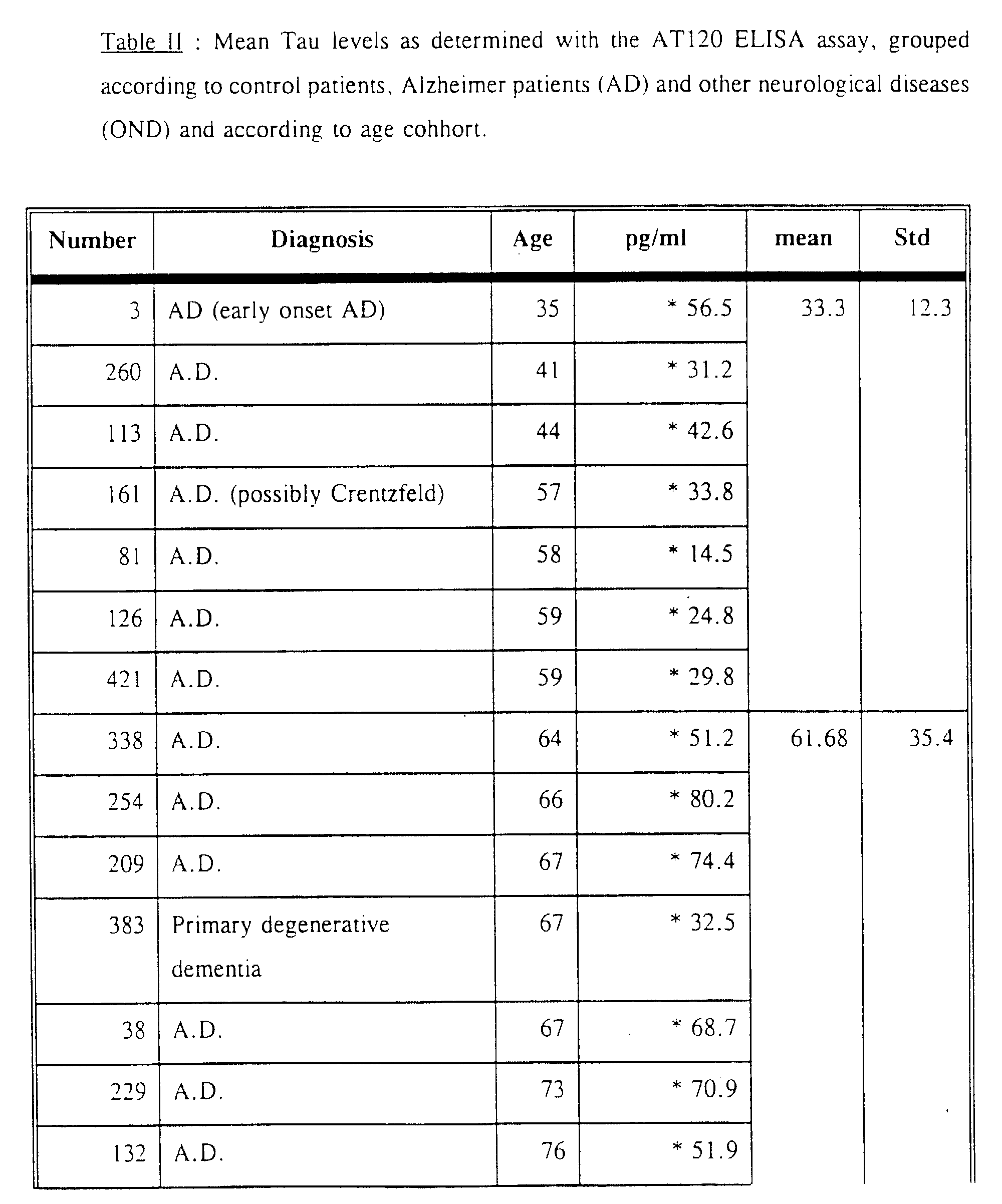
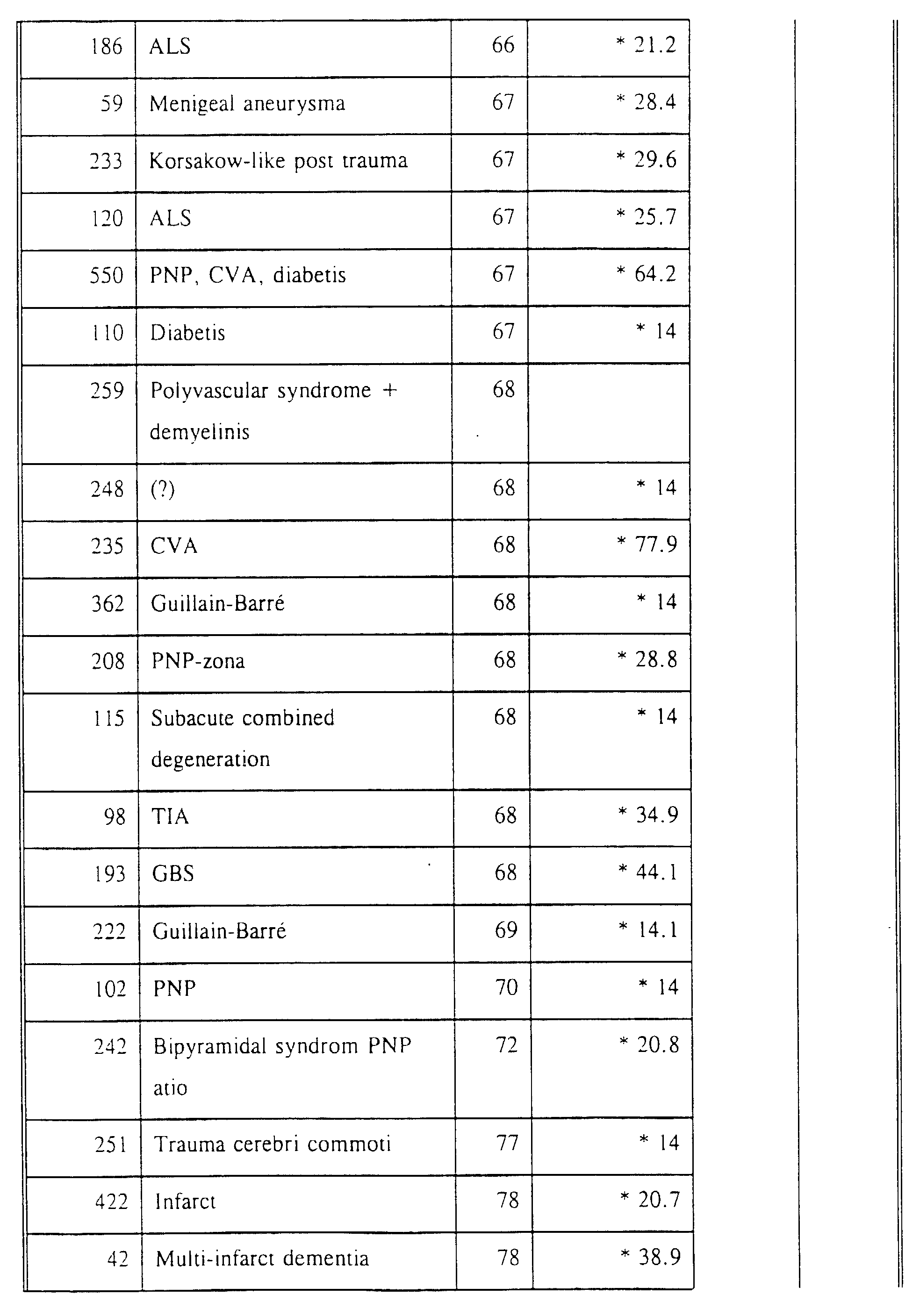
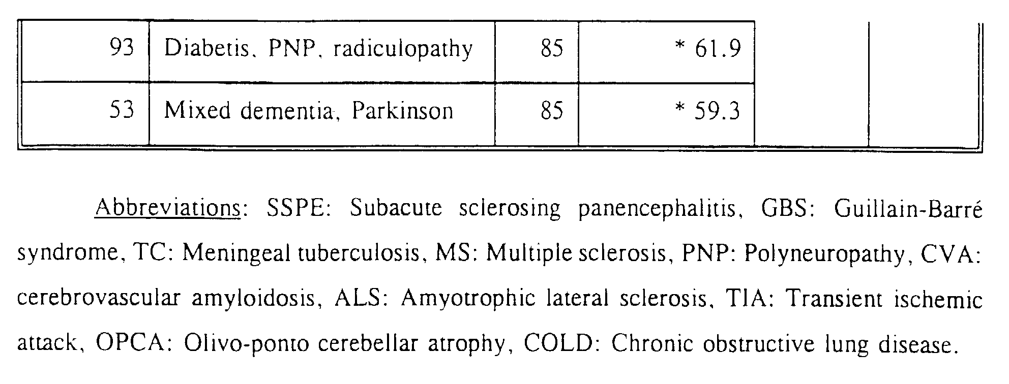
If a cut-off level of 27 pg/ml is adapted, 8% of the control samples are positive, while for the OND group and the Alzheimer group these values are 27% and 80% respectively.


| BIOTINYLATED | Unlabeled competitor Absorbance | ||
| AT8 | AT120 | BT2 | |
| AT8 | 0.001 | 0.372 | N.D. |
| AT120 | 0.476 | 0.001 | N.D. |
| BT2 | N.D. | 0.543 | 0.054 |
| N.D. refers to not determined absorbances |
| Reactivity of monoclonal antibodies BT2, HT7 and AT120 with solid phase bound nonapeptides. The nonapeptides are designated in the one-letter amino acid code. | |||
| Nonapeptides | Detecting Monoclonal antibody Absorbance | ||
| HT7 | BT2 | AT120 | |
| GAAPPGQKG | 3.00 | 0.168 | 0.089 |
| (SEQ ID NO 2) | |||
| GDRSGYSSP | 0.493 | 3.00 | 0.553 |
| (SEQ ID NO 3) |
Weingarten M, Lockwood A. Hwo S, Kirschner M (1975) A protein factor essential for microtubule assembly. Proc Natl Acad Sci USA 72:1858-1862.



Claims (14)
- Monoclonal antibody which forms an immunological complex with an epitope which is present in both human normal tau protein as well as abnormally phosphorylated tau protein, with said tau protein being obtainable from a brain homogenate, itself isolated from the human cerebral cortex, characterized by the fact that:it does not form an immunological complex with other phosphorylated proteins which share part of their sequence with tau protein, as determined by means of an ELISA,and it is able to detect human normal tau protein as well as abnormally phosphorylated tau protein in cerebrospinal fluid (CSF), with said tau protein being at a concentration as low as 1.0 pg/ml,and it is able to detect said tau proteins with 100% recovery upon the addition of a fixed amount of tau proteins in tau-protein-negative CSF.
- Monoclonal antibody according to claim 1, characterized by the fact that it forms an immunological complex:either with an epitope located within the following amino acid sequence of human tau protein:

 or with any other peptide capable of forming an immunological complex with a monoclonal antibody, which is capable of forming a complex with an epitope located in the tau protein region as shown in SEQ ID NO 1.
 - Monoclonal antibody according to claims 1 or 2 obtainable from the hybridoma deposited at ECACC on October 8, 1992 under No. 92100853.
- Hybridoma, which secretes a monoclonal antibody according to anyone of claims 1 to 3, more particularly the hybridoma deposited at ECACC on October 8, 1992 under No. 92100853.
- Peptides which are obtainable from a brain homogenate, itself isolated from the human cerebral cortex or from cerebral cortex obtained from a patient having Alzheimer's disease and which form an immunological complex with the monoclonal antibody according to anyone of claims 1 to 3 and with said peptides being different from human tau proteins.
- Peptides which are able to form an immunological complex with any of the monoclonal antibodies, according to anyone of claims 1 to 3,which are contained in, or are constituted by parts of the sequence as shown in SEQ ID NO 1,which contain or are constituted by the sequence of the peptides liable to form an immunological complex with a monoclonal antibody according to anyone of claims 1 to 3, which itself forms a complex with the epitope located in the tau protein region as shown in SEQ ID NO 1, with said peptides being different from human tau proteins.
- Peptides of about 100 amino acidswhich contain the sequence as shown in SEQ ID NO 1,which contain the sequence of the peptides able to form an immunological complex with a monoclonal antibody according to anyone of claims 1 to 3, which itself is liable to form a complex with a peptide within sequence as shown in SEQ ID NO 1, with said peptides being different from human tau proteins.
- Peptides according to anyone of claims 5 to 7, which are able to generate a monoclonal antibody according to anyone of claims 1 to 3 upon immunization.
- Process for obtaining and isolating a hybridoma according to claim 4, secreting a monoclonal antibody according to anyone of claims 1 to 3, characterized in that it involves:starting from spleen cells of an animal, e.g. mouse or rat, previously immunized in vivo, or from spleen cells of such cells previously immunized in vitro with an antigen recognized by the monoclonal antibody deposited at ECACC on October 8, 1992 under No. 92100853,fusing said immunized cells with myeloma cells under hybridoma-forming conditions; andselecting those of the hybridomas which secrete the monoclonal antibodies according to any of claims 1 to 3.
- Process for producing monoclonal antibodies according to anyone of claims 1 to 3 which involves:culturing the selected hybridomas according to claim 4, in an appropriate medium culture; andrecovering the monoclonal antibodies excreted by said selected hybridomas; or alternatively:implanting the selected hybridomas of claim 4 into the peritoneum of a mouse and, when ascites has been produced by the animal, recovering the monoclonal antibodies then formed from said ascites.
- Process for the detection or diagnosis in vitro of brain disease involving either or both paired helical filament (PHF)-tau or normal tau protein, e.g. Alzheimer's disease, which involves:contacting a monoclonal antibody according to anyone of claims 1 to 3, with a preparation of neurofibrillary tangles (NFT) or with a detergent-extracted brain homogenate isolated from a patient having had Alzheimer's disease or any other disease involving tau protein or abnormally phosphorylated tau protein under conditions suitable for producing an antigen-antibody complex; andseparating the antigen from said complex and recovering the antigen sought in a purified form.
- Process for the detection or diagnosis in vitro of brain disease involving abnormally phosphorylated tau protein, e.g. Alzheimer's disease, which includes:bringing a sample of brain homogenate, or of cerebrospinal fluid, or of serum from a patient suspected of suffering from a neurological disorder involving tau protein or paired helical filament (PHF), more particularly Alzheimer's disease, into contact under in vitro conditions with a monoclonal antibody according to anyone of claims 1 to 3, with said conditions being suitable for producing an antigen-antibody complex; anddetecting the immunological binding of said antibody to said sample of brain homogenate, or of cerebrospinal fluid, or of serum.
- Process for the detection or diagnosis in vitro of brain diseases involving paired helical filament (PHF) and/or normal tau protein, e.g. Alzheimer's disease, comprising the steps of :bringing a sample of unconcentrated cerebrospinal fluid sample isolated from a patient suspected to suffer from a neurological disorder involving normal or abnormally phosphorylated tau protein, more particulary Alzheimer's disease, into contact under in vitro conditions with a monoclonal antibody according to anyone of claim 1 to 3, under conditions suitable for producing an antigen-antibody complex;
and,detecting the immunological binding of said antibody to said sample of cerebrospinal fluid by means of a sandwich ELISA, preferably by applying the catalysed reporter diagnosis enhancement (CARD) procedure. - Kit for the diagnosis in vitro of one of the following diseases: Alzheimer's disease, Down's syndrome, Pick's disease, SSPE and other neurological disorders in which normal tau protein or abnormally phosphorylated tau protein or paired helical filaments are implicated, characterized in that the kit comprises:at least a microplate having deposited thereon a monoclonal antibody according to anyone of claims 1 to 3;a preparation containing the sample to be diagnosed in vitro, possibly together with a labeled peptide containing the epitope of the invention and preferably with a peptide having a sequence lying within the sequence of SEQ ID NO 1;a second antibodywhich can be a monoclonal antibody recognizing an epitope of normal tau, or of abnormally phosphorylated tau protein, or of any peptide according to anyone of claims 5 to 8, with said epitope being different from the one of the invention, orwhich can be a polyclonal antibody recognising normal tau or abnormally phosphorylated tau or a peptide according to anyone of claims 5 to 8, with said polyclonal antibody being liable to form an immunological complex with epitopes which are all different from the epitope of the invention, with said polyclonal antibody being preferably purified by immunoaffinity chromatography using immobilized tau protein, ora marker either for specific tagging or coupling with said second antibody;appropriate buffer solutions for carrying out the immunological reaction between the monoclonal antibody of the invention and a test sample on the one hand, and the bound second antibody and the marker on the other hand,possibly a peptide according to anyone of claims 5 to 8 for standard purposes, or for competition purposes with respect to the antigen which is sought.
Priority Applications (1)
| Application Number | Priority Date | Filing Date | Title |
|---|---|---|---|
| EP94903752A EP0673418B1 (en) | 1992-12-14 | 1993-12-10 | Monoclonal antibodies directed against the microtubule-associated protein tau, hybridomas secreting these antibodies, antigen recognition by these monoclonal antibodies and their applications |
Applications Claiming Priority (4)
| Application Number | Priority Date | Filing Date | Title |
|---|---|---|---|
| EP92403403 | 1992-12-14 | ||
| EP92403403 | 1992-12-14 | ||
| PCT/EP1993/003499 WO1994013795A1 (en) | 1992-12-14 | 1993-12-10 | Monoclonal antibodies directed against the microtubule-associated protein tau, hybridomas secreting these antibodies, antigen recognition by these monoclonal antibodies and their applications |
| EP94903752A EP0673418B1 (en) | 1992-12-14 | 1993-12-10 | Monoclonal antibodies directed against the microtubule-associated protein tau, hybridomas secreting these antibodies, antigen recognition by these monoclonal antibodies and their applications |
Publications (2)
| Publication Number | Publication Date |
|---|---|
| EP0673418A1 EP0673418A1 (en) | 1995-09-27 |
| EP0673418B1 true EP0673418B1 (en) | 1998-05-06 |
Family
ID=8211736
Family Applications (1)
| Application Number | Title | Priority Date | Filing Date |
|---|---|---|---|
| EP94903752A Expired - Lifetime EP0673418B1 (en) | 1992-12-14 | 1993-12-10 | Monoclonal antibodies directed against the microtubule-associated protein tau, hybridomas secreting these antibodies, antigen recognition by these monoclonal antibodies and their applications |
Country Status (9)
| Country | Link |
|---|---|
| US (1) | US5843779A (en) |
| EP (1) | EP0673418B1 (en) |
| JP (1) | JP2879975B2 (en) |
| AT (1) | ATE165866T1 (en) |
| AU (1) | AU690092B2 (en) |
| DE (1) | DE69318420T2 (en) |
| DK (1) | DK0673418T3 (en) |
| ES (1) | ES2118373T3 (en) |
| WO (1) | WO1994013795A1 (en) |
Cited By (5)
| Publication number | Priority date | Publication date | Assignee | Title |
|---|---|---|---|---|
| US11492393B2 (en) | 2016-05-02 | 2022-11-08 | Prothena Biosciences Limited | Tau immunotherapy |
| US11584791B2 (en) | 2016-05-02 | 2023-02-21 | Prothena Biosciences Limited | Antibodies recognizing tau |
| US11643457B2 (en) | 2013-03-13 | 2023-05-09 | Prothena Biosciences Limited | Tau immunotherapy |
| US11926659B2 (en) | 2019-03-03 | 2024-03-12 | Prothena Biosciences Limited | Antibodies recognizing tau |
| US11958896B2 (en) | 2017-05-02 | 2024-04-16 | Prothena Biosciences Limited | Antibodies recognizing tau |
Families Citing this family (40)
| Publication number | Priority date | Publication date | Assignee | Title |
|---|---|---|---|---|
| US5811310A (en) * | 1986-09-30 | 1998-09-22 | Albert Einstein College Of Medicine Of Yeshiva Univ. | The Alz-50 monoclonal antibody and diagnostic assay for alzheimer's disease |
| DK0610330T3 (en) * | 1991-10-25 | 1998-01-26 | Innogenetics Nv | Monoclonal antibodies against microtubule-associated protein tau |
| US6121003A (en) * | 1994-07-29 | 2000-09-19 | Innogentics N.V. | Monoclonal antibodies specific for an epitope of phosphorylated tau, and their use |
| US6797478B1 (en) * | 1998-03-05 | 2004-09-28 | University Of Cincinnati | Method of detecting axonal damage, from associated disease states using tau monoclonal antibodies |
| CN1325491A (en) * | 1998-09-08 | 2001-12-05 | 基因创新有限公司 | Tau as a marker of early CNS injury |
| CA2361013A1 (en) * | 1999-01-25 | 2000-07-27 | Minerva Biotechnologies Corporation | Rapid and sensitive detection of aberrant protein aggregation in neurodegenerative diseases |
| WO2001023894A1 (en) * | 1999-09-28 | 2001-04-05 | Evotec Oai Ag | Quantitative analysis and typification of subcellular particles |
| BR0107851A (en) * | 2000-01-24 | 2002-10-29 | Innogenetics Nv | Tauopathy diagnosis |
| US20030162230A1 (en) | 2000-09-27 | 2003-08-28 | Reagan Kevin J. | Method for quantifying phosphokinase activity on proteins |
| WO2004081534A2 (en) * | 2003-03-12 | 2004-09-23 | Assay Designs, Inc. | Phosphokinase assay |
| CA2445420A1 (en) | 2003-07-29 | 2005-01-29 | Invitrogen Corporation | Kinase and phosphatase assays |
| US7619059B2 (en) | 2003-07-29 | 2009-11-17 | Life Technologies Corporation | Bimolecular optical probes |
| US7727752B2 (en) | 2003-07-29 | 2010-06-01 | Life Technologies Corporation | Kinase and phosphatase assays |
| WO2005080986A1 (en) * | 2004-02-18 | 2005-09-01 | University Of Iowa Research Foundation | Antibodies to phosphorylated tau, methods of making and methods of use |
| US20070178447A1 (en) * | 2006-01-31 | 2007-08-02 | Bethyl Laboratories, Inc. | Method for decreasing interference in results of immunochemical methods |
| EP3978517A1 (en) * | 2009-03-18 | 2022-04-06 | AC Immune SA | Method for therapeutic use |
| UA107571C2 (en) | 2009-04-03 | 2015-01-26 | PHARMACEUTICAL COMPOSITION | |
| SG189174A1 (en) | 2010-10-11 | 2013-05-31 | Biogen Idec Internat Neuroscience Gmbh | Human anti-tau antibodies |
| WO2012142301A2 (en) | 2011-04-12 | 2012-10-18 | Quanterix Corporation | Methods of determining a treatment protocol for and/or a prognosis of a patients recovery from a brain injury |
| GB201111361D0 (en) * | 2011-07-04 | 2011-08-17 | Nordic Bioscience As | Biochemical markers for neurodegenerative conditions |
| EP2834270B1 (en) | 2012-04-05 | 2019-10-30 | AC Immune S.A. | Humanized tau antibody |
| DK2885010T3 (en) | 2012-08-16 | 2020-02-03 | Ipierian Inc | METHODS OF TREATING TAUOPATHY |
| US9200068B2 (en) | 2012-12-18 | 2015-12-01 | Regents Of The University Of Minnesota | Compositions and methods related to tauopathy |
| HRP20201422T1 (en) | 2012-12-21 | 2021-02-19 | Biogen Ma Inc. | Human anti-tau antibodies |
| US8980270B2 (en) | 2013-01-18 | 2015-03-17 | Ipierian, Inc. | Methods of treating a tauopathy |
| BR112015023262B8 (en) | 2013-03-15 | 2024-02-06 | Ac Immune Sa | Isolated antibody, immunoconjugate, pharmaceutical formulation and uses of antibody |
| JP2017512751A (en) | 2014-02-14 | 2017-05-25 | アイピエリアン,インコーポレイティド | Tau peptides, anti-tau antibodies, and methods for their use |
| EA202091012A1 (en) | 2017-10-25 | 2020-08-27 | Янссен Фармасьютикалз, Инк. | COMPOSITIONS OF PHOSPHORYLATED TAU PEPTIDES AND THEIR APPLICATION |
| US20220018856A1 (en) | 2018-11-22 | 2022-01-20 | Fujirebio Inc. | Antibody conjugate |
| CN113710269A (en) | 2019-02-08 | 2021-11-26 | Ac免疫有限公司 | Safe administration method of phosphorylated Tau peptide vaccine |
| MA55902A (en) | 2019-04-24 | 2022-03-16 | Ac Immune Sa | HETEROLOGY ADMINISTRATION OF TAU VACCINES |
| EP4040153A4 (en) * | 2019-09-30 | 2023-07-26 | Nipro Corporation | Method for detecting tau protein using blood sample as specimen |
| BR112022020753A2 (en) | 2020-04-15 | 2022-12-20 | Voyager Therapeutics Inc | TAU-BINDING COMPOUNDS |
| CN111349617B (en) * | 2020-05-25 | 2020-09-22 | 苏州仁端生物医药科技有限公司 | Hybridoma cell strain secreting anti-Tau or pTau-181/231/396 monoclonal antibody and application thereof |
| JP2023554382A (en) | 2020-12-16 | 2023-12-27 | ボイジャー セラピューティクス インコーポレイテッド | tau binding compounds |
| WO2022144406A1 (en) | 2020-12-29 | 2022-07-07 | Neurimmune Ag | Human anti-tau antibodies |
| CN113005096B (en) * | 2021-01-19 | 2022-10-14 | 华中科技大学 | Hybridoma cell strain secreting anti-serine phosphorylation tau protein monoclonal antibody |
| CN114539400B (en) * | 2022-02-15 | 2024-06-07 | 武汉天德生物科技有限公司 | Phosphorylated Tau pT217 protein monoclonal antibody, ELISA kit and application thereof |
| WO2023250388A1 (en) | 2022-06-22 | 2023-12-28 | Voyager Therapeutics, Inc. | Tau binding compounds |
| CN120019071A (en) | 2022-09-15 | 2025-05-16 | 沃雅戈治疗公司 | TAU-binding compounds |
-
1993
- 1993-12-10 DK DK94903752T patent/DK0673418T3/en active
- 1993-12-10 US US08/244,951 patent/US5843779A/en not_active Expired - Lifetime
- 1993-12-10 EP EP94903752A patent/EP0673418B1/en not_active Expired - Lifetime
- 1993-12-10 DE DE69318420T patent/DE69318420T2/en not_active Expired - Lifetime
- 1993-12-10 AU AU58097/94A patent/AU690092B2/en not_active Expired
- 1993-12-10 WO PCT/EP1993/003499 patent/WO1994013795A1/en not_active Ceased
- 1993-12-10 AT AT94903752T patent/ATE165866T1/en active
- 1993-12-10 ES ES94903752T patent/ES2118373T3/en not_active Expired - Lifetime
- 1993-12-10 JP JP6513790A patent/JP2879975B2/en not_active Expired - Lifetime
Cited By (5)
| Publication number | Priority date | Publication date | Assignee | Title |
|---|---|---|---|---|
| US11643457B2 (en) | 2013-03-13 | 2023-05-09 | Prothena Biosciences Limited | Tau immunotherapy |
| US11492393B2 (en) | 2016-05-02 | 2022-11-08 | Prothena Biosciences Limited | Tau immunotherapy |
| US11584791B2 (en) | 2016-05-02 | 2023-02-21 | Prothena Biosciences Limited | Antibodies recognizing tau |
| US11958896B2 (en) | 2017-05-02 | 2024-04-16 | Prothena Biosciences Limited | Antibodies recognizing tau |
| US11926659B2 (en) | 2019-03-03 | 2024-03-12 | Prothena Biosciences Limited | Antibodies recognizing tau |
Also Published As
| Publication number | Publication date |
|---|---|
| ATE165866T1 (en) | 1998-05-15 |
| ES2118373T3 (en) | 1998-09-16 |
| DE69318420D1 (en) | 1998-06-10 |
| JP2879975B2 (en) | 1999-04-05 |
| DE69318420T2 (en) | 1999-01-28 |
| US5843779A (en) | 1998-12-01 |
| AU5809794A (en) | 1994-07-04 |
| EP0673418A1 (en) | 1995-09-27 |
| AU690092B2 (en) | 1998-04-23 |
| DK0673418T3 (en) | 1999-03-01 |
| JPH08502898A (en) | 1996-04-02 |
| WO1994013795A1 (en) | 1994-06-23 |
Similar Documents
| Publication | Publication Date | Title |
|---|---|---|
| AU690092B2 (en) | Monoclonal antibodies directed against the microtubule-associated protein tau, hybridomas secreting these antibodies, antigen recognition by these monoclonal antibodies and their applications | |
| US6232437B1 (en) | Isolated human tau peptide epitope which specifically binds monoclonal antibody AT120. | |
| AU662178B2 (en) | Monoclonal antibodies directed against the microtubule-associated protein tau | |
| EP0737208B1 (en) | Monoclonal antibodies specific for phf-tau, hybridomas secreting them, antigen recognition by these antibodies and their applications | |
| US6121003A (en) | Monoclonal antibodies specific for an epitope of phosphorylated tau, and their use | |
| CA2150816C (en) | Monoclonal antibodies directed against the microtubule- associated protein tau, hybridomas secreting these antibodies, antigen recognition by these monoclonal antibodies and theirapplications |
Legal Events
| Date | Code | Title | Description |
|---|---|---|---|
| PUAI | Public reference made under article 153(3) epc to a published international application that has entered the european phase |
Free format text: ORIGINAL CODE: 0009012 |
|
| 17P | Request for examination filed |
Effective date: 19950621 |
|
| AK | Designated contracting states |
Kind code of ref document: A1 Designated state(s): AT BE CH DE DK ES FR GB GR IE IT LI LU MC NL PT SE |
|
| 17Q | First examination report despatched |
Effective date: 19970228 |
|
| GRAG | Despatch of communication of intention to grant |
Free format text: ORIGINAL CODE: EPIDOS AGRA |
|
| RHK1 | Main classification (correction) |
Ipc: C12N 15/06 |
|
| GRAG | Despatch of communication of intention to grant |
Free format text: ORIGINAL CODE: EPIDOS AGRA |
|
| GRAH | Despatch of communication of intention to grant a patent |
Free format text: ORIGINAL CODE: EPIDOS IGRA |
|
| GRAA | (expected) grant |
Free format text: ORIGINAL CODE: 0009210 |
|
| AK | Designated contracting states |
Kind code of ref document: B1 Designated state(s): AT BE CH DE DK ES FR GB GR IE IT LI LU MC NL PT SE |
|
| REF | Corresponds to: |
Ref document number: 165866 Country of ref document: AT Date of ref document: 19980515 Kind code of ref document: T |
|
| REG | Reference to a national code |
Ref country code: CH Ref legal event code: EP |
|
| REF | Corresponds to: |
Ref document number: 69318420 Country of ref document: DE Date of ref document: 19980610 |
|
| ITF | It: translation for a ep patent filed | ||
| REG | Reference to a national code |
Ref country code: CH Ref legal event code: NV Representative=s name: BUECHEL & PARTNER AG PATENTBUERO |
|
| REG | Reference to a national code |
Ref country code: IE Ref legal event code: FG4D Free format text: 80203 |
|
| ET | Fr: translation filed | ||
| REG | Reference to a national code |
Ref country code: ES Ref legal event code: FG2A Ref document number: 2118373 Country of ref document: ES Kind code of ref document: T3 |
|
| REG | Reference to a national code |
Ref country code: PT Ref legal event code: SC4A Free format text: AVAILABILITY OF NATIONAL TRANSLATION Effective date: 19980729 |
|
| REG | Reference to a national code |
Ref country code: DK Ref legal event code: T3 |
|
| PLBE | No opposition filed within time limit |
Free format text: ORIGINAL CODE: 0009261 |
|
| STAA | Information on the status of an ep patent application or granted ep patent |
Free format text: STATUS: NO OPPOSITION FILED WITHIN TIME LIMIT |
|
| 26N | No opposition filed | ||
| REG | Reference to a national code |
Ref country code: GB Ref legal event code: IF02 |
|
| PGFP | Annual fee paid to national office [announced via postgrant information from national office to epo] |
Ref country code: DK Payment date: 20121219 Year of fee payment: 20 Ref country code: IE Payment date: 20121220 Year of fee payment: 20 Ref country code: CH Payment date: 20121221 Year of fee payment: 20 Ref country code: LU Payment date: 20121227 Year of fee payment: 20 Ref country code: MC Payment date: 20121212 Year of fee payment: 20 |
|
| PGFP | Annual fee paid to national office [announced via postgrant information from national office to epo] |
Ref country code: PT Payment date: 20121205 Year of fee payment: 20 Ref country code: GB Payment date: 20121220 Year of fee payment: 20 Ref country code: IT Payment date: 20121227 Year of fee payment: 20 Ref country code: SE Payment date: 20121220 Year of fee payment: 20 Ref country code: GR Payment date: 20121221 Year of fee payment: 20 |
|
| PGFP | Annual fee paid to national office [announced via postgrant information from national office to epo] |
Ref country code: AT Payment date: 20121212 Year of fee payment: 20 |
|
| PGFP | Annual fee paid to national office [announced via postgrant information from national office to epo] |
Ref country code: ES Payment date: 20121226 Year of fee payment: 20 Ref country code: BE Payment date: 20121219 Year of fee payment: 20 Ref country code: DE Payment date: 20121220 Year of fee payment: 20 Ref country code: FR Payment date: 20130130 Year of fee payment: 20 |
|
| PGFP | Annual fee paid to national office [announced via postgrant information from national office to epo] |
Ref country code: NL Payment date: 20121220 Year of fee payment: 20 |
|
| REG | Reference to a national code |
Ref country code: DE Ref legal event code: R071 Ref document number: 69318420 Country of ref document: DE |
|
| REG | Reference to a national code |
Ref country code: DE Ref legal event code: R071 Ref document number: 69318420 Country of ref document: DE |
|
| REG | Reference to a national code |
Ref country code: DK Ref legal event code: EUP Effective date: 20131210 |
|
| REG | Reference to a national code |
Ref country code: NL Ref legal event code: V4 Effective date: 20131210 Ref country code: PT Ref legal event code: MM4A Free format text: MAXIMUM VALIDITY LIMIT REACHED Effective date: 20131210 |
|
| BE20 | Be: patent expired |
Owner name: S.A. *INNOGENETICS N.V. Effective date: 20131210 |
|
| REG | Reference to a national code |
Ref country code: CH Ref legal event code: PL |
|
| REG | Reference to a national code |
Ref country code: GB Ref legal event code: PE20 Expiry date: 20131209 |
|
| REG | Reference to a national code |
Ref country code: AT Ref legal event code: MK07 Ref document number: 165866 Country of ref document: AT Kind code of ref document: T Effective date: 20131210 |
|
| REG | Reference to a national code |
Ref country code: IE Ref legal event code: MK9A |
|
| PG25 | Lapsed in a contracting state [announced via postgrant information from national office to epo] |
Ref country code: PT Free format text: LAPSE BECAUSE OF EXPIRATION OF PROTECTION Effective date: 20131218 Ref country code: DE Free format text: LAPSE BECAUSE OF EXPIRATION OF PROTECTION Effective date: 20131211 Ref country code: GB Free format text: LAPSE BECAUSE OF EXPIRATION OF PROTECTION Effective date: 20131209 |
|
| REG | Reference to a national code |
Ref country code: SE Ref legal event code: EUG |
|
| REG | Reference to a national code |
Ref country code: GR Ref legal event code: MA Ref document number: 980401643 Country of ref document: GR Effective date: 20131211 |
|
| REG | Reference to a national code |
Ref country code: ES Ref legal event code: FD2A Effective date: 20140306 |
|
| PG25 | Lapsed in a contracting state [announced via postgrant information from national office to epo] |
Ref country code: IE Free format text: LAPSE BECAUSE OF EXPIRATION OF PROTECTION Effective date: 20131210 |
|
| PG25 | Lapsed in a contracting state [announced via postgrant information from national office to epo] |
Ref country code: ES Free format text: LAPSE BECAUSE OF EXPIRATION OF PROTECTION Effective date: 20131211 |