EP0668781B1 - Lymphokine gene therapy of cancer in combination with tumor antigens - Google Patents
Lymphokine gene therapy of cancer in combination with tumor antigens Download PDFInfo
- Publication number
- EP0668781B1 EP0668781B1 EP92922731A EP92922731A EP0668781B1 EP 0668781 B1 EP0668781 B1 EP 0668781B1 EP 92922731 A EP92922731 A EP 92922731A EP 92922731 A EP92922731 A EP 92922731A EP 0668781 B1 EP0668781 B1 EP 0668781B1
- Authority
- EP
- European Patent Office
- Prior art keywords
- tumor
- cells
- fibroblasts
- cytokine
- interleukin
- Prior art date
- Legal status (The legal status is an assumption and is not a legal conclusion. Google has not performed a legal analysis and makes no representation as to the accuracy of the status listed.)
- Revoked
Links
- 206010028980 Neoplasm Diseases 0.000 title claims abstract description 141
- 108091007433 antigens Proteins 0.000 title claims abstract description 57
- 239000000427 antigen Substances 0.000 title claims abstract description 55
- 102000036639 antigens Human genes 0.000 title claims abstract description 55
- 238000001415 gene therapy Methods 0.000 title description 11
- 201000011510 cancer Diseases 0.000 title description 6
- 210000004027 cell Anatomy 0.000 claims abstract description 150
- 210000002950 fibroblast Anatomy 0.000 claims abstract description 128
- 108090000695 Cytokines Proteins 0.000 claims abstract description 70
- 230000028993 immune response Effects 0.000 claims abstract description 17
- 239000000203 mixture Substances 0.000 claims abstract description 16
- 238000009472 formulation Methods 0.000 claims abstract description 7
- 230000028327 secretion Effects 0.000 claims abstract description 6
- 108010002350 Interleukin-2 Proteins 0.000 claims description 112
- 102000000588 Interleukin-2 Human genes 0.000 claims description 107
- 210000004881 tumor cell Anatomy 0.000 claims description 96
- 102000004127 Cytokines Human genes 0.000 claims description 47
- 108090000623 proteins and genes Proteins 0.000 claims description 45
- 230000003053 immunization Effects 0.000 claims description 40
- 230000001177 retroviral effect Effects 0.000 claims description 29
- 238000002360 preparation method Methods 0.000 claims description 11
- 231100000057 systemic toxicity Toxicity 0.000 claims description 8
- 238000000338 in vitro Methods 0.000 claims description 7
- 102000004388 Interleukin-4 Human genes 0.000 claims description 6
- 108090000978 Interleukin-4 Proteins 0.000 claims description 6
- 239000003814 drug Substances 0.000 claims description 6
- 229940028885 interleukin-4 Drugs 0.000 claims description 6
- 210000000612 antigen-presenting cell Anatomy 0.000 claims description 4
- 230000004936 stimulating effect Effects 0.000 claims description 4
- 102000008070 Interferon-gamma Human genes 0.000 claims description 3
- 108010074328 Interferon-gamma Proteins 0.000 claims description 3
- 229940044627 gamma-interferon Drugs 0.000 claims description 3
- 238000010188 recombinant method Methods 0.000 claims description 2
- 230000002459 sustained effect Effects 0.000 claims description 2
- 239000013604 expression vector Substances 0.000 claims 6
- 230000001093 anti-cancer Effects 0.000 claims 3
- 102000000589 Interleukin-1 Human genes 0.000 claims 2
- 108010002352 Interleukin-1 Proteins 0.000 claims 2
- 102000000646 Interleukin-3 Human genes 0.000 claims 2
- 108010002386 Interleukin-3 Proteins 0.000 claims 2
- 102000000743 Interleukin-5 Human genes 0.000 claims 2
- 108010002616 Interleukin-5 Proteins 0.000 claims 2
- 102000004889 Interleukin-6 Human genes 0.000 claims 2
- 108090001005 Interleukin-6 Proteins 0.000 claims 2
- 230000003308 immunostimulating effect Effects 0.000 claims 2
- 229940076264 interleukin-3 Drugs 0.000 claims 2
- 229940100602 interleukin-5 Drugs 0.000 claims 2
- 229940100601 interleukin-6 Drugs 0.000 claims 2
- 238000000034 method Methods 0.000 abstract description 29
- 238000012239 gene modification Methods 0.000 abstract description 4
- 230000005017 genetic modification Effects 0.000 abstract description 4
- 235000013617 genetically modified food Nutrition 0.000 abstract description 4
- 238000009169 immunotherapy Methods 0.000 abstract description 2
- 239000013598 vector Substances 0.000 description 55
- 241001465754 Metazoa Species 0.000 description 43
- 238000002649 immunization Methods 0.000 description 38
- 230000014509 gene expression Effects 0.000 description 28
- 230000000694 effects Effects 0.000 description 23
- 238000012546 transfer Methods 0.000 description 19
- 230000005809 anti-tumor immunity Effects 0.000 description 17
- 230000009885 systemic effect Effects 0.000 description 15
- 238000011282 treatment Methods 0.000 description 15
- 241000699670 Mus sp. Species 0.000 description 13
- 230000004614 tumor growth Effects 0.000 description 13
- 238000002347 injection Methods 0.000 description 12
- 239000007924 injection Substances 0.000 description 12
- 241001529936 Murinae Species 0.000 description 10
- 239000006228 supernatant Substances 0.000 description 10
- 229930193140 Neomycin Natural products 0.000 description 8
- 108020004440 Thymidine kinase Proteins 0.000 description 8
- 239000002299 complementary DNA Substances 0.000 description 8
- 210000000987 immune system Anatomy 0.000 description 8
- 229960004927 neomycin Drugs 0.000 description 8
- 238000004806 packaging method and process Methods 0.000 description 8
- 230000003612 virological effect Effects 0.000 description 8
- 241001068263 Replication competent viruses Species 0.000 description 7
- 102000006601 Thymidine Kinase Human genes 0.000 description 7
- 238000013459 approach Methods 0.000 description 7
- 230000006698 induction Effects 0.000 description 7
- 210000004698 lymphocyte Anatomy 0.000 description 7
- 230000001404 mediated effect Effects 0.000 description 7
- 239000000047 product Substances 0.000 description 7
- 241001430294 unidentified retrovirus Species 0.000 description 7
- PHEDXBVPIONUQT-UHFFFAOYSA-N Cocarcinogen A1 Natural products CCCCCCCCCCCCCC(=O)OC1C(C)C2(O)C3C=C(C)C(=O)C3(O)CC(CO)=CC2C2C1(OC(C)=O)C2(C)C PHEDXBVPIONUQT-UHFFFAOYSA-N 0.000 description 6
- VYPSYNLAJGMNEJ-UHFFFAOYSA-N Silicium dioxide Chemical compound O=[Si]=O VYPSYNLAJGMNEJ-UHFFFAOYSA-N 0.000 description 6
- 241000700605 Viruses Species 0.000 description 6
- 239000003710 calcium ionophore Substances 0.000 description 6
- 239000003795 chemical substances by application Substances 0.000 description 6
- 238000011161 development Methods 0.000 description 6
- 230000018109 developmental process Effects 0.000 description 6
- CJAONIOAQZUHPN-KKLWWLSJSA-N ethyl 12-[[2-[(2r,3r)-3-[2-[(12-ethoxy-12-oxododecyl)-methylamino]-2-oxoethoxy]butan-2-yl]oxyacetyl]-methylamino]dodecanoate Chemical compound CCOC(=O)CCCCCCCCCCCN(C)C(=O)CO[C@H](C)[C@@H](C)OCC(=O)N(C)CCCCCCCCCCCC(=O)OCC CJAONIOAQZUHPN-KKLWWLSJSA-N 0.000 description 6
- 238000002474 experimental method Methods 0.000 description 6
- 238000002955 isolation Methods 0.000 description 6
- PHEDXBVPIONUQT-RGYGYFBISA-N phorbol 13-acetate 12-myristate Chemical compound C([C@]1(O)C(=O)C(C)=C[C@H]1[C@@]1(O)[C@H](C)[C@H]2OC(=O)CCCCCCCCCCCCC)C(CO)=C[C@H]1[C@H]1[C@]2(OC(C)=O)C1(C)C PHEDXBVPIONUQT-RGYGYFBISA-N 0.000 description 6
- 239000013612 plasmid Substances 0.000 description 6
- 230000003248 secreting effect Effects 0.000 description 6
- 238000002560 therapeutic procedure Methods 0.000 description 6
- 238000010361 transduction Methods 0.000 description 6
- 230000026683 transduction Effects 0.000 description 6
- 108091003079 Bovine Serum Albumin Proteins 0.000 description 5
- 238000002965 ELISA Methods 0.000 description 5
- 101001002657 Homo sapiens Interleukin-2 Proteins 0.000 description 5
- 108010025815 Kanamycin Kinase Proteins 0.000 description 5
- FAPWRFPIFSIZLT-UHFFFAOYSA-M Sodium chloride Chemical compound [Na+].[Cl-] FAPWRFPIFSIZLT-UHFFFAOYSA-M 0.000 description 5
- 210000001744 T-lymphocyte Anatomy 0.000 description 5
- 230000005975 antitumor immune response Effects 0.000 description 5
- 230000006378 damage Effects 0.000 description 5
- 230000012010 growth Effects 0.000 description 5
- 238000001727 in vivo Methods 0.000 description 5
- 208000015181 infectious disease Diseases 0.000 description 5
- 238000001990 intravenous administration Methods 0.000 description 5
- 238000004519 manufacturing process Methods 0.000 description 5
- 239000002609 medium Substances 0.000 description 5
- 230000004044 response Effects 0.000 description 5
- 230000000638 stimulation Effects 0.000 description 5
- 231100000419 toxicity Toxicity 0.000 description 5
- 230000001988 toxicity Effects 0.000 description 5
- 239000006144 Dulbecco’s modified Eagle's medium Substances 0.000 description 4
- 230000008901 benefit Effects 0.000 description 4
- 238000001574 biopsy Methods 0.000 description 4
- 238000011156 evaluation Methods 0.000 description 4
- 230000001939 inductive effect Effects 0.000 description 4
- NOESYZHRGYRDHS-UHFFFAOYSA-N insulin Chemical compound N1C(=O)C(NC(=O)C(CCC(N)=O)NC(=O)C(CCC(O)=O)NC(=O)C(C(C)C)NC(=O)C(NC(=O)CN)C(C)CC)CSSCC(C(NC(CO)C(=O)NC(CC(C)C)C(=O)NC(CC=2C=CC(O)=CC=2)C(=O)NC(CCC(N)=O)C(=O)NC(CC(C)C)C(=O)NC(CCC(O)=O)C(=O)NC(CC(N)=O)C(=O)NC(CC=2C=CC(O)=CC=2)C(=O)NC(CSSCC(NC(=O)C(C(C)C)NC(=O)C(CC(C)C)NC(=O)C(CC=2C=CC(O)=CC=2)NC(=O)C(CC(C)C)NC(=O)C(C)NC(=O)C(CCC(O)=O)NC(=O)C(C(C)C)NC(=O)C(CC(C)C)NC(=O)C(CC=2NC=NC=2)NC(=O)C(CO)NC(=O)CNC2=O)C(=O)NCC(=O)NC(CCC(O)=O)C(=O)NC(CCCNC(N)=N)C(=O)NCC(=O)NC(CC=3C=CC=CC=3)C(=O)NC(CC=3C=CC=CC=3)C(=O)NC(CC=3C=CC(O)=CC=3)C(=O)NC(C(C)O)C(=O)N3C(CCC3)C(=O)NC(CCCCN)C(=O)NC(C)C(O)=O)C(=O)NC(CC(N)=O)C(O)=O)=O)NC(=O)C(C(C)CC)NC(=O)C(CO)NC(=O)C(C(C)O)NC(=O)C1CSSCC2NC(=O)C(CC(C)C)NC(=O)C(NC(=O)C(CCC(N)=O)NC(=O)C(CC(N)=O)NC(=O)C(NC(=O)C(N)CC=1C=CC=CC=1)C(C)C)CC1=CN=CN1 NOESYZHRGYRDHS-UHFFFAOYSA-N 0.000 description 4
- 230000010076 replication Effects 0.000 description 4
- NHBKXEKEPDILRR-UHFFFAOYSA-N 2,3-bis(butanoylsulfanyl)propyl butanoate Chemical class CCCC(=O)OCC(SC(=O)CCC)CSC(=O)CCC NHBKXEKEPDILRR-UHFFFAOYSA-N 0.000 description 3
- 108091026890 Coding region Proteins 0.000 description 3
- 102000008072 Lymphokines Human genes 0.000 description 3
- 108010074338 Lymphokines Proteins 0.000 description 3
- 239000012980 RPMI-1640 medium Substances 0.000 description 3
- 238000004458 analytical method Methods 0.000 description 3
- 230000000259 anti-tumor effect Effects 0.000 description 3
- 238000010276 construction Methods 0.000 description 3
- 230000003111 delayed effect Effects 0.000 description 3
- 230000001419 dependent effect Effects 0.000 description 3
- 239000012091 fetal bovine serum Substances 0.000 description 3
- 239000001963 growth medium Substances 0.000 description 3
- 238000002513 implantation Methods 0.000 description 3
- 230000002779 inactivation Effects 0.000 description 3
- 238000010348 incorporation Methods 0.000 description 3
- 238000001802 infusion Methods 0.000 description 3
- 238000011835 investigation Methods 0.000 description 3
- 239000003550 marker Substances 0.000 description 3
- 239000002773 nucleotide Substances 0.000 description 3
- 125000003729 nucleotide group Chemical group 0.000 description 3
- 210000005259 peripheral blood Anatomy 0.000 description 3
- 239000011886 peripheral blood Substances 0.000 description 3
- 239000000377 silicon dioxide Substances 0.000 description 3
- 239000011780 sodium chloride Substances 0.000 description 3
- 239000000243 solution Substances 0.000 description 3
- 238000010561 standard procedure Methods 0.000 description 3
- 238000012109 statistical procedure Methods 0.000 description 3
- 238000007920 subcutaneous administration Methods 0.000 description 3
- 238000012360 testing method Methods 0.000 description 3
- 230000001225 therapeutic effect Effects 0.000 description 3
- 210000001519 tissue Anatomy 0.000 description 3
- 210000003171 tumor-infiltrating lymphocyte Anatomy 0.000 description 3
- IJGRMHOSHXDMSA-UHFFFAOYSA-N Atomic nitrogen Chemical compound N#N IJGRMHOSHXDMSA-UHFFFAOYSA-N 0.000 description 2
- BPYKTIZUTYGOLE-IFADSCNNSA-N Bilirubin Chemical compound N1C(=O)C(C)=C(C=C)\C1=C\C1=C(C)C(CCC(O)=O)=C(CC2=C(C(C)=C(\C=C/3C(=C(C=C)C(=O)N\3)C)N2)CCC(O)=O)N1 BPYKTIZUTYGOLE-IFADSCNNSA-N 0.000 description 2
- LFQSCWFLJHTTHZ-UHFFFAOYSA-N Ethanol Chemical compound CCO LFQSCWFLJHTTHZ-UHFFFAOYSA-N 0.000 description 2
- 102000004877 Insulin Human genes 0.000 description 2
- 108090001061 Insulin Proteins 0.000 description 2
- 108060001084 Luciferase Proteins 0.000 description 2
- 241000829100 Macaca mulatta polyomavirus 1 Species 0.000 description 2
- 241000713869 Moloney murine leukemia virus Species 0.000 description 2
- 238000002944 PCR assay Methods 0.000 description 2
- 241000288906 Primates Species 0.000 description 2
- 108010076504 Protein Sorting Signals Proteins 0.000 description 2
- 108020004511 Recombinant DNA Proteins 0.000 description 2
- 108700019146 Transgenes Proteins 0.000 description 2
- 229960004150 aciclovir Drugs 0.000 description 2
- MKUXAQIIEYXACX-UHFFFAOYSA-N aciclovir Chemical compound N1C(N)=NC(=O)C2=C1N(COCCO)C=N2 MKUXAQIIEYXACX-UHFFFAOYSA-N 0.000 description 2
- 229940125715 antihistaminic agent Drugs 0.000 description 2
- 239000000739 antihistaminic agent Substances 0.000 description 2
- 238000003556 assay Methods 0.000 description 2
- 230000004071 biological effect Effects 0.000 description 2
- 230000004663 cell proliferation Effects 0.000 description 2
- 239000006285 cell suspension Substances 0.000 description 2
- 239000000356 contaminant Substances 0.000 description 2
- DDRJAANPRJIHGJ-UHFFFAOYSA-N creatinine Chemical compound CN1CC(=O)NC1=N DDRJAANPRJIHGJ-UHFFFAOYSA-N 0.000 description 2
- 239000012228 culture supernatant Substances 0.000 description 2
- 231100000433 cytotoxic Toxicity 0.000 description 2
- 230000001472 cytotoxic effect Effects 0.000 description 2
- 238000010586 diagram Methods 0.000 description 2
- 230000009266 disease activity Effects 0.000 description 2
- 229940079593 drug Drugs 0.000 description 2
- 239000012894 fetal calf serum Substances 0.000 description 2
- 229960002963 ganciclovir Drugs 0.000 description 2
- IRSCQMHQWWYFCW-UHFFFAOYSA-N ganciclovir Chemical compound O=C1NC(N)=NC2=C1N=CN2COC(CO)CO IRSCQMHQWWYFCW-UHFFFAOYSA-N 0.000 description 2
- 230000001900 immune effect Effects 0.000 description 2
- 230000002458 infectious effect Effects 0.000 description 2
- 230000005764 inhibitory process Effects 0.000 description 2
- 229940125396 insulin Drugs 0.000 description 2
- 210000001165 lymph node Anatomy 0.000 description 2
- 210000002540 macrophage Anatomy 0.000 description 2
- 238000005259 measurement Methods 0.000 description 2
- 210000001616 monocyte Anatomy 0.000 description 2
- 239000013642 negative control Substances 0.000 description 2
- 231100000956 nontoxicity Toxicity 0.000 description 2
- 108700004029 pol Genes Proteins 0.000 description 2
- 229920001184 polypeptide Polymers 0.000 description 2
- 231100000683 possible toxicity Toxicity 0.000 description 2
- 108090000765 processed proteins & peptides Proteins 0.000 description 2
- 102000004196 processed proteins & peptides Human genes 0.000 description 2
- 102000004169 proteins and genes Human genes 0.000 description 2
- 238000007388 punch biopsy Methods 0.000 description 2
- 238000005215 recombination Methods 0.000 description 2
- 230000006798 recombination Effects 0.000 description 2
- 230000002829 reductive effect Effects 0.000 description 2
- 238000000926 separation method Methods 0.000 description 2
- 210000002966 serum Anatomy 0.000 description 2
- 238000007390 skin biopsy Methods 0.000 description 2
- 210000001626 skin fibroblast Anatomy 0.000 description 2
- 150000003431 steroids Chemical class 0.000 description 2
- 230000002618 waking effect Effects 0.000 description 2
- UCTWMZQNUQWSLP-VIFPVBQESA-N (R)-adrenaline Chemical compound CNC[C@H](O)C1=CC=C(O)C(O)=C1 UCTWMZQNUQWSLP-VIFPVBQESA-N 0.000 description 1
- 229930182837 (R)-adrenaline Natural products 0.000 description 1
- NKDFYOWSKOHCCO-YPVLXUMRSA-N 20-hydroxyecdysone Chemical compound C1[C@@H](O)[C@@H](O)C[C@]2(C)[C@@H](CC[C@@]3([C@@H]([C@@](C)(O)[C@H](O)CCC(C)(O)C)CC[C@]33O)C)C3=CC(=O)[C@@H]21 NKDFYOWSKOHCCO-YPVLXUMRSA-N 0.000 description 1
- 206010001052 Acute respiratory distress syndrome Diseases 0.000 description 1
- 108010082126 Alanine transaminase Proteins 0.000 description 1
- 102100027211 Albumin Human genes 0.000 description 1
- 108010088751 Albumins Proteins 0.000 description 1
- 102000002260 Alkaline Phosphatase Human genes 0.000 description 1
- 108020004774 Alkaline Phosphatase Proteins 0.000 description 1
- 206010002198 Anaphylactic reaction Diseases 0.000 description 1
- 208000006820 Arthralgia Diseases 0.000 description 1
- 108010003415 Aspartate Aminotransferases Proteins 0.000 description 1
- 102000004625 Aspartate Aminotransferases Human genes 0.000 description 1
- 241000894006 Bacteria Species 0.000 description 1
- 101100268670 Caenorhabditis elegans acc-3 gene Proteins 0.000 description 1
- 101100328086 Caenorhabditis elegans cla-1 gene Proteins 0.000 description 1
- 101100421423 Caenorhabditis elegans spl-1 gene Proteins 0.000 description 1
- OYPRJOBELJOOCE-UHFFFAOYSA-N Calcium Chemical compound [Ca] OYPRJOBELJOOCE-UHFFFAOYSA-N 0.000 description 1
- 102000029816 Collagenase Human genes 0.000 description 1
- 108060005980 Collagenase Proteins 0.000 description 1
- 206010009944 Colon cancer Diseases 0.000 description 1
- 208000001333 Colorectal Neoplasms Diseases 0.000 description 1
- 206010010144 Completed suicide Diseases 0.000 description 1
- IAZDPXIOMUYVGZ-UHFFFAOYSA-N Dimethylsulphoxide Chemical compound CS(C)=O IAZDPXIOMUYVGZ-UHFFFAOYSA-N 0.000 description 1
- 102000004190 Enzymes Human genes 0.000 description 1
- 108090000790 Enzymes Proteins 0.000 description 1
- 208000010201 Exanthema Diseases 0.000 description 1
- 230000010190 G1 phase Effects 0.000 description 1
- WQZGKKKJIJFFOK-GASJEMHNSA-N Glucose Natural products OC[C@H]1OC(O)[C@H](O)[C@@H](O)[C@@H]1O WQZGKKKJIJFFOK-GASJEMHNSA-N 0.000 description 1
- 102100024025 Heparanase Human genes 0.000 description 1
- 208000009889 Herpes Simplex Diseases 0.000 description 1
- 229920000209 Hexadimethrine bromide Polymers 0.000 description 1
- 101001047819 Homo sapiens Heparanase Proteins 0.000 description 1
- 101000740205 Homo sapiens Sal-like protein 1 Proteins 0.000 description 1
- 208000002682 Hyperkalemia Diseases 0.000 description 1
- 206010020751 Hypersensitivity Diseases 0.000 description 1
- 208000001953 Hypotension Diseases 0.000 description 1
- 102000006992 Interferon-alpha Human genes 0.000 description 1
- 108010047761 Interferon-alpha Proteins 0.000 description 1
- 239000005089 Luciferase Substances 0.000 description 1
- 208000008771 Lymphadenopathy Diseases 0.000 description 1
- 208000007433 Lymphatic Metastasis Diseases 0.000 description 1
- 206010027459 Metastases to lymph nodes Diseases 0.000 description 1
- 206010027465 Metastases to skin Diseases 0.000 description 1
- 241000713862 Moloney murine sarcoma virus Species 0.000 description 1
- 241000204031 Mycoplasma Species 0.000 description 1
- 238000000636 Northern blotting Methods 0.000 description 1
- 108091028043 Nucleic acid sequence Proteins 0.000 description 1
- 238000010222 PCR analysis Methods 0.000 description 1
- 206010037660 Pyrexia Diseases 0.000 description 1
- 208000013616 Respiratory Distress Syndrome Diseases 0.000 description 1
- 206010038997 Retroviral infections Diseases 0.000 description 1
- 208000005074 Retroviridae Infections Diseases 0.000 description 1
- 102100037204 Sal-like protein 1 Human genes 0.000 description 1
- 206010039491 Sarcoma Diseases 0.000 description 1
- 241000700584 Simplexvirus Species 0.000 description 1
- 210000000662 T-lymphocyte subset Anatomy 0.000 description 1
- 101150061655 TLE5 gene Proteins 0.000 description 1
- IQFYYKKMVGJFEH-XLPZGREQSA-N Thymidine Chemical compound O=C1NC(=O)C(C)=CN1[C@@H]1O[C@H](CO)[C@@H](O)C1 IQFYYKKMVGJFEH-XLPZGREQSA-N 0.000 description 1
- 239000007984 Tris EDTA buffer Substances 0.000 description 1
- 206010046337 Urate nephropathy Diseases 0.000 description 1
- LEHOTFFKMJEONL-UHFFFAOYSA-N Uric Acid Chemical compound N1C(=O)NC(=O)C2=C1NC(=O)N2 LEHOTFFKMJEONL-UHFFFAOYSA-N 0.000 description 1
- TVWHNULVHGKJHS-UHFFFAOYSA-N Uric acid Natural products N1C(=O)NC(=O)C2NC(=O)NC21 TVWHNULVHGKJHS-UHFFFAOYSA-N 0.000 description 1
- 108700005077 Viral Genes Proteins 0.000 description 1
- 239000002253 acid Substances 0.000 description 1
- 208000011341 adult acute respiratory distress syndrome Diseases 0.000 description 1
- 201000000028 adult respiratory distress syndrome Diseases 0.000 description 1
- 230000002411 adverse Effects 0.000 description 1
- 239000011543 agarose gel Substances 0.000 description 1
- 230000000735 allogeneic effect Effects 0.000 description 1
- 230000003321 amplification Effects 0.000 description 1
- 230000036783 anaphylactic response Effects 0.000 description 1
- 208000003455 anaphylaxis Diseases 0.000 description 1
- 238000010171 animal model Methods 0.000 description 1
- 230000001754 anti-pyretic effect Effects 0.000 description 1
- 239000002221 antipyretic Substances 0.000 description 1
- 229940125716 antipyretic agent Drugs 0.000 description 1
- 230000003416 augmentation Effects 0.000 description 1
- 230000003190 augmentative effect Effects 0.000 description 1
- 210000000649 b-lymphocyte subset Anatomy 0.000 description 1
- 230000001580 bacterial effect Effects 0.000 description 1
- 102000005936 beta-Galactosidase Human genes 0.000 description 1
- 108010005774 beta-Galactosidase Proteins 0.000 description 1
- 238000004166 bioassay Methods 0.000 description 1
- 210000001185 bone marrow Anatomy 0.000 description 1
- 238000009583 bone marrow aspiration Methods 0.000 description 1
- 229910052792 caesium Inorganic materials 0.000 description 1
- TVFDJXOCXUVLDH-UHFFFAOYSA-N caesium atom Chemical compound [Cs] TVFDJXOCXUVLDH-UHFFFAOYSA-N 0.000 description 1
- 229910052791 calcium Inorganic materials 0.000 description 1
- 239000011575 calcium Substances 0.000 description 1
- 239000001506 calcium phosphate Substances 0.000 description 1
- 229910000389 calcium phosphate Inorganic materials 0.000 description 1
- 235000011010 calcium phosphates Nutrition 0.000 description 1
- 238000002619 cancer immunotherapy Methods 0.000 description 1
- 230000006037 cell lysis Effects 0.000 description 1
- 230000001413 cellular effect Effects 0.000 description 1
- 230000008614 cellular interaction Effects 0.000 description 1
- 238000005119 centrifugation Methods 0.000 description 1
- 238000010367 cloning Methods 0.000 description 1
- 229960002424 collagenase Drugs 0.000 description 1
- 201000010989 colorectal carcinoma Diseases 0.000 description 1
- 230000000295 complement effect Effects 0.000 description 1
- 238000011443 conventional therapy Methods 0.000 description 1
- 238000001816 cooling Methods 0.000 description 1
- 239000003246 corticosteroid Substances 0.000 description 1
- 229960001334 corticosteroids Drugs 0.000 description 1
- 229940109239 creatinine Drugs 0.000 description 1
- 210000004748 cultured cell Anatomy 0.000 description 1
- 210000001151 cytotoxic T lymphocyte Anatomy 0.000 description 1
- 230000002939 deleterious effect Effects 0.000 description 1
- 238000012217 deletion Methods 0.000 description 1
- 230000037430 deletion Effects 0.000 description 1
- 238000003745 diagnosis Methods 0.000 description 1
- 230000029087 digestion Effects 0.000 description 1
- 229960001760 dimethyl sulfoxide Drugs 0.000 description 1
- 208000009190 disseminated intravascular coagulation Diseases 0.000 description 1
- 238000009826 distribution Methods 0.000 description 1
- 231100000673 dose–response relationship Toxicity 0.000 description 1
- 239000003792 electrolyte Substances 0.000 description 1
- 238000001962 electrophoresis Methods 0.000 description 1
- 238000004520 electroporation Methods 0.000 description 1
- 108700004025 env Genes Proteins 0.000 description 1
- 229940088598 enzyme Drugs 0.000 description 1
- 229960005139 epinephrine Drugs 0.000 description 1
- 201000005884 exanthem Diseases 0.000 description 1
- 239000013020 final formulation Substances 0.000 description 1
- 239000012530 fluid Substances 0.000 description 1
- 239000012634 fragment Substances 0.000 description 1
- 238000007710 freezing Methods 0.000 description 1
- 230000008014 freezing Effects 0.000 description 1
- 239000012737 fresh medium Substances 0.000 description 1
- 239000000499 gel Substances 0.000 description 1
- 239000011521 glass Substances 0.000 description 1
- 239000008103 glucose Substances 0.000 description 1
- 230000003394 haemopoietic effect Effects 0.000 description 1
- 238000010438 heat treatment Methods 0.000 description 1
- 210000002443 helper t lymphocyte Anatomy 0.000 description 1
- 201000005787 hematologic cancer Diseases 0.000 description 1
- 208000024200 hematopoietic and lymphoid system neoplasm Diseases 0.000 description 1
- 210000005104 human peripheral blood lymphocyte Anatomy 0.000 description 1
- 235000003642 hunger Nutrition 0.000 description 1
- 230000036543 hypotension Effects 0.000 description 1
- 230000036039 immunity Effects 0.000 description 1
- 230000002163 immunogen Effects 0.000 description 1
- 239000002955 immunomodulating agent Substances 0.000 description 1
- 229940121354 immunomodulator Drugs 0.000 description 1
- 238000013394 immunophenotyping Methods 0.000 description 1
- 238000001114 immunoprecipitation Methods 0.000 description 1
- 239000000411 inducer Substances 0.000 description 1
- 230000036512 infertility Effects 0.000 description 1
- 239000002054 inoculum Substances 0.000 description 1
- 230000004073 interleukin-2 production Effects 0.000 description 1
- 230000002147 killing effect Effects 0.000 description 1
- 230000002045 lasting effect Effects 0.000 description 1
- 208000032839 leukemia Diseases 0.000 description 1
- 210000000265 leukocyte Anatomy 0.000 description 1
- 230000000670 limiting effect Effects 0.000 description 1
- 238000001638 lipofection Methods 0.000 description 1
- 239000007788 liquid Substances 0.000 description 1
- 210000004185 liver Anatomy 0.000 description 1
- 210000004072 lung Anatomy 0.000 description 1
- 208000018555 lymphatic system disease Diseases 0.000 description 1
- 238000002595 magnetic resonance imaging Methods 0.000 description 1
- 230000003211 malignant effect Effects 0.000 description 1
- 210000000822 natural killer cell Anatomy 0.000 description 1
- 229910052757 nitrogen Inorganic materials 0.000 description 1
- 231100000957 no side effect Toxicity 0.000 description 1
- 238000003199 nucleic acid amplification method Methods 0.000 description 1
- 210000001672 ovary Anatomy 0.000 description 1
- 230000036961 partial effect Effects 0.000 description 1
- 210000004976 peripheral blood cell Anatomy 0.000 description 1
- 210000005105 peripheral blood lymphocyte Anatomy 0.000 description 1
- 238000003752 polymerase chain reaction Methods 0.000 description 1
- 239000013641 positive control Substances 0.000 description 1
- 239000000843 powder Substances 0.000 description 1
- 238000001556 precipitation Methods 0.000 description 1
- 230000035755 proliferation Effects 0.000 description 1
- 230000001681 protective effect Effects 0.000 description 1
- 238000011002 quantification Methods 0.000 description 1
- 206010037844 rash Diseases 0.000 description 1
- 230000008085 renal dysfunction Effects 0.000 description 1
- 238000011160 research Methods 0.000 description 1
- 238000002271 resection Methods 0.000 description 1
- 238000002976 reverse transcriptase assay Methods 0.000 description 1
- 238000009781 safety test method Methods 0.000 description 1
- 239000000523 sample Substances 0.000 description 1
- 231100000004 severe toxicity Toxicity 0.000 description 1
- 241000894007 species Species 0.000 description 1
- 210000000952 spleen Anatomy 0.000 description 1
- 238000012289 standard assay Methods 0.000 description 1
- 230000037351 starvation Effects 0.000 description 1
- 239000013595 supernatant sample Substances 0.000 description 1
- 230000004083 survival effect Effects 0.000 description 1
- 208000024891 symptom Diseases 0.000 description 1
- 230000001360 synchronised effect Effects 0.000 description 1
- 238000010257 thawing Methods 0.000 description 1
- 230000002992 thymic effect Effects 0.000 description 1
- 238000003325 tomography Methods 0.000 description 1
- 230000000699 topical effect Effects 0.000 description 1
- 231100000331 toxic Toxicity 0.000 description 1
- 230000002588 toxic effect Effects 0.000 description 1
- 231100000041 toxicology testing Toxicity 0.000 description 1
- 230000002463 transducing effect Effects 0.000 description 1
- 238000001890 transfection Methods 0.000 description 1
- QORWJWZARLRLPR-UHFFFAOYSA-H tricalcium bis(phosphate) Chemical compound [Ca+2].[Ca+2].[Ca+2].[O-]P([O-])([O-])=O.[O-]P([O-])([O-])=O QORWJWZARLRLPR-UHFFFAOYSA-H 0.000 description 1
- 230000005748 tumor development Effects 0.000 description 1
- 229940116269 uric acid Drugs 0.000 description 1
- 238000002562 urinalysis Methods 0.000 description 1
- 230000035899 viability Effects 0.000 description 1
- 230000029812 viral genome replication Effects 0.000 description 1
- 238000005406 washing Methods 0.000 description 1
Images


Classifications
-
- C—CHEMISTRY; METALLURGY
- C12—BIOCHEMISTRY; BEER; SPIRITS; WINE; VINEGAR; MICROBIOLOGY; ENZYMOLOGY; MUTATION OR GENETIC ENGINEERING
- C12N—MICROORGANISMS OR ENZYMES; COMPOSITIONS THEREOF; PROPAGATING, PRESERVING, OR MAINTAINING MICROORGANISMS; MUTATION OR GENETIC ENGINEERING; CULTURE MEDIA
- C12N15/00—Mutation or genetic engineering; DNA or RNA concerning genetic engineering, vectors, e.g. plasmids, or their isolation, preparation or purification; Use of hosts therefor
- C12N15/09—Recombinant DNA-technology
- C12N15/63—Introduction of foreign genetic material using vectors; Vectors; Use of hosts therefor; Regulation of expression
- C12N15/79—Vectors or expression systems specially adapted for eukaryotic hosts
- C12N15/85—Vectors or expression systems specially adapted for eukaryotic hosts for animal cells
-
- A—HUMAN NECESSITIES
- A61—MEDICAL OR VETERINARY SCIENCE; HYGIENE
- A61K—PREPARATIONS FOR MEDICAL, DENTAL OR TOILETRY PURPOSES
- A61K40/00—Cellular immunotherapy
- A61K40/10—Cellular immunotherapy characterised by the cell type used
-
- A—HUMAN NECESSITIES
- A61—MEDICAL OR VETERINARY SCIENCE; HYGIENE
- A61K—PREPARATIONS FOR MEDICAL, DENTAL OR TOILETRY PURPOSES
- A61K40/00—Cellular immunotherapy
- A61K40/40—Cellular immunotherapy characterised by antigens that are targeted or presented by cells of the immune system
- A61K40/41—Vertebrate antigens
- A61K40/42—Cancer antigens
- A61K40/428—Undefined tumor antigens, e.g. tumor lysate or antigens targeted by cells isolated from tumor
-
- A—HUMAN NECESSITIES
- A61—MEDICAL OR VETERINARY SCIENCE; HYGIENE
- A61P—SPECIFIC THERAPEUTIC ACTIVITY OF CHEMICAL COMPOUNDS OR MEDICINAL PREPARATIONS
- A61P35/00—Antineoplastic agents
-
- C—CHEMISTRY; METALLURGY
- C07—ORGANIC CHEMISTRY
- C07K—PEPTIDES
- C07K14/00—Peptides having more than 20 amino acids; Gastrins; Somatostatins; Melanotropins; Derivatives thereof
- C07K14/435—Peptides having more than 20 amino acids; Gastrins; Somatostatins; Melanotropins; Derivatives thereof from animals; from humans
- C07K14/52—Cytokines; Lymphokines; Interferons
- C07K14/54—Interleukins [IL]
- C07K14/55—IL-2
-
- A—HUMAN NECESSITIES
- A61—MEDICAL OR VETERINARY SCIENCE; HYGIENE
- A61K—PREPARATIONS FOR MEDICAL, DENTAL OR TOILETRY PURPOSES
- A61K2239/00—Indexing codes associated with cellular immunotherapy of group A61K40/00
- A61K2239/31—Indexing codes associated with cellular immunotherapy of group A61K40/00 characterized by the route of administration
-
- A—HUMAN NECESSITIES
- A61—MEDICAL OR VETERINARY SCIENCE; HYGIENE
- A61K—PREPARATIONS FOR MEDICAL, DENTAL OR TOILETRY PURPOSES
- A61K2239/00—Indexing codes associated with cellular immunotherapy of group A61K40/00
- A61K2239/38—Indexing codes associated with cellular immunotherapy of group A61K40/00 characterised by the dose, timing or administration schedule
-
- A—HUMAN NECESSITIES
- A61—MEDICAL OR VETERINARY SCIENCE; HYGIENE
- A61K—PREPARATIONS FOR MEDICAL, DENTAL OR TOILETRY PURPOSES
- A61K2239/00—Indexing codes associated with cellular immunotherapy of group A61K40/00
- A61K2239/46—Indexing codes associated with cellular immunotherapy of group A61K40/00 characterised by the cancer treated
- A61K2239/50—Colon
-
- A—HUMAN NECESSITIES
- A61—MEDICAL OR VETERINARY SCIENCE; HYGIENE
- A61K—PREPARATIONS FOR MEDICAL, DENTAL OR TOILETRY PURPOSES
- A61K38/00—Medicinal preparations containing peptides
-
- A—HUMAN NECESSITIES
- A61—MEDICAL OR VETERINARY SCIENCE; HYGIENE
- A61K—PREPARATIONS FOR MEDICAL, DENTAL OR TOILETRY PURPOSES
- A61K48/00—Medicinal preparations containing genetic material which is inserted into cells of the living body to treat genetic diseases; Gene therapy
Definitions
- lymphokines a subset of the cytokines. These agents mediate many of the immune responses involved in anti-tumor immunity.
- cytokines Several of these cytokines have been produced by recombinant DNA methodology and evaluated for their anti-tumor effects.
- the administration of lymphokines and related immunomodulators has resulted in objective tumor responses in patients with various types of neoplasms (4-7).
- current modes of cytokine administration are frequently associated with toxicities that limit the therapeutic value of these agents.
- interleukin-2 is an important lymphokine in the generation of anti-tumor immunity (4).
- helper T-cells secrete small quantities of IL-2.
- This IL-2 acts locally at the site of tumor antigen stimulation to activate cytotoxic T-cells and natural killer cells which mediate systemic tumor cell destruction.
- Intravenous, intralymphatic and intralesional administration of IL-2 has resulted in clinically significant responses in some cancer patients (4-6).
- severe toxicities hyperotension and adema
- limit the dose and efficacy of intravenous and intralymphatic IL-2 administration (5-7).
- the toxicity of systemically administered lymphokines is not surprising as these agents mediate local cellular interactions and they are normally secreted in only very small quantities.
- cytokines such as interleukin-4 (IL-4), alpha interferon ( ⁇ -INF) and gamma interferon ( ⁇ -INF) have been used to stimulate immune responses to tumor cells.
- IL-4 interleukin-4
- ⁇ -INF alpha interferon
- ⁇ -INF gamma interferon
- IL-2 the current modes of administration have adverse side effects.
- the present invention demonstrates a novel, more practical method of cytokine cancer immunotherapy.
- selected cells from a patient such as fibroblasts, obtained, for example, from a routine skin biopsy, are genetically modified to express one or more cytokines.
- patient cells which may normally serve as antigen presenting cells in the immune system such as macrophages, monocytes, and lymphocytes may also be genetically modified to express one or more cytokines.
- These modified cells are hereafter called cytokine-expressing cells, or CE cells.
- the CE cells are then mixed with the patient's tumor antigens, for example in the form of irradiated tumor cells, or alternatively in the form of purified natural or recombinant tumor antigen, and employed in immunizations, for example subcutaneously, to induce systemic anti-tumor immunity.
- tumor antigens for example in the form of irradiated tumor cells, or alternatively in the form of purified natural or recombinant tumor antigen, and employed in immunizations, for example subcutaneously, to induce systemic anti-tumor immunity.
- the cytokines are locally expressed at levels sufficient to induce or augment systemic anti-tumor immune responses via local immunization at sites other than active tumor sites.
- Systemic toxicity related to cytokine administration should not occur because the levels of cytokine secreted by the CE cells should not significantly affect systemic cytokine concentrations.
- this approach provides the benefit of local cytokine administration.
- this novel method obviates the need for intralesional injections, which may produce morbidity.
- the continuous local expression of cytokine(s) at the sites of immunization may also augment anti-tumor immune responses compared to intermittent cytokine injections.
- This method also provides the advantage of local immunization with the CE cells, as opposed to cumbersome intravenous infusions. This method also eliminates the need for establishing tumor cell lines in vitro as well as transfer of genes into these tumor cells.
- This invention also provides an alternative means of localized expression of cytokines to induce and/or increase immune responses to a patient's tumor through genetic modification of cellular expression of both cytokine(s) and tumor antigen(s).
- selected cells from a patient are isolated and transduced with cytokine gene(s) as well as gene(s) coding for tumor antigen(s).
- the transduced cells are called "carrier cells.”
- Carrier cells can include fibroblasts and cells which may normally serve as antigen presenting cells in the immune system such as macrophages, monocytes, and lymphocytes.
- Transduced carrier cells actively expressing both the cytokine(s) and the tumor antigen(s) are selected and utilized in local immunizations at a site other than active tumor sites to induce anti-tumor immune responses.
- these carrier cells should not produce substantial systemic toxicities, as the levels of cytokine(s) secreted by the carrier cells should not significantly affect systemic cytokine concentrations.
- This alternate embodiment is advantageous because it obviates the need to obtain samples of the tumor, which is sometimes difficult.
- carrier cells can be utilized in local immunizations in conjunction with tumor cells, tumor cell homogenates, purified tumor antigens, or recombinant tumor antigens to enhance anti-tumor immunity.
- this second embodiment retains the same advantages as the first embodiment in that the level of cytokine released by the carrier cells is sufficient to induce anti-tumor immunity but is too low to produce substantial systemic toxicity.
- this method obviates the need for intralesional injections, and allows for continuous expression of cytokine(s). This method also eliminates the need for establishing continuous cultures in vitro of tumor cells as well as transfer of genes into these tumor cells, and provides the advantage of local immunization with the carrier cells, as opposed to cumbersome lengthy intravenous infusions.
- a novel method of tumor immunotherapy comprising the genetic modification of cells resulting in the secretion of cytokine gene products to stimulate a patient's immune response to tumor antigens.
- Gene is defined herein to be a nucleotide sequence encoding the desired protein.
- autologous fibroblasts genetically modified to secrete at least one cytokine gene product are utilized to immunize the patient in a formulation with tumor antigens at a site other than an active tumor site.
- cells genetically modified to express at least one tumor antigen gene product and to secrete at least one cytokine gene product are utilized in formulation to immunize the patient at a site other than an active tumor site.
- Cytokines are preferably expressed in cells which efficiently secrete these proteins into the surrounding milieu.
- Fibroblasts are an example of such cells.
- Fibroblasts or other cells can be genetically modified to express and secrete one or more cytokines, as described later in this specification.
- Tumor antigens can be provided by several methods, including, but not limited to the following: 1) CE cells can be transduced with gene(s) coding for tumor antigens. These "carrier cells" are then utilized in patient immunizations. 2) Cloned gene sequences coding for appropriate tumor antigens can be transferred into cells such as fibroblasts or antigen-presenting cells. These cells are then mixed with CE or carrier cells to immunize the patient. 3) Tumor antigens can be cloned in bacteria or other types of cells by recombinant procedures. These antigens are then purified and employed an immunization with CE and/or carrier cells. 4) Tumor antigens can be purified from tumor cells and used, along with CE or carrier cells, to immunize the patient. 5) Tumor cells may be irradiated or mechanically disrupted and mixed with CE and/or carrier cells for patient immunizations.
- This invention encompasses the following steps:
- Cells to be utilized as CE cells and carrier cells can be selected from a variety of locations in the patient's body.
- skin punch biopsies provide a readily available source of fibroblasts for use in generating CE cells, with a minimal amount of intrusion to the patient.
- these fibroblasts can be obtained from the tumor sample itself.
- Cells of hematopoietic origin may be obtained by venipuncture, bone marrow aspiration, lymph node biopsies, or from tumor samples.
- Other appropriate cells for the generation of CE or carrier cells can be isolated by means known in the art. Non-autologous cells similarly selected and processed can also be used.
- cytokine genes Numerous cytokine genes have been cloned and are available for use in this protocol. The genes for IL-2, ⁇ -INF and other cytokines are readily available (1-5, 11-14). Cloned genes of the appropriate tumor antigens are isolated according to means known in the art.
- Selectable marker genes such as neomycin resistance (Neo R ) are readily available. Incorporation of a selectable marker gene(s) allows for the selection of cells that have successfully received and express the desired genes. Other selectable markers known to those in the art of gene transfer may also be utilized to generate CE cells or carrier cells expressing the desired transgenes.
- “Suicide” genes can be incorporated into the CE cells or carrier cells to allow for selective inducible killing after stimulation of the immune response.
- a gene such as the herpes simplex virus thymidine kinase gene (TK) can be used to create an inducible destruction of the CE cells or carrier cells.
- TK herpes simplex virus thymidine kinase gene
- a drug such as acyclovir or gancyclovir can be administered. Either of these drugs will selectively kill cells expressing TK, thus eliminating the implanted transduced cells.
- a suicide gene may be a gene coding for a non-secreted cytotoxic polypeptide attached to an inducible promoter. When destruction of the CE or carrier cells is desired, the appropriate inducer of the promoter is administered so that the suicide gene is induced to produce cytotoxic polypeptide which subsequently kills the CE or carrier cell. However, destruction of the CE or carrier cells may not be required.
- Genes coding for tumor antigen(s) of interest can be cloned by recombinant methods.
- the coding sequence of an antigen expressed by multiple tumors may be utilized for many individual patients.
- genes into cultured cells Numerous methods are available for transferring genes into cultured cells (15). For example, the appropriate genes can be inserted into vectors such as plasmids or retroviruses and transferred into the cells. Electroporation, lipofection and a variety of other methods are known in the field and can be implemented.
- TILs tumor infiltrating lymphocytes
- Neo R Neomycin resistance
- replication competent retroviruses When retroviruses are used for gene transfer, replication competent retroviruses may theoretically develop by recombination between the retroviral vector and viral gene sequences in the packaging cell line utilized to produce the retroviral vector.
- packaging cell lines in which the production of replication competent virus by recombination has been reduced or eliminated.
- all retroviral vector supernatants used to infect patient cells will be screened for replication competent virus by standard assays such as PCR and reverse transcriptase assays (16).
- exposure to replication competent virus may not be harmful.
- the retrovirus was cleared by the primate immune system (17). No clinical illnesses or sequelae resulting from replication competent virus have been observed three years after exposure. In summary, it is not expected that patients will be exposed to replication competent murine retrovirus and it appears that such exposure may not be deleterious (17).
- Tumor cells bearing tumor associated antigens are isolated from the patient. These cells can derive either from solid tumors or from leukemic tumors. For solid tumors, single-cell suspensions can be made by mechanical separation and washing of biopsy tissue (18).
- Hematopoietic tumors may be isolated from peripheral blood or bone marrow by standard methods (19).
- a second variant is the use of homogenates of tumor cells.
- Such homogenates would contain tumor antigens available for recognition by the patient's immune system upon stimulation by this invention.
- purified tumor antigens obtained for example by immunoprecipitation or recombinant DNA methods, could be used. Purified antigens would then be utilized for immunizations together with the CE cells and/or carrier cells described above to induce or enhance the patient's immune response to these antigens.
- tumor antigens are available through their expression by the carrier cells. These carrier cells can be injected alone or in conjunction with other tumor antigen preparations or CE cells. Likewise, when CE cells are used, purified recombinant tumor antigen, produced by methods known in the art, can be used.
- autologous tumor cells are not readily available, heterologous tumor cells, their homogenates, their purified antigens, or carrier cells expressing such antigens could be used.
- the tumor cells When viable tumor cells are utilized in immunizations as a source of tumor antigens, the tumor cells can be inactivated so that they do not grow in the patient. Inactivation can be accomplished by several methods. the cells can be irradiated prior to immunization (18). This irradiation will be at a level which will prevent their replication. Such viable cells can then present their tumor antigens to the patient's immune system, but cannot multiply to create new tumors.
- tumor cells that can be cultured may be transduced with a suicide gene.
- a gene such as the herpes simplex thymidine kinase (TK) gene can be transferred into tumor cells to induce their destruction by administration of acyclovir or gancyclovir.
- the TK expressing tumor cells can present their tumor antigens, and are capable of proliferation.
- the cells can be selectively killed. This approach might allow longer viability of the tumor cells utilized for immunizations, which may be advantageous in the induction or augmentation of anti-tumor immunity.
- CE cells and/or carrier cells and tumor cells, and/or homogenates of tumor cells and/or purified tumor antigen(s), are combined for patient immunization. Approximately 10 7 tumor cells will be required. If homogenates of tumor cells or purified or non-purified fractions of tumor antigens are used, the tumor dose can be adjusted based on the normal number of tumor antigens usually present on 10 7 intact tumor cells.
- the tumor preparation should be mixed with numbers of CE or carrier cells sufficient to secrete cytokine levels that induce anti-tumor immunity (11-12) without producing substantial systemic toxicity which would interfere with therapy.
- the cytokines should be produced by the CE cells or the carrier cells at levels sufficient to induce or augment immune response but low enough to avoid substantial systemic toxicity. This prevents side effects created by previous methods' administration of greater than physiological levels of the cytokines.
- formulations will be formulated for injection in any manner known in the art acceptable for immunization. Because it is important that at least the CE cells and carrier cells remain viable, the formulations must be compatible with cell survival. Formulations can be injected subcutaneously, intramuscularly, or in any manner acceptable for immunization.
- Contaminants in the preparation which may focus the immune response on undesired antigens should be removed prior to the immunizations.
- Skin punch biopsies will be obtained from each patient under sterile conditions.
- the biopsy tissue will be minced and placed in RPMI 1640 media containing 10% fetal calf serum (or similar media) to establish growth of the skin fibroblasts in culture.
- the cultured fibroblasts will be utilized to generate IL-2 secreting CE cells by retroviral mediated IL-2 gene transfer.
- the cultured skin fibroblasts will then be infected with a retroviral vector containing the IL-2 and Neomycin resistance (Neo R ) genes.
- An N2 vector containing the Neo R gene will be used, and has been previously utilized by a number of investigators for in vitro and in vivo work, including investigations with human subjects (16).
- the IL-2 vector will be generated from an N2-derived vector, LLRNL, developed and described by Friedmann and his colleagues (20). It will be made by replacement of the luciferase gene of LLRNL with a full-length cDNA encoding human IL-2.
- Retroviral vector free of contaminating replication-competent virus is produced by transfection of vector plasmid constructions into the helper-free packaging cell line PA317.
- the vector Before infection of patients' cells, the vector will have been shown to be free of helper virus. In the event that helper virus is detected, the vector will be produced in the GP + envAM12 packaging cell line in which the viral gag and pol genes are separated from the env, further reducing the likelihood of helper virus production.
- the cultured primary fibroblasts will be incubated with supernatant from the packaging cell line as described (20). Supernatant from these cells will be tested for adventitious agents and replication competent virus as described (16) and outlined in Table 1.
- the fibroblasts are washed and then grown in culture media containing G418, (a neomycin analogue) to select for transduced cells expressing the Neo R gene.
- the G418-resistant cells will be tested for expression of the IL-2 gene by measuring the concentration of IL-2 in the culture supernatant by an enzyme linked immunosorbent assay (ELISA) (12). G418-resilient cells expressing IL-2 will be stored at -70°C until required for subsequent use in immunizations.
- ELISA enzyme linked immunosorbent assay
- Tumors obtained form clinically indicated surgical resections or from superficial lymph node or skin metastases will be minced into 2-3 mm pieces and treated with collagenase and DNAse to facilitate separation of the tumor into a single cell suspension.
- the collected cells will be centrifuged and washed in RPMI 1640 media and then cryopreserved in a solution containing 10% dimethyl sulphoxide and 50% fetal calf serum in RPMI 1640 media.
- the cells will be stored in liquid nitrogen until the time of administration.
- the cells Prior to their use in subcutaneous immunizations, the cells will be thawed, washed in media free of immunogenic contaminants, and irradiated with 4,000 rads per minute for a total of 20,000 rads in a cesium irradiator.
- Patients will have a histologically confirmed diagnosis of cancer. Patients with tumors that must be resected for therapeutic purposes or with tumors readily accessible for biopsy are most appropriate for this embodiment of the invention.
- Each patient will receive subcutaneous immunizations with a mixture if irradiated tumor cells and autologous fibroblast CE cells genetically modified to secrete IL-2.
- Approximately 10 7 tumor cells will be mixed with 10 7 fibroblasts known to secrete at least 20 units/ml of IL-2 in tissue culture when semi-confluent (12).
- the irradiated tumor cells and genetically modified fibroblasts will be placed in a final volume of 0.2 ml normal saline for immunization.
- At least two subcutaneous immunizations will be administered, two weeks apart, with irradiated tumor cells and autologous fibroblasts genetically modified to secrete IL-2. If no toxicity is observed, subsequent booster immunizations may be administered periodically (at least one week apart) to optimize the anti-tumor immune response.
- any resulting uric acid nephropathy, adult respiratory distress syndrome, disseminated intravascular coagulation or hyperkalemia will be treated using standard methods.
- Hypersensitivity reactions such as chills, fever and/or rash will be treated symptomatically with antipyretics and antihistamines. Patients should not be treated prophylactically. Should arthralgias, lymphadenopathy or renal dysfunction occur, treatment with corticosteroids and/or antihistamines will be instituted. Anaphylaxis will be treated by standard means such as administration of epinephrine, fluids, and steroids.
- Retroviral vectors were employed to transfer and express IL-2 and neomycin phosphotransferase genes in murine and primary human fibroblasts.
- the retroviral vector DC/TKIL2 produced by Gilboa and co-workers (Gansbacher, et al., J. Exp. Med. 172:1217-1223, 1990, which is incorporated herein by reference) was utilized to transduce murine fibroblasts for application in an animal tumor model (see Section B below).
- Human fibroblasts were transduced with the retroviral vector LXSN-RI-IL2. Schematic diagrams of the structure of these retroviral vectors are provided in Figure 1.
- a more complete description of the LXSN-RI-IL2 vector, including its nucleotide sequence, is provided in Example III and in Tables 2, 3 and 4.
- FIG. 1 depicts the levels of IL-2 secreted by the transduced fibroblasts.
- Biological activity of the IL-2 expressed by the transduced human fibroblasts was confirmed by a cell proliferation bioassay employing an IL-2 dependent T cell line.
- a cell proliferation bioassay employing an IL-2 dependent T cell line.
- supernatant from the transduced fibroblasts and control unmodified fibroblasts were incubated with the IL-2 dependent T cell line CTLL-2.
- Incorporation of 3 H-thymidine was measured as an indicator of cell proliferation and IL-2 activity (Figure 3).
- fibroblasts genetically modified to secrete IL-2 was tested in an animal model of colorectal carcinoma.
- the Balb/c CT26 tumor cell line was injected subcutaneously with Balb/c fibroblasts transduced to express IL-2.
- Control groups included animals injected with 1) a mixture of CT26 tumor cells and unmodified fibroblasts; 2) CT26 tumor cells without fibroblasts and 3) transduced fibroblasts alone. No tumors were detected in 3/8 animals treated with transduced fibroblasts and CT26 cells. In contrast, all untreated control animals (8/8) injected with CT26 tumor cells developed palpable tumors. No tumors were detected in the animals inoculated with transduced fibroblasts without CT26 tumor cells.
- Lymphokine gene therapy of cancer will be evaluated in cancer patients who have failed conventional therapy.
- An N2-derived vector containing the neomycin phosphotransferase gene will be used. This vector has been employed by a number of investigators for in vitro and in vivo studies including recently approved investigations with human subjects (Rosenberg et al., N. Eng. J. Med., 323:570-578, 1990).
- the lymphokine vectors used in this investigation will be generated from the N2-derived vector, LXSN, developed and described by Miller et al., Mol. Cell Biol. 6:2895, 1986 and Miller et al., BioTechniques 7:980, 1989, which are incorporated herein by reference.
- the vector LXSN-RI-IL2 contains human IL-2 cDNA under the control of the retroviral 5' LTR promoter and the neomycin phosphotransferase gene under the control of the SV40 promoter (see Figure 1).
- the normal human IL-2 leader sequence has been replaced with a chimeric sequence containing rat insulin and human IL-2 leader sequences (see Tables 2, 3 and 4). This chimeric leader sequence enhances IL-2 gene expression.
- the bacterial plasmid pBC12/CMV/IL2 (Cullen, B.R., DNA 7:645-650, 1988, which is incorporated herein by reference) containing the full-length IL-2 cDNA and chimeric leader sequence was digested with Hin dIII and the ends were blunted using Klenow polymerase.
- IL-2 cDNA was subsequently released from the plasmid by digestion with Bam HI.
- the IL-2 fragment was purified by electrophoresis in a 1% agarose gel and the appropriate band was extracted utilizing a glass powder method. Briefly, the gel slice was dissolved in 4M NaI at 55°.
- oxidized silica solution (BIO-101, La Jolla, CA) was added to adsorb the DNA.
- the silica was ythen washed with a cold solution of 50% ethanol containing 0.1 M NaCl in TE buffer.
- the DNA was eluted from the silica by heating at 55° in distilled H 2 O.
- the purified IL-2 cDNA was then directionally ligated into the Hpa I- Bam HI cloning sites of the pLXSN vector.
- pLXSN-RI-IL2 DNA was transfected into the ecotropic packaging cell line PE501 by standard calcium phosphate precipitation methods (Miller et al., Mol. Cell Biol. 6:2895, 1986).
- the transfected PE501 cell line was grown in DMEM medium with 10% FCS. The medium was changed after 24 hours and supernatant harvested 24 hours later to infect the amphotropic packaging cell line PA317 as described (Miller et al., Mol. Cell Biol. 6:2895, 1986 and Miller et al., BioTechniques 7:980, 1989).
- the infected PA317 cells were harvested by trypsinization 24 hours later and replated 1:20 in DMEM containing 10% FCS and the neomycin analogue G418 (400 ⁇ g/ml). The cells were grown at 37°C in 7% CO 2 atmosphere. The selection medium was changed every 5 days until colonies appeared. On day 14, twenty colonies were selected, expanded and tested for viral production by standard methods (Xu et al., Virology 171:331-341, 1989). Briefly, supernatants were harvested from confluent culture dishes, passed through a .45 ⁇ m filter, diluted with DMEM with 10% FCS and utilized to infect NIH 3T3 cells in the presence of 8 ⁇ g/ml polybrene.
- the infected NIH 3T3 cells were grown in culture medium that contained the neomycin analogue G418. After 12-14 days, the colonies were stained, counted and the viral titer calculated as described (Xu et al., Virology 171:331-341, 1989).
- Colonies with the highest viral titers (>10 4 infectious units/ml) were tested for IL-2 expression by Northern blot analyses. Colonies with the highest viral titers and documented IL-2 expression were cryopreserved and will be utilized as stock cultures to produce the LXSN-RI-IL2 retroviral vector trial.
- vectors were used containing different promoters to drive IL-2 expression, and a human IL-2 cDNA was directionally sub-cloned into the insulin secretory signal peptide (17).
- the IL-2 cDNA was directionally sub-cloned into the parental plasmids of the LXSN (LTR promoter) and LNCX (CMV promoter) vectors (gifts of Dr. A.D. Miller) (18).
- the newly constructed vectors ( Figure 1), designated as LXSN-IL2 and LNCX-IL2, were packaged in the PA317 cell line for production of retroviral supernatant.
- the high level expressing, double copy vector DC/TKIL-2 vector thymidine kinase promoter
- the culture conditions for the growth of primary fibroblasts retroviral transduction were optimized. Primary fibroblasts were successfully cultured. The optimal conditions enable the growth of approximately 3-4 x 10 6 primary fibroblasts from a 12 mm 2 skin biopsy in approximately 4-6 weeks. Retroviral infection, G418 selection, and expansion of the genetically modified fibroblasts takes an additional 4-6 weeks.
- fibroblasts are synchronized in G1 phase by serum starvation, followed by stimulation with medium containing 15% fetal bovine serum 15 hours prior to transduction.
- the cells are then subjected to 2 cycles of retrovirus infection, each cycle lasting approximately 3 hours.
- the cells are refed with fresh media overnight, and then selection in G418 is initiated the next day.
- This method is capable of transducing 5-15% of the fibroblasts in a culture, depending on the multiplicity of infection.
- Table 8 compares the expression levels of IL-2 in fibroblast lines transduced with LXSN-IL2. Expression of IL-2 by fibroblasts transduced with LXSN-IL2. Fibroblast Line Species Origin no IL-2 Units IL-2 per 10 6 cells per day Balb/c 3T3 Murine Transformed 6.6 ⁇ 6% 13 MCR9 Human Embryonic 25.5 ⁇ 15% 51 NHDF 313 Human Skin 25.0 ⁇ 10% 50 GT1 Human Skin 15.0 ⁇ 5% 30
- nPBL peripheral blood lymphocytes
- Lymphocytes Control (non-activated) 5 ⁇ 50% PMA + Calcium Ionophore 2,000 ⁇ 6%
- Transduced fibroblasts MCR9-LXSN-IL2 24,000 ⁇ 5% MCR9-LNCX-IL2 162,000 ⁇ 20% MCR9-DC/TKIL-2 10,000 ⁇ 6%
- the results of each experiment are presented with two figures and one table.
- the rate of tumor growth for each treatment group is presented as the mean tumor size in the group over time.
- a Kaplan-Meier curve presents the time of tumor onset for the individual animals in each treatment group.
- the number of animals, the number and percentage of tumor free animals, and the tumor size distribution patterns for each experiment are presented in a table.
- mice were injected subcutaneously with mixtures of 5 x 10 4 CT26 cells and 2 x 10 6 fibroblasts genetically modified by different retroviral vectors to express IL-2.
- 31 of 33 animals 9
- 22 out of the 34 animals (65%) receiving fibroblast mediated cytokine gene therapy were tumor free at 3 weeks, and 5 animals (18%) remain tumor free after 12 weeks.
- Those animals that received fibroblast mediated IL-2 therapy and developed tumor were characterized by a delayed onset and rate of tumor growth.
- LNCX-IL2 the highest expressing vector caused substantially greater inhibition of tumorigenicity than the lower expressing vector DC/TKIL-2.
- mice were injected with CT26 cells genetically modified to express IL-2 (results not shown). Injection of up to 1 x 10 6 IL-2 expressing tumor cells into Balb/c mice failed to produce tumors. Injection of higher numbers however, resulted in some animals developing tumors with delayed onset. These data confirm the results reported in the literature (1).
- Figures 10 and 11, and Table 13 show that DCTK-IL2 modified tumor cells are somewhat effective in preventing tumor development.
- the mean tumor size for the treatment arm is 303 mm 2 , compared to 620 mm 2 for the control arm. After 22 weeks, one animal (10%) remains tumor free, compared to none in the control arms. Data for animals treated under the same conditions with DCTK-IL2 modified fibroblasts in a separate experiment are included for comparison purposes. This comparison suggests that DCTK-IL2 modified tumor cells have an effect on tumor establishment similar to that of DCTK-IL2 modified fibroblasts.
- mice were immunized with 2.5 x 10 5 irradiated tumor cells either alone or mixed with 2 x 10 6 transduced or unmodified fibroblasts, and challenged one week later with 5 x 10 4 live tumor cells in the opposite flank.
- These results demonstrate that immunization with irradiated tumor cells and transduced fibroblasts protect some animals against a live tumor challenge, but that the protection is only slightly better than that achieved by immunization with irradiated tumor cells alone or irradiated tumor cells mixed with unmodified fibroblasts.
- the mean tumor size of this group was 41 mm 2 , compared to 180, 170, and 140 mm 2 for the three control groups.
- Animals treated with LNCX-IL2 modified fibroblasts were also protected against subsequent tumor challenge, but the results were less striking.
- 54% of the animals remain tumor free and the mean tumor size for the group at four weeks was 86 mm 2 .
- the number of tumor free animals in the group treated with LXSN-IL2 modified fibroblasts was similar to the control groups, although the tumors were slightly delayed in their onset.
- a multivariate non-parametric statistical procedure (19, 20), utilized to evaluate differences in tumor onset, demonstrated that the differences for the six arms presented in Figure 15 were significant (p 0.012).
Landscapes
- Health & Medical Sciences (AREA)
- Life Sciences & Earth Sciences (AREA)
- General Health & Medical Sciences (AREA)
- Chemical & Material Sciences (AREA)
- Genetics & Genomics (AREA)
- Veterinary Medicine (AREA)
- Public Health (AREA)
- Animal Behavior & Ethology (AREA)
- Organic Chemistry (AREA)
- Epidemiology (AREA)
- Zoology (AREA)
- Engineering & Computer Science (AREA)
- General Engineering & Computer Science (AREA)
- Medicinal Chemistry (AREA)
- Biomedical Technology (AREA)
- Bioinformatics & Cheminformatics (AREA)
- Biochemistry (AREA)
- Biophysics (AREA)
- Molecular Biology (AREA)
- Biotechnology (AREA)
- Wood Science & Technology (AREA)
- Pharmacology & Pharmacy (AREA)
- Gastroenterology & Hepatology (AREA)
- General Chemical & Material Sciences (AREA)
- Chemical Kinetics & Catalysis (AREA)
- Plant Pathology (AREA)
- Microbiology (AREA)
- Toxicology (AREA)
- Nuclear Medicine, Radiotherapy & Molecular Imaging (AREA)
- Proteomics, Peptides & Aminoacids (AREA)
- Physics & Mathematics (AREA)
- Medicines Containing Material From Animals Or Micro-Organisms (AREA)
- Medicines That Contain Protein Lipid Enzymes And Other Medicines (AREA)
- Micro-Organisms Or Cultivation Processes Thereof (AREA)
- Pharmaceuticals Containing Other Organic And Inorganic Compounds (AREA)
Abstract
Description
- Recent advances in our understanding of the biology of the immune system have lead to the identification of important modulators of immune responses, called cytokines (1-3). Immune system modulators produced by lymphocytes are termed lymphokines, a subset of the cytokines. These agents mediate many of the immune responses involved in anti-tumor immunity. Several of these cytokines have been produced by recombinant DNA methodology and evaluated for their anti-tumor effects. The administration of lymphokines and related immunomodulators has resulted in objective tumor responses in patients with various types of neoplasms (4-7). However, current modes of cytokine administration are frequently associated with toxicities that limit the therapeutic value of these agents.
- For example, interleukin-2 (IL-2) is an important lymphokine in the generation of anti-tumor immunity (4). In response to tumor antigens, a subset of lymphocytes termed helper T-cells secrete small quantities of IL-2. This IL-2 acts locally at the site of tumor antigen stimulation to activate cytotoxic T-cells and natural killer cells which mediate systemic tumor cell destruction. Intravenous, intralymphatic and intralesional administration of IL-2 has resulted in clinically significant responses in some cancer patients (4-6). However, severe toxicities (hypotension and adema) limit the dose and efficacy of intravenous and intralymphatic IL-2 administration (5-7). The toxicity of systemically administered lymphokines is not surprising as these agents mediate local cellular interactions and they are normally secreted in only very small quantities.
- Additionally, other cytokines, such as interleukin-4 (IL-4), alpha interferon (α-INF) and gamma interferon (γ-INF) have been used to stimulate immune responses to tumor cells. Like IL-2, the current modes of administration have adverse side effects.
- To circumvent the toxicity of systemic cytokine administration, several investigators have examined intralesional injection of IL-2. This approach eliminates the toxicity associated with systemic IL-2 administration (8,9,10). However, multiple intralesional injections are required to optimize therapeutic efficacy (9,10). Hence, these injections are impractical for many patients, particularly when tumor sites are not accessible for injection without potential morbidity.
- An alternative approach, involving cytokine gene transfer into tumor cells, has resulted in significant anti-tumor immune responses in several animal tumor models (11-14). In these studies, the expression of cytokine gene products following cytokine gene transfer into tumor cells has abrogated the tumorigenicity of the cytokine-secreting tumor cells when implanted into syngeneic hosts. The transfer of genes for IL-2 (11,12) γ-INF (13) or interleukin-4 (IL-4) (14) significantly reduced or eliminated the growth of several different histological types of murine tumors. In the studies employing IL-2 gene transfer, the treated animals also developed systemic anti-tumor immunity and were protected against subsequent tumor challenges with the unmodified parental tumor (11,12). Similar inhibition of tumor growth and protective immunity was also demonstrated when immunizations were performed with a mixture of unmodified parental tumor cells and genetically modified tumor cells engineered to express the IL-2 gene. No toxicity associates with localized lymphokine transgene expression was reported in these animal tumor studies (11-14).
- While the above gene-transfer procedure has been shown to provide anti-tumor immunity, it still retains practical difficulties. This approach is limited by the inability to transfer functional cytokine genes into many patients' tumor cells, as most patients' tumors cannot be established to grown in vitro and methods for human in vivo gene transfer are not available.
- The present invention demonstrates a novel, more practical method of cytokine cancer immunotherapy. In one approach, selected cells from a patient, such as fibroblasts, obtained, for example, from a routine skin biopsy, are genetically modified to express one or more cytokines. Alternatively, patient cells which may normally serve as antigen presenting cells in the immune system such as macrophages, monocytes, and lymphocytes may also be genetically modified to express one or more cytokines. These modified cells are hereafter called cytokine-expressing cells, or CE cells. The CE cells are then mixed with the patient's tumor antigens, for example in the form of irradiated tumor cells, or alternatively in the form of purified natural or recombinant tumor antigen, and employed in immunizations, for example subcutaneously, to induce systemic anti-tumor immunity.
- The cytokines are locally expressed at levels sufficient to induce or augment systemic anti-tumor immune responses via local immunization at sites other than active tumor sites. Systemic toxicity related to cytokine administration should not occur because the levels of cytokine secreted by the CE cells should not significantly affect systemic cytokine concentrations.
- As the amount of cytokine secreted by the CE cells is sufficient to induce anti-tumor immunity but is too low to produce substantial systemic toxicity, this approach provides the benefit of local cytokine administration. In addition, this novel method obviates the need for intralesional injections, which may produce morbidity. Furthermore, the continuous local expression of cytokine(s) at the sites of immunization may also augment anti-tumor immune responses compared to intermittent cytokine injections. This method also provides the advantage of local immunization with the CE cells, as opposed to cumbersome intravenous infusions. This method also eliminates the need for establishing tumor cell lines in vitro as well as transfer of genes into these tumor cells.
- This invention also provides an alternative means of localized expression of cytokines to induce and/or increase immune responses to a patient's tumor through genetic modification of cellular expression of both cytokine(s) and tumor antigen(s). In this embodiment, selected cells from a patient are isolated and transduced with cytokine gene(s) as well as gene(s) coding for tumor antigen(s). The transduced cells are called "carrier cells." Carrier cells can include fibroblasts and cells which may normally serve as antigen presenting cells in the immune system such as macrophages, monocytes, and lymphocytes. Transduced carrier cells actively expressing both the cytokine(s) and the tumor antigen(s) are selected and utilized in local immunizations at a site other than active tumor sites to induce anti-tumor immune responses. As with the CE cells, these carrier cells should not produce substantial systemic toxicities, as the levels of cytokine(s) secreted by the carrier cells should not significantly affect systemic cytokine concentrations. This alternate embodiment is advantageous because it obviates the need to obtain samples of the tumor, which is sometimes difficult. However, carrier cells can be utilized in local immunizations in conjunction with tumor cells, tumor cell homogenates, purified tumor antigens, or recombinant tumor antigens to enhance anti-tumor immunity.
- Additionally, this second embodiment retains the same advantages as the first embodiment in that the level of cytokine released by the carrier cells is sufficient to induce anti-tumor immunity but is too low to produce substantial systemic toxicity. In addition, as with the first embodiment, this method obviates the need for intralesional injections, and allows for continuous expression of cytokine(s). This method also eliminates the need for establishing continuous cultures in vitro of tumor cells as well as transfer of genes into these tumor cells, and provides the advantage of local immunization with the carrier cells, as opposed to cumbersome lengthy intravenous infusions.
- These approaches may also find application in inducing or augmenting immune responses to other antigens of clinical significance in other areas of medical practice.
-
- Figure 1 shows schematic diagrams of retroviral vectors DC/TKIL2, LXSN-IL2, and LNCX-IL2.
- Figure 2 shows a mean IL-2 concentration of triplicate supernatant samples measured by ELISA. Supernatants were harvested from overnight cultures of approximately 1.5 x 10⁶ semi-confluent fibroblasts.
- Figure 3 shows biological activity of the IL-2 secreted by the transduced fibroblasts was demonstrated by measuring mean 3H-TdR incorporation of an IL-2 dependent T-cell line incubated with triplicate samples of supernatants. Supernatants were harvested from overnight cultures of approximately 1.5 x 10⁶ semi-confluent fibroblasts.
- Figure 4 shows comparisons between animals injected with 105 CT26 tumor cells alone (□); 105 CT26 tumor cells and 2 x 10⁶ unmodified BALB/C fibroblasts (▪); 105 CT26 tumor cells and 2 x 10⁶ IL-2 transduced BALB/C fibroblasts (); and 105 CT26 tumor cells and 1 x 10⁶ transduced BALB/C fibroblasts (○). Tumor measurements are the mean products of the cross-sectional diameter of the tumors from four animals in each treatment group. The (*) indicates statistically significant difference (P < 0.05) in tumor growth curves.
- Figure 5 shows PCR analysis of neomycin phosphotransferase
DNA sequences. Lane 1 - positive control
pLXSN-RI-IL2.
Lanes 2 through 4 tests genomic DNA;Lanes - Figure 6 shows the effect of IL-2 modified fibroblasts on tumor establishment and development using 2 x 10⁶ fibroblasts mixed with 5 x 10⁴ CT26 tumor cells concentrating on the rate of tumor growth.
- Figure 7 shows the effect of IL-2 modified fibroblasts on tumor establishment and development using 2 x 10⁶ fibroblasts mixed with 5 x 10⁴ CT26 tumor cells concentrating on the time of tumor onset for the individual animal in each treatment group.
- Figure 8 shows the effect of IL-2 modified fibroblasts on tumor establishment and development using 2 x 10⁶ fibroblasts mixed with 1 x 10⁵ CT26 tumor cells concentrating on the rate of tumor growth.
- Figure 9 shows the effect of IL-2 modified fibroblasts on tumor establishment and development using 2 x 10⁶ fibroblasts mixed with 1 x 10⁵ CT26 tumor cells concentrating on the time of tumor onset for the individual animal in each treatment group.
- Figure 10 shows the effect of IL-2 modified cells on tumor establishment and development using 2 x 10⁶ DCTK-IL2-modified CT26 tumor cells mixed with 1 x 10⁵ unmodified CT26 compared to 2 x 10⁶ DCTK-IL2-modified fibroblasts mixed with 1 x 10⁵ CT26 concentrating on the rate of tumor growth.
- Figure 11 shows the effect of IL-2 modified cells on tumor establishment and development using 2 x 10⁶ DCTK-IL2-modified CT26 tumor cells mixed with 1 x 10⁵ unmodified CT26 compared to 2 x 10⁶ DCTK-IL2-modified fibroblasts mixed with 1 x 10⁵ CT26 concentrating on the time of tumor onset for the individual animal in each treatment group.
- Figure 12 shows the effect of IL-2 modified
fibroblasts on induction of systemic anti-tumor immunity
and the rate of tumor growth. Mice were immunized with 2
x 106 fibroblasts mixed with 2.5 x 10⁵ irradiated CT26
tumor cells 7 days prior to challenge with 5 x 10⁴ fresh tumor cells. - Figure 13 shows the effect of IL-2 modified
fibroblasts on induction of systemic anti-tumor immunity
and the time of tumor onset for the individual animal in
each treatment group. Mice were immunized with 2 x 10⁶
fibroblasts mixed with 2.5 x 10⁵ irradiated CT26
tumor cells 7 days prior to challenge with 5 x 10⁴ fresh tumor cells. - Figure 14 shows the effect of IL-2 modified
fibroblasts on induction of systemic anti-tumor immunity
and the rate of tumor growth. Mice were immunized with 2
x 106 fibroblasts mixed with 2.5 x 10⁵ irradiated CT26
tumor cells 14 days prior to challenge with 5 x 10⁴ fresh tumor cells. - Figure 15 shows the effect of IL-2 modified
fibroblasts on induction of systemic anti-tumor immunity
and the time of tumor onset for the individual animal in
each treatment group. Mice were immunized with 2 x 10⁶
fibroblasts mixed with 2.5 x 10⁵ irradiated CT26
tumor cells 14 days prior to challenge with 5 x 10⁴ fresh tumor cells. -
- A novel method of tumor immunotherapy is described comprising the genetic modification of cells resulting in the secretion of cytokine gene products to stimulate a patient's immune response to tumor antigens. "Gene" is defined herein to be a nucleotide sequence encoding the desired protein. In one embodiment, autologous fibroblasts genetically modified to secrete at least one cytokine gene product are utilized to immunize the patient in a formulation with tumor antigens at a site other than an active tumor site. In another embodiment, cells genetically modified to express at least one tumor antigen gene product and to secrete at least one cytokine gene product are utilized in formulation to immunize the patient at a site other than an active tumor site. Cytokines are preferably expressed in cells which efficiently secrete these proteins into the surrounding milieu. Fibroblasts are an example of such cells. Fibroblasts or other cells can be genetically modified to express and secrete one or more cytokines, as described later in this specification.
- Tumor antigens can be provided by several methods, including, but not limited to the following: 1) CE cells can be transduced with gene(s) coding for tumor antigens. These "carrier cells" are then utilized in patient immunizations. 2) Cloned gene sequences coding for appropriate tumor antigens can be transferred into cells such as fibroblasts or antigen-presenting cells. These cells are then mixed with CE or carrier cells to immunize the patient. 3) Tumor antigens can be cloned in bacteria or other types of cells by recombinant procedures. These antigens are then purified and employed an immunization with CE and/or carrier cells. 4) Tumor antigens can be purified from tumor cells and used, along with CE or carrier cells, to immunize the patient. 5) Tumor cells may be irradiated or mechanically disrupted and mixed with CE and/or carrier cells for patient immunizations.
- This invention encompasses the following steps:
- (A) isolation of appropriate cells for generation of CE cells or carrier cells; (B) isolation of cytokine genes or isolation of cytokine genes and tumor antigen genes, as well as appropriate marker and/or suicide genes; (C) transfer of the genes from (B) to produce the CE cells or carrier cells; (D) preparation of immunological samples of the patient's tumor antigens or other suitable tumor antigens for immunization with CE or carrier cells; (E) inactivation of the malignant potential of tumor cells if they are used as a source of tumor antigens for immunization; and (F) preparation of samples for immunization. Following are several embodiments contemplated by the inventors. However, it is understood that any means known by those in the art to accomplish these steps will be usable in this invention.
-
- Cells to be utilized as CE cells and carrier cells can be selected from a variety of locations in the patient's body. For example, skin punch biopsies provide a readily available source of fibroblasts for use in generating CE cells, with a minimal amount of intrusion to the patient. alternatively, these fibroblasts can be obtained from the tumor sample itself. Cells of hematopoietic origin may be obtained by venipuncture, bone marrow aspiration, lymph node biopsies, or from tumor samples. Other appropriate cells for the generation of CE or carrier cells can be isolated by means known in the art. Non-autologous cells similarly selected and processed can also be used.
- Numerous cytokine genes have been cloned and are available for use in this protocol. The genes for IL-2, γ-INF and other cytokines are readily available (1-5, 11-14). Cloned genes of the appropriate tumor antigens are isolated according to means known in the art.
- Selectable marker genes such as neomycin resistance (NeoR) are readily available. Incorporation of a selectable marker gene(s) allows for the selection of cells that have successfully received and express the desired genes. Other selectable markers known to those in the art of gene transfer may also be utilized to generate CE cells or carrier cells expressing the desired transgenes.
- "Suicide" genes can be incorporated into the CE cells or carrier cells to allow for selective inducible killing after stimulation of the immune response. A gene such as the herpes simplex virus thymidine kinase gene (TK) can be used to create an inducible destruction of the CE cells or carrier cells. When the CE cells or carrier cells are no longer useful, a drug such as acyclovir or gancyclovir can be administered. Either of these drugs will selectively kill cells expressing TK, thus eliminating the implanted transduced cells. Additionally, a suicide gene may be a gene coding for a non-secreted cytotoxic polypeptide attached to an inducible promoter. When destruction of the CE or carrier cells is desired, the appropriate inducer of the promoter is administered so that the suicide gene is induced to produce cytotoxic polypeptide which subsequently kills the CE or carrier cell. However, destruction of the CE or carrier cells may not be required.
- Genes coding for tumor antigen(s) of interest can be cloned by recombinant methods. The coding sequence of an antigen expressed by multiple tumors may be utilized for many individual patients.
- Numerous methods are available for transferring genes into cultured cells (15). For example, the appropriate genes can be inserted into vectors such as plasmids or retroviruses and transferred into the cells. Electroporation, lipofection and a variety of other methods are known in the field and can be implemented.
- One method for gene transfer is a method similar to that employed in previous human gene transfer studies, where tumor infiltrating lymphocytes (TILs) were modified by retroviral gene transduction and administered to cancer patients (16). In this Phase I safety study of retroviral mediated gene transfer, TILs were genetically modified to express the Neomycin resistance (NeoR) gene. Following intravenous infusion, polymerase chain reaction analyses consistently found genetically modified cells in the circulation for as long as two months after administration. No infectious retroviruses were identified in these patients and no side effects due to gene transfer were noted in any patients (16). These retroviral vectors have been altered to prevent viral replication by the deletion of viral gag, pol and env genes.
- When retroviruses are used for gene transfer, replication competent retroviruses may theoretically develop by recombination between the retroviral vector and viral gene sequences in the packaging cell line utilized to produce the retroviral vector. We will use packaging cell lines in which the production of replication competent virus by recombination has been reduced or eliminated. Hence, all retroviral vector supernatants used to infect patient cells will be screened for replication competent virus by standard assays such as PCR and reverse transcriptase assays (16). Furthermore, exposure to replication competent virus may not be harmful. In studies of subhuman primates injected with a large inoculum of replication competent murine retrovirus, the retrovirus was cleared by the primate immune system (17). No clinical illnesses or sequelae resulting from replication competent virus have been observed three years after exposure. In summary, it is not expected that patients will be exposed to replication competent murine retrovirus and it appears that such exposure may not be deleterious (17).
- Tumor cells bearing tumor associated antigens are isolated from the patient. These cells can derive either from solid tumors or from leukemic tumors. For solid tumors, single-cell suspensions can be made by mechanical separation and washing of biopsy tissue (18).
- Hematopoietic tumors may be isolated from peripheral blood or bone marrow by standard methods (19).
- A second variant is the use of homogenates of tumor cells. Such homogenates would contain tumor antigens available for recognition by the patient's immune system upon stimulation by this invention. Either unfractionated cell homogenates, made, for example, by mechanical disruption or by freezing and thawing the cells, or fractions of homogenates preferably with concentrated levels of tumor antigens, can be used.
- Likewise, purified tumor antigens, obtained for example by immunoprecipitation or recombinant DNA methods, could be used. Purified antigens would then be utilized for immunizations together with the CE cells and/or carrier cells described above to induce or enhance the patient's immune response to these antigens.
- In the embodiments employing carrier cells, tumor antigens are available through their expression by the carrier cells. These carrier cells can be injected alone or in conjunction with other tumor antigen preparations or CE cells. Likewise, when CE cells are used, purified recombinant tumor antigen, produced by methods known in the art, can be used.
- If autologous tumor cells are not readily available, heterologous tumor cells, their homogenates, their purified antigens, or carrier cells expressing such antigens could be used.
- When viable tumor cells are utilized in immunizations as a source of tumor antigens, the tumor cells can be inactivated so that they do not grow in the patient. Inactivation can be accomplished by several methods. the cells can be irradiated prior to immunization (18). This irradiation will be at a level which will prevent their replication. Such viable cells can then present their tumor antigens to the patient's immune system, but cannot multiply to create new tumors.
- Alternatively, tumor cells that can be cultured may be transduced with a suicide gene. As described above, a gene such as the herpes simplex thymidine kinase (TK) gene can be transferred into tumor cells to induce their destruction by administration of acyclovir or gancyclovir. After immunization, the TK expressing tumor cells can present their tumor antigens, and are capable of proliferation. After a period of time during which the patients's immune response is stimulated, the cells can be selectively killed. This approach might allow longer viability of the tumor cells utilized for immunizations, which may be advantageous in the induction or augmentation of anti-tumor immunity.
- CE cells and/or carrier cells and tumor cells, and/or homogenates of tumor cells and/or purified tumor antigen(s), are combined for patient immunization. Approximately 107 tumor cells will be required. If homogenates of tumor cells or purified or non-purified fractions of tumor antigens are used, the tumor dose can be adjusted based on the normal number of tumor antigens usually present on 107 intact tumor cells. The tumor preparation should be mixed with numbers of CE or carrier cells sufficient to secrete cytokine levels that induce anti-tumor immunity (11-12) without producing substantial systemic toxicity which would interfere with therapy.
- The cytokines should be produced by the CE cells or the carrier cells at levels sufficient to induce or augment immune response but low enough to avoid substantial systemic toxicity. This prevents side effects created by previous methods' administration of greater than physiological levels of the cytokines.
- These mixtures, as well as carrier cells that are utilized alone, will be formulated for injection in any manner known in the art acceptable for immunization. Because it is important that at least the CE cells and carrier cells remain viable, the formulations must be compatible with cell survival. Formulations can be injected subcutaneously, intramuscularly, or in any manner acceptable for immunization.
- Contaminants in the preparation which may focus the immune response on undesired antigens should be removed prior to the immunizations.
- The following examples are provided for illustration of several embodiments of the invention and should not be interpreted as limiting the scope of the invention.
- Skin punch biopsies will be obtained from each patient under sterile conditions. The biopsy tissue will be minced and placed in RPMI 1640 media containing 10% fetal calf serum (or similar media) to establish growth of the skin fibroblasts in culture. The cultured fibroblasts will be utilized to generate IL-2 secreting CE cells by retroviral mediated IL-2 gene transfer.
- The cultured skin fibroblasts will then be infected with a retroviral vector containing the IL-2 and Neomycin resistance (NeoR) genes. An N2 vector containing the NeoR gene will be used, and has been previously utilized by a number of investigators for in vitro and in vivo work, including investigations with human subjects (16). The IL-2 vector will be generated from an N2-derived vector, LLRNL, developed and described by Friedmann and his colleagues (20). It will be made by replacement of the luciferase gene of LLRNL with a full-length cDNA encoding human IL-2. Retroviral vector free of contaminating replication-competent virus is produced by transfection of vector plasmid constructions into the helper-free packaging cell line PA317. Before infection of patients' cells, the vector will have been shown to be free of helper virus. In the event that helper virus is detected, the vector will be produced in the GP + envAM12 packaging cell line in which the viral gag and pol genes are separated from the env, further reducing the likelihood of helper virus production.
- The cultured primary fibroblasts will be incubated with supernatant from the packaging cell line as described (20). Supernatant from these cells will be tested for adventitious agents and replication competent virus as described (16) and outlined in Table 1. The fibroblasts are washed and then grown in culture media containing G418, (a neomycin analogue) to select for transduced cells expressing the NeoR gene. The G418-resistant cells will be tested for expression of the IL-2 gene by measuring the concentration of IL-2 in the culture supernatant by an enzyme linked immunosorbent assay (ELISA) (12). G418-resilient cells expressing IL-2 will be stored at -70°C until required for subsequent use in immunizations.
Adventitious Agents and Safety Testing 1. Sterility 2. Mycoplasma 3. General Safety 4. Viral Testing LCM Virus Thymic agent S+/L- eco S+/L-xeno S+/L- ampho 3T3 amplification MRC-5/Vero - Tumors obtained form clinically indicated surgical resections or from superficial lymph node or skin metastases will be minced into 2-3 mm pieces and treated with collagenase and DNAse to facilitate separation of the tumor into a single cell suspension. The collected cells will be centrifuged and washed in RPMI 1640 media and then cryopreserved in a solution containing 10% dimethyl sulphoxide and 50% fetal calf serum in RPMI 1640 media. The cells will be stored in liquid nitrogen until the time of administration. Prior to their use in subcutaneous immunizations, the cells will be thawed, washed in media free of immunogenic contaminants, and irradiated with 4,000 rads per minute for a total of 20,000 rads in a cesium irradiator.
- Patients will have a histologically confirmed diagnosis of cancer. Patients with tumors that must be resected for therapeutic purposes or with tumors readily accessible for biopsy are most appropriate for this embodiment of the invention.
- The following pretreatment evaluations will be performed:
- 1) History and physical examination including a description and quantification of disease activity.
- 2) Performance Status Assessment
- 0 =
- Normal, no symptoms
- 1 =
- Restricted, but ambulatory
- 2 =
- Up greater than 50% of waking hours, capable of self-care
- 3 =
- Greater than 50% of waking hours confined to bed or chair, limited self-care
- 4 =
- Bedridden
- 3) Pretreatment Laboratory; CBC with differential, platelet count, PT, PTT, glucose, BUN, creatinine, electrolytes, SGOT, SGPT, LDH, alkaline phosphatase, bilirubin, uric acid, calcium, total protein albumin.
- 4) Other Analyses:
- Urinalysis
- CH50, C3 and C4 serum complement levels Immunophenotyping of peripheral blood B cell and T cell subsets
- Assays for detectable replication-competent virus in peripheral blood cells
- PCR assays of peripheral blood leukocytes for NeoR, IL-2 and viral env
- 5) Other Pretreatment Evaluation: Chest X-ray and other diagnostic studies including computerized tomography (CT), magnetic resonance imaging (MRI) or radionuclide scans may be performed to document and quantify the extent of disease activity.
-
- Follow-up evaluations of these assessments at regular intervals during the course of therapy (approximately every 1 to 3 months) will be useful in determining response to therapy and potential toxicity, permitting adjustments in the number of immunizations administered.
- For optimal effects of this treatment, patients should receive no concurrent therapy which is known to suppress the immune system.
- Each patient will receive subcutaneous immunizations with a mixture if irradiated tumor cells and autologous fibroblast CE cells genetically modified to secrete IL-2. Approximately 107 tumor cells will be mixed with 107 fibroblasts known to secrete at least 20 units/ml of IL-2 in tissue culture when semi-confluent (12). The irradiated tumor cells and genetically modified fibroblasts will be placed in a final volume of 0.2 ml normal saline for immunization.
- At least two subcutaneous immunizations will be administered, two weeks apart, with irradiated tumor cells and autologous fibroblasts genetically modified to secrete IL-2. If no toxicity is observed, subsequent booster immunizations may be administered periodically (at least one week apart) to optimize the anti-tumor immune response.
- Toxic side effects are not expected to result from these immunizations. However, potential side effects of these immunizations are treatable in the following manner:
- If massive tumor cell lysis results, any resulting uric acid nephropathy, adult respiratory distress syndrome, disseminated intravascular coagulation or hyperkalemia will be treated using standard methods.
- Local toxicity at the sites of immunization will be treated with either topical steroids and/or surgical excision of the injection site as deemed appropriate.
- Hypersensitivity reactions such as chills, fever and/or rash will be treated symptomatically with antipyretics and antihistamines. Patients should not be treated prophylactically. Should arthralgias, lymphadenopathy or renal dysfunction occur, treatment with corticosteroids and/or antihistamines will be instituted. Anaphylaxis will be treated by standard means such as administration of epinephrine, fluids, and steroids.
- Retroviral vectors were employed to transfer and express IL-2 and neomycin phosphotransferase genes in murine and primary human fibroblasts. The retroviral vector DC/TKIL2 produced by Gilboa and co-workers (Gansbacher, et al., J. Exp. Med. 172:1217-1223, 1990, which is incorporated herein by reference) was utilized to transduce murine fibroblasts for application in an animal tumor model (see Section B below). Human fibroblasts were transduced with the retroviral vector LXSN-RI-IL2. Schematic diagrams of the structure of these retroviral vectors are provided in Figure 1. A more complete description of the LXSN-RI-IL2 vector, including its nucleotide sequence, is provided in Example III and in Tables 2, 3 and 4.
- Following infection with the described vectors and selection for 2-3 weeks in growth media containing the neomycin analogue G418, Balb/c and human embryonic fibroblast culture supernatants were harvested and tested for IL-2 by an enzyme-linked immunosorbent assay (ELISA). Figure 2 depicts the levels of IL-2 secreted by the transduced fibroblasts.
- These results can be confirmed using negative control fibroblasts infected with an N2-derived retroviral vector expressing an irrelevant gene such as luciferase or β-galactosidase and studies with adult human fibroblasts.
- Biological activity of the IL-2 expressed by the transduced human fibroblasts was confirmed by a cell proliferation bioassay employing an IL-2 dependent T cell line. In this assay, supernatant from the transduced fibroblasts and control unmodified fibroblasts were incubated with the IL-2 dependent T cell line CTLL-2. Incorporation of 3H-thymidine was measured as an indicator of cell proliferation and IL-2 activity (Figure 3).
- The efficacy of fibroblasts genetically modified to secrete IL-2 was tested in an animal model of colorectal carcinoma. In these studies, the Balb/c CT26 tumor cell line was injected subcutaneously with Balb/c fibroblasts transduced to express IL-2. Control groups included animals injected with 1) a mixture of CT26 tumor cells and unmodified fibroblasts; 2) CT26 tumor cells without fibroblasts and 3) transduced fibroblasts alone. No tumors were detected in 3/8 animals treated with transduced fibroblasts and CT26 cells. In contrast, all untreated control animals (8/8) injected with CT26 tumor cells developed palpable tumors. No tumors were detected in the animals inoculated with transduced fibroblasts without CT26 tumor cells. The mean CT26 tumor size in Balb/c mice injected with the IL-2 secreting fibroblasts was considerably smaller compared to the control groups (Figure 4). A multivariate non-parametric statistical procedure (Koziol, et al., Biometries 37:383-390, 1981 and Koziol, et al., Computer Prog. Biomed. 19:69-74, 1984, which is incorporated herein by reference) was utilized to evaluate differences in tumor growth among the treatment groups. The tumor growth curves for the four treatment groups presented in Figure 4 were significantly different (p=0.048). Subsequent comparisons between treatment groups revealed a significant difference (p < 0.05) in tumor growth between animals injected with CT26 tumor cells alone and animals treated with 2 x 10⁶ transduced fibroblasts and CT26 tumor cells (Figure 4).
- Lymphokine gene therapy of cancer will be evaluated in cancer patients who have failed conventional therapy. An N2-derived vector containing the neomycin phosphotransferase gene will be used. This vector has been employed by a number of investigators for in vitro and in vivo studies including recently approved investigations with human subjects (Rosenberg et al., N. Eng. J. Med., 323:570-578, 1990). The lymphokine vectors used in this investigation will be generated from the N2-derived vector, LXSN, developed and described by Miller et al., Mol. Cell Biol. 6:2895, 1986 and Miller et al., BioTechniques 7:980, 1989, which are incorporated herein by reference. The vector LXSN-RI-IL2 contains human IL-2 cDNA under the control of the retroviral 5' LTR promoter and the neomycin phosphotransferase gene under the control of the SV40 promoter (see Figure 1). The normal human IL-2 leader sequence has been replaced with a chimeric sequence containing rat insulin and human IL-2 leader sequences (see Tables 2, 3 and 4). This chimeric leader sequence enhances IL-2 gene expression.
- To construct the LXSN-RI-IL2 vector, the bacterial plasmid pBC12/CMV/IL2 (Cullen, B.R., DNA 7:645-650, 1988, which is incorporated herein by reference) containing the full-length IL-2 cDNA and chimeric leader sequence was digested with HindIII and the ends were blunted using Klenow polymerase. IL-2 cDNA was subsequently released from the plasmid by digestion with BamHI. The IL-2 fragment was purified by electrophoresis in a 1% agarose gel and the appropriate band was extracted utilizing a glass powder method. Briefly, the gel slice was dissolved in 4M NaI at 55°. After cooling to room temperature, 4 µl of oxidized silica solution (BIO-101, La Jolla, CA) was added to adsorb the DNA. The silica was ythen washed with a cold solution of 50% ethanol containing 0.1 M NaCl in TE buffer. The DNA was eluted from the silica by heating at 55° in distilled H2O. The purified IL-2 cDNA was then directionally ligated into the HpaI-BamHI cloning sites of the pLXSN vector. A more complete description of the pLXSN-RI-IL2 vector and its partial nucleotide sequence are provided in Tables 2, 3, 4, 5 and 6.
Description of the LXSN-RI-IL2 from position 1 to 6365Bases Description 1-589 Moloney murine sarcoma virus 5' LTR 659-1458 The sequence of die extended packaging signal 1469-2151 IL-2 cDNA with chimeric leader sequence 1469-1718 IL-2 chimeric leader sequence 1647-1718 coding region of the signal peptide 1719-2151 Mature IL-2 coding sequence 2158-2159 Mo mu sarcoma virus end/SV 40 start 2159-2503 Simian virus 40 early promoter 2521-2522 Simian virus DNA end/Tn5 DNA start 2557-3351 Neomycin phosphotransferase 3370-3371 Tn5 DNA end/Moloney murine leukemia virus start 3411-4004 Moloney murine leukemia virus 3' LTR 4073-4074 Moloney murine leukemia DNA end/pBR322 DNA start 4074-6365 Plasmid backbone 
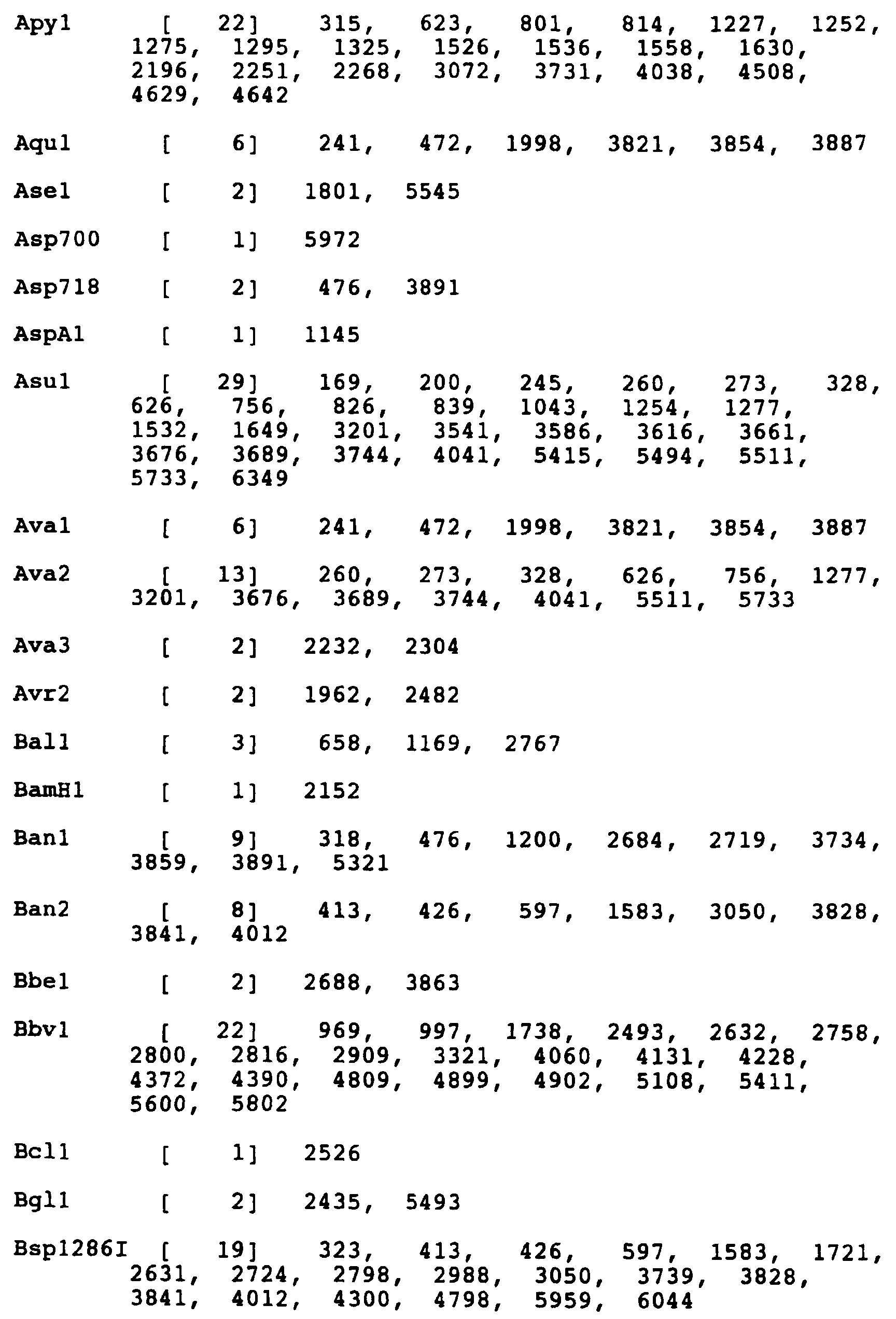


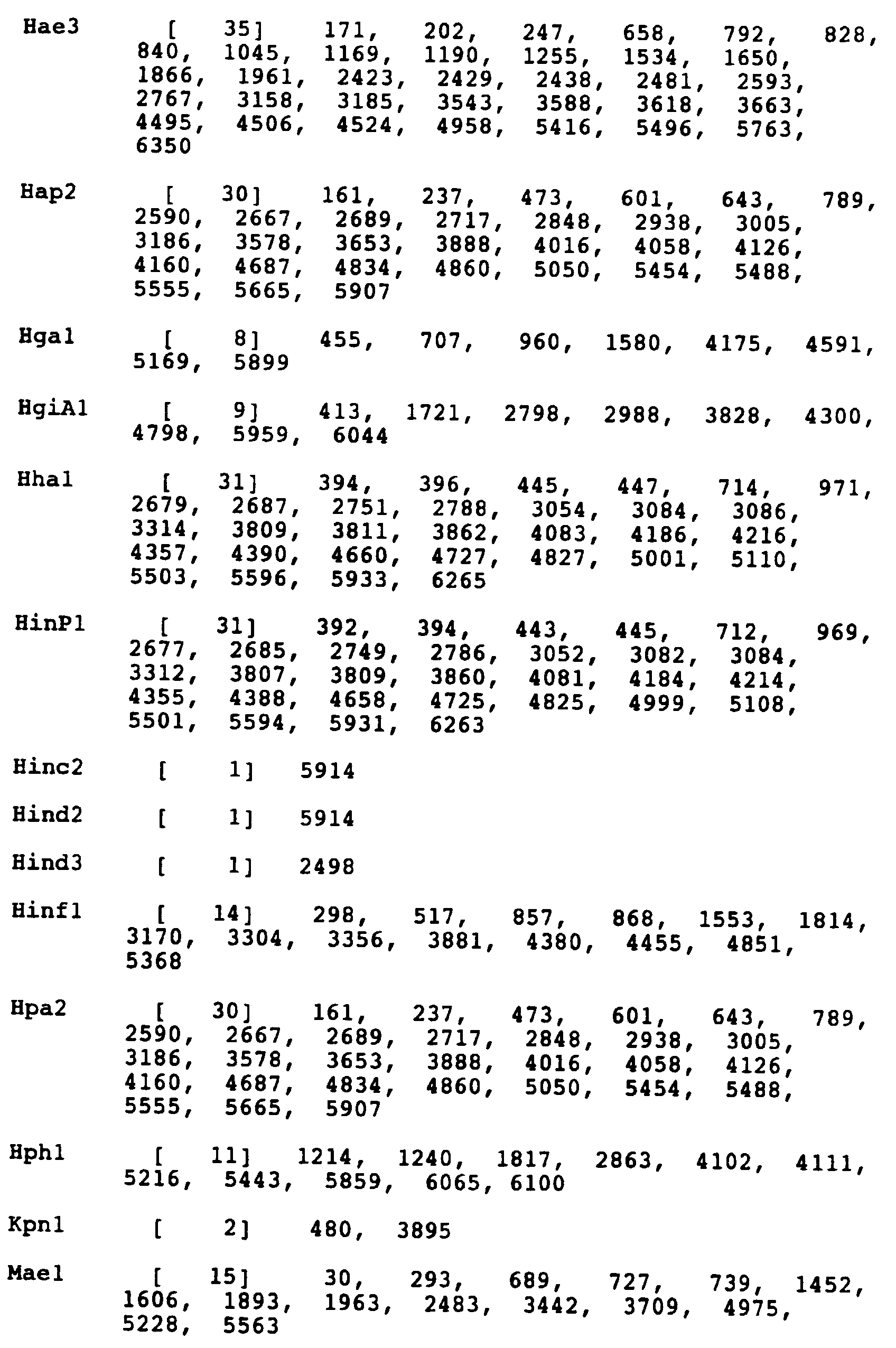

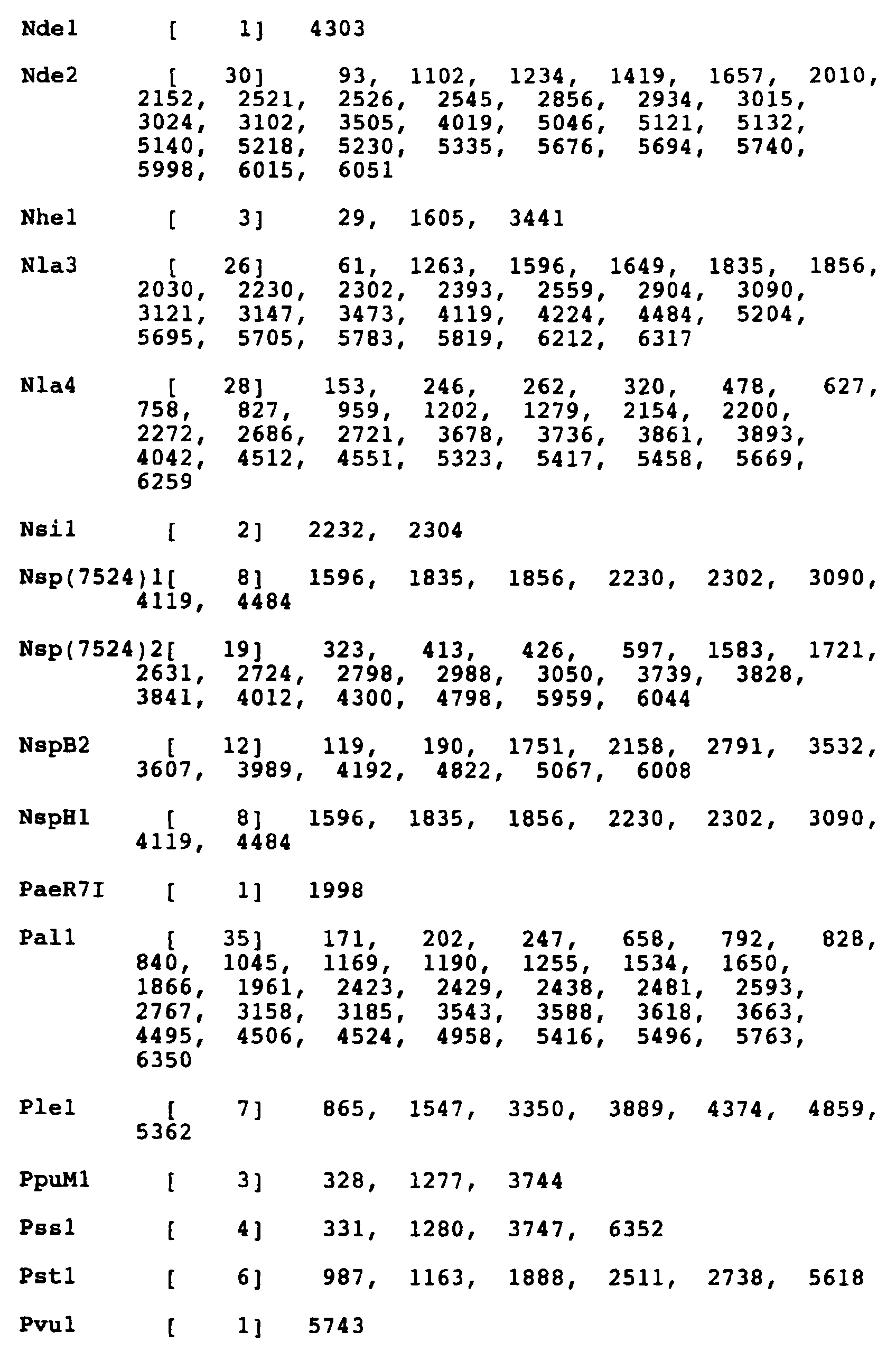
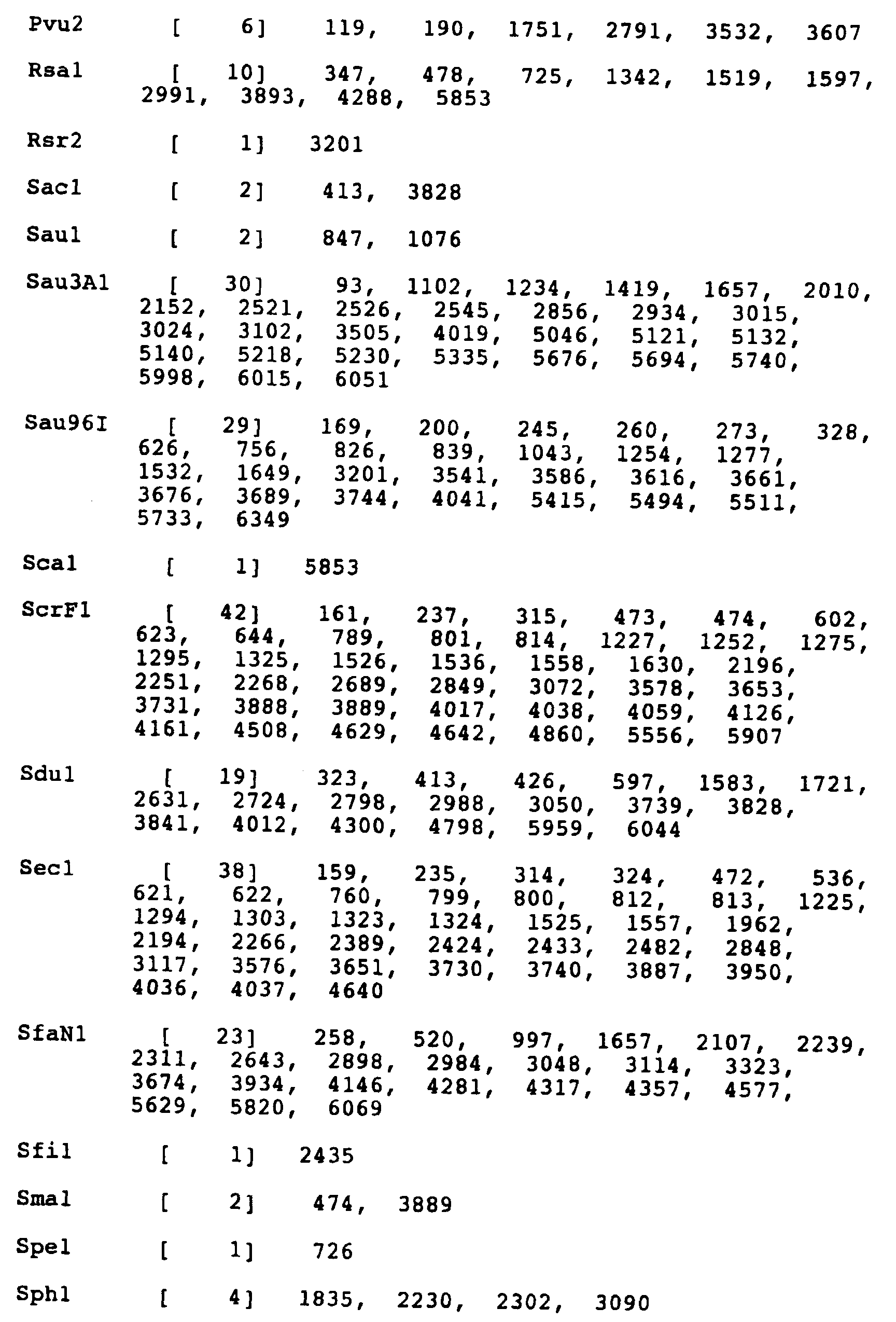

Enzymes which do not cut LXSNRII.L2: Acc3 Bgl2 Cla1 Hpa1 Nru1 SnaB1 Apa1 Bsm1 Dra3 Mlu1 PflM1 Spl1 Asu2 BspM2 Eco47III Mro1 Sac2 Sst2 Ban3 BstB1 Esp1 Not1 Sal1 


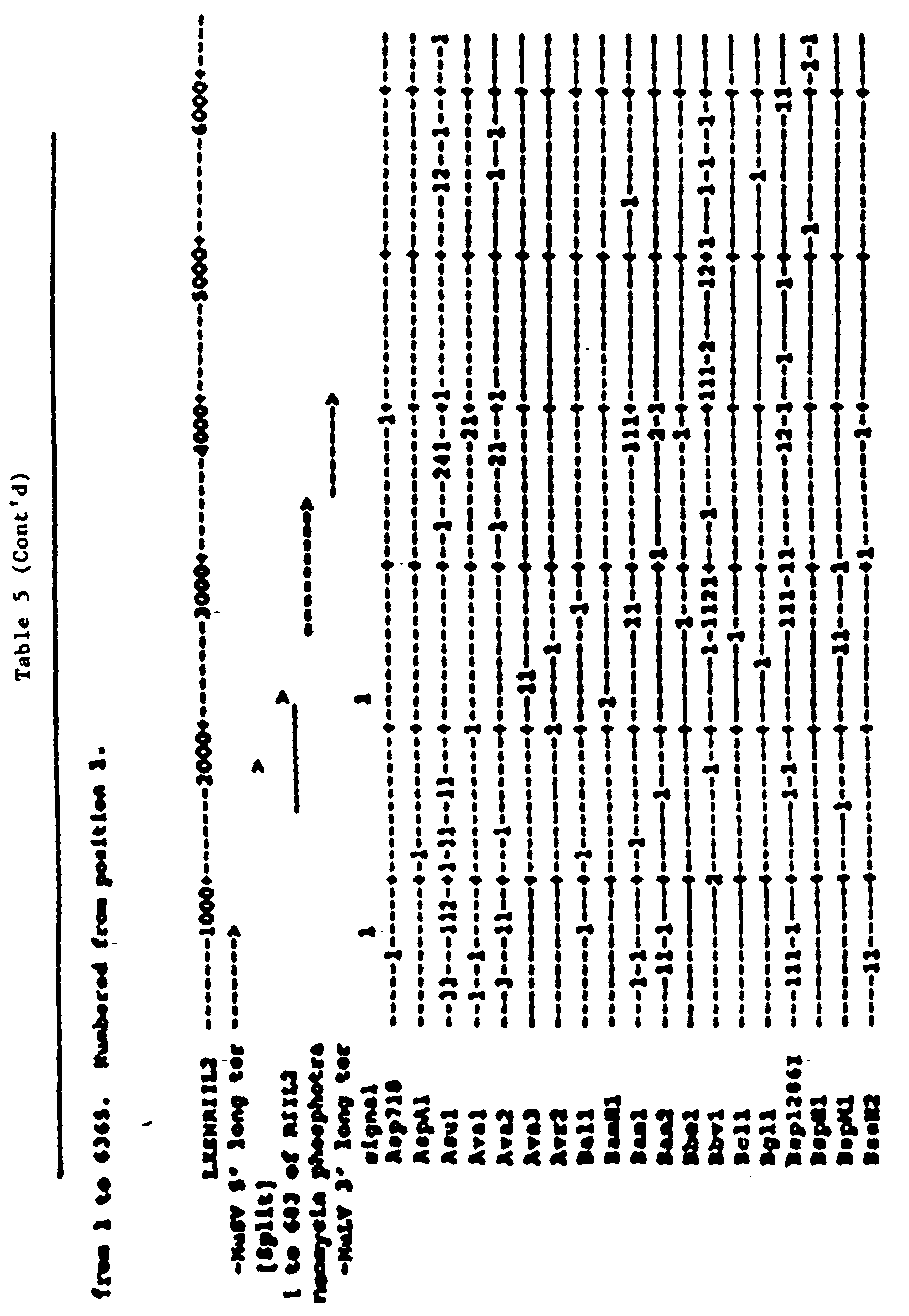

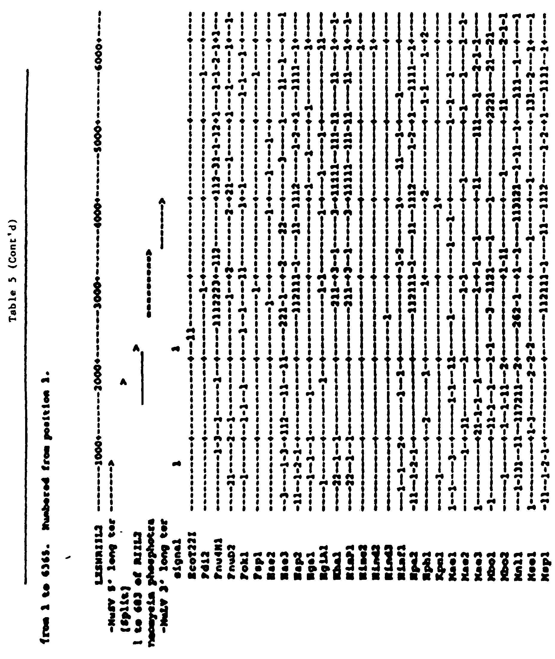
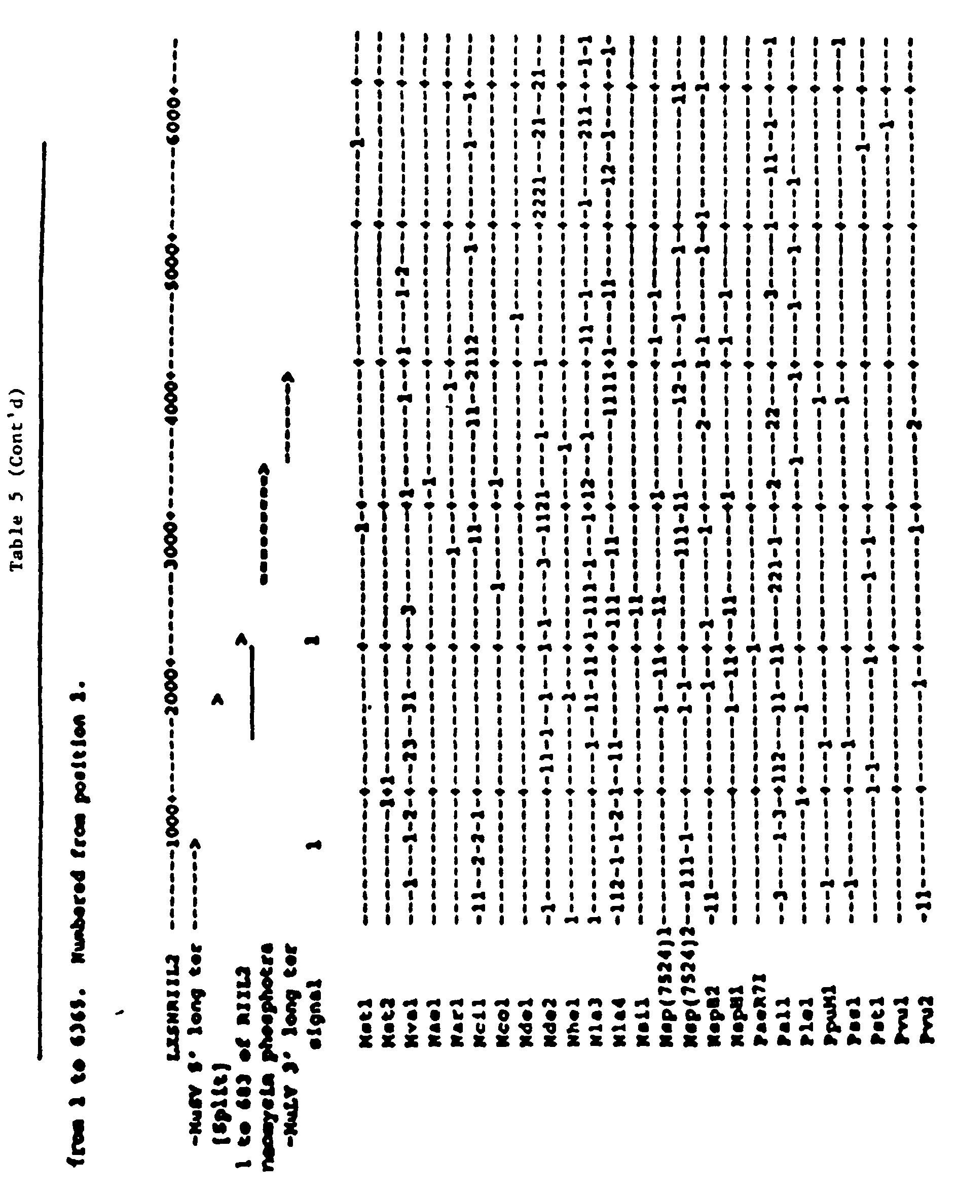
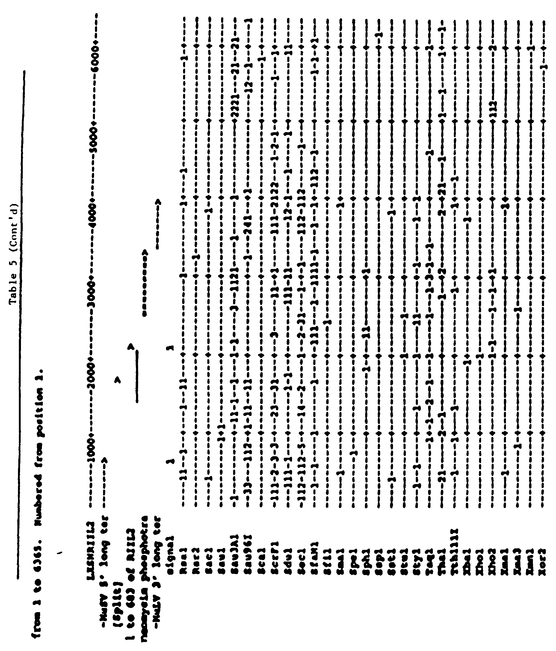


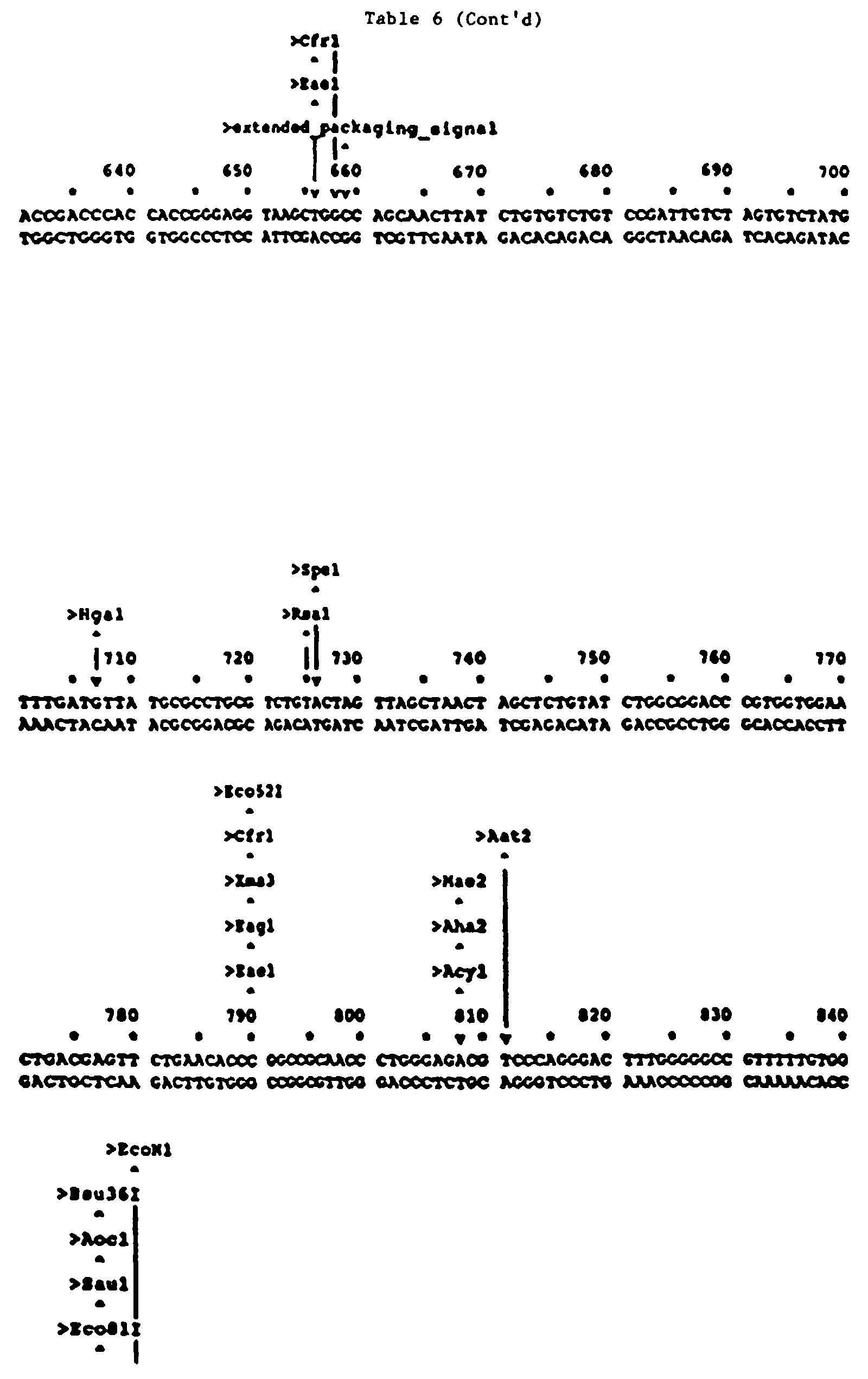
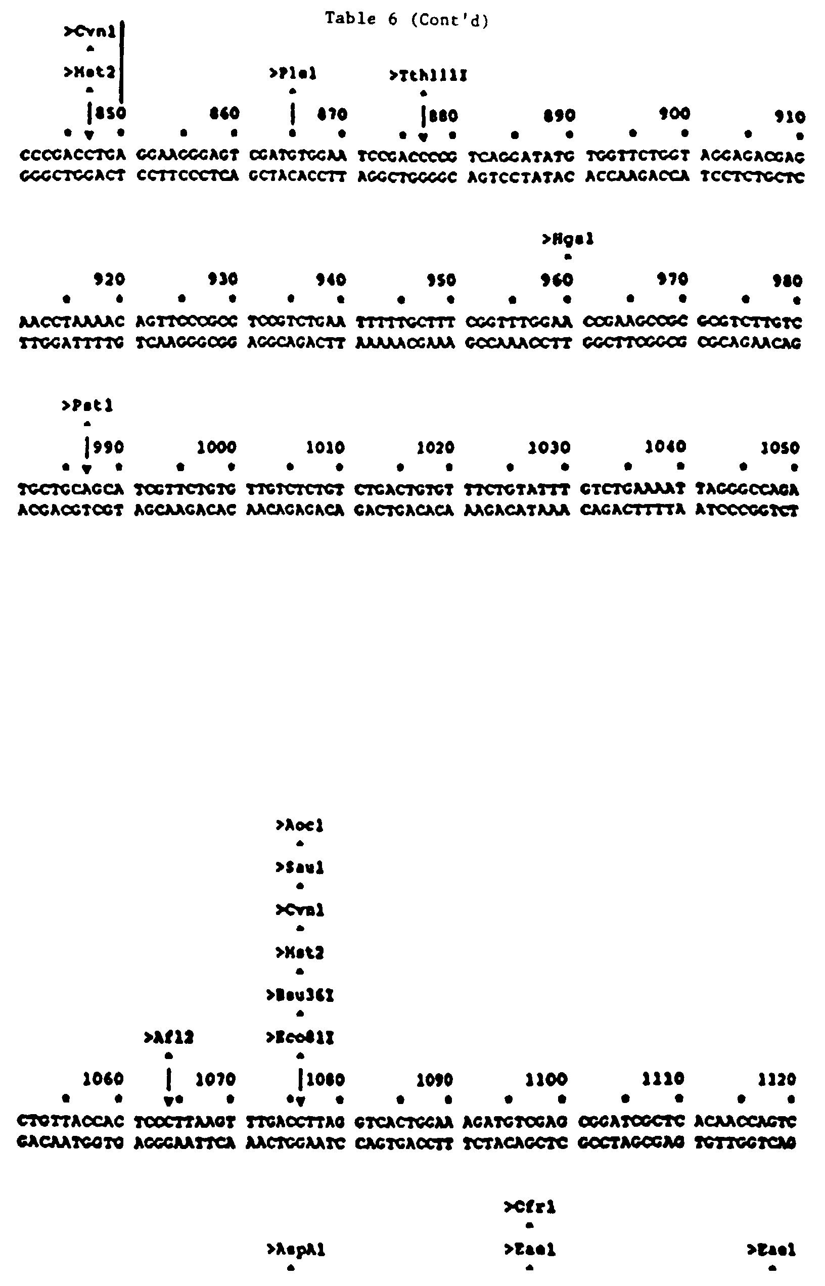
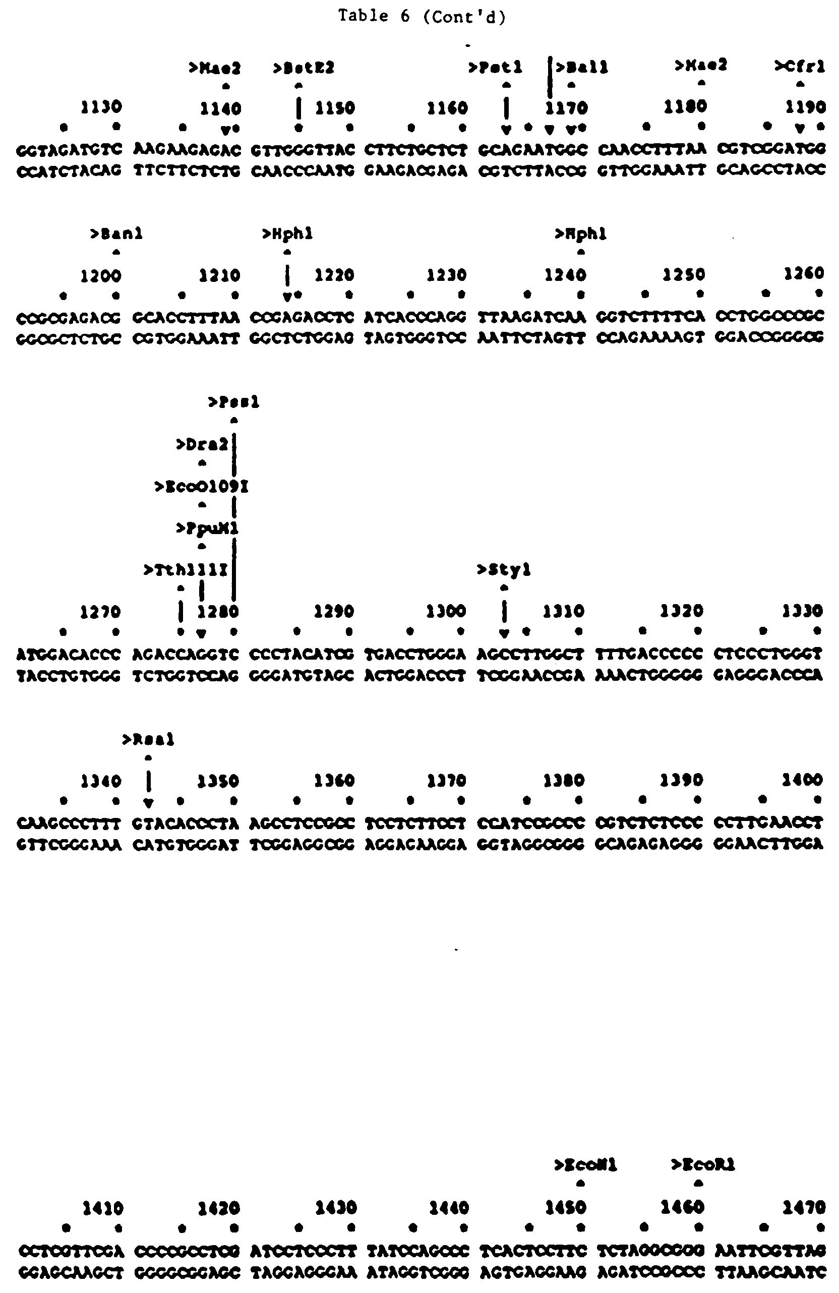

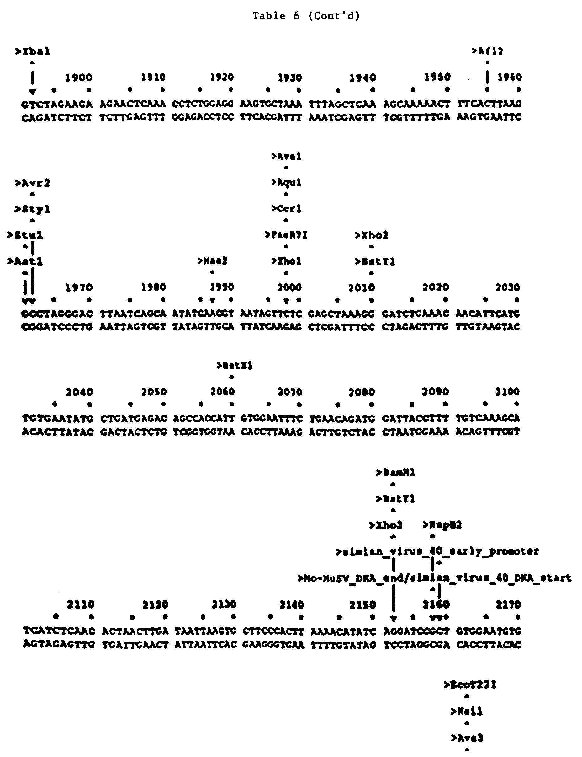

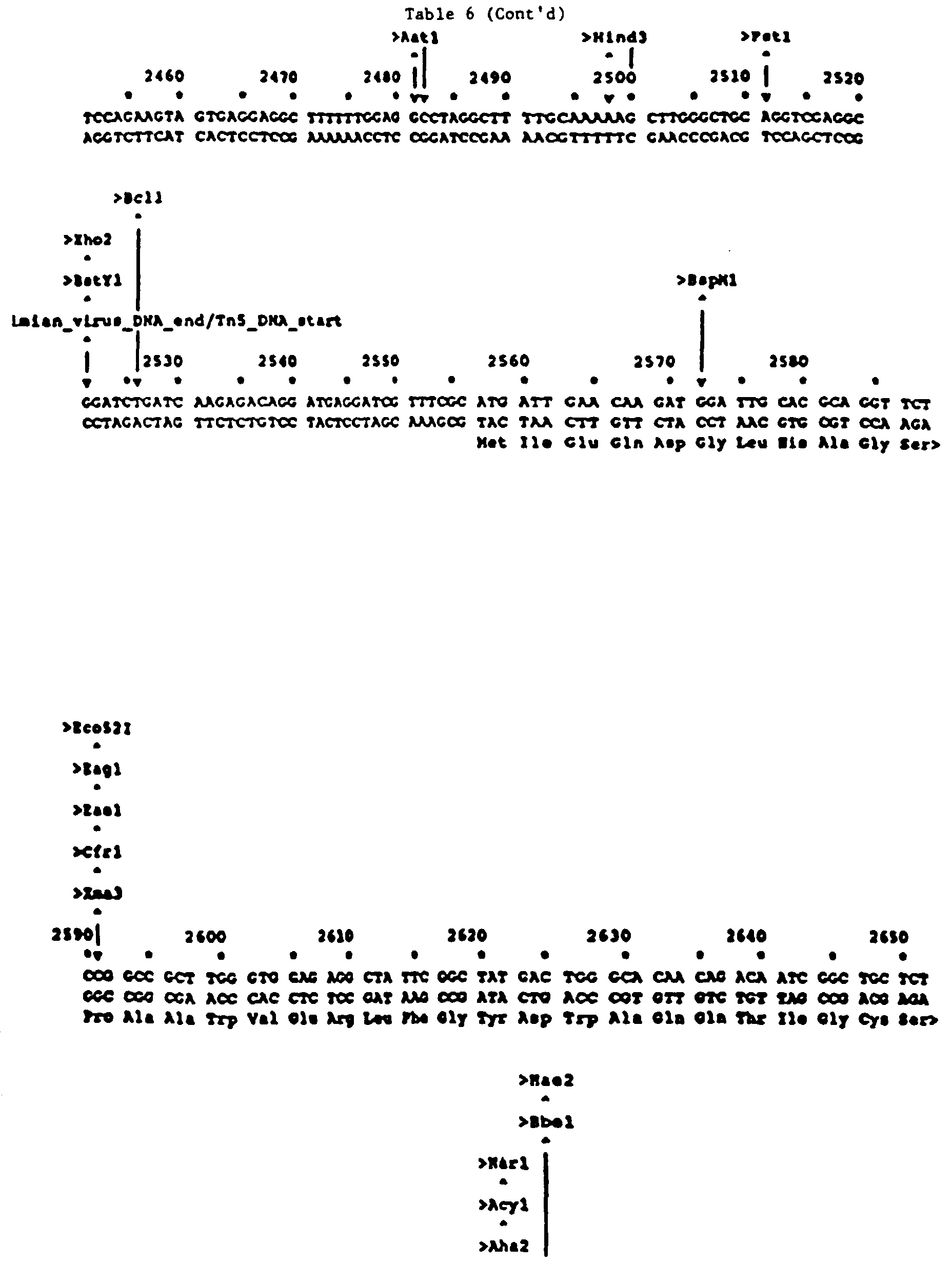


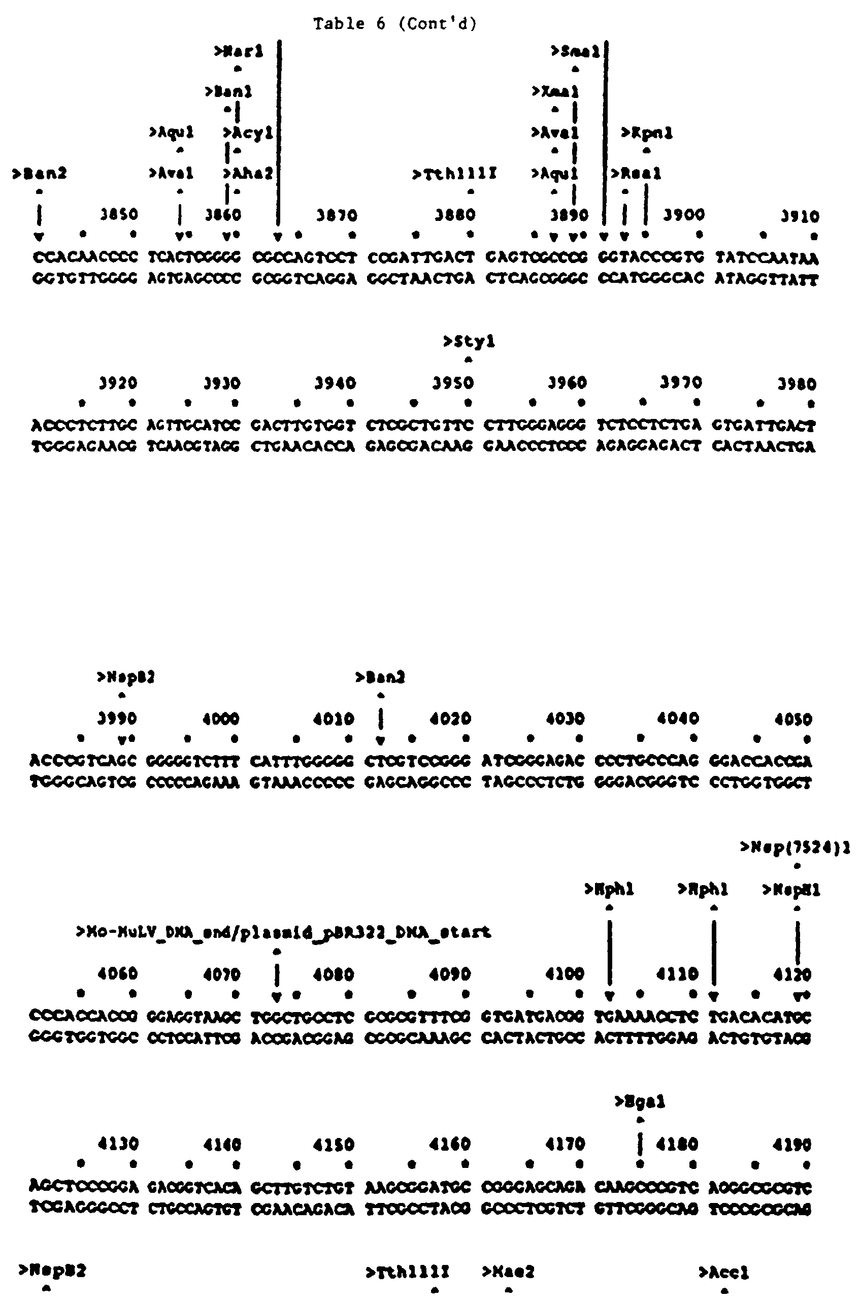


- To generate the LXSN-RI-IL2 retroviral vector, 10 micrograms of pLXSN-RI-IL2 DNA was transfected into the ecotropic packaging cell line PE501 by standard calcium phosphate precipitation methods (Miller et al., Mol. Cell Biol. 6:2895, 1986). The transfected PE501 cell line was grown in DMEM medium with 10% FCS. The medium was changed after 24 hours and supernatant harvested 24 hours later to infect the amphotropic packaging cell line PA317 as described (Miller et al., Mol. Cell Biol. 6:2895, 1986 and Miller et al., BioTechniques 7:980, 1989). The infected PA317 cells were harvested by trypsinization 24 hours later and replated 1:20 in DMEM containing 10% FCS and the neomycin analogue G418 (400 µg/ml). The cells were grown at 37°C in 7% CO2 atmosphere. The selection medium was changed every 5 days until colonies appeared. On
day 14, twenty colonies were selected, expanded and tested for viral production by standard methods (Xu et al., Virology 171:331-341, 1989). Briefly, supernatants were harvested from confluent culture dishes, passed through a .45 µm filter, diluted with DMEM with 10% FCS and utilized to infect NIH 3T3 cells in the presence of 8 µg/ml polybrene. After 24 hours, the infected NIH 3T3 cells were grown in culture medium that contained the neomycin analogue G418. After 12-14 days, the colonies were stained, counted and the viral titer calculated as described (Xu et al., Virology 171:331-341, 1989). - Colonies with the highest viral titers (>104 infectious units/ml) were tested for IL-2 expression by Northern blot analyses. Colonies with the highest viral titers and documented IL-2 expression were cryopreserved and will be utilized as stock cultures to produce the LXSN-RI-IL2 retroviral vector trial.
- To increase IL-2 production by transduced cell lines, vectors were used containing different promoters to drive IL-2 expression, and a human IL-2 cDNA was directionally sub-cloned into the insulin secretory signal peptide (17). The IL-2 cDNA was directionally sub-cloned into the parental plasmids of the LXSN (LTR promoter) and LNCX (CMV promoter) vectors (gifts of Dr. A.D. Miller) (18). The newly constructed vectors (Figure 1), designated as LXSN-IL2 and LNCX-IL2, were packaged in the PA317 cell line for production of retroviral supernatant. As a control, the high level expressing, double copy vector DC/TKIL-2 vector (thymidine kinase promoter) (a gift of Dr. E. Gilboa) was used for comparison.
- These vectors were used to transduce a number of murine and human, primary and established cell lines. Pools of transduced cells were selected and expanded in DMEM medium, containing 10% fetal bovine serum (FBS) and 400 µg/ml of active G-418, a neomycin analogue. The results of expression studies in the MCR9 and Balb/c 3T3 cell lines are presented in Table 7.
Comparison of IL-2 expression by fibroblasts transduced with different IL-2 vectors. Fibroblast Vector ng IL-2 Units IL-2 per 106 cells per day Murine LNCX (Control) 0.4 ±50% <1 LNCX-IL2 33.7 ±11% 67 LXSN-IL2 6.6 ± 6% 13 DC/TKIL-2 1.9 ± 5% 4 Human LXSN (Control) 0.7 ±29% 1 LNCX-IL2 159.5 ±17% 319 LXSN-IL2 25.5 ±15% 51 DC/TKIL-2 3.0 ±10% 6 - The culture conditions for the growth of primary fibroblasts retroviral transduction were optimized. Primary fibroblasts were successfully cultured. The optimal conditions enable the growth of approximately 3-4
x 106 primary fibroblasts from a 12 mm2 skin biopsy in approximately 4-6 weeks. Retroviral infection, G418 selection, and expansion of the genetically modified fibroblasts takes an additional 4-6 weeks. - Exploring the conditions for genetic modification of primary fibroblasts suggests that optimal transduction may be obtained by the following procedure: The fibroblasts are synchronized in G1 phase by serum starvation, followed by stimulation with medium containing 15% fetal
bovine serum 15 hours prior to transduction. The cells are then subjected to 2 cycles of retrovirus infection, each cycle lasting approximately 3 hours. The cells are refed with fresh media overnight, and then selection in G418 is initiated the next day. This method is capable of transducing 5-15% of the fibroblasts in a culture, depending on the multiplicity of infection. - This procedure was used to transduce a large number of primary and established fibroblasts. As an example, Table 8 compares the expression levels of IL-2 in fibroblast lines transduced with LXSN-IL2.
Expression of IL-2 by fibroblasts transduced with LXSN-IL2. Fibroblast Line Species Origin no IL-2 Units IL-2 per 106 cells per day Balb/c 3T3 Murine Transformed 6.6 ± 6% 13 MCR9 Human Embryonic 25.5 ±15% 51 NHDF 313 Human Skin 25.0 ±10% 50 GT1 Human Skin 15.0 ± 5% 30 - These results indicate that the IL-2 expression levels in established, embryonic, and primary fibroblast cultures are similar. Comparison of these data with Table 7 suggest that IL-2 expression is affected more by factors such as different promoters than by the fibroblast line used. Similarly, changes in culture conditions can have important effects on IL-2 expression. Table 9 shows that transduced GT1 cells, a primary human fibroblast culture expressed 15-fold more IL-2 under 100 µg/ml G418 selection than under 25 µg/mi G418 selection. Several other primary fibroblast lines have also been transduced with our vectors and are currently growing under G418 selection.
Effect of G418 concentration on IL-2 expression by GT1 cells transduced with LXSN-IL2. Selection dose of G418 ng IL-2 secreted per 106 cells per day 25 µg/ml 1.0 ± 10% 50 µg/ml 3.0 ± 6% 100 µg/ml 15.0 ± 5% - In order to compare the production of IL-2 by genetically modified fibroblasts to that achieved by stimulating normal human peripheral blood lymphocytes (nPBL) in vitro, nPBL were isolated by Ficol-Paque density centrifugation, and cultured in the presence of allogeneic nPBL (mixed lymphocyte culture, MLC) or 2 µM calcium ionophore (CI) (A23187) free acid) plus 17 nM phorbol 12-myristate 13-acetate (PMA). The results of this experiment, present in Table 10, indicate that the level of IL-2 expression in the PMA/CI stimulated normal T cell population was 2 ng/106 cells/24 hours. This is equivalent to IL-2 expression by Balb/c 3T3 fibroblasts transduced with DC/TKIL-2 (Table 7), our least productive vector. The level of IL-2 expression in the MLC was 130 pg/106 cells/24 hours. This was lower than the PMA/CI stimulated culture, presumably because PMA/CI induced a nonspecific response while MLC resulted in specific Th stimulation. When the estimated percentage of antigen-specific Th in the MLC-stimulated population is taken into consideration, the level of IL-2 expression per stimulated T cell becomes equivalent for both methods.
Levels of IL-2 secretion by different cells. Cells pg IL-2 secreted per 106 cells per day Lymphocytes: Control (non-activated) 5 ± 50% PMA + Calcium Ionophore 2,000 ± 6% Mixed lymphocyte culture 130 ± 90% Transduced fibroblasts: MCR9-LXSN-IL2 24,000 ± 5% MCR9-LNCX-IL2 162,000 ± 20% MCR9-DC/TKIL-2 10,000 ± 6% - Two experimental protocols were used to study the efficacy of fibroblast-mediated cytokine gene therapy on induction of anti-tumor immunity. The first protocol was designed to test the effects of genetically modified fibroblasts on tumor implantation, while the second protocol was designed to induce a systemic anti-tumor immunity. The results of each experiment are presented with two figures and one table. In the first figure, the rate of tumor growth for each treatment group is presented as the mean tumor size in the group over time. In the second figure, a Kaplan-Meier curve presents the time of tumor onset for the individual animals in each treatment group. The number of animals, the number and percentage of tumor free animals, and the tumor size distribution patterns for each experiment are presented in a table.
- Mice were injected subcutaneously with mixtures of 5 x 10⁴ CT26 cells and 2 x 10⁶ fibroblasts genetically modified by different retroviral vectors to express IL-2. In the control arms injected with tumor cells only, or with tumor cells mixed with unmodified fibroblasts, 31 of 33 animals (94%) developed tumors by 4 weeks (Figures 6 and 7, Table 9). In contrast, 22 out of the 34 animals (65%) receiving fibroblast mediated cytokine gene therapy were tumor free at 3 weeks, and 5 animals (18%) remain tumor free after 12 weeks. Those animals that received fibroblast mediated IL-2 therapy and developed tumor were characterized by a delayed onset and rate of tumor growth.
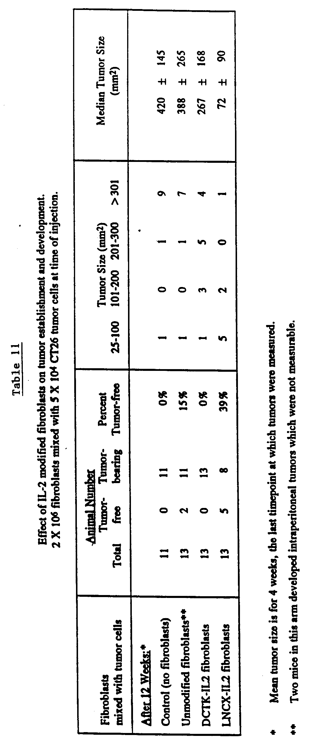
- After 3 weeks the mean tumor size (measured as the product of the longest and widest tumor axes) in the control group of mice was 128 mm2, compared to 68 and 7 mm2 in groups of mice injected with tumor cells mixed with fibroblasts transduced with DC/TKIL-2 or LNCX-IL2, respectively. This resulted in a highly significant difference (corrected x2 = 18.69, p = 0.001) between the IL-2 treated animals compared to the mice treated with CT26 alone or CT26 mixed with unmodified fibroblasts. After four weeks the equivalent measurements were 373,300 and 72 mm2 (Table 11). It is notable that LNCX-IL2, the highest expressing vector caused substantially greater inhibition of tumorigenicity than the lower expressing vector DC/TKIL-2. A multivariate non-parametric statistical procedure (19,20), utilized to evaluate differences in tumor growth, demonstrated that after 4 weeks the differences between the growth curves for the four groups presented in Figure 2 were highly significant (p < 0.001). Subsequent comparisons between the control arm and animals that received tumor cells mixed with IL-2 transduced fibroblasts revealed a significant difference (P < 0.05). The differences between the animals injected with tumor cells alone, and those injected with tumor cells plus unmodified fibroblasts were not significant, while the differences between animals receiving low IL-2 expressing fibroblast, and those receiving high IL-2 expressing fibroblasts was significant (P = 0.05).
- When mice were injected with 2 x 10⁶ modified fibroblasts mixed with 1 x 10⁵ live tumor cells the results became more striking (see Figures 8 and 9, and Table 12). All the control animals developed tumors after 4 weeks whereas 33% and 27% of the animals treated with fibroblasts modified with the DCTK-IL2 or LXSN-IL2 vectors (respectively) remain tumor free after 7 weeks (the experiment is ongoing). More dramatically, 75% of the animals treated with fibroblasts modified with the highest IL-2 producing vector, LNCX-IL2, remain tumor free after 7 weeks. These data clearly demonstrate the importance of an initial high dose of IL-2 to prevent tumor establishment.
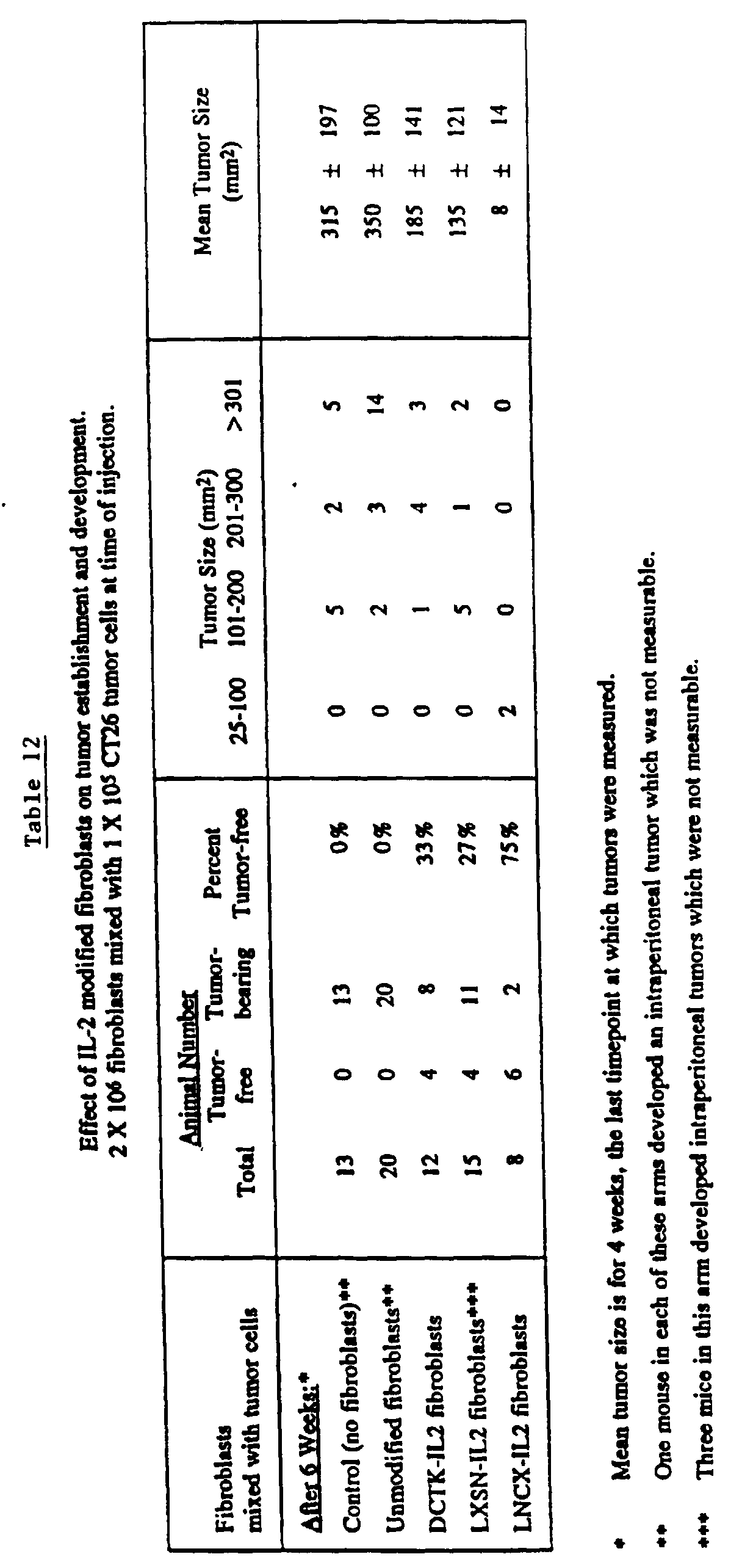
- As an additional control, mice were injected with CT26 cells genetically modified to express IL-2 (results not shown). Injection of up to 1 x 10⁶ IL-2 expressing tumor cells into Balb/c mice failed to produce tumors. Injection of higher numbers however, resulted in some animals developing tumors with delayed onset. These data confirm the results reported in the literature (1). In order to compare the efficacy of IL-2 producing fibroblasts to IL-2 producing tumor cells, we mixed 2 x 10⁶ CT26 tumor cells modified with the DCTK-IL2 vector with 1 x 10⁵ unmodified tumor cells. Figures 10 and 11, and Table 13 show that DCTK-IL2 modified tumor cells are somewhat effective in preventing tumor development. Four weeks after injection, the mean tumor size for the treatment arm is 303 mm2, compared to 620 mm2 for the control arm. After 22 weeks, one animal (10%) remains tumor free, compared to none in the control arms. Data for animals treated under the same conditions with DCTK-IL2 modified fibroblasts in a separate experiment are included for comparison purposes. This comparison suggests that DCTK-IL2 modified tumor cells have an effect on tumor establishment similar to that of DCTK-IL2 modified fibroblasts.

- Groups of Balb/c mice were immunized with 2.5 x 10⁵ irradiated tumor cells either alone or mixed with 2 x 10⁶ transduced or unmodified fibroblasts, and challenged one week later with 5 x 10⁴ live tumor cells in the opposite flank. These results (Figures 12 and 13, and Table 14) demonstrate that immunization with irradiated tumor cells and transduced fibroblasts protect some animals against a live tumor challenge, but that the protection is only slightly better than that achieved by immunization with irradiated tumor cells alone or irradiated tumor cells mixed with unmodified fibroblasts.
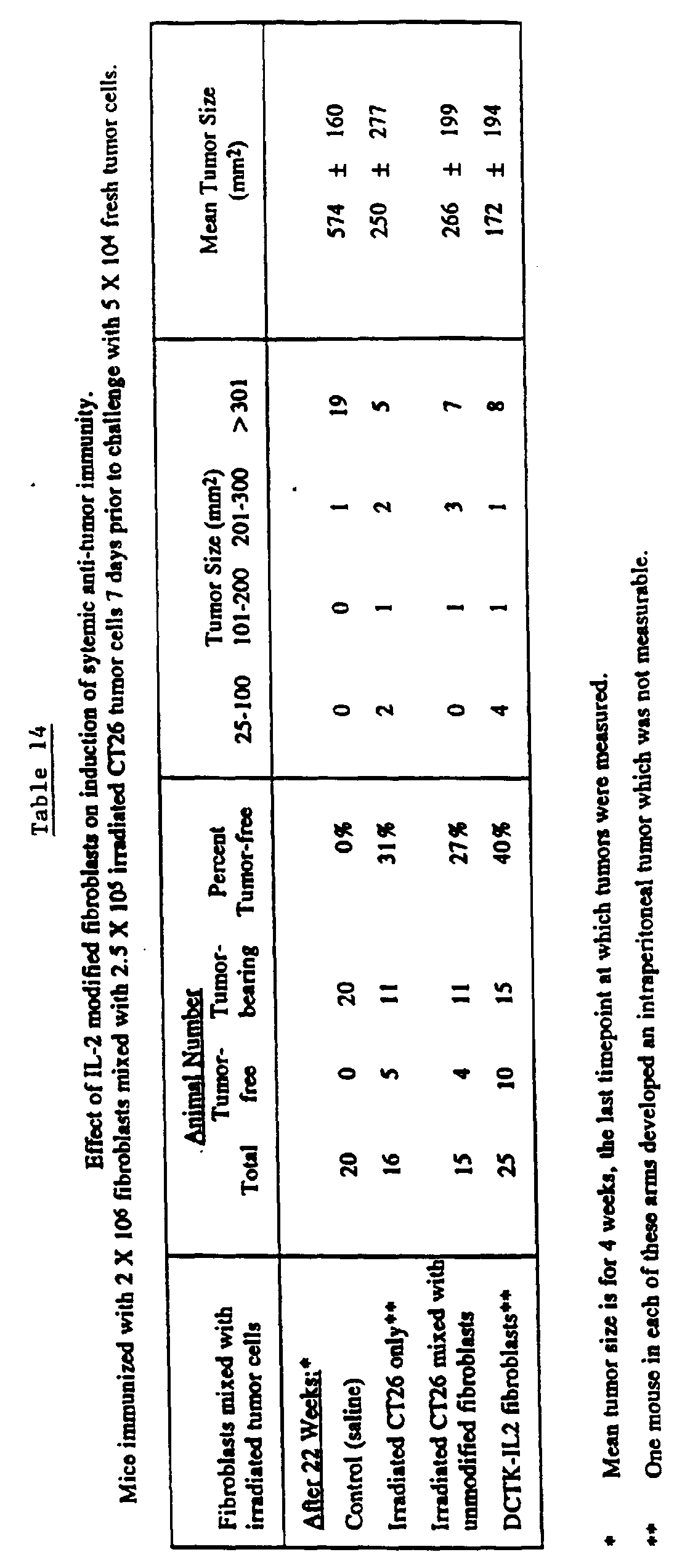
- In a second protocol similar to the one described above, animals were challenged with fresh tumor cells two weeks following immunization with irradiated tumor cells mixed with fibroblasts. The results, shown in Figures 14 and 15, and in Table 15, demonstrate that DCTK-IL2 modified fibroblasts mixed with irradiated tumor cells confers superior protection to subsequent tumor challenge than irradiated tumor cells alone, irradiated tumor cells mixed with unmodified fibroblasts, or irradiated tumor cells mixed with LNCX-modified fibroblasts. After 7 weeks, seven of ten animals (70%) treated with DCTK-IL2 modified fibroblasts remain tumor free compared to only one third of the control animals. At four weeks, the mean tumor size of this group was 41 mm2, compared to 180, 170, and 140 mm2 for the three control groups. Animals treated with LNCX-IL2 modified fibroblasts were also protected against subsequent tumor challenge, but the results were less striking. In this group, 54% of the animals remain tumor free and the mean tumor size for the group at four weeks was 86 mm2. The number of tumor free animals in the group treated with LXSN-IL2 modified fibroblasts was similar to the control groups, although the tumors were slightly delayed in their onset. A multivariate non-parametric statistical procedure (19, 20), utilized to evaluate differences in tumor onset, demonstrated that the differences for the six arms presented in Figure 15 were significant (p = 0.012). It further showed that the saline control arm and the arms that received irradiated tumor cells alone or mixed with unmodified or LNCX vector modified fibroblasts formed a statistical group. A second, distinct statistical group was formed by the three arms that received IL-2 vector modified fibroblasts mixed with irradiated tumor cells. Subsequent comparisons between the saline injected control arm and animals that received tumor cells mixed with IL2 transduced fibroblasts revealed a significant difference for all vectors (p < 0.05).
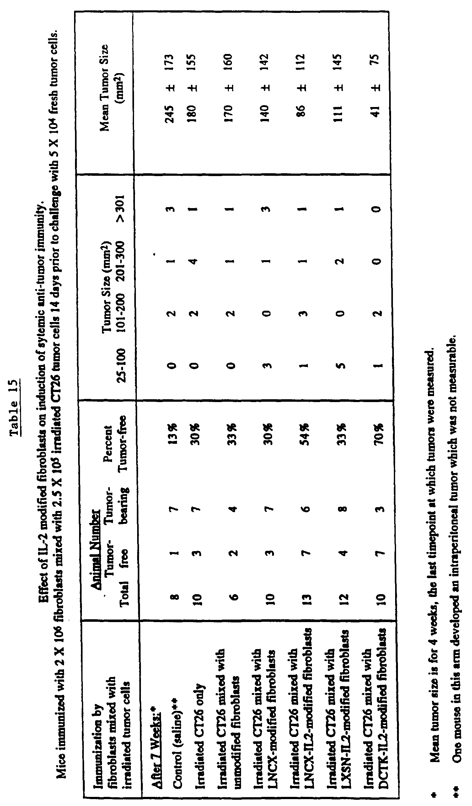
- These results demonstrate the feasibility of using genetically modified fibroblasts as a means of delivering cytokine gene therapy. In all experiments, the LNCX-L2 vector proved superior in preventing tumor establishment while the DCTK-IL2 vector was better in the induction of systemic protection against subsequent tumor challenges. These contrasting effects, although somewhat surprising, can be explained by the observation that the CMV promoter is turned off in vivo five days after implantation while the TK promoter remains active for a longer period of time. The implication of this finding is that to apply this method of gene therapy successfully we have to use promoters that result in high level, sustained expression of IL-2 in vivo in the transduced fibroblasts.
- The data obtained from this research effort has important implications for all cytokines that have either direct or indirect anti-tumor effects. Furthermore, this data suggests that anti-tumor efficacy is IL-2 dose dependent. Hence, construction of vectors which result in higher levels of cytokine secretion will be a significant advance toward the application of this method of gene therapy.
-
- 1. Gabrilove, J.L. et al., Monogr. J. Natl. Cancer Inst. 10:73-7 (1990).
- 2. Kelso, A., Current Opinion in Immunology, 2:215-25 (1989).
- 3. Borden, E.C. et al., Cancer, 65:800-14 (1990).
- 4. Rosenberg, S.A. et al., Ann. Intern. Med., 108:853-864 (1988).
- 5. Lotze, M.T. et al., JAMA, 256:3117-3124 (1986).
- 6. Pizza, G. et al., Lymphokine Research, 7:45-8 (1988).
- 7. Sarna, G. et al., Journal of Biological Response Modifiers, 9:81-6 (1990).
- 8. Gandolfi, L. et al., Hepato-Gastroenterology, 36:352-6 (1989).
- 9. Bubenik, J. et al., Immunol. Letters, 19:279-82 (1988).
- 10. Bubenik et al., Immunol. Letters, 23:287-292 (1990).
- 11. Fearon, E.R. et al., Cell, 60:387-403 (1990).
- 12. Gansbacher, B. et al., J. Exp. Med., 172:1217-1224 (1990).
- 13. Watanabe, Y. et al., Proc. Natl. Acad. Sci., 86:9456-9460 (1989).
- 14. Tepper, R.I. et al., Cell, 57:503-512 (1989).
- 15. Kriegler, M., Gene Transfer and Expression: A Laboratory Manual, Stockton Press (1990).
- 16. Rosenberg, S.A. et al., N. Eng. J. Med., 370 (1990).
- 17. Cornetta, K. et al., Prog. Nucl. Acid Res. Mol. Biol., 36:311-22 (1989).
- 18. Hoover, H.C. et al., Cancer Res., 44:1671-76 (1984).
- 19. Sobol et al. New Eng. J. Med. 316:1111-1117 (1987).
- 20. Li Xu, et al., Virology, 171:331-341 (1989).
-
Claims (16)
- Use of a formulation comprising a tumor antigen and cytokine-expressing cells genetically modified to express an immunostimulatory cytokine gene product, wherein said cytokine-expressing cells are not tumor cells, for the preparation of a medicament for stimulating an anti-cancer immune response in a patient, wherein said medicament is for immunizing said patient at a site other than an active tumor site.
- The use of claim 1, wherein tumor cells previously isolated from said patient provide the tumor antigens.
- The use of claim 1, wherein the cytokine gene is selected from the group consisting of interleukin-1, interleukin-2, interleukin-3, interleukin-4, interleukin-5, interleukin-6 and gamma interferon.
- The use of claim 1, wherein the cytokine gene is interleukin-2.
- The use of claim 1, wherein cytokine-expressing cells are generated by transferring a cytokine gene into cells by recombinant methods.
- The use of claim 5, wherein the cytokine gene is present in an expression vector.
- The use of claim 6, wherein said expression vector additionally contains a suicide gene.
- The use of claim 5, wherein the cytokine-expressing cells are generated from fibroblasts and antigen-presenting cells.
- Use of fibroblasts that express IL-2 at a level sufficient to enhance an immune response but low enough to avoid substantial systemic toxicity and that express a tumor antigen for the preparation of a medicament for stimulating an anti-cancer immune response, wherein:a) fibroblasts being isolated from said patient;b) said fibroblasts being cultured in vitro;c) said fibroblasts being transduced with a retroviral expression vector containing the gene coding for IL-2 and a gene coding for a tumor antigen in a retroviral expression vector, to express said tumor antigen and to express and secrete said IL-2 by said fibroblasts; and wherein said medicament is for immunizing said patient at a site other than an active tumor site.
- The use of claim 9, wherein said fibroblasts are further modified to express a suicide gene.
- The use of claim 9, wherein said retroviral expression vector has a promoter causing sustained secretion of IL-2.
- The use of claim 11, wherein said retroviral expression vector causes the secretion of about two nanograms to about 160 nanograms of IL-2 per 106 cells per day.
- A composition for stimulating an anti-cancer immune response in a patient comprising a tumor antigen and cytokine-expressing cells genetically modified to express an immunostimulatory cytokine gene product, wherein said cytokine-expressing cells are not tumor cells.
- A composition of claim 13, wherein the cytokine gene is selected from the group consisting of interleukin-1, interleukin-2, interleukin-3, interleukin-4, interleukin-5, interleukin-6, and gamma interferon.
- The composition of claim 13, wherein the cytokine gene is interleukin-2.
- The composition of claim 13, wherein the cytokine gene is expressed at a level sufficient to stimulate the immune response but low enough to avoid substantial systemic toxicities.
Applications Claiming Priority (5)
| Application Number | Priority Date | Filing Date | Title |
|---|---|---|---|
| US78135691A | 1991-10-25 | 1991-10-25 | |
| US781356 | 1991-10-25 | ||
| US86364192A | 1992-04-03 | 1992-04-03 | |
| US863641 | 1992-04-03 | ||
| PCT/US1992/008999 WO1993007906A1 (en) | 1991-10-25 | 1992-10-23 | Lymphokine gene therapy of cancer |
Publications (3)
| Publication Number | Publication Date |
|---|---|
| EP0668781A4 EP0668781A4 (en) | 1995-06-14 |
| EP0668781A1 EP0668781A1 (en) | 1995-08-30 |
| EP0668781B1 true EP0668781B1 (en) | 1999-12-29 |
Family
ID=27119842
Family Applications (1)
| Application Number | Title | Priority Date | Filing Date |
|---|---|---|---|
| EP92922731A Revoked EP0668781B1 (en) | 1991-10-25 | 1992-10-23 | Lymphokine gene therapy of cancer in combination with tumor antigens |
Country Status (9)
| Country | Link |
|---|---|
| EP (1) | EP0668781B1 (en) |
| JP (1) | JP3524918B2 (en) |
| AT (1) | ATE188125T1 (en) |
| CA (1) | CA2121127A1 (en) |
| DE (1) | DE69230506T2 (en) |
| DK (1) | DK0668781T3 (en) |
| ES (1) | ES2144426T3 (en) |
| GR (1) | GR3032660T3 (en) |
| WO (1) | WO1993007906A1 (en) |
Families Citing this family (28)
| Publication number | Priority date | Publication date | Assignee | Title |
|---|---|---|---|---|
| US6241982B1 (en) | 1988-03-21 | 2001-06-05 | Chiron Corporation | Method for treating brain cancer with a conditionally lethal gene |
| US5662896A (en) | 1988-03-21 | 1997-09-02 | Chiron Viagene, Inc. | Compositions and methods for cancer immunotherapy |
| US5716826A (en) * | 1988-03-21 | 1998-02-10 | Chiron Viagene, Inc. | Recombinant retroviruses |
| US6569679B1 (en) | 1988-03-21 | 2003-05-27 | Chiron Corporation | Producer cell that generates adenoviral vectors encoding a cytokine and a conditionally lethal gene |
| US6133029A (en) * | 1988-03-21 | 2000-10-17 | Chiron Corporation | Replication defective viral vectors for infecting human cells |
| US5631236A (en) | 1993-08-26 | 1997-05-20 | Baylor College Of Medicine | Gene therapy for solid tumors, using a DNA sequence encoding HSV-Tk or VZV-Tk |
| FR2710536B1 (en) * | 1993-09-29 | 1995-12-22 | Transgene Sa | Anti-cancer use of a viral vector comprising a gene which modulates the immune and / or inflammatory response. |
| AU1257495A (en) | 1993-11-18 | 1995-06-06 | Chiron Corporation | Compositions and methods for utilizing conditionally lethal genes |
| FR2713657B1 (en) * | 1993-12-13 | 1996-03-01 | Transgene Sa | New vectors for the treatment of AIDS. |
| CA2178950A1 (en) * | 1993-12-14 | 1995-06-22 | Yajun Guo | Tumor cell fusions and methods for use of such tumor cell fusions |
| DE4406073A1 (en) * | 1994-02-24 | 1995-08-31 | Univ Ludwigs Albert | Process for the production of human, clonogenic fibroblasts, process for gene transfection of fibroblasts and fibroblasts obtained in this way |
| US5759535A (en) * | 1994-05-13 | 1998-06-02 | Research Corporation Technologies, Inc. | Immunotherapeutic strategies for the treatment of cancer |
| US5772995A (en) * | 1994-07-18 | 1998-06-30 | Sidney Kimmel Cancer Center | Compositions and methods for enhanced tumor cell immunity in vivo |
| JP3822261B2 (en) * | 1994-09-09 | 2006-09-13 | 財団法人癌研究会 | Cancer gene therapy |
| WO1996013275A1 (en) * | 1994-10-31 | 1996-05-09 | Tkb Associates Limited Partnership | Methods and products for enhancing immune responses |
| DE69627644T2 (en) | 1995-03-09 | 2004-02-19 | GSF-Forschungszentrum für Umwelt und Gesundheit GmbH | VECTORS CONTAINING THERAPEUTIC GENES FOR ANTIMICROBIAL PEPTIDES FOR USE IN GENE THERAPY |
| FR2746109B1 (en) | 1996-03-12 | 1998-04-17 | Rhone Poulenc Rorer Sa | ENVIRONMENT FOR THE CONSERVATION OF BIOLOGICAL MATERIAL |
| US6277368B1 (en) | 1996-07-25 | 2001-08-21 | The Regents Of The University Of California | Cancer immunotherapy using autologous tumor cells combined with cells expressing a membrane cytokine |
| EP0834323A1 (en) * | 1996-09-30 | 1998-04-08 | Introgene B.V. | Cytokine gene therapy for treatment of malignacies |
| WO1998033527A2 (en) | 1997-01-31 | 1998-08-06 | Cohen Edward P | Cancer immunotherapy with semi-allogeneic cells |
| CA2318372C (en) | 1998-02-02 | 2008-08-19 | Johns Hopkins University School Of Medicine | A universal immunomodulatory cytokine-expressing bystander cell line and related compositions and methods of manufacture and use |
| US6342216B1 (en) * | 1999-03-17 | 2002-01-29 | The Board Of Regents, The University Of Texas System | Therapy of cancer by insect cells containing recombinant baculovirus encoding genes |
| AU2001253031A1 (en) * | 2000-03-31 | 2001-10-15 | Novarx | Compositions containing genetically modified lung cancer cells expressing a tgf-beta inhibitor |
| US7101543B2 (en) | 2000-03-31 | 2006-09-05 | Novarx | Genetically modified cells expressing a TGFβ inhibitor, the cells being lung cancer cells |
| US6872385B2 (en) | 2000-05-31 | 2005-03-29 | Board Of Regents, The University Of Texas System | Adjuvant preparation for the induction of specific immunity |
| JPWO2005067981A1 (en) * | 2004-01-13 | 2007-12-27 | 株式会社ディナベック研究所 | Tumor gene therapy using minus-strand RNA viral vectors encoding immunostimulatory cytokines |
| CA2603191A1 (en) | 2005-03-29 | 2006-10-05 | The Board Of Trustees Of The University Of Illinois | Cancer vaccines and therapeutic methods |
| EP4051305A4 (en) * | 2019-11-02 | 2023-11-01 | Figene, LLC | INTRATUMORAL ADMINISTRATION OF IMMUNE CELL THERAPEUTICS |
Family Cites Families (1)
| Publication number | Priority date | Publication date | Assignee | Title |
|---|---|---|---|---|
| DK0551401T3 (en) * | 1990-09-14 | 1996-01-22 | Univ Texas | Methods and Preparations for Gene Therapy and Potential of Anti-Tumor Immunity |
-
1992
- 1992-10-23 CA CA002121127A patent/CA2121127A1/en not_active Abandoned
- 1992-10-23 DK DK92922731T patent/DK0668781T3/en active
- 1992-10-23 DE DE69230506T patent/DE69230506T2/en not_active Revoked
- 1992-10-23 ES ES92922731T patent/ES2144426T3/en not_active Expired - Lifetime
- 1992-10-23 EP EP92922731A patent/EP0668781B1/en not_active Revoked
- 1992-10-23 JP JP50790393A patent/JP3524918B2/en not_active Expired - Lifetime
- 1992-10-23 AT AT92922731T patent/ATE188125T1/en not_active IP Right Cessation
- 1992-10-23 WO PCT/US1992/008999 patent/WO1993007906A1/en not_active Ceased
-
2000
- 2000-02-14 GR GR20000400358T patent/GR3032660T3/en not_active IP Right Cessation
Also Published As
| Publication number | Publication date |
|---|---|
| ATE188125T1 (en) | 2000-01-15 |
| DE69230506T2 (en) | 2000-06-21 |
| DE69230506D1 (en) | 2000-02-03 |
| CA2121127A1 (en) | 1993-04-29 |
| WO1993007906A1 (en) | 1993-04-29 |
| JP3524918B2 (en) | 2004-05-10 |
| JPH07503455A (en) | 1995-04-13 |
| GR3032660T3 (en) | 2000-06-30 |
| EP0668781A4 (en) | 1995-06-14 |
| ES2144426T3 (en) | 2000-06-16 |
| DK0668781T3 (en) | 2000-04-17 |
| EP0668781A1 (en) | 1995-08-30 |
Similar Documents
| Publication | Publication Date | Title |
|---|---|---|
| US5674486A (en) | Cancer immunotherapy with carrier cells | |
| JP3524918B2 (en) | Lymphokine gene therapy for cancer | |
| US5681562A (en) | Lymphokine gene therapy of cancer | |
| EP0915708B1 (en) | Immunogenic composition comprising tumor associated antigen and human cytokine | |
| McBride et al. | Genetic modification of a murine fibrosarcoma to produce interleukin 7 stimulates host cell infiltration and tumor immunity | |
| ES2204922T3 (en) | ANTI-BANK GENE THERAPY BY MODULATION OF THE IMMUNE AND / OR INFLAMMATORY RESPONSE. | |
| Prince | Gene transfer: a review of methods and applications | |
| US7101543B2 (en) | Genetically modified cells expressing a TGFβ inhibitor, the cells being lung cancer cells | |
| Cao et al. | Cytokine gene transfer in cancer therapy | |
| JP3541950B2 (en) | Use of modified TALL-104 cells to treat cancer and viral diseases | |
| Gansbacher et al. | Retroviral vector—mediated cytokine-gene transfer into tumor cells | |
| US6063375A (en) | Semiallogeneic cell hybrids and related methods for treating cancer | |
| Santin et al. | Development and characterization of an IL-4-secreting human ovarian carcinoma cell line | |
| EP0879294B1 (en) | Transduced human hematopoietic stem cells | |
| JP2002506980A (en) | Novel complementary receptor-ligand pairs and adoptive immunotherapy using the same | |
| JPH09504786A (en) | Haplotype-matched cytokine secreting cells and methods of use to stimulate immune responses | |
| EP1267945B1 (en) | Genetically modified cells expressing a tgf-beta inhibitor, the cells being lung cancer cells | |
| Heslop | 7 Cytokine gene transfer in the therapy of malignancy | |
| WO2005037218A2 (en) | Lentiviral vector delivery of il-21 for treatment of cancer | |
| Shimada | Current status and future prospects of human gene therapy | |
| Lattime et al. | In Situ Gene Insertion for Immunotherapy Using Vaccinia Virus Vectors | |
| Santin et al. | Development and in vitro characterization of a GM‐CSF secreting human ovarian carcinoma tumor vaccine | |
| Freeman et al. | The Role of the Blood Bank in Human Gene Therapy Trials | |
| Yokoyamaa et al. | Initiates Renal Injury | |
| JP2004262797A (en) | Antitumor agent utilizing interloinkin-23 gene |
Legal Events
| Date | Code | Title | Description |
|---|---|---|---|
| A4 | Supplementary search report drawn up and despatched | ||
| AK | Designated contracting states |
Kind code of ref document: A4 Designated state(s): AT BE CH DE DK ES FR GB GR IE IT LI LU MC NL SE |
|
| PUAI | Public reference made under article 153(3) epc to a published international application that has entered the european phase |
Free format text: ORIGINAL CODE: 0009012 |
|
| 17P | Request for examination filed |
Effective date: 19940425 |
|
| AK | Designated contracting states |
Kind code of ref document: A1 Designated state(s): AT BE CH DE DK ES FR GB GR IE IT LI LU MC NL SE |
|
| 17Q | First examination report despatched |
Effective date: 19970902 |
|
| RAP1 | Party data changed (applicant data changed or rights of an application transferred) |
Owner name: SIDNEY KIMMEL CANCER CENTER |
|
| GRAG | Despatch of communication of intention to grant |
Free format text: ORIGINAL CODE: EPIDOS AGRA |
|
| GRAG | Despatch of communication of intention to grant |
Free format text: ORIGINAL CODE: EPIDOS AGRA |
|
| GRAH | Despatch of communication of intention to grant a patent |
Free format text: ORIGINAL CODE: EPIDOS IGRA |
|
| GRAH | Despatch of communication of intention to grant a patent |
Free format text: ORIGINAL CODE: EPIDOS IGRA |
|
| GRAA | (expected) grant |
Free format text: ORIGINAL CODE: 0009210 |
|
| AK | Designated contracting states |
Kind code of ref document: B1 Designated state(s): AT BE CH DE DK ES FR GB GR IE IT LI LU MC NL SE |
|
| REF | Corresponds to: |
Ref document number: 188125 Country of ref document: AT Date of ref document: 20000115 Kind code of ref document: T |
|
| REG | Reference to a national code |
Ref country code: CH Ref legal event code: EP |
|
| REF | Corresponds to: |
Ref document number: 69230506 Country of ref document: DE Date of ref document: 20000203 |
|
| ET | Fr: translation filed | ||
| REG | Reference to a national code |
Ref country code: CH Ref legal event code: NV Representative=s name: E. BLUM & CO. PATENTANWAELTE |
|
| REG | Reference to a national code |
Ref country code: IE Ref legal event code: FG4D |
|
| ITF | It: translation for a ep patent filed | ||
| REG | Reference to a national code |
Ref country code: DK Ref legal event code: T3 |
|
| REG | Reference to a national code |
Ref country code: ES Ref legal event code: FG2A Ref document number: 2144426 Country of ref document: ES Kind code of ref document: T3 |
|
| PLBQ | Unpublished change to opponent data |
Free format text: ORIGINAL CODE: EPIDOS OPPO |
|
| PLBI | Opposition filed |
Free format text: ORIGINAL CODE: 0009260 |
|
| PLBF | Reply of patent proprietor to notice(s) of opposition |
Free format text: ORIGINAL CODE: EPIDOS OBSO |
|
| 26 | Opposition filed |
Opponent name: AVENTIS PHARMA DEUTSCHLAND GMBH Effective date: 20000929 |
|
| NLR1 | Nl: opposition has been filed with the epo |
Opponent name: AVENTIS PHARMA DEUTSCHLAND GMBH |
|
| PLBF | Reply of patent proprietor to notice(s) of opposition |
Free format text: ORIGINAL CODE: EPIDOS OBSO |
|
| PLBF | Reply of patent proprietor to notice(s) of opposition |
Free format text: ORIGINAL CODE: EPIDOS OBSO |
|
| PLBF | Reply of patent proprietor to notice(s) of opposition |
Free format text: ORIGINAL CODE: EPIDOS OBSO |
|
| PGFP | Annual fee paid to national office [announced via postgrant information from national office to epo] |
Ref country code: DK Payment date: 20011025 Year of fee payment: 10 |
|
| PGFP | Annual fee paid to national office [announced via postgrant information from national office to epo] |
Ref country code: SE Payment date: 20011026 Year of fee payment: 10 Ref country code: AT Payment date: 20011026 Year of fee payment: 10 |
|
| PGFP | Annual fee paid to national office [announced via postgrant information from national office to epo] |
Ref country code: MC Payment date: 20011031 Year of fee payment: 10 Ref country code: GR Payment date: 20011031 Year of fee payment: 10 |
|
| PGFP | Annual fee paid to national office [announced via postgrant information from national office to epo] |
Ref country code: IE Payment date: 20011107 Year of fee payment: 10 |
|
| PGFP | Annual fee paid to national office [announced via postgrant information from national office to epo] |
Ref country code: LU Payment date: 20011108 Year of fee payment: 10 |
|
| PGFP | Annual fee paid to national office [announced via postgrant information from national office to epo] |
Ref country code: CH Payment date: 20011114 Year of fee payment: 10 |
|
| PGFP | Annual fee paid to national office [announced via postgrant information from national office to epo] |
Ref country code: BE Payment date: 20011214 Year of fee payment: 10 |
|
| REG | Reference to a national code |
Ref country code: GB Ref legal event code: IF02 |
|
| PG25 | Lapsed in a contracting state [announced via postgrant information from national office to epo] |
Ref country code: LU Free format text: LAPSE BECAUSE OF NON-PAYMENT OF DUE FEES Effective date: 20021023 Ref country code: IE Free format text: LAPSE BECAUSE OF NON-PAYMENT OF DUE FEES Effective date: 20021023 Ref country code: AT Free format text: LAPSE BECAUSE OF NON-PAYMENT OF DUE FEES Effective date: 20021023 |
|
| PG25 | Lapsed in a contracting state [announced via postgrant information from national office to epo] |
Ref country code: SE Free format text: LAPSE BECAUSE OF NON-PAYMENT OF DUE FEES Effective date: 20021024 |
|
| PG25 | Lapsed in a contracting state [announced via postgrant information from national office to epo] |
Ref country code: LI Free format text: LAPSE BECAUSE OF NON-PAYMENT OF DUE FEES Effective date: 20021031 Ref country code: DK Free format text: LAPSE BECAUSE OF NON-PAYMENT OF DUE FEES Effective date: 20021031 Ref country code: CH Free format text: LAPSE BECAUSE OF NON-PAYMENT OF DUE FEES Effective date: 20021031 Ref country code: BE Free format text: LAPSE BECAUSE OF NON-PAYMENT OF DUE FEES Effective date: 20021031 |
|
| PLBQ | Unpublished change to opponent data |
Free format text: ORIGINAL CODE: EPIDOS OPPO |
|
| PLAB | Opposition data, opponent's data or that of the opponent's representative modified |
Free format text: ORIGINAL CODE: 0009299OPPO |
|
| R26 | Opposition filed (corrected) |
Opponent name: AVENTIS PHARMA DEUTSCHLAND GMBH Effective date: 20000929 |
|
| BERE | Be: lapsed |
Owner name: *SIDNEY KIMMEL CANCER CENTER Effective date: 20021031 |
|
| PG25 | Lapsed in a contracting state [announced via postgrant information from national office to epo] |
Ref country code: MC Free format text: LAPSE BECAUSE OF NON-PAYMENT OF DUE FEES Effective date: 20030501 |
|
| PG25 | Lapsed in a contracting state [announced via postgrant information from national office to epo] |
Ref country code: GR Free format text: LAPSE BECAUSE OF NON-PAYMENT OF DUE FEES Effective date: 20030506 |
|
| EUG | Se: european patent has lapsed | ||
| REG | Reference to a national code |
Ref country code: DK Ref legal event code: EBP |
|
| REG | Reference to a national code |
Ref country code: CH Ref legal event code: PL |
|
| REG | Reference to a national code |
Ref country code: IE Ref legal event code: MM4A |
|
| RDAF | Communication despatched that patent is revoked |
Free format text: ORIGINAL CODE: EPIDOSNREV1 |
|
| APBP | Date of receipt of notice of appeal recorded |
Free format text: ORIGINAL CODE: EPIDOSNNOA2O |
|
| APBQ | Date of receipt of statement of grounds of appeal recorded |
Free format text: ORIGINAL CODE: EPIDOSNNOA3O |
|
| APAH | Appeal reference modified |
Free format text: ORIGINAL CODE: EPIDOSCREFNO |
|
| PGFP | Annual fee paid to national office [announced via postgrant information from national office to epo] |
Ref country code: GB Payment date: 20051019 Year of fee payment: 14 |
|
| PGFP | Annual fee paid to national office [announced via postgrant information from national office to epo] |
Ref country code: DE Payment date: 20051020 Year of fee payment: 14 |
|
| PGFP | Annual fee paid to national office [announced via postgrant information from national office to epo] |
Ref country code: NL Payment date: 20051023 Year of fee payment: 14 |
|
| PGFP | Annual fee paid to national office [announced via postgrant information from national office to epo] |
Ref country code: FR Payment date: 20051025 Year of fee payment: 14 |
|
| PGFP | Annual fee paid to national office [announced via postgrant information from national office to epo] |
Ref country code: ES Payment date: 20051121 Year of fee payment: 14 |
|
| APBU | Appeal procedure closed |
Free format text: ORIGINAL CODE: EPIDOSNNOA9O |
|
| RDAG | Patent revoked |
Free format text: ORIGINAL CODE: 0009271 |
|
| STAA | Information on the status of an ep patent application or granted ep patent |
Free format text: STATUS: PATENT REVOKED |
|
| 27W | Patent revoked |
Effective date: 20060309 |
|
| GBPR | Gb: patent revoked under art. 102 of the ep convention designating the uk as contracting state |
Free format text: 20060309 |
|
| NLR2 | Nl: decision of opposition |
Effective date: 20060309 |
|
| PGFP | Annual fee paid to national office [announced via postgrant information from national office to epo] |
Ref country code: IT Payment date: 20061031 Year of fee payment: 15 |
|
| PLAB | Opposition data, opponent's data or that of the opponent's representative modified |
Free format text: ORIGINAL CODE: 0009299OPPO |